Note: Descriptions are shown in the official language in which they were submitted.
81801172
METHODS AND COMPOSITIONS RELATING TO EXOSOMES
RELATED APPLICATION
This application claims the benefit under 35 U.S.C. 119(e) of U.S.
provisional
application filed May 18, 2014, entitled "METHODS AND COMPOSITIONS RELATING
TO EXOSOMES", Serial No.61/994,974.
BACKGROUND OF INVENTION
Exosomes are cell-derived vesicles that are present in many and perhaps all
biological
fluids, including blood, urine, and conditioned media from cell cultures. The
reported diameter
of exosomes is typically between 30 and 100 nm, which, for comparison, is
larger than LDL
but significantly smaller than red blood cells. Exosomes are known to be
released from cells
when multivesicular bodies fuse with the plasma membrane or when they are
released directly
from the plasma membrane. It is becoming increasingly clear that exosomes have
specialized
functions and play a key role in, for example, coagulation, intercellular
signaling, and waste
management. Consequently, there is a growing interest in the clinical
applications of
exosomes, including synthetic exosomes which recapitulate aspects of cell-
derived exosomes.
Exosomes can potentially be used for prognosis, therapy, and biomarkers for
health and
disease.
SUMMARY OF INVENTION
The disclosure provides compositions comprising exosomes and methods of use
thereof
in the treatment and/or prevention of various diseases or disorders.
Accordingly, one aspect of the disclosure provides an isolated exosome. In
some
embodiments, the isolated exosome comprises one or more markers selected from
the group
consisting of ALIX, TSG101, TGFBR2, SMAD1, SMAD2, SMAD3, SMAD5 and CD105,
and/or the isolated exosome does not comprise one or more markers selected
from the group
consisting of FLOT1, CD9, CD81, CAV1, EGFR, AKT1 and AKT2. In some
embodiments,
the isolated exosome comprises 2, 3, 4, 5, 6, 7 or 8 markers selected from the
group consisting
of ALIX, TSG101, TGFBR2, SMAD1, SMAD2, SMAD3, SMAD5 and CD105. In some
embodiments, the isolated exosome comprises the markers ALIX, TSG101, TGFBR2,
1
Date Recue/Date Received 2021-06-18
CA 02949083 2016-11-14
WO 2015/179227 PCT/US2015/031008
SMAD1, SMAD2, SMAD3. SMAD5 and CD105. In some embodiments, the isolated
exosome does not comprise 2, 3, 4, 5, 6 or 7 markers selected from the group
consisting of
FLOT1, CD9, CD81, CAV1, EGFR, AKT1 and AKT2. In some embodiments, the isolated
exosome does not comprise the markers FLOT1, CD9, CD81, CAV1, EGFR, AKT1 and
AKT2. In some embodiments, the isolated exosome has spherical morphology and
appears
radiolucent upon negative staining in transmission electron microscopy, and/or
the isolated
exosome does not have a cup shape morphology in negative staining transmission
electron
microscopy. The isolated exosome may have a diameter of about 10-150 nm. In
some
embodiments, the isolated exosome has a diameter of about 30-100 nm. In some
embodiments, the isolated exosome is isolated from a mesenchymal stem cell
(MSC),
fibroblast, or macrophage. In some embodiments, the MSC, fibroblast, or
macrophage is a
human MSC, human fibroblast, or human macrophage. In some embodiments, the MSC
is
isolated from Wharton's jelly, umbilical cord blood, placenta, peripheral
blood, bone marrow,
or adipose tissue. In some embodiments, the isolated exosome is comprised in a
composition.
In some embodiments, the composition is a pharmaceutical composition.
According to another aspect of the disclosure, an isolated exosome is
provided. In
some embodiments, the isolated exosome comprises one or more markers selected
from the
group consisting of FLOT1, CD9, CD81, CAV1. EGFR, AKT1 and AKT2, and/or the
isolated
exosome does not comprise one or markers selected from the group consisting of
ALIX,
TSG101, TGFBR2, SMAD1, SMAD2, SMAD3, SMAD5 and CD105. In some embodiments,
the isolated exosome comprises 2, 3, 4, 5, 6 or 7 markers selected from the
group consisting of
FLOT1, CD9, CD81, CAV1, EGFR, AKT1 and AKT2. In some embodiments, the isolated
exosome comprises the markers FLOT1, CD9, CD81, CAV1, EGFR, and AKT1 and AKT2.
In some embodiments, the isolated exosome does not comprise 2, 3, 4, 5, 6. 7
or 8 markers
selected from the group consisting of ALIX, TSG101, TGFBR2, SMAD1, SMAD2,
SMAD3,
SMAD5 and CD105. In some embodiments, the isolated exosome does not comprise
the
markers ALIX, TSG101, TGFBR2, SMADl , SMAD2. SMAD3, SMAD5, and CD105. In
some embodiments, the isolated exosome has cup shaped morphology in negative
staining
transmission electron microscopy and/or does not have a spherical morphology
in negative
staining transmission electron microscopy. In some embodiments, the isolated
exosome has a
diameter of about 10-250 nm. In some embodiments, the isolated exosome has a
diameter of
about 30-200 nm.
2
CA 02949083 2016-11-14
WO 2015/179227 PCT/US2015/031008
According to another aspect, a method for treating a lung disorder, a
cardiovascular
disorder, a renal disorder, or an ischemic neural disorder is provided. In
some embodiments,
the method comprises administering to a subject having or at risk of having a
lung disorder, a
cardiovascular disorder, a renal disorder, or an ischemic neural disorder a
therapeutically
effective amount of an isolated exosome. In some embodiments, the isolated
exosome
comprises one or more markers selected from the group consisting of ALIX,
TSG101,
TGFBR2, SMAD1, SMAD2. SMAD3, SMAD5 and CD105, and/or the isolated exosome does
not comprise one or more markers selected from the group consisting of FLOT1,
CD9, CD81,
CAV1, EGFR. AKT1 and AKT2. In some embodiments, the isolated exosome comprises
2, 3,
4, 5, 6, 7 or 8 markers selected from the group consisting of ALIX. TSG101,
TGFBR2,
SMAD1, SMAD2, SMAD3, SMAD5 and CD105. In some embodiments, the isolated
exosome comprises the markers ALIX, TSG101, TGFBR2, SMAD1, SMAD2, SMAD3,
SMAD5 and CD105. In some embodiments, the isolated exosome does not comprise
2, 3, 4,
5, 6 or 7 markers selected from the group consisting of FLOT1, CD9, CD81,
CAV1, EGFR,
AKT1 and AKT2. In some embodiments, the isolated exosome does not comprise the
markers
FLOT1, CD9, CD81, CAV1, EGFR, AKT1 and AKT2. In some embodiments, the isolated
exosome has spherical morphology and appears radiolucent upon negative
staining in
transmission electron microscopy and/or the isolated exosome does not have a
cup shape
morphology in negative staining transmission electron microscopy. In some
embodiments, the
isolated exosome has a diameter of about 10-150 nm. In some embodiments, the
isolated
exosome has a diameter of about 30-100 nm. In some embodiments, the isolated
exosome is
isolated from a mesenchymal stem cell (MSC), fibroblast, or macrophage. In
some
embodiments, the MSC, fibroblast, or macrophage is a human MSC, human
fibroblast, or
human macrophage. In some embodiments, the MSC is isolated from Wharton's
jelly,
umbilical cord blood, placenta, peripheral blood, bone marrow, or adipose
tissue. In some
embodiments, the lung disorder being treated is inflammatory lung disease,
lung vascular
disease, or acute lung injury. In some embodiments, the inflammatory lung
disease is hypoxia-
induced lung inflammation. pulmonary hypertension, asthma, bronchopulmonary
dysplasia
(BPD), allergy, or idiopathic pulmonary fibrosis. In some embodiments, the
acute lung injury
is associated with sepsis or is ventilator-induced acute respiratory distress
syndrome (ARDS).
In some embodiments, a cardiovascular disorder being treated according to the
method is
myocardial infarction, cardiovascular disease, hypertension, atherosclerosis,
or heart failure.
In some embodiments involving the treatment of renal disorders, the renal
disorder is ischemic
3
CA 02949083 2016-11-14
WO 2015/179227 PCT/US2015/031008
renal injury, acute renal failure, or renal fibrosis. In embodiments of the
method involving the
treatment of ischemic neural disorders, the disorder is hypoxic ischemic
encephalopathy or
ischemic stroke.
According to yet another aspect of the disclosure, use of an isolated exosome
for
treating a lung disorder, a cardiovascular disorder, a renal disorder, or an
ischemic neural
disorder is provided. In some embodiments, the isolated exosome comprises one
or more
markers selected from the group consisting of ALIX, TSG101. TGFBR2, SMAD1,
SMAD2,
SMAD3, SMAD5 and CD105, and/or the isolated exosome does not comprise one or
more
markers selected from the group consisting of FLOT1, CD9, CD81, CAV1. EGFR,
AKT1 and
AKT2. In some embodiments, the isolated exosome comprises 2, 3, 4, 5, 6, 7 or
8 markers
selected from the group consisting of ALIX, TSG101, TGFBR2, SMAD1, SMAD2,
SMAD3,
SMAD5 and CD105. In some embodiments, the isolated exosome the markers ALIX,
TSGl01, TGFBR2, SMADI. SMAD2, SMAD3, SMAD5 and CD105. In some embodiments,
the isolated exosome does not comprise 2, 3, 4, 5, 6 or 7 markers selected
from the group
consisting of FLOT1, CD9, CD81, CAV1, EGFR, AKT1 and AKT2. In some
embodiments,
the isolated exosome does not comprise the markers FLOT1, CD9, CD81, CAV1,
EGFR,
AKT1 and AKT2. In some embodiments, the isolated exosome has spherical
morphology and
appears radiolucent upon negative staining in transmission electron microscopy
and/or the
isolated exosome does not have a cup shape morphology in negative staining
transmission
electron microscopy. In some embodiments, the isolated exosome has a diameter
of about 10-
150 nm. In some embodiments, the isolated exosome has a diameter of about 30-
100 nm. In
some embodiments, the isolated exosome is isolated from a mesenchymal stem
cell (MSC),
fibroblast, or macrophage. In some embodiments, the MSC, fibroblast, or
macrophage is a
human MSC, human fibroblast, or human macrophage. In some embodiments, the MSC
is
isolated from Wharton's jelly, umbilical cord blood, placenta, peripheral
blood, bone marrow,
or adipose tissue. In some embodiments, the lung disorder is inflammatory lung
disease, lung
vascular disease, or acute lung injury. In some embodiments, the inflammatory
lung disease is
hypoxia-induced lung inflammation, pulmonary hypertension, asthma,
bronchopulmonary
dysplasia (BPD), allergy, or idiopathic pulmonary fibrosis. In some
embodiments, the acute
lung injury is associated with sepsis or is ventilator-induced acute
respiratory distress
syndrome (ARDS). In some embodiments, the cardiovascular disorder is
myocardial
infarction, cardiovascular disease, hypertension, atherosclerosis, or heart
failure. In some
embodiments, the renal disorder is ischemic renal injury, acute renal failure,
or renal fibrosis.
4
CA 02949083 2016-11-14
WO 2015/179227 PCT/US2015/031008
In some embodiments, the ischemic neural disorder is hypoxic ischemic
encephalopathy or
ischemic stroke.
According to another aspect, use of an isolated exosome in the manufacture of
a
medicament for treating a lung disorder, a cardiovascular disorder, a renal
disorder, or an
ischemic neural disorder is provided. In some embodiments, the isolated
exosome comprises
one or more markers selected from the group consisting of ALIX, TSG101,
TGFBR2,
SMAD1, SMAD2, SMAD3. SMAD5 and CD105, and/or the isolated exosome does not
comprise one or more markers selected from the group consisting of FLOT1, CD9,
CD81,
CAV1, EGFR. AKT1 and AKT2. In some embodiments, the isolated exosome comprises
2, 3,
4, 5, 6, 7 or 8 markers selected from the group consisting of ALIX. TSG101,
TGFBR2,
SMAD1, SMAD2, SMAD3, SMAD5 and CD105. In some embodiments, the isolated
exosome comprises the markers ALIX, TSG101, TGFBR2, SMAD1, SMAD2, SMAD3,
SMAD5 and CD 05. In some embodiments, the isolated exosome does not comprise
2, 3, 4,
5, 6 or 7 markers selected from the group consisting of FLOT1, CD9, CD81 ,
CAV1, EGFR,
.. AKT1 and AKT2. In some embodiments, the isolated exosome does not comprise
the markers
FLOT1, CD9, CD81, CAV1, EGFR, AKT1 and AKT2. In some embodiments, the isolated
exosome has spherical morphology and appears radiolucent upon negative
staining in
transmission electron microscopy; and/or the isolated exosome does not have a
cup shape
morphology in negative staining transmission electron microscopy. In some
embodiments, the
isolated exosome has a diameter of about 10-150 nm. In some embodiments, the
isolated
exosome has a diameter of about 30-100 nm. In some embodiments, the isolated
exosome is
isolated from a mesenchymal stem cell (MSC), fibroblast, or macrophage. In
some
embodiments, the MSC, fibroblast, or macrophage is a human MSC, human
fibroblast, or
human macrophage. In some embodiments, the MSC is isolated from Wharton's
jelly,
.. umbilical cord blood, placenta, peripheral blood, bone marrow, or adipose
tissue. In some
embodiments involving the use of exosomes for the manufacture of a medicament
for the
treatment of lung disorders, the disorder is inflammatory lung disease, lung
vascular disease, or
acute lung injury. In some embodiments, the inflammatory lung disease is
hypoxia-induced
lung inflammation, pulmonary hypertension, asthma, bronchopulmonary dysplasia
(BPD),
allergy, or idiopathic pulmonary fibrosis. In some embodiments, the acute lung
injury is
associated with sepsis or is ventilator-induced acute respiratory distress
syndrome (ARDS). In
some embodiments. use of the exosome for the manufacture of medicament for the
treatment
of a cardiovascular disorder is provided, the disorder being myocardial
infarction,
5
CA 02949083 2016-11-14
WO 2015/179227 PCT/US2015/031008
cardiovascular disease, hypertension, atherosclerosis, or heart failure. In
some embodiments
involving the use of exosomes for the manufacture of a medicament for the
treatment of renal
disorders, the renal disorder is ischemic renal injury, acute renal failure,
or renal fibrosis. In
some embodiments involving the use of exosomes for the manufacture of a
medicament for the
treatment of ischemic neural disorders, the disorder is hypoxic ischemic
encephalopathy or
ischemic stroke.
According to yet another aspect of the disclosure, a method for producing an
exosome(s) is provided. In some embodiments, the method comprises culturing a
cell so as to
produce conditioned media and isolating the exosome from the conditioned
media. In some
.. embodiments, the isolated exosome comprises one or more markers selected
from the group
consisting of ALIX, TSG101, TGFBR2, SMAD1, SMAD2, SMAD3, SMAD5 and CD105,
and/or the isolated exosome does not comprise one or more markers selected
from the group
consisting of FLOT1, CD9, CD81, CAVl , EGFR, AKT1 and AKT2. In some
embodiments,
the isolated exosome comprises 2, 3, 4, 5, 6, 7 or 8 markers selected from the
group consisting
of ALIX, TSG101, TGFBR2, SMAD1, SMAD2, SMAD3, SMAD5 and CD105. In some
embodiments, the isolated exosome the markers ALIX, TSG101, TGFBR2, SMAD1,
SMAD2,
SMAD3, SMAD5 and CD105. In some embodiments, the isolated exosome does not
comprise
2, 3, 4, 5, 6 or 7 markers selected from the group consisting of FLOT1, CD9,
CD81, CAV1,
EGFR, AKT1 and AKT2. In some embodiments, the isolated exosome does not
comprise the
.. markers FLOT1, CD9, CD81, CAV1, EGFR, AKT1 and AKT2. In some embodiments,
the
isolated exosome has spherical morphology and appears radiolucent upon
negative staining in
transmission electron microscopy and/or the isolated exosome does not have a
cup shape
morphology in negative staining transmission electron microscopy. In some
embodiments, the
isolated exosome has a diameter of about 10-150 nm. In some embodiments, the
isolated
exosome has a diameter of about 30-100 nm. In some embodiments, the isolated
exosome is
isolated from a mesenchymal stem cell (MSC), fibroblast, or macrophage. In
some
embodiments, the MSC, fibroblast, or macrophage is a human MSC, human
fibroblast, or
human macrophage. In some embodiments, the MSC is isolated from Wharton's
jelly,
umbilical cord blood, placenta, peripheral blood, bone marrow, or adipose
tissue. In some
embodiments, the culturing involves two-dimensional (2D) or three-dimensional
(3D)
culturing. In some embodiments. the 3D culturing comprises hanging drop
culturing, culturing
on matrices, culturing on microcarriers, culturing on synthetic extracellular
scaffolds, culturing
on chitosan membranes, culturing under magnetic levitation, suspension culture
in rotating
6
CA 02949083 2016-11-14
WO 2015/179227 PCT/US2015/031008
bioreactors, or culturing under non-contact inhibition conditions. In some
embodiments, the
culturing comprises use of one or more growth factors selected from TGFI3
superfamily
(TGFI31, Activins, BMPs. GDFs, GDNFs, Inhibins. Nodal, Lefty, MIS) EGF, PDGF,
and FGF.
In some embodiments, the method enhances the production of exosomes that
comprise one or
.. more markers selected from ALIX, TSG101, TGFBR2, SMAD1, SMAD2, SMAD3, SMAD5
and CD105 relative to exosomes that comprise one or more markers selected from
the group
consisting of FLOT1, CD9, CD81, CAV1, EGFR, AKT1 and AKT2. In some
embodiments,
the enhancement comprises a 1.5-fold, 2.0-fold, 2.5-fold, 3.0-fold, 3.5-fold,
4.0-fold, 4.5-fold,
or 5.0-fold, 6.0-fold, 7.0-fold, 8.0-fold, 9.0-fold, or 10.0-fold or more
increase in the
production of exosomes that comprise one or more markers selected from ALIX,
TSG101,
TGFBR2, SMAD1, SMAD2, SMAD3, SMAD5 and CD105 relative to exosomes that
comprise one or more markers selected from the group consisting of FLOT1, CD9,
CD81,
CAV1, EGFR. AKTI and AKT2.
These and other aspects and embodiments of the disclosure will be described in
greater
detail herein.
BRIEF DESCRIPTION OF DRAWINGS
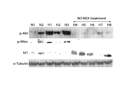
FIG. 1. Treatment of mice by I.V. injection of mesenchymal stem cell exosome
(MEX)
preparations down-regulates the hypoxic activation of signaling associated
with vascular
remodeling & pulmonary hypertension and ameliorates the hypoxia-induced lung
inflammation. (A) Age-matched FVB/n mice were injected with human MEX (20
billion
particles/mouse) and exposed to hypoxia for 2.5 days. Hypoxia-induced
phosphorylation of
AKT and its downstream target mTOR are reduced by MEX treatment. Lung protein
levels of
the Inhibitor of DNA-binding/differentiation protein ID1, a direct SMAD-
targeted gene and
downstream signal of BMPR2 are suppressed by hypoxia but increased with MEX.
Alpha
.. tubulin serves as normalizing control. (B) mRNA levels of CCL2, an early
inflammatory
marker, are suppressed with MEX treatment. RNA levels are normalized to RPS9.
FIG. 2. Isolation of exosome subpopulations enriched for a-MEX and f-MEX.
Media
conditioned by monolayer cultures of WJ-MSC were concentrated and adjusted to
45%
sucrose. This prep was layered on a 60% sucrose cushion and overlayered with a
step gradient
of 35% - 5% sucrose. Preparations were centrifuged for 20 hrs at 180k xg. The
gradient was
collected in 14 x lml fractions. Particle number in each fraction was measured
by Nanosight.
20 microL from each fraction were analyzed by Western for the presence of
ALIX, FLOTI
7
CA 02949083 2016-11-14
WO 2015/179227 PCT/US2015/031008
CAV1 and SMAD 2/3. Two distinct populations of vesicles were identified with
different
sedimentation velocities (f-MEX and a-MEX) and different marker composition,
based on the
above markers.
FIG. 3. WJ-MSC preparations representing a-MEX and f-MEX were analyzed by
western blotting. Exosome biogenesis markers, such as Alix and Tsg101 are
preferentially
enriched in a-MEX, while tetraspanins CD9 and CD81 and lipid raft markers,
such as flotilinl
and caveolinl are enriched f-MEX. Also specific for a-MEX are TGFBR2 as well
as CD105
and members of the SMAD family (not shown here). EGF Receptor and members of
the AKT
family (not shown here) are specific to f-MEX.
FIG. 4. Negative staining electron microscopy shows differences in size, shape
and
radiolucency between a-MEX and f-MEX exosome subpopulations. Magnification:
30,000X.
FIG. 5. FVB/n mice were exposed to hypoxia as in FIG. 1 and treated with WJ-
MSC
exosome preparations enriched in either a-MEX or f-MEX. Lungs were harvested
after 2.5
days in hypoxia and the mRNA levels of hypoxia-induced inflammatory markers
were
determined by RT-PCR, using RPS9 as a normalizer. Treatment with a-MEX but not
f-MEX
results in decrease in mRNA levels of CCL2, IL6 and ADM.
FIG. 6. A schematic of the process of spheroid formation. Addition of TGFpb
(10
ng/ml) to monolayer cultures accelerates spheroid formation. The insert
represents a section of
a spheroid of WJ-MSCs stained with toluidine blue.
FIG. 7. WJ-MSC spheroids predominantly secrete a-MEX. WJ-MSCs in standard
monolayer culture were trypsinized and the single cell suspension was divided
in two equal
parts, containing 15 million cells each. One part was propagated in standard
media and vessel
as a monolayer (2D Culture). The other part was induced into spheroid
formation (3D culture)
by the hanging drop method (30 spheroids containing 500,000 cells each). An
equal volume of
clarified media conditioned by the two cultures was assayed for the presence
of the indicated
markers by Western blotting. Markers specific for a-MEX (SMAD 2/3. ALIX) are
enriched in
media conditioned by spheroids, and FLOT1, specific for f-MEX is observed
predominantly in
media conditioned by the monolayer.
FIG. 8. The f-MEX to a-MEX ratio secreted by MSCs from independent donors is
inversely correlated with spheroid-forming efficiency. WJ-MSCs isolated from
two different
donors (clone D and Clone C) were cultured under non-adherent conditions
(hanging drop
technique) for 24h. (A) An equal amount of media conditioned by monolayer
cultures of each
8
CA 02949083 2016-11-14
WO 2015/179227 PCT/US2015/031008
clone was assayed for the a-MEX marker SMAD 2/3 and the f-MEX marker FLOT1 by
western blot. The SMAD vs FLOT1 ratio indicated that clone D produces
predominately
f-MEX, whereas exosome production of clone C includes significant amounts of
both a-MEX
and f-MEX. (B) Single cell suspensions of Clone D did not exhibit spheroid
forming ability
under conditions where identically treated suspensions of Clone C WJ-MSCs were
able to
form compact spheroids after 24h as examined by light microscopy. Viability
was above 95%
after 24h as assessed by trypan blue. Conditioned media were collected from
the hanging drops
and exosomes where isolated (as described in Methods). Clone A exosomes were
enriched in
flotilinl while clone B in Alix, reflecting differential enrichment of exosome
subpopulations in
CM.
FIG. 9. Addition of TGFP (10 ng/ml) to monolayer cultures of WJ-MSCs
accelerates
the process of spheroid formation and results in an increase of the ratio of a-
MEX markers
(such as SMAD2/3) to f-MEX markers (such as FLOT1), indicating an increased
proportion of
a-MEX in the secreted population.
FIG. 10. a-MEX harbor functional modules of TGF signaling. (A) PBS "C", TGFP,
FGF or PDGF (10 ng/ml), was added to equal aliquots of a-MEX preparations,
followed by
incubation at 37 C for 30 min. The preparations were then lysed and subjected
to analysis by
western blotting. Increased phosphorylation of exosome-associated SMAD 2/3
indicates a
functional TGFB2R receptor complex. The effect is not observed by treatment
with FGF or
PDGF, growth factors not employing the SMAD pathway in their primary
signaling. (B)
a-MEX or f-MEX preparations were treated with TGFP (10 ng/ml) and incubated
and analyzed
as above. SMAD phosphorylation was specifically observed in a-MEX.
FIG. 11. a-MEX but not f-MEX inhibit the TGFP-induced fibroblast to
myofibroblast
transition and the LPS - mediated fibroblast activation. (A) Human lung
fibroblasts, after 3
days serum starvation (0.1% FBS), were stimulated with TGFp to undergo
myofibroblast
differentiation, marked by increased alpha-smooth muscle actin (SMA)
expression.
Pretreatment with hMEX (2ug/m1) abrogates the effect of TGFP on alpha-SMA
protein levels.
(B) Human lung fibroblasts, after 24h serum starvation were stimulated with
TGFP to induce
PAI1, a myofibroblast marker. Treatment with a-MEX prevented PAI1
upregulation, an effect
.. not observed with f-MEX treatment. (C) a-MEX treatment down regulates
baseline MCP-I and
ILlpmRNA levels in human lung fibroblasts in vitro.
9
CA 02949083 2016-11-14
WO 2015/179227 PCT/US2015/031008
DETAILED DESCRIPTION OF INVENTION
The disclosure is based, in part, on the surprising finding that two types of
exosomes
are derived from cells such as mesenchyrnal stem cells, and the exosomes can
be distinguished
based on molecular markers, size, morphology, and function. For example, one
of the two
.. subpopulations comprises distinct markers and has therapeutic efficacy in
the treatment of
certain disorders, whereas the other subpopulation comprises a separate and
distinct group of
markers and lacks therapeutic efficacy in treating certain disorders.
The disclosure relates broadly to compositions of isolated exosomes and
methods of
their use in the treatment and/or prevention of certain diseases or disorders
including but not
.. limited to lung disorders, cardiovascular disorders, renal disorders, and
ischemic neural
disorders.
Exosomes and Exosome Preparation
The exosomes of the disclosure are membrane (e.g., lipid bilayer) vesicles
that are
.. released from cells such as mesenchymal stem cells (MSCs), fibroblasts, and
macrophages. By
electron microscopy, exosomes have typically been described as having a cup-
shaped
morphology. However, aspects of the present disclosure relate to the novel
finding that some
exosomes (e.g., those having therapeutic efficacy as described herein) within
a given
preparation display a spherical morphology as opposed to cup-shaped, and are
also radiolucent
.. (e.g., translucent) as determined by negative staining transmission
electron microscopy.
Exosomes sediment at about 100,000 x g and have a buoyant density in sucrose
of about 1.10
to about 1.21 g/ml. Exosomes may be referred to as microvesicles or
nanovesicles.
Some aspects of the disclosure refer to isolated exosomes. As used herein, an
isolated
exosome is one which is physically separated from its natural environment. An
isolated
exosome may be physically separated, in whole or in part, from tissue or cells
with which it
naturally exists, including MSCs, fibroblasts, and macrophages. In some
embodiments of the
disclosure, a composition of isolated exosomes may be free of cells such as
MSCs, fibroblasts,
and macrophages, or it may be free or substantially free of conditioned media.
Typically, the
isolated exosomes are provided at a higher concentration than exosomes present
in
.. unmanipulated conditioned media.
Exosomes may be isolated from conditioned media from cultures of cells
including, but
not limited to, MSCs, fibroblasts, and macrophages. A method for harvest of
exosomes from
MSCs is provided in the Examples. Briefly, such method involves first
culturing MSCs under
CA 02949083 2016-11-14
WO 2015/179227 PCT/US2015/031008
standard conditions until they reach about 70% confluency, and then culturing
the cells in a
serum-free media for 24 hours, following which the conditioned media is
collected and
subjected to differential centrifugation at 400 xg for 10 minutes and 12000 xg
for 10 minutes
in order to remove cells and cellular debris. The clarified conditioned media
is then
concentrated by ultrafiltration using a 100 kDa MWCO filter (Millipore), and
then centrifuged
again at 12000 xg for 10 minutes. Exosomes are then isolated using size
exclusion
chromatography by loading the concentrated conditioned media on a PBS-
equilibrated Chroma
S-200 column (Clontech), eluting with PBS, and collecting fractions of 350-550
microliters.
Fractions containing exosomes are identified and potentially pooled. Protein
concentration is
measured using a standard Bradford assay (Bio-Rad). Aliquots of the enriched
exosome
preparations can be stored at -80 C.
Exosomes can also be purified by ultracentrifugation of clarified conditioned
media at
100,000 x g. They can also be purified by ultracentrifugation into a sucrose
cushion. GMP
methods for exosome purification from dendritic cells have been described in J
Immunol
.. Methods. 2002;270:211-226.
Exosomes can also be purified by differential filtration, through nylon
membrane filters
of defined pore size. A first filtration though a large pore size will retain
cellular fragments
and debris. A subsequent filtration through a smaller pore size will retain
exosomes and purify
them from smaller size contaminants.
In some embodiments, the exosomes are fractionated into the two subpopulations
enriched for certain markers described herein. Methods for fractionating the
two
subpopulations are described in the Examples, and include, for example,
velocity
ultracentrifugation in step gradients of sucrose (5%-60%), iodixanol
(OptiprepTM, 0%-60%)
or similar isolation media.
In some embodiments, the disclosure provides two distinct types of exosomes
that are
distinguished based on molecular markers, size, morphology, and function. The
two distinct
types are referred to as the "a-type" and "f-type" throughout the disclosure,
and when derived
from MSCs are interchangeably referred to as "a-MEX" (for "a" type MSC derived
exosome)
or "f-MEX" (for "1" type MSC derived exosome). Surprisingly, it is the a-type
exosomes that
exhibit therapeutic efficacy in the treatment of certain disorders, for
example lung disorders;
whereas the f-type exosomes do not exhibit any therapeutic efficacy in the
same treatment
paradigms. While not being bound by any particular mechanism, it is believed
that the
11
CA 02949083 2016-11-14
WO 2015/179227 PCT/US2015/031008
molecular signature of each type of exosome specifies its effect or function
on target cells or
tissues.
For example, in some embodiments, isolated exosomes of the a-type comprise one
or
more markers (e.g., proteins) selected from the group consisting of ALIX (also
known as
"programmed cell death 6 interacting protein" or PDCD6IP; Homo1oGene:22614;
e.g., NCBI
Reference Sequence: NP_001155901.1), TSG101 (tumor susceptibility gene 101;
HomoloGene:4584; e.g., NCBI Reference Sequence: NP_006283.1), TGFBR2
(transforming
growth factor, beta receptor II; HomoloGene:2435; e.g., NCBI Reference
Sequence:
NP_001020018.1), SMAD1 (SMAD family member 1; HomoloGene:21196; e.g., NCBI
Reference Sequence: NP_001003688.1), SMAD2 (SMAD family member 2;
HomoloGene:21197; e.g., NCBI Reference Sequence: NP_001003652.1), SMAD3 (SMAD
family member 3; HomoloGene:55937; e.g., NCBI Reference Sequence:
NP_001138574.1),
SMAD5 (SMAD family member 5; HomoloGene:4313; e.g.. NCBI Reference Sequence:
NP_001001419.1) and CD] 05 (also known as Endoglin or ENG; HomoloGene:92;
e.g., NCBI
.. Reference Sequence: NP_000109.1; NP_001108225.1; NP_001265067.1). In some
embodiments, the a-type comprises 2, 3, 4, 5, 6, 7, or 8 of these markers. In
some
embodiments, when an exosome "comprises" a particular marker, it is meant that
the exosome
contains detectable levels (e.g., as determined by Western blotting) of the
marker and/or levels
sufficient to elicit a certain response in a target cell or tissue or elicit a
certain response in a
subject in the context of methods of treatment as described herein. Some of
these markers
(e.g., proteins) which may be found in a-type exosomes comprise part of the
TGF/BMP
superfamily of growth factors, which are believed to contribute to their
function and
therapeutic effects. In some embodiments, isolated exosomes of the a-type do
not comprise
one or more markers selected from the group consisting of FLOT1 (flotillin 1;
HomoloGene:31337; e.g., NCBI Reference Sequence: NP_005794.1), CD9 (CD9
molecule;
HomoloGene:20420; e.g.. NCBI Reference Sequence: NP_001760.1), CD81 (CD81
molecule;
HomoloGene:20915; e.g., NCBI Reference Sequence: NP_004347.1), CAV1 (caveolin
1;
HomoloGene:1330; e.g., NCBI Reference Sequence: NP_001166366.1), EGFR
(epidermal
growth factor receptor; HomoloGene:74545: e.g., NCBI Reference Sequence:
NP_005219.2;
.. NP_958439.1; NP_958440.1; NP_958441.1). AKT1 (v-akt murine thymoma viral
oncogene
homolog 1; HomoloGene:3785; e.g., NCBI Reference Sequence: NP_001014431.1) and
AKT2
(v-akt murine thymoma viral oncogene homolog 2; HomoloGene:48773; e.g., NCBI
Reference
Sequence: NP_001229956.1). In some embodiments, the a-type does not comprise
2, 3, 4, 5,
12
CA 02949083 2016-11-14
WO 2015/179227 PCT/US2015/031008
6, or 7 of these markers. In some embodiments, when an exosome "does not
comprise" a
particular marker, it is meant that the exosome contains none of or only
insignificant amounts
of the particular marker. For example, an insignificant amount may be an
amount that is
undetectable, or an amount that is detectable at only trace amounts.
In some embodiments, an a-type exosome may be distinguished from an f-type
exosome based on morphology. Exosomes have been typically described as having
a cup-
shaped morphology. Surprisingly, the present disclosure provides exosomes (the
a-type)
having a spherical as opposed to a cup-shaped morphology. Methods for
assessing exosome
morphology are known in the art, and include transmission electron microscopy
and negative
staining in transmission electron microscopy. Further, a-type exosomes were
found to be
radiolucent (e.g., translucent) using negative staining in transmission
electron microscopy.
Conversely, the f-type exosomes display cup-shaped morphology and are not
radiolucent as
determined by negative staining in transmission electron microscopy.
In some embodiments, the a-type exosomes are distinguished from f-type
exosomes
based on size. For example, in some embodiments, a-type exosomes have a
diameter of about
10-150 nm, about 20- 120 nm, or about 30-100 nm.
In other aspects, the disclosure provides isolated exosomes of the f-type. In
some
embodiments, isolated exosomes of the f-type comprise one or more markers
(e.g., proteins)
selected from the group consisting of FLOT1, CD9, CD81, CAV1, EGFR, AKT1 and
AKT2.
In some embodiments, isolated exosomes of the f-type comprise 2, 3, 4, 5, 6,
or 7 of these
markers. In some embodiments, when an exosome "comprises" a particular marker,
it is meant
that the exosome contains detectable levels (e.g., as determined by Western
blotting) of the
marker and/or levels sufficient to elicit a certain response in a target cell
or tissue or elicit a
certain response in a subject in the context of methods of treatment as
described herein. Some
of these markers (e.g., proteins) which may be found in f-type exosomes
comprise part of the
FGF/PDGF superfamily of growth factors. The FGF/PDGF signaling pathway is
involved in
angiogenesis. Accordingly, f-type exosomes (e.g., compositions thereof) are
believed to be
useful for augmenting angiogenesis. In some embodiments, isolated f-type
exosomes do not
comprise one or markers selected from the group consisting of ALIX, TSG101,
TGFBR2,
SMAD1, SMAD2, SMAD3. SMAD5 and CD105. In some embodiments, f-type exosomes do
not comprise 2, 3, 4, 5, 6, 7, or 8 of these markers. In some embodiments,
when an exosome
"does not comprise" a particular marker, it is meant that the exosome contains
none of or only
13
CA 02949083 2016-11-14
WO 2015/179227 PCT/US2015/031008
insignificant amounts of the particular marker. For example, an insignificant
amount may be
an amount that is undetectable, or an amount that is detectable at only trace
amounts.
As described above and in the Examples, f-type exosomes display cup-shaped
morphology as determined by negative staining transmission electron
microscopy. In some
embodiments, f-type exosomes have a diameter of about 10-250 nm, about 20-230
nm, or
about 30-200 nm. In some embodiments, f-type exosomes have a diameter of no
less than 100
nm.
Exosomes, including both a-type and f-type exosomes, are produced by a number
of
different cell types, including, but not limited to, MSCs, fibroblasts, and
macrophages.
Methods for obtaining such cells are well known in the art. Sources of MSCs
are described in
more detail herein.
The disclosure also contemplates the use of synthetic exosomes having some or
all the
characteristics of the isolated exosomes described herein. These synthetic
exosomes would be
synthesized in vitro (rather than derived and isolated from cells or
conditioned media). They
may be synthetic liposomes having one or more, including 2, 3, 4, 5, 6, 7, 8
or more of the
proteins provided herein. They may or may not comprise nucleic acids that
encode one or
more, including 2. 3, 4, 5, 6, 7, 8 or more of these proteins. Liposome
synthesis is known in
the art, and liposomes may be purchased from commercial sources. It is to be
understood that
the various compositions, formulations, methods and uses described herein
relating to
exosomes derived and isolated from cells (or conditioned media from cells)
such as MSCs,
fibroblasts, or macrophages are also contemplated in the context of synthetic
exosomes.
The disclosure contemplates immediate use of exosomes or alternatively short-
and/or
long-term storage of exosomes, for example, in a cryopreserved state prior to
use. Proteinase
inhibitors are typically included in freezing media as they provide exosome
integrity during
long-term storage. Freezing at -20 C is not preferable since it is associated
with increased loss
of exosome activity. Quick freezing at -80 C is more preferred as it preserves
activity. (See
for example Kidney International (2006) 69, 1471-1476.) Additives to the
freezing media may
be used in order to enhance preservation of exosome biological activity. Such
additives will be
similar to the ones used for cryopreservation of intact cells and may include,
but are not limited
to DMSO, glycerol and polyethylene glycol.
Cells
14
CA 02949083 2016-11-14
WO 2015/179227 PCT/US2015/031008
A mesenchymal stem cell (MSC) is a progenitor cell having the capacity to
differentiate into neuronal cells, adipocytes, chondrocytes, osteoblasts,
myocytes, cardiac
tissue, and other endothelial and epithelial cells. (See for example Wang,
Stem Cells
2004;22(7);1330-7; McElreavey;1991 Biochem Soc Trans (1);29s; Takechi,
Placenta 1993
March/April; 14 (2); 235-45; Takechi, 1993; Kobayashi; Early Human
Development;1998;
July 10: 51(3); 223-33; Yen; Stem Cells; 2005; 23 (1) 3-9.) These cells may be
defined
phenotypically by gene or protein expression. These cells have been
characterized to express
(and thus be positive for) one or more of CD13, CD29, CD44, CD49a, b, c, e, f,
CD51, CD54,
CD58, CD71, CD73, CD90, CD102, CD105, CD106, CDw119, CD120a, CD120b, CD123,
CD124, CD126, CD127, CD140a, CD166, P75, TGF-bIR, TGF-bIIR, HLA-A, B, C, SSEA-
3,
SSEA-4, D7 and PD-Li. These cells have also been characterized as not
expressing (and thus
being negative for) CD3, CD5, CD6, CD9, CD10, CD11a, CD14, CD15, CD18, CD21,
CD25,
CD31, CD34, CD36, CD38, CD45, CD49d, CD50, CD62E, L. S. CD80, CD86, CD95,
CD117,
CD133, SSEA-1, and ABO. Thus, MSCs may be characterized phenotypically and/or
functionally according to their differentiative potential.
MSCs may be harvested from a number of sources including but not limited to
bone
marrow, blood, periosteum, dermis, umbilical cord blood and/or matrix (e.g.,
Wharton's Jelly),
and placenta. Methods for harvest of MSCs are described in greater detail in
the Examples.
Reference can also be made to US Patent No. 5486359 for other harvest methods
that can be
used in the present disclosure.
A fibroblast is a type of cell that synthesizes the extracellular matrix and
collagen, the
structural framework (e.g., stroma) for animal tissues, and plays a critical
role in wound
healing. Fibroblasts are the most common cells of connective tissue in
animals. Fibroblasts
typically have a branched cytoplasm surrounding an elliptical, speckled
nucleus having two or
more nucleoli. Active fibroblasts can be recognized by their abundant rough
endoplasmic
reticulum. Inactive fibroblasts, which are also called fibrocytes, are smaller
and spindle
shaped. They have a reduced rough endoplasmic reticulum.
Sources of fibroblasts include connective tissues such as loose, dense,
elastic, reticular,
and adipose connective tissue. In addition, there are embryonic connective
tissues, as well as
specialized connective tissues, which include bone, cartilage, and blood.
Other sources include
the skin. Methods for isolating and culturing fibroblasts are well known in
the art (See, e.g.,
Weber et al., "Isolation and Culture of Fibroblasts, Vascular Smooth Muscle,
and Endothelial
Cells From the Fetal Rat Ductus Arteriosus." Pediatric Research. 2011; 70, 236-
241;
81801172
Huschtscha et al., "Enhanced isolation of fibroblasts from human skin
explants."
Biotechniques. 2012;53(4):239-44).
In some embodiments, fibroblasts, or fibroblast conditioned media, are used
for the
production and isolation of exosomes as described herein. Methods for
producing and
isolating exosomes from fibroblasts are known in the art (See, e.g., Luga et
al., "Exosomes
mediate stromal mobilization of autocrine Wnt-PCP signaling in breast cancer
cell migration."
Cell. 2012;151(7):1542-56; Bang etal., "Cardiac fibroblast-derived microRNA
passenger
strand-enriched exosomes mediate cardiomyocyte hypertrophy." J Clin Invest.
2014;124(5):2136-46; Hoffman, "Stromal-cell and cancer-cell exosomes leading
the metastatic
exodus for the promised niche." Breast Cancer Research, 2013; 15:310).
A macrophage is a cell produced by the differentiation of monocytes in
tissues.
Macrophages function in both non-specific defense (innate immunity) as well as
help initiate
specific defense mechanisms (adaptive immunity) of vertebrate animals. They
are specialized
phagocytic cells that attack foreign substances, infectious microbes and
cancer cells through
destruction and ingestion. They are present in all living tissues, and have a
function in
regeneration. Macrophages can be identified by specific expression of a number
of proteins
including CD14, CD40, CD11b, CD64, F4/80 (mice)/EMR1 (human), lysozyme M, MAC-
1/MAC-3 and CD68 by flow cytometry, immunohistochemical staining, or other
suitable
methods.
Sources of macrophages include nearly any tissue, and are readily sourced from
blood
and bone marrow. Methods of isolating and culturing macrophages are well known
in the art
(See, e.g., Bennet, "Isolation and cultivation in vitro of macrophages from
various sources in
the mouse." Am J Pathol. Jan 1966; 48(1): 165-181; Davies and Gordon,
"Isolation and
culture of murine macrophages." Methods Mol Biol. 2005;290:91-103;
Weischenfeldt and
Porse, "Bone Marrow-Derived Macrophages (BMM): Isolation and Applications."
CSH
Protoc. 2008:pdb.prot5080. doi: 10.1101/pdb.pr0t5080).
In some embodiments, macrophages, or macrophage conditioned media, are used
for
the production and isolation of exosomes as described herein. Methods for
producing and
isolating exosomes from macrophages are known in the art (See, e.g., Lee et
al., "Exosomes
derived from human macrophages suppress endothelial cell migration by
controlling integrin
16
Date Recue/Date Received 2021-06-18
81801172
trafficking." Ear J Irnmunol. 2014; 44(4):1156-69; Yang et al., "Microvesicles
secreted by
macrophages shuttle invasion-potentiating microRNAs into breast cancer cells."
Mol Cancer.
2011; 10:117; Lee et al., "Exosome release of ADAM15 and the functional
implications of
human macrophage-derived ADAM15 exosomes." FASEB J. 2012;26(7):3084-95).
The MSCs, fibroblasts, and/or macrophages, and thus the exosomes derived
therefrom,
contemplated for use in the methods of the disclosure may be derived from the
same subject to
be treated (and therefore would be referred to as autologous to the subject)
or they may be
derived from a different subject preferably of the same species (and therefore
would be
referred to as allogeneic to the subject).
As used herein, it is to be understood that aspects and embodiments of the
disclosure
relate to cells as well as cell populations, unless otherwise indicated. Thus,
where a cell is
recited, it is to be understood that a cell population is also contemplated
unless otherwise
indicated.
As used herein, an isolated cell (e.g., MSC, fibroblast, and/or macrophage) is
a cell that
has been physically separated from its natural environment, including physical
separation from
one or more components of its natural environment. Thus, an isolated cell or
cell population
embraces a cell or a cell population that has been manipulated in vitro or ex
vivo. As an
example, isolated cells (e.g., MSCs, fibroblasts, and/or macrophages) may be
cells that have
been physically separated from at least 50%, preferably at least 60%, more
preferably at least
70%, and even more preferably a least 80% of the cells in the tissue from
which the cells are
harvested. In some instances, the isolated cells are present in a population
that is at least 40%,
at least 50%, at least 60%, at least 70%, at least 80%, at least 85%, at least
90%, at least 95%,
at least 96%, at least 97%, at least 98%, at least 99%, or 100% cells as
phenotypically and/or
functionally defined herein. Preferably the ratio of MSCs, fibroblasts and/or
macrophages to
.. other cells is increased in the isolated preparation as compared to the
starting population of
cells.
MSCs can be isolated using methods known in the art, e.g., from bone marrow
mononuclear cells, umbilical cord blood, adipose tissue, placental tissue,
based on their
adherence to tissue culture plastic. For example, MSCs can be isolated from
commercially
available bone marrow aspirates. Enrichment of MSCs within a population of
cells can be
achieved using methods known in the art including but not limited to FACS.
17
Date Recue/Date Received 2021-06-18
CA 02949083 2016-11-14
WO 2015/179227 PCT/US2015/031008
Commercially available media may be used for the growth, culture and
maintenance of
MSCs, fibroblasts, and macrophages. Such media include but are not limited to
Dulbecco's
modified Eagle's medium (DMEM). Components in such media that are useful for
the growth,
culture and maintenance of MSCs, fibroblasts, and macrophages include but are
not limited to
amino acids, vitamins, a carbon source (natural and non-natural), salts,
sugars, plant derived
hydrolysates, sodium pyruvate, surfactants, ammonia, lipids, hormones or
growth factors,
buffers, non-natural amino acids, sugar precursors, indicators, nucleosides
and/or nucleotides,
butyrate or organics, DMSO, animal derived products, gene inducers, non-
natural sugars,
regulators of intracellular pH, betaine or osmoprotectant, trace elements,
minerals. non-natural
vitamins. Additional components that can be used to supplement a commercially
available
tissue culture medium include, for example, animal serum (e.g., fetal bovine
serum (FBS), fetal
calf serum (FCS), horse serum (HS)), antibiotics (e.g., including but not
limited to, penicillin,
streptomycin, neomycin sulfate, amphotericin B, blasticidin, chloramphenicol,
amoxicillin,
bacitracin, bleomycin, cephalosporin, chlortetracycline, zeocin, and
puromycin), and glutamine
(e.g., L-glutamine). Mesenchymal stem cell survival and growth also depends on
the
maintenance of an appropriate aerobic environment, pH, and temperature. MSCs
can be
maintained using methods known in the art. (See for example Pittenger et al.,
Science,
284:143-147 (1999).)
Certain aspects of the disclosure relate to the unexpected finding that
culture conditions
can bias the production of exosome type. For example, as described in the
Examples, the
culture conditions can bias the production of a-type exosomes versus f-type
exosomes. In
some embodiments. three-dimensional (3D) culturing enhances the production of
a-type
exosomes over f-type exosomes, as compared to traditional two-dimensional
(e.g., monolayer)
culturing. Methods for 3D culture are well known in the art, and include, but
are not limited to
hanging drop culture, culturing on matrices, culturing on microcarriers,
culturing on synthetic
extracellular scaffolds, culturing on chitosan membranes, culturing under
magnetic levitation,
suspension culture in rotating bioreactors, or culturing under non-contact
inhibition conditions.
See, e.g., Haycock JW. (2011). "3D cell culture: a review of current
approaches and
techniques.". Methods Mol Biol. 695: 1-15; Lee, J; Cuddihy MJ, Kotov NA. (14
March 2008).
Three-dimensional cell culture matrices: state of the art..
doi:10.1089/teb.2007 .0150;
Pampaloni, Francesco (October 2007). "The third dimension bridges the gap
between cell
culture and live tissue". Nature Reviews 8: 839-845; and Souza, Glauco (14
March 2010).
"Three-dimensional tissue culture based on magnetic cell levitation". Nature
Nanotechnology:
18
81801172
291-296. Additionally, it was also unexpectedly found that the addition of
certain growth
factors enhances the production of a-type exosome over f-type exosomes. For
example,
the addition of more growth factors selected from TG93 superfamily (TGFI31,
Activins,
BMPs, GDFs, GDNFs, Inhibins, Nodal, Lefty, MIS) EGF, PDGF, or FGF can enhance
the
production of a-type exosomes over f-type exosomes.
In some embodiments, the enhancement (e.g., in the context of 3D culturing
and/or
growth factor addition) comprises a 1.1-fold, a 1.2-fold, a 1.3-fold, a 1.5-
fold, a 1.6-fold, a 1.7-
fold, a 1.8-fold, a 1.9-fold, a 2.0-fold, a 2.5-fold, a 3.0-fold, a 3.5-fold,
a 4.0-fold, a 4.5-fold, a
5.0-fold, a 5.5-fold, a 6.0-fold, a 6.5-fold, a 7.0-fold, a 7.5-fold, a 8.5-
fold, a 9.0-fold, a 9.5-
fold, a 10.0-fold, a 12.0-fold, a 15.0-fold, or a 20.0-fold or more increase
of a-type exosomes
relative to f-type exosomes.
Subjects
The methods of the disclosure may be performed on any subject likely to derive
benefit
therefrom, including human subjects, agricultural livestock (e.g., cows, pigs,
etc.), prized
animals (e.g., horses), companion animals (e.g., dogs, cats, etc.), and the
like. In various
aspects of the disclosure, human subjects are preferred. In some aspect, human
subjects and
human MSC exosomes are used.
The subjects may be those that have a disease (or condition) described herein
amenable
to treatment using the exosomes described in this disclosure, or they may be
those that are at
risk of developing such a disease (or condition). Such subjects include
neonates and
particularly neonates born at low gestational age. As used herein, a human
neonate refers to an
human from the time of birth to about 4 weeks of age. As used herein, a human
infant refers to
a human from about the age of 4 weeks of age to about 3 years of age. As used
herein, low
gestational age refers to birth (or delivery) that occurs before a normal
gestational term for a
given species. In humans, a full gestational term is about 40 weeks and may
range from 37
weeks to more than 40 weeks. Low gestational age, in humans, akin to a
premature birth is
defined as birth that occurs before 37 weeks of gestation. The disclosure
therefore
contemplates prevention and/or treatment of subjects born before 37 weeks of
gestation,
including those born at even shorter gestational terms (e.g., before 36,
before 35, before 34,
before 33, before 32, before 31, before 30, before 29, before 28, before 27,
before 26, or before
25 weeks of gestation). Typically such premature infants will be treated as
neonates, however
19
Date Recue/Date Received 2021-06-18
81801172
the disclosure contemplates their treatment even beyond the neonate stage and
into childhood
and/or adulthood. Certain subjects may have a genetic predisposition to
certain forms of the
diseases (or conditions) described herein such as for example pulmonary
hypertension, and
those subjects may also be treated according to the disclosure.
Methods of Preventing and Treating Diseases
The disclosure contemplates preventing and treating certain diseases or
disorders.
Preventing a disease means reducing the likelihood that the disease manifests
itself and/or
delaying the onset of the disease. Treating a disease means reducing or
eliminating the
symptoms of the disease. As described herein, exosomes of the a-type comprise
functional
signaling components of the TGF/BMP pathway, and are therapeutically effective
in the
treatment of disorders involving this pathway. Such disorders include certain
lung and
vascular disorders, as described herein (See, e.g., Cai et al., "BMP signaling
in vascular
diseases" FEBS Lett. 2012 (14):1993-2002.; Davies et al., "TGF-13/BMP
Signaling in
Pulmonary Vascular Disease." Vascular Complications in Human Disease.
Springer, 2008, pp
46-59; Alejandre-Alcazar et al., "Hyperoxia modulates TGF-beta/BMP signaling
in a mouse
model of bronchopulmonary dysplasia." Am J Physiol Lung Cell Mol Physiol.
2007;
292(2):L537-49; Stumm et al., "Lung Remodeling in a Mouse Model of Asthma
Involves a
Balance between TGF-I31 and BMP-7." PLoS One, 2014; DOT:
10.1371/journal,pone.0095959).
Indeed, as demonstrated in the Examples, treatment of mice by I.V. injection
of a-type
exosome preparations down-regulated the hypoxic activation of signaling
associated with
vascular remodeling and pulmonary hypertension and ameliorated the hypoxia-
induced
lung inflammation.
Accordingly, aspects of the disclosure provide compositions and methods to
prevent
and/or treat a number of lung (or pulmonary) diseases. These diseases include
inflammatory
lung diseases such as but not limited to pulmonary hypertension (PH) which is
also referred to
as pulmonary artery hypertension (PAH), asthma, bronchopulmonary dysplasia
(BPD),
allergies, sarcoidosis, and idiopathic pulmonary fibrosis. These diseases also
include lung
vascular diseases which may not have an inflammatory component. Still other
pulmonary
conditions that may be treated according to the disclosure include acute lung
injury which may
be associated with sepsis or with ventilation. An example of this latter
condition is acute
respiratory distress syndrome which occurs in older children and adults.
Date Recue/Date Received 2021-06-18
CA 02949083 2016-11-14
WO 2015/179227 PCT/US2015/031008
Pulmonary hypertension is a lung disease characterized by blood pressure in
the
pulmonary artery that is far above normal levels. Symptoms include shortness
of breath, chest
pain particularly during physical activity, weakness, fatigue, fainting, light
headedness
particularly during exercise, dizziness, abnormal heart sounds and murmurs,
engorgement of
the jugular vein, retention of fluid in the abdomen, legs and ankles, and
bluish coloring in the
nail bed.
Bronchopulmonary dysplasia is a condition that afflicts neonates who have been
given
oxygen or have been on ventilators, or neonates born prematurely particularly
those born very
prematurely (e.g., those born before 32 weeks of gestation). It is also
referred to as neonatal
chronic lung disease. Causes of BPD include mechanical injury for example as a
result of
ventilation, oxygen toxicity for example as a result of oxygen therapy, and
infection. The
disease may progress from non-inflammatory to inflammatory with time. Symptoms
include
bluish skin, chronic cough, rapid breathing, and shortness of breath. Subjects
having BPD are
more susceptible to infections such as respiratory syncytial virus infection.
Subjects having
BPD may develop pulmonary hypertension.
Acute respiratory distress syndrome (ARDS), also known as respiratory distress
syndrome (RDS) or adult respiratory distress syndrome is a condition that
arises as a result of
injury to the lungs or acute illness. The injury to the lung may be a result
of ventilation,
trauma, burns, and/or aspiration. The acute illness may be infectious
pneumonia or sepsis. It
is considered a severe form of acute lung injury, and it is often fatal. It is
characterized by lung
inflammation, impaired gas exchange, and release of inflammatory mediators.
hypoxemia, and
multiple organ failure. ARDS can also be defined as the ratio of arterial
partial oxygen tension
(Pa02) as a fraction of inspired oxygen (Fi07) below 200 mmHg in the presence
of bilateral
infiltrates on the chest x-ray. A Pa07/Fi02 ratio less than 300 mmHg with
bilateral infiltrates
indicates acute lung injury, which is often a precursor to ARDS. Symptoms of
ARDS include
shortness of breath, tachypnea, and mental confusion due to low oxygen levels.
Idiopathic pulmonary fibrosis is characterized by scarring or thickening of
the lungs
without a known cause. It occurs most often in persons 50-70 years of age. Its
symptoms
include shortness of breath, regular cough (typically a dry cough), chest
pain, and decreased
activity level.
Allergy is a hypersensitivity disorder of the immune system, with symptoms
including
red eyes, itchiness, and runny nose, eczema, hives, or an asthma attack.
Allergies play a major
role in conditions such as asthma. Severe allergies to environmental or
dietary allergens or to
21
81801172
medication may result in life-threatening reactions called anaphylaxis.
Allergic reactions can
occur when a person's immune system reacts to what is often a normally
harmless substance in
the environment. Allergy is one of four forms of hypersensitivity and is
sometimes called type
I (or immediate) hypersensitivity. Allergic reactions are distinctive because
of excessive
activation of certain white blood cells (mast cells and basophils) by
Immunoglobulin E (IgE).
This reaction results in an inflammatory response which can range from mild
discomfort to
dangerous. A variety of tests exist to diagnose allergic conditions. Tests
include placing
possible allergens on the skin and looking for a reaction such as swelling and
blood tests to
look for an allergen-specific IgE.
Hypoxia-induced lung inflammation is a condition often resulting from acute
lung
injury and/or ARDS, whereby an inflammatory response results from prolonged
exposure to
hypoxic conditions. Such inflanunatory response includes increased
macrophages,
neutrophils, and inflammatory cytokines, including IL-113, IL-6, IL-8, and TNF-
a, in the
bronchoalveolar lavage fluid of humans exposed to hypobaric hypoxia.
Other disorders which are amenable to treatment using the a-type exosomes,
e.g., by
augmenting the TGF/BMP pathway include cardiovascular disorders, for example
myocardial
infarction, cardiovascular disease, hypertension, atherosclerosis, and heart
failure (See, e.g.,
Pardali et al., "TGFI3 Signaling and Cardiovascular Diseases." Int J Biol Sci
2012; 8(2):195-
213; Garside et al., "Coordinating Notch, BMP, and TGF-I3 signaling during
heart valve
development." Cell Mol Life Sci. 2013; 70(16):2899-917; Wang et al., "Bmp
Signaling in
Congenital Heart Disease: New Developments and Future Directions." Birth
Defects Res A
Clin Mol Teratol. 2011; 91(6): 441-448; Ruiz-Ortega et al., "TGF-beta
signaling in vascular
fibrosis." Cardiovasc Res. 2007; 1;74(2):196-206; Bujak et al., "The role of
TGF-I3 Signaling
in Myocardial Infarction and Cardiac Remodeling." Cardiovasc Res. 2007; 74(2):
184-195;
Chang et al., "Impact of myocardial infarct proteins and oscillating pressure
on the
differentiation of mesenchymal stem cells: effect of acute myocardial
infarction on stem cell
differentiation." Stem Cells. 2008; 26(7):1901-12; Koitabashi et al., "Pivotal
role of
cardiomyocyte TGF-I3 signaling in the murine pathological response to
sustained pressure
overload." J Clin Invest. 2011;121(6):2301-2312; and Blann et al., "Serum
levels of the TGF-
beta receptor are increased in atherosclerosis." Atherosclerosis. 1996;120(1-
2):221-6).
Myocardial infarction is the medical term for an event commonly known as a
heart
attack. Myocardial infarction occurs when blood stops flowing properly to part
of the heart
22
Date Recue/Date Received 2021-06-18
CA 02949083 2016-11-14
WO 2015/179227 PCT/US2015/031008
and the heart muscle is injured due to not receiving enough oxygen. This is
usually caused
when one of the coronary arteries that supplies blood to the heart develops a
blockage due to
an unstable buildup of white blood cells, cholesterol and fat. The event is
called "acute" if it is
sudden and serious. Symptoms of an acute myocardial infarction include sudden
chest pain
that is felt behind the breast bone and sometimes travels to the left arm or
the left side of the
neck. Additionally, the individual may have shortness of breath, sweating,
nausea, vomiting,
abnormal heartbeats, and anxiety. Women experience fewer of these symptoms
than men, but
usually have shortness of breath, weakness, a feeling of indigestion, and
fatigue.
Cardiovascular disease refers to any disease that affects the cardiovascular
system,
principally cardiac disease, vascular diseases of the brain and kidney, and
peripheral arterial
disease. The causes of cardiovascular disease are diverse but atherosclerosis
and/or
hypertension are the most common. In addition, with aging come a number of
physiological
and morphological changes that alter cardiovascular function and lead to
increased risk of
cardiovascular disease, even in healthy asymptomatic individuals.
Atherosclerosis is a specific form of arteriosclerosis in which an artery wall
thickens as
a result of invasion and accumulation of white blood cells and containing both
living active
WBCs (inflammation) and remnants of dead cells, including cholesterol and
triglycerides,
eventually calcium and other crystallized materials, within the outer-most and
oldest plaque.
These changes reduce the elasticity of the artery walls but do not affect
blood flow for decades
because the artery muscular wall enlarges at the locations of plaque. However,
the wall
stiffening may eventually increase pulse pressure; widened pulse pressure
being one possible
result of advanced disease within the major arteries. Symptoms can result from
a marked
narrowing in the coronary arteries, which are responsible for bringing
oxygenated blood to the
heart, producing symptoms such as the chest pain of angina and shortness of
breath, sweating,
nausea, dizziness or light-headedness, breathlessness or palpitations. Marked
narrowing of the
carotid arteries can also present with symptoms such as a feeling of weakness,
not being able
to think straight, difficulty speaking, becoming dizzy and difficulty in
walking or standing up
straight, blurred vision, numbness of the face, arms, and legs, severe
headache and losing
consciousness. These symptoms are also related to stroke i.e., death of brain
cells. Stroke is
caused by marked narrowing/closure of arteries going to the brain; lack of
adequate of blood
supply leads to the death of the cells of the affected tissue. Peripheral
arteries, which supply
blood to the legs, arms, and pelvis, also experience marked narrowing due to
plaque rupture
and clots. Symptoms for the marked narrowing are numbness within the alms or
legs, as well
23
CA 02949083 2016-11-14
WO 2015/179227 PCT/US2015/031008
as pain. Another significant location for the plaque formation are the renal
arteries, which
would supply blood to the kidneys. Plaque occurrence and accumulation leads to
decreased
kidney blood flow and chronic kidney disease, which, like all other areas, are
typically
asymptomatic until late stages.
Hypertension or high blood pressure, sometimes called arterial hypertension,
is a
chronic medical condition in which the blood pressure in the arteries is
elevated. Hypertension
is classified as either primary (essential) hypertension or secondary
hypertension; about 90-
95% of cases are categorized as "primary hypertension" which means high blood
pressure with
no obvious underlying medical cause. The remaining 5-10% of cases (secondary
hypertension) are caused by other conditions that affect the kidneys,
arteries, heart or
endocrine system. Hypertension puts strain on the heart, leading to
hypertensive heart disease
and coronary artery disease if not treated. Hypertension is also a major risk
factor for stroke,
aneurysms of the arteries (e.g. aortic aneurysm), peripheral arterial disease
and is a cause of
chronic kidney disease. Hypertension is rarely accompanied by any symptoms,
and its
identification is usually through screening, or when seeking healthcare for an
unrelated
problem. A proportion of people with high blood pressure report headaches
(particularly at the
back of the head and in the morning), as well as lightheadedness, vertigo,
tinnitus (buzzing or
hissing in the ears), altered vision or fainting episodes.
Heart failure, often used to mean chronic heart failure, occurs when the heart
is unable
to provide sufficient pump action to maintain blood flow to meet the needs of
the body. When
edema is present in addition to the above it is called congestive heart
failure (CHF) or
congestive cardiac failure (CCF). Heart failure can cause a number of symptoms
including
shortness of breath, leg swelling, and exercise intolerance. Common causes of
heart failure
include myocardial infarction (heart attack) and other forms of coronary
artery disease,
hypertension, valvular heart disease, and cardiomyopathy. The term heart
failure is sometimes
incorrectly used for myocardial infarction (which may cause heart failure, but
is not heart
failure in itself) or for cardiac arrest (in which blood flow effectively
stops altogether).
Still other disorders which are amenable to treatment using the a-type
exosomes, e.g.,
by augmenting the TGF/BMP pathway include renal disorders, for example
ischemic renal
injury, acute renal failure, and renal fibrosis (See, e.g., Meng et al., "Role
of the TGF-13/BMP-
7/Smad pathways in renal diseases." Cita Sci (Lund). 2013;124(4):243-54;
Zerisberg et al.,
"BMP-7 counteracts TGF-betal-induced epithelial-to-mesenchymal transition and
reverses
chronic renal injury." Nat Med. 2003 ;9(7):964-8; and Yanagita,
`Inhibitors/antagonists of
24
81801172
TGF-I3 system in kidney fibrosis." Nephrol Dial Transplant. 2012; 27(10):3686-
91).
Ischemic renal injury or ischemic nephropathy occurs when there is inadequate
blood
flow (hypoperfusion) to the kidneys. Hypoperfusion can results in loss of
kidney function and
kidney atrophy (shrinkage). Renal failure results when this process damages
both kidneys.
One of the following clinical situations is often present in ischemic
nephropathy: bilateral renal
artery stenosis (RAS; a narrowing of the large arteries that supply both
kidneys); unilateral
RAS in a person who has only one functioning kidney; or unilateral RAS with
hypertensive
(high blood pressure) damage to the other kidney. Symptoms of ischemic renal
injury include
uremia (high blood levels of protein by-products, such as urea); acute
episodes of dyspnea
(labored or difficult breathing) caused by sudden accumulation of fluid in the
lungs; and
hypertension may be present, depending on the severity of the injury. Bruits
(sound or
murmurs heard with a stethoscope) caused by turbulent blood flow within the
arteries may be
detected in the neck (carotid artery bruit), abdomen (which may reflect
narrowing of the renal
artery), and groin (femoral artery bruit).
Acute renal failure or acute kidney injury (AM), is an abrupt loss of kidney
function
that typically develops within 7 days. It generally occurs as a result of
damage to the kidney
tissue caused by decreased renal blood flow (renal ischemia) from any cause
(e.g. low blood
pressure), exposure to substances harmful to the kidney, an inflammatory
process in the
kidney, or an obstruction of the urinary tract which impedes the flow of
urine. Acute renal
failure is diagnosed on the basis of characteristic laboratory findings, such
as elevated blood
urea nitrogen and creatinine, or inability of the kidneys to produce
sufficient amounts of urine.
Acute renal failure may lead to a number of complications, including metabolic
acidosis, high
potassium levels, uremia, changes in body fluid balance, and effects to other
organ systems.
Symptoms of acute kidney injury include accumulation of urea and other
nitrogen-containing
substances in the bloodstream lead to a number of symptoms, such as fatigue,
loss of appetite,
headache, nausea and vomiting. Increases in the potassium level can lead to
irregularities in
the heartbeat, which can be severe and life-threatening. Fluid balance is
often affected, though
hypertension is rare. Pain in the flanks is encountered in some conditions
(e.g., thrombosis of
the renal blood vessels or inflammation of the kidney); this is the result of
stretching of the
fibrous tissue capsule surrounding the kidney. If the kidney injury is the
result of dehydration,
there may be thirst as well as evidence of fluid depletion on physical
examination. Physical
examination may also provide other clues as to the underlying cause of the
kidney problem,
Date Recue/Date Received 2021-06-18
81801172
such as a rash in interstitial nephritis and a palpable bladder. Decreased
ability to excrete
sufficient fluid from the body can cause accumulation of fluid in the limbs
(peripheral edema)
and the lungs (pulmonary edema), as well as cardiac tamponade as a result of
fluid effusions.
Renal fibrosis results from an excessive accumulation of extracellular matrix
that
occurs in virtually every type of chronic kidney disease. The pathogenesis of
renal fibrosis is a
progressive process that typically leads to end-stage renal failure. In some
aspects, renal
fibrosis represents a failed wound-healing process of the kidney tissue after
chronic, sustained
injury. Many cellular pathways, e.g., mesangial and fibroblast activation as
well as tubular
epithelial¨mesenchymal transition, have been identified as the major causes
for the generation
of the matrix-producing cells in diseased conditions. Fibrogenic factors that
regulate renal
fibrotic process, such as transforming growth factor (TGF), contribute to the
condition. Recent
discoveries on endogenous antifibrotic factors have evolved novel strategies
aimed at
antagonizing the fibrogenic action of TGF-/Smad signaling.
Yet other disorders which are amenable to treatment using the a-type exosomes,
e.g.,
by augmenting the TGF/BMP pathway include ischemic neural disorders such as
hypoxic
ischemic encephalopathy or ischemic stroke (See, e.g., Harvey et al., "Stroke
and TGF-beta
proteins: glial cell line-derived neurotrophic factor and bone morphogenetic
protein."
Pharmacol Ther. 2005;105(2):113-25; Krampert et al., "Smad7 regulates the
adult neural
stem/progenitor cell pool in a transforming growth factor beta- and bone
morphogenetic
protein-independent manner." Mol Cell Biol. 2010; 30(14):3685-94; and Yin et
al., "Effect of
hyperbaric oxygen on neurological recovery of neonatal rats following hypoxic-
ischemic brain
damage and its underlying mechanism." Int J Clin Exp Pathol. 2013; 6(1): 66-
75).
Hypoxic ischemic encephalopathy (H1E) or perinatal asphyxia, is characterized
by
clinical and laboratory evidence of acute or sub-acute brain injury due to
asphyxia. The
primary causes of this condition are systemic hypoxemia and/or reduced
cerebral blood flow.
Birth asphyxia causes 840,000 or 23% of all neonatal deaths worldwide. Signs
and symptoms
of mild hypoxic-ischemic encephalopathy include: muscle tone being slightly
increased; and
transient behavioral abnormalities (such as poor feeding, irritability, or
excessive crying or
sleepiness) may be observed. Symptoms of moderately severe hypoxic-ischemic
encephalopathy include: the infant being lethargic, with significant hypotonia
and diminished
deep tendon reflexes; the grasping, Moro, and sucking reflexes may be sluggish
or absent; the
infant may experience occasional periods of apnea; and seizures may occur
early within the
26
Date Recue/Date Received 2021-06-18
CA 02949083 2016-11-14
WO 2015/179227 PCT/US2015/031008
first 24 hours after birth. Symptoms of severe hypoxic-ischemic encephalopathy
include
seizures which are delayed and severe and which may be initially resistant to
conventional
treatments. The seizures are usually generalized, and their frequency may
increase during the
24-48 hours after onset, correlating with a phase of reperfusion injury. Other
symptoms
include: stupor or coma (the infant may not respond to any physical stimulus
except the most
noxious); irregular breathing; generalized hypotonia and depressed deep tendon
reflexes;
absent neonatal reflexes (e.g., sucking, swallowing, grasping, Moro);
disturbances of ocular
motion (e.g., a skewed deviation of the eyes, nystagmus, bobbing); dilated
pupils, fixed, or
poorly reactive to light; and irregularities of heart rate and blood pressure.
Ischemic stroke is the loss of brain function due to a disturbance in the
blood supply to
the brain. Ischemia is caused by either blockage of a blood vessel via
thrombosis or arterial
embolism, or by cerebral hypoperfusion. As a result, the affected area of the
brain cannot
function normally, which might result in an inability to move one or more
limbs on one side of
the body, failure to understand or formulate speech, or a vision impairment of
one side of the
visual field.
Prevention and/or treatment may involve in some instances use of the a-type
exosomes
alone or together with one or more secondary agents. Subjects may also be
subjected to
mechanical interventions such as ventilation with or without exogenous oxygen
administration.
With respect to neonates and particularly low gestation age neonates, the
disclosure
contemplates administration of a-type exosomes within 4 weeks, 3 weeks, 2
weeks, 1 week, 6
days, 5 days, 4 days, 3 days, 2 days, 1 day, 12 hours, 6 hours, 3 hours, or 1
hour of birth. In
some important instances, the a-type exosomes are administered within 1 hour
of birth.
The disclosure further contemplates administration of a-type exosomes even in
the
absence of symptoms indicative of a disease or disorder as described herein.
The disclosure also contemplates repeated administration of a-type exosomes,
including
two, three, four, five or more administrations of a-type exosomes. In some
instances, the a-
type exosomes may be administered continuously. Repeated or continuous
administration may
occur over a period of several hours (e.g., 1-2, 1-3, 1-6, 1-12, 1-18, or 1-24
hours), several days
(e.g., 1-2. 1-3, 1-4, 1-5. 1-6 days, or 1-7 days) or several weeks (e.g., 1-2
weeks, 1-3 weeks, or
1-4 weeks) depending on the severity of the condition being treated. If
administration is
repeated but not continuous, the time in between administrations may be hours
(e.g., 4 hours, 6
hours, or 12 hours), days (e.g., 1 day, 2 days, 3 days, 4 days, 5 days, or 6
days), or weeks (e.g.,
1 week, 2 weeks, 3 weeks, or 4 weeks). The time between administrations may be
the same or
27
CA 02949083 2016-11-14
WO 2015/179227 PCT/US2015/031008
they may differ. As an example, if the symptoms of the disease appear to be
worsening the a-
type exosomes may be administered more frequently, and then once the symptoms
are
stabilized or diminishing the a-type exosomes may be administered less
frequently.
In some important instances, the a-type exosomes are administered at least
once within
.. 24 hours of birth and then at least once more within 1 week of birth. Even
more preferably, the
a-type exosomes are administered at least once within 1 hour of birth and then
at least once
more within 3-4 days of birth.
In some instances, repeated intravenous administration of low doses of a-type
exosomes may occur. Accordingly, the disclosure contemplates repeated
administration of low
dosage forms of a-type exosomes as well as single administrations of high
dosage forms of a-
type exosomes. Low dosage forms may range from, without limitation, 1-50
micrograms per
kilogram, while high dosage forms may range from, without limitation, 51-1000
micrograms
per kilogram. It will be understood that, depending on the severity of the
disease, the health of
the subject, and the route of administration, inter alia, the single or
repeated administration of
low or high dose a-type exosomes are contemplated by the disclosure.
Administration, Pharmaceutical Compositions, Effective Amounts
The a-type exosomes may be used (e.g., administered) in pharmaceutically
acceptable
preparations (or pharmaceutically acceptable compositions), typically when
combined with a
pharmaceutically acceptable carrier. Such preparations may routinely contain
pharmaceutically acceptable concentrations of salt, buffering agents,
preservatives, compatible
carriers, and may optionally comprise other (i.e., secondary) therapeutic
agents.
A pharmaceutically acceptable carrier is a pharmaceutically acceptable
material,
composition or vehicle, such as a liquid or solid filler, diluent, excipient,
solvent or
encapsulating material, involved in carrying or transporting a
prophylactically or
therapeutically active agent. Each carrier must be "acceptable" in the sense
of being compatible
with the other ingredients of the formulation and not injurious to the
subject. Some examples
of materials which can serve as pharmaceutically acceptable carriers include
sugars, such as
lactose, glucose and sucrose; glycols, such as propylene glycol; polyols, such
as glycerin,
sorbitol, mannitol and polyethylene glycol; esters, such as ethyl oleate and
ethyl laurate;
buffering agents, such as magnesium hydroxide and aluminum hydroxide; pyrogen-
free water;
isotonic saline; Ringer's solution; ethyl alcohol; phosphate buffer solutions;
and other non-
toxic compatible substances employed in pharmaceutical formulations.
28
CA 02949083 2016-11-14
WO 2015/179227 PCT/US2015/031008
Secondary Therapeutic Agents. The exosomes may be administered with one or
more
secondary therapeutic agents. As used herein, a therapeutic agent refers to
any agent which can
be used in the prevention, treatment and/or management of a lung disease such
as those
discussed herein. These include but are not limited to surfactants, inhaled
nitric oxide,
almitrine bismesylate, immunomodulators, and antioxidants. Examples of
immunomodulators
include steroids and corticosteroids such as but not limited to
methylprednisolone. Examples
of antioxidants include but are not limited to superoxide dismutase.
Certain secondary therapeutic agents used in the treatment or management of
certain
lung and vascular diseases including but not limited to pulmonary hypertension
include
oxygen, anticoagulants such as warfarin (Coumadin); diuretics such as
furosemide (Lasix ) or
spironalactone (Aldactone ); calcium channel blockers; potassium such as K-dur
; inotropic
agents such as digoxin; vasodilators such as nifedipine (Procardia ) or
diltiazem (Cardizem );
endothelin receptor antagonists such as bosentan (Tracleer ) and ambrisentan
(Letairis );
prostacyclin analogues such as epoprostenol (Flolan ), treprostinil sodium
(Remodulin ,
Tyvase), and iloprost (Ventavie); and PDE-5 inhibitors such as sildenafil
(Revatio ) and
tad alafil (Adcirca. ).
Surfactants. The a-type exosomes may be administered with pulmonary
surfactants. A
pulmonary surfactant is a lipoprotein mixture useful in keeping lung airways
open (e.g., by
preventing adhesion of alveolar walls to each other). Pulmonary surfactants
may be comprised
of phospholipids such as dipalmitoylphosphatidylcholine (DPPC),
phosphotidylcholine (PC),
phosphotidylglycerol (PG); cholesterol; and proteins such as SP-A, B, C and D.
Pulmonary
surfactants may be derived from naturally occurring sources such as bovine or
porcine lung
tissue. Examples include AlveofactTm (from cow lung lavage), Curosurfrm (from
minced pig
InfasurfTm (from calf lung lavage), and SurvantaTM (from minced cow lung, with
additional components including DPPC, palmitic acid, and tripalmitin).
Pulmonary surfactants
may also be synthetic. Examples include Exosurf'm (comprised of DPPC with
hexadecanol
and tyloxapol), PumactantTM or Artificial Lung Expanding Compound (ALEC)
(comprised of
DPPC and PG), KL-4 (comprised of DPPC, palmitoyl-oleoyl phosphatidylglyercol,
palmitic
acid, and synthetic peptide that mimics SP-B), VenticuteTM (comprised of DPPC,
PG, palmitic
acid, and recombinant SP-C). Pulmonary surfactants may be obtained from
commercial
suppliers.
Effective Amounts. The preparations of the disclosure are administered in
effective
amounts. An effective amount is that amount of an agent that alone stimulates
the desired
29
CA 02949083 2016-11-14
WO 2015/179227 PCT/US2015/031008
outcome. The absolute amount will depend upon a variety of factors, including
the material
selected for administration, whether the administration is in single or
multiple doses, and
individual patient parameters including age, physical condition, size, weight,
and the stage of
the disease. These factors are well known to those of ordinary skill in the
art and can be
addressed with no more than routine experimentation.
Administration Route. The a-type exosomes may be administered by any route
that
effects delivery to the lungs or other tissues. Systemic administration routes
such as
intravenous bolus injection or continuous infusion are suitable. More direct
routes such as
intranasal administration, intratracheal administration (e.g., via
intubation), and inhalation
(e.g., via an aerosol through the mouth or nose) are also contemplated by the
disclosure and in
some instances may be more appropriate particularly where rapid action is
necessary. As used
herein, an aerosol is a suspension of liquid dispersed as small particles in a
gas, and it includes
a fine mist or a spray containing such particles. As used herein, aerosolizati
on is the process of
producing of an aerosol by transforming a liquid suspension into small
particles or droplets.
This may be done using an aerosol delivery system such as a pressurized pack
or a nebulizer.
Nebulizers include air-jet (i.e., pneumatic), ultrasonic, and vibrating-mesh
nebulizers, for
example with the use of a suitable propellant such as but not limited to
dichlorodifluoromethane, trichlorofluoromethane. dichlorotetrafluoroethane,
carbon dioxide or
other suitable gas. In addition to nebulizers, other devices for pulmonary
delivery include but
are not limited to metered dose inhalers (MDIs) and dry powder inhalers
(DPIs). Capsules and
cartridges of for example gelatin for use in an inhaler or insufflator may be
formulated
containing lyophilized exosomes and a suitable powder base such as lactose or
starch.
The exosomes, when it is desirable to deliver them systemically, may be
formulated for
parenteral administration by injection, including for example by bolus
injection or continuous
infusion. Formulations for injection may be presented in unit dosage form,
e.g., in ampoules
or in multi-dose containers, with or without an added preservative.
The compositions may take such forms as water-soluble suspensions, solutions
or
emulsions in oily or aqueous vehicles, and may contain formulatory agents such
as suspending,
stabilizing and/or dispersing agents. Suitable lipophilic solvents or vehicles
include fatty oils
such as sesame oil, or synthetic fatty acid esters, such as ethyl oleate or
triglycerides. Aqueous
injection suspensions may contain substances which increase the viscosity of
the suspension,
such as sodium carboxymethyl cellulose, sorbitol, or dextran. Optionally, the
suspension may
also contain suitable stabilizers or agents which increase solubility.
Alternatively, the
CA 02949083 2016-11-14
WO 2015/179227 PCT/US2015/031008
exosomes may be in lyophilized or other powder or solid form for constitution
with a suitable
vehicle, e.g., sterile pyrogen-free water, before use.
It is to be understood that other agents to be administered to subjects being
treated
according to the disclosure may be administered by any suitable route
including oral
administration, intranasal administration, intratracheal administration,
inhalation, intravenous
administration, etc. Those of ordinary skill in the art will know the
customary routes of
administration for such secondary agents.
Kits
The disclosure also encompasses a packaged and labelled pharmaceutical
product. This
article of manufacture or kit includes the appropriate unit dosage form in an
appropriate vessel
or container such as a glass vial or plastic ampoule or other container that
is hermetically
sealed. The unit dosage form should be suitable for pulmonary delivery for
example by
aerosol. Preferably, the article of manufacture or kit further comprises
instructions on how to
use including how to administer the pharmaceutical product. The instructions
may further
contain informational material that advises a medical practitioner, technician
or subject on how
to appropriately prevent or treat the disease or disorder in question. In
other words, the article
of manufacture includes instructions indicating or suggesting a dosing regimen
for use
including but not limited to actual doses, monitoring procedures, and other
monitoring
information.
As with any pharmaceutical product, the packaging material and container are
designed
to protect the stability of the product during storage and shipment.
The kits may include exosomes in sterile aqueous suspensions that may be used
directly
or may be diluted with normal saline for intravenous injection or use in a
nebulizer, or dilution
or combination with surfactant for intratracheal administration. The kits may
therefore also
contain the diluent solution or agent, such as saline or surfactant. The kit
may also include a
pulmonary delivery device such as a nebulizer or disposable components
therefore such as the
mouthpiece, nosepiece, or mask.
EXAMPLES
MATERIALS AND METHODS
Isolation of human MSCs from human umbilical cord Wharton 's Jelly. Human
31
CA 02949083 2016-11-14
WO 2015/179227 PCT/US2015/031008
umbilical cord Wharton's jelly derived MSCs (hUC-MSCs) were isolated according
to
published methods (Mitchell, K. E. et al.. 2003, Stem Cells 21:50-60; and
Penolazzi, L. et al.,
2011, J Cell Physiol) with minor modifications. Cord was rinsed twice with
cold sterile PBS,
cut longitudinally, and arteries and vein were removed. The soft gel tissues
were scraped out,
finely chopped (2 - 3 mm2) and directly placed on 100 mm dishes (15 pieces per
dish) with
DMEM/F12 (1:1) (Invitrogen) supplemented with 10% fetal bovine serum
(Hyclone), 2 mM
L-glutamine, and penicillin! streptomycin, and incubated for 5 days at 37 C in
a humidified
atmosphere of 5% CO2. After removal of tissue and medium, the plates were
washed 3 times
with PBS, the attached cells were cultured and fresh media replaced 3 times
per week. At 70-
80% confluence, cells were collected and stained with PE conjugated antibodies
for CD34
(Miltenybiotec) and CD45 (Miltenybiotec). Immunodepletion was performed using
the anti-
PE-microbeads (Miltenybiotec) and MSCS column (Miltenybiotec) according to
manufacturer's instructions. The CD34 and CD45 negative populations were
further
propagated and selected for the expression of MSC markers (CD105, CD90, CD44,
and CD73)
and the absence of CD11b, CD19, and HLA-DR by using a set of fluorescently-
labeled
antibodies specific for the characterization of human MSCs (BD Biosciences)
using a MoFlo
flow cytometry (Beckman Coulter).
Preparation of conditioned media. To exclude contamination from serum-derived
microvesicles, serum used for propagation of cell cultures and the collection
of conditioned
media was clarified by ultracentrifugation at 100,000 x g for 18 hrs. MSCs
were cultured in a-
MEM media supplemented with 10% (v/v) fetal bovine serum (FBS, Hyclone), 10%
(v/v)
Horse Serum (Hyclone) and 5 mM L-glutamine (Gibco). Cultures at 70% confluence
were
washed twice with PBS and incubated with serum-free media supplemented with 2
mM L-
glutamine for 24 hours under standard culture conditions. Conditioned media
were collected
from nearly confluent cultures of human MSCs over 24-48 hours or over a period
of 3-4 days
(in microparticle-depleted FBS cultures) and cells and debris were removed by
differential
centrifugations at 400 x g for 5 min, at 2,000 x g for 10 min, and at 13,000 x
g for 30 min. The
clarified conditioned media were subsequently filtered through a 0.2 jim
filter unit and
concentrated at least 10-fold using a tangential flow filtration system using
l 00kD or 300kD or
500kD cutoff. Protein levels in the conditioned media were determined by
Bradford assay
(Bio-Rad).
Isolation of exosomes. MSC exosomes were isolated from the concentrated
conditioned media by banding on a 10%-60% step gradient of Iodixanol by
centrifugation at
32
81801172
100k xg for 3.5 hours. Exosomes were further isolated from the conditioned
media by
differential centrifugation, occasionally followed by a 30% sucrose cushion or
sucrose velocity
gradients. The particle number of isolated exosome preparations was determined
by
Nanosight.
Western Blot of exosomes Exosome preparations were separated on 12%
polyacrylamide gel and then transferred onto 0.45 [tm PVDF membrane
(Millipore). Goat
polyclonal anti-CD63 (1:1,000; Santa Cruz) antibody, polyclonal rabbit anti-
CD81 (1:1,000,
Santa Cruz), and monoclonal anti-Dicer (1:1,000, Abeam) were used. Primary
antibodies
against AL1X, FLOT-1, TSG101, TGFI3R2, SMAD, CD105, CD9, CD81, CAV1, EFGR and
AKT were also used to identify subpopulations of exosomes. Peroxidase-
conjugated anti-
rabbit secondary antibody (Santa Cruz) was used in 1:20,000 dilution to
visualize
immunoreactive bands either by the enhanced chemiluminescence reagent (Pierce)
or Lumi-
LightPLUS (Roche). The ImageJ program from NIH was used for quantitation
through
densitometric analysis after appropriate background subtraction.
Quantification of microRNAs. Total exosome RNA was extracted by the method of
Chomczynski & Sacchi (1987 Anal Biochem 162:156-159) and 750 ng was used as a
template
for reverse transcriptase with specific primers for each target microRNA
(TaqMan Reverse
Transcription Kit, Applied Biosystems, Foster City, CA). Each reverse
transcription reaction
included also the primer for the small nuclear RNA sno202, which was used as
an internal
control. 37.5 ng cDNA was used for each 20 lid qPCR reaction with TaqMan TM
universal
master mix II with no UNG (Applied Biosystems) in the presence of probes
specific for the
indicated microRNAs and the internal control (TaqMan microRNA assay, Applied
Biosystems).
Amplification was performed at 50 C for 2 mm, 95 C for 10 min, followed by 40
cycles of
95 C for 15 sec, 60 C for 1 min, on a StepOne Plus platform (Applied
Biosystems).
Animals and hypoxic exposure. 8-week-old FVB male mice were either obtained
from
Charles River Laboratories (Wilmington, MA) or were raised in the Animal
Facility at
Children's Hospital Boston. Mice in each group were exposed to 8.5% oxygen in
a Plexiglas
chamber (OxyCycler, BioSpherix, Redfield, NY) for variable experimental
periods.
Ventilation was adjusted to remove CO2 so that it did not exceed 5,000 ppm
(0.5%) (average
range 1,000-3,000 ppm). Ammonia was removed by ventilation and activated
charcoal
filtration through an air purifier. All animal protocols were approved by the
Children's
Hospital Animal Care and Use Committee.
33
Date Recue/Date Received 2021-06-18
CA 02949083 2016-11-14
WO 2015/179227 PCT/US2015/031008
Example 1
Identification and Characterization of Two Distinct Populations of Mesenchymal
Stem Cell-
derived Exosomes
Accumulated evidence supports a critical role for MSCs in lung homeostasis and
repair.
It has been previously reported that MSC exosomes (MEX) mediate the
cytoprotective effect
of mesenchymal stem cells on hypoxia-induced pulmonary hypertension.
Intravenous mouse
MEX delivery suppressed the hypoxic pulmonary influx of macrophages and the
induction of
pro-inflammatory and pro-proliferative mediators and eventually inhibited
vascular remodeling
and hypoxic pulmonary hypertension in a murine PH model.
The goal of this study was to investigate whether MSCs isolated from human
umbilical
cord stroma secrete microvesicles exhibiting similar protective properties,
and to characterize
the biochemical properties of the therapeutic MSC exosome and study its effect
on the vascular
cell phenotypic changes triggered by the signals that initiate lung vascular
remodeling.
Isolation of MEX
Exosomes (MEX) were isolated from tissue culture media conditioned by human
mesenchymal stem cells (MSCs). Here, the source of human MSCs is Wharton's
jelly (WJ),
although other possible sources are umbilical cord blood, bone marrow, adipose
or other
tissues. MSCs were isolated, immunoselected, cultured and their
differentiation potential
assessed. Negative selections for human WJ-MSCs included CD34 and CD45. MSCs
were
positive for CD90, CD73, CD105 and CD44.
Treatment of Mice with MEX
One dose of MEX was delivered to animals through tail veil injection and
recipient
mice were exposed to normobaric hypoxia (8-10% 02) for 2.5 days. A decrease in
hypoxia-
induced inflammatory markers (for example CCL2, IL6), was used as a metric for
the
effectiveness of the preparation. The dosage used was equivalent to
approximately 10-20
billion particles as measured by NanoSight or approximately equivalent to
total exosomes
produced by 10-20 million MSCs in monolayer cultures over 24 hrs.
Results
Treatment of mice by I.V. injection of MEX preparations down-regulated the
hypoxic
activation of signaling associated with vascular remodeling & pulmonary
hypertension (FIG.
34
CA 02949083 2016-11-14
WO 2015/179227 PCT/US2015/031008
1A) and ameliorated the hypoxia-induced lung inflammation (FIG. 1B). FIG. lA
shows
hypoxia-induced phosphorylation of AKT and its downstream target mTOR are
reduced by
WJ-MEX treatment. As demonstrated in FIG. 1B, lung protein levels of the
Inhibitor of
DNA-binding/differentiation protein ID1, a direct SMAD -targeted gene and
downstream signal
of BMPR2, are suppressed by hypoxia but increased with MEX. Alpha tubulin
serves as
normalizing control.
The presence of more than one population of MEX subtype was investigated by
density
and velocity ultracentrifugation. Analysis of the MEX preparations through
density and
velocity ultracentrifugation revealed that the population of extracellular
microvesicles
produced by MSCs is composed by two types, based on molecular markers (FIG. 2
& FIG. 3).
The two types have been termed a-MEX and f-MEX. Exosome biogenesis markers,
such as
ALIX and TSG101 are preferentially enriched in fractions 12-14 (a-MEX), while
tetraspanins
CD9 and CD81 and lipid raft markers, such as flotilinl (FLOT1) and caveolinl
(CAV1) are
enriched in fractions 5-8 (f-MEX) . Table 1 shows the a-MEX and f-MEX specific
markers:
TABLE 1
a-MEX specific markers f-MEX specific markers
ALIX FLOT1
TSG101 CD9
TGFI3R2 CD81
SMADs (I, 2, 3. 5) CAV1
CD105 EFGR
AKT (2, 3)
Preparations of a-MEX or f-MEX reveal distinct morphology by EM (FIG. 4). f-
MEX
are 30-200 nm in diameter and exhibit typical deep cup shape morphology of
exosomes.
whereas a-MEX are 30-100nm, with a translucent and spherical morphology.
To prepare exosome subpopulations highly enriched in a-MEX or f-MEX from bulk
exosome preparations obtained from MSC grown in monolayer cultures, the bulk
exosome
preparations were adjusted to 45% sucrose, layered on a 60% sucrose cushion
and overlayered
with a step gradient of 35% - 5% sucrose. Preparations were centrifuged for 20
hrs at 180k xg
to separate a-MEX and f-MEX subpopulations. Gradient fractions containing a-
MEX or f-
CA 02949083 2016-11-14
WO 2015/179227 PCT/US2015/031008
MEX were concentrated though ultracentifugation (100k xg, 3 hrs) and
resuspended in PBS or
though tangential flow filtration.
Administration of isolated a-MEX or f-MEX to mice exposed to hypoxia revealed
that
the immunosuppressive properties of MEX reside exclusively with the a-MEX type
(FIG. 5).
Treatment with a-MEX caused a significant decrease in the mRNA levels of
hypoxia-induced
inflammatory markers, including CCL2, IL6 and ADM.
Production of a-MEX is augmented in 3-D cultures
MSCs and certain other cell types can form cell aggregates termed spheroids.
FIG. 6
presents microscopy images depicting the formation of MSC spheroids from both
2-D and 3D
culture conditions. Spheroid formation is assumed to represent more
physiological conditions
than the 2-dimensional monolayer cultures. For MSCs, the spheroid state is
assumed to
represent the more undifferentiated stem-like state. 3-dimensional culture
systems that
enhance the tendency to form spheroids are commercially available (specific
membranes).
MSCs will spontaneously form spheroids outgrowing from monolayer cultures at
high density,
and can be induced to form spheroids if suspended in culture media (hanging
drop technique).
The hanging drop technique is a well-known method of culture for many cell
types.
Briefly, MSCs grown in monolayer culture were trypsinized to produce a single
cell
suspension. 30 iuL of this suspension containing 500,000 cells was applied to
the inside surface
of a petri dish cover and the cover was placed over a dish containing a small
amount of PBS to
preserve humidity. The dish with the hanging droplets was placed into a tissue
culture
incubator for 1-2 days, during which time MSCs aggregated into spheroids. A
typical spheroid
preparation consisted of 15 million cells divided into 30 droplets.
The data indicate that MSCs grown in 3D culture (spheroids) produce
significantly
more a-MEX compared with the same MSC clone, at the same passage, grown in 2D
culture
(monolayer) (FIG. 7). The increase in a-MEX production is evidenced by the
strong
immunoreactivity of samples to SMAD 2/3 and ALIX antibodies but not FLOT1, as
shown in
FIG. 7.
It was also found that production of a-MEX is variable between different MSC
clones,
and the producing cells can be enriched through 3D culture and/or growth
factor
supplementation. Various independently derived MSC clones were characterized
for their
relative a-MEX production (determined the ratio of FLOT1 vs ALIX (or SMAD)
markers in
36
CA 02949083 2016-11-14
WO 2015/179227 PCT/US2015/031008
their conditioned media during 2D culture and noticed substantial variation)
(Fig. 8A). Certain
clones, such as Clone D did not produce significant amounts of SMAD (a-MEX
marker
compared to FLOT1 (f-MEX marker)). This property of Clone D is associated with
a very
inefficient spheroid formation using the hanging drop technique (FIG 8B).
Significantly, when rare spheroids formed by Clone D cells were expanded in
monolayer, the resultant 2D-culture produced a-MEX in a high ratio, and formed
spheroids
with high efficiency. These observations indicate that selection through
spheroid formation
can be used as a tool to enrich inefficient MSC clones for the subpopulation
of cells retaining
their sternness and producing a-MEX at high ratios. Towards this goal it was
observed that
manipulation of the concentrations of growth factors such as TGFP1 can both
accelerate
spheroid formation and enhance a-MEX production (FIG. 9). TGF31 stimulation
was
achieved at a concentration of 10 ng/ml.
a -MEX and f-MEX carry distinct signal transduction modules for specific
growth factors.
a-MEX harbor the TGF receptor and the downstream transcription factor SMAD
(FIG.
2 & FIG. 3). Addition of TGFP1 to isolated preparations of a-MEX reveals that
the signaling
module is functional, since the SMAD molecule within the a-MEX is efficiently
and
specifically phosphorylated in vitro (FIG. 10A). This property is specific for
a-MEX (FIG.
10B). f-MEX contain the EGF receptor and can phosphorylate endogenous AKT in
vitro (not
shown).
The data indicate that MSCs produce two different types of exosomes: the a-MEX
type
carries functional signaling modules for the TGF/BMP superfamily of growth
factors, whereas
the f-MEX carry the corresponding modules for the FGF/PDGF superfamily. These
exosomes
transfer the modules to target cells and enhance the corresponding signaling
pathway, and their
relative secretion ratio depends on the programming of the MSC population
producing them.
Furthermore, it was found that a-MEX but not f-MEX are able to inhibit the
TGFP-
induced fibroblast to myofibroblast transition in human lung cells, as shown
in FIG. 11.
Human lung fibroblasts. after 3 days serum starvation (0.1% FBS), were
stimulated with TGFP
to undergo myofibroblast differentiation, marked by increased alpha-smooth
muscle actin
(SMA) expression. FIG. 11A demonstrates that pre-treatment with hMEX (2ug/m1)
abrogates
the effect of TGFP on alpha-SMA protein levels. To further examine this
finding, human lung
fibroblasts, after 24h serum starvation were stimulated with TGFP to induce
PAIl, a
37
81801172
myofibroblast marker. Treatment with a-MEX prevented PAH up-regulation, an
effect not
observed with f-MEX treatment (FIG. 11B). It was also found that a-MEX
treatment down-
regulates baseline MCP-1 and IL113mRNA levels in human lung fibroblasts in
vitro (FIG.
C).
EQUIVALENTS
This disclosure is not limited in its application to the details of
construction and the
arrangement of components set forth in the following description or
illustrated in the drawings.
The disclosure is capable of other embodiments and of being practiced or of
being carried out
in various ways. Also, the phraseology and terminology used herein is for the
purpose of
description and should not be regarded as limiting. The use of "including,"
"comprising," or
"having," "containing," "involving," and variations thereof herein, is meant
to encompass the
items listed thereafter and equivalents thereof as well as additional items.
Having thus described several aspects of at least one embodiment of this
disclosure, it
is to be appreciated various alterations, modifications, and improvements will
readily occur to
those skilled in the art. Such alterations, modifications, and improvements
are intended to be
part of this disclosure, and are intended to be within the spirit and scope of
the disclosure.
Accordingly, the foregoing description and drawings are by way of example
only.
38
Date Recue/Date Received 2021-06-18