Note: Descriptions are shown in the official language in which they were submitted.
CA 02949644 2016-11-18
[DESCRIPTION]
[Invention Title]
COMPOSITION FOR TREATING OR PREVENTING METABOLIC DISEASE,
CONTAINING, AS ACTIVE INGREDIENT, EXTRACELLULAR VESICLES
DERIVED FROM AKKERMANSIA MUCINIPHILA BACTERIA
[Technical Field]
The present invention relates to a pharmaceutical and food composition
containing cxtracellular vesicles derived from Akkermansio inuciniphila as an
active
ingredient for treating, preventing or alleviating a metabolic disease.
[Background Art]
A metabolic disease is a general term for diseases that occur due to in vivo
metabolic disorders. The metabolic disease generally is caused by the
imbalance of
carbohydrates, lipids, proteins, vitamins, electrolytes, water, and the like.
Examples
of the metabolic diseases include obesity, diabetes, hyperlipidcmia,
arteriosclerosis,
hypertension, and the like. Among these, type 2 diabetes which is adult-onset
diabetes is characterized by an increase in insulin resistance. When the
number of
insulin receptors decreases, the sensitivity of insulin receptors lowers, or a
problem
of a second messenger that activates intracellular glycogenesis arises,
sensitivity to
insulin lowers. Therefore, type 2 diabetes which is referred to as non-insulin
dependent diabetes accounts for 85 to 90% of diabetes.
Meanwhile, in recent years, it has been reported that a prokaryotic cell or a
eukaryotic cell secretes extracellular vesicles and the secreted vesicles
perform
various functions. Extracellular vesicles secreted from bacteria are known
to
1
CA 02949644 2016-11-18
contain a lipopolysaccharide (LI'S) and bacteria-derived proteins, and to
induce an
inflammatory disease. However, a mechanism of inhibiting the local
inflammatory
response caused by pathogenic bacteria-derived vesicles is not known yet.
In addition, it has been reported that extracellular vesicles are found in
various secreted substances, excreta, or tissue lavatze fluid from humans or
animals,
and that extracellular vesicles present in tissue are known to reflect a state
of the
tissue that secretes the vesicles, and can be used to diagnose a disease.
In the large intestine, gut microbiota is present in a number about 10 times
the
number of host cells. Akkermansia muciniphila is known as a mucin-degrading
Gram-negative bacterium that lives in the large intestine of mammals including
humans in symbiosis, and is associated with diabetes and obesity. Also, it has
been
known that Akkermansia muciniphila itself aggravates an inflammatory bowel
disease. In recent years, the present inventors have reported that Akkermansia
muciniphila secretes extracellular vesicles, which are able to prevent an
inflammatory bowel disease (Kang CS et at., Extracellular vesicles derived
from Gut
microbiota, especially Akkermansia muciniphila, protect the progression of
dextral'
sulfate sodium-induced colitis; PLOS ONE 2013).
Meanwhile, 5' AMP-activated protein kinasc (AMPK) is an enzyme that
plays an important role in cellular energy homeostasis, and is expressed in
the liver,
brain, muscle, adipose tissue, pancreas, and the like. It is known that when
AMPK
is activated, cholesterol synthesis and triglyccride synthesis are inhibited
in the liver,
fatty acid oxidation and glucose uptake are stimulated in muscle, and insulin
secretion is regulated in the pancreas. Also, it is known that a metabolic
disease is
improved by activatinii, AMPK through metformin, which is one of the
therapeutic
agents for diabetes, for recovering insulin resistance, or exercise, etc.
2
CA 02949644 2016-11-18
However, there is no research finding that shows a metabolic disease such as
obesity or diabetes, etc. can be alleviated or treated by Akkermansia
muciniplula-
derived extracellular vesicles, not by Akkermansia muciniphila itself.
[Disclosure]
[Technical Problem]
The present inventors found that extracellular vesicles secreted from
Akkermansia muciniphila inhibit obesity and diabetes induced by a high fat
diet, and
recover insulin resistance which is important to an etiological cause of
diabetes
caused by a high fat diet, all of which occur when AMPK is activated.
Therefore,
the present invention was completed based on this fact.
Therefore, an object of the present invention is to provide a pharmaceutical
and food composition for treating or preventing metabolic diseases such as
obesity,
diabetes, hyperlipidemia, hypertension, etc., using Akkermansia muciniphila-
derived
extracellular vesicles.
[Technical Solution]
To achieve such an object, the present invention provides a pharmaceutical
composition containing Akkermansia muciniphila-derived extracellular vesicles
as an
active ingredient for treating or preventing a metabolic disease.
In addition, the present invention provides a food composition containing
Akkermansia muciniphila-derived extracellular vesicles for preventing or
alleviating
a metabolic disease.
3
CA 02949644 2016-11-18
In addition, the present invention provides a method for preventing or
treating
a metabolic disease, which includes administering Akkermansia muciniphila-
dcrival
extracellular vesicles to a subject.
In addition, the present invention provides a use of Akkermansia muciniphila
-derived extracellular vesicles for preventing or treating a metabolic
disease.
In an embodiment of the present invention, the extracellular vesicles may be
naturally or artificially secreted from Akkermansia mucimphi/a.
In another embodiment of the present invention, the extracellular vesicles
may have an average diameter of 20 to 300 nm, and preferably, 50 to 200 nm.
In still another embodiment of the present invention, the metabolic disease
may be selected from the group consisting of obesity, diabetes,
hyperlipidemia, and
hypertension.
In yet another embodiment of the present invention, the food may be a
fermented food prepared through fermentation by adding Akkermansia
17711Chliphila.
[Advantageous Effects]
A pharmaceutical composition according to the present invention, which
contains Akkermansia muciniphila-derived extracellular vesicles as an active
ingredient, can treat and prevent metabolic diseases such as obesity,
diabetes,
hyperlipidemia, hypertension, etc., which are induced by a high fat diet.
In addition, a fermented food composition according to the present invention,
which contains Akkermansia muciniphila-derived extracellular vesicles, can
prevent
and alleviate metabolic diseases such as obesity, diabetes, hyperlipidemia,
hypertension, etc., which are induced by a high fat diet.
[Description of Drawings]
4
CA 02949644 2016-11-18
FIG. 1 illustrates a result obtained by observing extracellular vesicles
isolated
from a culture medium of Akkermansia muciniphila through an electron
microscope.
FIG. 2 illustrates a result obtained by measuring an average diameter of
Akkermansia muciniphila-derived extracellular vesicles through a light
scattering
method.
FIG. 3 illustrates a result obtained by comparing and evaluating protein
expression patterns of Akkermansia mucthiphila and Akkermansia
derived extracellular vesicles through SDS-PAGE.
FIG. 4 illustrates a result showing that secretion of pro-inflammatory
cytokines (IL-6) caused by LPS is inhibited when a macrophage is pretreated
with
Akkermansia muciniphila-derived extracellular vesicles (EV).
FIG. 5 illustrates an experimental protocol for confirming a therapeutic
effect
on a metabolic disease after Akkermansia muciniphila-derived extracellular
vesicles
(EV) are directly administered to the stomach of obese and diabetic mouse
models
induced by a high fat diet.
FIG. 6 illustrates a result obtained by observing a change in weight after
Akkermansia muciniphila-derived extracellular vesicles (Akk EV) are directly
administered to the stomach of metabolic disease mouse models induced by a
high
fat diet.
FIG. 7 illustrates a result obtained by measuring a plasma glucose level when
fasting after Akkermansia muciniphila-derivcd extracellular vesicles (Akk EV)
are
directly administered to the stomach of metabolic disease mouse models induced
by
a high fat diet.
FIG. 8 illustrates a result obtained by measuring an insulin concentration in
blood after Akkermansia muciniphila-derived extracellular vesicles (Akk EV)
are
5
CA 02949644 2016-11-18
directly administered to the stomach of metabolic disease mouse models induced
by
a high fat diet.
FIG. 9 illustrates a result obtained by measuring concentrations of 1FN-y and
IL-17 in supernatant liquid obtained by isolating splenocytes from the body
and then
stimulating with an anti-CD3 antibody and an anti-CD28 antibody for 48 hours
after
Akkermansia muciniphila-derived extracellular vesicles (Akk EV) arc directly
administered to the stomach of metabolic disease mouse models induced by a
high
fat diet.
FIG. 10 illustrates a result obtained by confirming whether inhibited insulin
signal transduction is recovered by Akkermansia muciniphila-dcrival
extracellular
vesicles (Akk EV) alter mouse myocytcs arc treated with high fatty acids to
induce
insulin resistance.
FIG. 11 illustrates a result obtained by comparing and evaluating in vivo
distribution patterns of Akkermansia muciniphila (bacteria) and Akkermansia
mueiniphila-derived extracellular vesicles (EV) after each of Akkermansia
mueiniphila (bacteria) and Akkermansia muciniphila-derived extracellular
vesicles
(EV) are directly administered to a mouse's stomach.
FIG. 12 illustrates a result obtained by comparing and evaluating in vivo
distribution patterns of Akkermansia muciniphila (bacteria) and Akkermansia
muciniphila-dcrived extracellular vesicles (EV) by extracting blood and
various
organs (such as heart, lungs, liver, kidney, spleen, adipose tissue, and
muscle) at 12
hours after each of Akkermansia inucinipinla (bacteria) and Akkermansia
ninciniphila-derived extraccllular vesicles (EV) arc directly administered to
a
mouse's stomach.
6
CA 02949644 2016-11-18
FIG. 13 illustrates a result obtained by comparing and evaluating whether
Akkermansia mueiniphila (bacteria) and A kkermansiu muciniphila-dcrived
extracellular vesicles (EV) penetrate an intestinal barrier and then are
absorbed into
tissue after each of Akkertnansia mueiniphila (bacteria) and Akkermansia
muciniphila-dcrived extracellular vesicles (EV) arc directly administered to a
mouse's large intestine.
FIG. 14 illustrates a result obtained by confirming whether extracellular
vesicles are absorbed into intestinal capillaries after Akkermansia
nmeiniphila-
derived extracellular vesicles (EV) arc directly administered to a mouse's
large
intestine.
FIG. 15 illustrates a result obtained by confirming AMPK activation at 60
minutes after Akkermansia muciniphila-denived extracellular vesicles (EV) are
administered to myocytes in vitro at varying concentrations (0.1, 1, and 10
ng/mL).
FIG. 16 illustrates a result obtained by confirming AMPK activation at
varying times (10, 20, 30, and 60 minutes) after Akkermansia muciniphila-
derival
extracellular vesicles (EV) are administered to myocytes in vitro at a dose of
1 pg.
FIG. 17 illustrates a result obtained by confirming AMPK activation after
metformin, an AMPK signal inhibitor (compound C), and E. con-derived
extracellular vesicles are respectively administered to myocytes in vitro.
FIG. 18 illustrates a result obtained by comparing and evaluating the degree
of glucose uptake after insulin, metformin, and Akkermansia muciniphila-
derived
extracellular vesicles (EV) are respectively administered to myocytes in
vitro.
FIG. 19 illustrates a result obtained by comparing and evaluating the degree
of expression of a GLUT4 transporter, a receptor for glucose uptake, after
insulin,
7
CA 02949644 2016-11-18
metformin, and Akkermansia muciniphila-dcrived extracellular vesicles (EV) are
respectively administered to a myocyte in vitro.
FIG. 20 illustrates a result obtained by confirming an effect of an AMPK
signal inhibitor (compound C) on glucose uptake after Akkermansia muciniphila-
derived extracellular vesicles (EV) are administered to myocytes in vitro.
(Modes of the Invention]
The present invention relates to a pharmaceutical/food composition
containing extracellular vesicles derived from enteric bacteria, particularly
Akkermansia muciniphila, as an active ingredient for treating, preventing or
alleviating a metabolic disease.
The present inventors have found that when Akkermansia
inuciniphila-
derived extracellular vesicles (EV) are administered to the stomach of
metabolic
disease mouse models induced by a high fat diet, obesity is inhibited, insulin
resistance induced by a fatty acid is recovered, the diabetes phenotype is
inhibited,
and an immune function is enhanced.
In addition, the present inventors have found that protein expression patterns
of Akkermausla muciniphila itself and an Akkermansia muciniphila-dcrivcd
extracellular vesicle are different from each other.
In addition, the present inventors have found, through a result obtained by
comparing in vivo absorption patterns of Akkerman,qa muciniphila itself and an
Akkermansia muciniphila-derivcd extracellular vesicle, that the in vivo
absorption of
extracellular vesicles is remarkably superior.
In addition, the present inventors have found that when Akkermansia
muciniphila-dcrivcd extracellular vesicles are administered to myocytes,
expression
8
CA 02949644 2016-11-18
of a glucose uptake receptor (GLUT4) in myocytcs is promoted by AMPK
activation,
thereby increasing glucose uptake.
In this specification, a "metabolic disease" refers to a general term for
diseases that occur due to an in vivo metabolic disorder. The metabolic
disease is
generally caused by the imbalance of carbohydrates, lipids, proteins,
vitamins,
electrolytes, water, and the like. Representative examples thereof include
obesity,
diabetes, hyperlipidemia, arteriosclerosis, and the like, all of which are
caused by a
high fat diet.
In this specification, "treating or preventing a metabolic disease" includes
alleviating and mitigating a metabolic disease, and improving symptoms, and
also,
includes lowering the probability of getting a metabolic disease.
In this specification, an "Akkermansia muciniphila-derived extracellular
vesicle" may be isolated from a culture medium of Akkermansia nmciniphila or a
food fermented with Akkermansia nmeiniphila. A method for
isolating the
extracellular vesicle from the culture medium of Akkermansia mueiniphila or
the
food fermented with Akkermansia mueiniphila is not specifically limited as
long as
extracellular vesicles are included. For example, extracellular vesicles may
be
isolated from a bacteria culture medium or a fermented food using a method
such as
centrifugation, ultracentrifugation, filtration with a filter, gel filtration
chromatography, free-flow electrophoresis, capillary electrophoresis,
isolation using
a polymer, or a combination thereof In addition, processes such as washing for
removing impurities and concentration of obtained extracellular vesicles may
be
further included.
The extracellular vesicles include extracellular vesicles naturally or
artificially secreted.
9
CA 02949644 2016-11-18
The extracellular vesicles isolated by the method may have an average
diameter of 20 to 300 nm, and preferably, 30 to 200 nm.
In an embodiment of the present invention, the composition for treating or
preventing a metabolic disease may be prepared into a pharmaceutical
composition.
In order to use the composition for treatment and prevention, it is possible
to
administer extracellular vesicles according to the present invention
themselves, but it
is preferable that the pharmaceutical composition contains the extracellular
vesicles
as an active ingredient.
The pharmaceutical composition contains the isolated extracellular vesicles
as an active ingredient and may include a pharmaceutically acceptable carrier.
The
pharmaceutically acceptable carrier that can generally be used in formulation
includes saline, sterile water, Ringer's solution, buffered saline,
cyclodextrins, a
dextrose solution, a maltodextrin solution, glycerol, ethanol, a liposome, and
the like,
but the present invention is not limited thereto. As necessary, other
conventional
additives such as an antioxidant, a buffer, etc. may be further included.
Also, a
diluent, a dispersant, a surfactant, a binder, a lubricant, and the like may
be further
added to prepare the composition into an injectable formulation such as an
aqueous
solution, a suspension, an emulsion, etc., a pill, a capsule, a granule, or a
tablet. A
suitable pharmaceutically acceptable carrier and a preparation thereof may be
preferably referenced from the method disclosed in the Remington's document
(Remington's Pharmaceutical Science, Mack Publishing Company, Easton PA)
according to a component. The pharmaceutical composition according to the
present invention is not limited to a formulation, but may be prepared into
injections,
inhalations, skin remedies for external use, and the like.
CA 02949644 2016-11-18
A method for administering a pharmaceutical composition according to the
present invention is not specifically limited, but may include parcnteral
administration such as intravenous administration, subcutaneous
administration,
intraperitoneal administration, inhalation, dermal application, or topical
application,
or oral administration depending on a desired method.
A dose range may vary depending on weight, age, gender, and health
condition of a patient, a diet, the duration of administration, an
administration mode,
an excretion rate, severity of disease, and the like. A dose used daily refers
to a
sufficient amount of the therapeutic substance of the present invention, which
is
administered to a subject requiring treatment so as to alleviate a disease. An
effective dose of the therapeutic substance may vary depending on a specific
compound, the state and severity of a disease, and a subject that requires
treatment
and may be generally determined by skilled practitioners. As a non-limiting
example, a dose of the composition according to the present invention for a
human
body may vary depending on age, weight, and gentler of a patient, an
administration
mode, health condition, and the severity of disease. In the case of an adult
patient
having a weight of 70 kg, the composition may be generally administered at a
dose
of 0.01 to 1000 mg/day, and preferably, 1 to 500 mg/day. In this case, divided
administration may be performed at a predetermined time interval once or
several
times a day.
In an embodiment of the present invention, the composition for preventing or
alleviating a metabolic disease may be prepared into a food composition. When
the
composition according to the present invention is prepared into a food
composition,
the food composition may include the extracellular vesicles as an active
ingredient,
and also include ingredients that are generally added in food production, for
example,
11
CA 02949644 2016-11-18
proteins, carbohydrates, lipids, nutrients, a seasoning agent, and a flavoring
agent.
For example, when the food composition according to the present invention is
prepared into drinks, citric acid, high fructose corn syrup, sugar, glucose,
acetic acid,
malic acid, fruit juice, and the like in addition to extracellular vesicles
according to
the present invention may be further included.
When the composition according to the present invention is prepared into a
food composition, a fermented food such as kimchi on its own may be used.
Hereinafter, exemplary examples of the invention will be described for
promoting understanding of the present invention. However, the
following
examples should be considered in a descriptive sense only, and the scope of
the
invention is not limited to the examples.
Example 1. Isolation of extracellular vesicles from culture medium of
Akkermansia in zicinipinla
An Akkermansia muciniphila strain (ATCC BAA-835) was cultured in 2 L of
autoclaved Brain-heart infusion broth (BD 237500) in an anaerobic chamber for
72
hours so that an 0.D value became 1.5, and then a culture medium was
centrifuged at
a high speed (10,000 xg) for 20 minutes to obtain a supernatant excluding a
precipitated bacterial cell pellet. The supernatant was filtered with a 0.45
um filter
and a 0.22 um filter, thus obtaining a culture medium concentrated about 14
fold
using the Quixstand benchtop system. The culture medium was ultraccntrifuged
(150,000 xg) at 4 C for 2 hours to obtain pellets, and then the pellets was
dissolved
with sterile saline (PBS), followed by protein quantification,
12
CA 02949644 2016-11-18
Example 2. Characteristic analysis of extracellular vesicles
In order to confirm whether the substance obtained by the method of
Example 1 is Akkermansia muciniphila-dcrivcd extracellular vesicles, a 50
pg/m1
sample obtained by quantifying proteins was observed through the .1EM 1011
electron microscope (commercially available from Jeol Ltd., Japan). As a
result, as
shown in FIG. I, it can be seen that the Akkermansia muciniphila-derived
extracellular vesicle is spherical.
In addition, in order to confirm a size of an extracellular vesicle isolated
from
Akkermansia muciniphila using a dynamic light scattering (DLS) method, a 50
vtg/m1
sample was measured through the Zetasizcr Nano ZS (commercially available from
Malvern Instruments Ltd, UK). As a result, as shown in FIG. 2, it can be seen
that
Akkermansia muciniphila-derived extracellular vesicles have an average
diameter of
to 300 nm.
In addition, in order to confirm a protein expression pattern of extracellular
15 vesicles isolated from Akkermansia muciniphila, a 10 tig/m1 sample was
assessed by
SDS-PAGE. As a result, as shown in FIG. 3, it can be seen that the protein
expression pattern of Akkerman.sla muciniphila-derived extracellular vesicles
(EV) is
different from that of Akkermansia nizreiniphila (bacteria).
In addition, in order to evaluate an anti-inflammatory effect of extracellular
20 vesicles isolated from Akkermansia muciniphila, secreted pro-inflammatory
cytokines were assessed by enzyme-linked immunosorbcnt assay (ELISA) after
macrophages had been pretreated with extracellular vesicles (I p.g/m1) before
treatment with LPS (100 ng/ml). As a result, as shown in FIG. 4, it can be
seen that
when the macrophages are pretreated with Akkermansia muciniphila-dcrivcd
extracellular vesicles, an amount of IL-6 secreted by LPS decreases.
13
CA 02949644 2016-11-18
Example 3. Therapeutic effects of Akkermansia mueiniphila-derived
extracellular vesicles in obese and diabetic mouse induced by a hi2h fat diet
In order to confirm whether the Akkermansia muciniphila-derived
extracellular vesicles obtained in Example 1 inhibit obesity and diabetes
induced by
a high fat diet, an experiment was performed as follows.
The extracellular vesicles obtained in Example I were administered to each
of normal mice which were fed a regular diet (RD) for 2 months and obese and
diabetic mouse models induced by being fed a high fat diet (.1-1FD) for 2
months at a
dose of 10 ng/mouse for 3 weeks at two day intervals (See FIG. 5). In this
case, a
group of only RD-fed mice and a group of RD-fed and extracellular vesicle-
administered mice were used as negative controls.
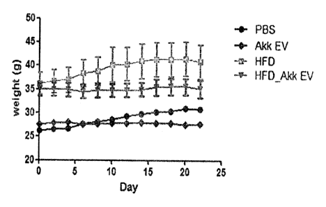
In a result obtained by measuring weight while extracellular vesicles are
administered at two day intervals, as shown in FIG. 6, it can be seen that a
weight
was not increased for 3 weeks in a IIFD-fed, extracellular vesicle-
administered
mouse group (HFD_Akk EV), compared to an only HFD-fed mouse group (IIED).
In addition, in a result obtained by measuring a plasma glucose level when
fasting for 6 hours after administration of extracellular vesicles was
completed, as
shown in FIG. 7, it can be seen that the plasma glucose level decreases in a
HFD-fed,
extracellular vesicle-administered mouse group (RFD + Akk EV), compared to an
only HFD-fed mouse group (HE'D - Akk EV).
In addition, in a result obtained by measuring an insulin concentration in
blood collected from a mouse heart 24 hours after administration of
extracellular
vesicles had completed, as shown in FIG. 8, it can be seen that an insulin
concentration is more increased in a HFD-fed, extracellular vesicle-
administered
14
CA 02949644 2016-11-18
mouse group (HFD Akk EV), compared to an only HFD-fed mouse group (1-IFD -
Akk EV).
The result indicates that Akkermansia muciniphila-dcrived extracellular
vesicles are effective in treating obesity and diabetes induced by a high fat
diet.
Example 4. Immune function regulatory effect
In order to confirm whether the Akkermansia muciniphila-derived
extracellular vesicles obtained in Example 1 affect an immune function, an
experiment was performed as follows.
Both blood and spleen were collected from mice 24 hours after administration
of extracellular vesicles had completed in the same manner as in Example 3.
Immunocytes were extracted from tissues of spleen collected from respective
mouse
groups using 100 pm and 40 pm cell strainers. An extract was centrifuged (400
xg)
for 5 minutes and then counted cells were plated at 5x l05 cells/well onto a
cell
culture plate. Thereafter, anti-CD3 and anti-CD28, which stimulate co-
stimulatory
molecules of T cells (cytokinc secretion), were added to the plate at
concentrations of
1 pg/mL and 0.5 pg/mL, respectively, and then cells were cultured for 48
hours.
In order to confirm a secretion difference of pro-inflammatory cytokines
among respective groups after culture is completed, supernatants of respective
groups collected from the cell culture plate were centrifuged (400 xg) for 5
minutes
to remove cells present in the supernatants, and then only supernatants were
collected
again. In a result obtained by confirming concentrations of IFN-7 and IL-17 in
the
collected supernatants through ELISA, as shown in FIG. 9, it can be seen that
while
concentrations of 1FN-7 and IL-17 increase in an only HE'D-fed mouse group
(HFD -
Akk EV), concentrations of I FIN-7 and IL-17 decrease in a HFD-fed mouse group
to
CA 02949644 2016-11-18
which AkkerinalLs'ia muciniphila-derived extracellular vesicles wcrc
administered
(IIED + Akk EV).
A decrease in pro-inflammatory cytokines such as 117181-7 and IL-17 indicates
that it is possible to treat obesity and diabetes by improving an immune
function
because obesity and diabetes are related to inflammation.
Example 5. Influence of insulin resistance on inhibition of signal
transduction
In order to confirm whether the Akkermansia muciniphila-derived
cxtracellular vesicles obtained in Example I recover insulin resistance, an
experiment was performed as follows.
In order to implement insulin resistance in vitro, a culture medium of mouse-
derived myocytcs (C2C I 2, ATCC CRL-1772) was treated with a I mM fatty acid
(sodium palmitatc, SIGMA) and 4% bovine serum albumin (BSA) for 48 hours.
Thereafter, Akkermansia muciniphila-derived extracellular vesicles were added
at a
concentration of I tig/m1 to the treated culture medium, cultured for 6 hours,
and then
treated with 2 nM insulin to confirm intracellular signal transduction.
In a result obtained by confirming, by the western blot, a difference in an
amount of phospho-insulin receptor substrate-I (p-IRS-1) used as an important
indicator in intracellular insulin signal transduction between a group which
was not
treated with insulin and a group which was treated with insulin, as shown in
FIG. 10,
it can be seen that p-IRS-1 was observed in an extracellular vesicle-treated
group,
which indicates that intracellular signal transduction inhibited by insulin
resistance
was recovered.
16
CA 02949644 2016-11-18
Thc result indicates that Akkermansia mucimphila-derived extracellular
vesicles can play a role in recovering insulin resistance induced by high
fatty acids in
myocytes, that is, inhibition of intracellular signal transduction by insulin.
Example 6. In vivo absorption, distribution, and excretion patterns of
bacteria and bacteria-derived extracellular vesicles
In order to compare and evaluate whether Akkermansia muciniphila and
Akkermansia muciniphila-dcrivcd extracellular vesicles are systemically
absorbed
through the gastrointestinal tract, an experiment was performed as follows.
First, PBS was used as a control group. Each of Akkermansia mucimphila
and Akkermansia muciniphila-derived extracellular vesicles, which were labeled
with
fluorescence, were administered to a mouse gastrointestinal tract at a dose of
50 [Hz,
and then fluorescence was measured using a fluorescence and bioluminescence
imaging system (In vivo imaging system (1VIS)) after 0 minute, 5 minutes, 3
hours, 6
hours, and 12 hours had elapsed. As a result, as shown in FIG. 11, it can be
seen
that the bacteria were not absorbed systemically, but the bacteria-derived
extracellular vesicles were absorbed systemically in just 5 minutes after
administration, excreted through urinary organs, which was seen from strong
fluorescence in the bladder at 3 hours after administration, and present in
the body
even at 12 hours after administration.
In addition, in order to confirm a pattern in which extracellular vesicles are
infiltrated into various organs after Akkermcmsio mucirilphila-derived
extracellular
vesicles are systemically absorbed, blood, heart, lungs, liver, kidney,
spleen, adipose
tissue, and muscle samples were extracted at 12 hours after fluorescence-
labeled
extracellular vesicles had been administered to a mouse's gastrointestinal
tract at a
17
CA 02949644 2016-11-18
dose o50 tig. In a result obtained by observing fluorescence in the extracted
tissue
samples by 1VIS, as shown in FIG. 12, it can be seen that the extraccIlular
vesicles
arc distributed in all of the blood and organ samples at 12 hours after the
extracellular vesicles have been administered to the gastrointestinal tract.
But
fluorescence of the bacterium itself was not observed.
In addition, in order to confirm whether Akkermansia muciniphila-derived
extracellular vesicles penetrate an intestinal barrier, each of Akkermansia
muciniphila bacteria and Akkermansia muciniphila-derived cxtraccIlular
vesicles
were administered to a mouse's large intestine at a dose of 10 ug for 10
minutes. In
a result obtained by checking the isolated large intestine by
immunohistochcmistry
(IBC), as shown in FIG. 13, it can be seen that the bacteria do not penetrate
an
intestinal barrier, but the extracellular vesicles penetrate an intestinal
barrier to be
absorbed into the tissue.
In addition, in order to confirm whether Akkermansia muciniphila-derived
extracellular vesicles penetrate an intestinal barrier to be absorbed into
intestinal
capillaries, fluorescence-labeled extracellular vesicles were administered to
a
mouse's large intestine at a dose of 10 tg. In a result obtained by observing
intestinal capillaries by a live imaging method, as shown in FIG. 14, it can
be seen
that the extracellular vesicles are absorbed into blood vessels to migrate in
blood
vessels.
Example 7. Influence of Akkermansia muciniphila-derived extracellular
vesicles on AMPK activation in myocytes
In order to evaluate an influence of Akkermansia muciniphila-dcrived
extracellular vesicles on glucose metabolism by AMPK activation in myocytes,
18
CA 02949644 2016-11-18
because AMPK is known as a protein that plays an important role in maintaining
energy homeostasis, an experiment was performed as follows.
First, in order to evaluate AMPK activation based on a concentration at which
treatment with Akkermansia muciniphila-dcrived extracellular vesicles is
performed
in vitro, myocytcs were treated with extracellular vesicles at concentrations
of 0, 0.1,
1, and 10 kig/rril for 1 hour. In a result obtained by measuring, by the
western blot, a
difference in expression levels of phospho-5' AMP-activated protein kinase
(pAMPK) and phospho-acetyl-CoA carboxylasc (pACC), which are used as an
important indicator in AMPK signal transduction, as shown in FIG. 15,
expression of
pAMPK and pACC increases according to a concentration of the extracellular
vesicles treated, and thus it can be seen that AMPK is activated depending on
a
concentration of extracellular vesicles.
In addition, in order to evaluate AMPK activation over time for treatment
with Akkermansia muciniphila-derived extracellular vesicles in vitro, myocytes
were
treated with extracellular vesicles at a concentration of 10 ug/m1 for 0, 10,
20, 30,
and 60 minutes. In a result obtained by measuring, by the western blot, the
expression patterns of pAMPK and pACC, which are used as an important
indicator
in AMPK signal transduction, as shown in FIG. 16, it can be seen that an
increase in
the pAMPK and pACC expression starts at 10 minutes after treatment with
extracellular vesicles and continues for 60 minutes.
Meanwhile, in order to further compare an influence of extracellular vesicles
derived from bacteria other than the Akkermansia muciniphila on AMPK
activation,
myocytes were treated with E.co/i-derived extracellular vesicles at a
concentration of
10 ug/ml. In a result obtained by measuring, by the western blot, expression
levels
of pAMPK and pACC, which are used as an important indicator in AMPK signal
19
CA 02949644 2016-11-18
transduction, as shown in FIG. 17, it can be seen that the expression levels
of
pAMPK and pACC remarkably decrease, demonstrating that the AMPK signal
transduction is inhibited. On the other hand, the AMPK signal transduction was
increased by metformin (therapeutic agent for diabetes) and decreased by
compound
C (AMPK signal transduction inhibitor).
Example 8. Influence of Akkermansia mueiniphila-dcrived extracellular
vesicles on glucose uptake in myocvtes
In order to evaluate an influence of Akkermansia muciniphila-derived
extracellular vesicles on glucose uptake in myocytes, myocytes were treated
with 10
ktg/m1 of Akkermansia muciniphila-derived extracellular vesicles in vitro for
1 hour,
and glucose uptake was assessed using a radioisotope (2-114Cideoxyglucose). As
a
result, as shown in FIG. 18, it can be seen that, compared to a negative
control (No
Treatment (NT)), the glucose uptake in the myocytes was increased by
Akkermansia
muciniphi/a-derived extracellular vesicles (EV), which is similar in extent to
a
glucose uptake increase when a myocyte is treated with insulin (10 nm) or
metformin
(50 mM).
In addition, in order to evaluate an influence Akkermansia
inuciniphila-
derived extracellular vesicles on expression of a glucose transport protein
(GLUT4
transporter) in myocytcs, myocytes were treated with 10 p.g/m1 of Akkernionsia
muciniphila-derived extracellular vesicles in vitro for I hour, and then the
degree of
GLUT4 expression in a cell membrane was measured by an o-phenylenediamine
(OPD) assay. As a result, as shown in FIG. 19, it can be seen that, compared
to a
negative control (No treatment (NT)), the GLUT4 expression was increased by
Akkermansia nweiniphila-derived extracellular vesicles (EV) which is similar
in
CA 02949644 2016-11-18
extent to the increased expression of GLUT4 when a myocytc is treated with
insulin
(10 nm) or metformin (50 mM).
In addition, in order to evaluate whether glucose uptake, which is increased
by Akkermansia muciniphila-derived extracellular vesicles, is inhibited by AM
PK
signal transduction, myocytcs were pretreated with compound C (AMPK signal
transduction inhibitor), and then treated with 10 _t.g/m1 of extracellular
vesicles for 1
hour, followed by assessment of the glucose uptake using a radioisotope (2-
[4C]dcoxyglucose). As a result, as shown in FIG. 20, it can be seen that the
glucose uptake, which is increased by the Akkermansia muciniphila-derived
extracellular vesicles, is decreased by compound C. This means that
improvement
of insulin resistance in myocytcs by the Akkermansia mucimphila-derived
extracellular vesicles is closely related to an AMPK signal transduction
process.
While the present invention has been described above with reference to the
exemplary embodiments of the present invention, it should be understood by
those
skilled in the art that various modifications and alterations may be made
without
departing from the spirit and scope of the present invention described in the
appended claims.
21