Note: Descriptions are shown in the official language in which they were submitted.
CA 02949842 2016-11-21
WO 2015/184160 PCT/US2015/033020
CHITOSAN AND POLYETHYLENE GLYCOL COPOLYMERS AND METHODS AND
DEVICES FOR USING SAME FOR SEALING A VASCULAR PUNCTURE
FIELD
[0001] Several embodiments of the inventions disclosed herein relate
generally to
sealants, apparatus, and methods for sealing punctures in a body. Some
embodiments relate to
copolymers that provide enhanced sealing effects. In several embodiments,
apparatus and
methods are disclosed that employ such copolymers for sealing a vascular
puncture extending
through tissue to a blood vessel.
BACKGROUND
[0002] Apparatus and methods are known for accessing a patient's
vasculature
percutaneously, e.g., to perform a procedure within the vasculature, and for
sealing the puncture
that results after completing the procedure. For example, a hollow needle may
be inserted
through a patient's skin and overlying tissue into a blood vessel. A guide
wire may be passed
through the needle lumen into the blood vessel, whereupon the needle may be
removed. An
introducer, procedural, or femoral sheath may then be advanced over the guide
wire into the
vessel, e.g., in conjunction with or subsequent to one or more dilators. A
catheter or other device
may be advanced through the introducer sheath and over the guide wire into a
position for
performing a medical procedure. Thus, the introducer sheath may facilitate
accessing and/or
introducing various devices into the vessel, while minimizing trauma to the
vessel wall and/or
minimizing blood loss.
[0003] Wounds such as arteriotomies can arise in the blood vessel from
these various
medical procedures, especially for blood vessels acting as sites for catheter
insertion during
diagnostic and/or interventional catheterization. After such procedures have
been completed, the
arteriotomy that was created as an access point during the medical procedure
needs to be closed.
[0004] Upon completing the procedure, the device(s) and introducer
sheath may be
removed, leaving a puncture extending between the skin and the vessel wall. To
seal the
puncture, external pressure may be applied to the overlying tissue, e.g.,
manually and/or using
1
CA 02949842 2016-11-21
WO 2015/184160 PCT/US2015/033020
sandbags, until hemostasis occurs. This procedure, however, may be time
consuming and
expensive, requiring as much as an hour of a medical professional's time. It
is also
uncomfortable for the patient, and may require the patient to remain
immobilized in the
operating room, catheter lab, or holding area. In addition, a risk of hematoma
exists from
bleeding before hemostasis occurs.
[0005]
Vascular closure devices can be used to achieve hemostasis (e.g., sealing) of
small holes that are formed in a blood vessel (either artery or vein) as the
result of an
intravascular procedure (e.g., cannulation). Such procedures may be for
diagnosis, drug
delivery, therapy (e.g., stent placement or angioplasty) and the like. The
procedures involve the
formation of a small incision in the wall of a vessel to gain access to the
intravascular space.
This incision, the vascular puncture or arteriotomy, must be closed at the
completion of the
procedure.
Rapid hemostasis at the vascular puncture is ideal, as it reduces patient
complications, improves time to patient ambulation and time to hospital
discharge.
[0006]
For example, a mechanical based device can be utilized for vascular closure.
A percutaneous surgical device can comprise a combination wound suturing and
crimping and
cutting device. The combined device may locate a vessel wound and pass suture
through the
vessel walls surrounding the wound. Then, the crimping and cutting portion may
detach, the
suturing portion may be removed, and the crimping and cutting portion may be
located to the
wound site to apply a fastener (e.g., a ferrule).
[0007]
Another mechanical based device can have two components: a needle
advancing apparatus slidable longitudinally along a catheter to advance
needles into a tissue
membrane, such as a blood vessel wall, around an opening in the membrane; and,
a suture
retrieval assembly insertable through the catheter beyond a distal side of the
tissue membrane.
The needle advancing apparatus advances suture through the tissue wall. The
suture retrieval
assembly grabs the suture on the distal side of the tissue membrane for
extraction thereof through
the opening in the tissue membrane.
[0008]
Such mechanical approaches tend to require precise positioning within the
tissue tract, typically provide point (instead of a continuum of tissue
purchase) support, and lead
to permanent foreign-body implants that interfere with subsequent
catheterization at the same
vascular site. Additionally, a purely mechanical support of the wound could
lead to implanting
substantially non-absorbable foreign material that provides only point-support
to the wound lips.
2
CA 02949842 2016-11-21
WO 2015/184160 PCT/US2015/033020
In addition, purely mechanical closures still can leave behind open micro-
spaces, or small gaps,
between the sutures that are not entirely closed.
[0009]
Previously (and currently, in some cases), manual compression was the main
method for closing the vascular puncture. This could involve extended periods
of manual
pressure, clamping, exogenous weights, etc., applied directly to the site of
the vascular puncture.
As hemostasis could take 20 to 60 minutes, patients often experienced
discomfort, and extended
periods of bed rest were required.
[0010]
In addition to, or in place of manual compression, vascular closure devices
were developed to reduce the time to achieve hemostasis. Some such devices
used sutures or
collagen plugs to seal the vascular puncture.
However, many such devices result in an
intravascular component being retained within the vessel, which can lead to
future
complications.
[0011]
More recently biodegradable materials have been employed to seal the
vascular puncture and, due to their dissolution over time, improve patient
comfort and reduce
complications. Because rapid hemostasis can improve patient outcome and reduce
medical
costs, further improvements in vascular sealants would be beneficial.
[0012]
Though presently in use, many current sealant technologies facilitate
hemostasis of a wound puncture by physically clogging the tissue tract. This
physical occlusion
replaces manual compression, but certain of such polymeric sealants have a
relatively weak
polymer network integrity, which can increase time to hemostasis.
[0013]
Various biological approaches to vascular closure have been used such as a
device and method that includes inserting a vessel plug or sealant into the
incision or puncture
until the distal end of the vessel plug is adjacent to the outer lumen of the
blood vessel. The
vessel plug is positioned so that it does not obstruct the flow of fluid
through the blood vessel or
target organ. The precise positioning of the vessel plug in the incision or
puncture is
accomplished through the use of a balloon catheter or a cylindrical insertion
assembly having a
proximal plunger member associated therewith. Another biological closure can
deploy a
collagen plug to seal the closure. In order to block the collagen from
entering the vessel, a
footplate is installed on the interior of the blood vessel. The footplate is
held in place with a
suture.
3
CA 02949842 2016-11-21
WO 2015/184160 PCT/US2015/033020
[0014] In one instance, a vascular closure device can include two
synthetic
polyethylene glycol ("PEG") polymer powders that are mixed with appropriate
buffers and
injected through a femoral sheath at an arteriotomy site, e.g., as disclosed
in U.S. Patent No.
7,316,704. Accordingly, apparatus and methods for sealing a puncture through
tissue would be
useful. In particular, improving the efficacy (e.g., speed and/efficiency) of
sealing a puncture
would be useful.
SUMMARY
[0015] Provided for herein, in several embodiments, are vascular
sealants, apparatus,
and methods for sealing vascular punctures, the vascular sealants comprising
copolymers that
provide enhanced sealing effects.
[0016] As such, several embodiments herein provide vascular sealants
comprising
copolymers that provide enhanced hemostasis. In some embodiments, this is due
to
supplementation of the physical occlusion of the vascular puncture by sealants
that have
hemostatic and/or procoagulative properties. In several embodiments, the
sealants attract
platelets and/or other coagulation promoting co-factors. In several
embodiments, the copolymer
sealants provide enhanced "grip" at the site of a vascular puncture, thereby
improving the
occlusion of the puncture, and even allow the use of the copolymer sealants on
larger puncture
sizes.
[0017] In several embodiments, the copolymer sealant comprises
chitosan and one or
more polyethylene glycol polymers that exhibits a more rapid hemostasis
compared to sealants
that comprise only chitosan or only polyethylene glycol polymer sealants.
Chitosan is a natural
biopolymer found in crustaceans with a wide range of applications in tissue
engineering, tissue
repair and wound healing. Chitosan can be produced by deacetylation of chitin,
which is the
structural element in the exoskeleton of crustaceans (e.g., crabs, shrimps,
etc.). Variation in the
degree of deacetylation (%DA) can result in varying functionality of the
chitosan in different
applications. Chitosan is also biodegradable, for example, by chitosanase,
papain, cellulose, acid
proteases, and the like. Chitosan can form hydrogels depending on the
molecular weight, the
degree of deacetylation and the pH. In addition, a variety of cross linkers
can be utilized to
crosslink chitosan polymer chains and result in the formation of hydrogels.
4
CA 02949842 2016-11-21
WO 2015/184160 PCT/US2015/033020
[0018] Chitosan has been known to have hemostatic properties, which
are described,
for example in US Patent No. 4,394,373 and US Patent No. 8,012,167. However,
in several
embodiments, the chitosan of the copolymer sealants disclosed herein, is not
simply mixed with
the other polymeric component (or components), but rather is bound (e.g.,
covalently or non-
covalently) to the other component (or components) of the sealant. For
example, in several
embodiments, the copolymer sealant comprises chitosan covalently (or non-
covalently) bound to
two types of polyethylene glycol (PEG), PEG-amine and PEG-ester.
Advantageously, this
improves the structural integrity of the sealant when deployed, but also
imparts pro-coagulant
and hemostatic properties to the sealant that improve efficacy in sealing
vascular punctures.
[0019] Several embodiments of the sealants disclosed herein comprise
both
polyethylene glycol and chitosan in the freeze dried polymer hydrogel (e.g.,
the sealant).
Sealants comprising only PEG (with no chitosan incorporated) elicit hemostasis
of a wound
puncture essentially by clogging the tissue tract upon expansion of the
hydrogel sealant in the
presence of physiological fluids. Freeze dried hydrogel sealant containing
only PEG would lack
the hemostatic and pro-coagulative properties of chitosan that is included in
the sealants
disclosed herein and thus PEG-only sealants would result in slower and less
efficient hemostasis.
[0020] On the other hand a freeze dried hydrogel sealant utilizing
chitosan only (with
no PEG components incorporated) would lack the porosity characteristics (size
and number of
pores) that partially cross-linked PEG hydrogels can create upon freeze
drying. Such a hydrogel
would lack the rapid swelling capability that PEG components impart. These
hydrogels would
have reduced capacity to absorb physiological fluids and swell and
subsequently clog the tissue
tract. In addition these hydrogels would have reduced capacity to rapidly
absorb blood and
indirectly boost the inherit capability of chitosan since less blood would be
available for chitosan
to promote clotting. This would result in a reduced capacity to promote
hemostasis as compared
to hydrogels that contain both polyethylene glycol and chitosan, such as those
disclosed herein.
[0021] Thus, freeze dried hydrogels comprising both chitosan and
polyethylene
glycol advantageously, and unexpectedly, lead to faster hemostasis of wounds
since they
combine the swelling characteristics of the PEG moiety along with the
hemostatic and pro-
coagulative properties of chitosan. The absorbance of blood by the porous PEG
components can
supplement and enhance chitosans ability for clotting because more blood
volume is available
for chitosan per surface area. These chitosan-PEG hydrogels may therefore have
application to
CA 02949842 2016-11-21
WO 2015/184160 PCT/US2015/033020
larger wounds versus the limited applicability when the sealant is composed of
only polyethylene
glycol or only chitosan.
[0022] Therefore, there are provided, in several embodiments, sealants
for sealing
punctures in tissues. In one embodiment, there is provided a sealant for
sealing a puncture
through tissue, comprising an elongate first section having a proximal end, a
distal end, and a
cross-section sized for delivery into a puncture through tissue and a second
section extending
from the distal end of the first section, the first section comprising a
hydrogel comprising
chitosan bound to at least one polymer. In several embodiments, the first
section is formed from
a freeze-dried hydrogel and the first section is configured to expand when
exposed to
physiological fluid within a puncture. In several embodiments, upon exposure
to an aqueous
physiological fluid, the hydrogel expands and seals the puncture through the
tissue. In several
embodiments, the puncture is a vascular puncture.
[0023] Also provided, in several embodiments, is a sealant for sealing
a puncture
through tissue, comprising a first section formed from a freeze-dried
hydrogel, the first section
being configured to expand when exposed to physiological fluid within a
puncture. In several
embodiments, the first section comprises a hydrogel comprising chitosan bound
to at least one
polymer, and upon exposure to an aqueous physiological fluid, the hydrogel
expands and seals
the puncture through the tissue.
[0024] In several embodiments, the first section has an elongated
shape with a
proximal end, a distal end, and a cross-section sized for delivery into a
puncture through tissue.
In several embodiments, the chitosan comprises chitosan that is at least
partially deacetylated.
For example, in one embodiment, the chitosan has a degree of deacetylation of
at least 60%. In
additional embodiments, the degree of deacetylation is between about 40% to
50%, about 50% to
about 60% about 60% to about 70% about 70% to about 80%, about 80% to about
90%, about
90% to about 95%, about 95% to about 99% (and overlapping ranges between those
listed).
Greater or lesser degrees of deacetylation are also used, in other
embodiments.
[0025] In several embodiments, the chitosan has a molecular weight
between about
kilodaltons and about 600 kilodaltons, including about 10 kilodaltons to about
50 kilodaltons,
about 50 kilodaltons to about 100 kilodaltons, about 100 kilodaltons to about
200 kilodaltons,
about 200 kilodaltons to about 300 kilodaltons, about 300 kilodaltons to about
400 kilodaltons,
6
CA 02949842 2016-11-21
WO 2015/184160 PCT/US2015/033020
about 400 kilodaltons to about 500 kilodaltons, about 500 kilodaltons to about
600 kilodaltons,
or any molecular weight between or including those values.
[0026] Depending on the embodiment, the chitosan can be of a varied
type. For
example, in several embodiments, the chitosan can be free chitosan, chitosan
chloride, chitosan
glutamate, chitosan acetate, chitosan dicarboxylic acid salts, chitosan
adipate, chitosan succinate,
or chitosan fumarate. In some embodiments, combinations of two or more forms
of chitosan are
used.
[0027] In several embodiments, the at least one polymer is a
polyethylene glycol
polymer chain. In some embodiments, the at least one polymer is a polyethylene
glycol polymer
chain with side group functionality. In several embodiments, the at least one
polymer is an amine
modified polyethylene glycol or an ester modified polyethylene glycol.
Combinations of amine
modified polyethylene glycols and ester modified polyethylene glycols can also
be used, in
several embodiments. In several embodiments, the chitosan is bound to the at
least one polymer
by a covalent bond. In additional embodiments, the chitosan is bound to the at
least one polymer
by a non-covalent bond. In one embodiment, the at least one polymer is cross-
linked
polyethylene glycol that is bound to the chitosan.
[0028] In several embodiments, the amount of chitosan is varied. For
example, in
several embodiments, the first section of the sealant comprises between about
0.1% and about
30% (by weight) chitosan. For example, in several embodiments the chitosan is
present in an
amount (by weight) between about 0.1% to about 1.0%, about 1.0% to about 5.0%,
about 5.0%
to about 10.0%, about 10.0% to about 15.0%, about 15.0% to about 20.0%, about
20.0% to about
25.0%, about 25.0% to about 30.0%, and any amount between or including those
amounts. In
one embodiment the first section comprises between about 0.5% and about 8% (by
weight)
chitosan. In one embodiment, the first section comprises between about 2% and
about 4% (by
weight) chitosan. In one embodiment, the first section comprises between about
4% and about
6% (by weight) chitosan. Greater or lesser amounts of chitosan are also used,
in some
embodiments.
[0029] In several embodiments, the at least one polymer comprises
polyethylene
glycol-amine (PEG-amine) and polyethylene glycol-ester (PEG-ester). In some
embodiments,
the PEG-amine and PEG-ester are present in a molar ratio of PEG-amine to PEG-
ester between 4
to 1 and 1 to 4. In some embodiments, the PEG-amine and PEG-ester are present
in a molar
7
CA 02949842 2016-11-21
WO 2015/184160 PCT/US2015/033020
ratio of PEG-amine to PEG-ester between 2 to 1 and 1 to 2. In some
embodiments, the PEG-
amine and PEG-ester are present in a molar ratio of PEG-amine to PEG-ester
between about 0.8
to about 1.2. In some embodiments, the PEG-amine and PEG-ester are present in
a molar ratio of
PEG-amine to PEG-ester between about 0.9 to about 1.
[0030] In several embodiments, the PEG-amine and PEG-ester are present
in a ratio
of equivalent active groups that ranges from about 0.1 to about 5. In some
embodiments, the
PEG-amine and PEG-ester are present in a ratio of equivalent active groups
that ranges from
about 0.5 to about 3. In some embodiments, the PEG-amine and PEG-ester are
present in a ratio
of equivalent active group sites that ranges from between about 0.5 to about
2Ø In some
embodiments, the PEG-amine and PEG-ester are present in a ratio of equivalent
active group
sites that ranges from about 0.8 to about 1.2. In some embodiments, the PEG-
amine and PEG-
ester are present in a ratio of equivalent active group sites that ranges from
about 0.9 to about 1.
[0031] In several embodiments, the at least one polymer comprises
polyethylene
glycol-ester (PEG-ester). In one embodiment, the PEG-ester can be present in
an amount (by
weight) between about 99.0% to about 1.0%, about 90.0% to about 10.0%, about
80.0% to about
20.0%, about 70.0% to about 30.0%, about 60.0% to about 40.0%, about 55.0% to
about 45.0%,
about 53.0% to about 47.0%, about 52.0% to about 48.0%, about 52.0% to about
50.0%, and any
amount between or including those amounts.
[0032] In several embodiments, the at least one polymer comprises
polyethylene
glycol-amine (PEG-amine) and a mixture of polyethylene glycol-esters (PEG-
esters). In some
embodiments, the PEG-amine and PEG-ester mixture can be present in a molar
ratio of PEG-
amine to PEG-ester of between 4 to 1 and 1 to 4. In some embodiments, the PEG-
amine and
PEG-ester mixture can be present in a molar ratio of PEG-amine to PEG-ester
between 2 to 1
and 1 to 2. In some embodiments, the PEG-amine and PEG-ester can be present in
a molar ratio
of PEG-amine to PEG-ester between about 0.8 to about 1.2. In some embodiments,
the PEG-
amine and PEG-ester can be present in a molar ratio of PEG-amine to PEG-ester
between about
0.9 to about 1.
[0033] In several embodiments, the sealant can also include a second
section. In
some embodiments, the second section can extend from the distal end of the
first section. In
some such embodiments, the second section can be made up of non-cross-linked
precursors. In
some embodiments, the non-cross-linked precursors comprise polyethylene glycol-
amine and/or
8
CA 02949842 2016-11-21
WO 2015/184160 PCT/US2015/033020
polyethylene glycol-ester. Depending on the embodiments, the second section
also optionally
includes chitosan. For example, in one embodiment, the second section can be a
mixture of non-
cross-linked polyethylene glycols bound to the chitosan. In several
embodiments, the second
section (when chitosan is included) can include between about 0.1% and about
30% (by weight)
chitosan. For example, the second section may, in some embodiments, include
between about
0.1% and about 30% (by weight) chitosan, including about 0.1% to about 1%,
about 1.0% to
about 5.0%, about 5% to about 10.0%, about 10.0% to about 15.0%, about 15.0%
to about
20.0%, about 20.0% to about 25.0%, about 25.0% to about 30.0%, and any amount
between or
including those amounts.
[0034]
Also, in several embodiments, the second section may also include one or
more reinforcement elements. In some embodiments, the reinforcement elements
have
hemostatic properties including but not limited to chitosan reinforcing
fibers, chitosan mesh,
chitosan particles, or combinations thereof.
[0035]
In several embodiments, the chitosan mesh is configured as a helical coil
within the second section. In several embodiments, the chitosan mesh
(regardless of its
conformation) includes cross-linked chitosan, wherein the cross-links were
formed using
genipin. In several embodiments, chitosan fibers are configured as a helical
coil within the
second section. In some such embodiments, the chitosan fibers are formed by
electrospinning.
In several embodiments, the chitosan can be in the form of particles that are
incorporated into the
second section. The chitosan particles can be incorporated, depending on the
embodiment, in a
random manner throughout the second section, in a substantially uniform manner
throughout the
second section, or a patterned manner throughout the second section.
In additional
embodiments, random, uniform, or patterned particle distribution can be used
in different
portions of the second section.
[0036]
In several embodiments, the sealant can be configured to seal a vascular
puncture, wherein the sealant expands after exposure to an aqueous
physiological fluid, and
wherein a second section of the sealant can have hemostatic and pro-
coagulative properties. In
several embodiments, the second section further comprises a pH adjusting
agent. In several
embodiments, the sealant further comprises a therapeutic agent. In several
embodiments, the
sealant is dimensioned with a first section having a length (e.g., between
proximal and distal
9
CA 02949842 2016-11-21
WO 2015/184160 PCT/US2015/033020
ends) of between about 1 and about 20 millimeters. In several embodiments
including a second
section, the second section has a length of between about 0.5 and about 5
millimeters.
[0037] In several embodiments having both first and second sections,
the first and
second sections can have a substantially uniform outer cross-section along
their lengths between
about 1 and about 8 millimeters. Upon exposure to an aqueous physiological
fluid, the sealants
are configured to expand. In some embodiments, the first section (and second
section, if
included) is configured to expand in the dimension of the outer cross section
of the sealant of at
least 15%, including at least 20%, at least 25%, at least 30%, at least 40%,
or at least 50%.
[0038] There are also provided herein methods for sealing a vascular
puncture
comprising applying a sealant as described herein to the vascular puncture.
[0039] In several embodiments, therefore, there are provided sealants
and associated
methods for sealing a puncture in a body. More particularly, several
embodiments are directed
to sealants made from chitosan and polyethylene glycol for sealing a puncture
through tissue,
and to methods for making such sealants. In addition, several embodiments of
the invention are
directed to sealants and methods for providing temporary or permanent
hemostasis within a
puncture extending through tissue.
[0040] In accordance with one embodiment, a sealant is provided for
sealing a
puncture through tissue that includes a first section including a proximal
end, a distal end, and a
cross-section sized for delivery into a puncture through tissue, and a second
section fused to and
extending from the distal end of the first section. In several embodiments,
the first section is
formed from a freeze-dried hydrogel made of chitosan and polyethylene glycol
polymer chains
and/or crosslinks that expands when exposed to physiological fluid within a
puncture. In several
embodiments, the second section is formed from a solid mass of non-freeze-
dried, non-cross-
linked hydrogel precursors, the precursors remaining in an unreactive state
until exposed to an
aqueous physiological environment, whereupon the precursors undergo in-situ
crosslinking with
one another to provide an improved adhesion of the sealant to the arteriotomy.
[0041] In one embodiment, the first section may consist essentially of
freeze-dried
hydrogel, and the second section may consist essentially of the non-cross-
linked precursors.
Alternatively, the second section may include one or more reinforcement
elements with
hemostatic properties, e.g., chitosan reinforcing fibers, a chitosan mesh or
chitosan particles. In
CA 02949842 2016-11-21
WO 2015/184160 PCT/US2015/033020
addition or alternatively, the second section may include one or more diluents
to enhance one or
more properties of the second section.
[0042] In another embodiment, the sealant includes only one section of
a freeze-dried
hydrogel made of chitosan and polyethylene glycol polymer chains and/or
crosslinks that
expands when exposed to physiological fluid within a puncture.
[0043] Optionally, the sealant may include one or more pH adjusting
agents, e.g.,
impregnated into, coated over, or otherwise included in the first and/or
second sections. For
example, when the sealant is exposed within a puncture, the agent(s) may alter
the localized pH
on or around the sealant, e.g., to enhance cross-linking of the precursors
and/or creation of a
desired adhesive material. Alternatively, the materials for the precursors may
be selected such
that the pH and/or buffering capacity of interstitial body fluids and/or blood
are effective to drive
or otherwise facilitate cross-linking of the precursors. In such embodiments,
the pH adjusting
agents may be omitted.
[0044] In several embodiments, the first section of the sealant may be
composed of a
freeze-dried hydrogel that contains polyethylene glycol chains covalently
bonded with chitosan
polymer chains that has hemostatic and pro-coagulative properties and that
expands when
exposed to physiological fluids within a puncture. A solid mass of non-cross-
linked hydrogel
precursors such as polyethylene glycol with ester end groups, polyethylene
glycol with amine
end groups and chitosan with various degrees of deacetylation, may be fused or
otherwise
attached onto the distal end of the sealant. Until such time that the
precursors are exposed to an
aqueous physiological environment, the precursors remain in an unreactive
state. At such time,
the precursors undergo in-situ crosslinking with one another to provide an
improved adhesion to
the arteriotomy.
[0045] In an additional embodiment, chitosan fibers, chitosan mesh or
chitosan
particles may be incorporated or fused together with the non-cross-linked
hydrogel precursors.
For example, the solid mass may be formed as a substantially uniform solid
plug or may be
formed as a sintered mass of powder and fibers or mesh. The chitosan fibers,
mesh or particles
may act as a reinforcement element to increase the integrity of the cross-
linked network. The
melted precursors, which may or may not comprise chitosan fibers, chitosan
mesh or chitosan
particles may be applied to the distal end of the tubular roll within the
tubular member, and
allowed to solidify to create the solid mass fused to the distal end of the
tubular roll.
11
CA 02949842 2016-11-21
WO 2015/184160 PCT/US2015/033020
[0046] In accordance with one embodiment, a sealant is provided for
sealing a
puncture through tissue that includes a first section including a proximal
end, a distal end, and a
cross-section sized for delivery into a puncture through tissue, and a second
section fused to and
extending from the distal end of the first section. The first section may be
formed from a freeze-
dried hydrogel that expands when exposed to physiological fluid within a
puncture. The second
section may be formed from a solid mass of non-freeze-dried, non-cross-linked
hydrogel
precursors, the precursors remaining in an unreactive state until exposed to
an aqueous
physiological fluid, whereupon the precursors undergo in-situ crosslinking
with one another to
provide an improved adhesion of the sealant to the arteriotomy.
[0047] In one embodiment, the first section may consist essentially of
freeze-dried
hydrogel, and the second section may consist essentially of the non-cross-
linked precursors.
Alternatively, the second section may include one or more reinforcement
elements, e.g., a
plurality of filaments or particles, mixed with, embedded in, or surrounding
the precursors. In
addition or alternatively, the second section may include one or more diluents
to enhance one or
more properties of the second section. As discussed above, the sealant may (or
may not) include
one or more pH adjusting agents, e.g., impregnated into, coated over, or
otherwise included in
the first and/or second sections.
[0048] In another embodiment, a sealant is provided for sealing
percutaneous
vascular large bore punctures, the sealant includes a first section including
a proximal end, a
distal end, and a cross-section sized for transcatheter delivery into the
tissue tract and further
including chitosan, and can optionally include a second section fused to and
extending from the
distal end of the first section. The large bore punctures can be sealed with a
sealant that is
initially sized the same as or larger than the large bore puncture or,
alternatively, can be sealed
with a sealant that is initially sized smaller than the large bore puncture by
being loaded onto a
small bore delivery device. It is understood that the physical properties of
the sealant are such
that it expands to occupy the full space of the device it is loaded upon, such
that the same size
sealant can be loaded onto a large bore delivery device as well as a small
bore delivery device,
just that upon being loaded onto a small bore delivery device the sealant
would be compressed to
fit into the smaller space. Large bore punctures can be arterial punctures
that are sized from
about 7 French to about 24 French. It is typically understood that small bore
punctures can be
arterial punctures sized up to about 7Fr.
12
CA 02949842 2016-11-21
WO 2015/184160 PCT/US2015/033020
BRIEF DESCRIPTION OF THE DRAWINGS
[0049] It will be appreciated that the drawings are not necessarily
drawn to scale,
with emphasis instead being placed on illustrating the various aspects and
features of the
illustrated embodiments.
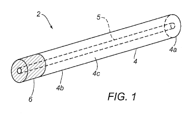
[0050] FIG. 1 is a perspective view of an exemplary embodiment of a
sealant
member comprising a freeze-dried hydrogel made of chitosan and polyethylene
glycol polymer
chains and/or crosslinks that expands when exposed to physiological fluid
within a puncture.
[0051] FIG. lA is a cross-sectional view of a transfer tube and
mandrel, showing a
method for making the sealant member of FIG. 1.
[0052] FIGS. 2A and 2B are side views of various embodiments in which
chitosan is
incorporated into a sealant. FIG. 2A shows as chitosan mesh, while FIG 2B
shows chitosan
particles.
[0053] FIGS. 3A and 3B are perspective and side views, respectively,
of another
embodiment of an apparatus for delivering a sealant into a puncture through
tissue.
[0054] FIG. 3C is a side view of the apparatus of FIGS. 3A and 3B with
a portion of
an outer housing removed to show internal components of the apparatus.
[0055] FIG. 3D is a perspective view of an introducer sheath and
dilator assembly
that may be used in cooperation with the apparatus of FIGS. 3A-3C.
[0056] FIG. 4A-4F illustrate a method of delivering a sealant to an
arteriotomy site.
[0057] FIGS. 5A, 5A-1, 5B, and 5B-1 illustrate a mechanism for
controlling fluid
flow through an inflation line.
[0058] FIGS. 6A, 6A-1, 6B, and 6B-1 illustrate another mechanism for
controlling
fluid flow through an inflation line.
[0059] FIGS. 6C-6D illustrate yet another mechanism for controlling
fluid flow
through an inflation line.
[0060] FIGS. 6E-6F illustrate yet another mechanism for controlling
fluid flow
through an inflation line.
[0061] FIGS. 7A, 7A-1, 7B, and 7B-1 illustrate yet another mechanism
for
controlling fluid flow through an inflation line.
13
CA 02949842 2016-11-21
WO 2015/184160 PCT/US2015/033020
[0062] FIGS. 8A-8B illustrate a mechanism for controlling movement of
an outer
housing relative to an inner housing.
[0063] FIGS. 9A-9B illustrate another mechanism for controlling
movement of an
outer housing relative to an inner housing.
[0064] FIGS. 10A-10B illustrate yet another mechanism for controlling
movement of
an outer housing relative to an inner housing.
[0065] FIGS. 11A-11C illustrate a locking mechanism to prevent
actuation of a
support member.
[0066] FIGS 12A-12B illustrate a mechanism for advancing a support
member.
[0067] FIGS. 13A-13B illustrate another mechanism for advancing a
support
member.
[0068] FIGS. 14A-14B illustrate a retraction lock to restrict movement
of a
positioning assembly.
[0069] FIGS. 15A-15F illustrate another method for delivering a
sealant to an
arteriotomy site.
[0070] FIGS. 16A-16B illustrate an apparatus for delivering a sealant
to an
arteriotomy including an inflation indicator.
[0071] FIGS. 17A-17D illustrate an embodiment of a dilator configured
to engage a
sheath.
[0072] FIGS. 18A-18C illustrate another embodiment of a dilator
configured to
engage a sheath.
[0073] FIGS. 19A-19D-1 illustrate a mechanism for engaging a
positioning assembly
and a sheath.
[0074] FIG. 20 illustrates another mechanism for engaging a
positioning assembly
and a sheath.
[0075] FIGS. 21A-21I illustrate a method for delivering a sealant to
an arteriotomy
site.
DETAILED DESCRIPTION
[0076] The apparatus, sealant and method disclosed herein capitalize
on the
interactions between chitosan and PEG moieties (e.g., PEG-amine and PEG-ester)
to achieve
14
CA 02949842 2016-11-21
WO 2015/184160 PCT/US2015/033020
enhanced hemostatic and procoagulative properties with improved integrity of
the cross-linked
sealant (both the grip and the freeze dried portion) after activation by
physiological fluids.
Chitosan can be covalently or non-covalently bonded, depending on the
embodiment, with PEG
to create the sealants. In addition, various cross linkers (e.g., genipin) can
be used to crosslink
chitosan polymer chains to create high molecular weight hydrogels of pure
chitosan. The
hydrogels can then be dehydrated by freeze drying to make a porous mesh that
can be
incorporated in the second section (the "grip" section) of a sealant to
improve the integrity and
stability of the final cross-linked network (after contact with physiological
fluids). Sealants
[0077] FIG. 1 shows a non-limiting embodiment of a sealant 2 for
sealing a puncture
extending through tissue (not shown), such as a blood vessel. Generally, the
sealant 2 can
include a first, proximal, or main section 4 including proximal and distal
ends 4a, 4b, and a
second, distal, or tip section 6 formed from a plurality of non-freeze-dried
and/or non-cross-
linked precursors, e.g., formed as a solid mass or solid plug, fused or
otherwise attached to and
extending distally from the distal end 4b of the first section 4. As described
further below, the
non-cross-linked precursors may remain in an unreactive state, e.g., before or
until exposure to
an aqueous physiological environment, e.g., when deployed or otherwise exposed
within a
puncture extending through tissue.
[0078] For example, this configuration of sealant 2 may combine
crosslinking of the
second section 6 to create an adhesive material in-situ with swell
characteristics and pro-
coagulative properties of a freeze-dried hydrogel or other expandable material
of the first section
4. By incorporating chitosan into a polyethylene glycol polymer network, the
overall freeze
dried hydrogel results in unexpectedly enhanced extra-vascular closure by
providing expansion
of the freeze dried hydrogel within the tissue tract upon contact with
physiological fluid and
providing hemostatic and pro-coagulative properties that, in combination,
result in faster overall
hemostasis of the vessel.
[0079] In one embodiment, the first section 4 can be formed from a
sheet of freeze-
dried hydrogel rolled into a tubular shape. It will be appreciated that the
first section 4 may have
other tubular or solid rod cross-sections or shapes, as desired, such as
elliptical, triangular,
square, conical, disk, polygonal shapes, and the like (not shown).
[0080] The first section 4 can be formed from a freeze-dried and cross-
linked
hydrogel that comprises two components, one being polyethylene glycol ("PEG")
and the other
CA 02949842 2016-11-21
WO 2015/184160 PCT/US2015/033020
component being chitosan. The two polymers, PEG and chitosan may be covalently
bonded or
blended together to form a freeze dried polymer hydrogel that expands upon
contact with
physiological fluids and that has hemostatic properties. Non-covalent bonding
may also be used,
in several embodiments. Optionally, a transition zone (not shown) may be
included where the
material of the second section 6 can penetrate partially into the distal end
4b of the first section 4,
e.g., during fusion, as described further below. Some such embodiments enhance
the structural
stability of the sealant, further enhancing hemostasis.
[0081]
In several embodiments, the material of the first section 4 may be at least
partially absorbed by the body over time, e.g., over a period of days, weeks,
or months.
Likewise, the material of the second section 6 may also be at least partially
absorbed by the body
over time, e.g., over a period of days, weeks, or months. Depending on the
embodiment, the first
section 4 and second section 6 can be made of the same material. In some
embodiments, the
composition of the first section 4 and the second section 6 can be adjusted to
accommodate their
relative roles in the hemostatic process and the eventual healing of the
puncture. For example, in
several embodiments, the rate of absorption of the second section 6 can be
slower than that of the
first section 4, thereby maintaining the sealant over the puncture for a
longer period of time, thus
allowing the underlying vessel time to heal. The rate of degradation (and thus
the specific make-
up of the sealant) can be selected based on the size of puncture, rate of
blood flow (or interstitial
fluid flow) or blood pressure at the puncture site, or the degree of mobility
that the puncture site
will experience (e.g., healing may take longer at a puncture site that
experiences frequent forces
from body motion).
[0082]
The PEG/chitosan co-polymer sealant can comprise two portions PEG (one
portion PEG-amine and one portion PEG-ester) to one portion chitosan.
In several
embodiments, the chitosan can be at least partially deacetylated. It should be
noted that the term
"portion", as used herein, does not necessary indicate a quantity or ratio of
the various
components. Rather, specific details about further aspects of the sealants,
including their specific
compositions, are discussed below.
Polyethylene Glycol
[0083]
The PEG used in the sealant can be varied, depending on the embodiment and
factors such as the anticipated puncture size, the normal rate of blood flow
in the area of the
puncture, the physical status of a patient (e.g., on anti-coagulant
medication, etc.). In several
16
CA 02949842 2016-11-21
WO 2015/184160 PCT/US2015/033020
embodiments, the PEG-amine portion may be a polymer such as 8A20K-NH2 (e.g., 8-
arm 20
kilodalton (kDa) molecular weight, with amine terminated arms). In several
embodiments, the
PEG-ester portion may be a polymer such as 4A10K-CM-HBA-NHS (e.g., 4-arm,
10kDa
molecular weight, with carboxymethyl - hydroxybutyrate -N- hydroxysuccinimidyl
functional
groups on the arms). In another embodiment the PEG-ester portion may be a
polymer such as 4A
10K-SS-NHS (e.g., 4-arm, 10kDa molecular weight with succinimidyl succinate
functional
groups on the arms) or a polymer such as 4A10K-SG-NHS (e.g., 4-arm, 10kDa
molecular weight
with succinimidyl glutarate functional groups on the arms) or a mixture of
these polymers.
[0084] In various embodiments, different precursors may be used to
manufacture
both the first section 4 and the second section 6 of the sealant. For example,
the precursors may
comprise polyethylene glycol derivatives or polyethylene glycols with at least
two end groups
(e.g., 2 arms) and having at least one cross-linkable end group. The first
functional group may
chemically react with the second functional group in-situ to form covalent
bonds and thereby
form a cross-linkable gel. In some embodiments, the first functional group or
second functional
group can comprise strong electrophiles. For example, the first and/or second
functional group
may be one or more of epoxide, succinimide, N-hydroxysuccinimide, acrylate,
methacrylate,
maleimide, and N-hydroxysulfosuccinimide. Additionally, in some embodiments,
the first
and/or second functional group may be an amine group, a sulfhydryl group, a
carboxyl group,
and/or a hydroxyl group.
[0085] Depending on the embodiments, PEGs of various molecular weights
may be
used. As discussed above, the determination of molecular weight can be made
based on the
desired structural integrity that the sealant will need to possess, the rate
of blood or fluid flow at
the puncture site, the disappearance time and other clinical variables. In
several embodiments,
the molecular weight of the polyethylene glycols may range from about 2500
Daltons to about
50,000 Daltons. This includes polyethylene glycols with molecular weights
ranging from about
2500 Daltons to about 5000 Daltons, about 5000 Daltons to about 10,000
Daltons, about 10,000
Daltons to about 15,000 Daltons, about 15,000 Daltons to about 20,000 Daltons,
about 20,000
Daltons to about 25,000 Daltons, about 25,000 Daltons to about 30,000 Daltons,
about 30,000
Daltons to about 35,000 Daltons, about 35,000 Daltons to about 40,000 Daltons,
about 40,000
Daltons to about 45,000 Daltons, about 45,000 Daltons to about 50,000 Daltons,
and any
molecular weight between those listed.
17
CA 02949842 2016-11-21
WO 2015/184160 PCT/US2015/033020
[0086] Depending on the embodiments, the polyethylene glycols may have
a varied
number of functional groups. For example, in several embodiments, the
polyethylene glycols
may include two to eight functional groups, including three, four, five, six,
or seven functional
groups. Mixtures of polyethylene glycols with varied numbers of functional
groups are also used
in some embodiments.
[0087] Various derivatives of polyethylene glycol can also be used,
depending on the
embodiment. Non-limiting examples of the polyethylene glycol derivatives that
may be used
include, but are not limited to, branched polyethylene glycol derivatives,
heterofunctional
polyethylene glycol derivatives, linear monofunctional polyethylene glycol
derivatives, and even
combinations thereof. Non-limiting examples of branched polyethylene glycol
derivatives
include, but are not limited to, Y-Shape PEG NHS ester (molecular weight of ¨
40000 Da), Y-
Shape PEG maleimide (molecular weight of ¨ 40000 Da), Y-Shape PEG acetaldehyde
(molecular weight of ¨ 40000 Da), Y-Shape PEG propionaldehyde (molecular
weight of ¨ 40000
Da). Non-limiting examples of heterofunctional polyethylene glycol derivatives
include, but are
not limited to, hydroxyl PEG carboxyl (molecular weight of ¨ 3500 Da),
hydroxyl PEG amine,
HC1 Salt (molecular weight of ¨ 3500 Da), amine PEG carboxyl, HC1 Salt,
(molecular weight of
¨ 3500 Da), acrylate PEG NHS ester (molecular weight of ¨ 3500 Da), maleimide
PEG amine,
TFA Salt (molecular weight of ¨ 3500 Da), maleimide PEG NHS ester (molecular
weight of ¨
3500 Da), 4-arm PEG succinimidyl succinate (pentaerythritol) (molecular weight
of ¨ 10000
Da), 8-arms PEG amine (molecular weight of ¨ 10000 - ¨20000 Da). Non-limiting
examples of
linear monofunctional polyethylene glycol derivatives include, but are not
limited to methoxy
PEG succinimidyl carboxymethyl ester, (molecular weight of ¨ 10000 - ¨20000
Da), methoxy
PEG maleimide (molecular weight of ¨ 10000 - ¨20000 Da), methoxy PEG
vinylsulfone
(molecular weight of ¨ 10000 - ¨20000 Da), methoxy PEG thiol (molecular weight
of ¨ 10000 -
¨20000 Da), methoxy PEG propionaldehyde (molecular weight of ¨ 10000 - ¨20000
Da),
methoxy PEG amine, HC1 Salt (molecular weight of ¨ 10000 - ¨20000 Da).
Chitosan
[0088] As discussed above, the copolymer sealant can comprise one
portion chitosan.
In several embodiments, the chitosan can be at least partially deacetylated.
In one embodiment,
the chitosan can be at least about 50% deacetylated. Chitosan that has a
degree of deacetylation
between about 60% and about 99% is used in several embodiments, including
chitosan having a
18
CA 02949842 2016-11-21
WO 2015/184160 PCT/US2015/033020
degree of deacetylation between about 60% and about 65%, between about 65% and
about 70%,
between about 70% and about 75%, between about 75% and about 80%, between
about 80% and
about 85%, between about 85% and about 90%, between about 90% and about 95%,
between
about 95% and about 96%, between about 96% and about 97%, between about 97%
and about
98%, between about 98% and about 99%, and any degree of deacetylation between
those values.
[0089] As with the PEG components, the chitosan can have a varied
molecular
weight, depending on the embodiment. While chitosan can have a varied
molecular weight
based on its production method, several embodiments of the sealant comprise
chitosan having
molecular weights between about 10 kilodaltons (kDa) and about 600 kDa. For
example, in
several embodiments, the chitosan component has a molecular weight of between
about 10 kDa
and about 50 kDa, between about 50 kDa and about 100 kDa, between about 100
kDa and about
150 kDa, between about 150 kDa and about 200 kDa, between about 200 kDa and
about 250
kDa, between about 250 kDa and about 300 kDa, between about 300 kDa and about
350 kDa,
between about 350 kDa and about 400 kDa, between about 400 kDa and about 500
kDa, between
about 500 kDa and about 600 kDa, and any molecular weight between these
ranges.
[0090] In one embodiment, the chitosan component comprises a chitosan
having a
molecular weight between 150kDa and 400kDa and a degree of deacetylation of at
least 90%.
[0091] In another embodiment, the chitosan component comprises a
chitosan having
a molecular weight between 150kDa and 400kDa and a degree of deacetylation
between 75%
and 90%.
[0092] The chitosan precursors can optionally be in the free amine
form or,
alternatively in a salt form of chitosan. Suitable salts include, but are not
limited to chitosan
chloride, chitosan glutamate, chitosan acetate or other salt forms of
chitosan. Mixtures of
various salts and/or salts with the free amine form of chitosan may also be
used.
PEG-Chitosan Ratios
[0093] As discussed above, in several embodiments, the sealant can
comprise two
portions PEG (e.g., PEG amine and PEG ester) and one portion chitosan. The
molar ratio of the
components can be varied, depending on the desired properties of the sealant
(e.g., time to
hemostasis, etc.). Depending on the embodiment, chitosan may be present in a
molar ratio of
chitosan to PEG of about 0.0001 to about 1Ø For example, the chitosan may be
present in a
molar ratio of chitosan to PEG of from about 0.0001 to about 0.0005, from
about 0.0005 to about
19
CA 02949842 2016-11-21
WO 2015/184160 PCT/US2015/033020
0.001, from about 0.001 to about 0.005, from about 0.005 to about 0.01, from
about 0.01 to about
0.05, from about 0.05 to about 0.1, from about 0.1 to about 0.2, from about
0.2 to about 0.3, from
about 0.3 to about 0.4, from about 0.4 to about 0.5, from about 0.5 to about
0.6, from about 0.6 to
about 0.7, from about 0.7 to about 0.8, from about 0.8 to about 0.9, from
about 0.9 to about 1,or
any ratios there between (and including endpoints).
[0094] Depending on the embodiment, the chitosan may also be present
in the sealant
composition based on a percentage of the sealant formulation (weight/weight,
weight per
volume, or volume/volume). For example, the chitosan may be present in a
weight percentage in
the entire formulation from about 0.1 % to about 30%, such as about 0.1%,
about 1%, about 3%,
about 4%, about 5%, about 6%, about 10%, about 15%, about 20%, about 25%, or
about 30% (or
percentages between those listed). In several embodiments, the chitosan can be
present in an
amount from about 0.1% to about 30 %, about 0.5 % to about 25 %, about 0.5 %
to about 15 %,
about 0.5 % to about 10 %, about 0.5 % to about 8 %, about 0.5 % to about 6 %,
about 0.5 % to
about 4 %, about 2 % to about 4 %, or any amount there between. In another
embodiment, the
first section comprises between about 4% and about 6% (by weight) chitosan.
Greater or lesser
amounts of chitosan can also be used. In still additional embodiments, the
weight ratio of
chitosan in the final hydrogel formulation is between about 1% and about 6% by
weight of
chitosan, including about 1% to about 2%, about 2% to about 3%, about 3% to
about 4%, about
4% to about 5%, about 5% to about 6%, and percentages in between those listed
(and including
endpoints).
[0095] Depending on the embodiment, PEG-amine may be present in a
molar ratio of
PEG-amine to PEG-ester and chitosan of about 0.09 to about 9.9. For example,
the PEG-amine
may be present in a molar ratio of PEG-amine to the PEG-ester and chitosan of
about 0.09 to
about 0.1, about 0.1 to about 0.2, of about 0.2 to about 0.3, of about 0.3 to
about 0.4, of about 0.4
to about 0.5, of about 0.5 to about 0.6, of about 0.6 to about 0.7, of about
0.7 to about 0.8, of
about 0.8 to about 0.9, about 0.9 to about 1.0, about 1.0 to about 2.0, about
2.0 to about 3.0,
about 3.0 to about 4.0, about 4.0 to about 5.0, about 5.0 to about 6.0, about
6.0 to about 7.0,
about 7.0 to about 8.0, about 8.0 to about 9.0, about 9.0 to about 9.9, or any
amount there
between (and including endpoints).
[0096] Alternatively, PEG-amine may be present in the sealant
composition based on
a percentage of the sealant formulation (weight/weight, weight per volume, or
volume/volume).
CA 02949842 2016-11-21
WO 2015/184160 PCT/US2015/033020
For example, the PEG-amine may be present in a weight percentage in the entire
formulation
from about 99.0% to about 1.0%, about 90.0% to about 10.0%, about 80.0% to
about 20.0%,
about 70.0% to about 30.0%, about 60.0% to about 40.0%, about 55.0% to about
45.0%, about
53.0% to about 47.0%, about 52.0% to about 48.0%, about 50.0% to about 48.0%,
and any
percentage between or including those amounts.
[0097] Depending on the embodiment, PEG-ester may be present in a
molar ratio of
PEG-ester to PEG-amine and chitosan of about 0.09 to 19.9. For example, the
PEG-ester may be
present in a molar ratio of PEG-ester to PEG-amine and chitosan of about 0.09
to about 0.1,
about 0.1 to about 0.2, of about 0.2 to about 0.3, of about 0.3 to about 0.4,
of about 0.4 to about
0.5, of about 0.5 to about 0.6, of about 0.6 to about 0.7, of about 0.7 to
about 0.8, of about 0.8 to
about 0.9, about 0.9 to about 1.0, about 1.0 to about 2.0, about 2.0 to about
3.0, about 3.0 to
about 4.0, about 4.0 to about 5.0, about 5.0 to about 6.0, about 6.0 to about
7.0, about 7.0 to
about 8.0, about 8.0 to about 9.0, about 10 to about 11, about 11 to about 12,
about 12 to about
13, about 13 to about 14, about 14 to about 15, about 15 to about 16, about 16
to about 17, about
17 to about 18, about 18 to about 19, 19 to about 19.9, or any amount there
between.
[0098] Depending on the embodiment, PEG-ester may be present in the
sealant
composition based on a percentage of the sealant formulation (weight/weight,
weight per
volume, or volume/volume). For example, the PEG-ester may be present in a
weight percentage
in the entire formulation from about 99.0% to about 1.0%, about 90.0% to about
10.0%, about
80.0% to about 20.0%, about 70.0% to about 30.0%, about 60.0% to about 40.0%,
about 55.0%
to about 45.0%, about 53.0% to about 47.0%, about 52.0% to about 48.0%, about
52.0% to about
50.0%, and any percentage between or including those amounts.
[0099] In several embodiments, the molar ratio of chitosan to PEG-
ester is between
approximately 0.0001 to about 1. In another embodiment, the molar ratio of
chitosan to PEG-
ester is between approximately 0.0001 to about 0.005. In yet another
embodiment the molar ratio
of chitosan to PEG-ester is between approximately 0.005 to about 0.01. In
several embodiments
the equivalent ratio of active group sites of chitosan to the active group
sites of PEG-ester is
between approximately 0.01 to about 9. In another embodiment the equivalent
ratio of active
group sites of chitosan to the active group sites of PEG-ester is between
approximately 0.01 to
about 2. In another embodiment the equivalent ratio of active group sites of
chitosan to the active
group sites of PEG-ester is between approximately 0.1 to about 2. In another
embodiment the
21
CA 02949842 2016-11-21
WO 2015/184160 PCT/US2015/033020
equivalent ratio of active group sites of chitosan to the active group sites
of PEG-ester is between
approximately 0.5 to about 1.5.
[0100] As discussed above, in several embodiments a second section may
be present
and may consist essentially of the non-cross-linked precursors. In several
embodiments, the
second section can be formed from a solid mass of non-freeze-dried, non-cross-
linked hydrogel
precursors, the precursors remaining in an unreactive state until exposed to
an aqueous
physiological environment, whereupon the precursors undergo in-situ
crosslinking with one
another to provide an improved adhesion of the sealant to the arteriotomy. The
hydrogel
precursors may comprise polyethylene glycol with ester end groups,
polyethylene glycol with
amine end groups that are fused or otherwise attached onto the distal end of
the sealant. Chitosan
with various degrees of deacetylation may or may not be present in the second
section.
Chitosan's weight percentage in the second section may vary from 0.1% to 80%,
if present. In
another embodiment chitosan is present in the second section in a weight
percentage between 1%
and 30%. In yet another embodiment chitosan is present in the second section
in a weight
percentage between 10% and 30%. In an additional embodiment, chitosan fibers,
chitosan mesh
or chitosan particles may be incorporated or fused together with the non-cross-
linked hydrogel
precursors. For example, the solid mass may be formed as a substantially
uniform solid plug or
may be formed as a sintered mass of powder and fibers or mesh. The chitosan
fibers, mesh or
particles may act as a reinforcement element to increase the integrity of the
cross-linked network.
The melted precursors, which may or may not comprise chitosan fibers, chitosan
mesh or
chitosan particles may be applied to the distal end of the tubular roll within
the tubular member,
and allowed to solidify to create the solid mass fused to the distal end of
the tubular roll.
[0101] While several embodiments relate to the use of chitosan-
containing
copolymers, the chitosan may also be used independently as a sealant to reduce
the time to
hemostasis. In such embodiments, the chitosan ranges from about 0.01% of the
sealant to about
99.9% of the sealant.
Additional Agents
[0102] In additional embodiments, one or more additional compositions
can be added
to the co-polymer sealant. In several embodiments, the additional agents are
added to the sealant
to facilitate sealing of the puncture. In several embodiments, pro-thrombotic
agents may be
included in the sealant. For example, biological pro-thrombotics are included,
in several
22
CA 02949842 2016-11-21
WO 2015/184160 PCT/US2015/033020
embodiments. These include, but are not limited to, one or more of collagen,
fibrin, fibrinogen,
thrombin, Factor VIII, Factor IX, Factor X, calcium salts,
carboxymethylcellulose, oxidized
cellulose, alginates, gelatin, or other protein-based material. Synthetic
materials that facilitate
thrombosis may include polyglycolic acids (PGA's), polylactides (PLA's),
polyvinyl alcohol
(PVA), and the like.
[0103] In several embodiments, the first section 4 (and/or second
section 6) may
further include therapeutic and/or pharmaceutical agents, e.g., to promote
healing, prevent
infection and/or other adverse medical events, and the like.
[0104] For example, in several embodiments, the sealant may further
comprise one or
more drugs provided below, either alone or in combination. The drugs utilized
may also be the
equivalent of, derivatives of, or analogs of one or more of the drugs provided
below. The drugs
may include but are not limited to pharmaceutical agents including
antimicrobial agents (e.g.,
antibiotic, antiviral, antiparasitic, antifungal agents), anti-inflammatory
agents (including steroids
or non-steroidal anti-inflammatory), biological agents including hormones,
enzymes or enzyme-
related components, antibodies or antibody-related components,
oligonucleotides (including
DNA, RNA, short-interfering RNA, antisense oligonucleotides, and the like),
DNA/RNA
vectors, viruses (either wild type or genetically modified) or viral vectors,
peptides, proteins,
enzymes, extracellular matrix components, and live cells configured to produce
one or more
biological components. The use of any particular drug is not limited to its
primary effect or
regulatory body-approved treatment indication or manner of use. Drugs also
include compounds
or other materials that reduce or treat one or more side effects of another
drug or therapeutic
agent. As many drugs have more than a single mode of action, the listing of
any particular drug
within any one therapeutic class below is only representative of one possible
use of the drug and
is not intended to limit the scope of its use with the ophthalmic implant
system.
[0105] As discussed above, the therapeutic agents that are included in
the sealant may
be combined with any number of excipients as is known in the art. Excipients
that are suitable
for use include, but are not limited to, biodegradable polymeric excipients,
benzyl alcohol,
ethylcellulose, methylcellulose, hydroxymethylcellulose, cetyl alcohol,
croscarmellose sodium,
dextrans, dextrose, fructose, gelatin, glycerin, monoglycerides, diglycerides,
kaolin, calcium
chloride, lactose, lactose monohydrate, maltodextrins, polysorbates,
pregelatinized starch,
calcium stearate, magnesium stearate, silicon dioxide, cornstarch, talc, and
the like. The one or
23
CA 02949842 2016-11-21
WO 2015/184160 PCT/US2015/033020
more excipients may be included in total amounts as low as about 1%, 5%, or
10% and in other
embodiments may be included in total amounts as high as about 50%, 70% or 90%.
[0106] Examples of drugs that may be used in the sealant may include
various anti-
secretory agents; antimitotics and other anti-proliferative agents, adrenergic
antagonists,
including for example, beta-blocker agents such as atenolol propranolol,
metipranolol, betaxolol,
carteolol, levobetaxolol, levobunolol and timolol; adrenergic agonists or
sympathomimetic
agents such as epinephrine, dipivefrin, clonidine, aparclonidine, and
brimonidine;
parasympathomimetics or cholingeric agonists such as pilocarpine, carbachol,
phospholine
iodine, and physostigmine, salicylate, acetylcholine chloride, eserine,
diisopropyl
fluorophosphate, demecarium bromide); muscarinics; carbonic anhydrase
inhibitor agents,
including topical and/or systemic agents, for example acetozolamide,
brinzolamide, dorzolamide
and methazolamide, ethoxzolamide, diamox, and dichlorphenamide; mydriatic-
cycloplegic
agents such as atropine, cyclopentolate, succinylcholine, homatropine,
phenylephrine,
scopolamine and tropicamide; prostaglandins such as prostaglandin F2 alpha,
antiprostaglandins,
prostaglandin precursors, or prostaglandin analog agents such as bimatoprost,
latanoprost,
travoprost and unoprostone.
[0107] Other examples of drugs that may be included in the sealant may
also include
anti-inflammatory agents including for example glucocorticoids and
corticosteroids such as
betamethasone, cortisone, dexamethasone, dexamethasone 21-phosphate,
methylprednisolone,
prednisolone 21-phosphate, prednisolone acetate, prednisolone,
fluroometholone, loteprednol,
medrysone, fluocinolone acetonide, triamcinolone acetonide, triamcinolone,
triamcinolone
acetonide, beclomethasone, budesonide, flunisolide, fluorometholone,
fluticasone,
hydrocortisone, hydrocortisone acetate, loteprednol, rimexolone and non-
steroidal anti-
inflammatory agents including, for example, diclofenac, flurbiprofen,
ibuprofen, bromfenac,
nepafenac, and ketorolac, salicylate, indomethacin, ibuprofen, naxopren,
piroxicam and
nabumetone; anti-infective or antimicrobial agents such as antibiotics
including, for example,
tetracycline, chlortetracycline, bacitracin, neomycin, polymyxin, gramicidin,
cephalexin,
oxytetracycline, chloramphenicol, rifampicin, ciprofloxacin, tobramycin,
gentamycin,
erythromycin, penicillin, sulfonamides, sulfadiazine, sulfacetamide,
sulfamethizole,
sulfisoxazole, nitrofurazone, sodium propionate, aminoglycosides such as
gentamicin and
tobramycin; fluoroquinolones such as ciprofloxacin, gatifloxacin,
levofloxacin, moxifloxacin,
24
CA 02949842 2016-11-21
WO 2015/184160 PCT/US2015/033020
norfloxacin, ofloxacin; bacitracin, erythromycin, fusidic acid, neomycin,
polymyxin B,
gramicidin, trimethoprim and sulfacetamide; antifungals such as amphotericin B
and
miconazole; antivirals such as idoxuridine trifluorothymidine, acyclovir,
gancyclovir, interferon;
antimicotics; immune-modulating agents such as antiallergenics, including, for
example, sodium
chromoglycate, antazoline, methapyriline, chlorpheniramine, cetrizine,
pyrilamine,
prophenpyridamine; anti-histamine agents such as azelastine, emedastine and
levocabastine;
immunological drugs (such as vaccines and immune stimulants); MAST cell
stabilizer agents
such as cromolyn sodium, ketotifen, lodoxamide, nedocrimil, olopatadine and
pemirolastciliary
body ablative agents, such as gentimicin and cidofovir; and other ophthalmic
agents such as
verteporfin, proparacaine, tetracaine, cyclosporine and pilocarpine;
inhibitors of cell-surface
glycoprotein receptors; decongestants such as phenylephrine, naphazoline,
tetrahydrazoline;
lipids or hypotensive lipids; dopaminergic agonists and/or antagonists such as
quinpirole,
fenoldopam, and ibopamine; vasospasm inhibitors; vasodilators;
antihypertensive agents;
angiotensin converting enzyme (ACE) inhibitors; angiotensin-1 receptor
antagonists such as
olmesartan; microtubule inhibitors; molecular motor (dynein and/or kinesin)
inhibitors; actin
cytoskeleton regulatory agents such as cyctchalasin, latrunculin, swinholide
A, ethacrynic acid,
H-7, and Rho-kinase (ROCK) inhibitors; remodeling inhibitors; modulators of
the extracellular
matrix such as tert-butylhydro-quinolone and AL-3037A; adenosine receptor
agonists and/or
antagonists such as N-6-cylclophexyladenosine and (R)-
phenylisopropyladenosine; serotonin
agonists; hormonal agents such as estrogens, estradiol, progestational
hormones, progesterone,
insulin, calcitonin, parathyroid hormone, peptide and vasopressin hypothalamus
releasing factor;
growth factor antagonists or growth factors, including, for example, epidermal
growth factor,
fibroblast growth factor, platelet derived growth factor or antagonists
thereof, transforming
growth factor beta, somatotrapin, fibronectin, connective tissue growth
factor, bone morphogenic
proteins (BMPs); cytokines such as interleukins, CD44, cochlin, and serum
amyloids, such as
serum amyloid A.
[0108] Other therapeutic agents may include neuroprotective agents
such as lubezole,
nimodipine and related compounds, and including blood flow enhancers such as
dorzolamide or
betaxolol; compounds that promote blood oxygenation such as erythropoeitin;
sodium channels
blockers; calcium channel blockers such as nilvadipine or lomerizine;
glutamate inhibitors such
as memantine nitromemantine, riluzole, dextromethorphan or agmatine;
acetylcholinsterase
CA 02949842 2016-11-21
WO 2015/184160 PCT/US2015/033020
inhibitors such as galantamine; hydroxylamines or derivatives thereof, such as
the water soluble
hydroxylamine derivative OT-440; synaptic modulators such as hydrogen sulfide
compounds
containing flavonoid glycosides and/or terpenoids, such as ginkgo biloba;
neurotrophic factors
such as glial cell-line derived neutrophic factor, brain derived neurotrophic
factor; cytokines of
the IL-6 family of proteins such as ciliary neurotrophic factor or leukemia
inhibitory factor;
compounds or factors that affect nitric oxide levels, such as nitric oxide,
nitroglycerin, or nitric
oxide synthase inhibitors; cannabinoid receptor agonsists such as WIN55-212-2;
free radical
scavengers such as methoxypolyethylene glycol thioester (MPDTE) or
methoxypolyethlene
glycol thiol coupled with EDTA methyl triester (MPSEDE); anti-oxidants such as
astaxathin,
dithiolethione, vitamin E, or metallocorroles (e.g., iron, manganese or
gallium corroles);
compounds or factors involved in oxygen homeostasis such as neuroglobin or
cytoglobin;
inhibitors or factors that impact mitochondrial division or fission, such as
Mdivi-1 (a selective
inhibitor of dynamin related protein 1 (Drpl)); kinase inhibitors or
modulators such as the Rho-
kinase inhibitor H-1152 or the tyrosine kinase inhibitor AG1478; compounds or
factors that
affect integrin function, such as the Beta 1-integrin activating antibody HUTS-
21; N-acyl-
ethanaolamines and their precursors, N-acyl-ethanolamine phospholipids;
stimulators of
glucagon-like peptide 1 receptors (e.g., glucagon-like peptide 1); polyphenol
containing
compounds such as resveratrol; chelating compounds; apoptosis-related protease
inhibitors;
compounds that reduce new protein synthesis; radio-therapeutic agents;
photodynamic therapy
agents; gene therapy agents; genetic modulators; auto-immune modulators that
prevent damage
to nerves or portions of nerves (e.g., demyelination) such as glatimir; myelin
inhibitors such as
anti-NgR Blocking Protein, NgR(310)ecto-Fc; other immune modulators such as
FK506 binding
proteins (e.g., FKBP51).
[0109] Other therapeutic agents that may be used include: other beta-
blocker agents
such as acebutolol, atenolol, bisoprolol, carvedilol, asmolol, labetalol,
nadolol, penbutolol, and
pindolol; other corticosteroidal and non-steroidal anti-inflammatory agents
such aspirin,
betamethasone, cortisone, diflunisal, etodolac, fenoprofen, fludrocortisone,
flurbiprofen,
hydrocortisone, ibuprofen, indomethacine, ketoprofen, meclofenamate, mefenamic
acid,
meloxicam, methylprednisolone, nabumetone, naproxen, oxaprozin, prednisolone,
prioxicam,
salsalate, sulindac and tolmetin; COX-2 inhibitors like celecoxib, rofecoxib
and. Valdecoxib;
other immune-modulating agents such as aldesleukin, adalimumab (HUMIRA10),
azathioprine,
26
CA 02949842 2016-11-21
WO 2015/184160 PCT/US2015/033020
basiliximab, daclizumab, etanercept (ENBREUD), hydroxychloroquine, infliximab
(REMICADED), leflunomide, methotrexate, mycophenolate mofetil, and
sulfasalazine; other
anti-histamine agents such as loratadine, desloratadine, cetirizine,
diphenhydramine,
chlorpheniramine, dexchlorpheniramine, clemastine, cyproheptadine,
fexofenadine, hydroxyzine
and promethazine; other anti-infective agents such as aminoglycosides such as
amikacin and
streptomycin; anti-fungal agents such as amphotericin B, caspofungin,
clotrimazole, fluconazole,
itraconazole, ketoconazole, voriconazole, terbinafine and nystatin; anti-
malarial agents such as
chloroquine, atovaquone, mefloquine, primaquine, quinidine and quinine; anti-
mycobacterium
agents such as ethambutol, isoniazid, pyrazinamide, rifampin and rifabutin;
anti-parasitic agents
such as albendazole, mebendazole, thiobendazole, metronidazole, pyrantel,
atovaquone,
iodoquinaol, ivermectin, paromycin, praziquantel, and trimatrexate; other anti-
viral agents,
including anti-CMV or anti-herpetic agents such as acyclovir, cidofovir,
famciclovir,
gangciclovir, valacyclovir, valganciclovir, vidarabine, trifluridine and
foscarnet; protease
inhibitors such as ritonavir, saquinavir, lopinavir, indinavir, atazanavir,
amprenavir and
nelfinavir; nucleotide/nucleoside/non-nucleoside reverse transcriptase
inhibitors such as
abacavir, ddI, 3TC, d4T, ddC, tenofovir and emtricitabine, delavirdine,
efavirenz and nevirapine;
other anti-viral agents such as interferons, ribavirin and trifluridiene;
other anti-bacterial agents,
including cabapenems like ertapenem, imipenem and meropenem; cephalosporins
such as
cefadroxil, cefazolin, cefdinir, cefditoren, cephalexin, cefaclor, cefepime,
cefoperazone,
cefotaxime, cefotetan, cefoxitin, cefpodoxime, cefprozil, ceftaxidime,
ceftibuten, ceftizoxime,
ceftriaxone, cefuroxime and loracarbef; other macrolides and ketolides such as
azithromycin,
clarithromycin, dirithromycin and telithromycin; penicillins (with and without
clavulanate)
including amoxicillin, ampicillin, pivampicillin, dicloxacillin, nafcillin,
oxacillin, piperacillin,
and ticarcillin; tetracyclines such as doxycycline, minocycline and
tetracycline; other anti-
bacterials such as aztreonam, chloramphenicol, clindamycin, linezolid,
nitrofurantoin and
vancomycin; alpha blocker agents such as doxazosin, prazosin and terazosin;
calcium-channel
blockers such as amlodipine, bepridil, diltiazem, felodipine, isradipine,
nicardipine, nifedipine,
nisoldipine and verapamil; other anti-hypertensive agents such as clonidine,
diazoxide,
fenoldopan, hydralazine, minoxidil, nitroprusside, phenoxybenzamine,
epoprostenol, tolazoline,
treprostinil and nitrate-based agents; prostaglandin PDE-5 inhibitors and
other prostaglandin
agents such as alprostadil, carboprost, sildenafil, tadalafil and vardenafil;
anti-proliferative
27
CA 02949842 2016-11-21
WO 2015/184160 PCT/US2015/033020
agents such as sirolimus, tacrolimus, everolimus, zotarolimus, paclitaxel and
mycophenolic acid;
hormonal-related agents including levothyroxine, fluoxymestrone,
methyltestosterone,
nandrolone, oxandrolone, testosterone, estradiol, estrone, estropipate,
clomiphene,
gonadotropins, hydroxyprogesterone, levonorgestrel, medroxyprogesterone,
megestrol,
mifepristone, norethindrone, oxytocin, progesterone, raloxifene and tamoxifen;
anti-neoplastic
agents, including alkylating agents such as carmustine lomustine, melphalan,
cisplatin,
fluorouraci13, and procarbazine antibiotic-like agents such as bleomycin,
daunorubicin,
doxorubicin, idarubicin, mitomycin and plicamycin; anti proliferative agents
(such as 1,3-cis
retinoic acid, 5-fluorouracil, taxol, rapamycin, mitomycin C and cisplatin);
antimetabolite agents
such as cytarabine, fludarabine, hydroxyurea, mercaptopurine and 5-
fluorouracil (5-FU);
immune modulating agents such as aldesleukin, imatinib, rituximab and
tositumomab; mitotic
inhibitors docetaxel, etoposide, vinblastine and vincristine; radioactive
agents such as strontium-
89; and other anti-neoplastic agents such as irinotecan, topotecan and
mitotane.
[0110] Optionally, the second section may further include one or more
pH adjusting
agents. For example, a pH adjusting agent, e.g., sodium borate, sodium
phosphate, sodium
bicarbonate, and/or other salts, such as Na2B407.10H20 in crystalline or
powder form, may be
melted with the precursors (as discussed in more detail below) and then
applied with the
precursors to the distal end 4b of the first section 4. Alternatively, the pH
adjusting agent may be
applied to the second section 6 after fusing the melted precursors to the
first section 4, e.g., by
bonding or impregnating crystals of borate or other salts to the outer surface
of the solid mass of
non-cross-linked precursors and/or by melting and applying a coating of melted
salts to the outer
surface, e.g., similar to embodiments disclosed in the references incorporated
by reference
elsewhere herein. In addition or alternatively, one or more pH adjusting
agents may be provided
on the first section 4, if desired.
[0111] In this manner, the pH adjusting agent may alter the localized
pH on or around
the sealant 2, e.g., when deployed within a puncture to enhance cross-linking
and/or creation of a
desired adhesive material. Alternatively, the pH and/or buffering capacity of
interstitial body
fluids and/or blood may be effective to drive or facilitate cross-linking of
the second section 6.
For example, the precursors of the second section 6 may be optimized to take
into account all of
these factors and/or form a robust adherence to tissue.
28
CA 02949842 2016-11-21
WO 2015/184160 PCT/US2015/033020
[0112] In additional embodiments, other agents such as diluents,
including but not
limited to, low molecular PEG and/or glycerol, may be added to the sealant.
[0113] These additional agents may be embedded in the sealant, encased
in the
sealant (e.g., as a "core"), co-fabricated with the sealant, and/or applied as
one or more coatings
or layers. In addition, the material of the first section 4 may have a
substantially uniform
composition or the composition may be varied, e.g., along its length and/or
within underlying
layers within the first section 4.
Sealant Fabrication
[0114] In several embodiments, the first section 4 may be formed
entirely from
freeze-dried hydrogel, e.g., initially formed as a thin sheet of freeze-dried
polymer. For example,
to fabricate the first section 4 from blends of PEG and chitosan, PEG-amine,
PEG-ester and
chitosan powders intended to form the hydrogel may be filled into separate
vessels (e.g., vials).
Phosphate and borate buffers may be made, e.g., by dissolving the sodium
borate and sodium
phosphate in sterile water for injection (WFI) and adjusting the pH of each
solution to meet pre-
established requirements. The chitosan used may be in the form of chitosan
salt (e.g. chitosan
chloride, chitosan glutamate, chitosan acetate or other salt forms of
chitosan). The chitosan salt
powder may be mixed (or pre-mixed, depending on the embodiment) with the PEG-
ester or
PEG-amine powder in predetermined amounts. The powders may then be dissolved
in their
respective buffer solutions, e.g. in one vial the PEG-ester and chitosan in
the phosphate buffer
solution, and in the other vial PEG-amine in the borate buffer solution.
Alternatively, the
chitosan powder can be mixed with the PEG-amine in the borate buffer solution.
Still
alternatively, the chitosan powder can be mixed and dissolved in each of the
vials, e.g., with both
PEG-amine and PEG-ester, and then later combined. The molar ratio of the PEG-
ester to PEG-
amine may be such that the PEG-ester groups are in excess of PEG-amine so that
PEG-ester
groups are available to react with the amine groups of the chitosan polymer
chains to create
covalent bonding between the PEG and the chitosan polymer chains. Additional
information on
the ratio of the various PEG precursors is disclosed in more detail above.
These precursor
solutions may be mixed together, poured into trays, and freeze-dried. The
freeze-dried material
may optionally be subjected to a series of heat and/or humidity conditioning
cycles, e.g., to
complete the polymerization reaction.
29
CA 02949842 2016-11-21
WO 2015/184160 PCT/US2015/033020
[0115] In several embodiments, the freeze-dried and conditioned sheet
of hydrogel
sealant may then be trimmed according to size and mass requirements, e.g., cut
to a desired
length for the finished first section 4. For example, as shown in FIG. 1A, the
trimmed hydrogel
may be dried, rolled, and loaded into a transfer tube 8 for subsequent
attachment to the second
section 6.
[0116] To fabricate the non-freeze-dried, non-cross-linked distal
section 6 of the
sealant 2, PEG-amine and PEG-ester powders (or other cross-linkable polymer
precursors) may
be melted in an appropriate vessel (e.g., a beaker or flask), mixed, and
heated at a pre-determined
temperature and for a duration sufficient to fully melt and uniformly mix the
mixture. One of
ordinary skill in the art will appreciate that the melting point of the
various precursors will
depend, at least in part on their molecular weight. However, one of ordinary
skill in the art will
readily be able to, based on the disclosure provided herein, prepare the
precursors appropriately
to generate the co-polymer sealants. In another embodiment the non-freeze
dried section may
additionally contain chitosan fibers, a chitosan mesh or chitosan particles
incorporated in the
melted section. For example, the precursors may be melted in a substantially
dry air or inert gas
environment, e.g., a vacuum chamber. This approach can reduce entrapment of
moisture, which
may otherwise cause premature degradation and crosslinking. Using a vacuum
generator, the
melted PEG, which may or may not comprise chitosan fibers, chitosan mesh or
chitosan
particles, may then be applied onto the distal end 4b of the rolled freeze-
dried first section 4.
[0117] For example, as described above, the first section 4 may be
formed from a
rolled sheet and loaded into a transfer tube 8, as shown in FIG. 1A. The
transfer tube 8 may
have an inner diameter or other cross-section corresponding to the desired
outer diameter or
cross-section for the finished sealant 2. The transfer tube 8 may be formed
from any material
sufficient to handle the processing parameters of the assembly process, such
as polymers, metals,
or composite materials, and may optionally include desired coatings, e.g.,
PTFE to facilitate
insertion of the first section 4 and/or removal of the sealant 2.
[0118] The first section 4 may be loaded into the transfer tube 8 such
that the distal
end 4b of the first section 4 is offset inwardly a predetermined distance L6
from the end of the
transfer tube 8, e.g., corresponding to or greater than the desired length of
the second section 6.
For example, for a desired finished length of the second section 6 of about
1.5 millimeters, the
distal end 4b may be offset inwardly about two millimeters (2.0 mm) from the
end of the transfer
CA 02949842 2016-11-21
WO 2015/184160 PCT/US2015/033020
tube 8 (with any excess material may trimmed off later, as described below).
Using the vacuum
generator, the melted non-cross-linked PEG, which may or may not comprise
chitosan fibers,
chitosan mesh or chitosan particles, is then applied onto the distal end 4b of
the rolled freeze-
dried sealant, e.g., the vacuum directing the melted PEG into the transfer
tube 8 and against the
distal end 4b of the first section 4 (as represented by the arrow labeled
"vacuum"). Thus, the
transfer tube 8 may mold the melted PEG into the desired shape, e.g., diameter
and/or length, for
the second section 6.
[0119] The vacuum may cause the melted precursors to nominally abut
the distal end
4b of the first section 4, and/or may partially draw the melted precursors
into the pores and/or
other open spaces within the first section 4, e.g., due to capillary action
and the like. In this
situation, a transition zone 7 may be created within the distal end 4b of the
first section 4 in
which the melted precursors permeate the freeze-dried hydrogel or other
material of the first
section 4, which may enhance fusing the second section 6 to the first section
4. For example, the
melted precursors may quickly cool under ambient conditions such that the
penetration into the
distal end 4b may be relatively short, e.g., resulting in a transition zone 7
of less than a few
millimeters (mm) (e.g., less than about five mm, less than about 4 mm, less
than about 3 mm,
less than about 2mm, less than about one millimeter, or less).
[0120] The melted precursors may be dried under ambient conditions,
e.g., simply
allowed to cool and solidify, or alternatively, the melted and applied
precursors may be exposed
to desired conditions to accelerate or facilitate solidification of the melted
precursors. The
vacuum process effectively fuses the two sections together to provide a length
of sealant 2.
[0121] Chitosan fibers may be manufactured by the technique of fiber
spinning from
solutions of chitosan in volatile solvents (e.g., electrospinning). A chitosan
mesh may be
manufactured by freeze drying a solution of high concentration of chitosan.
Alternatively
chitosan may be cross-linked by a variety of cross-linkers to create highly
cross-linked chitosan
polymer chains which may further be processed to manufacture chitosan fibers
or a mesh. While
in some embodiments, chemical cross-linking agents can be used (e.g.,
gluteraldehdye,
formaldehyde), in several embodiments, natural cross-linkers such as genipin
are used.
Electrospinning methods can also be used to manufacture cross-linked chitosan
fibers (e.g.,
fibrous mats or meshes). Vapor cross-linking may also be used, in several
embodiments.
31
CA 02949842 2016-11-21
WO 2015/184160 PCT/US2015/033020
[0122] As discussed above, various ratios of PEG and chitosan can be
used to provide
a final freeze dried hydrogel that has high swelling capacity upon contact
with physiological
fluids as well as hemostatic properties. In several embodiments, two PEG
precursors are
combined with chitosan. In certain such embodiments, one PEG precursor
contains ester end
groups and one contains amine end groups. The PEG precursors can react with
chitosan (the
PEG ester can react with the amine groups of chitosan) and with each other
(PEG-ester reacts
with PEG-amine) and can provide a partially cross-linked network that upon
freeze drying can
result in a highly porous hydrogel material.
[0123] The ratio of PEG-ester to PEG-amine precursors as well as the
ratio of the
PEG precursors to the chitosan can alter final properties of the freeze dried
hydrogel. As
discussed above, the weight ratio of chitosan in the final hydrogel
formulation can be between
about 0.1 and about 30% wt, though in several embodiments, the weight ratio of
chitosan in the
final hydrogel formulation is between about 1% and about 10%. In one
embodiment, the first
section comprises between about 2% and about 4% (by weight) chitosan. In still
additional
embodiments, the final freeze dried hydrogel contains between about 4% and
about 6% by
weight of chitosan. The ratio of the PEG precursor that has ester active
groups with regards to
the PEG precursor that has amine end groups can impact the crosslinking
density, porosity and
integrity of the freeze dried polymer network. In some embodiments, the PEG-
ester is in excess
of the PEG-amine in order for some ester groups to be able to covalently react
with the amine
groups of the chitosan. These resulting hydrogels contain chitosan within
their polymer network
where chitosan is covalently bonded to the PEG components. This method
increases the
swelling capacity of the final freeze dried hydrogel material as well as the
hemostatic ability by
increasing the total surface area of the hydrogel material.
[0124] As shown in FIG. 2A, a chitosan bioabsorbable mesh 6a' may be
embedded
within and/or surround the precursors 6b' of a second section 6'. The mesh 6a'
of bioabsorbable
chitosan may have greater rigidity, elasticity, and/or other desired
properties than the solidified
precursors 6b.' In addition, as shown, the mesh 6a' may include one or more
fibers or filaments
having a helical configuration (one helical filament shown), or alternatively
the mesh 6a' may
include a braid of filaments, a rolled porous mat, and the like (not shown).
In one embodiment,
the mesh 6a' may be embedded in the precursors 6b' of the second section 6,'
e.g., by inserting
the reinforcement element(s) into the end of the transfer tube 8 (not shown,
see FIG. 1A) before
32
CA 02949842 2016-11-21
WO 2015/184160 PCT/US2015/033020
applying the melted precursors (not shown), as described above. Thus, as the
applied precursors
are drawn into the transfer tube 8 and cool (or are otherwise dried and/or
solidified), the
precursors 6b' may permeate through and/or surround the mesh 6a,' thereby
embedding the
element(s) in the second section 6.'
[0125] As shown in FIG. 2B, chitosan reinforcing particles or fillers
6a" may be
provided in a second section 6". For example, compositions of chitosan may be
mixed into the
melted precursor mixture, and then the reinforcing fillers 6a" may be applied
to the distal end 4b
of the first section 4 (not shown) along with the precursors 6b," e.g., using
the vacuum process
described above. Thus, the filler material 6a" may be distributed randomly,
substantially
uniformly, or in a desired pattern throughout the second section 6," thereby
enhancing the
rigidity, reducing the brittleness, and/or otherwise modifying the properties
of the precursors 6h"
of the second section 6" in a desired manner.
[0126] As discussed above, diluents may be included in the formulation
in some
embodiments. In some such embodiments, the diluents are added to the
formulation, (e.g., the
melted precursors) before application to the first section 4, so as to improve
the mechanical
strength and/or integrity of the first section 6 and/or to minimize the
brittleness of the second
section 6.
[0127] It will be appreciated that the shape of any of the sealants
herein may be
modified to have a shape that is conducive to controlled deformation. Examples
include an
inverted golf tee, an hourglass, swept or wavy surfaces, tubular or solid rod
cross-sections or
shapes, elliptical, triangular, square, conical, disk, polygonal shapes, and
the like (not shown).
[0128] As shown in Figure 1, the first section 4 and the second
section 6 (or
alternatively section 6, if no first section 4 is included) may include a
lumen 5 extending between
the proximal and distal ends 4a, 4b of the first section 4 and through the
second section 6, e.g., to
facilitate delivery of the sealant 2. For example, the lumen 5 may be
dimensioned to
accommodate receiving a balloon catheter or other positioning member
therethrough, e.g., such
that the sealant 2 may slide relative to or pass over the positioning member
and/or the positioning
member may be directed axially relative to the sealant. Alternatively, the
sealant 2 may be a
substantially continuous rod of material, e.g., such that the sealant 2 may be
delivered into a
puncture using a cartridge or shuttle without a positioning member.
33
CA 02949842 2016-11-21
WO 2015/184160 PCT/US2015/033020
[0129] In addition (or alternatively), if the sealant 2 includes a
lumen 5, the lumen 5
may be created when the first section 4 is formed, e.g., if the first section
4 is rolled from one or
more sheets or layers of material or formed by molding. Alternatively, the
lumen 5 may be
formed by boring into or otherwise removing material from an already formed
and solid first
section 4, second section 6, or through the entire sealant 2. For example, if
the first section 4 is
formed from a rolled sheet, a rod or other mandrel 9 (which may be fabricated
similar to the
transfer tube 8) may be inserted through the lumen 5 before the second section
6 is applied to the
distal end 4b, e.g., that extends from the transfer tube 8, as shown in FIG.
1A. Thus, the second
section 6 may be molded and fused to distal end 4b around the mandrel 9, e.g.,
within the
transfer tube 8. The mandrel 8 may be removed once the melted precursors have
solidified,
resulting in a continuous lumen through the second section 6 and the first
section 4.
Alternatively, the portion of the lumen 5 through the second section 6 may be
bored, drilled, or
otherwise created after the second section 6 is formed and fused to the first
section 5.
[0130] The dimensions of the sealant can be tailored to the specific
application (e.g.,
larger width and/or longer to seal larger punctures, or smaller/shorter for
smaller punctures). In
several embodiments, the sealant 2 has an overall length between about three
and twenty
millimeters (3-20 mm), including between about 3 and about 5 mm, between about
5 and about 7
mm, between about 7 and about 9 mm, between about 9 and about 11 mm, between
about 11 and
about 13 mm, between about 13 and about 15 mm, between about 15 and about 15.5
mm,
between about 15.5 and about 16 mm, between about 16 and about 16.5 mm,
between about 16.5
and about 17 mm, between about 17 and about 20 mm, or any length therebetween.
Shorter or
longer sealants may also be used, as is needed for specific sealing
applications.
[0131] The second portion 6 of the sealant can be any percentage of
the total length
of the sealant. For example, while the non-limiting embodiment shown in FIG. 1
depicts a
sealant with the first section 4 being substantially longer than the second
section 6, it will be
appreciated that, alternatively, the sections 4, 6 may have similar lengths,
or the second section 6
may be longer than the first section 4. In a further alternative embodiment,
the first section 4
may be omitted, and the second section 6 may provide the entire length of the
sealant 2 (not
shown), e.g., having a length between about three and twenty millimeters (3-20
mm).
[0132] For example, the first section 4 may have a length between
about zero (if the
sealant 2 is formed entirely from the second section 6) and twenty millimeters
(0-20 mm), e.g.,
34
CA 02949842 2016-11-21
WO 2015/184160 PCT/US2015/033020
between about five and twenty millimeters (5-20 mm), e.g., about fifteen
millimeters (15 mm).
The second section 6 may have an outer diameter similar to the first section
4, but may have a
length that is substantially shorter, e.g., between about zero (if the sealant
2 is formed entirely
from the first section 4) and eight millimeters (0-8 mm), e.g., between about
half and five
millimeters (0.5-5.0 mm), e.g., about 1.5 millimeters.
[0133] Depending on the application the, outer diameter (or other
cross-sectional
dimension) of the sealant is between about one and about eight millimeters,
including between
about 1 mm to about 3 mm, about 3 mm to about 5 mm, about 5 to about 8 mm, and
any
diameter or dimension between those listed. For example, in several
embodiments, the lateral
dimension of the sealant is between about 1 and about 3 mm, including between
about 1 mm and
about 1.25 mm, between about 1.25 mm and about 1.5 mm, between about 1.5 mm
and about
1.75 mm, between about 1.75 mm and about 2 mm, between about 2 mm and about
2.5 mm,
between about 2.5 mm and about 3 mm, and any dimension between those listed.
Sealants with
larger or smaller lateral dimensions may also be used.
Devices for Sealant Deployment
[0134] Turning to FIGS. 3A-3D, an apparatus 710 is shown that
generally includes a
positioning member 714 and a cartridge 716 carried on the positioning member
714 for
delivering a sealant 2 therein into a puncture (not shown). The cartridge 716
can include a
sealant sleeve 750 carrying sealant 2 therein (which can include any of the
sealant features
described herein), and surrounding a distal end 734 of a support member 730
adjacent the sealant
2, and a handle or hub 723 on the proximal end 732 of the support member 730.
The sealant
sleeve 750 can include a relatively large diameter proximal portion 752
surrounding a portion of
the distal end 734 of the support member 730, e.g., sized to abut or otherwise
contact a hub or
proximal end 783 of an introducer sheath 780, such as that shown in FIG. 3D,
and a relatively
small diameter distal portion 754 surrounding the sealant 2, e.g., sized to
enter the hub 783
and/or lumen 786 of the introducer sheath 780. The hub 783 can include a
cavity adapted to
releasably receive the small diameter portion of the sealant sleeve. The
cartridge 716 can be
initially provided such that the sealant sleeve 750 and sealant 2 are located
immediately adjacent
a positioning element 746 of the positioning member 714.
CA 02949842 2016-11-21
WO 2015/184160 PCT/US2015/033020
[0135] The handle 723 can include an outer housing or shroud 772
surrounding an
inner housing or frame 774 and one or more actuators 760-764 for allowing
and/or causing
movement of one or more components of the apparatus 710 relative to one
another, as described
further below. As shown, the outer housing 772 can include a first opening or
slot 773 within
which first and second actuators 760 and 762 are provided, and a second slot
775 within which
third actuator 764 is provided. The opening 773 may include one or more
features for interacting
with first and/or second actuators 760, 762, as described further below.
[0136] The inner housing 774 may be slidable axially relative the
outer housing 772,
e.g., between an initial, proximal position and a distal position. For
example, the outer housing
772 may include clam-shell halves or other components that may be attached
around the inner
housing 774 such that cooperating rails and grooves (not shown) allow the
inner housing 774 to
slide axially without substantial lateral motion. In an exemplary embodiment,
one or more
elongate ribs or rails (not shown) may be molded or otherwise provided on the
inner surfaces of
the outer housing 772 that may be slidably received between rails or grooves
(also not shown) in
the inner housing 774.
[0137] The handle 723 can include a distal shroud 776 integrally
formed with or
otherwise extending from the outer housing 772. One or more detents or other
features, e.g., a
pair of tines 778, may be provided on the shroud 776 for engaging the hub 723
to an introducer
sheath, such as the sheath 780 shown in FIG. 3D. For example, the sheath 780
may include a
hub 783 that includes a pair of pockets 783a that extend axially along
opposite sides of the hub
783. The tines 778 include tabs or detents 778a that may be slidably received
within the pockets
783a, e.g., when the apparatus 710 is introduced into the sheath 780 during
use, as described
below. The relative length of the tines 778 and pockets 783a are configured
such that the detents
783a pass through the pockets 783a and extend out the distal ends thereof. The
detents 783a may
include ramped or tapered distal edges that facilitate insertion, and blunt
proximal edges that
may engage distal ends of the pockets 783a to prevent the tines 778 from being
withdrawn back
through the pockets 783a, thereby coupling movement of the sheath 780 and
outer housing 772
of the hub 723, also as described further below.
[0138] As can be seen in FIG. 3C, the apparatus 710 can include a rack
and pinion
arrangement. For example, as shown, a rack 766 may be coupled to a proximal
end 732 of the
support member 730 and slidably received within the outer and/or inner
housings 772, 774. A
36
CA 02949842 2016-11-21
WO 2015/184160 PCT/US2015/033020
pinion 768 may be rotatably mounted to the inner housing 774 that is coupled
to the rack 766 by
a plurality of interlocking teeth 766a, 768a. The second or support actuator
762, e.g., a button
pivotably coupled to the inner housing 774, is coupled to the pinion 768,
e.g., by interlocking
teeth 762b, 768b, for selectively rotating the pinion 768. For example, as
described further
below, the second actuator 762 may be depressed to cause the pinion 768 to
rotate, thereby
causing the rack 766 to advance distally, thereby advancing the support member
730.
[0139] Optionally, as shown, a first or locking actuator 760 may be
provided on the
hub 723 for preventing relative movement of the outer and inner/or housings
772, 774 until
activated and/or limiting movement of the support member 730. For example, as
best seen in
FIG. 3C, the locking actuator 760 may be pivotably mounted to the inner
housing 774 and
include a distal end 760a that abuts or otherwise engages a distal edge 773b
of the opening 773
in the outer housing 772. As a result, the inner housing 774 may be
substantially secured in the
proximal position and cannot be directed towards the distal position until the
locking actuator
760 is activated to disengage the distal end 760a of the actuator 760 from the
distal edge 773b of
the opening 773.
[0140] In addition or alternatively, the first actuator 760 may
include a detent or other
locking feature 760b for selectively locking the support member 730 relative
to the inner housing
774. For example, as shown in FIG. 3C, a detent 760b extends inwardly from the
first actuator
760 that is not engaged with any other features. When the first actuator 760
is activated, i.e.,
directed inwardly to disengage the distal end 760a of the actuator 760 from
the distal edge 773b
of the outer housing 772, the detent 760b may drop downwardly into the inner
housing 774. As
discussed herein, once the inner and outer housing portions 774, 772 are
movable relative to one
another, the handle 723 can be moved proximally causing the outer sheath 780
to retract and
uncover the sealant.
[0141] Subsequently, when the support actuator 762 is subsequently
activated, the
rack 766 may advance, causing the support member 730 to tamp the sealant
toward the
arteriotomy, as described herein, until a distal end 766b of the rack 766
passes under the detent
760b and the detent 760b is captured in a pocket (not shown) therein. With
detent 760b captured
in the pocket, the rack 766 cannot be directed proximally, thereby preventing
proximal
movement of the support member 730 coupled to the rack 766.
37
CA 02949842 2016-11-21
WO 2015/184160 PCT/US2015/033020
[0142] The apparatus 710 may also include a third or balloon
retraction actuator 764,
e.g., for selectively retracting the positioning element 746 through the
sealant 2 after
deployment. For example, as shown in FIG. 3C, the third actuator 764 may be
slidably mounted
to the inner housing 774 and may be selectively coupled to the hub 748 of the
positioning
member 714.
[0143] Initially, the third actuator 764 may be coupled with the inner
housing 774 but
may be decoupled from the inner housing 774 once the sealant 2 has been
deployed and/or
tamped. For example, as best seen in FIG. 3C, the third actuator 764 may
include a third arm
764c that may be decoupled from the inner housing 774 such that proximal
movement of the
third actuator 764 relative to the outer and/or inner housings 772, 774 causes
similar proximal
movement of the hub 748, thereby directing the positioning element 746
proximally.
[0144] In addition, the third actuator 764 can include a second arm
764b that may be
slidably positioned adjacent a proximal end 766c of the rack 766. With the
second arm 764b
positioned in this manner, the third arm 764c may remain coupled with the hub
748. When the
rack 766 is directed distally, e.g., by activating the second actuator 762,
the second arm 764b
may slide off the proximal end 766c of the rack 766, thereby decoupling the
third arm 764c from
the inner housing 774. For example, as shown, a spring or other biasing
mechanism 764a may
be provided on the third actuator 764 (or optionally, the outer housing 772)
for biasing the
second arm 764b outwardly when the rack 766 is directed distally to clear the
second arm 764b
from the proximal end 766c of the rack 766. In addition, the spring or biasing
mechanism 764a
may require that the actuator be depressed in order to decouple the third arm
764c from the inner
housing thereby preventing inadvertent movement of the positioning element
746. Thereafter,
the third actuator 764 may be directed proximally to retract the hub 748 and
the positioning
element 746.
[0145] The apparatus 710 may be used to deliver the sealant 2 into a
puncture, e.g.,
communicating with a body lumen within a patient's body. Initially, the
introducer sheath 780
shown in FIG. 3D may be positioned through the puncture into the body lumen.
[0146] Optionally, the introducer sheath 780 may be provided as part
of an introducer
kit, e.g., including a dilator 790 and a guidewire 799, and/or a system also
including the
apparatus 710. The dilator 790 may include a proximal end 792 and a distal end
794 sized to be
slidably received through the lumen 786 of the introducer sheath 780, e.g.,
terminating a tapered,
38
CA 02949842 2016-11-21
WO 2015/184160 PCT/US2015/033020
atraumatic and/or other distal tip to facilitate introduction of the dilator
790 and introducer sheath
780 into a puncture (not shown), e.g., over the guidewire 799. As shown, the
dilator 790 can
include a proximal housing 796 include tines 798 and detents 798a configured
similar to the
distal shroud 776 of the apparatus 710. The dilator 790 may be directed into
the hub 783 and
lumen 786 of the introducer sheath 780 until the tines 798 enter and the
detents 798a exit the
passages 783a in the hub 783.
[0147] Thus, the dilator 790 and introducer sheath 780 may be coupled
together such
that the guidewire 799 (already placed through a puncture into a body lumen,
not shown, as
described elsewhere herein) may be backloaded into the distal end 794 and
lumen 796 of the
dilator 790 to introduce the dilator 790 and introducer sheath 780 into the
puncture. Once the
introducer sheath 780 is positioned as desired, the tines 798 may be squeezed
inwardly to
disengage the detents 798a from the pockets 783a and allow the dilator 790 to
be withdrawn
from the lumen 796 of the introducer sheath 790. The introducer sheath 780 may
then be used to
access the body lumen and perform one or more procedures, as described
elsewhere herein.
[0148] When it is desired to seal the puncture, any instruments
introduced through
the introducer sheath 780 may be removed and the apparatus 710 may be
prepared, e.g., as
shown in FIGS. 3A and 3B. With the positioning element 746 collapsed, the
distal end 744 of
the positioning member 714 may be directed into the hub 783 of the introducer
sheath 780,
through the lumen 786, and into the body lumen. Because the sealant sleeve 750
and sealant 2
are located immediately adjacent the positioning element 746, as the distal
end 744 enters the
introducer sheath 780, the sleeve 750 may contact the introducer sheath 780,
which may prevent
further advancement of the sleeve 750. For example, the distal portion 754 of
the sleeve 750
may at least partially enter the hub 783 of the introducer sheath 780 and the
proximal portion 752
of the sleeve 750 may abut the hub 783, thereby preventing further advancement
of the sleeve
750. If the sleeve 450 is releasably attached to the support member 730,
advancement of the
positioning member 714 may release the sleeve 750 from the distal end 734 of
the support
member 730.
[0149] The positioning member 714 may be advanced further into the
introducer
sheath 780, whereupon the sleeve 750 may remain substantially stationary
relative to the
introducer sheath 780 and, consequently, slide proximally over the support
member 730. Thus,
the distal end 734 of the support member 730 may exit the distal portion 754
of the sleeve 750
39
CA 02949842 2016-11-21
WO 2015/184160 PCT/US2015/033020
and enter the introducer sheath lumen 786, thereby ejecting the sealant 2 from
the sleeve 750 and
into the sheath lumen 786. Optionally, the distal portion 754 of the sleeve
750 may have
sufficient length and/or other features to at least partially open the
valve(s) (not shown) within
the introducer sheath hub 783, e.g., to facilitate the sealant 2 and distal
end 734 of the support
member 730 being advanced into the introducer sheath lumen 786. Thus, the
sleeve 750 may
protect the sealant 2 until the sealant 2 passes through the hub 783 and any
valves therein, into
the lumen 786 of the introducer sheath 780.
[0150] The positioning member 714 may then be advanced until the
positioning
element 746 is disposed beyond the distal end 784 of the introducer sheath
780, i.e., within the
body lumen. As this occurs, the tines 778 on the housing shroud 776 may be
aligned with and
enter the pockets 783a on the sheath hub 783, e.g., until the detents 778a
engage the distal ends
of the pockets 783a, as described above. With the detents 778a engaged with
the pockets 783a,
the introducer sheath 780 and outer housing 772 may be coupled together such
that they move
together.
[0151] The relative length of the positioning member 714 and the
introducer sheath
780 may be configured such that the sealant 2 remains within the sheath lumen
786, e.g.,
proximal to the distal end 784 of the introducer sheath 780, while the
positioning element 746 is
exposed beyond the distal end 784. The positioning element 746 may then be
expanded, e.g., by
inflating the positioning element 746 using fluid from the syringe 148. The
entire apparatus 710
and introducer sheath 780 may then be retracted (regardless of whether the
apparatus hub 723 or
the sheath hub 783 is manipulated) until the expanded positioning element 746
contacts the wall
of the body lumen adjacent the puncture.
[0152] Once properly positioned, the first actuator 760 may be
activated to decouple
movement of the outer and inner members 772, 774. For example, while holding
the outer
housing 772, the first actuator 760 may be pressed inwardly to disengage the
distal end 760a of
the first actuator 760 from the distal end 773b of the outer housing 772, and
then the outer
housing 772 may be retracted proximally, i.e., away from the patient and
puncture. With the
inner housing 774 coupled to the positioning member 714 and support member
730, this action
causes the inner housing 774 to slide within the outer housing 772, i.e., from
the proximal
position (shown in FIGS. 3A-3C) to the distal position, thereby retracting the
introducer sheath
CA 02949842 2016-11-21
WO 2015/184160 PCT/US2015/033020
780 relative to the support member 730 and exposing the sealant 2 within the
puncture adjacent
the positioning element 746.
[0153] With the inner housing 774 in the distal position, the second
actuator 762 may
be activated to advance the support member 730, e.g., to tamp or compress the
sealant 2 against
the expanded positioning element 746 and/or outer wall of the body lumen,
e.g., over an
arteriotomy. For example, with particular reference to FIG. 3C, the second
actuator 762 may be
pressed inwardly, thereby rotating the pinion 768, advancing the rack 766, and
consequently
advancing the support member 730 to direct the distal end 734 towards the
positioning element
746 and compress the sealant 2 therebetween.
[0154] Optionally, the second actuator 762 may include one or more
features, e.g.,
tabs or detents 762a that may be engaged with the outer housing 772 when the
second actuator
762 is fully depressed. For example, as shown in FIGS. 3A and 3B, the opening
773 in the outer
housing 772 may include one or more pockets or recesses 773a that may be
aligned with the tabs
762a on the second actuator 762 when the inner housing 774 has been directed
fully to the distal
position. With the tabs 762a received within the pockets 773a, the inner
housing 774 cannot be
moved proximally relative to the outer housing 772, thereby securing the outer
and inner
housings 772, 774 relative to one another.
[0155] Once the sealant 2 has been exposed for sufficient time and/or
tamped by the
support member 730, the positioning element 746 may be collapsed, and the
positioning member
714 withdrawn from the body lumen, e.g., pulling the collapsed positioning
element 746 through
the sealant 2 and support member 730. For example, the positioning element 746
may be
deflated using the syringe 148, and then the third actuator 764 may be
activated to withdraw the
collapsed positioning element 746 through the sealant 2 and into the distal
end 734 of the support
member 730.
[0156] Optionally, as described above, the third actuator 764 may
remain coupled
with the inner housing 774 until the rack 766 is advanced sufficiently to
release the third arm
764c of the third actuator. Thereafter, proximal movement of the third
actuator 764 relative to
the outer and inner housings 772, 774 causes the hub 748 and the entire
positioning member 714
to also move proximally, thereby withdrawing the positioning element 746
through the sealant 2
into the distal end 734 of the support member 730. The length of the slot 775
in the outer
41
CA 02949842 2016-11-21
WO 2015/184160 PCT/US2015/033020
housing 772 may be configured to withdrawn the positioning element 746 a
desired distance into
the distal end 734.
[0157] Once the positioning element 746 is withdrawn through the
sealant 2, the
entire apparatus 710 may be withdrawn to remove the support member 730 from
the puncture,
leaving the sealant 2 within the puncture.
[0158] FIGS. 4A-4F schematically illustrate a method of delivering a
sealant from
another apparatus 810 to an arteriotomy site. The apparatus 810 can include
any of the features
described in connection with the apparatus 710. For example, the apparatus 810
can include a
sealant 2 positioned at a distal portion of a positioning assembly 814. The
positioning assembly
814 extends through the puncture and into the vessel, such that the
positioning element 856 is
within the vessel lumen and the sealant 2 is outside the vessel wall (FIG.
4A). Expanding the
positioning element 846 secures the apparatus 810 relative to the arteriotomy
site (FIG. 4B).
Withdrawing a sheath 880 exposes the sealant 2 to the arteriotomy site (FIG.
4C), and advancing
a support member 830 tamps the sealant 2 (FIG. 4D). After the positioning
element 846 deflates
(FIG. 4E), the positioning element 846 can move proximally through the sealant
2 (FIG. 4F),
leaving the sealant 2 outside the vessel. The support member 830 can maintain
the position of
the sealant 2, while the positioning element 846 is withdrawn. After the
positioning element 846
is withdrawn, the entire apparatus 810, including the sheath 880 and the
positioning assembly
814 can be withdrawn from the patient. The apparatus 810 and methods of using
the apparatus
810 are described in detail below.
[0159] As shown in FIGS. 4A through 4F, the apparatus 810 can include
a handle
823. The handle 823 can include an outer housing 872 and an inner housing 874.
The outer
housing 872 can move relative to the inner housing 874, for example, when the
sheath 880
moves proximally relative to the positioning assembly 814.
[0160] The handle 823 can include one or more actuators for
controlling the
apparatus 810. Each actuator can control one or more functions of the
apparatus 810. The one
or more actuators can be positioned anywhere along the handle 823. In FIGS. 4A
through 4F,
the actuators 860, 862, 864, and 848 are positioned along the handle 823 based
on the procedural
step each actuator controls. The configuration of actuators shown in FIGS. 4A
through 4F
reduces confusion associated with operating the apparatus 810 by only
requiring the user to
move his/her hand proximally for each subsequent step of the procedure.
Although FIGS. 4A
42
CA 02949842 2016-11-21
WO 2015/184160 PCT/US2015/033020
through 4E illustrate four actuators 860, 862, 864, and 848, fewer or
additional actuators may be
used to perform the same functions.
[0161] The apparatus 810 can include the inflation line 48c. The
inflation line 48c is
in fluid communication with the positioning element 846. The inflation line
48c connects to the
syringe 148 or other device for delivering fluid to the positioning element
846.
[0162] The apparatus 810 can include a first actuator 860 to control
fluid flow
through the inflation line 48c. The first actuator 860 moves between an open
position and a
closed position. As shown in FIG. 4A, when the first actuator 860 is in the
open position, the
syringe 148 can deliver a fluid through the inflation line 48c to expand the
positioning element
846. In FIG. 4B, the first actuator 860 moves to the closed position and
restricts fluid flow
through the inflation line 48c to maintain the expanded state of the
positioning element 846.
After the positioning element 846 expands, the apparatus 810 moves proximally
so the
positioning element 846 is adjacent to the arteriotomy.
[0163] The apparatus 810 can include a second actuator 862 to control
movement of
the sheath 880 relative to the positioning assembly 814. The second actuator
862 moves between
a first position and a second position. In the first position (FIGS. 4A and
4B), the sheath 880
cannot move relative to the positioning assembly 814, thus preventing
inadvertent exposure of
the sealant 2. Moving the second actuator 862 from the first position to the
second position, as
shown in FIG. 4C, permits the sheath 880 to move relative to the positioning
assembly 814.
Retracting the sheath 880 exposes the sealant 2 to the arteriotomy site, while
the positioning
assembly 814 remains stationary. Retracting the sheath 880 can also cause a
portion of the outer
housing 872 to at least partially cover the second actuator 862.
[0164] The apparatus 810 can include a locking mechanism to prevent
the inner
housing 874 from moving relative to the outer housing 872. As the sheath 880
retracts, the outer
housing 872 moves between a first position and a second position. When the
outer housing 872
is in the first position (FIGS. 4A and 4B), the inner housing 874 can move
relative to the outer
housing 872. When the outer housing 872 is in the second position (FIG. 4C),
the inner housing
874 is unable to move proximally relative to the outer housing 872.
[0165] As shown by FIGS. 4C and 4D, the apparatus 810 can include a
third actuator
864. The third actuator 864 moves between a first position and a second
position. Moving the
third actuator 864 from the first position to the second position advances the
support member
43
CA 02949842 2016-11-21
WO 2015/184160 PCT/US2015/033020
830 to tamp the sealant 2. Tamping the sealant 2 can prevent substantial
movement of the
sealant 2 and facilitate hemostasis.
[0166] Moving the third actuator 864 from the first position to the
second position
can release a retraction lock 816. The retraction lock 816 prevents the
positioning assembly 814
from inadvertently retracting prior to tamping the sealant 2. Releasing the
retraction lock 816
permits at least a portion of the positioning assembly 814 to move proximally
relative to the
support member 830.
[0167] The apparatus can include a fourth actuator 848 capable of
moving between a
first position and a second position. Unlocking the retracting lock 816
permits movement of the
fourth actuator 848. Moving the fourth actuator 848 from the first position to
the second position
retracts at least a portion the positioning assembly 814 relative to the
support member 830.
[0168] In FIG. 4E, the first actuator 860 moves to the open position
to permit fluid
flow through the inflation line 48c. When the first actuator 860 is in the
open position, the
syringe 148 can deflate the positioning element 846. In FIG. 4F, the
positioning member 814 is
retracted through the sealant 2, so the entire apparatus 810 can be removed
from the patient.
[0169] As described above, the apparatus 810 can include an actuation
mechanism to
control fluid flow to the positioning element 846. The actuation mechanism can
include any of
the features described below in connection with FIGS. 5A-7B, alone or in
combination with each
other.
[0170] FIGS. 5A and 5B depict the first actuator 860a moving between
the opening
position and the closed position. FIGS. 5A-1 and 5B-1 illustrate a cross-
sectional view of the
inflation line 48a. The outer housing 872a of the handle includes an opening
through which a
portion of the first actuator 860a extends. In FIGS. 5A and 5B, the first
actuator 860a is a valve,
but the first actuator 860a and the valve can also be separate components. The
valve can include
a pinch mechanism to restrict fluid flow through the inflation line 48a.
[0171] The first actuator 860a can move between the open position
(FIG. 5A) and the
closed position (FIG. 5B). In the open position, fluid can flow through
inflation line 48a. In the
closed configuration, fluid cannot flow through the inflation line 48a.
Although FIGS. 5A and
5B depict the first actuator 860a as a rocker, the first actuator 860a can
take on other shapes.
[0172] FIGS. 6A and 6B depict an apparatus having the first actuator
860b and a
deflation actuator 866b. FIGS. 6A-1 and 6B-1 illustrate cross-sectional views
of the inflation
44
CA 02949842 2016-11-21
WO 2015/184160 PCT/US2015/033020
line 48b. A linkage portion 867b connects the first actuator 860b to the
deflation actuator 866b.
Although the linkage portion 867b shown in FIGS. 6A and 6B includes multiple
link members,
the linkage portion 867b may only include one link member (see FIGS. 6E-6F).
The outer
housing 872b includes two openings through which a portion of the first
actuator 860b and the
deflation actuator 866b extend.
[0173] Similar to FIGS. 5A and 5B, the first actuator 860b can move
from a first
position to a second position to restrict fluid flow through the inflation
line 48b. Moving the first
actuator 860b from the first position to the second position causes the
deflation actuator 866b to
move from a first position to a second position. Moving the deflation actuator
866b from the
second position to the first position causes the first actuator 860b to move
from the second
position to the open position to permit fluid flow through the inflation line
48b.
[0174] Similar to FIGS. 6A and 6B, FIGS. 6C and 6D, can include a
first actuator
860c and a deflation actuator 866c connected by linkage portion 867c. The
linkage portion 867c
can include one or more link members. Unlike FIGS. 6A and 6B, the first
actuator 860c and the
deflation actuator 866c are different from the valve 884c. For example, the
valve 884c can be
positioned distal to the first actuator 860c and the deflation actuator 866c.
[0175] The first actuator 860c can move from a first position to a
second position to
close the valve 884c and restrict fluid flow through the inflation line.
Moving the first actuator
860c from the first position to the second position causes the deflation
actuator 866c to move
from a first position to a second position. Moving the deflation actuator from
the second
position to the first position causes the first actuator 860c to move from the
second position to
the first position and open the valve 884c.
[0176] Similar to FIGS. 6A-D, FIGS. 6E and 6F can include a first
actuator 860d and
a deflation actuator 866d connected by linkage portion 867d. Unlike FIGS. 6A
and 6B, the
linkage portion 867d only includes one link member. In addition, similar to
FIGS. 6C and 6D,
the first actuator 860d and the deflation actuator 866d are different from the
valve 884d. For
example, the valve 884d can be positioned distal to the first actuator 860d
and the deflation
actuator 866d.
[0177] The first actuator 860d can move from a first position to a
second position to
close the valve 884d and restrict fluid flow through the inflation line.
Moving the first actuator
860d from the first position to the second position causes the deflation
actuator 866d to move
CA 02949842 2016-11-21
WO 2015/184160 PCT/US2015/033020
from a first position to a second position. Moving the deflation actuator from
the second
position to the first position causes the first actuator 860d to move from the
second position to
the first position and open the valve 884d.
[0178] The apparatus having the first actuator and the deflation
actuator may be
useful to minimize confusion associated with operating the apparatus. For
example, if the
apparatus includes additional actuators to control steps performed between
inflating and
deflating the positioning element, the additional actuators can be positioned
along the handle
between the first actuator and the deflation actuator. The actuators can be
positioned based on
the procedural step each actuator controls, such that the user can move
his/her hand proximally
for each subsequent step of the procedure. The deflation actuator may be
positioned proximally
of the additional actuators because deflating the positioning element is the
final step before
withdrawing the apparatus.
[0179] As described above, the first actuator and the valve can be
separate
components. As shown in FIGS. 7A-B, the first actuator 960 moves between a
first position and
a second position to control the position of the valve 961. Moving the first
actuator 960 from the
first position (FIG. 7A) to the second position (FIG. 7B) moves the valve 961
from an open
position to a closed position. In the closed position, the valve 961 restricts
fluid flow through the
inflation line 948. FIGS. 7A-1 and 7A-2 illustrate cross sectional views of
the inflation line 948
moving from an open configuration to a closed configuration. Moving the first
actuator 960
from the second position to the first position moves the valve 961 from the
closed position to the
open position, thus permitting fluid to flow through the inflation line 948.
[0180] The first actuator 960 can be a lever. A pin connects the first
actuator 960 to
the valve 961. The valve 961 can be a sliding valve having a pinch mechanism
to restrict fluid
flow through the inflation line 948. Moving the first actuator 960 between the
first position and
the second position slides the valve 961 linearly between the open position
and the closed
position. Although FIGS. 7A-B depict the first actuator 960 as a lever, the
apparatus can include
any other mechanism capable of moving the valve 961, such as a rack and pinion
arrangement, a
cam mechanism, or any other actuator.
[0181] The apparatus 810 can include the second actuator 862 to
control movement
of the sheath 880 relative to the positioning assembly 814. The outer housing
872 can include an
46
CA 02949842 2016-11-21
WO 2015/184160 PCT/US2015/033020
opening through which at least a portion of the second actuator 862 extends.
As shown in FIGS.
8A and 8B, the second actuator 862 can be a spring-actuated button.
[0182] The second actuator 862a moves between a first position (FIG.
8A) and a
second position (FIG. 8B). When the second actuator 862a is in the first
position, the second
actuator 862a prevents proximal movement of the sheath relative to the
positioning assembly.
When the second actuator 862a is in the second position, the sheath can move
proximally relative
to the positioning assembly. As the sheath moves proximally, the outer housing
872a prevents
the second actuator 862a from moving to the first position. Although the
second actuator 862a
illustrated in FIGS. 8A and 8B includes a spring mechanism 868a, any other
locking mechanism
described herein can be used to control movement of the sheath relative to the
positioning
assembly.
[0183] As shown in FIGS. 9A and 9B, the second actuator 862b can
include a detent
869b. When the second actuator 862b is in the first position (FIG. 9A), the
sheath cannot move
relative to the positioning assembly. When the second actuator 862b is in the
second position
(FIG. 9B), the detent 869b locks the second actuator 862b in a depressed
position, thus
permitting the sheath to move proximally relative to the positioning assembly.
As the sheath
moves proximally, the outer housing 872b moves over the second actuator 862b
and keeps the
second actuator 862b depressed.
[0184] The apparatus 810 can also include a mechanism to restrict the
distance the
sheath 880 can move relative to the positioning assembly 814. For example, as
shown in FIGS.
8B and 8B, the sheath can only move until the distal end of the inner housing
874 abuts the distal
end of the outer housing 872 or a different feature in the handle 823.
[0185] As described earlier, the handle 823 can include a locking
mechanism to lock
the inner housing 874 relative to the outer housing 872. As shown in FIGS. 4A-
4F, the locking
mechanism can include one or more protrusions 863 positioned along an inner
wall of the outer
housing 872 and one or more resilient members 875 positioned on the inner
housing 874. As the
sheath 880 moves proximally, the one or more resilient members 875 flex
inwardly and move
past the one or more protrusions 863. After the one or more resilient member
875 move past the
one or more protrusions 863, the inner housing 874 is unable to move
proximally relative to the
outer housing 872.
47
CA 02949842 2016-11-21
WO 2015/184160 PCT/US2015/033020
[0186] In FIGS 10A and 10B, the locking mechanism includes at least
two
protrusions 863 along the inner wall of the outer housing 872 and at least two
resilient members
875 positioned at a proximal end of the inner housing 874. The resilient
members 875 are
capable of flexing inward to move distally past the one or more protrusions
863. As the sheath
880 is withdrawn, the resilient members 875 flex inward and move past the
protrusions 863.
After the resilient members 875 move past the protrusions, the inner housing
874 cannot move
proximally relative to the outer housing 872.
[0187] Alternatively, the locking mechanism can include one or more
protrusions 863
positioned on the inner housing 874 and one or more resilient members
positioned along the
inner wall of the outer housing 872. Other locking mechanisms described herein
can also be
used to lock the inner housing 874 relative to the outer housing 872.
[0188] The apparatus 810 can include a mechanism to release the
positioning
assembly 814 from the inner housing 874. Releasing the positioning assembly
814 permits the
positioning assembly 814 to move proximally while maintaining the position of
the support
member 830. Alternatively, the apparatus 810 can include a mechanism to
release the inner
housing from the outer housing.
[0189] FIGS. 11A through 11C illustrate a mechanism to prevent the
support member
830 from advancing prior to retracting the outer sheath 880. As shown in FIG.
11A, locking
mechanism can be a tab 873 that prevents movement of the third actuator 864.
However, after
the sheath 880 moves proximally (FIG. 11B), the tab 873 moves proximally to
enable movement
of the third actuator 864 from a first position (FIG. 11B) to a second
position (FIG. 11C). Other
locking mechanisms described herein can also be used to prevent the support
member 830 from
advancing.
[0190] FIGS. 12A and 12B illustrate one mechanism for advancing the
support
member 830. Moving the third actuator 864 from the first position to the
second position causes
a linkage element 865 to extend and advance the support member 830. The
support member 830
can extend until a portion of the support member 830 abuts a feature of the
handle, such as the
distal end of the inner housing 874 or the outer housing 872. The distance the
support member
830 can advance may also be limited by the distance the linkage element 865
can extend.
[0191] FIGS. 13A and 13B illustrate the apparatus 810 having a spring
member 870.
Moving the third actuator 864 from the first position to the second position
causes the spring
48
CA 02949842 2016-11-21
WO 2015/184160 PCT/US2015/033020
member 870 to expand and advance the support member 830 distally. The support
member 830
can extend until a portion of the support member 830 abuts a feature of the
handle, such as the
distal end of the inner housing 874 or the outer housing 872. The distance the
support member
830 can advance may also be limited by the distance the spring member 870 can
expand. Other
mechanisms can be used to advance the support member 830, such as the rack and
pinion
arrangement described in connection with apparatus 710 or any other actuator.
[0192] As described earlier, the apparatus 810 can include a
retraction lock 816 to
lock the position of the positioning assembly 814 relative to the inner
housing 874. Moving the
third actuator 864 from the first position to the second position can release
the retraction lock
816 by moving a lever 817 from a first position to a second position. When the
lever 817 is in
the second position, the positioning assembly 814 can move relative to the
outer housing 872.
Retracting the fourth actuator 848 of the positioning assembly 814 causes the
positioning
assembly 814 to retract past the sealant 2. The support member 830 can retain
the position of the
sealant 2 while the positioning assembly 814 retracts. After the positioning
element 814 retracts,
the entire apparatus 810 can be removed from the patient. Other locking
mechanisms described
herein can also be used to lock the position of the positioning assembly 814
relative to the inner
housing 874.
[0193] FIGS. 15A-15F schematically illustrate a method of delivering a
sealant
similar to the method shown in FIGS. 4A-4F. However, as described earlier, the
handle 823
does not have to include four actuators 860, 862, 864, and 848. For example,
as shown in FIGS.
15A-15F, the handle does not include the first actuator 860. Instead, the
inflation line 48c
includes a valve 882. The valve 882 moves between a first position and a
second position.
When the valve 882 is in the first position, as shown in FIG. 15A, fluid can
flow from the
syringe to the positioning member 846. When the valve 882 moves from the first
position to the
second position, as shown in FIG. 15B, fluid can no longer flow from the
syringe to the
positioning member 846.
[0194] FIGS. 16A-B illustrate an apparatus 1010 for delivering a
sealant to an
arteriotomy site. The apparatus 1010 can include any of the features of the
sealant delivering
apparatuses discussed herein. For example, the apparatus 1010 can include a
positioning
assembly 1014 having a handle 1023 and a positioning element 1046. At least a
part of the
positioning assembly 1014 can extend through a sheath 1080. An inflation line
48c can extend
49
CA 02949842 2016-11-21
WO 2015/184160 PCT/US2015/033020
from the positioning element 1046 to a syringe 148 or any other mechanism for
inflating and
deflating the positioning element 1046. The inflation line 48c can include a
first actuator 1082
for controlling fluid flow to the positioning element 1046. The handle 1023
can include a second
actuator 1062 to permit the sheath 1080 to retract relative to the positioning
element 1014, a third
actuator 1064 to advance a support member (not shown), and/or a fourth
actuator 1048 for
retracting at least a portion of the positioning assembly 1014 relative to the
sheath 1080.
[0195] The sheath 1080 can include a mechanism to indicate when a
distal portion of
the sheath enters a vessel. For example, the sheath 1080 can include one or
more inlet openings
1089 at a distal portion of the sheath 1080. As the sheath 1080 enters the
vessel, blood can flow
into the openings 1080 and out of an outlet opening outside of the user.
[0196] As shown in FIG. 16A, the sheath 1080 can also include a hub
1083 for
engaging the handle 1023. For example, the hub 1083 can include one or more
openings for
engaging one or flanges of the handle, or vice versa. Depressing the sheath
hub 1083 can release
the sheath 1080 from the positioning assembly 1014. The sheath hub can also
include a catch to
engage the sealant sleeve (not shown). As the positioning assembly 1014 enters
the sheath 1080,
the sheath catch can engage the sealant sleeve to transfer the sealant from
the sealant sleeve to
the sheath 1080.
[0197] The apparatus 1010 can also include an inflation indicator
1002. The inflation
indicator 1002 indicates when the positioning element 1046 is inflated to a
pre-determined
pressure and signals a user to seal the inflation line 48c. As shown in FIG.
16B, the inflation line
connects to a plunger system 1004. As the positioning element 1046 inflates,
the shaft member
1005 moves from a first position to a second position. As the shaft member
1005 move to the
second position, the indicator 1002 moves from a first position to a second
position. When the
indicator 1002 is in the second position, the positioning element 1046 is
fully inflated. As the
positioning element 1046 deflates, the shaft member 1005 moves from the second
position to the
first position and the indicator 1002 moves from the second position to the
first position. When
the indicator 1002 is in the first position, the positioning element 1046 is
not fully inflated.
[0198] The indicator 1002 can include a first indicator 1003a and a
second indicator
1003b. When the positioning element 1046 is not fully inflated, the first
indicator 1003a can be
seen through the opening 1006 of the handle 1023. When the positioning element
1046 is fully
inflated, the second indicator 1003b can be seen through the opening 1006 of
the handle 1023.
CA 02949842 2016-11-21
WO 2015/184160 PCT/US2015/033020
[0199] Any of the sealant delivering apparatuses discussed herein can
be a
component of a system including, but not limited to, a guidewire or a dilator.
The guidewire can
include any of the features described in connection with guidewire 799
described above. The
dilator can also include one or more of the features described in connection
with the dilator 790
described above and/or dilator 1190 (FIGS. 17A-17D) or dilator 1290 (FIGS. 18A-
18C)
described below.
[0200] As described shown in FIGS. 17A-18C, the dilator can contain a
fluid lumen
that allows blood to flow from an inlet opening near the distal tip of the
dilator to an outlet
opening near the proximal end of the dilator. Blood flow exits the proximal
port when the tip of
the sheath enters the vessel. The sheath can then further advanced to ensure
that the distal tip of
the sheath is in the vessel lumen.
[0201] As shown in FIGS. 17A-17D, the dilator 1190 includes an
elongate structure
1191 having a lumen extending therethrough. The dilator 1190 can also include
a proximal
portion 1193 having a dilator hub 1196 for engaging the sheath and/or a distal
portion 1192
having a tapered end. As shown in FIG. 17A, the dilator hub 1196 can be U-
shaped. The U-
shaped dilator hub 1196 defines an opening for receiving a proximal end of the
sheath. The
dilator hub 1196 can also include hub members 1197a, 1197b configured to
engage an outer
surface of the sheath. The dilator hub 1196 can also include one or more
flanges to engage a
corresponding feature of the sheath. For example, as shown in FIG. 17A, the
hub members
1197a, 1197b can include flanges 1198a, 1198b to and/or the hub 1196 can
include flanges
1199a, 1199b near a top surface of the dilator hub.
[0202] The dilator 1190 can also include a bleed back feature to help
determine when
the distal portion 1192 of the dilator 1190 enters a vessel. For example, the
dilator 1190 can
include one or more inlet openings 1194 at a distal portion 1192 of the
dilator 1190. As shown
in FIG. 17A, the dilator 1190 can include two inlet openings 1194. The inlet
openings 1194 can
be positioned proximal to the tapered portion of the elongate structure 1191
and/or along the
same plane transverse to the longitudinal axis of the dilator 1190. The
dilator 1190 can also
include one or more outlet openings 1195 positioned proximal to the dilator
hub 1196. As
shown in FIG. 17A, the dilator 1190 can include one outlet opening 1195. The
outlet opening
1195 can be positioned along the same plane as one of the inlet openings 1194.
The dilator hub
can include a direction feature 1197 for indicating the direction the blood
flow will exit. As
51
CA 02949842 2016-11-21
WO 2015/184160 PCT/US2015/033020
shown in FIG. 17C, the direction feature 1197 can be an arrow along a top
surface of the dilator
hub 1196.
[0203] The lumen extending through the elongate structure 1191 can
have a varying
diameter. For example, the lumen can have a first diameter 1189 at the distal
portion 1192 and
proximal portion 1193 of the elongate structure 1192 and a second diameter
1188 between the
distal portion 1192 and proximal portion 1193. The first diameter 1189 can be
less than the
second diameter 1188. The first diameter 1189 can include a diameter that is
larger than the
outer diameter of the guide wire and smaller than the second diameter 1188. In
some
embodiments, the first diameter 1189 at least about half of the second
diameter 1188 and/or less
than or equal to about three-fourths of the second diameter 1188. In some
embodiments, the first
diameter 1189 is about two-thirds the second diameter 1188.
[0204] The lumen diameter can vary while the outer diameter of the
elongate
structure 1191 remains the same. For example, the proximal portion 1193 can
have an outer
diameter that is the same as a portion between the proximal portion 1193 and
the distal portion
1192. The varying diameter permits the proximal portion 1193 and the distal
portion 1192 of the
dilator 1190 to form a seal around the guide wire. As such, blood only flows
through the inlet
openings 1194 to the outlet opening 1195.
[0205] FIGS. 18A-C illustrate a dilator 1290 includes an elongate
structure 1291
having a lumen extending therethrough. The dilator 1290 can also include a
proximal portion
1293 having a dilator hub 1296 for engaging the sheath and/or a distal portion
1292 having a
tapered end. As shown in FIG. 18A, the dilator hub 1296 can include hub
members 1297a,
1297b configured to engage the sheath. For example, the sheath can include
corresponding
features for receiving the hub members 1297a, 1297b. The hub members 1297a,
1297b can also
include one or more flanges to engage a corresponding feature of the sheath.
For example, as
shown in FIG. 18A, the hub members 1297a, 1297b, can include outward facing
flanges 1298a,
1298b and/or inward facing flanges 1299a, 1299b. The flanges can be positioned
near (e.g.,
flange 1299a, 1299b) and/or at a distal portion (e.g., flange 1298a, 1298b) of
the hub members
1297a, 1297b.
[0206] The dilator 1290 can also include a bleed back feature to help
determine when
the distal portion 1292 of the dilator 1290 enters a vessel. For example, the
dilator 1290 can
include one or more inlet openings 1294 at a distal portion 1292 of the
dilator 1290. As shown
52
CA 02949842 2016-11-21
WO 2015/184160 PCT/US2015/033020
in FIG. 18A, the dilator 1290 can include two inlet openings 1294. The inlet
openings 1294 can
be positioned proximal to the tapered portion of the elongate structure 1291
and/or along the
same plane transverse to the longitudinal axis of the dilator 1290. The
dilator 1290 can also
include one or more outlet openings 1295. As shown in FIG. 18A, the dilator
1290 can include
one outlet opening 1295. In some embodiments, the outlet opening 1295 can be
positioned along
the same plane as one of the inlet openings 1294. In other embodiments, the
outlet opening 1295
can be positioned along a different plane from any of the inlet openings 1294.
For example, the
outlet opening 1295 can positioned along a plane that is perpendicular to the
plane passing
through the inlet openings 1294.
[0207] The lumen extending through the elongate structure 1291 can
have a varying
diameter. For example, the lumen can have a first diameter 1289 at the distal
portion 1292 and
proximal portion 1293 of the elongate structure 1292 and a second diameter
1288 between the
distal portion 1292 and proximal portion 1293. The first diameter 1289 can be
less than the
second diameter 1288. The first diameter 1289 can include a diameter that is
larger than the
outer diameter of the guide wire and smaller than the second diameter 1288. In
some
embodiments, the first diameter 1289 at least about half of the second
diameter 1288 and/or less
than or equal to about three-fourths of the second diameter 1288. In some
embodiments, the first
diameter 1289 is about two-thirds the second diameter 1288.
[0208] The lumen diameter can vary while the outer diameter of the
elongate
structure 1291 remains the same. For example, the proximal portion 1293 can
have an outer
diameter that is the same as a portion between the proximal portion 1293 and
the distal portion
1292. The varying diameter permits the proximal portion 1293 and the distal
portion 1292 of the
dilator 1290 to form a seal around the guide wire. As such, blood only flows
through the inlet
openings 1294 to the outlet opening 1295.
[0209] In any of the above mentioned dilators, the diameter of any of
the outlet
opening can be smaller than a diameter of any of the inlet openings. For
example, the diameter
of any of the outlet opening can be less than or equal to half of the diameter
of any of the inlet
openings.
[0210] FIGS. 19A-19E illustrate how any of the above mentioned
positioning
assemblies can engage a sheath. FIG. 19A illustrates an apparatus 1310 before
the apparatus
1310 is delivered through the sheath 1380. The apparatus 1310 can include any
of the features of
53
CA 02949842 2016-11-21
WO 2015/184160 PCT/US2015/033020
the sealant delivering apparatuses described above. The positioning assembly
1314 can engage
the sheath 1380, such that movement of the handle 1323 can also move the
sheath 1380. For
example, the handle 1323 can include a shroud portion 1376 configured to
engage a hub 1383 of
the sheath 1380. As shown in FIG. 19A, the shroud 1376 can include two tines
1378, and each
tine 1378 can include a barb 1379 positioned at a distal portion of the tine
1378. The hub 1383
can include openings 1385 to receive the tines 1378. Other fastening
mechanisms without tines
can also be used to couple the apparatus 1310 with the sheath 1380, such as a
snap fit,
interference fit, or screw mechanism.
[0211] As described above, the sealant 1302 is initially positioned at
a distal portion
of the positioning assembly 1314 (FIG. 19A). Before the positioning assembly
1314 enters the
sheath 1380, a sealant sleeve 1350 covers the sealant 1302 to prevent exposure
of the sealant
1302 to the environment. The sealant sleeve 1350 can include any of the
features of the sealant
sleeve 450 described above. As the positioning assembly 1314 enters the sheath
1380, the
sealant 1302 is transferred from the sealant sleeve 1350 to the sheath 1380
(FIG. 19B). The
sheath hub 1383 and/or shroud 1376 retains the sealant sleeve 1350. The sheath
hub 1383 and/or
shroud 1375 retain the sealant sleeve 1350 even as the sheath 1380 is
retracted (FIG. 19C) or the
sealant 1302 is tamped using the support member 1330 (FIG. 19D).
[0212] In some embodiments, as shown in FIG. 20, the tines engage an
exterior
portion of the sheath hub 1483. For example, the hub 1483 can include grooves
1486 configured
to engage the barbs 1478. The sheath hub 1483 can also include an inner
diameter that is smaller
than an outer diameter of the sealant sleeve 1450 to facilitate the sealant
transfer from the sealant
sleeve 1450 to the sheath 1480.
[0213] FIGS. 21A-21I describe a method of using the system including
any of the
sealant delivering apparatuses and dilators described herein. The method can
include one or
more of the steps described below. A procedural sheath (not shown) can be
inserted through a
puncture 1504 in a vessel wall 1506 to gain access to a vessel lumen. After
the guidewire 1502
extends through the procedural sheath and into the vessel, the procedural
sheath can be removed
from the tissue tract, leaving the guidewire 1502 in place with the distal tip
of the guidewire
1502 positioned within the vessel lumen. The dilator 1508 can then be advanced
through the
closure system sheath 1510, and the dilator-sheath assembly can be advanced
over the guidewire
1502 (FIG. 21A). Any of the mechanisms described herein can be used to
determine when the
54
CA 02949842 2016-11-21
WO 2015/184160 PCT/US2015/033020
dilator-sheath assembly enters the vessel lumen (e.g., a bleed back port on
the dilator and/or
sheath).
[0214] After a distal end of the sheath 1510 extends into the vessel
lumen, the dilator
1508 and guidewire 1502 can be proximally retracted and removed leaving the
distal end of the
sheath 1510 inside the vessel lumen (FIG. 21B). A positioning assembly 1512
can then be
introduced into the proximal end of the sheath 1510 and advanced distally
through the sheath
1510 (FIGS. 21C-E). As described herein, the positioning assembly 1512 can
include a sealant
1516 positioned at a distal portion of the positioning assembly 1512 prior to
entering the sheath
1510. After a positioning element 1514 extends out from the distal end of the
sheath 1510 and
into the vessel lumen, the positioning element 1514 can be expanded within the
vessel lumen
(FIG. 21F).
[0215] The positioning assembly 1512 can then be withdrawn to seat the
positioning
element 1514 against the vessel puncture 1504, and the sealant 1516 and sheath
1510 outside the
vessel wall 1506 (FIG. 21G). The sheath 1510 can then be partially retracted
to expose the
sealant 1516 (FIG. 21H). The support member 1518 can then be advanced to tamp
the sealant
1516 against the vessel wall 1506 (FIG. 211). The positioning element 1514 may
thereafter be
reduced in cross-section (e.g. deflated) and proximally retracted through the
sealant 1516. The
support member 1518 may be left in position against the sealant during
proximal retraction of the
positioning element 1514, to maintain the location of the sealant. After
removal of the
positioning element 1514, the support member 1518 and sheath 1510 if still
present within the
tissue tract may be removed from the patient, leaving the sealant 1516
positioned adjacent the
vessel wall 1506.
[0216] In one implementation of the invention, the positioning element
1514 is an
inflatable balloon carried on a distal region of an elongate balloon catheter
shaft. The balloon
catheter shaft comprises an elongate tubular body having a central lumen
extending therethrough
to place the inflatable balloon in fluid communication with a source of
inflation media, which
may be coupled to the proximal end of the shaft. A central core wire extends
through at least a
portion of the central lumen, and through the balloon, to support the distal
end of the balloon.
The core wire may extend distally beyond the balloon for a length of at least
about 2 mm to 10
cm, and preferably at least about 3 cm to 5 cm to provide a flexible advance
segment.
CA 02949842 2016-11-21
WO 2015/184160 PCT/US2015/033020
[0217] The inside diameter of the central lumen is greater than the
outside diameter
of the core wire, to provide an inflation lumen and enable inflation of the
balloon.
[0218] The sealant 1516 is preferably provided with a central lumen
such that it can
be pre-mounted on a distal end of the balloon catheter shaft, proximally of
the inflatable balloon.
The sealant 1516 may be formed as a cylindrical plug, having a central lumen
extending
therethrough. Alternatively, the sealant 1516 may be provided in a form of a
sheet or membrane,
which can be wrapped in one, two, three, four, or more layers around the
catheter shaft.
[0219] Referring, for example, to Figures 21F and 21G, the sealant is
prepositioned
on the distal catheter shaft and spaced a short distance from the proximal
surface of the inflated
balloon. That space may be dimensioned to cooperate with the anticipated wall
thickness of the
vessel, such as is illustrated in FIG. 21G, so that the inflated balloon can
be positioned against
the interior wall of the vessel and the sealant will be positioned directly
outside of the puncture
adjacent the outside wall of the vessel. The space measured in an axial
direction between the
distal end of the sealant and the proximal surface of the balloon will
typically be no greater than
about 4 mm, and, in some embodiments, no greater than about 3 mm or 2 mm.
[0220] Using this construction, the sealant may be prepositioned on
the balloon
catheter shaft at the point of manufacture, or, in any event, at the clinical
site prior to
introduction of the balloon catheter into the patient. The balloon catheter
and the sealant are
thereafter guided as a single unit by the sheath 1510, from outside of the
patient, into the
proximal end of the sheath 1510, and guided by the sheath 1510 to the vessel
wall. The balloon
may thereafter be inflated within the vessel, and the system may be proximally
withdrawn as a
unit without any internal relative motion between the balloon catheter and the
sealant from the
distal position illustrated in FIG. 21F to the proximal, seated position in
FIG. 21G. Thereafter,
proximal retraction of the outer sleeve exposes the sealant.
EXAMPLES
Example 1
[0221] Chitosan salt (chloride salt, Protasan UP CL 214 from FMC
BioPolymer,
Molecular Weight 150-400 kDa, degree of deacetylation >90%) was mixed with PEG-
ester (4-
arm-10K-CM-HBA-NHS, MW 10kDa) and PEG-amine (8-arm-20K-PEG-NH3 C1-, MW 20kDa)
56
CA 02949842 2016-11-21
WO 2015/184160 PCT/US2015/033020
precursors in the appropriate buffers (phosphate and borate buffers,
respectively) and allowed to
react to form hydrogels, which were subsequently frozen at about -37 C and
then allowed to
gradually freeze dry over a period of about 20 hours. The freeze dried
hydrogels were then
conditioned through various humidity and temperature steps to yield freeze
dried hydrogels with
structural integrity able to be sliced into rectangular shapes (about 6mm by
about 15mm). Table
1 below summarizes the thickness and blood swelling data of hydrogels
synthesized by blending
chitosan with PEG-ester and PEG-amine precursors in the appropriate buffers
(Samples 1 to 10),
before sterilization, compared to a control sample (PEG only hydrogel) that
does not contain
chitosan and is also tested before sterilization (Samples 11 and 12). The mole
equivalent ratio of
PEG-ester to PEG-amine has been varied in this example, and was tested at a
range of about 1 to
about 1.5. Chitosan has been varied between 0 to about 6.9% by weight in this
example. The
blood swelling tests were performed by dipping the freeze dried hydrogels (pre-
sterilization) in
bovine blood at about 37 C for about 45 seconds and measuring the percentage
of swelling by
measuring the difference in weight before and after dipping in the blood.
57
CA 02949842 2016-11-21
WO 2015/184160 PCT/US2015/033020
Table 1
iiliMENEMME= Final
PEG-
Sainpk No Thickness
Anune (g) Ester (g) Chloride (g) bovrne blood1
1 0.817 0.803 0.120 1.66 3025, 3114
2 0.817 0.803 0.120 1.83 2933, 2613
3 0.860 0.845 0.085 2.10 4532, 4552
4 0.860 0.845 0.085 2.12 4536, 3914
0.648 0.972 0.120 1.51 3062, 2660
6 0.648 0.972 0.120 1.64 2648, 2813
7 0.682 1.023 0.085 2.05 4103, 3756
8 0.682 1.023 0.085 2.13 3850, 4026
9 0.767 0.938 0.085 1.89 3347, 3280
0.767 0.938 0.085 1.71 3051, 2960
11 0.903 0.887 0 1.10 2566, 2436
12 0.903 0.887 0 1.17 2922, 2681
1 Pre-sterile rectangular (6mm by 15mm) freeze dried hydrogels were tested
with two samples
per formulation tested for % of swelling in bovine blood.
[0222] The results of the Bovine Blood Swell are indicated in Table 1
above.
Samples 11 and 12, which were made from PEG precursors only (no chitosan
incorporated),
demonstrate a substantial ability to swell upon contact with blood. It is
believed that this
swelling ability of PEG only hydrogels is due to the porosity characteristics
(size and number of
pores) that partially cross-linked PEG hydrogels can create upon freeze
drying. The data in
Table 1 demonstrates that pre-sterilization, freeze dried PEG/Chitosan
copolymer sealants
(Samples 1 to 10) made by covalently bonding chitosan with the PEG precursors
can exhibit a
swelling ability that is comparable to the swelling ability of the PEG-only
hydrogels,
independent of the amount of chitosan incorporated (for the ratios tested), or
can even exceed the
swelling ability of PEG-only hydrogels.
58
CA 02949842 2016-11-21
WO 2015/184160 PCT/US2015/033020
Example 2
[0223] Chitosan salt (sodium salt from Xianju Tengwang) was mixed with
PEG-ester
(4-arm-10K-CM-HBA-NHS) and PEG-amine (8-arm-20K-PEG-NH3+C1-) precursors in the
appropriate buffers (phosphate and borate buffers, respectively) and reacted
until a gel is formed.
The resultant hydrogel was frozen at about -37 C and then allowed to gradually
freeze dry over a
period of about 20 hrs. The freeze dried hydrogels were subsequently
conditioned through
various humidity and temperature steps to yield freeze dried hydrogels with
structural integrity
allowing them to be manipulated (e.g., sliced, rolled and loaded on the distal
end of a delivery
catheter (e.g., MYNXGRIP catheter). Table 2 below summarizes the amounts used
and the
thickness and swelling data of the final hydrogels synthesized by blending
chitosan with PEG-
ester and PEG-amine precursors in the appropriate buffers. The table below
shows that freeze
dried hydrogels synthesized by covalently bonding chitosan with the PEG
precursors can
substantially swell upon contact with bovine blood and that the percent swell
is comparable to
the control samples.
Table 2
ggggggggggggggg EMMMMMMUlatAibte(gatibl)inininininiMini
1 Controll 1.68 3001,
3139
2 Controll 1.58 3336,
3230
3 Same as control add 0.5%wt chitosan 2.36 3339,
3758
4 Same as control add 0.5%wt chitosan 2.43 3670,
3257
Same as control add 1%wt chitosan 2.98 1277, 1055
1
Control contains 0.903g PEG-amine (8-arm-20K-PEG-NH3+C1-) and 0.887g PEG-ester
(4-
arm-10K-CM-HBA-NHS).
2 Two samples from each hydrogel cake were tested for % of swelling in bovine
blood.
Example 3
[0224] Chitosan salt (chloride salt, Protasan UP CL 213 from FMC
BioPolymer,
Molecular Weight 150-400 kDa, degree of deacetylation 75-90%) was mixed with
PEG-ester (4-
arm-10K-CM-HBA-NHS, MW 10kDa) and PEG-amine (8-arm-20K-PEG-NH3 C1-, MW 20kDa)
59
CA 02949842 2016-11-21
WO 2015/184160 PCT/US2015/033020
precursors, at the amounts shown in Table 3 below, in the appropriate buffers
(phosphate and
borate buffers, respectively) and allowed to react to form hydrogels, which
were subsequently
frozen at about -37 C and then allowed to gradually freeze dry over a period
of about 20 hours.
The mole equivalent ratio of PEG-ester to PEG-amine is about 1 in this
example. The chitosan
was varied between 0 to about 5.5% by weight in this example. The freeze dried
hydrogels were
then conditioned through various humidity and temperature steps to yield
freeze dried hydrogels
with structural integrity such that it is able to be sliced (about 6mm by
about 15mm rectangles)
and rolled into a cylindrical shape. Un-reacted PEG-ester and PEG-amine
components (which
are the same PEG components used for the freeze-dried portion of the Hydrogel
sealant with no
chitosan) were mixed together (at a mole equivalent ratio of 1 to 1) by
melting and applied to the
distal end of the freeze-dried sealant. The rolled freeze dried hydrogels with
the un-reacted PEG
components on the distal end were then loaded onto the distal end of a
delivery catheter (i.e., a 6
French extravascular delivery catheter, MYNXGRIP Catheter).
[0225] The delivery catheters were then subject to sterilization by e-
beam. After
sterilization, the hydrogels were discharged from the catheter device by using
a simulated
technique as in an actual use of the extravascular delivery system to assess
their blood swelling
performance in bovine blood. The samples that were tested were chitosan with
PEG-ester and
PEG-amine precursors (Formulations 3-2 to 3-6) compared to a control sample
(PEG only
hydrogel) that did not contain chitosan (Formulation 3-1). The blood swelling
test was
performed by immersing the freeze dried hydrogels (post-sterile) in bovine
blood at about 37 C
for about 45 seconds and measuring the percentage of swelling by measuring the
difference in
weight before and after dipping in the blood (e.g., % Swell = (((Swelled
weight of hydrogel ¨
Excess Fluid Weight) ¨ Initial Hydro gel Weight)/Initial Hydro gel Weight) x
100%; where the
excess fluid weight is considered as the blood that is not incorporated within
the hydrogel
structure). The results of the blood swelling test are reproduced below in
Table 3.
CA 02949842 2016-11-21
WO 2015/184160
PCT/US2015/033020
Table 3
Formulation PEGAnune PEG-ESter Chitosan
Thickness % Swell in bovine
blood
immelNimeni momeifamemin
mmEmEn mwmagmEm mwmwmgmmwm
3-1 0.865 0.925 0 1.10 1549 239
3-2 0.858 0.907 0.025 2.10 NT2
3-3 0.850 0.890 0.050 1.60 939
191
3-4 0.844 0.886 0.060 1.40 1358 196
3-5 0.834 0.876 0.080 1.50 1389 249
3-6 0.824 0.866 0.100 1.75 748
169
1
Post-sterile freeze dried hydrogels after loaded onto a 6Fr extravascular
delivery system; 10
samples per formulation were tested for % of swelling in bovine blood.
2
NT: Not tested. Formulation 3-2 was not tested for blood swelling because it
could not be
loaded onto the MYNXGRIP catheter system because of its thickness.
[0226] Formulation 3-1 (Control) from Table 3 above demonstrated a
substantial
ability to swell upon contact with blood. The data for Formulations 3-3
through 3-5
demonstrated that post-sterile freeze dried PEG/Chitosan copolymer sealants
that were loaded
onto a 6Fr extravascular delivery catheter and then discharged can exhibit a
swelling ability that
is comparable to the swelling ability of the PEG-only hydrogels. Although the
swelling ability
of Formulation No. 3-6 was lower as compared to the control sample, this value
(about 750%
swelling in blood) is also considered to have comparable swelling as to the
control (Formulation
No. 3-1).
Example 4
[0227] Freeze dried PEG/Chitosan hydrogels were made as in Example 3 except
that
the mole equivalent ratio of PEG-ester to PEG-amine was 1.1. The hydrogels
were rolled and
loaded on the distal end of a delivery catheter as before (e.g., 6Fr
extravascular delivery catheter,
MYNXGRIP Catheter) and all catheters were sterilized by e-beam. The chitosan
was varied
between 0 to about 5.5% by weight. The blood swelling tests were performed as
in Example 3.
Formulation 4-1 was made from PEG precursors only (no chitosan incorporated),
which is the
control sample, and Formulations 4-2 to 4-6 were made with varying amounts of
PEG/Chitosan,
61
CA 02949842 2016-11-21
WO 2015/184160 PCT/US2015/033020
as shown in Table 4 below. The results of the blood swelling test are
reproduced below in
Table 4.
Table 4
PEGAnirne PEG-Ester Chitosan Thickness
Sample No blood
...............................................................................
...............................................................................
...............................................................................
...
4-1 0.865 0.925 0 1.10 1549 239
4-2 0.827 0.938 0.025 2.10 NT2
4-3 0.815 0.925 0.050 1.65 643 107
4-4 0.810 0.920 0.060 1.90 NT2
4-5 0.801 0.909 0.080 1.40 912 207
4-6 0.792 0.898 0.100 1.60 973 255
1
Post-sterile freeze dried hydrogels after loaded onto a 6Fr extravascular
delivery system; 10
samples per formulation were tested for % of swelling in bovine blood.
2
NT: Not tested. Formulations 4-2 and 4-4 were not tested for blood swelling
because it could
not be loaded on the MYNXGRIP catheter system because of its thickness.
[0228] Formulation 4-1 (Control) demonstrated a substantial ability to
swell upon
contact with blood when loaded onto the 6Fr extravascular delivery catheter
and after
sterilization. In evaluating Formulations 4-3, 4-5 and 4-6, these samples
demonstrated that post-
sterilization, freeze dried PEG/Chitosan copolymer sealants that have been
loaded onto 6Fr
extravascular delivery catheters can exhibit a swelling ability that is
considered to be comparable
to that of the PEG-only hydrogels.
Example 5
[0229] The blood clotting ability of PEG/Chitosan copolymer hydrogels
(Formulations 3-2 to 3-6 from Example 3) was compared with the blood clotting
ability of PEG-
only hydrogels (control, Formulation 3-1 from Example 3) before sterilization.
The samples
were prepared in advance of performing the blood clotting test by cutting the
freeze-dried
hydrogels into disks with a diameter of about 8 mm. In performing the blood
clotting test, the
lyophilized disk samples were treated with bovine whole blood (anticoagulated
with Acid Citrate
Dextrose - ACD) and CaC12 and put in the oven at about 37 C for about 10
minutes as part of the
62
CA 02949842 2016-11-21
WO 2015/184160 PCT/US2015/033020
incubation period. After the incubation period, red blood cells that were not
trapped in the clot
were hemolyzed in DI water and the UV absorbance of the resulting hemoglobin
solution was
measured at a wavelength of about 540nm. The higher absorbance value of the
hemoglobin
solution indicates a slower clotting rate, while a lower absorbance value
indicates a faster
clotting rate. Table 5 below summarizes the results of the blood clotting test
of hydrogels made
with PEG-only (Formulation 3-1, Control) compared to the hydrogels made with
PEG/Chitosan
copolymers (Formulation numbers 3-2 through 3-6).
Table 5
UV absorbauce
ii11.0tolutatiolv iPEG-Amine
at 540itmaiNg
3-1 0.865 0.925 0 1.10 0.170
0.040
3-2 0.858 0.907 0.025 2.10 0.036
0.020
3-3 0.850 0.890 0.050 1.60 0.033
0.017
3-4 0.844 0.886 0.060 1.40 0.055
0.031
3-5 0.834 0.876 0.080 1.50 0.045
0.034
3-6 0.824 0.866 0.100 1.75 0.006
0.006
1
Three samples from each hydrogel formulation were tested.
[0230] From the results, it can be seen that Formulations 3-2 to 3-6,
which comprise
the PEG/Chitosan copolymers, result in faster clotting rates compared to
hydrogels that comprise
only PEG, as exhibited by the lower UV absorbance rates of the PEG/Chitosan
samples. All of
the PEG/Chitosan copolymers tested indicate a substantial improvement in the
blood clotting
ability compared to the PEG-only Control sample, independent of the amount of
Chitosan
incorporated.
Example 6
[0231] Similar to Example 5, the blood clotting ability of pre-sterile
PEG/Chitosan
copolymer hydrogels (Formulations 4-2 to 4-6 from Example 4, prior to
sterilization) was
compared to a pre-sterile PEG-only hydrogel (control, Formulation 4-1 from
Example 4, prior to
sterilization) using the blood clotting test explained above in Example 5. The
test parameters of
63
CA 02949842 2016-11-21
WO 2015/184160 PCT/US2015/033020
the blood clotting test were kept the same as in Example 5. Table 6 below
summarizes results of
the blot clotting test of hydrogels manufactured with PEG-only (Formulation 4-
1, Control)
compared to hydrogels manufactured with PEG/Chitosan copolymers (Formulation
numbers 4-2
through 4-6).
Table 6
ONMEMMiii
at S4Onm
(A gSt1ef;
4-1 0.865 0.925 0 1.10 0.201
0.045
4-2 0.827 0.938 0.025 2.10 0.003
0.005
4-3 0.815 0.925 0.050 1.65 0.033
0.010
4-4 0.810 0.920 0.060 1.90 0.008
0.006
4-5 0.801 0.909 0.080 1.40 0. 083
0.045
4-6 0.792 0.898 0.100 1.60 0.034
0.027
1
Three samples from each hydrogel formulation were tested.
[0232] Table 6 shows that the hydrogels that comprise PEG/Chitosan
copolymers
(Formulations 4-2 to 4-6) result in faster clotting rates due to the lower UV
absorbance values as
compared to the hydrogels that comprise PEG only (Formulation 4-1, Control).
All of the
PEG/Chitosan copolymers tested indicate a substantial improvement in the blood
clotting ability
when compared to PEG-only, independent of the amount of Chitosan incorporated.
Example 7
[0233] Similar to Example 5, the blood clotting ability of PEG only
hydrogels (no
chitosan incorporated) of various thicknesses was tested in order to evaluate
the effect of
thickness on the blood clotting ability of the hydrogel disks (diameter of
about 8mm) before
sterilization. The test parameters of the blood clotting test were kept the
same as in Example 5.
Table 7 below summarizes results of the blot clotting test of hydrogels
manufactured with PEG-
only (Controls) of various thicknesses as shown below in the table.
64
CA 02949842 2016-11-21
WO 2015/184160 PCT/US2015/033020
Table 7
FonnuIaton nnM'MWMWMW'M'UNMMMUV ibsorbauv*
P.E=GwAiiiihemPEGUtb
at 540tutem
io]]Ennwnw ENE*.mwmw*.
...iim]]]]]mnmmimmiimmttww*+.StzVtevaiiii
7-1 0.788 0.814 0 0.80 0.226
0.040
7-2 0.788 0.814 0 0.99 0.227
0.084
7-3 0.881 0.909 0 1.00 0.225
0.008
7-4 0.881 0.909 0 1.22 0.113
0.023
7-5 0.873 0.917 0 1.47 0.156
0.025
1
Three samples from each hydrogel formulation were tested.
[0234] Table 7
indicates that as expected the blood clotting ability of PEG-only
hydrogels increases with increasing thickness up to a certain point and then
the blood clotting
ability starts to decrease, unaffected by the thicker sample pieces. However,
the effect of
thickness on the blood clotting ability of the hydrogel disks (without
chitosan) is not as
significant as is the incorporation of chitosan into these hydrogel sealants.
The blood clotting test
data of hydrogels of similar thickness of PEG/Chitosan hydrogels as compared
to PEG-only
hydrogels (e.g. Formulation 7-5 from Table 7 above compared to Formulations 3-
4 and 4-5 from
Tables 5 and 6 respectively which are all about 1.4 mm thick) show that the
formulations that
contain chitosan result in substantially faster clotting rates.
Example 8
[0235] Hydrogel
prototypes comprising PEG/Chitosan copolymer sealants as made in
Example 3, Formulation 3-3, were loaded onto a 6 French delivery system (i.e.,
the
MYNXGRIP vascular closure device) and tested in an ovine model. The
PEG/chitosan sealants
were sized small enough to fit, i.e., be loaded, onto the 6Fr delivery device.
Seven femoral
access sites were sealed using the PEG/Chitosan copolymer sealants in this
study to assess their
performance in femoral punctures that range in size from small bore sizes to
large bore sizes.
Standard catheterization techniques were followed including contemporary
anticoagulation.
Procedural sheaths utilized were sized to create femoral artery punctures from
7Fr, 8.5Fr, 9Fr,
and 10Fr. The 6Fr delivery systems loaded with the PEG/chitosan sealants were
each deployed
into one of the seven punctures. All deployments of the PEG/chitosan sealants
(Formulation 3-
3) using the 6Fr delivery systems were clinically successful, e.g., the
PEG/chitosan sealed the
CA 02949842 2016-11-21
WO 2015/184160 PCT/US2015/033020
puncture. These results demonstrate that arterial closure (up to a puncture
size from a 10Fr
sheath) can be feasible using a 6Fr-compatible PEG/Chitosan sealant. The 6
French delivery
device was utilized to show that a large bore puncture can be closed with a
small bore device,
i.e., a device that is sized smaller than the size of the puncture. However, a
delivery device sized
larger than 6 Fr may also be used and, in particular, a delivery device sized
similar to the size of
the puncture may of course be used.
[0236] It is contemplated that various combinations or subcombinations
of the
specific features and aspects of the embodiments disclosed above may be made
and still fall
within one or more of the embodiments herein. Further, the disclosure herein
of any particular
feature, aspect, method, property, characteristic, quality, attribute,
element, or the like in
connection with an embodiment can be used in all other embodiments set forth
herein.
Accordigly, it should be understood that various features and aspects of the
disclosed
embodiments can be combined with or substituted for one another in order to
form varying
modes of the disclosed embodiments. Thus, it is intended that the scope of the
present
embodiments herein disclosed should not be limited by the particular disclosed
embodiments
described above. Moreover, while the sealant, apparatus and/or method
disclosed herein can be
susceptible to various modifications, and alternative forms, specific examples
thereof have been
shown in the drawings and are herein described in detail. It should be
understood, however, that
the sealant, device and method are not to be limited to the particular forms
or methods disclosed,
but to the contrary, can cover all modifications, equivalents, and
alternatives falling within the
spirit and scope of the various embodiments described and the appended claims.
Any methods
disclosed herein need not be performed in the order recited. The methods
disclosed herein
include certain actions taken by a practitioner; however, they can also
include any third-party
instruction of those actions, either expressly or by implication. For example,
actions such as
"inserting a vascular sealant to seal a vascular puncture" include
"instructing the insertion of
vascular sealant to seal a vascular puncture."
[0237] The ranges disclosed herein also encompass any and all overlap,
sub-ranges,
and combinations thereof. Language such as "up to," "at least," "greater
than," "less than,"
"between," and the like includes the number recited. Numbers preceded by a
term such as
"about" or "approximately" include the recited numbers. For example, "about 10
nanometers"
includes "10 nanometers."
66