Note: Descriptions are shown in the official language in which they were submitted.
CA 02950043 2016-11-22
WO 2015/189336 PCT/EP2015/063076
1
Title: Nucleotide sequence Exclusion Enrichment by Droplet Sorting
(NEEDLS)
TECHNICAL FIELD
The invention relates to a method for enrichment or isolation of a complex
nucleotide fragment comprising a known nucleotide sequence element, i.e. a
sequence encoding a conserved active site or domain, the method being
applicable i.a. to high throughput screening for DNA fragments containing a
known sequence element.
BACKGROUND OF THE INVENTION
Sequencing of DNA is a major driver in genetics research. The 'next generation
sequencing' technological revolution is gathering momentum as new robust high-
throughput sequencing instruments are becoming available. New and improved
methods and protocols have been developed to support a diverse range of
applications, including analysis of genetic variation. As part of this,
methods
have been developed that aim to achieve targeted enrichment of genome sub-
regions such as targeted cancer panels or complete human exomes. By selective
recovery of genomic loci of interest, costs and effort can be reduced
significantly
compared with whole-genome sequencing.
Current techniques for targeted enrichment fall into three categories; Hybrid
capture, selective circularization, and PCR amplification. In hybrid capture
techniques, short fragment libraries (typically 100-250 base pairs) are
hybridized
specifically to complementary DNA fragments so that one can physically capture
and isolate the sequences of interest. Selective circularization describes
methods
wherein single-stranded DNA circles including target sequences are formed,
creating structures with common DNA elements that are then used for selective
amplification of the target sequence. Finally, PCR amplification based
enrichment
is directed toward the target region by conducting multiple long range PCR
reactions in parallel.
Common for the current enrichment methods is that they require a significant
knowledge of the target sequence, a relatively pure sample and a significant
amount of target sequence.
CA 02950043 2016-11-22
WO 2015/189336 PCT/EP2015/063076
2
SUMMARY OF THE INVENTION
The present invention provides an in vitro method for enriching one of more
target DNA molecule from a sample of mixed DNA molecules comprising the
steps of:
a) providing a liquid sample of mixed DNA molecules comprising one or
more target DNA molecule and reagents for general amplification of
DNA (401),
b) formation of a multiple of liquid droplets each comprising mixed DNA
molecules from said liquid sample (403),
c) general amplification of the mixed DNA molecules in the multiple of
droplets, wherein each droplet contains less than 0.5, preferably less
than 0.25 or even more preferably less than 0.1 of said one of more
target DNA molecule on average (404),
d) specific detection of droplets containing at least one of said target
DNA molecule (405), and
e) selecting droplets containing at least one of said target DNA molecule
(406); wherein the frequency of the target DNA molecule compared to
its frequency in the sample of mixed DNA molecules in step (a) is
increased between 0.1 x (number of droplets without target DNA) x
(number of droplets with target DNA)-1 and 10 x (number of droplets
without target DNA) x (number of droplets with target DNA)-1.
CA 02950043 2016-11-22
WO 2015/189336 PCT/EP2015/063076
3
In a further embodiment the invention provides an apparatus for enriching one
or more target DNA molecule from a sample of mixed DNA molecules, the
apparatus comprising components for:
a) generation of droplets containing the sample of mixed DNA molecules,
b) isothermal incubation,
c) merging the droplets with reagents for detection of the one or more
target DNA molecule, and
d) specific detection and physical selection of droplets comprising at least
one of the target DNA molecule.
LEGENDS TO THE FIGURES
Figure 1: Comparison of Droplet Exclusion Enrichment to current methods of
specific DNA enrichment. The filled black lines represent a DNA target of
approximately 10 kb. The dotted lines represent the nucleotide sequence
information in respect of the target sequence that is needed in advance in
order
to perform the enrichment. The nucleotide sequence information required for
NEEDLS can be located at any position on the DNA target sequence.
Figure 2: Enrichment of target DNA (fold) after 1, 2, 3, and 4 rounds of
NEEDLS
shown for 1, 5 and 20 targets multiplexed in one reaction assuming four
positive
droplets per target sequence and a total of 20,000 droplets.
Figure 3: Correlation between number of targets, the average number of
positive droplets and the resulting target enrichment. Here exemplified using
one
round of NEEDLS, 20,000 droplets and 10 positive droplets per target.
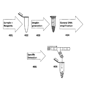
Figure 4: Outline of a scheme for performing NEEDLS
401: DNA sample containing one or more DNA target and reagents for general
amplification are mixed.
402: Components required for droplet generation are added to the mixture.
403: Droplets containing the DNA and reagents are generated.
404: General DNA amplification is carried out on all droplets.
CA 02950043 2016-11-22
WO 2015/189336
PCT/EP2015/063076
4
405: Specific amplification to detect the one or more target DNA is performed
on
DNA amplified on each droplet.
406: Droplets, where a specific amplification is detected are isolated using
an
apparatus for physical selection of droplets.
Figure 5: Outline of a scheme for performing NEEDLS as described in Example
1.
501: Mixing of template DNA, reagents for general DNA amplification and a
component required for droplet generation.
502: All material from (501) is converted into droplets of approx. 1 nL each.
503: All droplets are collected in one tube.
504: The droplets are incubated under conditions suitable for general DNA
amplification.
505: Droplets are aligned and individually merged with a surplus of liquid
comprising a complete dUTP-PCR mixture for selective target DNA amplification
and all droplets are collected in a PCR-tube.
506: The PCR tube is incubated under PCR reaction conditions.
507: The droplets in which a specific amplification has taken place are
collected
and the droplet generating component (e.g. oil) is removed. Uracil-DNA
Glycosylase (UDG) treatment can be used to inactivate all PCR generated DNA
into which uracil is incorporated. The DNA content is purified by ethanol
precipitation.
508: The precipitated DNA (507) is re-amplified.
509: The re-amplified DNA (508) is used as template for a final analysis.
510: DNA sequencing sequencing is carried out on the DNA generated in (509).
511: Sequencing results are retrieved from (510).
Figure 6: A schematic illustration of primers and gaps used for GAP-closing.
The horizontal black line illustrates a DNA sequence of 5712 bp. GAP
illustrates
the desired sequence where sequence data could not be obtained from the
paired end library. Primers are placed on both sides of the gap, and each set
is
placed in close proximity to the GAP. Primers are designed to target DNA and
generate fragments of 50-100 bp.
CA 02950043 2016-11-22
WO 2015/189336
PCT/EP2015/063076
Figure 7: Outline of a scheme for performing NEEDLS as described in example
2.
701: DNA, reagents for general amplification, and droplet oil are mixed.
702: Vortex is applied to mix liquids and to generate droplets of variable
sizes.
5 703: Droplets are incubated.
704: Large droplets are excluded and discarded.
705: Droplets are merged with dUTP mixtures wherein a subset of primers are
present. #1 - #4 is included in the figure for illustration purpose, whereas
10
different mixtures are included in the example.
706: PCR is performed.
707: Droplets where a positive PCR reaction is observed are sorted from the
mixture. Droplet oil is removed by sonication followed by DNA purification.
UDG
treatment is applied to break down all uracil containing DNA. The DNA content
is
then purified by ethanol precipitation.
708: Re-amplification of the eluted DNA is carried out to ensure a sufficient
quantity of DNA for nucleotide sequencing (e.g.NGS).
709: The amplified DNA is prepared for sequencing.
710: The complete mixture is sequenced and results are prepared for assembly.
711: Alignments of the received sequence is used to close gaps of the genome
sequence.
Figure 8: Target DNA fragment identified by NEEDLS comprising sequence of
16S rRNA and 23S rRNA genes assigned to Staphylococcus genus [SEQ ID No.
10]
DETAILED DESCRIPTION OF THE INVENTION
The present invention pertains to an in vitro method in which the
concentration
of a specific target DNA molecule is increased relative to the concentration
of
total DNA in a sample, by 1) dilution of a sample into multiple sub-
compartments such as droplets (separation), 2) non-specifically amplifying DNA
within the droplets (general amplification), 3) addition of reagents for
specific
detection of the target sequence to the droplets, 4) detection of the specific
target sequence within the droplets and, 5) physical selection of droplets
containing the target sequence (selection).
CA 02950043 2016-11-22
WO 2015/189336 PCT/EP2015/063076
6
The invention is based on the principle that, if only a fraction of the
droplets
contain the target sequence, the concentration of the target relative to total
DNA
is higher in these droplets, compared to the concentration in the original
sample.
The fraction of droplets containing the target determines the degree of
enrichment; if the fraction is low, the enrichment is high.
In the context of the present invention, the presence or absence of the target
DNA molecule in a sample of total DNA or a dilution thereof or droplet, is
defined
by the presence of detectable target DNA molecule in the sample or a dilution
thereof or droplet using the selected method of detection (e.g. PCR).
The target can be further enriched by further rounds of selection (as above),
until it can be sequenced by standard methods such as Sanger sequencing or
Pyro sequencing or similar detection of DNA sequence or by PCR, hybridization
or other detection assays, or it can be used directly from the first round of
selection.
Droplets amplified and sorted according to the invention contain DNA fragments
of 5-100 kb containing the DNA sequence used for detection and identification.
Surprisingly little prior DNA sequence information is needed for the
enrichment
according to the invention. In comparison to current enrichment technologies,
only approximately 40 nucleotide base pairs of specific target information is
required as compared to at least 5-8000 and 300 base pairs respectively for
hybridization based and long range PCR based methods respectively (Figure 1).
It is known in the art, that the most sensitive PCR reactions are obtained
when
the fragment is short, such as 100-250 base pairs. Therefore, long range PCR,
designed to amplify DNA fragments that are longer than 250 base pairs, for
example 500 to 5000 base pairs in length, may not be applicable when the
sample contains high amounts of background DNA. Also, hybridization based
methods require the sample to be relatively pure to avoid non-specific
hybridization. The only way to obtain sequence information from a mixed sample
may therefore be sequencing of the entire DNA sample by e.g. next generation
sequencing methods, and, although the cost of next generation sequencing is
rapidly decreasing, the cost of sequencing thousands of genomes is still high.
CA 02950043 2016-11-22
WO 2015/189336
PCT/EP2015/063076
7
The method of the invention is surprisingly efficient, whereby the extent of
DNA
sequencing can be reduced by a factor of more than 1 billion for 5 multiplexed
DNA targets by three rounds of NEEDLS or a factor of 25 million for one target
sequence using two rounds of NEEDLS (Figure 2).
I: Droplet Exclusion Enrichment of Nucleotide Sequences
The essential steps of the NEEDLS method are further described below:
a) Providing a DNA sample comprising one or more specific target DNA
molecule and reaaents for general amplification of DNA (401)
A sample of mixed DNA molecules (i.e. a mixed population of DNA
molecules) known to comprise a target DNA molecule, is selected for
performing NEEDLS. One or more unique nucleotide sequences of at least
10 (or 15) nucleotides located within the target DNA molecule is selected
for screening and detecting the DNA molecule by a desired method, such
as PCR detection, DNA detection with hybridization probes or similar. A
target DNA molecule may contain more than one unique nucleotide
sequences, each sequence corresponding to a given genetic marker, for
example a first genetic marker sequence diagnostic of an infectious agent
and a second genetic marker diagnostic of an antibiotic resistance gene.
Typically, the frequency of the target DNA molecule in the sample of
mixed DNA molecules is less than 10-2, it may for example lie between
10-3 and 10-9 (calculated as base pairs of target sequence divided by base
pairs of total DNA in the sample). Prior to amplification, the liquid sample
of mixed DNA molecules is serially diluted by a desired number of
dilutions until each droplet that is generated and processed in the
subsequent droplet formation step contains mixed DNA molecules but
contains less than 0.5 target DNA molecule on average, preferably less
than 0.25 or even more preferably less than 0.1 specific target DNA
molecule on average. Thus, if the liquid sample of mixed DNA molecules
is separated into 100 droplets, each containing mixed DNA molecules,
then on average the target DNA molecule will be present in less than 50
of these droplets, preferably less than 25 of these droplets, even more
preferably less than 10 of these droplets. The presence or absence of the
target DNA molecule in a droplet is defined herein as the presence or
absence of detectable target DNA molecule when employing methods for
CA 02950043 2016-11-22
WO 2015/189336 PCT/EP2015/063076
8
specific detection of the target DNA molecule, such as those exemplified
in the present application. This dilution is performed to ensure target
enrichment; if the average number of droplets containing target is low,
the frequency of target relative to non-target molecules within the droplet
is high. The frequency and abundance of the target DNA molecule in the
mixed DNA sample may be determined by PCR, real time PCR, by
hybridization based assays or by assays detecting an RNA or protein
product of the target sequence.
b) Formation of a multiple of droplets containing said DNA sample (403),
Droplets containing diluted sample mixed DNA molecules and reagents
necessary for general amplification of DNA are generated using any
method of droplet generation to isolate target DNA sequences in closed
compartments. Suitable methods for droplet generation include active
methods such as acoustic energy ejected droplets, dielectrophoresis
(DEP) and electrowetting on dielectric (EWOD), and passive methods
such as T-junction and flow focusing [1]. In addition to droplets, the
general amplification can occur in other micro-volume compartments,
such as reaction chambers in microfluidic chips.
c) 3eneral amplification of DNA molecules in a multiple of droplets each
containing mixed DNA molecules and less than 0.5, preferably less than
0.25 or even more preferably less than 0.1 specific target DNA molecule
on average (404)
DNA in each droplet is amplified using any method of total DNA
amplification to increase the abundance of the DNA in each sample.
Suitable amplification methods including Degenerate Oligonucleotide
Primed PCR (DOP-PCR), Multiple Displacement Amplification (MDA)[4],
randomly primed PCR or similar.
d) Specific detection of droplets containing said specific target DNA(405)
Following general amplification of total DNA in step c) the droplets are
screened for the presence of the target DNA molecule using the desired
detection technique. In at least one or more screened droplets that are
shown to contain the target DNA molecule, the frequency of the target
DNA molecule will be increased compared to its frequency in the sample
of mixed DNA molecules in step (a). The increase in frequency is typically
between 0.1 x (number of droplets without target) x (number of droplets
CA 02950043 2016-11-22
WO 2015/189336
PCT/EP2015/063076
9
with targetyl and 10 x (number of droplets without target) x (number of
droplets with target)* Alternatively, increase in frequency is calculated
to lie between 0.1 x (number of droplets without target) x (total number
of DNA containing droplets)-land 10 x (number of droplets without
target) x (total number of DNA containing droplets)* The number of
droplets containing target is typically between 2 and 100 per target
sequence. The total number of droplets is at least 1,000, but typically
greater than 10,000.
The presence of the target DNA molecule in the droplets may be
determined by PCR including qPCR, by hybridization based assays or by
assays detecting an RNA or protein product of the target sequence. The
reagents for specific detection may contain dUTP to make it possible to
selectively inactivate, degrade or remove the DNA amplified in the
detection step using UDG, in a subsequent step.
e) Physical selection of droplets containing said specific target DNA (406)
Based on the detection of target DNA in step d) droplets are sorted into
at least two different streams. When more than one specific target is
detected in step d), the droplets may be sorted into 3, 4, 5 or more
streams. In the stream containing droplets wherein the target DNA is
detected, the abundance of the target DNA relative to non-target DNA in
the droplets is enriched as compared to the sample of mixed DNA
molecules in step a).
Optional steps:
f) inactivating, degrading or removing DNA produced for specific detection
of target DNA
When the enriched target DNA is used for further rounds of NEEDLS or
other applications where the presence of the detection product interferes
with these further processing, the amplification of DNA in the detection
step c) can be performed using dUTP in place of one of the
deoxyribonucleotide (dNTPs), where the product may then be optionally
selectively degraded, inactivated or removed. This inactivation may be
performed using an enzyme such as Uracil-DNA glycosylase, also known
as UNG or UDG.
CA 02950043 2016-11-22
WO 2015/189336 PCT/EP2015/063076
g) Reoeatino steps (a) to (e)
Using the droplets containing enriched target DNA obtained in (e) in a
new step (a), the target DNA may be further enriched.
h) Amplifying the enriched sample
5 Using the droplets containing enriched target DNA obtained in (d), the
target DNA may be further amplified using a general amplification such as
MDA or a specific amplification such as PCR.
Scheme for performing NEEDLS
10 The scheme is outlined in Figure 4. (401) Determine the concentration of
target
DNA molecules in the original DNA sample of mixed DNA molecules. Dilute the
sample until the expected average number of droplets containing a target
molecule (positive droplets) is less than 0.5. If the abundance of only one
target
is enriched relative to non-target DNA in a droplet, the average number of
positive droplets should be smaller resulting in a greater enrichment.
Ideally, the
entire sample of droplets should contain 2-100 positive droplets per target
preferably 3-50 and more preferably 5-20. If more than one sequence variant of
the target may be present, each variant counts as a separate target DNA
molecule. The correlation between number of targets, the average number of
positive droplets and the resulting enrichment after NEEDLS is shown in Figure
3
using an example of one round of NEEDLS, 20,000 droplets and 10 positive
droplets per target.
The observed enrichment may be higher than the expected enrichment of figure
3, as each of the positive droplets may contain different amounts of DNA
amplified in the general amplification due to bias of this amplification step.
In
our experience the resulting enrichment may be at least two fold higher. Also,
if
smaller droplets are used such that more separate compartments are obtained,
this will result in a greater enrichment.
Add reagents for multiple displacement amplification (MDA) to the diluted
original sample; denature the DNA, anneal primers, and add the DNA
polymerase for general amplification (e.g. 029 DNA polymerase). Following the
addition of the polymerase, generate droplets containing the MDA-ready sample
in order to isolate the target molecules in separate compartments (403). Then,
CA 02950043 2016-11-22
WO 2015/189336 PCT/EP2015/063076
11
amplify the targets within the droplets by incubating the samples under
conditions suitable for MDA (404).
Add reagents for detection to the droplets in order to allow detection of the
target sequences or target sequences in the sample. These may be added either
together with the reagents for general amplification (401) or after general
amplification (between 404 and 405). If detection is performed using PCR;
transfer the amplified droplet sample to a PCR unit and perform PCR
amplification and then sort the droplets according to the presence (positive)
or
absence (negative) of the target molecule. Where fluorescent labelled PCR
primers are used, the presence of the target molecule can be detected by the
fluorescence of the samples using a fluorescence-activated droplet sorter (405
and 406).
Some positive droplets will give a higher fluorescence. When using a cut-off
value to select the droplets with the highest fluorescence, this will select
for
droplets having a correspondingly greater enrichment. Determine the abundance
of the target DNA molecule, and if this is sufficient for analysis methods,
such as
sequencing, the selected sample may be analysed directly; otherwise additional
rounds of NEEDLS can be applied.
When it is necessary to perform further rounds of enrichment, it may be
preferred to degrade or remove the DNA generated in the detection step. It may
be preferred to use dUTP in the PCR reagents. The MDA reaction is then
performed using standard dNTPs. After detection and physical selection of
droplets, the DNA generated in the PCR can be degraded and the treated sample
can be used for an additional round of enrichment, starting with dilution and
multiple displacement amplification in droplets.
When a sufficient enrichment is reached using NEEDLS, the droplets are
coalesced. The enriched DNA in the coalesced droplets may also be further
purified and may be further amplified using general or specific amplification
such
as MDA and PCR respectively.
II: Multiplex NEEDLS
CA 02950043 2016-11-22
WO 2015/189336
PCT/EP2015/063076
12
NEEDLS can be adapted to perform multiplex NEEDLS. Multiplex NEEDLS
employs additional features that are designed to detect a 2nd consecutive
sequence of at least 10 (or 15) nucleotides in the sample of mixed DNA
molecules analysed, by amplification of this 2nd consecutive sequence with
sequence specific primers to generate a 2nd target DNA molecule. If several
droplets show specific detection (e.g. by fluorescence signal) from both the
1st
and 2nd consecutive sequence then they must be located on the same MDA-
amplified target DNA molecule. Similarly, it is envisaged that the unique
sequence of from 1 to 20 or more different specific target DNA molecules can
be
detected in a mixed sample of DNA molecules, using the method of the
invention, by employing specific primers to detect each of the different
specific
target DNA molecules.
Accordingly, in addition to providing information concerning co-localisation
of
targets, multiplex NEEDLS provides simultaneous purification of up to
thousands
of different target molecule, each comprising more than 5,000 base pairs.
Separate detection molecules can be provided to separate droplets or can be
added as a mixture.
III Samples analysed by NEEDLS and Multiplex NEEDLS
111.1 Sample of mixed DNA molecules
NEEDLS may be applied to a sample of mixed DNA molecules known to comprise
a target DNA molecule. A sample of mixed DNA molecules comprises a
population of DNA molecules (e.g. chromosomal DNA molecules or plasmid DNA
molecules) where the individual DNA molecules within the population differ by
at
least one nucleotide within a known consecutive sequence of at least 10 (or
15)
nucleic acid base pairs in their DNA, such that a target molecule comprising
the
known consecutive sequence differs from, and can be distinguished from, non-
target molecules in the sample. The sample of mixed DNA molecules may
additionally comprise single stranded RNA or DNA polynucleotides. The
population of DNA molecules in the sample of mixed DNA molecules comprises
the target DNA molecule.
The target DNA molecule can be in linear or circular forms. Circular DNA can
occur naturally or can be obtained by cloning DNA into plasmids, fosmids,
CA 02950043 2016-11-22
WO 2015/189336 PCT/EP2015/063076
13
cosmids, BAC clones, or generated by ligation or through Cre/LoxP mediated
recombination.
A target DNA molecule comprises one or more known unique consecutive
sequence of at least 10 (or 15) nucleic acid base pairs (or nucleotides). A
target
DNA molecule can be selected from a sample of mixed DNA molecules, by
selecting for a target DNA molecule comprising this unique consecutive
sequence
of at least 10 nucleic acid base pairs (or nucleotides). The target DNA
molecule
can also be selected from the sample of mixed DNA molecules, by selecting for
a
target DNA molecule comprising at least two unique consecutive sequences of at
least 10 (or 15) nucleic acid base pairs (or nucleotides), wherein the two
consecutive sequences are comprised within a DNA molecule of 50 to 100,000
nucleic acid base pairs, preferably 150 to 3,000 nucleic acid base pairs, more
preferably 150 to 1500 nucleic acid base pairs.
Typically, the frequency of the target DNA molecule in the sample of mixed DNA
molecules is less than 10-2, it may for example lie between 10-3 and 10-9
(calculated as base pairs of target sequence divided by base pairs of total
DNA in
the sample).
The method of the invention is particularly suitable where the frequency of
the
target DNA molecule in the sample of mixed DNA molecules is less than 10-2.
The method of the invention is also suitable where the frequency of the target
DNA molecule in the sample of mixed DNA molecules is 10-4, 10-5, 10-6, 10-7,
10-
8, 10-9, 10-10, 10-11, or lower. In many instances, the sample of mixed DNA
molecules will be derived from a cell population comprising genomic DNA, while
in other instances, the sample may be derived from samples where the DNA
molecules are of diverse origin, such as samples collected from nature.
Irrespective of its source, the frequency of the target DNA molecule is
defined as
the number of specific target DNA base pairs divided by the number of total
base
pairs in the sample. The frequency of the target DNA molecule in the sample of
mixed DNA molecules is determined by making a dilution series in triplicate,
detecting the presence or absence of target and determining the number of
targets using, for instance, most probable number methods. The frequency of
the target molecule can also be determined using qPCR or digital droplet PCR
[2]. The concentration of DNA is measured and the number of total genome
CA 02950043 2016-11-22
WO 2015/189336 PCT/EP2015/063076
14
equivalents is determined by dividing this concentration by the average
molecular weight of the genome.
III.ii Source of the sample of mixed DNA molecules
According to one embodiment of the present invention, the target DNA molecule
is derived from the genome of a cell, where the genome may be either
chromosomal or extra-chromosomal DNA. Further, the target DNA molecule may
be derived from a cell, where the cell is selected from amongst a microbial
cell, a
plant cell, an animal cell, or a mammalian cell. The mammalian cell may be a
human cell. The microbial cell may be a bacterial cell, a yeast cell or a
fungal
cell. Furthermore, the target DNA molecule may be derived from a fungal
mycelium or fungal spores.
When the target DNA molecule is derived from one or more cell, the cell(s) may
be.part of a multicellular tissue or multicellular organism.
Furthermore, the target DNA molecule may be derived from one or more viral
particles, where the virus has an RNA or DNA genome. Alternatively the target
DNA molecule may be derived from a host genome comprising integrated DNA
derived from a virus. The target DNA molecule may also be derived from a
bacteriophage.
Irrespective of the derivation of the target DNA or RNA molecule, the target
DNA
or RNA molecule is present in a sample of mixed DNA molecules, where the
mixed DNA molecules may be derived from a sample collected from nature, for
example a sample of soil, water or air. Alternatively, the sample may be
derived
from a multicellular organism, such as a mammal, for example an animal or a
human subject. When the sample is derived from a mammal, the sample (for
example a biopsy) may be derived from a body fluid (e.g. blood, plasma, serum,
lymph and urine), from faeces or from a body tissue or organ. The
multicellular
organism from which the sample is derived may be a living or may be a dead
organism.
III.iii Preparation of the sample of mixed DNA molecules
CA 02950043 2016-11-22
WO 2015/189336 PCT/EP2015/063076
The sample of mixed DNA molecules comprising the target DNA molecule may be
prepared from a sample collected from nature or from an organism (e.g. a
biopsy). Methods for selective extraction of DNA molecules are known in the
art
[3]. When the target DNA molecule is derived from a cell, the step of cell
5 disruption or cell permeabilisation is normally required in order to
release total
nucleic acid molecules (including DNA or RNA) from a cell, this step preceding
the subsequent step of selective extraction of DNA molecules.
Where the target DNA molecule is derived from an RNA genome, the RNA
10 genome or parts thereof are first reverse transcribed to provide a cDNA
molecule, where the nucleotide sequence of the cDNA corresponds to (is a
reverse transcript of) the RNA genome.
III.iv Generation of droplets
15 Methods of the invention include forming multiple sample droplets where
the
droplets each contain less than 0.5 specific target molecule on average. In
the
preferred embodiment the distribution of specific target molecules follows
Poisson distribution. Some droplets may contain non-target molecules present
at
10 fold or higher concentrations as compared to the target molecule, while
other
droplets may contain only the target molecule.
Generally, droplets can be formed by a variety of techniques such as those
described in [4-6]. Methods of the invention may involve forming a two phase
system comprising aqueous droplets surrounded by an immiscible carrier fluid.
In a preferred embodiment, the aqueous sample within the droplet is prepared
by preparing a mixture containing sample DNA, primers such as random
hexamer primers and buffer solutions. The DNA mixture is subjected to
conditions resulting in denaturing of the DNA such as temperatures of around
94 C for 1-10 minutes. The mixture is rapidly cooled and added to a mixture
containing dNTPs and a polymerase useful for general amplification, such as
Phi29 polymerase. The resulting mixture is used as the aqueous sample in two-
phase liquid droplet formation using two immiscible liquid phases. Aqueous
droplets are either generated in an apparatus having means for creating a
vortex/turbulence in a sample comprising two immiscible liquid phases in
controlled environments, creating droplets of liquid (phase 1) in a 2nd phase
liquid by controlling the mechanical parameters and thereby also the liquid
CA 02950043 2016-11-22
W02015/189336 PCT/EP2015/063076
16
volume of each generated droplet or by a means for extruding droplets of one
liquid phase in a 2nd immiscible liquid phase, where the so formed droplets
remain discrete and wherein the volume of the droplet is controlled by the
diameter of the means for droplet extrusion.
The carrier fluid is one that is immiscible with the sample fluid. The carrier
fluid
can be a non-polar solvent, decane, fluorocarbon oil, silicone oil or any
other oil
(for example mineral oil).
In certain embodiments, the carrier fluid contains one or more additives such
as
agents which increase, reduce, or otherwise create non-Newtonian surface
tensions (surfactants) and/or stabilize droplets against spontaneous
coalescence
or contact.
IV Methods of General Amplification of DNA suitable for NEEDLS
A range of different approaches have been suggested for general amplification
of
DNA, such as randomly degenerate primed PCR, linker ligation PCR, or,
Degenerate Oligonucleotide Primed (DOP) PCR and Multiple Displacement
Amplification (MDA). MDA has proven efficient in performing whole-genome
amplification (WGA) of even very small amounts of DNA [7]. Compared with
more traditional PCR-based WGA methods, MDA generates DNA molecules with a
higher molecular weight, having better genome coverage. MDA employs a strand
displacement polymerase that possesses two enzymatic activities: DNA synthesis
(polymerase) and an exonucleolytic activity that degrades single stranded DNA
in the 3'- to 5'-direction, as exemplified by bacteriophage phi29 DNA
polymerase, that belongs to eukaryotic B-type DNA polymerases
(UniProtKB/TrEMBL: Q38545). Other useful polymerases include BstI
polymerase.
To obtain the- enrichment according to the invention, general amplification is
performed within droplets. The droplets serve to isolate target molecules into
compartments separate from compartments not containing target DNA.
Amplification is performed in each droplet for example by using any of the
above
listed general DNA amplification methods. In some embodiments, the
amplification is performed at around 30 C for one hour, preferably for less
than
30 minutes.
V Methods of adding detection reagents to droplets
CA 02950043 2016-11-22
WO 2015/189336 PCT/EP2015/063076
17
Following general amplification, reagents for detection of target DNA
molecules
may be added to the droplets. Alternatively, the reagents for detection may be
added before droplet generation. Addition of reagents to droplets may be
performed using an apparatus with means for providing aliquots of an aqueous
liquid (e.g. comprising PCR reaction or other detection mixture), and means
for
fusing said aliquots with droplets of an aqueous liquid that are suspended in
a
2nd immiscible liquid (e.g. droplets from general amplification step), and
means
for delivering said fused liquid droplets suspended in a 2nd immiscible liquid
to a
further compartment. Examples of droplet fusion or droplet injection
techniques
are described in [5, 8]. In certain embodiments of the invention, the reagents
added to the droplets containing generally amplified DNA comprise specific
primers or a specific probe complementary to the specific target DNA to be
detected. When specific primers are added, the reagents comprise a DNA
polymerase such as Taq polymerase and dNTPs. In addition, the reagents may
contain dUTP, to enable subsequent degradation of DNA generated in the
detection step, and/or a nucleic acid dye enabling detection based on
fluorescence. In other embodiments the detection is based on fluorescence from
labelled probes or primers.
VI Methods of detecting the target sequence
Methods of the invention further involve detection of the target nucleic acid
molecule within the droplets containing DNA amplified using general
amplification. In certain embodiments the detection involves amplification of
a
part of the target molecule. The amplification reaction, that is suitable for
amplifying nucleic acid molecules, includes the polymerase chain reaction, or
nested polymerase chain reaction including or excluding probes such as Taqman
probes, Scorpion probes, Molecular Beacon probes, and any other probe that
functions by sequence specific recognition of target DNA by hybridization and
result in increased fluorescence on amplification of the target sequence.
Methods according to the invention also include methods wherein detection is
based on fluorescence from optically labelled probes such as fluorescently
labelled probes wherein the target is not amplified after general
amplification. In
this case, the DNA is denatured e.g. by increasing the temperature to around
95 C and the probe is subsequently allowed to anneal to the target, resulting
in
activation of the probe or probes. Such optically labelled probes can be
Molecular
CA 02950043 2016-11-22
WO 2015/189336 PCT/EP2015/063076
18
Beacons, where a single-stranded bi-labeled fluorescent probe is held in a
hairpin-loop conformation of around 20 to 25 nt by a complementary stem
sequences of around 4 to 6 nt. Due to the loop-structure the desired
fluorochrome attached to one end of the sequence is in close proximity of the
light quencher attached to the other end. When the structure is released
during
denaturing and then re-annealed, the probe anneals to an amplified target.
When annealed, the hairpin structure is no longer maintained, and the quencher
no longer quenches emitted light from the fluorochrome. The optically labelled
probes can also be FRET (fluorescence resonance energy transfer) probes.
VII Methods of physically selecting droplets based on presence of the
target sequence
To selectively separate droplets comprising a detectable target DNA molecule
from droplets wherein the target is not detected, a variety of different
methods
for physical selection of droplets or droplet sorting can be employed
including
steering, heating, and acoustic waves [5]. Such physical selection can be
carried
out using an apparatus with means for receiving droplets of an aqueous liquid
that are suspended in a 2nd immiscible liquid (e.g. droplets from specific
detection step), and means for passing each droplet past a detection unit
capable of detecting a detectable component in said droplet, and means for
addressing said droplet for delivery to a selected compartment as determined
by the presence or absence of the detectable component and means for
delivering said droplet to the selected compartment.
VIII Methods of removing the detection signal molecules after physically
selecting the droplets
After physically selecting the droplets based on the presence of the specific
target sequence, it may in some cases be necessary to remove a detection
signal, such as a PCR product. Several methods for removing such signals are
known in the art. If dUTP has been used in the detection reaction, the
detection
molecule may be removed using uracil-DNA N-glycosylase [9]. Alternatively, as
the molecules produced by general amplification are significantly longer than
the
detection molecules, the detection molecules can be separated using methods
based on size separation such as size exclusion based on differential binding
affinity of small and large DNA to silica particles [10]. Such silica surfaces
have
CA 02950043 2016-11-22
WO 2015/189336 PCT/EP2015/063076
19
limited binding efficiency to DNA fragments smaller than 100 bp, and
consequently only DNA fragments smaller than 100 bp will be efficiently
discarded, when silica based purification in applied. In some applications,
however, it may not be necessary to remove the detection molecule. For
instance, since Phi29 and some other polymerases have low activity on DNA
molecules shorter than 1000 base pairs, it may not be necessary to remove the
detection molecule if the step following NEEDLS enrichment is a general
amplification, since the detection molecule will only be amplified to a
limited
extent, if at all amplified, compared to the actual targeted larger DNA
molecule.
IX Sequence determination of the target DNA molecule
Enrichment of the target DNA molecule by NEEDLS is based on detection of one
or more unique consecutive sequence of at least 15 nucleotides in said DNA
molecule. When detection is based on PCR, where the one or more unique
consecutive sequences are amplified to generate a target DNA molecule, the
nucleotide sequence of this molecule can be determined. In addition, the
nucleotide sequences flanking the target DNA molecule in the 5' and 3'
direction
can be determined by rapid genome walking (RGW)[11]. RGW is a simple, PCR-
based method for determining sequences upstream or downstream in a larger
DNA molecule starting from a known sequence, such as a target DNA molecule.
RGW enables individual amplification of up to 6 kb in a large DNA molecule
using
PCR. The sequences can be extended simply by performing multiple cycles of
RGW, using new primers based on the sequence obtained in previous cycles.
Typically libraries are constructed from a purified sample of the large target
DNA
molecule, by digesting the DNA separately with four different restriction
enzymes
and ligating the products to a specially designed adaptor. The ligated DNA is
then sequenced with primers annealing to the adaptors or to known sequences
within the DNA, using the desired DNA sequencing method, such as Sanger
sequencing, pyro sequencing, sequencing by synthesis, ligation or two base-
coding sequencing or similar methods [12].
The enriched target DNA sequence can also be sequenced using e.g. Sanger
sequencing, Emulsion PCR, Shotgun sequencing, SOLiD sequencing, bridge PCR,
Ion Torrent sequencing, Polony sequencing, Pyrosequencing, Sequencing by
synthesis, DNA nanoball sequencing, Heliscope single molecule sequencing,
CA 02950043 2016-11-22
WO 2015/189336 PCT/EP2015/063076
Nanopore DNA sequencing, Tunnelling currents DNA sequencing, Sequencing by
hybridization, Sequencing with mass spectrometry, Transmission electron
microscopy DNA sequencing, RNAP (RNA Polymerase) sequencing or Single-
molecule real-time sequencing.
5
IX.i Research and development applications of NEEDLS
Use of NEEDLS to isolate or enrich target DNA in a mixed sample of DNA
molecules extracted from samples collected from nature, or from clinical
samples
provides direct access to the genome, or parts thereof, that cannot be
analysed
10 by other methods because of the sample complexity. NEEDLS is
particularly
useful for isolating or enriching DNA molecules involved in hereditary
diseases,
cancer, and infectious diseases.
Multiplex NEEDLS is particularly useful for simultaneous isolation or
enrichment
of more than one target DNA from samples comprising several target DNA
15 sequences, such as a sample to be analysed for the DNA sequence of more
than
one virus, more than one hereditary disease or more than one cancer related
gene.
NEEDLS is also particularly useful for obtaining target DNA sequence
information
from samples where only a small part of the sequence is known prior to
20 enrichment, since the technique only requires a small part of the target
DNA
sequence to be known in order to perform the detection step and furthermore
takes advantage of the high fidelity of polymerases (like Phi29) in the MDA
amplification to generate large amplified DNA molecules of up to 100,000 bp.
IX.ii Sample preparation for sequencing
When performing sequencing of large DNA molecules such as genonnes, the
sequence information obtained will often contain gaps where the sequenced
molecules do not overlap or cannot be assembled. NEEDLS is particularly useful
for gap closing of DNA sequences, as NEEDLS will retrieve up to 100,000 bp of
sequence surrounding the small detection area and can therefore be designed to
cover the unknown gap region.
NEEDLS is also particularly useful when sequencing samples containing more
than one variant of a target DNA sequence such as chromosomal DNA containing
CA 02950043 2016-11-22
WO 2015/189336 PCT/EP2015/063076
21
two alleles of a gene. In this case, droplets containing each detection
sequence
are collected in separate compartments to ensure that only one copy of the
sequence is present. The droplet may subsequently be barcoded separately to
enable the separate sequencing of each variant of the target DNA sequence.
IX.iii Diagnostic applications of NEEDLS
NEEDLS may be used to analyse target DNA in samples derived from
multicellular organisms, such as a biopsy or a sample of body fluid or faeces
obtained from a subject (e.g. human or animal subject), for the diagnosis or
monitoring the progress of a medical indication or disease.
Diagnosis of a wide range of medical indications in a subject such as a
disease
caused by an infectious agent (e.g. micro-organism or virus) can be assisted
by
the isolation or enrichment and detection of a target DNA or RNA molecule that
is derived from the genome of the infectious agent by NEEDLS or Multiplex
NEEDLS, where the target DNA molecule is detected in a sample of mixed DNA
molecules derived from a biopsy or sample of body fluid obtained from a
patient.
Use of NEEDLS in target DNA molecule isolation provides the additional
feature,
that additional diagnostic features of the disease can be determined. For
example, where the genome of the infectious agent comprises resistance genes
that confer resistance to certain therapeutic agents, enrichment of a
resistance
gene may also retrieve the region surrounding the resistance gene and may
provide information about the specific infectious agent carrying the
resistance.
NEEDLS may be applied to diagnostics such as detection of presence of an
infectious agent such as a prokaryotic organism and antibiotic resistance.
Such
cases can be the presence of methicillin resistance (MR) in Staphylococcus
aureus (SA). The combination of (MRSA) is a well-known problem in hospitals
and similar facilities, whereas MR may not cause comparable problems if it is
not
present in SA. NEEDLS can be used for detection of co-existence of MR and SA
if
a method such as duplex detection is applied. Such duplex detection can be a
dual-reporter detection system, where one detection system is used to monitor
the presence of one event such as MR whereas another detection system is used
to monitor the presence of another event, such as presence of SA within the
CA 02950043 2016-11-22
WO 2015/189336 PCT/EP2015/063076
22
same droplet. When both MR and SA are present in the same droplet, the two
loci are likely to be localized on the same DNA fragment. Moreover, NEEDLS can
be applied to selectively retrieve additional DNA sequences from the host
genome based on the presence of genes such as MR or SA. Thus, it can be used
to provide additional sequence information from the genome of the infectious
organism.
NEEDLS may also be used to assist diagnosis of a disease caused by, or
originating from the presence of a viral agent in a subject. Using multiplex
NEEDLS, the presence or absence of viral DNA at a known integration site can
be
determined by tracking the co-enrichment of the viral DNA and integration site
DNA sequence.
NEEDLS may be applied to multiple genes within a single genome or within
multiple genomes, which may be detected by any suitable molecular detection
method. If required, a series of multiplex PCR reactions can be carried out
and
differentiated using specific dyes for each reaction. This can be done by
introduction of detection systems such as probes such as Taqman probes,
Scorpion probes, Molecular Beacon probes or similar. Moreover, NEEDLS can also
be applied to a series of genes which are not necessarily differentiated at
the
point of detection. Differentiation can be applied after sequence retrieval,
using
such methods as bar-coded PCR or similar.
X NEEDLS and bias in amplification
NEEDLS is based on specifically selecting samples where amplification of a
desired DNA region from a complex, mixed DNA sample has occurred. Although
Phi29 based amplification (MDA) has been described repeatedly as the most
reliable genome amplification currently available, it is known to introduce
significant bias. Pan et al. [18] states in general terms that a highly
specific
whole genome amplification (WGA) of complex DNA pools which avoids
amplification bias remains a challenge. Moreover, similar observations are
seen
with alternative amplification methods such as DOP-PCR and random priming
PCR. These two amplification methods are described as being much less
efficient
at reproducing the locus representation [19], resulting in even more biased
amplification products. While bias is seemingly unavoidable, regardless of the
amount of reaction template [20] present, the amount of template independent
CA 02950043 2016-11-22
WO 2015/189336 PCT/EP2015/063076
23
product (TIP) or bias introduced during amplification is seemingly correlated
negatively to the amount of DNA template in the reaction and has in some
studies been documented to represent 70-75% of the total yield [18]. Whole
genome amplification is applied to amplify DNA in the NEEDLS process and it
would therefore be expected that these general challenges relating to bias in
genome amplifications would also apply to NEEDLS. As a procedure including
WGA it would thus be expected that significant bias against the target DNA
molecule should be observed. Hence a procedure such as NEEDLS employing
WGA would not have been considered as a method capable of enriching for a
specific region of DNA in a mixed sample.
Surprisingly, the challenge of TIP/bias is not seen when applying the NEEDLS
technique even though the initial concentration of the target DNA molecule is
very low. The minimal negative TIP/bias observed in the NEEDLS system is
significantly lower than the overall gain obtained from the amplification
process
and the net result is therefore a substantial enrichment of the target DNA
molecule.
The method does not require a dilute initial DNA template, as high
concentrations of DNA also can efficiently be subjected to NEEDLS. In this
case
only a few fold of amplification by the WGA method is needed.
XI NEEDLS reduces analytical burden
NEEDLS provides a highly efficient analytical tool for detecting and
characterising
a DNA target in a large genome of a biological sample, for example the human
genome of a biopsy. This can be illustrated with the following example:
The analytical task is to detect and determine the nucleotide sequence of a
target DNA molecule in a sample of mixed DNA fragments comprising 100,000
DNA molecules with an average size of 30 kb, wherein 1 molecule is the target
DNA. This sample of mixed DNA fragments corresponds to approximately 1
human genome of 3 billion base pairs with an average DNA fragment length of
30 kb. The nucleotide sequence of only 200 base pairs out of the 30 kb target
DNA is known at the beginning of the analytical procedure.
When implementing the method of the present invention, then:
CA 02950043 2016-11-22
WO 2015/189336 PCT/EP2015/063076
24
1. The DNA is aliquoted into droplets such that each droplet contains mixed
DNA
fragments, and such that there are 5 DNA fragments per droplet, on average.
The number of droplets will therefore be 100,000/5 = 20,000 droplets. Only one
of the 20,000 droplets will contain the target DNA molecule (thus, each
droplet
will contain less than 0.1 copy of the target DNA molecule).
2. The DNA in all droplets is then copied by general amplification resulting
in
20,000 droplets containing amplified DNA, and the target DNA molecule is
specifically detected in the same droplets (e.g. by PCR). The droplet
containing
target DNA is physically selected (sorted), and the DNA sequenced. Note that
selection of the droplet comprising the one target DNA molecule, and (on
average) 4 non-target DNA molecules, corresponds to an increase in frequency
(enrichment) in the range of 2 x 10³ and 100 x 103 [i.e. between 0.1 x (number
of droplets without target) x (number of droplets with target)-1 and10 x
(number
of droplets without target) x (number of droplets with target)-1].
3. Thus, when using the NEEDLS method of the invention, a total of 20,000
droplets must be prepared with amplification reagents (and detection
reagents),
and the detected droplet comprising the amplification product of the 5 DNA
molecules (on average) each of 30 kb (on average) (=150,000 base pairs) must
be individually sequenced in order to detect and analyse the 1 target DNA
molecule.
When traditional methods for analysing mixed DNA samples are used for this
analytical task, the analytical burden is many fold increased because the
mixed
DNA sample must first be converted into a library of cloned DNA fragments or
amplified DNA fragments, where each member must be analysed, as illustrated
by a classical protocol set out below:
1. The mixed DNA sample is aliquoted into droplets under conditions where the
likelihood of a droplet containing 2 DNA molecules is low, e.g 0.1%
corresponding to a statistically conservative probability of p=0.001 or less.
The
number of droplets required for this method is based on the number of DNA
molecules and the probability of finding <1 molecule in an aliquot. This can
be
calculated (approximately) as the square root of the probability of finding 2
DNA
molecules in the same droplet (i.e. 0.03162).
CA 02950043 2016-11-22
WO 2015/189336 PCT/EP2015/063076
Since the mixture DNA sample has 100,000 DNA molecules, then a conservative
estimate of the number of droplets required (to ensure that the likelihood of
2
DNA molecules in one droplet is statistically insignificant) is 100,000 x
5 1/0.03162 or 3.16 x 10⁶ droplets (0.03162 x 0.03162 = 0.001). The DNA in
each
droplet can then be either 1) copied by general amplification or cloning,
and/or
2) the target DNA is specifically amplified by PCR.
2. All 3.16 x 10⁶ droplets are then generally amplified; wherein 100,000
droplets
10 will contain amplified DNA (of which 1 droplet will contain the target
DNA). The
100,000 droplets comprising amplicons will be selected, and all 100,000
selected
droplets must then be analysed in order to detect the target DNA molecule,
since
no enrichment of the target (increase in the target DNA/non-target DNA ratio)
is
achieved using this procedure.
15 If the 3.16 x 10⁶ droplets are directly and specifically amplified using
PCR with
primers designed to amplify the known sequence of 200 base pairs (as the rest
of the fragment is unknown it cannot be specifically amplified), then the 1
droplet containing target DNA may be detected and selected. However, here,
only 200 base pairs out of the 30 kb have been amplified and its flanking
20 sequences will remain unknown.
Accordingly, using this classical method, at least 3.16 million droplets must
be
prepared with amplification reagents, and the amplification product of at
least
100,000 DNA-containing droplets must be screened by PCR individually in order
to detect and to sequence the 1 target DNA molecule.
25 Table 1
Classical screening method Current invention
General Specific General
amplificication amplification amplification
Original sample
Target DNA molecules @
kb 1 1 1
CA 02950043 2016-11-22
WO 2015/189336
PCT/E1'2015/063076
26
Total DNA molecules @ 30
kb 1,00E+05 1,00E+05 1,00E+05
Target base pairs 3,00E+04 3,00E+04 3,00E+04
Total base pairs 3,00E+09 3,00E+09 3,00E+09
Ratio target to non-target 1,00E-05 1,00E-05 1,00E-05
Droplets
# Total droplets 3,16E+06 3,16E+06 2,00E+04
# With non-target DNA 1,00E+05 1,00E+05 2,00E+04
# With target DNA 1 1 1
Fraction of partitions with
more than one nucleic
acid Insignificant Insignificant High
After amplification
Total droplets 3,16E+06 3,16E+06 2,00E+04
Droplets with amplified
DNA 1,00E+05 1 2,00E+04
Droplets with amplified
known part of target DNA 1 1 1
Droplets with generally
amplified target DNA 1 0 1
After detection
Droplets selected/sorted 1,00E+05 1 1
End result
Amplified DNA (target bp) 3,00E+04 200 3,00E+04
Amplified DNA (bp total) 3,00E+09 200 1,50E+05
Ratio target to non-target 1,00E-05 Pure 2,00E-01
Enrichment none 200 bp fragments 10000 fold
CA 02950043 2016-11-22
WO 2015/189336 PCT/EP2015/063076
27 .
EXAMPLES
Example 1. Complete operon coding for a targeted enzymatic activity
from a compost sample
In this example it is demonstrated how a dual channel micro fluidics system
can
be applied to separate, amplify, identify, isolate, and re-amplify a targeted
lipase
DNA molecule. The targeted DNA including the adjacent DNA is made available
for sequencing from the indigenous host. The procedure is shown schematically
in Figure 5.
DNA extraction and initial PCR
A mixed sample from a compost pile (core temperature 38 C) was used for
retrieval of the initial DNA sample. DNA was extracted from 5 gram sample
using
bead-beating as described elsewhere [13]. The extracted DNA was initially
tested for presence of the targeted lipase gene, and was found to contain
sufficient amounts of DNA for the applied PCR primers Lip-Fw: 5' - CTG AAT
GGG GGA ATA ATG ACA AGC C - 3' [SEQ ID 1] and Lip-Re: 5' - CTA TAC TCT
TCT ITT AAT TCC TCA GC - 3' [SEQ ID 2] to yield a PCR product of
approximately 105 bp. PCR conditions were 94 , 62.1 , 72 (15 sec / 15 sec /
90 sec) for a total of 40 cycles.
Droplet MDA/Fusion/Sorting (physical selection)
A Phi29 reaction volume of 100 pL was created as described by Pan et al. [14].
While still keeping the reaction mixture cold (max. +4 C), the Phi29 mixture
was loaded into a well in an eight-channel disposable droplet generator
cartridge
(Bio-Rad). The same procedure was followed for additional wells, to use the
full
capacity of the droplet cartridge. Then, the remaining channels were filled
with
droplet oil (BioRad). The fully loaded droplet generator cartridge was then
placed
into the droplet generator (Bio-Rad) for droplet formation of the full
reaction
volumes in all reaction compartments of the cartridge (502).
After droplet formation had been completed, all droplets were manually
transferred to a 1.5 ml Eppendorf collection tube, and the amplification
reaction
was thereafter placed in an Eppendorf Thermoshaker for 16 hours at 30 C (504).
CA 02950043 2016-11-22
WO 2015/189336 PCT/EP2015/063076
28
Following incubation, the reaction was terminated by increasing the
temperature
to 65 C for 10 minutes. The entire reaction volume (still in the form of
separate
droplets inside the Eppendorf-tube) was then transferred to the droplet-fusion-
device (504) where the fusion of two separate streams were merged to generate
fusion droplets each having a 10 times larger volume than the original
SampliPhi
droplets.
The volume increase was applied by merging the streams (Stream 1: SampliPhi-
droplets where amplification had occurred & Stream2: dUTP-PCR where all the
reactions components for PCR detection are present). By merging the two
streams and carrying out the associated PCR reaction, it was enabled to
monitor
which merged droplets contained the desired target. Stream1 consisted of 1 nL
individual droplets of the sampliPhi reaction mentioned above, whereas 9 nL
droplets (stream 2) were formed from a mixture consisting of: 537 pL H20, 220
pL PCR buffer with Mg++ (x5), 110 pL dNTP/dUTP mix (2 mM), 110 pL of each
primer (10 pmol/pL), 110 pL BSA, 1 pL SybrGreen (1:10.000), 22 pL DreamTaq
polymerase (5U/pL).
The 10 fold volume surplus of dUTP-PCR was applied to ensure a proper dilution
of MDA-components in order to avoid PCR inhibition during dUTP-PCR
amplification and at the same time to establish a thorough detection basis in
the
following screening.
The fused droplets were collected in single PCR-tubes and were subjected to
the
same PCR conditions as described in the primary screening for target presence
(506). After finalizing PCR, the 10-11 nL droplets were aligned in a separate
micro fluidics chamber, and successfully amplified droplets were detected and
selectively separated based on a 525nm emission of fluorescent signal when
excitated with a 488nm laser beam (507).
A total of 16 droplets (out of 19.836) were detected as positive and were
physically selected using a micro channel sorting cartridge combining all
positive
droplets in one chamber. The sorted droplets were manually transferred to a
1.5
ml Eppendorf-tupe, by adding 20 pL 5 mM Tris to the collection compartment of
the micro channel sorting cartridge. Droplet immersion oil was removed by
addition of SDS, and the amplified products were purified using ethanol
CA 02950043 2016-11-22
WO 2015/189336 PCT/EP2015/063076
29
precipitation. The purified products were re-suspended in 5 pL Nuclease free
water.
The 5 pL volume of eluted dUTP-PCR products including the initial template was
subjected to selective degradation using the Uracil-DNA Glycosylase (UDG) kit
AmpErase (Life Technologies) in a reaction volume of 25 pL, leaving only the
initial template intact. Thereafter, the mixture was re-amplified using
SampliPhi
amplification (508) resulting in a final product concentration of 200 ng/pL in
a
total reaction volume of 20 pL.
PCR, identical to the initial screening, was used to verify the presence of
the
desired DNA target molecule, and RGW was used to obtain sequence from the
gene and DNA in both 5' and 3' to the gene to describe the full gene, with
promoter region, Shine-Dalgarno sequence and stop codon (510 and 511).
Example 2 ¨ Gap closing of genomes
This example describes how an unfinished genome sequence can be "gap-closed"
using a single pass of NEEDLS by selectively choosing the regions of interest
by
traditional primer design. In this example, gap-closing is implemented on a
paired end library which shows the presence of 57 gaps in the genome of
Thermoanaerobacter italicus (CP001936). In the current example, gaps range in
sizes from 422 bp to 5,201 bp. One single pass of NEEDLS followed by re-
sequencing by Next Generation Sequencing (NGS) efficiently closes all gaps in
the otherwise unfinished genome. The gaps are closed by specifically selecting
and sequencing those droplets in which amplification of the desired regions
have
taken place. By doing so, a selectively targeted re-sequencing was
accomplished, and all gaps were closed to generate a full circular genome.
DNA preparation.
2 ml anaerobic fully grown bacterial culture was prepared as described in
(BG10
patent) and extractions were carried out using Thermo Scientific Gene Jet DNA
Purification kit, as described by the manufacturer. Eluted, extracted DNA had
a
concentration of 20 ng/pL in a final volume of 100 pL.
MDA droplet generation and droplet amplification.
CA 02950043 2016-11-22
WO 2015/189336 PCT/EP2015/063076
The sample DNA is diluted to 5x10⁻⁴ and 1 pL is mixed with random Exo-
resistant heptamer and Exo-resistant hexamer primers in annealing buffer
(SampliPhi), and primers are annealed to the template by raising the
temperature to 94 C for 3 minutes and then gradually decreasing the
5 temperature to 20 C by using a BioRad MyCycler PCR machine at a ramp rate
of
5 C / min. Having reached a temperature of 20 C the mixture was transferred to
ice. Phi29 reaction buffer, dNTP, Water, and Phi29 polymerase was added to the
mixture as described in Example 1 (701).
Droplet generation oil was added to the reaction tube, and droplets were
10 generated by vortex treatment for 2 minutes, at 3000 RPM (Velp
Scientifica)
(702). Huge droplets were size excluded, by pumping the entire volume through
a funnel in a micro reaction chamber, and only droplets smaller than 5 nL were
allowed to continue to the amplification step. Size excluded droplets were
discarded (704).
15 The droplets having successfully passed size exclusion were incubated at
30 C
for 2 hours, and the amplification process was thereafter terminated by
raising
the temperature to 65 C for 5 minutes.
Primer design.
Primers are designed manually to target locations approximately 100 bp either
in
20 the 5' or the 3' direction of the gap. For gaps larger than 5000 bp two
sets of
primers were applied with one set of primers on each side of the gap as
illustrated in Figure 6.
Primerl-GAP-Fw: 5'- GAAGGGTGACAGGATTGATAC [SEQ ID No. 5]
Primer1-GAP-Re: 5'- CGGATTTCCTCCTTTCTATTCC [SEQ ID No. 6]
25 Primer2-GAP-Fw: 5'- GCCTTGCAAATTCTACATTGACAG [SEQ ID No. 7]
Primer2-GAP-Re: 5' - CCAAGAAAATCATGGGAGATAGTTC [SEQ ID No. 8]
Droplet fusion
Ten combinations of dUTP reaction mixtures (#1 - #10) each containing 5-6
pairs of designed primers (10 pmol/pL each) were developed in silico from the
30 GAP-filled paired-end genomic sequence and each combination contained 10-
12
CA 02950043 2016-11-22
WO 2015/189336 PCT/EP2015/063076
31
primers. The primer combinations were kept in separate liquid solutions and
aligned sequentially and thereafter combined with each of the MDA droplets, by
merging droplets and PCR-liquids sizes as described in [15] (705). The
resulting
final droplet sizes ranged between 10-20 nL. All merged droplets were then
collected in one reaction tube, and the dUTP-PCR reaction was initiated, as
described below.
dUTP-PCR
PCR amplification was performed on all merged droplets in one single tube,
carried out as a two stage PCR in a MyCycler PCR machine (BioRad) with the
following cycling parameters: 94 C (15 sec), 25 cycles consisting of a 15
second
denaturation at 94 C and a 15 second extension at 72 C (706). Following PCR
amplification 50,087 droplets were analysed and 1,807 were selectively sorted
using micro fluidics device for measurements and isolation as described in
[15]
(707).
UDG-treatment
Droplet oil was removed by adding SDS to a final concentration of 10% (w/w) to
the reaction and thereafter adding Ammonium Acetate to a final concentration
of
2M. The solution was placed on ice for 5 minutes, and was then precipitated by
centrifugation at 16.000 G for 10 minutes at 4 C. The double volume of 7M
Guanidine-HCI was added, and the mixture was cleaned up using a clean-up spin
column (GeneJet DNA extraction, Thermo Scientific). One wash procedure was
applied using the wash buffer included in the DNA extraction kit. 20 pL eluted
DNA was subsequently treated with UDG (Thermo Scientific) as described by the
manufacturer and the reaction was terminated by heat inactivation at 95 C for
ten minutes. The resulting product was ethanol precipitated by centrifugation,
and the DNA pellet was thereafter re-suspended in 5 pL (5mM) Tris buffer.
Pre-seauencing amplification and genome assembly
The total volume of 5 pL (UDG treated and precipitated) DNA template was used
as template in a SampliPhi re-amplification in a total volume of 50 pL to
create a
total of 1 pg amplified DNA (708). The total volume was nucleotide sequenced
by Eurofins Genomic (Ebersberg, Germany) (710), and the assembly of the
initial gap-filled genomes and the obtained GAP genome data were carried out
CA 02950043 2016-11-22
WO 2015/189336 PCT/EP2015/063076
32
using CLC Genomics Workbench version 6Ø4 for genome assembly at default
parameters. The final result showed a complete assembly of the genome and the
final circular genome sequence was found to have a total of 2,451,061
nucleotides. The closed GAP sequence is given as GAP in [SEQ IN No: 9].
Example 3 - Amplifying a specific sub population in a mixture
(Staphylococcus).
The current example illustrates how droplet based SampliPhi can be used to
isolate DNA from a specifically targeted sub-group of a mixed sample, while
still
maintaining the original variation in the sample. In this example
Staphylococci
are targeted by specific primers. Droplets selectively amplified with these
primers are isolated and finally PCR spanning the complete 16S rRNA conserved
sequence, the intergenic transcribed spacers (ITS) between the 16S and 23S
rRNA and the conserved 23S rRNA gene region is used for classification to
create
a high resolution phylogenetic analysis.
Specific primer design
Primers targeting 16S ribosomal DNA of Staphylococci were designed using
Primrose primer design software [16]. The conserved regions of the 16S genes
were targeted by specific primers Staph-Fw: 5' - AGA CTG GGA TAA CTT CGG
GA - 3' [SEQ ID NO: 3], and Staph-Re: 5' - CGT CTT TCA CTT TTG AAC CAT GC
- 3' [SEQ ID NO: 4] generating a PCR product of 76 bp.
Sample preparation.
A swab sample from a supposed infected tissue from a volunteer was used as
template, and DNA was extracted using GeneJet Genomic DNA purification
(Thermo Scientific). A preliminary PCR was used to verify the presence of the
targeted DNA sequence (Staphylococcus aureus). A 10-fold dilution series was
used for verification of quantity, based on Cq-values from RT-PCR analysis
together with melt curve characteristics. MPN, as described by Garcia-Armisen
et
al. [17] was used to convert the obtained PCR results to an estimated target
quantity. A total quantity of approx. 8,300 16S rRNA gene copies were re-
calculated to 1,709 targeted Staphylococci per pL, as each bacterial target is
estimated to have an average of 5.5 copies of 16S rRNA genes (701).
CA 02950043 2016-11-22
WO 2015/189336 PCT/EP2015/063076
33
Droplet formation
A 20 pL SampliPhi mixture containing 1 pL DNA template was prepared and
added to a droplet generator as described in Example 1, generating
approximately 20,000 droplets, each with 1 nL of SampliPhi Phi29 reaction
mixture (502). The reaction was incubated for 2 hours, at 30 C (504) and the
temperature was subsequently lowered to +4 C to halt the reaction.
Droplet fusion.
Immediately following finalized amplification, each droplet was merged with 9
nL
RT-PCR mixture to establish a reaction volume of 10 nL, with RT-mixture (SSO
Advance Supermix, BioRad) providing all required PCR reaction components. The
SSO Advance supermix was prepared as described by the manufacturer
supplemented with specific primers (Staph-F [SEQ ID NO: 3] + Staph-R [SEQ ID
NO: 4]) (505).
The total collection of merged droplets (approx. 10 nl/droplet) were separated
into 5 PCR tubes, each with 40 pL reaction volume corresponding to approx.
5,000 droplets in each tube. Specific amplification with Staphylococcus aureus
specific primers was then carried out in each of the five PCR tubes in a PCR
machine (BioRad Connect) where cycle conditions were: 30 cycles of (94 C,
60 C, 72 C) with each temperature interval maintained for 15 seconds (506).
After amplification, droplets were screened for successful reaction using
fluorescence activated droplet sorting (FADS) as described by [18] (507). The
number of positive reactions was 1,562 in a total of 20,238 droplets analyzed,
and all droplets with a monitored positive reaction (SybrGreen fluorescence)
were collected in a separate reaction tube.
Cleanup & Re-amplification.
Sorted SampliPhi amplified droplets were extracted using a spin column DNA
extraction procedure described by Yu et al. [19]. The procedure results
primarily
in purification of products larger than 100 bp as a result of size-dependent
binding efficiency of the silica membrane applied during cleanup.
Consequently,
mainly DNA products of sizes >100 bp are purified during cleanup. Thus, close
to
all PCR products were removed from amplification, whereas Phi29 amplified DNA
was easily recovered. 10pL eluted DNA (depleted of PCR-products due to PCR
CA 02950043 2016-11-22
WO 2015/189336 PCT/EP2015/063076
34
product size) were re-amplified by adding the entire volume to a SampliPhi
reaction of 50 pL (508). The final product was measured to have a
concentration
of 220 ng/pL corresponding to a total amount of 11 pg DNA. The nucleotide
sequence of the final product was determined.
Results - Example 3
A 16S rRNA clone library study of the sequence of the obtained DNA revealed
that all 16S rRNA genes were identical and that they could be assigned to the
Staphylococcus genus and, thus, it was concluded that the studied infection
originated from one single strain of staphylococcus (Figure 8) [SEQ ID: 10].
Moreover, the resolution of the sequence data obtained during this study
strengthens the degree of phylogenetic similarity of the obtained sequences
illustrating a clonal distribution rather than a multiplex infectious culture,
as all
obtained sequences were 100% identical. The combined analysis results, where
both 16S rRNA and 23S rRNA genes were present, provide a high accuracy of
phylogenetic grouping even if distantly related bacteria were present in the
analyzed sample. In addition, as the highly variable ITS region between the
16S
rRNA and 23S rRNA genes was also included in the analysis, the illustrated
example also provides information to distinguish even extremely closely
related
strains. We have used that information to distinguish between an outbreak of
an
infectious disease of clonal origin, and an infection initiated from multiple
strains.
Assembly of the sequences demonstrated a close to complete elimination of the
generated PCR products from the droplet screening procedure. Although the
small sizes of PCR products present an effect on silica based purification, it
is
surprising that also the Phi29 reaction seemingly does not initiate
displacement
amplification, probably due to the lack of sufficiently large sized template.
Phi29
amplification is known to be efficient when amplifying large pieces of DNA
while
amplification of DNA molecules of 76 bp is practically impossible.
Example 4 - Amplifying a specific E. con gene from a mixture of E. coli
and HeLa DNA.
The current example illustrates how droplet based SampliPhi was used to
isolate
DNA from a specifically targeted DNA sequence (ThrA gene from E. coli) in a
sample where non-target background (HeLa) DNA initially is abundant. Droplets
CA 02950043 2016-11-22
WO 2015/189336 PCT/EP2015/063076
with Phi29 (Enzymatics) reaction components were generated using a droplet
generation chip creating mono disperse droplets of approximately 800-900 pl in
a fluorocarbon carrier oil. PCR ingredients were added to each droplet and
those
droplets where the target sequence was present were amplified by PCR and
5 thereafter detected as fluorescent droplets and collected using a
microfluidic
device for sorting.
Phi reaction was set up by adding 1pL DNA template to a total volume of 20 pl
(x1 Phi29 reaction buffer, 0.05 mM dNTP, lpl (ready to use) x1 Random
Hexamer primers (Thermo Scientific), 0.25 pl Phi29 polymerase(Enzymatics).
10 The mixture was then distributed into aqueous droplet aliquots of
approximately
700-800 pl each by separating the Phi reaction by fluorocarbon oil using a
microfluidics droplet device. The droplets were thereafter incubated at 30 C
for 4
hours.
After droplet formation and subsequent incubation, a mixture of PCR
ingredients
15 was merged into each of the droplets and a specific detection was
carried out
using an optimized PCR system with a molecular beacon used as reporter
molecule. In this example E. coli aspartokinase (ThrA) was targeted by
specific
primers (MB8 fw1 & MB8 Rel) and a specific molecular beacon (MB8.9) designed
to anneal to the DNA sequence between the two specific primers. Those
droplets,
20 where a specific amplification was not observed are then selectively
removed
using a micro fluidic device, and the content of the collected droplets were
re-
amplified to reach sufficient amount of DNA to enable sequencing.
Specific primer design
Primers and beacon targeting the thrA region of E. coli were designed
manually.
25 Beacon folding structure was verified using mFold software [20].
MB8 fw1: 5' - GACGGTAGATTCGAGGTAATGC - 3' [SEQ ID: 11]
MB8 Rel: 5' - TATGGCCGGCGTATTAGAAG - 3' [SEQ ID: 12].
MB8.9: 5' (HEX) - CGTTTGTGTTTTCGACCGGATCGATAACAGTAACG - 3' (BHQ)
[SEQ ID: 13].
Sample preparation.
DNA from E. coli (Life Technologies) and HeLa (Promega) were mixed in final
concentrations of 1 pg/pl E. coli and 4 pg/pl Hela. From calculations based on
CA 02950043 2016-11-22
WO 2015/189336 PCT/EP2015/063076
36
genome size this mixture contains approximately 200 copies of the targeted
gene (ThrA) pr. pl and Real Time PCR measurements confirmed the initial
amount of target to be 200 target copies/pg.
Droplet formation
A 15pL Ph129 mixture containing 1 pL DNA sample was prepared and added to a
droplet generator as described in Example 1. The procedure generated
approximately 16.500 droplets, each with 800-900 pL of Ph129 reaction mixture.
Droplet generation was carried out at 4 C to ensure that amplification would
not
occur in the system prior to droplet generation. The reaction was then
incubated
for 4 hours, at 30 C.
Droplet fusion.
Immediately after finalized amplification, each Phi29 droplet was merged with
4
nL PCR mixture containing (x1 PCR buffer, 0.1 mM dNTP, 0.2 mM Mg+, 0.05 U
GoTaq2, 0.025 pmol/pl MB8-Fw1, 0.025 pmol/pl MB8-Rel, 0.018 pmol/pl
MB8.9-BHQ-HEX) using a x-junction chip for merging. The total collection of
merged droplets (approx. 5 nl/droplet) was pumped into a 2 reagent droplet
chip
(gate size: 50 pm) used to create a total of 210.000 droplets with an average
size of 80 pl. PCR amplifications in each merged and re-droplet formed
droplets
were carried out in a standard PCR machine (MyCycler, BioRad) using a two-step
amplification protocol: 95 C (2 min) + 35 cycles of 94 C (3 sec) + 56 C (15
sec). Those droplets where a target template had initially been amplified
generated a strong fluorescent signal easily distinguishable from those
droplets
where no specific target was present. Discrimination between positive and
negative droplets was done using a standard fluorescence microscope (Nikon)
with a HEX-fluorescence emission source and detection filter.
The amplified droplets were pumped through the detection/collection section of
a
droplet generation chip (100pm X-gate, 2-reagent droplet chip) used for
sorting
and a total of 50 droplets were collected by applying sufficient suction to an
empty channel to ensure isolation of the positive droplets while discarding
negative droplets.
Cleanup & Reamplification
The collected droplets were retrieved from the microfluidics device by gravity
flow and were collected into a 200 pl PCR tube into 50 pl fluorocarbon oil. 10
pl
water was added to the tube used for isolation of the aqueous phase of the
mixture. Then, 10 pl sample was collected and the entire volume of 10 pl was
CA 02950043 2016-11-22
WO 2015/189336 PCT/EP2015/063076
37
used as template in a subsequent re-amplification, using identical conditions
as
mentioned in the initial Phi amplification (Enzymatics).
The final product was measured to a concentration of 330 ng/pL in a total
reaction volume of 20 pl. RT-PCR (SSO Advance, BioRad) and total DNA
quantification (Promega, BioFluorometer) was used to determine the enrichment,
and the final quantity was 3160 target copies/pg corresponding to a target
increase of x79.
Amplified and targeted nucleotide sequence (97 bp):
GACGGTAGATTCGAGGTAATGCCCCACTGCCAGCAGTTTTTCGACCGGATCGATAACA
GTAACGTTGTGACCGCGCGCTTCTAATACGCCGGCCATA [SEQ ID No: 14]
Next generation sequencing (IIlumina, 150 bp - paired end Library) was
produced from the enriched product and results showed 12.258 bp with
coverages ranging from x4 to x63. The highest coverage was located at the
targeted sequence (Amplified and targeted nucleotide sequence, see above) and
the lowest coverage at the furthest 5' end of the assembly. The obtained
sequence aligned 100% to the reference genome (Accession no. CP011324) over
the full range of the assembly.
REFERENCES
1. Sharma, S., et al., Droplet-based microfluidics. Methods in Molecular
Biology, 2013.
949: p. 207-30.
2. Hindson, B.J., et al., High-throughput droplet digital PCR system for
absolute
quantitation of DNA copy number. Anal Chem, 2011. 83(22): p. 8604-10.
3. Sambrook, J. and D.W. Russell, Molecular Cloning a laboratory
manua/2001: Cold
Spring Harbor Laboratory Press.
4. Walter, N.G., Single molecule tools: fluorescence based approaches, part
A. Preface.
Methods in Enzymology, 2010. 472: p. xxi-xxii.
5. Kintses, B., et al., Microfluidic droplets: new integrated workflows for
biological
experiments. Current Opinion in Chemical Biology, 2010. 14(5): p. 548-55.
6. Rinke, C., et al., Obtaining genomes from uncultivated environmental
microorganisms using FACS-based single-cell genomics. Nat. Protocols, 2014.
9(5):
p. 1038-1048.
7. Raghunathan, A., et al., Genomic DNA amplification from a single
bacterium.
Applied and Environmental Microbiology, 2005. 71(6): p. 3342-3347.
8. Day, P., Manz, A., Zhang, Y., Microdroplet Technology: Principles and
Emerging
Applications in Biology and Chemistry. Integrated analytical systems, ed. R.A.
Potyrailo2012.
CA 02950043 2016-11-22
WO 2015/189336 PCT/EP2015/063076
38
9. Longo, M.C., M.S. Berninger, and J.L. Hartley, Use of uracil DNA
glycosylase to
control carry-over contamination in polymerase chain reactions. Gene, 1990.
93(1):
p. 125-8.
10. Prodelalova, J., et al., Isolation of genomic DNA using magnetic cobalt
ferrite and
silica particles. Journal of Chromatography A, 2004. 1056(1-2): p. 43-48.
11. Kilstrup, M. and K.N. Kristiansen, Rapid genome walking: a simplified
oligo-cassette
mediated polymerase chain reaction using a single genome-specific primer.
Nucleic
Acids Res, 2000. 28(11): p. e55.
12. Liu, L., et al., Comparison of next-generation sequencing systems. J
Biomed
Biotechnol, 2012. 2012: p. 251364.
13. Kvist, T., et al., Diversity of thermophilic and non-thermophilic
crenarchaeota at 80
degrees C. FEMS Microbiollett., 2005. 244(1): p. 61-68.
14. Pan, X., et al., A procedure for highly specific, sensitive, and
unbiased whole-genome
amplification. Proceedings of the National Academy of Sciences, 2008. 105(40):
p.
15499-15504.
15. Brouzes, E., et al., Droplet microfluidic technology for single-cell
high-throughput
screening. Proceedings of the National Academy of Sciences, 2009. 106(34): p.
14195-14200.
16. Ashelford, K.E., A.J. Weightman, and J.C. Fry, PRIMROSE: a computer
program for
generating and estimating the phylogenetic range of 16S rRNA oligonucleotide
probes and primers in conjunction with the RDP-II database. Nucleic Acids
Res.,
2002. 30(15): p. 3481-3489.
17. Garcia-Arnnisen, T. and P. Servais, Enumeration of viable E. coli in
rivers and
wastewaters by fluorescent in situ hybridization. J Microbiol.Methods, 2004.
58(2):
p. 269-279.
18. Baret, J.C., et al., Fluorescence-activated droplet sorting (FADS):
efficient
microfluidic cell sorting based on enzymatic activity. Lab Chip, 2009. 9(13):
p. 1850-
8.
19. Yu, Z. and W. Mohn, Killing two birds with one stone: simultaneous
extraction of
DNA and RNA from activated sludge biomass Canadian Journal of Microbiology,
1999. 45(3): p. 269-272.
20. Zuker, M., Mfold web server for nucleic acid folding and hybridization
prediction.
Nucleic Acids Res, 2003. 31(13): p. 3406-15.