Note: Descriptions are shown in the official language in which they were submitted.
CA 02950510 2016-11-28
WO 2015/181534
PCT/GB2015/051529
ANTI-BACTERIAL LYSATE OF PROBIOTIC BACTERIA
Field of the Invention
The present invention relates to probiotic bacteria and particularly, although
not
exclusively, to anti-bacterial compositions derived from probiotic bacteria.
Background to the Invention
The concept that probiotics are beneficial to gut health has been investigated
for a
number of years. Studies have demonstrated that probiotics potentially improve
gut
function through a number of mechanisms including increasing epithelial
barrier function
(40) and modulation of the immune response (6, 51). There is also evidence
that
probiotics can prevent colonisation of the gut by pathogens. This can be via
mechanisms
such as down regulation of virulence factors and inhibition of pathogen
adherence to the
epithelium (2). For example, lactobacillus species inhibit the adhesion of
Enterobacter
sakazakii to intestinal mucus by competitive exclusion (32). Other studies
demonstrated
that some probiotics increase the production of intestinal mucin thus
inhibiting pathogen
adherence to intestinal epithelial cells (31). Probiotics are also able to
produce
antimicrobial peptides (bacteriocins) and acids. Collectively, there are
numerous probiotic
mediated mechanisms that limit pathogen colonisation (33).
Since probiotics may have positive impacts on the gut, their potential effects
on other
systems, such as the mouth (18) and the urogenital tract (44) have also begun
to be
investigated. A study in 2001, examining the impact of oral administration of
Lactobacilli
in a clinical trial of women with bacterial vaginosis, showed that
Lactobacilli could indeed
inhibit the colonization of uro-epithelial cells by pathogens (44). Recently,
the topical
application of probiotics to the skin has been investigated in a limited
number of studies.
Topical application of sonicated Streptococcus salivarius strains to patients
suffering from
atopic dermatitis resulted in improved barrier function apparently through
increasing the
level of ceramides in the stratum corneum (13). Topically applied L. plantarum
for
treatment of infected wounds resulted in improved tissue repair in a mouse
burn model
and prevention of infection in chronic leg ulcers and burns in humans (41,
42). However,
in general the mechanisms underlying these effects are not well understood.
Staphylococcus aureus is both a transient coloniser of skin and a major
opportunistic skin
pathogen, causing diseases ranging from impetigo to life threatening
conditions such as
sepsis (25). Previously, our lab demonstrated that the probiotic L.reuteri
could protect
1
CA 02950510 2016-11-28
WO 2015/181534
PCT/GB2015/051529
epidermal keratinocytes from the toxic effects of S. aureus via competitive
exclusion of
the pathogen from keratinocyte binding sites (43). The inventors have now
identified
L.rhamnosus GG as a second probiotic with the ability to protect skin cells
from the
effects of S.aureus. However, L.rhamnosus GG uses multiple mechanisms to
protect
against infection including inhibition of S.aureus growth, competitive
exclusion and
displacement of the pathogen from keratinocytes.
Summary of the Invention
Few studies have evaluated the potential benefits of the topical application
of probiotic
bacteria or material derived from them. The inventors have investigated
whether a
probiotic bacterium, Lactobacillus rhamnosus GG can inhibit Staphylococcus
aureus
infection of human primary keratinocytes in culture. When primary human
keratinocytes
were exposed to S.aureus, only 25% of the keratinocytes remained viable at 24
h
afterwards. However, in the presence of 108 CFU/m1 of live L.rhamnosus GG, the
viability
of the infected keratinocytes increased to 57% (P=0.01). Interestingly,
L.rhamnosus GO
lysates and spent culture fluid also provided significant protection to
keratinocytes with
65% (P=0.006), and 57% (P=0.01) of cells respectively, being viable following
24h
incubation. Keratinocyte survival was significantly enhanced regardless of
whether the
probiotic was applied in viable form, or as lysates, 2h before or
simultaneously (P=0,005)
or 12h after (P=0.01) S. aureus infection. However, spent culture fluid was
only protective
if added before or simultaneously to S. aureus. With respect to mechanism,
both
L.rhamnosus GG lysate or spent culture fluid apparently inhibited adherence of
S. aureus
to keratinocytes by competitive exclusion but, only viable bacteria or the
lysate could
displace S. aureus (P=0.04 and 0.01, respectively). Furthermore, growth of S.
aureus
was inhibited by either live bacteria or lysate but not spent culture fluid.
Together, these
data suggest at least two separate activities involved in the protective
effects of
L.rhamnosus GO against S. aureus, growth inhibition and reduction of bacterial
adhesion.
The inventors have previously demonstrated that probiotic bacteria and lysates
thereof in
protecting cells against infection by pathogenic bacteria such as S.aureus
(see
W02013/153358). They have now demonstrated that cell free culture supernatant,
in
which the probiotic bacteria have previously been cultured, is also capable of
preventing
pathogenic bacteria adhering to, or infecting, cells. Thus, probiotic bacteria
are able to
protect cells from infection by at least two mechanisms. Firstly, the
probiotic bacteria may
be able to reduce or inhibit the growth of pathogenic bacteria through one or
more agents
contained within the probiotic bacterium that are able to directly inhibit
growth and/or
2
CA 02950510 2016-11-28
WO 2015/181534
PCT/GB2015/051529
viability of the pathogenic bacteria. Secondly, and as identified herein, one
or more
agents that are secreted from the probiotic bacteria (and thus present in the
culture
media) are able to inhibit the ability of the pathogenic bacteria to infect
the cells, possibly
through preventing adhesion of the pathogenic bacteria to the cells. Material
secreted by
the probiotic bacteria is therefore protective against pathogenic bacterial
infection. Thus,
the secreted material has anti-bacterial, or anti-infective properties that
can be harnessed
in a variety of anti-bacterial compositions as described here.
Description
The invention includes the combination of the aspects and preferred features
described
except where such a combination is clearly impermissible or expressly avoided.
The section headings used herein are for organizational purposes only and are
not to be
construed as limiting the subject matter described.
Probiotic Bacteria
The invention relates to the use of probiotic bacteria. Probiotics are
commonly defined as
"live microorganisms which when administered in adequate amounts confer a
health
benefit on the host". Studies in the gut have demonstrated the ability of
probiotic bacteria
to inhibit colonisation by pathogens through mechanisms including exclusion,
competition
and displacement of pathogen attachment to the host tissues. As used herein,
the term
"probiotic bacterium" may also refer to such bacteria when they are no longer
alive, for
example following inactivation by heat or radiation.
Lactobacillus rhamnosus
The invention particularly relates to probiotic bacteria of the species
Lactobacillus
rhamnosus. Such bacteria were originally considered a subspecies of
Lactobacillus
case!, but later genetic research found it to be a species of its own. A
number of
L.rhamnosus strains are known. For example, strains 1-1720 (Pasteur collection
Nationale de Cultures de Microorganismes), AC413, GR-1 (Karlsson et at., BMC
microbiology 2012, 12:15), JB-1 (Bravo et al., PNAS 2011 108(38) 16050-16055)
GG and
LC705 (Savijok et al., J. Proteome Research 201110(8) 3460-3474). Other
strains of L.
rhamnosus may be readily isolated.
In particular, the invention relates to L.rhamnosus GG. L.rhamnosus GG (also
referred to
herein as LGG) is deposited at ATCC (American Tissue Culture Collection) under
3
CA 02950510 2016-11-28
WO 2015/181534
PCT/GB2015/051529
accession number ATCC 53103. LGG was isolated in 1983 from the intestinal
tract of a
healthy human being by Gorbach and Goldin.
Compositions
The compositions according to the invention comprise or consist of secreted
material
from probiotic bacteria.
Secreted material refers to material secreted from a probiotic bacterium. The
secreted
material may be a single agent. It may be a mixture of more than one agent.
The
secreted material may include proteins, carbohydrates, nucleic acids or
lipids. Secreted
material may include the secretome, which is all of the secreted proteins and
secretory
machinery of the probiotic bacterium. It may additionally encompass molecules
that are
not proteins, such as carbohydrates, lipids and nucleic acid.
Some compositions described herein contain secreted material in a carrier. The
carrier is
usually a solution in which the secreted material is dissolved, suspended,
diluted or
admixed.
In some cases the carrier may be the medium which has been in contact with the
probiotic bacterium during culturing. The composition of the medium will have
changed
during the culture, for example by the secretion of material from the
probiotic bacterium.
The compositions may consist or comprise culture medium in which the probiotic
bacteria
have been growing in.
Media suitable for culturing probiotic bacteria is well known to those of
skill in the art. As
used herein the terms "media" and "medium" encompasses any nutrient containing
liquid
in which microorganisms such as bacteria may be supported, kept alive, grown
and/or
expanded. The media may contain the minimal nutrients to support bacterial
life, and
optionally other nutrients. Exemplary nutrients contained within the broth
include sugar,
magnesium, phosphate, phosphorous and sulphur. The media may be made to, or
modified from, a combination of nutrients that is well known in the art, such
as Wilkins-
Chalgren Broth. Media may be obtained pre-mixed from a commercial source, or
may be
made in-house.
The probiotic bacterium may have been in contact with the media for at least
six hours, at
least twelve hours, at least eighteen hours, at least twenty four hours, at
least 3 days, at
4
CA 02950510 2016-11-28
WO 2015/181534
PCT/GB2015/051529
least 4 days, at least 5 days, at least 6 days, at least 7 days, at least 8
days, at least 9
days, at least 10 days, at least two weeks or longer.
The probiotic bacteria may have been cultured in the media, or in contact with
the media,
under aerobic or anaerobic conditions. Preferably the probiotic bacteria have
been
cultured under anaerobic conditions. For example, the culture may be performed
under
10% H2, 10% CO2, 80% N2.
The probiotic bacteria may have been cultured in the media under conditions
that
facilitate growth and expansion of the probiotic bacteria. Such conditions are
well known
to those of skill in the art. For example, the culture may be incubated at 37
C.
Preferably the composition does not contain any probiotic bacteria. The
probiotic bacteria
may have been removed from the media, for example by centrifugation and/or
filtration.
For example, the bacteria may be removed by sedimenting them from the media in
a
centrifuge at 15,000 x g for a period of time sufficient for substantially all
of the bacteria to
sediment from the media. The media may be filtered using a microporous filter
with pores
of a suitable size to remove substantially all of the bacteria from the media.
These
methods may remove intact bacteria, and may also remove bacterial debris, such
as the
remains of any bacteria that have undergone cell lysis such as by apoptosis.
The media
containing secreted material has not been obtained from a culture that has
undergone a
lysis process, and thus is not, and has not been obtained from, a lysate.
The composition may be sterile. That is to say that the secreted material has
been
subject to a sterilisation process, such as irradiation, heat, chemicals,
pressure or
filtration, or any combination thereof. This may include autoclaving, x-ray
sterilization or
UV-light sterilisation. In the case of media containing the secreted material,
the media
may have been sterilised before the probiotic bacteria were introduced and
cultured, and
also after the bacteria had been removed from that media.
In some cases the composition comprising secreted material contains
substantially no
intact bacteria. The composition may also be substantially free from lysed
bacteria or
bacterial fragments, such as bacteria that have undergone apoptosis. The
intact bacteria
and/or lysed bacteria or bacterial fragments may have been separated from the
secreted
material. Separation may occur by any suitable means known in the art, such as
centrifugation or filtration. By "substantially free from" we mean that the
secreted material
5
CA 02950510 2016-11-28
WO 2015/181534
PCT/GB2015/051529
contains no or minimal contamination of non-secreted bacterial components,
such as
whole bacteria, lysed bacteria, or bacterial fragments. Thus, the composition
may contain
100% secreted material, at least 99% secreted material, at least 95% secreted
material,
at least 90% secreted material, at least 85% secreted material, at least 80%
secreted
material, at least 75% secreted material or at least 70% secreted material.
The secreted
material may comprise additional components of non-bacterial origin, such as
carrier
solutions, other active agents, or preservatives, as described herein.
Compositions as described herein may be prepared by culturing a probiotic
bacteria in
media, separating the probiotic bacteria from the media, and preparing a
composition
from the media. The probiotic bacteria may be cultured under anaerobic
conditions. The
probiotic bacteria may be cultured at a temperature above the normal
temperature of the
human body. The probiotic bacteria may be cultured at 30 C, 31 C, 32 C, 33 C,
34 C,
35 C, 36 C, 37 C, 38 C, 39 C, 40 C or 41 C. Preferably the probiotic bacteria
are
cultured at 37 C. The probiotic bacteria may be cultured in the media for 1
day, 2 days, 3
days, 4 days, 5 days, 6 days, 7 days, 8 days, 9 days, 10 days, 11 days, 12
days, 13 days
or 14 days. The probiotic bacteria or lysed bacteria or fragments of bacteria
may be
separated from the media by centrifugation, such as centrifugation at 15000 x
g. The
media may be separated from the probiotic bacteria, lysed bacteria or
fragments of
bacteria by filtration. The media may be separated by a combination of
filtration and
centrifugation. The media may be subject to sterilisation, before or after the
probiotic
bacteria are removed. For example, following separation of the media from the
whole
bacteria, lysed bacteria or bacterial fragments, the media may be subject to
sterilisation.
The media may be subject to concentration, such that the proportion of
secreted material
increases relative to the total volume of media. Concentration may occur by
any method
known in the art, such as evaporation. Secreted material may be separated from
the
media. Any method of separating material from a carrier solution may be used.
For
example the secreted material may be separated from the media by
chromatography,
crystallisation, distillation, drying, electrophoresis or precipitation. Once
isolated from the
media, or concentrated in the media, the secreted material may be dissolved or
diluted in
a carrier, or otherwise formulated into a composition as disclosed herein.
Therapeutic applications
The compounds and compositions of the present invention are useful in the
treatment of
a wide range of diseases and conditions. In particular they are useful in the
treatment
and prevention of skin infections, including bacterial infections. In
particular, the
6
CA 02950510 2016-11-28
WO 2015/181534
PCT/GB2015/051529
compounds and compositions are useful in the treatment or prevention of
S.aureus
infections. The compounds and compositions are particularly useful in the
treatment of
soft tissue bacterial infections, such as skin infections. The compounds and
compositions
of the present invention are particularly useful in the prevention or
treatment of S.aureus
skin infections.
The invention relates to the prevention or treatment of infections. The
probiotic
compositions of the present invention exhibit anti-infection activity. For
example, anti-
adhesion activity, including preventing the adhesion of S.aureus to cells.
Thus, the
compositions are useful for the prevention or treatment of infections
including bacterial
infections, such as the prevention or treatment of multi-drug resistant
bacterial infections,
hospital acquired bacterial infections, antibiotic resistant bacterial
infections, infections by
gram negative and/or gram positive bacterial infections.
The compositions of the invention are useful in the prevention of infections
by
Staphylococcus spp., such as S.saprophyticus, S.xylosus, Slugdenensis,
S.schleiferi,
S.caprae, S.epidermidis, S.saprophyticus, S.wameri, S.aureus, S.hominis,
Methicilin
resistant S.aureus (MRSA), S.pyrogenes, S.salivariu, S.mutans and S.pneumonia.
In particular the compositions of the invention exhibit anti-Staphylococcus
adhesion
activity, and are therefore useful in the prevention or treatment of
Staphylococcus
infection. For example, the compositions of the invention exhibit anti-
Staphylococcus
aureus activity, and are therefore useful in the prevention or treatment of
S.aureus
infections.
Infections occur where disease causing microorganisms invade the tissues of
the body.
Multiplication of those microorganisms and the toxins that they produce react
with the
tissues of the body, often causing immune reactions by the infected host.
Infections may
be caused by bacteria, viruses, viroids, fungi and other parasites. Infections
may occur
via any of the tissues of the body, such as the skin, gut or membranes. In
some
embodiments of the invention the probiotic bacteria or lysates of the
invention are used to
treat infection of tissues other than the gut, for example in some embodiments
the
probiotic bacterium or lysate according to the invention is not used for the
treatment of
infection of the alimentary canal, esophagus, stomach, intestines, rectum or
anus. In
particular aspects the invention relates to the treatment or prevention of
infection of the
external surface of the body, and particularly the skin.
7
CA 02950510 2016-11-28
WO 2015/181534
PCT/GB2015/051529
The compositions according to the invention may be used in the prevention or
treatment
of skin infections. The infection may be due to a bacterium, such as a
Staphylococcus
bacteria, including S.aureus. The composition may be applied separately,
sequentially or
simultaneously with exposure to the infective agent. Preferably, the
composition is
applied before exposure to the infective agent.
The compositions of the invention are preferably used for the prevention of
bacterial
infection. They are preferentially administered to a subject before that
subject is exposed
to the infective agent, such as S.aureus. The subject may have been identified
as being
at risk of infection by the infective agent. Subjects may be identified as
being at risk of
infection by an infective agent because of their environment, for example
being situated in
an environment where the inventive agent is known to exist, or due to the
health of the
subject, such as the existence of an open wound or poor immune health. For
example,
the compositions may be used in a hospital or other clinical environment in
which a
pathological bacteria is known to, or suspected to, be present.
In some cases, the patient is about to undergo, or has recently undergone,
surgery. The
compositions described herein may be used to prevent infection of an open
wound such
as a surgical incision or graft by a pathogenic bacteria.
In some cases the subject is determined not to have an infection by the
infective agent.
For example, the subject may be determined not to have a S.aureus infection.
Methods
for determining whether a subject has an infection are well known in the art,
and may
include the analysis of a sample obtained from the subject for the presence of
the
infective agent.
A composition may be administered alone or in combination with other
treatments, either
simultaneously or sequentially dependent upon the condition to be treated.
The secreted material may be dissolved in, suspended in, or admixed with one
or more
other pharmaceutically acceptable ingredients. The probiotic bacterium or
lysate thereof
may be presented in a liposome or other microparticulate.
8
CA 02950510 2016-11-28
WO 2015/181534
PCT/GB2015/051529
In some embodiments, the secreted material may be provided as a suspension in
a
pharmaceutically acceptable excipient, diluent or carrier. In some embodiments
probiotic
bacterium may be provided as a lyophilisate.
Non therapeutic applications
The invention also provides antibacterial compositions in the form of cleaning
products,
washes, surface coatings or other compositions which are not for medical
treatment of
the human or animal body.
Such agents may be useful for removing, killing, or preventing the
accumulation of
bacteria on a surface, or inhibiting the action or growth of the bacteria. The
secreted
material is formulated as an antibacterial composition.
Anti-bacterial compositions according to the invention may be useful for
treating
biomaterials, implants and prosthesis (including stents, valves, eyes, hearing
aids, gastric
bands, dentures, artificial joint replacements etc), surgical instruments or
other medical
devices prior to administration to, or treatment of, or use with, a patient or
subject. The
antibacterial compositions may be useful for treating surfaces prone to
colonisation or
exposure to bacterial, such as handrails, food preparation surfaces, kitchen
surfaces or
equipment, tables, sinks, toilets or other bathroom hardware,
Antibacterial compositions may comprise agents in addition to the lysate, such
as
cleaning agents, stabilisers, anionic surfactants, perfumes, chelating agents,
acids,
alkalis, buffers or detergents. Such agents may facilitate or enhance the
antibacterial
properties of the agent, such as killing or inhibiting bacteria, or preventing
the
recolonisation of the cleaned surface.
The present invention also gives rise to a method of preparing a surface
comprising
applying secreted material to the surface. The method may result in reduced
colonisation
of the surface by pathogenic microorganisms.
Fomulations
Whilst it is possible for the secreted material to be used alone, it is
preferable to present it
as a formulation comprising the material and a carrier. The secreted material
may be
dissolved in, suspended in, or admixed with one or more other ingredients. In
some
cases the secreted material is presented in a liposome or other
microparticulate.
9
CA 02950510 2016-11-28
WO 2015/181534
PCT/GB2015/051529
Formulations disclosed herein include skin care, wound care, respiratory care
and oral
care formulations, including medical, personal care and consumer products.
Formulations may suitably be in the form of liquids, solutions (e.g., aqueous,
non-
aqueous), suspensions (e.g., aqueous, non-aqueous), emulsions (e.g., oil-in-
water,
water-in-oil), elixirs, syrups, electuaries, mouthwashes, drops, tablets
(including, e.g.,
coated tablets), granules, powders, losenges, pastilles, capsules (including,
e.g., hard
and soft gelatin capsules), cachets, pills, ampoules, boluses, suppositories,
pessaries,
tinctures, gels, pastes, ointments, creams, lotions, oils, foams, sprays,
mists, or aerosols.
Formulations may suitably be provided as a patch, adhesive plaster, bandage,
dressing,
or the like which is impregnated with one or more active compounds and
optionally one or
more other pharmaceutically acceptable ingredients, including, for example,
penetration,
permeation, and absorption enhancers. Formulations may also suitably be
provided in
the form of a depot or reservoir.
In some formulations, the secreted material is formulated with one or more
pharmaceutically acceptable ingredients. Pharmaceutically acceptable
ingredients are
well known to those skilled in the art, and include, but are not limited to,
pharmaceutically
acceptable carriers, adjuvants, excipients, diluents, fillers, buffers,
preservatives, anti-
oxidants, lubricants, stabilisers, solubilisers, surfactants (e.g., wetting
agents), masking
agents, colouring agents, flavouring agents, and sweetening agents. The
formulation
may further comprise other active agents, for example, other therapeutic or
prophylactic
agents.
Certain products and formulations herein are suitable for skin care or wound
care. "Skin
care" means topical personal care and/or health care products including
products useful
for the treatment of adult or infant skin to maintain or improve the health of
the skin or
improve the appearance of the skin. "Wound care" includes products for the
treatment of
a wound to assist in the closure or healing of the wound, and/or to reduce the
pain or
scarring associated with the wound, maintaining or improving the health of
such tissue or
skin, repairing such tissue or skin, and reducing irritation, itching and/or
redness of such
tissue or skin.
In some embodiments the secreted material according to the invention is
formulated for
topical administration, particularly for use or application to, or on, the
skin.
CA 02950510 2016-11-28
WO 2015/181534
PCT/GB2015/051529
Formulations suitable for topical administration include gels, pastes,
ointments, creams,
lotions, and oils, as well as patches, adhesive plasters, bandages, dressings,
depots,
cements, glues, and reservoirs.
Ointments are typically prepared from the secreted material and a paraffinic
or a water-
miscible ointment base.
Creams are typically prepared from the probiotic bacterium or lysate and an
oil-in-water
cream base. If desired, the aqueous phase of the cream base may include, for
example,
at least about 30% w/w of a polyhydric alcohol, i.e., an alcohol having two or
more
hydroxyl groups such as propylene glycol, butane-1,3-diol, mannitol, sorbitol,
glycerol and
polyethylene glycol and mixtures thereof. The topical formulations may
desirably include
a compound which enhances absorption or penetration of the active compound
through
the skin or other affected areas. Examples of such dermal penetration
enhancers include
dimethylsulfoxide and related analogues.
Emulsions are typically prepared from the probiotic bacterium or lysate and an
oily phase,
which may optionally comprise merely an emulsifier (otherwise known as an
emulgent),
or it may comprises a mixture of at least one emulsifier with a fat or an oil
or with both a
fat and an oil. Preferably, a hydrophilic emulsifier is included together with
a lipophilic
emulsifier which acts as a stabiliser. It is also preferred to include both an
oil and a fat.
Together, the emulsifier(s) with or without stabiliser(s) make up the so-
called emulsifying
wax, and the wax together with the oil and/or fat make up the so-called
emulsifying
ointment base which forms the oily dispersed phase of the cream formulations.
Suitable emulgents and emulsion stabilisers include Tween 60, Span 80,
cetostearyl
alcohol, myristyl alcohol, glyceryl monostearate and sodium lauryl sulphate.
The choice
of suitable oils or fats for the formulation is based on achieving the desired
cosmetic
properties, since the solubility of the active compound in most oils likely to
be used in
pharmaceutical emulsion formulations may be very low. Thus the cream should
preferably be a non-greasy, non-staining and washable product with suitable
consistency
to avoid leakage from tubes or other containers. Straight or branched chain,
mono- or
dibasic alkyl esters such as di-isoadipate, isocetyl stearate, propylene
glycol diester of
coconut fatty acids, isopropyl myristate, decyl oleate, isopropyl palmitate,
butyl stearate,
2-ethylhexyl palmitate or a blend of branched chain esters known as Crodamol
CAP may
be used, the last three being preferred esters. These may be used alone or in
11
CA 02950510 2016-11-28
WO 2015/181534
PCT/GB2015/051529
combination depending on the properties required. Alternatively, high melting
point lipids
such as white soft paraffin and/or liquid paraffin or other mineral oils can
be used.
Some products and formulations described herein are suitable for oral care.
"Oral care"
means products for use and/or uses of materials in the oral cavity or any
portion thereof,
including products for use on the teeth, mucosa, tongue, and the like.
Products and uses
in the field of oral care include those intended for tooth aesthetics
including, for example,
tooth whitening, stain prevention, and the like, as well as anti-plaque, anti-
gingivitis, anti-
sensitivity, anti-caries, breath freshening, dry mouth relief, erosion repair
and prevention,
active delivery and retention, sensory enhancement and mouth feel alteration,
and the
like.
Formulations for oral care include dental sprays, mouthwashes, toothpastes,
lozenges,
antibacterial washes, drinks (e.g. milk, yoghurt), food items (such as
yoghurt, ice cream,
candy bars), or powdered foods (such as powdered milk). Formulations suitable
for oral
care include formulations suitable for oral and/or buccal administration.
Formulations suitable for oral administration (e.g, by ingestion) include
liquids, solutions
(e.g., aqueous, non-aqueous), suspensions (e.g., aqueous, non-aqueous),
emulsions
(e.g., oil-in-water, water-in-oil), elixirs, syrups, electuaries, tablets,
granules, powders,
capsules, cachets, pills, ampoules, boluses.
Formulations suitable for buccal administration include mouthwashes, losenges,
pastilles,
as well as patches, adhesive plasters, depots, and reservoirs. Losenges
typically
comprise the active compound in a flavored basis, usually sucrose and acacia
or
tragacanth. Pastilles typically comprise the active compound in an inert
matrix, such as
gelatin and glycerin, or sucrose and acacia. Mouthwashes typically comprise
the active
compound in a suitable liquid carrier.
Some formulations disclosed herein are suitably provided as a patch, adhesive
plaster,
bandage, dressing, or the like which is impregnated with, or coated with, one
or more
secreted material according to the invention and optionally one or more other
pharmaceutically acceptable ingredients, including, for example, penetration,
permeation,
and absorption enhancers. The probiotic bacteria, lysates or culture media may
also be
12
CA 02950510 2016-11-28
WO 2015/181534
PCT/GB2015/051529
provided in the form of coatings for medical devices such as implants,
prosthetics,
surgical instruments, gloves, catheters, valves, pacemakers and the like.
Some compositions and formulations disclosed herein are suitable for
respiratory care.
"Respiratory care" means products for the treatment of conditions including
prevention
and treatment of rhinitis, sinusitis, seasonal allergies, nasal congestion and
colds. The
compositions may be useful for preventing a bacterial infection of the
respiratory tract,
including the sinuses, airways, throat or lungs. In some cases such
formulations are
formulated for intranasal administration or pulmonary administration.
Formulations suitable for intranasal administration, where the carrier is a
liquid, include,
for example, nasal spray, nasal drops, or by aerosol administration by
nebuliser, include
aqueous or oily solutions of the active compound.
Formulations suitable for intranasal administration, where the carrier is a
solid, include,
for example, those presented as a coarse powder having a particle size, for
example, in
the range of about 20 to about 500 microns which is administered in the manner
in which
snuff is taken, i.e., by rapid inhalation through the nasal passage from a
container of the
powder held close up to the nose.
Formulations suitable for pulmonary administration (e.g., by inhalation or
insufflation
therapy) include those presented as an aerosol spray from a pressurised pack,
with the
use of a suitable propellant, such as dichlorodifluoromethane,
trichlorofluoromethane,
dichoro-tetrafluoroethane, carbon dioxide, or other suitable gases.
Compositions and formulations according to the invention may further comprise
other
active agents, for example other anti-bacterial agents such as bactericidal
agents.
In some embodiments a formulation for use according to the present invention
may
comprise at least about 0.01 %, about 0.05%, about 0.1 %, about 0.2%, about
0.3%,
about 0.4%, about 0.5%, about 0.6%, about 0.7%, about 0.8%, about 0.9%, about
1.0%,
about 1.5%, about 2.0%, about 3.0%, about 4.0%, about 5.0%, about 6.0%, about
7.0%,
about 8.0%, about 9.0%, about 1 0.0%, about 11.0%, about 12.0%, about 13.0%,
about
14.0%, about 15.0%, about 16.0%, about 17.0%, about 18.0%, about 19.0%, about
20.0%, about 25.0%, about 30.0%, about 35.0%, about 40.0%, about 45.0%, about
50.0% by weight of secreted material.
13
CA 02950510 2016-11-28
WO 2015/181534
PCT/GB2015/051529
In some embodiments the formulation may comprise, one of at least about 0.01%
to
about 30%, about 0.01% to about 20%, about 0.01% to about 5%, about 0.1% to
about
30%, about 0.1% to about 20%, about 0.1% to about 15%, about 0.1% to about
10%,
about 0.1% to about 5%, about 0.2% to about 5%, about 0.3% to about 5%, about
0.4%
to about 5%, about 0.5% to about 5%, about 1% to 10 about 5%, by weight of
secreted
material.
Pharmaceutical preparations
The probiotic preparations according to the invention may be formulated as
pharmaceutical compositions for clinical use and may comprise a
pharmaceutically
acceptable carrier, diluent or adjuvant. They may be formulated for topical
administration.
Administration is preferably in a prophylactically or therapeutically
effective amount, this
being an amount sufficient to show benefit to the individual. The actual
amount
administered, and rate and time-course of administration will depend on the
nature and
severity of the disease being treated. Prescription of treatment, e.g.
decisions on dosage
etc., is within the responsibility of general practitioners and other medical
doctors, and
typically takes account of the disorder to be treated or prevented, the
condition of the
individual patient, the site of delivery, the method of administration and
other factors
known to practitioners. Examples of the techniques and protocols mentioned
above can
be found in Remington's Pharmaceutical Sciences, 20th Edition, 2000, pub.
Lippincott,
Williams & Wilkins. It will be appreciated by one of skill in the art that
appropriate
dosages of the active compounds and compositions comprising the active
compounds
can vary from patient to patient.
The compositions of the present invention may be formulated as medicaments,
that is to
say formulated as a medicine. The medicament may include other
pharmaceutically
acceptable ingredients well known to those skilled in the art, including , but
not limited to,
pharmaceutically acceptable carriers, adjuvants, excipients, diluents,
fillers, buffers,
preservatives, anti-oxidants, lubricants, stabilisers, solubilisers,
surfactants (e.g. wetting
agents), masking agents, colouring agents, flavouring agents, and sweetening
agents.
The formulation may further comprise other active agents, for example other
therapeutic
or prophylactic agents.
Aspects and embodiments of the present invention will now be illustrated, by
way of
example, with reference to the accompanying figures. Further aspects and
embodiments
14
will be apparent to those skilled in the art.
Brief Description of the Figures
Embodiments and experiments illustrating the principles of the invention will
now be
discussed with reference to the accompanying figures in which:
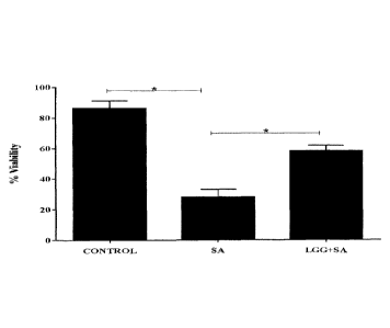
Figure 1: L.rhamnosus GG protects keratinocytes from the toxic effects of S.
aureus. Uninfected cells had a mean viability of (90%). S. aureus infected
cells had a
mean viability of (25% + 3.4). In keratinocytes infected with a combination of
S.aureus
and L.rhamnosus GG (LGG+SA), the viability after 24 hours was 57%+ 2.7
(P=0.01,
n=3).
Figure 2: Lysate and spent culture fluid (CM) from L.rhamnosus GG protect
keratinocytes from the effects of S.aureus. The viability of S.aureus infected
keratinocytes with L.rhamnosus GG lysate (LGGLYS+SA) was 65%+ 2.4 and with
spent
culture fluid (LGGCM+SA) was 57%+1.5 compared to 25%+3.1 in keratinocytes
infected
with S.aureus (SA) alone (P=0.006, P= 0.01 respectively, n=3).
Figure 3: L.rhamnosus GG protects keratinocytes from infection with S. aureus.
Percentage viability of keratinocytes was significantly higher in cells that
were pre-
exposed to L.rhamnosus GG (LGG +SA), lysate (LGG LYS+ SA) or spent culture
fluid
(LGG CM+SA) (58% + 1.4, 57% + 1.9, 55 % + 0.5, P=0.006, P=0.005, P=0.004)
compared to S. aureus (SA) infected cells (25% +1.3) (n=3).
Figure 4: L.rhamnosus GG but not its spent culture fluid rescuesd
keratinocytes
from S. aureus mediated toxicity. A) Uninfected keratinocytes were incubated
overnight; approximately 90% of the cells were viable after 24 hours. The
viability of S.
aureus infected keratinocytes was significantly higher in cells post-exposed
with
L.rhamnosus GG (P=0.003, n=3) 2 h (52% +1.6), 4 h (54%+1.4), 6h (57%+1.3), 8h
(58%+1.3) or 12h (58%+1.5). B) There was significant difference between the
viability of
cells (P=0.01, n=3) treated with L rhamnosus GG lysate 2 h (574%+3.1), 4h
(58%+2.1),6
h (63%+1.2),8h (63%+1.3) or 12h (55 /0+2.4) after infection had begun, whereas
the
viability of keratinocytes had been infected with S. aureus (SA) alone
(25%+1.7). C) Cells
post-exposed with L.rhamnosus GG spent culture fluid (CM) did not have
significant
protection (P=0.15, n=3) 2 h (32% +2.6), 4 h (39%+2.4), 6h(37%+1.8),8h
(36%+1.3) or
Date Recue/Date Received 2022-10-13
CA 02950510 2016-11-28
WO 2015/181534
PCT/GB2015/051529
12h (35%+3.5), whereas, the co-exposed cells had significant protection from
S.aureus
infection (56%+2.1, P=0.01, n=3).
Figure 5: L.rhamnosus GG lysate, but not spent culture fluid, reduced
staphylococcal viability. A) The optical densities of cultures of S. aureus
(SA) growing
in the presence of keratinocytes and treated with L.rhamnosus GG lysate (LGG
LYS) or
spent culture fluid ( LGG CM) was determined every hour to monitor the growth
of the
bacteria. The growth of S. aureus in the presence of the probiotic lysates was
significantly
lower than in S. aureus cultures (P=0.02, n=3), whereas, the spent culture
fluid had no
effect. B) The number of viable S. aureus (SA) in keratinocytes culture alone
was 8 log
CFU/ml or with spent culture fluid (LGG CM) 7.87Iog CFU/ml, whereas 5 log
CFU/ml of S.
aureus (SA) was viable in the present of L.rhamnosus GG lysate. C) The total
number of
viable staphylococci in keratinocytes culture was reduced by the L rhamnosus
GG lysate
in a post infection assay (2-4-6-8 and 12 hours), also showed significant
reduction in S.
aureus viability after 2h incubation (P=0.05, n=3) compared to S. aureus
alone.
Figure 6: Live L.rhamnosus GG, lysate or spent culture fluid inhibited S.
aureus
from adhering to keratinocytes by competitive exclusion from binding sites.
Ability
of bacteria to adhere to keratinocytes S. aureus (SA) (106), when applied to
keratinocytes
for 1h adhered to cells at approximately 7.5 + 0.6 log CFU / ml, while
L.rhamnosus GG
(LGG) (108) adhered at approximately 7.9 + 0.5 log CFU / ml. In pre-exposed
cells with
L.rhamnosus GG (LGG+SA), lysate (LGGLYS+SA) or spent culture fluid (LGG CM+SA)
had significantly less staphylococci adhered to them (5.83 + 0.2 log CFU / ml,
5.9 + 0.6
log CFU / ml and 6.4 + 0.7 log CFU / ml respectively) compared to cells
infected with S.
aureus (SA) alone (7.9 + 0.6 log CFU / ml) (P=0.04, n=3).
Figure 7: Live L.rhamnosus GG or lysate inhibited S. aureus from adhering to
keratinocytes by competitive displacement to binding sites. Post exposed cells
with
L.rhamnosus GG (LGG-12h) or lysate (LGGLYS-12h) had significantly less
staphylococci
adhered to them after 12h(5.8 + 0.7 log CFU / ml, 5.4 + 0.3 log CFU / ml
respectively)
compared to cells infected with S. aureus (SA) alone (7.95 + 0.6 log CFU / ml)
(P=0.01,
n=3). Whereas, the post exposed cells with L.rhamnosus GG spent culture fluid
(LGG
CM-12h) did not reduce the adhesion number of S. aureus (7 0.6 log CFU / ml,
P>0.05,
n=3).
Examples
16
CA 02950510 2016-11-28
WO 2015/181534
PCT/GB2015/051529
Example 1: Materials and Methods
Mammalian cell culture
Normal human epidermal keratinocytes (NHEK) cultured in keratinocyte basal
medium
(Promocell, Heidelberg, Germany) containing a supplement mix (bovine pituitary
extract
0.004mg/ml, epidermal growth factor (recombinant human) 0.125ng/ml, insulin
(recombinant human) 5pg/ml, hydrocortisone 0.33pg/ml, epinephrine 0.39pg/mland
transferrin, holo (human) 10pg/m1) and 0.06mM CaCl2 (Promocell, Heidelberg,
Germany),
were used as a model system. These were cultured routinely at 37 C in a humid
atmosphere of 5% CO2 in T-75 culture flasks as described previously (43).
Bacterial cell culture
Lactobacillus rhamnosus Goldin and Gorbach (L.rhamnosus GG) (ATCC 53103,ATCC,
Middlesex, UK ) was grown routinely in Wilkins-Chalgren Broth or Agar (Oxoid,
Basingstoke, UK) at 37 C in incubated in an anaerobic cabinet
(atmosphere,10:10:80, H2-
CO2-N2). Staphylococcus aureus was grown aerobically at 37 C in Nutrient Broth
(Oxoid,
Basingstoke, UK) as described previously (43).
Treatment of keratinocytes with bacteria
Bacteria (108 CFU/ml of probiotics and108 CFU/ml of S. aureus) were
centrifuged at
15,000 x g, washed twice in 0.85% NaCl and resuspended in keratinocyte basal
medium.
This suspension was added directly to 5 x 10³ cells/cm2of NHEK growing in 24
well
plates. For experiments using a probiotic lysate, 10m1 of 108 CFU/ml of L
rhamnosus GG
were centrifuged, washed, resuspended in Phosphate Buffer Saline (PBS) pH=7.4
(10mM) and lysed using a MSE Soniprep 150. Samples were filtered using a
0.22pm
pore filter (Millipore, Billerica, USA) to remove any whole bacteria
remaining.
Approximately 100p1 of this lysate was used to treat keratinocytes (5 x 10³
cells/cm2). In
some experiments, cells were sedimented in a centrifuge at 15,000 x g for 5
minutes and
the cell-free supernatant (spent culture fluid) collected and filtered using a
0.22pm pore
filter (Millipore, Billerica, USA) to remove any whole bacteria remaining. In
other
experiments, keratinocytes monolayers were co-infected with pathogen plus
probiotics or
lysates simultaneously. In separate experiments, cells were exposed to
L.rhamnosus GG
lysate 2, 4, 6, 8 and 12 hours after S. aureus infection had consumed. In all
experiments
keratinocytes were detached and cell viability was determined using trypan
blue exclusion
assays as described in (43).
Measurement of S. aureus viability in cell culture
17
CA 02950510 2016-11-28
WO 2015/181534
PCT/GB2015/051529
To determine whether L.rhamnosus GG lysates or keratinocytes were able to
inhibit the
growth of S. aureus in cell culture, keratinocytes were grown to confluence in
a 24 well
plate. These were exposed to S. aureus alone, or S. aureus plus L.rhamnosus GG
lysates or conditioned medium. In separate experiments, cells were exposed to
L.rhamnosus GG lysates 2, 4, 6, 8 and 12hours post infection with S. aureus.
The total
number of viable staphylococci was determined by counting the colonies as
described
previously (43).
Measurement of bacterial adhesion to keratinocytes
Confluent keratinocytes were exposed to bacteria for 1h. Cells were then
washed three
times in phosphate buffered saline (PBS) pH=7.4(10mM) (Invitrogen, Life
Technologies
Ltd, Paisley, UK) to remove non adherent bacteria. The cells were trypsinised
and serial
dilution plate counts performed to assess the number of adherent bacteria.
Selective agar
was used for growth of staphylococci.
Determination of bacterial antagonism
A 10p1 aliquot of an overnight culture of S. aureus was inoculated into 7m1 of
the soft-agar
media (0.7% agar) and was added directly onto plates, pre-poured with agar
base. 100p1
of each organism or extract of L.rhamnosus GG cultures were spotted onto the
S. aureus
lawn.
Determination of the outcome of co-culture (competition assays)
Aliquots (100p1) of L.rhamnosus GG lysates and S. aureus were inoculated into
10m1
WCB broths. The pH and optical density of cultures was measured at 0 and 24h.
At
regular intervals (indicated in the text) bacteria were counted by serial
dilution plate
counts using selective agar.
Statistical analyses
All experiments were performed a minimum of three times, with three replicates
within
each experiment. Data generated were analysed by one way ANOVA and post hoc
Tukey
test using SPSS (IBM SPSS Statistics version 16.0) program. Results were
considered
significant if P<0.05. Data are expressed as means standard errors of the
means
(SEM).
Example 2: Results
L.rhamnosus GG protects keratinocytes from the pathogenic effects of S.
aureus.
18
CA 02950510 2016-11-28
WO 2015/181534
PCT/GB2015/051529
Initially, we investigated whether the viability of keratinocytes was affected
by incubation
with L.rhamnosus GG. However, following 24h incubation, there was no
difference in the
viability of keratinocytes incubated with the probiotic bacteria vs the
control of untreated
keratinocytes (data not shown). Next, the ability of L.rhamnosus GG to protect
keratinocytes from the effects of S. aureus was investigated. In agreement
with our
previous findings (43) 24h exposure of keratinocytes to 106 CFU/ml S.aureus
resulted in
significant keratinocyte cell death. However, keratinocytes incubated
simultaneously with
pathogen and L.rhamnosus GG had a significantly higher percentage viability
(57%
P=0.01) than monolayers infected with pathogen alone (Figure 1).
L.rhamnosus GG lysates and spent culture fluid protect keratinocytes from the
effects of S.aureus.
We investigated whether live bacterium was required for the protective effect
of
L.rhamnosus GG by examining the effect of probiotic lysate and spent culture
fluid on
S.aureus infected keratinocytes. Neither lysate nor spent culture fluid
significantly
affected the viability of keratinocytes (P>0.05) (data not shown). However,
both the lysate
and spent culture fluid reduced the toxicity of S.aureus such that the
viability of treated
keratinocytes was 65% and 57.93% respectively compared to 25% in keratinocytes
infected with S.aureus alone (P= 0.006 and P=0.01 respectively) (Figure 2).
L.rhamnosus GG, lysate but not spent culture fluid rescues keratinocytes from
S.
aureus toxicity.
We next investigated the timing of the protective effect of L.rhamnosus GG by
adding the
live bacteria or the lysate either pre or post infection of keratinocytes with
S. aureus. The
percentage of keratinocyte viability was significantly greater in monolayers
exposed to
L.rhamnosus GG or spent culture fluid for 2h prior to infection with S.
aureus, than in
monolayers infected with S. aureus alone (P=0.006). Both the lysate and spent
culture
fluid afforded a similar levels of protection (P=0.005, p=0.004), (Figure3).
In post-
infection experiment, keratinocytes were exposed to S.aureus for 2h, 4h, 6h,
8h and 12h
before addition of the live L.rhamnosus GG, lysate, or spent culture fluid.
The viability of
the keratinocytes was then measured at 24h post infection with S.aureus. The
data in
Figure 4 (A, B) shows that both live probiotic and its lysate could protect
the keratinocytes
when added after S. aureus. Even at 12 h post S. aureus infection, L.rhamnosus
GG or
lysate still afforded protection to the keratinocytes such that 58% and 55%
respectively
of cells remained viable compared to 25% when exposed to S. aureus alone
(P=0.003,
19
CA 02950510 2016-11-28
WO 2015/181534
PCT/GB2015/051529
P=0.01 respectively). However, the spent culture fluid from L.rhamnosus GG had
no
protective effect on keratinocytes when added after S. aureus (Figure 4 C).
L.rhamnosus GG lysate, but not spent culture fluid, inhibits the growth of S.
aureus.
The mechanism by which the L.rhamnosus GG lysate exerted its protective effect
was
explored. We investigated whether the probiotic lysate had direct effects on
the growth of
the pathogen by growing them simultaneously in culture. Competition assays
showed a
significant reduction in S. aureus growth over a period of 24 h in
keratinocyte culture
medium in the presence of the L.rhamnosus GG lysate compared to untreated
cultures
(P=0.02) (Figure 5A). However, the spent culture fluid from L.rhamnosus GG had
no
effect on the growth of S. aureus (Figure 5A).The total number of viable
staphylococci
was also significantly reduced in the presence of the lysate (but not the
spent culture
fluid) to 5 logio cfu/ml, compared to 8 logio cfu/ml for S. aureus grown alone
(P=0.02)
(Figure 5B). Furthermore, the total number of viable staphylococci culture was
reduced
with time by the L.rhamnosus GG lysate (Figure 5C). Since Lactobacilli can
produce
organic acids, we measured the pH of keratinocyte media infected for 24 h with
S.
aureus, L.rhamnosus GG lysate or both simultaneously. However, there was no
significant difference in the pH between treatments group (data not shown). We
also
measured the pH of lysate alone and found it be pH= 7.2 thus eliminating the
possibility
of acid mediated effects.
L.rhamnosus GG inhibits adhesion of S. aureus to keratinocytes.
Another mechanism by which live bacteria, lysate or spent culture fluid of
L.rhamnosus
GG may protect the keratinocytes is by inhibition of pathogenic adhesion.
Previously, we
showed that adhesion is a requirement for the toxic effects of S.aureus and
specific
probiotic such as L. reuteri protected keratinocytes by competitive exclusion
of pathogen
from keratinocyte binding sites (43). Hence, we considered that inhibition of
adhesion
also be part of the protective mechanism of L.rhamnosus GG, lysate or spent
culture
fluid. Adhesion assays were performed to determine whether inhibition was due
to
competition, exclusion or displacement of pathogen from binding sites on
keratinocytes
(Figure 6 A&B and Figure 7). Our results demonstrated that live L.rhamnosus GG
or
lysate were able to inhibit pathogen adhesion if keratinocytes were co-
infected
(competition, P= 0.03), pre-exposed (exclusion, P= 0.04) or even applied 12h
after
infection with S.aureus had begun (displacement, P= 0.01). However, the spent
culture
CA 02950510 2016-11-28
WO 2015/181534
PCT/GB2015/051529
fluid only inhibited pathogen adhesion if it was added to keratinocytes either
before or at
the same time as the pathogen (Figure 7).
Discussion
This study explored whether an enteric probiotic, L.rhamnosus GG could protect
keratinocytes from the pathogenic effects of S. aureus. Preliminary
experiments to
determine the effect of adding S. aureus and L.rhamnosus GG simultaneously on
keratinocyte viability indicated a significant protective effect as observed
by an increase
in the number of viable keratinocytes in the presence of the probiotic
compared to
keratinocytes infected with S. aureus alone (Figure 1). Furthermore, the
protective effect
of L.rhamnosus GG did not require viable bacteria because a lysate and spent
culture
fluid from the probiotic also afforded protection of keratinocytes from S.
aureus.
The timing of application of L.rhamnosus GC or lysate did not affect the
degree of
protection conferred by the probiotic or lysate to protect keratinocytes
against S. aureus
induced cell death (Figure 3). The data demonstrate that keratinocytes pre-,
post or co-
exposed to L.rhamnosus GG or lysate were protected from S. aureus induced cell
death.
However, the probiotic spent culture fluid only protected keratinocytes if it
was added
either before or at the same time as pathogen. These data suggest that there
are at least
two separate activities involved in the protective effects of L.rhamnosus GG
against S.
aureus, one contained within the spent culture fluid and one contained within
the lysate.
Our data shows that the activity contained within the spent culture fluid
probably has anti-
adhesive effects. This is based on the following observations: A) L.rhamnosus
GG-spent
culture fluid only inhibits pathogen adhesion to keratinocytes if it is added
before or at the
same time as infection with S. aureus. B) We have shown previously that S.
aureus must
adhere to keratinocytes in order to be toxic to them and agents inhibiting
adhesion protect
keratinocytes from this pathogen (43). C) In agreement with this, spent
culture fluid is only
protective when added pre or co-infection with S. aureus. Other studies in
vitro
demonstrated that cell-free culture supernatants (CFCS) from the putative
probiotics
(Lactobacillus, Bifidobacterium, Lactococcus, Streptococcus) were able to
inhibit the
adhesion of several pathogens such as Salmonella Typhimurium, S. aureus and
Escherichia coil, to Caco-2 cells (11).
Keratinocyte protection by the lactobacillus lysate may involve at least two
mechanisms.
Firstly, the lysate may be able to reduce the growth of S aureus. Competition
assays
demonstrated that L.rhamnosus GG lysate reduced the total number of viable
staphylococci (Figure 5 A, B, C). In addition, in inhibition assays, zones of
inhibition were
21
CA 02950510 2016-11-28
WO 2015/181534
PCT/GB2015/051529
observed when S. aureus was challenged with lysates from probiotic grown
anaerobically
(Table 1). These data suggest an ability of L rhamnosus GG lysate to inhibit
growth of S.
aureus. This could be due to the presence of a toxic molecule(s) within the
probiotic that
are able to directly inhibit S. aureus growth and/or viability. It is possible
that this
molecule(s) may be synthesized, but not secreted because there was no effect
of
L.rhamnosus GG spent culture fluid on the viability of S. aureus. If
L.rhamnosus GG
contains bacteriostatic substances, then this may also, at least partially
explain the
protective effect of the probiotic in keratinocyte survival assays.
Probiotics, especially
lactobacilli, have previously been shown to exert a strong inhibitory effect
on S. aureus
growth. Certain Lactobacillus strains have been reported to be highly
antagonistic to
biofilm-forming S. aureus (28, 30). Other studies have reported that
probiotics can
improve gut health by inhibiting growth of pathogens through production of
bacteriocins
(16, 48). Moreover, L.rhamnosus GG has been shown to inhibit the growth of
Salmonella
enterica through production of lactic acid (29). However, in the present
study, we could
find no evidence of the involvement of acid production as part of the
protective effects of
L.rhamnosus GG. Indeed, the lysate from this organism was neutral (pH 7.2) but
was still
able to inhibit S. aureus growth.
Organisms (ZOOmm
SA+LGG Anaerobic 11+1.3
SA+LGG Lysate Anaerobic 18+0.7
SA+LGG Aerobic No inhibition
SA+LGG Lysate Aerobic No inhibition
Table 1: Zones of Inhibition (Z01) for S. aureus in Spot-on-the-lawn assays
(n=3). Spot on the lawn assay demonstrating zones of inhibition produced by
L.rhamnosus (LGG) and lysate (LOG LYS) under anaerobic condition, but not
under aerobic condition. Results are expressed as the mean SEM
A second mechanism by which live bacterium or lysate of L.rhamnosus GG could
protect
the keratinocytes is by inhibition of pathogenic adhesion. Indeed, our data
demonstrated
a reduction in adhesion of S. aureus to keratinocytes in the presence of
L.rhamnosus GG
or its lysate. This data suggests a mechanism of exclusion as we have observed
previously for L. reuteri (43). However, interestingly, viable L .rhamnosus GG
or its lysate
22
CA 02950510 2016-11-28
WO 2015/181534
PCT/GB2015/051529
also inhibited adhesion of S. aureus when added to existing infections
demonstrating
another mechanism of protection i.e. that L.rhamnosus GG can displace pathogen
from
keratinocytes (Figure7). Similarly, live L.rhamnosus GG has been shown to
displace
pathogens from the intestinal cells in the gut (47). However, our data
demonstrated that
the presence of live bacterium is not necessary for displacement of S.aureus
from
keratinocytes. Importantly, our data demonstrated species dependent
differences in the
mechanisms used by lactobacilli to reduce pathogen toxicity. Our previous work
highlighted L. reuteri as an organism capable of excluding S. aureus form
keratinocyte
binding sites (43). In this study we have shown that L.rhamnosus GG can, not
only,
exclude pathogens but can also reduce pathogen growth and displace pathogen
from
keratinocytes. Of course, it is possible that this displacement activity may
be related to
the ability of L.rhamnosus GG to inhibit growth and further studies will be
required to
clarify this point.
In conclusion, we report that Lrhamnosus GG is a potential new agent to
inhibit the
pathogenicity of S. aureus. Furthermore, our data shows that the utility of L
.rhamnosus
GG on skin will not be limited by whether it can grow and survive on skin
because a
lysate of the organisms is just as efficacious at preventing S. aureus
colonization as live
bacteria. Furthermore, the lysate could be useful as prophylaxis e.g. in hand
washes, but
potentially as an adjunct or even an alternative to antibiotics in existing
infection.
References
1. Aly R, Shinefield HR, Litz C, Maibach HI, 1980. Role of Teichoic Acid in
the
Binding of Staphylococcus aureus to Nasal Epithelial Cells. J. Infect. Dis.
141:463-465.
2. Backhed F, Ley RE, Sonnenburg JL, Peterson DA, Gordon JI. 2005. Host-
Bacterial Mutualism in the Human Intestine. Science. 307:1915-1920.
3, Balma-Mena A, Lara-Corrales I, Zeller J, Richardson S. McGavin MJ,
Weinstein M. 2011. Colonization with community-acquired methicillin-resistant
Staphylococcus aureus in children with atopic dermatitis: a cross-sectional
study.
J. Dermatolõ 50:682-688.
4. Banerjee P, Merkel G, Bhunia A. 2009. Lactobacillus delbrueckii ssp.
bulgaricus
B-30892 can inhibit cytotoxic effects and adhesion of pathogenic Clostridium
difficile to Caco-2 cells. Gut Pathogens. 1:8-16.
5. Bek-Thomsen M, Lomholt HB, Kilian M. 2008. Acne is Not Associated with Yet-
Uncultured Bacteria. J. Clin. Microbiol. 46:3355-3360.
23
CA 02950510 2016-11-28
WO 2015/181534
PCT/GB2015/051529
6. Borruel N, CaseIlas F, Antolin M, Llopis M, Carol M, Espiin E. 2003.
Effects
of non-pathogenic bacteria on cytokine secretion by human intestinal mucosa.
J.
American Gastroenterology. 98: 865-870.
7. Bowler PG, Duerden BI, Armstrong DG. 2001. Wound Microbiology and
Associated Approaches to Wound Management. Clin. Microbiol. Rev. 14:244-269.
8. Burkhart CN, Burkhart CG. 2003. Microbiology's principle of biofilms as a
major
factor in the pathogenesis of acne vulgaris. J. Dermatol. 42:925-927.
9. Caglar E, Kavaloglu Cildir S, Ergeneli S, Sandalli N, Twetman S. 2006.
Salivary mutans streptococci and lactobacilli levels after ingestion of the
probiotic
bacterium Lactobacillus reuteri ATCC 55730 by straws or tablets. Acta Odontol.
Scand. 64:314-318.
10. Chen X, Xu J, Shuai J, Chen J, Zhang Z, Fang W. 2007. The S-layer proteins
of
Lactobacillus crispatus strain ZJO01 is responsible for competitive exclusion
against Escherichia coil 0157:H7 and Salmonella typhimurium. Int. J. Food
Microbiol. 115:307-312.
11. Da bedi, Feizizadeh S, Jafarian-Dehkordi A. 2013. In vitro anti-bacterial
and
anti-adherence effects of Lactobacillus
delbrueckii subsp bulgaricus on Escherichia coll. Res Pharm Sci.4: 260-268.
12. Deepika G, Charalampopoulos D, Allen IL, Sima S, Geoffrey MG. 2010.
Surface and Adhesion Properties of Lactobacilli, Adv. Appl. Microbiol. 70:127-
152.
13. Di Marzio, Cinque LB, De Simone C, Cifone MG. 1999. Effect of the Lactic
Acid
Bacterium Streptococcus thermophilus on Ceramide Levels in Human
Keratinocytes In Vitro and Stratum Corneum In Vivo. J. Invest. Dermatol.
113:98-
106.
14. Evera Pingitore, Salvucci E, Sesma F, Nader-Macias MA.2007. Different
strategies for purification of antimicrobial peptides from Lactic Acid
Bacteria (LAB)
. Lett. Appl .Microbiol .46:174 - 180
15. Gan B S, Kim J, Reid G, Cadieux P. Howard JC. 2002. Lactobacillus
fermentum
RC-14 Inhibits Staphylococcus aureus Infection of Surgical Implants in Rats.
J.
Infect. Dis. 185:1369-1372.
16. Granato D, Perotti F, Masserey I, Rouvet M, Golliard M, Servin A, Brassart
D.
1999. Cell Surface-Associated Lipoteichoic Acid Acts as an Adhesion Factor for
Attachment of Lactobacillus johnsonii La1 to Human Enterocyte-Like Caco-2
Cells. Appl. Environ. Microbiol. 65:1071-1077.
24
CA 02950510 2016-11-28
WO 2015/181534
PCT/GB2015/051529
17. Gueniche A, Bastien P, Ovigne JM, Kermici M, Courchay G, Chevalier V,
Breton L. 2010. Bifidobacterium longum lysate, a new ingredient for reactive
skin.
Exp. Dermatol. 19:e1-e8.
18. Haukioja A, Tenovuo J. 2008. Probiotic bacteria affect the composition of
salivary pellicle and streptococcal adhesion in vitro. Oral Microbiology and
Immunology. 23: 336-343.
19. Heinemann C, Hylckama Vlieg JET, Janssen DB, Busscher HJ, Van der
Mei, Reid G. 2000. Purification and characterization of a surface-binding
protein
from Lactobacillus fermentum RC-14 that inhibits adhesion of Enterococcus
faecalis 1131. FEMS Microbiol. Lett. 190:177-180.
20. Helgeland L, Vaage JT, Rolstad B, Midtvedt T, Brandtzaeg P. 1996.
Microbial
colonization influences composition and T-cell receptor V beta repertoire of
intraepithelial lymphocytes in rat intestine. Immunology 89:494-501.
21. Holder IA, Boyce ST. 1994. Agar well diffusion assay testing of bacterial
susceptibility to various antimicrobials in concentrations non-toxic for human
cells
in culture. Burns. 20:426-429.
22. lwase T, Uehara Y, Shinji H, Tajima A, Seo H, Takada K, Agata T, Mizunoe
Y. 2010. Staphylococcus epidermidis Esp inhibits Staphylococcus aureus biofilm
formation and nasal colonization. Nature. 465:346-349.
23. Kintarak S, Whawell SA, Speight PM, Packer S, Nair SP. 2004.
Internalization
of Staphylococcus aureus by Human Keratinocytes. Infect. lmmun. 72:5668-5675.
24. Kluytmans J, Van Belkum A, Verbrugh H. 1997. Nasal carriage of
Staphylococcus aureus: Epidemiology, underlying mechanisms, and associated
risks. Clin. Microbiol. Rev. 10:505-520.
25. Krut 0, Utermohlen 0, Schlossherr S, Kronke M. 2003. Strain-Specific
Association of Cytotoxic Activity and Virulence of Clinical Staphylococcus
aureus
Isolates. Infect. Immun. 71:2716-2723.
26. Lai Y, Cogen AL, Radek KA, Park HJ, MacLeod DT, Leichtle A, Ryan A, Di
Nardo A, Gallo RL. 2010. Activation of TLR2 by a Small Molecule Produced by
Staphylococcus epidermidis Increases Antimicrobial Defense against Bacterial
Skin Infections. J. Invest. Dermatol. 130:2211-2221.
27. Lai Y, Cogen AL, Radek KA, Park HJ, MacLeod DT, Leichtle A, Ryan A, Di
Nardo A, Gallo RL. 2009. Commensal bacteria regulate Toll-like receptor 3-
dependent inflammation after skin injury. Nat. Med. 15:1377-1382.
CA 02950510 2016-11-28
WO 2015/181534
PCT/GB2015/051529
28. Lee YK, Puong KY, Ouwehand AC, Salminen S. 2003. Displacement of
bacterial pathogens from mucus and Caco-2 cell surface by lactobacilli. J.
Med.
Microbiol. 52:925-930.
29. Lefteris M, Vagelis T, Domitille M, Tom A, 2006. Kinetic analysis of the
antibacterial activity of probiotic lactobacilli towards Salmonella enterica
serovar
Typhimurium reveals a role for lactic acid and other inhibitory compounds.
Research in Microbiology.157: 241-247.
30. Lin MY, Chang FJ. 2000. Antioxidative Effect of Intestinal Bacteria
Bifidobacterium Ion gum ATCC 15708 and Lactobacillus acidophilus ATCC 4356.
Digestive Diseases and Sciences, 45: 1617-1622.
31. Mack DR, Michail S, Wei S. McDougall L, Hollingsworth MA. 1999. Probiotics
inhibit enteropathogenic E. coil adherence in vitro by inducing intestinal
mucin
gene expression. J .Physiol Gastrointest Liver Physiol. 276:G941-950
32. Maria C. Collado, Erika lsolauri , Seppo Salminen. 2008. Specicic
probiotic
strains and their combinations counteract adhesion ofEnterobacter sakazakii to
intestinal mucus. FEMS Microbial Lett. 285: 58-64.
33. Maudsdotter L, Jonsson H, Roos S, AB Jonsson. 2011. Lactobacilli Reduce
Cell Cytotoxicity Caused by Streptococcus pyogenes by Producing Lactic Acid
that Degrades the Toxic Component Lipoteichoic Acid, Antimicrob. Agents
Chemother. 55:1622-88.
34. Mempel M, Schmidt T, Weidinger 5, Schnopp C, Foster T, Ring T, Abeck D.
1998. Role of Staphylococcus Aureus Surface-Associated Proteins in the
Attachment to Cultured HaCaT Keratinocytes in a New Adhesion Assay. J. Invest.
Dermatol. 111:452-456.
35. Mempel M, Schnopp C, Hojka H, Fesq H, Weidinger S, Schaller M, Korting
HC, Ring J, Abeck D. 2002. Invasion of human keratinocytes by Staphylococcus
aureus and intracellular bacterial persistence represent haemolysin-
independent
virulence mechanisms that are followed by features of necrotic and apoptotic
keratinocyte cell death. Br. J. Dermatol. 146:943-951.
36. Miyoshi Y, Okada S, Uchimura T, Satoh E. 2006. A Mucus Adhesion Promoting
Protein, MapA, Mediates the Adhesion of Lactobacillus reuteri to Caco-2 Human
Intestinal Epithelial Cells. Biosci. Biotechnol. Biochem. 70:1622-1628.
37. Mbernet F, Kerneis S, Chauviere G, Fourniat J, Servin G. 1993. Inhibition
of
adhesion of enteroinvasive pathogens to human intestinal Caco-2 cells by
Lactobacillus acidophilus strain LB decreases bacterial invasion. FEMS
Microbiol.
Lett. 110:299-306.
26
CA 02950510 2016-11-28
WO 2015/181534
PCT/GB2015/051529
38. Nikawa H Makihira 5, Fukushima H, Nishimura H, Ozaki Y, lshida K,
Darmawan S, Hamada T, Hara K, Matsumoto A, Takemoto T, and Aimi R.
2004. Lactobacillus reuteri in bovine milk fermented decreases the oral
carriage of
mutans streptococci. Int. J. Food Microbial. 95:219-223.
39. Ouwehand AC, Isolauri E, Kirjavainen PV, olkkti ST, Salminen SJ. 2000. The
mucus binding of Bifidobacterium lactis E3b12 is enhanced in the presence of
Lactobacillus GG and Lact. delbrueckii subsp. bulgaricus. Lett. Appl.
Microbial.
30:10-13.
40. Parsool N, Rampal P. 2005. Lactobacillus casei DN-114 001 inhibits the
increase
in paracellular permeability of enteropathogenic Escherichia coli-infected T84
cells. Research in Microbiology. 156: 256-262.
41. Peral MC, Huaman Martinez MH, Valdez CJ. 2009b. Bacteriotherapy with
Lactobacillus plantarum in burns. International Wound Journal. 6:73-81.
42. Peral MC, Rachid MM, Gobbato MN, Martinez MH, Valdez JC. 2009a.
Interleukin-8 production by polymorphonuclear leukocytes from patients with
chronic infected leg ulcers treated with Lactobacillus plantarum. Clin.
Microbial.
Infect. 16:281-286.
43. Tessa P, Andrew J McBain and Catherine A O'Nei11.2012.Lactobacillus
Reuteri
Protects Epidermal Keratinocytes from Staphylococcus Aureus Induced Cell
Death by Competitive Exclusion. Appl. Environ. Microbial. 15:78-5119.
44. Reid G, Beuerman D, Bruce AW. 2001. Probiotic Lactobacillus dose required
to
restore and maintain a normal vaginal flora. FEMS Immunology and Medical
Microbiology. 32: 37-41.
45. Rembacken BJ, Snelling AM, Hawkey PM, Chalmers DM, Axon RT. 1999.
Non-pathogenic Escherichia coli versus mesalazine for the treatment of
ulcerative
colitis: a randomised trial. Lancet. 354:635-639.
46. Ron EZ, Rosenberg E. 2001. Natural roles of biosurfactants. Environmental
Microbiology. 3: 229-236.
47. Satu V, Matti K, Seppo 5, Arthur C. Ouwehand. 2006. Staphylococcus aureus
adheres to human intestinal mucus but can be displaced by certain lactic acid
bacteria. Microbiology. 152: 1819-1826.
48. Sarika AR, Lipton AP, Aishwarya MS. 2010. Bacteriocin production by a new
isolate of Lacobacillus rhamnosus GPI under different culture conditions.
Advance Journal of Food Science and Technology. 2: 291-297.
49. Strober W. 1997. Trypan blue exclusion test of cell viability, p. Appendix
3B.
Current Protocols in Immunology. John Wiley & Sons, Inc.
27
CA 02950510 2016-11-28
WO 2015/181534
PCT/GB2015/051529
50. Valdez JC, Peral MC, Rachid M, Santana M, Perdigon G. 2005. Interference
of
Lactobacillus plantarum with Pseudomonas aeruginosa in vitro and in infected
burns: the potential use of probiotics in wound treatment. Clin. Microbiol.
Infect.
11:472-479
51. Wehkamp J, Harder J, Wehkamp K, Meissner BW, Sch lee V, Enders M,
Sonnenborn C, Nuding Ii, Bengmark S. Fellermann S, Schroder K, Stange
EJ. 2004. NF-kB- and AP-1-Mediated Induction of Human Beta Defensin-2 in
Intestinal Epithelial Cells by Escherichia coli Nissle 1917: a Novel Effect of
a
Probiotic Bacterium. Infect. Immun.72: 5750-5758.
52. Zarate G, Nader-Macias ME. 2006. Influence of probiotic vaginal
lactobacilli on in
vitro adhesion of urogenital pathogens to vaginal epithelial cells. Lett Appl
Microbiol. 43:174 ¨ 180.
28