Note: Descriptions are shown in the official language in which they were submitted.
1
CA 02950576 2016-11-28
WO 2015/186988 PCT/KR2015/005651
Description
Title of Invention: METHOD FOR DECREASING IMMUNO-
GENICITY OF PROTEIN AND PEPTIDE
Technical Field
[1] The present invention relates to a method for increasing serum half-
life of a protein
or peptide and decreasing immunogenicity thereof by site-specifically binding
a carrier
to a protein or peptide, and to the use thereof.
[2]
Background Art
1Z31 Immune responses to biological therapeutic agents can be widely
induced for both
non-human and human-derived proteins. These responses may weaken clinical
effects,
limit the efficacy, and sometimes lead to pathological diseases or even cause
the death
of the patient. In particular, the production of neutralizing antibodies that
target the re-
combinant self-protein may induce a cross-reaction with the protein inherent
in the
body of the patient and thus lead to serious consequences (see, Lim LC.
Hematology
2005 10(3):255-9). Problems of biopharmaceuticals such as monoclonal
antibodies
were greatly reduced with the development of molecular biology. However, many
re-
combinant protein pharmaceuticals are identical with the protein sequences
which are
expressed in the body and thus, there still remain a possibility of causing a
neutralizing
immune response (see, Namaka M et al, Curr Med Res Opin 2006 22(2):223-39).
Although the mechanism by which it is possible to induce immunogenicity is not
wholly clear, it is known that the resistance to self-proteins can be broken
by products
administered to the patient and by various factors of the patient (reviewed in
Chester,
K, Baker, MP and Mayer A. Expert Rev Clin Immunol 2005 1(4): 549-559, Baker MP
and Jones TD. Curr. Opin. Drug Disc Dev 2007 10(2):219-227). Factors for
immuno-
genicity include dosage, frequency and route of administration,
immunomodulatory
ability of protein drugs, their preparation and the like. The most important
factor to
induce the immune response is whether there is an antigen recognition site
(epitope)
that can effectively stimulate a CD4 + T cell response (reviewed Baker MP and
Jones
TD. Curr. Opin. Drug Disc Dev 2007 10(2):219-227).
[4]
1Z51 On the other hand, exendin-4 is a natural peptide discovered in the
salivary gland of
the Gila monster lizard and has a 52% sequence similarity with human GLP-1
(glucagon-like peptide-1). Exendin-4 and GLP-1 have a similar insulin
secretion
function. However, GLP-1 is rapidly deactivated by dipeptidyl peptidase-IV
(DPP-IV),
thus having a very short half-life, whereas exendin-4 keeps the resistance to
DPP-IV
2
CA 02950576 2016-11-28
WO 2015/186988 PCT/KR2015/005651
by glycine being present instead of alanine in the second amino acid sequence
and thus
can be more effective as a therapeutic agent of type II diabetes. In addition,
insulin or
analogs thereof, and dual agonists of GLP-1/glucagon are also used as
therapeutic
agents for diabetes and obesity. However, the presence of these non-human
amino acid
sequences can act as an antigen recognition site of T cells. Exenatide
(Byetta) which
was approved as therapeutic agents of type II diabetes as synthetic exendin-4
has
produced an antibody to exenatide for more than 30% of patients who received
admin-
istration of exenatide for one year in clinical trials. Lixisenatide, approved
recently, has
produced an antibody for about 60-71% of patients (see, Zinman, B. et al.,
Annals of
Internal Medicines. 2007 146(7): 477-486; Schnabel CA et al, Peptides 2006
27:1902-1910; DeFronzo, R.A. et al, Diabetes Care 2005 28:1092-1100; Buse,
J.B. et
al, Diabetes Care 2004 27:2628-2635). That is, exentide was recognized as an
in vivo
extraneous substance to be treated and the antibody was produced. For this
reason, the
problem that is difficult to reliably expect a therapeutic effect is
increasing prevalent.
[6]
171 Therefore, in the case of a physiologically active protein or peptide
which has been
administered within the body for the purpose of treatment or prevention for a
long
period of time, it is important to control the immunogenicity. In particular,
adult
disease-related physiologically active proteins or peptides such as insulin or
in-
sulinotropic peptide and anti-obesity protein have often been developed as as
long-
acting formulations capable of lasting in the body after administration. In
addition,
even if they are not long-acting formulations, there are many cases in which
they must
be administered several times for a long period of time. Therefore, not
inducing an
immune response is an important issue.
[81 Under these circumstances, the present inventors have conducted
numerous studies
and experiments to develop pharmaceutical formulations of a protein or peptide
which
do not induce an immune response. As a result, the inventors have discovered
that,
when a carrier site-specifically binds to a protein or peptide, the
immunogenicity can
be decreased as compared to that of a protein or peptide to which the carrier
has not
been bound, thus completing the present invention.
[91
Disclosure of Invention
Technical Problem
[10] One object of the present inveniton is to provide a method for
decreasing immuno-
genicity of physiologically active proteins or peptides.
[11] Another object of the present invention is to provide a composition,
comprising a
conjugate of a physiologically active protein or peptide in which a carrier is
bound to
3
CA 02950576 2016-11-28
WO 2015/186988 PCT/KR2015/005651
the non-terminal, internal residue of a physiologically active protein or
peptide, via a
non-peptidyl linker.
[12] Another object of the present invention is to provide a method for
preparing the
conjugate of the physiologically active protein or peptide, in which the
carrier is bound
to the non-terminal, internal residue of the physiologically active protein or
peptide.
[13]
Solution to Problem
[14] In one aspect, the present invention provides a method for decreasing
immuno-
genicity of a physiologically active protein or peptide as compared to that of
a physio-
logically active protein or peptide to which a carrier is not bound, which
comprises
binding a carrier to the non-terminal, internal residue of the physiologically
active
protein or peptide.
[15] In one specific embodiment of the invention, the above carrier is
characterized in that
it is selected from the group consisting of a polyethylene glycol, a fatty
acid, a
cholesterol, an albumin or a fragment thereof, an albumin-binding substance, a
polymer having repeating units of a particular amino acid sequence, an
antibody, an
antibody fragment, an FcRn binding substance, an in-vivo connective tissue or
a
derivative thereof, a nucleotide, a fibronectin, a transferrin, an elastin-
like
polypeptide(ELP), an XTEN polypeptide, a carboxy-terminal peptide (CTP), a
structure inducing probe (SIP), a saccharide and a high molecular weight
polymer.
[16] In another specific embodiment of the invention, the FcRn binding
substance is char-
acterized in that it includes an immunoglobulin Fc region.
[17] In another specific embodiment of the invention, the physiologically
active protein or
peptide and the carrier are characterized by being bound via a linker
interposed
therebetween.
[18] In another specific embodiment of the invention, the linker is
characterized in that it
is a non-peptidyl linker.
[19] In another specific embodiment of the invention, the non-peptidyl
linker is char-
acterized in that it is selected from the group consisting of a polyethylene
glycol, a
polypropylene glycol, an ethylene glycol-propylene glycol copolymer, a poly-
oxyethylated polyol, a polyvinyl alcohol, a polysaccharide, a dextran, a
polyvinyl ethyl
ether, a biodegradable polymer, a lipid polymer, a chitin, a hyaluronic acid
and a com-
bination thereof.
[20] In another specific embodiment of the invention, it is characterized
in that the physi-
ologically active protein or peptide is bound to an immunoglobulin Fc region
via a
non-peptidyl polymer which is selected from the group consisting of a
polyethylene
glycol, a polypropylene glycol, an ethylene glycol-propylene glycol copolymer,
a poly-
4
CA 02950576 2016-11-28
WO 2015/186988 PCT/KR2015/005651
oxyethylated polyol, a polyvinyl alcohol, a polysaccharide, a dextran, a
polyvinyl ethyl
ether, a biodegradable polymer, a lipid polymer, a chitin, a hyaluronic acid
and a com-
bination thereof.
[21] In another specific embodiment of the invention, the physiologically
active protein or
peptide is characterized in that it is selected from the group consisting of
an anti-
obesity peptide, an insulinotropic peptide or an analog thereof, a leptin, an
insulin, an
insulin analog, a glucagon, a human growth hormone, a growth hormone releasing
hormone, a growth hormone releasing peptide, an interferon, an interferon
receptor, a
colony stimulating factor, a glucagon-like peptide such as GLP-1, a GLP-
1/glucagon
dual agonist, a gastric inhibitory polypeptide (GIP), a G-protein-coupled
receptor, an
interleukin, an interleukin receptor, an enzyme, an interleukin binding
protein, a
cytokine binding protein, a macrophage activating factor, a macrophage
peptide, a B
cell factor, a T cell factor, a protein A, an allergy inhibitory factor, a
cell necrosis gly-
coprotein, an immunotoxin, a lymphotoxin, a tumor necrosis factor, a tumor
inhibitory
factor, a metastasis growth factor, an alpha-1 antitrypsin, an albumin, an a-
lactalbumin, an apolipoprotein-E, an erythropoiesis factor, a highly
glycosylated ery-
thropoiesis factor, an angiopoietin, a hemoglobin, a thrombin, a thrombin
receptor ac-
tivating peptide, a thrombomodulin, blood factors VII, VIIa, VIII, IX and
XIII, a
plasminogen activating factor, a fibrin-binding peptide, an urokinase, a
streptokinase, a
hirudine, a protein C,C-reactive protein, a renin inhibitor, a collagenase
inhibitor, a su-
peroxide dismutase, a platelet-derived growth factor, an epithelial cell
growth factor,
an epidermal growth factor, an angiostatin, an angiotensin, a bone growth
factor, a
bone stimulating protein, a calcitonin, an atriopeptin, a cartilage inducing
factor, an
elcatonin, a connective tissue activating factor, a tissue factor pathway
inhibitor, a
follicle stimulating hormone, a luteinizing hormone, a luteinizing hormone
releasing
hormone, a nerve growth factor, a parathyroid hormone, a relaxin, a secretin,
a so-
matomedin, an insulin-like growth factor, an adrenocortical hormone, a
glucagon, a
cholecystokinin, a pancreatic polypeptide, a gastrin-releasing peptide, a
cortincotropin
releasing factor, a thyroid stimulating hormone, an autotaxin, a lactoferrin,
a myostatin,
a receptor, a receptor antagonist, a cell surface antigen, a virus-derived
vaccine
antigen, a monoclonal antibody, a polyclonal antibody, and an antibody
fragment.
[22] In another specific embodiment of the invention, the physiologically
active protein or
peptide is characterized in that it is selected from the group consisting of
an exendin-4,
an exendin-4 derivative, a GLP-1 agonist, an insulin and a GLP-1/glucagon dual
agonist.
[23] In another specific embodiment of the invention, the exendin-4
derivative is char-
acterized in that it is an exendin-4 derivative in which the charge on the N-
terminal of
exendin-4 is modified, which is selected from the group consisting of an
exendin-4
5
CA 02950576 2016-11-28
WO 2015/186988 PCT/KR2015/005651
derivative in which N-terminal amine group of exendin-4 is deleted, an exendin-
4
derivative in which N-terminal amine group of exendin-4 is substituted with
hydroxl
group, an exendin-4 derivative in which N-terminal amine group of exendin-4 is
sub-
stituted with carboxly group, an exendin-4 derivative in which N-terminal
amine group
of exendin-4 is modified with dimethyl group, and an exendin-4 derivative in
which
alpha carbon of N-terminal histidine residue of exendin-4 is deleted.
[24] In another specific embodiment of the invention, the above-described
internal residue
is characterized in that it is a lysine residue at position 12 or 27 of the
exendin-4
derivative in which N-terminal charge of exendin-4 is modified.
[25] In another specific embodiment of the invention, the above-described
internal residue
is characterized in that it is a lysine residue at position 27 of the exendin-
4 derivative in
which N-terminal charge of exendin-4 is modified.
[26] In another specific embodiment of the invention, the exendin-4
derivative in which
the charge on the N-terminal of the exendin-4 is changed is characterized in
that it is an
exendin-4 derivative in which alpha carbon of N-terminal histidine residue of
exendin-
4 is deleted.
[27] Another aspect, the preset the present invention provides a
composition, comprising
a conjugate of a physiologically active protein or peptide in which a carrier
is bound to
the non-terminal, internal residue of a physiologically active protein or
peptide, via a
non-peptidyl linker, wherein the conjugate exhibits decreased immunogenicity
as
compared to that of the physiologically active protein or peptide to which the
carrier is
not bound.
[28] In one specific embodiment of the invention, the above-described
conjugate is char-
acterized in that it has decreased immunogenicity, which is a side effect of a
long-
acting preparation.
[29] In another specific embodiment of the invention, the non-peptidyl
linker is char-
acterized in that it is a polyethylene glycol.
[30] Another aspect, the present invention provides a method for preparing
the conjugate
of the physiologically active protein or peptide, in which the carrier is
bound to the
non-terminal, internal residue of the physiologically active protein or
peptide.
[31]
Advantageous Effects of Invention
[32] The physiologically active protein or peptide conjugate of the present
invention can
significantly decrease immunogenicity in the human body and thus reduce
antibody
production rate against proteins or peptides. Therefore, the present conjugate
has ad-
vantages in that the phenomenon of reduced clinical effects of the
physiologically
active protein or peptide is low, and it can be effectively used in the
development of
6
CA 02950576 2016-11-28
WO 2015/186988 PCT/KR2015/005651
long-acting formulations having a high safety against the immune response.
[33]
Brief Description of Drawings
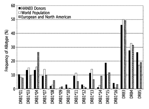
[34] Figure 1 is a diagram showing a comparison of HLA-DR genotype
frequency of a
donor in the ex vivo T cell activity test with that of the population in the
world, Europe
and North America.
[35]
Best Mode for Carrying out the Invention
[36] The present invention relates to a method for decreasing the
immunogenicity of a
physiologically active protein or peptide compared to that of the protein or
peptide to
which a carrier has not been bound, which comprises a step of binding a
carrier to the
non-terminal, internal residue of the physiologically active protein or
peptide.
[37]
[38] In the present invention, the inventors have discovered a method for
decreasing the
immunogenicity of a physiologically active protein or peptide in which a non-
peptide
linker and Fc fragment are bound to the internal residue rather than the
terminal of a
physiologically active protein or peptide, thus inhibiting the mechanism in
which the
desired protein or peptide acts as an antigen. The inventors have identified
that, in the
case of using the method as described above, the activation of T cells and the
antibody
production reaction in animals is significantly inhibited compared with the
method for
preparing a conjugate by the modification at other sites such as N-terminal of
the
peptide. As a result, the present inventors have found that the
physiologically active
protein or peptide conjugate used as a conventional protein pharmaceutical
preparation
has a novel use as the composition and method for deacreasing the
immunogenicity of
a physiologically active protein or peptide.
[39] The decrease of immunogenicity in the body can be measured without
limitation by a
known method. For example, the difference in immunogenicity can be confirmed
by
the ex-vivo activity measurement method of T cells which comprises coupling
each of
the carriers to the N-terminal or the sites other than the N-terminal
including the C-
terminal. Aldehyde reactive group selectively reacts with the N-terminal at a
low pH,
and also it can form a covalent bond with a lysine residue at the condition of
high pH,
for example pH 9Ø The pegylation reaction is conducted while changing the
reaction
pH, and then positional isomers can be separated from the reaction mixture
using an
ion exchange column.
[40]
[41] When the coupling is made at a position other than N-terminal end
which is an
important site in the activity of the protein or peptide in vivo, a reactive
thiol group can
7
CA 02950576 2016-11-28
WO 2015/186988 PCT/KR2015/005651
be introduced to an amino acid residue position to be modified, thus forming a
covalent bond between the protein or peptide and a maleimide group of the non-
peptidyl polymer.
[42]
[43] When the coupling is made at a position other than N-terminal end
which is an
important site in the activity of the protein or peptide in vivo, an amine
group is in-
troduced to an amino acid residue position to be modified, thus forming a
covalent
bond between the protein or peptide and an aldehyde group of the non-peptidyl
polymer.
[44]
[45] The method of protection of the N-terminal end includes methylation,
deamination or
acetylation in addition to dimethylation, but is not limited thereto.
[46]
[47] In the present invention, "physiologically active protein or peptide"
refers to a protein
or peptide that can control the genetic expression or physiological function.
The physi-
ologically active protein or peptide can be included, without limitation, in
the scope of
the present invention, as long as a carrier is bound to the non-terminal,
internal residue
of the physiologically active protein or peptide according to the present
invention, thus
exhibiting descresed immunogenicity compared to that of the protein or peptide
to
which a carrier is not bound. As described below, the carrier can be bound via
a linker,
specifically a non-peptidyl linker, to a physiologically active protein or
peptide.
[48]
[49] In addition, the physiologically active protein or peptide includes,
in addition to
native biologically active protein or peptide, derivatives, variants, or
fragments thereof.
[50]
[51] Examples of the physiologically active protein or peptide include an
anti-obesity
peptide, an insulinotropic peptide or an analog thereof, a leptin, an insulin,
an insulin
analog, a glucagon, a human growth hormone, a growth hormone releasing
hormone, a
growth hormone releasing peptide, an interferon, an interferon receptor, a
colony
stimulating factor, a glucagon-like peptide (GLP-1, etc.), a GLP-1/glucagon
dual
agonist, a gastric inhibitory polypeptide (GIP), a G-protein-coupled receptor,
an in-
terleukin, an interleukin receptor, an enzyme, an interleukin binding protein,
a cytokine
binding protein, a macrophage activating factor, a macrophage peptide, a B
cell factor,
a T cell factor, a protein A, an allergy inhibitory factor, a cell necrosis
glycoprotein, an
immunotoxin, a lymphotoxin, a tumor necrosis factor, a tumor inhibitory
factor, a
metastasis growth factor, an alpha-1 antitrypsin, an albumin, an a-
lactalbumin, an
apolipoprotein-E, an erythropoiesis factor, a highly glycosylated
erythropoiesis factor,
an angiopoietin, a hemoglobin, a thrombin, a thrombin receptor activating
peptide, a
8
CA 02950576 2016-11-28
WO 2015/186988 PCT/KR2015/005651
thrombomodulin, blood factors VII, VIIa, VIII, IX and XIII, a plasminogen
activating
factor, a fibrin-binding peptide, an urokinase, a streptokinase, a hirudine, a
protein C,
C-reactive protein, a renin inhibitor, a collagenase inhibitor, a superoxide
dismutase, a
platelet-derived growth factor, an epithelial cell growth factor, an epidermal
cell
growth factor, an angiostatin, an angiotensin, a bone growth factor, a bone
stimulating
protein, a calcitonin, an atripeptin, a cartilage inducing factor, an
elcatonin, a
connective tissue activating factor, a tissue factor pathway inhibitor, a
follicle
stimulating hormone, a luteinizing hormone, a luteinizing hormone releasing
hormone,
a nerve growth factor, a parathyroid hormone, a relaxin, a secretin, a
somatomedin, an
insulin-like growth factor, an adrenocortical hormone, a glucagon, a
cholecystokinin, a
pancreatic polypeptide, a gastrin-releasing peptide, a cortincotropin
releasing factor, a
thyroid stimulating hormone, an autotaxin, a lactoferrin, a myostatin, a
receptor, a
receptor antagonist, a cell surface antigen, a virus-derived vaccine antigen,
a
monoclonal antibody, a polyclonal antibody, and an antibody fragment, without
limitation.
[521 More specifically, the physiologically active protein or peptide may
include an
insulin, an insulinotropic peptide, or a GLP-1/glucagon dual agonist, but is
not limited
thereto.
[531 In the present invention, the term "insulin" includes all peptides or
modified peptides
which have a stimulating effect on insulin receptors. The insulin may be, for
example,
a native insulin, a rapid-acting insulin, a basal insulin, an insulin analog
in which any
amino acids of the native insulin is changed by any one method selected from
sub-
stitution, addition, deletion, and modification, or a combination of these
methods, or
may be a fragment thereof. Also, the insulin used in the present invention may
be a
long-acting insulin to which long-acting techniques applied to overcome the
short half-
life. In particular, the insulin may be a long-acting insulin or a long-acting
insulin
analog which can be administered once a week, but is not limited thereto.
[541 Some specific examples of the insulin according to the present
invention include an
insulin or an insulin analog and its long-acting type as disclosed in Korean
Patent No.
10-1058290 (or International Publication WO 2008-082274) or Korean Patent Ap-
plication Publication No. 2014-0106452 (or International Publication WO
2014-133324), the entire contents of which are incorporated herein by
reference, but
are not limited thereto.
[551
[561 As used herein, the term "insulin analog" refers to a substance which
retains the same
function of controlling the blood glucose level in vivo as a native insulin.
Specifically,
the insulin analogs include those in which one or more amino acids in the
native
insulin sequence have been modified. The insulin analog may be an insulin
analog in
9
CA 02950576 2016-11-28
WO 2015/186988 PCT/KR2015/005651
which A-chain or B-chain amino acid of native insulin is changed. The native
insulin
amino acid sequence is as follows.
[57]
[58] A chain:
[59] Gly-Ile-Val-Glu-Gln-Cys-Cys-Thr-Ser-Ile-Cys-Ser-Leu-Tyr-Gln-Leu-Glu-
Asn-Tyr-
Cys-Asn (SEQ ID NO: 1)
[60]
[61] B chain:
[62] Phe-Val-Asn-Gln-His-Leu-Cys-Gly-Ser-His-Leu-Val-Glu-Ala-Leu-Tyr-Leu-
Val-Cys
-Gly-Glu-Arg-Gly-Phe-Phe-Tyr-Thr-Pro-Lys-Thr (SEQ ID NO: 2)
[63]
[64] Specifically, at least one amino acid in the native insulin may have a
modififation
selected from the group consisting of substitution, addition, deletion,
modification and
a combination thereof, but are not limited thereto.
[65] In the substitution or addition of the amino acids, 20 amino acids
that are normally
observed in a human protein as well as atyhpical or non-naturally occurring
amino
acids can be used. The commercial sources of the atypical amino acids may
include
Sigma-Aldrich, ChemPep and Genzyme pharmaceuticals. The peptides including
such
amino acids and a typical peptide sequence can be synthesized or purchased
from
commercial peptide synthesis companies, for example, American peptide company
Inc., and Bachem (USA), or Anygen (Korea).
[66]
[67] Specifically, the above-described insulin analogs include an inverted
insulin, an
insulin variant, an insulin fragment, an insulin agonist, an insulin
derivative and the
like, and the preparation method thereof includes a genetic recombination as
well as a
solid phase method, but is not limited thereto.
[68]
[69] The term "insulin derivative" shows an amino acid sequence homolgy
with A-chain
and B-chain of native insulin, while retaining the function to control the
blood glucose
level in the body, and includes a peptide form which may have some groups on
the
amino acid redidues chemically substituted (e.g., alpha-methylation, alpha-hy-
droxylation), deleted (e.g., deamination), or modified (e.g., N-methylation).
In
addition, the insulin derivative includes a peptide mimic, and a low molecular
or high
molecular compound, which can bind with an insulin receptor to control blood
glucose
levels in the body, even without homology with a native insulin and an amino
acid
sequence.
[70] As used herein, the term "insulin fragment" refers to a fragment
having one or more
amino acids added or deleted in insulin. The added amino acid may be an amino
acid
10
CA 02950576 2016-11-28
WO 2015/186988 PCT/KR2015/005651
that is not present in the native state (e.g., D-type amino acid). Such
insulin fragment
retains a function to control blood glucose levels in the body.
[71] As used herein, the term "insulin variant" is a peptide having one or
more amino acid
sequences different from those of insulin, and retaining a function to control
blood
glucose levels in the body.
[72] Methods for preparing the insulin agonist, derivative, fragment and
variant of the
present invention, respectively, can be used alone and in combination thereof.
For
example, the present invention includes a peptide which has one or more amino
acid
sequence different from those of native insulin, has deamination at the
terminal amino
acid residue, and retains a function to control blood glucose levels in the
body, can be
included.
[73] The description of the agonists, derivatives, fragments and variants
may be applied
evewn to other types of proteins or peptides.
[74]
[75] Specifically, the insulin analogs may be those in which one or more
amino acids
selected from the group consisting of amino acids at position 1, amino acids
at position
2, amino acids at position 3, amino acids at position 5, amino acids at
position 8, amino
acids at position 10, amino acids at position 12, amino acids at position 16,
amino
acids at position 23, amino acids at position 24, amino acids at position 25,
amino
acids at position 26, amino acids at position 27, amino acids at position 28,
amino
acids at position 29, amino acids at position 30 of the chain B; amino acids
at position
1, amino acids at position 2, amino acids at position 5, amino acids at
position 8, amino
acids at position 10, amino acids at position 12, amino acids at position 14,
amino
acids at position 16, amino acids at position 17, amino acids at position 18,
amino
acids at position 19 and amino acids at position 21 of the chain A have been
substituted
with other amino acids, and more specifically those in which one or more amino
acids
selected from the group consisting of amino acids at position 8, amino acids
at position
23, amino acids at position 24, amino acids at position 25 of the chain B;
amino acids
at position 1, amino acids at position 2, amino acids at position 14 and amino
acids at
position 19 of the chain A have been substituted with other amino acids.
[76] Specifically, among the foregoing amino acids, those in which one or
more, two or
more, three or more, four or more, five or more, six or more, seven or more,
eight or
more, nine or more, ten or more, 11 or more, 12 or more, 13 or more, 14 or
more, more
than 15, 16 or more, 17 or more, 18 or more, 19 or more, 20 or more, 21 or
more, 22 or
more, 23 or more, 24 or more, 25 or more, 26 or more, or 27 or more amino
acids have
been substituted with other amino acids may be used, but are not limited
thereto.
[77] The amino acid residues at the above-described positions may be
substituted with
alanine, glutamic acid, asparagine, isoleucine, valine, glutamine, glycine,
lysine,
11
CA 02950576 2016-11-28
WO 2015/186988 PCT/KR2015/005651
histidine, cysteine, phenylalanine, tryptophan, proline, serine, threonine,
and/or
aspartic acids.
[78]
[79] In the present invention, "insulinotropic peptide" refers to a peptide
that retains the
function of secreting insulin. The insulinotropic peptide may stimulate
synthesis or ex-
pression of insulin in the beta cells of the pancreas. Specifically, the
insulinotropic
peptide is GLP (Glucagon like peptide)-1, exendin-3, or exendin-4, but is not
limited
thereto. The insulinotropic peptide includes native insulinotropic peptides,
precursors
thereof, agonists thereof, derivatives thereof, fragments thereof, and
variants thereof.
Further, a combination thereof as previously described can be included.
[80]
[81] GLP-1 is a hormone secreted by the small intestine, and generally
promotes
biosynthesis and secretion of insulin, inhibits glucagon secretion, and
promotes
glucose uptake by cells. In the small intestine, a glucagon precursor is
decomposed
into three peptides, that is, glucagon, GLP-1, and GLP-2. Here, the GLP-1
means
GLP-1 (1-37), which is originally in the form having no insulinotropic
function, but is
then processed and converted into the activated GLP-1 (7-37) forms.
[82]
[83] Exendin-4 refers to peptides having 39 amino acids, which show a 53%
amino acid
sequence homology with GLP-1. The exendin-4 may have the following sequence,
but
is not limited thereto:
[84]
[85] Exendin-4:
[86] His Gly Glu Gly Thr Phe Thr Ser Asp Leu Ser Lys Gln Met Glu Glu Glu
Ala Val
Arg Leu Phe Ile Glu Trp Leu Lys Asn Gly Gly Pro Ser Ser Gly Ala Pro Pro Pro
Ser
(SEQ ID NO: 3)
[87]
[88] Meanwhile, exendin-3 is a polypeptide having different amino acids at
positions 2
and 3 from those of exendin-4. Exendin-3 is that in which amino acids at
positions 2
and 3 of exendin 4 are substituted with serine and aspartic acid,
respectively, and it can
be represented as Ser2Asp3-exendin-4(1-39). Specifically, the exendin-3 may
have the
following sequence, but is not limited thereto:
[89]
[90] Exendin-3:
[91] His Ser Asp Gly Thr Phe Thr Ser Asp Leu Ser Lys Gln Met Glu Glu Glu
Ala Val
Arg Leu Phe Ile Glu Trp Leu Lys Asn Gly Gly Pro Ser Ser Gly Ala Pro Pro Pro
Ser
(SEQ ID NO: 4)
[921
12
CA 02950576 2016-11-28
WO 2015/186988 PCT/KR2015/005651
[93] The above-described insulinotropic peptide derivative may be that in
which N-
terminus of the insulinotropic peptide has been modified. More specifically,
the in-
sulinotropic peptide derivative can cause a rapid dissociation of the receptor
by
changing the charge on the N-terminal, and it may be a derivative in which the
positive
charge on the N-terminal is changed to neutral or net negative charges.
[94] The insulinotropic peptide derivative of the present invention may
include a
desamino-histidyl derivative where the N-terminal amino (or amine) group of in-
sulinotropic peptide is deleted, beta-hydroxy imidazopropionyl-derivative
where the
amino group is substituted with a hydroxyl group, dimethyl-histidyl derivative
where
the amino group is modified with two methyl groups, beta-car-
boxyimidazopropionyl-derivative where the N-terminal amino group is
substituted
with a carboxyl group, or an imidazoacetyl-derivative where the alpha carbon
of the N-
terminal histidine residue is deleted to retain only the imidazoacetyl group
and thus the
positive charge of the amino group is removed, and other N-terminal amino
group-
modified derivatives are included within the scope of the present invention.
[95] By way of example, the insulinotropic peptide derivative may be a
derivative in
which N-terminal amino (or amine) group or amino acid residue of exendin-4 is
chemically modified. Specifically, it is an exendin-4 derivative which is
prepared by
substituting or removing the alpha amino group present in the alpha carbon of
the N-
terminal histidine residue (the first amino acid) of exendin-4. More
specifically, it can
include desamino-histidyl-exendin-4 (DA-Exendin-4) with removal of the N-
terminal
amino group, beta-hydroxy imidazopropyl-exendin-4 (HY-exendin-4) prepared by
sub-
stitution of the N-terminal amino group with a hydroxyl group, beta-carboxy
imida-
zopropyl-exendin-4 (CX-exendin-4) prepared by substitution of the N-terminal
amino
group with a carboxyl group, dimethyl-histidyl-exendin-4 (DM-exendin-4)
prepared by
modification of the N-terminal amino group with two methyl residues, or imida-
zoacetyl-exendin-4 (CA-exendin-4) with removal of alpha carbon of N-terminal
histidine residue, and the like.
[96] It is obvious that the insulinotropic peptide as disclosed in Korean
Patent Application
Publication No. 10-2012-0135123 (or international publication WO 2012/165915)
or
international publication WO 2014/107035 is also included in the scope of the
present
invention. The entire contents of these publications are incorporated herein
by
reference.
[97]
[98] In the present invention, the "GLP-1/glucagon dual agonist" includes
peptides or
fragments, precursors, variants or derivatives thereof which have GLP-
1/glucagon dual
activity, like oxyntomodulin, a native GLP-1/glucagon dual agonist. In the
present
invention, the GLP-1/glucagon dual agonist may be a GLP-1/glucagon dual
agonist to
13
CA 02950576 2016-11-28
WO 2015/186988 PCT/KR2015/005651
which the long-acting techniques applied to overcome the short half-life, and
preferably a long-acting GLP-1/glucagon dual agonist which can be administered
once
a week, but is not limited thereto.
[99] The GLP-1/glucagon dual agonist includes oxyntomodulin.
[100] The "oxyntomodulin" refers to a peptide produced from a pre-glucagon,
a percursor
of glucagon. In the present invention, oxyntomodulin includes a native
oxyntomodulin,
a precursor thereof, a derivative thereof, a fragment thereof, a variant
thereof and the
like as previously described.
[101] The oxyntomodulin may have specifically the amino acid sequence of
HSQGTFTS-
DYSKYLDSRRAQDFVQWLMNTKRNRNNIA (SEQ ID NO: 5), but is not limited
thereto.
[102] The oxyntomodulin derivative includes a peptide, a peptide derivative
or a peptide
mimic that is prepared by the addition, deletion or substitution of any amino
acid of
sequences of oxyntomodulin and can activate both GLP-1 receptor and glucagon
receptor, and particularly, can activate each receptor at a higher level
compared to the
level activated by native oxyntomodulin.
[103] Some specific examples of the GLP-1/glucagon dual agonist according
to the present
invention include a GLP-1/glucagon dual agonist and its derivative or its long-
acting
type as disclosed in Korean Patent Application Publication Nos. 10-20125-
01372771
(or International Publication WO 2012-169798) and 10-2012-01639579 (or Inter-
national Publication WO 2012-173422), the entire contents of which are
incorporated
herein by reference.
[104] In the present invention, the carrier that is bound to the
physiological active protein
or peptide may be a material which can increase the in vivo half-life of the
physi-
ological active protein or peptide.
[105] Examples of the physiologically active protein or peptide include
various substances
capable of reducing the renal clearance of the physiologically active protein
or peptide,
for example, a polyethylene glycol, a fatty acid, a cholesterol, an albumin or
a
fragment thereof, an albumin-binding substance, a polymer of repeating units
of a
particular amino acid sequence, an antibody, an antibody fragment, a FcRn
binding
substance, an in-vivo connective tissue or a derivative thereof, a nucleotide,
a fi-
bronectin, a transferrin, an elastin-like polypeptide(ELP), a XTEN
polypeptide, a
carboxy-terminal peptide (CTP), a structure inducing probe (SIP), a
saccharide, a high
molecular polymer, a particular amino acid sequence, a polymer of repeating
units of a
particular amino acid sequence, and the like. In addition, the linkage between
the phys-
iologically active protein or peptide and the carrier includes a genetic
recombination
and an in vitro linkage, but is not limited thereto.
[106] The carrier may be covalently or non-covalently linked to the
physiologically active
14
CA 02950576 2016-11-28
WO 2015/186988 PCT/KR2015/005651
protein or peptide. The above described FcRn binding substance may be an im-
munoglobulin Fc region, for example, IgG Fc.
[107] When polyethylene glycol is used as the carrier, a Recode technique
by Ambrx Inc.
which enables a site-specific binding to polyethylene glycol may be used.
Also, a gly-
copegylation technique by Neose company which enables a specific binding to
the gly-
cosylated moiety may be used. Furthermore, a releasable PEG technique in which
polyethylene glycol is slowly deleted in the body may be used, but is not
limited
thereto. Also, the techniques which can be used in the present invention
include
techniques which increase bioavailability using PEG. In addition, the non-
peptidyl
polymers such as polyethylene glycol, polypropylene glycol, ethylene glycol-
propylene glycol copolymer, polyoxyethylated polyol, polyvinyl alcohol,
polysac-
charides, dextran, polyvinyl ethyl ether, biodegradable polymer, lipid
polymer, chitins,
or hyaluronic acid can also be bound to the physiologically active protein or
peptide
using the above described techniques.
[108] When albumin is used as a carrier, the technique which can be used in
the present
invention includes a technique in which albumins or albumin fragments can be
directly
covalently linked to the physiologically active protein or peptide to increase
the in vivo
stability. Even if albumin is not directly linked, a technique in which the
albumin
binding materials, for example, albumin-specific binding antibody or antibody
fragment are bound to the physiologically active protein or peptide to thereby
bind to
albumin can be used, and a technique in which a certain peptide/protein having
a
binding affinity to albumin is bound to the physiologically active protein or
peptide
can be used. In addition, a technique in which a fatty acid having a binding
affinity to
albumin is bound to the physiologically active protein or peptide can be used,
but is not
limited thereto. Any technique or binding method which can increase the in
vivo
stability using albumin can be included here.
[109] The technique for binding to the physiologically active protein or
peptide by using
the antibody or antibody fragment as a carrier in order to increase the in
vivo half-life
may also be included in the present invention. The antibody or antibody
fragment
having a FcRn binding site can be used, and any antibody fragment containing
no
FcRn binding site such as Fab can be used. CovX-body technique of CovX company
using a catalytic antibody can be included herein, and the technique which
increases
the in vivo half-life using the immunoglobulin Fc region may be included in
the
present invention.
[110] When the immunoglobulin Fc region is used, the linker binding to the
Fc region and
the physiologically active protein or peptide and its binding method may
include a
peptide bond or a polyethylene glycol or the like, but is not limited thereto
and any
chemical binding method may be available. In addition, the binding ratio of
the Fc
15
CA 02950576 2016-11-28
WO 2015/186988 PCT/KR2015/005651
region and the insulin analog may be 1:1 or 1:2, but is not limited thereto.
[111]
[112] An immunoglobulin constant region including Fc region is a
biodegradable
polypeptide which can be metabolized in vivo, so that it can safely be used as
a drug
carrier. In addition, an immunoglobulin Fc region is more advantageous in
terms of
production, purification and production yield of a complex than an entire im-
munoglobulin molecule owing to its relatively lower molecular weight. Further,
since
it is devoid of Fab, which exhibits high non-homogeneity due to the difference
in
amino acid sequence from one antibody to another, the immunoglobulin Fc alone
provides the complex with significantly enhanced homogeneity, and reduces the
pos-
sibility of inducing blood antigenicity.
[113] Also, the aforementioned PEG is non-specifically bound to a specific
site or various
sites of the target peptide and thus increases the molecular weight of the
peptide.
Therefore, the PEG is effective in inhibiting the renal clearance and
preventing hy-
drolysis and further it does not cause special side effects. In addition, when
PEG is
bound to an exogenous peptide, it can inhibit the recognition of antigenic
sites being
present in the exogenous peptide by the immune cells. Specifically, the PEG
can
inhibit the peptide to be phagocytosed by antigen presenting cell and
proteolysed.
Therefore, it is able to lower the potential for the peptide to act as an
antigen. Es-
pecially for the exogenous protein to stimulate the activation of CD4+T cells
as an
antigen, about 14-24 short peptides in the form of being bound to MHC class II
must
be presented on the antigen-presenting cells. This can be inhibited in the
course of
being degraded as an appropriate size depending on the binding site of PEG.
[114]
[115] In one embodiment of the present invention, the carrier and the
physiologically
active protein or peptide is connected via a linker, in particular, a non-
peptidyl linker.
[116] In the present invention, the non-peptidyl linker refers to a
biocompatible polymer
including two or more repeating units, the repeating units being bound with
each other
by any covalent bond excluding a peptide linkage. The non-peptidyl linker may
be in-
terchangeably used with the non-peptidyl polymer.
[117] The non-peptidyl linker useful in the present invention may be
selected from the
group consisting of a biodegradable polymer, a lipid polymer, a chitin, a
hyaluronic
acid, and a combination thereof. The biodegradable polymer used herein may be
polyethylene glycol, polypropylene glycol, ethylene glycol-propylene glycol
copolymer, polyoxyethylatedpolyol, polyvinyl alcohol, polysaccharide, dextran,
polyvinyl ethyl ether, polylactic acid (PLA) or polylactic-glycolic acid
(PLGA). In one
specific embodiment of the present invention, the non-peptidyl polymer is
polyethylene glycol. In addition, derivatives thereof known in the art and
derivatives
16
CA 02950576 2016-11-28
WO 2015/186988 PCT/KR2015/005651
easily prepared by a method known in the art may be included in the scope of
the
present invention.
[118] The peptide linker which is used in the fused protein obtained by a
conventional
inframe fusion method has drawbacks in that it is easily cleaved in vivo by a
pro-
teolytic enzyme, and thus a sufficient effect of increasing the serum half-
life of the
active drug by a carrier cannot be obtained as expected. However, since the
non-
peptydyl polymer of the present invention is a substance that has no peptide
linkage, it
can have basically a resistance to the proteolytic enzyme, thus increasing the
serum
half-life of the peptide. The molecular weight of the non-peptidyl polymer
which can
be used in the present invention ranges specifically from 1 to 100 kDa, and
more
specifically from 1 to 20 kDa. The non-peptidyl polymer of the present
invention,
linked to the immunoglobulin Fc region, may be one type of polymer or a
combination
of different types of polymers.
[119]
[120] In the present invention, the carrier is characterized in that it is
bound to a non-
terminal internal residue of the physiologically active protein or peptide. In
this case,
as described above, the carrier may be bound to the non-terminal internal
residue of the
physiologically active protein or peptide via a linker.
[121] The non-terminal internal residue of the physiologically active
protein or peptide
may include, without limitation, any residue if it can, when a carrier is
bound to the
physiologically active protein or peptide, decrease the immunogenicity
thereof,
compared to that of a protein or peptide to which a carrier is not bound or a
protein or
peptide in which a carrier is bound to terminal site of the protein or
peptide.
[122] The non-terminal, internal amino acid of the physiologically active
protein or peptide
may be lysine, cysteine, or the like.
[123]
[124] More specifically, when the physiologically active protein or peptide
is an in-
sulinotropic peptide, particularly exendin-4 or a derivative of exendin-4, its
internal
residue may be lysine residues at positions 12 or 27, but is not limited
thereto.
[125]
[126] In addition, when using an aldehyde linker as the non-peptidyl
polymer, the N-
terminal is reacted with an amine group in the lysine residue, and a modified
form of
insulinotropic peptide can be used to improve the reaction yield. For example,
a
reactive amine group can be maintained at a desired position using a method of
blocking the N-terminal, a method of substitutig the lysine residue, a method
of in-
troducing an amine group, and further the pegylation and coupling yield can be
improved.
[127]
17
CA 02950576 2016-11-28
WO 2015/186988 PCT/KR2015/005651
[128] In a preferred embodiment of the present invention, an insulinotropic
peptide
conjugate in which a carrier is bound to the non-terminal internal resiue of
the in-
sulinotropic peptide of the invention, refers to an insulinotropic peptide
conjugate in
which an immunoglobulin Fc region is specifically bound with an amine group
other
than the N-terminal of the insulinotropic peptide.
[129] In one specific embodiment, the present inventors have conducted a
series of ex-
periments; that is, in a method for selectively binding PEG to a lysine
residue of the in-
sulinotropic peptide, when binding PEG to a native exendin-4, the reaction was
conducted at pH 9.0, thus inducing a pegylation to lysine residue; whereas in
the other
method, when binding PEG to a N-terminus-removed or protected form of exendin-
4
derivative, the reaction was conducted at pH 7.5, thus inducing a pegylation
to lysine
residue. As a result, it was comfimed that, contray to the N-terminal binding,
when
bound to the lysine residue, the ex vivo T-cell antivities were significantly
inhibited
(Tables 2 to 4).
[130]
[131] Further, the term "immunoglobulin Fc region" as used herein refers to
the heavy-
chain constant region 2 (CH2) and the heavy-chain constant region 3 (CH3) of
an im-
munoglobulin, excluding the variable regions of the heavy and light chains,
heavy-
chain constant region 1 (CH1) and the light-chain constant region 1 (CL1) of
the im-
munoglobulin. It may further include a hinge region at the heavy-chain
constant
region.
[132] Also, the immunoglobulin Fc region of the present invention may
contain a part or
all of the Fc region including the heavy-chain constant region 1 (CH1) and/or
the light-
chain constant region 1 (CL1), except for the variable regions of the heavy
and light
chains of the immunoglobulin, as long as it has a physiological effect
substantially
similar to or better than that of the native protein. Furthermore, the
immunoglobulin Fc
region may be a fragment having a deletion in a relatively long portion of the
amino
acid sequence of CH2 and/or CH3. That is, the immunoglobulin Fc region of the
present invention may comprise 1) a CH1 domain, a CH2 domain, a CH3 domain and
a CH4 domain, 2) a CH1 domain and a CH2 domain, 3) a CH1 domain and a CH3
domain, 4) a CH2 domain and a CH3 domain, 5) a combination of one or more
domains and an immunoglobulin hinge region (or a portion of the hinge region),
and 6)
a dimer of each domain of the heavy-chain constant regions and the light-chain
constant region.
[133] Further, the immunoglobulin Fc region of the present invention
includes a native
amino acid sequence as well as a sequence derivative (mutant) thereof. An
amino acid
sequence derivative has a different sequence due to a deletion, an insertion,
a non-
conservative or conservative substitution or combinations thereof of one or
more
18
CA 02950576 2016-11-28
WO 2015/186988 PCT/KR2015/005651
amino acid residues of the native amino acid sequences. For example, in an IgG
Fc,
amino acid residues at positions 214 to 238, 297 to 299, 318 to 322, or 327 to
331,
known to be important in the binding, may be used as a suitable target for
modi-
fication. Further, various kinds of derivatives are possible, including one in
which a
region capable of forming a disulfide bond is deleted, or certain amino acid
residues
are removed at the N-terminal end of a native Fc form or a methionine residue
is added
thereto. Further, to remove effector functions, a deletion may occur in a
complement-
binding site, such as a Clq-binding site and an antibody dependent cell
mediated cyto-
toxicity (ADCC) site. Techniques of preparing such sequence derivatives of the
im-
munoglobulin Fc region are disclosed in International Publications, WO
97/34631,
WO 96/32478 and the like.
[134] Amino acid exchanges in proteins and peptides, which do not wholly
alter the
activity of the moleculars, are known in the art (H. Neurath, R. L. Hill, The
Proteins,
Academic Press, New York, 1979). The most commonly occurring exchanges are
exchanges between amino acid residues Ala/Ser, Val/Ile, Asp/Glu, Thr/Ser,
Ala/Gly,
Ala/Thr, Ser/Asn, Ala/Val, Ser/Gly, Thy/Phe, Ala/Pro, Lys/Arg, Asp/Asn,
Leu/Ile,
Leu/Val, Ala/Glu and Asp/Gly.
[135] In addition, the Fc region, if desired, may be modified by
phosphorylation, sulfation,
acrylation, glycosylation, methylation, farnesylation, acetylation, amidation,
and the
like.
[136] The above-described Fc derivatives may be derivatives that exhibit
the same bi-
ological activity as the Fc region of the present invention or improve a
structural
stability against heat, pH or the like of the Fc region.
[137] Furthermore, these Fc regions may be obtained from native forms
isolated from
humans and other animals including cows, goats, pigs, mice, rabbits, hamsters,
rats or
guinea pigs, or may be recombinants or derivatives thereof, obtained from
transformed
animal cells or microorganisms. Herein, the method for obtaining from a native
im-
munoglobulin includes isolating whole immunoglobulins from human or animal
organisms and then treating them with a proteolytic enzyme. Papain treatment
results
in the digestion of the native immunoglobulin into Fab and Fc, and pepsin
treatment
results in the production of pFc' and F(ab)2 fragments. These fragments may be
subjected to size exclusion chromatography and the like to isolate Fc or pFc'
fragments.
[138] Specifically, a human-derived Fc region is a recombinant
immunoglobulin Fc region
that is obtained from a microorganism.
[139] In addition, the immunoglobulin Fc region be in the form of having
native sugar
chains, increased sugar chains compared to a native form or decreased sugar
chains
compared to the native form, or may be in a deglycosylated form. The increase,
19
CA 02950576 2016-11-28
WO 2015/186988 PCT/KR2015/005651
decrease or removal of the immunoglobulin Fc sugar chains may be achieved by
methods common in the art, such as a chemical method, an enzymatic method and
a
genetic engineering method using a microorganism. The removal of sugar chains
from
an immunoglobulin Fc region results in a sharp decrease in binding affinity to
the Clq
part of the complement component and a decrease or removal in antibody-
dependent
cell-mediated cytotoxicity or complement-dependent cytotoxicity, thereby not
inducing
unnecessary immune responses in vivo. In this regard, an immunoglobulin Fc
region in
a deglycosylated or aglycosylated form may be more suitable to the object of
the
present invention as a drug carrier.
[140] As used herein, the term "deglycosylation" refers to enzymatically
removing sugar
moieties from an Fc region, and the term "aglycosylation" means that an Fc
region is
produced in an unglycosylated form by a prokaryote, specifically E. coli.
[141] Meanwhile, the immunoglobulin Fc region may be derived from humans or
other
animals including cows, goats, pigs, mice, rabbits, hamsters, rats and guinea
pigs, and
preferably from humans.
[142] Also, the immunoglobulin Fc region may be an Fc region that is
derived from IgG,
IgA, IgD, IgE and IgM, or that is made by combinations thereof or hybrids
thereof.
Specifically, it is derived from IgG or IgM, which are among the most abundant
proteins in human blood, and most specifically from IgG, which is known to
enhance
the half-lives of ligand-binding proteins, but is not limited thereto.
[143] On the other hand, the term "combination", as used herein, means that
polypeptides
encoding single-chain immunoglobulin Fc regions of the same origin are bound
to a
single-chain polypeptide of a different origin to form a dimer or multimer.
That is, a
dimer or multimer may be formed from two or more fragments selected from the
group
consisting of IgG Fc, IgA Fc, IgM Fc, IgD Fc, and IgE Fc fragments.
[144] In the present invention, the term "hybrid" means that a sequence
corresponding to at
least two Fc fragments of a different origin is present in a single-chain im-
munoglobulin Fc region. In the present invention, various types of hybrid are
available.
That is, the hybrid consisting of 1 to 4 domains selected from the group
consisting of
CH1, CH2, CH3 and CH4 of IgG Fc, IgM Fc, IgA Fc, IgE Fc and IgD Fc is
available,
and may include a hinge. On the other hand, IgG can also be divided into sub-
classes
of IgGl, IgG2, IgG3 and IgG4, and in the present invention, a combination or
hy-
bridization thereof is possible. It is specifically sub-classes of IgG2 and
IgG4, and
most specifically Fc region of IgG4 rarely having effector function, such as a
complement dependent cytotoxicity (CDC).
[145] That is, the immunoglobulin Fc region for the carrier of the drug of
the present
invention may be, for example, human IgG4-derived aglycosylated Fc region, but
is
not limited thereto. The human-derived Fc region is preferable as compared
with
20
CA 02950576 2016-11-28
WO 2015/186988 PCT/KR2015/005651
nonhuman-derived Fc region which can cause undesirable immune responses, for
example, which can act as an antigen in the human body to produce a new
antibody.
[146] The non-peptidyl polymer used in one specific embodiment of the
present invention
has a reactive group capable of binding to the immunoglobulin Fc region and
the phys-
iologically active protein or peptide. In a further specific embodiment, this
reactive
group is located at both terminal ends. The both terminal reactive group of
the non-
peptidyl polymer is preferably selected from the group consisting of a
reactive
aldehyde group, a propionaldehyde group, a butyraldehyde group, a maleimide
group
and a succinimide derivative. The succinimide derivative may be succinimidyl
propionate, hydroxy succinimidyl, succinimidyl carboxymethyl, or succinimidyl
carbonate. In particular, when the non-peptidyl polymer has a reactive group
of the
reactive aldehyde group at both terminal ends thereof, it is effective in
linking at both
terminal ends with a physiologically active polypeptide and an immunoglobulin
with
minimal non-specific reactions. A final product produced by reductive
alkylation by an
aldehyde linkate is much more stable than that bound by an amide linkage. The
aldehyde reactive group selectively reacts at an N-terminus at a low pH, and
forms a
covalent bond with a lysine residue at a high pH, such as pH 9Ø
[147] The both terminal reactive groups of the non-peptidyl polymer may be
the same as or
different from each other.
[148] For example, the non-peptidyl polymer may possess a maleimide group
at one
terminal end, and an aldehyde group, a propionaldehyde group or a
butyraldehyde
group at the other terminal end. When a polyethylene glycol having a reactive
hydroxy
group at both terminal ends thereof is used as the non-peptidyl polymer, the
hydroxy
group may be activated to various reactive groups by known chemical reactions,
or a
polyethylene glycol having a commercially available modified reactive group
may be
used to thereby prepare a physiologically active protein or peptide conjugate,
specifically an insulinotropic peptide conjugatge, according to the present
invention.
[149] The insulinotropic peptide conjugate of the present invention can not
only maintain
in vivo activities of a conventional insulinotropic peptide, such as a
promotion of
insulin synthesis and secretion, an appetite suppression, a weight loss, an
increase in
blood glucose sensitivity of beta cells, a promotion of beta cell
proliferation, or a
gastric emptying delay, but also it can dramatically increase the serum half-
life of the
insulinotropic peptide and hence in vivo lasting effects of the peptide.
Accordinlgy,
this insulinotropic peptide conjugate is useful in the treatment of diabetes,
obesity,
acute coronary syndrome or polycystic ovary syndrome.
[150]
[151] In another embodiment, the present invention provides a composition,
comprising a
conjugate of a physiologically active protein or peptide in which a carrier is
bound to
21
CA 02950576 2016-11-28
WO 2015/186988 PCT/KR2015/005651
the non-terminal, internal residue of a physiologically active protein or
peptide, via a
non-peptidyl linker, wherein the conjugate exhibits decreased immunogenicity
as
compared to that of the physiologically active protein or peptide to which the
carrier is
not bound.
[152] Specifically, the above-described conjugate is characterized in that
it decreases im-
munogenicity, which is a side effect of a long-acting preparation.
[153] Moreover, the non-peptidyl linker may be polyethylene glycol.
[154] The physiologically active protein or peptide, the linker and the
conjucate are as
described above.
[155]
[156] In another aspect, the present invention provides a method for
preparing the
conjugate of the physiologically active protein or peptide.
[157]
[158] In detail, the present invention provides a method for preparing the
conjugate of the
physiologically active protein or peptide which comprises the follwing steps:
[159] (1) covalently binding a non-peptidyl polymer having aldehyde,
maleimide or
succinimid reactive groups at the both terminal ends to an amine or thiol
group of the
physiologically active protein or peptide;
[160] (2) separating the physiologically active protein or peptide which is
covalently bound
to the non-peptidyl polymer through a site other than the N-terminal end of
the physio-
logically protein or peptide from the reaction mixture of step (1); and
[161] (3) covalently binding an immunoglobulin Fc region to the other
terminal end of the
non-peptidyl polymer covalently bound to the physiologically active protein or
peptide
to produce a conjugate of the physiologically active protein or peptide in
which both
terminal ends of the non-peptidyl polymer are bound with the immunoglobulin Fc
region and the physiologically active protein or peptide, respectively.
[162]
[163] In a preferred aspect, the present invention provides a method for
preparing a protein
conjugate which comprises the follwing steps:
[164] (1) covalently binding a non-peptidyl polymer haivng aldehyde
reactive groups at the
both terminal ends to a lysine residue of the physiologically active protein
or peptide;
[165] (2) separating the physiologically active protein or peptide
covalently bound to the
non-peptidyl polymer through the lysine residue of the physiologically active
protein
or peptide from the reaction mixture of step (1); and
[166] (3) covalently linking an immunoglobulin Fc region to the other
terminal end of the
non-peptidyl polymer covalently bound to the physiologically active protein or
peptide
to produce a conjugate in which both terminal ends of the non-peptidyl polymer
are
bound with the immunoglobulin Fc region and the physiologically active protein
or
22
CA 02950576 2016-11-28
WO 2015/186988 PCT/KR2015/005651
peptide, respectively.
[167] More specifically, the non-peptidyl polymer of step (1) and the
lysine residue of the
insulinotropic peptide, which is a physiologically active protein or peptide,
are bound
at pH 7.5 or higher.
[168]
Mode for the Invention
[169] Hereinafter, the present invention will be described in more detail
by the following
examples. However, the following examples are intended to illustrate the
invention and
not to limit the scope of the invention thereto.
[170]
[171] Example 1: Pegylation of exendin-4 and separation of positional
isomer of pegylated
exendin-4
[172]
[173] For PEGylation of the N-terminus of native exendin-4 (American
Peptides) with
3.4K PropionALD (2) PEG (PEG with two propionaldehyde groups of molecular
weight of 3.4 kDa, IDB Inc., Korea), the peptide and PEG were reacted at a
molar ratio
of 1:15 with a peptide concentration of 3mg/m1 at 4 C for 90 minutes. At this
time, the
reaction was conducted in a 100mM Na0Ac buffer (pH 4.0), and a reducing agent,
20mM SCB (NaCNBH3) was added thereto.
[174] Also, for PEGylation of the lysine (Lys) of exendin-4 with 3.4K
PropionALD (2)
PEG, the peptide and PEG were reacted at a molar ratio of 1:30 with a peptide
con-
centration of 3mg /ml at 4 C for 3 hours. At this time, the reaction was
conducted in a
100mM Na-phosphate buffer (pH 9.0), and a reducing agent, 20mM SCB was added
thereto. The mono-pegylated peptide was primarily purified from the reaction
solution
through a SOURCE Q (XK 16m1, Amersham Biosciences), and the isomer was
separated through a SOURCES (XK 16m1, Amersham Biosciences). It could be seen
that the N-terminus-pegylated peak appeared earlier, and then two lysine
(Lys)-pegylated peaks appeared in order. The pegylated sites were confirmed
from the
eluted peak by a peptide mapping method.
[175] The Lys12-pegylated conjugate was eluted first, and then the Lys27-
pegylated
conjugate was eluted in the last portion. A perfect peak separation between N-
terminal
positional isomer and the Lys12 positional isomer was possible.
[176]
[177] Column: SOURCE Q (XK 16m1, Amersham Biosciences) 58-27
[178] Flow rate: 2.0m1/min
[179] Gradient: A 0 ¨> 40% 80min B (A: 20mM tris pH8.5, B: A + 0.5M NaC1)
[180]
23
CA 02950576 2016-11-28
WO 2015/186988 PCT/KR2015/005651
[181] Column: SOURCE S (XK 16m1, Amersham Biosciences)
[182] Flow rate: 2.0m1/min
[183] Gradient: A 0 ¨> 100% 50 min B (A : 20mM citric acid pH 3.0, B : A +
0.5M KC1)
[184]
[185] Example 2: Pegylation of CA exendin-4 lysine residue and separation
of positional
isomer
[186]
[187] For PEGylation of the lysine (Lys) residue of CA exendin-4 (American
American
Peptides) with 3.4K PropionALD (2) PEG, the CA exendin-4 and PEG were reacted
at
a molar ratio of 1:30 with a CA exendin-4 concentration of 3mg/m1 at 4 C for 3
hours.
CA exendin-4 is a N-terminal-modified exendin-4 in which the alpha carbon is
deleted
from the N-terminal histidine residue of a native exendin and the 13-carbon of
the side
chain is directly bound to a carboxyl carbon. At this time, the reaction was
conducted
in a 100mM Na-phosphate buffer (pH 9.0), and a reducing agent, 20mM SCB was
added thereto. The mono-peglated peptide was primarily purified from the
reaction
solution through a SOURCE Q (XK 16m1, Amersham Biosciences), and the isomer
was separated through a SOURCES (XK 16m1, Amersham Biosciences).
[188] It could be seen that two lysine(Lys)-pegylated peaks appeared. The
pegylated sites
were confirmed from the eluted peaks by a peptide mapping method.
[189] The Lys12-pegylated conjugate was eluted first, and then the Lys27-
pegylated
conjugate was eluted in the last portion. A perfect peak separation between N-
terminal
positional isomer N-terminal positional isomer and the Lys12 positional isomer
allowed was possible
[190]
[191] Column: SOURCE Q (XK 16m1, Amersham Biosciences)
[192] Flow rate: 2.0m1/min
[193] Gradient: A 0 ¨> 40% 80min B (A: 20mM tris pH8.5, B: A + 0.5M NaC1)
[194]
[195] Column: SOURCE S (XK 16m1, Amersham Biosciences)
[196] Flow rate: 2.0m1/min
[197] Gradient: A 0 ¨> 100% 50 min B (A : 20mM citric acid pH 3.0, B : A +
0.5M KC1)
[198]
[199] Example 3: Preparation of imidazo-acetyl exendin-4 (Lys27)-
immunoglobulin Fc
conjugate
[200]
[201] 3.4K PropionALD(2) PEG was reacted with the Lys of CA exendin-4 using
imidazo-
acetyl exendin-4 (CA exendin-4, AP, USA) in the same manner as in Example 2.
The
coupling reaction was then conducted using the last isomer peak (positional
isomer of
24
CA 02950576 2016-11-28
WO 2015/186988 PCT/KR2015/005651
Lys 27), which shows a lot of reactivity and is easily distinguished from the
N-terminal
isomer, among the two Lys isomer peaks. The peptide and the immunoglobulin Fc
were reacted at a molar ratio of 1:8, and a total protein concentration of 60
mg/mL at
4 C for 20 hours. The reaction wsa performed in a solution of 100mM K-P (pH
6.0)
and a reducing agent, 20mM SCB, was added thereto. After the coupling
reaction, the
two step purification using 16m1 of SOURCE Q and 16m1 of SOURCE ISO was the
same as in Example 2. The result of the reverse phase HPLC analysis showed a
purity
of 95.8%.
[202]
[203] Example 4: Separation of human peripheral blood mononuclear cells
(PBMC) for the
ex vivo test and selection of the donors
[204]
[205] Human peripheral blood mononuclear cells (PBMC) were separated within
24 hours
from blood collected from healthy donors. Donating blood has been supplied by
UK
National Blood Transfusion Service (Addenbrooke Hospital, Cambridge, UK). The
pe-
ripheral blood mononuclear cells were separated from a buffy coat obtained by
a
density gradient centrifugation method using LymphoprepTM (Axis-shield,
Dundee,
Scotland). Among them, CD8+T cells were removed using CD8+ RosetteSepTM
(StemCell Technologies Inc, London, UK). The peripheral blood mononuclear
cells of
each donor were stored in liquid nitrogen until before use. HLA-DR haploid
genotype
of the cells of the donor were analyzed using HLA SSP-PCR based tissue-typing
kit
(Biotest, Solihull, UK). The reactivity of the T cells was tested using KLH
(Keyhole
Limpet Haemocyanin, Pierce (Perbio), Northumberland, UK), which is an antigen
peptide derived from influenza A and Epstein Barr virus.
[206]
[207] 50 donors representing the frequency of HLA-DR type of the world's
population
were selected and composed of a single cohort. MHC class II haploid genotypes
and
the reactivity of T cells for each donor constituting the cohort is shown in
Table 1
below. The frequency of the genotype of the donor was compared with the
frequency
of the world's population and the results are shown in Figure 1. Table 1 below
shows
the HLA-DR genotypes and the reactivity of T-cells on the antigenic peptides
KLH for
each donor.
25
CA 02950576 2016-11-28
WO 2015/186988
PCT/KR2015/005651
[208] [Table 11
Donor No. Haplotype KLH
Test 1 HANO3
1 DRB1*01,DRB1*13;DRB3* 2.25 18.69
2 DRB1*07,DRB1*12;DRB3*;DRB4* 1.11 1.60
3 DRB1*03,DRB1*15;DRB3*;DRB5* 2.66 2.87
4 DRB1*01,DRB1*07;DRB3*;DRB4* 5.54 7.54
DRB1*03,DRB1*16;DRB3*;DRB5* 7.02 3.46
6 DRB1*01,DRB1*13;DRB3* 4.36 14.17
7 DRB1*03,DRB1*04;DRB3*;DRB4* 4.45 9.98
8 DRB1*03,DRB1*13;DRB3* 8.85 4.37
9 DRB1*01,DRB1*12;DRB3* 4.79 8.45
DRB1*01,DRB1*13;DRB3* 2.53 3.14
11 DRB1*07,DRB1*15;DRB4*;DRB5* 3.00 10.29
12 DRB1*04,DRB1*13;DRB3*;DRB4* 2.70 9.31
13 DRB1*01,DRB1*12;DRB3* 2.55 15.07
14 DRB1*11,DRB1*15;DRB3*;DRB5* 0.27 1.55
DRB1*07,DRB1*15;DRB3*;DRB5* 3.03 8.78
16 DRB1*10,DRB1*13;DRB3* 4.08 4.65
17 DRB1*07,DRB1*11;DRB3*;DRB4* 1.13 5.80
18 DRB1*03,DRB1*04;DRB3*;DRB4* 0.61 5.34
19 DRB1*03,DRB1*13;DRB3* 2.42 12.17
DRB1*04,DRB1*12;DRB3*;DRB4* 2.76 6.51
21 DRB1*15;DRB5* 3.38 3.27
22 DRB1*04,DRB1*15;DRB4*;DRB5* 2.11 3.55
23 DRB1*04,DRB1*11;DRB3*;DRB4* 1.93 3.28
24 DRB1*13,DRB1*15;DRB3*;DRB5* 8.93 6.66
DRB1*11,DRB1*13;DRB3* 2.02 2.99
26 DRB1*04,DRB1*07;DRB4* 2.42 1.97
27 DRB1*11,DRB1*13;DRB3* 5.55 1.20
28 DRB1*04,DRB1*11;DRB3*;DRB4* 3.97 3.93
26
CA 02950576 2016-11-28
WO 2015/186988 PCT/KR2015/005651
29 DRB1*03,DRB1*04;DRB3*;DRB4* 2.00 6.76
30 DRB1*03,DRB1*15;DRB3*;DRB5* 1.22 13.32
31 DRB1*15,DRB1*16;DRB5* 3.95 5.75
32 DRB1*03,DRB1*11;DRB3* 2.82 3.74
33 DRB1*13,DRB1*15;DRB3*;DRB5* 2.43 1.97
34 DRB1*04,DRB1*15;DRB4*;DRB5* 3.79 4.70
35 DRB1*01,DRB1*04;DRB4* 9.24 8.67
36 DRB1*03,DRB1*04;DRB3*;DRB4* 2.21 3.06
37 DRB1*10,DRB1*15;DRB5* 12.11 4.03
38 DRB1*08,DRB1*13;DRB3* 4.85 3.22
39 DRB1*04,DRB1*11;DRB3*;DRB4* 5.37 6.43
40 DRB1*01,DRB1*16;DRB5* 3.22 4.15
41 DRB1*08,DRB1*15;DRB5* 2.24 2.92
42 DRB1*14;DRB1*15;DRB3*;DRB5* 20.58 13.67
43 DRB1*15,DRB1*16;DRB5* 3.50 4.88
44 DRB1*15;DRB5* 2.01 7.01
45 DRB1*07,DRB1*11;DRB3*;DRB4* 1.93 13.71
46 DRB1*01,DRB1*04;DRB4* 29.18 19.33
47 DRB1*03,DRB1*07;DRB3*;DRB4* 2.31 3.49
48 DRB1*07,DRB1*15;DRB4*;DRB5* 2.20 29.21
49 DRB1*03,DRB1*07;DRB3*;DRB4* 0.94 1.72
50 DRB1*03,DRB1*15;DRB3*;DRB5* 0.73 3.27
[209] In Table 1 above, the bolded section (donors 17, 18, 27, 30, and 50)
shows cases in
which the reactivity with KLH before and after thawing of the donor cells was
sig-
nificently different.
[210]
[211] Example 5: EpiScreenTmex vivo T cell proliferation test
[212]
[213] In order to identify the immunogenicity suppression mechanism
according to the
pegylated sites of insulinotropic peptides that are representative
physiologically active
protein or peptide, the T cell proliferative capacities of unbound native
exendin-4 and
unbound CA exendin-4, CA exendin-4 (CA Exendin-4-PEG(inter)) pegylated at the
27
CA 02950576 2016-11-28
WO 2015/186988 PCT/KR2015/005651
lysine residue, and the native exendin-4 (Exendin-4-PEG(N-term)) pegylated at
the N-
terminus were campared. At this time, since the CA exendin-4 does not have N-
terminal residue that can be pegylated, the N-terminal pegylated CA-exendin-4
was not
prepared for the CA exendin-4.
[214]
[215] For T cell proliferation test, the peripheral blood mononuclear cells
(PBMC) of the
donor were thawed to measure the cell number and viability. Cells were diluted
to
4-6x10⁶ cells/ml in AIM-V culture medium. After dispensing the cells of each
donor in
24-well culture plates, the test samples were added to a final concentration
of 50 g/m1
(n=3). The antigen peptide KLH treated group was placed to indentify the
reproducility
of each donor cell. All the test groups and the control groups were cultured
at 37 C and
5% CO2 incubator condition for 8 days. A part of the cells was taken on the
5th, 6th,
7th and 8th day and transferred to the 96-well culture plates to measure the
cell pro-
liferation rate. For the measurement of the cell proliferation rate, 0.75[1i P
H1-Thymidine (Perkin Elmer Buckinghamshire, UK) was added per well and
cultured
for 18 hours, and the cells were then collected with the 96-well filter plates
using
TomTec Mach III cell collecting device.
[216] The radioactivity of each well (count per minute, cpm) was measured
using 1450
Microbeta Wallac Trilux Liquid Scintillation Counter (Perkin Elmer
Buckinghamshire,
UK). The results have been determined based on the experimental thresholds of
simulation index (SI) that two or more SI (SI>2, p <0.05) was positive. In the
case of
including the boundary values corresponding to SI>1.9, it was separately
indicated as
(P*). As a result, the CA exendin-4 and exendin-4 exhibited positive in 12%
and 10%
of donors, respectively. However, the CA exendin-4 pegylated to the internal
residue
of the peptide exhibited negative in all donors. On the other hand, the
exendin-4
pegylated to the N-terminus exhibited positive in 6% of donors. Accordingly,
if the pe-
gylation was made to the internal lysine residue of the peptide rather than
the N-
terminus, the immunogenicity of the peptide was significantly inhibited (Table
2).
Table 2 shows T-cell proliferation and interleukin-2 (IL-2) secretion.
28
CA 02950576 2016-11-28
WO 2015/186988 PCT/KR2015/005651
[217] [Table 2]
CA Exendin- CA Exendin- Humanise KLH
Exendin- 4 Exendin- 4-PEG d A33
4 4-PEG (N-term)
(inter)
Donor 1 PE E PE
Donor 2 E
Donor 3 E PE
Donor 4 P E* PE
Donor 5 PE
Donor 6 PE
Donor 7 E* P*E PE
Donor 8 P
Donor 9 PE* PE E* PE
Donor 10 P
Donor 11 PE
Donor 12 PE
Donor 13 PE PE
Donor 14 E
Donor 15 P
Donor 16 PE PE* PE
Donor 17 P PE PE PE
Donor 18 PE* PE
Donor 19 PE
Donor 20 PE
Donor 21 E E PE PE
Donor 22 PE PE
Donor 23 E PE
Donor 24 PE
Donor 25 PE
Donor 26 P*E
29
CA 02950576 2016-11-28
WO 2015/186988 PCT/KR2015/005651
Donor 27 PE P*E E
Donor 28 PE
Donor 29 PE* PE
Donor 30 P
Donor 31 PE
Donor 32 PE*
Donor 33 P*E P*E
Donor 34 PE
Donor 35 PE
Donor 36 PE PE
Donor 37 PE
Donor 38 P
Donor 39 PE
Donor 40 E* PE
Donor 41 P*E PE
Donor 42 P
Donor 43 PE PE PE
Donor 44 PE PE
Donor 45 PE
Donor 46 P*E PE
Donor 47 PE
Donor 48 PE PE
Donor 49
Donor 50 E PE
% Proliferation 12 10 0 6 22 92
ELISpot % 12 16 2 6 30 86
Proliferation 10 10 0 4 22 80
and ELISpot %
Correlation % 83 100 N/A 67 100 87
[218] Table 3 shows the strength and frequency of T-cell proliferation
resonse (including
SI1.9 boundary value).
30
CA 02950576 2016-11-28
WO 2015/186988 PCT/KR2015/005651
[219] [Table 3]
Mean SI Standard Frequency of
Deviation Response (%)
CA Exendin-4 2.09 0.2 12
Exendin-4 2.68 1.46 10
CA Exendin-4-PEG(inter) N/A N/A 0
Exendin-4-PEG(N-term) 2.33 0.17 6
Humanised A33 2.17 0.28 22
KLH 5.16 3.94 92
[220] The above-described results suggest that the immunogenicity of the
physiologically
active protein or peptide bound to the non-peptidyl polymer, particularly PEG,
through
internal residue other than the terminal of the physiologically protein or
peptide is
inhibited.
[221]
[222] Example 6: EpiScreenTmex vivo interleukin-2 (IL-2) secretion test
[223]
[224] In order to identify the immunogenicity suppression mechanism
according to the
pegylated sites of insulinotropic peptides that are representative protein or
peptide, the
IL-2 secretory capacities of the unbound exendin-4 and the peglyated exendin-4
of
Example 5 was compared and measured using donor cells and the samples, which
are
the same as in EpiScreenTM T cell proliferation assays. The anti-interleukin-2
antibody (R & D Systems, Abingdon, UK) was bound to ELISpot plates (Millipore,
Herts, UK). The plate was washed three times with PBS (phosphate-buffered
saline),
and then PBS, supplemented with 1% bovine serum albumin, was added and
reacted.
After washing with AIM-V culture medium, the donor cells diluted with AIM-V
medium (4-6x10⁶ cells/m1) were dispensed per 100 0/we11. The test sample was
added
each 500 (n=6) so that the final concentration is 50 ,ttg/m1(n = 6). After
culturing for 8
days, biotinylated IL-2 detection antibody and streptavidin-AP (R & D Systems,
Abingdon, UK) were bound sequentially to ELISpot plates and then BCIP/NBT (R&D
Systems, Abingdon, UK) was added to the plates to express a spot. The reaction
was
completed by washing with distilled water and then the plate was dried. Spots
per well
(spw) were scanned and analyzed using Immunoscan Analyser. The results of the
activity measurement test of ex vivo T cells were determined based on the
experimental
threshold of stimulation index (SI) that two or more SI (SI>2, p <0.05) was
positive. In
the case of including the boundary values corresponding to SI>1.9, it was
separately
31
CA 02950576 2016-11-28
WO 2015/186988 PCT/KR2015/005651
indicated as (P*). As a result, the CA exendin-4 and exendin-4 exhibited
positive in
12% and 16% of donors, respectively. However, the CA exendin-4 pegylated to
the
internal residue of the peptide exhibited positive only in 2% of donors.. On
the other
hand, the exendin-4 pegylated to the N-terminus exhibited positive in 6% of
donors.
Accordingly, if the pegylation was made to the internal lysine of the peptide
rather
than the N-terminus, the immunogenicity of the peptide was significantly
inhibited
(Tables 2 to 4). Table 4 shows the strength and fequency of interleukin-2 (IL-
2)
secretion response of T cells (including SI>1.9 boundary value).
[225] [Table 41
Mean SI Standard Frequency of
Deviation Response (%)
CA Exendin-4 2.18 0.23 12
Exendin-4 2.22 0.19 16
PEG-CA Exendin-4 2.35 N/A 2
3.4K PEG(N-term) 2.09 0.17 6
Exendin-4
Humanised A33 2.24 0.43 30
KLH 3.79 1.84 86
[226]
[227] The above-described results suggest that the immunogenicity of the
physiologically
active protein or peptide bound to the non-peptidyl polymer, particularly PEG,
through
internal residue other than the terminal of the physiologically protein or
peptide is
inhibited.
[228]
[229] Example 7: Production of antibodies against long-acting
insulinotropic peptides in
normal rats
[230]
[231] The conjugates in which the CA exendin-4 was linked to immunoglobulin
Fc
fragment via PEG prepared in Example 3 were administrated subcutaneously to a
normal Sprague Dawley rat once a week for 26 weeks (low, mild or high dosage),
and
then placed during the recovery period of 4 weeks (n = 40-60/group). The blood
was
collected before and during administration, at the 13rd, 19th and 26th week,
and at the
end of the recovery period, the serum was separated from this. It was
determined on
whether to produce the antibodies against the insulinotropic peptide.
[232] As a result, among 160 subjects administered with a drug, the
antibodies were
32
CA 02950576 2016-11-28
WO 2015/186988 PCT/KR2015/005651
detected in only two objects after the recovery period of 13 weeks. However,
these an-
tibodies were confirmed to be not neutralizing antibodies for the drug (Table
5). Table
shows the production of the antibody at 26-week administration in rats (SD
Rat).
[233] [Table 51
Group Dose n/group Positive Time % positive Neutralizing
point Ab
1 vehicle 60 0 - 0 0
2 low dose 40 1 Week 13 2.5 0
3 mid dose 40 0 - 0 0
4 high dose 60 1 recovery 1.6 0
Total 200 2 1.0 0
[234]
[235] Example 8: Production test of antibodies against persistent insulin
secretion peptide
in Cynomolgus monkey
[236]
[237] The conjugates in which the CA exendin-4 was bound to immunoglobulin
Fc
fragment via PEG prepared in Example 3 were subcutaneously administered to
Cynomolgus monkey once a week for 26 weeks, and then placed during the
recovery
period of 4 weeks (n=8-12/group). The blood was taken before and during admin-
istration, at the 12th, 19th and 26th week, and at the end of the recovery
period, the
serum was separated from this. It was determined whether to produce the
antibodies
against the insulinotropic peptide.
[238] As a result, no production of the antibodies in all subjects was
detected (Table 6).
Table 6 shows production of antibodies at the 26-week administration in
Cynomolgus
monkey.
[239] [Table 61
Group Dose n/group Positive/ total % positive
1 vehicle 12 0 0
2 low dose 8 0 0
3 mid dose 8 0 0
4 high dose 12 0 0
Total 40 0 0
[240]
[241] Example 9: Detection of the anti-drug antibody in the blood and
evaluation of the
33
CA 02950576 2016-11-28
WO 2015/186988 PCT/KR2015/005651
neutralizing capacity
[242]
[243] In order to detect whether the conjugate of Example 3 has produced an
anti-drug
antibody (ADA) in the body of rat or Cynomolgus monkey, the conjugate was
examined by the bridging ELISA method. The biotinylated conjugate of Example 3
was bound to the 96-well microplate in which streptavidin was coupled to the
bottom
thereof, and washed with water. Digoxigenin(DIG)-labeled conjugate of Example
3
(hereinafter, HM11260C) was added along with the serum samples of rat or
monkey to
react and then washed with water. Then, the horseradish peroxidase-coupled
anti-DIG
antibody (anti-DIG-POD antibody) was added and developed by TMB substrate
(3,3',5,5'-tetramethylbenzidine substrate).
[244] Measurement sensitivity in the rat serum was 3.1 ng/ml, and the
measurement sen-
sitivity in monkey serum was 12.5 ng/ml. To evaluate the neutralizing capacity
against
HM11260C of detected anti HM11260C antibody, serum samples and HM11260C
were added to the human GLP-1 overexpressed cell line (GLP-1R/CHO) and then
the
inhibiton rate of cAMP-induction was measured. The antibodies produced by only
two
of the 160 animals were confirmed to have no neutralizing ability.
[245]
[246] The above-described results suggest that the immunogenicity of the
physiologically
active protein or peptide was decreased by binding a non-peptide linker and Fc
fragment to the internal residue other than the terminal of the
physiologically active
protein or peptide, thus inhibiting the mechanism in which the desired peptide
acts as
an antigen. The results also support that, in the case of using the producing
method as
described above, the activation of T cells and the antibody production
reaction in
animals were significantly inhibited.
[247]
[248] From the foregoing description, it will be understood by those
skilled in the art that
the present invention may be embodied in other specific forms without changing
the
technical spirit or essential characteristics of the invention. In this
regard, the above-
described embodiments are for illustrative purposes and should be understood
to be not
limited thereto. It should be interpreted as encompassing all changes or
modified forms
derived from the meaning and range and equivalents thereof of the appended
claims
rather than the foregoing detailed description.