Note: Descriptions are shown in the official language in which they were submitted.
CA 02950923 2016-12-07
=
=
1
MULTI-LAYERED CATHETER SHAFT CONSTRUCTION WITH
EMBEDDED SINGLE AXIAL SENSORS, AND RELATED METHODS
FIELD OF INVENTION
[0001] This invention relates to a catheter, in particular, a catheter
whose shaft portion is
adapted for position sensing to provide visualization of the shaft portion.
BACKGROUND
[0002] Electrode catheters have been in common use in medical practice
for many years. They
are used to stimulate and map electrical activity in the heart and to ablate
sites of aberrant electrical
activity. Atrial fibrillation is a common sustained cardiac arrhythmia and a
major cause of stroke.
This condition is perpetuated by reentrant wavelets propagating in an abnormal
atrial-tissue
substrate. Various approaches have been developed to interrupt wavelets,
including surgical or
catheter-mediated atriotomy. Prior to treating the condition, one has to first
determine the location
of the wavelets. Various techniques have been proposed for making such a
determination,
including the use of catheters with a distal mapping and/or ablation electrode
assembly that is
adapted to measure activity within a pulmonary vein, coronary sinus or other
tubular structure
about the inner circumference of the structure. For visualization of a distal
electrode assembly, one
or more single Axis Sensors (SAS) may be mounted on a support member of the
distal electrode
assembly, as described in U.S. Patent No. 8,792,962, issued July 29, 2014,
entire content of which
is incorporated herein by reference.
[0003] Visualization of a catheter shaft proximal of a distal
electrode assembly, including any
portion of the catheter shaft, such as a proximal portion or a distal
deflectable portion, may also be
helpful to an operator during mapping and/or ablation procedures. It is
therefore desirable for a
-1-
CA 02950923 2016-12-07
,
1
catheter shaft to enable visualization, and especially where such
visualization can be accomplished
for catheter shafts with smaller diameters without increasing shaft diameter.
SUMMARY OF THE INVENTION
[0004] The present invention is directed to a catheter with improved
position and/or location
sensing with the use of magnetic-based, single axis sensors (SAS) that are
embedded in a multi-
layered sidewall of catheter tubing to enable position sensing and
visualization of the catheter
tubing.
[0005] In some embodiments of the present invention, a catheter
comprises an elongated body
having a multi-layered portion with a magnetic-based sensor subassembly, a
control handle
proximal of the elongated body, and a distal section distal of the elongated
body, the distal section
having an electrode. Advantageously, the multi-layered portion has a first
layer, a braided mesh
over the first layer, and a second layer, the first layer defining an inner
lumen, the second layer
having a reflowed construction over the braided mesh and the first layer, and
the first and second
layers being of similar thermoplastic materials. Mounted on top of the second
layer is the
magnetic-based sensor subassembly with a first wire sensor with a first wire
coil portion wounded
on the second layer at a first location, and a first wire distal portion and a
first wire proximal
portion extending longitudinally toward a proximal end of the elongated body.
[0006] In detailed embodiments, the magnetic-based sensor subassembly
has a second wire
sensor with a second wire coil portion, a second wire distal portion and a
second wire proximal
portion, the second wire coil portion wounded on the second layer at a second
location proximal of
the first location, the second wire distal portion and the second wire
proximal portion extending
longitudinally toward a proximal end of the elongated body.
[0007] In detailed embodiments, the first wire distal portion and the
first proximal portion of
the first wire sensor pass between the second layer and the second wire coil
portion.
-2-
CA 02950923 2016-12-07
1
[0008] In detailed embodiments, the magnetic-based sensor assembly
includes a nonconductive
sleeve fitted on the second layer separating the first wire distal and
proximal portions from
contacting the second wire coil portion.
[0009] In other embodiments, the magnetic-based sensor assembly has a
third wire sensor with
a third wire coil portion, a third wire distal portion and a third wire
proximal portion, the third wire
coil portion being at a third location on the second layer of the elongated
body, the third location
being proximal of the first and second locations, the third wire distal
portion and the third wire
proximal portion extending longitudinally toward a proximal end of the
elongated body.
[0010] In detailed embodiments, the first distal portion and the
second proximal portion of the
second wire sensor pass between the second layer and the second wire coil
portion at the second
location and between the second layer and the third wire coil portion at the
third location.
[0011] In detailed embodiments, the magnetic-based sensor assembly
includes a nonconductive
sleeve fitted on the second layer separating the first and second wire distal
and proximal portions
from contacting the third wire coil portion.
[0012] In other embodiments, the elongated body has a third layer
covering at least the multi-
portion of the elongated body to seal the magnetic-based sensor subassembly.
[0013] In some embodiments of the present invention, a catheter
comprises an elongated body
having a multi-layered portion with a magnetic-based sensor subassembly, a
control handle
proximal of the elongated body, and a distal section distal of the elongated
body, the distal section
having an electrode. Advantageously, the multi-layered portion has a first
layer with multiple
lumens, a braided mesh over the first layer, and a second layer, the first
layer defining an inner
lumen, the second layer having a reflowed construction over the braided mesh
and the first layer,
and the first and second layers being of similar thermoplastic materials.
Mounted on top of the
second layer is the magnetic-based sensor subassembly with a first wire sensor
with a first wire coil
portion wounded on the second layer at a first location, and a first wire
distal portion and a first
-3-
CA 02950923 2016-12-07
=
1
wire proximal portion extending longitudinally toward a proximal end of the
elongated body.
[0014] In detailed embodiments, the first wire distal portion and the
second wire proximal
portion pass through respective through-holes formed in the multi-layered
portion in
communication with the inner lumen, wherein the first wire distal portion and
the first wire
proximal portion extend longitudinally toward a proximal end of the elongated
body through the
inner lumen.
[0015] The present invention is also directed to a method of method of
manufacturing a
catheter tubing with improved position and/or location sensing with the use of
magnetic-based,
single axis sensors (SAS) that are embedded in a multi-layered sidewall of
catheter tubing to enable
position sensing and visualization of the catheter tubing.
[0016] In some embodiments, the method comprises extruding the first
layer, placing the
braided mesh on the first layer, placing a first heat shrink tubing as the
second layer over the
braided mesh and the first layer, and heating the first heat shrink tubing to
reflow the second layer
over the braided mesh and the first layer.
[0017] In some embodiments, the method further comprises placing a
second heat shrink
tubing over at least the first coil portion, and heating the second heat
shrink tubing to form a seal
over at least the first coil portion.
[0018] In other embodiments, method of manufacturing comprising extruding
the first layer,
placing the braided mesh on the first layer, placing a first heat shrink
tubing as the second layer
over the braided mesh and the first layer, heating the first heat shrink
tubing to a temperature within
the overlapping temperature ranges of the first and second thermoplastic
materials, and wrapping
the first wire sensor on the second layer.
[0019] In some embodiments, the method further comprises supporting the
first layer with a
mandrel that remains with the first layer during at least the wrapping the
first wire sensor on the
second layer.
-4-
CA 02950923 2016-12-07
1
[0020] In yet other embodiments, a method of manufacturing comprises
extruding the first
layer, placing the braided mesh on the first layer, placing a first heat
shrink tubing over the braided
mesh and the first layer, heating the first heat shrink tubing to a
temperature to sufficiently melt the
first and second layers to adhere to each other, placing a respective sleeve
on the second layer for
each wire sensor, and wrapping each wire sensor on the second layer with a
mandrel supporting the
first layer, the braided mesh and the second layer.
[0021] In detailed embodiments, the method further comprises placing a
second heat shrink
tubing as a third layer over each wire sensor, and heating the second heat
shrink tubing to seal each
wire sensor on the elongated body.
[0022] In detailed embodiments, the method further comprises injecting
epoxy through the
second heat shrink tubing to encase each wire sensor.
BRIEF DESCRIPTION OF THE DRAWINGS
[0023] These and other features and advantages of the present
invention will be better
understood by reference to the following detailed description when considered
in conjunction with
the accompanying drawings. It is understood that selected structures and
features have not been
shown in certain drawings so as to provide better viewing of the remaining
structures and features.
[0024] FIG. 1 is a top plan view of a catheter of the present invention, in
accordance with one
embodiment.
[0025] FIG. 2A is a side view of a catheter tubing of the catheter of
FIG. 1, with parts broken
away.
[0026] FIG. 2B is an end cross-sectional view of the catheter tubing
of FIG. 2A, taken along
line B¨B.
[0027] FIG. 3A, FIG. 3B and FIG. 3C are side views of a catheter
tubing of FIG. 2A, during
manufacturing, in accordance one embodiment of the present invention.
-5-
CA 02950923 2016-12-07
=
=
1
[0028] FIG. 4A is a side view of a catheter tubing, with parts broken
away, in accordance with
another embodiment of the present invention.
[0029] FIG. 4B is an end cross-sectional view of the catheter tubing of
FIG. 4B, taken along
line B¨B.
DETAILED DESCRIPTION OF THE INVENTION
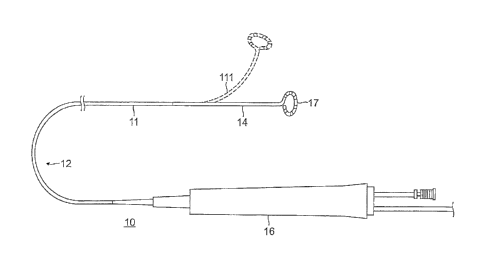
[0030] Referring to FIG. 1, the present invention is directed to a
catheter 10 with a multi-
layered catheter shaft portion 11 adapted for position sensing for
visualization of the shaft portion
11. The shaft portion 11 may be part of an elongated catheter tubing, for
example, an elongated
catheter body 12, or a shorter deflection portion 14 distal of the catheter
body 12, wherein position
sensing is accomplished by one or more single axis sensors (SAS) encased in
the shaft portion 11
which is constructed of multiple layers of similar materials, for example,
with similar melting
temperatures to promote a composite construction and adherence of the layers.
[0031] Proximal of the catheter body 12 is a control handle 16 with
mechanisms that are
manipulated by a user to accomplish, for example, bi-directional deflection of
the deflection section
14. Distal of the deflection portion 14 is a distal electrode assembly 17 with
one or more electrodes
arranged in a 2-D or 3-D configuration.
[0032] With reference to FIGS. 2A and 2B, the catheter body 12 comprises a
single, central or
axial lumen 18. The catheter body 12 is flexible, i.e., bendable, but
substantially non-compressible
along its length. As part of the catheter body 12, the shaft portion 11 and
the catheter body 12 have
a similar construction comprising an inner wall or first layer 21 of a
thermoplastic material, an
imbedded braided mesh 22, and a thin wall or second layer 23 of a
thermoplastic material
surrounding the braided mesh 22 and the first layer 21. Suitable thermoplastic
materials include,
for example, thermoplastic elastomers (TPEs) and thermoplastic polyurethanes
(TPUs), such as
PELLETHANE or PEBAX, where PEBAX has a melting temperature ranging between
about
-6-
CA 02950923 2016-12-07
=
=
1
272 F (133 C) and 345 F (174 C) and PELLETHANE has a melting temperature
ranging between
about 360 F (182 C) and 441 F (227 C). In some embodiments, the same
thermoplastic material
is used for the first layer 21 and the second layer 23. In some embodiments,
the first layer 21
comprises a first thermoplastic material and the second layer 23 comprises a
second thermoplastic
material similar to the first thermoplastic material. Similar thermoplastic
materials are understood
herein to be thermoplastic materials have melting temperatures such that
heating and reflowing of
at least one layer promote and enable bonding and adherence of one layer to
the other layer. In
some embodiments, similar thermoplastic materials have melting temperature
ranges that are
similar, which include thermoplastic materials with melting temperature ranges
that overlap by or
have in common at least about one degree in Fahrenheit (one degree in
Celsius), preferably about
five degrees in Fahrenheit (three degrees in Celsius), and more preferably
about ten degrees in
Fahrenheit (five degrees in Celsius). It is understood that "similar" can
refer to the same chemical
materials having the same melting temperatures, and to different chemical
materials having
different chemical make-ups but similar melting temperature ranges as defined
herein. In some
embodiments, the "different chemical materials" might include, for example,
similar polymer
backbones but different pendant groups, or different polymer backbones.
[0033] The imbedded braided mesh 22 of stainless steel or the like is
provided to increase
torsional stiffness of the catheter body 12 so that when the control handle 16
is rotated the length of
the catheter body 12 rotates in a corresponding manner. The single lumen 18
permits components
passing therethrough (including, for example, irrigation tubing 25, electrode
lead wires 26, puller
wires 13a, 13b, etc.) to float freely within the catheter body 12. However, if
desired or appropriate,
the catheter body 12 may also have a multi-lumened extrusion construction.
[0034] The thin wall or second layer 23 is constructed of a second
thermoplastic material which
is reflowed over the braided mesh 22. With the first and second layers 21 and
23 being of the same
or similar thermoplastic materials, reflowing the second layer 23 over the
braided mesh 22 and the
-7-
CA 02950923 2016-12-07
=
=
=
1
first layer 21 promotes the catheter body 12 having a composite construction
and adherence of the
first and second layers 21 and 23 to each other.
[0035] The first layer 21 may have an outer diameter ranging between about
0.069" and
0.073", and preferably, a diameter of about 0.071". A sidewall of the first
layer 21 may have a
thickness ranging between about 0.003" and 0.006", and preferably, a thickness
of about 0.004".
[0036] The second layer 23 may have an outer diameter ranging between
about 0.100" and
0.109", and preferably, a diameter of about 0.104". A sidewall of the second
layer 23 may have a
thickness ranging between about 0.002" and 0.006", and preferably, a thickness
of about 0.003".
[0037] As shown in FIG. 2A, one or more linear single axis sensors
(SAS) 40A, 40B and 40C
forming a SAS subassembly are mounted on the bonded composite catheter shaft
portion 11 as part
of the catheter body 12. The SAS 40A comprises a coil 32A of multiple windings
of an electrical
conductor (e.g., very fine small gauge wire 34A) situated on an outer surface
of the second layer
23. A distal portion 35A of the wire passes under the coil 32A and extends in
a longitudinal
direction toward a proximal end of the catheter shaft portion 11 and the
control handle 16. A
proximal portion 36A of the wire 34A also extends in the longitudinal
direction toward the
proximal end of the catheter shaft 11 and the control handle 16. The coil 32A
may incorporate
strain relief adaptations, including slack and/or windings, as disclosed in
U.S. Patent No.
8,792,962, issued July 29, 2014, entire content of which is incorporated
herein by reference. The
SAS 40B and 40C have a similar construction, and thus similar components
thereof are identified
in the Figures with similar reference numbers with letter designation of B or
C.
[0038] Each SAS interacts with at least one external magnetic field
generated by a magnetic
field generator positioned, for example, below the patient bed. Each SAS
generates signals
representative of the relative strengths of the field as sensed by its coil,
which signals are
transmitted proximally toward the control handle 16 and further to a highly
accurate mapping
system, such as CARTO, CARTO XP or CARTO 3, available from Biosense Webster,
to provide
-8-
CA 02950923 2016-12-07
=
=
1
visualization of the shaft portion 11 and to create 3-D anatomical maps of
tissue chamber or region
of interest in the patient, based on location and orientation of the shaft
portion 11 on which the SAS
subassembly is mounted.
[0039] As shown in FIG. 2A, distal SAS 40A has wire distal portion 35A
and wire proximal
portion 36A, mid SAS 40B has wire distal portion 35B and wire proximal portion
36B, and
proximal SAS 40C has wire distal portion 35C and wire proximal portion 36C. To
insulate the
wire distal and proximal portions of the more distal SAS from the more
proximal SAS, a
nonconductive sleeve 38 is placed and fitted on the shaft portion 11 between
the second layer 23
and the coil 32, with the wire distal and proximal portions of more distal SAS
passing between the
sleeve 38 and the second layer 23. In the embodiment of FIG. 2A, insulating
sleeve 38B is
provided under the coil 32B to insulate wire portions 35A and 36A from the
coil 32A, and
insulating sleeve 38C (also shown in FIG. 2B) is provided under the coil 32C
to insulate wire
portions 35A, 36A, 35B and 36B from the coil 32C. In that regard, the sleeves
38B and 38C are
shaped and sized to provide sufficient and adequate insulation surfaces on
which the coils 32B and
32C may be wounded without contacting the underpassing wire portions. The wire
34 may
comprise flat ribbon wires that can lie flatter against the second layer 23
for a minimized profile
when passed under the sleeves 38B and 38C.
[0040] In some embodiments, each SAS includes an encapsulation coating or
layer 42 encasing
the coil 32, surrounding it circumferentially on the catheter shaft portion 11
(also shown in FIG.
2B). The layer 42 may be of any suitable material, including, for example,
epoxy, UV glue, or the
like. The encapsulation layer 42 provides a number of benefits, including
protecting the coil 32
from exposure to increased temperatures during reflow process, and providing
strain relief to
minimize wire breakage or damage during assembly and use. For distal SAS 40A,
the
encapsulation layer 42A encases the coil 32A with the second layer 23. For mid
and proximal SAS
-9-
CA 02950923 2016-12-07
=
=
1
40B and 40C, the encapsulating layer 42B and 42C encases the coils 32B and 32C
with the sleeves
38B and 38C, respectively.
[0041] In some embodiments, the shaft portion 11 includes an outer wall or
third layer 24 that
extends over the SAS subassembly, if not also the length of the catheter body
12. As shown in
FIG. 2A, the third layer 24 protects the coils 32A, 32B and 32C, and the wire
distal and proximal
portions 35A, 36A, 35B, 36B, 35C and 36C.
[0042] In construction of the catheter body 12, including the shaft
portion 11, according to
some embodiments of the present invention, as shown in FIG. 3A, the first
layer 21 is extruded
from an extruder 45 over a mandrel 30 which forms the central lumen 18 (FIG.
2A) of the shaft
portion 11. As shown in FIG. 3B, the mandrel 30 (in broken lines) may remain
under the extruded
first layer 21 as the mesh 22 is braided over the first layer 21. As shown in
FIG. 3B, the mandrel
30 may remain under the extruded first layer 21 and the braided mesh 22 as a
heat shrink tubing 52
forming the second layer 23 is extruded over or otherwise fitted on the first
layer 21 and braided
mesh 22. The mandrel 30 may remain in the first layer 21 as heat is applied to
the heat shrink
tubing 52 to reflow over the braided mesh 22 and the first layer 21 in forming
the second layer 23.
As described above, the heated tubing 52 is reflowed so that the second
thermoplastic material can
seep through the braided mesh 22 and bond with the first thermoplastic
material of the first layer
21. The similarity in melting temperatures of the first and second
thermoplastic materials
facilitates such bonding and adherence.
[0043] As shown in FIG. 3C, the distal most SAS, for example, SAS 40A
is mounted first.
Wire distal portion 35A of thin wire 34A is laid longitudinally on the outer
surface of the second
layer 23 and the thin wire 34A is coiled around the shaft portion 11, on top
of the wire distal
portion 35A. The remainder of the wire distal portion 35A extends proximally
of the coil 32A
toward a proximal end of the catheter body 12. Proximal of the coil 32, wire
proximal portion 36A
-10-
CA 02950923 2016-12-07
=
=
1
of the wire 34A is laid longitudinally on the outer surface of the second
layer 23 also extending
proximally toward a proximal end of the catheter body 12.
[0044] Before mounting the next distal SAS at a selected location proximal
of the distal-most
SAS 40A, for example, the mid SAS 40B, sleeve 38B is mounted over the second
layer 23 and the
wire distal and proximal portions 35A and 36A at the selected location. In
some embodiments, the
sleeve 38B may be a short heat-shrink tubing that is reflowed over the wire
portions 35A and 36A,
and the second layer 23. To mount the mid SAS 40B, wire distal portion 35B of
thin wire 348 is
laid longitudinally on the sleeve 38B, and the thin wire 34B is coiled around
the shaft portion 11
over the wire distal portion 35B and the sleeve 38B (which covers and
insulates the wire distal
portion 35A and the wire proximal portion 36A from the coil 34B). Wire
proximal portion 36B of
the wire 34B is laid longitudinally on the sleeve 38B and further on the outer
surface of the second
layer 23 as it extends proximally toward a proximal end of the catheter body
12.
[0045] Additional SAS, including SAS 40C may be mounted in the same manner
as described
above for SAS 40B.
[0046] As shown in FIG. 3C, the third layer 24 may also be applied as
a heat shrink tubing 54
which seals in all the components mounted and carried on the shaft portion 11.
The tubing 54 is
reflowed over the second layer 23, the coils 32A and 32B, the sleeves 38B, and
the wire portions
35A, 36A, 35A, 35B. The encapsulation coatings or layers 42A, 42B and 42C (see
FIG. 2B) may
applied to the coils before the tubing 54 is fitted over the coils, or they
may be applied via syringe
injection through the heat shrink-tubing 54 before it is reflowed into forming
the third layer 24.
The third layer 24 is constructed of a third thermoplastic material which may
be the same as the
first and/or second thermoplastic material, or be similar to the first and/or
second thermoplastic
material, in promoting bonding and adherence of one or more layers of the
multi-layer construction
of the shaft portion 11.
-11-
CA 02950923 2016-12-07
= =
1
[0047] As shown in FIG. 3A, FIG. 3B and 3C, the mandrel 30 may remain
in the first layer 21
during at least the winding of the coil of the one or more SASes on the second
layer 23, and if not
also during the application/reflow of the third layer 24, so as to maintain
the structural shape of the
shaft portion 11 and the central lumen 18. It is understood that the mandrel
supporting the
structural shape need not be the same mandrel used throughout the
manufacturing of the shaft
portion 11 but that the mandrel 30 may be removed and replaced with one or
more mandrels as
suitable or appropriate during the winding of the coil of the one or more SAS
on the second layer
23, and/or any of the reflow stages during manufacturing of the shaft portion
11.
[0048] It is understood that FIG. 3A, FIG. 3B and FIG. 3C are
representative illustrations
demonstrating various steps of constructing a multi-layered catheter body with
an embedded SAS
subassembly within the side wall of the catheter body, in accordance with some
embodiments of
the present invention. Although the steps illustrated may be performed in an
assembly line fashion,
with progression from FIG. 3A, to FIG. 3B to FIG. 3C, the steps may also be
performed discretely,
in different assembly lines, by different machinery and/or at different
locations. For example,
while FIG. 3B illustrates the reflowing of the heat shrink tubing 52 at one
location on the catheter
body as occurring simultaneously with the application of the braided mesh 22
at another location
on the catheter body, it is understood that the application of the braided
mesh may be completed
entirely along the length of the catheter body 12 before the heat shrink
tubing 52 is fitted over the
catheter body 12 and before heat is applied to reflow the tubing 52.
[0049] At the proximal end of the catheter body 12 that is received in
a distal end of the control
handle, the proximal and distal portions 35A, 36A, 35B, 36B, 35C, 36C which
have extended
longitudinally along the catheter shaft 12 between the second layer 23 and the
third layer 24 enter
the interior of the control handle 16 for connection to a printed circuit
board for processing,
including, for example, amplification, as known in the art.
-12-
CA 02950923 2016-12-07
=
1
[0050] In other embodiments of the present invention, the wire distal
and proximal portions
35A, 36A, 35B, 36B, 35C, 36C of each coil 32A, 32B and 32C may extend
proximally through a
lumen 61 of the catheter shaft, as shown in FIG. 4A and FIG. 4B. A through-
hole 70 is formed into
the lumen through the sidewall of the catheter shaft portion (through the
first layer 21, the braided
mesh 22 and the second layer 23) for each wire portion 35A, 36A, 35B, 36B, 35C
and 36C. As
such, sleeves 38B and 38C are not needed. As shown in FIG. 4A, the extruded
first layer 21 may
be formed as a multi-lumened tubing with lumens 50, 51, 52 and 53 (with use of
one or more
suitable mandrels). The through-hole 70 may be formed to communicate with the
lumen 61, such
that the wire portions 35A, 36A, 35B, 36B, 35C and 36C all pass through the
dedicated lumen 61
along the length of the catheter shaft.
[0051] In some embodiments, lumen 62 may be provided for irrigation
tubing 25 and lumen 65
may be provided for tip electrode lead wires 26. Diametrically opposing lumens
63 and 64 may be
suitable for a pair of puller wires 13a and 13b to provide the catheter with
bi-directional deflection.
In that regard, the shaft portion 11 with the one or more embedded SAS in its
layered construction
is suitable as segment of the deflection portion 14 (as shown in FIG. 1), for
example, that extends
distal of a single lumened catheter body through which the pair of puller
wires extends. Each
puller wire has a proximal end anchored in the control handle 16 and a distal
end anchored at or
near a distal end of the deflection portion 14. Surrounding each puller wire
is a compression coil
(now shown) having a proximal end at a proximal end of the catheter body, and
a distal end at or
near a proximal end of the deflection portion 14, as known in the art and
understood by one of
ordinary skill in the art.
[0052] The preceding description has been presented with reference to
presently preferred
embodiments of the invention. Workers skilled in the art and technology to
which this invention
pertains will appreciate that alterations and changes in the described
structure may be practiced
without meaningfully departing from the principal, spirit and scope of this
invention. Any feature
-13-
CA 02950923 2016-12-07
=
=
1
or structure disclosed in one embodiment may be incorporated in lieu of or in
addition to other
features of any other embodiments, as needed or appropriate. It is understood
that a feature of the
present invention is applicable to multiplying linear motion of a puller wire,
contraction wire, or
any other object requiring insertion, removal, or tensioning within a medical
device, including the
disclosed electrophysiology catheter. As understood by one of ordinary skill
in the art, the
drawings are not necessarily to scale. Accordingly, the foregoing description
should not be read as
pertaining only to the precise structures described and illustrated in the
accompanying drawings,
but rather should be read consistent with and as support to the following
claims which are to have
their fullest and fair scope.
20
-14-