Note: Descriptions are shown in the official language in which they were submitted.
CA 02951183 2016-12-05
WO 2015/185655 PCT/EP2015/062430
STRAND-INVASION BASED DNA AMPLIFICATION METHOD
Field of the Invention
The invention relates to methods of amplification of a target nucleic acid
sequence
comprising strand invasion. The invention further relates to kits and
compositions suitable
for use in such methods.
Background to the Invention
Methods for amplification of a target nucleic acid sequence by strand invasion
have
been described for example in W02009/150467. Invasion of the target nucleic
acid
sequence is mediated by a single strand invasion oligonucleotide, which opens
up a target
duplex to allow binding of both upstream and downstream primers.
Brief Description of the Figures
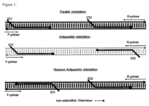
Figure 1: Amplification of a target DNA using two invasion oligonucleotides
with
either parallel invasion oligonucleotide configuration, anti-parallel invasion
oligonucleotide configuration or reverse anti-parallel configuration. 101 ¨
first strand
invasion oligonucleotide; 102 ¨ second strand invasion oligonucleotide. F-
primer ¨
forward or upstream primer; R-primer ¨ reverse or downstream primer. The non-
extendible
terminus of 101 and 102 is shown as a dashed line.
Figure 2: Amplification of a target DNA using two invasion oligonucleotides.
Amplification plots are shown with for (a) parallel invasion oligonucleotide
configuration,
(c) anti-parallel invasion oligonucleotide configuration and (e) anti-parallel
reverse
invasion oligonucleotide configuration. Duplicate reactions presented.
Amplification was
monitored by detecting Sybr Green I. X-axis for amplification plots: time
(minutes), Y-
axis: SybrGreen I fluorescence (fluorescence intensity, arbitrary units).
Specificity of the
reactions were further assccessed with melt-curve analyses. Melt curve
analyses are shown
in (b) for parallel invasion oligonucleotide configuration,. (d) for anti-
parallel invasion
oligonucleotide configuration and (f) for anti-parallel reverse invasion
oligonucleotide
configuration. X-axis for melt curve analyses: Temperature (degrees
Centigrade), Y-axis (-
d(fluorescence/d(temperature), (arbitrary units).
1
CA 02951183 2016-12-05
WO 2015/185655 PCT/EP2015/062430
Figure 3: Amplification of target DNA using two invasion oligonucleotides.
Reactions were either performed with two complementary invasion
oligonucleotides or
with one complementary invasion oligonucleotide and one non-complementary
invasion
oligonucleotide. X and Y-axis for amplification plots as for Figure 2. (a)
shows results with
parallel configuration of oligonucleotides used. (b) shows results with anti-
parallel
configuration of oligonucleotides. Duplicate reactions presented.
Figure 4: Specificity of primers in the amplification reaction using two
invasion
oligonucleotides. Reactions were performed either with complementary forward
and
reverse primers or with complementary forward and non-complementary primer.
Concentration of target DNA was 1pM. X and Y-axis for amplification plot as
for Figure
2.
Figure 5: Compatibility of reaction using two invasion oligonucleotides with
target
specific probes. (a) shows schematic representation of configurations
supporting the use of
target specific probes. (b) and c) show amplification and real-time detection
of target DNA
with either (b) two invasion oligonucleotides or (c) a single strand invasion
oligonucleotide
(SIBA). Real-time monitoring of amplification was achieved either with Sybr
green I or a
target specific probe, as shown in the labels for the traces. X-axis for each
chart: Time
(minutes). Y-axis: fluorescence of Sybr green I or probe (arbitrary units).
Figure 6: Resistance of (a) reaction using two invasion oligonucleotides and
(b)
standard reaction using a single strand invasion oligonucleotide (SIBA) to
detection of
non-specific amplification. Standard SIBA is was less resistant to detection
of non-specific
amplification with short primers than amplification carried out with two
invasion
oligonucleotides. Concentration of target DNA was 1 pM for long primers and 1
fM for
short primers. Amplification was monitored using Sybr green I or a probe
having a binding
site which is non-overlapping with the binding site of the strand invasion
oligonucleotides
or primers. X-axis for each chart: Time (minutes). Y-axis: fluorescence of
Sybr green I or
probe (arbitrary units). (a) shows monitoring of amplification with Sybr green
I or probe
during amplification with two invasion oligonucleotides. (b) shows monitoring
of
amplification with Sybr green I in SIBA.
Figure 7: Amplification of a target DNA from a plasmid DNA using two strand
invasion oligonucleotides. Plasmid DNA was either used directly or treated
with EcoRV-
2
CA 02951183 2016-12-05
WO 2015/185655 PCT/EP2015/062430
HF restriction enzyme. Amplification was monitored using Sybr green I. X-axis:
Time
(minutes). Y-axis: fluorescence of Sybr green I (arbitrary units).
Figure 8: Amplification of a target DNA having two identical invasion sites.
Reactions were performed with a single invasion oligonucleotide that binds to
both
invasion sites of the target DNA. (a), (c), (e) and (g) show amplification
plots for real-time
monitoring of target DNA amplification using Sybr green I. (b), (d), (f) and
(h) show
corresponding melt curve analyses. (i) shows non-denaturing electrophoresis of
reaction
products. a) and b): parallel configuration of invasion oligonucleotides used
to amplify a
324 base pair a duplex target DNA. (c) and (d): parallel configuration of
invasion
oligonucleotides used to amplify a target DNA. (e) and (f): anti-parallel
configuration of
invasion oligonucleotides used to amplify a target DNA. (g) and (h): reverse
anti-parallel
configuration of invasion oligonucleotides used to amplify a target DNA. (i)
antiparallel
configuration of invasion oligonucleotides used to amplify a target DNA. X and
Y-axes for
amplification plots and melt curve analyses as for Figure 2. Real-time
monitoring of target
DNA amplification was achieved using Sybr green I. (i) Lanes for
electrophoretogram as
follows: Lane 1, BioRad EZ Load 20 bp Molecular Ruler; lanes 2-6 copied 107,
106, 105,
104 and 103 respectively; lane 7, water control.
Figure 9: FRET based system for real-time monitoring invasion and
amplification:
(a) schematic representation of labelled primers and invasion
oligonucleotides. (b) Real
time monitoring of invasion and amplification and detection of target DNA
using FRET
labelled oligonucleotides in parallel configuration. X-axis for (b): Time
(minutes). Y-axis:
fluorescence of probe (arbitrary units).
Figure 10: Sensitivity of strand invasion based amplification using two strand
invasion oligonucleotides. Sensitivity was assessed with three different
assays using serial
dilutions of 106 to 1 copy of target DNA. Real-time monitoring of target DNA
amplification was achieved using Sybr green I. Amplification plots: (a) Assay
1 (b) Assay
2 and (c) Assay 3. X-axis): Time (minutes). Y-axis: fluorescence of Sybr green
I (arbitrary
units).
Brief description of the sequences
SEQ ID NO: 1 is the nucleotide sequence of an invasion oligonucleotide.
SEQ ID NO: 2 is the nucleotide sequence of an invasion oligonucleotide.
3
CA 02951183 2016-12-05
WO 2015/185655 PCT/EP2015/062430
SEQ ID NO: 3 is the nucleotide sequence of a DNA primer.
SEQ ID NO: 4 is the nucleotide sequence of a DNA primer.
SEQ ID NO: 5 is the nucleotide sequence of a DNA primer.
SEQ ID NO: 6 is the nucleotide sequence of a non-complementary invasion
oligonucleotide.
SEQ ID NO: 7 is the nucleotide sequence of a non-complementary DNA primer.
SEQ ID NO: 8 is the nucleotide sequence of a probe.
SEQ ID NO: 9 is the nucleotide sequence of a probe.
SEQ ID NO: 10 is the nucleotide sequence of a DNA primer.
SEQ ID NO: 11 is the nucleotide sequence of a target template.
SEQ ID NO: 12 is the nucleotide sequence of a target template.
SEQ ID NO: 13 is the nucleotide sequence of an invasion oligonucleotide.
SEQ ID NO: 14 is the nucleotide sequence of a DNA primer.
SEQ ID NO: 15 is the nucleotide sequence of a DNA primer.
SEQ ID NO: 16 is the nucleotide sequence of a target template.
SEQ ID NO: 17 is the nucleotide sequence of a target template.
SEQ ID NO: 18 is the nucleotide sequence of a target template.
SEQ ID NO: 19 is the nucleotide sequence of a target template.
SEQ ID NO: 20 is the nucleotide sequence of a target template.
SEQ ID NO: 21 is the nucleotide sequence of a labelled invasion
oligonucleotide.
SEQ ID NO: 22 is the nucleotide sequence of a labelled invasion
oligonucleotide.
SEQ ID NO: 23 is the nucleotide sequence of a labelled primer.
SEQ ID NO: 24 is the nucleotide sequence of a labelled primer.
SEQ ID NO: 25 is the nucleotide sequence of a DNA primer.
SEQ ID NO: 26 is the nucleotide sequence of a target template.
SEQ ID NO: 27 is the nucleotide sequence of an adaptor.
SEQ ID NO: 28 is the nucleotide sequence of an adaptor.
SEQ ID NO: 29 is the nucleotide sequence of an adaptor.
SEQ ID NO: 30 is the nucleotide sequence of an adaptor.
SEQ ID NO: 31 is the nucleotide sequence of an adaptor.
4
CA 02951183 2016-12-05
WO 2015/185655 PCT/EP2015/062430
Summary of the Invention
The present invention relates to a system for strand invasion of a target
nucleic acid
sequence at at least two locations. The methods of the invention use one or
more strand
invasion oligonucleotides to bind and invade upstream and downstream regions
of the
target nucleic acid sequence, allowing binding of upstream and downstream
primers to
effect amplification of the target nucleic acid sequence. Providing for strand
invasion of a
target nucleic acid sequence at both an upstream and a downstream location
couples each
primer binding event to an independent strand invasion event and provides
increased
possibilities for use of strand invasion oligonucleotide sequences that do not
have overlap
with amplification primers. Strand invasion mediated at two different
locations also
provides advantages for amplification of target nucleic acid sequences that
are longer than
those that can typically be amplified from a single point of strand invasion.
Additionally, the same strand invasion species can invade both at an upstream
and
downstream location provided suitable binding sequences are present in both
regions of the
target sequence. Similarly, a single primer species may be used where a
suitable binding
sequence is present in both regions of the target sequence. These embodiments
permit
amplification and sequencing of unknown sequences where known binding regions
(such
as adaptor sequences) are present in a template comprising the target
sequence. Strand
invasion oligonucleotides may also be designed to bind to upstream and
downstream
binding regions of a duplex target nucleic acid sequence in alternative
configurations. This
provides opportunities to vary design of sequences for targeting a particular
amplicon to
optimise amplification parameters. Furthermore, strand invasion
oliognucleotides and
primers may be designed to have non-overlapping binding regions such that a
region of the
amplicon remains free for binding of a probe, thus reducing competition
between
oligonucleotide species for binding the amplicon during amplification and
avoiding
detection of non-specific amplification products.
Accordingly, the present invention provides a method for amplification of a
target
nucleic acid sequence, said method comprising contacting said target nucleic
acid sequence
with at least one upstream primer, at least one downstream primer and first
and second
strand invasion oligonucleotides under conditions promoting amplification of
said target
nucleic acid sequence, wherein the first strand invasion oligonucleotide
renders an
5
CA 02951183 2016-12-05
WO 2015/185655 PCT/EP2015/062430
upstream binding region of the target nucleic acid sequence single-stranded to
allow the
binding of the upstream primer, and the second strand invasion oligonucleotide
renders a
downstream binding region of the target nucleic acid sequence single-stranded
to allow the
binding of the downstream primer.
The invention further provides a method for amplification of a target nucleic
sequence comprising upstream and downstream binding regions for a strand
invasion
oligonucleotide, comprising contacting said target nucleic acid sequence with
a strand
invasion oligonucleotide and one or more primers capable of amplifying the
target nucleic
acid sequence, wherein the strand invasion oligonucleotide renders the
upstream and
downstream strand invasion oligonucleotide binding regions of the target
nucleic acid
sequence single-stranded to allow the binding of said one or more primers.
The invention also provides a kit comprising at least one upstream and at
least one
downstream primer for a target nucleic acid sequence, and first and second
strand invasion
oligonucleotides which respectively have upstream and downstream binding
regions in a
target nucleic acid sequence.
The invention further provides a kit comprising a strand invasion
oligonucleotide
and one or more primers, and at least one DNA adaptor, wherein said strand
invasion
oligonucleotide can bind the DNA adaptor when present in an upstream binding
region and
a downstream binding region of a target nucleic acid sequence, and wherein
said one or
more primers are capable of amplifying said target nucleic acid sequence.
The invention additionally provides a method of amplifying a target nucleic
acid
sequence comprising a region of unknown sequence comprising creating a target
nucleic
acid sequence comprising strand invasion oligonucleotide binding regions
upstream and
downstream of said region of unknown sequence, and carrying out a method of
the
invention employing strand invasion oligonucleotides and primers to amplify
the target
nucleic acid sequence.
The invention further provides a method of determining the sequence of a
target
nucleic acid comprising a region of unknown sequence, comprising creating a
target
nucleic acid sequence comprising strand invasion oligonucleotide binding
regions
upstream and downstream of said region of unknown sequence, carryng out a
method of
the invention employing strand invasion oligonucleotides and primers to
amplify the target
nucleic acid sequence, and determining the sequence of said region of unknown
sequence.
6
CA 02951183 2016-12-05
WO 2015/185655 PCT/EP2015/062430
Detailed Description of the Invention
It is to be understood that different applications of the disclosed methods
may be
tailored to the specific needs in the art. It is also to be understood that
the terminology used
herein is for the purpose of describing particular embodiments of the
invention only, and is
not intended to be limiting. In addition as used in this specification and the
appended
claims, the singular forms "a", "an", and "the" include plural referents
unless the content
clearly dictates otherwise. Thus, for example, reference to "a polypeptide"
includes two or
more such polypeptides, and the like. All publications, patents and patent
applications cited
herein, whether supra or infra, are hereby incorporated by reference in their
entirety.
Methods for amplification of a target nucleic acid sequence
The methods of the invention provide for amplification of a target nucleic
acid
sequence by strand invasion of a nucleic acid at two separate sites. Strand
invasion at each
site, mediated by a strand invasion oligonucleotide, renders the target
nucleic acid
sequence single-stranded to allow for binding for a primer. The primers are
typically not
able to amplify the target nucleic acid sequence when contacted thereto in the
absence of
the strand invasion oligonucleotide(s). In other words, the primers are not
able to bind to
their binding regions in the target nucleic acid sequence unless their binding
regions are
exposed by strand invasion oligonucleotides which render their binding regions
single-
stranded. The strand invasion oligonucleotides are also typically not capable
of extension
by a DNA polymerase. In particular, the methods of the invention preferably
amplify a
target nucleic acid sequence under isothermal conditions in which a target
nucleic acid
sequence is present in a nucleic acid duplex. Strand invasion at at least two
sites of the
duplex renders the target nucleic acid sequence single-stranded under
isothermal
conditions, permitting primer-based amplification.
Target nucleic acid sequence
The target nucleic acid sequence may be of any origin and may for example be
artificial or naturally occurring. The target nucleic acid sequence may
comprise a known
sequence or regions of known and unknown sequence. The target nucleic acid
sequence
may be human, mammalian, bacterial or viral. The target nucleic acid sequence
may be a
7
CA 02951183 2016-12-05
WO 2015/185655 PCT/EP2015/062430
region of a gene or chromosome. The target nucleic acid sequence may be
specific for a
genotype or an organism (such as a pathogen) to be detected by DNA
amplification. The
target nucleic acid sequence may be unique to the genome of a particular
species. Thus, the
target nucleic acid sequence for detecting a particular species will typically
differ from any
homologous nucleic acid sequence in a related species. Typically, the target
nucleic acid
sequence will comprise several mismatches with a homologous nucleic acid
sequence in a
related species. The target nucleic acid sequence may be a sequence specific
to a particular
strain of bacteria or a particular serotype, isolate or clade of a virus.
The target nucleic acid sequence to be detected may be of any size and have
any
sequence. The target nucleic acid sequence or amplicon is of a sufficient
length to provide
for hybridisation of the upstream and downstream primers and binding of strand
invasion
oligonucleotide(s) in a suitable manner to upstream and downstream portions of
the target
sequence. The amplicon is typically at least 60 nucleotides in length, more
preferably at
least 65, or at least 70 nucleotides in length as measured from the 5' site of
binding of the
upstream primer to the 5' site of binding of the downstream primer. The
amplicon may be
about 60 to about 80 nucleotides in length. In some embodiments, the amplicon
may be
greater than 80, such as greater than 100 nucleotides in length, such as
greater than 150,
200, 300, 400, 500, 1000 or more nucleotides in length. The amplicon may be
from about
70 to about 1000 nucleotides in length, such as from about 70 to about 800,
about 70 to
about 600, about 70 to about 500 nucleotides in length, about 70 to about 400,
about 100 to
about 400, or about 100 to about 200 nucleotides in length.
Examples of suitable target nucleic acid sequences for methods of the
invention
include SEQ ID NOs 11, 12, 16, 17, 18, 19, 20 and 26.
The target nucleic acid sequence comprises upstream (5') and downstream (3')
regions which each include binding regions for a strand invasion
oligonucleotide and a
primer. The upstream binding regions for a strand invasion oligonucleotide and
primer
may overlap in sequence or be non-overlapping. Similarly, the downstream
binding
regions for a strand invasion oligonucleotide and primer may overlap in
sequence or be
non-overlapping. The target nucleic acid sequence may also comprise binding
regions for
one or more oligonucleotide probes. The binding regions for a probe may
overlap in
sequence with the upstream or downstream binding regions for a strand invasion
oligonucleotide and/or primer or be non-overlapping with a binding region for
any strand
8
CA 02951183 2016-12-05
WO 2015/185655 PCT/EP2015/062430
invasion oligonucleotide or primer. The binding region for a probe may
preferably be
located in between the upstream and downstream strand invasion oligonucleotide
binding
regions of the target nucleic acid sequence. Selection of binding regions for
strand invasion
oligonucleotides, primers and probes, and design of appropriate sequences for
these is
discussed in more detail below.
The lengths of the binding regions for the strand invasion oligonucleotides,
primers
and probes are defined by the lengths of complementary sequences to the target
that are
included therein, as described below in more detail. As described below, a
strand invasion
oligonucleotide typically includes at least 25 nucleotides of complementary
sequence to
the target, and a primer at least 10. Thus each strand invasion
oligonucleotide binding
region of the target sequence may be at least 25 nucleotides in length and
each primer
binding region at least 10 nucleotides in length. The target sequence may
further comprise
a probe binding region of typically at least 10 nucleotides in length.
The upstream and downstream strand invasion oligonucleotide binding regions
may
be present in the same strand of the target nucleic acid sequence, or may be
located in
opposing strands of a duplex comprising the target nucleic acid sequence. The
strand
invasion oligonucleotide(s) may thus bind the target nucleic acid sequence in
a parallel
orientation on the same strand, aligning 5' to 3' in the same direction.
Alternatively, the
strand invasion oligonucleotide(s) may bind opposing strands of the target
nucleic acid
sequence in an antiparallel orientation, aligning 5' to 3' in opposing
directions on the target
duplex. In an antiparallel orientation, the 3' terminus of each strand
invasion
oligonucleotide may be directed towards or away from each other. Thus, the 3'
termini of
each strand invasion oligonucleotide may face towards the centre of the
amplicon
(antiparallel configuration) or towards its respective amplicon end (reverse
antiparallel
configuration). The 3' terminus or the 5' terminus of a strand invasion
oligonucleotide may
bind proximal to the binding region for a respective primer. The above binding
configurations are shown in Figure 1.
The use of particular binding configurations can provide alternative effects
on
amplification parameters. For example, where a strand invasion oligonucleotide
binds with
its 5' end located proximal to the binding region for a respective primer, the
binding of the
primer may have a different specificity and kinetic profile as compared to a
primer binding
proximal to the 3' end of a strand invasion oligonucleotide. The 3' terminus
of a strand
9
CA 02951183 2016-12-05
WO 2015/185655
PCT/EP2015/062430
invasion oligonucleotide typically comprises a number of modified nucleotides
(such as 2'-
0-methyl RNA nucleotides) which may influence binding interactions of a primer
binding
proximally thereto. In the parallel and reverse antiparallel configurations,
it is also
considered that specificity of amplification may be enhanced since branch
migration of the
3' termini of the strand invasion oligonucleotides (which typically comprise
modified
nucleotides) is required before primer binding is possible. Accordingly, the
methods of the
invention provide for variation of amplification rate and specificity of
amplification by
variation of binding configurations of the strand invasion oligonucleotide(s).
The upstream and downstream strand invasion oligonucleotide binding regions of
the target nucleic acid sequence may bind the same species of strand invasion
oligonucleotide. Thus, a single species of strand invasion oligonucleotide may
be provided
to initiate strand invasion at two points in the target nucleic acid sequence,
as discussed
further below. In this embodiment, the upstream and downstream binding regions
each
comprise complementary sequence to at least a portion of the strand invasion
oligonucleotide. The upstream and downstream binding regions are typically
homologous
or identical to one another. The upstream and downstream binding regions may
be at least
85%, at least 90%, at least 95% homologous or identical to one another or
fully identical.
The upstream and downstream binding regions may have 1, 2, 3, 4, 5, 6, 7 or 8,
such as 1
to 5 or 1 to 3 mismatches between each other. Additionally, a single species
of primer may
be provided to initiate amplification at two points in the target nucleic acid
sequence, as
discussed further below. The target nucleic acid sequence will in this case
comprise
upstream and downstream binding regions which each comprise complementary
sequence
to at least a portion of the primer, and may be homologous or identical to one
another as
described above.
More than one target nucleic acid sequence may be detected in a method of the
invention by providing multiple combinations of strand invasion
oligonucleotide(s),
primers (and optionally probes) each specific for a different target nucleic
acid sequence.
Typically, strand invasion oligonucleotide/primer pairs and/or probes binding
to different
target nucleic acid sequences will be labeled with different
fluorophore/quencher pairs,
thus allowing for multiplexing. At least two, three, four, five, ten or more
different target
sequences may be detected. More than one target nucleic acid sequence from the
same
organism may be detected. Alternatively, target nucleic acid sequences
specific for at least
CA 02951183 2016-12-05
WO 2015/185655 PCT/EP2015/062430
two, three, four, five, ten or more different genotypes, organisms or
pathogens may be
detected.
Upstream and downstream primers
Suitable upstream and downstream primers are selected based on the target
nucleic
acid sequence of interest, and having regard to the site of binding of the
respective strand
invasion oligonucleotide that renders an upstream or downstream binding region
of the
target nucleic acid sequence single-stranded to allow the binding of the
respective primer.
The upstream and downstream primers comprise a sequence that is partly or
fully
complementary to the target and optionally a 5' and/or 3' flanking non-
complementary
sequence. Alternatively, the upstream and downstream primers may consist
entirely of
partly or fully complementary sequence to the target. The length of the primer
sequence
that is complementary to the target is sufficient to provide specific
hybridisation to the
target nucleic acid sequence. The length of complementary sequence is
typically at least 10
nucleotides, more preferably at least 15, at least 16, or at least 17
nucleotides. The length
of complementary sequence may be 10-25, 15-25, 10-30 or 15-30 nucleotides.
It should be understood that the above sequence lengths refer to portions of
the
primers which may be partly or fully complementary to the target nucleic acid
sequence.
Mismatches may be present between the primers and the target sequence at
particular
positions while still allowing for specific amplification and detection of the
target
sequence, in particular having regard to the combined use of upstream and
downstream
primers and binding of strand invasion oligonucleotide(s) to upstream and
downstream
regions of the target nucleic acid sequence to achieve amplification. There
may be 1, 2, 3,
4 or 5 mismatches between the complementary region of the primer and the
corresponding
region of the target sequence.
Typically the upstream and downstream primer will be less than 30 nucleotides
in
total in length, more preferably less than 25 nucleotides in length, such as
15 to 25, or 15 to
23 nucleotides in length. It is particularly preferred that primers of less
than 30 nucleotides
in length are used where a recombinase is used for strand invasion. Such
primers are not
capable of acting as substrates for recombinases. In some embodiments primers
of less
than 15 nucleotides in length may be used, such as primers of about 8 to about
14, about 10
to about 14 or about 12 to about 14 nucleotides in length. The use of such
short primers is
11
CA 02951183 2016-12-05
WO 2015/185655 PCT/EP2015/062430
preferred in combination with a probe having a binding region in the target
nucleic acid
sequence that does not overlap with the binding region for a primer or strand
invasion
oligonucleotide. Detection of non-specific amplification products produced by
short
primers can be reduced or eliminated by using a probe with a non-overlapping
binding site.
The upstream (or forward) primer binds to the 3' region of one strand of the
duplex
target nucleic acid sequence, at a position proximal or overlapping with the
binding site of
the strand invasion oligonucleotide. The downstream (or reverse) primer binds
to the 3'
region of the opposing strand of the duplex target nucleic acid sequence to
the upstream
primer, at a position proximal or overlapping with the binding site of the
strand invasion
oligonucleotide. The 5' binding sites of the upstream and downstream primers
are typically
at least 60 nucleotides apart, more preferably at least 65, or at least 70
nucleotides in length
on the duplex target sequence.
Depending on the binding configuration of the strand invasion oligonucleotide,
as
shown in Figure 1, the upstream primer may have a region of sequence overlap
or a region
of complementarity with the sequence of the respective strand invasion
oligonucleotide.
The region of sequence overlap or complementarity may be 1-8 nucleotides in
length, and
may be at least 5 or at least 6 nucleotides in length. The downstream primer
may likewise
have a region of sequence overlap or a region of sequence complementarity of 1-
8
nucleotides, such as at least 5 or at least 6 nucleotides in length with the
sequence of the
respective strand invasion oligonucleotide.
Alternatively, there may be no sequence overlap or complementarity between the
upstream primer and the respective strand invasion oligonucleotide, and/or no
sequence
overlap or complementarity between the downstream primer and the respective
strand
invasion oligonucleotide, with the relevant primer binding instead at a
position that is
proximal in the target sequence to the binding site of the strand invasion
oligonucleotide.
The use of one or more primers that have binding regions in the target that do
not
overlap with binding regions for strand invasion oligonucleotides can provide
various
advantages. In embodiments where the methods of the invention utilise
oligonucleotide
probes to detect DNA amplification, there may also be no sequence overlap or
complementarity between a strand invasion oligonucleotide and the probe and/or
an
upstream and/or downstream primer and the probe. There may be no sequence
overlap
between the binding regions within the target nucleic acid sequence for the
upstream
12
CA 02951183 2016-12-05
WO 2015/185655 PCT/EP2015/062430
primer, the downstream primer, each strand invasion oligonucleotide, and of
any probe.
There may also be no complementarity between any of the primers, strand
invasion
oligonucleotides or probes. Design of sequences for the various
oligonucleotide species
such that they can bind the target nucleic acid sequence at independent, non-
overlapping
regions in the target may provide for reduced competition between the
oligonucleotide
species for binding to the target nucleic acid sequence, and also reduce
formation and/or
avoid detection of undesired amplification products.
In more detail, primers of between 16 and 23 bases in length are typically
used in
strand invasion based amplification methods using a single strand invasion
oligonucleotide
(SIBA methods). The sequences at the 3' ends of the primers have usually about
8 bases
overlap or complementarity with the strand invasion oligonucleotide (the
upstream primer
overlaps the strand invasion oligonucleotide while the downstream primer is
complementary to the strand invasion oligonucleotide). This configuration
ensures efficient
amplification of the target DNA and minimizes the risk of non-specific
amplification. It is
also possible to use short primers < 14 bases in length, which do not overlap
with the
strand invasion oligonucleotide. Short primers which do not have sequences
that overlap
with the strand invasion oligonucleotide are able to amplify the target DNA
more
efficiently than long overlapping primers. This is because the 3' end of a
longer
overlapping primer competes with the strand invasion oligonucleotide for a
binding site of
the target template. For example, the upstream primer needs to first branch
migrate onto
the duplex before displacing the strand invasion oligonucleotide.
However, short primers (< 14 bases) can generate non-specific amplification
products. To avoid this problem, longer primers (16-23 bases) with 3'ends that
overlap or
are complementary with the strand invasion oligonucleotide are typically used
in SIBA. In
this configuration, the region peripheral to the strand invasion
oligonucleotide is still
around 14 bases long. This leaves only a short peripheral region that
dissociates when the
target DNA is amplified.
In the methods of the invention comprising strand invasion at two points in
the
target (upstream and downstream), shorter primers can be used than in SIBA.
Furthermore,
non-overlapping primers can be used more efficiently. This is because it is
possible to
incorporate a probe binding site on the target DNA that is independent of the
strand
invasion oligonucleotides and primers. Furthermore, the ability to use
different primer and
13
CA 02951183 2016-12-05
WO 2015/185655 PCT/EP2015/062430
strand invasion oligonucleotide configurations such as the reverse anti-
parallel
configuration in the methods of the invention minimize or abolish the risk of
short primer-
induced non-specific amplification.
Where a primer binds proximal to its respective strand invasion
oligonucleotide
(without sequence overlap or complementarity), typically there is 15
nucleotides or less,
preferably 10 nucleotides or less, such as about 1 to about 15 nucleotides,
about 5 to about
nucleotides, about 5 to about 10 nucleotides, or about 3 to about 8
nucleotides between
the closest boundary of the binding region of the strand invasion
oligonucleotide and the
binding region of the respective primer. This ensures that the primer is able
to hybridise to
10 the single-stranded region created by binding of the strand invasion
oligonucleotide.
Preferably, each primer is designed to allow for specific detection of a
particular
target nucleic acid sequence, such as a particular genotype, or a nucleic acid
sequence
present in a particular target, such as a particular organism or a particular
pathogen. Thus,
each primer typically specifically or selectively hybridises to a
complementary sequence
15 found only in the target. However, each primer may also hybridise to
other sequences, such
as sequences found in other species, provided that when used in combination
with the
second primer, strand invasion oligonucleotide(s) and optional oligonucleotide
probe,
specific detection of the target nucleic acid sequence is obtained.
Any upstream or downstream primer used in the invention may comprise one or
more modified nucleotides and/or a detectable label, for example a fluorescent
dye. In
some embodiments an upstream or downstream primer may form a FRET pair with a
respective strand invasion oligonucleotide, and thus comprise a fluorophore or
quencher,
as discussed below.
It should be understood that the methods of the invention may comprise use of
more than one pair of upstream and downstream primers, typically where more
than one
target sequence is to be detected in parallel in a multiplex system.
Strand invasion oligonucleotide(s)
One or more suitable strand invasion oligonucleotides are selected based on
the
target nucleic acid sequence of interest, and having regard to the site of
binding of the
upstream and downstream primers and the requirement for the strand invasion
oligonucleotide(s) to render the target nucleic acid sequence single-stranded
in the relevant
14
CA 02951183 2016-12-05
WO 2015/185655 PCT/EP2015/062430
regions to allow for the binding of the upstream primer and downstream primer.
Where the
target nucleic acid sequence comprises homologous or identical upstream and
downstream
strand invasion oligonucleotide binding regions, a single species of strand
invasion
oligonucleotide may be provided to effect amplification. Alternatively, two
separate
species of strand invasion oligonucleotides (first and second) binding
divergent sequences
in the upstream and downstream portions of the target nucleic acid sequence
may be
provided. The following description of the features of a strand invasion
oligonucleotide is
applicable to both first and second strand invasion oligonucleotides when
these are used.
Each strand invasion oligonucleotide comprises a sequence that is
complementary
to the target and optionally additional flanking non-complementary
sequence(s). The
length of the sequence that is complementary to the target may be determined
by the
skilled person empirically and is sufficient to provide for efficient strand
invasion of the
target nucleic acid sequence, optionally under isothermal conditions. The
complementary
sequence may comprise RNA-DNA complementary base pairing and modified
nucleotides.
Typically, the length of complementary sequence is at least 25 or at least 27
nucleotides,
typically at least 30 nucleotides, such as least 32, at least 33 or at least
35 nucleotides,
more preferably at least 36, 37, 38, 39 or 40 nucleotides in length or
greater. The length of
complementary sequence may be 30-50, 32-50, 35-50, 40-50, 35 to 48, 35 to 46,
38 to 45
or 40 to 45 nucleotides in length.
It should be understood that the above sequence lengths refer to a portion of
a
strand invasion oligonucleotide which may be partly or fully complementary to
the target
nucleic acid sequence. Mismatches may be present between the strand invasion
oligonucleotide and the target sequence at particular positions while still
allowing for
specific amplification and detection of the target sequence, in particular
having regard to
the combined use of upstream and downstream primers and a strand invasion
oligonucleotide to achieve amplification. There may be 1, 2, 3, 4, 5, 6, 7, or
8, such as 1 to
5 or 1 to 3 mismatches between the complementary region of the strand invasion
oligonucleotide and the corresponding region of the target sequence, depending
on the total
length of complementary sequence.
The complementary sequence of a strand invasion oligonucleotide hybridises to
a
portion of the target sequence which may or may not overlap with a portion of
the target
sequence forming a binding region for a primer. The strand invasion
oligonucleotide may
CA 02951183 2016-12-05
WO 2015/185655
PCT/EP2015/062430
have a region of overlap or complementarity of 1-8 nucleotides, such as a
region of at least
or at least 6 nucleotides in length, with a respective upstream or downstream
primer.
Alternatively, the sequence of a strand invasion oligonucleotide may have no
region of
overlap with the sequence of an upstream or downstream primer. In this
embodiment, as
5 discussed above, a strand invasion oligonucleotide will bind at a
position proximal to the
binding region for an upstream or downstream primer, such that it can render
the binding
region for the primer single-stranded.
The closest boundaries of the upstream and downstream strand invasion
oligonucleotide binding regions of the target nucleic acid sequence may be
located at least
15, such as at least 20 or at least 25 nucleotides apart in the target nucleic
acid sequence,
but shorter distances between the binding regions may also be used in some
embodiments.
The 5' portion of the complementary sequence of a strand invasion
oligonucleotide
typically binds within 25 nucleotides or less, more preferably 20 nucleotides
or less from
the relevant boundary of the duplex target nucleotide sequence to be melted
(the
amplicon).
A strand invasion oligonucleotide optionally further comprises non-
complementary
sequence region(s) to the target that flank the complementary sequence region.
A strand
invasion oligonucleotide may comprise a non-complementary 5' region which may
be of
any nucleotide sequence. The 5' non-complementary region is typically at least
3
nucleotides in length, more typically at least 6, at least 8, preferably at
least 10, at least 12
or at least 14 nucleotides in length. The 5' non-complementary region may
assist binding
of recombinase, since recombinase binds cooperatively. A strand invasion
oligonucleotide
may comprise a 3' non-complementary region typically of 1-3 nucleotides in
length which
comprises nucleotides which block polymerase extension, such as 3'-prime
inverted dT.
A strand invasion oligonucleotide is typically at least 30 nucleotides in
length
where a recombinase is used for strand invasion in the amplification method in
conjunction
with the strand invasion oligonucleotide. A strand invasion oligonucleotide is
preferably at
least 35, at least 40 or at least 45 nucleotides in length, more preferably at
least 50, and
may be at least 55 nucleotides in length or greater. The strand invasion
oligonucleotide
may be 40-70, 45-70, 45-70, 50-70, 55-70, 45-65, 50-65, 50-60 or 55-65
nucleotides in
length.
16
CA 02951183 2016-12-05
WO 2015/185655 PCT/EP2015/062430
Typically a strand invasion oligonucleotide has a non-extendible 3'terminus,
such
that it cannot serve as a substrate for a DNA polymerase, and the target
sequence is then
only amplified on the further binding of the specific upstream and downstream
primers.
This avoids formation of non-specific amplification products. A strand
invasion
oligonucleotide may comprise one, two, three, four, five, six, seven, eight or
more
modified nucleotides in its 3' region, such as in the 10-15 or 10-20
nucleotides from the 3'
terminus. A strand-invasion oligonucleotide may comprise a 3' modification of
the 3'
terminal nucleotide, and may be a dideoxynucleotide, or comprise a 3' amino-
allyl group, a
3' carbon spacer, 3' phosphate, 3' biotin, 3' sialyl, or 3' thiol. The 3'
nucleotide may be a
nucleotide incorporated in a reversed orientation by a 3'-3' linkage.
Alternatively or
additionally, the 3' region of the strand-invasion oligonucleotide may
comprise nucleotides
with poor substrate capability for DNA polymerases, such as PNA (peptide
nucleic acid)
nucleotides, LNA (locked nucleic acid), 2'-5' linked DNA, 2'-fluoro RNA or 2'-
0-methyl
RNA, or combinations thereof.
Where the strand-invasion oligonucleotide is a PNA oligomer comprising,
consisting of or consisting essentially of PNA nucleotides, such an
oligonucleotide can
destabilise and invade duplex DNA in the absence of a recombinase enzyme.
Thus, where
a PNA oligonucleotide is used, the methods of the invention may be performed
without
presence of a recombinase enzyme. A PNA oligonucleotide may comprise PNA
nucleotides and other nucleotides, such as DNA nucleotides, provided that the
oligonucleotide comprises sufficient PNA nucleotides to mediate strand
invasion of a
duplex. The skilled person can empirically determine the level of PNA to be
incorporated
into an oligonucleotide by testing its ability to effect strand invasion and
allow for DNA
amplification.
A strand invasion oligonucleotide may comprise a detectable label, for example
a
fluorescent dye. In some embodiments a strand invasion oligonucleotide may
form a FRET
pair with an upstream or downstream primer and thus comprise a fluorophore or
quencher,
as discussed below.
The methods of the invention comprise strand invasion at at least two sites of
a
target nucleic acid sequence, mediated by first and second strand invasion
oligonucleotides, or by the same species of strand invasion oligonucleotide
where the
target nucleic acid sequence comprises two binding sites for the same strand
invasion
17
CA 02951183 2016-12-05
WO 2015/185655 PCT/EP2015/062430
oligonucleotide. It should be understood that the methods of the invention may
further
comprise strand invasion by additional strand invasion oligonucleotides at
additional sites
of a target nucleic acid sequence, such as at 3 or more, 4 or more, 5 or more,
8 or more, or
or more sites. Additionally, in a multiplex system, the methods of the
invention may
5 comprise use of additional strand invasion oligonucleotides targeting
upstream and
downstream binding regions of additional target sequences.
Amplification of the target nucleic acid sequence
The DNA amplification method comprises strand invasion based amplification.
The
10 strand invasion amplification comprises strand invasion at at least two
sites in the target
nucleic acid sequences. Strand invasion occurs at both upstream and downstream
regions
of the target nucleic acid sequence.
The target nucleic acid sequence is incubated with the upstream primer,
downstream primer, and one or more (such as first and second) strand invasion
oligonucleotides capable of rendering both the upstream and downstream binding
regions
for the respective primers single-stranded, under conditions promoting
amplification of
said target nucleic acid sequence. In some embodiments, a single species of
primer may
serve as both the upstream and the downstream primer.
Such conditions typically comprise the presence of a DNA polymerase enzyme.
Suitable conditions include any conditions used to provide for activity of
polymerase
enzymes known in the art. The conditions typically include the presence of
dNTPs selected
from dATP, dTTP, dCTP, dGTP, dUTP and analogues of any thereof, suitable
buffering
agents/pH and other factors which are required for enzyme performance or
stability.
Typically all four of dATP, dTTP, dCTP and dGTP will be present. The
conditions may
include the presence of detergents and stabilising agents. The temperature
used is typically
isothermal, i.e. constant throughout the amplification process. The
temperature used
typically depends on the nature of the polymerase enzyme and other enzyme
components,
and also reflects the hybridisation temperature required for the primers and
strand invasion
oligonucleotides.
The polymerase used typically has strand-displacement activity. The term
"strand
displacement" is used herein to describe the ability of a DNA polymerase,
optionally in
conjunction with accessory proteins, to displace complementary strands on
encountering a
18
CA 02951183 2016-12-05
WO 2015/185655 PCT/EP2015/062430
region of double stranded DNA during DNA synthesis. Suitable DNA polymerases
include
poll from E. coli, B. subtilis, or B. stearothermophilus, and functional
fragments or
variants thereof, and T4 and T7 DNA polymerases and functional fragments or
variants
thereof A preferred polymerase is Bsu DNA polymerase or a functional fragment
or
variant thereof
The amplification conditions preferably comprise the presence of a
recombinase.
Any recombinase system may be used in the method of the invention. The
recombinase
system may be of prokaryotic or eukaryotic origin, and may be bacterial,
yeast, phage, or
mammalian. The recombinase may polymerise onto a single-stranded
oligonucleotide in
the 5'-3' or 3'-5; direction. The recombinase may be derived from a myoviridae
phage,
such as T4, T2, T6, Rb69, Aehl, KVP40, Acinetobacter phage 133, Aeromonas
phage 65,
cyanophage P-55M2, cyanophage PSSM4, cyanophage S-PM2, Rb14, Rb32, Aeromonas
phage 25, Vibrio phage nt-1, phi-1, Rb16, Rb43, Phage 31, phage 44RR2.8t,
Rb49, phage
Rb3, or phage LZ2. In a preferred embodiment, the T4 recombinase UvsX
(Accession
number: P04529) or a functional variant or fragment thereof is used. The Rad
systems of
eukaryotes or the recA-Reco system of E. coli or other prokaryotic systems may
also be
used. The recombinase may be E. coli RecA.
The conditions may further comprise the presence of recombinase accessory
proteins, such as single-stranded binding protein (e.g. T4 gp32, accession
number P03695)
and recombinase loading agent (e.g. UvsY, accession number NP 049799.2). In a
preferred embodiment, the conditions comprise the presence of the T4 gp32,
UvsX and
UvsY proteins. The recombinase (such as UvsX), and where used the recombinase
loading
agent (such as UvsY) and single stranded DNA binding protein (such as gp32),
can each be
native, hybrid or mutant proteins from the same or different myoviridae phage
sources. A
native protein may be a wild type or natural variant of a protein.
The conditions may further comprise other factors used to enhance the
efficiency of
the recombinase such as compounds used to control DNA interactions, for
example
proline, DMSO, BSA, PEG or other crowding agents which are known to enhance
loading
of recombinases onto DNA (Lavery P. et al. J. Biol. Chem. 1992, 267, (13),
9307-9314).
The conditions may also comprise the presence of an ATP regeneration system.
Various ATP regeneration systems are known to the person skilled in the art,
and include
glycolytic enzymes. Suitable components of an ATP regeneration system may
include one
19
CA 02951183 2016-12-05
WO 2015/185655 PCT/EP2015/062430
or more of phosphocreatine, creatine kinase, myokinase, pyrophosphatase,
sucrose and
sucrose phosphorylase. The conditions may further comprise the presence of
ATP.
Additional components such as magnesium ions, DTT or other reducing agents,
salts may also be included.
Further components may include one or more restriction enzymes (such as one or
more restriction endonucleases) to digest a nucleic acid comprising a target
nucleic acid
sequence prior to, or at the same time as contacting the target nucleic acid
sequence with
other amplification reagents. Amplification rate of a target nucleic acid
sequence
comprised in DNA plasmid may be increased by digestion of the plasmid with a
restriction
enzyme, to thus linearise the starting template. Thus, the methods of the
invention may
comprise contacting a nucleic acid comprising the target nucleic acid to be
amplified with
a restriction enzyme. Any suitable restriction enzyme having a suitable
recognition site in a
nucleic acid comprising the target nucleic acid sequence may be used for
digestion. The
recognition site is typically located in a region of the nucleic acid other
than the target
nucleic acid sequence.
The various components described above may be provided in varying
concentrations to provide for DNA amplification. The skilled person can select
suitable
working concentrations of the various components in practice.
Detection of presence of amplified DNA
The presence of amplified DNA resulting from the contacting of the target
nucleic
acid sequence with the primers and strand invasion oligonucleotide(s) under
conditions
promoting DNA amplification may be monitored by any suitable means.
One or both of the primers, or one or more of the strand invasion
oligonucleotide(s)
(such as the first and/or second strand invasion oligonucleotide(s)) may
incorporate a label
or other detectable moiety. Any label or detectable moiety may be used.
Examples of
suitable labels include fluorescent moieties, and FRET pairs of a fluorophore
and acceptor
moiety. For example, the upstream primer may form a FRET pair with a strand
invasion
oligonucleotide having an upstream binding region in the target nucleic acid
sequence,
and/or the downstream primer may form a FRET pair with a strand invasion
oligonucleotide having a downstream binding region in the target nucleic acid
sequence.
The primer(s) may be labelled with a fluorophore or a quencher, with the
strand invasion
CA 02951183 2016-12-05
WO 2015/185655 PCT/EP2015/062430
oligonucleotide(s) labelled with the corresponding member of a FRET pair, a
quencher or a
fluorophore. Suitable labels and attachment sites are described below. The use
of such
FRET pairs can provide for methods which detect strand invasion and
amplification of a
target nucleic acid sequence. Other quenching systems detecting changes in
interaction of
two detectable moieties may also be employed, including contact quenching.
More preferably, or additionally, one or more probes that detect the amplified
DNA
may be used, again incorporating a label or other detectable moiety.
Preferably, the signal
from the probe is monitored in real time in conjunction with amplification of
the target
nucleic acid sequence. A probe may bind at any suitable location in the target
nucleic acid
sequence. A probe may particularly preferably bind to a region of the target
nucleic acid
sequence that does not overlap with the binding region for a primer and/or a
strand
invasion oligonucleotide. Thus, a probe may particularly preferably have a
binding site
within the target nucleic sequence that is independent from the binding
site(s) for one or
more other oligonucleotide species. Selection of a non-overlapping binding
region for the
probe may reduce competition for binding of the probe during amplification.
The use of a
probe binding at an independent location in the target nucleic acid sequence
may also
reduce or eliminate detection of non-specific amplification products such as
primer-dimers,
providing a more accurate detection of amplification of the target nucleic
acid sequence.
Probes detecting different amplified target sequences may signal at different
fluorescent wavelengths to provide for multiplex detection. Two or more, such
as three,
four, five, six, eight, ten or more different probes may be used for multiplex
detection of
several different target sequences in a single reaction. An oligonucleotide
probe for use in
the methods of the invention is typically about 8 to about 25 nucleotides in
length, such as
about 10 to about 20, about 12 to about 25, or about 15 to about 25
nucleotides in length.
In some embodiments the probe may also function as a strand invasion
oligonucleotide
(and thus have features described for strand invasion oligonucleotides above).
For
example, an additional labelled strand invasion oligonucleotide acting as a
probe may be
provided which has a binding region in the target nucleic acid sequence
proximal to the
upstream or downstream strand invasion oligonucleotide binding region, such
that it can
form a FRET pair with the respective strand invasion oligonucleotide binding
to the
upstream or downstream region. In this embodiment, the strand invasion
oligonucleotide
binding to the upstream or downstream region may be labelled with a
fluorophore or
21
CA 02951183 2016-12-05
WO 2015/185655 PCT/EP2015/062430
quencher, and the additional strand invasion oligonucleotide labelled with the
corresponding interacting detectable moiety (quencher or fluorophore).
The probe may comprise a sequence which is fully complementary in sequence to
the target nucleic acid sequence or may have one or more mismatches, such as 2
or 3
mismatches to the target sequence, provided that it is able to specifically
detect the target
sequence in combination with the strand invasion oligonucleotide(s) and
primer(s). An
oligonucleotide probe for use in the invention may be a hybridisation probe
showing
conformational changes on target binding (as described for example in
US7241596), a
molecular beacon (as described for example in US5925517), or a cleavable
probe, such as
an endonuclease-cleavable probe (as described for example in US7435561 and
US20050214809) or a restriction enzyme-cleavable probe
A primer, strand invasion oligonucleotide, or probe used in the methods of the
invention may be labeled with any fluorophore or quencher. The fluorophore and
quencher
will be selected such that the absorption spectrum of the quencher overlaps
with the
emission spectrum of the fluorophore. The fluorophore and quencher will
further be
selected and positioned such that, upon hybridization with a target template,
the
fluorophore produces an increase in signal due to reduced quenching effect.
The quencher may be non-fluorescent, for example a non-fluorescent
chromophore.
The quencher may be a dark quencher. Alternatively, the quencher may fluoresce
with a
different emission spectrum to the fluorophore, such that when specifically
monitoring
fluorescence of the fluorophore or the quencher, a change in either signal may
report on
hybridisation to the target template. A fluorophore or quencher may be
positioned at the 5'
or 3' termini of a labelled oligonucleotide species. A 3' terminal location
may be useful in
particular in embodiments where polymerase-dependent extension is undesirable.
A
fluorophore or quencher may also be located at an internal position, such as
ten or less
nucleotides away from the 5' or 3' terminus of the labelled species.
The fluorophore may be any fluorescent moiety, typically a fluorescent organic
dye. The quencher may be any moiety which quenches the fluorescence of the
fluorophore,
and is typically a chromogenic molecule, such as an organic dye. The skilled
person is able
to select appropriate fluorophore-quencher pairs for an oligonucleotide probe
based on
their common general knowledge. Suitable pairings are discussed for example in
the
following references: Marras SE: Selection of Fluorophore and Quencher Pairs
for
22
CA 02951183 2016-12-05
WO 2015/185655 PCT/EP2015/062430
Fluorescent Nucleic Acid Hybridization Probes. In: Fluorescent Energy Transfer
Nucleic
Acid Probes. Edited by Didenko V, vol. 335: Humana Press; 2006: 3-16, and
Didenko VV:
DNA probes using fluorescence resonance energy transfer (FRET): designs and
applications. Biotechniques 2001, 31(5):1106-1116, 1118, 1120-1101.
Suitable fluorophores include, but are not limited to, fluorescein and
fluorescein
derivatives, such as carboxyfluoresceins (FAM, including 6-FAM, 5-FAM, dT
FAM),
VIC, hexachloro-6-carboxyfluorescein (HEX), and JOE, 5-(2'-
aminoethyl)aminonaphthalene-1-sulphonic acid (EDANS), coumarin and coumarin
derivatives such as 3-phenyl-7-isocyanatocoumarin, Lucifer yellow, NED, Texas
red,
tetramethylrhodamine, carboxytetramethylrhodamine (TAMRA), 6-carboxy-X-
rhodamine
(ROX), 5 carboxyrhodamine, N-(p-2-benzoxazolyl)phenyl)maleimide, cyanine dyes
such
as CY5, rhodamine dyes, xanthene dyes, naphthlyamines, acridines,
benzoxadiazoles,
stilbenes, and pyrenes. Suitable quenchers include, but are not limited to,
DABSYL, 4'-(4-
dimethylaminophenylazo)benzoic acid (DABCYL), 4-dimethylaminophenylazopheny1-
4'-
maleimide (DABMI), tetramethylrhodamine, carboxytetramethylrhodamine (TAMRA),
Black Hole Quencher 1, Black Hole Quencher 2, Black Hole Quencher 3, Dark
Quencher
1, Dark Quencher 2, Iowa Black RQ, Iowa Black FQ.
Preferred fluorophore/quencher pairs include:
- TAMRA and Black Hole Quencher 2;
- ROX and Black Hole Quencher 2;
- ROX and DABCYL;
- FAM (such as dT-FAM) and Iowa Black FQ;
- FAM (such as dT-FAM) and DABCYL;
- ROX and Iowa Black FQ;
- CY5 and Iowa Black RQ.
The fluorophore or quencher is typically covalently attached to the labelled
species
of oligonucleotide. The fluorophore or quencher may be attached by any
suitable linker to
one or more nucleotides present in the sequence of the oliognucleotide
species. The skilled
person is able to select any appropriate linker based on their common general
knowledge.
Suitable linkers are discussed for example in Agrawal S (ed.): Protocols for
Oligonucleotides and Analogs: Synthesis and Properties: Humana Press; 1993.
23
CA 02951183 2016-12-05
WO 2015/185655 PCT/EP2015/062430
In some embodiments, the methods of the invention may comprise use of one or
more probes comprising a region complementary to the target nucleic acid
sequence, a
fluorophore and a quencher. The sequence of such an oligonucleotide probe may
comprise
at least 20% RNA nucleotides, modified RNA nucleotides and/or PNA nucleotides.
The
use of such probes has advantages for preventing a fluorescent signal from the
probe in the
presence of a protein capable of binding to single-stranded DNA (such as a
recombinase)
in the absence of a complementary template sequence. In other words, at least
20% of the
nucleotides present in the oligonucleotide probe are RNA nucleotides, modified
RNA
nucleotides and/or PNA nucleotides. More preferably, the sequence of the
oligonucleotide
probe may comprise at least 25%, at least 30%, at least 35%, at least 40%, at
least 50%, at
least 60%, at least 70%, at least 80%, or at least 90% RNA nucleotides,
modified RNA
nucleotides and/or PNA nucleotides. Where RNA bases are included in a probe,
an RNase
H enzyme, such as RNase H2 may be provided in the method of the invention to
enhance
signal from the probe by cleaving the probe-target duplex and reducing
quenching. A
preferred RNase H2 enzyme is Thermococcus gammatolerans RNase H2.
Alternatively, as
described above, other forms of cleavable probe may be used, such as
restriction enzyme
or endonuclease-cleavable probes.
Where a probe labelled with a fluorophore and quencher is used, the
fluorophore
and quencher are typically positioned at least eight nucleotides apart in the
sequence of the
probe, more preferably at least ten, or at least twelve nucleotides apart,
depending on the
length of the probe. The fluorophore and quencher may be located at the 5' and
3' termini,
and thus the maximum distance apart that is possible in the probe. The
distance between
the fluorophore and quencher will be selected such that when the probe is
hybridised to the
target nucleic acid sequence (in an open or linear conformation) there will be
reduced
quenching of the fluorophore by the quencher, leading to a detectable signal
for the
presence of the target nucleic acid sequence. An appropriate distance between
the
fluorophore and quencher may be optimised empirically.
Dyes which intercalate with amplified DNA may also be used to detect the
amplified DNA, such as Sybr green I and thiazole orange.
The detection of the signal from the amplified DNA may be made by any suitable
system, including real-time detection methods.
24
CA 02951183 2016-12-05
WO 2015/185655 PCT/EP2015/062430
Applications for amplification methods
The amplification methods of the invention may be used for any application
where
specific amplification of a target nucleic acid sequence is desired.
The methods of the invention may be used for detection of a target nucleic
acid
sequence, and for example for diagnosis of whether a clinical sample contains
a target
nucleic acid sequence. The present invention is particularly advantageous in
the medical
setting. The detection methods of the invention provide a highly specific test
to allow for
determination of presence of a target nucleic acid sequence. The method may be
applied to
a range of disease settings. The invention provides a method for diagnosis of
a disease in a
subject, comprising carrying out a method of amplification of a target nucleic
acid
sequence of the invention in a sample from said subject to detect a target
nucleic acid
sequence associated with said disease.
Any sample may be used for detection of the target nucleic acid sequence,
provided
that nucleic acid can be obtained or derived from the sample. The sample may
be for
instance an environmental sample, a reference sample or a clinical sample.
Where the
methods of the invention are used for diagnosis of a disease by detection of a
target nucleic
acid sequence, the sample is commonly a clinical sample, for example a sample
obtained
from a patient suspected of having, or having the disease. Suitable types of
clinical sample
vary according to the particular type of disease or infection that is present,
or suspected of
being present in a subject. The sample may be a saliva, sputum, blood, plasma,
serum,
urine or stool sample. The sample may be a cell or tissue sample. In preferred
embodiments, the samples are taken from animal subjects, such as mammalian
subjects.
The samples will commonly be taken from human subjects, but the present
invention is
also applicable in general to domestic animals, livestock, birds and fish. For
example, the
invention may be applied in a veterinary or agricultural setting. The sample
comprises
nucleic acid which may be DNA or RNA. If the nucleic acid is present in the
sample in a
suitable form allowing for detection according to the invention, the sample
may be used
directly. However, typically, nucleic acid is derived, obtained or extracted
from the
sample. Methods for processing samples containing nucleic acids, extracting
nucleic acids
and/or purifying nucleic acids for use in detection methods are well-known in
the art. Total
nucleic acid may be isolated or DNA and RNA may be isolated separately.
CA 02951183 2016-12-05
WO 2015/185655 PCT/EP2015/062430
Typically, a sample is processed in an appropriate manner such that nucleic
acid is
provided in a convenient form for contacting with the primers and strand
invasion
oligonucleotide(s) and optional further reagents. Where the nucleic acid is
DNA, the DNA
is typically provided in double-stranded form. Where the nucleic acid is an
RNA, it is
typically converted to cDNA using reverse transcriptase or a polymerase with
reverse
transcriptase activity. RNA may be useful for bacterial detection, owing to
the very large
number of ribosomes present in bacterial cells which effectively amplify the
concentration
of target sequences. In addition to ribosomal RNA (rRNA), other forms of RNA,
for
examples transfer RNAs (tRNA), messenger RNAs (mRNA), small interfering RNAs
(siRNA), small nuclear ribonucleic acid (snRNA), microRNAs (miRNA) may also be
useful for prokaryote and eukaryote detection.
A method of the invention may be used for diagnosis of an infection by a
pathogen
in a subject, comprising detection of a target nucleic acid sequence from said
pathogen.
The determination of whether or not the pathogen is present may be in the
context of any
disease or illness present or suspected of being present in a patient. Such
diseases may
include those caused by, linked to, or exacerbated by the presence of the
pathogen. Thus, a
patient may display symptoms indicating the presence of the pathogen, and a
sample may
be obtained from the patient in order to determine the presence of pathogen by
the method
described above.
Any pathogen may be detected. The pathogen may be a virus or bacterium or
parasite. The pathogen may be a pathogen such as, but not limited to, fungi,
viruses
including Human Papilloma Viruses (HPV), HIV, HSV2/HSV1, Influenza virus
(types A,
B and C), Polio virus, RSV virus, Rhinoviruses, Rotaviruses, Hepatitis A
virus, Norwalk
Virus Group, Enteroviruses, Astroviruses, Measles virus, Parainfluenza virus,
Mumps
virus, Varicella-Zoster virus, Cytomegalovirus, Epstein-Barr virus,
Adenoviruses, Rubella
virus, Human T-cell Lymphoma type I virus (HTLV-I), Hepatitis B virus (HBV),
Hepatitis
C virus (HCV), Hepatitis D virus, Pox virus, Marburg and Ebola; bacteria
including
Mycobacterium tuberculosis, Chlamydia, Neisseria gonorrhoeae, Shigella,
Salmonella,
Vibrio cholerae, Treponema pallidum, Pseudomonas, Bordetella pertussis,
Brucella,
Franciscella tularensis, Helicobacter pylori, Leptospira interrogans,
Legionella
pneumophila, Yersinia pestis, Streptococcus (types A and B), Pneumococcus,
Meningococcus, Haemophilus influenza (type b), Toxoplasma gondii,
Campylobacteriosis,
26
CA 02951183 2016-12-05
WO 2015/185655
PCT/EP2015/062430
Moraxella catarrhalis, Donovanosis, and Actinomycosis; fungal pathogens
including
Candidiasis and Aspergillosis; parasitic pathogens including Taenia, Flukes,
Roundworms,
Amoebiasis, Giardiasis, Cryptosporidium, Schistosoma, Pneumocystis carinii,
Trichomoniasis and Trichinosis.
Further applications of the methods of the invention include fragment
analysis,
cloning, and single-nucleotide polymorphism (SNP) detection.
In another aspect of the invention, a target nucleic acid sequence may be
amplified
to allow for its sequence to be determined. In such an embodiment a nucleic
acid sequence
whose sequence is partly or entirely unknown may be amplified by provision of
suitable
binding regions for one or more strand invasion oligonucleotide(s) flanking
the region
whose sequence is to be determined. The invention accordingly provides a
method of
determining the sequence of a target nucleic acid comprising a region of
unknown
sequence, comprising creating a target nucleic acid sequence comprising strand
invasion
oligonucleotide binding regions upstream and downstream of said region of
unknown
sequence, amplifying said target nucleic acid sequence in accordance with an
amplification
method of the invention described above, and determining the sequence of said
region of
unknown sequence. The invention further provides a method of amplifying a
target nucleic
acid sequence comprising a region of unknown sequence comprising creating a
target
nucleic acid sequence comprising strand invasion oligonucleotide binding
regions
upstream and downstream of said region of unknown sequence, and amplifying
said target
nucleic acid sequence in accordance with an amplification method of the
invention
described above. The target nucleic acid sequence comprising upstream and
downstream
strand invasion oligonucleotide binding regions may be created by ligation of
oligonucleotides comprising strand invasion oligonucleotide binding regions to
the 5'
and/or 3' ends of a nucleic acid sequence of interest. Alternatively, a
nucleic acid sequence
of interest may be inserted or ligated into a suitable nucleic acid vector,
such as a plasmid,
which comprises the strand invasion oligonucleotide binding regions flanking
the site at
which the nucleic acid sequence is to be introduced, thereby creating the
target nucleic acid
sequence. In other embodiments, the sequence to be determined may be partially
known,
such that one species of strand invasion oligonucleotide (and its respective
primer) may be
designed to bind to the region of known sequence, and the other species of
strand invasion
oligonucleotide and primer to bind an adaptor sequence introduced flanking the
region of
27
CA 02951183 2016-12-05
WO 2015/185655 PCT/EP2015/062430
unknown sequence. Strand invasion-based amplification upstream of the known
sequence
and downstream of the unknown sequence may then be used to amplify the region
of
unknown sequence, such that its sequence can be determined.
The oligonucleotides comprising strand invasion oligonucleotide binding
regions
are suitably DNA adaptors, typically provided in double-stranded form, with or
without
overhangs. The adaptor is generally blunt ended when provided as a double-
stranded
oligonucleotide, to permit its ligation to a DNA fragment of interest. The
oligonucleotides
comprising strand invasion oligonucleotide binding regions may further
comprise primer
binding regions. A single species of oligonucleotide comprising a strand
invasion
oligonucleotide binding region (and optionally also a primer binding region)
may be
provided where the same species of strand invasion oligonucleotide (and
optionally the
same species of primer) is used to invade the target nucleic sequence at
upstream and
downstream locations.
The determination of the sequence of the target nucleic acid may be carried
out
using any suitable sequencing method. Suitable sequencing methods include
Sanger
sequencing or next generation sequencing methods like Ion Torrent, SOLiD,
Illumina and
454 sequencing. Fragments to be sequenced can be preamplified directly from
their
attached adaptors or can be cloned into sequencing plasmids first. In the
latter case the
cloned fragment may contain adaptor sequence(s) or these can be provided by
the plasmid
the fragment is ligated into.
Any suitable adaptor sequence may be used which permits binding of a strand
invasion oligonucleotide and/or a primer when incorporated at a position
flanking or within
a target nucleic acid sequence to permit amplification of the target nucleic
acid sequence.
The adaptor sequence incorporated at a position flanking or within the
upstream region of
the target nucleic acid sequence will typically be identical to the adaptor
sequence
incorporated at a position flanking or within the downstream region of the
target nucleic
acid sequence. However, different adaptor sequences may be used for the
upstream and
downstream ends of the target nucleic acid sequence provided that one or more
strand
invasion oligonucleotides and one or more primers capable of amplifying the
target nucleic
acid sequence based on the different adaptors are provided. The skilled person
is able to
select an appropriate adaptor sequence for a particular target sequence.
Adaptor sequences
can be chosen freely so as to not interfere with any sequences potentially
present. Adaptor
28
CA 02951183 2016-12-05
WO 2015/185655 PCT/EP2015/062430
sequences can also be chosen to cater for recombinase preference to
pyrimidine. Adaptor
sequences can comprise tags for purification or separation prior to
amplification or
thereafter. Restriction sites or nicking enzyme recognition sites can be added
to aid in
further processing of the amplicons.
Kits and compositions
The invention provides a kit or composition comprising at least one upstream
and
at least one downstream primer for a target nucleic acid sequence, and first
and second
strand invasion oligonucleotides which have upstream and downstream binding
regions in
said target nucleic acid sequence.
The invention further provides a kit or composition comprising a strand
invasion
oligonucleotide which can bind both to an upstream binding region and a
downstream
binding region in a target nucleic acid sequence, one or more primers capable
of
amplifying said target nucleic acid sequence and at least one DNA adaptor. The
DNA
adaptor is typically in double-stranded form. The kit or composition may
further comprise
a DNA ligase, which can be used to ligate the DNA adaptor to a nucleic acid of
interest.
The kit or composition may further comprise one or more restriction enzymes.
Typically,
the upstream and downstream binding region for the strand invasion
oligonucleotide
includes the sequence of the DNA adaptor, and thus the strand invasion
oligonucleotide is
capable of binding to at least a portion of the DNA adaptor. The strand
invasion
oligonucleotide is capable of rendering upstream and downstream binding
regions for the
one or more primers single-stranded. The one or more primers thus bind to
regions in the
target nucleic acid sequence proximal to the strand invasion oligoucleotide
binding
regions. The one or more primers may also bind to the sequence of the DNA
adaptor, such
that the DNA adaptor provides the mechanism both for strand invasion and
amplification
of the unknown sequence. The kit or composition may comprise a single species
of primer
which can bind both to an upstream and a downstream bindng region in the
target nucleic
acid sequence (typically an adaptor sequence), or upstream and downstream
primers for
said target nucleic acid sequence..
In a related aspect, the invention provides a kit or composition comprising a
strand
invasion oligonucleotide which can bind both to an upstream binding region and
a
downstream binding region in a target nucleic acid sequence, and one or more
primers
29
CA 02951183 2016-12-05
WO 2015/185655 PCT/EP2015/062430
capable of amplifying said target nucleic acid sequence, wherein the strand
invasion
oligonuclotide is capable of rendering upstream and downstream binding regions
for the
one or more primers single-stranded, and wherein the strand invasion
oligonucleotide and
the one or more primers each bind to a DNA adaptor sequence. The strand
invasion
oligonucleotide and the one or more primers may thus each be capable of
binding an
identical DNA adaptor sequence present in upstream and downstream locations in
a target
nucleic acid sequence. The kit or composition may comprise a nucleic acid
vector
comprising adaptor sequences flanking a cloning site.
The primer(s) and strand invasion oligonucleotide(s) provided in the above
kits or
compositions may be any of those described above for use in the relevant
methods of the
invention. The kits and compositions of the invention may further comprise one
or more
additional strand invasion oligonucleotides.
The composition may be for example a solution, lyophilisate, suspension, or an
emulsion in an oily or aqueous vehicle.
In a kit of the invention, the different oligonucleotide species (such as the
primer(s)
and strand invasion oligonucleotide(s)) may be provided as a mixture, or in
separate
containers. The kit may optionally further comprise instructions for use in a
method of the
invention. Thus, a kit comprising first and second strand invasion
oligonucleotides may
comprise instructions for use in the method of the invention for amplification
of a target
nucleic acid sequence which comprises use of first and second strand invasion
oligonucleotides. The kit may comprise a means for detection of amplified DNA.
The kit
may comprise reagents for sequencing DNA.
A kit or composition of the invention may optionally comprise one or more
probes
that detect amplified DNA. A probe provided in the kit or composition may be
any of those
described above for use in the methods of the invention.
The kit or composition may optionally comprise one or more of a DNA
polymerase, a recombinase, and recombinase accessory proteins. Preferably, the
DNA
polymerase is Bsu polymerase. Preferably, the recombinase is bacteriophage T4
UvsX,
optionally in combination with the recombinase accessory proteins UvsY and
gp32. The
kit or composition may further comprise dNTPs, suitable buffers and other
factors which
are required for DNA amplification in the method of the invention as described
above.
CA 02951183 2016-12-05
WO 2015/185655 PCT/EP2015/062430
The following Examples illustrate the invention.
Examples
Example 1 Amplification of target DNA using two invasion oligonucleotides
Use of two invasion oligonucleotides in the amplification of an artificial
target
DNA is shown in Figures 1 and 2. All oligonucleotides used were either
purchased from
MWG Eurofins (Germany) or IDT DNA Technologies (Belgium). All reagents and
buffers
including creatine kinase and sucrose phosphorylase were purchased from Sigma-
Aldrich
(St. Louis, MO, USA). T4 single strand binding protein (gp32) and BSU
polymerase were
purchased from New England Bio labs (Ipswich, MA, USA). UysX and UvsY were
purified as previously described [1, 2]. Thermococcus gammatolerans RNase HII
was
purchased from GeneSys Ltd (United Kingdom)
Isothermal DNA amplification reactions were performed at 40 C for at least 90
minutes. Reaction volume was 20 1, unless otherwise stated. The buffer
solution for the
reactions was 10mM Tris-acetate pH 8.0, 10mM Magnesium acetate, 5% DMSO, 5%
PEG
1000, 4mM DTT, 0.5mM EDTA, 0.1mg/m1 BSA, 150mM Sucrose, 2mM ATP, 200 M
DNTPs, 1:100000 SybrGreen I, 60mM Tris-Phosphocreatine. Proteins included in
the
buffer were 250ng/ 1 gp32, 5 M UysX, 0.0625U/ 1BSU, 0.0125U/ 1 sucrose
phosphorylase, and 0.025U/ 1 creatine kinase. The concentration of each primer
and
invasion oligonucleotide was 200nM, unless otherwise stated. The invasion
oligonucleotides were designed to bind either in a parallel or an anti-
parallel configuration
to the target duplex (Figure 1). The reactions were started by adding
magnesium acetate
together with or separately from the target DNA, which was present ateither 10
fM or
1pM. Real-time detection of amplification was performed by using an Agilent MX
pro -
instrument. The instrument was programmed with cycles of 40 C for 60 seconds
with
fluorescence of each cycle was detected. The specificity of reaction products
was assessed
by performing melt analysis immediately after the cycles. This was done by
heating the
reaction rapidly to 95 C for 15 seconds, followed by a rapid cooling step to
25 C for 60
seconds. Then, the reactions were slowly heated from 25 C to 85 C, with
fluorescence
measured at 0.5 C intervals.
For the parallel configuration, two invasion oligonucleotides (SEQ ID NO: 1
and 2)
and forward (SEQ ID NO: 3) and reverse (SEQ ID NO: 4) primers were used.
31
CA 02951183 2016-12-05
WO 2015/185655 PCT/EP2015/062430
Amplification was only detected in reactions that contained a target DNA
comprising the
sequence of SEQ ID NO: 11. Reactions without the target DNA (no template
control,
NTC) did not produce amplification as detected by the absence of Sybr Green I
signal
(Figure 2a). Specificity of the parallel configuration was further
demonstrated by adding a
mixture of genomic DNA from 15 bacteria species which do not contain the
target DNA
(1000 copies of genomic DNA per reaction was added per species). Amplification
was not
detected in this mixture, further demonstrating that this configuration detect
only the target
DNA. Melt analysis with Sybr Green I further confirmed that specific
amplification
reactions occurred in reaction tubes containing the target DNA (Figure 2b)
For the anti-parallel configuration, two invasion oligonucleotides (SEQ ID NO:
1
and 2) and forward (SEQ ID NO: 3) and reverse (SEQ ID NO: 5) primers were
used.
Amplification was only detected when target DNA comprising the target sequence
of SEQ
ID NO: 12 was added to the reaction (Figure 2c). In addition, neither the NTC
nor the
mixture of genomic DNA from bacteria species resulted in amplification with
this
configuration. Melt analysis with Sybr Green I confirmed that specific
reactions occurred
in reaction tubes containing the target DNA (Figure 2d).
The reverse anti-parallel configuration shown in Figure lc was also tested.
Two
invasion oligonucleotides (SEQ ID NO: 1 and 13) and forward (SEQ ID NO: 14)
and
reverse (SEQ ID NO: 25) primers were used to amplify a target DNA template
comprising
the sequence of SEQ ID NO: 26. Amplification was only detected when the target
DNA
was added to the reaction (Figure 2e). No detectable signal was observed in
the sample in
the absence of target DNA. Melt analysis with Sybr Green I further confirmed
that specific
reactions occurred in reaction tubes containing the target DNA (Figure 20
Example 2 Requirement of invasion oligonucleotides for amplification
Both invasion oligonucleotides were required for exponential amplification of
the
target DNA. This was demonstrated by using either two invasion
oligonucleotides
complementary to the target DNA sequence or with one complementary and one non-
complementary invasion oligonucleotide. In the parallel (Figure 3a) and the
anti-parallel
(Figure 3b) configurations, the other invasion oligonucleotide (SEQ ID NO: 1)
complementary to the target DNA sequence was replaced with a non-complementary
invasion oligonucleotide (SEQ ID NO: 6). Primers and reagents used for both
32
CA 02951183 2016-12-05
WO 2015/185655 PCT/EP2015/062430
configurations were as described in Example 1. Amplification reactions were
performed in
the presence of 1pM target DNA. The target DNA only amplified when both
invasion
oligonucleotides were complementary to the target DNA sequence (Figure 3).
This also
suggests that the amplified product contains the full length of the target
template.
Furthermore, the NTC or the replacement of complementary invasion
oligonucleotide with
a non-complementary one, did not result in amplification. This demonstrates
that primers
do not amplify the target DNA in the absence of the invasion oligonucleotide.
Example 3 Requirement of primers for amplification
Forward and reverse primers are required for amplification of the target DNA.
This
was demonstrated by substituting one of the primers complementary to the
target DNA
with a non-complementary primer. Amplification reactions were performed either
in the
presence of the reverse primer (SEQ ID NO: 4) or in the presence of a reverse
primer non-
complementary to the target DNA (SEQ ID NO: 7). Concentration of the target
DNA was
1pM. Amplification was only detected in reactions containing the reverse
primer (SEQ ID
NO: 4) complementary to the target DNA (Figure 4). In reactions where the
complementary reverse primer (SEQ ID NO: 4) was substituted with the non-
complementary reverse primer (SEQ ID NO: 7) no amplification was detected.
This
indicates that invasion oligonucleotides do not amplify the target DNA
sequence
independently due to their inability to act as polymerase substrates.
Furthermore, this
demonstrates that all oligonucleotides are required for amplification to
occur.
Example 4 Target specific probe compatibility with two invasion
oligonucleotides
Strand invasion based amplification with two invasion oligonucleotides allows
improved possibilities to design target DNA specific probes. In both parallel
and anti-
parallel configurations there are regions on the target DNA that are neither
complementary
to the invasion oligonucleotides nor to the primers. This implies that
additional probes can
be designed to bind these regions without competing with the invasion
oligonucleotides or
the primers (Figure 5a). Amplification in the anti-parallel configuration was
tested in the
presence of the target specific probe. The probe comprises a fluorophore and a
quencher
separated by an RNA base. The probe is also blocked at the 3'-end to further
ensure that
non-specific extension does not occur. Concentration of the invasion
oligonucleotides
33
CA 02951183 2016-12-05
WO 2015/185655 PCT/EP2015/062430
(SEQ ID NO: 1 and 2) was 200nM, while concentration of the primers (SEQ ID NO:
3 and
4) and the probe (SEQ ID NO: 8) was 400nM. Standard amplification reaction was
conducted as described in Example 1 in the presence of 10 g/m1 of Thermococcus
gammatolerans RNase H2. Real-time amplification was detected with either Sybr
Green I
or with the probe labelled with a fluorophore and a quencher (Figure 5b). This
shows that
the probe binds efficiently to the target DNA during the reaction and produces
a detectable
signal.
In previously described strand invasion based amplification using only one
invasion
oligonucleotide, (SIBA) designing of probe chemistry is complex, because the
configuration does not readily support a free binding of probe on the target
DNA region.
This is because all regions of the target DNA typically either serve as a
binding site for the
invasion oligonucleotide or the primers. Thus the target specific probe will
have to
compete with the invasion oligonucleotide and the primers for binding, unless
specially
designed. The results shown in Figure Sc illustrated the problem associated
with using a
target specific probe specific for the same target region as the invasion
oligonucleotide in
SIBA. In the SIBA assay, invasion oligonucleotide (SEQ ID NO: 1), forward
primer (SEQ
ID NO: 3), and reverse primer (SEQ ID NO: 5) concentrations were 200nM, while
probe
(SEQ ID NO: 9) concentration was 400nM. Real-time amplification was monitored
with
either Sybr Green I or the probe (Figure Sc). The increase in Sybr Green I
signal in
reaction containing the target DNA showed that amplification occurred.
However, with the
probe overlapping the invasion oligonucleotide binding site no signal was
detected,
suggesting that the probe was unable to bind the target DNA. That is likely to
be due to
competition between the invasion oligonucleotide and the probe.
Example 5 Use of target specific probe in configuration with two invasion
oligonucleotides
reduces signal of non-specific amplification created by short primers.
The use of very short primers may lead to non-specific amplification in SIBA
due
to potential extension of the invasion oligonucleotide. Such a non-specific
amplification
was detected with certain short primers (14 bases) that can potentially extend
the DNA
region of the invasion oligonucleotide (Figure 6b). Thus, in standard SIBA
primers are
designed to be around 16-23 bases in length and their 3'-ends are partly
homologous to the
invasion oligonucleotide. In the configuration with two invasion
oligonucleotides the
34
CA 02951183 2016-12-05
WO 2015/185655 PCT/EP2015/062430
amplification method is less prone to detection of non-specific amplification
products from
short primers. This is due to the fact that a probe binding site can be used
which is not
overlapping with the binding sites of either of the invasion oligonucleotides
or the primers.
Figure 6 demonstrates amplification reaction performed with either two
invasion
oligonucleotides in the anti-parallel configuration or with the conventional
SIBA. In both
strand invasion based amplification methods, the use of short primers (14
bases in this
example) and long primers (in this example 21 bases) led to an increase in
Sybr Green I
signal with the target template. However, with short primers a small signal
was also
detected from NTC, while long primers did not produce signal in NTC. The
configuration
with two invasion oligonucleotides however allowed probe binding to the target
sequence
without competition with the invasion oligonucleotide and the primers. Such a
configuration eliminated detection of the signal created with short primers in
NTC (Figure
6a).
Example 6 Amplification of plasmid DNA using two invasion oligonucleotides
The two invasion oligonucleotide amplification method can be used to amplify
different types of DNA templates. The target DNA duplex (target for
anti/parallel
configuration, SEQ ID NO: 20) was cloned into commercially available pCR2.1
vector
MWG Eurofins (Germany). The insert was flanked by EcoRI restriction sites. The
plasmid
was then used as a template for anti-parallel configuration of two invasion
oligonucleotides
method as described in Example 1. The concentration of each invasion
oligonucleotide,
primer and probe was 200, 400 and 600nM, respectively. Appropriate amounts
(100fM-
10pM) of digested or non-digested plasmid were added to the reaction, as shown
in Figure
7. Digested plasmid was prepared by incubating the target plasmid with EcoRV-
HF
restriction enzyme (New England Biolabs, Ipswich, MA, USA) for 3 hours at 37
C. As
shown in Figure 7, the method was able to amplify the target DNA from both
digested and
undigested plasmid. Plasmid DNA digested with restriction enzyme was however
detected
earlier than the non-digested plasmid. This may be due to a delay associated
with the first
round of amplifcation when circularized plasmids are used as a target
template. This also
suggests that restriction enzymes could be used for minimizing the lag time
during the first
round of amplification, when complex DNA are used as the template. This could
either be
CA 02951183 2016-12-05
WO 2015/185655 PCT/EP2015/062430
done by first incubating the complex DNA with the appropriate restriction
enzymes or by
including the restriction enzyme to amplification reagents.
Example 7 Amplification of a target DNA with multiple invasion site
The target duplex can be designed to have terminal regions containing
identical
-- binding sites for invasion oligonucleotides. This implies that only one
invasion
oligonucleotide is required to dissociate both ends of the target duplex. Such
a target
duplex mimics a library of unknown DNA fragments that has been ligated with
adaptors
(known sequence) or where only part of the DNA fragment sequence is known.
These
unknown DNA fragments can then be amplified by using adaptor specific primers.
The
-- amplified products then serve as a template for downstream applications
such as DNA
sequencing. Such a system can also be used for other downstream applications
such as
fragment analysis, cloning, single-nucleotide polymorphism (SNP) detection.
DNA
fragments can be efficiently amplified using two identical target sequences
for a single
invasion oligonucleotide in parallel, anti-parallel or reverse anti-parallel
configurations
-- (Figure 8). In this example, standard amplification reaction was conducted
as described in
Example 1 except that 5% PEG 1000 was replaced with 7.5 % PEG 400.
For the parallel configuration, 400nM of an invasion oligonucleotide (SEQ ID
NO:
13), 200nM forward (SEQ ID NO: 14), and 200nM reverse primer (SEQ ID NO: 15)
were
used to amplify an appropriate amount of a 324 bp target duplex DNA (SEQ ID
NO: 16).
-- The target duplex (SEQ ID NO: 16) contained a 200 bp human lactase (LCT)
gene
fragment flanked by sequences that served as binding sites for the invasion
oligonucleotide
(SEQ ID NOs 28 and 29). Amplification was only detected in reactions that
contained the
target duplex DNA (SEQ ID NO: 16) and reactions without the target duplex DNA
(no
template control, NTC) did not produce any detectable Sybr Green I signal
(Figure 8A).
-- The rate of amplification was very fast and efficient with 1000 copies of
target DNA being
detected within 20 minutes of starting the reaction. Melt analysis with Sybr
Green I further
confirmed that the reactions were specific (Figure 8B).
The parallel configuration was further demonstrated by using 400nM of an
invasion
oligonucleotide (SEQ ID NO: 2), 200nM forward (SEQ ID NO: 5), and 200nM
reverse
-- primer (SEQ ID NO: 4) to amplify another target DNA (SEQ ID NO: 17)
comprising
flanking sequences that served as binding sites for the invasion
oligonucleotide (SEQ ID
NOs 30 and 31). The reaction displayed similar performance as previously seen
in Figure
36
CA 02951183 2016-12-05
WO 2015/185655 PCT/EP2015/062430
8A. Amplification of the target DNA (SEQ ID NO: 17) was also found to be very
efficient
with a 1000 copies being detected within 20 minutes of starting the reaction
(Figure 8C).
Melt analysis with Sybr Green I further confirmed that specific amplification
reactions
occurred in reaction tubes containing the target DNA (Figure 8D)
For the anti-parallel configuration, 400nM of invasion oligonucleotide (SEQ ID
NO: 2) and 400nM primer (SEQ ID NO: 5) was used to amplify a target DNA (SEQ
ID
NO: 18) comprising an adaptor sequence (SEQ ID NO:27). The primer (SEQ ID NO:
5)
served as both the forward and reverse primer. Accordingly, only one 10 and
one species
of primer may be used to amplify a target DNA where appropriate binding
sequences are
provided. Amplification was only detected in reactions that contained the
target DNA
(SEQ ID NO: 18). Reactions without the target DNA (no template control, NTC)
did not
produce any detectable signal with Sybr Green I signal (Figure 8E). Melt
analysis with
Sybr Green I further demonstrated that the reaction was target specific.
(Figure 8F).
Similar studies were conducted using the reverse anti-parallel configuration.
400nM of invasion oligonucleotide (SEQ ID NO: 13) and 400nM primer (SEQ ID NO:
15)
was used to amplify the target DNA (SEQ ID NO: 19). The primer (SEQ ID NO: 15)
served as both the forward and reverse primer. This configuration was also
able to
specifically amplify the target DNA and non-specific reactions were not seen
in the NTC
(Figure 8G). Melt analysis with Sybr Green I further demonstrated the reaction
was target
specific (Figure 8H).
The rate of amplification using the reverse anti-parallel configuration
appeared to
be slower than the parallel and anti-parallel configurations. The parallel
configuration
seemed to display the fastest amplification rate. Differences in amplification
rate may be
mediated by various factors. For example, although all three systems use a
similar invasion
oligonucleotide, their primers are different. This could account for some
differences in
amplification rate since the melt temperature and length of primers can impact
on
amplification rate.
Reactions using the anti-parallel configuration were further subjected to non-
denaturing polyacrylamide gel electrophoresis (PAGE) (Figure 8i). A standard
amplification reaction was conducted as described in example 1 except that
magnesium
acetate was used at 20mM, 5% PEG 1000 was replaced with 7.5 % PEG 400 and the
reactions were performed at 44 C by using a Bio Rad CFX 96 PCR device for 120
37
CA 02951183 2016-12-05
WO 2015/185655 PCT/EP2015/062430
minuites. 200nM of invasion oligonucleotide (SEQ ID NO: 2) and 400nM primer
(SEQ ID
NO: 5) was used to amplify appropriate amount of target DNA (SEQ ID NO: 18).
The
primer (SEQ ID NO: 5) served as both the forward and reverse primer.
Accordingly, only
one 10 and one species of primer were used to amplify the target DNA since
appropriate
binding sequences are provided. For PAGE, a 5 1 aliquot of the reaction
mixture was
loaded into a 8% TBE gel (Invitrogen, United Kingdom) and electrophoresed at
150 V
(constant) for 60 min. Gels were stained with a fluorescent nucleic acid gel
stain (GelRed;
Biotium, United States) and visualized using a Gel D0cTM EZ System (BioRad,
United
Kingdom). A distinct band corresponding to the expected length of
amplification product
appeared only in reactions that contained the target template. No band was
detected in
samples without the target template, demonstrating that non-specific
amplification
products were absent.
Example 8 FRET based probes for monitoring strand invasion and amplification
Example 8 describes the development of FRET probe chemistry for monitoring
invasion and amplification of target DNA. The chemistry allows simultaneous
monitoring
of the invasion process that takes place in both terminal regions of the
target duplex. This
is also a confirmatory method to ensure that the full length of target DNA was
amplified.
Primers and invasion oligonucleotides are labelled with fluorophores and
quenchers
respectively to form a fluorescence resonance energy transfer (FRET) system as
shown in
Figure 9A. The primers which determine the terminal region of the target
duplex are
labelled with a fluorophore at the 5'-end or internally (different fluorophore
for the forward
and reverse primer). The invasion oligonucleotide that binds to the downstream
terminal of
the target DNA is labelled with a quencher at the 3'-end while the invasion
oligonucleotide
that binds to the downstream terminal of the target DNA is labelled with a
quencher at the
5'-end.
A standard amplification reaction using the parallel configuration was
conducted as
described in Example 1 except that 5% PEG 1000 was replaced with 7.5 % PEG
400.
200nM of an invasion oligonucleotide labelled with a quencher close to its 5'-
end (SEQ ID
NO: 21) and 200nM of an invasion oligonucleotide labelled with a quencher at
the 3'-end
(SEQ ID NO: 22) were used. The forward primer (SEQ ID NO: 23) and reverse
primer
(SEQ ID NO: 24) with internally labelled fluorophore ROX and Cy5 respectively
were
38
CA 02951183 2016-12-05
WO 2015/185655 PCT/EP2015/062430
used at 200nM. These oligonucleotides were used to amplify a target DNA (SEQ
ID NO:
17). Real-time detection of amplification was performed by using a Bio Rad CFX
96 PCR
device. The signals were measured after each cycle for 120 cycles (each cycle
equals 30
seconds) and signals reported as relative fluorescence unit (RFU).
Figure 9B shows the signal profile of the FRET system during amplification of
the
target template (SEQ ID NO: 17). During the reaction, the terminal regions of
the amplicon
become incorporated with different fluorophores. The upstream region in this
case will
incorporate the ROX fluorophore while the downstream region incorporates the
Cy5
fluorophore. During invasion of the amplicon, the fluorophore signal emitted
decreased
(since the invasion oligonucleotides labelled with quencher became in close
proximity with
the terminal region of the amplicon) as seen in Figure 9B. The simultaneousl
decrease in
signal emitted by the forward and reverse primers suggested that both primers
were
incorporated into the amplicon
Example 9 Sensitivity of multiple invasion system
In this example, the analytical sensitivity of three different assays was
evaluated. A
standard amplification reaction was conducted as described in Example 1 except
that 5%
PEG 1000 was replaced with 7.5 % PEG 400. Assays 1, 2 and 3 were used to
amplify
target DNA 1 (SEQ ID NO: 16), target DNA 2 (SEQ ID NO: 17) and target DNA 3
(SEQ
ID NO: 18) respectively. The oligonucleotides as well as their concentrations
used were as
described in Example 7. A tenfold serial dilution for target DNA from 106
copies to 1 copy
was tested. The results are presented in Figure 10. All three assays were
sensitive to at least
100 DNA target copies (Assay 1, 2 and 3 are shown in Figure 10A, 10B and 10C
respectively). Assay 3 showed in some experiments sensitivity to detect even a
single copy
of target DNA (Figure 11C).
39