Note: Descriptions are shown in the official language in which they were submitted.
HEART VALVE REPAIR DEVICES FOR PLACEMENT IN VENTRICLE AND
DELIVERY SYSTEMS FOR IMPLANTING HEART VALVE REPAIR DEVICES
CROSS REFERENCE TO RELATED APPLICATION
[0001] The present application claims priority to United States
Application Serial No.
14/315,749 filed June 26, 2014.
FIELD OF THE INVENTION
[0002] The invention relates to devices and methods for the repair of
the functioning
of heart valves, in particular the mitral valve.
BACKGROUND OF THE INVENTION
[0003] Heart valves regulate the movement of blood into and out of the
chambers of
the heart. The mitral valve, positioned between the left atrium and the left
ventricle, can
be subject to a condition known as mitral regurgitation, in which the mitral
valve does not
close properly and some backflow of blood occurs from the left ventricle back
into the
left atrium. For example, a mitral valve leaflet can experience prolapse
during systole,
thereby inhibiting leaflet coaptation and permitting backflow of blood into
the left atrium.
[0004] Various procedures and devices have been proposed to address the
condition
of mitral regurgitation. For example, some mitral valve repair procedures
involve
removing a section of a valve leaflet in order to reduce its propensity for
prolapse. Other
procedures involve mitral valve replacement. The MITRACLIP (Abbott Vascular)
is a
device intended to be positioned across the mitral valve to create a double
orifice, in an
effort to allow the valve to close fully during systole.
[0005] US 2010/0331971 discloses cardiac valve downsizing devices and
methods.
The objective of these downsizing devices is to downsize the annulus of the
valve by
1
Date recue / Date received 2021-11-08
CA 02951218 2016-12-05
WO 2015/198125
PCT/IB2015/001026
circumflexing all or substantially all of the chords. A downsizing device as
disclosed in
US 2010/0331971 is formed as a helix wherein a lower part of the helix is
designed to
extend on the ventricular side of the valve along an outer periphery adjacent
the heart
wall around the outermost chords. This outer periphery is accessed by
extending the
helix through a commissure at the periphery of the valve or through the
annulus itself.
Rotating the helix causes advancement of the helix so that part of the helix
extends into
the ventricle at the outer periphery around the outermost chords, while part
of the helix is
in the atrium, adjacent the annulus, thereby anchoring the device with respect
to the
atrium.
[0006] The Applicant's prior application US 2013/0006352 also relates to
devices
and methods for the repair of the functioning of heart valves. US 2013/0006352
discloses heart valve repair devices designed to draw the desired leaflet edge
areas
together. A device as disclosed in US 2013/0006352 comprises a first section
having a
generally spiral shape, with the spiral shape emanating from a center of the
spiral, and a
second section connected to the first section at the center of the first
section. The first
section is positioned on the ventricular side of the heart valve, with the
selected chords
positioned within the path of the generally spiral shape, and the second
section is
positioned on the atrial side of the heart valve. US 2013/0006352 discloses
that, in a
device as described therein, the ventricular section draws the captured chords
together,
=thereby pulling the desired valve leaflet areas together, while the atrial
section stabilizes
or anchors the device relative to the atrium.
[0007] There is a continuing need for improved treatment for mitral valve
regurgitation and for the repair of the functioning of heart valves in
general. The various
procedures and devices previously proposed can be improved upon in terms of
their
overall clinical outcome, ease of use, reduction of procedure time and risk,
and/or
reduction of cost.
SUMMARY OF THE INVENTION
[0008] The present invention provides devices and methods for the repair
of the
functioning of heart valves.
2
CA 02951218 2016-12-05
WO 2015/198125
PCT/IB2015/001026
[0009] In prior heart valve repair devices for capturing leaflet chords
as exemplified
in certain prior devices as discussed above, the devices have included parts
or sections for
anchoring the devices relative to the atrium, in order to ensure that the
devices remained
stable in the heart once implanted. The inventive heart valve repair devices
as described
herein depart from these prior teachings. A heart valve repair device as
described herein
comprises a ventricular winding for capturing leaflet chords and drawing them
together,
without having any connected atrial stabilizing section that stabilizes or
anchors the
device relative to the atrium. The device, without any atrial stabilizing
section, has
freedom to move with respect to the atrium, providing previously unrecognized
advantages as described below that could not be attained by prior devices that
were
anchored to the atrium. While prior devices as discussed above have included
atrial
anchoring in order to ensure stability after implantation, the inventors have
found, in both
ex vivo testing and in vivo animal testing, that a device as described herein
with a
ventricular winding and without a connected atrial stabilizing section is
sufficiently held
in place by the interaction between the ventricular winding and the chords,
thereby
allowing the device to be practiced without a connected atrial stabilizing
section,
realizing advantages as described herein.
[00101 In some embodiments, the implantable heart valve repair device
comprises,
consists essentially of, or consists of a ventricular winding having a
generally spiral shape
adapted to be positioned on a ventricular side of the heart valve such that
chords
associated with the heart valve are positioned within the path of the
generally spiral shape
of the ventricular winding.. The ventricular winding is designed to draw
chords
associated with the heart valve closer together, thereby pulling the valve
leaflets closer
together in order to facilitate their coaptation and proper closing. The
implantable heart
valve repair device in these embodiments is "free of any atrial stabilizing
section,"
meaning that the device does not have any part that is adapted to stabilize
the device by
engaging tissue on the atrial side of the valve, such as the wall of the
atrium or the
annulus of the valve on the atrial side.
[0011] In some embodiments, the implantable heart valve repair device has
a
stabilizing section that consists only of a ventricular stabilizing section
that is adapted to
engage tissue only on the ventricular side of the valve. The ventricular
stabilizing section
3
CA 02951218 2016-12-05
WO 2015/198125
PCT/IB2015/001026
may consist essentially of, or consist only of, or may be in the form of a
ventricular
winding having a generally spiral shape as described above for drawing chords
associated
with the heart valve together. The ventricular winding is adapted to engage
tissue only
on the ventricular side of the valve, and the ventricular winding stabilizes
the device by
the interaction between the ventricular winding and the chords on the
ventricular side of
the valve.
[0012] In some embodiments, the implantable heart valve repair device may
include a
grasping element for facilitating grasping and maneuvering the device during
implantation. In some embodiments, the implantable heart valve repair device
may
include an end portion that is bent downwardly from the general plane of the
ventricular
winding. In some embodiments, the implantable heart valve repair device may
include
one or more anti-rotation elements for resisting a backwards rotation of the
ventricular
winding.
[0013] In some embodiments of a method of repairing a heart valve, a
heart valve
repair device is delivered to the area of the heart valve, wherein the device
comprises,
consists essentially of, or consists of a ventricular winding having a
generally spiral shape.
The method further includes positioning the ventricular winding on a
ventricular side of
the heart valve such that chords associated with the heart valve are
positioned within the
path of the generally spiral shape of the ventricular winding. The step of
positioning the
ventricular winding may further include turning the ventricular winding in a
first
direction such that the chords move closer to the center of the ventricular
winding. This
movement of the chords pulls the valve leaflets closer together in order to
facilitate their
coaptation and proper closing. The method may be practiced with a heart valve
repair
device that is free of any atrial stabilizing section, as described above.
[0014] In some embodiments of a delivery system for implanting a heart
valve repair
device, the delivery system comprises an applicator tube and an internal rod
within the
applicator tube. The internal rod may be adapted to hold the heart valve
repair device
during maneuvering of the device. The delivery system is adapted to release
the heart
valve repair device after positioning of the heart valve repair device in the
desired
location. The delivery system may include a window through which all or part
of the
4
heart valve repair device may be ejected. The delivery system may also include
a ramp
surface for facilitating ejection of the heart valve repair device.
BRIEF DESCRIPTION OF THE DRAWINGS
[0015] FIG. 1 shows a perspective view of an embodiment of a heart valve
repair
device.
[0016] FIG. 2 shows a side view of another embodiment of a heart valve
repair
device.
[0017] FIG. 3 shows a side view of another embodiment of a heart valve
repair
device.
[0018] FIG. 4 shows a top view of another embodiment of a heart valve
repair device.
[0019] FIGS. 5A-5C show alternative versions of heart valve repair
devices with anti-
rotation elements.
[0020] FIG. 6 illustrates a proximal end of a delivery system for
implanting a heart
valve repair device.
[0021] FIG. 7A shows a top view of a distal end of the delivery system
of FIG. 6.
[0022] FIG. 7B shows a side view of a distal end of the delivery system
of FIG. 6.
[0023] FIG. 8 shows a perspective view of the distal end of the delivery
system of
FIG. 6 with a heart valve repair device being held by the delivery system.
[0024] FIG. 9 shows a perspective view of the distal end of the delivery
system of
FIG. 6 with a heart valve repair device being ejected from the delivery
system.
[0025] FIG. 10 shows a retention wire threaded through a grasping
element of a heart
valve repair device.
[0026] FIG. 11 shows another embodiment of a delivery system hook and
grasping
element.
[0027] FIG. 12 shows a top view diagram of leaflets of a mitral valve.
DETAILED DESCRIPTION
[0028] The Applicant's prior application US 2013/0006352 discloses
various heart
valve repair devices and methods of implanting them.
5
Date recue / Date received 2021-11-08
[0029] Certain embodiments of heart valve repair devices and methods of
using them
are described herein with reference to the accompanying drawings. These
embodiments
are only examples, as numerous variations of the invention disclosed herein
are possible.
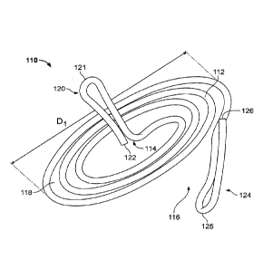
[0030] FIG. 1 shows a first embodiment of a heart valve assisting device
110. The
.. device 110 comprises a ventricular winding 112 and a grasping element 120.
As
described below, the ventricular winding 112 serves the functions of both
facilitating
valve leaflet coaptation and stabilizing or anchoring the device with respect
to the chords.
[0031] The term "spiral" is used herein to refer broadly to shapes
defined by a
structure forming a winding around a center wherein the winding gradually
moves away
from the center as it winds around the center. The structure of the winding
may begin or
emanate from an area at or near the center of the winding. The winding may
move away
from the center at a constant rate or at a non-constant rate, and the general
outline of the
spiral may take various shapes, such as substantially circular, substantially
elliptical, or
other shapes. The spiral may be symmetrical or asymmetrical, and the center
around
.. which the winding structure winds may be a point at the geometric center of
the spiral or
a point that is offset from the geometric center of the spiral. The winding
may be in one
plane, such that the spiral is substantially flat. Alternatively, the winding
may not be in
one plane, with the winding moving up or down at a constant or non-constant
rate. Thus,
for example, the spiral may be substantially conical. The winding may make
multiple
turns around the center or less than a full turn around the center. The
winding structure
of the spiral forms a path that starts from an opening at the outer periphery
of the spiral
and that moves toward the center of the spiral as the path winds around the
center of the
spiral.
[0032] As can be seen in FIG. 1, the ventricular winding 112 has a
generally spiral
shape. The spiral shape is defined by the wire structure of the ventricular
winding 112
forming a winding around a center 114 of the ventricular winding 112, wherein
the wire
structure of the winding begins or emanates from an area at or near the center
114 and
gradually moves away from the center 114 as it winds around the center 114. In
the case
of FIG. 1, the winding of the ventricular winding 112 moves away from the
center 114 at
6
Date recue / Date received 2021-11-08
CA 02951218 2016-12-05
WO 2015/198125
PCT/IB2015/001026
a generally constant rate, and the general outline of the spiral of the
ventricular winding
112 has a substantially circular shape.
[0033] In the embodiment of FIG. 1, the ventricular winding 112 is
generally in one
plane, with an end portion 124 of the ventricular winding 112 being bent or
angled
downwardly as shown. In an alternative embodiment, the ventricular winding 112
may
move gradually out of plane.
[0034] As shown in FIG. 1, the winding structure of the ventricular
winding 112
forms a path 118 that starts from an opening 116 at the outer periphery of the
spiral and
that moves toward the center 114 of the spiral as the path 118 winds around
the center
114 of the spiral. In this illustrated embodiment, the path 118 comprises
about three
turns around the center 114. More or fewer turns may be used.
[0035] As described above, the spiral may take other shapes. In addition,
the
ventricular winding may be comprised of more than one spiral. For example, the
ventricular winding may have two, three, four, or more spirals, which may be
similar or
dissimilar to each other. In one example, two spirals may emanate from a
common
center, each being similar to the other except that each starts in a direction
that is 180
degrees from the other. This example results in nested spirals in which the
opening of
each of the spirals is 180 degrees from the opening of the other spiral. In
other examples,
three spirals may emanate from a common center, starting 120 degrees apart and
having
openings 120 degrees apart, or four spirals may emanate from a common center,
starting
90 degrees apart and having openings 90 degrees apart.
[0036] The overall diameter D1 of the ventricular winding may be
substantially
smaller than the diameter of the annulus of the valve. This enables
maneuvering the
ventricular winding to capture only selected groups of chords, in order to
pull together
desired areas of the valve leaflets. For example, the overall diameter D1 of
the
ventricular winding 112 may be approximately 1.0-2.0 centimeters (e.g., 1.2,
1.5, or 1.8
centimeters), but larger or smaller diameters are possible.
[0037] At its outer end, the ventricular winding 112 terminates at the
end portion 124.
In the embodiment of FIG. 1, the end portion 124 is bent downwardly from the
general
plane of the ventricular winding 112. The end portion 124 is formed as a loop
ofthe wire
7
CA 02951218 2016-12-05
WO 2015/198125
PCT/IB2015/001026
structure of the ventricular winding 112, connected at junction 126. In this
manner, the
end portion 124 terminates at a rounded atraumatic tip 125.
[0038] In this
embodiment, the length of the end portion 124 is approximately 5 mm,
and it may have other lengths, such as 8 mm, or longer or shorter lengths. The
end
portion 124 in this embodiment bends downwardly from the general plane of the
ventricular winding 112 by an angle of approximately 15 degrees, and it may
bend at
other angles, such as 25 degrees, or larger or smaller angles. In this
embodiment, the
design results in a gap in the axial direction between the general plane of
the ventricular
winding 112 and the tip 125 of the end portion 124 of approximately 1 mm to 5
mm, but
larger or smaller gaps are possible.
[0039] The grasping element 120 is connected to the center 114 of the
ventricular
winding 112 and extends upwardly from the center 114 of the ventricular
winding 112.
As shown in FIG. 1, the grasping element 120 is formed of a continuation of
the wire
structure of the ventricular winding 112. The wire structure forming the
grasping
element 120 extends upwardly from the general plane of the ventricular winding
112 at
an angle of approximately 90 degrees, although other angles may be used. After
extending upwardly from the ventricular winding 112, the wire structure of the
grasping
element 120 bends at a top bend 121 and extends downwardly to an end 122 of
the wire
structure, thereby forming a loop. The top bend 121 forms an atraumatic tip,
and the end
122 may be blunt or rounded or may form a junction with the adjacent portion
of the wire
structure, similar to the junction 126. In alternative embodiments, the
grasping element
120 may be substantially straight, curved, bent, helical, or any other
suitable shape. In
one example, the length of the grasping element 120 (from the connection with
the
ventricular winding 112 to its top at bend 121) may be approximately 5 mm to
20 mm,
for example 6 mm to 8 mm or 10 mm, but longer or shorter lengths are possible.
[0040] As can
be seen in FIG. I, the implantable heart valve repair device 110 is free
of any atrial stabilizing section, i.e., the device does not have any part
that is adapted to
stabilize the device by engaging tissue on the atrial side of the valve, such
as the wall of
the atrium or the annulus of the valve on the atrial side. It is possible, for
example, that
after implantation the grasping element 120 extends through the valve to the
atrial side,
and it may contact the leaflets as they close. However, neither the grasping
element 120
8
CA 02951218 2016-12-05
WO 2015/198125
PCT/IB2015/001026
nor any other part of the device 110 is adapted to engage tissue on the atrial
side of the
valve in a manner that stabilizes or anchors the device with respect to the
atrium.
[0041] The device 110, including the ventricular winding 112 and the
grasping
element 120, is comprised of a wire. In alternative embodiments, all or part
of the device
may comprise a wire, a bundle of wires, a strip, a rod, or a tube, and
different sections of
the device or parts thereof may comprise a wire, a bundle of wires, a strip, a
rod, a tube,
or a combination thereof. The structure may be founed by bending or otherwise
shaping
a wire, a bundle of wires, a strip, a rod, or a tube into the desired shape.
The desired
shape may be obtained by "baking" the material in a certain shape at a certain
temperature such that the material will remember that shape. Alternatively,
the shape
may be formed as the wire, bundle of wires, strip, rod, or tube is formed. For
example,
the spiral shape of the ventricular winding may be chemically or laser etched
or otherwise
cut from a sheet of material, in which case the strip or rod is formed
simultaneously with
the spiral shape. The device may be formed of more than a single structure or
material;
for example, a tube with a wire core may form the ventricular winding and/or
the
grasping element, with the other element formed of a similar or dissimilar
structural
component.
[0042] The use of a bundle of wires can provide the device with high
axial strength as
well as high flexibility. For example, the use of several thin wires in a
twisted bundle or
in a braided bundle provides high axial strength and flexibility that can be
determined by
the twisting or braiding structure.
[0043] The wire, bundle of wires, strip, rod or tube may have any
suitable cross-
sectional shape. For example, the wire, bundle of wires, strip, rod or tube
may have a
circular, elliptical, square, rectangular, hexagonal, or other cross-sectional
shape. The
wire, bundle of wires, strip, rod, or tube may have different cross-sectional
shapes or
sizes at different places along its length. The wire of device 110 has a
circular cross-
sectional shape along its length. In one example, the wire, bundle of wires,
strip, rod, or
tube may have a diameter, width or thickness of approximately 0.2-1.0
millimeters (e.g.,
0.4 millimeters), but larger or smaller dimensions are possible.
[0044] The wire of device 110 is formed from a suitable shape memory metal,
for
example nitinol. Other suitable materials may be used for all or part of the
wire(s), rod(s),
9
CA 02951218 2016-12-05
WO 2015/198125
PCT/IB2015/001026
or tube(s) of the device, for example other shape memory materials, other
metallic
materials, plastic materials and/or composite materials.
[0045] The device 110 of FIG. 1 has rounded ends 121, 125 at the ends of
the
grasping element 120 and end portion 124. In alternative embodiments, one or
more ends
of the wire, bundle of wires, strip, rod, or tube may be rounded, squared-off,
or pointed.
As described further below, the device may have one or more anti-rotation
elements.
[0046] As can be seen in FIG. 1, the spiral of ventricular winding 112
can be
considered as being wound in a clockwise direction when viewed from the top
and
starting from the center and moving outward. In an alternative embodiment, the
spiral of
the ventricular winding 112 can be wound in an opposite direction.
[0047] The wire, bundle of wires, strip, rod, or tube may have one or
more grooves in
its outer surface. The groove in the outer surface of the wire, bundle of
wires, strip, rod,
or tube may extend around the perimeter of the wire, bundle of wires, strip,
rod, or tube
and/or in the direction of the length of the wire, bundle of wires, strip,
rod, or tube. As
one example, the wire, bundle of wires, strip, rod, or tube may have one more
grooves
that extend in a substantially helical path along the wire, bundle of wires,
strip, rod, or
tube. Such grooves may serve different purposes. For example, one or more
grooves
may be used to create different flexibilities at different places of the
device, to facilitate
ingrowth of tissue, to facilitate grasping and manipulation (e.g., pushing,
pulling, turning,
etc.) of the device, and/or as channels for drug delivery. For example, a
helical groove
can be used to facilitate rotation of the device as it is being delivered from
or withdrawn
into a delivery catheter. Similarly, a helical or other groove can direct cell
growth in
layers in a preferred direction, thereby reducing scar formation.
[0048] The wire, bundle of wires, strip, rod, or tube may have one or
more holes in it.
The holes may be through-holes extending all the way through the thickness of
the wire,
bundle of wires, strip, rod, or tube, and/or the holes may be pockets or
dimples in the
outer surface of the wire, bundle of wires, strip, rod, or tube. The holes may
be a series
of holes extending along the length and around the periphery of the wire,
bundle of wires,
strip, rod, or tube. The holes may serve different purposes. For example, one
or more
holes may be used to create different flexibilities at different places of the
device, to
CA 02951218 2016-12-05
WO 2015/198125
PCT/IB2015/001026
facilitate ingrowth of tissue, to facilitate grasping and manipulation of the
device, to
provide ports for injection of a contrast agent, and/or as sites for drug
delivery.
[0049] The device may comprise a coating on the wire, bundle of wires,
strip, rod, or
tube. The coating is preferably a biocompatible coating that may be used, for
example, to
reduce possible negative reactions from the tissue where the device is
implanted, to
reduce friction (as a lubricious coating) to assist in delivery of the device,
to reduce
friction in areas where the device is designed to be moved against tissue (for
example,
along the path of the spiral of the ventricular winding), to increase friction
in areas where
it is desired to reduce movement or to anchor the device, to deliver a
suitable drug, for
radiopacity, to encourage cell and tissue growth that would assist in fixation
(e.g., of the
upper section), to encourage tissue growth between the chords and/or leaflets,
and/or for
other purposes. With respect to radiopacity, the entire device or selected
points on the
device may be coated or plated with a material allowing the physician to
understand the
location of the device during and/or after the implantation procedure. For
example, the
ends of the spiral may be plated with a radiopaque material. If selected
points on the
device are plated, the plating at the selected points may have a certain shape
(e.g., a line,
arrow, etc.) to assist in understanding the orientation of the device. In
another example,
in the case of a device formed of a tube, the tube may be coated to ensure
that the coated
tube is sealed in order that the tube may be used, for example, for pressure
measurement.
When the coating is a drug-release coating, the coating may comprise a carrier
(for
example, a polymer) with the drug in the carrier for drug elution over a
suitable period of
time. The drug eluting mechanism may use a biodegradable carrier (e.g., a
biodegradable
polymer) or a stable carrier (e.g., a stable polymer) that allows the drug
elution through
diffusion of drug molecules.
[0050] FIG. 2 shows another embodiment of a heart valve repair device 130.
The
device 130 comprises a ventricular winding 132 and a grasping element 140. The
ventricular winding 132 has a generally spiral shape, defined by the wire
structure of the
ventricular winding 132 forming a winding around a center 134 of the
ventricular
winding 132. The wire structure of the winding emanates from the center 134
and
gradually moves away from the center 134 as it winds around the center 134. In
the case
of device 130, the winding of the ventricular winding 132 moves outward from
the center
11
CA 02951218 2016-12-05
WO 2015/198125
PCT/IB2015/001026
134 at a generally constant rate, thereby forming a substantially circular
shape (in top
view), while at the same time the winding moves upward from its starting point
at the
center 134, thereby forming a substantially conical helix opening upward, with
the base
of the cone above the vertex. In an alternative embodiment, the winding of the
ventricular winding moves outward from its starting point at the center while
at the same
time moving downward from its starting point at the center, thereby forming a
substantially conical helix opening downward, with the base of the cone below
the vertex.
As shown in FIG. 2, the ventricular winding 132 terminates at its outer
periphery at an
atraumatic end portion 144, which is bent downwardly, similar to end portion
124.
[0051] In the device 130, like the device 110, the winding structure of the
ventricular
winding 132 forms a path that starts from an opening at the outer periphery of
the spiral
and that moves toward the center 134 of the spiral as the path winds around
the center
134 of the spiral.
[0052] The device 130, like the device 110, may be comprised of a wire
having a
circular cross-section. The wire of device 130 may be formed of a suitable
shape
memory metal, for example nitinol.
[0053] The grasping element 140 of device 130 is similar in construction
to the
grasping element 120 of device 110. As can be seen in FIG. 2, the device 130
is free of
any atrial stabilizing section.
[0054] As would be understood by persons of ordinary skill in the art from
the above
descriptions, alternative embodiments of the device 130 may be formed, using
the
variations described above with respect to the device 110. Thus, for example,
the
ventricular winding 132 and the grasping element 140 may comprise other forms,
shapes,
sizes and/or materials as described above with respect to the device 110. The
ends of the
device may be rounded, squared-off, or pointed. The device 130 may have one or
more
anti-rotation elements, as described further below. The ventricular winding
132 and/or
the grasping element 140 may have one or more grooves and/or holes, as
described above.
The device may comprise a coating, as described above.
[0055] FIG. 3 shows another embodiment of a heart valve repair device
150. The
device 150 comprises a ventricular winding 152 and a grasping element 160. The
ventricular winding 152 has a generally spiral shape, defined by the wire
structure of the
12
CA 02951218 2016-12-05
WO 2015/198125
PCT/IB2015/001026
ventricular winding 152 forming a winding around a center 154 of the
ventricular
winding 152. The wire structure of the winding emanates from the center 154
and
gradually moves away from the center 154 as it winds around the center 154. In
the case
of device 150, in an inner section 151, the winding of the ventricular winding
152 moves
outward from the center 154 at a generally constant rate, thereby forming a
substantially
circular shape (in top view), while at the same time the winding moves upward
from its
starting point at the center 154, thereby forming a substantially conical
helix opening
upward, with the base of the cone above the vertex. Then, the inner section
151
transitions to an outer section 153, in which the winding of the ventricular
winding 152
stays substantially in a single plane as it moves outward from the center 154
at a
generally constant rate. In an alternative embodiment, the inner section may
stay
substantially in a single plane with the outer section forming a section of a
substantially
conical helix. As shown in FIG. 3, the ventricular winding 152 terminates at
its outer
periphery at an atraumatic end portion 164, which is bent downwardly, similar
to end
portion 124.
[0056] In the device 150, like the device 110, the winding structure of
the ventricular
winding 152 forms a path that starts from an opening at the outer periphery of
the spiral
and that moves toward the center 154 of the spiral as the path winds around
the center
154 of the spiral.
[0057] The device 150, like the device 110, may be comprised of a wire
having a
circular cross-section. The wire of device 150 may be formed of a suitable
shape
memory metal, for example nitinol.
100581 The grasping element 160 of device 150 is similar in construction
to the
grasping element 120 of device 110. As can be seen in FIG. 3, the device 150
is free of
any atrial stabilizing section.
100591 As would be understood by persons of ordinary skill in the art
from the above
descriptions, alternative embodiments of the device 150 may be formed, using
the
variations described above with respect to the device 110. Thus, for example,
the
ventricular winding 152 and the grasping element 160 may comprise other forms,
shapes,
sizes and/or materials as described above with respect to the device 110. The
ends of the
device may be rounded, squared-off, or pointed. The device 150 may have one or
more
13
CA 02951218 2016-12-05
WO 2015/198125
PCT/IB2015/001026
anti-rotation elements, as described further below. The ventricular winding
152 and/or
the grasping element 160 may have one or more grooves and/or holes, as
described above.
The device may comprise a coating, as described above.
[0060] FIG. 4 shows another embodiment of a heart valve repair device
170. The
device 170 comprises a ventricular winding 172 and a grasping element 180. The
ventricular winding 172 has a generally spiral shape, defined by the wire
structure of the
ventricular winding 172 forming a winding around a center 174 of the
ventricular
winding 172. The wire structure of the winding emanates from the center 174
and
gradually moves away from the center 174 as it winds around the center 174. In
the case
of device 170, the winding of the ventricular winding 172 moves outward from
the center
174 at an uneven rate. Thus, the gap between adjacent turns of the winding is
non-
constant, as can be seen by a comparison between smaller inner gap 172A and
larger
outer gap 172B. As shown in FIG. 4, the ventricular winding 172 terminates at
its outer
periphery at an atraumatic end portion 184.
[0061] In the device 170, like the device 110, the winding structure of the
ventricular
winding 172 forms a path that starts from an opening at the outer periphery of
the spiral
and that moves toward the center 174 of the spiral as the path winds around
the center
174 of the spiral.
[0062] The device 170, like the device 110, may be comprised of a wire
having a
circular cross-section. The wire of device 170 may be formed of a suitable
shape
memory metal, for example nitinol.
[0063] The grasping element 180 of device 170 may be similar in
construction to the
grasping element 120 of device 110 or may be generally in the same plane as
the
ventricular winding 172. As can be seen in FIG. 4, the device 170 is free of
any atrial
stabilizing section.
[0064] As would be understood by persons of ordinary skill in the art
from the above
descriptions, alternative embodiments of the device 170 may be formed, using
the
variations described above with respect to the device 110. Thus, for example,
the
ventricular winding 172 and the grasping element 180 may comprise other
foul's, shapes,
sizes and/or materials as described above with respect to the device 110. The
ends of the
device may be rounded, squared-off, or pointed. The device 170 may have one or
more
14
CA 02951218 2016-12-05
WO 2015/198125 PCT/IB2015/001026
anti-rotation elements, as described further below. The ventricular winding
172 and/or
the grasping element 180 may have one or more grooves and/or holes, as
described above.
The device may comprise a coating, as described above.
[0065] As mentioned above, the implantable heart valve repair devices
130, 150, and
170, like the heart valve repair device 110 and other heart valve repair
devices described
herein, are free of any atrial stabilizing section. Each of the implantable
heart valve
repair devices 110, 130, 150, and 170 has a stabilizing section that consists
only of a
ventricular stabilizing section in the form of a ventricular winding 112, 132,
152, and 172,
which is adapted to engage tissue only on the ventricular side of the valve
and to stabilize
the device by the interaction between the ventricular winding 112, 132, 152,
and 172 and
the chords on the ventricular side of the valve. In some embodiments, these
heart valve
repair devices also may be described as not having any part that, after
implantation,
contacts tissue in the atrium or on the atrial side of the valve and/or not
having any part
that, after implantation, extends into the atrium or on the atrial side of the
valve.
However, as described above, it is possible in some embodiments for the
grasping
element to extend through the valve to the atrial side, and it may contact the
leaflets.
However, these embodiments may be constructed such that neither the grasping
element
nor any other part of the device is adapted to engage tissue on the atrial
side of the valve
in a manner that stabilizes or anchors the device with respect to the atrium.
[0066] FIGS. 5A-5C illustrate examples of anti-rotation elements that may
be used
with a heart valve repair device, including any of the heart valve repair
devices described
herein. FIG. 5A shows a heart valve repair device 190 having an anti-rotation
element
191 in the form of a protrusion on the end of the ventricular winding. FIG. 5B
shows a
heart valve repair device 192 having an anti-rotation element 193 in the form
of an
enlarged tooth on the end of the ventricular winding. FIG. 5C shows a heart
valve repair
device 194 having an anti-rotation element 195 in the form of an enlarged area
on an
inner turn of the winding, coming close to or touching an adjacent turn. The
anti-rotation
elements can help prevent backward rotation of the device after implantation,
by allowing
easier rotation of the device in the direction of bringing the chords together
than in the
opposite direction. Thus, for example, a tooth having a slanted front face (on
the side
facing the outer opening of the path of the spiral) and steep back face can
permit rotation
CA 02951218 2016-12-05
WO 2015/198125 PCT/IB2015/001026
of the device in the direction that brings the leaflets together (by the
chords passing over
the slanted front face) and can help resist rotation in the opposite direction
(by the chords
acting against steep back face, thereby resisting backward rotation).
[0067] In the example of one or more protrusions 191 as shown in FIG. 5A,
the force
applied by the delivery system during the process of turning the device 190 to
capture the
chords can result in the wire structure flexing sufficiently to create a large
enough gap
between the protrusion 191 and the adjacent turn of the winding in order to
allow the
chords to pass therethrough, so that the device 190 may be wound around the
chords.
Similarly, in the example of one or more protrusions 193 as shown in FIG. 5B,
the force
applied by the delivery system during the process of turning the device 192 to
capture the
chords can result in the wire structure flexing sufficiently to create a large
enough gap
between the tooth 193 and the adjacent turn of the winding in order to allow
the chords to
pass therethrough, so that the device 192 may be wound around the chords.
Similarly, in
the example of one or more enlarged areas 195 as shown in FIG. 5C, the force
applied by
the delivery system during the process of turning the device 194 to capture
the chords can
result in the wire structure flexing sufficiently to create a large enough gap
between the
enlarged area 195 and the adjacent turn of the winding in order to allow the
chords to
pass therethrough, so that the device 194 may be wound around the chords. In
each of
these examples, the geometry of the anti-rotation element(s) helps prevent the
device
from unintentionally rotating in the opposite direction.
[0068] FIG. 6 illustrates a proximal end of a delivery system of a type
that may be
used for implanting a heart valve repair device, such as any of the heart
valve repair
devices described herein. The delivery system 300 includes a flexible
applicator 400,
which is generally tubular in shape, and an internal rod 450, which is
moveable within
the applicator 400. An applicator gripper 310 may be used to help push,
withdraw, and
rotate the applicator 400, in both clockwise and counterclockwise directions.
An
applicator irrigation port 320 allows injecting irrigation fluids into the
applicator 400. An
internal rod torquer 330 may be used to rotate the internal rod 450 within the
applicator
400. An internal rod grip 340 is connected to the internal rod 450 and may be
used to
control movements of the internal rod 450, including pushing it forward in
order to eject
a hook that is connected to internal rod 450, as described below. A scale or
ruler 350
16
CA 02951218 2016-12-05
WO 2015/198125
PCT/IB2015/001026
facilitates measuring how far the internal rod 450 has been advanced, so as to
determine
the position of the hook inside the applicator 400. A safety plate 360
prevents
inadvertent advancement of the internal rod grip 340, in order to eliminate
the possibility
of accidentally pushing the hook outside of the applicator 400.
[0069] FIGS. 7A and 7B show top and side views, respectively, of a distal
end of the
delivery system 300, with a heart valve repair device 110 loaded on the
delivery system
300. In these figures, it can be seen that the flexible applicator 400 has
lateral slots 402
to facilitate bending. Other manners of imparting flexibility to a catheter
may be used,
including selection of appropriate flexible material(s). The applicator 400
has a rounded,
atraumatic distal end 404. The applicator 400 has a window 410 through which
all or
part of the heart valve repair device may be ejected. A ramp surface 420
adjacent the
distal end of the window 410 facilitates ejection of the device, as described
below.
[0070] The internal rod 450 terminates in a hook 460. The hook 460 is
designed to
hold the grasping element of the heart valve repair device while the hook 460
is inside the
applicator 400, proximal to the window 410.
[0071] In a first example implantation, the delivery system 300 holds a
heart valve
repair device 110 as shown in FIGS. 7A and 7B, with the ventricular winding
112
positioned outside of the lumen of the applicator 400 and with the grasping
element 120
positioned inside the lumen of the applicator 400 and held by the hook 460. In
this
position, the internal wall of the lumen of the applicator 400 prevents the
grasping
element 120 from exiting the hook 460. Thus, as long as the hook 460 is inside
the
lumen of the applicator 400 (and not in the window 410), the grasping element
120
remains hooked on the internal rod 450 and is thereby locked to the internal
rod 450.
[0072] The delivery system 300 is used in conjunction with a catheter
tube, for
example a steerable catheter as is known in the art. One example of a
steerable catheter
is the AGILIS catheter of St. Jude Medical, Inc. The catheter is sized to
accommodate
the applicator 400 of the delivery system 300. For example, if the applicator
400 has a
size of 7.5 French, the outer catheter may have a size of 12 French. This is
just an
example, as other sizes may be used.
[0073] In this first example, with the ventricular winding 112 positioned
outside of
the lumen of the applicator 400, the distal end of the applicator 400 is
advanced into the
17
CA 02951218 2016-12-05
WO 2015/198125
PCT/IB2015/001026
proximal end of the steerable catheter. Because the lumen of the steerable
catheter is
only slightly larger than the outer diameter of the applicator 400, and
smaller than the
outer periphery of the ventricular winding 112, as the applicator 400 is
further advanced
into the catheter, an internal turn of the ventricular winding 112 comes into
contact with
the edge of the catheter tube at its proximal end. Further advancement of the
applicator
400 into the catheter will thereby cause the ventricular winding 112 to unwind
and
straighten as it is advanced into the catheter along with the applicator 400.
It will be
appreciated that the center part of the ventricular winding 112 will be
advanced into the
catheter first, and the ventricular winding 112 will unwind from the center to
the outer
periphery as the ventricular winding 112 is advanced into the catheter. When
fully
advanced into the catheter, the generally unwound ventricular winding 112 is
held in a
relatively straightened position between the outer wall of the applicator 400
and the inner
wall of the catheter lumen. The applicator 400 may be advanced into the
catheter either
before or after the catheter is tracked to the patient's heart.
[0074] The catheter is positioned adjacent the heart valve to be treated,
for example a
mitral valve, by a method known in the art. The approach may be, for example,
a
transseptal approach, with the catheter entering the left atrium through the
septum
between the right atrium and the left atrium. To facilitate a transseptal
approach, the
delivery system may include an atrial septum dilator. Other approaches
alternatively may
be used, including, for example, a transfemoral approach through the femoral
artery and
through the aorta and into the left ventricle, a transapical approach through
the heart wall
at the heart apex into the left ventricle, or a transatrial approach through
the heart wall
into the left atrium. Similarly, when the valve to be treated is the tricuspid
valve, the
catheter is positioned adjacent the valve by a method known in the art (such
as being
introduced to the heart via a jugular vein or the vena cava).
100751 Once the guide catheter is adjacent the heart valve, the tip of
the guide
catheter may be moved and/or turned so that it is facing the heart valve
leaflets. The
applicator 400 then may be advanced relative to the catheter, thereby ejecting
the
ventricular winding 112 from the catheter. Because of the shape memory of the
ventricular winding 112, the heart valve repair device 110 returns to a
position as shown
in FIGS. 7A and 7B, inside the heart. The ejection of the ventricular winding
112 from
18
CA 02951218 2016-12-05
WO 2015/198125 PCT/IB2015/001026
the catheter may be performed in the atrium. Alternatively, the ejection of
the ventricular
winding 112 from the catheter may be performed in the ventricle. If ejected in
the atrium,
the delivery system 300 then may be used to advance the distal end of the
applicator 400,
and with it the ventricular winding 112, into the ventricle. The distal end of
the
applicator 400 and the ventricular winding 112 may be pushed through the valve
into the
ventricle.
[0076] FIG. 8 shows a perspective view of the distal end of the delivery
system 300,
with the ventricular winding 112 ejected from the guide catheter and outside
of the tube
of the applicator 400, and with the grasping element 120 still inside the tube
of the
applicator 400 and connected to the hook 460. In this position, rotation of
the applicator
400 causes the heart valve repair device 110 to be rotated along with it.
During rotation
of the applicator 400, the action of the frame of the window 410 against the
device 110
causes the device 110 to rotate with the applicator 400. Additionally or
alternatively, the
hook 460 or another part of the delivery system 300 may sufficiently hold the
device 110
to cause the device 110 to rotate. Due to the geometry of the device, the axis
of the
delivery system can be generally aligned with the axis of rotation of the
ventricular
winding.
[0077] In the condition as shown in FIG. 8, and with the ventricular
winding 112
positioned in the ventricle, the delivery system may be used to capture the
desired chords
with the ventricular winding 112. The physician may maneuver the ventricular
winding
112 from side to side to capture specific chords in order to bring desired
areas of the
leaflets together. For example, by suitably moving and turning the ventricular
winding
112, the chords associated with the leaflet areas Al and P1 (FIG. 12) may be
captured.
Additionally or alternatively, the chords associated with the leaflet areas A2
and P2
and/or A3 and P3 (FIG. 12) may be captured.
[0078] In this manner, the desired chords associated with the anterior
papillary
muscle and the desired chords associated with the posterior papillary muscle
are
positioned within the path 118 of the generally spiral shape of the
ventricular winding
112. By turning the ventricular winding 112, whether by turning the applicator
400 or by
another suitable mechanism, the ventricular winding 112 is thereby turned to
wind
around the selected anterior and posterior chords. As the ventricular winding
112 is
19
CA 02951218 2016-12-05
WO 2015/198125 PCT/IB2015/001026
turned, the spiral shape forces the chords within the path 118 closer to the
center 114 of
the ventricular winding 112. In this manner, the captured anterior chords and
posterior
chords are forced closer together, thereby reducing a gap between the selected
chords
associated with the anterior papillary muscle and the selected chords
associated with the
posterior papillary muscle. By doing this, because the selected chords are
attached to the
selected areas of the leaflets, the selected areas of the leaflets are brought
closer together.
[0079] In order that the ventricular winding may be turned to move the
chords in this
manner and may hold the chords, the heart valve repair device, or at least the
ventricular
winding, should have sufficient stiffness such that the spiral shape is
generally
maintained. Thus, the device should be sufficiently rigid so as to maintain
the spiral
shape on its own and under the forces applied to it by the chords.
[0080] In alternative embodiments in which the ventricular winding
comprises more
than one spiral, the device may be formed so that it can gather and move the
chords with
fewer rotations. Thus, for example, with the ventricular winding comprising
multiple
spirals and with the openings for the spirals positioned at different places
around the
perimeter of the ventricular winding, chords at different places around the
perimeter of
the ventricular winding may be gathered simultaneously and moved toward the
center
simultaneously.
[0081] In order to adjust the device, after the physician has turned the
ventricular
winding 112 in a first direction as described above, the physician may turn
the ventricular
winding 112 back in the opposite direction in order to allow the chords to
move apart by
some amount. Thus, in this example, after the positioning resulting from
clockwise
turning, the physician may turn the ventricular winding 112 counterclockwise
(when
viewed from the top) in order to allow the captured chords to move away from
the center
114 of the ventricular winding 112, thereby allowing them to separate by some
distance.
The physician can monitor the positioning of the chords and leaflets and turn
the
ventricular winding 112 clockwise or counterclockwise as needed in order to
obtain the
desired result.
[0082] If desired, after the ventricular winding 112 has been rotated
into the desired
rotational position, the physician may pull the ventricular winding 112 to
bring it closer
to the heart valve. This may be accomplished by retracting the applicator 400.
CA 02951218 2016-12-05
WO 2015/198125
PCT/IB2015/001026
[0083] When the ventricular winding 112 is in the desired position, the
remainder of
the device 110 is ejected from the applicator 400, as shown in FIG. 9. With
the
applicator 400 held relatively stable, the internal rod 450 is advanced
distally within the
applicator 400. This moves the hook 460 to be located at the window 410.
During this
distal advancement, the grasping element 120 is advanced distally against the
ramp
surface 420, which forces the grasping element 120 away from the applicator
400 and the
hook 460. With the hook 460 in the window 410, the grasping element 120 is no
longer
prevented from exiting the hook 460 by the internal wall of the lumen of the
applicator
400. The grasping element 120 is released from the hook 460, and the device
110 is left
in place. The delivery device 300 is then withdrawn from the patient. This
leaves the
heart valve repair device 110 implanted in the patient.
[0084] If the procedure is one in which the valve is approached from the
atrium as
described above, once the device is implanted in the ventricle, the grasping
element will
be pointing generally toward the atrium. If, on the other hand, the procedure
is one in
which the valve is approached from the ventricle, once the device is implanted
in the
ventricle, the grasping element will be pointing generally away from the
atrium and
generally toward the apex of the heart.
[0085] Other heart valve repair devices as described herein, such as the
heart valve
repair devices 130, 150, 170, and 190, and the variations described above with
respect to
.. the heart valve repair devices 110, 130, 150, 170, and 190, may be
implanted in a similar
manner as described above.
[0086] The inventive heart valve repair devices as described herein
provide certain
advantages with respect to prior devices. As discussed above, in prior heart
valve repair
devices for capturing leaflet chords as exemplified in certain prior devices,
the devices
have included parts or sections for anchoring the devices relative to the
atrium, in order to
ensure that the devices remained stable in the heart after implantation. By
contrast, the
inventive heart valve repair devices 110, 130, 150, 170, and 190, and the
variations
described above with respect to these devices, depart from these prior
teachings and are
free of any atrial stabilizing section. The inventors have found that not only
is it possible
to implant the inventive heart valve repair devices and have them remain
stable in the
heart after implantation despite the absence of any atrial stabilizing
section, but the
21
CA 02951218 2016-12-05
WO 2015/198125
PCT/IB2015/001026
inventors also have found that the inventive heart valve repair devices
provide previously
unrecognized advantages that could not be attained by prior devices that were
anchored
to the atrium.
100871 As the heart pumps, the various parts of the heart are in motion.
The chords
connecting the papillary muscles to the leaflets are in motion. A heart valve
repair device
as described herein without any atrial stabilizing section is free to move
along with the
chords, while at the same time retaining the chords in their drawn-in
condition for leaflet
coaptation. Over time, tissue may grow around the ventricular winding such
that the
ventricular winding becomes substantially embedded in the chords. This tissue
enveloping of the device results in a fixation of the device to the chords.
[0088] In prior devices that have included atrial stabilizing sections,
the devices have
been anchored relative to the atrium. However, during the beating of the heart
and the
opening and closing of the valves, the chords move relative to the atrium.
Thus, when
such a prior device is implanted and anchored to the atrium, the chord
movement relative
to the atrium results in chord movement relative to the device. A device
anchored to the
atrium is constrained from moving freely with the chords. This constraint can
occur at
the time of the implantation procedure and longer. When a device is
constrained in this
manner, the chords can rub relative to such a device, which can cause
irritation, injury,
and/or rupture of the chords.
[0089] By contrast, a heart valve repair device as described herein,
without any atrial
stabilizing section, can be affixed only to the chords. Thus, the device is
free to move
along with the chords, such as in an up-and-down direction generally in the
direction of
the axis of implantation, while at the same time maintaining the chords in
their drawn-in
condition for leaflet coaptation. This movement of the device can occur
relative to the
atrium. Thus, by being free of any atrial stabilizing section, the device has
the previously
unrealized advantages of reducing or eliminating the potential for chord
movement
relative to the device and reducing or eliminating the consequent potential
for irritation,
injury, and/or rupture of the chords that can be caused by such relative
movement.
[0090] While prior devices as discussed above have included atrial
anchoring in order
to ensure stability after implantation, the inventors have found that a device
as described
herein, in both ex vivo testing and in vivo animal testing, is stable in the
heart after
22
CA 02951218 2016-12-05
WO 2015/198125
PCT/IB2015/001026
implantation, despite not having an atrial stabilizing section. The device is
sufficiently
held in place by the interaction between the ventricular winding and the
chords.
Accordingly, the inventors have found that it is possible to realize the
advantages as
described herein and have the device be stable in the heart after
implantation, without the
need for a connected atrial stabilizing section as in the prior devices
discussed above.
[0091] The use of a device without any atrial stabilizing section can
have several
additional advantages. For example, by not having an atrial stabilizing
section, the
device can be smaller, simpler and less expensive to manufacture, easier to
implant, more
maneuverable to facilitate targeted treatment, less prone to tissue injury,
and may
improve overall outcome.
[0092] The presence of an atrial stabilizing section can result in a
relatively larger or
longer device, requiring positioning the device on the atrial side as well as
on the
ventricular side of the valve. Thus, the device without any atrial stabilizing
section can
lead to easier implantation. Implantation is also facilitated because the
device is easier to
visualize without any atrial stabilizing section.
[0093] Without an atrial stabilizing section, it can be easier to
maneuver the relatively
small ventricular winding to capture specific groups of chords. The presence
of an atrial
stabilizing section can limit the range of placement of the device. That is, a
relatively
large atrial stabilizing section can limit the side-to-side range of the
device, potentially
limiting the areas in which the device can be placed, and limiting the groups
of chords
that can be captured. By contrast, the smaller, more maneuverable device leads
to an
improved ability to address specific chords and/or specific areas of a valve.
[0094] The device without an atrial stabilizing section also reduces the
chance of
tissue injury on the atrial side of the valve, which can occur due to the
engagement of
tissue on the atrial side. Such engagement can occur during the delivery
procedure or
after implantation.
[0095] A person of ordinary skill in the art will understand that the
various heart
valve repair devices described herein may be implanted according to the method
described above. Various features of the device can facilitate the procedure
and
functioning.
= 23
CA 02951218 2016-12-05
WO 2015/198125
PCT/IB2015/001026
[0096] For example, the grasping element allows the device to be held by
the delivery
system, so that it can be held, maneuvered, turned, and released as described
above.
Because the grasping element can extend from at or near the center of the
ventricular
winding, the device can be turned by simply rotating the ventricular winding
generally
around the axis of the grasping element. When the device is fully deployed,
the grasping
element may be located fully within the ventricle or may extend into the
atrium.
[0097] The end portion of the ventricular winding, bent away from the
general plane
of the winding, can facilitate capturing of the chords. The distance of the
end portion
away from the general plane of the winding can determine the span of the
potential
.. chords to be grasped. The rounded atraumatic tip of the end portion can
help prevent
injury to the chords, leaflets, and/or other tissue.
[0098] When all or part of the ventricular winding is out of plane, such
as in a conical
or partially-conical embodiment as shown in FIGS. 2 and 3, the height
dimension of the
ventricular winding can help maintain the vertical positioning of the device
within the
.. ventricle. The height dimension also allows more contact with the chords in
the vertical
direction. This can reduce the friction between the device and the chords and
can
increase tissue coverage of the device. In addition, the height dimension can
allow easier
visualization of the device.
[0099] When the spacing of the turns of the ventricular winding is larger
toward the
.. outer periphery, as shown in FIG. 4, the design can facilitate capturing
the chords (by the
larger outer spacing) while also bringing them close together (by the narrower
inner
spacing).
[0100] The device can have anti-rotation elements that can help prevent
the
ventricular winding from turning backward (in the loosening direction) after
implantation.
Thus, the ventricular winding can have one or more anti-rotation elements 191,
193, 195,
as shown in FIGS. 5A-5C, that help keep the device in place. Other mechanisms
for
resisting unwinding include the use of different shapes. For example, if the
ventricular
spiral is in an elliptical shape, the chords will tend to gather in the apices
of the long axis
of the ellipse. In order for the device to rotate, the chords would need to be
drawn closer
together, which is a movement they would tend to resist. Accordingly, such an
elliptical
shape can assist in preventing an unwanted rotation of the device.
24
CA 02951218 2016-12-05
WO 2015/198125
PCT/IB2015/001026
[0101] It will be appreciated that in procedures in which the delivery
system
approaches the heart valve from the ventricular side (e.g., in transfemoral
and transapical
approaches), similar methods to those described above may be used, modified to
account
for the fact that the delivery system approaches the valve from the opposite
side.
[0102] The delivery system may have means for retrieving and/or moving the
heart
valve repair device after it has been partially or fully deployed. For
example, FIG. 10
shows a retraction wire 500 that may be placed inside the grasping element 121
of the
heart valve repair device. The retraction wire 500 can be located inside the
applicator
400, but can also remain inside the grasping element 121 after the heart valve
repair
device has been ejected from the applicator 400. Pulling the retraction wire
500 can
withdraw the heart valve repair device back, so that it can be moved to a
different
position or fully removed from the patient.
[0103] FIG. 11 illustrates an alternate form of a hook 470 used to engage
a relatively
flat grasping element 472. As can be seen, in this embodiment the grasping
element 472
is generally in the same plane as the ventricular winding. In this embodiment,
the device
is released simply by turning the hook 470 relative to the grasping element
472, such that
the hook 470 can be withdrawn from the grasping element 472.
[0104] While the above method has been described with respect to a device
in which
the ventricular winding is positioned outside of the applicator for delivery,
as in FIG. 8,
variations are possible. For example, the ventricular winding may be
positioned inside of
the applicator 400, whereby it is held in a relatively straightened position.
For
deployment, the device is ejected from the applicator, at which time the
ventricular
winding resumes its spiral shape.
[0105] As would be understood from the above descriptions by persons of
ordinary
skill in the art, alternative embodiments of the devices 110, 130, 150, 170,
and/or 190
may be implanted generally as described above. The method of implantation may
be
varied as appropriate with respect to the particular embodiment used and the
particular
patient being treated.
[0106] When a device as described is placed in position as described, the
spiral of the
ventricular winding reduces a gap between selected chords associated with the
anterior
papillary muscle and selected chords associated with the posterior papillary
muscle. In
CA 02951218 2016-12-05
WO 2015/198125
PCT/IB2015/001026
this manner, the selected areas of the leaflets of the valve are drawn closer
together. In
some instances, the control of the chords also can reduce the movement of the
leaflets, in
order to help prevent prolapse. The control of the chords and the drawing of
the leaflets
closer together facilitate coaptation of the leaflets, such that they can
close together
sufficiently to correct the regurgitation issue. The device can be left in
place as a long-
term treatment.
[0107] FIG. 12 shows a top view of leaflets of a mitral valve. A device
as described
herein may be used in various positions and for gathering various chords. For
example,
the device may be positioned approximately in the area of A2 and P2 near the
center of
the anterior and posterior leaflets. The chords on A2 and P2 are trapped and
gathered by
the spiral(s) of the ventricular winding. A rotation of the spiral would
eventually bring
all such chords to the same location, which is the spiral center. In this
situation, the gap
between A2 and P2 could be brought to zero. Rotating the spiral a little less
would result
in a narrow gap. The device alternatively may be positioned approximately in
the area of
Al and Pl, in which case the chords on Al and P1 are trapped and gathered by
the
spiral(s) of the ventricular winding, reducing the distance between Al and Pl.
The
device alternatively may be positioned approximately in the area of A3 and P3,
in which
case the chords on A3 and P3 are trapped and gathered by the spiral(s) of the
ventricular
winding, reducing the distance between A3 and P3. A device with a large spiral
positioned approximately in the area of A2 and P2 may trap and gather chords
on A2, P2,
Al, Pl, A3 and/or P3, and can be used to reduce the distance between P1 and
P3, for
example, or Al and A3.
[0108] With a relatively small heart valve repair device as described
herein, for
example having a ventricular winding with an outside diameter of 1.0-2.0
centimeters, for
example, it is possible to capture less than substantially all of the chords
and to capture
small groups of chords. Implanting such a device may involve positioning the
spiral of
the ventricular winding between chords rather than around substantially all of
the chords.
Also, with a relatively small heart valve repair device as described herein,
it is possible to
implant multiple devices, capturing different sets of chords. The flow through
the valve
can be adjusted by selectively adjusting the different devices. For example,
one device
could be placed capturing the chords on Al and PI, a second device could be
placed
26
CA 02951218 2016-12-05
WO 2015/198125
PCT/IB2015/001026
capturing the chords on A2 and P2, and a third device could be placed
capturing the
chords on A3 and P3. The flow can be evaluated, and if necessary, adjusted.
For
example, the device at Al and PI could be rotated to bring the associated
chords closer
together, while the device at A3 and P3 could be rotated in an opposite
direction.
[0109] In some instances, it may be desired to use the device to draw the
leaflets
closer and then position a clip anchored to both leaflets or stitch or suture
the leaflets
together. Thus, the device in conjunction with one or more clips, stitches or
sutures can
facilitate coaptation of the leaflets.
[0110] If desired, the device may be adjusted or withdrawn at a later
time, either
shortly or long after the implantation. A catheter may be used to access the
device. To
adjust the device, the physician may turn the spiral of the ventricular
winding as
described above (e.g., by turning the device) in order to bring the chords
closer together
or to allow them to separate further apart, as desired. Thus, the turning may
be done
while performing the initial implantation procedure and/or as an additional
later
procedure that is separate from the implantation procedure. In this manner,
the
regurgitation grade can be controlled. Alternatively, if it is desired to
withdraw the
device altogether, a grasping mechanism may be used to grasp the device and
pull it back
into the catheter, in essentially the reverse of the procedure that was used
to deliver the
device.
[0111] Numerous alternatives are possible within the scope of the
invention. For
example, as mentioned above, the winding of the spiral may move away from the
center
at a non-constant rate. Thus, the spiral density need not be constant.
[0112] If the device is formed as a tube, a wire or stiffening element
may be placed
into the tube in order to change the stiffness and/or shape of the tube or a
section of it.
For example, a stiffening element may be used to maintain the device in a
first shape for
delivery (e.g., relatively straight), and the stiffening element may be
withdrawn upon
delivery of the device from the delivery catheter in order to allow the device
to take its
implantation shape. In another example, an inner wire may be attached to the
distal end
of the tube, and the inner wire may be pulled relative to the tube to change
the shape of
the tube. Pulling the inner wire applies a compressive force to the tube. The
tube may be
formed with pre-shaped side cuts along the tube, such that it bends in a
predetermined
27
CA 02951218 2016-12-05
WO 2015/198125
PCT/IB2015/001026
pattern, e.g., a spiral pattern, when such a load is applied. A locking
mechanism may be
used to lock the wire in its loaded position relative to the tube. Different
depths and
widths of the side cuts and the distance between the side cuts would determine
the final
shape of the tube element once a load is applied.
[0113] The device may have other elements to monitor the functioning of the
device
and the heart valve. For example, the device may be equipped with a sensor
attached to
the device. The sensor may be, for example, a pressure sensor, a temperature
sensor,
and/or a velocity sensor. In this way, the operation of the valve and the
blood flow can
be monitored. Similarly, the device itself when formed as a tube can be used
as a "pig
tail" for measuring pressure during or after the implantation procedure.
[0114] In one example of the use of sensors, the use of MEMS
(microelectromechanical systems) sensors on the device may assist in the
implantation
procedure or during the years after it. Such sensors may monitor temperature,
oxygen
saturation, pressure, blood velocity or similar physical characteristics.
During the
implantation procedure, it is possible to use an xyz (positioning) sensor on
the device to
assist in the accurate location and positioning of the device by using an
external system
that reads the information transmitted from the sensor.
[0115] Sensor(s) on the device or delivery system may be part of a
closed-loop
system that uses the signals from the sensor(s) as feedback for automatic
delivery and
positioning. By using pressure sensors in the ventricle and atrium, the
pressure can be
continuously monitored as the device is automatically adjusted. The
adjustments and
monitoring can be continued until target pressure readings are achieved. This
automatic
positioning with the use of feedback can eliminate the need for manual
monitoring and
positioning that can be complicated and less accurate.
[0116] The device may also have an energy-producing element that produces
energy
by the flow of blood around the device and/or by the pressure changes using a
converter
(such as piezoelectric element that is capable of converting mechanical pulse
into electric
current). The energy may charge a battery that, for example, can be used to
transmit
signals from one or more sensors as described above.
[0117] From the description herein, a person of ordinary skill in the art
can recognize
that certain embodiments of devices and methods disclosed herein can have
several
28
CA 02951218 2016-12-05
WO 2015/198125
PCT/IB2015/001026
advantages. For example, the device can safely hold the chords without
requiring
grasping of the leaflets. The movement of the chords toward each other can be
controlled
by the structure of the device, including, for example, the number of turns of
the spiral of
the ventricular winding, the radii of those turns, and their shape.
[0118] Based on the above description and the accompanying drawings, the
principles and operation of the invention, as well as how to make and use the
invention,
can be understood by persons of ordinary skill in the art. Many embodiments
and
variations are possible that take advantage of the principles and operation of
the invention
described herein. The examples described herein and shown in the accompanying
drawings are meant as examples only and are not intended to be limiting of the
scope of
the invention defined by the appended claims.
29