Note: Descriptions are shown in the official language in which they were submitted.
CA 02951722 2016-12-08
WO 2015/191905 PCT/US2015/035405
PORTABLE HEART MOTION MONITOR
CROSS-REFERENCE TO RELATED APPLICATIONS
[0001] This application claims the benefit of priority to U.S. Provisional
Patent Application
Ser. No. 62/010,653, filed on June 11, 2014 and U.S. Provisional Patent
Application Ser. No.
62/145,649, filed on April 10, 2015, each of which is hereby incorporated by
reference in its
entirety.
STATEMENT REGARDING FEDERALLY SPONSORED
RESEARCH OR DEVELOPMENT
[0002] The invention was made with the support of the United States government
under the
Small Business Technology Transfer Award 1449060 by the National Science
Foundation. The
government may have certain rights in the invention.
BACKGROUND
[0003] The development of cardiac testing and monitoring devices that can
be used in the
home has the potential to reduce healthcare costs, increase patient
compliance, and improve the
quality of life of patients. The elderly, incapacitated, and those without
easy access to healthcare
facilities can greatly benefit from home testing devices. However, current
home testing devices
are often uncomfortable, difficult to use, and expensive. Several challenges
still exist in the
creation of cost-effective, simple-to-use, and accurate home testing.
SUMMARY OF THE INVENTION
[0004] In some embodiments, the invention provides a method of detecting an
irregular
heartbeat in a subject, the method comprising: a) transmitting a wavelength of
electromagnetic
radiation to the heart of the subject; b) detecting an electromagnetic signal
reflected off the heart
of the subject; and c) determining based on the electromagnetic signal
reflected off the heart of
the subject whether the subject has an irregular heartbeat.
[0005] In some embodiments, the invention provides a method comprising: a)
receiving by a
computer system data associated with an electromagnetic signal reflected off a
heart of a subject;
b) comparing by a processor of the computer system the data associated with
the electromagnetic
signal reflected off the heart of the subject to a reference; c) determining
based on the
comparison of the data associated with the electromagnetic signal reflected
off the heart of the
subject to the reference whether the subject has an irregular heartbeat; and
d) outputting a result
of the determination.
[0006] In some embodiments, the invention provides a device comprising: a) an
antenna
configured to transmit electromagnetic radiation into a thoracic cavity of a
subject; b) a receiver
configured to detect an electromagnetic signal reflected off the subject's
heart; and c) a processor
1
CA 02951722 2016-12-08
WO 2015/191905 PCT/US2015/035405
configured to identify an irregular heartbeat in the subject based on the
detected electromagnetic
signal reflected off the subject's heart.
[0007] In some embodiments, the invention provides a device comprising: a) an
antenna
configured to transmit electromagnetic radiation into a thoracic cavity of a
subject; b) a receiver
configured to detect an electromagnetic signal reflected off the subject's
heart; and c) a
transmitter configured to transmit data associated with the received
electromagnetic signal
reflected off the subject's heart.
[0008] In some embodiments, the invention provides a method comprising: a)
administering
to a subject having an irregular heartbeat an intervention for the irregular
heartbeat; b)
monitoring the subject with a radar device; and c) determining based on the
monitoring whether the intervention for the irregular heartbeat modulates the
irregular heartbeat
in the subject.
[0009] In some embodiments, the invention provides a method comprising: a)
administering
to a subject an intervention; b) monitoring the subject with a radar device;
and c) determining
based on the monitoring whether the intervention induces an irregular
heartbeat in the subject.
BRIEF DESCRIPTION OF THE FIGURES
[0010] FIGURE 1 depicts a representative device of the invention.
[0011] FIGURE 2 shows comparative ECGs for atrial fibrillation and a normal
heartbeat.
[0012] FIGURE 3 is a representative ECG for atrial flutter.
[0013] FIGURE 4 is a representative ECG for ventricular fibrillation.
[0014] FIGURE 5 depicts a function of an example device of the invention.
[0015] FIGURE 6 illustrates the thickness of different tissues in the human
body.
[0016] FIGURE 7 depicts signal loss inside human tissue.
[0017] FIGURE 8 is a block diagram illustrating a first example
architecture of a computer
system that can be used in connection with example embodiments of the present
invention.
[0018] FIGURE 9 is a diagram illustrating a computer network that can be used
in
connection with example embodiments of the present invention.
[0019] FIGURE 10 is a block diagram illustrating a second example
architecture of a
computer system that can be used in connection with example embodiments of the
present
invention.
[0020] FIGURE 11 illustrates a global network that can transmit a product
of the invention.
[0021] FIGURE 12 depicts a representative device of the invention.
[0022] FIGURE 13 depicts a representative device of the invention.
[0023] FIGURE 14 illustrates representative implementations of an example
device of the
invention.
2
CA 02951722 2016-12-08
WO 2015/191905 PCT/US2015/035405
[0024] FIGURE 15 is paired ECGs and measurements with a representative device
of the
invention in human subjects.
[0025] FIGURE 16 is a paired ECG and measurement with a representative device
of the
invention in a human subject.
[0026] FIGURE 17 is paired ECGs and measurements with a representative device
of the
invention in a human subject.
[0027] FIGURE 18 is a paired ECG and measurement with a representative device
of the
invention in a human subject.
[0028] FIGURE 19 depicts a representative device of the invention and measured
waveforms
from the representative radar device.
[0029] FIGURE 20 illustrates a method of monitoring that can be used in
connection with
example embodiments of the present invention.
[0030] FIGURE 21 depicts measured waveforms from the representative radar
device.
DETAILED DESCRIPTION
[0031] Portable testing devices have the potential to reduce healthcare
costs and improve the
delivery of healthcare to patients who do not have immediate or easy access to
healthcare
facilities. A portable heart motion monitor can rapidly and conveniently
determine the cardiac
status of patients without requiring travel to a hospital or doctor's office.
This convenience can
reduce costs by reducing frequency of hospital visits and improving diagnosis
rates due to
increased patient compliance. A compact and portable heart motion monitor
provides a special
benefit to patients who are elderly, incapacitated, or living in remote areas,
who would otherwise
have to travel tediously to receive adequate healthcare. The invention
described herein provides
a simple, cost-effective, and efficient method to obtain information about a
patient's heart from
anywhere that the patient goes, even in the comfort of the patient's home.
[0032] Described herein are methods and devices to measure an irregular
heartbeat in a
subject. A mobile device of the invention can be used in a subject's home
without clinical
intervention. The device described herein can detect heart motion in a subject
continuously and
non-invasively. The device can be worn by the subject, attached to the
subject, mounted,
stationary, or otherwise convenient for outpatient use.
[0033] An irregular heartbeat, also known as cardiac dysrhythmia or
arrhythmia, is any of a
group of conditions wherein the electrical activity of the heart is irregular
and often faster or
slower than is normal. An irregular heartbeat can be symptomatic of deeper
underlying cardiac
conditions. The standard diagnostic test for arrhythmias is
electrocardiography (ECG). An ECG
is a recording of the electrical activity of the heart, the output of which is
a series of waveforms
corresponding to the electrical impulses generated by the polarization and
depolarization of
3
CA 02951722 2016-12-08
WO 2015/191905 PCT/US2015/035405
cardiac tissue, known as an electrocardiogram (also ECG). While an ECG is a
powerful method
to glean a variety of information about a patient's cardiac status, the test
requires a significant
amount of clinical expertise and a visit to the clinic or hospital. This
inconvenience makes the
process of obtaining an ECG unappealing, especially if the patient is elderly,
incapacitated, or
resides in a rural area where access to a clinic can be difficult.
Device.
[0034] Diagnosis of an irregular heartbeat can be essential in determining
underlying cardiac
abnormalities or disease. Subjects with atrial fibrillation, for example, can
have a higher risk of
stroke due to the pooling of blood in atria, which can lead to blood clots.
Thus, development of
devices that can rapidly and easily monitor cardiac activity can lead to
efficient and effective
diagnoses of cardiac conditions. Devices that can be used remotely can
increase patient
compliance, thereby improving diagnosis success rates.
[0035] A device described herein can be worn by a subject to monitor
cardiac activity in
various environments. Activity can be monitored in a care setting, such as a
clinic, hospital, or
doctor's office, or in a place away from a clinic or a hospital, for example,
at home, at school, or
any place where the user wishes to wear the device. A device described herein
can also be used
during everyday activity, for example, while driving a car, doing daily
errands, exercising,
shopping, or during periods of rest or sleep. The device described herein can
use, for example,
electromagnetic signals to determine the motion of a subject's heart to
monitor and diagnose the
heart for irregularities.
[0036] A device described herein can be used during short-term visits to a
clinic, hospital, or
doctor's office. The device can also be used by a subject during an inpatient
visit to a hospital, or
while a subject is recovering in a hospital, but needs the freedom to be
ambulatory.
[0037] FIGURE 1 illustrates a device 101 to determine the motion of the
heart of a subject.
The transmitter 102 of the transceiver circuit 109 generates a signal that is
routed to an antenna
105 via the duplexer 104. The signal can then propagate 108 from the antenna
to an object of
interest 106, such as a heart of portion thereof The signal can be, for
example, pulsed or
continuous. In some embodiments, the signal is electromagnetic radiation such
as a radio wave,
an electromagnetic signal, a wavelength or frequency of the electromagnetic
spectrum, a
wavelength of light, or a photon. After transmission of the signal, the signal
can be reflected 107
off the object of interest 106, such as the heart. The signal is detected by
the antenna 105 and
routed to the receiver 103 via the duplexer 104. In some embodiments, the
device comprises a
radar system. Non-limiting examples of the types of radar that can be used in
the device include
ultrawide bandwidth radar, continuous wave Doppler radar, pulsed Doppler
radar, frequency-
4
CA 02951722 2016-12-08
WO 2015/191905 PCT/US2015/035405
modulated continuous wave radar, or pseudorandom code modulated continuous
wave radar.
[0038] FIGURE 12 illustrates an embodiment of a device to determine the motion
of the
heart of a subject. The pulse generator 1201 generates a pulse 1202 that is
routed through a
pulsed sine wave generator 1203 to generate a pulse waveform 1204. The pulse
waveform 1204
is then routed to the antenna 1206 via the duplexer 1205. The pulse waveform
1204 can then
propagate from the antenna 1206 to a target 1207, such as a heart of portion
thereof. In some
embodiments, the pulse waveform 1204 is electromagnetic radiation such as a
radio wave, an
electromagnetic signal, a wavelength or frequency of the electromagnetic
spectrum, a
wavelength of light, or a photon. After transmission of the pulse waveform
1204, the pulse
waveform 1204 can be reflected off the target 1207, such as the heart. The
pulse waveform 1204
is detected by the antenna 1206 and routed to the mixer 1209 via the duplexer
1205, which
converts the detected pulse waveform into a duplexed waveform 1208. The
duplexed waveform
1208 is propagated from the mixer 1209 to the amplifier and filters 1210 to
generate the filtered
waveform 1211. The filtered waveform 1211 is then propagated to the signal
processing and
display unit 1212. In some embodiments, the device comprises a radar system.
Non-limiting
examples of the types of radar that can be used in the device include
ultrawide bandwidth radar,
continuous wave Doppler radar, pulsed Doppler radar, frequency-modulated
continuous wave
radar, and pseudorandom code modulated continuous wave radar.
[0039] In some embodiments, multiple radar sensors can be used to increase
the accuracy of
the cardiac measurements. Multiple radar sensors also measure heart motion
profiles from
different positions of view and generate a multi-dimensional data set that can
be inverted to solve
for the motion of the heart in two dimensions. This method can provide
accurate measurements
by reducing the effect of random movement or misalignment of individual radar
sensors.
[0040] FIGURE 13 illustrates an embodiment of a device to determine the motion
of the
heart of a subject. Within a printed circuit board 1301, a voltage controlled
oscillator 1302
generates a waveform. The waveform is then propagated through a splitter 1303
and a first
amplifier 1304 to the circulator 1305. The waveform is then carried from a
circulator 1305 to an
antenna 1306. A reflected waveform is then carried from the antenna 1306 to
the circulator 1305.
The waveform is then propagated to a second amplifier 1307. The waveform is
then filtered
through a bandpass filter 1308. The filtered waveform is then decoded using a
quadrature
demodulation chip 1309. The decoded waveform is then transmitted to a signal
acquisition unit
1310. In some embodiments, the device comprises a radar system. Non-limiting
examples of the
types of radar that can be used in the device include ultrawide bandwidth
radar, continuous wave
Doppler radar, pulsed Doppler radar, frequency-modulated continuous wave
radar, and
pseudorandom code modulated continuous wave radar.
CA 02951722 2016-12-08
WO 2015/191905 PCT/US2015/035405
[0041] In some embodiments, a device described herein comprises a
monostatic radar
architecture, wherein only one antenna is used for both transmission and
reception. In some
embodiments, a device described herein comprises a duplexer, which can
separate transmitted
and received signals when one antenna is used for both transmission and
reception. In a
monostatic radar system, signals generated are passed directly to the antenna,
while received
signals from the antenna are routed to the receiver portion. A duplexer can
provide isolation
between the transmit and receive paths, allowing for one antenna to perform
both functions.
[0042] In some embodiments, a device described herein comprises a bistatic
radar
architecture. In a device comprising a bistatic radar architecture, two
antennas are spatially
separated for the transmit and receive paths.
[0043] Non-limiting examples of antennae that can be used in the device
include an isotropic
radiator, a dipole antenna, a Yagi-Uda antenna, a random wire antenna, a horn
antenna, a
parabolic antenna, and a patch antenna. In some embodiments, the antenna can
be detachable or
removable from the device. In some embodiments, the antenna can be
interchangeable or
exchangeable for a different antenna, for example, an antenna of a differing
strength. The
antenna can be placed, for example, inside, outside, in proximity to, adjacent
to, on top of, or
below the device.
[0044] In some embodiments, the device can determine if the subject has a
condition
associated with an irregular heartbeat. Non-limiting examples of conditions
associated with an
irregular heartbeat include paroxysmal atrial fibrillation, paroxysmal atrial
flutter, atrial
fibrillation, atrial flutter, ventricular fibrillation, ventricular flutter,
supraventricular tachycardia,
Wolff-Parkinson-White syndrome, premature ventricular contraction, premature
atrial
contraction, sick sinus syndrome, sinus arrhythmia, sinus tachycardia,
multifocal atrial
tachycardia, or bradycardia.
[0045] A device can comprise a computer system that can receive data
associated with a
signal reflecting off the subject's heart. The data that is received by the
computer system can
then be compared by a processor of the computer system to a reference to
determine if the
subject has an irregular heartbeat. Non-limiting examples of references that
can be used by the
computer system include past measurements from the subject, measurements from
a healthy
subject, statistical averages of the symptom being measured, and reference
texts. The computer
system can then output a result of the determination. In some embodiments, the
processor is
located in a housing common to the source of the signal in the device. In some
embodiments, the
processor is not located in a housing common to the source of the signal in
the device.
[0046] In some embodiments, the device comprises a processor coupled to a
transmitter
configured to transmit data from the device to a remote location, for example,
a hospital, a clinic,
6
CA 02951722 2016-12-08
WO 2015/191905 PCT/US2015/035405
or a doctor's office. The transmitter can be configured to transmit data
wirelessly, for example,
via Bluetooth, wireless networks, cell phone networks, a cloud network, or the
internet. For
example, the device can use Bluetooth to connect to an analysis device,
including but not limited
to, a cell phone or computer system. In some embodiments, the transmission is
wired. The
processor can be configured to transmit data to a plurality of receivers in a
plurality of
geographic locations. In some embodiments, the processor can transmit data
over a distance of
about 1 mile, about 2 miles, about 3 miles, about 4 miles, about 5 miles,
about 6 miles, about 7
miles, about 8 miles, about 9 miles, or about 10 miles. In some embodiments,
the processor can
transmit data over a distance of at least 10 miles. In some embodiments, the
processor can
transmit data over a distance of at least 50 miles. In some embodiments, the
device comprises a
Global Positioning System (GPS).
[0047] A device described herein can be, or cannot be, worn by a subject. The
device can be
attached to a subject's body using, for example, a chest strap, a chest vest,
an arm band, a wrist
band, a headband, a belt, an adhesive tape, or glue. A device described herein
can be embedded
in a subject's clothing, for example, an undergarment, a bra, a shirt, a
jacket, or a sweater. A
device described herein can be embedded in, for example, a watch, an earring,
a necklace, a ring,
or a bracelet. The device can also be unattached from the subject's body. A
device described
herein can be attached to, for example, a wall, a headboard, a bed, a mirror,
a nightstand, a chair,
or other furniture in proximity to the subject. The device can be embedded in,
for example, a
mattress, a pillow, a comforter, or a sofa.
[0048] A device described herein can be, or cannot be, at a distance from a
subject. The
distance between the device and the subject can be zero, at least about 1
centimeter (cm), at least
about 2 cm, at least about 3 cm, at least about 4 cm, at least about 5 cm, at
least about 6 cm, at
least about 7 cm, at least about 8 cm, at least about 9 cm, at least about 10
cm, at least about 11
cm, at least about 12 cm, at least about 13 cm, at least about 14 cm, at least
about 15 cm, at least
about 16 cm, at least about 17 cm, at least about 18 cm, at least about 19 cm,
at least about 20 cm,
at least about 21 cm, at least about 22 cm, at least about 23 cm, at least
about 24 cm, at least
about 25 cm, at least about 26 cm, at least about 27 cm, at least about 28 cm,
at least about 29 cm,
at least about 30 cm, at least about 31, at least about 32 cm, at least about
33 cm, at least about
34 cm, at least about 35 cm, at least about 36 cm, at least about 37 cm, at
least about 38 cm, at
least about 39 cm, at least about 40 cm, at least about 41 cm, at least about
42 cm, at least about
43 cm, at least about 44 cm, at least about 45 cm, at least about 46 cm, at
least about 47 cm, at
least about 48 cm, at least about 49 cm, at least about 50 cm, at least about
60 cm, at least about
70 cm, at least about 80 cm, at least about 90 cm, at least about 1 meter (m),
at least about 2 m,
at least about 3 m, at least about 4 m, at least about 5 m, at least about 6
m, at least about 7 m, at
7
CA 02951722 2016-12-08
WO 2015/191905 PCT/US2015/035405
least about 8 m, at least about 9 m, at least about 10 m, at least about 15 m,
or at least about 20 m.
[0049] The distance between the device and the subject can be at most about
1 centimeter
(cm), at most about 2 cm, at most about 3 cm, at most about 4 cm, at most
about 5 cm, at most
about 6 cm, at most about 7 cm, at most about 8 cm, at most about 9 cm, at
most about 10 cm, at
most about 11 cm, at most about 12 cm, at most about 13 cm, at most about 14
cm, at most
about 15 cm, at most about 16 cm, at most about 17 cm, at most about 18 cm, at
most about 19
cm, at most about 20 cm, at most about 21 cm, at most about 22 cm, at most
about 23 cm, at
most about 24 cm, at most about 25 cm, at most about 26 cm, at most about 27
cm, at most about
28 cm, at most about 29 cm, at most about 30 cm, at most about 31, at most
about 32 cm, at most
about 33 cm, at most about 34 cm, at most about 35 cm, at most about 36 cm, at
most about 37
cm, at most about 38 cm, at most about 39 cm, at most about 40 cm, at most
about 41 cm, at
most about 42 cm, at most about 43 cm, at most about 44 cm, at most about 45
cm, at most about
46 cm, at most about 47 cm, at most about 48 cm, at most about 49 cm, at most
about 50 cm, at
most about 60 cm, at most about 70 cm, at most about 80 cm, at most about 90
cm, at most about
1 meter (m), at most about 2 m, at most about 3 m, at most about 4 m, at most
about 5 m, at most
about 6 m, at most about 7 m, at most about 8 m, at most about 9 m, at most
about 10 m, at most
about 15 m, or at most about 20 m.
[0050] A device described herein can be, or cannot be, in contact with a
subject's skin. The
device can be placed in proximity to, for example, the chest, the sternum, the
heart, or the
thoracic cavity of a subject. The device can be placed directly on, for
example, the chest, the
sternum, or the thoracic cavity of a subject. In some embodiments, the device
can be placed in
the center of the chest, the upper part of the chest, the lower part of the
chest, the left part of the
center of the chest, or the right part of the center of the chest of a
subject. In some embodiments,
the device can be placed on the back of a subject, for example, in line with,
above, below, left, or
right of the sternum. In some embodiments, the device can be placed in front
of, for example, the
chest, the sternum, or the thoracic cavity of a subject.
[0051] A device described herein can be used by a subject holding breath.
In some
embodiments, the device can be used by a subject breathing normally.
[0052] A device described herein can be used by a subject hourly, daily,
weekly, monthly,
yearly, occasionally, frequently, continuously, or chronically. A device
described herein can be
used by a subject as needed based on a condition of the subject, upon a
doctor's recommendation,
as desired by the subject, as required to monitor the condition of the subject
properly, or for
diagnostic or research purposes.
[0053] In some embodiments, a device of the invention has an average output
power of about
1 W, about 2 W, about 3 W, about 4 W, about 5 W, about 6 W, about 7 W,
about 8
8
CA 02951722 2016-12-08
WO 2015/191905 PCT/US2015/035405
W, about 9 W, about 10 W, about 20 W, about 30 W, about 40 W, about 50
W, about
60 W, about 70 W, about 80 W, about 90 W, about 100 W, about 200 W,
about 300 W,
about 400 W, about 500 W, about 600 W, about 700 W, about 800 W, about
900 W,
about 1 mW, about 2 mW, about 3 mW, about 4 mW, about 5 mW, about 6 mW, about
7 mW,
about 8 mW, about 9 mW, about 10 mW, about 15 mW, about 20 mW, about 25 mW,
about 30
mW, about 35 mW, about 40 mW, about 45 mW, about 50 mW, about 60 mW, about 70
mW,
about 80 mW, about 90 mW, or about 100 mW.
[0054] A device described herein can produce pulses of electromagnetic waves.
The duration
of the pulses can be about 1 ps, about 2 ps, about 3 ps, about 4 ps, about 5
ps, about 6 ps, about 7
ps, about 8 ps, about 9 ps, about 10 ps, about 20 ps, about 30 ps, about 40
ps, about 50 ps, about
60 ps, about 70 ps, about 80 ps, about 90 ps, about 100 ps, about 110 ps,
about 120 ps, about 130
ps, about 140 ps, about 150 ps, about 160 ps, about 170 ps, about 180 ps,
about 190 ps, about
200 ps, about 250 ps, about 300 ps, about 350 ps, about 400 ps, about 450 ps,
about 500 ps,
about 600 ps, about 700 ps, about 800 ps, about 900 ps, about 1 ns, about 2
ns, about 3 ns, about
4 ns, about 5 ns, about 6 ns, about 7 ns, about 8 ns, about 9 ns, about 10 ns,
about 20 ns, about 30
ns, about 40 ns, about 50 ns, about 60 ns, about 70 ns, about 80 ns, about 90
ns, about 100 ns,
about 200 ns, about 300 ns, about 400 ns, about 500 ns, about 600 ns, about
700 ns, about 800 ns,
about 900 ns, or about 1 iLts. The repetition rate of the pulses can be about
0.1 MHz, about 0.2
MHz, about 0.3 MHz, about 0.4 MHz, about 0.5 MHz, about 0.6 MHz, about 0.7
MHz, about 0.8
MHz, about 0.9 MHz, about 1 MHz, about 2 MHz, about 3 MHz, about 4 MHz, about
5 MHz,
about 6 MHz, about 7 MHz, about 8 MHz, about 9 MHz, about 10 MHz, about 15
MHz, about
20 MHz, about 25 MHz, about 30 MHz, about 35 MHz, about 40 MHz, about 45 MHz,
about 50
MHz, about 60 MHz, about 70 MHz, about 80 MHz, about 90 MHz, or about 100 MHz.
[0055] Non-limiting examples of device shape include a cube, a sphere, a
cylinder, a square, a
rectangle, and a circle. A device described herein can have a height (H),
width (W), and depth
(D), each independently of about 0.05 inches, about 0.1 inches, about 0.15
inches, about 0.2
inches, about 0.25 inches, about 0.3 inches, about 0.35 inches, about 0.4
inches, about 0.45
inches, about 0.5 inches, about 0.6 inches, about 0.7 inches, about 0.8
inches, about 0.9 inches,
or about 1 inch. In some embodiments, the device is a cube. In some
embodiments, the device
can have dimensions of about 1 inch height by about 1 inch width by about 0.2
inches depth.
[0056] Non-limiting examples of materials that can be used in the manufacture
of the device
include polyvinyl chloride, polyethylene, polypropylene, polystyrene,
polyurethane,
polyethylene terephthalate, polycarbonate, silicone, and combinations thereof
Further non-
limiting examples of materials that can be used in the manufacture of the
device include steel,
low-carbon steel, medium-carbon steel, high-carbon steel, aluminum, brass,
copper, lead,
9
CA 02951722 2016-12-08
WO 2015/191905 PCT/US2015/035405
magnesium, nickel, titanium, zinc, and combinations thereof. Additional non-
limiting examples
of materials that can be used in the manufacture of the device include copper
wire, aluminum
wire, XHHW wire, THWN wire, and THHN wire.
[0057] Non-limiting examples of chips that can be used in the manufacture of
the device
include dynamic random access memory chips, microprocessors, application
specific integrated
circuits, digital signal processors, programmable memory chips, and
combinations thereof
[0058] Non-limiting examples of semiconductors that can be used in the
manufacture of the
device include diamond, silicon, germanium, tin, silicon carbide, selenium,
tellurium, boron
nitride, zinc oxide, copper (I) oxide, and combinations thereof
[0059] In some embodiments, the device has a total mass of less than about
100 grams. The
total mass of the device can be about 1 gram, about 2 grams, about 3 grams,
about 4 grams,
about 5 grams, about 6 grams, about 7 grams, about 8 grams, about 9 grams,
about 10 grams,
about 15 grams, about 20 grams, about 25 grams, about 30 grams, about 35
grams, about 40
grams, about 45 grams, about 50 grams, about 60 grams, about 70 grams, about
80 grams, about
90 grams, about 100 grams, about 110 grams, about 120 grams, about 130 grams,
about 140
grams, about 150 grams, about 200 grams, about 250 grams, about 300 grams,
about 350 grams,
about 400 grams, about 450 grams, about 500 grams, about 550 grams, about 600
grams, about
650 grams, about 700 grams, about 750 grams, about 800 grams, about 850 grams,
about 900
grams, about 950 grams, or about 1000 grams.
[0060] Any tool, interface, engine, application, program, service, command,
or other
executable item can be provided as a module encoded on a computer-readable
medium in
computer executable code. In some embodiments, the invention provides a
computer-readable
medium encoded therein computer-executable code that encodes a method for
performing any
action described herein, wherein the method comprises providing a system
comprising any
number of modules described herein, each module performing any function
described herein to
provide a result, such as an output, to a user.
Applications of a Device of the Invention.
[0061] The device described herein can be used to monitor the cardiac
activity of a subject,
and detect abnormalities. The monitoring can detect the motion of the
subject's heart. The device
can also detect, for example, the relative position of a portion of the heart
as compared to the rest
of the heart, a movement of the left atrium, a movement of the right atrium, a
movement of the
left ventricle, a movement of the right ventricle, a change in a dimension of
the heart, the heart
rate, the pattern of the heart rate, the regularity of the heartbeat, the
irregularity of the heartbeat,
the strength of the heartbeat, the intensity of the heartbeat, the position of
the heart muscles, the
CA 02951722 2016-12-08
WO 2015/191905 PCT/US2015/035405
velocity of the heart muscles, the relative strength of diastole, the relative
strength of systole, the
sinus rhythm of the atria, the sinus rhythm of the ventricles, ejection
fraction, cardiac output, and
stroke volume.
[0062] The device described herein can obtain and record measurements, for
example, when
the subject is at rest, in motion, doing light exercise, doing heavy exercise,
walking, running,
jogging, biking, or sleeping. Measurements taken during these times can be
compared to
readings taken during other times to determine the cardiac activity of the
subject.
[0063] A subject can be, for example, an elderly adult, an adult, an
adolescent, a child, a
toddler, or an infant. A subject can be, for example, an individual with a
heart condition or an
individual without a heart condition. A subject can be a patient.
[0064] The device described herein can be used to detect an irregular
heartbeat in a subject.
An irregular heartbeat, also known as cardiac dysrhythmia or arrhythmia, is
characterized by an
abnormal heart rate or rhythm, and a change in the electrical activity of the
heart. An irregular
heartbeat can be caused by, for example, coronary artery disease, electrolyte
imbalances,
scarring of the heart muscle from a previous heart attack, cardiomyopathy,
hypertension,
diabetes, hyperthyroidism, stress, smoking, medication side effects (for
example, chemotherapy-
induced cardiotoxicity), excessive consumption of alcohol, excessive
consumption of caffeine, or
drug abuse. The major symptoms of an irregular heartbeat include, for example,
a fluttering
sensation in the chest, palpitations, chest pain, shortness of breath, slow
heartbeat, shortness of
breath, dizziness, and syncope.
[0065] Atrial fibrillation is a common condition associated with an
irregular heartbeat, with
almost half a million cases diagnosed in the United States annually. In this
condition, the normal
electrical impulses produced by the sinus node of the heart for a regular
heartbeat are
overwhelmed by rapid electrical discharges produced in the atria and adjacent
parts of the
pulmonary veins. These rapid and irregular abnormal discharges can exceed 350
discharges per
minute, cause ineffective contractions of the atria, and reduce the ability of
the atria to pump
blood into the ventricles. These complications further lead to irregular
ventricular contractions,
causing a discrepancy in the rate of contractions in the atria and ventricles.
[0066] FIGURE 2 shows an ECG for a subject with a normal heart rate and one
for a subject
with atrial fibrillation. The arrow in the bottom ECG denotes a P wave found
in subjects with a
normal heartbeat. The P wave on an ECG represents the depolarization that
spreads through the
sinoatrial node to the atria, also known as atrial depolarization. Atrial
depolarization is the first
step of the cardiac cycle and occurs when there is an influx of Ca2 ions,
which leads to
contractions within the atrium. A patient with atrial fibrillation will not
have a P wave on an
ECG due to the lack of atrial depolarization. In the place of the P wave,
small spikes in electrical
11
CA 02951722 2016-12-08
WO 2015/191905 PCT/US2015/035405
activity provide evidence of electrical disturbance, as denoted by the arrow
in the top ECG. The
major symptoms of atrial fibrillation can include palpitations, chest pain,
shortness of breath, and
syncope. The heart rate can exceed about 100 beats per minute. Paroxysmal
atrial fibrillation can
be an episode of atrial fibrillation that occurs and then stops and does not
happen persistently or
consistently.
[0067] Atrial flutter is an abnormal heart rhythm that occurs within the
atria. Atrial flutter can
be caused by a rapid electrical impulse that begins, most commonly, in the
right atrium which
moves in a localized self-perpetuating loop. The circuit can go around the
atria at about 300
beats per minute, in turn causing the ventricles to beat very rapidly. The
self-perpetuating loop
circles the right atrium and passes through the cavo-tricuspid isthmus, an
area of fibrous tissue in
the lower atrium between the inferior vena cava and the tricuspid valve. A
characteristic ECG of
a patient with atrial flutter is shown in FIGURE 3. The rapid, but regular,
beating of the atria
can lead to a saw tooth pattern on an ECG. The major symptoms of atrial
flutter can include
palpitations, shortness of breath, rapid heart rate, chest pain,
lightheadedness, fatigue, and low
blood pressure. Paroxysmal atrial flutter can be an episode of atrial flutter
that occurs and then
stops and does not happen persistently or consistently.
[0068] Ventricular flutter can be characterized by a rapid heartbeat that
originates in the
ventricles. Ventricular flutter generally progresses to ventricular
fibrillation and is short-lived.
Ventricular fibrillation can occur by an uncoordinated contraction in the
ventricles, causing a
quiver rather than proper contractions. The heartbeat can exceed 350 beats per
minute. This
improper contraction of the ventricles most often leads to cardiac arrest due
to an inability of the
ventricles to pump blood through the heart. The most common cause of
ventricular fibrillation is
coronary artery disease. The most immediate symptoms of ventricular
fibrillation are sudden
collapse or loss of consciousness, and fainting. Early signs of ventricular
fibrillation can include
dizziness, nausea, chest pain, rapid heart rate, or palpitations. Generally,
if a subject is not treated
within five minutes of collapse due to ventricular fibrillation, the subject
will undergo cardiac
arrest. The erratic heartbeats present during ventricular fibrillation can be
seen in the ECG in
FIGURE 4.
[0069] Supraventricular tachycardia (SVT) is a rapid heart rate that
originates in or above the
atrioventricular node. Episodes of SVT can last for a few seconds, minutes,
hours, or days. The
major symptoms of SVT include a pounding heart, shortness of breath, rapid
heartbeat, dizziness,
chest pain, and syncope.
[0070] Wolff-Parkinson-White (WPW) syndrome is caused by the presence of an
abnormal
accessory electrical conduction pathway between the atria and the ventricles.
Electrical signals
travelling down this abnormal pathway can stimulate the ventricles to contract
prematurely,
12
CA 02951722 2016-12-08
WO 2015/191905 PCT/US2015/035405
resulting in a unique type of supraventricular tachycardia, referred to as an
atrioventricular
reciprocating tachycardia. Often, WPW syndrome is asymptomatic, but symptoms
can include
palpitations, shortness of breath, dizziness, and syncope.
[0071] Premature ventricular contraction (PVC) occurs when a heartbeat is
initiated in the
Purkinje fibers of the ventricles, rather than the sinoatrial node, where a
normal heartbeat
originates. PVC is a relatively common event and is generally considered
benign, but can
indicate hypoxia in the myocardium. PVC most commonly occurs in the elderly
and in men, and
is generally asymptomatic and difficult to detect without an ECG. Possible
signs of a PVC event
include palpitations, shortness of breath, dizziness, increased awareness of
one's heartbeat, and a
feeling of forceful beats.
[0072] Premature atrial contractions (PACs) occur when the heartbeat
originates in the atria,
rather than the sinoatrial node. PACs are a common event that can manifest in
patients without
underlying cardiac abnormalities, and are considered fairly benign. PACs are
generally
asymptomatic, but rarely present with palpitations.
[0073] Sick sinus syndrome (SSS) includes arrhythmias that originate in the
sinus node. SSS
is often associated with coronary artery disease and valvular lesions. SSS is
most common in the
elderly and in children who have undergone cardiac surgery in the atria.
Symptoms of SSS
include syncope, dizziness, shortness of breath, chest pain, fatigue,
headache, and nausea.
[0074] Sinus arrhythmia refers to the natural variation in heartbeat that
occurs with breathing.
A mild slowing and acceleration of heart rate occurs with breathing. Sinus
arrhythmia is most
pronounced in children and steadily decreases with age.
[0075] Sinus tachycardia is a rapid heart rhythm originating in the
sinoatrial node with a
heartbeat that can exceed 100 beats per minute. Sinus tachycardia is generally
a response to
normal physiological situations such an exercise and stress. The main symptom
of sinus
tachycardia is an increased heart rate.
[0076] Multifocal atrial tachycardia is a cardiac arrhythmia associated
with chronic
obstructive pulmonary disease (COPD). Multifocal atrial tachycardia occurs
when groups of
cells outside of the sinoatrial node begin to control the heartbeat, and the
heart rate exceeds 100
beats per minute. Symptoms can include tightness in the chest, light-
headedness, syncope,
palpitations, shortness of breath, and dizziness.
[0077] Bradycardia is a resting heart rate of less than 60 beats per
minute, generally
remaining asymptomatic until the rate drops below 50 beats per minute.
Symptoms of
bradycardia include fatigue, weakness, dizziness, and syncope. Bradycardia can
be caused by
recreational drug use, metabolic or hormonal imbalances, electrolyte
imbalances, coronary artery
disease, vascular heart disease, or valvular heart disease. Generally,
bradycardia can be caused
13
CA 02951722 2016-12-08
WO 2015/191905 PCT/US2015/035405
by problems that arise in the sinoatrial node or atrioventricular node.
[0078] A device described herein can be used to determine, observe, record,
time, track, or
calculate, the burden, or duration, of an irregular heartbeat in a subject.
The device can record
measurements for a specified period of time to determine the percentage of
time the subject has
an irregular heartbeat. The device can record measurements for about one
minute, about 2
minutes, about 3 minutes, about 4 minutes, about 5 minutes, about 10 minutes,
about 15 minutes,
about 20 minutes, about 25 minutes, about 30 minutes, about 40 minutes, about
50 minutes,
about one hour, about 2 hours, about 3 hours, about 4 hours, about 5 hours,
about 10 hours, about
20 hours, about one day, about 2 days, about 3 days, about 4 days, about 5
days, about 6 days,
about one week, about 2 weeks, about 3 weeks, about one month, about 2 months,
about 3
months, about 4 months, about 5 months, about 6 months, about one year, about
2 years, or about
3 years. The burden can be determined over any time period by an analysis of
the data,
comparing episodes of irregular heartbeat to the subject's own ordinary
heartbeat or to a
reference heartbeat.
[0079] The device described herein can be used to monitor cardiac activity
in a subject
undergoing an intervention for an irregular heartbeat. The intervention can
involve
pharmacological agents, devices that are, or are not, implanted in the subject
to modulate the
heartbeat, surgery, and combinations thereof The device can be used to
determine if the
intervention is modulating the irregular heartbeat by comparing readings taken
before and after
administration of the intervention, or during the course of therapy. Non-
limiting examples of
interventions used by a subject that can be monitored by the present invention
include
amiodarone, bepridil hydrochloride, disopyramide, dofetilide, dronedarone,
flecainide, ibutilide,
lidocaine, procainamide, propafenone, propranolol, quinidine, sotalol,
tocainide, amlodipine,
diltiazem, felodipine, isradipine, nicardipine, nifedipine, nimodipine,
nisoldipine, verapamil,
acebutolol, atenolol, betaxolol, bisoprolol, hydrochlorothiazide, carteolol,
esmolol, metoprolol,
nadolol, penbutolol, pindolol, timolol, warfarin, dalterparin, enoxaparin,
heparin, tinzaparin,
aspirin, ticlopidine, clopidogrel, dipyridamole, benazepril, captopril,
enalapril, fosinopril,
lisinopril, moexipril, perindopril, quinapril, ramipril, trandolapril,
candesartan, eprosartan,
irbesartan, losartan, temisartan, valsartan, amiloride, bumetanide,
chlorothiazide, chlorthalidone,
furosemide, indapamide, spironolactone, isosorbide dinitrate, nesiritide,
hydralazine, minoxidil,
lanoxin, atorvastatin, fluvastatin, lovastatin, pitavastatin, pravastatin,
rosuvastatin, simvastatin,
clofibrate, gemfibrozil, digoxin, adenosine, radiofrequency ablation,
transcatheter ablation,
defibrillation, a pacemaker, an implantable cardioverter defibrillator, and
combinations thereof
[0080] The device described herein can be used to monitor cardiac activity
in a subject
undergoing an intervention for a non-irregular heartbeat condition. The
intervention can
14
CA 02951722 2016-12-08
WO 2015/191905 PCT/US2015/035405
comprise pharmacological agents, surgery, and combinations thereof The device
can be used to
determine whether the intervention is inducing an irregular heartbeat by
comparing readings
taken before and after administration of the intervention, or during the
course of therapy. Non-
limiting examples of interventions used by a subject that can be monitored by
the present
invention include diphenhydramine, chlorpheni rat-nine, clemastine,
bromplieniramille,
hydroxyzine, cetirizine, fexofenadinc., loratadinc., dextroamphetamine,
methamphetamine,
methylphenidate, fenfluramine, dex.fenflurarnine, MIDMA, cocaine,
pseudoephedrine, albuterol,
isoproterenol, salme,terol, isoetharine,, phencyclidine, tranylcyprominc,
phenelzine, theophylline,
aminophylline, caffeinc., nortriptyline, amitriptyline, irnipranuine,
desipramine, scopolarrnne,
propantlaeline, atropine, cisapri.de, erythromycin lactobionate, pentamidine,
chloroquine,
amantadine, and combinations thereof.
[0081] The device described herein can be used to monitor cardiac activity
in a subject
undergoing an intervention for a cancer, tumor, hyperproliferative disorder,
or neoplasia. The
intervention can comprise pharmacological agents, surgery, and combinations
thereof The
device can be used to determine whether the intervention is inducing an
irregular heartbeat by
comparing readings taken before and after administration of the intervention,
or during the
course of therapy. Non-limiting examples of interventions used by a subject
that can be
monitored by the present invention include doxorubicin, adriamycin,
capecitabine, gemcitabine,
cytarabine, paclitaxel, docetaxel, cisplatin, carboplatin, oxaliplatin, 5-
fluorouracil, chlorambucil,
cyclophosphamide, busulfan, melphalan, arsenic trioxide, IL-2, methotrexate,
trastuzumab,
sunitinib, cetuximab, alemtuzumab, rituximab, thalidomide, amsacrine,
dispeptide, and
combinations thereof
[0082] The device described herein can be used to monitor cardiac activity
in a s-ubjec,t using
recreational drugs. The device can be used to determine whether the
recreational drug use is
inducing an irregular heartbeat by comparing readings taken in the presence
and. absence of
recreational drug use, or during the course of recreational drug use. -Non-
limiting examples of
recreational drugs used by a subject that can be monitored by the present
invention include
dextroamphetamine, methamplietamine, methylphenidate, fenflurarnine.
dexfenfluramine,
MDMA, cocaine, phencyclidine, lysergic acid diethylamide, psiloeybin,
morphine, heroin,
volatile inhalants, cannabis and combinations thereof,
Signals Suitable for Use.
[0083] A detection system of the invention can comprise a transmitter, a
receiver, and an
antenna. The transmitter can generate a signal that is radiated into a space
containing an object of
interest by the antenna. The signal can then be reflected off the object of
interest, and a reflected
CA 02951722 2016-12-08
WO 2015/191905 PCT/US2015/035405
signal can be detected by the receiver. The receiver can amplify the signal
for conversion to, for
example, visual or audio data.
[0084] Ultrasound involves the use of high frequency sound waves outside the
range of
human hearing to create images of, for example, organs and systems within the
human body.
Medical sonography is the practice of imaging organs within the body.
Ultrasound images
(sonograms) are made by sending a pulse of ultrasound into tissue using an
ultrasound transducer.
The sound reflects and echoes off parts of the tissue and this echo is
recorded and displayed as
an image to the operator.
[0085] The electromagnetic (EM) spectrum is a continuum of all the possible
frequencies of
electromagnetic radiation. Electromagnetic radiation can be described by
physical properties
including frequency, wavelength, and energy. The different regions of the EM
spectrum, in
decreasing order of wavelength and increasing order of frequency, include
radio waves,
microwaves, far infrared, near infrared, visible, ultraviolet, X-rays, gamma
rays, and high-energy
gamma rays.
[0086] Radio waves are generally propagated via the use of an antenna and can
have
wavelengths that range from hundreds of kilometers to a millimeter. Radio
waves can be used
for communication satellites, navigation systems, radio communication,
broadcasting, and radar.
[0087] Microwaves have wavelengths that range from one meter to millimeters.
Microwaves
are used in spacecraft communication and radar technology. Some television and
telephone
communications are transmitted long distances by microwaves between ground
stations and
communications satellites. Microwaves can be absorbed by molecules that have
dipole moments
in liquids.
[0088] Infrared radiation is characterized by wavelengths that range from
about a millimeter
to several hundred nanometers. Infrared energy is emitted or absorbed by
molecules when
changing rotational-vibrational movements. Infrared energy elicits vibrational
modes in a
molecule through a change in the dipole moment, making infrared a useful
frequency range for
study of these energy states for molecules. Most thermal energy emitted from
objects at room
temperature is infrared.
[0089] The visible region of the EM spectrum is the portion of the spectrum to
which the
human eye is most sensitive. Electromagnetic radiation with wavelengths of
between 380 and
760 nanometers is detectable by the human eye and perceived as visible light.
[0090] Ultraviolet (UV) radiation typically has wavelengths between 100 and
400 nanometers.
UV light can be found in sunlight and has the potential to damage biological
molecules due to its
ability to alter chemical bonds. UV rays having very short wavelengths can
ionize molecules.
[0091] X-rays have wavelengths in the range of about one to tenths of a
nanometer. X-rays
16
CA 02951722 2016-12-08
WO 2015/191905 PCT/US2015/035405
have the ability to penetrate through relatively thick objects without much
scattering or
absorption, thus making them useful for imaging visually opaque objects and
are widely used in
medical radiography and airport security scanners.
[0092] Gamma rays have extremely short wavelengths and a very high frequency.
Natural
sources of gamma rays include decay from naturally occurring radioisotopes.
Gamma rays are
also found in space as a result of supernova explosions. Due to their high
energy, gamma rays
are highly penetrating and can diffuse throughout the human body and cause
radiation sickness.
[0093] Radar (radio detection and ranging) is a system that can use radio
waves or
microwaves to determine the range, altitude, speed, and direction of objects.
Radio waves are a
portion of the electromagnetic spectrum and are characterized by low frequency
and long
wavelengths. A radar system can use radio waves as a mechanism for the
detection of objects.
[0094] Ultra-wideband (UWB) radar systems can use radio waves to transmit
information
spread over large bandwidths, for example, greater than 500 MHz. UWB radar
systems can
accomplish this task via pulse-modulation of the signal, in that UWB
transmissions transmit
information by generating radio waves at specific time intervals over a large
bandwidth. Non-
UWB transmissions can employ continuous signaling in which only the frequency,
power level,
or phase of the wave, but not the time interval, is changed.
[0095] Doppler radar utilizes the Doppler effect to produce velocity data
about objects at a
distance. Doppler radar can beam a microwave signal toward a desired target
and listen for a
reflection. This process allows for analysis of how the object's motion alters
the frequency of the
returned signal motion, and provides data about the object's velocity.
[0096] Continuous wave Doppler radar transmits a continuous wave of radio
energy, allowing
for the determination of an object's velocity without providing any range or
distance data.
Frequency-modulated continuous wave (FMCW) Doppler radar differs from
continuous wave
Doppler radar in that the frequency of the transmitted signal can be varied,
allowing for
measurements of an object's distance. Use of pseudorandom code modulated
continuous wave
radar can provide further refinement as to an object's distance and range.
This refinement occurs
via modulation of the transmitter's codes to meet frequency and range
requirements for the
objects of interest.
[0097] Pulsed Doppler radar uses pulse-timing techniques and the Doppler
effect to determine
the distance of an object. Pulsed Doppler systems differ from continuous wave
systems by
sending short pulses of radio energy rather than a continuous transmission of
radio energy to an
object. The range of an object is determined by the measuring the elapsed time
between pulses
sent to and reflected off the object.
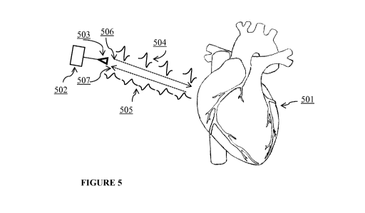
[0098] FIGURE 5 depicts an example device of the present invention. The device
502 can
17
CA 02951722 2016-12-08
WO 2015/191905 PCT/US2015/035405
comprise an antenna 503 and be positioned in proximity to, for example, a
human heart 501. The
antenna can transmit 506 a signal 504 to the heart. The signal 504 can reflect
off, for example,
the muscle tissue of the heart. The reflected signal 505 can then be received
507 by the device
502 and processed for analysis.
EXAMPLES
EXAMPLE 1: Analysis of Signal Loss Inside Human Tissues.
[0099] Performance can be optimized by positioning a device on a subject for
minimal signal
loss in tissue. FIGURE 6 illustrates the tissue thickness, in inches, of skin,
fat, muscle, and bone
anterior to the heart of a human. The amount of muscle tissue is relatively
low. When radar
signals were radiated through various tissues including skin, fat, muscle, and
bone, the greatest
loss of radar signal occurred in the muscle tissue, as demonstrated in FIGURE
7.
[00100] The loss of signal intensity was correlated positively with the
frequency of the signal,
as shown in FIGURE 7. When the frequency (GHz) of the signal was increased,
the total loss of
signal (dB) was most significant in the muscle, while other tissues only
accounted for a minor
portion of signal loss. This analysis further indicated that the sternum,
having minimal
musculature, should be an effective placement for the device. This placement
allows for less
signal loss and dispersion.
EXAMPLE 2: Modeling Methodology.
[00101] To calculate the interaction of transmitted signals generated by a
device described
herein with heart muscles, a three-dimensional full-wave simulation was
employed. In this
simulation, a three-dimensional model of the heart, or chest cavity, was used.
First, the
complexity of the model was reduced by removing portions of the chest cavity
that do not move,
and thus are not relevant for modeling the motion of the heart. Next, the
heart model was
imported into a wave simulation program to determine the signal received at
the antenna in the
form of a magnetic or electric field distribution. Finally, the extracted
waveforms were fed into a
circuit simulator to determine the correlation between the output signal and
the motion of the
heart.
EXAMPLE 3: Computer architectures.
[00102] Various computer architectures are suitable for use with the
invention. FIGURE 8 is
a block diagram illustrating afirst example architecture of a computer system
800 that can be
used in connection with example embodiments of the present invention. As
depicted in FIGURE
8, the example computer system can include a processor 802 for processing
instructions. Non-
18
CA 02951722 2016-12-08
WO 2015/191905 PCT/US2015/035405
limiting examples of processors include: Intel Core i7TM processor, Intel Core
i5TM processor,
Intel Core i3TM processor, Intel XeonTM processor, AMD OpteronTM processor,
Samsung 32-
bit RISC ARM 1176JZ(F)-S v1.0TM processor, ARM Cortex-A8 Samsung S5PC100TM
processor, ARM Cortex-A8 Apple A4TM processor, Marvell PXA 930TM processor, or
a
functionally-equivalent processor. Multiple threads of execution can be used
for parallel
processing. In some embodiments, multiple processors or processors with
multiple cores can be
used, whether in a single computer system, in a cluster, or distributed across
systems over a
network comprising a plurality of computers, cell phones, tablet computing
devices, watch based
devices, wrist band devices, armband devices, or personal data assistant
devices.
Data acquisition, processing and storage.
[00103] As illustrated in FIGURE 8, a high speed cache 801 can be connected
to, or
incorporated in, the processor 802 to provide a high speed memory for
instructions or data that
have been recently, or are frequently, used by processor 802. The processor
802 is connected to a
north bridge 806 by a processor bus 805. The north bridge 806 is connected to
random access
memory (RAM) 803 by a memory bus 804 and manages access to the RAM 803 by the
processor 802. The north bridge 806 is also connected to a south bridge 808 by
a chipset bus 807.
The south bridge 808 is, in turn, connected to a peripheral bus 809. The
peripheral bus can be,
for example, PCI, PCI-X, PCI Express, or other peripheral bus. The north
bridge and south
bridge are often referred to as a processor chipset and manage data transfer
between the
processor, RAM, and peripheral components on the peripheral bus 809. In some
architectures,
the functionality of the north bridge can be incorporated into the processor
instead of using a
separate north bridge chip.
[00104] In some embodiments, system 800 can include an accelerator card 812
attached to the
peripheral bus 809. The accelerator can include field programmable gate arrays
(FPGAs) or
other hardware for accelerating certain processing.
Software interface(s).
[00105] Software and data are stored in external storage 813 and can be loaded
into RAM 803
and/or cache 801 for use by the processor. The system 800 includes an
operating system for
managing system resources; non-limiting examples of operating systems include:
Linux,
WindowsTM, MACOSTM, BlackBerry OSTM, iOSTM, AndroidTM and other functionally-
equivalent operating systems, as well as application software running on top
of the operating
system.
[00106] In this example, system 800 also includes network interface cards
(NICs) 810 and 811
19
CA 02951722 2016-12-08
WO 2015/191905 PCT/US2015/035405
connected to the peripheral bus for providing network interfaces to external
storage, such as
Network Attached Storage (NAS) and other computer systems that can be used for
distributed
parallel processing.
Computer systems.
[00107] FIGURE 9 is a diagram showing a network 900 with a plurality of
computer systems
902a, and 902b, a plurality of cell phones and personal data assistants 902c,
and Network
Attached Storage (NAS) 901a, and 901b. In some embodiments, systems 902a,
902b, and 902c
can manage data storage and optimize data access for data stored in Network
Attached Storage
(NAS) 901a and 902b. A mathematical model can be used for the data and be
evaluated using
distributed parallel processing across computer systems 902a, and 902b, and
cell phone and
personal data assistant systems 902c. Computer systems 902a, and 902b, and
cell phone and
personal data assistant systems 902c can also provide parallel processing for
adaptive data
restructuring of the data stored in Network Attached Storage (NAS) 901a and
901b. FIGURE 9
illustrates an example only, and a wide variety of other computer
architectures and systems can
be used in conjunction with the various embodiments of the present invention.
For example, a
blade server can be used to provide parallel processing. Processor blades can
be connected
through a back plane to provide parallel processing. Storage can also be
connected to the back
plane or as Network Attached Storage (NAS) through a separate network
interface.
[00108] In some embodiments, processors can maintain separate memory spaces
and transmit
data through network interfaces, back plane, or other connectors for parallel
processing by other
processors. In some embodiments, some or all of the processors can use a
shared virtual address
memory space.
Virtual systems.
[00109] FIGURE 10 is a block diagram of a multiprocessor computer system using
a shared
virtual address memory space. The system includes a plurality of processors
1001a-f that can
access a shared memory subsystem 1002. The system incorporates a plurality of
programmable
hardware memory algorithm processors (MAPs) 1003a-f in the memory subsystem
1002. Each
MAP 1003a-f can comprise a memory 1004a-f and one or more field programmable
gate arrays
(FPGAs) 1005a-f. The MAP provides a configurable functional unit and
particular algorithms or
portions of algorithms can be provided to the FPGAs 1005a-f for processing in
close
coordination with a respective processor. In this example, each MAP is
globally accessible by all
of the processors for these purposes. In one configuration, each MAP can use
Direct Memory
Access (DMA) to access an associated memory 1004a-f, allowing it to execute
tasks
CA 02951722 2016-12-08
WO 2015/191905 PCT/US2015/035405
independently of, and asynchronously from, the respective microprocessor 1001a-
f. In this
configuration, a MAP can feed results directly to another MAP for pipelining
and parallel
execution of algorithms.
[00110] The above computer architectures and systems are examples only, and a
wide variety
of other computer, cell phone, and personal data assistant architectures and
systems can be used
in connection with example embodiments, including systems using any
combination of general
processors, co-processors, FPGAs and other programmable logic devices, system
on chips
(SOCs), application specific integrated circuits (ASICs), and other processing
and logic elements.
Any variety of data storage media can be used in connection with example
embodiments,
including random access memory, hard drives, flash memory, tape drives, disk
arrays, Network
Attached Storage (NAS) and other local or distributed data storage devices and
systems.
[00111] In example embodiments, the computer system can be implemented using
software
modules executing on any of the above or other computer architectures and
systems. In other
embodiments, the functions of the system can be implemented partially or
completely in
firmware, programmable logic devices such as field programmable gate arrays
(FPGAs) as
referenced in FIGURE 10, system on chips (SOCs), application specific
integrated circuits
(ASICs), or other processing and logic elements. For example, the Set
Processor and Optimizer
can be implemented with hardware acceleration through the use of a hardware
accelerator card,
such as accelerator card 812 illustrated in FIGURE 8.
[00112] Any embodiment of the invention described herein can be, for example,
produced and
transmitted by a user within the same geographical location. A product of the
invention can be,
for example, produced and/or transmitted from a geographic location in one
country and a user
of the invention can be present in a different country. In some embodiments,
the data accessed
by a system of the invention is a computer program product that can be
transmitted from one of a
plurality of geographic locations 1101 to a user 1102 (FIGURE 11). Data
generated by a
computer program product of the invention can be transmitted back and forth
among a plurality
of geographic locations, for example, by a network, a secure network, an
insecure network, an
internet, or an intranet. In some embodiments, an ontological hierarchy
provided by the
invention is encoded on a physical and tangible product.
[00113] Any embodiment of the invention described herein can be produced
and/or transmitted
in an encoded form, for example, a radio frequency identification tag or
barcode. In some
embodiments, the data accessed by a system of the invention can be accessed
from the encoded
form either directly or as part of a health record. In some embodiments, the
health record can be
an electronic health record or digital health record. In some embodiments, the
health record can
be accessed by the subject or a health care provider for the subject.
21
CA 02951722 2016-12-08
WO 2015/191905 PCT/US2015/035405
EXAMPLE 4: Implementations of the device.
[00114] One embodiment of the invention described herein is shown in FIGURE
14, panel A.
A human subject stands or sits in a way that they can avoid shaking their
body. The subject holds
a radar device attached to their body using either their hand or a chest
strap. An ECG is
simultaneously recorded by a three-electrode ECG circuit with three electrodes
attached to a
wrist and the left and right ankles. The device and ECG circuit is connected
to a computer that
performs the signal processing and waveform display.
[00115] As shown in FIGURE 14, panel B, the device can be disposed in various
positions on
the subject's chest, resulting in differences in the efficacy and output. The
device can also be put
against the back of the human subject to record data.
[00116] The human subject is in a comfortable and warm environment and is
relaxed. The
subject can stand, sit, walk or lie down on a bed. The device can be
positioned to contact the skin
or can be placed outside the subject's clothing. In the clothed setting, the
device can be placed
tightly against chest of the subject.
EXAMPLE 5: Representative subject measurements.
[00117] Three subjects were monitored using a device of the invention. All
data were taken
with the subjects in a held breath state. FIGURE 15, panel A shows the ECG
signal on the top
graph and the radar signal on the bottom graph detected in a healthy young
man. FIGURE 15,
panel B shows the ECG signal on the top graph and the radar signal on the
bottom graph
detected in a healthy elderly man. FIGURE 15, panel C shows the ECG signal on
the top graph
and the radar signal on the bottom graph detected in an elderly man with an
irregular heartbeat,
who is in atrial fibrillation. In panels A and B, the small first pulse was
generated by the motion
of the atrium and the larger second pulse was generated by the motion of the
ventricles. In
panels A and B, the radar signals from the invention correlated with the ECG
signals and were in
the same periodicity as the ECG signals. In panel C, the radar signal
correlated with the ECG
signal but showed little evidence of atrial motion.
EXAMPLE 6: Subject measurements while breathing.
[00118] FIGURE 16 shows the ECG signal on the top graph and the radar signal
on the
bottom graph detected in the healthy young man in Example 5 while breathing.
The additional
low frequency signal superimposed on the radar heart motion signal represents
the motion of the
chest cavity as a result of breathing.
22
CA 02951722 2016-12-08
WO 2015/191905 PCT/US2015/035405
EXAMPLE 7: Subject measurements at different chest positions.
[00119] As described in Example 4, FIGURE 17 shows the ECG signal on the top
graph and
the radar signal on the bottom graph when a representative device of the
invention was
positioned at different locations on the chest of the healthy young man from
Example 5. Panel A
illustrates the ECG and radar signals measured at the center of the chest.
Panel B illustrates the
ECG and radar signals measured above the center of the chest Panel C
illustrates the ECG and
radar signals measured below the center of the chest. Panel D illustrates the
ECG and radar
signals measured left of the center of the chest. Panel E illustrates the ECG
and radar signals
measured right of the center of the chest. These graphs show that positioning
the device at
various locations on the chest allowed views of different portions of the
heart. Viewing different
portions of the heart can assist evaluation of various heart abnormalities.
EXAMPLE 8: Subject measurements from the back.
[00120] FIGURE 18 shows the ECG signal on the top graph and the radar signal
on the
bottom graph obtained when a device of the invention was positioned on the
back of the healthy
young man from Example 5. These data represent measurements of atrial motion
by positioning
the device on the back of the subject.
EXAMPLE 9: Subject measurements at a distance.
[00121] FIGURE 21 shows three measurements taken from a healthy male subject
at different
distances. All of these data were taken with the subject in a held breath
state. These data
represent measurements of atrial motion by positioning an embodiment of the
invention at a
distance from the subject.
EXAMPLE 10: Improving data quality by quadrature demodulation.
[00122] FIGURE 19, panel A illustrates an embodiment of a device to determine
the motion
of the heart of a subject. The radar system 1901 connected to an antenna 1902
propagates a
transmitted waveform 1903 towards a target 1904, for example, a heart of a
subject. A reflected
waveform 1905 is then received by the antenna 1902. The distance between the
antenna 1902
and the target 1904 is defined by the function 1906 d-x(t), where d is a
constant distance between
the antenna 1902 and the target 1904 and x(t) is the motion function of the
target 1904, for
example, the original motion of the heart of the subject. Panels B-D represent
measured
waveforms at three different distances between the antenna 1902 and target
1904.
[00123] The radar system 1901 transmits a cosine signal written as
T (t) = A = cos(271-ft) ;
23
CA 02951722 2016-12-08
WO 2015/191905 PCT/US2015/035405
where A is the amplitude of the transmitted waveform 1903, f is the frequency
of transmitted
waveform 1903, and t is time.
[00124] The reflected signal is written as
R(t)= B = cos(22-cft 42z-d 42-1- = x(t));
2 2
where B is the amplitude of the reflected waveform 1905 and lambda is the
wavelength of
reflected waveform 1905.
[00125] After demodulation, two output signals can exist. One is called an in-
phase signal (I)
and the other is called a quadrature signal (Q). The in-phase signal is
written as
/(t)= C = cos(42rd +42-1- = x(t))
2 2 ; and the quadrature signal is written as
Q(t)=C=sin(42rd +42-1- = x(t))
2 2 ; where C is the amplitude of the signal.
[00126] By dividing Q(t) by I(t), the signal ratio Y is obtained, which is
written as
Y(t) = tan(42rd +42-1-=x(t)
___________________ ).
2 2
[00127] Thus, solution is possible for the motion function of the target in
terms of the signal
ratio, with x(t) written as
x(t) = ¨2 = tan1 - (Y(t))¨ d.
42z-
[00128] Quadrature demodulation as described herein can allow for precise
calculation of the
motion function of the target 1904, since motion of the target 1904 is quite
small compared with
the wavelength of the reflected waveform 1905.
EXAMPLE 11: Continuous monitoring to inform decision-making on interventions.
[00129] FIGURE 20 illustrates an embodiment of the method of continuous
monitoring
described in the invention. For example, a subject can wear a device
continuously for long-term
monitoring 2001 to determine the percentage of cardiac arrhythmic time 2002.
These data are
recorded and stored over long periods of time, for example, hours, days,
weeks, or months.
Signal recognition software detects and highlights important events for review
by a health care
provider. The health care provider uses the percentage of cardiac arrhythmic
time 2002 to
determine the appropriate medical intervention 2003. Non-limiting examples of
medical
interventions include continued monitoring; cardioversion with or without
specific
pharmaceuticals; cardiac ablation with or without specific pharmaceuticals;
and cardiac ablation
with or without introduction of a pacemaker.
24
CA 02951722 2016-12-08
WO 2015/191905 PCT/US2015/035405
EMBODIMENTS
[00130] The following non-limiting embodiments provide illustrative examples
of the
invention, but do not limit the scope of the invention.
[00131] Embodiment 1. A method of detecting an irregular heartbeat in a
subject, the
method comprising: a) transmitting a wavelength of electromagnetic radiation
to the heart of the
subject; b) detecting an electromagnetic signal reflected off the heart of the
subject; and c)
determining based on the electromagnetic signal reflected off the heart of the
subject whether the
subject has an irregular heartbeat.
[00132] Embodiment 2. The method of Embodiment 1, wherein the subject is
undergoing
an intervention for the irregular heartbeat, the method further comprising
determining based on
the electromagnetic signal reflected off the heart of the subject whether the
intervention for the
irregular heartbeat has modulated the irregular heartbeat.
[00133] Embodiment 3. The method of Embodiment 1, wherein the subject is
undergoing
an intervention for a non-irregular heartbeat condition, the method further
comprising
determining based on the electromagnetic signal reflected off the heart of the
subject whether the
intervention for the non-irregular heartbeat condition has induced the
irregular heartbeat.
[00134] Embodiment 4. The method of any one of Embodiments 1-3, wherein the
determination whether the subject has an irregular heartbeat is determined by
an analysis of a
movement of a portion of the heart.
[00135] Embodiment 5. The method of any one of Embodiments 1-4, further
comprising
attaching a source of electromagnetic radiation to the subject's body.
[00136] Embodiment 6. The method of Embodiment 5, wherein the source of
electromagnetic radiation is attached to the subject's body in proximity to
the subject's heart.
[00137] Embodiment 7. The method of Embodiment 5, wherein the source of
electromagnetic radiation is attached to the subject's chest.
[00138] Embodiment 8. The method of Embodiment 5, wherein the source of
electromagnetic radiation is attached to the subject's back.
[00139] Embodiment 9. The method of any one of Embodiments 1-8, wherein the
subject is
in a held-breath state.
[00140] Embodiment 10. The method of any one of Embodiments 1-9, wherein
the
wavelength of electromagnetic radiation transmitted to the heart of the
subject is a radio wave.
[00141] Embodiment 11. The method of any one of Embodiments 1-10, wherein
the
irregular heartbeat is associated with atrial fibrillation.
[00142] Embodiment 12. The method of any one of Embodiments 1-10, wherein
the
CA 02951722 2016-12-08
WO 2015/191905 PCT/US2015/035405
irregular heartbeat is associated with atrial flutter.
[00143] Embodiment 13. The method of any one of Embodiments 1-10, wherein
the
irregular heartbeat is associated with ventricular fibrillation.
[00144] Embodiment 14. The method of any one of Embodiments 1-10, wherein
the
irregular heartbeat is associated with ventricular flutter.
[00145] Embodiment 15. The method of any one of Embodiments 1-10, wherein
the
irregular heartbeat is associated with cardiac arrhythmia.
[00146] Embodiment 16. The method of any one of Embodiments 1-15, wherein
the subject
is human.
[00147] Embodiment 17. A method comprising: a) receiving by a computer
system data
associated with an electromagnetic signal reflected off a heart of a subject;
b) comparing by a
processor of the computer system the data associated with the electromagnetic
signal reflected
off the heart of the subject to a reference; c) determining based on the
comparison of the data
associated with the electromagnetic signal reflected off the heart of the
subject to the reference
whether the subject has an irregular heartbeat; and d) outputting a result of
the determination.
[00148] Embodiment 18. The method of Embodiment 17, wherein the subject is
undergoing
an intervention for the irregular heartbeat, the method further comprising
determining based on
the electromagnetic signal reflected off the heart of the subject whether the
intervention for the
irregular heartbeat has modulated the irregular heartbeat.
[00149] Embodiment 19. The method of Embodiment 17, wherein the subject is
undergoing
an intervention for a non-irregular heartbeat condition, the method further
comprising
determining based on the electromagnetic signal reflected off the heart of the
subject whether the
intervention for the non-irregular heartbeat condition has induced the
irregular heartbeat.
[00150] Embodiment 20. The method of any one of Embodiments 17-19, wherein
the
determination that the subject has an irregular heartbeat is determined by an
analysis of a
movement of a portion of the heart.
[00151] Embodiment 21. The method of any one of Embodiments 17-20, wherein
the
irregular heartbeat is associated with atrial fibrillation.
[00152] Embodiment 22. The method of any one of Embodiments 17-20, wherein
the
irregular heartbeat is associated with atrial flutter.
[00153] Embodiment 23. The method of any one of Embodiments 17-20, wherein
the
irregular heartbeat is associated with ventricular fibrillation.
[00154] Embodiment 24. The method of any one of Embodiments 17-20, wherein
the
irregular heartbeat is associated with ventricular flutter.
[00155] Embodiment 25. The method of any one of Embodiments 17-20, wherein
the
26
CA 02951722 2016-12-08
WO 2015/191905 PCT/US2015/035405
irregular heartbeat is associated with cardiac arrhythmia.
[00156] Embodiment 26. The method of any one of Embodiments 17-25, wherein
the
subject is human.
[00157] Embodiment 27. A device comprising: a) an antenna configured to
transmit
electromagnetic radiation into a thoracic cavity of a subject; b) a receiver
configured to detect an
electromagnetic signal reflected off the subject's heart; and c) a processor
configured to identify
an irregular heartbeat in the subject based on the detected electromagnetic
signal reflected off the
subject's heart.
[00158] Embodiment 28. The device of Embodiment 27, wherein the antenna
configured to
transmit the electromagnetic radiation into the thoracic cavity of the
subject, the receiver
configured to detect the electromagnetic signal reflected off the subject's
heart, and the processor
configured to identify the irregular heartbeat in the subject based on the
detected electromagnetic
signal reflected off the subject's heart, are contained in a common housing.
[00159] Embodiment 29. The device of any one of Embodiments 27-28, further
comprising
a circuit configured to generate a signal suitable for transmission into the
thoracic cavity of the
subject by the antenna.
[00160] Embodiment 30. The device of any one of Embodiments 27-29, wherein
the
processor is configured to detect the irregular heartbeat in a subject by
measuring time intervals
among signals received after reflection off the subject's heart.
[00161] Embodiment 31. The device of any one of Embodiments 27-30, wherein
the antenna
is configured to transmit electromagnetic radiation that is a radio wave.
[00162] Embodiment 32. The device of any one of Embodiments 27-31, wherein
the device
is configured to be attached to the subject in proximity to the subject's
heart.
[00163] Embodiment 33. The device of any one of Embodiments 27-32, wherein
the device
is configured to be attached to the subject on the subject's chest.
[00164] Embodiment 34. The device of any one of Embodiments 27-32, wherein
the device
is configured to be attached to the subject on the subject's back.
[00165] Embodiment 35. The device of any one of Embodiments 24-34, wherein
the
processor is configured to identify an irregular heartbeat associated with
atrial fibrillation.
[00166] Embodiment 36. The device of any one of Embodiments 24-34, wherein
the
processor is configured to identify an irregular heartbeat associated with
atrial flutter.
[00167] Embodiment 37. The device of any one of Embodiments 24-34, wherein
the
processor is configured to identify an irregular heartbeat associated with
ventricular fibrillation.
[00168] Embodiment 38. The device of any one of Embodiments 24-34, wherein
the
processor is configured to identify an irregular heartbeat associated with
ventricular flutter.
27
CA 02951722 2016-12-08
WO 2015/191905 PCT/US2015/035405
[00169] Embodiment 39. The device of any one of Embodiments 24-34, wherein
the
processor is configured to identify an irregular heartbeat associated with
cardiac arrhythmia.
[00170] Embodiment 40. A device comprising: a) an antenna configured to
transmit
electromagnetic radiation into a thoracic cavity of a subject; b) a receiver
configured to detect an
electromagnetic signal reflected off the subject's heart; and c) a transmitter
configured to
transmit data associated with the received electromagnetic signal reflected
off the subject's heart.
[00171] Embodiment 41. The device of Embodiment 40, wherein the antenna
configured to
transmit the electromagnetic radiation into the thoracic cavity of the
subject, the receiver
configured to detect the electromagnetic signal reflected off the subject's
heart, and the
transmitter configured to transmit data associated with the received
electromagnetic signal
reflected off the subject's heart, are contained in a common housing.
[00172] Embodiment 42. The device of any one of Embodiments 40-41, further
comprising
a circuit configured to generate a signal suitable for transmission into the
thoracic cavity of the
subject by the antenna.
[00173] Embodiment 43. The device of any one of Embodiments 40-42, wherein
the
transmitter is configured to transmit wirelessly to a remote processor.
[00174] Embodiment 44. The device of any one of Embodiments 40-43, wherein
the antenna
is configured to transmit electromagnetic radiation that is a radio wave.
[00175] Embodiment 45. The device of any one of Embodiments 40-44, wherein
the device
is configured to be attached to the subject in proximity to the subject's
heart.
[00176] Embodiment 46. The device of any one of Embodiments 40-45, wherein
the device
is configured to be attached to the subject on the subject's chest.
[00177] Embodiment 47. The device of any one of Embodiments 40-45, wherein
the device
is configured to be attached to the subject on the subject's back.
[00178] Embodiment 48. A method comprising: a) administering to a subject
having an
irregular heartbeat an intervention for the irregular heartbeat; b) monitoring
the subject with a
radar device; and c) determining based on the monitoring whether the
intervention for the
irregular heartbeat modulates the irregular heartbeat in the subject.
[00179] Embodiment 49. The method of Embodiment 48, wherein the radar
device monitors
a movement of a portion of the subject's heart.
[00180] Embodiment 50. The method of any one of Embodiments 48-49, wherein
the
processor is configured to identify an irregular heartbeat associated with
atrial fibrillation.
[00181] Embodiment 51. The method of any one of Embodiments 48-49, wherein
the
processor is configured to identify an irregular heartbeat associated with
atrial flutter.
[00182] Embodiment 52. The method of any one of Embodiments 48-49, wherein
the
28
CA 02951722 2016-12-08
WO 2015/191905 PCT/US2015/035405
processor is configured to identify an irregular heartbeat associated with
ventricular fibrillation.
[00183] Embodiment 53. The method of any one of Embodiments 48-49, wherein
the
processor is configured to identify an irregular heartbeat associated with
ventricular flutter.
[00184] Embodiment 54. The method of any one of Embodiments 48-49, wherein
the
processor is configured to identify an irregular heartbeat associated with
cardiac arrhythmia.
[00185] Embodiment 55. A method comprising: a) administering to a subject
an intervention;
b) monitoring the subject with a radar device; and c) determining based on the
monitoring
whether the intervention induces an irregular heartbeat in the subject.
[00186] Embodiment 56. The method of Embodiment 55, wherein the radar
device monitors
a movement of a portion of the subject's heart.
[00187] Embodiment 57. The method of any one of Embodiments 55-56, wherein
the
processor is configured to identify an irregular heartbeat associated with
atrial fibrillation.
[00188] Embodiment 58. The method of any one of Embodiments 55-56, wherein
the
processor is configured to identify an irregular heartbeat associated with
atrial flutter.
[00189] Embodiment 59. The method of any one of Embodiments 55-56, wherein
the
processor is configured to identify an irregular heartbeat associated with
ventricular fibrillation.
[00190] Embodiment 60. The method of any one of Embodiments 55-56, wherein
the
processor is configured to identify an irregular heartbeat associated with
ventricular flutter.
[00191] Embodiment 61. The method of any one of Embodiments 55-56, wherein
the
processor is configured to identify an irregular heartbeat associated with
cardiac arrhythmia.
[00192] Embodiment 62. The method of any one of Embodiments 55-61, wherein
the subject
has a non-irregular heartbeat condition, and the intervention is for the non-
irregular heartbeat
condition.
[00193] Embodiment 63. A method comprising: a) transmitting a wavelength of
electromagnetic radiation to the heart of the subject; b) detecting an
electromagnetic signal
reflected off the heart of the subject; and c) determining based on the
electromagnetic signal
reflected off the heart of the subject a change in volume of the heart of the
subject.
[00194] Embodiment 64. The method of Embodiment 63, further comprising
determining a
volume of blood output from the heart of the subject based on the change in
volume of the heart
of the subject.
[00195] Embodiment 65. The method of any one of Embodiments 63-64, wherein
the change
in volume of the heart of the subject is determined within a single chamber of
the heart of the
subject.
29