Note: Descriptions are shown in the official language in which they were submitted.
CA 02951769 2016-12-09
WO 2016/001825 PCT/1B2015/054872
1
Method for segmenting and predicting tissue regions in patients
with acute cerebral ischemia
TECHNICAL FIELD OF THE INVENTION
The present invention relates to the field of multi-dimensional imaging and,
in
particular, to the field of classifying volumetric elements of affected
regions of the
brains of acute ischemic stroke patients in order to differentiate between
salvageable
and non-salvageable brain tissue.
BACKGROUND OF THE INVENTION
Acute ischemic stroke, or cerebral ischemia, is a neurological emergency which
may be reversible if treated rapidly. Outcomes for stroke patients are
strongly
influenced by the speed and accuracy with which the ischemia can be identified
and
treated. Effective reperfusion and revascularization therapies are available
for
salvaging regions of brain tissue which are characterized by reversible
hypoxia, and
these regions must be identified and distinguished from tissue which is
destined to
infarct. Volumetric imaging of the brain tissue, using computer tomography
(CT) or
magnetic resonance imaging (MRI) may be used to generate 4D (spatial and
temporal) scans of the brain tissue of the patient. Skilled clinical
practitioners, aided
by image analysis software, can read such image sequences to assess the likely
extent of the eventual infarct region. Image analysis and treatment decision
may be
performed visually by a neuroradiologist or a stroke neurologist. The ratio,
or
mismatch, between the infarct volume and the penumbra volume may be taken as
an
indicator of the likely effectiveness of reperfusion therapy. The larger the
mismatch,
the more likely the patient is to have a favorable prognosis. In order to
provide an
accurate measure of this ratio, it is important to achieve a fast and accurate
classification of volumetric elements into those which will infarct, those
which belong
to the penumbra, and those which comprise healthy tissue. This analysis may be
performed on CT image sets or MRI image sets, in which the infarct core can be
identified by diffusion-weighted imaging (DWI), and the hypo-perfused, yet
vital,
CA 02951769 2016-12-09
WO 2016/001825 PCT/1B2015/054872
2
potentially salvageable tissue adjacent to the infarct core can be identified
using
perfusion-weighted imaging (PWI). This segregation technique may be referred
to as
diffusion-perfusion mismatch analysis. DWI and PWI are well-known abstraction
techniques and will not be described here.
PRIOR ART
It has been considered to use computer-assisted image analysis to quantify the
mismatch mentioned above. However, previous proposals have usually focused on
the segmentation of the infarct only, or on the hypo-perfused region only.
Approaches
have been proposed which consider both regions simultaneously, but these have
used relatively simplistic classification models and have limited accuracy.
In M. Straka et al, "Real-Time Diffusion-Perfusion Mismatch Analysis in Acute
Stroke", Journal of Magnetic Resonance Imaging, JMRI, vol 32, no. 5, pages
1024-
1037, November 2010, an automated image analysis tool was described for
identifying candidates for acute stroke treatment. This approach relies on DWI
and
PWI to quantify the mismatch. For identification of the ischemic (infarct)
core, the
Apparent Diffusion Coefficient (ADC), a quantitative measure derived from
diffusion
images, is thresholded by taking diffusion rates of less than 600x10-6 mm2/s.
To
identify the penumbra region, the Tmax map derived from dynamic susceptibility
contrast (DSC) perfusion images is thresholded by taking perfusion times
greater
than 6 seconds. Some additional morphological constraints are applied, to
suppress
outliers. While this technique appears promising, its mismatch analysis
performance
stands to be improved.
An automated segmentation method using multiple MRI modalities has been
proposed for MRI analysis of brain tumors. S. Bauer et al, "Fully automatic
segmentation of brain tumor images using support vector machine classification
in
combination with hierarchical conditional random field regularization",
International
Conference on Medical Image Computing and Computer-Assisted Intervention, vol.
14, no. Pt 3. Jan. 2011, pp. 354-61, proposed a method for delineating brain
tumors
using multiple structural modalities.
CA 02951769 2016-12-09
WO 2016/001825 PCT/1B2015/054872
3
A tissue outcome prediction method was proposed in US patent application
US2007/0167727, using a combination of diffusion weighted images (DWI) and
perfusion weighted images (PWI).
The prior art methods have the disadvantage that their outputs are not
sufficiently reliable or accurate to enable confident use of the methods in
automated
tissue classification, outcome prediction or assessment for therapy.
BRIEF DESCRIPTION OF THE INVENTION
The present invention aims to overcome the above and other shortcomings
inherent in the prior art. In particular, the invention aims to provide a
method as set
out in claim 1. Further variants of the inventive method are set out in the
dependent
claims.
The invention and its advantages will further be explained in the following
detailed description, together with illustrations of example embodiments and
implementations given in the accompanying drawings, in which:
Figure 1 shows a simplified flow diagram of an example segmentation method
for use in a segmentation/prediction method according to the invention.
Figure 2 shows a simplified flow diagram of an example
segmentation/prediction method according to the invention.
Figure 3a shows, in greatly simplified, schematic form, an example of an MRI
image of an axial brain section of a stroke patient.
Figure 3b shows an MRI segmentation generated, using a prior art
segmentation method, for the patient whose brain is depicted in figure 3a.
Figure 3c shows an MRI segmentation generated for the patient whose brain is
depicted in figure 3a, using a segmentation/prediction method according to a
first
embodiment of the invention.
CA 02951769 2016-12-09
WO 2016/001825 PCT/1B2015/054872
4
Figure 3d shows an MRI segmentation generated for the patient whose brain is
depicted in figure 3a, using a segmentation/prediction method according to a
second
embodiment of the invention.
DETAILED DESCRIPTION OF THE INVENTION
The invention will now be described in detail with reference to the drawings.
Note that the drawings are intended merely as illustrations of example
embodiments
of the invention, and are not to be construed as limiting the scope of the
invention.
Where the same reference numerals are used in different drawings, these
reference
numerals are intended to refer to the same or corresponding features. However,
the
use of different reference numerals should not necessarily be taken as an
indication
that the referenced features are dissimilar.
The examples and discussion below are described with reference to the
application of the method of the invention to the use of MRI imaging. However,
it
should be understood that the principles of the invention may also be applied
in other
tomographic or volumetric imaging regimes such as CT imaging.
Similarly, the invention has been described in relation to segmenting or
labeling
volume elements into infarct, penumbra and healthy tissue. However, the
segmentation or prediction may be used to identify tissue types other than
these
three. A greater number of tissue-types (labels) may be identified, for
example, than
the three mentioned.
Stroke MRI protocols include a wealth of information which includes structural
information such as non-enhanced and enhanced Ti-weighted, T2-weighted, fluid
attenuated inversion recovery (FLAIR), and functional information such as PWI
and
DWI image datasets and vessel imaging (magnetic resonance angiography, MRA).
By combining structural and functional information, and by employing modern
machine learning concepts, the method of the present invention provides a
segmentation and prediction of volumetric elements which offers a significant
improvement over prior art methods of identifying infarct core and penumbra
tissue. A
supervised classification approach is used for performing a multi-parametric
CA 02951769 2016-12-09
WO 2016/001825 PCT/1B2015/054872
segmentation from a plurality of different MRI modalities. The classification
may be
trained using manually-labeled samples.
An overview example of a method according to the invention will now be
described with reference to figure 1. The segmentation is based on structural
and
5 functional magnetic resonance (MR) images, however it should be understood
that
the principles underlying the invention may also be implemented using other
types of
tomographic images. In the illustrated example, T1-weighted images with
contrast
enhancement (referred to as the T1contrast modality), T2-weighted images,
diffusion-
weighted images (DWI) and dynamic susceptibility contrast (DSC) perfusion-
weighted images (PWI) may be acquired from acute ischemic stroke patients
before
and after treatment. This image acquisition step is indicated by reference
number 1 in
figure 1.
Apparent diffusion coefficient (ADC) maps are extracted from the diffusion-
weighted images, as indicated by reference 2. Standard perfusion maps (of
which
there may be four, for example, representing four different modalities) may be
computed from the DSC perfusion-weighted images, as indicated by reference 3,
using known techniques. The perfusion maps may for example comprise cerebral
blood flow (CBF), cerebral blood volume (CBV), mean transit time (MTT) and the
peak time (Tmax) modalities. All seven modalities (T1contrast, T2, ADC, CBF,
CBV,
MTT, Tmax) from before and after treatment may then be rigidly registered, for
example to the pre-treatment T1contrast image of the patient, as indicated by
reference 4. A skull-stripping step 5 may be automatically performed which, as
will be
seen, may improve the quality of the tissue classification 6. Skull-stripping
involves
detecting and removing the skull regions from the images. The skull regions
may give
rise to unwanted outliers and false positives in the classification process.
In the illustrated overview example, the seven pre-treatment MRI modalities
(T1contrast, T2, ADC, CBF, CBV, MTT, Tmax) are used as an input for a
segmentation/prediction algorithm which will be described in relation to
figure 2. The
proposed segmentation/prediction method used in this example may employ a
classification method adapted from the method proposed for brain tumors in the
article by S. Bauer et al, mentioned earlier. The segmentation task may for
example
CA 02951769 2016-12-09
WO 2016/001825 PCT/1B2015/054872
6
be cast as an energy minimization problem in a conditional random field
context
(CRF), with the energy to be minimized being expressed as
E = )7V(yi,x) + >;;;;W (yi,y;,xi,x) EQ1
where the first term in equation EQ1 corresponds to the voxel-wise singleton
potentials, and the second term corresponds to the pairwise potentials,
modeling
voxel-to-voxel interactions. x is a voxel-wise feature vector and y is the
final
segmentation label. The singleton potentials may be computed by a decision
forest
classifier, as indicated by reference 13 in figure 2. A decision forest is a
supervised
classifier that makes use of training data for computing a probabilistic
output label for
every voxel based on a certain feature vector. By way of example, a 283-
dimensional
feature vector x may be extracted (8 and 12 in figure 2) and used as an input
for the
classifier 13, comprising the voxel-wise intensities and multi-scale local
texture,
gradient, symmetry and position descriptors of each modality. These singleton
potentials are computed according to equation (EQ2), with p(ilx) being the
output
probability from the classifier and 6 is the Kronecker-6, function.
V(Yi,x) = P(9i1x) = (1 ¨ 4.96.0 EQ2
The second term in equation EQ1 corresponds to the pairwise potentials,
introducing a spatial regularization in order to suppress noisy outputs caused
by
outliers. It is computed according to equation EQ3, where ws(i,D is a
weighting
function that depends on the voxel spacing of the image in each dimension. The
term
(/ ¨ 6(yi,y1)) penalizes different labels of neighboring voxels, and the
degree of
neighborhood smoothing is regulated by the difference of the feature vectors
in the
term exp ________ jr\
=
\ 2.7
VII(Y6y1,xi,x1) = ws(i, j).(1 ¨ 6(yi,y1)) = exp ____________ EQ3
\
CA 02951769 2016-12-09
WO 2016/001825 PCT/1B2015/054872
7
Optimization of the energy function in equation EQ1 may be achieved using
known optimization strategies.
As described above, a multi-dimensional feature vector is derived for each
volume element, and may for example comprise more than 100 features. The
example of a 283-dimensional feature vector has been mentioned above, however
it
has been found that a number of features greater than 50, or preferably
greater than
100, or more preferably greater than 200 may achieve the advantageous effects
of
the invention. In the particular example case, the 283 features concerned may
for
example be made up as follows from the combination of seven image modalities
(T1contrast, T2, ADC, CBF, CBV, MTT, Tmax):
Voxel-wise multi-modal intensities - 1 feature per modality (normalized voxel
intensity
values):
= T2 intensity
= T1contrast intensity
= ADC intensity
= CBF intensity
= CBV intensity
= MTT intensity
= Tmax intensity
Textures from patches in 3x3x3 neighborhood - 15 features per modality
(values computed based on intensities from local patches: Mean, Variance,
Skewness, Kurtosis, Energy, Entropy, Min, Max, Percentile10, Percentile25,
Percentile50, Percentile75, Percentile90, Range, SNR):
= T2 texture3
= T1contrast texture3
= ADC texture3
= CBF texture3
= CBV texture3
= MTT texture3
= Tmax texture3
CA 02951769 2016-12-09
WO 2016/001825 PCT/1B2015/054872
8
Textures from patches in 5x5x5 neighborhood - 15 features per modality
(values computed based on intensities from local patches: Mean, Variance,
Skewness, Kurtosis, Energy, Entropy, Min, Max, Percentile10, Percentile25,
Percentile50, Percentile75, Percentile90, Range, SNR):
= T2 texture5
= T1contrast texture5
= ADC texture5
= CBF texture5
= CBV texture5
= MTT texture5
= Tmax texture5
Gradient statistics from patches in 3x3x3 neighborhood - 3 features per
modality
(values computed based on gradient magnitude from local patches:
gradMagCenter,
gradMagMean, gradMagVariance):
= T2 grad3
= T1contrast grad3
= ADC texture grad3
= CBF texture grad3
= CBV grad3
= MTT grad3
= Tmax grad3
Gradient statistics from patches in 5x5x5 neighborhood - 3 features per
modality
(values computed based on gradient magnitude from local patches:
gradMagCenter,
gradMagMean, gradMagVariance):
= T2 grad5
= T1contrast grads
= ADC texture grad5
= CBF texture grad5
= CBV grad5
CA 02951769 2016-12-09
WO 2016/001825 PCT/1B2015/054872
9
= MTT grad5
= Tmax grad5
Location features - 3 features (values computed from smoothed or approximated
coordinates of registered atlas image in three spatial dimensions):
= Smoothed or approximated coordinates in standard atlas
Multi-scale symmetry features - 3 features per modality (values computed from
intensity difference across midsagittal symmetry plane: intensityDiff
approximatedScalel, intensityDiff approximatedScale2, intensityDiff
approximatedScale3):
= T2 sym
= Tlcontrast sym
= ADC sym
= CBF sym
= CBV sym
= MTT sym
= Tmax sym
While the above example relates to the application of the invention to MRI
image datasets, it should be noted that a similar approach can be used with
other
types of volumetric or tomographic imaging, such as CT imaging. In the case of
CT
imaging, the method may for example be performed with a smaller number of
modalities, for example the four perfusion (functional) modalities and the
structural
CT modality, and with a smaller number (e.g. around 200) of features than the
e.g.
283 features mentioned for the feature vector in the MRI implementation.
When using MRI images, the infarct regions may advantageously be defined
with reference to the DWI or T2 image, whereas with CT images, the infarct
region
may be defined with reference to one of the perfusion maps, such as the CBV
modality, for the training datasets.
CA 02951769 2016-12-09
WO 2016/001825 PCT/1B2015/054872
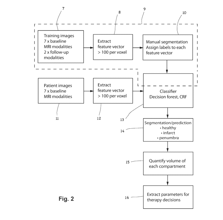
A schematic representation of an example method according to the invention is
illustrated in figure 2. In the illustrated method, two data acquisition
branches are
shown. The first branch, indicated by dotted line 9, comprises the steps 7, 8
and 10
of acquiring training datasets, which are performed "off-line", i.e. in one or
more pre-
5 processing sequences, before the method is used in the an examination of a
patient.
The second branch comprises the steps 11, 12 performed in acquiring and
processing MRI datasets of the patient.
As will be described in relation to the first embodiment of the invention, the
training data may comprise image datasets, 7, whose modalities and feature
vectors,
10 8, correspond to the image dataset(s), 11, and feature vector(s), 12, of
patients. The
training data comprises pre-treatment images comprising hypoxic regions of
previous
stroke patients, and the voxels may be manually segmented, 10, for example by
an
experienced neuroradiologist, in order to generate training data for training
the
classifier, 13.
As will described in relation to the second embodiment of the invention, and
as
illustrated in figure 2, the training data 7 may additionally comprise follow-
up image
datasets, for example post-treatment image datasets corresponding to (i.e.
relating to
the same patients as) at least some of the pre-treatment MRI images of the
hypoxic
regions of the previous stroke patients mentioned above. In the example
illustrated in
figure 2, the follow-up MRI image datasets may comprise only structural
modalities
(e.g. T1contrast and T2) This allows the learning process to benefit from the
outcome
information present in the structural modality information. Advantageously,
the
training data 7 may optionally include information about the treatment which
was
carried out on the patients whose follow-up MRI image data is included. Such
treatment parameter information (for example the type of treatment, or the
frequency,
dosage, drug details, therapy duration, surgical interventions etc) may also
be
included in the training of the classifier in order to improve the quality of
the
prediction in step 14 and the parameters for taking therapy decisions in step
16. The
latter parameters may, for example, include a proposal for therapy parameters
which
may offer the patient under examination the best or the least-worst outcomes.
Two example embodiments of the invention are described below. The
embodiments differ principally in the training sets used. According to a first
CA 02951769 2016-12-09
WO 2016/001825 PCT/1B2015/054872
11
embodiment of the present invention, segmentation is based on manual
segmentations of infarct core and penumbra on the pre-treatment images of
patients
(i.e. without taking into account MRI datasets from follow-up scans).
According to a
second embodiment of the invention, the method aims for prediction instead of
(or in
addition to) segmentation. As in the first embodiment, the training may be
based on
manual segmentation, but in this case only the penumbra is defined on the pre-
treatment images, whereas the infarct core is the real infarct, which is
defined on real
follow-up datasets (for example the T2-weighted images from a follow-up
examination several weeks or months after the stroke incident). The follow-up
images
are only needed for generating the training data; once the classifier 13 has
already
been trained, only the pre-treatment images are needed when assessing new
patients. According to a variant of the second embodiment, separate
classifiers 13
may be trained for best- and/or worst-case prediction of the extent of
infarction,
dependent on the outcome of a procedure for limiting tissue damage (such as
mechanical thrombectomy). Thus, a first classifier 13 (for predicting a
favorable
outcome) may be trained using the datasets of patients who responded well to
treatment, and/or a second classifier 13 (for predicting an unfavorable
outcome) may
be trained using the datasets of patients who responded poorly to treatment,
or who
did receive treatment. As mentioned above, the follow-up images are only
needed for
generating the training data, so that the approach can be used for decision-
making
before treatment of new patients. If both the best-case and worst-case
classifiers are
provided, then a surgeon, faced with the decision of whether or not to proceed
with a
particular treatment, can weigh the best-case prediction of the first
classifier (which
represents a prediction of a best-case outcome following the proposed
treatment)
against the worst-case prediction of the second classifier (representing for
example
the outcome prediction if the treatment is not performed). Alternatively, if
only the
second (worst-case) classifier is provided, then the surgeon may use the worst-
case
prediction of the second classifier to assess the predicted worst-case outcome
against an expected treatment outcome based on his or her own experience. By
training the classifiers using data-sets limited to worst-case (or best-case),
the quality
of the classifier prediction performance can be significantly enhanced. The
best-case
and/or worst-case datasets (and hence their corresponding classifiers) may
advantageously be limited to those obtained following one particular treatment
procedure (such as the mechanical thrombectomy mentioned above). Further best-
CA 02951769 2016-12-09
WO 2016/001825
PCT/1B2015/054872
12
and/or worst case datasets may be used to provide best and/or worst-case
classifiers
for other treatments (e.g. thrombolysis, endartorectomy or angioplasty). For
some
treatment procedures (e.g. thrombolysis), a worst-case classifier may be
trained to
predict a harm outcome (i.e. an unfavorable outcome such as a hemorrhage which
results from carrying out the procedure, and which is worse than not carrying
out the
procedure). Note that the above terms worst-case and best-case may be defined
in
terms of the extent and/or the location of the revascularization, rather than
in terms of
the effect on the patient's wellbeing.
Figures 3a to 3d show in highly schematic form four axial slices which
illustrate
how the method according to the invention can achieve significant improvements
over prior art segmentation/prediction methods. Figure 3a shows a groundtruth
image representing a true segmentation between infarct region 10 and penumbra
region 18 in a patient's brain 17. Such a groundtruth image may be arrived at,
for
example, by manual segmentation by an expert.
Figure 3b illustrates the same axial slice, on which segmentation has been
performed by a prior art method, such as the method described in Straka et al,
using
a DWI/PWI mismatch method. As can be seen in figure 3b, the penumbra 18'
identified by this method is a similar shape to the groundtruth penumbra, but
has a
significantly smaller volume. Some false-positive outliers 18" are also
identified by
this method, which may be due to the use of a simple thresholding procedure.
By
contrast, the infarct region 19' was identified as being much larger than its
true size in
this method. Significant outliers were also identified, also as a result of a
naïve
thresholding procedure. Taken together, these segmentation errors may
aggregate to
produce a very significant error in the volumes, and thus the
diffusion/perfusion
mismatch (ratio). In the illustrated case, for example, the patent will be
classified as
having a much smaller mismatch than is the case in reality, and thus will be
incorrectly assessed as unsuitable for reperfusion or revascularization
therapy.
Figure 3c shows the same axial slice from the same patient, on which
segmentation has been performed using a method according to the first
embodiment
of the present invention. In this case, it can be seen that the use of a
classifier,
trained using pre-treatment images of other patients, has significantly
improved the
segmentation when compared with the prior art, thresholded method whose
results
CA 02951769 2016-12-09
WO 2016/001825 PCT/1B2015/054872
13
are shown in figure 3b. The use of manifold (e.g. >50, or preferably >100, or
more
preferably >200) feature vectors for the training and classification results
in greatly
improved segmentation accuracy. By running the classifier training offline,
the active
operation of the classifier can also be made significantly faster.
Figure 3d shows the same axial slice from the same patient, on which
segmentation has been performed using a method according to the second
embodiment of the present invention. The relative volumes of the infarct 18'
and the
penumbra 19' are significantly more similar to those of the groundtruth image
than
those produced by either the prior art method or the first embodiment. In
particular,
the prediction approach of the second embodiment, by taking into account real
follow-up training datasets, performs better at predicting the real infarct
core.
The methods of the first and second embodiment also perform significantly
better than prior art methods in patients who have no infarct core at the
follow-up
examination. However, both the prior art and the first embodiment are more
prone to
detect false positive infarct regions. Also here, the predictive approach of
the second
embodiment seems to do a better job because only penumbra (no infarct region)
is
detected. Integrating all the information that is available within routine MRI
datasets
offers advantages for treatment selection in individual patients. Experimental
clinical
observations suggest that the inventive method provides significantly and
consistently better segmentation, and thereby better patient assessment, than
prior
art methods. For further improvements in accurate prediction, the method may
include clinically meaningful information such as the stroke topography,
severity, the
vascular supply of the hypo-perfused tissue and other prognostic factors as
modeling
parameters.