Note: Descriptions are shown in the official language in which they were submitted.
CA 02952613 2016-12-15
WO 2015/195798
PCT/US2015/036226
A METHOD FOR DIRECTING PROTEINS TO SPECIFIC LOCI IN THE GENOME
AND USES THEREOF
Related Applications
[0001] This application claims the benefit of provisional application USSN
62/013,382, filed
June 17, 2014 and USSN 62/163,565, filed May 19, 2015, the contents of which
are each
herein incorporated by reference in their entirety.
Incorporation of Sequence Listing
[0002] The contents of the text file named "POTH-001/001WO_SeqList.txt," which
was
created on June 5, 2015 and is 2 KB in size, are hereby incorporated by
reference in their
entirety.
Field of the Disclosure
[0003] The present disclosure relates generally to compositions and methods
for site-directed
genome modification.
Background
[0004] There are many instances in which it would be desirable to localize a
protein to a
specific locus in the genome of an organism in order for the protein to carry
out a specific
function. For example, a protein might serve the function of cutting DNA,
methylating
DNA, inducing fluorescence, etc. Most proteins have endogenous DNA binding
domains
that either target many sites in the genome (which results in poor
specificity) or can only
target a single site in the genome (limiting the ability to customize the
targeting). It is,
therefore, oftentimes desirable to remove the endogenous DNA binding domain
from a
protein and replace it with the DNA binding domain from another protein which
has
more desirable features. Alternatively, it may be desirable to add a DNA
binding domain
from another protein in order to localize a protein to a site that is not
normally bound by
that protein. This strategy has been used with great success, for example, in
the gene
editing field by fusing a modular DNA binding domain, such as a zinc finger
array, a
transcription activator-like array, or a Cas9 protein (which can be directed
to a specific
site in the genome through a "guide RNA", to a nuclease domain).
[0005] One instance in which it is desirable to localize a protein to a
specific location in
the genome is in the case of gene editing. In all such examples of gene
editing tools, a
1
CA 02952613 2016-12-15
WO 2015/195798
PCT/US2015/036226
DNA binding domain is fused to a nuclease domain through a covalent linkage
via a
peptide bond. This is easily carried out by adding the DNA coding sequence of
one
protein downstream of the coding sequence for a second protein, such that the
two will be
translated as a single polypeptide. However, one problem with this strategy is
that the
protein can only be linked conveniently to another protein at the protein's
amino terminus
(N-terminus) or carboxy terminus (C-terminus). Unfortunately, attaching a
protein in this
manner will oftentimes cause one or both of the proteins to fold incorrectly,
thereby
increasing the likelihood of compromised function. Even if the fused proteins
do, in fact,
fold correctly, it is not uncommon for one or both of the fused proteins to be
non-
functional due to one protein physically blocking the ability of the other
protein to
function normally as a result of the covalent bond. These problems may
sometimes
alleviated by the use of a flexible linker that is encoded between the two
polypeptides.
However, many protein fusions are still not functional despite the use of a
linker
sequence. For example, even when an acceptable linker is found, the specific
architecture may still greatly limit the function of the fusion protein. For
example, it has
been shown that a FokI-dCas9 fusion protein must always be in "PAM out"
configuration
and must contain a certain spacer region (Keith Joung, J.K, "Dimeric CRISPR
RNA-
guided Fokl nucleases for highly specific genome editing," Nature
Biotechnology 2014).
Thus, despite the advantages of a linker, this method still greatly limits the
number of
sites that can be successfully targeted.
[0001] Another problem with the use of fusion protein strategies such as those
described
above, is that the process creates one large protein that is much larger than
either of the
individual single proteins. This too can compromise function or the ability of
the fused
protein to access the desired locations in vivo. Further, it is often
desirable to instead
deliver DNA that encodes for the desired fused protein into cells via viral
delivery
methods. However, viral delivery methods are limited by the amount of DNA that
they
can contain. DNA encoding large fusion proteins may not fit in viral delivery
vehicles
(such as, for example, Adeno Associated Virus (AAV)), thereby limiting the
utility of
this method.
[0002] Thus, the methods known in the art for gene editing using fusion
proteins are
currently limited and have one or more of the problems described above. The
instant
disclosure seeks to address one or more such problems in the art.
2
CA 02952613 2016-12-15
WO 2015/195798
PCT/US2015/036226
Summary
[0003] Disclosed are compositions and methods for directing proteins to
specific loci in
the genome and uses thereof In one aspect, the disclosed methods allow for
directing
proteins to specific loci in the genome of an organism, including the steps of
providing a
DNA localization component and an effector molecule, wherein the DNA
localization
component and the effector molecule are capable of being operatively linked
via a non-
covalent linkage.
[0004] The disclosure provides a method for directing proteins to specific
loci in a
genome of an organism comprising the steps of (a) providing a DNA localization
component; and (b) providing an effector molecule; wherein the DNA
localization
component and the effector molecule are capable of operatively linking via a
non-
covalent linkage. In certain embodiments of this method, the DNA localization
component is capable of binding a specific DNA sequence.
[0005] The disclosure provides a method for modifying a genome of an organism
comprising the steps of (a) providing a DNA localization component; and (b)
providing
an effector molecule; wherein the DNA localization component and the effector
molecule
are capable of operatively linking via a non-covalent linkage. According to
this method, a
genome may be modified when one or more genomic sequences or base pairs are
separated by an endonuclease and/or when one or more genomic sequences or base
pairs
are deleted, inserted, substituted, inverted, or relocated. Moreover, the
disclosure
provides a cell comprising a genomic sequence or base pair modified by a
method of the
disclosure. Cells modified by the methods of the disclosure may comprise, for
example, a
deletion, an insertion, a substitution, an inversion, or a relocation of a
genomic sequence
or base pair of the genome. Cells modified according to the methods of the
disclosure
may comprise, for example, an exogenous, artificial, or heterologous sequence
that does
not naturally-occur within the genome of that cell. The cell may be modified
according to
a method of the disclosure in vivo, ex vivo, or in vitro. In certain
embodiments, the cell is
neither a human cell nor a human embryonic cell.
[0006] Exemplary DNA localization components of the disclosure include, but
are not
limited to, a DNA-binding oligonucleotide, a DNA-binding protein, a DNA
binding
protein complex, and any combination thereof
3
CA 02952613 2016-12-15
WO 2015/195798
PCT/US2015/036226
[0007] DNA localization components of the disclosure may comprise an
oligonucleotide
directed to a specific locus in the genome. Exemplary oligonucleotides
include, but are
not limited to, DNA, RNA, DNA/RNA hybrids, and any combination thereof
[0008] DNA localization components of the disclosure may comprise a protein or
a
protein complex capable of recognizing a feature selected from RNA-DNA
heteroduplexes, R-loops, and any combination thereof Exemplary proteins or
protein
complexes capable of recognizing an R-loop include, but are not limited to,
Cas9,
Cascade complex, RecA, RNase H, RNA polymerase, DNA polymerase, and any
combination thereof In certain embodiments of the methods of the disclosure,
the protein
or protein complex capable of recognizing an R-loop comprises Cas9.
[0009] DNA localization components of the disclosure may comprise a protein
capable
of binding a DNA sequence selected from meganuclease, Zinc Finger array, TAL
array,
and any combination thereof
[0010] DNA localization components of the disclosure may comprise a protein
comprising a naturally occurring DNA binding domain. Exemplary naturally
occurring
DNA binding domains include, but are not limited to, a bZIP domain, a Helix-
loop-helix,
a Helix-turn-helix, a HMG-box, a Leucine zipper, a Zinc finger, and any
combination
thereof
[0011] DNA localization components of the disclosure may comprise an
oligonucleotide
directed to a target location in a genome and a protein capable of binding to
a target DNA
sequence.
[0012] Exemplary effector molecules of the disclosure are capable of a
predetermined
effect at a specific locus in the genome.
[0013] Exemplary effector molecules of the disclosure include, but are not
limited to, a
transcription factor (activator or repressor), chromatin remodeling factor,
nuclease,
exonuclease, endonuclease, transposase, methytransferase, demethylase,
acetyltransferase, deacetylase, kinase, phosphatase, integrase, recombinase,
ligase,
topoisomerase, gyrase, helicase, fluorophore, or any combination thereof
[0014] Exemplary effector molecules of the disclosure comprise a nuclease. Non-
limiting
examples of nucleases include restriction endonucleases, homing endonucleases,
Si
Nuclease, mung bean nuclease, pancreatic DNase I, micrococcal nuclease, yeast
HO
endonuclease, or any combination thereof In certain embodiments, the effector
molecule
4
CA 02952613 2016-12-15
WO 2015/195798
PCT/US2015/036226
comprises a restriction endonuclease. In certain embodiments, the effector
molecule
comprises a Type ITS restriction endonuclease.
[0015] Exemplary effector molecules of the disclosure may comprise an
endonuclease.
Non-limiting examples of the endonuclease include AciI, Mn 1I, AlwI, BbvI,
BccI,
BceAI, BsmAI, BsmFI, BspCNI, BsrI, BtsCI, HgaI, HphI, HpyAV, MbolI, My1I,
PleI,
SfaNI, AcuI, BciVI, BfuAI, BmgBI, BmrI, BpmI, BpuEI, BsaI, BseRI, BsgI, BsmI,
BspMI, BsrBI, BsrBI, BsrDI, BtgZI, BtsI, Earl, EciI, MmeI, NmeAIII, BbvCI,
Bpul0I,
BspQI, SapI, BaeI, BsaXI, CspCI, FokI, BfiI, MboII, Acc36I and C1o051. In
certain
embodiments, the effector molecule comprises BmrI, BfiI, or Clo051. The
effector
molecule may comprise BmrI. The effector molecule may comprise BfiI. The
effector
molecule may comprise C1o051.The effector molecule may comprise Fold.
[0016] Exemplary effector molecules of the disclosure may comprise a
transposase.
[0017] Exemplary non-covalent linkages of the disclosure may comprise an
antibody
fragment covalently attached to an effector molecule, which non-covalently
binds
directly to a DNA localization component.
[0018] Exemplary non-covalent linkages of the disclosure may comprise an
antibody
fragment covalently attached to a DNA localization component, non-covalently
binds
directly to an effector component.
[0019] Exemplary non-covalent linkages of the disclosure may comprise an
antibody
fragment covalently attached to either an effector molecule or a DNA
localization
component, which non-covalently binds to an epitope tag covalently attached to
the
opposite component. In certain embodiments of the disclosure, antibody
fragments may
comprise or consist of a single-chain variable fragment (scFv), a single
domain antibody
(sdAB), a small modular immunopharmaceutical (SMIP) molecule, or a nanobody.
[0020] Exemplary non-covalent linkages of the disclosure may comprise a
protein
binding domain covalently attached to either an effector molecule or a DNA
localization
component, which non-covalently binds to the opposite component
[0021] Exemplary non-covalent linkages of the disclosure may comprise a
protein
covalently attached to either an effector molecule or a DNA localization
component
capable of binding to a protein covalently attached to the opposite component.
[0022] Non-covalent linkages of the disclosure may comprise or consist of an
antibody
mimetic. Exemplary antbody mimetics include, but are not limited to, an
organic
CA 02952613 2016-12-15
WO 2015/195798
PCT/US2015/036226
compound that specifically binds a target sequence and has a structure
distinct from a
naturally-occurring antibody. Moreover, Exemplary antibody mimetics include,
but are
not limited to, a protein, a nucleic acid, or a small molecule. In certain
embodiments of
the disclosure, the antibody mimetic comprises or consists of an affibody, an
afflilin, an
affimer, an affitin, an alphabody, an anticalin, and avimer, a DARPin, a
Fynomer, a
Kunitz domain peptide, or a monobody.
[0023] Exemplary non-covalent linkages of the disclosure may comprise a small
molecule covalently attached either to an effector molecule or a DNA
localization
component, which non-covalently binds to a protein or other small molecule
covalently
attached to the opposite component.
Brief Description of the Drawings
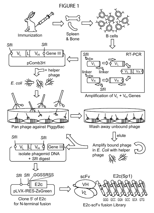
[0024] Figure 1 is a schematic representation depicting the method of phage
display to
generate scFy against piggyBac. Rabbits are immunized with PB transposase
protein
(PBase) for expanding relevant B cells. Variable regions from heavy and light
chain (VH
and VL) genes are amplified from cDNA by PCR to form fusion products
containing an
18 amino acid linker (L). Phagemid are produced, panned against PBase,
amplified in
E.coli, and repeated once or twice. The resulting phagemid DNA library is
cloned into
the pLVX-IRES-ZsGreen vector containing the E2c PZF with a linker sequence. An
E2c-scFy N-terminal fusion library is then produced in Lentivirus.
[0025] Figures 2A and 2B are a pair of schematic representations depicting
site-specific
complementation. Figure 2A shows that the E2c-SA-PAC141 cassette contains an
E2c
site (GGGGCCGGAGCCGCAGTG; SEQ ID NO: 1) located in the center of a 10.47 Kb
NgoMIV -Banafil fragment from the p53 intron 1, flanked by 2 inverted copies
of the
Adenovirus 2 splice acceptor (SA), the 141 amino acid C-terminal fragment of
the
puromycin acetyltransferase (PAC) gene followed by an 5V40 polyadenylation
(pA)
signal. Figure 2B shows that the BII-iPAC58-SD transposon contains a CpG-less
promoter consisting of the cytomegalovirus (CMV) enhancer and a human Efl a
promoter
that drives expression of the N-terminal fragment of the PAC gene. An Ad2
splice donor
provides a poly-A trap. Insulators (Insul.) from the chicken beta globin locus
HSIV
ensures stable expression. Insertions upstream of the E2c-SA-PAC141 cassette
result in
splicing and production of a functional PAC transcript.
6
CA 02952613 2016-12-15
WO 2015/195798
PCT/US2015/036226
Detailed Description
[0026] Definitions
[0027] The present disclosure may be understood more readily by reference to
the
following detailed description of preferred embodiments of the disclosure and
the
Examples included therein and to the Figures and their previous and following
description. Although any methods and materials similar or equivalent to those
described
herein can be used in the practice or testing of the present invention, the
preferred
methods, devices, and materials are now described. All references,
publications, patents,
patent applications, and commercial materials mentioned herein are
incorporated herein
by reference for the purpose of describing and disclosing the materials and/or
methodologies which are reported in the publications which might be used in
connection
with the invention. Nothing herein is to be construed as an admission that the
invention
is not entitled to antedate such disclosure by virtue of prior invention.
[0028] Before the present compounds, compositions, articles, devices, and/or
methods
are disclosed and described, it is to be understood that this invention is not
limited to
specific synthetic methods, specific recombinant biotechnology methods unless
otherwise
specified, or to particular reagents unless otherwise specified, as such may,
of course,
vary. It is also to be understood that the terminology used herein is for the
purpose of
describing particular embodiments only and is not intended to be limiting.
[0029] Throughout this application, reference is made to various proteins and
nucleic
acids. It is understood that any names used for proteins or nucleic acids are
art-
recognized names, such that the reference to the name constitutes a disclosure
of the
molecule itself
[0030] As used herein and in the appended claims, the singular forms "a,"
"and," and
"the" include plural referents unless the context clearly dictates otherwise.
Thus, for
example, reference to "a method" includes a plurality of such methods and
reference to
"a dose" includes reference to one or more doses and equivalents thereof known
to those
skilled in the art, and so forth.
[0031] The term "about" or "approximately" means within an acceptable error
range for
the particular value as determined by one of ordinary skill in the art, which
will depend in
7
CA 02952613 2016-12-15
WO 2015/195798
PCT/US2015/036226
part on how the value is measured or determined, e.g., the limitations of the
measurement
system. For example, "about" can mean within 1 or more than 1 standard
deviations per
the practice in the art. Alternatively, "about" can mean a range of up to 20%,
or up to
10%, or up to 5%, or up to 1% of a given value. Alternatively, particularly
with respect to
biological systems or processes, the term can mean within an order of
magnitude,
preferably within 5-fold, and more preferably within 2-fold, of a value. Where
particular
values are described in the application and claims, unless otherwise stated
the term
"about" meaning within an acceptable error range for the particular value
should be
assumed.
[0032] The term "antibody" is used in the broadest sense and specifically
covers single
monoclonal antibodies (including agonist and antagonist antibodies) and
antibody
compositions with polyepitopic specificity. It is also within the scope hereof
to use
natural or synthetic analogs, mutants, variants, alleles, homologs and
orthologs (herein
collectively referred to as "analogs") of the antibodies hereof as defined
herein. Thus,
according to one embodiment hereof, the term "antibody hereof" in its broadest
sense
also covers such analogs. Generally, in such analogs, one or more amino acid
residues
may have been replaced, deleted and/or added, compared to the antibodies
hereof as
defined herein.
[0033] "Antibody fragment", and all grammatical variants thereof, as used
herein are
defined as a portion of an intact antibody comprising the antigen binding site
or variable
region of the intact antibody, wherein the portion is free of the constant
heavy chain
domains (i.e. CH2, CH3, and CH4, depending on antibody isotype) of the Fc
region of
the intact antibody. Examples of antibody fragments include Fab, Fab', Fab'-
SH, F(ab')2,
and Fy fragments; diabodies; any antibody fragment that is a polypeptide
having a
primary structure consisting of one uninterrupted sequence of contiguous amino
acid
residues (referred to herein as a "single-chain antibody fragment" or "single
chain
polypeptide"), including without limitation (1) single-chain Fy (scFv)
molecules (2) single
chain polypeptides containing only one light chain variable domain, or a
fragment thereof
that contains the three CDRs of the light chain variable domain, without an
associated
heavy chain moiety and (3) single chain polypeptides containing only one heavy
chain
variable region, or a fragment thereof containing the three CDRs of the heavy
chain
variable region, without an associated light chain moiety; and multispecific
or multivalent
8
CA 02952613 2016-12-15
WO 2015/195798
PCT/US2015/036226
structures formed from antibody fragments. In an antibody fragment comprising
one or
more heavy chains, the heavy chain(s) can contain any constant domain sequence
(e.g.
CHI in the IgG isotype) found in a non-Fc region of an intact antibody, and/or
can
contain any hinge region sequence found in an intact antibody, and/or can
contain a
leucine zipper sequence fused to or situated in the hinge region sequence or
the constant
domain sequence of the heavy chain(s). The term further includes single domain
antibodies ("sdAB") which generally refers to an antibody fragment having a
single
monomeric variable antibody domain, (for example, from camelids). Such
antibody
fragment types will be readily understood by a person having ordinary skill in
the art.
[0034] "Binding" refers to a sequence-specific, non-covalent interaction
between
macromolecules (e.g., between a protein and a nucleic acid). Not all
components of a
binding interaction need be sequence-specific (e.g., contacts with phosphate
residues in a
DNA backbone), as long as the interaction as a whole is sequence-specific.
[0035] A "binding protein" is a protein that is able to bind non-covalently to
another
molecule. A binding protein can bind to, for example, a DNA molecule (a DNA-
binding
protein), an RNA molecule (an RNA-binding protein) and/or a protein molecule
(a
protein-binding protein). In the case of a protein-binding protein, it can
bind to itself (to
form homodimers, homotrimers, etc.) and/or it can bind to one or more
molecules of a
different protein or proteins. A binding protein can have more than one type
of binding
activity. For example, zinc finger proteins have DNA-binding, RNA-binding and
protein-
binding activity.
[0036] As used herein, the term "comprising" is intended to mean that the
compositions
and methods include the recited elements, but do not exclude others.
"Consisting
essentially of" when used to define compositions and methods, shall mean
excluding
other elements of any essential significance to the combination when used for
the
intended purpose. Thus, a composition consisting essentially of the elements
as defined
herein would not exclude trace contaminants or inert carriers. "Consisting of
shall mean
excluding more than trace elements of other ingredients and substantial method
steps.
Embodiments defined by each of these transition terms are within the scope of
this
invention.
[0037] As used herein, the term "effector molecule" means a molecule, such as
a protein
or protein domain, oftentimes an enzymatic protein, capable of exerting a
localized effect
9
CA 02952613 2016-12-15
WO 2015/195798
PCT/US2015/036226
in a cell. . The effector molecule may take a variety of different forms,
including
selectively binding to a protein or to DNA, for example, to regulate a
biological activity.
Effector molecules may have a wide variety of different activities, including,
but not
limited to nuclease activity, increasing or decreasing enzyme activity,
increasing or
decreasing gene expression, or affecting cell signalling. Other examples of
effector
molecules will be readily appreciated by one having ordinary skill in the art.
[0038] As used herein, the term "epitope tag", or otherwise "affinity tag",
refers to a short
amino acid sequence or peptide enabling a specific interaction with a protein
or a ligand.
[0039] As used herein, "epitope" refers to an antigenic determinant of a
polypeptide. An
epitope could comprise three amino acids in a spatial conformation, which is
unique to
the epitope. Generally, an epitope consists of at least 4, 5, 6, or 7 such
amino acids, and
more usually, consists of at least 8, 9, or 10 such amino acids. Methods of
determining
the spatial conformation of amino acids are known in the art, and include, for
example, x-
ray crystallography and two-dimensional nuclear magnetic resonance.
[0040] As used herein, "expression" refers to the process by which
polynucleotides are
transcribed into mRNA and/or the process by which the transcribed mRNA is
subsequently being translated into peptides, polypeptides, or proteins. If the
polynucleotide is derived from genomic DNA, expression may include splicing of
the
mRNA in an eukaryotic cell.
[0041] "Gene expression" refers to the conversion of the information,
contained in a
gene, into a gene product. A gene product can be the direct transcriptional
product of a
gene (e.g., mRNA, tRNA, rRNA, antisense RNA, ribozyme, shRNA, micro RNA,
structural RNA or any other type of RNA) or a protein produced by translation
of a
mRNA. Gene products also include RNAs which are modified, by processes such as
capping, polyadenylation, methylation, and editing, and proteins modified by,
for
example, methylation, acetylation, phosphorylation, ubiquitination, ADP-
ribosylation,
myristilation, and glycosylation.
[0042] "Modulation" or "regulation" of gene expression refers to a change in
the activity
of a gene. Modulation of expression can include, but is not limited to, gene
activation and
gene repression.
[0043] As used herein, the term "operatively linked" or its equivalents (e.g.,
"linked
operatively") means two or more molecules are positioned with respect to each
other
CA 02952613 2016-12-15
WO 2015/195798
PCT/US2015/036226
such that they are capable of interacting to affect a function attributable to
one or both
molecules or a combination thereof
[0044] The term "scFv" refers to a single-chain variable fragment. scFy is a
fusion
protein of the variable regions of the heavy (VH) and light chains (VL) of
immunoglobulins, connected with a linker peptide. The linker peptide may be
from about
to 40 amino acids or from about 10 to 30 amino acids or about 5, 10, 15, 20,
25, 30, 35,
or 40 amino acids in length. Single-chain variable fragments lack the constant
Fc region
found in complete antibody molecules, and, thus, the common binding sites
(e.g., Protein
G) used to purify antibodies. The term further includes a scFy that is an
intrabody, an
antibody that is stable in the cytoplasm of the cell, and which may bind to an
intracellular
protein.
[0045] As used herein, the term "single domain antibody" means an antibody
fragment
having a single monomeric variable antibody domain which is able to bind
selectively to
a specific antigen. A single-domain antibody generally is a peptide chain of
about 110
amino acids long, comprising one variable domain (VH) of a heavy-chain
antibody, or of
a common IgG, which generally have similar affinity to antigens as whole
antibodies, but
are more heat-resistant and stable towards detergents and high concentrations
of urea.
Examples are those derived from camelid or fish antibodies. Alternatively,
single-
domain antibodies can be made from common murine or human IgG with four
chains.
[0046] The terms "specifically bind" and "specific binding" as used herein
refer to the
ability of an antibody, an antibody fragment or a nanobody to preferentially
bind to a
particular antigen that is present in a homogeneous mixture of different
antigens. In
certain embodiments, a specific binding interaction will discriminate between
desirable
and undesirable antigens in a sample, in some embodiments more than about ten-
to 100-
fold or more (e.g., more than about 1000- or 10,000-fold). "Specificity"
refers to the
ability of an immunoglobulin or an immunoglobulin fragment, such as a
nanobody, to
bind preferentially to one antigenic target versus a different antigenic
target and does not
necessarily imply high affinity.
[0047] A "target site" or "target sequence" is a nucleic acid sequence that
defines a
portion of a nucleic acid to which a binding molecule will bind, provided
sufficient
conditions for binding exist.
11
CA 02952613 2016-12-15
WO 2015/195798
PCT/US2015/036226
[0048] Additional advantages of the invention will be set forth in part in the
description
which follows, and in part will be obvious from the description, or may be
learned by
practice of the invention. The advantages of the invention will be realized
and attained by
means of the elements and combinations particularly pointed out in the
appended claims.
It is to be understood that both the foregoing general description and the
following
detailed description are exemplary and explanatory only and are not
restrictive of the
invention, as claimed.
[0049] Disclosed herein are compositions and methods for addressing one or
more of the
aforementioned problems in the art. In one aspect, non-covalently linked
components
and methods of making and using non-covalently linked components, are
disclosed. The
various components may take a variety of different forms as described herein.
For
example, non-covalently linked (i.e., operatively linked) proteins may be used
to allow
temporary interactions that avoid one or more problems in the art. The ability
of non-
covalently linked components, such as proteins, to associate and dissociate
enables a
functional association only or primarily under circumstances where such
association is
needed for the desired activity. The linkage may be of duration sufficient to
allow the
desired effect.
[0050] In one aspect, a method for directing proteins to a specific locus in a
genome of
an organism is disclosed. The method may comprise the steps of providing a DNA
localization component and providing an effector molecule, wherein the DNA
localization component and the effector molecule are capable of operatively
linking via a
non-covalent linkage.
[0051] DNA Localization Component
[0052] In one aspect, the DNA localization component may be capable of binding
a
specific DNA sequence. The DNA localization component may be selected from,
for
example, a DNA-binding oligonucleotide, a DNA-binding protein, a DNA binding
protein complex, and combinations thereof Other suitable DNA binding
components
will be recognized by one of ordinary skill in the art.
[0053] In one aspect, the DNA localization component may comprise an
oligonucleotide
directed to a specific locus or loci in the genome. The oligonucleotide may be
selected
from DNA, RNA, DNA/RNA hybrids, and combinations thereof
12
CA 02952613 2016-12-15
WO 2015/195798
PCT/US2015/036226
[0054] In one aspect, the DNA localization component may comprise a nucleotide
binding protein or protein complex that binds an oligonucleotide when bound to
a target
DNA. The protein or protein complex may be capable of recognizing a feature
selected
from RNA-DNA heteroduplexes, R-loops, or combinations thereof In one aspect,
the
DNA localization component may comprise a protein or protein complex capable
of
recognizing an R-loop selected from Cas9, Cascade complex, RecA, RNase H, RNA
polymerase, DNA polymerase, or a combination thereof
[0055] In one aspect, the DNA localization component may comprise an
engineered
protein capable of binding to target DNA. In this aspect, the DNA localization
component may comprise a protein capable of binding a DNA sequence selected
from
meganuclease, zinc finger array, transcription activator-like (TAL) array, and
combinations thereof
[0056] In other aspects, the DNA localization component may comprise a protein
that
contains a naturally occurring DNA binding domain. The DNA localization
component
may comprise, for example, a protein comprising a naturally occurring DNA
binding
domain is selected from a bZIP domain, a Helix-loop-helix, a Helix-turn-helix,
a HMG-
box, a Leucine zipper, a Zinc finger, or a combination thereof
[0057] Effector Molecule
[0058] In one aspect, the method comprises providing an effector molecule.
[0059] In one aspect, the effector molecule may be selected from a
transcription factor
(activator or repressor), chromatin remodeling factor, exonuclease,
endonuclease,
transposase, methytransferase, demethylase, acetyltransferase, deacetylase,
kinase,
phosphatase, integrase, recombinase, ligase, topoisomerase, gyrase, helicase,
fluorophore,
and combinations thereof
[0060] In one aspect, the effector molecule may comprise a nuclease. The
nuclease may
be any nuclease readily appreciated by one of skill in the art. Suitable
nucleases include,
for example, a restriction endonuclease, homing endonuclease, 51 Nuclease,
mung bean
nuclease, pancreatic DNase I, micrococcal nuclease, yeast HO endonuclease, or
a
combination thereof In one aspect, the effector molecule may comprise a Type
IIS
restriction endonuclease. For example, in some aspects, the effector molecule
may
comprise an endonuclease selected from AciI, Mn1I, AlwI, BbvI, BccI, BceAI,
BsmAI,
BsmFI, BspCNI, BsrI, BtsCI, HgaI, HphI, HpyAV, MbolI, My1I, PleI, SfaNI, AcuI,
13
CA 02952613 2016-12-15
WO 2015/195798
PCT/US2015/036226
BciVI, BfuAI, BmgBI, BmrI, BpmI, BpuEI, BsaI, BseRI, BsgI, BsmI, BspMI, BsrBI,
BsrBI, BsrDI, BtgZI, BtsI, Earl, EciI, MmeI, NmeAIII, BbyCI, Bpul0I, BspQI,
SapI,
BaeI, BsaXI, CspCI, Fold, BfiI, MboII, Acc36I and C1o051. In other aspects,
the effector
molecule may comprise a PB transposase (PBase).
[0061] In one aspect, the effector molecule may be an endonuclease. In certain
embodiments, the effector molecule may be Fold. In certain embodiments, the
effector
molecule may be BfiI. In certain embodiments, the effector molecule may be
BmrI. In
certain embodiments, the effector molecule may be Clo051.
[0062] Linkage
[0063] In one aspect, the method may comprise a non-covalent linkage between
the DNA
localization component and the effector molecule. The non-covalent linkage may
comprise an antibody, an antibody fragment, an antibody mimetic, or a scaffold
protein.
[0064] Antibodies and fragments thereof, include, but are not limited to,
single-chain
variable fragment (scFy), single domain antibodies (sdAB), monobodies, and
nanobodies.
For example, the non-covalent linkage may comprise, a single-chain variable
fragment
(scFy) or a single domain antibody (sdAB) coyalently attached to one or more
effector
molecules, and which is capable of a non-covalent association to the DNA
localization
component. In a further aspect, the non-covalent linkage may comprise a single-
chain
variable fragment (scFy) coyalently attached to the DNA localization component
and
which non-coyalently binds directly to the effector component. In a further
aspect, the
non-covalent linkage may comprise a single-chain variable fragment (scFy)
coyalently
attached to either the effector molecule or the DNA localization component.
The scFV
may then non-coyalently bind to an epitope tag coyalently attached to the
opposite
component (i.e., to the DNA localization component or the effector molecule).
[0065] The non-covalent linkage may comprise, for example, an antibody
mimetic.
As used herein, the term "antibody mimetic" is intended to describe an organic
compound that specifically binds a target sequence and has a structure
distinct from a
naturally-occurring antibody. Antibody mimetics may comprise a protein, a
nucleic
acid, or a small molecule. The target sequence to which an antibody mimetic of
the
disclosure specifically binds may be an antigen. Antibody mimetics may provide
superior properties over antibodies including, but not limited to, superior
solubility,
tissue penetration, stability towards heat and enzymes (e.g. resistance to
enzymatic
14
CA 02952613 2016-12-15
WO 2015/195798
PCT/US2015/036226
degradation), and lower production costs. Exemplary antibody mimetics include,
but
are not limited to, an affibody, an afflilin, an affimer, an affitin, an
alphabody, an
anticalin, and avimer (also known as avidity multimer), a DARPin (Designed
Ankyrin
Repeat Protein), a Fynomer, a Kunitz domain peptide, and a monobody.
[0066] Affibody molecules of the disclosure comprise a protein scaffold
comprising
or consisting of one or more alpha helix without any disulfide bridges.
Preferably,
affibody molecules of the disclosure comprise or consist of three alpha
helices. For
example, an affibody molecule of the disclosure may comprise an immunoglobulin
binding domain. An affibody molecule of the disclosure may comprise the Z
domain
of protein A.
[0067] Affilin molecules of the disclosure comprise a protein scaffold
produced by
modification of exposed amino acids of, for example, either gamma-B crystallin
or
ubiquitin. Affilin molecules functionally mimic an antibody's affinity to
antigen, but
do not structurally mimic an antibody. In any protein scaffold used to make an
affilin,
those amino acids that are accessible to solvent or possible binding partners
in a
properly-folded protein molecule are considered exposed amino acids. Any one
or
more of these exposed amino acids may be modified to specifically bind to a
target
sequence or antigen.
[0068] Affimer molecules of the disclosure comprise a protein scaffold
comprising a
highly stable protein engineered to display peptide loops that provide a high
affinity
binding site for a specific target sequence. Exemplary affimer molecules of
the
disclosure comprise a protein scaffold based upon a cystatin protein or
tertiary
structure thereof Exemplary affimer molecules of the disclosure may share a
common tertiary structure of comprising an alpha-helix lying on top of an anti-
parallel
beta-sheet.
[0069] Affitin molecules of the disclosure comprise an artificial protein
scaffold, the
structure of which may be derived, for example, from a DNA binding protein
(e.g. the
DNA binding protein Sac7d). Affitins of the disclosure selectively bind a
target
sequence, which may be the entirety or part of an antigen. Exemplary affitins
of the
disclosure are manufactured by randomizing one or more amino acid sequences on
the
binding surface of a DNA binding protein and subjecting the resultant protein
to
ribosome display and selection. Target sequences of affitins of the disclosure
may be
CA 02952613 2016-12-15
WO 2015/195798
PCT/US2015/036226
found, for example, in the genome or on the surface of a peptide, protein,
virus, or
bacteria. In certain embodiments of the disclosure, an affitin molecule may be
used as
a specific inhibitor of an enzyme. Affitin molecules of the disclosure may
include
heat-resistant proteins or derivatives thereof
[0070] Alphabody molecules of the disclosure may also be referred to as Cell-
Penetrating Alphabodies (CPAB). Alphabody molecules of the disclosure comprise
small proteins (typically of less than 10 IcDa) that bind to a variety of
target sequences
(including antigens). Alphabody molecules are capable of reaching and binding
to
intracellular target sequences. Structurally, alphabody molecules of the
disclosure
comprise an artificial sequence forming single chain alpha helix (similar to
naturally
occurring coiled-coil structures). Alphabody molecules of the disclosure may
comprise a protein scaffold comprising one or more amino acids that are
modified to
specifically bind target proteins. Regardless of the binding specificity of
the
molecule, alphabody molecules of the disclosure maintain correct folding and
thermostability.
[0071] Anticalin molecules of the disclosure comprise artificial proteins that
bind to
target sequences or sites in either proteins or small molecules. Anticalin
molecules of
the disclosure may comprise an artificial protein derived from a human
lipocalin.
Anticalin molecules of the disclosure may be used in place of, for example,
monoclonal antibodies or fragments thereof Anticalin molecules may demonstrate
superior tissue penetration and thermostability than monoclonal antibodies or
fragments thereof Exemplary anticalin molecules of the disclosure may comprise
about 180 amino acids, having a mass of approximately 20 IcDa. Structurally,
anticalin molecules of the disclosure comprise a barrel structure comprising
antiparallel beta-strands pairwise connected by loops and an attached alpha
helix. In
preferred embodiments, anticalin molecules of the disclosure comprise a barrel
structure comprising eight antiparallel beta-strands pairwise connected by
loops and
an attached alpha helix.
[0072] Avimer molecules of the disclosure comprise an artificial protein that
specifically binds to a target sequence (which may also be an antigen).
Avimers of the
disclosure may recognize multiple binding sites within the same target or
within
distinct targets. When an avimer of the disclosure recognize more than one
target, the
16
CA 02952613 2016-12-15
WO 2015/195798
PCT/US2015/036226
avimer mimics function of a bi-specific antibody. The artificial protein
avimer may
comprise two or more peptide sequences of approximately 30-35 amino acids
each.
These peptides may be connected via one or more linker peptides. Amino acid
sequences of one or more of the peptides of the avimer may be derived from an
A
domain of a membrane receptor. Avimers have a rigid structure that may
optionally
comprise disulfide bonds and/or calcium. Avimers of the disclosure may
demonstrate
greater heat stability compared to an antibody.
[0073] DARPins (Designed Ankyrin Repeat Proteins) of the disclosure comprise
genetically-engineered, recombinant, or chimeric proteins having high
specificity and
high affinity for a target sequence. In certain embodiments, DARPins of the
disclosure are derived from ankyrin proteins and, optionally, comprise at
least three
repeat motifs (also referred to as repetitive structural units) of the ankyrin
protein.
Ankyrin proteins mediate high-affinity protein-protein interactions. DARPins
of the
disclosure comprise a large target interaction surface.
[0074] Fynomers of the disclosure comprise small binding proteins (about 7
kDa)
derived from the human Fyn SH3 domain and engineered to bind to target
sequences
and molecules with equal affinity and equal specificity as an antibody.
[0075] Kunitz domain peptides of the disclosure comprise a protein scaffold
comprising a Kunitz domain. Kunitz domains comprise an active site for
inhibiting
protease activity. Structurally, Kunitz domains of the disclosure comprise a
disulfide-
rich alpha+beta fold. This structure is exemplified by the bovine pancreatic
trypsin
inhibitor. Kunitz domain peptides recognize specific protein structures and
serve as
competitive protease inhibitors. Kunitz domains of the disclosure may comprise
Ecallantide (derived from a human lipoprotein-associated coagulation inhibitor
(LAC)).
[0076] Monobodies of the disclosure are small proteins (comprising about 94
amino
acids and having a mass of about 10 kDa) comparable in size to a single chain
antibody. These genetically engineered proteins specifically bind target
sequences
including antigens. Monobodies of the disclosure may specifically target one
or more
distinct proteins or target sequences. In preferred embodiments, monobodies of
the
disclosure comprise a protein scaffold mimicking the structure of human
fibronectin,
and more preferably, mimicking the structure of the tenth extracellular type
III
17
CA 02952613 2016-12-15
WO 2015/195798
PCT/US2015/036226
domain of fibronectin. The tenth extracellular type III domain of fibronectin,
as well
as a monobody mimetic thereof, contains seven beta sheets forming a barrel and
three
exposed loops on each side corresponding to the three complementarity
determining
regions (CDRs) of an antibody. In contrast to the structure of the variable
domain of
an antibody, a monobody lacks any binding site for metal ions as well as a
central
disulfide bond. Multispecific monobodies may be optimized by modifying the
loops
BC and FG. Monobodies of the disclosure may comprise an adnectin.
[0077] The non-covalent linkage may comprise, for example, a scaffold protein.
Scaffold proteins of the disclosure include, for example, antibody mimetics of
the
disclosure. Scaffold proteins of the disclosure further include, for example,
small
modular immunopharmaceutical (SMIP) molecules, a domain antibody, and a
nanobody.
[0078] SMIP molecules of the disclosure are artificial proteins comprising one
or
more sequences or portions of an immunoglobulin (antibody) that are
monospecific
for a target sequence or antigen. SMIPs of the disclosure may substitute for
the use of
a monoclonal antibody. Structurally, SMIPs are single chain proteins
comprising a
binding region, a hinge region (i.e. a connector), and an effector domain. The
binding
region of a SMIP may comprise a modified single-chain variable fragment
(scFv).
SMIPs may be produced from genetically-modified cells as dimers.
[0079] Domain antibodies of the disclosure comprise a single monomeric
variable
antibody domain (i.e. either heavy or light variable domain). Domain
antibodies of the
disclosure demonstrate the same antigen specificity as a whole and intact
antibody.
Domain antibodies of the disclosure may be manufactured, at least in part, by
immunization of dromedaries, camels, llamas, alpacas or sharks with the
desired
antigen and subsequent isolation of the mRNA coding for heavy-chain
antibodies.
[0080] Nanobodies of the disclosure comprise a VHH single domain antibody.
Nanobodies of the disclosure may comprise single domain antibodies of the
disclosure.
[0081] In one aspect, the non-covalent linkage may comprise a protein binding
domain
covalently attached to either the effector molecule or the DNA localization
component
and which is capable of a non-covalent interaction with the opposite
component. Non-
18
CA 02952613 2016-12-15
WO 2015/195798
PCT/US2015/036226
limiting examples of protein binding domains include, for example, SH2, SH3,
PTB,
LIM, SAM, PDZ, FERM, CH, Pleckstin, WW, WSxWS, and the E3 ligase domain.
[0082] In one aspect, the non-covalent linkage may comprise a protein
covalently
attached to either the effector molecule or the DNA localization component
that is
capable of binding to a protein covalently attached to the opposite component.
Non-
limiting examples include any two proteins that interact non-covalently. Such
proteins
are readily identified via the Database of Interacting Proteins (DIP), STRING,
BioGRID,
MIPS, or the like.
[0083] In one aspect, the non-covalent linkage may comprise a small molecule
covalently
attached either to an effector molecule or a DNA localization component, and
is capable
of forming a non-covalent bond to a protein or other small molecule covalently
attached
to the opposite component. One such example would include biotin attached to
an
oligonucleotide and avidin covalently linked to an effector molecule.
[0084] The above described methods and compositions may be used, for example,
in
situations in which a particular protein may have several functions.
Transposase proteins,
for example, must perform several steps to achieve the desired function,
including
transposon recognition, cleavage of DNA to excise a transposon, movement of a
transposon sequence to a new genomic location, recognition of a new target
site, and
cleavage of DNA to integrate the transposon at a new locus. In certain
aspects, it may be
desirable to direct a transposase to integrate a transposon at a particular
site in the
genome. In these aspects, this could be carried out by, for example, adding a
heterologous protein with site-specific DNA binding activity. However, the
heterologous
protein with site-specific DNA binding activity would only be required during
the target
site recognition step, and the presence of this protein at earlier stages in
the process
described above may be detrimental to the other steps. As such, in this
aspect, a
temporary association of the heterologous protein with site-specific DNA
binding activity
with the transposase would allow the transposase to be directed to the genomic
site of
interest while allowing for the other steps of the process to be carried out
with limited
interference of the protein due to the non-covalent binding.
[0085] As another example, it may be desirable to have an enzymatic protein,
such as a
nuclease, methylase, deacetylase, etc. to temporarily interact with a specific
DNA
binding domain so that its activity occurs at a specific location in the
genome. For
19
CA 02952613 2016-12-15
WO 2015/195798
PCT/US2015/036226
example, it may be desired to cause a Fold restriction nuclease to temporarily
interact
with a Cas9 protein that is catalytically inactive for DNA cleavage.
[0086] In one aspect, the linker comprises a non-covalent linkage between the
DNA
binding element and the effector. For example, in one aspect, phage display
(PhD) may
be used to produce single-chain variable fragment (scFv) antibodies or single
domain
antibodies (sdAbs) against a particular target. PhD may be used to identify a
scFy
antibody against an effector, for example piggyBBac (PB) transposase that
provides a
linkage. A large diversity in scFy affinity may be obtained by limiting the
stringency of
the affinity selection process. In one aspect, the linkage may be between PB
transposase
(PBase) and a modular DNA binding domain such as a polydactyl zinc finger, a
TAL
array, or a dCas9 protein (with associated guide RNA). In some aspects, a scFy
antibody
with a faster off-rate may provide permissive "breathing" of the complex. In
other
aspects, conformation and/or flexibility of an effector and DNA binding
element may be
critical. Non-covalent linkages may provide conformational pliability to the
disclosed
gene editing compositions. Alternatively, slower off-rates (and a higher Kd)
of an scFy
that binds particular epitopes of an effector may provide an optimal stability
and
conformation of the gene editing complex that would not otherwise be
obtainable through
traditional peptide linkage. A near-exhaustive search among scFy antibodies
allows one
to select from among a large diversity of possible conformations of a gene
editing
complex. A PhD strategy creates such diversity through the generation of
unique
monovalent scFvs against multiple unique epitopes.
[0087] Furthermore, a non-covalent linkage method, such as that achieved
through the
use of a scFy antibody, may employ an unmodified and native effector (e.g.,
PB). This
provides a reversible associate between the effector and the DNA binding
element, which
may circumvent any permanent interference with the activity of an effector
that may
occur when it is subjected to covalent linkage. Certain non-covalent
associations could
introduce steric hindrances that compromise the effector reaction. As several
activities
may be involved (site recognition, strand cleavage, transposon binding and
integration) it
is likely that each separate step may be differentially affected by a
particular steric
hindrance. For example, if transposase association with the DNA transposon
(during
transposon mobilization from one genomic site to another) has a very slow off-
rate, then
it would be detrimental to have a very high affinity association between a DNA
binding
CA 02952613 2016-12-15
WO 2015/195798
PCT/US2015/036226
element-scFy and the PBase that disrupts this association. However, if the DNA
binding
element-scFy protein binds with a lower, but significant affinity, it could be
temporarily
displaced during transposon mobilization. It is possible that such an early
step could
involve temporary dissociation of DNA binding factor-scFy with the PBase, with
subsequent reassembly of the complex at later steps to create a fully
functional and DNA
binding factor-enabled site-specific transposase.
[0088] Examples
[0089] Phage display is used to identify an scFy antibody against PBase that
provides an
optimal linkage. A large diversity in scFy affinity can be obtained by
limiting the
stringency of the affinity selection process. This diversity may represent a
key advantage
of a PhD approach for identifying a successful linkage between PBase and a
modular
DNA binding protein (DBP). In some instances, an scFy antibody with a faster
off-rate
may provide permissive "breathing" of the DBP-PBase complex. Previous studies
show
that, when E2c is fused to the SB transposase, the efficiency is almost
doubled if there is
complete mismatch in one half-site of the 18 bp recognition sequence [36].
Even though
a "flexible" 15-residue linker is used (-GGS5-) for SB-E2c fusion, it has been
hypothesized that the flexibility provided by E2c half-site recognition
enables efficient
site-specific transposition. This may also be true for fusions with PBase.
Regardless, the
conformation and/or flexibility of the DBP and fused transposase appear
critical, and a
non-covalent linkage may provide this conformational pliability.
Alternatively, slower
off-rates (and a higher Kd) of an scFy that binds particular epitopes of PBase
may
provide an optimal stability and conformation of the DBP-PBase complex¨a
conformation otherwise not attainable through simple peptide linkage. A near-
exhaustive
search among scFy antibodies allows one to select from among a large diversity
of
possible conformations of DBP-PBase complexes. A PhD strategy may create such
diversity through the generation of unique monovalent scFvs against multiple
unique
epitopes.
[0090] A non-covalent linkage method, such as that achieved through the use of
an scFy
antibody employs an unmodified and native PBase protein. This is believed to
provide a
reversible association between PBase and the DBP, which may circumvent any
permanent interference with PBase catalytic activity that may occur when it is
subjected
to covalent linkage. Certain non-covalent associations could introduce steric
hindrances
21
CA 02952613 2016-12-15
WO 2015/195798
PCT/US2015/036226
that compromise the transposase reaction, but since the transposition reaction
involves
separate catalytic steps (site recognition, strand cleavage, transposon
binding, and
integration), it is likely that each separate step would be differentially
affected by a
particular steric hindrance. For example, if transposase association with the
DNA
transposon (during transposon mobilization from one genomic site to another)
has a very
slow off-rate, then it would be clearly detrimental to have a very high
affinity association
between E2c-scFy and the PBase that disrupts this association. However, if the
E2c-scFy
protein binds with a lower, but significant affinity, it could be temporarily
displaced
during transposon mobilization. It is possible that such an early step could
involve
temporary dissociation of E2c-scFy with the PBase, with subsequent reassembly
of the
complex at later steps to create a fully functional and E2c-enabled site-
specific
transposase.
[0091] Immunization for producing anti-PB antibodies.
[0092] An antibody library is produced from immunized rabbits using methods
well
known in the art. Rabbits provide two key advantages: 1) their size provides
large
amounts of tissue (spleen and bone marrow) and ample serum for titering and 2)
fewer
PCR primers are needed for antibody gene amplification since fewer gene
segments are
rearranged during B-cell development in rabbits. Six New Zealand White rabbits
are
immunized each with 200 pg of recombinant PBase protein plus adjuvant, and
serum is
collected six weeks after immunization for determining antibody titers. Titers
are
determined by ELISAs on immobilized recombinant PBase protein and the animals
with
the highest titers (at least 1:1000) are sacrificed for isolating the spleen
and bone marrow.
If rabbits do not produce sufficient titers, a naïve library from embryonic
rabbit tissue is
used. This provides an unbiased collection of un-rearranged heavy and light
chain genes.
Total RNA will be extracted from tissues using Trizol (Invitrogen), and cDNA
synthesis
is performed with the iScript cDNA synthesis kit (BioRad).
[0093] Generating scFv gene fusions.
[0094] To isolate expressed variable regions of heavy and light chain genes
from rabbit,
several primers are used. Eight primers are used for kappa and lambda light
chain
amplification and five primers are used for heavy chain gene amplification.
Primers also
contain the coding sequence for an 18 amino acid linker sequence
(SSGGGGSGGGGGGSSRSS) (SEQ ID NO: 2), which links the variable regions of the
22
CA 02952613 2016-12-15
WO 2015/195798
PCT/US2015/036226
heavy and light chains (VH and VL). This longer linker sequence provides
better stability
of monomeric forms of scFy fragments. The PCR products of the VH and VL genes
overlap in this linker region and can then be assembled by overlap-extension
(OLE) PCR
(FIG 1). PCR products are then digested with Sfil, ligated with Sfil-digested
pComb3H,
and DNA will then be size-selected by gel electrophoresis. This plasmid
enables
phagemid display of an scFy fused to the pill coat protein. About 5 molecules
of pill
phage coat protein is present on each phage particle. The pComb3H plasmid
expresses
the scFv-p111 fusion at a level such that about one or two molecules are
integrated with
wild-type pill (which is provided by helper phage). Since up to 1012 phage
particles can
be generated in a single preparation, a very large number of scFvs can thus be
screened.
In PhD the scFy coding sequence is always linked to the phage particle
displaying the
protein, so subsequent DNA sub-cloning is conveniently achieved.
[0095] Producing and screening the phage library.
[0096] Ligated plasmid DNA (50 to 100 ng) is electroporated into ER2538 E.
coli (New
England Biolabs). E. coli will then be recovered by shaking for 1 hour at 37 C
in 5 mLs
of SOC. Phage is produced with the VCSM13 helper phage, which has a defective
origin
of replication. Phage particles will be precipitated with PEG-8000 and then
isolated by
further centrifugation. This phage prep is the primary library, and will be
affinity selected
by "panning." Double recognition panning is performed in which the phage
elution is re-
incubated with the immobilized antigen, washed, and eluted again. This helps
eliminate
non-specific phage. To test each round of selection, phage pools are assayed
by ELISAs
for affinity to the PBase antigen. PBase or BSA are coated to 96-well plates,
incubated
with phage, and then incubated with a horseradish peroxidase (HRP) conjugated
anti-
M13 antibody, which recognizes the M13 phage coat protein. An increasing ELISA
titer
indicates successful affinity selection of each phage pool.
[0097] Transferring the scFy library into a lentiviral vector, and expansion
in E.
coli.
[0098] Phagemid DNA is isolated from bacteria after the 2nd (R2) and 3rd (R3)
rounds
of panning by infecting E. coli with each phage pool, selecting with
carbenicillin,
followed by standard plasmid preparation. Plasmid DNA is digested with Sfil to
liberate
the scFy coding sequence, and ligated upstream of the E2c coding sequence
within the
pLVX-IRES-ZsGreen1 (Clontech) vector. The E2c coding sequence also has a short
23
CA 02952613 2016-12-15
WO 2015/195798
PCT/US2015/036226
linker sequence (GGSSRSS) (SEQ ID NO: 3) and creates a fusion of the scFy
library to
the N-terminal portion of E2c. The two ensuing plasmid libraries (R2 and R3)
will then
be prepared as in Aim 2, for production of two lentivirus libraries.
[0099] Lentivirus library production.
[00100] For production of lentivirus particles, the Lenti-X HT Packaging
System
(Clontech) is used, which produces viral titers as high as 5x10⁸ infectious
units per mL.
Virus is produced according to the manufacturer's specifications. Viral
supernatants are
titered on HepG2 and Huh7 cells, followed by FACS fluorescence produced by the
ZsGreen1 reporter to count transduced cells.
[00101] HEK293 cells are also be infected with viral supernatants to
determine the
ability of scFv-E2c fusion proteins to bind the E2c target sequence, to ensure
there is no
loss of binding affinity of the E2c domain. Nuclear lysates will be prepared
from
transduced cells and used for electrophoretic mobility shift assays (EMSAs)
with labeled
DNA containing the E2c target sequence. The affinity is compared to nuclear
lysates
from cells transduced for expression of an unmodified E2c. Since this
procedure will
screen a mixture of E2c fusion proteins (from the library), the affinities
will represent an
average for the library¨some fusions may have compromised affinities, while
others
may not. The objective is to ensure that the overall average affinity is not
dramatically
reduced (by 50%), which would otherwise indicate that the fusion process
itself has
adversely affected E2c affinity. Affinities will be calculated as understood
in the art.
[00102] Screening strategy.
[00103] To screen for effective site-specific integration, a puromycin
acetyltransferase (PAC) complementation strategy in which site-specific
integration
yields a functional PAC transgene is used. Similar strategies have been used
to detect
chromosomal translocations. This selection is achieved by separating the PAC
coding
region into two separate sequences that can be linked through splicing. The
first
component (E2c-SA-PAC141) consists of a 3' fragment of the PAC open reading
frame
(ORF) that encodes the C-terminal 141 amino acids immediately downstream of
the
splice acceptor (SA) from the intronl/exon2 boundary of the Adenovirus II
(Ad2) late
major transcript (FIG 2). The 5V40 late polyadenylation signal, providing a
transcript
termination signal, is located just 3' of the PAC ORF fragment (PAC141). An
E2c
recognition sequence is inserted within a 10.47 Kb fragment of the p53 intron,
which
24
CA 02952613 2016-12-15
WO 2015/195798
PCT/US2015/036226
lacks splice donor and acceptor sequences, which is flanked by two identical
but inverted
copies of the SA-PC141 fragment (FIG 2). This arrangement enables splicing,
complementation, and PAC expression following a site-specific insertion in
either
orientation. A large intron fragment is used because it is likely deficient
for cryptic splice
sites. Stable HepG2 and Huh7 cell lines containing the E2c-SA-PAC141 cassette
are
generated by co-transfecting cells with a hygromycin resistance cassette
driven by the
thymidine kinase promoter (TK-Hygro), followed by selection with hygromycin.
Stable
lines are treated with puro to ensure sensitivity to this antibiotic.
[00104] The 5' portion of the PAC gene (PAC58), containing the remaining
coding
sequence of the PAC ORF, is mobilized via PB into a region between the PAC141
sequences by site-specific integration near the E2c sequence (FIG 2). This PB
transposon
(B11-iPAC58-SD) has a cDNA containing the 5' coding region of the PAC gene
instead
of the EGFP-IRES sequence. The expression of a functional PAC transcript is
facilitated
by splicing between the Ad2 acceptor and donor sites. These potent splice
sites do not
undergo alternative splicing.
[00105] Selection for site-specific integration.
[00106] Sixty million cells from HepG2 or Huh7 stable cell lines are
transduced in
x 10 cm dishes at a multiplicity of infection (M01) of one with each of the
ten
retroviral libraries (eight linker libraries and two scFv libraries). HepG2
and Huh7 can
be transduced with lentivirus (LV) vectors at an efficiency of about 30% to
70% and 70%
to 95%, respectively. LV infection of these hepatocyte cell lines does not
appear to
compromise the hepatocyte phenotype. Cells receiving the scFv lentiviral
library are co-
transduced with lentivirus generated with the PBase coding sequences. Twenty-
four
hours later, the medium is changed and cells incubated for an additional 24
hours, then
transfected with a plasmid containing the BII-iPAC58-SD transposon. The
transposon is
supplied as transfected DNA because in all likelihood, for actual gene
therapy, DNA will
be delivered either by liposomes, nanoparticles, or adenovirus in the form of
a DNA
episome. Cells are then incubated for an additional 72 hours followed by
selection with
puro for 48 hours. This will select for site-specific integration of the BII-
iPAC58-SD
transposon upstream of the E2c-SA-PAC141 cassette. Multiple cell lines (2 to
3) for
HepG2 and Huh7 will be screened to account for different genomic contexts of
the E2c-
SA-PAC141 transgene. Transduction with PBase alone, via lentivirus, (without
E2c or
CA 02952613 2016-12-15
WO 2015/195798
PCT/US2015/036226
E2c-scFv) will represent the negative control, and will likely yield few puro-
resistant
cells, if any.
[00107] Identifying and testing the effective linkage(s).
[00108] Genomic DNA from puro-resistant cells is isolated for PCR using
primers
that flank the library cloning sites. These cells will generally contain a
site-specific
BIIiPAC58-SD transposon integration upstream of the E2c-SA-PAC141 cassette.
Among
these cells, many will contain proviral DNA with specific linker or scFy
antibody
sequences that will have facilitated PB-mediated site-specific integration. To
further
enrich for the most efficient linkage strategy (whether covalent or non-
covalent), a
secondary library is generated from PCR-amplified linker or scFy sequences by
digesting
PCR products with Sfil and repeating the library production and selection, as
above.
After screening three library generations (GO, Gl, G2) for each of ten (8
peptide linker
libraries, and 2 scFy libraries) retroviral preps, the final PCR-amplified
proviral
insertions are cloned and sequenced to identify the linkers and/or scFy
antibodies that
yield efficient site-specific targeting. Testing is performed by assessing the
efficiency of
integration, as measured by the number of puroresistant cells obtained through
transient
transfection and PAC complementation. Linkage strategies identified are cloned
and
tested individually in the PAC complementation assay in HepG2 and Huh7 stable
cell
lines containing the E2c-SA-PAC141 cassette. PB-linker-E2c and E2c-scFy clones
are
inserted into pcDNA3.1 (Invitrogen) for transient transfection and expression.
For E2c-
scFy clones, the PBase in pcDNA3.1 is co-transfected with the pcDNA3.1-scFy
plasmid
to provide the PBase protein target. The BII-iPAC58-SD transposon is also
supplied as
plasmid DNA via co-transfection. Approximately 0.5-1 x 10⁶ cells are
transfected in 6-
well plates along with an equal amount of a CMV-GFP plasmid to determine
transfection
efficiency, as assessed at 48 hours by fluorescent microscopy. After 72 hours
cells will be
split into 10 cm dishes and puro added, and resistant colonies will be counted
after one
week of selection.
[00109] Determining Off-Target Frequency.
[00110] To determine off-target frequency, non-specific insertions are
quantified
by Southern blot and QPCR. Ten puro-resistant colonies generated by the best
variants
(PB-linker-E2c or E2c-scFy clone, as identified above) are expanded and gDNA
extracted. Southern blots are performed by digesting gDNA with BsrGI (for the
ERBB2
26
CA 02952613 2016-12-15
WO 2015/195798
PCT/US2015/036226
locus) or PacI + Sad I (for the E2c-SA-PAC141 cassette). DNA is probed with
unique
sequences within the p53 locus or ERBB2 gene. Fragments lacking an insertion
are
approximately 5 Kb for either p53 or ERBB2, while BII-iPAC58-SD integrations
will
add 2.4 Kb for each transposon insertion. Bands on the Southern blot, up to
about 15 Kb,
may be discernible, representing four transposon insertions for either the p53
intron or
ERBB2 5'UTR target sites. This method cannot distinguish between the
endogenous p53
genomic fragment and the p53 fragment in the E2c-SA-PAC141 cassette. However,
the
endogenous p53 intron region represents 11625000 of the haploid genome; the
remaining
99.99984% of the genome is still measurable in the assay. Total copy number is
determined by QPCR of gDNA with primers specific to the BIT-iPAC58-SD
transposon,
along with copy number standards. This allows one to calculate the ratio of
site-specific
insertions to total insertions. A variant that yields 10-25% site-specific
insertions is
identified. Successful enrichment for efficient site-specific integration will
be evident by
an increasing number of resistant cells in each round of puro selection. It is
possible that
some puro-resistant cells could result from non-specific integration followed
by
chromosomal translocations. This would be rare and site-specific.
[00111] The practice of the present invention will employ, unless otherwise
indicated, conventional techniques of molecular biology (including recombinant
techniques), microbiology, cell biology and biochemistry, which are within the
skill of
the art.
[00112] All percentages and ratios are calculated by weight unless
otherwise
indicated.
[00113] All percentages and ratios are calculated based on the total
composition
unless otherwise indicated.
[00114] It should be understood that every maximum numerical limitation
given
throughout this specification includes every lower numerical limitation, as if
such lower
numerical limitations were expressly written herein. Every minimum numerical
limitation
given throughout this specification will include every higher numerical
limitation, as if
such higher numerical limitations were expressly written herein. Every
numerical range
given throughout this specification will include every narrower numerical
range that falls
within such broader numerical range, as if such narrower numerical ranges were
all
expressly written herein.
27
CA 02952613 2016-12-15
WO 2015/195798
PCT/US2015/036226
[00115] The dimensions and values disclosed herein are not to be understood
as
being strictly limited to the exact numerical values recited. Instead, unless
otherwise
specified, each such dimension is intended to mean both the recited value and
a
functionally equivalent range surrounding that value. For example, a dimension
disclosed
as "20 mm" is intended to mean "about 20 mm."
[00116] Every document cited herein, including any cross referenced or
related
patent or application, is hereby incorporated herein by reference in its
entirety unless
expressly excluded or otherwise limited. The citation of any document is not
an
admission that it is prior art with respect to any invention disclosed or
claimed herein or
that it alone, or in any combination with any other reference or references,
teaches,
suggests or discloses any such invention. Further, to the extent that any
meaning or
definition of a term in this document conflicts with any meaning or definition
of the same
term in a document incorporated by reference, the meaning or definition
assigned to that
term in this document shall govern.
[00117] While particular embodiments of the present invention have been
illustrated and described, it would be obvious to those skilled in the art
that various other
changes and modifications can be made without departing from the spirit and
scope of the
invention. It is therefore intended to cover in the appended claims all such
changes and
modifications that are within the scope of this invention.
28