Note: Descriptions are shown in the official language in which they were submitted.
CA 02952697 2016-12-15
WO 2015/195621
PCT/US2015/035964
COMPOSITIONS AND METHODS FOR THE EXPRESSION OF CRISPR GUIDE
RNAS USING THE H1 PROMOTER
CROSS-REFERENCE TO RELATED APPLICATIONS
This application claims the benefit of U.S. Provisional Application No.
62/012,802, filed June 16, 2014, which is incorporated herein by reference in
its
entirety.
INCORPORATION-BY-REFERENCE OF MATERIAL SUBMITTED
ELECTRONICALLY
This application contains a sequence listing. It has been submitted
electronically via EFS-Web as an ASCII text file entitled "111232-
00401_ST25.txt".
The sequence listing is 14,827 bytes in size, and was created on June 2, 2015.
It is
hereby incorporated herein by reference in its entirety.
BACKGROUND
Clustered Regularly Interspaced Short Palindromic Repeats (CRISPR)
together with cas (CRISPR-associated) genes comprise an adaptive immune system
that provides acquired resistance against invading foreign nucleic acids in
bacteria
and archaea (Barrangou et al. (2007) Science 315:1709-12). CRISPR consists of
arrays of short conserved repeat sequences interspaced by unique variable DNA
sequences of similar size called spacers, which often originate from phage or
plasmid
DNA (Barrangou et al. (2007) Science 315:1709-12; Bolotin et al. (2005)
Microbiology 151 :2551-61; Mojica et al. (2005) J. Mol. Evol. 60:174-82). The
CRISPR-Cas system functions by acquiring short pieces of foreign DNA (spacers)
which are inserted into the CRISPR region and provide immunity against
subsequent
exposures to phages and plasmids that carry matching sequences (Barrangou et
al.
(2007) Science 315:1709-12; Brouns et al. (2008) Science 321:960-64). It is
this
CRISPR-Cas interference/immunity that enables crRNA-mediated silencing of
foreign nucleic acids (Horvath & Barrangou (2010) Science 327:167-70; Deveau
et al.
(2010) Annu. Rev. Microbiol. 64:475-93; Marraffini & Sontheimer (2010) Nat.
Rev.
Genet. 11:181-90; Bhaya et al. (2011) Annu. Rev. Genet. 45:273-97; Wiedenheft
et al.
(2012) Nature 482:331-338).
1
CA 02952697 2016-12-15
WO 2015/195621
PCT/US2015/035964
Use of CRISPR constructs that rely upon the nuclease activity of the Cas9
protein (Makarova et al. (2011) Nat. Rev. Microbiol. 9:467-77) coupled with a
synthetic guide RNA (gRNA) has recently revolutionized genomic-engineering,
allowing for unprecedented manipulation of DNA sequences. CRISPR/Cas9
constructs are simple and fast to synthesize and can be multiplexed. However,
despite
the relative ease of their synthesis, CRISPRs have technological restrictions
related to
their access to targetable genome space, which is a function of both the
properties of
Cas9 itself and the synthesis of its gRNA.
Cleavage by the CRISPR system requires complementary base pairing of the
gRNA to a 20-nucleotide DNA sequence and the requisite protospacer-adjacent
motif
(PAM), a short nucleotide motif found 3' to the target site (Jinek et al.
(2012) Science
337: 816-821). One can, theoretically, target any unique N20-PAM sequence in
the
genome using CRISPR technology. The DNA binding specificity of the PAM
sequence, which varies depending upon the species of origin of the specific
Cas9
employed, provides one constraint. Currently, the least restrictive and most
commonly used Cas9 protein is from S. pyogenes, which recognizes the sequence
NGG, and thus, any unique 21-nucleotide sequence in the genome followed by two
guanosine nucleotides (N20NGG) can be targeted. Expansion of the available
targeting space imposed by the protein component is limited to the discovery
and use
of novel Cas9 proteins with altered PAM requirements (Cong et al. (2013)
Science
339: 819-823; Hou et al. (2013) Proc. Natl. Acad. Sci. U.S.A. 110(39):15644-
9), or
pending the generation of novel Cas9 variants via mutagenesis or directed
evolution.
The second technological constraint of the CRISPR system arises from gRNA
expression initiating at a 5' guanosine nucleotide. Use of the type III class
of RNA
polymerase III promoters has been particularly amenable for gRNA expression
because these short non-coding transcripts have well-defined ends, and all the
necessary elements for transcription, with the exclusion of the 1+ nucleotide,
are
contained in the upstream promoter region. However, since the commonly used U6
promoter requires a guanosine nucleotide to initiate transcription, use of the
U6
promoter has further constrained genomic targeting sites to GN19NGG (Mali et
al.
(2013) Science 339:823-826; Ding et al. (2013) Cell Stem Cell 12:393-394).
Alternative approaches, such as in vitro transcription by T7, T3, or SP6
promoters,
would also require initiating guanosine nucleotide(s) (Adhya et al. (1981)
Proc. Natl.
2
CA 02952697 2016-12-15
WO 2015/195621
PCT/US2015/035964
Acad. Sci. U.S.A. 78:147-151; Melton etal. (1984) Nucleic Acids Res. 12:7035-
7056;
Pleiss etal. (1998) RNA 4:1313-1317).
SUMMARY
The practice of the present invention will typically employ, unless otherwise
indicated, conventional techniques of cell biology, cell culture, molecular
biology,
transgenic biology, microbiology, recombinant nucleic acid (e.g., DNA)
technology,
immunology, and RNA interference (RNAi) which are within the skill of the art.
Non-
limiting descriptions of certain of these techniques are found in the
following
publications: Ausubel, F., et al., (eds.), Current Protocols in Molecular
Biology,
Current Protocols in Immunology, Current Protocols in Protein Science, and
Current
Protocols in Cell Biology, all John Wiley & Sons, N.Y., edition as of December
2008;
Sambrook, Russell, and Sambrook, Molecular Cloning. A Laboratory Manual, 3rd
ed.,
Cold Spring Harbor Laboratory Press, Cold Spring Harbor, 2001; Harlow, E. and
Lane, D., Antibodies¨A Laboratory Manual, Cold Spring Harbor Laboratory Press,
Cold Spring Harbor, 1988; Freshney, R. I., "Culture of Animal Cells, A Manual
of
Basic Technique", 5th ed., John Wiley & Sons, Hoboken, N.J., 2005. Non-
limiting
information regarding therapeutic agents and human diseases is found in
Goodman
and Gilman's The Pharmacological Basis of Therapeutics, 11th Ed., McGraw Hill,
2005, Katzung, B. (ed.) Basic and Clinical Pharmacology, McGraw-Hill/Appleton
&
Lange 10th ed. (2006) or 11th edition (July 2009). Non-limiting information
regarding
genes and genetic disorders is found in McKusick, V. A.: Mendelian Inheritance
in
Man. A Catalog of Human Genes and Genetic Disorders. Baltimore: Johns Hopkins
University Press, 1998 (12th edition) or the more recent online database:
Online
Mendelian Inheritance in Man, OMIMTm. McKusick-Nathans Institute of Genetic
Medicine, Johns Hopkins University (Baltimore, Md.) and National Center for
Biotechnology Information, National Library of Medicine (Bethesda, Md.), as of
May
1, 2010, available on the World Wide Web: http://www.ncbi.nlm.nih.gov/omim/
and
in Online Mendelian Inheritance in Animals (OMIA), a database of genes,
inherited
disorders and traits in animal species (other than human and mouse), available
on the
World Wide Web: http://omia.angis.org.au/contact.shtml. All patents, patent
applications, and other publications (e.g., scientific articles, books,
websites, and
databases) mentioned herein are incorporated by reference in their entirety.
In case of
3
CA 02952697 2016-12-15
WO 2015/195621
PCT/US2015/035964
a conflict between the specification and any of the incorporated references,
the
specification (including any amendments thereof, which may be based on an
incorporated reference), shall control. Standard art-accepted meanings of
terms are
used herein unless indicated otherwise. Standard abbreviations for various
terms are
used herein.
The presently disclosed subject matter provides compositions and methods for
the expression of CRISPR guide RNAs using the H1 promoter. The presently
disclosed subject matter provides a non-naturally occurring CRISPR-Cas system
comprising one or more vectors comprising: a) an H1 promoter operably linked
to at
least one nucleotide sequence encoding a CRISPR-Cas system guide RNA (gRNA),
wherein the gRNA hybridizes with a target sequence of a DNA molecule in a
cell, and
wherein the DNA molecule encodes one or more gene products expressed in the
cell;
and b) a regulatory element operable in a cell operably linked to a nucleotide
sequence encoding a Cas9 protein, wherein components (a) and (b) are located
on the
same or different vectors of the system, wherein the gRNA targets and
hybridizes
with the target sequence and the Cas9 protein cleaves the DNA molecule to
alter
expression of the one or more gene products. In some aspects, the target
sequence
comprises the nucleotide sequence AN19NGG, GN19NGG, CN19NGG, or TN19NGG.
In some aspects, the cell is a eukaryotic cell. In some aspects, the
eukaryotic cell is a
mammalian or human cell. In some aspects, the eukaryotic cell is a retinal
photoreceptor cell. In some aspects, the Cas9 protein is codon optimized for
expression in the cell. In some aspects, the Cas9 protein is a Type-II Cas9
protein. In
some aspects, the expression of the one or more gene products is decreased. In
some
aspects, the one or more gene products are rhodopsin. In some aspects, the
system is
packaged into a single adeno-associated virus (AAV) particle.
In some aspects, the presently disclosed subject matter provides a non-
naturally occurring CRISPR-Cas system comprising one or more vectors
comprising:
a) an H1 promoter operably linked to at least one nucleotide sequence encoding
a
CRISPR-Cas system guide RNA (gRNA), wherein the gRNA hybridizes with a target
sequence of a DNA molecule in a eukaryotic cell, and wherein the DNA molecule
encodes one or more gene products expressed in the eukaryotic cell; and b) a
regulatory element operable in a eukaryotic cell operably linked to a
nucleotide
sequence encoding a Type-II Cas9 protein, wherein components (a) and (b) are
4
CA 02952697 2016-12-15
WO 2015/195621
PCT/US2015/035964
located on the same or different vectors of the system, whereby the gRNA
targets and
hybridizes with the target sequence and the Cas9 protein cleaves the DNA
molecule,
and whereby expression of the one or more gene products is altered. In another
aspect, the target sequence comprises the nucleotide sequence AN19NGG,
GN19NGG,
CNi9NGG, or TN19NGG. In another aspect, the Cas9 protein is codon optimized
for
expression in the cell. In yet another aspect, the Cas9 protein is codon
optimized for
expression in the eukaryotic cell. In a further aspect, the eukaryotic cell is
a
mammalian or human cell. In another aspect, the expression of the one or more
gene
products is decreased.
The presently disclosed subject matter also provides a method of altering
expression of one or more gene products in a cell, wherein the cell comprises
a DNA
molecule encoding the one or more gene products, the method comprising
introducing
into the cell a non-naturally occurring CRISPR-Cas system comprising one or
more
vectors comprising: a) an H1 promoter operably linked to at least one
nucleotide
sequence encoding a CRISPR-Cas system guide RNA (gRNA), wherein the gRNA
hybridizes with a target sequence of the DNA molecule; and b) a regulatory
element
operable in the cell operably linked to a nucleotide sequence encoding a Cas9
protein,
wherein components (a) and (b) are located on the same or different vectors of
the
system, wherein the gRNA targets and hybridizes with the target sequence and
the
Cas9 protein cleaves the DNA molecule to alter expression of the one or more
gene
products. In some aspects, the target sequence comprises the nucleotide
sequence
AN19NGG, GN19NGG, CN19NGG, or TN19NGG. In some aspects, the cell is a
eukaryotic cell. In some aspects, the eukaryotic cell is a mammalian or human
cell.
In some aspects, the eukaryotic cell is a retinal photoreceptor cell. In some
aspects,
the Cas9 protein is codon optimized for expression in the cell. In some
aspects, the
Cas9 protein is a Type-II Cas9 protein. In some aspects, the expression of the
one or
more gene products is decreased. In some aspects, the one or more gene
products are
rhodopsin. In some aspects, the system is packaged into a single adeno-
associated
virus (AAV) particle.
In some aspects, the presently disclosed subject matter provides a method of
altering expression of one or more gene products in a eukaryotic cell, wherein
the cell
comprises a DNA molecule encoding the one or more gene products, the method
comprising introducing into the cell a non-naturally occurring CRISPR-Cas
system
5
CA 02952697 2016-12-15
WO 2015/195621
PCT/US2015/035964
comprising one or more vectors comprising: a) an H1 promoter operably linked
to at
least one nucleotide sequence encoding a CRISPR-Cas system guide RNA (gRNA),
wherein the gRNA hybridizes with a target sequence of the DNA molecule; and b)
a
regulatory element operable in the eukaryotic cell operably linked to a
nucleotide
sequence encoding a Type-II Cas9 protein, wherein components (a) and (b) are
located on the same or different vectors of the system, whereby the gRNA
targets and
hybridizes with the target sequence and the Cas9 protein cleaves the DNA
molecule,
and whereby expression of the one or more gene products is altered. In another
aspect, the target sequence comprises the nucleotide sequence AN19NGG,
GN19NGG,
CN19NGG, or TN19NGG. In another aspect, the Cas9 protein is codon optimized
for
expression in the cell. In yet another aspect, the Cas9 protein is codon
optimized for
expression in the eukaryotic cell. In a further aspect, the eukaryotic cell is
a
mammalian or human cell. In another aspect, the expression of the one or more
gene
products is decreased.
The presently disclosed subject matter also provides a non-naturally occurring
CRISPR-Cas system comprising a vector comprising a bidirectional H1 promoter,
wherein the bidirectional H1 promoter comprises: a) control elements that
provide for
transcription in one direction of at least one nucleotide sequence encoding a
CRISPR-
Cas system guide RNA (gRNA), wherein the gRNA hybridizes with a target
sequence
of a DNA molecule in a cell, and wherein the DNA molecule encodes one or more
gene products expressed in the cell; and b) control elements that provide for
transcription in the opposite direction of a nucleotide sequence encoding a
Cas9
protein, wherein the gRNA targets and hybridizes with the target sequence and
the
Cas9 protein cleaves the DNA molecule to alter expression of the one or more
gene
products. In some aspects, the target sequence comprises the nucleotide
sequence
AN19NGG, GN19NGG, CN19NGG, or TN19NGG. In some aspects, the cell is a
eukaryotic cell. In some aspects, the eukaryotic cell is a mammalian or human
cell.
In some aspects, the eukaryotic cell is a retinal photoreceptor cell. In some
aspects,
the Cas9 protein is codon optimized for expression in the cell. In some
aspects, the
Cas9 protein is a Type-II Cas9 protein. In some aspects, the expression of the
one or
more gene products is decreased. In some aspects, the one or more gene
products are
rhodopsin. In some aspects, the system is packaged into a single adeno-
associated
virus (AAV) particle.
6
CA 02952697 2016-12-15
WO 2015/195621
PCT/US2015/035964
In some embodiments, the presently disclosed subject matter provides a non-
naturally occurring CRISPR-Cas system comprising a vector comprising a
bidirectional H1 promoter, wherein the bidirectional H1 promoter comprises: a)
control elements that provide for transcription in one direction of at least
one
nucleotide sequence encoding a CRISPR-Cas system guide RNA (gRNA), wherein
the gRNA hybridizes with a target sequence of a DNA molecule in a eukaryotic
cell,
and wherein the DNA molecule encodes one or more gene products expressed in
the
eukaryotic cell; and b) control elements that provide for transcription in the
opposite direction of a nucleotide sequence encoding a Type-II Cas9 protein,
whereby
the gRNA targets and hybridizes with the target sequence and the Cas9 protein
cleaves the DNA molecule, and whereby expression of the one or more gene
products
is altered. In another aspect, the target sequence comprises the nucleotide
sequence
AN19NGG, GN19NGG, CN19NGG, or TN19NGG. In yet another aspect, the Cas9
protein is codon optimized for expression in the eukaryotic cell. In a further
aspect,
the eukaryotic cell is a mammalian or human cell. In another aspect, the
expression
of the one or more gene products is decreased.
The presently disclosed subject matter also provides a method of altering
expression of one or more gene products in a cell, wherein the cell comprises
a DNA
molecule encoding the one or more gene products, the method comprising
introducing
into the cell a non-naturally occurring CRISPR-Cas system comprising a vector
comprising a bidirectional H1 promoter, wherein the bidirectional H1 promoter
comprises: a) control elements that provide for transcription in one direction
of at
least one nucleotide sequence encoding a CRISPR-Cas system guide RNA (gRNA),
wherein the gRNA hybridizes with a target sequence of the DNA molecule; and b)
control elements that provide for transcription in the opposite direction of a
nucleotide
sequence encoding a Cas9 protein, wherein the gRNA targets and hybridizes with
the
target sequence and the Cas9 protein cleaves the DNA molecule to alter
expression of
the one or more gene products in the cell. In some aspects, the target
sequence
comprises the nucleotide sequence AN19NGG, GN19NGG, CN19NGG, or TN19NGG.
In some aspects, the cell is a eukaryotic cell. In some aspects, the
eukaryotic cell is a
mammalian or human cell. In some aspects, the eukaryotic cell is a retinal
photoreceptor cell. In some aspects, the Cas9 protein is codon optimized for
expression in the cell. In some aspects, the Cas9 protein is a Type-II Cas9
protein. In
7
CA 02952697 2016-12-15
WO 2015/195621
PCT/US2015/035964
some aspects, the expression of the one or more gene products is decreased. In
some
aspects, the one or more gene products are rhodopsin. In some aspects, the
system is
packaged into a single adeno-associated virus (AAV) particle.
The presently disclosed subject matter also provides a method of altering
expression of one or more gene products in a eukaryotic cell, wherein the cell
comprises a DNA molecule encoding the one or more gene products, the method
comprising introducing into the cell a non-naturally occurring CRISPR-Cas
system
comprising a vector comprising a bidirectional H1 promoter, wherein the
bidirectional
H1 promoter comprises: a) control elements that provide for transcription in
one
direction of at least one nucleotide sequence encoding a CRISPR-Cas system
guide
RNA (gRNA), wherein the gRNA hybridizes with a target sequence of the DNA
molecule; and b) control elements that provide for transcription in the
opposite
direction of a nucleotide sequence encoding a Type-II Cas9 protein, whereby
the
gRNA targets and hybridizes with the target sequence and the Cas9 protein
cleaves
the DNA molecule, and whereby expression of the one or more gene products is
altered. In another aspect, the target sequence comprises the nucleotide
sequence
AN19NGG, GN19NGG, CN19NGG, or TN19NGG. In yet another aspect, the Cas9
protein is codon optimized for expression in the eukaryotic cell. In a further
aspect,
the eukaryotic cell is a mammalian or human cell. In another aspect, the
expression
of the one or more gene products is decreased.
The presently disclosed subject matter also provides an aptamer-regulated
ribozyme, comprising: a) a cis-acting hammerhead ribozyme comprising a
catalytic
core and helix I, helix II, and helix III duplex regions extending therefrom,
wherein
the helix II duplex region and the helix III duplex region each comprise a
loop region
opposite the catalytic core, and wherein the helix II duplex region comprises
an
aptamer that binds to a ligand; b) a nucleotide sequence encoding a CRISPR-Cas
system guide RNA (gRNA), wherein the gRNA hybridizes with a target sequence of
a
DNA molecule in a eukaryotic cell, and wherein the DNA molecule encodes one or
more gene products expressed in the eukaryotic cell, wherein the nucleotide
sequence
comprises a 5' end and a 3' end, and wherein the 5' end of the nucleotide
sequence is
directly coupled to the helix III duplex region; wherein binding of the ligand
to the
aptamer produces a conformational change in the ribozyme such that the
ribozyme
undergoes self-cleavage between the 5' end of the nucleotide sequence and the
helix
8
CA 02952697 2016-12-15
WO 2015/195621
PCT/US2015/035964
III duplex region, whereby the gRNA is produced. An expression construct is
also
provided comprising: (i) a coding sequence which, when transcribed to RNA,
produces the aptamer-regulated ribozyme; and (ii) one or more transcriptional
regulatory sequences that regulate transcription of the RNA in a eukaryotic
cell. A
eukaryotic cell comprising the expression construct is also provided. A method
of
altering expression of one or more gene products in a eukaryotic cell is also
provided,
wherein the cell comprises a DNA molecule encoding the one or more gene
products,
the method comprising introducing the expression construct into the cell and
contacting the cell with the ligand in an amount that alters the activity of
the
ribozyme, particularly wherein the cell is in mammalian or human subject. In
one
aspect, the ligand is theophylline.
The presently disclosed subject matter also provides a method for treating an
ocular neurodegenerative disease in a subject in need thereof, the method
comprising:
(a) providing a non-naturally occurring CRISPR-Cas system comprising one or
more
vectors comprising: i) an H1 promoter operably linked to at least one
nucleotide
sequence encoding a CRISPR-Cas system guide RNA (gRNA), wherein the gRNA
hybridizes with a target sequence of a DNA molecule in a cell of the subject,
and
wherein the DNA molecule encodes one or more gene products expressed in the
cell;
and ii) a regulatory element operable in a cell operably linked to a
nucleotide
sequence encoding a Cas9 protein, wherein components (i) and (ii) are located
on the
same or different vectors of the system, wherein the gRNA targets and
hybridizes
with the target sequence and the Cas9 protein cleaves the DNA molecule to
alter
expression of the one or more gene products; and (b) administering to the
subject an
effective amount of the system. In some aspects, the dysfunction and/or death
of
retinal photoreceptor cells has been observed in the subject. In some aspects,
the
ocular neurodegenerative disease is selected from the group consisting of
glaucoma,
retinal degeneration, and age-related macular degeneration. In some aspects,
the
ocular neurodegenerative disease is retinitis pigmentosa (RP). In some
aspects, the
cell is a retinal photoreceptor cell. In some aspects, one or more gene
products are
rhodopsin. In some aspects, the H1 promoter is bidirectional. In some aspects,
the
system is packaged into a single adeno-associated virus (AAV) particle before
administering to the subject. In some aspects, administering to the subject
occurs by
subretinal injection. In some aspects, the subject is a human. In some
aspects, the
9
CA 02952697 2016-12-15
WO 2015/195621
PCT/US2015/035964
Cas9 protein is a Type-II Cas9 protein. In some aspects, the target sequence
comprises the nucleotide sequence AN19NGG, GN19NGG, CN19NGG, or TN19NGG.
In some aspects, the Cas9 protein is codon optimized for expression in the
cell. In
some aspects, the presently disclosed method further comprises administering
the
expression construct and the ligand in an amount that alters the activity of
the
ribozyme. In some aspects, the ligand is theophylline.
Certain aspects of the presently disclosed subject matter having been stated
hereinabove, which are addressed in whole or in part by the presently
disclosed
subject matter, other aspects will become evident as the description proceeds
when
taken in connection with the accompanying Examples and Figures as best
described
herein below.
BRIEF DESCRIPTION OF THE FIGURES
Having thus described the presently disclosed subject matter in general terms,
reference will now be made to the accompanying Figures, which are not
necessarily
drawn to scale, and wherein:
FIG. 1A, FIG. 1B, FIG. 1C, and FIG. 1D show an evaluation of the ability to
direct CRISPR targeting via gRNA synthesis from the H1 promoter. A schematic
illustration depicting the gRNA expression constructs is shown in FIG. 1A.
Above,
the U6 promoter only expresses gRNAs with a +1 guanosine nucleotide; below,
the
H1 promoter can drive expression of gRNAs initiating at either purine
(adenosine or
guanosine) nucleotide. Below, a cartoon depiction of the Cas9 protein with
gRNA
targeting genomic sequence AN19NGG is shown (sequence shown is SEQ ID NO:
30). The location of the +1 A is indicated. A schematic overview of the eGFP
targeted disruption assay is shown in FIG. 1B. eGFP fluorescence is disrupted
by
CRISPR targeting followed by error-prone NHEJ-mediated repair resulting in
frameshift mutations that disrupt the coding sequence, resulting in loss of
fluorescence. FIG. 1C shows microscope images demonstrating successful CRISPR
targeting by U6 or H1 promoter expressed gRNAs. H7 ES cells were stained and
colonies were visualized to show nuclei (left, magenta), eGFP fluorescence
(middle,
green), and merged images (right) indicating areas of GFP fluorescence
mosaicism in
the colony. To the right is shown the quantification of eGFP fluorescence loss
by
CA 02952697 2016-12-15
WO 2015/195621
PCT/US2015/035964
flow cytometry for the respective constructs. Below is a higher magnification
of an
H7 colony targeted by an H1 expressed gRNA showing expression mosaicism. Scale
bar, 50 M. Surveyor assay-based quantitation of the frequency of NHEJ is
shown in
FIG. 1D. Bioanalyzer gel image depicting control (first lane), U6 expressed
gRNA
(second lane), H1 expressed gRNA (third lane), and marker (fourth lane). The %
indel (as calculated by the fraction of uncut (u) to cut (c) bands) is
indicated below;
FIG. 2 shows Surveyor analysis and quantification of NHEJ in HEK-293 cells.
Shown above is an eGFP schematic with arrows indicating the targeting sites.
Target
sites on the plus strand are indicated pointing to the right, and minus strand
targets are
indicated pointing to the left; blue arrows indicate H1 promoter gRNAs and
orange
arrows indicate U6 promoter gRNAs. Shown below is the Bioanalyzer gel from the
Surveyor assay. The target site coordinates are listed above and the
calculated %
indel is indicated below;
FIG. 3A, FIG. 3B, and FIG. 3C show targeting and homologous
recombination at the AAVS1 locus. Surveyor analysis of three gRNAs expressed
by
the H1 promoter (AAVS1-la through -1-3a), three gRNAs expressed by the U6
promoter (AAVS-1-1 through -1-3), and a control nontargeting gRNA are shown in
FIG. 3A. FIG. 3B shows a schematic of AAVS-1 targeting donor vector (shown
above the AAVS1 Locus (labeled "AAVS1")) and cell imaging of an GFP-positive
H7 ES cell colony following electroporation with H1::AAVS1-3a gRNA and the
AAVS-1 targeting vector. Sanger sequencing of the targeting junction region
indicating correct integration by homologous recombination is shown in FIG. 3C
(sequence shown is SEQ ID NO:31);
FIG. 4A, FIG. 4B, FIG. 4C, and FIG. 4D show bioinformatics analysis of
GN19NGG and AN19NGG sites in the genome. A Circos plot depicting the frequency
of CRIPSR sites in the human genome is shown in FIG. 4A. The outside circle
depicts the human chromosome ideograms. Moving inwards, GN19NGG (orange),
AN19NGG (blue), and RN19NGG (purple) CRISPR sites frequency is indicated along
the chromosomes. Plotted inside the circle is the human exon density (black),
and
OMIM disease loci (blue). The frequency and distance between CRISPR sites in
the
genome is shown in FIG. 4B. Barplot of the frequency and distance of adjacent
GN19NGG (orange), AN19NGG (blue) sites in the genome is shown. The mean and
11
CA 02952697 2016-12-15
WO 2015/195621
PCT/US2015/035964
median values are inset within the plot including RN19NGG sites. FIG. 4C shows
barplot quantification of GN19NGG vs AN19NGG site frequency at human genes
(left)
or OMIM disease loci (right). FIG. 4D shows a barplot quantifying the GN19NGG
vs.
AN19NGG frequency in six genomes: human, cow, mouse, rat, chicken, and
zebrafish;
FIG. 5A, FIG. 5B, FIG. 5C, FIG. 5D, FIG. 5E, and FIG. 5F show
bioinformatic analysis of GN19NGG and AN19NGG sites in the genome. Three
panels
depicting the density of each gRNA sites in the human genome are shown:
GN19NGG
(FIG. 5A), AN19NGG (FIG. 5B), and RN19NGG (FIG. 5C). Within each plot, the
density of CRISPR sites is plotted along each chromosome. Overlaid in semi-
transparent (orange, blue, or purple) is the density curve calculated as a
smooth
Gaussian kernel. The dotted line indicates 35 bp; as a reference, on average,
TALEN
targeting sites are estimated to occur every 35 base pairs and ZFN sites occur
every
couple hundred base pairs (Sander et al. (2011) Nature Methods 8:67-69; Cermak
et
al. (2011) Nucleic Acids Res. 39(12):e82). A barplot of the cumulative mean
CRISPR targeting density per human chromosome is shown in FIG. 5D. GN19NGG
(orange), AN19NGG (blue), and RN19NGG (purple) indicate the respective CRISPR
sites. The dotted line indicates the 35 bp reference. FIG. 5E shows the
frequency and
distance between adjacent CRISPR sites in the genome. Barplot of the frequency
and
distance of adjacent GN19NGG (orange) and AN19NGG (blue) sites is in the
genome
is shown. The mean and median values are inset within the plot. SeqLogo of all
GN19NGG (top left), AN19NGG (top right), and RN19NGG (bottom) sites in the
human genome are shown in FIG. 5F;
FIG. 6A, FIG. 6B, FIG. 6C, FIG. 6D, FIG. 6E, and FIG. 6F show AT/GC
genome content and CRISPR site frequency: The percent AT (blue) or GC (orange)
is indicated for human, cow, mouse, rat, chicken, and zebrafish genomes (FIG.
6A).
The frequency of GN19NGG (orange) and AN19NGG (blue) sites normalized to
AT/GC content are indicated (FIG. 6B). CRISPR site frequency by strand for
GN19NGG (left), AN19NGG (middle), and RN19NGG (right) sites is shown in FIG.
6C. The plus strand (left column) is indicated by blue-green, and minus strand
(right
column) in purple-red. The GN19NGG (orange) and AN19NGG (blue) site frequency
in Drosophila, C. elegans, and S. cerevisiae are indicated in FIG. 6D. FIG. 6E
shows
the percent AT (blue) or GC (orange) content and FIG. 6F shows the normalized
frequency of CRISPR sites;
12
CA 02952697 2016-12-15
WO 2015/195621
PCT/US2015/035964
FIG. 7A, FIG. 7B, FIG. 7C, and FIG. 7D show CRISPR targeting of
AN19NGG at an endogenous gene (MERTK) in H7 ES cells. A schematic diagram of
the MERTK locus and various protein domains is shown in FIG. 7A. Target site
in
exon 2 is shown below in larger scale, indicating the CRISPR AN19NGG target
site
(sequence shown is SEQ ID NO: 32). Quantification of CRISPR targeting at exon2
by the Surveyor assay is shown in FIG. 7B. The CRISPR site in exon 2 is
depicted
above, with the various primers (arrows) used in the Surveyor assay; both Fl
:R1 and
F2:R2 span the target site, while the control PCR product, F3 :R3, is just
outside the
target site. The gel from the Surveyor assay is shown below with the three
control
products shown on the left, and targeting is shown on the right. Below the %
indel
frequency is indicated. FIG. 7C shows Sanger sequencing of mutant lines.
Clonal
lines were isolated and sequenced indicating that CRISPR targeting at the
AN19NGG
sites resulted in mutagenesis at this region. The aligned chromatograms show
the 6
unique mutations that were cloned (wt is SEQ ID NO: 33; 412 is SEQ ID NO:34;
Al
is SEQ ID NO:35; 42, +2 is SEQ ID NO:36; 46 is SEQ ID NO:37; 47 is SEQ ID
NO:38). FIG. 7D shows Western Blot analysis for Mertk expression in H7-derived
RPE cells. Lanes 1, 3, and 4 indicate knockout lines and lane 2 indicates
expression
from heterozygous line;
FIG. 8A, FIG. 8B, FIG. 8C, and FIG. 8D show an analysis of off-target hits
induced at on-target and off-target sites by U6 or H1 expressed gRNAs. qRT-PCR
analysis of the VEGFA Ti gRNA expression levels from titrating amounts of
either
the H1 promoter (blue) or U6 promoter (orange) is shown in FIG. 8A. On-target
and
off-target analysis of the VEGFA Ti is shown in FIG. 8B. Surveyor analysis is
indicated on the left and the target sequences on the right with mismatches
indicated
in red (Ti, SEQ ID NO:20; OT1-3, SEQ ID NO:21; OT1-4, SEQ ID NO:22; OT1-6,
SEQ ID NO:23; OT1-11, SEQ ID NO:24). FIG. 8C is the same as FIG. 8B with the
VEGFA T3 target (VEGFA T3, SEQ ID NO:25; 0T3-1, SEQ ID NO:26; 0T3-2, SEQ
ID NO:27; 0T3-4, SEQ ID NO:28; 0T3-18, SEQ ID NO:29). On-target to off-target
specificity of VEGFA Ti is shown in FIG. 8D. The ratio of the on-target
mutagenesis/off-target mutagenesis between the H1 promoter (blue) or U6
promoter
(orange) is shown. Values below the dotted line at 1.0 indicate greater off-
target
mutagenesis than on-target mutagenesis. For all parts, the on-target and off-
target
13
CA 02952697 2016-12-15
WO 2015/195621
PCT/US2015/035964
sites are labeled as in Fu et al. ((2013) Nat. Biotechnol. 31(9):822-6) and
Cho et al.
((2014) Genome Research 24:132-141);
FIG. 9A and FIG. 9B show the properties of U6 versus H1 promoters in
expressing gRNAs for CRISPR targeting. The top diagram in FIG. 9A shows the
endogenous human U6 promoter and transcriptional start site (SEQ ID NO: 39).
The
bottom diagram in FIG. 9A indicates the use of the U6 promoter to drive gRNAs
with
different +1 nucleotides. Because U6 requires a G to initiate (top left), the
panels that
start with A (top right), C (bottom left), or T (bottom right) will likely
initiate the first
downstream G leading to a truncated gRNA (U6:GN19NGG is SEQ ID NO:40;
U6:AN19NGG is SEQ ID NO:41; U6:CN19NGG is SEQ ID NO:42; U6:TN19NGG
is SEQ ID NO:43). The top diagram in FIG. 9B shows the endogenous human H1
promoter and transcriptional start site (SEQ ID NO: 44). The bottom diagram in
FIG.
9B indicates the use of the H1 promoter to drive gRNAs with different +1
nucleotides. H1 can initiate with a G (top left) or an A (top right) leading
to full-
length gRNAs. Also, H1 has been reported to allow for transcription initiating
at C
and T nucleotides, which would allow for full-length transcripts for any +1
nucleotide
downstream of the H1 promoter (H1:GN19NGG is SEQ ID NO: 45; Hl:AN19NGG
is SEQ ID NO: 46; Hl:CN19NGG is SEQ ID NO: 47; Hl:TN19NGG is SEQ ID NO:
48);
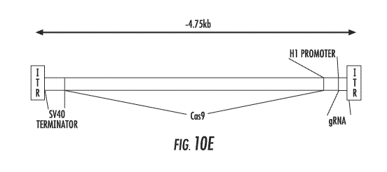
FIG. 10A, FIG. 10B, FIG. 10C, FIG. 10D, and FIG. 10E show use of the H1
promoter as a bidirectional promoter to simultaneously express the Cas9
protein and
guide RNA. The bidirectional H1 promoter is shown expressing Cas9 as a pol II
transcript towards the left (minus strand), and a guide RNA as a pol III
transcript
towards the right (plus strand) (FIG. 10A). The overall expression cassette is
approximately 4.4kb. FIG. 10B shows the construct used for testing the ability
to
direct CRISPR-mediated cleavage from a bidirectional H1 construct. The
bidirectional construct, using a gRNA targeting eGFP, was cloned into a
plasmid and
expressed in human stem cells expressing GFP. The loss of GFP is visually
detected
(middle panel, arrowheads) indicating the successful expression and targeting
of GFP
due to the expression construct (FIG. 10C). Successful CRISPR targeting is
also
shown through the Surveyor Assay with the presence of the two bands in lanes
2, and
3 (FIG. 10D). A bidirectional CRISPR construct using the H1 promoter to
generate a
compact targeting cassette of ¨4.75b, which is within the packaging range of
the
14
CA 02952697 2016-12-15
WO 2015/195621
PCT/US2015/035964
adeno-associated virus, is shown in FIG. 10E. The SV40 terminator is shown in
orange, and the construct is flanked by the inverted terminal repeat (ITR)
sequences
required for virus production;
FIG. 11A, FIG. 11B, and FIG. 11C shows a Hammerhead Ribozyme to
generate the 5' end of a guide RNA. Depiction of a 5' cis-hammerhead ribozyme
(SEQ ID NO: 49) and gRNA (SEQ ID NO: 50) is shown in FIG. 11A. The sequences
of the hammerhead ribozyme are indicated, and the nucleotides important for
catalysis
are indicated (critical in red, important in orange). The location of cleavage
is
indicated by the arrow. Upon ribozyme cleavage (lower), the resulting gRNA is
released, without constraint to any nucleotide at the newly formed 5'
position.
Constructs to express the hammerhead-gRNA are shown in FIG. 11B. A promoter,
generally a pol III promoter like U6, H1, or T7, can be used to express the 5'
cis-
hammerhead ribozyme, which after self-cleavage will release the gRNA.
Targeting
of two loci are shown with the Surveyor Assay (HH1 = SEQ ID NO: Si; HH2 = SEQ
ID NO: 52), with successful cleavage (arrows) by a 5' cis-hammerhead ribozyme
(FIG. 11C);
FIG. 12 shows a regulatable CRISPR construct, using aptazymes to process
gRNAs in the presence of specific aptamers. In particular, FIG. 12 depicts the
theophylline aptamer (orange) fused to helix II of the hammerhead ribozyme
forming
the theophylline aptazyme, which is 5' of the gRNA (blue). Binding of
theophylline
stabilizes helix II that then allows for hammerhead self-cleavage, and freeing
the
gRNA (SEQ ID NO:50). The gRNA, along with Cas9, is now able to target cleavage
by the CRISPR system. Hammerhead ribozyme, SEQ ID NO:55;
FIG. 13 shows genomic organization of the H1RNA and PARP-2 locus.
Shown above is a depiction of the PARP-2 gene (blue) transcribed toward the
right
and the H1RNA gene (orange) transcribed to the left, drawn to scale. Below is
an
enlarged region of the promoter region for both genes;
FIG. 14 shows eGFP reporter for H1 pol II activity. The human H1 promoter
sequence is orientated with pol II transcription of eGFP to the right. The
three
components to be optimized are indicated in italics;
FIG. 15 shows eGFP reporter expression. Top panels indicate endogenous H1
promoter, bottom panels indicate expression with Kozak sequence;
CA 02952697 2016-12-15
WO 2015/195621
PCT/US2015/035964
FIG. 16A and FIG. 16B show the bidirectional expression of Cas9 and gRNA.
A schematic diagram of the bidirectional targeting construct is shown in FIG.
16A.
Comparison of cleavage at two different loci using the standard two vector
delivery
(lanes 2 and 5) or delivery of single targeting plasmid (lanes 3 and 6) is
shown in FIG.
16B. % genomic modification, as determined by T7EI assay, is indicated below
each
lane;
FIG. 17 shows the rhodopsin locus from the hRho:GFP knockin mouse.
Above, the respective mouse and human sequences are indicated above the
schematic
of the rho promoter region to the end of the 3'UTR (drawn to scale). Below,
enlarged
region indicating the location of P23 and the gRNA, shown below (arrowhead);
FIG. 18A, FIG. 18B, and FIG. 18C show the specific targeting of the P23H
allele in vivo. FIG. 18A shows P23 targeting (WT(C57BL/6J, SEQ ID NO:56;
P23H(CCC¨*CAC), SEQ ID NO:57; WT(CAST/EiJ), SEQ ID NO:58). FIG. 18B
shows the sequencing of rhodopsin from two wildtype mouse strains; the SNP is
indicated by the arrow (C57BL/6J DNA sequence, SEQ ID NO:56; C57BL/6J protein
sequence, SEQ ID NO:59; CAST/EU-Pi+ DNA sequence, SEQ ID NO:58; CAST/EU-Pi+
protein sequence, SEQ ID NO:59). FIG. 18C shows the P23H breeding scheme: the
P23H homozygous mouse (black) is crossed with a WT Cast (white) and the
resulting
heterozygous pups (grey) will be treated by subretinal delivery of AAV5; and
FIG. 19 shows allele-specific targeting of the rhodopsin locus. Comparison of
cleavage of the C57BL/6J(P23H) allele vs a single base mismatch (Cast) is
shown. %
genomic modification determined by T7EI assay is indicated below.
The patent or application file contains at least one drawing executed in
color.
Copies of this patent or patent application publication with color drawings
will be
provided by the Office upon request and payment of the necessary fee.
DETAILED DESCRIPTION
The presently disclosed subject matter now will be described more fully
hereinafter with reference to the accompanying Figures, in which some, but not
all
embodiments of the presently disclosed subject matter are shown. Like numbers
refer
to like elements throughout. The presently disclosed subject matter may be
embodied
16
CA 02952697 2016-12-15
WO 2015/195621
PCT/US2015/035964
in many different forms and should not be construed as limited to the
embodiments
set forth herein; rather, these embodiments are provided so that this
disclosure will
satisfy applicable legal requirements. Indeed, many modifications and other
embodiments of the presently disclosed subject matter set forth herein will
come to
mind to one skilled in the art to which the presently disclosed subject matter
pertains
having the benefit of the teachings presented in the foregoing descriptions
and the
associated Figures. Therefore, it is to be understood that the presently
disclosed
subject matter is not to be limited to the specific embodiments disclosed and
that
modifications and other embodiments are intended to be included within the
scope of
the appended claims.
Genome-editing technologies such as zinc fingers nucleases (ZFN) (Porteus,
and Baltimore (2003) Science 300: 763; Miller et al. (2007) Nat. Biotechnol.
25:778-
785; Sander et al. (2011) Nature Methods 8:67-69; Wood et al. (2011) Science
333:307) and transcription activator¨like effectors nucleases (TALEN) (Wood et
al.
(2011) Science 333:307; Boch et al. (2009) Science 326:1509-1512; Moscou and
Bogdanove (2009) Science 326:1501; Christian et al. (2010) Genetics 186:757-
761;
Miller et al. (2011) Nat. Biotechnol. 29:143-148; Zhang et al. (2011) Nat.
Biotechnol.
29:149-153; Reyon et al. (2012) Nat. Biotechnol. 30:460-465) have empowered
the
ability to generate targeted genome modifications and offer the potential to
correct
disease mutations with precision. While effective, these technologies are
encumbered
by practical limitations as both ZFN and TALEN pairs require synthesizing
large and
unique recognition proteins for a given DNA target site. Several groups have
recently
reported high-efficiency genome editing through the use of an engineered type
II
CRISPR/Cas9 system that circumvents these key limitations (Cong et al. (2013)
Science 339:819-823; Jinek et al. (2013) eLife 2:e00471; Mali et al. (2013)
Science
339:823-826; Cho et al. (2013) Nat. Biotechnol. 31:230-232; Hwang et al.
(2013) Nat.
Biotechnol. 31:227-229). Unlike ZFNs and TALENs, which are relatively time
consuming and arduous to make, the CRISPR constructs, which rely upon the
nuclease activity of the Cas9 protein coupled with a synthetic guide RNA
(gRNA),
are simple and fast to synthesize and can be multiplexed. However, despite the
relative ease of their synthesis, CRISPRs have technological restrictions
related to
their access to targetable genome space, which is a function of both the
properties of
Cas9 itself and the synthesis of its gRNA.
17
CA 02952697 2016-12-15
WO 2015/195621
PCT/US2015/035964
Cleavage by the CRISPR system requires complementary base pairing of the
gRNA to a 20-nucleotide DNA sequence and the requisite protospacer-adjacent
motif
(PAM), a short nucleotide motif found 3' to the target site (Jinek et al.
(2012) Science
337: 816-821). One can, theoretically, target any unique N20-PAM sequence in
the
genome using CRISPR technology. The DNA binding specificity of the PAM
sequence, which varies depending upon the species of origin of the specific
Cas9
employed, provides one constraint. Currently, the least restrictive and most
commonly used Cas9 protein is from S. pyogenes, which recognizes the sequence
NGG, and thus, any unique 21-nucleotide sequence in the genome followed by two
guanosine nucleotides (N20NGG) can be targeted. Expansion of the available
targeting space imposed by the protein component is limited to the discovery
and use
of novel Cas9 proteins with altered PAM requirements (Cong et al. (2013)
Science
339: 819-823; Hou et al. (2013) Proc. Natl. Acad. Sci. U.S.A., 110(39):15644-
9), or
pending the generation of novel Cas9 variants via mutagenesis or directed
evolution.
The second technological constraint of the CRISPR system arises from gRNA
expression initiating at a 5' guanosine nucleotide. Use of the type III class
of RNA
polymerase III promoters has been particularly amenable for gRNA expression
because these short non-coding transcripts have well-defined ends, and all the
necessary elements for transcription, with the exclusion of the 1+ nucleotide,
are
contained in the upstream promoter region. However, since the commonly used U6
promoter requires a guanosine nucleotide to initiate transcription, use of the
U6
promoter has further constrained genomic targeting sites to GN19NGG (Mali et
al.
(2013) Science 339:823-826; Ding et al. (2013) Cell Stem Cell 12:393-394).
Alternative approaches, such as in vitro transcription by T7, T3, or SP6
promoters,
would also require initiating guanosine nucleotide(s) (Adhya et al. (1981)
Proc. Natl.
Acad. Sci. U.S.A. 78:147-151; Melton et al. (1984) Nucleic Acids Res. 12:7035-
7056;
Pleiss et al. (1998) RNA 4:1313-1317).
The presently disclosed subject matter relates to the discovery that use of
the
H1 promoter to express the guide-RNA (gRNA or sgRNA) more than doubles the
precision of the CRISPR/Cas9 system in many genomes due to altered specificity
of
the 5' nucleotide. The ability to express and modify endogenous genes using
the H1
promoter to express gRNAs can be used to target both AN19NGG and GN19NGG
genomic sites. AN19NGG sites occur 15% more frequently than GN19NGG sites in
the
18
CA 02952697 2016-12-15
WO 2015/195621
PCT/US2015/035964
human genome and the increase in targeting space is also enriched at human
genes
and disease loci. Accordingly, the presently disclosed subject matter enhances
the
versatility of the CRISPR technology by more than doubling the targeting space
within the human genome and other eukaryotic species. Moreover, this
modification
allows for higher-resolution targeting in the human genome than previously
existing
CRISPR, TALEN, or Zinc-finger technologies.
The presently disclosed subject matter also relates to the discovery that the
use
of the H1 promoter sequence as a bidirectional promoter to express Cas9 and
the
gRNA simultaneously allows for the generation of compact and fully-functional
expression cassettes that can be inserted and delivered by viral vectors.
The presently disclosed subject matter also relates to the use of RNA
ribozymes and regulatable aptazymes to express and regulate gRNA expression in
vivo.
I. EXPRESSION OF CRISPR GUIDE RNAS USING THE H1 PROMOTER.
A. Compositions
In some embodiments, the presently disclosed subject matter provides a non-
naturally occurring CRISPR-Cas system comprising one or more vectors
comprising:
a) an H1 promoter operably linked to at least one nucleotide sequence encoding
a
CRISPR-Cas system guide RNA (gRNA), wherein the gRNA hybridizes with a target
sequence of a DNA molecule in a cell, and wherein the DNA molecule encodes one
or more gene products expressed in the cell; and b) a regulatory element
operable in a
cell operably linked to a nucleotide sequence encoding a Cas9 protein, wherein
components (a) and (b) are located on the same or different vectors of the
system,
wherein the gRNA targets and hybridizes with the target sequence and the Cas9
protein cleaves the DNA molecule to alter expression of the one or more gene
products.
In some embodiments, the presently disclosed subject matter provides a non-
naturally occurring CRISPR-Cas system comprising one or more vectors
comprising:
a) an H1 promoter operably linked to at least one nucleotide sequence encoding
a
CRISPR-Cas system guide RNA (gRNA), wherein the gRNA hybridizes with a target
sequence of a DNA molecule in a eukaryotic cell, and wherein the DNA molecule
encodes one or more gene products expressed in the eukaryotic cell; and b) a
19
CA 02952697 2016-12-15
WO 2015/195621
PCT/US2015/035964
regulatory element operable in a eukaryotic cell operably linked to a
nucleotide
sequence encoding a Type-II Cas9 protein, wherein components (a) and (b) are
located on the same or different vectors of the system, whereby the gRNA
targets and
hybridizes with the target sequence and the Cas9 protein cleaves the DNA
molecule,
and whereby expression of the one or more gene products is altered. In one
aspect,
the target sequence can be a target sequence that starts with any nucleotide,
for
example, N2oNGG. In some embodiments, the target sequence comprises the
nucleotide sequence AN19NGG. In some embodiments, the target sequence
comprises the nucleotide sequence GN19NGG. In some embodiments, the target
sequence comprises the nucleotide sequence CN19NGG. In some embodiments, the
target sequence comprises the nucleotide sequence TN19NGG. In some
embodiments,
the target sequence comprises the nucleotide sequence AN19NGG or GN19NGG. In
another aspect, the Cas9 protein is codon optimized for expression in the
cell. In
another aspect, the Cas9 protein is codon optimized for expression in the
eukaryotic
cell. In a further aspect, the eukaryotic cell is a mammalian or human cell.
In yet
another aspect, the expression of the one or more gene products is decreased.
The presently disclosed subject matter also provides a non-naturally occurring
CRISPR-Cas system comprising a vector comprising a bidirectional H1 promoter,
wherein the bidirectional H1 promoter comprises: a) control elements that
provide for
transcription in one direction of at least one nucleotide sequence encoding a
CRISPR-
Cas system guide RNA (gRNA), wherein the gRNA hybridizes with a target
sequence
of a DNA molecule in a eukaryotic cell, and wherein the DNA molecule encodes
one
or more gene products expressed in the eukaryotic cell; and b) control
elements that
provide for transcription in the opposite direction of a nucleotide sequence
encoding a
Type-II Cas9 protein, whereby the gRNA targets and hybridizes with the target
sequence and the Cas9 protein cleaves the DNA molecule, and whereby expression
of
the one or more gene products is altered. In one aspect, the target sequence
can be a
target sequence that starts with any nucleotide, for example, N2oNGG. In some
embodiments, the target sequence comprises the nucleotide sequence AN19NGG. In
some embodiments, the target sequence comprises the nucleotide sequence
GN19NGG. In some embodiments, the target sequence comprises the nucleotide
sequence CN19NGG. In some embodiments, the target sequence comprises the
nucleotide sequence TN19NGG. In some embodiments, the target sequence
comprises
CA 02952697 2016-12-15
WO 2015/195621
PCT/US2015/035964
the nucleotide sequence ANDNGG or GNI9NGG. In another aspect, the Cas9 protein
is codon optimized for expression in the cell. In another aspect, the Cas9
protein is
codon optimized for expression in the eukaryotic cell. In a further aspect,
the
eukaryotic cell is a mammalian or human cell. In yet another aspect, the
expression
of the one or more gene products is decreased.
In some embodiments, the CRISPR complex comprises one or more nuclear
localization sequences of sufficient strength to drive accumulation of the
CRISPR
complex in a detectable amount in the nucleus of a cell (e.g., eukaryotic
cell).
Without wishing to be bound by theory, it is believed that a nuclear
localization
sequence is not necessary for CRISPR complex activity in eukaryotes, but that
including such sequences enhances activity of the system, especially as to
targeting
nucleic acid molecules in the nucleus. In some embodiments, the CRISPR enzyme
is
a type II CRISPR system enzyme. In some embodiments, the CRISPR enzyme is a
Cas9 enzyme. In some embodiments, the Cas9 enzyme is S. pneumoniae, S.
pyogenes, or S. thermophilus Cas9, and may include mutated Cas9 derived from
these
organisms. The enzyme may be a Cas9 homolog or ortholog.
In general, and throughout this specification, the term "vector" refers to a
nucleic acid molecule capable of transporting another nucleic acid to which it
has
been linked. Vectors include, but are not limited to, nucleic acid molecules
that are
single-stranded, double-stranded, or partially double-stranded; nucleic acid
molecules
that comprise one or more free ends, no free ends (e.g. circular); nucleic
acid
molecules that comprise DNA, RNA, or both; and other varieties of
polynucleotides
known in the art. One type of vector is a "plasmid," which refers to a
circular double
stranded DNA loop into which additional DNA segments can be inserted, such as
by
standard molecular cloning techniques. Another type of vector is a viral
vector,
wherein virally-derived DNA or RNA sequences are present in the vector for
packaging into a virus (e.g. retroviruses, replication defective retroviruses,
adenoviruses, replication defective adenovinises, and adeno-associated
viruses).
Viral vectors also include polynucleotides carried by a virus for transfection
into a
host cell.
Certain vectors are capable of autonomous replication in a host cell into
which
they are introduced (e.g. bacterial vectors having a bacterial origin of
replication and
episomal mammalian vectors). Other vectors (e.g., non-episomal mammalian
21
CA 02952697 2016-12-15
WO 2015/195621
PCT/US2015/035964
vectors) are integrated into the genome of a host cell upon introduction into
the host
cell, and thereby are replicated along with the host genome. Moreover, certain
vectors are capable of directing the expression of genes to which they are
operatively-
linked. Such vectors are referred to herein as "expression vectors." Common
expression vectors of utility in recombinant DNA techniques are often in the
form of
plasmids.
Recombinant expression vectors can comprise a nucleic acid of the presently
disclosed subject matter in a form suitable for expression of the nucleic acid
in a host
cell, which means that the recombinant expression vectors include one or more
regulatory elements, which may be selected on the basis of the host cells to
be used
for expression, that is operatively-linked to the nucleic acid sequence to be
expressed.
Within a recombinant expression vector, "operably linked" is intended to
mean that the nucleotide sequence of interest is linked to the regulatory
element(s) in
a manner that allows for expression of the nucleotide sequence (e.g. in an in
vitro
transcription/translation system or in a host cell when the vector is
introduced into the
host cell).
The term "regulatory element" is intended to include promoters, enhancers,
internal ribosomal entry sites (IRES), and other expression control elements
(e.g.
transcription termination signals, such as polyadenylation signals and poly-U
sequences). Such regulatory elements are described, for example, in Goeddel
(1990)
Gene Expression Technology: Methods in Enzymology 185, Academic Press, San
Diego, Calif. Regulatory elements include those that direct constitutive
expression of
a nucleotide sequence in many types of host cell and those that direct
expression of
the nucleotide sequence only in certain host cells (e.g., tissue-specific
regulatory
sequences). A tissue-specific promoter may direct expression primarily in a
desired
tissue of interest, such as muscle, neuron, bone, skin, blood, specific organs
(e.g. liver,
pancreas), or particular cell types (e.g. lymphocytes). Regulatory elements
may also
direct expression in a temporal-dependent manner, such as in a cell-cycle
dependent
or developmental stage-dependent manner, which may or may not also be tissue
or
cell-type specific.
In some embodiments, a vector comprises one or more poi III promoters, one
or more poi II promoters, one or more poll promoters, or combinations thereof.
Examples of pol III promoters include, but are not limited to, U6 and Hi
promoters.
22
CA 02952697 2016-12-15
WO 2015/195621
PCT/US2015/035964
Examples of pol 11 promoters include, but are not limited to, the retroviral
Rous
sarcoma virus (RSV) LTR promoter (optionally with the RSV enhancer), the
cytomegalovirus (CMV) promoter (optionally with the CMV enhancer) (e.g.,
Boshart
et al. (1985) Cell 41:521-530), the SV40 promoter, the dihydrofolate reductase
promoter, the 13-actin promoter, the phosphoglycerol lcinase (PGK) promoter,
and the
EF I a promoter.
Also encompassed by the term "regulatory element" are enhancer elements,
such as WPRE; CMV enhancers; the R-U5' segment in LTR of HTLV-I (Takebe et al.
(1988) Mol. Cell. Biol. 8:466-472); SV40 enhancer; and the intron sequence
between
exons 2 and 3 of rabbit (3-globin (O'Hare et al. (1981) Proc. Natl. Acad. Sci.
USA.
78(3):1527-31). It will be appreciated by those skilled in the art that the
design of the
expression vector can depend on such factors as the choice of the host cell to
be
transformed, the level of expression desired, etc. A vector can be introduced
into host
cells to thereby produce transcripts, proteins, or peptides, including fusion
proteins or
peptides, encoded by nucleic acids as described herein (e.g., clustered
regularly
interspersed short palindromic repeats (CRISPR) transcripts, proteins,
enzymes,
mutant forms thereof, fusion proteins thereof, etc.). Advantageous vectors
include
lentiviruses and adeno-associated viruses, and types of such vectors can also
be
selected for targeting particular types of cells.
The terms "polynucleotide", "nucleotide", "nucleotide sequence", "nucleic
acid" and "oligonucleotide" are used interchangeably. They refer to a
polymeric form
of nucleotides of any length, either deoxyribonucleotides or ribonucleotides,
or
analogs thereof. Polynucleotides may have any three dimensional structure, and
may
perform any function, known or unknown. The following are non-limiting
examples
of polynucleotides: coding or non-coding regions of a gene or gene fragment,
loci
(locus) defined from linkage analysis, exons, introns, messenger RNA (mRNA),
transfer RNA, ribosomal RNA, short interfering RNA (siRNA), short-hairpin RNA
(shRNA), micro-RNA (miRNA), ribozymes, cDNA, recombinant polynucleotides,
branched polynucleotides, plasmids, vectors, isolated DNA of any sequence,
isolated
RNA of any sequence, nucleic acid probes, and primers. A polynucleotide may
comprise one or more modified nucleotides, such as methylated nucleotides and
nucleotide analogs. If present, modifications to the nucleotide structure may
be
imparted before or after assembly of the polymer. The sequence of nucleotides
may
23
CA 02952697 2016-12-15
WO 2015/195621
PCT/US2015/035964
be interrupted by non-nucleotide components. A polynucleotide may be further
modified after polymerization, such as by conjugation with a labeling
component.
In aspects of the presently disclosed subject matter the terms "chimeric RNA",
"chimeric guide RNA", "guide RNA", "single guide RNA" and "synthetic guide
RNA" are used interchangeably and refer to the polynucleotide sequence
comprising
the guide sequence. The term "guide sequence" refers to the about 20 bp
sequence
within the guide RNA that specifies the target site and may be used
interchangeably
with the terms "guide" or "spacer".
As used herein the term "wild type" is a term of the art understood by skilled
persons and means the typical form of an organism, strain, gene or
characteristic as it
occurs in nature as distinguished from mutant or variant forms.
As used herein the term "variant" should be taken to mean the exhibition of
qualities that have a pattern that deviates from what occurs in nature.
The terms "non-naturally occurring" or "engineered" are used interchangeably
and indicate the involvement of the hand of man. The terms, when refeiring to
nucleic acid molecules or polypeptides mean that the nucleic acid molecule or
the
polypeptide is at least substantially free from. at least one other component
with which
they are naturally associated in nature and as found in nature.
"Complementarity" refers to the ability of a nucleic acid to form hydrogen
bond(s) with another nucleic acid sequence by either traditional Watson-Crick
or
other non-traditional types. A percent complementarity indicates the
percentage of
residues in a nucleic acid molecule which can form hydrogen bonds (e.g.,
Watson-
Crick base pairing) with a second nucleic acid sequence (e.g., 5, 6, 7, 8, 9,
10 out of
10 being 50%, 60%, 70%, 80%, 90%, and 100% complementary). "Perfectly
complementary" means that all the contiguous residues of a nucleic acid
sequence
will hydrogen bond with the same number of contiguous residues in a second
nucleic
acid sequence. "Substantially complementary" as used herein refers to a degree
of
complementarity that is at least 60%, 65%, 70%, 75%, 80%, 85%, 90%, 95%. 97%,
98%, 99%, or 100% over a region of 8, 9, 10, 11, 12, 13, 14, 15, 16, 17, 18,
19, 20,
21, 22, 23, 24, 25, 30, 35, 40,45, 50, or more nucleotides, or refers to two
nucleic
acids that hybridize under stringent conditions.
As used herein, "stringent conditions" for hybridization refer to conditions
under which a nucleic acid having complementarity to a target sequence
24
CA 02952697 2016-12-15
WO 2015/195621
PCT/US2015/035964
predominantly hybridizes with the target sequence, and substantially does not
hybridize to non-target sequences. Stringent conditions are generally sequence-
dependent, and vary depending on a number of factors. In general, the longer
the
sequence, the higher the temperature at which the sequence specifically
hybridizes to
its target sequence. Non-limiting examples of stringent conditions are
described in
detail in Tijssen (1993), Laboratory Techniques In Biochemistry And Molecular
Biology-Hybridization With Nucleic Acid Probes Part 1, Second Chapter
"Overview
of principles of hybridization and the strategy of nucleic acid probe assay",
Elsevier.
N.Y.
"Hybridization" refers to a reaction in which one or more polynucleotides
react to form a complex that is stabilized via hydrogen bonding between the
bases of
the nucleotide residues. The hydrogen bonding may occur by Watson Crick base
pairing, Hoogstein binding, or in any other sequence specific manner. The
complex
may comprise two strands forming a duplex structure, three or more strands
forming a
multi stranded complex, a single self hybridizing strand, or any combination
of these.
A hybridization reaction may constitute a step in a more extensive process,
such as
the initiation of PCR, or the cleavage of a polynucleotide by an enzyme. A
sequence
capable of hybridizing with a given sequence is referred to as the
"complement" of the
given sequence.
As used herein, "expression" refers to the process by which a polynucleotide
is
transcribed from a DNA template (such as into and mRNA or other RNA
transcript)
and/or the process by which a transcribed mRNA is subsequently translated into
peptides, polypeptides, or proteins. Transcripts and encoded polypeptides may
be
collectively referred to as "gene product." If the polynucleotide is derived
from
genomic DNA, expression may include splicing of the mRNA in a eukaryotic cell.
The terms "polypeptide", "peptide" and "protein" are used interchangeably
herein to refer to polymers of amino acids of any length. The polymer may be
linear
or branched, it may comprise modified amino acids, and it may be interrupted
by non
amino acids. The terms also encompass an amino acid polymer that has been
modified; for example, disulfide bond formation, glycosylation, lipidation,
acetylation, phosphorylation, or any other manipulation, such as conjugation
with a
labeling component.
CA 02952697 2016-12-15
WO 2015/195621
PCT/US2015/035964
As used herein the term "amino acid" includes natural and/or unnatural or
synthetic amino acids, including glycine and both the D or L optical isomers,
and
amino acid analogs and peptidomimetics.
The practice of the present presently disclosed subject matter employs, unless
otherwise indicated, conventional techniques of immunology, biochemistry,
chemistry, molecular biology, microbiology, cell biology, genomics and
recombinant
DNA, which are within the skill of the art (Sambrook, Fritsch and Maniatis
(1989)
Molecular Cloning: A Laboratory Manual, 2nd edition; Ausubel et al., eds.
(1987)
Current Protocols in Molecular Biology); MacPherson et al., eds. (1995)
Methods in
Enzymology (Academic Press, Inc.): PCR 2: A Practical Approach); Harlow and
Lane, eds. (1988) Antibodies, A Laboratory Manual; Freshney, ed. (1987) Animal
Cell Culture).
Several aspects of the presently disclosed subject matter relate to vector
systems comprising one or more vectors, or vectors as such. Vectors can be
designed
for expression of CRISPR transcripts (e.g. nucleic acid transcripts, proteins,
or
enzymes) in prokaryotic or eukaryotic cells. For example, CRISPR transcripts
can be
expressed in bacterial cells such as Escherichia coli, insect cells (using
baculovirus
expression vectors), yeast cells, or mammalian cells. Suitable host cells are
discussed
further in Goeddel (1990) Gene Expression Technology: Methods in Enzymology
185, Academic Press, San Diego, Calif. Alternatively, the recombinant
expression
vector can be transcribed and translated in vitro, for example using T7
promoter
regulatory sequences and T7 polymerase.
Vectors may be introduced and propagated in a prokaryote. In some
embodiments, a prokaryote is used to amplify copies of a vector to be
introduced into
a eukaryotic cell or as an intermediate vector in the production of a vector
to be
introduced into a eukaryotic cell (e.g. amplifying a plasmid as part of a
viral vector
packaging system). In some embodiments, a prokaryote is used to amplify copies
of a
vector and express one or more nucleic acids, such as to provide a source of
one or
more proteins for delivery to a host cell or host organism. Expression of
proteins in
prokaryotes is most often carried out in Escherichia coil with vectors
containing
constitutive or inducible promoters directing the expression of either fusion
or non-
fusion proteins.
26
CA 02952697 2016-12-15
WO 2015/195621
PCT/US2015/035964
Fusion vectors add a number of amino acids to a protein encoded therein, such
as to the amino terminus of the recombinant protein. Such fusion vectors may
serve
one or more purposes, such as: (i) to increase expression of recombinant
protein; (ii)
to increase the solubility of the recombinant protein; and (iii) to aid in the
purification
of the recombinant protein by acting as a ligand in affinity purification.
Often, in
fusion expression vectors, a proteolyfic cleavage site is introduced at the
junction of
the fusion moiety and the recombinant protein to enable separation of the
recombinant
protein from the fusion moiety subsequent to purification of the fusion
protein. Such
enzymes, and their cognate recognition sequences, include Factor Xa, thrombin
and
enterokinase. Example fusion expression vectors include pGEX (Pharmacia
Biotech
Inc; Smith and Johnson (1988) Gene 67: 31-40), pMAL (New England Biolabs,
Beverly, Mass.) and pRIT5 (Pharmacia, Piscataway, N.J.) that fuse glutathione
S-
transferase (GST), maltose E binding protein, or protein A. respectively, to
the target
recombinant protein.
Examples of suitable inducible non-fusion E. coil expression vectors include
pTrc (Amrann et al. (1988) Gene 69:301-315) and pET lld (Studier et al. (1990)
Gene Expression Technology: Methods in Enzymology 185, Academic Press, San
Diego, Calif.).
In some embodiments, a vector is a yeast expression vector. Examples of
vectors for expression in yeast Saecharomyees cerivisae include pYepSecl
(Baldari,
et al.(1987) EMBO J. 6: 229-234), pMFa (Kuijan and Herskowitz (1982) Cell 30:
933-943), piRY88 (Schultz et al. (1987) Gene 54: 113-123), pYES2 (Invitrogen
Corporation, San Diego, Calif.), and picZ (InVitrogen Corp, San Diego,
Calif.).
In some embodiments, a vector is capable of driving expression of one or
more sequences in mammalian cells using a mammalian expression vector.
Examples
of mammalian expression vectors include pCDM8 (Seed (1987) Nature 329: 840)
and pMT2PC (Kaufman et al. (1987) EMBO J. 6: 187-195). When used in
mammalian cells, the expression vector's control functions are typically
provided by
one or more regulatory elements. For example, commonly used promoters are
derived from polyoma, adenovirus 2, cytomegalovirus, simian virus 40, and
others
disclosed herein and known in the art. For other suitable expression systems
for both
prokaryotic and eukaryotic cells see, e.g., Chapters 16 and 17 of Sambrook et
al.
27
CA 02952697 2016-12-15
WO 2015/195621
PCT/US2015/035964
(1989) Molecular Cloning: A Laboratory Manual. 2nd ed., Cold Spring Harbor
Laboratory, Cold Spring Harbor Laboratory Press, Cold Spring Harbor, N.Y..
In some embodiments, the recombinant mammalian expression vector is
capable of directing expression of the nucleic acid preferentially in a
particular cell
type (e.g., tissue-specific regulatory elements are used to express the
nucleic acid).
Tissue-specific regulatory elements are known in the art. Non-limiting
examples of
suitable tissue-specific promoters include the albumin promoter (liver-
specific;
Pinkert et al. (1987) Genes Dev. 1: 268-277), lymphoid-specific promoters
(Calame
and Eaton (1988) Adv. Immuna 43: 235-275), in particular promoters of T cell
receptors (Winoto and Baltimore (1989) EMBO J.8: 729-733) and immunoglobulins
(Baneiji et al. (1983) Cell 33: 729-740; Queen and Baltimore (1983) Cell 33:
741-
748), neuron-specific promoters (e.g., the neurofilament promoter; Byrne and
Ruddle
(1989) Proc. Natl. Acad. Sci. USA 86: 5473-5477), pancreas-specific promoters
(Edlund et al.(1985) Science 230: 912-916), and mammary gland-specific
promoters
(e.g., milk whey promoter; U.S. Pat. No. 4,873,316 and European Application
Publication No. 264,166). Developmentally-regulated promoters are also
encompassed, e.g., the murine hox promoters (Kessel and Gruss (1990) Science
249:
374-379) and the a-fetoprotein promoter (Campes and Tilghman (1989) Genes
Dev. 3: 537-546).
In some embodiments, a regulatory element is operably linked to one or more
elements of a CRISPR system so as to drive expression of the one or more
elements
of the CRISPR system. In general, CRISPRs (Clustered Regularly Interspaced
Short
Palindromic Repeats), also known as SPIDRs (SPacer Interspersed Direct
Repeats),
constitute a family of DNA loci that are usually specific to a particular
bacterial
species. The CRISPR locus comprises a distinct class of interspersed short
sequence
repeats (SSRs) that were recognized in E. coil (Ishino et al. (1987) J.
Bacteriol.,
169:5429-5433; and Nakata et al. (1989) J. Bacteria, 171:3553-3556), and
associated genes. Similar interspersed SSRs have been identified in Haloferwc
mediterranei, Streptococcus pyogenes, Anabaena, and Mycobacterium
tuberculosis (Groenen et al. (1993) Ma Microbia,10:1057-1065; Hoe et al.
(1999)
Emerg. Infect. Dis., 5:254-263; Masepohl et al. (1996) Biochim. Biophys. Acta
1307:26-30; and Mojica et al. (1995) Ma Microbia, 17:85-93). The CRISPR loci
typically differ from other SSRs by the structure of the repeats, which have
been
28
CA 02952697 2016-12-15
WO 2015/195621
PCT/US2015/035964
termed short regularly spaced repeats (SRSRs) (Janssen et al. (2002) OMICS J.
Integ.
Biol., 6:23-33; and Mojica et al. (2000) Ma Microbia, 36:244-246). In general,
the
repeats are short elements that occur in clusters that are regularly spaced by
unique
intervening sequences with a substantially constant length (Mojica et al.
(2000) Ma
Microbia, 36:244-246). Although the repeat sequences are highly conserved
between
strains, the number of interspersed repeats and the sequences of the spacer
regions
typically differ from strain to strain (van Embden et al. (2000) j Bacteria,
182:2393-2401). CRISPR loci have been identified in more than 40 prokaryotes
(e.g.,
Jansen et al. (2002) Ma Microbia, 43:1565-1575; and Mojica et al. (2005).1 Ma
Eva 60:174-82) including, but not limited to Aeropyrum, Pyrobaculum,
Sulfblobus,
Archaeoglobus, Halocarcuia, Methanobacteriumn, Methanococcus, Methanosarcina,
Methanopyrus, Pyrococcus, Picrophilus, Thernioplasnia, Cotynebacterium,
Mycobacterium, Streptomyces, Aquifrx, .Porphyromonas, Chlorobium, Thermus,
Bacillus, Listeria, Staphylococcus, Clostridium, Thermoanaerobacter,
Mycoplasma,
Fusobacterium, Azarcus, Chromobacterium, Neisseria, Nit rosomonas,
Desul,lovibrio,
Geobacter, Myrococcus, Campylobacter, Wolinella, Acinetobacter, Erwinia,
Escherichia, Legionella, .Methylococcus, Pasteurella, Photobacterium,
Salmonella,
Xanthomonas, Yersinia, Treponema, and Therm otoga.
In general, "CRISPR system" refers collectively to transcripts and other
elements involved in the expression of or directing the activity of CRISPR.-
associated
("Cas") genes, including sequences encoding a Cas gene, a guide sequence (also
referred to as a "spacer" in the context of an endogenous CRISPR system), or
other
sequences and transcripts from a CRISPR locus. In some embodiments, one or
more
elements of a CRISPR system. is derived from a type I, type II, or type III
CRISPR
system. In some embodiments, one or more elements of a CRISPR system is
derived
from a particular organism comprising an endogenous CRISPR system, such
as Streptococcus pyogenes. In general, a CRISPR system is characterized by
elements that promote the formation of a CRISPR complex at the site of a
target
sequence (also referred to as a protospacer in the context of an endogenous
CRISPR
system).
In the context of formation of a CRISPR complex, "target sequence" refers to
a sequence to which a guide sequence is designed to have complementarity,
where
hybridization between a target sequence and a guide sequence promotes the
formation
29
CA 02952697 2016-12-15
WO 2015/195621
PCT/US2015/035964
of a CRISPR complex. Full complementarity is not necessarily required,
provided
there is sufficient complementarily to cause hybridization and promote
formation of a
CRISPR complex. A target sequence may comprise any polynucleotide, such as
DNA or RNA polynucleotides. In some embodiments, a target sequence is located
in
the nucleus or cytoplasm of a cell. In some embodiments, the target sequence
may be
within an organelle of a eukaryotic cell, for example, mitochondrion or
chloroplast. A
sequence or template that may be used for recombination into the targeted
locus
comprising the target sequences is referred to as an "editing template" or
"editing
polynucleotide" or "editing sequence". In aspects of the presently disclosed
subject
matter, an exogenous template polynucleotide may be referred to as an editing
template. In an aspect of the presently disclosed subject matter the
recombination is
homologous recombination.
In some embodiments, a vector comprises one or more insertion sites, such as
a restriction endonuclease recognition sequence (also referred to as a
"cloning site").
In some embodiments, one or more insertion sites (e.g. about or more than
about 1, 2,
3, 4, 5, 6, 7, 8, 9, 10, or more insertion sites) are located upstream and/or
downstream
of one or more sequence elements of one or more vectors. When multiple
different
guide sequences are used, a single expression construct may be used to target
CRISPR
activity to multiple different, corresponding target sequences within a cell.
For
example, a single vector may comprise about or more than about 1, 2, 3, 4, 5,
6, 7, 8,
9, 10, 15, 20, or more guide sequences. In some embodiments, about or more
than
about 1, 2, 3, 4, 5, 6, 7, 8, 9, 10, or more such guide-sequence-containing
vectors may
be provided, and optionally delivered to a cell.
In some embodiments, a vector comprises a regulatory element operably
linked to an enzyme-coding sequence encoding a CRISPR enzyme, such as a Cas
protein. Non-limiting examples of Cas proteins include Casl, Cas1B, Cas2,
Cas3,
Cas4, Cas5, Cas6, Cas7, Cas8, Cas9 (also known as Csnl and Csx12), Cas10,
Csyl,
Csy2, Csy3, Csel, Cse2, Cscl, Csc2, Csa5, Csn2, Csm2, Csm3, Csm4, Csm5, Csm6,
Cmrl, Cmr3, Cmr4, Cmr5, Cmr6, Csbl, Csb2, Csb3, Csx17, Csx14, Csx10, Csx16,
CsaX, Csx3, Csxl , Csx15, Csfl , Cs12, Cs13, Csf4, homolog,s thereof, or
modified
versions thereof. These enzymes are known; for example, the amino acid
sequence
of S. pyogenes Cas9 protein may be found in the SwissProt database under
accession
number Q991W2. In some embodiments, the unmodified CRISPR enzyme has DNA
CA 02952697 2016-12-15
WO 2015/195621
PCT/US2015/035964
cleavage activity, such as Cas9. In some embodiments the CRISPR enzyme is
Cas9,
and may be Cas9 from S. pyogenes or S. pneumoniae.
In some embodiments, the CRISPR enzyme directs cleavage of one or both
strands at the location of a target sequence, such as within the target
sequence and/or
within the complement of the target sequence. In some embodiments, the CRISPR
enzyme directs cleavage of one or both strands within about 1, 2, 3,4, 5, 6,
7, 8, 9, 10,
15, 20, 25, 50, 100, 200, 500, or more base pairs from the first or last
nucleotide of a
target sequence. In some embodiments, a vector encodes a CRISPR enzyme that is
mutated to with respect to a corresponding wild-type enzyme such that the
mutated
CRISPR enzyme lacks the ability to cleave one or both strands of a target
polynucleotide containing a target sequence.
In some embodiments, an enzyme coding sequence encoding a CRISPR
enzyme is codon optimized for expression in particular cells, such as
eukaryotic cells.
The eukaryotic cells may be those of or derived from a particular organism,
such as a
mammal, including but not limited to human, mouse, rat, rabbit, dog, or non-
human
primate. In general, codon optimization refers to a process of modifying a
nucleic
acid sequence for enhanced expression in the host cells of interest by
replacing at least
one codon (e.g. about or more than about 1, 2, 3, 4, 5, 10, 15, 20, 25, 50, or
more
codons) of the native sequence with codons that are more frequently or most
frequently used in the genes of that host cell while maintaining the native
amino acid
sequence. Various species exhibit particular bias for certain codons of a
particular
amino acid. Codon bias (differences in codon usage between organisms) often
correlates with the efficiency of translation of messenger RNA (mRNA), which
is in
turn believed to be dependent on, among other things, the properties of the
codons
being translated and the availability of particular transfer RNA (tRNA)
molecules.
The predominance of selected tRNAs in a cell is generally a reflection of the
codons
used most frequently in peptide synthesis. Accordingly, genes can be tailored
for
optimal gene expression in a given organism based on codon optimization. Codon
usage tables are readily available, for example, at the "Codon Usage
Database", and
these tables can be adapted in a number of ways. See Nakamura et al. (2000)
Nucl.
Acids Res. 28:292. Computer algorithms for codon optimizing a particular
sequence
for expression in a particular host cell are also available, such as Gene
Forge
(Aptagen; Jacobus, Pa.), are also available. In some embodiments, one or more
31
CA 02952697 2016-12-15
WO 2015/195621
PCT/US2015/035964
codons (e.g. 1, 2, 3, 4, 5, 10, 15, 20, 25, 50, or more, or all codons) in a
sequence
encoding a CRISPR enzyme correspond to the most frequently used codon for a
particular amino acid.
In general, a guide sequence is any polynucleotide sequence having sufficient
complementarity with a target polynucleotide sequence to hybridize with the
target
sequence and direct sequence-specific binding of a CRISPR complex to the
target
sequence. In some embodiments, the degree of complementarity between a guide
sequence and its corresponding target sequence, when optimally aligned using a
suitable alignment algorithm, is about or more than about 50%, 60%, 75%, 80%,
85%, 90%, 95%, 97.5%, 99%, or more. Optimal alignment may be determined with
the use of any suitable algorithm for aligning sequences, non-limiting example
of
which include the Smith-Waterman algorithm, the Needleman-Wunsch algorithm,
algorithms based on the Burrows-Wheeler Transform (e.g. the Burrows Wheeler
Aligner), ClustalW, Clustal X, BLAT, Novoalign (Novocraft Technologies, ELAND
allumina, San Diego, Calif.), SOAP (available at soap.genomics.org.cn), and
Maq
(available at maq.sourceforge.net). In some embodiments, a guide sequence is
about
or more than about 5, 10, 11, 12, 13, 14, 15, 16, 17, 18, 19, 20, 21, 22, 23,
24, 25, 26,
27, 28, 29, 30, 35, 40, 45, 50, 75, or more nucleotides in length. In some
embodiments, a guide sequence is less than about 75, 50, 45, 40, 35, 30, 25,
20, 15,
12, or fewer nucleotides in length.
The ability of a guide sequence to direct sequence-specific binding of a
CRISPR complex to a target sequence may be assessed by any suitable assay. For
example, the components of a CRISPR system sufficient to form a CRISPR
complex,
including the guide sequence to be tested, may be provided to a host cell
having the
corresponding target sequence, such as by transfection with vectors encoding
the
components of the CRISPR sequence, followed by an assessment of preferential
cleavage within the target sequence, such as by Surveyor assay as described
herein.
Similarly, cleavage of a target polynucleotide sequence may be evaluated in a
test
tube by providing the target sequence, components of a CRISPR complex,
including
the guide sequence to be tested and a control guide sequence different from
the test
guide sequence, and comparing binding or rate of cleavage at the target
sequence
between the test and control guide sequence reactions. Other assays are
possible, and
will occur to those skilled in the art.
32
CA 02952697 2016-12-15
WO 2015/195621
PCT/US2015/035964
A guide sequence may be selected to target any target sequence. In some
embodiments, the target sequence is a sequence within a genome of a cell.
Exemplary
target sequences include those that are unique in the target genome.
In some embodiments, the CRISPR enzyme is part of a fusion protein
comprising one or more heterologous protein domains (e.g. about or more than
about
1, 2, 3, 4, 5, 6, 7, 8, 9, 10, or more domains in addition to the CRISPR
enzyme). A
CR1SPR enzyme fusion protein may comprise any additional protein sequence, and
optionally a linker sequence between any two domains. Examples of protein
domains
that may be fused to a CRISPR enzyme include, without limitation, epitope
tags,
reporter gene sequences, and protein domains having one or more of the
following
activities: methylase activity, demethylase activity, transcription activation
activity,
transcription repression activity, transcription release factor activity,
histone
modification activity, RNA cleavage activity and nucleic acid binding
activity. Non-
limiting examples of epitope tags include histidine (His) tags, V5 tap, FLAG
tags,
influenza hemagglutinin (HA) tags, Myc tags, VSV-G tags, and thioredoxin (Trx)
tags. Examples of reporter genes include, but are not limited to, glutathione-
5-
transferase (GST), horseradish peroxidase (HRP), chloramphenicol
acetyltransferase
(CAT) beta-galactosidase, beta-glucuronidase, luciferase, green fluorescent
protein
(GFP), HcRed, DsRed, cyan fluorescent protein (CFP), yellow fluorescent
protein
(YFP), and autofluorescent proteins including blue fluorescent protein (BFP).
A
CR1SPR enzyme may be fused to a gene sequence encoding a protein or a fragment
of
a protein that bind DNA molecules or bind other cellular molecules, including
but not
limited to maltose binding protein (MBP), S-tag, Lex A DNA binding domain
(DBD)
fusions, GAL4A DNA binding domain fusions, and herpes simplex virus (HSV)
BP16 protein fusions. Additional domains that may form part of a fusion
protein
comprising a CR ISPR enzyme are described in US20110059502, incorporated
herein
by reference. In some embodiments, a tagged CRISPR enzyme is used to identify
the
location of a target sequence.
In an aspect of the presently disclosed subject matter, a reporter gene which
includes but is not limited to glutathione-5-transferase (GST), horseradish
peroxidase
(HRP), chloramphenicol acetyltransferase (CAT) beta-galactosidase, beta-
glucuronidase, luciferase, green fluorescent protein (GFP), HcRed, DsRed, cyan
fluorescent protein (CFP), yellow fluorescent protein (YFP), and
autofluorescent
33
CA 02952697 2016-12-15
WO 2015/195621
PCT/US2015/035964
proteins including blue fluorescent protein (BFP), may be introduced into a
cell to
encode a gene product which serves as a marker by which to measure the
alteration or
modification of expression of the gene product. In a further embodiment of the
presently disclosed subject matter, the DNA molecule encoding the gene product
may
be introduced into the cell via a vector. In a preferred embodiment of the
presently
disclosed subject matter the gene product is luciferase. In a further
embodiment of the
presently disclosed subject matter the expression of the gene product is
decreased.
Generally, promoter embodiments of the present presently disclosed subject
matter comprise: I) a complete Pol Ill promoter, which includes a TATA box, a
Proximal Sequence Element (PSE), and a Distal Sequence Element (DSE); and 2) a
second basic Poi III promoter that includes a PSE and TATA box fused to the 5'
terminus of the DSE in reverse orientation. The TATA box, which is named for
its
nucleotide sequence, is a major determinant of Poi III specificity. It is
usually located
at a position between nt. ¨23 and ¨30 relative to the transcribed sequence,
and is a
primary determinant of the beginning of the transcribed sequence. The PSE is
usually
located between nt. ¨45 and ¨66. The DSE enhances the activity of the basic
Pol
III promoter. In the HI promoter, there is no gap between the PSE and the DSE.
Bidirectional promoters consists of: 1) a complete, conventional,
unidirectional Pol Ill promoter that contains 3 external control elements: a
DSE, a
PSE, and a TATA box; and 2) a second basic Pol III promoter that includes a
PSE and
a TATA box fused to the 5' terminus of the DSE in reverse orientation. The
TATA
box, which is recognized by the TATA binding protein, is essential for
recruiting Pol
III to the promoter region. Binding of the TATA binding protein to the TATA
box is
stabilized by the interaction of SNAPc with the PSE. Together, these elements
position Poi III correctly so that it can transcribe the expressed sequence.
The DSE is
also essential for full activity of the Poi III promoter (Murphy et al. (1992)
Mol. Cell
Biol. 12:3247-3261; Mittal et al. (1996) Mol. Cell Biol. 16:1955-1965; Ford
and
Hernandez (1997) J.BioLChem., 272:16048-16055; Ford et al. (1998) Genes, Dev.,
12:3528-3540; Hovde et al. (2(X)2) Genes Dev. 16:2772-2777). Transcription is
enhanced up to 100-fold by interaction of the transcription factors Oct-1
and/or
SBF/Staf with their motifs within the DSE (Kunkel and Hixon (1998) Nucl. Acid
Res.,
26:1536-1543). Since the forward and reverse oriented basic promoters direct
transcription of sequences on opposing strands of the double-stranded DNA
34
CA 02952697 2016-12-15
WO 2015/195621
PCT/US2015/035964
templates, the positive strand of the reverse oriented basic promoter is
appended to the
5' end of the negative strand of the DSE. Transcripts expressed under the
control of
the HI promoter are terminated by an unbroken sequence of 4 or 5 Vs.
In the Hl promoter, the DSE is adjacent to the PSE and the TATA box
(Myslinski et al. (2001) Nucl. Acid Res. 29:2502-2509). To minimize sequence
repetition, this promoter was rendered bidirectional by creating a hybrid
promoter, in
which transcription in the reverse direction is controlled by appending a PSE
and
TATA box derived from the U6 promoter. To facilitate construction of the
bidirectional HI promoter, a small spacer sequence may also inserted between
the
reverse oriented basic promoter and the DSE.
B. Methods
In some embodiments, the presently disclosed subject matter also provides a
method of altering expression of one or more gene products in a cell, wherein
the cell
comprises a DNA molecule encoding the one or more gene products, the method
comprising introducing into the cell a non-naturally occurring CRISPR-Cas
system
comprising one or more vectors comprising: a) an H1 promoter operably linked
to at
least one nucleotide sequence encoding a CRISPR-Cas system guide RNA (gRNA),
wherein the gRNA hybridizes with a target sequence of the DNA molecule; and b)
a
regulatory element operable in the cell operably linked to a nucleotide
sequence
encoding a Cas9 protein, wherein components (a) and (b) are located on the
same or
different vectors of the system, wherein the gRNA targets and hybridizes with
the
target sequence and the Cas9 protein cleaves the DNA molecule to alter
expression of
the one or more gene products.
In some embodiments, the presently disclosed subject matter also provides a
method of altering expression of one or more gene products in a eukaryotic
cell,
wherein the cell comprises a DNA molecule encoding the one or more gene
products,
the method comprising introducing into the cell a non-naturally occurring
CRISPR-
Cas system comprising one or more vectors comprising: a) an H1 promoter
operably
linked to at least one nucleotide sequence encoding a CRISPR-Cas system guide
RNA
(gRNA), wherein the gRNA hybridizes with a target sequence of the DNA
molecule;
and b) a regulatory element operable in the eukaryotic cell operably linked to
a
nucleotide sequence encoding a Type-II Cas9 protein, wherein components (a)
and (b)
CA 02952697 2016-12-15
WO 2015/195621
PCT/US2015/035964
are located on the same or different vectors of the system, whereby the gRNA
targets
and hybridizes with the target sequence and the Cas9 protein cleaves the DNA
molecule, and whereby expression of the one or more gene products is altered.
In one
aspect, the target sequence can be a target sequence that starts with any
nucleotide, for
example, N2oNGG. In some embodiments, the target sequence comprises the
nucleotide sequence AN19NGG. In some embodiments, the target sequence
comprises the nucleotide sequence GN19NGG. In some embodiments, the target
sequence comprises the nucleotide sequence CN19NGG. In some embodiments, the
target sequence comprises the nucleotide sequence TN19NGG. In some
embodiments,
the target sequence comprises the nucleotide sequence AN19NGG or GN19NGG. In
another aspect, the Cas9 protein is codon optimized for expression in the
cell. In yet
another aspect, the Cas9 protein is codon optimized for expression in the
eukaryotic
cell. In a further aspect, the eukaryotic cell is a mammalian or human cell.
In another
aspect, the expression of the one or more gene products is decreased.
The presently disclosed subject matter also provides a method of altering
expression of one or more gene products in a eukaryotic cell, wherein the cell
comprises a DNA molecule encoding the one or more gene products, the method
comprising introducing into the cell a non-naturally occurring CRISPR-Cas
system
comprising a vector comprising a bidirectional H1 promoter, wherein the
bidirectional
H1 promoter comprises: a) control elements that provide for transcription in
one
direction of at least one nucleotide sequence encoding a CRISPR-Cas system
guide
RNA (gRNA), wherein the gRNA hybridizes with a target sequence of the DNA
molecule; and b) control elements that provide for transcription in the
opposite
direction of a nucleotide sequence encoding a Type-II Cas9 protein, whereby
the
gRNA targets and hybridizes with the target sequence and the Cas9 protein
cleaves
the DNA molecule, and whereby expression of the one or more gene products is
altered. In one aspect, the target sequence can be a target sequence that
starts with
any nucleotide, for example, N2oNGG. In some embodiments, the target sequence
comprises the nucleotide sequence AN19NGG. In some embodiments, the target
sequence comprises the nucleotide sequence GN19NGG. In some embodiments, the
target sequence comprises the nucleotide sequence CN19NGG. In some
embodiments,
the target sequence comprises the nucleotide sequence TN19NGG. In another
aspect,
the target sequence comprises the nucleotide sequence AN19NGG or GN19NGG. In
36
CA 02952697 2016-12-15
WO 2015/195621
PCT/US2015/035964
another aspect, the Cas9 protein is codon optimized for expression in the
cell. In yet
another aspect, the Cas9 protein is codon optimized for expression in the
eukaryotic
cell. In a further aspect, the eukaryotic cell is a mammalian or human cell.
In another
aspect, the expression of the one or more gene products is decreased.
In some aspects, the presently disclosed subject matter provides methods
comprising delivering one or more polynucleotides, such as or one or more
vectors as
described herein, one or more transcripts thereof, and/or one or proteins
transcribed
therefrom, to a host cell. In some aspects, the presently disclosed subject
matter
further provides cells produced by such methods, and organisms (such as
animals,
plants, or fungi) comprising or produced from such cells. In some embodiments,
a
CRISPR enzyme in combination with (and optionally complexed with) a guide
sequence is delivered to a cell. Conventional viral and non-viral based gene
transfer
methods can be used to introduce nucleic acids in mammalian cells or target
tissues.
Such methods can be used to administer nucleic acids encoding components of a
CRISPR system to cells in culture, or in a host organism. Non-viral vector
delively
systems include DNA plasmids, RNA (e.g. a transcript of a vector described
herein),
naked nucleic acid, and nucleic acid complexed with a delivery vehicle, such
as a
liposome. Viral vector delivery systems include DNA and RNA viruses, which
have
either episomal or integrated genomes after delivery to the cell. For a review
of gene
therapy procedures, see Anderson (1992) Science 256:808-813; Nabel and Feigner
(1993) TIBTECH 11:211-217; Mitani and Caskey (1993) TIBTECH 11:162-166;
Dillon (1993) TIBTECH 11:167-175; Miller (1992) Nature 357:455-460; Van Brunt
(1998) Biotechnology 6(10): 1149-1154; Vigne (1995) Restorative Neurology and
Neuroscience 8:35-36; Kremer and Perricaudet (1995) British Medical Bulletin
51(1):31-44; Haddada et al. (1995) Current Topics in Microbiology and
Immunology.
Doerfler and Bohm (eds); and Yu et al. (1994) Gene Therapy 1:13-26.
Methods of non-viral delivery of nucleic acids include lipofection,
nucleofection, microinjection, biolistics, virosomes, liposomes,
immunoliposomes,
poiycation or lipid:nucleic acid conjugates, naked DNA, artificial virions,
and agent-
enhanced uptake of DNA. Lipofection is described in e.g., U.S. Pat. Nos.
5,049,386,
4,946,787; and 4,897,355) and lipofection reagents are sold commercially
(e.g.,
TransfectamTm and LipofectinTm). Cationic and neutral lipids that are suitable
for
efficient receptor-recognition lipofection of polynucleotides include those of
Feigner,
37
CA 02952697 2016-12-15
WO 2015/195621
PCT/US2015/035964
WO 91/17424; WO 91/16024. Delivery can be to cells (e.g. in vitro or ex vivo
administration) or target tissues (e.g. in vivo administration).
The preparation of lipid:nucleic acid complexes, including targeted liposomes
such as immunolipid complexes, is well known to one of skill in the art (e.g.,
Crystal
(1995) Science 270:404-410; Blaese et al. (1995) Cancer Gene Ther. 2:291-297:
Behr
et al. (1994) Bioconjugate Chem. 5:382-389; Remy et al. (1994) Bioconjugate
Chem.
5:647-654; Gao et al. (1995) Gene Therapy 2:710-722; Ahmad et al. (1992)
Cancer
Res. 52:4817-4820; U.S. Pat. Nos. 4,186,183, 4,217,344, 4,235,871, 4,261,975,
4,485,054, 4,501,728, 4,774,085, 4,837,028, and 4,946,787).
The use of RNA or DNA viral based systems for the delivery of nucleic acids
take advantage of highly evolved processes for targeting a virus to specific
cells in the
body and trafficking the viral payload to the nucleus. Viral vectors can be
administered directly to patients (in vivo) or they can be used to treat cells
in vitro,
and the modified cells may optionally be administered to patients (ex vivo).
Conventional viral based systems could include retroviral, lentivirus,
adenoviral,
adeno-associated and herpes simplex virus vectors for gene transfer.
Integration in the
host genome is possible with the retrovirus, lentivirus, and adeno-associated
virus
gene transfer methods, often resulting in long term expression of the inserted
transgene. Additionally, high transduction efficiencies have been observed in
many
different cell types and target tissues.
The tropism of a retrovirus can be altered by incorporating foreign envelope
proteins, expanding the potential target population of target cells.
Lentiviral vectors
are retroviral vectors that are able to transduce or infect non-dividing cells
and
typically produce high viral titers. Selection of a retroviral gene transfer
system would
therefore depend on the target tissue. Retroviral vectors are comprised of cis-
acting
long terminal repeats with packaging capacity for up to 6-10 kb of foreign
sequence.
The minimum cis-acting LTRs are sufficient for replication and packaging of
the
vectors, which are then used to integrate the therapeutic gene into the target
cell to
provide permanent transgene expression. Widely used retroviral vectors include
those
based upon murine leukemia virus (MuLV), gibbon ape leukemia virus (GaLV),
Simian Immuno deficiency virus (SW), human immuno deficiency virus (HIV), and
combinations thereof (e.g., Buchscher et al. (1992) j Viral. 66:2731-2739;
Johann et
al. (1992) Virol. 66:1635-1640; Sommnerfelt et al. (1990) J. Virol. 176:58-59;
38
CA 02952697 2016-12-15
WO 2015/195621
PCT/US2015/035964
Wilson et al. (1989)J. Virol. 63:2374-2378; Miller et al. (1991)J. Prot.
65:2220-
2224; PCT/US94/05700). In applications where transient expression is
preferred,
adenoviral based systems may be used. Adenoviral based vectors are capable of
very
high transduction efficiency in many cell types and do not require cell
division. With
such vectors, high titer and levels of expression have been obtained. This
vector can
be produced in large quantities in a relatively simple system. Adeno-
associated virus
(AAV") vectors may also be used to transduce cells with target nucleic acids,
e.g., in
the in vitro production of nucleic acids and peptides, and for in vivo and ex
vivo gene
therapy procedures (e.g., West et al. (1987) Virology 160:38-47; U.S. Pat. No.
4,797,368; WO 93/24641; Kotin (1994) Human Gene Therapy 5:793-801; Muzyczka
(1994)J. Cl/n. Invest. 94:1351. Construction of recombinant AAV vectors are
described in a number of publications, including U.S. Pat. No. 5,173,414;
Tratschin et
al. (1985) Mol. Cell. Biol. 5:3251-3260; Tratschin et al. (1984) Mol. Cell.
Biol.
4:2072-2081; Hermonat and Muzyczka (1984) Proc. Natl. Acad. Sci. U.S.A.
81:6466-
6470; and Samulski et al. (1989)J. Virol. 63:03822-3828.
Packaging cells are typically used to form virus particles that are capable of
infecting a host cell. Such cells include 293 cells, which package adenovirus,
and kv2
cells or PA317 cells, which package retrovinis. Viral vectors used in gene
therapy are
usually generated by producing a cell line that packages a nucleic acid vector
into a
viral particle. The vectors typically contain the minimal viral sequences
required for
packaging and subsequent integration into a host, other viral sequences being
replaced
by an expression cassette for the polynucleotide(s) to be expressed. The
missing viral
functions are typically supplied in trans by the packaging cell line. For
example, AAV
vectors used in gene therapy typically only possess ITR sequences from the AAV
genome which are required for packaging and integration into the host genome.
Viral
DNA is packaged in a cell line, which contains a helper plasmid encoding the
other
AAV genes, namely rep and cap, but lacking ITR sequences. The cell line may
also
be infected with adenovirus as a helper. The helper virus promotes replication
of the
AAV vector and expression of AAV genes from the helper plasmid. The helper
plasmid is not packaged in significant amounts due to a lack of ITR sequences.
Contamination with adenovirus can be reduced by, e.g., heat treatment to which
adenovirus is more sensitive than AAV. Additional methods for the delivery of
39
CA 02952697 2016-12-15
WO 2015/195621
PCT/US2015/035964
nucleic acids to cells are known to those skilled in the art. See, for
example,
US20030087817, incorporated herein by reference.
In some embodiments, a host cell is transiently or non-transiently transfected
with one or more vectors described herein. In some embodiments, a cell is
transfected
as it naturally occurs in a subject. In some embodiments, a cell that is
transfected is
taken from a subject. In some embodiments, the cell is derived from cells
taken from
a subject, such as a cell line. A wide variety of cell lines for tissue
culture are known
in the art. Examples of cell lines include, but are not limited to, C8161,
CCRF-CEM,
MOLT, mIMCD-3, NHDF, HeLa-S3, Huh!, Huh4, Huh7, HUVEC, HASMC, FIEKn,
HEKa, MiaPaCell, Panel, PC-3, TF I, CTLL-2, C IR, Rat6, CV I, RPTE, Al 0, T24,
J82, A375, ARE-77, Calul, SW480, SW620, SKOV3, SK-UT, CaCo2, P388D1,
SEM-K2, WEHI-231, HB56, TIB55, Jurkat, J45.01, LRMB, Bcl-1, BC-3, IC21,
DLD2, Raw264.7, NRK, NRK-52E, MRCS, MEF, Hep 02, HeLa B, HeLa T4, COS,
COS-1, COS-6, COS-M6A, BS-C-1 monkey kidney epithelial. BALB/3T3 mouse
embryo fibroblast, 3T3 Swiss, 3T3-L I, 132-d5 human fetal fibroblasts; 10.1
mouse
fibroblasts, 293-T, 3T3, 721, 9L, A2780, A2780ADR, A2780cis, A172, A20, A253,
A431, A-549, ALC, B16, B35, BCP-1 cells, BEAS-2B, bEnd.3, BHK-21, BR 293,
BxPC3, C3H-I0T1/2, C6/36, Cal-27, CHO, CHO-7, CHO-IR, CHO-K1, CHO-K2,
CHO-T, CHO Dhfr -I-, COR-L23, COR-L23/CPR, COR-L23/5010, COR-L23/R23,
COS-7, COV-434, CML Ti, CMT, CT26, D17, DFI82, DIJ145, DuCaP, EL4, EM2,
EM3, EMT6/AR1, EMT6/AR10.0, FM3, H1299, H69, HB54, HB55, HCA2, HEK-
293, HeLa, Hepal cic7, HL-60, HMEC, HT-29, Jurkat, JY cells, K562 cells,
Ku812,
KCL22, KG I , KY01, LNCap, Ma-Mel 1-48, MC-38, MCF-7, MCF-10A, MDA-MB-
231, MDA-MB-468, MDA-MB-435, MDCK II, MDCK II, MOR/0.2R, MONO-MAC
6, MTD-1A, MyEnd, NCI-H69/CPR, NCI-H69/LX10, NCI-H69/LX20, NCI-
H69/LX4, NIH-3T3, NALM-1, NW-145, OPC'N/OPCT cell lines, Peer, PNT-1A/PNT
2, RenCa, RIN-5F, RMA/RMAS, Saos-2 cells, Sf-9, SkBr3, T2, T-47D, T84, THP I
cell line, U373, U87, U937, VCaP, Vero cells, WM39, WT-49, X63, YAC-1, YAR,
and transgenic varieties thereof. Cell lines are available from a variety of
sources
known to those with skill in the art (see, e.g., the American Type Culture
Collection
(ATCC) (Manassus, Va.)). In some embodiments, a cell transfected with one or
more
vectors described herein is used to establish a new cell line comprising one
or more
vector-derived sequences. In some embodiments, a cell transiently transfected
with
CA 02952697 2016-12-15
WO 2015/195621
PCT/US2015/035964
the components of a CRISPR system as described herein (such as by transient
transfection of one or more vectors, or transfection with RNA), and modified
through
the activity of a CRISPR complex, is used to establish a new cell line
comprising cells
containing the modification but lacking any other exogenous sequence. In some
embodiments, cells transiently or non-transiently transfected with one or more
vectors
described herein, or cell lines derived from such cells are used in assessing
one or
more test compounds.
In some embodiments, one or more vectors described herein are used to
produce a non-human transgenic animal. In some embodiments, the transgenic
animal
is a mammal, such as a mouse, rat, or rabbit. In certain embodiments, the
organism or
subject is a plant. Methods for producing transgenic animals are known in the
art, and
generally begin with a method of cell transfection, such as described herein.
In one aspect, the presently disclosed subject matter provides for methods of
modifying a target polynucleotide in a eukaryotic cell, which may be in vivo,
ex vivo
or in vitro. In some embodiments, the method comprises sampling a cell or
population of cells from a human or non-human animal, and modifying the cell
or
cells. Culturing may occur at any stage ex vivo. The cell or cells may even be
re-
introduced into the non-human animal.
In one aspect, the presently disclosed subject matter provides for methods of
modifying a target polynucleotide in a eukaryotic cell. In some embodiments,
the
method comprises allowing a CRISPR complex to bind to the target
polynucleotide to
effect cleavage of the target polynucleotide thereby modifying the target
polynucleotide, wherein the CRISPR complex comprises a CRISPR enzyme
complexed with a guide sequence hybridized to a target sequence within the
target
polynucleotide.
In one aspect, the presently disclosed subject matter provides a method of
modifying expression of a polynucleotide in a eukaryotic cell. In some
embodiments,
the method comprises allowing a CRISPR complex to bind to the polynucleotide
such
that the binding results in increased or decreased expression of the
polynucleotide;
wherein the CRISPR complex comprises a CRISPR enzyme complexed with a guide
sequence hybridized to a target sequence within the polynucleotide.
In one aspect, the presently disclosed subject matter provides methods for
using one or more elements of a CRISPR system. The CRISPR complex of the
41
CA 02952697 2016-12-15
WO 2015/195621
PCT/US2015/035964
presently disclosed subject matter provides an effective means for modifying a
target
polynucleotide. The CRISPR complex of the presently disclosed subject matter
has a
wide variety of utility including modifying (e.g., deleting, inserting,
translocating,
inactivating, activating) a target polynucleotide in a multiplicity of cell
types. As
such the CRISPR complex of the presently disclosed subject matter has a broad
spectrum of applications in, e.g., gene therapy, drug screening, disease
diagnosis, and
prognosis. An exemplary CRISPR complex comprises a CRISPR enzyme complexed
with a guide sequence hybridized to a target sequence within the target
polynucleotide.
The target polynucleotide of a CRISPR complex can be any polynucleotide
endogenous or exogenous to the eukaryotic cell. For example, the target
polynucleotide can be a polynucleotide residing in the nucleus of the
eukaryotic cell.
The target polynucleotide can be a sequence coding a gene product (e.g., a
protein) or
a non-coding sequence (e.g., a regulatory polynucleotide or a junk DNA).
Without
wishing to be bound by theory, it is believed that the target sequence should
be
associated with a PAM (protospacer adjacent motif); that is, a short sequence
recognized by the CRISPR complex. The precise sequence and length requirements
for the PAM differ depending on the CRISPR enzyme used, but PAMs are typically
2-5 base pair sequences adjacent the protospacer (that is, the target
sequence).
Examples of PAM sequences are given in the examples section below, and the
skilled
person will be able to identify further PAM sequences for use with a given
CRISPR
enzyme.
Examples of target polynucleotides include a sequence associated with a
signaling biochemical pathway, e.g., a signaling biochemical pathway-
associated gene
or polynucleotide. Examples of target polynucleotides include a disease
associated
gene or polynucleotide. A "disease-associated" gene or polynucleotide refers
to any
gene or polynucleotide which is yielding transcription or translation products
at an
abnormal level or in an abnormal form in cells derived from a disease-affected
tissues
compared with tissues or cells of a non disease control. It may be a gene that
becomes expressed at an abnormally high level; it may be a gene that becomes
expressed at an abnormally low level, where the altered expression correlates
with the
occurrence and/or progression of the disease. A disease-associated gene also
refers to
a gene possessing mutation(s) or genetic variation that is directly
responsible or is in
42
CA 02952697 2016-12-15
WO 2015/195621
PCT/US2015/035964
linkage disequilibrium with a gene(s) that is responsible for the etiology of
a disease.
The transcribed or translated products may be known or unknown, and may be at
a
normal or abnormal level.
Embodiments of the presently disclosed subject matter also relate to methods
and compositions related to knocking out genes, amplifying genes and repairing
particular mutations associated with DNA repeat instability and neurological
disorders (Robert D. Wells, Tetsuo Ashizawa, Genetic Instabilities and
Neurological
Diseases, Second Edition, Academic Press, Oct. 13, 2011-Medical). Specific
aspects
of tandem repeat sequences have been found to be responsible for more than
twenty
human diseases (McIvor et al. (2010) RNA Biol. 7(5):551-8). The CRISPR-Cas
system may be harnessed to correct these defects of genomic
In yet another aspect of the presently disclosed subject matter, the CRISPR-
Cas system may be used to correct ocular defects that arise from several
genetic
mutations further described in Traboulsi, ed. (2012) Genetic Diseases of the
Eye,
Second Edition, Oxford University Press.
Several further aspects of the presently disclosed subject matter relate to
correcting defects associated with a wide range of genetic diseases. For
example,
genetic brain diseases may include but are not limited to
Adrenoleukodystrophy,
Agenesis of the Corpus Callosum, Aicardi Syndrome, Alpers' Disease.
Alzheimer's
Disease, Barth Syndrome, Batten Disease, CADASIL, Cerebellar Degeneration,
Fabry's Disease, Gerstmann-Straussler-ScheinIcer Disease, Huntington's Disease
and
other Triplet Repeat Disorders, Leigh's Disease, Lesch-Nyhan Syndrome, Menkes
Disease, Mitochondria] Myopathies and NINDS Colpocephaly.
In some embodiments, the condition may be neoplasia. In some embodiments,
the condition may be Age-related Macular Degeneration. In some embodiments,
the
condition may be a Schizophrenic Disorder. In some embodiments, the condition
may be a Trinucleotide Repeat Disorder. In some embodiments, the condition may
be
Fragile X Syndrome. In some embodiments, the condition may be a Secretase
Related
Disorder. In some embodiments, the condition may be a Prion related
disorder. In
some embodiments, the condition may be ALS. In some embodiments, the condition
may be a drug addiction. in some embodiments, the condition may be Autism. in
some embodiments, the condition may be Alzheimer's Disease. In some
embodiments, the condition may be inflammation. In some
43
CA 02952697 2016-12-15
WO 2015/195621
PCT/US2015/035964
embodiments, the condition may be Parkinson's Disease.
Examples of proteins associated with Parkinson's disease include but are not
limited to a-synuclein, LRRK2, PINK
I, Parkin, UCHL1, Synphilin-i , and
NURRI.
Examples of addiction-related proteins may include ABAT for example.
Examples of inflammation-related proteins may include the monocyte
chemoattractant protein-1 (MCP1) encoded by the Ccr2 gene, the C¨C chemokine
receptor type 5 (CCR5) encoded by the Ccr5 gene, the IgG receptor IIB (FCGR2b,
also termed CD32) encoded by the Fcgr2b gene, or the Fe epsilon Rig (FCERI g)
protein encoded by the Fcerlg gene, for example.
Examples of cardiovascular disease associated proteins may include IL! B
(interleukin I, beta), XDH (xanthine dehydrogenase), TP53 (tumor protein p53),
PTGIS (prostaglandin 12 (prostacyclin) synthase), MB (myoglobin), 11,4
(interleukin
4), ANGPT1 (angiopoietin 1), ABCG8 (ATP-binding cassette, sub-family G
(WHITE), member 8), or CTSK (cathepsin K), for example.
Examples of Alzheimer's disease associated proteins may include the very low
density lipoprotein receptor protein (VLDLR) encoded by the VLDLR gene, the
ubiquitin-like modifier activating enzyme 1 (UBA1) encoded by the UBA I gene,
or
the NEDD8-activating enzyme El catalytic subunit protein (UBE1C) encoded by
the
UBA3 gene, for example.
Examples of proteins associated Autism Spectrum Disorder may include the
benwdiazapine receptor (peripheral) associated protein 1 (BZRAP 1) encoded by
the
BZRAP I gene, the AF4/FMR2 family member 2 protein (AFF2) encoded by the
AFF2 gene (also termed MFR2), the fragile X mental retardation autosomal
homolog
1 protein (FXR1) encoded by the FXRI gene, or the fragile X mental retardation
autosomal homolog 2 protein (FXR2) encoded by the FXR2 gene, for example.
Examples of proteins associated Macular Degeneration may include the ATP-
binding cassette, sub-family A (ABC!) member 4 protein (ABCA4) encoded by the
ABCR gene, the apolipoprotein E protein (APOE) encoded by the APOE gene, or
the
chemokine (C¨C motif) Ligand 2 protein (CCL2) encoded by the CCL2 gene, for
example.
Examples of proteins associated Schizophrenia may include NRG1, ErbB4,
CPLX I, TPH I, TPH2, NRXN I , GSK3A, BDNF, DISCI, GSK3B, and combinations
44
CA 02952697 2016-12-15
WO 2015/195621
PCT/US2015/035964
thereof.
Examples of proteins involved in tumor suppression may include ATM (ataxia
telangicctasia mutated), ATR (ataxia telangiectasia and Rad3 related), EGFR
(epidermal growth factor receptor), ERBB2 (v-erb-b2 erythroblastic leukemia
viral
oncogene homolog 2), ERBB3 (v-erb-b2 erythroblastic leukemia viral oncogene
homolog 3), ERBB4 (v-erb-b2 erythroblastic leukemia viral oncogene homolog 4),
Notch 1, Notch2, Notch 3, or Notch 4, for example.
Examples of proteins associated with a secretase disorder may include
PSENEN (presenilin enhancer 2 homolog (C. elegans)), CTSB (cathepsin B), PSEN1
(presenilin 1), APP (amyloid beta (A4) precursor protein), APH1B (anterior
pharynx
defective 1 homolog B (C elegans)), PSEN2 (presenilin 2 (Alzheimer disease
4)), or
BACE I (beta-site APP-cleaving enzyme I), for example.
Examples of proteins associated with Amyotrophic Lateral Sclerosis may
include SOD! (superoxide dismutase 1), ALS2 (amyotrophic lateral sclerosis 2).
FUS
(fused in sarcoma), TARDBP (TAR DNA binding protein), VAGFA (vascular
endothelial growth factor A), VAGFB (vascular endothelial growth factor B),
and
VAGFC (vascular endothelial growth factor C), and any combination thereof.
Examples of proteins associated with prion diseases may include SOD!
(superoxide dismutase 1), ALS2 (amyotrophic lateral sclerosis 2), FUS (fused
in
sarcoma), TARDBP (TAR DNA binding protein), VAGFA (vascular endothelial
growth factor A), VAGFB (vascular endothelial growth factor B), and VAGFC
(vascular endothelial growth factor C), and any combination thereof.
Examples of proteins related to neurodegenerative conditions in prion
disorders may include A2M (Alpha-2-Macroglobulin), AATF (Apoptosis
antagonizing transcription factor), ACPP (Acid phosphatase prostate), ACTA2
(Actin
alpha 2 smooth muscle aorta), ADAM22 (ADAM metallopeptidase domain),
ADORA3 (Adenosine A3 receptor), or ADRA1D (Alpha-I D adrenergic receptor for
Alpha-1D adrenoreceptor), for example.
Examples of proteins associated with immunodeficiency may include A2M
[alpha-2-macroglobulin]; AANAT [arylalkylamine N-acetyltransferase]; ABCA I
[ATP-binding cassette, sub-family A (ABC I), member I]; ABCA2 [ATP-binding
cassette, sub-family A (ABC!), member 2]; or ABCA3 [ATP-binding cassette, sub-
family A (ABC!), member 3]; for example.
CA 02952697 2016-12-15
WO 2015/195621
PCT/US2015/035964
Examples of proteins associated with Trinucleotide Repeat Disorders include
AR (androgen receptor), FMR1 (fragile X mental retardation 1), HTT
(huntingtin), or
DMPK (dystrophia myotonica-protein kinase), FXN (frataxin), ATXN2 (ataxin 2).
for
example.
Examples of proteins associated with Neurotransmission Disorders include
SST (somatostatin), NOS1 (nitric oxide synthase 1 (neuronal)), ADRA2A
(adrenergic, alpha-2A-, receptor), ADRA2C (adrenergic, alpha-2C-, receptor),
TACR1 (tachykinin receptor 1), or HTR2c (5-hydroxytryptamine (serotonin)
receptor
2C), for example.
Examples of neurodevelopmental-associated sequences include A2BP1 (ataxin
2-binding protein 1), AADAT (aminoadipate aminotransferase), AANAT
(arylalkylamine N-acetyltransferase), ABAT (4-aminobutyrate aminotransferase),
ABCA1 (ATP-binding cassette, sub-family A (ABC!), member 1), or ABCA13
(ATP-binding cassette, sub-family A (ABC!), member 13), for example.
Further examples of preferred conditions treatable with the present system
include may be selected from: Aicardi-Goutieres Syndrome; Alexander Disease;
Allan-Herndon-Dudley Syndrome; POLG-R..elated Disorders; Alpha-Mannosidosis
(Type II and III); Alstrom Syndrome; Angelman; Syndrome; Ataxia-
Telangiectasia;
Neuronal Ceroid-Lipofuscinoses; Beta-Thalassemia; Bilateral Optic Atrophy and
(Infantile) Optic Atrophy Type 1; Retinoblastoma (bilateral); Canavan Disease;
Cerebrooculofacioskeletal Syndrome 1 (COFS1); Cerebrotendinous Xanthomatosis;
Cornelia de Lange Syndrome; MAPT-Related Disorders; Genetic Prion Diseases;
Dravet Syndrome; Early-Onset Familial Alzheimer Disease; Friedreich Ataxia
[FRDA]; Fryns Syndrome; Fucosidosis; Fukuyama Congenital Muscular Dystrophy;
Galactosialidosis; Gaucher Disease; Organic Acidemias; Hemophagocytic
Lymphohistiocytosis; Hutchinson-Gilford Progeria Syndrome; Mucolipidosis II;
Infantile Free Sialic Acid Storage Disease; PLA2G6-Associated
Neurodegeneration;
Jervell and Lange-Nielsen Syndrome; Junctional Epidermolysis Bullosa;
Huntington
Disease; Krabbe Disease (Infantile); Mitochondria! DNA-Associated Leigh
Syndrome
and NARP; Lesch-Nyhan Syndrome; LIS1-Associated Lissencephaly; Lowe
Syndrome; Maple Syrup Urine Disease; MECP2 Duplication Syndrome; ATP7A-
Related Copper Transport Disorders; LAMA2-Related Muscular Dystrophy;
Arylsulfatase A Deficiency; Mucopolysaccharidosis Types I, II or III;
Peroxisome
46
CA 02952697 2016-12-15
WO 2015/195621
PCT/US2015/035964
Biogenesis Disorders, Zeltweger Syndrome Spectrum; Neurodegeneration with
Brain
Iron Accumulation Disorders; .Acid Sphingomyelinase Deficiency; Niemann-Pick
Disease Type C; Glycine Encephalepathy; ARX-R.elated Disorders; Urea Cycle
Disorders; COL IA 1/2-Related Osteogenesis Impeifecta; Mitochondrial DNA
Deletion Syndromes; PLP1-Related Disorders; Perry Syndrome; Phelan-McDermid
Syndrome; Glycogen Storage Disease Type II (Pompe Disease) (Infantile); MAPT-
Related Disorders; MECP2-Related Disorders; Rhizomelic Chondrodysplasia
Punctata Type 1; Roberts Syndrome; Sandhoff Disease; Schindler Disease Type
1;
Adenosine Deaminase Deficiency; Smith-Lemli-Opitz Syndrome; Spinal Muscular
Atrophy, Infantile-Onset Spinocerebellar Ataxia; Hexosaminidase A Deficiency;
Thanatophoric Dysplasia Type 1; Collagen Type VI-Related Disorders; Usher
Syndrome Type I; Congenital Muscular Dystrophy; Wolf-Hirschhorn Syndrome;
Lysosomal Acid Lipase Deficiency; and Xeroderrna Pigmentosum.
II. RNA RIBOZYMES AND REGULATABLE APTAZYMES TO EXPRESS
AND REGULATE GRNA EXPRESSION IN VIVO.
The presently disclosed subject matter also relates to the use of RNA
ribozymes and regulatable aptazymes to express and regulate gRNA expression in
vivo, particularly the use of a 5' Hammerhead ribozyme for cis-processing of
guide
RNAs with unrestricted 1st nucleotide specificity and in vivo regulation of
gRNA
function through RNA aptazymes.
Accordingly, the presently disclosed subject matter also provides an aptamer-
regulated ribozyme, comprising: a) a cis-acting hammerhead ribozyme comprising
a
catalytic core and helix I, helix II, and helix III duplex regions extending
therefrom,
wherein the helix II duplex region and the helix III duplex region each
comprise a
loop region opposite the catalytic core, and wherein the helix II duplex
region
comprises an aptamer that binds to a ligand; b) a nucleotide sequence encoding
a
CRISPR-Cas system guide RNA (gRNA), wherein the gRNA hybridizes with a target
sequence of a DNA molecule in a eukaryotic cell, and wherein the DNA molecule
encodes one or more gene products expressed in the eukaryotic cell, wherein
the
nucleotide sequence comprises a 5' end and a 3' end, and wherein the 5' end of
the
nucleotide sequence is directly coupled to the helix III duplex region;
wherein binding
of the ligand to the aptamer produces a conformational change in the ribozyme
such
47
CA 02952697 2016-12-15
WO 2015/195621
PCT/US2015/035964
that the ribozyme undergoes self-cleavage between the 5' end of the nucleotide
sequence and the helix III duplex region, whereby the gRNA is produced. An
expression construct is also provided comprising: (i) a coding sequence which,
when
transcribed to RNA, produces the aptamer-regulated ribozyme; and (ii) one or
more
transcriptional regulatory sequences that regulate transcription of the RNA in
a
eukaryotic cell. A eukaryotic cell comprising the expression construct is also
provided. A method of altering expression of one or more gene products in a
eukaryotic cell is also provided, wherein the cell comprises a DNA molecule
encoding the one or more gene products, the method comprising introducing the
expression construct into the cell and contacting the cell with the ligand in
an amount
that alters the activity of the ribozyme, particularly wherein the cell is in
mammalian
or human subject. In one aspect, the ligand is theophylline.
Ribozymes are RNA molecules that catalyze a variety of chemical reactions
such as self-cleavage or ligation (Long and Uhlenbeck (1993) FASEB J. 7:25-
30).
Various naturally occurring ribozymes have been identified in viruses,
viroids, and
protozoans. One of the first catalytic RNAs was discovered in the satellite
RNA of the
tobacco ring spot viroid (sTRSV) (De la Pena et al. (2003) EMBO 1 22: 5561-
70). In
vivo this pathogenic viroid was shown to act in cis and self-cleave during
replication.
Since the discovery of the first ribozyme, various classes of natural
ribozymes,
including hairpin and hammerhead ribozymes, have been identified and
extensively
characterized.
The hammerhead ribozyme (hRz) is one of the most extensively studied
ribozymes (Long and Uhlenbeck (1993) Faseb J. 7: 25-30; Pley et al. (1994)
Nature
372:68-74; Hammann et al. (2001) Proc. Natl. Acad. Sci. USA 98: 5503-8; Blount
and
Uhlenbeck (2005) Annu. Rev. Biophys. Biomol. Struct. 34:415-40). It is
comprised of
three helical regions that converge on a highly conserved catalytic core of
eleven
nucleotides (nts) (Khvorova et al. (2003) Nat. Struct. Biol. 10:708-12; Salehi-
Ashtiani
and Szostak (2001) Nature 414: 82-4). Cleavage is sequence-specific and
targets a 5'-
NUX-3' triplet, where N is any base, U is uracil, and X is any base except
guanine.
The optimal NUX for efficient and fast cleavage is GUC. Ribozyme cleavage is
catalyzed when the 2' hydroxyl group from X directly 3' of the cleavage site
is
deprotonated. This nucleophile then attacks the scissile phosphate and,
through a
48
CA 02952697 2016-12-15
WO 2015/195621
PCT/US2015/035964
penta-coordinated trigonal bi-pyramidal transition state, produces a 5' and 3'
product
(Blount and Uhlenbeck (2005) Annu. Rev. Biophys. Biomol. Struct. 34:415-40).
Folding of the hRz into an active conformation is postulated to proceed
through dual divalent ion binding events. A high affinity binding event occurs
at 500
1.1,M and orders the first set of tertiary interactions. The second low
affinity addition of
ion occurs at 10 mM and restructures the hRz stem orientations such that helix
I folds
away from helix III and interacts with helix II (Hammann et al. (2001) Proc.
NatL
Acad. Sci. USA 98: 5503-8). HRzs with conserved catalytic cores that do not
maintain
specific stem loops are called minimal hammerhead ribozymes (mhRzs). While
mhRzs are active at high divalent ion concentrations (10 mM), at lower
concentrations mhRzs are effectively inert (De la Pena et al. (2003) EMBO J.,
22:
5561-70; Khvorova et al. (2003) Nat. Struct. Biol. 10:708-12). Crystal
structures of
natural hRz depict a "Y"-shaped molecule that has two of the stem loops
interacting
as "kissing loops" (Pley et al. (1994) Nature. 372:68-74). These tertiary
interactions
between unpaired bases in the stem loops are proposed to stabilize the
catalytically
active conformation and obviate high divalent ion conditions. Researchers have
demonstrated restored in vitro catalytic activity at biologically-relevant
divalent ion
concentrations, between 100 and 500 ,M, by reincorporating the loops into
mhRz
designs (De la Pena et al. (2003) EMBO J. 22: 5561-70; Khvorova et al. (2003)
Nat.
Struct. Biol. 10:708-12; Canny et al. (2004) J. Am. Chem. Soc. 126: 10848-9;
Penedo
et al. (2004) RNA 10: 880-8; Saksmerprome et al. (2004) RNA 10:1916-24;
Weinberg
and Rossi (2005) FEBS Lett. 579:1619-24). Through elucidation of the design
rules
for in vivo catalytic activity, hRz are now poised to be effective regulators
of gene
expression.
Accordingly, a hammerhead ribozyme contains a core, three stems that extend
from the core. The terms "stem" and "helix" may be used interchangeably
herein.
Accordingly, the three stems extending from the core are referred to herein as
stem I,
stem II, and stem III (or helix I, helix II, and helix III), and at least one
loop, which is
located on the opposite end of a stem from the core. In embodiments of cis-
acting
ribozymes, the ribozyme contains two loops, one located at the end of stem II
(or
helix II) and the other located at the end of stem II (or helix III).
49
CA 02952697 2016-12-15
WO 2015/195621
PCT/US2015/035964
As used herein, a "cis-cleaving hammerhead ribozyme" is a hammerhead
ribozyme that, prior to cleavage, is comprised of a single polynucleotide. A
cis-
cleaving hammerhead ribozyme is capable of cleaving itself
A stem (or helix) is a nucleic acid motif that extends from the ribozyme core,
at least a portion of which is double-stranded. In certain embodiments, there
is a loop
at the opposite end of the stem from the ribozyme core, and this loop connects
the two
strands of the double-stranded stem. In certain embodiments, a stem comprises
2 to
20 complementary base pairs. In certain embodiments, a stem comprises 3, 4, 5,
6, 7,
8, or 9 complementary base pairs.
In certain embodiments, at least 30% of the nucleotides in a stem are part of
a
complementary base pair. The remaining base pairs may be mismatched, non-
complementary base pairs, or may be part of a bulge. In certain embodiments,
at least
40% of the nucleotides in a stem are part of a complementary base pair. In
certain
embodiments, at least 50% of the nucleotides in a stem are part of a
complementary
base pair. In certain embodiments, at least 60% of the nucleotides in a stem
are part
of a complementary base pair. In certain embodiments, at least 70% of the
nucleotides in a stem are part of a complementary base pair. In certain
embodiments,
at least 80% of the nucleotides in a stem are part of a complementary base
pair. In
certain embodiments, at least 90% of the nucleotides in a stem are part of a
complementary base pair. In certain embodiments, at least 95% of the
nucleotides in
a stem are part of a complementary base pair. In certain embodiments, at least
99% of
the nucleotides in a stem are part of a complementary base pair. In certain
embodiments, 100% of the nucleotides in a stem are part of a complementary
base
pair.
A loop is a sequence of nucleotides that is not paired with another strand and
is located at the distal end of a stem that is opposite the core. In certain
embodiments,
a loop is between 1 to 20 nucleotides long. In certain embodiments, a loop is
between
2 and 10 nucleotides long. In certain embodiments, a loop is between 3 and 8
nucleotides long. The loop is numbered according to the stem to which it is
attached.
Therefore, loop I is located at the end of stem I opposite the core, loop II
is located at
the end of stem II opposite the core, and loop III is located at the end of
stem III
opposite the core.
CA 02952697 2016-12-15
WO 2015/195621
PCT/US2015/035964
As used herein, a "stem/loop" refers to the entire stem (or helix), along with
any bulges within that stem, and the loop at the end of the stem. For example,
stem/loop II includes stem II, including any bulges within stem II, and loop
II. If a
stem lacks a loop, then stem/loop refers to the stem, along with any bulges
within that
stem. As used herein, a "bulge" is a sequence of nucleotides that is not
paired with
another strand and is flanked on both sides by double-stranded nucleic acid
sequences. In certain embodiments, a bulge is located within a stem. When a
bulge is
located within a stem, the nucleotides of the bulge are considered to be part
of the
stem. In certain embodiments, a hammerhead ribozyme comprises more than one
bulge. In certain embodiments, a bulge within a stem is located two base pairs
from
the core. In certain embodiments, one or both strands of the stem contain a
bulge.
As used herein, a nucleotide sequence encoding a CRISPR-Cas system gRNA
comprises a 5' end and a 3' end, and the 5' end of the nucleotide sequence is
directly
coupled to the helix III duplex region. "Directly coupled" means that the
loop,
relative to active rib ozyme structure in the absence of the aptamer, is
interrupted at
one only backbone phosphodiester bond between two residues of the loop, the
backbone phosphodiester bond being replaced with phosphodiester bonds to the
5'
and 3' ends of the aptamer. In the active form of the aptamer-regulated rib
ozyme, the
5' and 3' residues of the information transmission domain are based paired to
one
another to form a duplex region in order to preserve the structure of the
otherwise
interrupted loop.
"Ligand" or "analyte" or grammatical equivalents herein is meant to refer to
any molecule or compound to be detected and that can interact with an aptamer
to be
designed and/or selected as described here. Suitable ligands or analytes
include, but
are not limited to, small chemical molecules such as environmental or clinical
chemicals, pollutants or biomolecules, including, but not limited to,
pesticides,
insecticides, toxins, therapeutic and abused drugs, hormones, antibiotics,
antibodies,
organic materials, etc. Suitable biomolecules include, but are not limited to,
proteins
(including enzymes, immunoglobulins and glycoproteins), nucleic acids, lipids,
lectins, carbohydrates, hormones, whole cells (including prokaryotic (such as
pathogenic bacteria) and eukaryotic cells, including mammalian tumor cells),
viruses,
spores, etc. Illustrative analytes that are proteins include, but are not
limited to,
51
CA 02952697 2016-12-15
WO 2015/195621
PCT/US2015/035964
enzymes; drugs; cells; antibodies; antigens; cellular membrane antigens and
receptors
(neural, hormonal, nutrient, and cell surface receptors) or their natural
ligands.
The hammerhead ribozyme (hRz) is an RNA motif which is capable of
sustaining either in trans or in cis cleavage of a phosphodiester bond. The
cis-
acting hammerhead ribozyme (chRz) is a catalytic RNA that undergoes self-
cleavage
of its own backbone to produce two RNA products. Cis-acting hammerhead
ribozymes contain three base-paired stems and a highly conserved core of
residues
required for cleavage. The cleavage reaction proceeds by an attack of a 2'
hydroxyl
oxygen of a catalytic site cytosine on the phosphorus atom attached to the 3'
carbon of
the same residue. This breaks the sugar phosphate backbone and produces a
2',3'
cyclic phosphate.
The minimal hammerhead sequence that is required for the self-cleavage
reaction includes approximately 13 conserved or invariant "core" nucleotides,
most of
which are not involved in forming canonical Watson-Crick base-pairs. The core
region is flanked by stems I, II and III, which are in general comprised of
canonical
Watson-Crick base-pairs but are otherwise not constrained with respect to
sequence.
Cleavage specificity of the trans-acting hammerhead ribozyme (thRz) is
controlled by the hybridizing arms of the ribozyme, which anneal with the
substrate in
a complementary fashion and direct cleavage of the scissile phosphodiester
bond. This
activity is specifically directed to occur after the third nucleotide of the
cleavage
triplet.
The present presently disclosed subject matter provides aptamer-regulated
trans-acting hammerhead ribozymes and aptamer-regulated cis-acting
hammerhead ribozymes. The subject aptamer-regulated thRzs and chRzs are a
versatile class of ribozymes that can be readily engineered to be responsive
to a
variety of ligands, and are useful in many applications. For example, aptamer-
regulated thRzs and chRzs can be designed to modulate the activity of targeted
genes
in a ligand-dependent manner, and are therefore useful for modulating the
expression
of endogenous or heterologous genes.
The ribozyme domain (also herein the effector domain) can have at least two
conformational states, an "off' state and an "on" state, that is defined by
its activity
level (reaction rate, for example) for either undergoing self-cleavage in the
case of
chRzs, or cleaving a target sequence in the case of thRzs. The effector
domains of the
52
CA 02952697 2016-12-15
WO 2015/195621
PCT/US2015/035964
presently disclosed subject matter can be switched between their "on" and
"off'
conformational states in response to ligand binding to the aptamer domain.
Aptamer-
regulated ribozymes of the presently disclosed subject matter, therefore, act
as a
switch whose activity is turned "on" and "off' in response to ligand binding.
In certain
embodiments, the ribozyme domain's function is starkly dependent on the
presence or
absence of the ligand, or can show a more dose-response like dependency on
concentration of the ligand available to bind to the aptamer domain.
The choice of ligand to which the aptamer binds, and the ribozyme therefore is
regulated by, are vast. In certain instances, the ligand is a small molecule
having a
molecular weight less than 2500 amu. These can be naturally or non-naturally
occurring molecules, including peptides, small organic molecules (including
drugs
and certain metabolites and intermediates, cofactors, etc.), and metal ions
merely to
illustrate. Exemplary ligands that bind to an aptamer include, without
limitation,
small molecules, such as drugs, metabolites, intermediates, cofactors,
transition state
analogs, ions, metals, nucleic acids, and toxins. Aptamers may also bind
natural and
synthetic polymers, including proteins, peptides, nucleic acids,
polysaccharides,
glycoproteins, hormones, receptors and cell surfaces such as cell walls and
cell
membranes. The binding of a ligand to an aptamer, which is typically RNA,
alters the
base-pairing with the information transmission domain that is carried over as
a
structural change in the ribozyme domain and alters its ability to mediate
cleavage of
a phosphodiester bond (either self-cleavage or cleavage of a target sequence).
Therefore, ligand binding affects the effector domain's ability to mediate
gene
inactivation, transcription, translation, or otherwise interfere with the
normal activity
of the target gene or mRNA, for example.
An aptamer will most typically have been obtained by in vitro selection for
binding of a target molecule. However, in vivo selection of an aptamer is also
possible. Aptamers have specific binding regions which are capable of forming
complexes with an intended target molecule in an environment wherein other
substances in the same environment are not complexed to the nucleic acid. The
specificity of the binding is defined in terms of the comparative dissociation
constants
(Kd) of the aptamer for its ligand as compared to the dissociation constant of
the
aptamer for other materials in the environment or unrelated molecules in
general. A
ligand is one which binds to the aptamer with greater affinity than to
unrelated
53
CA 02952697 2016-12-15
WO 2015/195621
PCT/US2015/035964
material. Typically, the Kd for the aptamer with respect to its ligand will be
at least
about 10-fold less than the Kd for the aptamer with unrelated material or
accompanying material in the environment. Even more preferably, the Kd will be
at
least about 50-fold less, more preferably at least about 100-fold less, and
most
preferably at least about 200-fold less. An aptamer will typically be between
about 10
and about 300 nucleotides in length. More commonly, an aptamer will be between
about 30 and about 100 nucleotides in length.
Aptamers are readily made that bind to a wide variety of molecules. Each of
these molecules can be used as a modulator of the associated ribozyme using
the
methods of the presently disclosed subject matter. For example, organic
molecules,
nucleotides, amino acids, polypeptides, target features on cell surfaces,
ions, metals,
salts, saccharides, have all been shown to be suitable for isolating aptamers
that can
specifically bind to the respective ligand. For instance, organic dyes such as
Hoechst
33258 have been successfully used as target ligands for in vitro aptamer
selections
(Werstuck and Green (1998) Science 282:296-298). Other small organic molecules
like dopamine, theophylline, sulforhodamine B, and cellobiose have also been
used as
ligands in the isolation of aptamers. Aptamers have also been isolated for
antibiotics
such as kanamycin A, lividomycin, tobramycin, neomycin B, viomycin,
chloramphenicol and streptomycin. For a review of aptamers that recognize
small
molecules, see Famulok (1999) Science 9:324-9.
In certain embodiments, the ligand of the aptamer of an aptamer-regulated
ribozyme of the presently disclosed subject matter is a cell-permeable, small
organic
molecule. Small organic molecules which do not have a general inhibitory
effect on
translation are preferred as ligands. The small molecule preferably also
exhibits in
vivo persistence sufficient for achieving the desired level of inhibition of
translation.
The molecules also can be screened to identify those that are bioavailable
after, for
example, oral administration. In certain embodiments of the presently
disclosed
subject matter, the ligand is nontoxic. The ligand may optionally be a drug,
including,
for example, a steroid. However, in some of the methods of controlling gene
expression, it is preferable that the ligand be pharmacologically inert. In
some
embodiments, the ligand is a polypeptide whose presence in the cell is
indicative of a
disease or pathological condition. In other embodiments, the ligand for an
aptamer is
an antibiotic, such as chloramphenicol. In an alternative embodiment, the
ligand of
54
CA 02952697 2016-12-15
WO 2015/195621
PCT/US2015/035964
the aptamer is an organic dye such as Hoeschst dye 33258. In still another
embodiment, the ligand may be a metal ion. In a specific embodiment, the
aptamer
domain of an aptamer-regulated nucleic acid responds to binding to caffeine.
Aptamers are typically developed to bind particular ligands by employing
known in vivo or in vitro (most typically, in vitro) selection techniques
known as
SELEX (Ellington et al. (1990) Nature 346, 818-22; and Tuerk et al.
(1990) Science 249, 505-10). Methods of making aptamers are also described in,
for
example, U.S. Pat. No. 5,582,981; PCT Publication No. WO 00/20040; U.S. Pat.
No.
5,270,163; Lorsch and Szostak (1994) Biochemistry 33:973; Mannironi et al.
(1997)
Biochemistry 36:9726; Blind (1999) Proc. Natl. Acad. Sci. U.S.A. 96:3606-3610;
Huizenga and Szostak (1995) Biochemistry 34:656-665; PCT Publication Nos. WO
99/54506, WO 99/27133, WO 97/42317 and U.S. Pat. No. 5,756,291.
Generally, in their most basic form, in vitro selection techniques for
identifying aptamers involve first preparing a large pool of oligonucleotides
of the
desired length that contain at least some region that is randomized or
mutagenized.
For instance, a common oligonucleotide pool for aptamer selection might
contain a
region of 20-100 randomized nucleotides flanked on both ends by an about 15-25
nucleotide long region of defined sequence useful for the binding of PCR
primers.
The oligonucleotide pool is amplified using standard PCR techniques, although
any
means that will allow faithful, efficient amplification of selected nucleic
acid
sequences can be employed. The DNA pool is then in vitro transcribed to
produce
RNA transcripts. The RNA transcripts may then be subjected to affinity
chromatography, although any protocol which will allow selection of nucleic
acids
based on their ability to bind specifically to another molecule (e.g., a
protein or any
target molecule) may be used. In the case of affinity chromatography, the
transcripts
are most typically passed through a column or contacted with magnetic beads or
the
like on which the target ligand has been immobilized. RNA molecules in the
pool
which bind to the ligand are retained on the column or bead, while nonbinding
sequences are washed away. The RNA molecules which bind the ligand are then
reverse transcribed and amplified again by PCR (usually after elution). The
selected
pool sequences are then put through another round of the same type of
selection.
Typically, the pool sequences are put through a total of about three to ten
iterative
rounds of the selection procedure. The cDNA is then amplified, cloned, and
CA 02952697 2016-12-15
WO 2015/195621
PCT/US2015/035964
sequenced using standard procedures to identify the sequence of the RNA
molecules
which are capable of acting as aptamers for the target ligand. Once an aptamer
sequence has been successfully identified, the aptamer may be further
optimized by
performing additional rounds of selection starting from a pool of
oligonucleotides
comprising the mutagenized aptamer sequence. For use in the present presently
disclosed subject matter, the aptamer is preferably selected for ligand
binding in the
presence of salt concentrations and temperatures which mimic normal
physiological
conditions.
One can generally choose a suitable ligand without reference to whether an
aptamer is yet available. In most cases, an aptamer can be obtained which
binds the
ligand of choice by someone of ordinary skill in the art. The unique nature of
the in
vitro selection process allows for the isolation of a suitable aptamer that
binds a
desired ligand despite a complete dearth of prior knowledge as to what type of
structure might bind the desired ligand.
For an aptamer to be suitable for use in the present presently disclosed
subject
matter, the binding affinity of the aptamer for the ligand must be
sufficiently strong
and the structure formed by the aptamer when bound to its ligand must be
significant
enough so as to switch an aptamer-regulated ribozyme of the presently
disclosed
subject matter between "on" and "off' states or tune the functional level of
an
aptamer-regulated ribozyme.
The association constant for the aptamer and associated ligand is preferably
such that the ligand functions to bind to the aptamer and have the desired
effect at the
concentration of ligand obtained upon administration of the ligand. For in
vivo use,
for example, the association constant should be such that binding occurs well
below
the concentration of ligand that can be achieved in the serum or other tissue.
Preferably, the required ligand concentration for in vivo use is also below
that which
could have undesired effects on the organism.
Accordingly, certain embodiments provide methods of designing and selecting
aptamers or aptamer domains that are responsive to one or more pre-selected or
pre-
determined ligands. The subject aptamer-regulated ribozymes may also be
"tuned" so
that their switching behavior is more or less responsive to ligand binding.
Aptamer-
regulated ribozymes may also be "tuned" so that the binding affinity of the
aptamer
domain is more or less sensitive to its ligand. For instance, the
thermodynamic
56
CA 02952697 2016-12-15
WO 2015/195621
PCT/US2015/035964
properties of intramolecular duplex formation and other 2 and 3 structures
in the
aptamer-regulated ribozymes may be altered so that the aptamer domain is more
or
less amenable to ligand binding, i.e., such as may be manifest in the
dissociation
constant (Ka) or other kinetic parameters (such as K. and Koff rates).
Alternatively,
allosteric changes in the ribozyme domain may be more or less responsive to
ligand
binding upon alterations in hybridization and other intramolecular
interactions that
may effect 2 and 3 structures of the ribozyme domain. Forward engineering
strategies for altering the thermodynamic properties of nucleic acid
structures are well
known in the art. For instance, increased complementary nucleic acid pairing
may
increase the stability of a ribozyme domain or aptamer domain.
III. METHODS FOR TREATING NEURODEGENERATIVE DISEASES
The presently disclosed subject matter also provides methods for treating
neurodegenerative diseases, disorders, or conditions. In some embodiments, the
presently disclosed subject matter provides a method for treating an ocular
neurodegenerative disease in a subject in need thereof, the method comprising:
(a)
providing a non-naturally occurring CRISPR-Cas system comprising one or more
vectors comprising: i) an H1 promoter operably linked to at least one
nucleotide
sequence encoding a CRISPR-Cas system guide RNA (gRNA), wherein the gRNA
hybridizes with a target sequence of a DNA molecule in a cell of the subject,
and
wherein the DNA molecule encodes one or more gene products expressed in the
cell;
and ii) a regulatory element operable in a cell operably linked to a
nucleotide
sequence encoding a Cas9 protein, wherein components (i) and (ii) are located
on the
same or different vectors of the system, wherein the gRNA targets and
hybridizes
with the target sequence and the Cas9 protein cleaves the DNA molecule to
alter
expression of the one or more gene products; and (b) administering to the
subject an
effective amount of the system.
By "neurodegenerative disease, disorder, or condition" is meant a disease,
disorder, or condition (including a neuropathy) associated with degeneration
or
dysfunction of neurons or other neural cells, such as retinal photoreceptor
cells. A
neurodegenerative disease, disorder, or condition can be any disease,
disorder, or
condition in which decreased function or dysfunction of neurons, or loss or
neurons or
other neural cells, can occur.
57
CA 02952697 2016-12-15
WO 2015/195621
PCT/US2015/035964
Such diseases, disorders, or conditions include, but are not limited to,
glaucoma, and neurodegenerative diseases, disorders, or conditions of the
nervous
systems, such as or associated with amyotrophic lateral sclerosis (ALS),
trigeminal
neuralgia, glossopharyngeal neuralgia, Bell's Palsy, myasthenia gravis,
muscular
dystrophy, progressive muscular atrophy, primary lateral sclerosis (PLS),
pseudobulbar palsy, progressive bulbar palsy, spinal muscular atrophy,
inherited
muscular atrophy, invertebrate disk syndromes, cervical spondylosis, plexus
disorders, thoracic outlet destruction syndromes, peripheral neuropathies,
prophyria,
Alzheimer's disease, Huntington's disease, Parkinson's disease, Parkinson's-
plus
diseases, multiple system atrophy, progressive supranuclear palsy,
corticobasal
degeneration, dementia with Lewy bodies, frontotemporal dementia,
demyelinating
diseases, Guillain-Barre syndrome, multiple sclerosis, Charcot-Marie-Tooth
disease,
prion diseases, Creutzfeldt-Jakob disease, Gerstmann-Straussler-Scheinker
syndrome
(GSS), fatal familial insomnia (FFI), bovine spongiform encephalopathy (BSE),
Pick's disease, epilepsy, and AIDS demential complex.
Other neurodegenerative diseases, disorders, or conditions of the nervous
systems, such as or associated with alcoholism, Alexander's disease, Alper's
disease,
ataxia telangiectasia, Batten disease (also known as Spielmeyer-Vogt-Sjogren-
Batten
disease), Canavan disease, Cockayne syndrome, diabetic neuropathy,
frontotemporal
lobar degeneration, HIV-associated dementia, Kennedy's disease, Krabbe's
disease,
neuroborreliosis, Machado-Joseph disease (Spinocerebellar ataxia type 3), wet
or dry
macular degeneration, Niemann Pick disease, Pelizaeus-Merzbacher Disease,
photoreceptor degenerative diseases, such as retinitis pigmentosa and
associated
diseases, Refsum's disease, Sandhoffs disease, Schilder's disease, subacute
combined
degeneration of spinal cord secondary to pernicious anemia, Spielmeyer-Vogt-
Sjogren-Batten disease (also known as Batten disease), spinocerebellar ataxia
(multiple types with varying characteristics), Steele-Richardson-Olszewski
disease,
and tabes dorsalis.
Examples of ocular-related neurodegeneration include, but are not limited to,
glaucoma, lattice dystrophy, retinitis pigmentosa, age-related macular
degeneration
(AMD), photoreceptor degeneration associated with wet or dry AMD, other
retinal
degeneration such as retinitis pigmentosa (RP), optic nerve drusen, optic
neuropathy,
and optic neuritis, such as optic neuritis resulting from multiple sclerosis.
In some
58
CA 02952697 2016-12-15
WO 2015/195621
PCT/US2015/035964
embodiments, the ocular neurodegenerative disease is selected from the group
consisting of glaucoma, retinal degeneration, and age-related macular
degeneration.
In some embodiments, the ocular neurodegenerative disease is retinitis
pigmentosa
(RP).
Non-limiting examples of different types of glaucoma that can be prevented or
treated according to the presently disclosed subject matter include primary
glaucoma
(also known as primary open-angle glaucoma, chronic open-angle glaucoma,
chronic
simple glaucoma, and glaucoma simplex), low-tension glaucoma, primary angle-
closure glaucoma (also known as primary closed-angle glaucoma, narrow-angle
glaucoma, pupil-block glaucoma, and acute congestive glaucoma), acute angle-
closure glaucoma, chronic angle-closure glaucoma, intermittent angle-closure
glaucoma, chronic open-angle closure glaucoma, pigmentary glaucoma,
exfoliation
glaucoma (also known as pseudoexfoliative glaucoma or glaucoma capsulare),
developmental glaucoma (e.g., primary congenital glaucoma and infantile
glaucoma),
secondary glaucoma (e.g., inflammatory glaucoma (e.g., uveitis and Fuchs
heterochromic iridocyclitis)), phacogenic glaucoma (e.g., angle-closure
glaucoma
with mature cataract, phacoanaphylactic glaucoma secondary to rupture of lens
capsule, phacolytic glaucoma due to phacotoxic meshwork blockage, and
subluxation
of lens), glaucoma secondary to intraocular hemorrhage (e.g., hyphema and
hemolytic
glaucoma, also known as erythroclastic glaucoma), traumatic glaucoma (e.g.,
angle
recession glaucoma, traumatic recession on anterior chamber angle,
postsurgical
glaucoma, aphakic pupillary block, and ciliary block glaucoma), neovascular
glaucoma, drug-induced glaucoma (e.g., corticosteroid induced glaucoma and
alpha-
chymotrypsin glaucoma), toxic glaucoma, and glaucoma associated with
intraocular
tumors, retinal detachments, severe chemical burns of the eye, and iris
atrophy. In
certain embodiments, the neurodegenerative disease, disorder, or condition is
a
disease, disorder, or condition that is not associated with excessive
angiogenesis, for
example, a glaucoma that is not neovascular glaucoma.
As used herein, the term "disorder" in general refers to any condition that
would benefit from treatment with a compound against one of the identified
targets, or
pathways, including any disease, disorder, or condition that can be treated by
an
effective amount of a compound against one of the identified targets, or
pathways, or
a pharmaceutically acceptable salt thereof
59
CA 02952697 2016-12-15
WO 2015/195621
PCT/US2015/035964
As used herein, the term "treating" can include reversing, alleviating,
inhibiting the progression of, preventing or reducing the likelihood of the
disease,
disorder, or condition to which such term applies, or one or more symptoms or
manifestations of such disease, disorder or condition (e.g., a disease or
disorder that
causes dysfunction and/or death of retinal photoreceptor cells). In some
embodiments, the treatment reduces the dysfunction and/or death of retinal
photoreceptor cells. For example, the treatment can reduce the dysfunction
and/or
death of retinal photoreceptor cells by at least 5%, 10%, 15%, 20%, 25%, 30%,
33%,
35%, 40%, 45%, 50%, 55%, 60%, 66%, 70%, 75%, 80%, 85%, 90%, 91%, 92%,
93%, 94%, 95%, 96%, 97%, 98%, 99% or more as compared to the dysfunction
and/or death of retinal photoreceptor cells in a subject before undergoing
treatment or
in a subject who does not undergo treatment. In some embodiments, the
treatment
completely inhibits dysfunction and/or death of retinal photoreceptor cells in
the
subject. As used herein, a "retinal photoreceptor cell" is a specialized type
of neuron
found in the retina that is capable of phototransduction. In some embodiments,
at
least one gene product is rhodopsin.
In some embodiments, the system is packaged into a single adeno-associated
virus (AAV) particle before administering to the subject. In some embodiments,
administering to the subject occurs by subretinal injection. The treatment,
administration, or therapy can be consecutive or intermittent. Consecutive
treatment,
administration, or therapy refers to treatment on at least a daily basis
without
interruption in treatment by one or more days. Intermittent treatment or
administration, or treatment or administration in an intermittent fashion,
refers to
treatment that is not consecutive, but rather cyclic in nature. Treatment
according to
the presently disclosed methods can result in complete relief or cure from a
disease,
disorder, or condition, or partial amelioration of one or more symptoms of the
disease,
disease, or condition, and can be temporary or permanent. The term "treatment"
also
is intended to encompass prophylaxis, therapy and cure.
The term "effective amount" or "therapeutically effective amount" refers to
the
amount of an agent that is sufficient to effect beneficial or desired results.
The
therapeutically effective amount may vary depending upon one or more of: the
subject
and disease condition being treated, the weight and age of the subject, the
severity of
the disease condition, the manner of administration and the like, which can
readily be
CA 02952697 2016-12-15
WO 2015/195621
PCT/US2015/035964
determined by one of ordinary skill in the art. The term also applies to a
dose that
will provide an image for detection by any one of the imaging methods
described
herein. The specific dose may vary depending on one or more of: the particular
agent
chosen, the dosing regimen to be followed, whether it is administered in
combination
with other compounds, timing of administration, the tissue to be imaged, and
the
physical delivery system in which it is carried.
The terms "subject" and "patient" are used interchangeably herein. The
subject treated by the presently disclosed methods in their many embodiments
is
desirably a human subject, although it is to be understood that the methods
described
herein are effective with respect to all vertebrate species, which are
intended to be
included in the term "subject." Accordingly, a "subject" can include a human
subject
for medical purposes, such as for the treatment of an existing condition or
disease or
the prophylactic treatment for preventing the onset of a condition or disease,
or an
animal subject for medical, veterinary purposes, or developmental purposes.
Suitable
animal subjects include mammals including, but not limited to, primates, e.g.,
humans, monkeys, apes, and the like; bovines, e.g., cattle, oxen, and the
like; ovines,
e.g., sheep and the like; caprines, e.g., goats and the like; porcines, e.g.,
pigs, hogs,
and the like; equines, e.g., horses, donkeys, zebras, and the like; felines,
including
wild and domestic cats; canines, including dogs; lagomorphs, including
rabbits, hares,
and the like; and rodents, including mice, rats, and the like. An animal may
be a
transgenic animal. In some embodiments, the subject is a human including, but
not
limited to, fetal, neonatal, infant, juvenile, and adult subjects. Further, a
"subject" can
include a patient afflicted with or suspected of being afflicted with a
condition or
disease.
IV. GENERAL DEFINITIONS
Although specific terms are employed herein, they are used in a generic and
descriptive sense only and not for purposes of limitation. Unless otherwise
defined,
all technical and scientific terms used herein have the same meaning as
commonly
understood by one of ordinary skill in the art to which this presently
described subject
matter belongs.
Following long-standing patent law convention, the terms "a," "an," and "the"
refer to "one or more" when used in this application, including the claims.
Thus, for
61
CA 02952697 2016-12-15
WO 2015/195621
PCT/US2015/035964
example, reference to "a subject" includes a plurality of subjects, unless the
context
clearly is to the contrary (e.g., a plurality of subjects), and so forth.
Throughout this specification and the claims, the terms "comprise,"
"comprises," and "comprising" are used in a non-exclusive sense, except where
the
context requires otherwise. Likewise, the term "include" and its grammatical
variants
are intended to be non-limiting, such that recitation of items in a list is
not to the
exclusion of other like items that can be substituted or added to the listed
items.
For the purposes of this specification and appended claims, unless otherwise
indicated, all numbers expressing amounts, sizes, dimensions, proportions,
shapes,
formulations, parameters, percentages, parameters, quantities,
characteristics, and
other numerical values used in the specification and claims, are to be
understood as
being modified in all instances by the term "about" even though the term
"about" may
not expressly appear with the value, amount or range. Accordingly, unless
indicated
to the contrary, the numerical parameters set forth in the following
specification and
attached claims are not and need not be exact, but may be approximate and/or
larger
or smaller as desired, reflecting tolerances, conversion factors, rounding
off,
measurement error and the like, and other factors known to those of skill in
the art
depending on the desired properties sought to be obtained by the presently
disclosed
subject matter. For example, the term "about," when referring to a value can
be meant
to encompass variations of, in some embodiments, 100% in some embodiments
50%, in some embodiments 20%, in some embodiments 10%, in some
embodiments 5%, in some embodiments 1%, in some embodiments 0.5%, and in
some embodiments 0.1% from the specified amount, as such variations are
appropriate to perform the disclosed methods or employ the disclosed
compositions.
Further, the term "about" when used in connection with one or more numbers
or numerical ranges, should be understood to refer to all such numbers,
including all
numbers in a range and modifies that range by extending the boundaries above
and
below the numerical values set forth. The recitation of numerical ranges by
endpoints
includes all numbers, e.g., whole integers, including fractions thereof,
subsumed
within that range (for example, the recitation of 1 to 5 includes 1, 2, 3, 4,
and 5, as
well as fractions thereof, e.g., 1.5, 2.25, 3.75, 4.1, and the like) and any
range within
that range.
62
CA 02952697 2016-12-15
WO 2015/195621
PCT/US2015/035964
EXAMPLES
The following Examples have been included to provide guidance to one of
ordinary skill in the art for practicing representative embodiments of the
presently
disclosed subject matter. In light of the present disclosure and the general
level of
skill in the art, those of skill can appreciate that the following Examples
are intended
to be exemplary only and that numerous changes, modifications, and alterations
can
be employed without departing from the scope of the presently disclosed
subject
matter. The synthetic descriptions and specific examples that follow are only
intended for the purposes of illustration, and are not to be construed as
limiting in any
manner to make compounds of the disclosure by other methods.
EXAMPLE 1
Methods
Plasmid construction: To generate the H1 gRNA-expressing constructs (see
Tables 1, 2, and 3 below), overlapping oligonucleotides were assembled to
create the
H1 promoter fused to the 76bp gRNA scaffold and pol III termination signal. In
between the H1 promoter and the gRNA scaffold, a BamHI site was incorporated
to
allow for the insertion of targeting sequence. The H1::gRNA scaffold::pol III
terminator sequence was then TOPO cloned into pCR4-Blunt (Invitrogen,
Carlsbad,
CA), and sequenced verified; the resulting vector is in the reverse
orientation (see
below). To generate the various gRNAs used in this study, overlapping
oligonucleotides were annealed and amplified by PCR using two-step
amplification
Phusion Flash DNA polymerase (Thermo Fisher Scientific, Rockford, IL), and
subsequently purified using Carboxylate-Modified Sera-Mag Magnetic Beads
(Thermo Fisher Scientific) mixed with 2X volume 25% PEG and 1.5M NaCl. The
purified PCR products were then resuspended in H20 and quantitated using a
NanoDrop 1000 (Thermo Fisher Scientific). The gRNA-expressing constructs were
generated using the Gibson Assembly (New England Biolabs, Ipswich, MA) (Gibson
et al. (2009) Nature Methods 6:343-345) with slight modifications for either
the AflII
digested plasmid (#41824, Addgene, Cambridge MA) for U6 expression, or BamHI
digestion of plasmid just described for H1 expression. The total reaction
volume was
reduced from 20u1 to 2 1.
63
CA 02952697 2016-12-15
WO 2015/195621
PCT/US2015/035964
Cell culture: The hESC line H7 and IMR-90 iPS cells (WiCell, Madison WI)
were maintained by clonal propagation on growth factor reduced Matrigel (BD
Biosciences, Franklin Lakes, NJ) in mTeSR1 medium (Stem Cell Technologies,
Vancouver, BC), in a 10% CO2/5% 02 incubator according to previously described
protocols (Walker et al. (2010) Nat. Commun. 1:71; Maruotti et al. (2013) Stem
Cells
Translational Medicine 2:341-354). For passaging, hESC colonies were first
incubated with SuM blebbistatin (Sigma-Aldrich, St. Louis, MO) in mTesR1, and
then collected after 5-10 min treatment with Accutase (Sigma-Aldrich). Cell
clumps
were gently dissociated into a single cell suspension and pelleted by
centrifugation.
Thereafter, hPSCs were re-suspended in mTeSR1 with blebbistatin and plated at
approximately 1,000-1,500 cells/cm2. Two days after passage, medium was
replaced
with mTeSR1 (without blebbistatin) and changed daily.
Human embryonic kidney (HEK) cell line 293T (Life Technologies, Grand
Island, NY) was maintained at 37 C with 5% CO2 / 20% 02 in Dulbecco's modified
Eagle's Medium (DMEM) (Invitrogen) supplemented with 10% fetal bovine serum
(Gibco, Life Technologies, Grand Island, NY) and 2mM GlutaMAX (Invitrogen).
Gene targeting of H7 cells: hESC cells were cultured in 10 M Rho Kinase
inhibitor (DDD00033325, EMD Millipore, Billerica, MA) 24h prior to
electroporation. Electroporation was performed using the Neon kit
(Invitrogen),
according to the manufacturer instruction. Briefly, on the day of
electroporation,
hESC were digested with Accutase (Sigma-Aldrich) for 1-2 min until colonies
lifted.
Importantly, colonies were not dissociated into a single cell suspension.
After
colonies were harvested, wet pellets were kept on ice for 15 min, and then
resuspended in electroporation buffer containing gene targeting plasmids.
Electroporation parameters were as follows: voltage: 1400 ms; interval: 30 ms;
1
pulse. Following electroporation, cell colonies were slowly transferred to
mTeSR1
medium containing 10 M Rho Kinase inhibitor, and then kept at room temperature
for 20 min before plating on Matrigel-coated dishes and further cultured.
For analysis of clonally derived colonies, electroporated hESC were grown to
subconfluence, passaged as described in the previous paragraph and plated at a
density of 500 cells per 35mm dish. Subsequently, single colonies were
isolated by
manual picking and further cultured.
64
CA 02952697 2016-12-15
WO 2015/195621
PCT/US2015/035964
For 293T cell transfection, ¨100,000 cells/well were seeded in 24-well plates
(Falcon, Corning, NY) 24 hours prior to transfection. Cells were transfected
in
quadruplicates using Lipofectamine LTX Plus Reagent (Invitrogen) according to
manufacturer's recommended protocol. For each well of a 24-well plate, 400ng
of the
Cas9 plasmid and 200ng of the gRNA plasmid were mixed with 0.5 1 of Plus
Reagent
and 1.5 1 of Lipofectamine LTX reagent.
Generation of constitutively expressed GFP ESC lines: The H7 human ESC
line (WiCell) was maintained in mTeSR1 (Stem Cell Technologies) media on
Matrigel substrate. Prior to cell passaging, cells were subjected to a brief
pre-
treatment with blebbistatin (>5 min) to increase cell viability, treated with
Accutase
for 7 min, triturated to a single cell suspension, quenched with an equal
volume of
mTesR, pelleted at 80xg for 5 min and resuspended in mTesR containing
blebbistatin.
1x10⁶ cellswere pelleted, media carefully removed and cells placed on ice for
10-15
min. 10 ,g of AAV-CAGGSEGFP donor vector (#22212, Addgene) containing
homology to the AAVS1 safe-harbor locus, plus 5 ,g each of hAAVS1 1R + L
TALENs (#35431 and 35432, Addgene) (Hockemeyer et al. (2009) Nat. Biotechnol.
27: 851-857; Sanjana et al. (2012) Nature Protocols 7: 171-192) in R-buffer
were
electroporated with a 100 1 tip-type using the Neon Transfection System (Life
Technologies) with the following parameters: 1500V, 20ms pulse and 1 pulse.
Cells
were then added gently to 1 ml of medium and incubated at room temperature for
15
min and then plated onto Matrigel-coated 35mm dishes containing mTeSR and 5 M
blebbistatin. After 2 days, cells were seeded at a density of lx104 after
which time
stable clonal sublines were manually selected with a fluorescence equipped
Nikon
TS100 epifluorescence microscope.
Surveyor assay and sequencing analysis for genome modification: For
Surveyor analysis, genomic DNA was extracted by resuspending cells in
QuickExtract solution (Epicentre, Madison, WI), incubating at 65 C for 15 min,
and
then at 98 C for 10 min. The extract solution was cleaned using DNA Clean and
Concentrator (Zymo Research, Irvine, CA) and quantitated by NanoDrop (Thermo
Fisher Scientific). The genomic region surrounding the CRISPR target sites was
amplified from 10Ong of genomic DNA using Phusion DNA polymerase (New
England Biolabs). Multiple independent PCR reactions were pooled and purified
using Qiagen MinElute Spin Column following the manufacturer's protocol
(Qiagen,
CA 02952697 2016-12-15
WO 2015/195621
PCT/US2015/035964
Valencia, CA). An 8 1 volume containing 400ng of the PCR product in 12.5mM
Tris-HC1 (pH 8.8), 62.5mM KC1 and 1.875mM MgC12 was denatured and slowly
reannealed to allow for the formation of heteroduplexes: 95 C for 10 min, 95 C
to
85 C ramped at -1.0 C/sec, 85 C for 1 sec, 85 C to 75 C ramped at -1.0 C/sec,
75 C
for 1 sec, 75 C to 65 C ramped at -1.0 C/sec, 65 C for 1 sec, 65 C to 55 C
ramped at -
1.0 C/sec, 55 C for 1 sec, 55 C to 45 C ramped at -1.0 C/sec, 45 C for 1 sec,
45 C to
35 C ramped at -1.0 C/sec, 35 C for 1 sec, 35 C to 25 C ramped at -1.0 C/sec,
and
then held at 4 C. 1 1 of Surveyor Enhancer and 1 1 of Surveyor Nuclease
(Transgenomic, Omaha, NE) were added to each reaction, incubated at 42 C for
60
min, after which, 1 1 of the Stop Solution was added to the reaction. 1 1 of
the
reaction was quantitated on the 2100 Bioanalyzer using the DNA 1000 chip
(Agilent,
Santa Clara, CA). For gel analysis, 2 1 of 6X loading buffer (New England
Biolabs)
was added to the remaining reaction and loaded onto a 3% agarose gel
containing
ethidium bromide. Gels were visualized on a Gel Logic 200 Imaging System
(Kodak,
Rochester, NY), and quantitated using ImageJ v. 1.46. NHEJ frequencies were
calculated using the binomial-derived equation:
1 ____________________________________________ 100
% gene modification = a4-13 c
where the values of "a" and "b" are equal to the integrated area of the
cleaved
fragments after background subtraction and "c" is equal to the integrated area
of the
un-cleaved PCR product after background subtraction (Guschin et al. (2010)
Methods
in Molecular Biology 649: 247-256).
Flow Cytometry: Following blebbistatin treatment, sub-confluent hESC
colonies were harvested by Accutase treatment, dissociated into a single cell
suspension and pelleted. Cells were then resuspended in Live Cell Solution
(Invitrogen) containing Vybrant DyeCycle ruby stain (Invitrogen) and analyzed
on an
Accuri C6 flow cytometer (BD Biosciences).
Quantitative real-time qPCR: 293T cells were seeded at 250,000 cells/well in
12-well plates (Falcon) 24 hours prior to transfection. Cells were transfected
in
triplicate using Lipofectamine LTX with Plus Reagent (Invitrogen) according to
manufacturer's recommended protocol with a 6-dose titration of the gRNA
plasmid: 0
ng, 31.25ng, 62.5ng, 125ng, 25Ong, or 50Ong in each well. 48 hours
posttransfection,
total RNA was isolated using RNAzol RT (Molecular Research Center, Cincinnati,
66
CA 02952697 2016-12-15
WO 2015/195621
PCT/US2015/035964
OH), and purified using Direct-zol RNA MiniPrep (Zymo). 50Ong of total RNA was
dsDNase (ArticZymes; Plymouth Meeting, PA USA) treated to remove residual
genomic DNA contamination and reverse transcribed in a 20 reaction using
Superscript III reverse transcriptase (Invitrogen) following the
manufacturer's
recommendations. For each reaction, 0.1 M of the following oligonucleotides
were
used to prime each reaction; gRNA scaffold-
CTTCGATGTCGACTCGAGTCAAAAAGCACCGACTCGGTGCCAC
(SEQ ID NO:1), U6 snRNA-AAAATATGGAACGCTTCACGAATTTG (SEQ ID
NO:2). The underlined scaffold sequence denotes an anchor sequence added for
transcript stability. Each qPCR reaction was carried out in a Biorad CFX 96
real-time
PCR machine in a 10 volume using the SsoAdvancedTM Universal SYBRO Green
Supermix (Biorad) containing 250nM of oligonucleotide primers and 1 microliter
of a
1:15 dilution of the RT reaction product from above. Reactions were carried
out for
40 cycles with 95 C denaturation, 54 C annealing temperature and 60 C
extension
steps. The following primers were used for detecting the guide RNA and
reference
gene respectively: Flfor-
GTTTTAGAGCTAGAAATAGCAAGTTAA (SEQ ID NO:3) and
guideRNAscaffrev-
AAGCACCGACTCGGTGCCAC (SEQ ID NO:4) and U6snRNAF-
CTCGCTTCGGCAGCACATATACT (SEQ ID NO:5) and U6snRNARev-
ACGCTTCACGAATTTGCGTGTC (SEQ ID NO:6). Relative normalized
expression for each guide RNA sample and the s.e.m was calculated using the
Biorad's integrated CFX manager software.
Bioinformatics: To determine all the potential CRISPR sites in the human
genome, a custom Perl script was used to search both strands and overlapping
occurrences of the 23-mer CRISPR sequence sites GN19NGG or AN19NGG. To
calculate the mean and median distance values, the predicted CRISPR cut site
was
first defined as occurring between the third and fourth bases upstream of the
PAM
sequence. After sorting the sequences, the distances between all adjacent
gRNAs in
the genome were then calculated. This data was imported into R to calculate
the
mean and median statistical values, and to plot the data. To calculate the
mean
density, the gRNA cut sites were binned across the genome and calculated for
the
frequency of occurrences. This data was plotted in R using the ggplot2 package
or
67
CA 02952697 2016-12-15
WO 2015/195621
PCT/US2015/035964
Circos to generate a circular plot (Krzywinski et al. (2009) Genome Research
19:1639-1645). To calculate the occurrences in human genes or at disease loci,
BEDTools utility IntersectBED (Quinlan and Hall (2010) Bioinformatics 26:841-
842)
was used to find the occurrence of overlaps with either a RefSeq BED file
retrieved
from the UCSC Genome Browser or a BED file from OMIM (Online Mendelian
Inheritance in Man, OMIM. McKusick-Nathans Institute of Genetic Medicine,
Johns
Hopkins University (Baltimore, MD), 2013). The genomes used in this study were
human (hg19), mouse (mm10), rat (rn5), cow (bosTau7), chicken (galGa14),
zebrafish
(dr7), drosophila (dm3), C. elegans (ce10), and S. cerevisiae (sacCer3).
Table 1. gRNA targeting sequences and properties - eGFP targeting constructs
indicating the eGFP coordinates, gRNA promoter, 5' nucleotide, targeting
strand,
PAM motif, GC content, Tm, and thermodynamic stability
Construct Promoter 5' Strand
PAM GC* Tm* 3' Stability
nucleotide (%) ( C)
(kcaUmol)
(AG)**
GFP_213-191 U6 G - GGG 65
68.0 7.9
GFP_a214-192 H1 A - AGG 65
66.0 7.6
GFP_219-197 U6 G - AGG 65
69.4 11.1
GFP_285-307 U6 G + AGG 55
63.8 7.0
GFP_a292-314 H1 A + CGG 45
57.3 8.1
GFP_315-293 U6 G - TGG 55
62.8 6.7
GFP_360-382 U6 G + AGG 60
67.0 8.2
GFP_361-383 U6 G + GGG 55
64.8 7.0
GFP_583-561 U6 G - GGG 80
78.9 8.6
GFP_a584-562 H1 A - GGG 75
76.9 9.8
GFP_612-590 U6 G - CGG 55
57.6 6.4
GFP_a676_698 H1 A + CGG 70
72.5 6.1
GFP_705_683 U6 G - CGG 60
63.0 7.8
* calculated based on 20 bp target sequence
** calculated for the five 3' nucleotides based on predicted DNA:DNA
hybridization
values
Table 2. gRNA targeting sequences and properties - AAVS-1 targeting sequences
indicating the gRNA promoter, 5' nucleotide, targeting strand, PAM motif, GC
content, Tm, and thermodynamic stability
Construct Promoter 5' Strand PAM GC*
Tm* 3' Stability
nucleotide (%) ( C)
(kcaUmol)
(AG)**
AAVS1-gl U6 G + GGG 70 67.3 6.7
68
CA 02952697 2016-12-15
WO 2015/195621
PCT/US2015/035964
AAVS1-g2 U6 G + TGG 65 64.7 7.8
AAVS1-g3 U6 G - GGG 60 65.5 10.9
AAVS1-a 1 H1 A - CGG 45 54.3 6.0
AAVS1-a2 H1 A - TGG 60 65.5 12.4
AAVS1-a3 H1 A - CGG 45 55.3 8.2
Table 3. gRNA targeting sequences and properties - sequence of the 20 base
gRNA
constructs targeting eGFP
Construct CRISPR target SEQ ID NO:
GFP_213-191 5'GCACTGCACGCCGTAGGTCA-3' 7
GFP_a214-192 5'-AGCACTGCACGCCGTAGGTC-3' 8
GFP_219-197 5'-GCTGAAGCACTGCACGCCGT-3' 9
GFP_285-307 5'-GGGCGCACCATCTTCTTCA-3' 10
GFP_a292-314 5'-ACCATCTTCTTCAAGGACGA-3' 11
GFP_315-293 5'-GCCGTCGTCCTTGAAGAAGA-3' 12
GFP_360-382 5'-GGTGAACCGCATCGAGCTGA-3' 13
GFP_361-383 5'-GTGAACCGCATCGAGCTGAA-3' 14
GFP_583-561 5'-GCACGGGGCCGTCGCCGATG-3' 15
GFP_a584-562 5'-AGCACGGGGCCGTCGCCGAT-3' 16
GFP_612-590 5'-GGTGCTCAGGTAGTGGTTGT-3' 17
GFP_a676_698 5'-ACCGCCGCCGGGATCACTCT-3' 18
GFP_705_683 5'-GTCCATGCCGAGAGTGATCC-3' 19
Results
In order to expand the current limitations of CRISPR/Cas9 targeting, it was
tested whether, instead of U6, H1 pol III could be used as an alternative
promoter
(Baer et al. (1990) Nucleic Acids Res. 18:97-103). Because H1 can express
transcripts
with either purine (nucleotide R) located at the +1 position, it was
hypothesized that
along with the S. pyogenes Cas9, the CRISPR targeting space could be expanded
by
allowing for cleavage at both AN19NGG and GN19NGG sites (FIG. 1A). To
demonstrate site-specific cleavage by H1 expressed gRNAs, a reporter assay was
developed to measure CRISPR-mediated cleavage of a GFP target gene integrated
at
the AAVS-1 locus in the H7 human embryonic stem cell line (hESC; FIG. 1B)
(Hockemeyer et al. (2009) Nat. Biotechnol. 27:851-857). The loss of GFP
fluorescence due to coding sequence disruption was measured as a proxy for
error-
prone non-homologous end joining (NHEJ) frequency; notably, the assay would
underestimate NHEJ, as in-frame mutations or indels that do not disrupt GFP
fluorescence would not be detected (FIG. 1B and FIG. 1C). H7 cells were
electroporated with equimolar ratios of Cas9 and gRNA expression plasmids and
cells
were visualized for GFP fluorescence after colony formation. In contrast to
the
69
CA 02952697 2016-12-15
WO 2015/195621
PCT/US2015/035964
negative control electroporation, all gRNA constructs from the U6 and H1
promoters
tested showed a mosaic loss of GFP signals in cells undergoing targeted
mutation
(FIG. 1C and data not shown). Quantitation of total cell number with a nuclear
stain
enabled cell-based analysis of GFP fluorescence by flow cytometry. Although
100%
of constructs resulted in NHEJ, as demonstrated by loss of GFP fluorescence,
the
range of efficiencies varied for both U6 and H1 constructs (FIG. 1C, right and
data
not shown). By expressing gRNAs from either the U6 or H1 promoters, this
demonstrates that mutagenesis of the GFP gene can occur at GN19NGG or AN19NGG
sites, respectively.
To confirm and broaden these results with another cell line, a GFP expressing
HEK-293 cell line expressing GFP at the same locus was targeted with the same
gRNA constructs as above. By Surveyor analysis (Qiu et al. (2004)
BioTechniques
36:702-707), a range of editing efficiencies that varied by promoter type and
targeting
location was detected (FIG. 1D and FIG. 2). By using unmodified IMR90.4
induced
pluripotent cells (hiPSCs), the ability to modify an endogenous gene by
targeting the
AAVS-1 locus within the intronic region of the PPP1R12C gene was also
confirmed.
Targeted cleavage from H1 and U6 driven gRNAs was observed with comparable
efficiencies as measured by the Surveyor Assay (FIG. 3A, FIG. 3B, and FIG.
3C).
In order to determine the potential increase in targeting space, bioinformatic
analysis was performed to assess the available CRISPR sites in the human
genome.
While AN19NGG sites might be predicted to occur roughly at the same frequency
as
GN19NGG sites, it was found that they are actually 15% more common (FIG. 4A,
FIG. 4B, FIG. 4C, FIG. 4D, FIG. 5A, FIG. 5B, FIG. 5C, FIG. 5D, FIG. 5E, and
FIG.
5F); thus changing specificity from GN19NGG to RN19NGG more than doubles the
number of available sites (approximately 115% increase). With a few exceptions
(chr16, chr17, chr19, chr20, and chr22), AN19NGG sites are present at higher
frequencies than GN19NGG sites on each chromosome. To compare the average
genome-wide targeting densities, the mean distances between adjacent CRISPR
sites
in the genome were calculated for GN19NGG (59 bp), AN19NGG (47 bp), and
RN19NGG sites (26 bp) (FIG. 4B). Additionally, AN19NGG sites were even more
enriched at relevant regions of targeting in the human genome. A 20% increase
in
AN19NGG sites in human genes, and a 21% increase at disease loci obtained from
the
OMIM database were found (FIG. 4C). 1165 miRNA genes from the human genome
CA 02952697 2016-12-15
WO 2015/195621
PCT/US2015/035964
also were examined and it was found that 221 of these genes could be targeted
through one or more AN19NGG sites, but not through a GN19NGG site (data not
shown). Given that the efficiency of homologous recombination negatively
correlates
with increasing distance from cut sites, the increase in CRISPR targeting
sites by use
of the H1 promoter should facilitate more precise genomic targeting and
mutation
correction (Ran et al. (2013) Cell 6:1380-1389).
As CRISPR technology is increasingly utilized for genomic engineering
across a wide array of model organisms, the potential impact of the use of the
H1
promoter in other genomes was determined. This analysis was carried out on 5
other
vertebrate genomes that had high genomic conservation at the H1 promoter
(Mouse;
Rat; Chicken; Cow; and Zebrafish). In all cases, a higher number of AN19NGG
compared to GN19NGG sites was found: +9% Cow; +14% Chicken; +19% Rat; +
21% Mouse; and + 32% Zebrafish (FIG. 4C). One explanation for this prevalence
could be due to the higher AT content (FIG. 6A, FIG. 6B, FIG. 6C, FIG. 6D,
FIG. 6E,
and FIG. 6F). In the human genome, normalizing the GN19NGG and AN19NGG site
occurrences to AT content brings the frequencies closer to parity, although
this does
not hold true for all genomes (FIG. 6A and FIG. 6F). Nevertheless, this
demonstrates
the utility of using the H1 promoter, which more than doubles the currently
available
CRISPR targeting space in the human genome, and similarly in all other genomes
tested.
Next, the ability to target an AN19NGG site in an endogenous gene with the
H1 promoter construct was demonstrated. Using H7 cells, the second exon of the
MERTK locus, a gene involved with phagocytosis in the retinal pigment
epithelium
and macrophages and that when mutated causes retinal degeneration, was
targeted
(D'Cruz et al. (2000) Human Molecular Genetics 9:645-651) (FIG. 7A and FIG.
7B).
To estimate the overall targeting efficiency, DNA was harvested from a
population of
cells that were electroporated, and the Surveyor Assay was performed. The
region
surrounding the target sites was amplified with two independent PCR reactions
and a
9.5% and 9.7% indel frequency was calculated (FIG. 7B). Next, 42 randomly
chosen
clones were isolated and tested for mutation by Surveyor analysis (data not
shown).
Sequencing revealed that 7/42 (16.7%) harbored mutations clustering within 3-4
nucleotides upstream of the target PAM site. 6/7 clones had unique mutations
(1
clone was redundant) and 3 of these were bi-allelic frame-shift mutations
resulting in
71
CA 02952697 2016-12-15
WO 2015/195621
PCT/US2015/035964
a predicted null MERTK allele that was confirmed by Western Blot analysis
(FIG. 7C
and FIG. 7D). Taken together, these results demonstrate the ability to
effectively
target an AN19NGG site located at an endogenous locus.
Since the occurrence of off-target mutations with the CRISPR-Cas9 system
has become an increasing concern, how use of the H1 promoter might affect off-
targeting was examined, using the above-described GFP gRNA constructs as a
model
system. Surveyor Analysis was used to examine three genomic loci that were
bioinformatically predicted to be off-target sites (GFP_11-33, GFP 2i9-i97,
and
GFP 315-293). Two of these constructs (GFP 219-197, and GFP 315-293) were
GN19NGG target sites, allowing for expression with both promoters. One (GFP_11-
33), an AN19NGG site, was expressed from the U6 promoter by appending a 5'-G
nucleotide. In all three off-target loci examined, any off-target cleavage was
unable
to be detected (data not shown). However, the lack of detectable off-targets
could
result from the initial selection of the GFP gRNA targets, in which sites were
selected
based upon low homology to other genomic loci. Thus, it was reasoned that a
more
stringent challenge would be to compare gRNA expression from H1 and U6
promoters at targeting sites specifically known to elicit high levels of off-
target hits
(Fu et al. (2013) Nat. Biotechnol. 31:822-826; Pattanayak et al. (2013) Nat.
Biotechnol. 31(9):839-43; Cho et al. (2014) Genome Research 24:132-141).
Furthermore, the 5' nucleotide flexibility of the Hlpromoter allowed for a
direct
comparison of identical gRNAs targeting GN19NGG sites between U6 and H1
promoters. Two sites previously reported from Fu et al. (2013) were tested:
VEGFA
site 1 (Ti) and VEGFA site 3 (T3) (Table 4, FIG. 8A, FIG. 8B, FIG. 8C, and
FIG.
8D) ((Fu et al. (2013) Nat. Biotechnol. 31:822-826; Cho et al. (2014) Genome
Research 24:132-141). Because increased gRNA and Cas9 concentrations have been
shown to result in increased off-target hits ((Fu et al. (2013) Nat.
Biotechnol. 31:822-
826; Pattanayak et al. (2013) Nat. Biotechnol. 31(9):839-43; Hsu et al. (2013)
Nat.
Biotechnol. 31(9):827-32), it was reasoned that the lower gRNA expression
level
from the H1 promoter (Boden et al. (2003) Nucleic Acids Res. 31:5033-5038; An
et
al. (2006) Molecular Therapy: The Journal of the American Society of Gene
Therapy
14:494-504; Makinen et al. (2006) The Journal of Gene Medicine 8:433-44) might
also reduce off-target effects. Using qRT-PCR, the relative levels of VEGFA Ti
gRNA from the H1 and U6 promoters were tested, confirming the expected reduced
72
CA 02952697 2016-12-15
WO 2015/195621
PCT/US2015/035964
level of expression from the H1 promoter (FIG. 8A). For the VEGFA Ti site, the
efficiency of cutting at the on-target loci, as well as four off-target loci,
was tested. In
comparison with the U6 promoter, cutting at the on-target loci was comparable
or
slightly reduced; however, the H1 promoter expressed gRNAs were notable more
stringent at the examined off-target loci indicating greater specificity (Off-
target 1:
8% vs. 25%; Off-target 2: undetectable vs. 20%; and Off-target 4: 9% vs. 26%)
(Table 4, FIG. 8A, FIG. 8B, FIG. 8C, and FIG. 8D). At the VEGFA T3 site, equal
targeting between the two promoter constructs (26%) was detected, but again
lower
levels of off-target cutting were observed with the H1 promoter (Table 4, FIG.
8A,
FIG. 8B, FIG. 8C, and FIG. 8D). While further studies on H1 and U6 promoters
expressed gRNAs need to be performed, the data suggests possibly greater
specificity
from H1 expressed gRNAs.
An additional off-targeting related advantage of use of the H1 promoter
approach relates to the recently described and promising approach of employing
cooperative offset nicking with the DlOA Cas9 mutant to mitigate potential off-
target
effects ((Ran et al. (2013) Cell 6:1380-1389; Mali et al. (2013) Nat.
Biotechnol.
31(9):833-8). This approach has stringent targeting needs as it requires
identification
of two flanking CRISPR sites, oriented on opposing strands, and within
approximately 20 bp of the cut site (Ran et al. (2013) Cell 154(6):1380-9).
The
additional targeting density provided by use of the H1 promoter would be
expected to
aid in the identification of suitable flanking sites.
Accumulating evidence for S. pyogenes Cas9 targeting in vitro and in vivo
indicates that the Cas9:gRNA recognition extends throughout the entire 20 base
pair
targeting site. First, in testing >1012 distinct variants for gRNA specificity
in vitro,
one study found that the +1 nucleotide plays a role in target recognition.
Furthermore,
positional specificity calculations from this data show that the 5' nucleotide
contributes a greater role in target recognition than its 3' neighbor,
indicating that the
"seed" model for CRISPR specificity might overly simplify the contribution of
PAM-
proximal nucleotides (Pattanayak et al. (2013) Nat. Biotechnol. 31(9):839-
4328).
Secondly, alternative uses such as CRISPR interference (CRISPRi), which
repurposes
the CRISPR system for transcriptional repression, found that 5' truncations in
the
gRNA severely compromised repression, and 5' extensions with mismatched
nucleotides ¨ such as mismatched G bases for U6 expression ¨ also reduce the
73
CA 02952697 2016-12-15
WO 2015/195621
PCT/US2015/035964
repression efficiency, suggesting that both length (20 nt) and 5' nucleotide
context are
important for proper Cas9 targeting (Ran et al. (2013) Cell 154(6):1380-9;
Mali et al.
(2013) Nat. Biotechnol. 31(9):833-8; Larson et al. (2013) Nature Protocols
8:2180-
2196; Qi et al. (2013) Cell 152:1173-1183; Shan et al. (2013) Nat. Biotechnol.
31:686-688). Finally, crystal structure data further supports the experimental
data and
importance of the 5' nucleotide in Cas9, as significant contacts are made with
the
5'nucleotide of the gRNA and 3' end of the target DNA (Jinek et al. (2014)
Science
343:6176); Nishimasu et al. (2014) Cell 156:935-949).
For increased targeting space, the use of alternate Cas9 proteins has been
shown to be effective, as in N meningitides and S. thermophiles (Hou et al.
(2013)
Proc. Natl. Acad. Sci. U.S.A. 110(39):15644-9; Esvelt et al. (2013) Nature
Methods
10(11):1116-21). However, despite the potential of these alternative proteins,
the
PAM restrictions from the other type II systems that have been reported have
more
stringent requirements (data not shown; Cong et al. (2013) Science 339:819-
823; Hou
et al. (2013) Proc. Natl. Acad. Sci. U.S.A., 110(39):15644-9). In contrast,
modified
gRNA expression by use of the H1 promoter would be expected to greatly expand
the
targeting repertoire with any Cas9 protein irrespective of PAM differences.
When the
respective gRNAs targets for orthologous Cas9 proteins (AN23NNNNGATT vs.
GN23NNNNGATT for N. meningitides and ANuNNAGAAW vs. NuNNAGAAW
for S. thermophilus) was quantitated, a 64% and 69% increase in the gRNA sites
with
a 5'-A nucleotide were found, indicating an even greater expansion of
targeting space
through use of the H1 promoter with alternate Cas9 proteins (Table 5). As
suggested
in plants, use of different promoters can expand the frequency of CRISPR
sites.
While the U6 promoter is restricted to a 5' guanosine nucleotide, the U3
promoter
from rice is constrained to a 5' adenosine nucleotide further highlighting the
need for
different promoters in different systems to increase targeting space (Shan et
al. (2013)
Nat. Biotechnol. 31:686-688). Conveniently, sole use of the H1 promoter can be
leveraged to target AN19NGG and GN19NGG sites (and possibly CN19NGG or
TN19NGG sites (Tuschl (2002) Nat. Biotechnol. 20: 446-448)) via a single
promoter
system (FIG. 9A and FIG. 9B). This in turn can be employed to expand targeting
space of both current and future Cas9 variants with altered sites
restrictions.
With enhanced CRISPR targeting through judicious site selection, improved
Cas9 variants, optimized gRNA architecture, or additional cofactors, an
increase in
74
CA 02952697 2016-12-15
WO 2015/195621 PCT/US2015/035964
specificity throughout the targeting sequence will likely result, placing
greater
importance on the identity of the 5' nucleotide. As a research tool, this will
allow for
greater manipulation of the genome while minimizing confounding mutations, and
for
future clinical applications, high targeting densities coupled with high-
fidelity target
recognition will be paramount to delivering safe and effective therapeutics.
Table 4. Frequency of indels induced at on-target and off-target sites by U6
or H1
expressed gRNAs
Target Promoter Full-length target Indel
Seq
mutation ID
frequency NO:
(%)
VEGFA-T1 U6 GGGTGGGGGGAGTTTGCTCCtGG 24 20
VEGFA-T1 H1 GGGTGGGGGGAGTTTGCTCCtGG 16 20
OT1-3 U6 GCi ATGCi A GGGAGTTTGCTCCtGG 25 21
OT1-3 H1 GCi ATGG A GGGAGTTTGCTCCtGG 8 21
OT1-4 U6 GGCi A( i( i ( i TGGAGTTTGCTCCtGG 20 22
OT1-4 H1 GGG A( i( i ( iTGGAGTTTGCTCCtGG Not 22
detected
OT1-6 U6 C( i( iG( iGAGGGAGTTTGCTCCtGG Not 23
detected
OT1-6 H1 C( i( iG( iGAGGGAGTTTGCTCCtGG Not 23
detected
OT1-11 U6 GGG G AGGGG A AGTTTGCTCCtGG 26 24
OT1-11 H1 GGG GAGGGG AAGTTTGCTCCtGG 9 24
VEGFA-T3 U6 GGTGAGTGAGTGTGTGCGTGtGG 26 25
VEGFA-T3 H1 GGTGAGTGAGTGTGTGCGTGtGG 26 25
0T3-1 U6 GGTGAGTGAGTGTGTGTGTGaGG 20 26
0T3-2 H1 AGTGAGTGAGTGTGTGTGTGaGG 13 27
0T3-4 U6 (iCTGAGTGAGTGTATGCGTGtGG 16 28
0T3-4 H1 GCTGAGTGAGTGTATGCGTGtGG 11 28
0T3-18 U6 TGTGGGTGAGTGTGTGCGTGaGG Not 29
detected
0T3-18 H1 TGTGGGTGAGTGTGTGCGTGaGG Not 29
detected
Table 5. Bioinformatic analysis of alternative Cas9 targeting sites in the
human
genome. Columns moving from left to right indicate the Cas9 species of origin,
the
CRISPR target site, the frequency of occurrence in the unmasked human genome,
and
the frequency of occurrence in the repeat-masked human genome. The percent
increase is indicated next the appropriate values in bold.
Cas9 Target site Frequency Frequency
(unmasked) (masked)
CA 02952697 2016-12-15
WO 2015/195621 PCT/US2015/035964
S. pyogenes GN19NGG 69,041,571 33,076,776
AN19NGG 81,077,137 (17%) 37,795,743 (14%)
N. meningitis GN23NNNNGATT 4,055,280 3,227,027
AN23NNNNGATT 6,942,105 (71%) 1,966,548 (64%)
T thermophilus GN17NNAGAAW 5,400,222 2,723,164
AN17NNAGAAW 10,383,453 (92%) 4,593,021 (69%)
Discussion
Increasing CRISPR targeting space and reducing the potential for off-target
effects have broad implications for genomic engineering. For increased
targeting
space, the use of alternate Cas9 proteins has been shown to be effective, as
in S.
thermophilus (NNAGAAW) and N meningitides (NNNNGATT), yet PAM
restrictions from other type II systems reported so far have more stringent
requirements and therefore reduce the sequence space available for targeting
when
used alone (data not shown and Cong et al. (2013) Science 339:819-823; Hou et
al.
(2013) Proc. Natl. Acad. Sci. U.S.A., 110(39):15644-9). In contrast, modified
gRNA
expression by use of the H1 promoter would be expected to greatly expand the
targeting repertoire with any Cas9 protein. In plants, while the U6 promoter
is
restricted to a 5' guanosine nucleotide, the U3 promoter from rice is
constrained to a 5'
adenosine nucleotide. As recently suggested, use of both promoters could
expand the
frequency of CRISPR sites in plant genomes (Shan et al. (2013) Nat.
Biotechnol.
31:686-688). Conveniently, sole use of the H1 promoter can be leveraged to
target
AN19NGG and GN19NGG sites in vertebrate genomes via a single promoter system.
This in turn can be employed to expand targeting space of both current and
future
Cas9 variants with altered sites restrictions.
Similarly with ZFN or TALEN technologies, one approach to mitigate
potential off-target effects might be to employ cooperative offset nicking
with the
Cas9 mutant (D10A) (Mali et al. (2013) Nat. Biotechnol. 31(9):833-8; Ran et
al.
(2013) Cell 154(6):1380-9). This requires identification of two flanking
CRISPR
sites on opposing strands, and the additional targeting density provided by
AN19NGG
sites would be expected to augment this approach. An added benefit over the U6
promoter may also be to reduce spurious cleavage; as several groups have
reported
that increased gRNA and Cas9 concentrations correlate with an increase in the
76
CA 02952697 2016-12-15
WO 2015/195621
PCT/US2015/035964
propensity for off-target mutations (Pattanayak et al. (2013) Nat. Biotechnol.
31(9):839-43; Hsu etal. (2013) Nat. Biotechnol., 31(9):827-32; Fu etal. (2013)
Nat.
Biotechnol. 31(9):822-6), the lower level of expression provided by the H1
promoter
may result in reduced off-target cutting. Additionally, Pattanayak et al.
reported that
Cas9:gRNA recognition extends throughout the entire 20 base pair targeting
site
(Pattanayak et al. (2013) Nat. Biotechnol. 31(9):839-43). In testing >1012
distinct
variants for gRNA specificity, the authors found that the +1 nucleotide
contributed to
target recognition, indicating that the "seed" model (PAM-proximal
nucleotides) for
CRISPR specificity is overly simplified. With enhanced CRISPR targeting
through
judicious site selection, improved Cas9 variants, optimized gRNA architecture,
or
additional cofactors, an increase in specificity throughout the 23bp targeting
sequence
will likely result, placing greater importance on the identity of the 5'
nucleotide. As a
research tool, this will allow for greater manipulation of the genome while
minimizing confounding mutations, and for future clinical applications, high
targeting
densities coupled with high-fidelity target recognition will be paramount to
delivering
safe and effective therapeutics.
EXAMPLE 2
FIG. 10A, FIG. 10B, FIG. 10C, FIG. 10D, and FIG. 10E show use of the H1
promoter as a bidirectional promoter to simultaneously express the Cas9
protein and
guide RNA. The bidirectional H1 promoter is shown expressing Cas9 as a pol II
transcript towards the left (minus strand), and a guide RNA as a pol III
transcript
towards the right (plus strand). The overall expression cassette is
approximately
4.4kb (FIG. 10A). To test the ability to direct CRISPR-mediated cleavage from
a
bidirectional H1 construct, the bidirectional construct, using a gRNA
targeting eGFP,
was cloned into a plasmid and expressed in human stem cells expressing GFP
(FIG.
10B). The loss of GFP was visually detected (FIG. 10C; middle panel,
arrowheads)
indicating the successful expression and targeting of GFP due to the
expression
construct. Successful CRISPR targeting was also shown through the Surveyor
Assay
with the presence of the two bands in lanes 2, and 3 (FIG. 10D). A
bidirectional
CRISPR construct using the H1 promoter to generate a compact targeting
cassette of
¨4.75b, which is within the packaging range of the adeno-associated virus
(FIG. 10E).
77
CA 02952697 2016-12-15
WO 2015/195621
PCT/US2015/035964
The SV40 terminator is shown in orange, and the construct is flanked by the
inverted
terminal repeat (ITR) sequences required for virus production;
Methods
Plasmid construction: To generate the H1 bidirectional construct, the human
codon optimized Cas9 gene, and an SV40 terminator was fused to the 230bp H1
promoter (SEQ ID N0:54) where the pol II transcript is endogenously found
(minus
strand). In between the H1 promoter and the gRNA scaffold, an AvrII site was
engineered to allow for the insertion of targeting sequence. The
5V40[rev]::hcas9[rev]::H1::gRNA scaffold::pol III terminator sequence was then
cloned into an NdeI/XbaI digest pUC19 vector. To generate the various gRNAs
used
in this study, overlapping oligonucleotides were annealed and amplified by PCR
using
two-step amplification Phusion Flash DNA polymerase (Thermo Fisher Scientific,
Rockford, IL), and subsequently purified using Carboxylate-Modified Sera-Mag
Magnetic Beads (Thermo Fisher Scientific) mixed with 2X volume 25% PEG and
1.5M NaCl. The purified PCR products were then resuspended in H20 and
quantitated using a NanoDrop 1000 (Thermo Fisher Scientific). The gRNA-
expressing constructs were generated using the Gibson Assembly (New England
Biolabs, Ipswich, MA) (Gibson et al. (2009) Nature Methods 6:343-345) with
slight
modifications. The total reaction volume was reduced from 20u1 to 2 1.
Cell culture: The hESC line H7 and IMR-90 iPS cells (WiCell, Madison WI)
were maintained by clonal propagation on growth factor reduced Matrigel (BD
Biosciences, Franklin Lakes, NJ) in mTeSR1 medium (Stem Cell Technologies,
Vancouver, BC), in a 10% CO2/5% 02 incubator according to previously described
protocols (Walker et al. (2010) Nat. Commun. 1:71; Maruotti et al. (2013) Stem
Cells
Translational Medicine 2:341-354). For passaging, hESC colonies were first
incubated with 5 M blebbistatin (Sigma-Aldrich, St. Louis, MO) in mTesR1, and
then collected after 5-10 min treatment with Accutase (Sigma-Aldrich). Cell
clumps
were gently dissociated into a single cell suspension and pelleted by
centrifugation.
Thereafter, hPSCs were re-suspended in mTeSR1 with blebbistatin and plated at
approximately 1,000-1,500 cells/cm2. Two days after passage, medium was
replaced
with mTeSR1 (without blebbistatin) and changed daily.
78
CA 02952697 2016-12-15
WO 2015/195621
PCT/US2015/035964
Human embryonic kidney (HEK) cell line 293T (Life Technologies, Grand
Island, NY) was maintained at 37 C with 5% CO2 / 20% 02 in Dulbecco's modified
Eagle's Medium (DMEM) (Invitrogen) supplemented with 10% fetal bovine serum
(Gibco, Life Technologies, Grand Island, NY) and 2mM GlutaMAX (Invitrogen).
Gene targeting of H7 cells: hESC cells were cultured in 10 M Rho Kinase
inhibitor (DDD00033325, EMD Millipore, Billerica, MA) 24h prior to
electroporation. Electroporation was performed using the Neon kit
(Invitrogen),
according to the manufacturer instruction. Briefly, on the day of
electroporation,
hESC were digested with Accutase (Sigma-Aldrich) for 1-2 min until colonies
lifted.
Importantly, colonies were not dissociated into a single cell suspension.
After
colonies were harvested, wet pellets were kept on ice for 15 min, and then
resuspended in electroporation buffer containing gene targeting plasmids.
Electroporation parameters were as follows: voltage: 1400 ms; interval: 30 ms;
1
pulse. Following electroporation, cell colonies were slowly transferred to
mTeSR1
medium containing 10 M Rho Kinase inhibitor, and then kept at room temperature
for 20 min before plating on Matrigel-coated dishes and further cultured.
For analysis of clonally derived colonies, electroporated hESC were grown to
subconfluence, passaged as described in the previous paragraph and plated at a
density of 500 cells per 35mm dish. Subsequently, single colonies were
isolated by
manual picking and further cultured.
Generation of constitutively expressed GFP ESC lines: The H7 human ESC
line (WiCell) was maintained in mTeSR1 (Stem Cell Technologies) media on
Matrigel substrate. Prior to cell passaging, cells were subjected to a brief
pre-
treatment with blebbistatin (>5 min) to increase cell viability, treated with
Accutase
for 7 min, triturated to a single cell suspension, quenched with an equal
volume of
mTesR, pelleted at 80xg for 5 min and resuspended in mTesR containing
blebbistatin.
1x10⁶ cellswere pelleted, media carefully removed and cells placed on ice for
10-15
min. lOug of AAV-CAGGSEGFP donor vector (#22212, Addgene) containing
homology to the AAVS1 safe-harbor locus, plus 5ug each of hAAVS1 1R + L
TALENs (#35431 and 35432, Addgene) (Hockemeyer et al. (2009) Nat. Biotechnol.
27: 851-857; Sanjana et al. (2012) Nature Protocols 7: 171-192) in R-buffer
were
electroporated with a 100u1 tip-type using the Neon Transfection System (Life
Technologies) with the following parameters: 1500V, 20ms pulse and 1 pulse.
Cells
79
CA 02952697 2016-12-15
WO 2015/195621
PCT/US2015/035964
were then added gently to 1 ml of medium and incubated at room temperature for
15
min and then plated onto Matrigel-coated 35mm dishes containing mTeSR and 5 M
blebbistatin. After 2 days, cells were seeded at a density of lx iO4 after
which time
stable clonal sublines were manually selected with a fluorescence equipped
Nikon
TS100 epifluorescence microscope.
Surveyor assay and sequencing analysis for genome modification: For
Surveyor analysis, genomic DNA was extracted by resuspending cells in
QuickExtract solution (Epicentre, Madison, WI), incubating at 65 C for 15 min,
and
then at 98 C for 10 min. The extract solution was cleaned using DNA Clean and
Concentrator (Zymo Research, Irvine, CA) and quantitated by NanoDrop (Thermo
Fisher Scientific). The genomic region surrounding the CRISPR target sites was
amplified from 10Ong of genomic DNA using Phusion DNA polymerase (New
England Biolabs). Multiple independent PCR reactions were pooled and purified
using Qiagen MinElute Spin Column following the manufacturer's protocol
(Qiagen,
Valencia, CA). An 8 1 volume containing 400ng of the PCR product in 12.5mM
Tris-HC1 (pH 8.8), 62.5mM KC1 and 1.875mM MgC12 was denatured and slowly
reannealed to allow for the formation of heteroduplexes: 95 C for 10 min, 95 C
to
85 C ramped at -1.0 C/sec, 85 C for 1 sec, 85 C to 75 C ramped at -1.0 C/sec,
75 C
for 1 sec, 75 C to 65 C ramped at -1.0 C/sec, 65 C for 1 sec, 65 C to 55 C
ramped at -
1.0 C/sec, 55 C for 1 sec, 55 C to 45 C ramped at -1.0 C/sec, 45 C for 1 sec,
45 C to
35 C ramped at -1.0 C/sec, 35 C for 1 sec, 35 C to 25 C ramped at -1.0 C/sec,
and
then held at 4 C. 1 1 of Surveyor Enhancer and 1 1 of Surveyor Nuclease
(Transgenomic, Omaha, NE) were added to each reaction, incubated at 42 C for
60
min, after which, 1 1 of the Stop Solution was added to the reaction. 1 1 of
the
reaction was quantitated on the 2100 Bioanalyzer using the DNA 1000 chip
(Agilent,
Santa Clara, CA). For gel analysis, 2 1 of 6X loading buffer (New England
Biolabs)
was added to the remaining reaction and loaded onto a 3% agarose gel
containing
ethidium bromide. Gels were visualized on a Gel Logic 200 Imaging System
(Kodak,
Rochester, NY), and quantitated using ImageJ v. 1.46. NHEJ frequencies were
calculated using the binomial-derived equation:
¨ , __ N. 100
al-D c
% gene modification =
CA 02952697 2016-12-15
WO 2015/195621
PCT/US2015/035964
where the values of "a" and "b" are equal to the integrated area of the
cleaved
fragments after background subtraction and "c" is equal to the integrated area
of the
un-cleaved PCR product after background subtraction (Guschin et al. (2010)
Methods
in Molecular Biology 649: 247-256).
EXAMPLE 3
FIG. 11A, FIG. 11B, and FIG. 11C show a Hammerhead Ribozyme to
generate the 5' end of a guide RNA. A 5' cis-hammerhead ribozyme (SEQ ID NO:
49) and gRNA (SEQ ID NO: 50) are depicted in FIG. 11A. The sequences of the
hammerhead ribozyme are indicated, and the nucleotides important for catalysis
are
indicated (critical in red, important in orange). The location of cleavage is
indicated
by the arrow. Upon ribozyme cleavage (lower), the resulting gRNA is released,
without constraint to any nucleotide at the newly formed 5' position.
Constructs
shown to express the hammerhead-gRNA are shown in FIG. 11B. A promoter,
generally a pol III promoter like U6, H1, or T7, can be used to express the 5'
cis-
hammerhead ribozyme, which after self-cleavage will release the gRNA.
Targeting
of two loci are shown in FIG. 11C with the Surveyor Assay (HH1 + CGG PAM
sequence = SEQ ID NO: 51; HH2 + AGG PAM sequence = SEQ ID NO: 52), with
successful cleavage (arrows) by a 5' cis-hammerhead ribozyme.
FIG. 12 shows a regulatable CRISPR construct, using aptazymes to process
gRNAs in the presence of specific aptamers. In particular, FIG. 12 depicts the
theophylline aptamer (orange) fused to helix II of the hammerhead ribozyme
forming
the theophylline aptazyme, which is 5' of the gRNA (blue). Binding of
theophylline
stabilizes helix II that then allows for hammerhead self-cleavage, and freeing
the
gRNA. The gRNA, along with Cas9, is now able to target cleavage by the CRISPR
system.
Methods
Plasmid construction: To generate the 5' cis-hammerhead construct driven by
the U6, H1, or T7 promoter, the hammerhead sequence
(GTACGTTTCCTCTGATGAGTCCCAAATAGGACGAAACGCGCTTCGGTGCG
TC; SEQ ID NO:53) was placed downstream of the promoter, and upstream of the
gRNA target and scaffold. To form helix I, 10 nucleotides complementary to the
81
CA 02952697 2016-12-15
WO 2015/195621
PCT/US2015/035964
gRNA target sequence were placed 5' of the hammerhead sequence, which would
then bind to the complementary sequence found in the gRNA (FIG. 12). To
generate
the various gRNAs used in this study, overlapping oligonucleotides were
annealed
and amplified by PCR using two-step amplification Phusion Flash DNA polymerase
(Thermo Fisher Scientific, Rockford, IL), and subsequently purified using
Carboxylate-Modified Sera-Mag Magnetic Beads (Thermo Fisher Scientific) mixed
with 2X volume 25% PEG and 1.5M NaCl. The purified PCR products were then
resuspended in H20 and quantitated using a NanoDrop 1000 (Thermo Fisher
Scientific). The gRNA-expressing constructs were generated using the Gibson
Assembly (New England Biolabs, Ipswich, MA) (Gibson et al. (2009) Nature
Methods 6:343-345) with slight modifications. The total reaction volume was
reduced from 20u1 to 2 1.
Cell culture: The hESC line H7 and IMR-90 iPS cells (WiCell, Madison WI)
were maintained by clonal propagation on growth factor reduced Matrigel (BD
Biosciences, Franklin Lakes, NJ) in mTeSR1 medium (Stem Cell Technologies,
Vancouver, BC), in a 10% CO2/5% 02 incubator according to previously described
protocols (Walker et al. (2010) Nat. Commun. 1:71; Maruotti et al. (2013) Stem
Cells
Translational Medicine 2:341-354). For passaging, hESC colonies were first
incubated with 5 M blebbistatin (Sigma-Aldrich, St. Louis, MO) in mTesR1, and
then collected after 5-10 min treatment with Accutase (Sigma-Aldrich). Cell
clumps
were gently dissociated into a single cell suspension and pelleted by
centrifugation.
Thereafter, hPSCs were re-suspended in mTeSR1 with blebbistatin and plated at
approximately 1,000-1,500 cells/cm2. Two days after passage, medium was
replaced
with mTeSR1 (without blebbistatin) and changed daily.
Human embryonic kidney (HEK) cell line 293T (Life Technologies, Grand
Island, NY) was maintained at 37 C with 5% CO2 / 20% 02 in Dulbecco's modified
Eagle's Medium (DMEM) (Invitrogen) supplemented with 10% fetal bovine serum
(Gibco, Life Technologies, Grand Island, NY) and 2mM GlutaMAX (Invitrogen).
Gene targeting of H7 cells: hESC cells were cultured in 10 M Rho Kinase
inhibitor (DDD00033325, EMD Millipore, Billerica, MA) 24h prior to
electroporation. Electroporation was performed using the Neon kit
(Invitrogen),
according to the manufacturer instruction. Briefly, on the day of
electroporation,
hESC were digested with Accutase (Sigma-Aldrich) for 1-2 min until colonies
lifted.
82
CA 02952697 2016-12-15
WO 2015/195621
PCT/US2015/035964
Importantly, colonies were not dissociated into a single cell suspension.
After
colonies were harvested, wet pellets were kept on ice for 15 min, and then
resuspended in electroporation buffer containing gene targeting plasmids.
Electroporation parameters were as follows: voltage: 1400 ms; interval: 30 ms;
1
pulse. Following electroporation, cell colonies were slowly transferred to
mTeSR1
medium containing 10 M Rho Kinase inhibitor, and then kept at room temperature
for 20 min before plating on Matrigel-coated dishes and further cultured.
For analysis of clonally derived colonies, electroporated hESC were grown to
subconfluence, passaged as described in the previous paragraph and plated at a
density of 500 cells per 35mm dish. Subsequently, single colonies were
isolated by
manual picking and further cultured.
Generation of constitutively expressed GFP ESC lines: The H7 human ESC
line (WiCell) was maintained in mTeSR1 (Stem Cell Technologies) media on
Matrigel substrate. Prior to cell passaging, cells were subjected to a brief
pre-
treatment with blebbistatin (>5 min) to increase cell viability, treated with
Accutase
for 7 min, triturated to a single cell suspension, quenched with an equal
volume of
mTesR, pelleted at 80xg for 5 min and resuspended in mTesR containing
blebbistatin.
1x10⁶ cellswere pelleted, media carefully removed and cells placed on ice for
10-15
min. lOug of AAV-CAGGSEGFP donor vector (#22212, Addgene) containing
homology to the AAVS1 safe-harbor locus, plus Sig each of hAAVS1 1R + L
TALENs (#35431 and 35432, Addgene) (Hockemeyer et al. (2009) Nat. Biotechnol.
27: 851-857; Sanjana et al. (2012) Nature Protocols 7: 171-192) in R-buffer
were
electroporated with a 100u1 tip-type using the Neon Transfection System (Life
Technologies) with the following parameters: 1500V, 20ms pulse and 1 pulse.
Cells
were then added gently to 1 ml of medium and incubated at room temperature for
15
min and then plated onto Matrigel-coated 35mm dishes containing mTeSR and 5 M
blebbistatin. After 2 days, cells were seeded at a density of lx104 after
which time
stable clonal sublines were manually selected with a fluorescence equipped
Nikon
TS100 epifluorescence microscope.
Surveyor assay and sequencing analysis for genome modification: For
Surveyor analysis, genomic DNA was extracted by resuspending cells in
QuickExtract solution (Epicentre, Madison, WI), incubating at 65 C for 15 min,
and
then at 98 C for 10 min. The extract solution was cleaned using DNA Clean and
83
CA 02952697 2016-12-15
WO 2015/195621
PCT/US2015/035964
Concentrator (Zymo Research, Irvine, CA) and quantitated by NanoDrop (Thermo
Fisher Scientific). The genomic region surrounding the CRISPR target sites was
amplified from 10Ong of genomic DNA using Phusion DNA polymerase (New
England Biolabs). Multiple independent PCR reactions were pooled and purified
using Qiagen MinElute Spin Column following the manufacturer's protocol
(Qiagen,
Valencia, CA). An 8 1 volume containing 400ng of the PCR product in 12.5mM
Tris-HC1 (pH 8.8), 62.5mM KC1 and 1.875mM MgC12 was denatured and slowly
reannealed to allow for the formation of heteroduplexes: 95 C for 10 min, 95 C
to
85 C ramped at -1.0 C/sec, 85 C for 1 sec, 85 C to 75 C ramped at -1.0 C/sec,
75 C
for 1 sec, 75 C to 65 C ramped at -1.0 C/sec, 65 C for 1 sec, 65 C to 55 C
ramped at -
1.0 C/sec, 55 C for 1 sec, 55 C to 45 C ramped at -1.0 C/sec, 45 C for 1 sec,
45 C to
35 C ramped at -1.0 C/sec, 35 C for 1 sec, 35 C to 25 C ramped at -1.0 C/sec,
and
then held at 4 C. 1 1 of Surveyor Enhancer and 1 1 of Surveyor Nuclease
(Transgenomic, Omaha, NE) were added to each reaction, incubated at 42 C for
60
min, after which, 1 1 of the Stop Solution was added to the reaction. 1 1 of
the
reaction was quantitated on the 2100 Bioanalyzer using the DNA 1000 chip
(Agilent,
Santa Clara, CA). For gel analysis, 2 1 of 6X loading buffer (New England
Biolabs)
was added to the remaining reaction and loaded onto a 3% agarose gel
containing
ethidium bromide. Gels were visualized on a Gel Logic 200 Imaging System
(Kodak,
Rochester, NY), and quantitated using ImageJ v. 1.46. NHEJ frequencies were
calculated using the binomial-derived equation:
1 __________________________________________
( .0
-
____________________________________________ X 100
% gene modification =
where the values of "a" and "b" are equal to the integrated area of the
cleaved
fragments after background subtraction and "c" is equal to the integrated area
of the
un-cleaved PCR product after background subtraction (Guschin et al. (2010)
Methods
in Molecular Biology 649: 247-256).
EXAMPLE 4
Summary
Retinitis pigmentosa (RP) is an inherited retinal degenerative disease in
which
dysfunction and death of retinal photoreceptor cells (rods and cones) leads to
vision
loss and potentially to blindness. There are both Autosomal Recessive and
84
CA 02952697 2016-12-15
WO 2015/195621
PCT/US2015/035964
Autosomal Dominant genetic forms of RP (ARRP and ADRP, respectively). In ARRP
there are mutations in both copies of the gene responsible for the disease
(for most
genes, one copy of the gene is inherited from one's mother and the other from
one's
father). The disease causing mutations associated with ARRP generally lead to
the
loss of function of the gene involved, i.e. retinal degeneration is due to
loss of the
ability of the gene involved to perform its normal function. In such cases it
is pretty
clear, at least in theory, what needs to be done to develop an appropriate
treatment ¨
one needs to replace the lost gene function. Elegant examples of this approach
are the
ongoing human treatment studies for Leber Congenital Amaurosis (LCA) in which
gene therapy with an adeno-associated virus (AAV) is being used to replace the
function of the defective RPE65 gene that causes the disease.
In ADRP, in distinction to ARRP, only one of the two copies of the disease-
causing gene is mutated. In most cases, this single mutated gene does not
cause
retinal degeneration because it has lost function; rather, it causes disease
because the
mutation leads to production of a gene-product that has gained a new function,
a
function that is toxic or harmful to rod and/or cone photoreceptor cells. This
situation
makes gene replacement strategies more complex as introduction of a functional
gene
is not enough; effective therapy requires both developing an approach to get
rid of
expression of the "bad" gene-product produced from the gene with the toxic
mutation
and maintaining the function of the un-mutated copy of the gene, which
geneticists
refer to as the "wild-type" (WT) gene.
At present, there are no FDA-approved treatments for ADRP. Most of the
ongoing laboratory and animal research studies take a two-step approach: 1)
eliminate
the function of both the mutated and WT copies of the gene, and then 2)
introduce,
usually via AAV-mediated gene therapy, a new "hardened" form of the WT gene
that
is resistant to the therapy used in the first step that destroyed the
endogenous WT
gene.
The presently disclosed subject matter provides a novel strategy for ADRP
treatment, one that utilizes CRISPR/Cas9 gene editing to precisely target
editing of a
living organism's genomic information, i.e. it allows therapeutic modulation
of one's
genes. The presently disclosed methods use CRISPR/Cas9 gene editing to
specifically alter the mutated copy of the disease-causing gene so that it
does not
express its toxic gene product, while not affecting expression of the WT gene.
For
CA 02952697 2016-12-15
WO 2015/195621
PCT/US2015/035964
example, a mutant version of the rhodopsin gene associated with ADRP (P23H)
can
be specifically targeted, changing its sequence so that it no longer expresses
the toxic
gene product. In some embodiments, the CRISPR/Cas9 components are delivered to
the eye within a single AAV viral particle. This system is tested in the P23H
rhodopsin mouse mutant model of ADRP. These studies validate a new approach
for
gene therapy based on custom genetic engineering of retinal cells for the
treatment of
ADRP. The presently disclosed subject matter is applicable to various forms of
ADRP as well as other autosomal dominantly inherited retinal dystrophies.
Specific Goals
The autosomal dominant form of retinitis pigmentosa (ADRP) constitutes
approximately 30-40% of all cases of RP, and among ADRP patients the most
commonly mutated RP-associated gene is the one that encodes the rod visual
pigment
rhodopsin (Dryja et al. (1990) The New England Journal of Medicine 323, 1302-
1307;
Dryja et al. (1990) Nature 343, 364-366). The presently disclosed subject
matter
provides an approach to treating ADRP by using CRISPR/Cas9 gene editing
Technology (Doudna & Charpentier (2014) Science 346, 1258096; Hsu et al.
(2014)
Cell 157, 1262-1278) in which the RNA guided Cas9 endonuclease is used in
conjunction with customizable small guide RNAs (gRNAs) to target and cleave
the
mutant rhodopsin allele. Error-prone nonhomologous end joining (NHEJ)
specifically
knocks out expression of the mutant allele, without affecting the normal
allele. The
needed components can be delivered to photoreceptors by a single AAV5, an AAV
serotype with documented performance in mammalian rods. Even if expression of
only 50% of the wild-type level of rhodopsin occurs, animal data suggests that
this is
sufficient to provide clinically useful rod function (Liang et al. The Journal
of
Biological Chemistry 279, 48189-48196).
While CRISPR targeting of disease mutations has been shown to be effective
in vitro and in vivo, through mouse and other animal studies, all current
approaches
are still far from clinical use due in large part to delivery constraints. AAV
vectors
are the most frequently used viral vectors in ocular gene therapy (Dalkara &
Sahel
(2014) Comptes Rendus Biologies 337, 185-192; Day et al. (2014) Advances in
Experimental Medicine and Biology 801, 687-693; Willett & Bennett (2013)
Frontiers in Immunology 4, 261; Dinculescu et al. (2005) Human Gene Therapy
16,
649-663). Several features make AAV an attractive choice: the virus is
86
CA 02952697 2016-12-15
WO 2015/195621
PCT/US2015/035964
nonpathogenic, it infects both dividing and non-dividing cells, expression can
persist
for long periods of time, and it is particularly noteworthy for its history of
safety,
efficacy and a general lack of toxicity in clinical trials. Additionally,
combinations of
variant AAV serotypes and promoters that are effective in targeting
photoreceptor
cells after intravitreal injection are being developed. However, since in
their current
state these vectors trigger an immune response, and lack efficient panretinal
tropism
towards photoreceptors in the human-sized eye (Kotterman et al. (2015) Gene
therapy
22, 116-126; Mowat et al. (2014) Gene Therapy 21, 96-105; Dalkara et al.
(2013)
Science Translational Medicine 5, 189ra176), the focus will be on the already
well-
characterized use of AAV5 vector administered by subretinal injection.
The AAV genome is a 4.7kb single-stranded DNA molecule that can be
modified to carry up to 5.2 kb of recombinant DNA, although pushing this limit
leads
to reduced packaging efficiency and deleted inserts (Berns et al. (1986)
Fundamental
Virology, ed B.N. Fields and Knipe, D.M., 545-562 Raven Press). Due to the
large
size of the gene encoding the commonly used Cas9 protein (4.1kb) itself,
delivery
with a gRNA, including promoter, terminator and viral inverted terminal repeat
(ITR)
sequences necessary for expression through a single viral vector, is currently
limited
by this AAV packaging capacity. Indeed, reconstitution of the active CRISPR
complex necessitates co-transduction, which is less efficient than a single
transduction. Additionally, this requires a larger viral dose, which could
potentially
induce a larger immune response and associated toxicity. Also, it is likely
that
delivery of a second viral vector in human trials would lead to additional
challenges
for FDA approval.
The development of CRISPR/Cas9 technology has revolutionized the field of
gene editing. Earlier methods of genome-editing technologies, such as zinc
finger
nucleases (ZFN) and transcription activator¨like effectors nucleases (TALEN),
empowered the ability to generate targeted genome modifications and offer the
potential to correct disease mutations with precision. While effective, these
technologies are encumbered by practical limitations as both ZFN and TALEN
pairs
require synthesizing large and unique recognition proteins for a given DNA
target
site. A number of groups have recently reported high-efficiency genome editing
through the use of an engineered type II CRISPR/Cas9 system that circumvents
these
key limitations (Jinek et al. (2012) Science 337, 816-821; Cong et al. (2013)
Science
87
CA 02952697 2016-12-15
WO 2015/195621
PCT/US2015/035964
339, 819-823; Mali et al. (2013) Science 339, 823-826). The CRISPR/Cas9 system
is
composed of a guide RNA (gRNA) that targets the Cas9 nuclease to sequence-
specific DNA. Since CRISPR/Cas9 genome editing relies upon a short synthetic
gRNA for genomic targeting rather than unique combinations of DNA binding
domains within the nuclease as is required by ZFNs and TALENs, the time
consuming and arduous task of making the constructs necessary for ZFN and
TALEN
expression is eliminated. Generating constructs for the CRISPR/Cas9 system is
simple and fast, and targets can be multiplexed. Cleavage by the CRISPR system
requires complementary base pairing of the gRNA to a 20-nucleotide DNA
sequence
and the requisite protospacer-adjacent motif (PAM), a short nucleotide motif
found 3'
to the target site. One can, theoretically, target any unique N20-PAM sequence
in the
genome using CRISPR technology. Currently, the least restrictive and most
commonly used Cas9 protein is from S. pyogenes, which recognizes the sequence
NGG, and thus, the CRISPR targeting sequence is N20NGG. The degenerate N in
the
NGG sequence, means that given a unique sequence of 20 nucleotides (N20), Cas9
would cleave N20AGG, N20TGG, N20CGG, and N20GGG equally which can be an
issue for precise targeting of alleles.
For in vivo rhodopsin gene targeting, the required CRISPR/Cas9 effector
molecules are delivered to rod cells by subretinal administration of
appropriately
engineered AAV5 vectors. Serotype 5 vector has been shown to be very efficient
at
transducing both nonhuman primate (Mancuso et al. (2009) Nature 461, 784-787)
and
canine (Beltran et al. (2012) Proceedings of the National Academy of Sciences
of the
United States of America 109, 2132-2137) photoreceptors and to be capable of
mediating retinal therapy. Although capsid modified AAV vectors can penetrate
to
photoreceptors from the vitreous in the mouse (Petrs-Silva et al. (2011)
Molecular
Therapy: the Journal of the American Society of Gene Therapy 19, 293-301),
thus far
they have been unable to be similarly penetrant in dogs or nonhuman primates
(unpublished observations).
An important challenge in delivering Cas9 and guide RNAs via AAV is that
the DNA required to express both components exceeds the packaging limit of
AAV,
approximately 4.7-4.9 kb, while the DNA required to express Cas9 and the gRNA,
by
conventional methods, exceeds 5 kb (promoter, ¨500bp; spCas9, 4,140bp; Pol II
terminator, ¨250bp; U6 promoter, ¨315bp; and the gRNA, ¨100bp). Swiech et al.
88
CA 02952697 2016-12-15
WO 2015/195621
PCT/US2015/035964
(2015, Nature Biotechnology 33, 102-106) addressed this challenge by using a
two-
vector approach: one AAV vector to deliver the Cas9 and another AAV vector for
the
delivery of gRNA. However, the double AAV approach in this study took
advantage
of a particularly small promoter, the murine Mecp2 promoter, which although
expressed in retinal cells is not expressed in rods (Song et al. (2014)
Epigenetics &
chromatin 7, 17; Jain et al. (2010) Pediatric Neurology 43, 35-40). Thus this
system
as constructed would not be suitable for most cases of ADRP. The presently
disclosed subject matter provides a single vector approach for retinal gene
editing that
should increase efficiency, target photoreceptors specifically, and reduce
potential
toxicity from viral load delivery.
Results
The H1 promoter, rather than the more traditionally used U6 promoter, has
been used to direct gRNA transcription and allows an approximate doubling of
the
available CRISPR gene targeting space (Ranganathan et al. (2014) Nature
Communications 5, 4516). Notably, a lower propensity for off-target cutting
was
detected, suggesting that the H1 promoter is more favorable for therapeutic
approaches. During these studies, the presence of a protein-coding gene (PARP-
2) in
close genomic proximity to the endogenous H1RNA gene was noted (Baer et al.
(1990) Nucleic Acids Research 18, 97-103; Myslinski et al. (2001) Nucleic
Acids
Research 29, 2502-2509). The sequence between the start of the H1RNA (a pol
III
RNA transcript) and the PARP-2 gene (a pol II transcript) is 230bp (FIG. 13),
indicating that this relatively small sequence can function as a compact
bidirectional
promoter. It is believed that this is the only bidirectional promoter sequence
in
mammalian genomes that can direct both a pol II and a pol III transcript and
can be
used to overcome the size hurdles of packaging both CRISPR components into a
single AAV.
To develop use of H1 as a bidirectional pol II/III promoter for dual
Cas9/gRNA expression, and because its poll III activity is already well
characterized,
an eGFP reporter construct was created to better optimize its pol II activity
(FIG. 14).
The initial results in human (HEK293) and mouse cells (NIH3T3) demonstrated a
weak, but clearly detectable GFP fluorescence, indicating that the H1 promoter
could
direct pol II expression, albeit weakly. Using this GFP reporter system, pol
II
expression was increased while maintaining compactness of the promoter by
89
CA 02952697 2016-12-15
WO 2015/195621
PCT/US2015/035964
evaluating the three variable components in the system: the promoter sequence,
the
5'UTR, and the terminator sequence. Testing H1 promoter sequences from
different
organisms indicated that both mouse (176 bp) and rat (207 bp) sequences were
able to
drive stronger GFP expression than the human H1 promoter (-7 and ¨6-fold
higher,
respectively). However, since the goal is to derive a system for use with
human cells
in vivo, human promoter sequences were used where possible. To evaluate
different
terminator sequences, seven different sequences were tested and it was found
that the
SV40 (240 bp) terminator and a 49 bp synthetic poly(A) sequence (SPA) (Levitt
et al.
(1989) Genes & Development 3, 1019-1025) were both functional for GFP
expression. While optimizing translation efficiency through modification of
the
5'UTR to improve reporter expression, it was found that insertion of a 50 bp
sequence
taken from the beta-globin 5'UTR sequence was able to significantly improve
reporter expression. Consistent with this notion, the simple insertion of 9
bases
encoding a strong Kozak sequence (Kozak (1987) Nucleic Acids Research 15, 8125-
8148) (5'-GCCGCCACC-3') was sufficient to approximate these levels (FIG. 15.)
Based on the information derived from these GFP-based optimization
experiments, targeting constructs were generated using the human H1 promoter
sequence to simultaneously express the Cas9 protein and a targeting gRNA. To
test
the ability of these bidirectional constructs to direct cleavage in cells,
NIH3T3 cells
were electroporated with either a standard two plasmid approach (pCAAGS:Cas9
and
Hl:gRNA), or with the single-plasmid system expressing both components. Two
different loci in the mouse genome were targeted. Forty-eight hours after
electroporation, genomic DNA was harvested and a T7 Endo I (T7EI) assay (Ran
et
al. (2013) Nature protocols 8, 2281-2308) was performed to quantitate the
levels of
genomic modification. The T7EI assay was used rather than the more traditional
Surveyor assay because it has been reported to be more sensitive in detecting
deletions (Vouillot et al. (2015) G3). It was found that CRISPR cleavage could
be
effectively targeted to these two loci using the compact bidirectional system
that is
approximately 4.7 kb, well within the packaging capacity of AAV (FIG. 16A and
FIG. 16B). Further demonstrating the applicability and relevance of this
targeting
strategy in human cells, there is data for Cas9 targeting in the human H7
embryonic
stem cell line. By using the mouse H1 promoter instead of the human sequence,
and
the SPA terminator instead of the 5V40 terminator sequence, the size of the
targeting
CA 02952697 2016-12-15
WO 2015/195621
PCT/US2015/035964
constructs can theoretically be reduced by another 200 bp. These sequence
reductions
could allow for more efficient packaging, or potentially give added space for
sequence modifications that could boost, reduce, and even regulate expression
of the
Cas9 system; modifications that could be important for reducing potential off-
target
effects. Bidirectional plasmids have been generated with a unique restriction
site that
allows for simple target insertion using the Gibson cloning method (NEB),
along with
flanking NotI sites that can be easily cloned into the ITR containing vectors
from the
AAV Helper-free System (Agilent).
Design and optimize Cas9 and gRNA promoter, RNA processing, and
structural elements so that they can effectively be expressed from a single
AAV vector
system and generate appropriate GMP-like preclinical vector.
Through the combination of the bi-directional H1 promoter to simultaneously
drive expression of Cas9 and gRNA, and optimization efforts, substantial
progress has
already been made in reducing the size of CRISPR delivery under the AAV
packaging capacity. The various combinations from alternative promoter
sequences,
5'/3' UTR modifications, and different gRNAs provide a toolkit to test the
potential
spectrum of targeting efficiencies.
Once the constructs are further optimized in terms of size, expression, and
cutting efficiency, they can be used to generate AAV vectors for testing in
vitro and in
vivo. The constructs being used for the optimization studies contain a unique
restriction site that allows for simple target insertion, along with flanking
NotI sites
that allow cloning into the ITR containing vector plasmids for AAV production.
High
titer GMP-like preclinical AAV5 vector for the cell culture and mouse, studies
can be
generated in an independent vector production facility, using a helper-free,
plasmid
transfection method and purified by previously developed techniques (we
developed
(Dryja et al. (1990) The New England Journal of Medicine 323, 1302-1307; Dryja
et
al. (1990) Nature 343, 364-366). Each viral preparation can be produced using
the
pDG mini-Ad plasmid DNA helper system, which eliminates WT adenovirus and
replication-competent AAV contamination in the final vector. Vectors are
purified by
iodixanol gradient centrifugation followed by Q-column FPLC chromatography. To
establish the GMP-like purity of the AAV vector stocks, each vector can be
subjected
to a standardized battery of physical and biological assays including
assessment of
91
CA 02952697 2016-12-15
WO 2015/195621
PCT/US2015/035964
purity, bioburden, sterility, DNA containing particle titer, infectious titer,
particle-to-
infectivity ratio and potential contamination by replication-competent AAV.
Although the studies with the H1 promoter to date have indicated a low level
of off-target effects (Ranganathan et al. (2014) Nature Communications 5,
4516),
since the constructs are being developed with the goal of eventual clinical
use, they
should be carefully monitored for potential off-target activity (Wu et al.
(2014)
Quantitative Biology 2, 59-70). For this purpose, several complementary
approaches
can be pursued. Taking a bioinformatics approach, all the potential CRISPR
sites in
the human and mouse genome were determined using a custom Perl script written
to
search both strands and overlapping occurrences of the 23-mer CRISPR sequence
site
(Ranganathan et al., manuscript in preparation, 2015). For example, in the
human
genome, an initial set of 137,409,562 CRISPR sites were identified after
filtering out
repetitive sequences. Each site was then scored according to a custom
algorithm
which assigns values based on the uniqueness of the 23-base sequence biased
towards
the 3' or PAM end (seed region) (Jinek et al. (2012) Science 337: 816-821).
Finally,
the propensity for each site to exhibit off-target effects was calculated by
using
Bowtie (Langmead et al. (2009) Genome Biology 10, R25) to realign each CRISPR
site back onto the genome allowing up to three base mismatches throughout the
targeting sequence. Using the computationally predicted off-targets, each gRNA
can
be tested for any spurious targeting. PCR primers flanking the predicted
potential off-
target sites can be used to amplify the genomic sequence that can then be
tested for
cleavage efficiency with the T7EI assay. This will allow for monitoring of the
targeting accuracy for the optimization experiments both in vitro and in vivo.
Less
than 0.5% off-target cutting will be the aim, although less than 5% will be
acceptable.
While the focus has been on standard Cas9 targeting, alternative approaches
are also considered, including targeting alternative PAM sequences. Cas9 has
been
reported to target PAM motifs with NAG in addition to the standard NGG
sequences
(Hsu et al. (2013) Nature Biotechnology, doi:10.1038/nbt.2647). Two CRISPR
sequences in the human sequence and three targeting sequences in the mouse
genome
overlapping that P23H mutation have been identified, which could provide
additional
targeting sites. While NAG PAM sites are expected to target less efficiently
than
NGG sites (Zhang et al. (2014) Scientific Reports 4, 5405), this may provide a
mechanism to titrate dosage, which may be valuable if it is determined that
the
92
CA 02952697 2016-12-15
WO 2015/195621
PCT/US2015/035964
constructs have significant off-target effects. The five sequences using the
NAG
PAM site can be cloned initially into pH 1v126 using the Gibson assembly
(NEB).
The two human sequences can be co-transfected (Lipofectamine 3000) with Cas9
plasmid into 293 cells, while the mouse plasmids can be electroporated
(Invitrogen,
Neon) with Cas9 plasmid into NIH3T3 cells. To detect gRNA activity, the rates
of
indel mutations introduced by NHEJ at the Cas9 cleavage sites between the
canonical
NGG as well as non-canonical NAG sites can be quantified.
An alternative therapeutic approach, known as CRISPRi, which utilizes a
nuclease-dead version of Cas9 (dCas9) to specifically repress expression of
the P23H
allele, can also be used (Qi et al. (2013) Cell 152, 1173-1183; Gilbert et al.
(2013)
Cell 154, 442-451; Larson et al. (2013) Nature Protocols 8, 2180-2196; Fuller
et al.
(2014) Advances in Experimental Medicine and Biology 801, 773-781). Instead of
inducing cleavage, dCas9 stays bound tightly to the DNA sequence, and when
targeted inside an actively transcribed gene, inhibition of pol II progression
through a
steric hindrance mechanism can lead to efficient transcriptional repression.
By
achieving therapeutic repression of P23H without inducing DNA breaks, and
given
constitutive AAV expression, AAV5 delivery of a transcriptional inhibitor
could be
favorable from both a gene therapy and regulatory hurdle standpoint.
Transcriptional
repression by CRISPRi can be optimized using qRT-PCR to measure allele-
specific
expression of rhodopsin.
Validate the ability of the developed AA V5 vector to cut and knock out
expression of the mutant rhodopsin allele in vitro using primary photoreceptor
cultures from the P23H mouse.
Primary mouse photoreceptor cell cultures can be used to validate the
targeting constructs in vitro before progressing to animal studies. Postnatal
day 2-10
animals can be used to harvest and dissociate the mouse retina for isolating
cells for
targeting assays. Testing the constructs in the human (h) Rho:GFP mouse (Chan
et al.
(2004) Proceedings of the National Academy of Sciences of the United States of
America 101, 9109-9114) can allow further optimization of rhodopsin targeting.
The
hRho-GFP knock-in mouse contains a human rhodopsin-GFP fusion knocked into the
mouse rhodopsin open reading frame (FIG. 17). This partially humanized mouse
allows for targeting of human specific sequences in photoreceptor cells. The
human
rho sequence can be targeted and then the loss of GFP from photoreceptors can
be
93
CA 02952697 2016-12-15
WO 2015/195621
PCT/US2015/035964
quantitated. Although rhodopsin is being targeted, the GFP reporter is fused
in frame,
and thus loss of fluorescence serves as a convenient proxy for error-prone
NHEJ at
the upstream target site. With retinal cell electroporation, 10-20%
transfection
efficiency is routinely achieved, and in order to enrich for the population of
CRISPR
modified cells, the transfected population can be sorted based on intensity of
a Cas9
fluorescent reporter. Several Cas9 constructs fused with various P2A:reporter
proteins have been generated that allow monitoring of fluorescence activity
without
compromising Cas9 activity. Using retinal cultures from Rho:GFP mice, the
Cas9:P2A:mCherry reporter and a targeting gRNA can be electroporated. Then,
after
24 hrs of culture, doubly-positive cells can be sorted, thereby enriching for
photoreceptors that have been transfected. Forty-eight hours later, cells can
be
resuspended in QuickExtract buffer (Epicentre) to harvest genomic DNA, and
assayed
for genomic modification by the T7EI assay. Similarly, targeting of the
rhodopsin
mutation can be validated using primary photoreceptor cultures from the P23H
mouse. Even with a low level of transfection (10%), genome editing can be
detected
using the T7EI assay if the constructs' targeting efficiency is greater than
10%,
consistent with initial results. Additionally, the use of AAV5 vectors should
yield
significantly higher transduction efficiencies.
High-resolution and high-sensitivity site-specific deep sequencing analysis of
on-target and off-target sites also will be performed. Genomic sequences
flanking the
CRISPR target site and predicted off-target sites can be amplified using high-
fidelity
polymerase (NEB, Phusion) for 15 cycles, and then purified using DNA Clean &
Concentrator-5 (Zymo). Purified PCR products can be amplified for 5 cycles to
attach Illumina P5 adapters and sample-specific barcodes, purified again, and
then
quantitated by SYBR green fluorescence, analyzed on a BioAnalyzer, and finally
pooled in an equimolar ratio prior to sequencing with a MiSeq Personal
Sequencer.
To analyze the sequencing data, 300bp paired-end MiSeq reads will be de-
multiplexed using Illumina MiSeq Reporter software, followed by adapter and
quality
trimming of raw reads. Alignments will be performed on all reads to the wild-
type
sequence and NHEJ frequency will be calculated by: 100 x (number of indel
reads /
number of indel reads + number of WT reads).
94
CA 02952697 2016-12-15
WO 2015/195621
PCT/US2015/035964
Validate ability of the improved vector from SA2 to cut and knock out
expression of the mutant rhodopsin allele in vivo following subretinal
injection into
P23H mice.
The next step will be to demonstrate in vivo targeting of the P23H Rhodopsin
mutation in mice. From bioinformatics efforts, a high scoring CRISPR targeting
site
has been identified overlapping the mouse P23 codon. The CRISPR site in the
form
N2oNGG falls on the reverse strand: 5'-AGTACTGTGGGTACTCGAAGGGG-3'
(PAM underlined). The P23H mutation is a C¨>A transversion that changes a CCC
Proline codon to a CAC Histidine codon. Unfortunately the location of the
mouse
P23H mutation within the CRISPR site falls in the N of the NGG PAM motif, the
only location in the targeting site that is agnostic to bp identity. Since
this means that
a CRISPR directed against the P23H sequence would be unable to discriminate
between the wild-type and P23H sequence, and targeting would therefore be
expected
to cut both alleles, an alternative approach has been developed based on the
occurrence of single nucleotide polymorphisms (SNPs).
There are -17 million SNPs (including single base variations, indels, STRs,
MNPs, etc.) reported in the human genome (-1 every 180bp), and this variation
is
immensely important in personalized genomic medicine contexts. It was reasoned
that utilizing natural genetic variations might not only provide a method to
target
specifically the P23H rhodopsin allele in the mouse model, but also
demonstrate a
proof-of-concept approach that will likely become even more relevant for
future
genomic engineering and therapeutic approaches. It was found that the
castaneus
(Cast) mouse contains a SNP within the proline 23 codon of the rhodopsin gene
that
differs from the C57BL/6J sequence, and a P23H mutant mouse on a C57BL/6J
genetic background was obtained for analysis. The SNP is immediately adjacent
to
the causative C¨>A transversion in P23H, which provides an approach for
targeting of
the dominant P23H allele without targeting the wild-type rhodopsin allele.
Since the
background for the P23H mutation is C57BL/6J, after one generation of
Cast/P23H
breeding, heterozygous mice were obtained that contain both a CRISPR
targetable
rhodopsin P23H allele and, due to the tightly linked SNP difference, a wild-
type,
CRISPR resistant rhodopsin allele that differs by a single mismatch located at
position
20 in the "seed" region of the gRNA target (FIG. 18A, FIG. 18B, and FIG. 18C).
CA 02952697 2016-12-15
WO 2015/195621
PCT/US2015/035964
In order to validate the feasibility of the strategy, H1 bidirectional
constructs
were generated that target either the C57BL/6J proline 23 codon sequence, the
one
present in the P23H mutant allele, or the proline 23 codon sequence in the
Cast
mouse, the sequence that will be present in the WT rhodopsin allele of the
heterozygous P23H/Cast animals. NIH3T3 cells (which contain the C57BL/6J SNP)
were electroporated with both constructs independently, genomic DNA was
isolated,
and then the T7EI assay was performed to quantitate the level of genome
modification. Specific rhodopsin targeting was observed: only the C57BL/6J
(i.e.
P23H) directed construct yielded significant cutting, with levels of genome
modification approaching 50%, which is likely an underestimation of the
targeting
potential given that the overall electroporation efficiency was under 80%
(FIG. 19).
In addition to validating the rhodopsin targeting site, and the ability to
direct cleavage
by the compact bidirectional constructs, these results demonstrated in vitro
cutting
occurring specifically at the SNP/mutant sequence, as the gRNA based on the
Cast
rhodopsin sequence, containing a single base mismatch, failed to produce
detectable
Cas9 cleavage.
It is generally thought that the limiting factor of CRISPR targeting is
effective
delivery, and AAV5-mediated delivery has been show capable of transducing a
majority of photoreceptors, even in large eyes. Given this high transduction
rate, gene
editing occurring in 50% or more of transduced cells, and that 2/3 of NHEJ
events
result in frame shift mutations, knock-out of expression of the P23H allele
should be
achieved in a large plurality of rods and with further optimization, in a
majority of
rods. Studies suggest that this level of targeting should be sufficient to
support
photoreceptor survival and maintain a reasonably good level of vision, both
through
direct preservation of rods and through secondary effects on cone survival
(Leveillard
et al. (2004) Nature Genetics 36, 755-759; Leveillard & Sahel (2010) Science
Translational Medicine 2, 26ps16; Sahel et a/. (2013) Graefe's archive for
clinical
and experimental ophthalmology = Albrecht von Graefes Archly fur klinische und
experimentelle Ophthalmologie 251, 1669-1677). The optimized virus can be
injected
subretinally into one eye of 10 mice at P15, as previously described (Mao et
al. (2012)
Advances in Experimental Medicine and Biology 723, 199-205; Mao et al. (2012)
Human Gene Therapy 23, 356-366; Mao et al. (2011) Human Gene Therapy 22, 567-
575). ERG and SDOCT (Bioptigen) analyses of treated vs partner control eyes
can be
96
CA 02952697 2016-12-15
WO 2015/195621
PCT/US2015/035964
performed at 2, 6, and 12 weeks post-treatment. Longer-term in-life studies
can
follow, assuming functional and structural improvement is observed in the
treated
eyes at 12 weeks. Histological analyses will can be performed at sacrifice,
which will
include ONL thickness, spidergrams and immunohistological rhodopsin assays for
proper localization in outer segments and western blotting for rhodopsin
levels.
Off-target effects of the AAV5/CRISPR treatment can be assessed. Whole
genome sequencing is the least biased method for the assessment of off-target
mutations, and would be ideal for confirming the target sites. The mouse
retina from
AAV treated and untreated eyes can be harvested and dissociated and genomic
DNA
can be extracted with the DNeasy Blood & Tissue Kit (Qiagen) and the DNA
sheared
with a Covaris AFA. The DNA fragments can be end-repaired, A-tailed, and
ligated
to Illumina barcoded sequencing adaptors. The ligated products can be
amplified by
PCR to generate barcoded whole-genome sequencing libraries and sequenced on
the
HiSeq platform (Illumina) to a mean coverage of 15x. Sequencing reads can then
be
aligned to the human reference genome (hg19/GRCh37) using Burrows¨Wheeler
Aligner in the 'mem' mode ('bwa mem') with default parameters. Because every
CRISPR cleavage event results in a unique mutation, it is assumed that sites
of DNA
double-strand breaks will not result in the same de novo mutations. Thus
discarding
all variants shared by multiple samples will allow for filtering in subsequent
bioinformatics analysis.
REFERENCES
All publications, patent applications, patents, and other references mentioned
in the specification are indicative of the level of those skilled in the art
to which the
presently disclosed subject matter pertains. All publications, patent
applications,
patents, and other references are herein incorporated by reference to the same
extent
as if each individual publication, patent application, patent, and other
reference was
specifically and individually indicated to be incorporated by reference. It
will be
understood that, although a number of patent applications, patents, and other
references are referred to herein, such reference does not constitute an
admission that
any of these documents forms part of the common general knowledge in the art.
Although the foregoing subject matter has been described in some detail by
way of illustration and example for purposes of clarity of understanding, it
will be
97
CA 02952697 2016-12-15
WO 2015/195621
PCT/US2015/035964
understood by those skilled in the art that certain changes and modifications
can be
practiced within the scope of the appended claims.
98