Note: Descriptions are shown in the official language in which they were submitted.
CA 02952832 2016-12-16
WO 2016/004367 PCT/US2015/039079
1
TITLE OF THE INVENTION
Novel Methods of Tissue Processing and Imaging
STATEMENT REGARDING FEDERALLY SPONSORED RESEARCH OR
DEVELOPMENT
This invention was made with government support under grant No. DBI-
0953902 awarded by the National Science Foundation. The government has certain
rights
in the invention.
CROSS REFERENCE TO RELATED APPLICATIONS
The present application is entitled to priority to U.S. Patent Application
No. 14/324,019, filed July 3, 2014, which application is hereby incorporated
by reference
herein in its entirety.
BACKGROUND OF THE INVENTION
Automated histology laboratory instrumentation has significantly
improved the ability of pathology laboratories to process tissue samples,
particularly
biopsy samples, in a relatively rapid and consistent manner. These efforts
have also
reduced somewhat the dependence on skilled histology personnel and improved
the
quality of diagnostic material. Similarly, with all its limitations, the
current evolution of
slide-scanning technology has begun to make remote viewing and digital storage
of tissue
samples a reality. But there are aspects of traditional paraffin-embedded,
microtome-cut,
hematoxylin-eosin (H&E)¨stained slices for routine pathologic evaluation that
limit the
ability to make more significant advances in the speed, quality, and
completeness of
tissue biopsy evaluation.
Visual examination of tissue samples remains a mainstay of diagnostic
analysis of tissue but there is an increasing role of ancillary studies such
as that derived
from genetic and proteomic data. This trend is dependent on the availability
of sufficient
and adequately preserved tissue which competes with the interest for smaller
samples and
faster results. In addition, incomplete sample evaluation, artifacts of
preparation, non-
quantitative interpretation, limited growth pattern information, and an
extended manual
CA 02952832 2016-12-16
WO 2016/004367 PCT/US2015/039079
2
preparative process are some of the aspects of traditional slide-based
histologic analysis
of human samples that limit advancements in pathology. These are particularly
relevant
for the usual initial diagnostic step in pathologic assessment which is often
core biopsies
or fine needle aspirations.
Many alternative tissue processing and imaging approaches have been
proposed to address limitations of traditional processing techniques. More
recent ones
include high-resolution x-ray computed tomography (Zehbe et al., 2010, J. R.
Soc.
Interface 7:49-59; Ritman, 2011, Annu. Rev. Biomed. Eng. 13:531-552) and
optical
coherence tomography (Zysk et al., 2007, J. Biomed. Opt. 12:051403-051403-21;
Bizheva et al., 2005, J. Biomed. Opt. 10:11006-11006-07). These approaches
have the
advantages of being applicable to unprocessed fresh tissue and allowing
complete 3-
dimensional visual examination while leaving tissue unaltered and amenable to
further
characterization. At the present time, neither technique is able to produce
images of
sufficient resolution and contrast for adequate routine pathology evaluation.
Multiphoton microscopy (MPM), on the other hand, has the ability to
provide images with excellent cellular detail and is a popular, powerful
method for
analysis of research samples. Use of short-pulse laser light also permits
concurrent
mapping of second-harmonic generation (SHG), making it possible to
simultaneously
produce quantifiable images of repeating asymmetric protein structures such as
collagen
and amyloid. Unfortunately, although the long wavelengths used in MPM can
image
deeper into tissue than confocal microscopy, traditional methods can only
achieve clear
images at depths of at most 50 [tm with formalin-fixed specimens. Previous
attempts to
use MPM for imaging through fixed tissue have used serial sectioning (Ragan et
al.,
2007, J. Biomed. Opt. 12:014015-014015-9) or serial tissue ablation (Dechet et
al., 1999,
J. Urol. 162:1282-1284), which are either very labor intensive or result in
the destruction
of the tissue specimen during the course of imaging, making them nonviable for
routine
clinical use.
A significant proportion of surgeries involve intraoperative microscopic
consultation. The consultation is mainly for determining the need for
additional resection
or modification of procedure based on either the characterization of tumor
type or the
presence of malignancy at a margin. The risk of errors is highly consequential
¨ e.g.
CA 02952832 2016-12-16
WO 2016/004367 PCT/US2015/039079
3
repeat surgery, permanent physical harm from unnecessary procedures, and even
death.
However, there are many well-known limitations to current standard methods of
intraoperative microscopy, typically done with frozen sections. Chief among
these are
resistance to freezing of certain tissue types and morphology distortions
associated with
the flash freezing process that result in very poor image quality, in many
cases precluding
their use altogether.
The above noted points indicate that novel methods of tissue processing
for imaging of uncut and un-embedded samples are desirable. Tissue clearing
presents a
useful approach to practically and significantly increase the accessible depth
of imaging
for various modes of optical sectioning microscopy. Past efforts to obtain
high resolution
images at depth with clearing have been limited to a small set of
applications. These past
approaches have failed to develop a processing method that can achieve the
speed
necessary for adequate implementation in routine pathology and many types of
investigative work. They have also not been able to faithfully reproduce the
types of
coloration that trained specialists in morphologic evaluation are accustomed
to
interpreting.
Thus, there remains a need for a practical new processing method that can
obtain high resolution images of tissue at depth in a relatively short period
of time.
Additionally, there is a need for these depth images to be obtained in a
manner that
makes them instantly recognizable by pathologists and microanatomy
investigators. The
present invention addresses this unmet need.
BRIEF SUMMARY OF THE INVENTION
A method of processing a tissue sample is described. The method includes
the steps of obtaining a tissue sample, and contacting the tissue sample with
a fixative
solution comprising at least one fixative and at least one fluorescent dye. In
one
embodiment, the method further includes the step of contacting the tissue
sample with a
clearing solution. In another embodiment, the method further includes the step
of imaging
the tissue sample to produce a visual image of the tissue sample. In another
embodiment,
the at least one fluorescent dye is selected from the group consisting of
eosin, DAPI,
SYTOX green, acridine orange, rhodamine B, propidium iodide, and a Hoechst
dye. In
CA 02952832 2016-12-16
WO 2016/004367 PCT/US2015/039079
4
another embodiment, the at least one fixative is methacarn. In another
embodiment, the
fixative solution further comprises a permeation enhancer. In another
embodiment, the
step of contacting the tissue sample with a fixative solution is performed at
about 45 C.
In another embodiment, the fixative solution further comprises a red blood
cell lysing
agent. In another embodiment, the step of contacting the tissue sample with a
fixative
solution is performed over a period of time of about 1 hour. In another
embodiment, the
step of contacting the tissue sample with a fixative solution is performed
over a period of
less than 15 minutes. In another embodiment, the clearing solution comprises
benzyl
alcohol and benzyl benzoate. In another embodiment, the ratio of benzyl
alcohol to
benzyl benzoate is about 1:2. In another embodiment, the step of contacting
the tissue
sample with a clearing solution is performed over a period of time of about 10
minutes.
In another embodiment, a partially fixed and a partially cleared tissue is
placed in fixative
after imaging. In another embodiment, the steps of contacting the tissue
sample with a
fixative solution and contacting the tissue sample with a clearing solution
are performed
over a period of time of about 1.5 hours. In another embodiment, the tissue
sample has
been fixed prior to obtaining the tissue sample.
Also described is a method of imaging a tissue sample. The method
includes the steps of obtaining a tissue sample, contacting the tissue sample
with a
fixative solution comprising at least one fluorescent dye, contacting the
tissue sample
with a clearing solution, and producing a tissue sample image by measuring
intensity
values of the fluorescence of the tissue sample, and converting the intensity
values to
effective optical densities, such that the optical densities recreate the
coloration of a stain
in a produced image of the tissue sample. In one embodiment, the tissue sample
image is
produced using an optical sectioning microscope. In various embodiments, the
optical
sectioning microscope is selected from the group consisting of: a multiphoton
microscope
(MPM), a confocal microscope, a structured illumination microscope, a super-
resolution
microscope, a selective plane illumination microscope (SPIM), a side-plane
illumination
microscope, a spinning disk confocal microscope, and a deconvolution
microscope. In
another embodiment, the step of producing a tissue sample image further
comprises
second harmonic generation (SHG). In another embodiment, the sample image is a
three
dimensional (3-D) sample image. In another embodiment, the sample image is
obtained
CA 02952832 2016-12-16
WO 2016/004367 PCT/US2015/039079
at a sample depth of greater than 50 gm. In another embodiment, the intensity
values are
converted to effective optical densities using an exponential pseudo-
coloration process.
The invention also relates to a specimen holding device. The specimen
holding device comprises a first plate comprising a compressible material and
a second
5 plate comprising a window, wherein the window comprises a transparent
material. In one
embodiment, the compressible material has the form of a closed perimeter. In
another
embodiment, the compressible material has the form of a solid block. In one
embodiment,
the first plate and the second plate are dimensioned to fit on a microscope
stage. In one
embodiment, the first plate and the second plate engage to each other via a
tab and
reciprocal slot. In one embodiment, the compressible material is porous. In
various
embodiments, the compressible material is selected from the group consisting
of:
sponges, foams, meshes, rubbers, polymers, and corks.
The invention also relates to a system for imaging a specimen. The
system comprises the specimen holding device of the present invention and a
microscope.
The microscope comprises a laser source, a scanning mechanism, a scan lens, a
tube lens,
a microscope objective, and a translation stage, wherein the microscope
translation stage
is suitable for presenting the specimen held by the specimen holding device of
the present
invention under the microscope objective. In one embodiment, the microscope
further
comprises a beam shaper. In one embodiment, the microscope further comprises
at least
one dichroic mirror for reflecting fluorescent light. In one embodiment, the
microscope
further comprises at least one detector for detecting transmitted light
signals. In one
embodiment, the microscope further comprises at least one emission filter. In
various
embodiments, the microscope laser source is selected from the group consisting
of a
femtosecond laser, a picosecond laser, a pulsed fiber laser, and a non-tunable
laser. In one
embodiment, the microscope laser has a center wavelength of 800nm. In various
embodiments, the microscope scanning mechanism is selected from the group
consisting
of a resonant galvanometer and a spinning polygon having mirrored facets. In
one
embodiment, the microscope objective has a numerical aperture of at least 0.8.
In one
embodiment, the microscope objective has a field of view of at least 500 gm.
The invention also relates to a kit for processing a tissue sample. The kit
comprises at least one fixative solution comprising at least one fixative, at
least one
CA 02952832 2016-12-16
WO 2016/004367
PCT/US2015/039079
6
fluorescent dye, and instructional material for performing the method of the
present
invention. In one embodiment, the at least one fixative solution is methacarn.
In one
embodiment, the at least one fluorescent dye is selected from the group
consisting of
eosin, DAPI, SYTOX green, acridine orange, rhodamine B, propidium iodide, and
a
Hoechst dye. In one embodiment, the kit further comprises at least one
clearing solution.
In one embodiment, the at least one clearing solution comprises benzyl alcohol
and
benzyl benzoate. In one embodiment, the kit further comprises a specimen
holding device
having a first plate comprising a compressible material and a second plate
comprising a
window, wherein the window comprises a transparent material. In one
embodiment, the
kit further comprises the system of the invention as described elsewhere
herein.
BRIEF DESCRIPTION OF THE DRAWINGS
The following detailed description of preferred embodiments of the
invention will be better understood when read in conjunction with the appended
drawings. For the purpose of illustrating the invention, there are shown in
the drawings
embodiments which are presently preferred. It should be understood, however,
that the
invention is not limited to the precise arrangements and instrumentalities of
the
embodiments shown in the drawings.
The patent or application file contains at least one drawing executed in
color. Copies of this patent or patent application publication with color
drawing(s) will be
provided by the Office upon request and payment of the necessary fee.
Figure 1 depicts a flow chart illustrating an exemplary method for
processing and imaging a sample.
Figure 2, comprising Figures 2A-2D, depicts images of examples of
clearing. Formalin-fixed tissue sections of breast (Figure 2A) and liver
(Figure 2B)
before and after (Figure 2C and Figure 2D, respectively) a benzyl
alcohol/benzyl
benzoate clearing protocol. Note near-complete transparency of breast tissue
specimen
and translucency of liver specimen with some remaining pigment. Grid line
spacing is 0.9
cm.
Figure 3, comprising Figures 3A-3D, depicts multiphoton microscopy
images of clarified normal human tissue. Specimens were stained either with
SYTOX
CA 02952832 2016-12-16
WO 2016/004367 PCT/US2015/039079
7
Green or acridine orange nucleic acid dyes during dehydration steps. Figure 3A
is an
image of a prostate tissue sample acquired at medium power. Figure 3B is an
image of a
liver tissue sample acquired at high power. Figure 3C is an image of a breast
tissue
sample acquired at medium power. Figure 3D is an image of a kidney tissue
sample
acquired at medium power. Images are from depths ranging from 200 to 500 lam.
Morphologic detail was comparable at 1 mm in depth.
Figure 4, comprising Figures 4A-4C, depicts multichannel data for a
kidney section. Figure 4A is an image of intrinsic fluorescence dominated by
signal from
cell cytoplasm. Figure 4B is an image of inverted nucleic acid fluorescence
channel
highlighting predominantly nuclear DNA and some cytoplasmic RNA. Figure 4C is
an
image of combined intrinsic fluorescence and nuclear fluorescence (gray scale)
with
second-harmonic generation channel in red showing distribution of collagen
fibers
around a normal glomerulus.
Figure 5 is an image demonstrating pseudo-colorization. Prostate section
obtained with multiphoton microscopy on cleared tissue with SYTOX Green at
depth of
approximately 500 [tm (as in Figure 4), processed to mimic hematoxylin-eosin
section.
Figure 6, comprising Figures 6A-6D, depicts hematoxylin-eosin (H&E)¨
stained images post multiphoton microscopy (MPM) of clarified tissue. Sample
sections
from the same specimens depicted in Figure 3, including prostate (Figure 6A),
liver
(Figure 6B), breast (Figure 6C), and kidney (Figure 6D), show no perceptible
degradation
or other visual change with traditional wax embedding, cutting, and H&E
staining after
clarification of tissue and MPM imaging (original magnifications x20 [Figures
6A, 6C,
and 6D] and x50 [Figure 6B]).
Figure 7, comprising Figures 7A-7B, depicts images demonstrating
examples of compatibility of benzyl alcohol/benzyl benzoate clearing and SYTOX
Green
staining with traditional immunohistochemistry on human renal tissue. Figure
7A is an
image depicting cytokeratin (CK) 7 stain of normal kidney showing expected
pattern of
transition to positive staining on descending medullary cords. Figure 7B is an
image
depicting appropriate lack of staining of same renal tissue with CK20
(original
magnifications x4 [Figures 5A and 5B]).
CA 02952832 2016-12-16
WO 2016/004367 PCT/US2015/039079
8
Figure 8, comprising Figures 8A-8C, depicts representative large block 3-
D reconstructions of normal human tissue. Figure 8A is an image depicting
approximately 1-mm cubic section of normal human liver obtained by multiphoton
microscopy on cleared tissue without staining (intrinsic fluorescence only,
low power).
Figure 8B is an image depicting similar sized block of normal human breast
tissue, which
has been fixed, cleared, and stained with the nucleic acid dye SYTOX Green
(low
power). Figure 8C is a perspective image of 3-D reconstruction of collagen
from normal
human kidney (approximately 200x200x40 [an).
Figure 9, comprising Figures 9A-9C, depicts various tissue samples.
Figure 9A is an image of an uncleared sample. Figure 9B is an image of a
sample
produced with a traditional method of tissue clearing involving increasing
gradients of
ethanol (50%, 75%, 100%), followed by immersion in hexane, followed by
immersion in
benzyl alcohol:benzyl benzoate in a 1:2 ratio, executed in a time period of
1.25 hours.
Figure 9C is an image depicting a sample processed using the methods of the
present
invention over the same period of 1.25 hours. At time 1.25 hours (15 mins
clearing post
processing), clearing with the method of the present invention shows deeper
clearing
(smaller core of uncleared volume) compared to traditional processing. The
traditional
method also shows leeching of fluorescent dye into BABB (red tint to liquid).
Figure 10 is a graph depicting the normalized average dye staining with
depth at 1.5 h. At time 1.5 h, tissue processing using the methods of the
present invention
exhibits significantly better dye penetration than ethanol/hexane/BABB
traditional
processing as described for Figure 9.
Figure 11, comprising Figures 11A-11B, depicts images of tissues
processed using traditional processing methods and methods of the present
invention.
Figures 11A is a series of images of tissues processed using traditional
processing
methods. Figure 11B is a series of images of tissues processed using the
methods of the
present invention. The methods of the present invention result in better
separation of
nuclear and protein fluorescence signals with inexpensive dye combinations and
exhibit
improved detail at 500 [an deep with significantly less cell shrinkage.
Figure 12 is a series of images depicting images of tissue samples
processed using methacarn or methanol, and treated with heat or without heat.
CA 02952832 2016-12-16
WO 2016/004367 PCT/US2015/039079
9
Figure 13 is a graph depicting normalized staining intensity versus depth
for samples processed with methacarn, methanol only, and no heat.
Figure 14, comprising Figures 14A-14C, depicts images of tissues
processed using pseudo-H&E. Figure 14A is an image of tissue processed with
nuclear
stain. Figure 14B is an image of tissue processed with protein fluorescence.
Figure 14C is
an image of tissue imaged with an inversion of logarithmic matrix conversion
using
images depicted in Figures 14A and 14B.
Figure 15 depicts a series of images of tissue samples prepared using
methods of the present invention.
Figure 16 is a diagram depicting an exemplary specimen holding device of
the present invention.
Figure 17 is a schematic depicting the optical layout of an exemplary
microscope system of the present invention.
DETAILED DESCRIPTION
It is to be understood that the figures and descriptions of the present
invention have been simplified to illustrate elements that are relevant for a
clear
understanding of the present invention, while eliminating, for the purpose of
clarity,
many other elements found in the art related to histology, tissue processing,
and the like.
Those of ordinary skill in the art may recognize that other elements and/or
steps are
desirable and/or required in implementing the present invention. However,
because such
elements and steps are well known in the art, and because they do not
facilitate a better
understanding of the present invention, a discussion of such elements and
steps is not
provided herein. The disclosure herein is directed to all such variations and
modifications
to such elements and methods known to those skilled in the art. Although any
methods,
materials and components similar or equivalent to those described herein can
be used in
the practice or testing of the present invention, the preferred methods and
materials are
described.
CA 02952832 2016-12-16
WO 2016/004367 PCT/US2015/039079
Definitions
Unless defined otherwise, all technical and scientific terms used herein
have the same meaning as commonly understood by one of ordinary skill in the
art to
5 which this invention belongs. Although any methods and materials similar
or equivalent
to those described herein can be used in the practice or testing of the
present invention,
the preferred methods and materials are described.
As used herein, each of the following terms has the meaning associated
with it in this section.
10 The articles "a" and "an" are used herein to refer to one or to
more than
one (i.e., to at least one) of the grammatical object of the article. By way
of example, "an
element" means one element or more than one element.
"About" and "approximately" as used herein when referring to a
measurable value such as an amount, a temporal duration, and the like, are
meant to
encompass variations of 20% or 10%, more preferably 5%, even more
preferably
1%, and still more preferably 0.1% from the specified value, as such
variations are
appropriate to perform the disclosed methods.
The term "abnormal" when used in the context of organisms, tissues, cells
or components thereof, refers to those organisms, tissues, cells or components
thereof
that differ in at least one observable or detectable characteristic (e.g.,
age, treatment, time
of day, etc.) from those organisms, tissues, cells or components thereof that
display the
"normal" (expected) respective characteristic. Characteristics which are
normal or
expected for one cell or tissue type, might be abnormal for a different cell
or tissue type.
A "disease" is a state of health of an animal wherein the animal cannot
maintain homeostasis, and wherein if the disease is not ameliorated then the
animal's
health continues to deteriorate.
In contrast, a "disorder" in an animal is a state of health in which the
animal is able to maintain homeostasis, but in which the animal's state of
health is less
favorable than it would be in the absence of the disorder. Left untreated, a
disorder does
not necessarily cause a further decrease in the animal's state of health.
The terms "patient," "subject," "individual," and the like are used
CA 02952832 2016-12-16
WO 2016/004367 PCT/US2015/039079
11
interchangeably herein, and refer to any animal, or cells thereof whether in
vitro or in
situ, amenable to the methods described herein. In certain non-limiting
embodiments, the
patient, subject or individual is a human.
As used herein, the term "fixation" refers any process which halts cellular
degradation such as by arresting enzymatic function through protein
crosslinking or
dehydration.
As used herein, the term "dehydration" refers to removal of water from the
sample to aid in preparation for imaging by such effects as arresting
enzymatic function
and/or creating a solvent environment that is at least partially miscible with
a
hydrophobic fluid.
As used herein, the term "BABB" refers to a solution of benzyl alcohol
and benzyl benzoate. For example, BABB may refer to a solution of benzyl
alcohol and
benzyl benzoate, wherein the ratio of benzyl alcohol to benzyl benzoate is
about 1:2.
As used herein, the term "tissue" means any structure derived from an
organism. The term also encompasses any structure excised or removed from an
organism. As used herein, an organism from which "tissue" is derived need not
be
exclusively a human being, but rather the term encompasses tissue derived from
any
organism. With respect to humans, the term includes a structure derived from
either a
living human or a cadaver. In certain embodiments, tissue is derived from a
mammal,
including, but not limited to, humans, rats, mice and sheep.
As used herein, the term "deep imaging" refers to imaging at a distance
from a surface that is larger than what is typically accessible for the level
of contrast and
resolution associated with traditionally-cut thin slices of tissue. This
accessible depth
varies with tissue type and the presence and type of fixation. For practical
purposes and
in this context, deep refers to depths greater than approximately 50 gm.
Ranges: throughout this disclosure, various aspects of the invention can be
presented in a range format. It should be understood that the description in
range format
is merely for convenience and brevity and should not be construed as an
inflexible
limitation on the scope of the invention. Accordingly, the description of a
range should be
considered to have specifically disclosed all the possible subranges as well
as individual
numerical values within that range. For example, description of a range such
as from 1 to
CA 02952832 2016-12-16
WO 2016/004367 PCT/US2015/039079
12
6 should be considered to have specifically disclosed subranges such as from 1
to 3, from
1 to 4, from 1 to 5, from 2 to 4, from 2 to 6, from 3 to 6 etc., as well as
individual
numbers within that range, for example, 1, 2, 2.7, 3, 4, 5, 5.3, and 6. This
applies
regardless of the breadth of the range.
Description
The present invention relates to methods of tissue preparation and image-
analysis that allow for the practical implementation of the deep imaging of
tissue
specimens. The methods described herein reduce the number of steps for tissue
processing, decrease the time required to process tissues, and improve the
clarity and
contrast in samples, thereby permitting deep tissue imaging of the sample. As
demonstrated herein, the methods of the present invention provide complete
visualization
of biopsy-sized specimens without the need for the time-consuming and manually
intensive post-clearing steps, thereby reducing the time between biopsy
through
morphologic assessment. In non-limiting examples, cleared biopsy specimens can
be
provided to pathologists for direct visualization or scanned for image
distribution. In
another non-limiting example, a primary diagnosis may be rendered based on the
images,
with subsequent studies ordered if necessary. In another non-limiting example,
specimens
can be partially fixed and partially cleared for intra-operative evaluation or
for any other
clinical scenario where a fast visual examination is desired. In further
examples, partially
fixed specimens may be fully fixed at a later time and partially cleared
specimens may be
fully cleared at a later time.
In part, the present invention provides a method for image analysis that
allows reproduction of images essentially indistinguishable from traditional
histology
stains. This method provides images of samples that mimic common pathology
stains,
resulting in the accurate and efficient interpretation of the images. Contrary
to currently
used color separation algorithms, the methods described herein invert these
color
separation techniques using the fluorescence of the sample, whether inherent
or resulting
from a fluorescent dye, to faithfully recreate images comprising the expected
colorization
of tissues resulting from common stains such as hematoxylin/eosin and
wright/giemsa,
allowing the images to be easily interpreted by pathologists. Contrary to past
efforts of
CA 02952832 2016-12-16
WO 2016/004367 PCT/US2015/039079
13
pseudo-colorization, the methods described herein use exponential conversion
equations,
more closely matching the optical qualities of fluorescence emission to those
of light
absorption with traditional illumination of thin sections. As demonstrated
herein, the
methods of the present invention result in the production of images that have
resolution
and fields of view similar to those produced using current histological
methods, provide a
contrast similar to that obtained with commonly used histologic stains, and
permit
subsequent traditional processing without apparent adverse effects. The
multichannel
method described herein provides straightforward pseudocolorization that
represents
morphology in an analogous method to traditional stains, allowing pathologists
to easily
recognize salient histologic features.
With biopsies, there is often a trade-off between keeping sufficient tissue
for additional stains or molecular analysis and adequate hematoxylin and eosin
(H&E)
histology. The necessarily sparse sampling of traditional physical wax-
embedding and
cutting histology techniques can miss important features. For example, colonic
polyps
may be missed, small foci of prostate cancer may be non-diagnostic, and focal
renal
lesions may be unapparent. This problem is compounded by the need to discard
initial
block shavings for complete sections, particular in imperfectly embedded
specimens. The
methods described herein obviate these issues that occur when using current
histological
methods while permitting image reconstruction of entire or deep portions of
biopsy
specimens.
In the case of rapid analysis, the many artifacts and difficulties associated
with tissue freezing and sectioning can be overcome by visualizing un-frozen,
uncut
tissue with the process presented. In contrast to other related efforts to
visualize un-
frozen, un-cut fresh tissue with optical sectioning microscopes, the process
described
here fully addresses challenges that stem from poor refractive index matching
and slow
dye penetration that preclude imaging of adequate resolution and depth for
practical
diagnosis with any other known technique.
Methods
In one aspect, the present invention provides methods of processing and
imaging a histological sample. In one embodiment, the method comprises the
step of
CA 02952832 2016-12-16
WO 2016/004367 PCT/US2015/039079
14
obtaining a sample. The sample can generally be any type of sample. For
example, the
sample can be a cell or group of cells, an organism, a tissue, cell lysates, a
cell culture
medium, a bioreactor sample, and so on. In a preferred example, the sample is
a tissue
sample. In another embodiment, the sample is a fluid sample in which the
cellular
component has been concentrated such as by centrifugation or filtering. Non-
limiting
examples of tissues include skin, muscle, bowel, breast, heart, kidney, lung,
liver, skin,
placenta, prostate, pancreas, uterus, bone, bone marrow, brain, stomach,
muscle,
cartilage, lymph node, adipose tissue, tonsil, gall bladder, and spleen, as
well as the
cellular component of cerebrospinal fluid, pleural fluid, ascites fluid, or
synovial fluid. In
one embodiment, the tissue is liver tissue. In another embodiment, the tissue
is kidney
tissue. In another embodiment, the tissue is breast tissue. In another
embodiment, the
tissue is prostate tissue. The sample may be obtained through any method known
in the
art, as would be understood by one skilled in the art. In some embodiments,
the sample is
obtained during surgery, biopsy, fine needle aspiration, culture, or autopsy.
In one
embodiment, the sample is a fresh sample. In another embodiment, the sample is
a fixed
sample. In one embodiment, the tissue sample is fixed prior to obtaining the
tissue
sample.
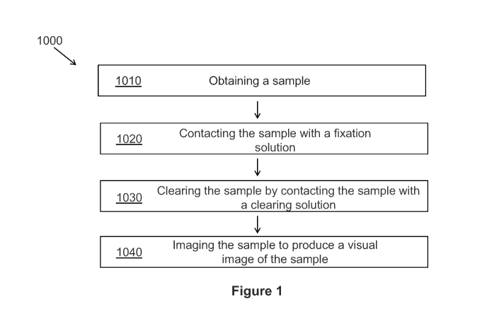
Figure 1 depicts a flow chart illustrating an exemplary method 1000 for
processing and imaging a sample. Method 1000 comprises obtaining a sample
1010, and
contacting the sample 1020 with a fixation solution. In a preferred
embodiment, the
sample is a tissue sample. In certain embodiments, the fixation solution
comprises at least
one dehydrant and at least one fluorescent dye. In one embodiment, method 1000
comprises clearing the sample 1030 by contacting the sample with a clearing
solution to
provide increased depth and clarity for imaging the sample 1040. In one
embodiment, the
sample is fixed prior to being contacted with a solution comprising a fixative
or
dehydrant and at least one fluorescent dye. In another embodiment the fresh
tissue is
placed directly in a combination fixation/dehydration fluid with dye or dyes.
In some
embodiments, the step of imaging the sample is performed in combination with
an
additional imaging method, such as second harmonic generation (SHG). (Figure
1). In
one embodiment, specimens can be partially fixed for intra-operative
evaluation or for
any other clinical scenario where a fast visual examination is desired.
Partially fixed
CA 02952832 2016-12-16
WO 2016/004367 PCT/US2015/039079
specimens may be fully fixed at a later time. In another embodiment, specimens
can be
partially cleared for intra-operative evaluation or for any other clinical
scenario where a
fast visual examination is desired. Partially cleared specimens may be fully
cleared at a
later time.
5 In one aspect, the method of the present invention further
comprises the
step of dehydrating the sample. Dehydration facilitates the removal of water
from a
sample so that clearing agents with low water solubility can subsequently be
used. It
should be appreciated that a dehydrant or a dehydration solution may also be
used as a
fixative or for fixing a sample. As used herein, the term "dehydrant" refers
to a water-
10 miscible anhydrous fluid. Non-limiting examples of dehydrants include
alcohols such as
methanol, ethanol, and propanol. In one embodiment, the dehydrant is
methacarn. In
another embodiment, the dehydrant is methanol. In one embodiment, the
dehydration step
functions as a fixative and takes place without prior fixation of the sample.
In other
embodiments, the dehydration step is performed after fixation of the sample.
In one
15 embodiment, the dehydration step is performed after fixation of the
sample using a
fixation solution comprising formalin.
The dehydration step can be performed for any suitable length of time.
The length of time can generally be any length of time suitable for rendering
the sample,
or a portion of the sample, miscible with the clearing agent. The length of
time can also
generally be any length of time suitable for preserving the sample or
preserving a portion
of the sample. In certain embodiments the period of time may be from about 1
minute,
about 5 minutes, about 10 minutes, about 15 minutes, about 30 minutes, about 1
hour,
about 2 hours, about 3 hours, about 4 hours, about 5 hours, about 6 hours,
about 12 hours,
or about 24 hours. In one embodiment, the dehydration step is performed over a
period of
time about 1 hour. In another embodiment the dehydration step is performed
over about
12 to 16 hours.
In one aspect, method of the present invention comprises the step of fixing
the sample. The tissue sample may be fixed using any method known in the art,
as would
be understood by one skilled in the art. In one embodiment, the sample is
fixed by
contacting the sample with a fixative. In another embodiment, the sample is
fixed by
contacting the sample with a fixation solution. In one embodiment, the
fixation solution
CA 02952832 2016-12-16
WO 2016/004367 PCT/US2015/039079
16
comprises at least one fixative. In one embodiment, the fixative is a
dehydrant. In another
embodiment, the fixation solution is a dehydrant. In another embodiment, the
fixation
solution comprises at least one fixative and at least one permeant. Non-
limiting examples
of fixatives include aldehydes (e.g., formaldehyde (paraformaldehyde,
formalin),
glutaraldehyde, acrolein (acrylic aldehyde), glyoxal (ethanedial, diformyl),
malonaldehyde (malonic dialdehyde), diacetyl (2,3- butanedione), and
polyaldehydes;
alcohols (i.e., protein-denaturing agents; e.g., acetic acid, methanol,
ethanol), polyvinyl
alcohols, heavy metal oxidizing agents (i.e., metallic ions and complexes;
e.g., osmium
tetroxide, chromic acid); agents of unknown mechanism, such as chloro-s-
triazides,
cyanuric chloride, carbodiimides, diisocyanates, diimido esters,
diethylpyrocarbonate
(diethyl oxydiformate, ethoxyformic anhydrate), picric acid, mercuric chloride
(corrosive
sublimate, bichloride of mercury), and other salts of mercury, and acetone. In
one
embodiment, a combination of fixatives is used. Such combinations give rise to
commonly termed formulations known to those in the art, such as Carnoy's
fixatives,
methacarn, Wolman's solution, Rossman's fluid, Gendre's fluid, Bouin's fluid,
Zenker's
fluid, Helly's fluid, B5 fixative, Susa fluid, Elftman's fixative, Swank and
Davenport's
fixative, Lillie's alcoholic lead nitrate, and cetylpyridinium chloride
(C.P.C.). Additives
can include, but are not limited to, such entities as tannic acid, phenol,
transition metal
salts (zinc), lanthanum, lithium, potassium. In one embodiment, the fixative
is methacarn.
In another embodiment, the fixative is formalin. In another embodiment, the
fixative is an
alcohol. In another embodiment, the fixative is methanol. In another
embodiment, the
fixative is a polyvinyl alcohol. In another embodiment, the fixative is
formaldehyde. In
another embodiment, fixation of the sample occurs ex vivo.
In some embodiments, at least one fluorescent dye is added to the sample
during the fixation step, resulting in simultaneous fixing and staining of the
sample. In
other embodiments, at least one fluorescent dye is added to the sample during
the
dehydration step, resulting in simultaneous dehydration and staining of the
sample. The
incorporation of a fluorescent dye obviates the need for post-processing
staining, which is
a time-consuming step of traditional sample preparation. The fluorescent dye
may be
added directly to the sample during the fixation step. For example, the
fluorescent dye
may be added to the fixation solution. In another embodiment, the fluorescent
dye is
CA 02952832 2016-12-16
WO 2016/004367 PCT/US2015/039079
17
added to the sample after completion of the fixation step. In one embodiment,
the fixation
solution comprises a fixative and a fluorescent dye. In another embodiment,
the fixative
solution comprises at least one dehydrant and at least one fluorescent dye. In
one
embodiment, the fluorescent dye is added directly to the sample during the
dehydration
step. In one embodiment, the method of processing a tissue sample comprises
the steps of
obtaining a tissue sample, and contacting the tissue sample with a fixative
solution
comprising at least one dehydrant and at least one fluorescent dye.
The skilled artisan will understand that the present invention contemplates
the use of any fluorescent dye that is compatible with the fixation step.
Examples of
fluorescent dyes include, but are not limited to, POPO-1, TOTO-3, TAMRA,
BOXTO,
BEBO, SYBR DX, SYTOX dyes, SYTO dyes, Alexa dyes, fluorescein, rhodamine,
propidium idodide, Hoechst dyes, tetramethylrhodamine, R-phycoerythrin, Cy-3,
Cy-5,
Cy-7, Texas Red, Phar-Red, allophycocyanin (APC), fluorescein amine, eosin,
dansyl,
umbelliferone, 5-carboxyfluorescein (FAM), 2'7'-dimethoxy-4'5'-dichloro-6-
carboxyfluorescein (JOE), 6 carboxyrhodamine (R6G), N,N,N',N'-tetramethy1-6-
carboxyrhodamine (TAMRA), 6-carboxy-X-rhodamine (ROX), 4-(4'-
dimethylaminophenylazo) benzoic acid (DABCYL), 5-(2'-aminoethyl)
aminonaphthalene-l-sulfonic acid (EDANS), 8-Anilino-1-naphthalenesulfonic acid
ammonium salt (ANS), 4-acetamido-4'-isothiocyanatostilbene-2,2' disulfonic
acid,
acridine, acridine isothiocyanate, acridine orange (N,N,N',N'-
tetramethylacridine-3,6-
diamine), R-amino-N-(3-vinylsulfonyl)phenylnaphthalimide-3,5, disulfonate
(Lucifer
Yellow VS), N-(4-anilino-1-naphthyl)maleimide, anthranilamide, Brilliant
Yellow,
coumarin, 7-amino-4-methylcoumarin, 7-amino-4-trifluoromethylcouluarin
(Coumarin
151), cyanosine, 2-(4-amidinopheny1)-1H-indole-6-carboxamidine (DAPI), 5',5"-
dibromopyrogallol-sulfonephthalein (Bromopyrogallol Red), 7-diethylamino-3-(4'-
isothiocyanatopheny1)-4-methylcoumarin diethylenetriamine pentaacetate, 4,4'-
diisothiocyanatodihydro-stilbene-2,2'-disulfonic acid, 4,4'-
diisothiocyanatostilbene-2,2'-
disulfonic acid, 4-dimethylaminophenylazopheny1-4'-isothiocyanate (DABITC),
eosin
isothiocyanate, erythrosin B, erythrosin isothiocyanate, ethidium, 5-(4,6-
dichlorotriazin-
2-y1) aminofluorescein (DTAF), QFITC (XRITC), fluorescamine, IR144, IR1446,
Malachite Green isothiocyanate, 4-methylumbelliferone, ortho cresolphthalein,
CA 02952832 2016-12-16
WO 2016/004367 PCT/US2015/039079
18
nitrotyrosine, pararosaniline, Phenol Red, B-phycoerythrin, o-
phthaldialdehyde, pyrene,
pyrene butyrate, succinimidyl 1-pyrene butyrate, Reactive Red 4 (Cibacron®
Brilliant Red 3B-A), lissamine rhodamine B sulfonyl chloride, rhodamine B,
rhodamine
123, rhodamine X, sulforhodamine B, sulforhodamine 101, sulfonyl chloride
derivative
of sulforhodamine 101, tetramethyl rhodamine, thiazole orange, riboflavin,
rosolic acid,
and terbium chelate derivatives. In one embodiment, the fluorescent dye is
eosin. In
another embodiment, the fluorescent dye is DAPI. In another embodiment, the
fluorescent dye is SYTOX green. In another embodiment, the fluorescent dye is
acridine
orange. In another embodiment, the fluorescent dye is rhodamine B. In another
embodiment, the fluorescent dye is a SYTO dye. In another embodiment, the
fluorescent
dye is propidium iodide. In another embodiment, the fluorescent dye is a
Hoechst dye.
In certain embodiments, the fluorescent dye can selectively stain a
particular organelle or component of a cell. In one embodiment, the
fluorescent dye is a
nuclear dye. Non-limiting examples of nuclear dyes include DAPI, SYTOX dyes,
SYTO
dyes, propidium iodide, acridine orange, and Hoechst dyes. In one embodiment,
the
nuclear dye is DAPI. In another embodiment, the fluorescent dye is a protein
dye.
Examples of protein dyes include, but are not limited to, eosin, Rhodamine B
(RhB), and
ANS. In another embodiment, intrinsic fluorescence of the cell is used to
image the
cellular protein. In another embodiment, the fluorescence is generated from a
combination of at least one nuclear dye and at least one protein dye. In
another
embodiment, the fluorescence is generated from a nuclear dye and intrinsic
fluorescence.
In one embodiment, the at least one protein dye is eosin.
In certain embodiments, a morphology preservative is added to the sample
during the fixation step and/or the dehydration step. The morphology
preservative
enhances maintenance of the nuclear structure of the cells, in that it
maintains cell
membranes intact for subsequent cytological staining and/or reduces shrinking
or
swelling during fixation or dehydration. Any morphology preservative that is
compatible
with the fixation step may be used in the invention, as would be understood by
one of
ordinary skill in the art. Non-limiting examples of morphology preservatives
include
acetic acid, trichloroacetic acid, formaldehyde, dioxane, chloroform, and the
like. In a
preferred embodiment, the morphology preservative is chloroform. The
morphology
CA 02952832 2016-12-16
WO 2016/004367 PCT/US2015/039079
19
preservative may be added directly to the sample during the fixation step.
Alternatively,
the morphology preservative may be added to the fixation solution. In one
embodiment,
the fixation solution comprises about 0% to about 50% of a morphology
preservative. In
another embodiment, the fixation solution comprises about 5% to about 15% of a
morphology preservative. In another embodiment, the fixation solution
comprises about
10% of a morphology preservative. In another embodiment, the fixation solution
comprises about 20% to about 40% of a morphology preservative. In a preferred
embodiment, the fixation solution comprises about 30% of a morphology
preservative.
In some embodiments, a permeation enhancer is added to the sample
during the fixation step and/or the dehydration step. The permeation enhancer
accelerates
the access of dye to the deeper portion of the sample, while overall improving
the dyeing
process. The permeation enhancer also accelerates the penetration of fixative,
dehydrant,
and/or clearing agent. Non-limiting examples of permeation enhancers include
acids such
as acetic acid, methacarn comprising acetic acid, sulphoxides such as
dimethylsulfoxide
(DMSO), azone, pyrrolidones, propylene glycol, fatty acids, essential oils,
phospholipids,
s-collidine, and surfactants such as Tween. In one embodiment, the fixation
solution
comprises about 0% to about 75% of a permeation enhancer. In another
embodiment, the
fixation solution comprises about 0% to about 25% of a permeation enhancer. In
another
embodiment, the fixation solution comprises about 5% to about 15% of a
permeation
enhancer. In a preferred embodiment, the fixation solution comprises about 10%
of a
permeation enhancer.
In some embodiments, the permeation enhancer is at least one acid. In
some embodiments, the acid is an organic acid. Non-limiting examples of
organic acids
include acetic acid, glacial acetic acid, lactic acid, propionic acid, butyric
acid, succinic
acid, citric acid, 3-hydroxypropionic acid, glycolic acid, or formic acid. In
one
embodiment, the acid is acetic acid. Acetic acid is useful for enhancing the
speed of
fixation, which is important for rapid sample processing, while also
significantly
improving the quality and depth of images from cleared samples using any type
of
sectioning image modalities. Acetic acid is also useful for lysing red blood
cells, which
allows for removal of heme pigment, which is a significant deterrent to
clarity of the
sample by virtue of its broad light absorption characteristics in the typical
workable
CA 02952832 2016-12-16
WO 2016/004367 PCT/US2015/039079
wavelength range of routine fluorescent imaging. In one embodiment, the step
of fixing
the sample further comprises the addition of a lysing agent to the sample. In
a particular
embodiment, the step of fixing the sample further comprises the addition of a
red blood
cell lysing agent to the sample. In another embodiment, the step of
dehydrating the
5 sample further comprises the addition of a lysing agent to the sample. In
another
embodiment, the fixative solution further comprises a red blood cell lysing
agent. In a
preferred embodiment, the red blood cell lysing agent is acetic acid. In other
embodiments, the acid is an inorganic acid. The solution may further comprise
at least
one organic solvent. Non-limiting examples of organic solvents include
methanol,
10 absolute methanol, chloroform, dichloromethane, ethanol, isopropanol,
acetone, ethyl
acetate, acetonitrile, hexane, hexene, octane, pentane, cyclohexane, iso-
octane, xylene
(ortho, meta, or para), and 1-hexene. In one embodiment, the organic solvent
is absolute
methanol. Methanol is useful for tissue processing by arresting enzymatic
function and
degradation while maximally preserving genetic and proteomic information. In
another
15 embodiment, the organic solvent is chloroform. In one embodiment, the
solution
comprises two organic solvents and an acid. As a non-limiting embodiment, the
fixative
solution comprises about 60% absolute methanol, about 30% chloroform, and
about 10%
glacial acetic acid, which is also known as methacarn. When in combination
with
fluorescent dyes and clearing, methacarn may be useful for deep fluorescent
tissue
20 section imaging of human samples for histologic evaluation, and for
creating the contrast
needed for accurate histologic evaluation.
The fixation step may be performed under any condition that promotes
rapid tissue processing, such as conditions that increase the rates of
chemical reaction and
diffusion, as would be understood by one of ordinary skill in the art. In some
embodiments, the fixation step is performed at an elevated temperature. As
used herein,
the term "elevated temperature" refers to temperatures above those experienced
in the
earth's atmosphere, preferably above 30 C. In one embodiment, the elevated
temperature
ranges from about 20 C to about 75 C. In another embodiment, the elevated
temperature ranges from about 35 C to about 50 C. In another embodiment, the
elevated temperature is about 45 C. In a non-limiting example, the fixation,
dehydration,
CA 02952832 2016-12-16
WO 2016/004367 PCT/US2015/039079
21
and/or staining is performed under microwave irradiation for the purpose of
accelerating
diffusion, chemical reaction, or temperature.
The fixation step can be performed for any suitable length of time. The
length of time can generally be any length of time suitable for preserving the
sample. In
certain embodiments, the period of time may be from about 1 minute, about 5
minutes,
about 10 minutes, about 15 minutes, about 30 minutes, about 1 hour, about 2
hours, about
3 hours, about 4 hours, about 5 hours, about 6 hours, about 12 hours, or about
24 hours.
In one embodiment, the fixation step is performed over a period of time about
1 hour. In
another embodiment the fixation step is performed over about 12 to 16 hours.
In another
embodiment the fixation step is performed over the course of weeks to years.
In one embodiment, the method of the present invention comprises the
step of simultaneously fixing, dehydrating, and staining the sample by
contacting the
sample with a dehydration solution comprising at least one dehydrant and at
least one
fluorescent dye. In another embodiment, the method of the present invention
comprises
the step of simultaneously dehydrating and staining the sample by contacting
the sample
with a dehydration solution comprising at least one dehydrant and at least one
fluorescent
dye.
In another aspect, the method of the present invention further comprises
the step of clearing the sample. Clearing the sample provides increased depth
and clarity
of imaging of the sample. In one embodiment, the clearing step is performed in
absence
of a fixation step. In some embodiments, the step comprises clearing the
sample by
contacting the sample with a clearing solution. As a non-limiting embodiment,
the sample
is cleared by replacing water with a clearing solution that has a higher
refractive index
than water that more closely resembles that of proteins and organelles,
thereby drastically
reducing light scattering and enabling imaging depths of millimeters instead
of
micrometers. In one embodiment, the clearing solution comprises at least one
solvent.
Any solvent may be used in the clearing solution, as long as the overall
refractive index
of the clearing solution is higher than the refractive index of water and the
solvent does
not damage the morphology of cellular components of the sample. In one
embodiment,
the refractive index of the clearing solution ranges from about 1.4 to about
1.6. In another
embodiment, the refractive index of the clearing solution ranges from about
1.33 to about
CA 02952832 2016-12-16
WO 2016/004367 PCT/US2015/039079
22
1.49. In another embodiment, the refractive index of the clearing solution is
greater than
about 1.4. In another embodiment, the refractive index of the clearing
solution is greater
than about 1.5. In one embodiment, the clearing solution further comprises an
agent that
is water soluble and has a high refractive index.
In some embodiments, the solvent is an organic solvent. Non-limiting
examples of organic solvents useful as clearing agents include, benzyl
alcohol, benzyl
benzoate, xylene, limonene, benzene, toluene, chloroform, petroleum ether,
carbon
bisulfide, carbon tetrachloride, dioxane, glycerol, sugar solutions, dibenzyl
ether, clove
oil, and cedar oil. In one embodiment, the solvent is benzyl alcohol. In
another
embodiment, the solvent is benzyl benzoate. In another embodiment, the solvent
is
xylene. In another embodiment, the solvent is glycerol. In another embodiment,
the
solvent is a sugar solution. In another embodiment, the solvent is dibenzyl
ether. In
another embodiment, the solvent is hexane.
In some embodiments, the clearing solution comprises a first solvent and a
second solvent. In one embodiment, the ratio of the first solvent to the
second solvent
ranges from about 100:1 to about 1:100. In another embodiment, the ratio of
the first
solvent to the second solvent ranges from about 10:1 to about 1:10. In another
embodiment, the ratio of the first solvent to the second solvent ranges from
about 5:1 to
about 1:5. In a preferred embodiment, the ratio of the first solvent to the
second solvent is
about 1:2. In some embodiments, the solvent is an organic solvent. In a
particular
embodiment, the clearing solution comprises benzyl alcohol and benzyl
benzoate. In one
embodiment, the ratio of benzyl alcohol to benzyl benzoate is about 1:2.
The clearing step may be performed under any condition that promotes
rapid clearing of the sample, as would be understood by one of ordinary skill
in the art. In
some embodiments, the clearing step is performed at an elevated temperature.
In one
embodiment, the elevated temperature ranges from about 20 C to about 75 C.
In
another embodiment, the elevated temperature ranges from about 35 C to about
50 C.
In another embodiment, the temperature is about 22 C.
The clearing step can be performed for any suitable length of time. The
length of time can generally be any length of time suitable for achieving
sufficient
reduction in light scattering to enable imaging to the desired depth in the
sample. In
CA 02952832 2016-12-16
WO 2016/004367 PCT/US2015/039079
23
certain embodiments, the period of time may be from about 1 minute, about 5
minutes,
about 10 minutes, about 15 minutes, about 30 minutes, about 1 hour, about 2
hours, about
3 hours, about 4 hours, about 5 hours, about 6 hours, about 12 hours, or about
24 hours.
In one embodiment, the clearing step is performed in about 2 minutes to about
1 hour. In
one embodiment, the clearing step is performed in about 2 minutes. In one
embodiment,
the clearing step is performed in about 30 minutes. In one embodiment, the
clearing step
is performed in about 12 hours.
In one embodiment, the clearing step further comprises the step of adding
a solvent to the sample prior to adding the clearing solution. In some
embodiments, the
solvent is an organic solvent. In one embodiment, the solvent is an alcohol.
The alcohol is
useful for dehydrating the sample. Non-limiting examples of alcohols include
methanol,
ethanol, propanol, isopropanol, butanol, isobutanol, ethyl butanol, t-butanol,
dioxane,
ethylene glycol, acetone, and amyl alcohol. In a preferred embodiment, the
solvent is
methanol. In one embodiment, the solvent is added in combination with a
permeation
enhancer. Non-limiting examples of permeation enhancers include acetic acid,
polyethylene glycol (PEG), mono- and dimethyleneglycol, propylene glycol,
polyvinyl
pyrrolidone, or the like, surfactants such as dimethyl sulfoxide (DMSO),
polyoxyethylene
sorbitan esters (e.g., TWEEN such as TWEEN 80), sodium dimethyl
sulfosuccinate, mild
household detergents, or the like. In one embodiment, the permeation enhancer
is
selected from the group consisting of acetic acid, DMSO, and TWEEN. The
addition of a
solvent in combination with a permeation enhancer increases the rate of
clearing with
BABB by improving miscibility and permeability, and also eliminates the need
for
gradual gradient steps of decreasing alcohol concentration.
In part, the present invention provides a method of rapidly processing a
tissue sample. The length of time can generally be any length of time suitable
for fixing
the sample and clearing the sample. The length of time can also generally be
any length
of time suitable for fixing a portion of the sample and clearing a portion of
the sample. In
certain embodiments, the period of time may be from about 1 minute, about 5
minutes,
about 10 minutes, about 15 minutes, about 30 minutes, about 1 hour, about 2
hours, about
3 hours, about 4 hours, about 5 hours, or about 6 hours. In one embodiment,
the steps of
fixing the sample and clearing the sample are performed in about 1 hour to
about 2 hours.
CA 02952832 2016-12-16
WO 2016/004367 PCT/US2015/039079
24
In one embodiment, the steps of fixing the sample and clearing the sample are
performed
over a period of time about 1.5 hours.
In another aspect, the method of the present invention further comprises
the step of imaging the sample. In one embodiment, the step of imaging the
sample
further comprises producing a visual image of the tissue sample. The sample
may be
imaged using any imaging method compatible with the sample processing methods
described herein. Preferred imaging methods include fluorescence based
sectioning
imaging methods. Contrary to destructive 3-D histology approaches such as
pigmented
plastic embedding systems and whole slide imaging (WSI), these fluorescence
based
methods are non-destructive, allowing for the preservation of samples for
ancillary
studies such as immunostains and molecular studies. Examples of fluorescence
based
sectioning imaging methods include, but are not limited to, multiphoton
microscopy
(MPM), side-plane illumination microscopy, traditional confocal microscopy,
spinning
disk confocal microscopy, structured illumination microscopy, and the like. In
animal
tissue (Parra et al., 2010, J. Biomed. Opt. 15:036017-036017-5; Vesuna et al.,
2011, J.
Biomed. Opt. 16:106009-106009-6; Fu et al., 2009, Gastroenterology 137:453-
465), the
depth of imaging can be increased over samples prepared using more traditional
methods,
such as formalin fixing, by combining MPM with optical clearing. In one
embodiment,
the sample is imaged using multiphoton microscopy (MPM). In another embodiment
the
sample is imaged using confocal microscopy. In another embodiment, the sample
is
imaged using structured illumination microscopy. In another embodiment the
sample is
imaged using selective plane illumination microscopy. In another embodiment
the sample
is imaged using deconvolution microscopy. In another embodiment the sample is
imaged
using super-resolution microscopy. In another embodiment, the sample is imaged
using
side-plane illumination microscopy. In another embodiment, the sample is
imaged using
spinning disk confocal microscopy. In one embodiment, the method of imaging a
tissue
sample comprises the steps of obtaining a tissue sample, contacting the tissue
sample
with a fixative solution comprising at least one fluorescent dye, contacting
the tissue
sample with a clearing solution, and producing a tissue sample image by
measuring
intensity values of the fluorescence of the tissue sample, and converting the
intensity
CA 02952832 2016-12-16
WO 2016/004367 PCT/US2015/039079
values to effective optical densities, such that the optical densities
recreate the coloration
of a stain in a produced image of the tissue sample.
The imaging methods of the present invention provide a method for image
analysis that allows reproduction of images essentially indistinguishable from
traditional
5 histology stains. In one embodiment, the method involves a multichannel
approach,
wherein intensity values of fluorescence from the sample are converted to
optical
densities using an exponential pseudo-coloring process, which is an inversion
of a
logarithmic pseudo-coloring process, wherein intensity values of fluorescence
are
converted to optical densities in red, green, and blue channels. In one
embodiment, the
10 step of imaging the sample further comprises the steps of measuring
intensity values of
the fluorescence of the sample, and converting the intensity values to
effective optical
densities recreate the coloration of a stain in the sample image. In one
embodiment, the
intensity values of one or more fluorescence channels are converted to
effective optical
densities in one or more pseudo-color display channels using an exponential
pseudo-
15 coloring process, wherein the equation that results in optical densities
includes a constant
to the power of the intensity values from fluorescence, as would be understood
by one of
ordinary skill in the art. Numerically:
Chi = CiA(aiN + biP)
Ch2 = C2A(a2N + b2P)
20 Ch3 = C31(a3N + b3P)
where Chi, Ch2, Ch3 are color display channels, such as Red, Green and
Blue; C1, C25 C3 are positive constants; al, a2, a35 1)15 b2, b3are constants
that may be
positive or negative; and N and P are fluorescence intensities recorded from
different
fluorescence channels. In one embodiment, the intensity values are converted
to effective
25 optical densities using an exponential pseudo-coloration process.
For an example of a logarithmic color deconvolution process, see Ruifrok
and Johnston, 2001, Anal. Quant. Cytol. Histol. 23:291-299, which is
incorporated by
reference herein in its entirety.
In one embodiment, the fluorescence is intrinsic fluorescence from the
sample. In another embodiment, the fluorescence is fluorescence from the
fluorescent
dye. In one embodiment, the fluorescent dye is a nuclear dye. In another
embodiment, the
CA 02952832 2016-12-16
WO 2016/004367 PCT/US2015/039079
26
fluorescent dye is a protein dye. The number of channels used may be varied as
needed to
achieve the desired image, as would be understood by one skilled in the art.
In one
embodiment, the number of channels is two channels. In a specific embodiment,
the two
channels are an intrinsic fluorescence channel and a fluorescent nucleic acid
dye channel.
In another embodiment, the two channels are a fluorescent nucleic acid dye
channel and a
fluorescent protein staining channel. In a non-limiting example, the intrinsic
fluorescence, emanating primarily from cross-linked proteins and corresponding
to the
staining typically achieved by protein stains such as eosin, can be augmented
by use of
formalin as the fixative, a feature that facilitates the reproduction of
normal coloration by
improving signal to noise of this channel and facilitating separation from
nucleic acid
fluorescence.
The imaging method of the present invention also provides images of
samples that mimic common pathology stains, resulting in the accurate and
efficient
interpretation of the images. Examples of pathology stains which can be
reproduced
using the methods of the present invention include, but are not limited to,
hematoxylin,
eosin, wright, giemsa, Masson's trichrome, Jones, trichrome, periodic acid
Schiff (PAS)
and reticulin stains. Combinations of pathology stains can also be reproduced
using
methods of the present invention. In one embodiment, the combination of
pathology
stains is hematoxylin and eosin (H&E). In another embodiment, the combination
of
pathology stains is wright and giemsa.
In some embodiments, the step of imaging the sample is performed in
combination with an additional imaging method, resulting in multi-modal
imaging. In
one embodiment, the additional imaging method is a higher-order harmonic
generation.
Higher-order harmonic generation permits the recreation of additional
specialized
histological stains, such as collagen stains like trichrome and silver stains
like Jones stain.
In one embodiment, the higher order harmonic generation is second harmonic
generation
(SHG). SHG results from multiphoton excitation of asymmetric repeating
proteins such
as collagen, and may be used for simple identification and quantification of
collagen
fibrosis and amyloid in combination with the imaging method, such as MPM. In
one
embodiment, the additional imaging method is Fluorescence Lifetime Imaging.
Fluorescence Lifetime Imaging may be used to provide additional contrast in
MPM by
CA 02952832 2016-12-16
WO 2016/004367 PCT/US2015/039079
27
distinguishing between fluorophores with differing lifetime characteristics.
In another
embodiment, the additional imaging method uses multiple fluorescent
antibodies.
Multiple fluorescent antibodies may be used to provide potential for
performing
immunohistochemistry in uncut samples with multiple antigens detectable on the
same
cells. In another embodiment, the imaging method is used in combination with
diode
lasers. See Dechet et al., 2003, J. Urol. 169:71-74 and Durfee et al., 2012,
Opt. Express
20:13677-13683, each which is incorporated by reference herein in its
entirety. Other
techniques known in the art to increase the rate of scanning of the sample
image may be
used in the imaging step, as would be understood by one of ordinary skill in
the art. Non-
limiting examples include multibeam scanning systems, spatiotemporal
multiplexing, and
temporal focusing. See Bewersdorf et al., 1998, Opt. Lett. 23:655-657, Amir et
al., 2007,
Opt. Lett. 32-1731-1733, Oron et al., 2005, Opt. Express 13:1468-1476, and Zhu
et al.,
2005, Opt. Express 13:2153-2159, each which is incorporated by reference
herein in its
entirety. In another embodiment, the imaging step is performed in real time
using video
imaging.
The methods of the present invention provide a clear, high-quality image
of the sample obtained at a greater sample depth as compared with more
traditional
histological methods, such as sample treated only with formalin fixation. In
one
embodiment, the sample image is obtained at a sample depth of about 100 nm to
about 2
cm. In another embodiment, the sample image is obtained at a sample depth of
about 100
nm to about 500 gm. In another embodiment, the sample image is obtained at a
sample
depth of about 100 nm to about 1 cm. In another embodiment, the sample image
is
obtained at a sample depth of about 50 gm to about 500 gm. In another
embodiment, the
sample image is obtained at a sample depth of about 100 nm to about 100 gm. In
another
embodiment, the sample image is obtained at a sample depth of about 200 gm. In
another
embodiment, the sample image is obtained at a sample depth of about 100 gm.
In one embodiment, imaging of the sample provides digital sample data.
This digital data may then be stored for later distribution, such as for
consultation and
health records, thereby improving the accessibility of the images for further
evaluation or
reevaluation. In addition, digital sample data is capable of maintaining the
integrity of the
CA 02952832 2016-12-16
WO 2016/004367 PCT/US2015/039079
28
data, as opposed to physical samples which may be lost or damaged and cannot
be stored
digitally.
In one embodiment, the sample image is a three dimensional (3-D) sample
image. A 3-D sample image can be produced using any method known in the art,
as
would be understood by one skilled in the art. In one embodiment, the 3-D
sample image
is produced from an entire biopsy. In another embodiment, a 3-D sample image
produced
from a whole biopsy provides a quantitative approach to diagnosing a disease.
In another
embodiment, a 3-D sample image of the present invention provides facile
identification
of subtle morphologic findings in the imaging sample. For example, 3-D sample
images
improve the quantitative and qualitative analysis of fibrosis observed in
various
conditions such as cirrhosis, hypertensive renal disease, interstitial lung
disease, and
ovarian cancer over other two-dimensional (2-D) histological methods currently
known
in the art. In another embodiment, a 3-D sample image of the present invention
is used to
diagnose a malignant growth. In one embodiment, the methods of the present
invention
provide full rotational control of 3-D sample images. Diagnosis of malignant
growth is
often dependent on visualizing growth patterns, particularly in glandular-
based disorders
such as prostate and breast cancer. Such analysis has been primarily based on
the two-
dimensional orientation, which may require pathologists to look back-and-forth
at
(hopefully) contiguous segments in order to render a diagnosis. In these 2D
methods,
visual inspection can be further complicated by poor embedding and orientation
differences of the sample. In non-limiting examples, the 3-D sample images of
the
present invention are used to diagnose metastatic colon cancer in liver and
for the
diagnosis of endometrial abnormalities. In another non-limiting example, 3-D
reconstructions of MPM imaging from clarified tissue may be used on complete
biopsy-
sized tissue specimens and may also be used to produce quantifiable
characterization of
collagen fibrosis. Other non-limiting examples of the use of 3-D sample image
include
identification of low-grade abnormalities in glandular cell growth, such as
with prostate
and breast neoplasia, the evaluation of depth of invasion of tumors, such as
for
determining depth of muscle invasion in bladder biopsies, and the more
complete
quantitative evaluation of fibrosis, of particular significance in kidney and
liver biopsies.
CA 02952832 2016-12-16
WO 2016/004367 PCT/US2015/039079
29
Specimen Holding Device
In another aspect, the invention relates to a specimen holding device. The
device is useful for holding a histological sample and facilitates processing
and imaging
the histological sample. In one embodiment, the device is amenable for use in
the
methods of the present invention as described elsewhere herein.
Referring now to Figure 1, an exemplary specimen holding device 10 is
depicted. Specimen holding device 10 comprises a first plate 12 and a second
plate 18.
First plate 12 comprises window 14, compressible material 16, and tabs 20.
Second plate
18 comprises window 14 and slots 22.
First plate 12 and second plate 18 may comprise any suitable material. For
example, first plate 12 and second plate 18 comprise a substantially rigid
material, such
as glass, metal plastic, and the like. First plate 12 and second plate 18 may
comprise any
suitable dimensions. In one embodiment, first plate 12 and second plate 18
comprise the
same length 11 and width 13. In one embodiment, first plate 12 and second
plate 18 are
dimensioned such that they fit on any microscope stage. For example, in one
embodiment, length 11 can be between 40mm and 100mm. In another embodiment,
width 13 can be between 20mm and 80mm. In one embodiment, length 11 is 75mm
and
width 13 is 25mm.
Specimen holding device 10 comprises window 14 for holding and
viewing specimens. Window 14 may comprise any suitable material. For example,
window 14 may comprise a transparent material, such as glass or clear plastic.
In certain
embodiments, window 14 may comprise any other substantially rigid material,
such as a
metal or an opaque plastic. In one embodiment, both first plate 12 and second
plate 18
comprise a transparent window 14. In another embodiment, only first plate 12
comprises
a transparent window 14. In a further embodiment, only second plate 18
comprises a
transparent window 14.
First plate 12 comprises compressible material 16. Compressible material
16 is placed atop first plate 12. In one embodiment, compressible material 16
is glued or
otherwise fastened to first plate 12. In one embodiment, compressible material
16 is
dimensioned such that it forms a perimeter. For example, compressible material
16 can
form the perimeter of the shape of window 14. In another embodiment,
compressible
CA 02952832 2016-12-16
WO 2016/004367 PCT/US2015/039079
material 16 is dimensioned such that it is a sold block. For example,
compressible
material 16 can be a solid block have the shape of window 14. Compressible
material 16
can comprise any suitable material including, but not limited to: foams,
rubbers, sponges,
polymers, cork, and the like. In one embodiment, compressible material 16
comprises
5 gaps or slits such that fluids are able to pass through it. In another
embodiment,
compressible material 16 comprises a porous material to allow fluids to pass
through it.
Non-limiting examples of porous materials include, but are not limited to:
solid foams,
sponges, plastic meshes, and the like. The pore size of the porous material
may be
selected to control the rate of fluid transfer. For example, the pore size can
be between
10 10 m and lmm in diameter.
First plate 12 comprises a mechanism by which to hold it at a fixed
distance from second plate 18. In one embodiment, the mechanism comprises a
tab and
reciprocal slot. For example, first plate 12 can comprise at least two tabs
20, and second
plate 18 can comprise at least two slots 22. In another embodiment, first
plate 12 can
15 comprise at least two slots, and second plate 18 can comprise at least
two tabs. In one
embodiment, the number of tabs 20 matches the number of slots 22. Tabs 20 are
dimensioned such that they fit within slots 22. In one embodiment, tabs 20
comprises
teeth 24 such that when tab 20 engages slot 22, teeth 24 provide frictional
resistance to
secure tab 20 within slot 22. In a further embodiment, teeth 24 provide
frictional
20 resistance to reversibly secure tab 20 within slot 22.
Method of Using the Specimen Holding Device
In another aspect, the invention relates to methods for securely holding a
specimen for sample processing. The invention also relates to methods for
securely
25 holding a specimen for imaging.
In one embodiment, the method uses specimen holding device 10 for
securely holding a specimen for sample processing. In one embodiment, a
specimen may
be placed on window 14 of first plate 12, such that the specimen is surrounded
by a
perimeter comprising compressible material 16. Second plate 18 is secured to
first plate
30 12 by engaging tabs 20 to slots 22, such that compressible material 16
fully contacts
second plate 18. In one embodiment, the specimen is not compressed against any
surface
CA 02952832 2016-12-16
WO 2016/004367 PCT/US2015/039079
31
when first plate 12 and second plate 18 are secured together. A compressible
material 16
comprising porous material allows for fluid transfer into and out of the
specimen holding
device during sample processing. For example, the specimen holding device may
be fully
submerged in a sample processing fluid to incubate the specimen in the fluid.
In another embodiment, the specimen is placed on a solid block of
compressible material on first plate 12 (not pictured). Second plate 18 is
secured to first
plate 12 by engaging tabs 20 to slots 22, such that the solid block of
compressible
material presses against second plate 18. A solid block of compressible
material
comprising a porous material allows for fluid transfer throughout the block
and for fluids
to contact the specimen during sample processing. For example, the specimen
holding
device may be fully submerged in a sample processing fluid to incubate the
specimen in
the fluid.
In one embodiment, the method uses specimen holding device 10 for
securely holding a specimen for imaging. A specimen is placed on window 14 of
first
plate 12, such that the specimen is surrounded by a perimeter comprising
compressible
material 16. Second plate 18 is secured to first plate 12 by engaging tabs 20
to slots 22.
Second plate 18 may be lowered onto the specimen such that the specimen is
compressed
against window 14 of both first plate 12 and second plate 18. Being compressed
against
both windows 14, the specimen is thereby immobilized. Specimen holding device
10 may
then be placed on any suitable imaging system, and the specimen may be imaged
through
at least one of window 14.
In another embodiment, the specimen is placed on a solid block of
compressible material 16 on first plate 12 (not pictured). Second plate 18 is
secured to
first plate 12 by engaging tabs 20 to slots 22. Second plate 18 may be lowered
onto the
specimen such that the specimen is compressed against the solid block of
compressible
material 16 on first plate 12 and window 14 of second plate 18, thereby
immobilizing the
specimen. Specimen holding device 10 may then be placed on any suitable
imaging
system, and the specimen may be imaged through window 14.
Microscope System
CA 02952832 2016-12-16
WO 2016/004367 PCT/US2015/039079
32
In another aspect, the invention relates to a microscope system for 2-D and
3-D imaging of specimens. In one embodiment, the microscope system is amenable
for
use in the methods of the present invention as described elsewhere herein.
Referring now to Figure 17, an exemplary microscope system 30 is
depicted. Microscope system comprises laser 32, an optional beam shaper 34,
spinning
polygon 36, scan lens 38, tube lens 40, microscope objective 41, dichroic
mirror A 42,
dichroic mirror B 44, a plurality of emission filters 46, a plurality of
detectors 48, and
translation stage 50. Microscope system 30 may further comprise any component
customarily used in other similar microscope systems, such as viewfinders,
power
sources, stage manipulation mechanisms, autofocus mechanisms, automation
systems,
and the like.
Laser 32 comprises a laser source providing, for example, multiphoton
excitation of dyes used in sample labeling. Laser 32 may comprise any suitable
laser,
such as femtosecond or picosecond pulsed fiber laser, or any other short-pulse
laser with
a center wavelength that is adjustable for optimum excitation of dyes.
Alternatively, the
center wavelength may be non-tunable but may be chosen to correspond to a
wavelength
suitable for exciting dyes used to stain a sample. In one embodiment, the
center
wavelength is 800nm.
A laser beam being emitted by laser 32 passes through beam shaper 34. In
one embodiment, beam shaper 34 expands the laser beam to a width required to
illuminate the back aperture of microscope objective 41. In another
embodiment, beam
shaper 34 may transform the laser beam profile. For example, a laser beam
having a
Gaussian beam profile may be transformed to have a flat top profile after
passing through
beam shaper 34, which results in more efficient illumination of the back
aperture of
microscope objective 41.
A laser beam passing through beam shaper 34 is rapidly scanned in angle
by spinning polygon 36 having a plurality of mirrored facets. In various
embodiments,
the laser beam may be rapidly scanned in angle by any one of several resonant
scanning
mechanisms commonly available for rapid beam scanning. Non-limiting examples
of
resonant scanning mechanisms used for rapid beam scanning include resonant
galvanometers and digital micromirror devices.
CA 02952832 2016-12-16
WO 2016/004367 PCT/US2015/039079
33
A rapidly scanned laser beam is imaged to the back aperture of
microscope objective 41 using scan lens 38 and tube lens 40. In one
embodiment, scan
lens 38 is a telecentric lens. In one embodiment, scan lens 38 is an F-theta
lens. In various
embodiments, scan lens 38 can be any suitable lens that images an angle-
scanned beam
from the surface of spinning polygon 36 to the back aperture of microscope
objective
41when used in conjunction with tube lens 40.
In one embodiment, microscope system 30 comprises dichroic mirror A
42, such that fluorescent light collected by the objective lens may be
reflected by dichroic
mirror A to be collected by one or more detectors 48. Alternatively, the beam
path may
be chosen such that dichroic mirror A is used to reflect the excitation laser
beam to the
back aperture of a microscope objective to transmits the collected
fluorescence to
detectors 48. In one embodiment, fluorescence or SHG may be collected as
transmitted
light with or without a collecting lens (not pictured) and detectors 48.
Detectors 48 may
be any detector sensitive to appropriate wavelengths. For example, in one
embodiment
detectors 48 may comprise photomultiplier tubes. Microscope system 30 may
further
comprise dichroic mirror B 44, or any number of additional dichroic mirrors as
is needed
to send different color fluorescence to different detectors 48. In one
embodiment,
microscope system 30 may further comprise any number of emission filters 46 as
is
needed, wherein emission filters 46 placed in front of detectors 48 reject
stray laser light
and other light interference.
In various embodiments, microscope system 30 comprises at least one
microscope objective 41. In one embodiment, microscope objective 41 is
compatible with
high-refractive-index immersion medium. In various embodiments, microscope
objective
41 is compatible with a high-refractive-index immersion medium having an index
between 1.45 and 1.6. In one embodiment, microscope objective 41 is compatible
with a
high-refractive-index immersion medium having an index of 1.54. In one
embodiment,
microscope objective 41 is compatible with an immersion medium that is the
BABB
clearing agent as disclosed elsewhere herein. In one embodiment, microscope
system 30
having an inverted microscope arrangement comprises a microscope objective 41
with a
relatively broad and flat aspect on the side that faces a sample such that
immersion fluid
can be easily maintained between microscope objective 41 and sample cartridge
10.
CA 02952832 2016-12-16
WO 2016/004367 PCT/US2015/039079
34
In one embodiment, microscope objective 41 comprises a numerical
aperture for high quality optical axial sectioning with a short depth-of-
field. In one
embodiment, the numerical aperture is at least 0.8. In another embodiment, the
numerical
aperture is at least 1Ø
In one embodiment, microscope objective 41 comprises a large field of
view for imaging a sample. In one embodiment, microscope objective 41 has a
field of
view that is 20x. In another embodiment, microscope objective 41 has a field
of view that
is greater than 20x.
Samples may be placed on translation stage 50 for imaging. The position
of translation stage 50 relative to microscope objective 41 may be adjusted to
any
position for imaging different regions of samples and for adjusting focus. For
example, in
various embodiments, translation stage 50 may be adjusted such that the
working
distance between microscope objective 41 and a sample is between 100 m and
2mm. In
one embodiment, the working distance may be adjusted to be at least 100 m to
achieve
sufficient depth such that complete optical sections may be obtained in
samples with
significant peaks and valleys from surface irregularities.
Methods of Using the Microscope System
In another aspect, the invention relates to methods of imaging samples
using the microscope system of the present invention. The method passes a
laser beam
through microscope system 30 such that microscope objective 41 focuses the
laser beam
upon a single point on a sample placed on translation stage 50. In some
embodiments,
spinning polygon 36 causes the laser beam to rapidly scan a line across the
sample. In
other embodiments, a resonant galvanometer directs the laser beam to rapidly
scan a line
across the sample.
In one embodiment, the laser beam is rapidly scanned across the sample in
a stepped linear fashion (raster scan), such that successive scans produces a
rectangular 2-
D image. In a further embodiment, the translation stage may be moved along the
imaging
plane after a rectangular 2-D image is obtained such that a second rectangular
2-D image
may be obtained by rapidly scanning the laser beam across the sample in a
stepped linear
fashion. The translation stage may be moved along the imaging plane in further
CA 02952832 2016-12-16
WO 2016/004367 PCT/US2015/039079
incremental steps such that a plurality of rectangular 2-D images are
obtained, until a full
cross-section of the specimen at a given imaging plane has been interrogated.
The
plurality of rectangular 2-D imagines are stitched together using any method
known in
the art, such as with microscopy software, to create a full cross-section
image of the
5 sample. In one embodiment, 2-D images having partial overlap aids the
automatic
assembly of the cross-section of the sample.
In further embodiments, the specimen may be translated axially relative to
objective 41 so that the process of obtaining 2-D images at a given image
plane may be
repeated, representing a different cross sectional image of the sample. In one
10 embodiment, successive rectangular 2-D image cross-sections are obtained
until the full
volume of the sample has been interrogated.
Kits of the Invention
The invention also includes a kit comprising components useful within the
15 methods of the invention and instructional material that describes, for
instance, the
method of processing tissue samples as described elsewhere herein. The kit may
comprise reagents useful for performing the methods of the invention. For
instance, the
kit may comprise reagents such as fixatives, dyes, and clearing solutions. The
kit may
further comprise devices useful for performing the methods of the invention.
For
20 instance, the kit may comprise the specimen holding device and the
microscope system of
the invention, as described elsewhere herein.
In one embodiment, the reagents are provided in concentrated form, such
that the weight and size of the kit can be reduced and the solutions need only
be diluted
for immediate use. In another embodiment, the kit further comprises
(preferably sterile)
25 the components of the reagents in lyophilized form. For instance, the
components may be
in premeasured amounts suitable for reconstitution and immediate use. The kit
can
further include one or more additional component, such as reconstitution
containers, and
additional reagents such as deionized water, wash buffer, and the like.
In certain embodiments, the kit comprises instructional material.
30 Instructional material may include a publication, a recording, a
diagram, or any other
medium of expression which can be used to communicate the usefulness of the
device or
CA 02952832 2016-12-16
WO 2016/004367 PCT/US2015/039079
36
kit described herein. The instructional material of the kit of the invention
may, for
example, be affixed to a package which contains one or more instruments which
may be
necessary for the desired procedure. Alternatively, the instructional material
may be
shipped separately from the package, or may be accessible electronically via a
communications network, such as the Internet.
EXPERIMENTAL EXAMPLES
The invention is further described in detail by reference to the following
experimental examples. These examples are provided for purposes of
illustration only,
and are not intended to be limiting unless otherwise specified. Thus, the
invention should
in no way be construed as being limited to the following examples, but rather,
should be
construed to encompass any and all variations which become evident as a result
of the
teaching provided herein.
Example 1: High-Resolution, 2- and 3-Dimensional Imaging of Uncut, Unembedded
Tissue Biopsy Samples
The results described herein demonstrate that the combination of clearing
agents and fluorescent dyes is useful for clinical application of multiphoton
imaging of
complete biopsy specimens, along with added informational content from SHG.
Excellent
cellular contrast can be achieved from both intrinsic fluorescence and with
extrinsic
nucleic acid dyes. Multichannel imaging facilitated a pseudocolorization
process that
mimicked the appearance of traditional stains. Three-dimensional
reconstructions of
MPM imaging from clarified tissue may be used on complete biopsy-sized tissue
specimens and may also be used to produce quantifiable characterization of
collagen
fibrosis.
The materials and methods employed in these experiments are now
described.
Tissue Clearing and Staining
CA 02952832 2016-12-16
WO 2016/004367 PCT/US2015/039079
37
Human tissue specimens were obtained from discarded pathologic tissue
of liver, kidney, breast, and prostate resections. Samples had been fixed in
4%
formaldehyde solution before clearing for a variable period of time ranging
from hours to
days. Random tissue sections of approximately 1 cm x 5 mm x 2 mm were immersed
directly in methacarn containing 10uM DAPI and 0.5% by volume eosin for 1
hour.
Subsequently samples were immersed in benzyl alcohol/benzyl benzoate in a 1:2
ratio for
1 hour.
Imaging
Multiphoton images, including intrinsic fluorescence, nuclear fluorescent
staining, and SHG, were obtained by using a custom home-built microscope based
on a
tunable 80-MHz¨pulsed Ti:Sapphire laser (Mai Tai, Spectra-Physics, Mountain
View,
California), a 3-axis motorized microscope stage (ASI Imaging, Eugene,
Oregon), and an
Olympus BX51 upright microscope head fitted with an x5 Nikon objective with a
numerical aperture of 0.5 (AZ-Plan Fluor 5x, Nikon Corp, Tokyo, Japan).
Both intrinsic and nucleic acid dye fluorescence were generated by using
740-nm incident light with a pulse width of 100 fsec. A 500-nm wavelength
dichroic
mirror separated intrinsic from exogenous fluorescence, both detected by using
photomultiplier tubes (H7422, Hamamatsu, Bridgewater, New Jersey). The
microscope
head incorporates a modified optical collection filter box to accommodate the
photomultiplier tubes. Second-harmonic generation was collected in
transmission by
using a 370/20 bandpass filter (Chroma Technologies, Rockingham, Vermont). An
adjustable 3-axis mount (New Focus, Santa Clara, California) was used to
manually
position the SHG photomultiplier tube (Hamamatsu HC-125-02).
Control and image collection were performed with the use of ScanImage
software (Howard Hughes Medical Institute, Janelia Farm Research Campus,
Ashburn,
Virginia)(Pologruto et al., 2003, Biomed. Eng. Online 2:13). Focusing was done
at
512x512 resolution with 1 millisecond per line scan times, giving a frame rate
of
approximately 2 frames per second. Image resolution at collection was
2056x2056 or
1024x1024 at a zoom factor of 1 to 6, depending on desired magnification.
Between 4
and 8 frames were averaged per slice for a total acquisition time of 20
seconds per slice.
CA 02952832 2016-12-16
WO 2016/004367 PCT/US2015/039079
38
Incident laser intensity was manually adjusted via a Pockels cell in the
excitation
pathway. Stacks were collected in 1- to 5-[tm steps at 16-bit depth and
processed by using
ImageJ software (developed at the National Institutes of Health, Bethesda,
Maryland).
Total imaging time for 1 mm cube reconstructions was approximately 6 hours.
Post-
image processing involved conversion to 8-bit, image inversion, manual global
contrast
adjustment using the built-in "brightness/contrast" plug-in on a random sample
section,
and application of the built-in "smooth" function.
Pseudo-coloring was performed by inverting the matrix conversion
process presented by Ruifrok and Johnston (Ruifrok and Johnston, 2001, Anal.
Quant.
Cytol. Histol. 23:291-299). Briefly, intensity values from intrinsic
fluorescence and
nuclear stains were converted to optical densities in red, green, and blue
channels
according to the published matrix values for H&E by using MATLAB (MathWorks,
Natick, Massachusetts). Intrinsic fluorescence intensity was assigned to the
eosin channel
(E) while nucleic acid stains were assigned to the hematoxylin channel (II).
Following
image normalization and scaling to achieve adequate contrast, the red (R),
green (G), and
blue (B) channel values for the combined pseudocolored images were calculated
as
follows:
R = 10^(-(0.644E + 0.093H))
G = 10^(-(0.717E + 0.954H))
B = 10^(-(0.267E + 0.283H))
Traditional Histology
Paraffin embedding, sectioning, and H&E staining were performed by
using established techniques with a Tissue-Tek VIP tissue processor (Sakura,
Torrance,
California). Immunohistochemical stains for cytokeratin (CK) 7 and CK20 used
commercial antibodies and were performed with standard commercial
immunohistochemistry equipment (Dako, Glostrup, Denmark).
The results of the experiments are now described.
CA 02952832 2016-12-16
WO 2016/004367 PCT/US2015/039079
39
Clearing allowed imaging with excellent cellular and nuclear resolution of
SYTOX Green or acridine orange¨stained specimens more than 500 [tm deep into
formalin-fixed human prostate, liver, breast, and kidney samples (Figures 2-
3).
Multiphoton images showed readily identifiable features, comparable to cut
slices of
H&E-stained tissue, that were amenable to visual pathologic diagnosis without
additional
morphology training. Normal prostatic glandular structure was readily
visualized with
adequate nuclear detail (Figure 3A). Similarly, high-power views of liver
tissue produced
recognizable chromatin patterns and cytoplasmic detail (Figure 3B). Breast
virtual
sectioning showed distinguishable tubular and glandular organization (Figure
3C).
Kidney samples showed clear glomerular structure and visible nuclear and
cellular detail
in adjacent tubules (Figure 3D). Clearing was most complete in less cellularly
dense
tissues such as breast and prostate, but with the BABB clearing protocol even
kidney and
liver cleared sufficiently to show good morphologic detail 1 mm deep into
formalin-fixed
tissue.
Multiphoton laser excitation and use of a fluorescent nuclear dye also
allowed isolation of cytoplasmic, nuclear, and collagen components of the
specimens. For
example, as illustrated with kidney images presented in Figure 4, intrinsic
fluorescence
corresponded to the cellular structure and allowed clear evaluation of the
glomerular
vasculature and tubular cellular organization (Figure 4A). The nucleic acid
stain channel
allowed independent evaluation of nuclei (Figure 4B). Combining these 2
channels with
the SHG by intervening collagen strands (pseudocolored in red) allowed clear
visualization of the low-level collagen banding that is present in normal
human kidney
(Figure 4C). It was also possible to replicate H&E-type coloration on
fluorescently
stained sample images obtained with MPM (Figure 5). The multiple channels
could be
individually matched to corresponding hues that mimic the effect of H&E.
Clearing and fluorescent staining did not have any detectable effect on the
subsequent paraffin embedding, sectioning, and H&E staining of the tissues.
The same
specimens shown in Figure 3 were further processed by traditional histologic
techniques
and showed no identifiable morphologic adverse effects (Figure 6). In
addition,
immunohistochemical staining of kidney tissue for CK7 and CK20 showed the
expected
specificity of CK7 for descending medullary renal tubules without binding of
CK20
CA 02952832 2016-12-16
WO 2016/004367 PCT/US2015/039079
(Figure 7). Thus, the sensitivity and specificity of these antibodies were
clearly
maintained after the use of BABB as a clearing agent and SYTOX Green or
acridine
orange as a fluorescent nuclear stain.
The acquisition of digital images also allowed 3-dimensional
5 reconstruction of 1-mm-thick blocks of tissue. Full-scale 3-D
reconstructions of intrinsic
fluorescence of liver provide a more complete perspective on normal tissue
growth, as
illustrated in the liver reconstruction presented in Figure 8A. The potential
for accurate
evaluation of neoplastic growth margins is apparent. Nuclear fluorescence
scans taken
every 4 [tm also allowed visualization of the arborizing structure of breast
glands as noted
10 in Figure 8B. The transparency of collagen fibers and fat to the nuclear
dye wavelengths
facilitated these large set reconstructions, which could be easily rotated and
manipulated
with the ImageJ 3-D volume-rendering plug-in on a standard 64-bit laptop
computer. In
addition, 3-dimensional reconstructions of SHG signal in liver and kidney
(Figure 8C)
demonstrate the ability to perform complete specimen quantitative analysis of
fibrosis
15 without the need for additional tissue processing. As expected, SHG was
brighter toward
the portion of the tissue distal to laser excitation (closest to detector),
but produced
resolved collagen detail throughout 1-mm-thick tissue.
Traditional techniques of fixation with formalin with physical wax
embedding and microtome sectioning for histology have been successfully used
in
20 routine pathology evaluation for more than a century. Part of their
success can be
attributed to the ease of use and forgiving nature of formalin fixation,
coupled with the
compatibility of wax embedding with a range of simple and inexpensive staining
techniques. Other important factors for the continued success of formalin-
fixed, wax-
embedded slides have been the long-term preservation that formalin fixation
affords and
25 the cumulative experience of pathologists, which increases the accuracy
and consistency
of interpretation.
Nonetheless, there remain considerable limitations associated with current
specimen processing methods and the evaluation of these by pathologists. For
biopsies,
these include the limited amount of tissue that is typically directly
visualized, a function
30 of both the desire to preserve tissue for ancillary testing and the time
required to inspect
multiple slides. Not infrequently, additional tissue evaluation is needed, but
requests for
CA 02952832 2016-12-16
WO 2016/004367 PCT/US2015/039079
41
recuts and levels delay diagnoses. Also, they usually still result in sampling
only a
portion of the tissue while reducing tissue availability for increasingly
important
immunostaining and molecular analysis. In addition, imperfect embedding
results in
tissue waste and can hamper interpretation, and the cutting process itself
produces
artifacts that may hinder evaluation. Imaging of unembedded and uncut tissue
addresses
these traditional processing limitations. It provides the opportunity to
visualize entire
biopsy specimens, reducing the likelihood of missing important features owing
to
incomplete sampling, and to preserve tissue for ancillary tests.
Other advantages of analyzing uncut, unembedded specimens relate to
savings in time and effort. Embedding, cutting, and staining are some of the
most time-
consuming and manual steps in tissue processing (Busea, 2007, Ann. Diagn.
Pathol.
11:206-211; Hassell et al., 2010, BMC Clin. Pathol. 10:2; Busea, 2009, Ann.
Diagn.
Pathol. 14:107-124), requiring personnel with significant expertise. While
automation
and microwave-based tissue processing have allowed some sites to begin to
offer same-
day diagnosis for some biopsy samples, the post-dehydration and clearing steps
are an
impediment in satisfying an increasing need on the part of providers and
patients for
rapid turnaround of morphologic evaluation.
Another aspect of interest in tissue biopsy evaluation that was explored is
the visualization of 3-dimensional structure. Previous attempts at creating
large 3-D data
sets from tissue have used methods producing poor contrast, poor depth
penetration, or
that successively remove tissue as the 3-D volume is imaged (Zysk et al.,
2007, J.
Biomed. Opt. 12:051403-051403-21; Bizheva et al., 2005, J. Biomed. Opt.
10:11006-
11006-07; Ragan et al., 2007, J. Biomed. Opt. 12:014015-014015-9; Dechet et
al., 1999,
J. Urol. 162:1282-1284). Thus, past 3-D reconstruction techniques failed to
improve upon
the most important limitations of traditional histology. However, 3-D
reconstructions of
clarified tissue using MPM showed excellent cellular contrast, sufficient
depth, such that
entire biopsy specimens could be imaged, and compatibility with subsequent
traditional
processing, including preservation of immunostain capability with the few
antibodies
tested.
A critical barrier to clinical adoption of new imaging technologies is
resistance from pathologists who have spent years honing their skills on a
specific set of
CA 02952832 2016-12-16
WO 2016/004367
PCT/US2015/039079
42
specialty stains and the desire to maintain full compatibility with
established clinical
practice. The MPM/clearing approach has produced images that have resolution
and
fields of view similar to those of current routine practice and provide
contrast similar to
that obtained with commonly used histologic stains, but that allow subsequent
traditional
processing without apparent adverse effects. The multichannel method described
herein
also allowed straightforward pseudo-colorization that represents morphology in
a method
analogous to traditional stains, allowing pathologists to easily recognize
salient histologic
features.
Example 2: Exemplary Tissue Staining Protocol
A core biopsy-sized tissue specimen is fixed in formalin for a period of
time from 20 minutes to 4 weeks. The specimen is them placed directly in a
solution of
methacarn which has 10 [iM DAPI and 0.5% by volume eosin added to the
solution. The
specimen is incubated at 45 C for 60 minutes. The specimen is transferred
directly to a
solution of 100% BABB, and incubated for 30 minutes. The specimen is imaged in
a
BABB bath. Figure 15 depicts images of tissue samples prepared according to
this
exemplary method.
Example 3: Exemplary Tissue Staining Protocol
A core biopsy-sized tissue specimen is placed directly in a solution of
methacarn which has 10 [iM DAPI and 0.5% by volume eosin added to the
solution. The
specimen is incubated at 45 C for 60 minutes. The specimen is transferred
directly to a
solution of 100% BABB, and incubated for 30 minutes. The specimen is imaged in
a
BABB bath.
Example 4: Exemplary Tissue Staining Protocol
A specimen is fixed in formalin and 10 [iM DAPI using traditional
methods or rapid formalin fixation methods, such as with a microwave. The
specimen is
incubated in methacarn at 40 C for 60 minutes. The specimen is transferred to
a solution
of 100% BABB, and incubated for 20 minutes. The specimen is imaged in a BABB
bath.
CA 02952832 2016-12-16
WO 2016/004367 PCT/US2015/039079
43
Example 5: Exemplary Tissue Samples Produced using the Methods of the
Invention
Figure 9 shows various tissue samples, such as an image of an uncleared
sample (Figure 9A) and sample produced with a traditional ethanol/hexane/BABB
method of tissue processing (Figure 9B). This traditional method was used as a
comparative example because it is recognized as being significantly faster
than
alternative clearing methods which typically take days to weeks for adequate
clearing.
Figure 9C is an image depicting a sample processed using the methods of the
present
invention. At time 1.25 hours (15 mins clearing post processing), clearing
with the
method of the present invention shows deeper clearing (smaller core of
uncleared
volume) compared to traditional processing. The traditional method also shows
leeching
of fluorescent dye into BABB (red tint to liquid), indicative of reduced dye
binding.
At time 1.5 h, tissue processing using the methods of the present invention
exhibits significantly better dye penetration than a fast processing method
using standard
reagent combinations of increasing ethanol concentrations, followed by hexane,
followed
by BABB clearing. The normalized average dye staining as a function of depth
at 1.5 h is
depicted in Figure 10. These results demonstrate that the use of a permeant
during
dehydration and dyeing can further increase the rate of sample processing when
a dye is
incorporated in the dehydration step.
Figure 11 depicts images of tissues processed using traditional
ethanol/hexane/BABB processing methods (Figure 11A) and using the methods of
the
present invention (Figure 11B). The methods of the present invention result in
better
separation of nuclear and protein fluorescence signals with inexpensive dye
combinations
(much brighter nuclei in Figure 11B) and exhibit improved detail at 500 [tm
deep with
significantly less cell shrinkage (smaller average cell size in Figure 11A) in
these images
from the same normal human liver. The artifacts created by the traditional
process make
it unacceptable for clinical interpretation. Also, the images in this figure
were obtained
after a total processing time of only 1.5 hours, faster than any routine
method for clinical
tissue slice preparation, including microwave-based methods which are known to
adversely affect morphology, and faster than other comparable clearing methods
currently in use. In addition, the tissues processed using the methods of the
present
invention were whole and un-embedded in paraffin, making them available in
their
CA 02952832 2016-12-16
WO 2016/004367 PCT/US2015/039079
44
entirety for additional testing, which cannot be accomplished using other
currently
available processing methods.
Figure 12 is a series of images depicting images of tissue samples
processed using methacarn or methanol alone, and treated with heat or without
heat.
Figure 13 is a graph derived from the images of Figure 12 depicting
normalized fluorescence staining versus depth for samples processed with
methacarn,
methanol only, and no heat. This graph demonstrates the benefits of using
methacarn,
which includes glacial acetic acid as a permeant, and heat for better dye
penetration.
Figure 14 depicts images of tissues processed using pseudo-H&E. Figure
14A is an image of tissue processed with nuclear stain. Figure 14B is an image
of tissue
processed with protein fluorescence. Figure 14C is an image of tissue imaged
with an
exponential matrix conversion of fluorescence intensity values using images
depicted in
Figures 14A and 14B. It reproduces near perfectly a traditionally fixed,
sectioned, and
hematoxylin and eosin stained pathology slide image.
The disclosures of each and every patent, patent application, and
publication cited herein are hereby incorporated herein by reference in their
entirety.
While this invention has been disclosed with reference to specific
embodiments, it is
apparent that other embodiments and variations of this invention may be
devised by
others skilled in the art without departing from the true spirit and scope of
the invention.
The appended claims are intended to be construed to include all such
embodiments and
equivalent variations.