Note: Descriptions are shown in the official language in which they were submitted.
CA 02953287 2016-12-02
WO 2014/204762
PCT/US2014/042004
1
IN VIVO TARGETING OF CELLS WITH LIGAND-CONJUGATED PARTICLES
RELATED APPLICATIONS
This application claims the benefit under 35 U.S.C. 119(e) of U.S.
provisional
application number 61/837,137, filed June 19, 2013, which is incorporated by
reference herein in
its entirety.
FEDERALLY SPONSORED RESEARCH
This invention was made with U.S. Government support under Grant Nos. CA140476
and CA172164 awarded by the National Institutes of Health and under Contract
No. W81XWH-
10-1-0290 awarded by the U.S. Army Medical Research and Material Command. The
U.S.
Government has certain rights in the invention.
BACKGROUND OF INVENTION
Immunotherapy treatments stimulating a patient's own immune system to attack
tumors
are beginning to show signs of clinical efficacy, demonstrating that the
immune system can be
harnessed for cancer therapy even in patients with advanced disease [1-3].
Among many
immunotherapy strategies in development, adoptive cell therapy (ACT) with
autologous tumor-
specific T-cells has shown particularly striking results in recent phase I
clinical trials [3, 4]. In
this approach, autologous T-cells isolated from tumor biopsies or peripheral
blood are treated
with cytokine/stimulatory cocktails ex vivo to promote expansion of large
numbers of tumor-
reactive cells that can be re-infused into the patient, following which the
transferred cells can
home to disseminated tumor sites and destroy metastatic tumors. ACT therapy
using completely
autologous patient-derived tumor-infiltrating lymphocytes [4, 5] or patient T-
cells transduced
with genetically engineered T-cell receptors [3, 6] (TCRs, either exogenous
TCR chains or
chimeric antigen receptors comprised of synthetic antigen-binding Ig domains
fused with TCR
signaling components) have been demonstrated to elicit objective response
rates in up to 70% of
patients with advanced metastatic melanoma [4-7] and dramatic cures in chronic
lymphoblastic
leukemia [3].
CA 02953287 2016-12-02
WO 2014/204762
PCT/US2014/042004
2
SUMMARY OF INVENTION
Aspects of the invention provide compositions and methods for enhancing
endogenous or
adoptively-transferred T-cell responses and include repeated (e.g., at least 2
times, at least 3
times, at least 4 times, at least 5 times, or more) in vivo delivery (e.g.,
systemic/intravenous
delivery) of agents to tumor- or pathogen-reactive T-cells. Methods of the
present disclosure, in
some embodiments, employ particles to deliver agents to target cells of
interest. For example,
methods of the present disclosure may be used to target agents to T cells or
other leukocytes,
including endogenous T cells and adoptively-transferred T cells. In the
context of adoptive cell
therapy, adoptively-transferred leukocytes (e.g., lymphocytes such as T cells)
can be repeatedly
stimulated with, for example, supporting adjuvants or other agents, thereby
providing continuous
supporting signals over prolonged durations that might be necessary for
elimination of large
tumor or pathogen burdens. Such "re-arming" of leukocytes (e.g., lymphocytes
such as T-cells)
with supporting agents can be achieved by repeated administration of targeting
particles. In this
manner, adoptively-transferred or endogenous leukocytes (e.g., lymphocytes
such as T-cells) can
be re-stimulated multiple times directly in vivo. Particles may target, or may
be targeted to,
particular cell types (e.g., T cells) using cell-specific targeting molecules,
such as, for example,
ligands or receptors such as antibodies or antibody fragments. In some
embodiments, further use
of internalizing targeting ligands minimizes the likelihood of immune
responses against the
particle carrier.
Surprisingly, compositions and methods of the present disclosure permit, in
some
embodiments, in vivo administration of a targeting molecule and/or an agent at
a dose higher
than otherwise possible if the same targeting molecule and/or an agent were
administered in
soluble form. For example, as shown in Figures 6A-6C, co-administration of a
mixture of
soluble forms of anti-CD137 antibody and IL-2-Fc fusion protein to tumor-
bearing subjects
resulted in a decrease in tumor volume (FIG. 6A), but the percent survival of
the subjects (an
indication of toxicity) decreased to about 50% (FIG. 6C). By contrast, co-
administration of a
mixture of liposomal-conjugated anti-CD137 antibody and liposomal-conjugated
IL-2-Fc fusion
protein to tumor-bearing subjects, at a dose comparable to the soluble forms
(e.g., 100 [ig anti-
CD137, 20 [ig IL-2-Fc), resulted in a decrease in tumor volume (FIG. 6A), and
a percent survival
rate of 100% (FIG. 6C).
CA 02953287 2016-12-02
WO 2014/204762
PCT/US2014/042004
3
Compositions and methods of the present disclosure, in some embodiments,
employ
particles having on their surface targeting molecules (e.g., ligands and/or
antibodies or antibody
fragments) that are specific for markers on the surface of target cells and,
thus, bind to (or are
bound by) the target cells. In some embodiments, target cells are leukocytes
such as, for
example, lymphocytes, including T cells, B cells and NK cells. In some
embodiments, target
cells are tumor-reactive cells, such as tumor-reactive T cells. In some
embodiments, target cells
are pathogen-reactive cells.
Targeting molecules (e.g., ligands and antibodies, or antibody fragments), in
some
embodiments, function solely to target the particle to a particular cell. In
other embodiments,
targeting molecules function solely to stimulate the target cell. In some
embodiments, targeting
molecules function to target the particle to the cell and to stimulate the
cell. An example of such
a targeting molecule is the ligand IL-2. Another example of a suitable
targeting molecule is an
antibody or antibody fragment that binds to Thyl or CD137. An example of an
antibody or
antibody fragment that can function to stimulate the target cells is an anti-
CTLA4 or an anti-PD-
1 antibody or antibody fragment.
Targeting particles (e.g., lymphocyte-targeting particles) of the present
disclosure may
further comprise active agents that act upon the target cells. The nature of
the active agent may
vary depending on the ultimate outcome that is sought. An example of a class
of active agents is
inhibitors of immunosuppression. In the context of adoptive cell therapy, such
active agents can
reduce or eliminate the immunosuppression occurring in or around a tumor,
thereby increasing
the anti-tumor immune response.
Aspects of the invention provide methods that comprise repeated systemic
administration
of a population of lymphocyte-targeting particles to a subject, wherein the
lymphocyte-targeting
particles comprise on their surface at least one lymphocyte-targeting molecule
that binds to a
lymphocyte cell surface marker. The present disclosure also contemplates, more
generally,
methods that comprise repeated systemic administration of a population of
leukocyte-targeting
particles to a subject, wherein the leukocyte-targeting particles comprise on
their surface at least
one leukocyte-targeting molecule that binds to a leukocyte cell surface
marker.
In some embodiments, the at least one lymphocyte-targeting molecule stimulates
lymphocytes.
CA 02953287 2016-12-02
WO 2014/204762
PCT/US2014/042004
4
In some embodiments, the at least one lymphocyte-targeting molecule is a
lymphocyte-
specific ligand that binds to a receptor on the surface of a lymphocyte. In
some embodiments,
the at least one lymphocyte-targeting molecule is an antibody or an antibody
fragment that binds
to a cell surface molecule on the surface of a lymphocyte.
In some embodiments, the lymphocyte-targeting particles further comprise an
active
agent. The active agent may be encapsulated in the lymphocyte-targeting
particles, or the active
agent may be bound to a surface of the lymphocyte-targeting particles.
In some embodiments, the population comprises (a) lymphocyte-targeting
particles
comprising a first lymphocyte-targeting molecule and (b) lymphocyte-targeting
particles
comprising a second lymphocyte-targeting molecule, wherein the second
lymphocyte-targeting
molecule is different from the first lymphocyte-targeting molecule.
In some embodiments, individual lymphocyte-targeting particles of the
population
comprise at least two lymphocyte-targeting molecules that are different from
each other.
In some embodiments, the lymphocyte-targeting particles target endogenous T
cells.
In some embodiments, the lymphocyte-targeting particles comprise an active
agent that
stimulates activity and/or proliferation of endogenous T cells.
In some embodiments, the lymphocyte-targeting particles target adoptively-
transferred T-
cells or T cells engineered to express a T cell receptor.
In some embodiments, the lymphocyte cell surface marker is ART2, CD la, CD 1d,
CD2,
CD3, CD4, CD5, CD7, CD8, CD11b, CD25, CD28, CD38, CD45RO, CD72, CD134, CD137,
CD150, CD154, CRTAM, FOXP3, FT2, GPCA, HLA-DR, HML-1, HT23A, LEU-22, LFA-1,
LY-2, LY-M22, MICG, MRC-OX-8, MRC-OX-22, OX-40, PD-1, RT-6, TCR, THY-1 (CD90),
TIM-3, CTLA-4 or TSA-2, or any combination thereof
In some embodiments, the lymphocyte-specific ligand is a cytokine,
interleukin,
chemokine or growth factor. In some embodiments, the lymphocyte-specific
ligand is a
cytokine.
In some embodiments, the cytokine is IL-2, IL-7, IL-15, CXCL10, CXCL5, MIP-la,
MIP- lb, or an Fc-fusion protein of any one of the foregoing cytokines.
In some embodiments, the antibody is anti-Thyl (e.g., anti-Thy1.1), anti-
CD137, anti-
CTLA-4, anti-PD-1, or an antibody fragment of any one of the foregoing
antibodies.
CA 02953287 2016-12-02
WO 2014/204762
PCT/US2014/042004
In some embodiments, the active agent is a chemical entity, a protein, a
polypeptide, a
peptide, a nucleic acid, a virus-like particle, a steroid, a proteoglycan, a
lipid or a carbohydrate.
In some embodiments, the active agent is a therapeutic agent.
In some embodiments, the active agent is an agent that inhibits
immunosuppression. For
5 example, the active agent that inhibits immunosuppression may be a Shp1/2
protein tyrosine
phosphatase (PTPase) inhibitor.
In some embodiments, the lymphocyte-targeting particles are lymphocyte-
targeting
liposomes. For example, the lymphocyte-targeting liposomes may be PEGylated
lymphocyte-
targeting liposomes.
In some embodiments, the lymphocyte-targeting particles are polymer-based
lymphocyte-targeting particles.
In some embodiments, repeated administration comprises daily, weekly or
biweekly
administration.
In some embodiments, the subject has cancer.
In some embodiments, the subject has an infection.
In some embodiments, the lymphocyte-targeting particles are administered
parenterally to
the subject.
In some embodiments, the subject is undergoing or has undergone adoptive cell
therapy.
In some embodiments, the targeting molecule and/or active agent is
administered at a
dose that is greater than the maximum tolerated dose of a soluble form of the
active agent.
Aspects of the invention provide a method comprising repeated administration
of
lymphocyte-targeting particles to a subject undergoing adoptive cell therapy,
wherein the
lymphocyte-targeting particles comprise
(a) on their surface, at least one of
(i) a lymphocyte specific ligand and
(ii) an antibody or antibody fragment that binds to a lymphocyte cell surface
marker, and
(b) internally, an active agent.
CA 02953287 2016-12-02
WO 2014/204762
PCT/US2014/042004
6
In another aspect, the invention provides a method comprising repeated
administration of
particles to a subject undergoing adoptive cell therapy, wherein the particles
comprise IL-2 or an
IL-2-Fc fusion protein on their surface.
In some embodiments, the administration is not local administration. In some
embodiments, the administration is systemic administration.
In some embodiments, the particles internally comprise an active agent. In
some
embodiments, the active agent is an agent that inhibits immunosuppression. In
some
embodiments, the agent that inhibits immunosuppression is a Shp1/2 protein
tyrosine
phosphatase (PTPase) inhibitor.
In some embodiments, the lymphocyte specific ligand is a cytokine. In some
embodiments, the lymphocyte specific ligand is IL-2 or an IL-2-Fc fusion
protein.
In some embodiments, the lymphocyte cell surface marker is Thyl (e.g., anti-
Thy1.1). In
some embodiments, the lymphocyte cell surface marker is CD137. In some
embodiments, the
lymphocyte cell surface marker is CTLA-4. In some embodiments, the lymphocyte
cell surface
marker is PD-1.
In some embodiments, repeated administration comprises daily, weekly, or
biweekly
administration. In some embodiments, repeated administration comprises
administration
substantially simultaneously with the administration of tumor-reactive
lymphocytes cells, and at
least one administration after administration of tumor-reactive lymphocytes.
In some
embodiments, the tumor-reactive lymphocytes are tumor-reactive T cells. In
some embodiments,
the tumor-reactive T cells are tumor-reactive CD8+ T cells.
In some embodiments, the adoptive cell therapy comprises administration of
tumor-
reactive CD8+ T cells.
In another aspect, the invention provides a method comprising repeated
administration of
lymphocyte-targeting particles to a subject, wherein the lymphocyte-targeting
particles comprise
(a) on their surface, at least one of
(i) a lymphocyte-specific ligand and
(ii) an antibody or antibody fragment that binds to a lymphocyte cell surface
marker, and
(b) internally, an active agent.
CA 02953287 2016-12-02
WO 2014/204762
PCT/US2014/042004
7
In another aspect, the invention provides a method comprising repeated
administration of
particles to a subject, wherein the particles comprise IL-2 or an IL-2-Fc
fusion protein on their
surface.
In some embodiments, the particles internally comprise an active agent. In
some
embodiments, the active agent is an agent that inhibits immunosuppression. In
some
embodiments, the agent that inhibits immunosuppression is a Shp1/2 protein
tyrosine
phosphatase (PTPase) inhibitor.
In some embodiments, the lymphocyte specific ligand is a cytokine. In some
embodiments, the lymphocyte specific ligand is IL-2 or an IL-2-Fc fusion
protein.
In some embodiments, the lymphocyte cell surface marker is Thyl (e.g., anti-
Thy1.1). In
some embodiments, the lymphocyte cell surface marker is CD137, CTLA-4 or PD-1.
In some embodiments, the particles are liposomes, including PEGylated
liposomes,
having on their surface either IL-2 (e.g., in the form of an IL-2-Fc fusion)
or anti-Thyl (e.g.,
anti-Thy1.1) antibodies or antibody fragments, and optionally comprising
Shp1/2 PTPase
inhibitor.
In some embodiments, repeated administration comprises daily, weekly or
biweekly
administration.
In some embodiments, the particles target endogenous T cells. In some
embodiments, the
particles comprise an agent that stimulates activity and/or proliferation of
endogenous T cells.
In some embodiments, the subject has an infection. In some embodiments, the
subject
has a cancer.
In some embodiments, the particles are liposomes. In some embodiments, the
liposomes
are PEGylated liposomes. In some embodiments, the particles are polymer-based
particles.
In some embodiments, the particles are administered parenterally. The
particles are
typically formulated and thereby administered without cells (e.g., such
formulations do not
contain cells that act as carriers for the particles).
It should be appreciated that all combinations of the foregoing concepts and
additional
concepts discussed in greater detail below (provided such concepts are not
mutually inconsistent)
are contemplated as being part of the inventive subject matter disclosed
herein. In particular, all
CA 02953287 2016-12-02
WO 2014/204762
PCT/US2014/042004
8
combinations of claimed subject matter appearing at the end of this disclosure
are contemplated
as being part of the inventive subject matter disclosed herein.
BRIEF DESCRIPTION OF DRAWINGS
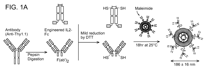
FIGs. 1A-1C show T-cell-targeted liposome synthesis and characterization. (A)
Schematic of immunoliposome preparation. (B) Typical particle size
distributions for liposomes
before antibody conjugation (open bars) and after conjugation (black filled
bars) determined by
dynamic light scattering. (C) Quantification of ligand (IL-2 cytokine
equivalent or anti-Thy1.1)
coupled to liposomes incorporating different mole fractions of maleimide-PEG
lipid: 2.5%
(black filled bar), 1% (open bar) or 0% (striped bar), assessed by IL-2 ELISA
and measuring
FITC-labeled anti-Thy1.1 incorporation respectively.
FIGs. 2A-2F show in vitro binding of IL-2-Fc-Lip and anti-Thy1.1 F(ab')2 -Lip
to
primary T-cells. (A) Flow cytometry analysis of cell surface expression of
CD25 and Thy1.1 on
naïve C57BL6/J (Thy1.1-) splenocytes vs. activated pmel-1 Thy1.1+CD8+ T-cells.
(B, C) Pmel-
1 CD8+ Thy1.1+ T-cells were incubated with 0.7 mg/ml (per 15x106 cells) DiD-
labeled
liposomes (IL-2-Fc or anti-Thy1.1 F(ab')2 conjugated) for 30 min at 37 C in
complete RPMI,
then analyzed by flow cytometry for liposome binding. Shown are representative
flow
cytometry scatter plots (B) and quantification of Mean Fluorescence Intensity
(MFI) of pmel-1
T-cells as a function of mol% of mal-PEG-DSPE included in the vesicles (C).
(D, E) Activated
pmel-1 CD8+ T-cells were mixed with naive C57BL/6 splenocytes in a 1:1 ratio
and incubated
with 0.07 mg/ml IL-2-Fc-Lip or 0.15 mg/ml anti-Thy1.1-Lip for 30 min at 37 C,
then analyzed
by flow cytometry. (D) Shown are scatter plots representing liposome
fluorescence on naive
C57BL/6 CD8+ T cells (Thy1.1-) and activated pmel-1 CD8+ T-cells (Thy1.1+)
with/without
0.24 mg/ml soluble IL-2-Fc or 1.34 mg/ml anti-Thy1.1 antibody added for 30 min
prior to
addition of liposomes. Cells incubated with 0.15 mg/ml IgG2a-lipo are also
shown. (E)
Quantification of the MFI of Pmel-1 CD8+ T cells when bound with respective
liposomes or pre-
blocked by free Ab/IL-2. (F) Titrated concentrations of fluorescent liposomes
were added to 5
x106 activated pmel-1 T-cells and incubated at 37 C for 30 min, then analyzed
by flow
cytometry for MFI of T-cell-associated liposomes. *, p<0.05; **, p<0.01; ***,
p<0.001.
CA 02953287 2016-12-02
WO 2014/204762
PCT/US2014/042004
9
FIG. 3 shows internalization of Thy1.1-targeted liposomes. Carboxy-fluorescein
(CF)-
labeled anti-Thy1.1-Lip (1.4 mg/ml) were incubated with 12x106 activated pmel-
1 CD8+ T-cells
in 500 jai RPMI containing 10% FCS for 1 hr at 4 C, washed, then incubated in
RPMI at 37 C
until analysis by flow cytometry 2 hr, 4 hr or 6 hrs later. (A) MFI of T-cell-
associated CF
fluorescence. (B) Confocal images of cells at time zero or after 6 hr at 37 C.
Scale bar= 20 p.m.
FIGs. 4A-4F show that IL-2-Fc- and anti-Thy1.1-Liposomes target transferred T-
cells in
vivo. C57B1/6 mice received i.v. adoptive transfer of 15x106pme1-1 CD8+Thy1.1+
T-cells,
followed by i.v. injection of 1.4 mg IL-2-Fc-Lip, anti-Thy1.1-Lip, or isotype
control IgG2a-Lip
either immediately after the T-cells or 3 days after the T-cells. Liposome
binding to cells
recovered from lymphoid organs and blood was analyzed 24 hr after liposome
injections by flow
cytometry. (A) Timeline of injections and analysis. (B) Representative flow
cytometry plots
illustrating gating strategy for analysis of liposome binding to transferred
pmel-1 T-cells or
endogenous CD8+ T-cells. (C) Representative histograms of pmel-1 T-cell or
endogenous
CD8+ T-cell labeling following day 0 liposome injections. (D-F) Quantification
of percentages
of endogenous or transferred T-cells labeled by day 0 or day 3 liposome
injections in the blood
(D), lymph nodes (E), and spleen (F). n=5 animals/group for IgG2a-Lip and anti-
Thy1.1-Lip and
n=3 for IL-2-Fc-Lip. *, p<0.05; **, p<0.01; ***, p<0.001.
FIGs. 5A-5E show that IL-2-Fc-liposomes allow repeated expansion of target ACT
T-
cells in vivo in tumor-bearing animals. (A-C) B16F10 tumor cells (1x10⁶) were
injected i.v. into
albino C57B1/6 mice and allowed to establish lung metastases for 7 days.
Animals were then
sublethally lymphodepleted by irradiation and received i.v. adoptive transfer
of 12x106
luciferase-expressing pmel-1 CD8+ T-cells the next day. One group of mice
additionally
received injections of IL-2-Fc-Lip (1 mg, carrying 60 lug IL-2-Fc or 20 lug IL-
2 cytokine
equivalent) i.v. immediately after T-cell transfer and again on day 6. (A)
Timelines of
cell/liposome injections and bioluminescence imaging of T-cells. (B)
Representative
bioluminescent images of ACT T-cells over time. (C) Quantification of average
whole-body T-
cell bioluminescence over time. (D-E) Groups of C57B/6 mice with established
lung metastases
were left untreated or were treated with T-cells as in A, then received either
IL-2-Fc-Lip or
equivalent total doses of systemic free IL-2 (10 lug day 0, 20 lug day 6)
injected i.v. on day 0 and
day 6. (D) Sample flow cytometry analyses showing percentages of tumor-
specific (vI313
CA 02953287 2016-12-02
WO 2014/204762
PCT/US2014/042004
TCR+) CD8+ T-cells among T-cells in inguinal lymph nodes on day 12 after
adoptive transfer.
(E) Quantification of average frequency of tumor-specific (vI313 TCR+) CD8+ T-
cells in
inguinal lymph nodes 12 days after adoptive transfer. n=3-4 animals/group. *,
p<0.05; **,
p<0.01.
5 FIG. 6A shows a graph representative of particle size of liposomes
measured by dynamic
light scattering. Medium gray: liposome-Maleimide; Dark gray: liposome-CD137;
Light gray:
liposome-IL-2-Fc. FIG. 6B shows a cryo-transmission electron microscopy (TEM)
image of
antibody-conjugated liposomes.
FIGs. 7A-7C show graphs representative of tumor growth inhibition (FIG. 7A),
relative
10 body weight changes (normalized to day 0) (FIG. 7B) and a survival curve
of the treatment (FIG.
7C) from B16-OVA tumor bearing mice that were given intravenous injections on
day 0, 2 and 4
with a 100 tg/dose of CD137 and a 20 1..tg/dose of IL-2-Fc (untreated, soluble
CD137/IL-2-Fc,
liposome-conjugated CD137 and liposome-conjugated IL-2-Fc, or liposome-
conjugated IgG).
FIG. 8 shows a graph representative of CD8+ T cell enrichment in peripheral
blood
mononucleated cells (PBMCs) on day 6 post injection. CD8+ T cell numbers were
analyzed by
flow cytometry.
FIGs. 9A-9B show graphs representative of intracellular cytokine staining of
IFNy (FIG.
9A) and TNFa (FIG. 9B) in CD8+ T cells from PBMC. Lymphocytes from PBMC (day 6
post
injection) were pulsed with 10 p.m OVA protein before analyzed by flow
cytometry.
FIGs. 10A-10C show graphs representative of tumor growth inhibition (FIG.
10A),
relative body weight changes (normalized to day 0) (FIG. 10B) and survival
curve of the
treatment (FIG. 10C) obtained from B16F10 tumor bearing mice that were given
intravenous
injections on day 0, 3 and 6 with a 100 tg/dose of CD137 and a 60 tg/dose of
IL-2-Fc.
FIG. 11 shows graphs representative of serum cytokine levels obtained from
mice after
systemic delivery of lipo-CD137/IL-2-Fc, showing prevention of lethal systemic
inflammatory
toxicity. Two days after single intravenous injection on B16F10 tumor bearing
mice, blood
serums were collected and serum cytokine levels were measured by LUMINEX
cytokine bead
assay.
CA 02953287 2016-12-02
WO 2014/204762
PCT/US2014/042004
11
DETAILED DESCRIPTION OF INVENTION
Aspects of the invention provide methods for augmenting lymphocyte function in
vivo by
repeated stimulation of lymphocytes in vivo using particles that comprise
stimulatory agents
and/or inhibitors of immunosuppression. In some embodiments, the lymphocytes
are adoptively-
transferred lymphocytes, such as those used in adoptive cell therapy, which
has been used in the
treatment of cancer. In some embodiments, the lymphocytes may be endogenous
lymphocytes.
Aspects of the invention are premised, in part, on the unexpected and, thus,
surprising finding
that repeated administration of particles comprising stimulatory agents and/or
inhibitors of
immunosuppression augments the activity of target cells in vivo more
efficiently than systemic
administration of stimulatory agents (see for example FIG. 5E). It is
therefore contemplated by
the invention that the beneficial effects of adoptive cell therapy may be
extended in time and
augmented in efficacy by boosting the activity and/or proliferation of
transferred cells at various
times post-transfer. Each administration of particles of the invention to a
subject may be
regarded as a "boost" since it will result in proliferation of the target
cells of interest (and thus
expansion of such cell populations), increased longevity of the target cells
of interest, and/or
increased activity of the target cells of interest.
Provided herein are experimental results evidencing specific targeting of
adoptive cell
therapy (ACT) T-cells (also referred to herein as adoptively-transferred T
cells) in vivo using
particles in the form of liposomes. Surprisingly, repeated systemic
administration to tumor-
bearing subjects did not lead to a toxic proinflammatory response, and
subjects survived and
cleared tumors, indicating that the easier route of administration (systemic,
instead of
intratumoral) is effective. In these illustrative examples, PEGylated
liposomes were conjugated
with two types of targeting molecules. The first type of targeting molecule is
an antibody against
a cell surface antigen expressed by the ACT T-cells. The cell surface antigen
may be one that
the target cell normally expresses or it may one that the target cell is made
to express, for
example, through genetic engineering strategies. An example of a cell surface
antigen is Thy1.1.
The second type of targeting molecule is a ligand, the receptor for which is
found on ACT T
cells. One such ligand is interleukin-2 (IL-2). IL-2 binds the trimeric IL-2
receptor (IL-2R)
expressed by activated T cells. These targeting molecules provide contrasting
targeting
strategies: anti-Thy1.1 provides highly specific targeting without overt
stimulation of target cells,
CA 02953287 2016-12-02
WO 2014/204762
PCT/US2014/042004
12
while IL-2 provides potentially less specific targeting (since IL-2R can be
expressed by some
endogenous T-cells) but also delivers a direct stimulatory signal to T cells.
Targeting liposomes were shown to label T cells in multiple systemic
compartments in
vivo, with anti-Thy1.1 liposomes binding to >90% of transferred cells
following a single
systemic injection. Additionally, multiple periodic administrations of
targeted stimulatory IL-2-
conjugated liposomes resulted in repeated expansion of ACT T-cells in vivo.
Accordingly, the aspects of the invention contemplate that these targeted
particle
strategies can be used to safely amplify the efficacy of ACT while avoiding
systemic toxicity
associated with many adjuvant drug treatments. Aspects of the invention
further contemplate
that the strategies are amenable and translatable to other immunotherapy
settings, such as
enhancement of cancer vaccines and therapeutic interventions in infectious
diseases such as
human immunodeficiency virus (HIV), which may rely on transferred cells and/or
on
endogenous cells.
It was also found, surprisingly, that administration of IL-2 conjugated to
particles of the
invention, and lacking another active agent, resulted in greater expansion of
tumor-reactive
CD8+ T cells as compared to the same dose of soluble IL-2. As described in
greater detail in the
Examples, administration of soluble IL-2 at the same dose provided no
enhancement in T cell
expansion. Accordingly, aspects of the invention also contemplate the use of
particles having
surface-conjugated IL-2 in expanding T cell populations in vivo, including but
not limited to
tumor-reactive T cells used in adoptive cell therapy (referred to as
adoptively-transferred T
cells).
Applications
Methods of the present disclosure embrace the unexpected findings that repeat
systemic
administration of agents, including targeting molecules, is therapeutically
effective when the
agents are delivered conjugated to a particle (e.g., liposome) and that in so
doing, it is possible to
administer the agents in a dose that would otherwise be toxic if administered
in soluble form. It
is to be understood that methods of the invention may be used in a variety of
applications in
which it is desirable to deliver agents specifically to a target cell
population (e.g., endogenous T
cells and/or adoptively-transferred T cells), and where it is desirable to
continually and
CA 02953287 2016-12-02
WO 2014/204762
PCT/US2014/042004
13
repeatedly boost an immune response (e.g., multiple boosts over the course of
several days or
weeks). One advantage of the methods of the invention is the ability to
deliver active agents to
particular cells of interest (e.g., lymphocytes), potentially at particular
regions of the interest in
the body, thereby avoiding the adverse effects associated with simple systemic
administration of
a soluble active agent.
Methods of the present disclosure may, therefore, be used in subjects
undergoing or who
have undergone adoptive cell therapy. Typically, such subjects have cancer or
are at risk of
developing cancer (e.g., they may be in remission or may be genetically or
environmentally
predisposed to developing cancer).
Methods of the present disclosure, however, may also be used in other
applications that
require enhanced immune responses, including prolonged enhanced immune
responses over a
period of time. Non-limiting examples include vaccine-based methods and cell-
based methods.
Target cells
Cells that may be targeted using particles of the invention may be those
occurring
endogenously in a subject, or those that are transferred (e.g., administered)
to a subject, for
example, for therapeutic or prophylactic benefit. A cell is considered
"endogenous" in a subject
if it originates from within the subject and has never been removed from the
subject. A cell is
considered an "adoptively-transferred cell" if it is obtained from a subject
and then transferred
back into the same subject or if it is obtained from a subject and transferred
into a new subject.
Adoptively-transferred cells include, for example, autologous subject-derived
(e.g., human
patient-derived) tumor-infiltrating lymphocytes as well as subject T cells
transduced with
engineered (e.g., genetically engineered) T cell receptors (TCRs). T cell
receptors may be, for
example, exogenous T cell chains or chimeric antigen receptors composed of
synthetic antigen-
binding Ig domains fused with TCR signaling components. A cell is considered
to be "subject-
derived" if it is obtained from (e.g., isolated from) the subject.
In the context of adoptive cell therapy, the target cells and the transferred
cells are
typically one and the same in the context of the invention. For example, tumor-
reactive CD8+ T
cells may be transferred to a subject in need of such therapy, and may also be
targeted by
particles of the invention. The target cells are typically immune cells such
as, but not limited, to
CA 02953287 2016-12-02
WO 2014/204762
PCT/US2014/042004
14
lymphocytes. Lymphocytes of the present disclosure may be T cells, such as
CD8+ T cells, B
cells or natural killer (NK) cells. In the context of adoptive cell therapy in
subjects having
cancer, the target cells are tumor-reactive (or tumor-specific) T cells.
Transferred cells may be
autologous to the subject being treated, or they may be allogeneic.
It is to be understood that any immune cell-based therapy may benefit from
methods of
the invention, including therapies that involve transfer of dendritic cells,
cell-based vaccines, and
the like and therapies that involve stimulation of endogenous lymphocytes.
Target cells may be tumor-reactive cells. This means that they recognize
and/or bind to
tumor cells and/or are involved in an immune response directed against the
tumor.
Target cells may be pathogen-reactive cells. This means that they recognize
and/or bind
to pathogens or pathogen-infected cells and/or are involved in an immune
response directed
against the pathogen or pathogen-infected cells.
Target cells (e.g., lymphocytes) of the present disclosure have cell surface
markers that
bind to (or are bound by) cognate recognition molecules (e.g., lymphocyte-
targeting molecules)
present on the surface of targeting particles (e.g., lymphocyte-targeting
particles). A "cell
surface marker" refers to a moiety present on the surface of cells that serves
as a marker of
specific cell types. Cell surface moieties include, without limitation, those
used for
immunophenotyping cells, such as CD (Classification Determinant) proteins.
Other cell surface
moieties are contemplated herein. It should be understood that cell-specific
targeting molecules
present on particles of the present disclosure typically confer cell-specific
targeting of the
particles. Thus, for example, a liposome conjugated to an anti-CD137 antibody
is considered a
lymphocyte-targeting particle (and more specifically, a T cell-targeting
particle) because anti-
CD137 antibody is a lymphocyte-targeting molecule that specifically recognizes
and binds to
CD137, which is expressed on T cells, thereby targeting the particle to the T
cells.
Examples of lymphocyte cell surface markers include, without limitation, ART2,
CD 1 a,
CD 1d, CD2, CD3, CD4, CD5, CD7, CD8, CD11b, CD25, CD28, CD38, CD45RO, CD72,
CD134, CD137, CD150, CD154, CRTAM, FOXP3, FT2, GPCA, HLA-DR, HML-1, HT23A,
LEU-22, LFA-1, LY-2, LY-M22, MICG, MRC-OX-8, MRC-OX-22, OX-40, PD-1, RT-6,
TCR,
THY-1 (CD90), TIM-3, CTLA-4 and TSA-2. Other cell-type specific (e.g.,
lymphocyte
specific) surface markers are contemplated herein.
CA 02953287 2016-12-02
WO 2014/204762
PCT/US2014/042004
Targeting Molecules
"Targeting molecules" refers to molecules (e.g., ligands, receptors and/or
antibodies/antibody fragments) that bind to (e.g., bind specifically to)
target cells of interest (e.g.,
lymphocytes). A targeting molecule is considered to bind to a target cell if
it binds to a cell
5 surface marker (e.g., antigen, ligand, receptor) of the target cell. In
some embodiments, targeting
molecules bind specifically to particular target cells ¨ that is, they bind to
cell surface markers
that are present only on the particular target cells. Thus, a targeting
molecule is considered to
bind specifically to a T cell if it binds a cell surface marker that is
expressed only on T cells.
In the context of adoptive cell therapy, for example, adoptively-transferred T
cells in a
10 subject may uniquely express a cell surface marker (e.g., Thy1.1), which
itself may be
considered, for example, a ligand or a receptor. A cell surface marker that is
"uniquely
expressed" by a particular cell type is expressed by no other cell types.
Thus, "specific binding"
occurs, for example, when an anti-Thy1.1 antibody that is conjugated to a T
cell-targeting
particle binds to Thy1.1 on the surface of T cells. Adoptively-transferred
cells may naturally
15 express a unique marker or they may be modified to express a unique
marker. Such modification
may include, without limitation, genetic engineering of the adoptively-
transferred cells.
Targeting molecules (e.g., ligands or antibodies) that are bound by (or bind
to)
lymphocytes are referred to herein as "lymphocyte-targeting molecules."
Lymphocyte-targeting
molecules include lymphocyte-targeting ligands and lymphocyte-targeting
antibodies and
antibody fragments such as a Fab fragment.
Ligands that are bound by (or that bind to) lymphocytes may be referred to
herein as
"lymphocyte-targeting ligands" or "lymphocyte-specific ligands." Examples of
lymphocyte-
targeting ligands include, without limitation, cytokines, which as used
generally herein
encompass cytokines, interleukins, chemokines and growth factors. Non-limiting
examples of
cytokines include IL-2, IL-7, IL-15, CXCL10, CXCL5, MIP-la and MIP- lb. In
some
embodiments, the cytokine is IL-2. In some embodiments, a ligand may be in the
form of an Fc
fusion protein. For example, an IL-2 ligand may be an IL-2-Fc fusion protein.
Other non-
limiting examples of Fc fusion proteins include IL-7, IL-15, CXCL10, CXCL5,
MIP-la and
MIP- lb Fc fusion proteins. Other ligands and Fc fusion proteins are
contemplated herein.
CA 02953287 2016-12-02
WO 2014/204762
PCT/US2014/042004
16
Antibodies that are bound by (or that bind to) lymphocytes may be referred to
herein as
"lymphocyte-targeting antibodies" or "lymphocyte-specific antibodies."
Antibody fragments
that are bound by (or that bind to) lymphocytes may be referred to herein as
"lymphocyte-
targeting antibody fragments" or "lymphocyte-specific antibody fragments."
Examples of
lymphocyte-targeting antibodies include, without limitation, antibodies that
bind specifically to
ART2, CD 1 a, CD 1d, CD2, CD3, CD4, CD5, CD7, CD8, CD11b, CD25, CD28, CD38,
CD45RO, CD72, CD134, CD137, CD150, CD154, CRTAM, FOXP3, FT2, GPCA, HLA-DR,
HML-1, HT23A, LEU-22, LFA-1, LY-2, LY-M22, MICG, MRC-OX-8, MRC-OX-22, OX-40,
PD-1, RT-6, TCR, THY-1 (CD90), TIM-3, CTLA-4 or TSA-2. Also contemplated
herein are
immunostimulatory antibodies including, without limitation, anti-PD-1, anti-
CTLA4, anti-PDL1
and anti-LaG3 antibodies. Antibody fragments of any of the foregoing
antibodies are also
contemplated herein. Other antibodies and antibody fragments are contemplated
herein. In
some embodiments, antibodies used in accordance with the present disclosure
are monoclonal
antibodies. In some embodiments, antibodies used in accordance with the
present disclosure are
chimeric antibodies.
It should be understood that a targeting particle of the present invention may
comprise at
least one (e.g., two or more) targeting molecules that are the same as each
other (e.g., targeting
ligands) or different from each other (e.g., targeting ligands and targeting
antibodies). For
example, a lymphocyte-targeting particle may comprise an anti-CD137 antibody
and IL-2.
Alternatively, a population of lymphocyte-targeting particles may comprise a
portion of
lymphocyte-targeting particles (e.g., half) that comprise one type of
lymphocyte-targeting
molecule (e.g., anti-CD137 antibody), and another portion of lymphocyte-
targeting particles that
comprise another, different, type of lymphocyte-targeting molecule (e.g., IL-
2). Thus, mixtures
of different targeting particles are contemplated herein. In some embodiments,
10%, 20%, 30%,
40%, 50%, 60%, 70%, 80% or 90% of a mixture comprises one type of lymphocyte-
targeting
molecule, while the remaining portion or portions of the mixture comprise(s)
another type(s) of
lymphocyte-targeting molecule(s).
CA 02953287 2016-12-02
WO 2014/204762
PCT/US2014/042004
17
Targeting Particles
Compositions and methods of the invention involve targeting particles (also
referred to as
targeted particles). "Targeting particles" refers to particles that comprise
on their surface
targeting molecules (e.g., ligands, receptors and/or antibodies/antibody
fragments) that bind to
(or are bound by) cell surface markers on target cells of interest, such as
lymphocytes (e.g., T
cells). A targeting particle is considered to comprise a targeting molecule on
its surface if the
targeting molecule is associated with or interacts with (e.g., is covalently
or non-covalently
conjugated to/bound to) the surface of the targeting particle.
"Particles," as used herein, refer to particulate carriers (e.g., are capable
of transporting
molecules), optionally with active agent encapsulated in or bound to (e.g.,
covalently or non-
covalently conjugated to) the particle surface. Examples of particles of the
present disclosure
include, without limitation, liposomes and polymeric particles, described in
greater detail below.
Targeting particles that are bound by (or bind to) targeting molecules that
bind to (e.g.,
bind specifically to) lymphocytes (e.g., bind to lymphocyte cell surface
markers) are referred to
as "lymphocyte-targeting particles."
Particles of the present disclosure may be of any suitable size. As used
herein,
nanoparticles are particles of approximate nanometer dimensions. As used
herein, microparticles
are particles of approximate micrometer dimensions. The invention contemplates
the use of
nanoparticles and/or microparticles.
The diameter of a particle may range from 1-1000 nanometers (nm). In some
embodiments, the diameter ranges in size from 20 to 750 nm, from 20 to 500 nm,
or from 20 to
250 nm. In some embodiments, the diameter ranges in size from 50 to 750 nm,
from 50 to 500
nm, from 50 to 250 nm, or from 100-300 nm. In some embodiments, the diameter
is 100, 150,
200 nm, 250 nm or 300 nm. In some embodiments, the diameter ranges in size
from about 20 to
750 nm, from about 20 to 500 nm, or from about 20 to 250 nm. In some
embodiments, the
diameter ranges in size from about 50 to 750 nm, from about 50 to 500 nm, from
about 50 to 250
nm, or from about 100-300 nm. In some embodiments, the diameter is about 100,
about 150,
about 200 nm, about 250 nm or about 300 nm.
In some embodiments, the diameter of a microparticle may range from 0.1 i.tm
to 100 i.tm
(or about 0.1 i.tm to about 100 i.tm), 0.1 i.tm to 90 i.tm, 0.1 i.tm to 80
i.tm, 0.1 i.tm to 70 i.tm, 0.1 i.tm
CA 02953287 2016-12-02
WO 2014/204762
PCT/US2014/042004
18
to 60 i.tm, 0.1 i.tm to 50 i.tm, 0.1 i.tm to 40 i.tm, 0.1 i.tm to 30 i.tm, 0.1
i.tm to 20 i.tm, 0.1 i.tm to 10
i.tm, 0.1 i.tm to 5 i.tm, 0.1 i.tm to 4 i.tm, 0.1 i.tm to 3 i.tm, 0.1 i.tm to
2 i.tm or 0.1 i.tm to 1 i.tm.
As used in the context of particle sizes and diameters, the term "about" means
+/- 5% of
the absolute value stated.
In some embodiments, particles of the present disclosure comprise an active
agent and
release the active agent over a period of time, ranging from hours to days.
The particles may
gradually degrade in an aqueous environment, such as occurs in vivo. If active
agents are
dispersed throughout the particles, then release of the active agents will
occur as the outermost
layers of the particle degrade or as pores within the particle enlarge.
In some embodiments, particles of the present disclosure comprise an active
agent and
release the active agent all at once as the particle "bursts."
Particles of the present disclosure are administered in a cell-free
formulation. This means
that they are not bound to cells and are not formulated with cells prior to
administration. As
described above, particles of the present disclosure may be referred to herein
as "lymphocyte-
targeting particle." Lymphocyte-targeting particles are able to target
lymphocytes in vivo
without the assistance of carrier cells or other carrier vehicles.
Particles of the present disclosure may be endocytosed when used in vivo,
although
methods of the invention are not dependent upon endocytosis of the particles.
In some embodiments, particles are porous particles. In some embodiments,
particles are
hollow core particles. Particles of the present disclosure are not viruses or
particles thereof (e.g.,
virus-like particles (VLPs)). Particles of the present disclosure, in some
embodiments, are
biodegradable and, thus, typically are not magnetic. Biodegradable particles
may be synthesized
using methods known in the art including, without limitation, solvent
evaporation, hot melt
microencapsulation, solvent removal and spray drying. Exemplary methods for
synthesizing
particles are described in Bershteyn et al., Soft Matter 4:1787-1787, 2008 and
in US
2008/0014144 Al, the specific teachings of which relating to particle
synthesis are incorporated
herein by reference.
Particles of the present disclosure may be natural particles or synthetic
polymer-based
particles (including nucleic acid-based particles) or they may be lipid-based
particles, such as
liposomes. They may be natural or synthetic polymer-based particles having a
lipid coating. In
CA 02953287 2016-12-02
WO 2014/204762
PCT/US2014/042004
19
some embodiments, particles of the invention are multilamellar lipid vesicles
(e.g., interbilayer-
crosslinked multilameller lipid vesicles) (e.g., Moon et al., Nature Materials
10, 243-251
(2011)).
Natural or synthetic polymer based particles
In some embodiments, particles of the present disclosure are formed from
polymers
including, without limitation, aliphatic polyesters, poly (lactic acid) (PLA),
poly (glycolic acid)
(PGA), co-polymers of lactic acid and glycolic acid (PLGA), polycarprolactone
(PCL),
polyanhydrides, poly(ortho)esters, polyurethanes, poly(butyric acid),
poly(valeric acid), and
poly(lactide-co-caprolactone), and natural polymers such as alginate and other
polysaccharides
including dextran and cellulose, collagen, chemical derivatives thereof,
including substitutions,
additions of chemical groups such as for example alkyl, alkylene,
hydroxylations, oxidations,
and other modifications routinely made by those skilled in the art), albumin
and other
hydrophilic proteins, zein and other prolamines and hydrophobic proteins,
copolymers and
mixtures thereof. In some embodiments, the particles may be biodegradable
particles such as,
for example, particles having a biodegradable polymer core. Such particles are
described in
greater detail in U.S. application number US 2008/0014144 Al, Bershteyn et
al., Soft Matter,
4:1787-1787, 2008, published international application number WO 2010/059253,
and published
U.S. application number 2011/0229556 Al, each of which is incorporated by
reference herein.
In some embodiments, the particles may comprise a nucleic acid core,
optionally with a
lipid coating. Such "DNA particles" or "DNA-hydrogel particles" are described
in greater detail
in published U.S. application number US 2007/0148246, the teachings of which
are incorporated
by reference herein.
In some embodiments, the particles may comprise a lipid bilayer on their
outermost
surface. This bilayer may be comprised of one or more lipids of the same or
different type.
Examples include, without limitation, phospholipids such as phosphocholines
and
phosphoinositols. Specific examples include, without limitation, DMPC, DOPC,
DSPC, DOPG
and various other lipids.
CA 02953287 2016-12-02
WO 2014/204762
PCT/US2014/042004
Lipid based particles
In some embodiments, particles are liposomes. Liposomes are vesicles
comprising at
least one lipid bilayer and an internal typically aqueous compartment.
Liposomes may be
anionic, neutral or cationic. Liposomes may comprise, without limitation,
DOPC, DOPG,
5 DOTMA, DOTAP, DOTIM, DDAB, alone or together with cholesterol, to yield
DOTMA and
cholesterol, DOTAP and cholesterol, DOTIM and cholesterol, and DDAB and
cholesterol. In
some embodiments, the particles of the invention may be unilamellar liposomal
vesicles. In
some embodiments, the particles of the invention may be multilamellar
liposomal vesicles. In
some embodiments, the particles may be interbilayer crosslinked multilamellar
vesicles
10 (ICMVs), which are multilamellar lipid vesicles having crosslinked lipid
bilayers. Such particles
are described in greater detail in U.S. application numbers US 2011/0229529 Al
and US
2012/0177724 Al, each of which is incorporated by reference herein.
Particle conjugation
15 In some embodiments, particles comprise antibodies or antibody fragments
on their
surface. In some embodiments, the particles comprise non-antibody-based
ligands on their
surface. Non-antibody based ligands include, but are not limited, to
cytokines, a term used
generically to embrace cytokines, interleukins, and growth factors generally.
In some embodiments, the antibodies are designed to bind to target cells
without
20 triggering their elimination by complement or other antibody effector
mechanisms. This is
achieved either by using antibody fragments or antibodies with mutations that
abrogate Fc
receptor binding or other effector mechanisms.
These antibody and non-antibody based ligands may be conjugated (or attached
or bound,
as the terms are used interchangeably herein) to the particle surface
covalently or non-covalently.
The particles may be synthesized or modified post-synthesis to comprise one or
more reactive
groups on their exterior surface that can be used to conjugate the antibody
and non-antibody
based ligands. These particle reactive groups include without limitation thiol-
reactive maleimide
head groups, haloacetyl (e.g., iodoacetyl) groups, imidoester groups, N-
hydroxysuccinimide
esters, pyridyl disulfide groups, and the like. As an example, particles may
be synthesized to
include maleimide conjugated phospholipids such as, without limitation, DSPE-
MaL-PEG2000.
CA 02953287 2016-12-02
WO 2014/204762
PCT/US2014/042004
21
It will be understood that when surface modified in this manner, the particles
are intended for use
with ligands having "complementary" reactive groups (i.e., reactive groups
that react with those
of the particles).
Methods for conjugating ligands or receptors such as antibodies to particle
surfaces are
described by Kwong et al. Cancer Research, 2013, 73:1547-1558, the entire
contents of which
are incorporated by reference herein.
Agents
The invention contemplates the delivery of agents to particular cells, and
thus potentially
to localized regions or tissues in vivo. As used herein, an agent is any atom
or molecule or
compound that can be used to provide benefit to a subject (including without
limitation
prophylactic or therapeutic benefit). The agents of particular interest, in
some embodiments, are
those that exert an effect on target cells, whether directly or indirectly.
Some agents may exert
their effects on tumor cells, pathogens, or pathogen-infected cells. The
nature of the agent will
depend on the particular application, as should be apparent.
The particles may carry the agent internally including for example in pores or
in a hollow
core. The particles may carry the agent on its surface. The particles may
carry the agent
internally and on its surface.
The invention further contemplates that one or more agents may be used
alongside of the
particles of the invention, although not conjugated to or encapsulated within.
For example, the
particles of the invention may be formulated together with one or more agents.
The agent may be without limitation a chemical entity, a protein, a
polypeptide, a peptide,
a nucleic acid, a virus-like particle, a steroid, a proteoglycan, a lipid, a
carbohydrate, and
analogs, derivatives, mixtures, fusions, combinations or conjugates thereof.
The agent may be a
pro-drug that is metabolized and thus converted in vivo to its active (and/or
stable) form.
The agents may be naturally occurring or non-naturally occurring. Naturally
occurring
agents include those capable of being synthesized by the subjects to whom the
particles are
administered. Non-naturally occurring are those that do not exist in nature
normally, whether
produced by plant, animal, microbe or other living organism.
CA 02953287 2016-12-02
WO 2014/204762
PCT/US2014/042004
22
One class of agents that can be delivered in a localized manner using the
particles of the
invention includes chemical compounds that are non-naturally occurring, or
chemical
compounds that are not naturally synthesized by mammalian (and in particular
human) cells.
A variety of agents that are currently used for therapeutic purposes can be
delivered
according to the invention and these include without limitation
immunomodulatory agents such
as immunostimulatory agents, antigens, adjuvants, imaging agents, anti-cancer
agents, anti-
infective agents, and the like.
One particular class of agents is inhibitors of immunosuppression. Examples
include
Shp1/2 protein tyrosine phosphatase (PTPase) inhibitor (NSC-87877; CAS 56932-
43-5),
sunitinib, or other inhibitors of receptor tyrosine kinases, or p38 MAPK
inhibitors including
MAPK pathway inhibitors.
The p38 MAPK pathway inhibitor may be a RAF inhibitor such as a pan-RAF
inhibitor
or a selective RAF inhibitor. Examples of RAF inhibitors include RAF265,
sorafenib,
dabrafenib (GSK2118436), 5B590885, PLX 4720, PLX4032, GDC-0879 and ZM 336372.
The p38 MAPK pathway inhibitor may be a MEK inhibitor. Examples of MEK
inhibitors include CI-1040/PD184352, AZD6244, PD318088, PD98059, PD334581,
RDEA119,
6-Methoxy-7-(3-morpholin-4-yl-propoxy)-4-(4-phenoxy-phenylamino)-quinoline-3-
carbonitrile
and 4-[3-Chloro-4-(1-methy1-1H-imidazol-2-ylsulfany1)-phenylaminol-6-methoxy-7-
(3-
morpholin-4-yl-propoxy)-quinoline-3-carbonitrile, trametinib (GSK1120212), and
ARRY-
438162.
The p38 MAPK pathway inhibitor may be an ERK inhibitor. Examples of ERK
inhibtors
include VTX11e, AEZS-131, PD98059, FR180204, and FR148083.
Still other p38 MAPK inhibitors are Tocriset, 5B239063, 5B203580, pamapimodõ
dilmapimod, and PH797804.
Imaging Agents. As used herein, an imaging agent is an agent that emits signal
directly
or indirectly thereby allowing its detection in vivo. Imaging agents such as
contrast agents and
radioactive agents that can be detected using medical imaging techniques such
as nuclear
medicine scans and magnetic resonance imaging (MRI). Imaging agents for
magnetic resonance
imaging (MRI) include Gd(DOTA), iron oxide or gold nanoparticles; imaging
agents for nuclear
medicine include 201T1, gamma-emitting radionuclide 99 mTc; imaging agents for
positron-
CA 02953287 2016-12-02
WO 2014/204762
PCT/US2014/042004
23
emission tomography (PET) include positron-emitting isotopes, (18)F-
fluorodeoxyglucose
((18)FDG), (18)F-fluoride, copper-64, gadoamide, and radioisotopes of Pb(II)
such as 203 Pb,
and 11In; imaging agents for in vivo fluorescence imaging such as fluorescent
dyes or dye-
conjugated nanoparticles. In other embodiments, the agent to be delivered is
conjugated, or
fused to, or mixed or combined with an imaging agent.
Immunostimulatory Agents. As used herein, an immunostimulatory agent is an
agent that
stimulates an immune response (including enhancing a pre-existing immune
response) in a
subject to whom it is administered, whether alone or in combination with
another agent.
Examples include antigens, adjuvants (e.g., TLR ligands such as imiquimod,
imidazoquinoline,
nucleic acids comprising an unmethylated CpG dinucleotide, monophosphoryl
lipid A or other
lipopolysaccharide derivatives, single-stranded or double-stranded RNA,
flagellin, muramyl
dipeptide), cytokines including interleukins (e.g., IL-2, IL-7, IL-15 (or
superagonist/mutant
forms of these cytokines), IL-12, IFN-gamma, IFN-alpha, GM-CSF, FLT3-ligand,
etc.),
immunostimulatory antibodies (e.g., anti-CTLA-4, anti-CD28, anti-CD3, or
single
chain/antibody fragments of these molecules), and the like.
Adjuvants. The adjuvant may be without limitation alum (e.g., aluminum
hydroxide,
aluminum phosphate); saponins purified from the bark of the Q. saponaria tree
such as QS21 (a
glycolipid that elutes in the 21st peak with HPLC fractionation; Antigenics,
Inc., Worcester,
Mass.); poly[di(carboxylatophenoxy)phosphazene (PCPP polymer; Virus Research
Institute,
USA), F1t3 ligand, Leishmania elongation factor (a purified Leishmania
protein; Corixa
Corporation, Seattle, Wash.), ISCOMS (immunostimulating complexes which
contain mixed
saponins, lipids and form virus-sized particles with pores that can hold
antigen; CSL, Melbourne,
Australia), Pam3Cys, SB-A54 (SmithKline Beecham adjuvant system #4 which
contains alum
and MPL; SBB, Belgium), non-ionic block copolymers that form micelles such as
CRL 1005
(these contain a linear chain of hydrophobic polyoxypropylene flanked by
chains of
polyoxyethylene, Vaxcel, Inc., Norcross, Ga.), and Montanide IMS (e.g., IMS
1312, water-based
nanoparticles combined with a soluble immunostimulant, Seppic)
Adjuvants may be TLR ligands. Adjuvants that act through TLR3 include without
limitation double-stranded RNA. Adjuvants that act through TLR4 include
without limitation
derivatives of lipopolysaccharides such as monophosphoryl lipid A (MPLA; Ribi
ImmunoChem
CA 02953287 2016-12-02
WO 2014/204762
PCT/US2014/042004
24
Research, Inc., Hamilton, Mont.) and muramyl dipeptide (MDP; Ribi) andthreonyl-
muramyl
dipeptide (t-MDP; Ribi); 0M-174 (a glucosamine disaccharide related to lipid
A; OM Pharma
SA, Meyrin, Switzerland). Adjuvants that act through TLR5 include without
limitation flagellin.
Adjuvants that act through TLR7 and/or TLR8 include single-stranded RNA,
oligoribonucleotides (ORN), synthetic low molecular weight compounds such as
imidazoquinolinamines (e.g., imiquimod, resiquimod). Adjuvants acting through
TLR9 include
DNA of viral or bacterial origin, or synthetic oligodeoxynucleotides (ODN),
such as CpG ODN.
Another adjuvant class is phosphorothioate containing molecules such as
phosphorothioate
nucleotide analogs and nucleic acids containing phosphorothioate backbone
linkages.
Immunoinhibitory Agents. As used herein, an immunoinhibitory agent is an agent
that
inhibits an immune response in a subject to whom it is administered, whether
alone or in
combination with another agent. Examples include steroids, retinoic acid,
dexamethasone,
cyclophosphamide, anti-CD3 antibody or antibody fragment, and other
immunosuppressants.
Anti-Cancer Agents. As used herein, an anti-cancer agent is an agent that at
least
partially inhibits the development or progression of a cancer, including
inhibiting in whole or in
part symptoms associated with the cancer even if only for the short term.
Several anti-cancer
agents can be categorized as DNA damaging agents and these include
topoisomerase inhibitors
(e.g., etoposide, ramptothecin, topotecan, teniposide, mitoxantrone), DNA
alkylating agents
(e.g., cisplatin, mechlorethamine, cyclophosphamide, ifosfamide, melphalan,
chorambucil,
busulfan, thiotepa, carmustine, lomustine, carboplatin, dacarbazine,
procarbazine), DNA strand
break inducing agents (e.g., bleomycin, doxorubicin, daunorubicin, idarubicin,
mitomycin C),
anti-microtubule agents (e.g., vincristine, vinblastine), anti-metabolic
agents (e.g., cytarabine,
methotrexate, hydroxyurea, 5-fluorouracil, floxuridine, 6-thioguanine, 6-
mercaptopurine,
fludarabine, pentostatin, chlorodeoxyadenosine), anthracyclines, vinca
alkaloids. or
epipodophyllotoxins.
Examples of anti-cancer agents include without limitation Acivicin;
Aclarubicin;
Acodazole Hydrochloride; Acronine; Adozelesin; Aldesleukin; Altretamine;
Ambomycin;
Ametantrone Acetate; Aminoglutethimide; Amsacrine; Anastrozole; Anthramycin;
Asparaginase; Asperlin; Azacitidine; Azetepa; Azotomycin; Batimastat;
Benzodepa;
Bicalutamide; Bisantrene Hydrochloride; Bisnafide Dimesylate; Bizelesin;
Bleomycin Sulfate;
CA 02953287 2016-12-02
WO 2014/204762
PCT/US2014/042004
Bortezomib (VELCADE); Brequinar Sodium; Bropirimine; Busulfan; Cactinomycin;
Calusterone; Caracemide; Carbetimer; Carboplatin (a platinum-containing
regimen);
Carmustine; Carubicin Hydrochloride; Carzelesin; Cedefingol; Chlorambucil;
Cirolemycin;
Cisplatin (a platinum-containing regimen); Cladribine; Crisnatol Mesylate;
Cyclophosphamide;
5 Cytarabine; Dacarbazine; Dactinomycin; Daunorubicin; Decitabine;
Dexormaplatin;
Dezaguanine; Diaziquone; Docetaxel (TAXOTERE); Doxorubicin; Droloxifene;
Dromostanolone; Duazomycin; Edatrexate; Eflornithine; Elsamitrucin;
Enloplatin; Enpromate;
Epipropidine; Epirubicin; Erbulozole; Erlotinib (TARCEVA), Esorubicin;
Estramustine;
Etanidazole; Etoposide; Etoprine; Fadrozole; Fazarabine; Fenretinide;
Floxuridine; Fludarabine;
10 5-Fluorouracil; Flurocitabine; Fosquidone; Fostriecin; Gefitinib
(IRESSA), Gemcitabine;
Hydroxyurea; Idarubicin; Ifosfamide; Ilmofosine; Imatinib mesylate (GLEEVAC);
Interferon
alpha-2a; Interferon alpha-2b; Interferon alpha-n1; Interferon alpha-n3;
Interferon beta-I a;
Interferon gamma-I b; Iproplatin; Irinotecan; Lanreotide; Lena I domide (REVL
IN1 ID,
REVIMID); Letrozole; Leuprolide; Liarozole; Lometrexol; Lomustine;
Losoxantrone;
15 Masoprocol; Maytansine; Mechlorethamine; Megestrol; Melengestrol;
Melphalan; Menogaril;
Mercaptopurine; Methotrexate; Metoprine; Meturedepa; Mitindomide; Mitocarcin;
Mitocromin;
Mitogillin; Mitomalcin; Mitomycin; Mitosper; Mitotane; Mitoxantrone;
Mycophenolic Acid;
Nocodazole; Nogalamycin; Ormaplatin; Oxisuran; Paclitaxel; Pemetrexed
(ALIMTA),
Pegaspargase; Peliomycin; Pentamustine; Pentomone; Peplomycin; Perfosfamide;
Pipobroman;
20 Piposulfan; Piritrexim Isethionate; Piroxantrone; Plicamycin;
Plomestane; Porfimer;
Porfiromycin; Prednimustine; Procarbazine; Puromycin; Pyrazofurin; Riboprine;
Rogletimide;
Safingol; Semustine; Simtrazene; Sitogluside; Sparfosate; Sparsomycin;
Spirogermanium;
Spiromustine; Spiroplatin; Streptonigrin; Streptozocin; Sulofenur;
Talisomycin; Tamsulosin;
Taxol; Taxotere; Tecogalan; Tegafur; Teloxantrone; Temoporfin; Temozolomide
(TEMODAR);
25 Teniposide; Teroxirone; Testolactone; Thalidomide (THALOMID) and
derivatives thereof;
Thiamiprine; Thioguanine; Thiotepa; Tiazofurin; Tirapazamine; Topotecan;
Toremifene;
Trestolone; Triciribine; Trimetrexate; Triptorelin; Tubulozole; Uracil
Mustard; Uredepa;
Vapreotide; Verteporfin; Vinblastine; Vincristine; Vindesine; Vinepidine;
Vinglycinate;
Vinleurosine; Vinorelbine; Vinrosidine; Vinzolidine; Vorozole; Zeniplatin;
Zinostatin;
Zorubicin.
CA 02953287 2016-12-02
WO 2014/204762
PCT/US2014/042004
26
The anti-cancer agent may be an enzyme inhibitor including without limitation
tyrosine
kinase inhibitor, a CDK inhibitor, a MAP kinase inhibitor, or an EGFR
inhibitor. The tyrosine
kinase inhibitor may be without limitation Genistein (4' ,5,7-
trihydroxyisoflavone), Tyrphostin
25 (3,4,5-trihydroxyphenyl), methylene]-propanedinitrile, Herbimycin A,
Daidzein (4',7-
dihydroxyisoflavone), AG-126, trans-143'-carboxy-4'-hydroxypheny1)-242",5"-
dihydroxy-
phenyl)ethane, or HDBA (2-Hydroxy5-(2,5-Dihydroxybenzylamino)-2-hydroxybenzoic
acid.
The CDK inhibitor may be without limitation p21, p27, p57, p15, p16, p18, or
p19. The MAP
kinase inhibitor may be without limitation KY12420 (C23H2408), CNI-1493,
PD98059, or 444-
Fluoropheny1)-2-(4-methylsulfinyl phenyl)-5-(4-pyridyl) 1H-imidazole. The EGFR
inhibitor
may be without limitation erlotinib (TARCEVA), gefitinib (IRESSA), WHI-P97
(quinazoline
derivative), LFM-Al2 (leflunomide metabolite analog), ABX-EGF, lapatinib,
canertinib, ZD-
6474 (ZACTIMA), AEE788, and AG1458.
The anti-cancer agent may be a VEGF inhibitor including without limitation
bevacizumab (AVASTIN), ranibizumab (LUCENTIS), pegaptanib (MACUGEN),
sorafenib,
sunitinib (SUTENT), vatalanib, ZD-6474 (ZACTIMA), anecortave (RETAANE),
squalamine
lactate, and semaphorin.
The anti-cancer agent may be an antibody or an antibody fragment including
without
limitation an antibody or an antibody fragment including but not limited to
bevacizumab
(AVASTIN), trastuzumab (HERCEPTIN), alemtuzumab (CAMPATH, indicated for B cell
chronic lymphocytic leukemia,), gemtuzumab (MYLOTARG, hP67.6, anti-CD33,
indicated for
leukemia such as acute myeloid leukemia), rituximab (RITUXAN), tositumomab
(BEXXAR,
anti-CD20, indicated for B cell malignancy), MDX-210 (bispecific antibody that
binds
simultaneously to HER-2/neu oncogene protein product and type I Fc receptors
for
immunoglobulin G (IgG) (Fc gamma RI)), oregovomab (OVAREX, indicated for
ovarian
cancer), edrecolomab (PANOREX), daclizumab (ZENAPAX), palivizumab (SYNAGIS,
indicated for respiratory conditions such as RSV infection), ibritumomab
tiuxetan (ZEVALIN,
indicated for Non-Hodgkin's lymphoma), cetuximab (ERBITUX), MDX-447, MDX-22,
MDX-
220 (anti-TAG-72), IOR-05, IOR-T6 (anti-CD1), IOR EGF/R3, celogovab (ONCOSCINT
OV103), epratuzumab (LYMPHOCIDE), pemtumomab (THERAGYN), and Gliomab-H
(indicated for brain cancer, melanoma).
CA 02953287 2016-12-02
WO 2014/204762
PCT/US2014/042004
27
Anti-Infective Agents. The agent may be an anti-infective agent including
without
limitation an anti-bacterial agent, an anti-viral agent, an anti-parasitic
agent, an anti-fungal agent,
and an anti-mycobacterial agent.
Anti-bacterial agents may be without limitation 13-1actam antibiotics,
penicillins (such as
natural penicillins, aminopenicillins, penicillinase-resistant penicillins,
carboxy penicillins,
ureido penicillins), cephalosporins (first generation, second generation, and
third generation
cephalosporins), other 13-1actams (such as imipenem, monobactams), 13-
1actamase inhibitors,
vancomycin, aminoglycosides and spectinomycin, tetracyclines, chloramphenicol,
erythromycin,
lincomycin, clindamycin, rifampin, metronidazole, polymyxins, sulfonamides and
trimethoprim,
or quinolines.
Other anti-bacterials may be without limitation Acedapsone; Acetosulfone
Sodium;
Alamecin; Alexidine; Amdinocillin; Amdinocillin Pivoxil; Amicycline;
Amifloxacin;
Amifloxacin Mesylate; Amikacin; Amikacin Sulfate; Aminosalicylic acid;
Aminosalicylate
sodium; Amoxicillin; Amphomycin; Ampicillin; Ampicillin Sodium; Apalcillin
Sodium;
Apramycin; Aspartocin; Astromicin Sulfate; Avilamycin; Avoparcin;
Azithromycin; Azlocillin;
Azlocillin Sodium; Bacampicillin Hydrochloride; Bacitracin; Bacitracin
Methylene Disalicylate;
Bacitracin Zinc; Bambermycins; Benzoylpas Calcium; Berythromycin; Betamicin
Sulfate;
Biapenem; Biniramycin; Biphenamine Hydrochloride; Bispyrithione Magsulfex;
Butikacin;
Butirosin Sulfate; Capreomycin Sulfate; Carbadox; Carbenicillin Disodium;
Carbenicillin
Indanyl Sodium; Carbenicillin Phenyl Sodium; Carbenicillin Potassium;
Carumonam Sodium;
Cefaclor; Cefadroxil; Cefamandole; Cefamandole Nafate; Cefamandole Sodium;
Cefaparole;
Cefatrizine; Cefazaflur Sodium; Cefazolin; Cefazolin Sodium; Cefbuperazone;
Cefdinir;
Cefepime; Cefepime Hydrochloride; Cefetecol; Cefixime; Cefmenoxime
Hydrochloride;
Cefmetazole; Cefmetazole Sodium; Cefonicid Monosodium; Cefonicid Sodium;
Cefoperazone
Sodium; Ceforanide; Cefotaxime Sodium; Cefotetan; Cefotetan Disodium; Cefotiam
Hydrochloride; Cefoxitin; Cefoxitin Sodium; Cefpimizole; Cefpimizole Sodium;
Cefpiramide;
Cefpiramide Sodium; Cefpirome Sulfate; Cefpodoxime Proxetil; Cefprozil;
Cefroxadine;
Cefsulodin Sodium; Ceftazidime; Ceftibuten; Ceftizoxime Sodium; Ceftriaxone
Sodium;
Cefuroxime; Cefuroxime Axetil; Cefuroxime Pivoxetil; Cefuroxime Sodium;
Cephacetrile
Sodium; Cephalexin; Cephalexin Hydrochloride; Cephaloglycin; Cephaloridine;
Cephalothin
CA 02953287 2016-12-02
WO 2014/204762
PCT/US2014/042004
28
Sodium; Cephapirin Sodium; Cephradine; Cetocycline Hydrochloride;
Cetophenicol;
Chloramphenicol; Chloramphenicol PaImitate; Chloramphenicol Pantothenate
Complex;
Chloramphenicol Sodium Succinate; Chlorhexidine Phosphanilate; Chloroxylenol;
Chlortetracycline Bisulfate; Chlortetracycline Hydrochloride; Cinoxacin;
Ciprofloxacin;
Ciprofloxacin Hydrochloride; Cirolemycin; Clarithromycin; Clinafloxacin
Hydrochloride;
Clindamycin; Clindamycin Hydrochloride; Clindamycin PaImitate Hydrochloride;
Clindamycin
Phosphate; Clofazimine; Cloxacillin Benzathine; Cloxacillin Sodium; Cloxyquin;
Colistimethate
Sodium; Colistin Sulfate; Coumermycin; Coumermycin Sodium; Cyclacillin;
Cycloserine;
Dalfopristin; Dapsone; Daptomycin; Demeclocycline; Demeclocycline
Hydrochloride;
Demecycline; Denofungin; Diaveridine; Dicloxacillin; Dicloxacillin Sodium;
Dihydrostreptomycin Sulfate; Dipyrithione; Dirithromycin; Doxycycline;
Doxycycline Calcium;
Doxycycline Fosfatex; Doxycycline Hyclate; Droxacin Sodium; Enoxacin;
Epicillin;
Epitetracycline Hydrochloride; Erythromycin; Erythromycin Acistrate;
Erythromycin Estolate;
Erythromycin Ethylsuccinate; Erythromycin Gluceptate; Erythromycin
Lactobionate;
Erythromycin Propionate; Erythromycin Stearate; Ethambutol Hydrochloride;
Ethionamide;
Fleroxacin; Floxacillin; Fludalanine; Flumequine; Fosfomycin; Fosfomycin
Tromethamine;
Fumoxicillin; Furazolium Chloride; Furazolium Tartrate; Fusidate Sodium;
Fusidic Acid;
Gentamicin Sulfate; Gloximonam; Gramicidin; Haloprogin; Hetacillin; Hetacillin
Potassium;
Hexedine; Ibafloxacin; Imipenem; Isoconazole; Isepamicin; Isoniazid;
Josamycin; Kanamycin
Sulfate; Kitasamycin; Levofuraltadone; Levopropylcillin Potassium;
Lexithromycin;
Lincomycin; Lincomycin Hydrochloride; Lomefloxacin; Lomefloxacin
Hydrochloride;
Lomefloxacin Mesylate; Loracarbef; Mafenide; Meclocycline; Meclocycline
Sulfosalicylate;
Megalomicin Potassium Phosphate; Mequidox; Meropenem; Methacycline;
Methacycline
Hydrochloride; Methenamine; Methenamine Hippurate; Methenamine Mandelate;
Methicillin
Sodium; Metioprim; Metronidazole Hydrochloride; Metronidazole Phosphate;
Mezlocillin;
Mezlocillin Sodium; Minocycline; Minocycline Hydrochloride; Mirincamycin
Hydrochloride;
Monensin; Monensin Sodium; Nafcillin Sodium; Nalidixate Sodium; Nalidixic
Acid;
Natamycin; Nebramycin; Neomycin PaImitate; Neomycin Sulfate; Neomycin
Undecylenate;
Netilmicin Sulfate; Neutramycin; Nifuradene; Nifuraldezone; Nifuratel;
Nifuratrone; Nifurdazil;
Nifurimide; Nifurpirinol; Nifurquinazol; Nifurthiazole; Nitrocycline;
Nitrofurantoin; Nitromide;
CA 02953287 2016-12-02
WO 2014/204762
PCT/US2014/042004
29
Norfloxacin; Novobiocin Sodium; Ofloxacin; Ormetoprim; Oxacillin Sodium;
Oximonam;
Oximonam Sodium; Oxolinic Acid; Oxytetracycline; Oxytetracycline Calcium;
Oxytetracycline
Hydrochloride; Paldimycin; Parachlorophenol; Paulomycin; Pefloxacin;
Pefloxacin Mesylate;
Penamecillin; Penicillin G Benzathine; Penicillin G Potassium; Penicillin G
Procaine; Penicillin
G Sodium; Penicillin V; Penicillin V Benzathine; Penicillin V Hydrabamine;
Penicillin V
Potassium; Pentizidone Sodium; Phenyl Aminosalicylate; Piperacillin Sodium;
Pirbenicillin
Sodium; Piridicillin Sodium; Pirlimycin Hydrochloride; Pivampicillin
Hydrochloride;
Pivampicillin Pamoate; Pivampicillin Probenate; Polymyxin B Sulfate;
Porfiromycin;
Propikacin; Pyrazinamide; Pyrithione Zinc; Quindecamine Acetate; Quinupristin;
Racephenicol;
Ramoplanin; Ranimycin; Relomycin; Repromicin; Rifabutin; Rifametane;
Rifamexil; Rifamide;
Rifampin; Rifapentine; Rifaximin; Rolitetracycline; Rolitetracycline Nitrate;
Rosaramicin;
Rosaramicin Butyrate; Rosaramicin Propionate; Rosaramicin Sodium Phosphate;
Rosaramicin
Stearate; Rosoxacin; Roxarsone; Roxithromycin; Sancycline; Sanfetrinem Sodium;
Sarmoxicillin; Sarpicillin; Scopafungin; Sisomicin; Sisomicin Sulfate;
Sparfloxacin;
Spectinomycin Hydrochloride; Spiramycin; Stallimycin Hydrochloride;
Steffimycin;
Streptomycin Sulfate; Streptonicozid; Sulfabenz; Sulfabenzamide;
Sulfacetamide; Sulfacetamide
Sodium; Sulfacytine; Sulfadiazine; Sulfadiazine Sodium; Sulfadoxine;
Sulfalene; Sulfamerazine;
Sulfameter; Sulfamethazine; Sulfamethizole; Sulfamethoxazole;
Sulfamonomethoxine;
Sulfamoxole; Sulfanilate Zinc; Sulfanitran; Sulfasalazine; Sulfasomizole;
Sulfathiazole;
Sulfazamet; Sulfisoxazole; Sulfisoxazole Acetyl; Sulfisoxazole Diolamine;
Sulfomyxin;
Sulopenem; Sultamicillin; Suncillin Sodium; Talampicillin Hydrochloride;
Teicoplanin;
Temafloxacin Hydrochloride; Temocillin; Tetracycline; Tetracycline
Hydrochloride;
Tetracycline Phosphate Complex; Tetroxoprim; Thiamphenicol; Thiphencillin
Potassium;
Ticarcillin Cresyl Sodium; Ticarcillin Disodium; Ticarcillin Monosodium;
Ticlatone; Tiodonium
Chloride; Tobramycin; Tobramycin Sulfate; Tosufloxacin; Trimethoprim;
Trimethoprim Sulfate;
Trisulfapyrimidines; Troleandomycin; Trospectomycin Sulfate; Tyrothricin;
Vancomycin;
Vancomycin Hydrochloride; Virginiamycin; or Zorbamycin.
Anti-mycobacterial agents may be without limitation Myambutol (Ethambutol
Hydrochloride), Dapsone (4,4' -diaminodiphenylsulfone), Paser Granules
(aminosalicylic acid
granules), Priftin (rifapentine), Pyrazinamide, Isoniazid, Rifadin (Rifampin),
Rifadin IV,
CA 02953287 2016-12-02
WO 2014/204762
PCT/US2014/042004
Rifamate (Rifampin and Isoniazid), Rifater (Rifampin, Isoniazid, and
Pyrazinamide),
Streptomycin Sulfate or Trecator-SC (Ethionamide).
Anti-viral agents may be without limitation amantidine and rimantadine,
ribivarin,
acyclovir, vidarabine, trifluorothymidine, ganciclovir, zidovudine, retinovir,
and interferons.
5 Anti-viral agents may be without limitation further include Acemannan;
Acyclovir;
Acyclovir Sodium; Adefovir; Alovudine; Alvircept Sudotox; Amantadine
Hydrochloride;
Aranotin; Arildone; Atevirdine Mesylate; Avridine; Cidofovir; Cipamfylline;
Cytarabine
Hydrochloride; Delavirdine Mesylate; Desciclovir; Didanosine; Disoxaril;
Edoxudine;
Enviradene; Enviroxime; Famciclovir; Famotine Hydrochloride; Fiacitabine;
Fialuridine;
10 Fosarilate; Foscarnet Sodium; Fosfonet Sodium; Ganciclovir; Ganciclovir
Sodium; Idoxuridine;
Kethoxal; Lamivudine; Lobucavir; Memotine Hydrochloride; Methisazone;
Nevirapine;
Penciclovir; Pirodavir; Ribavirin; Rimantadine Hydrochloride; Saquinavir
Mesylate;
Somantadine Hydrochloride; Sorivudine; Statolon; Stavudine; Tilorone
Hydrochloride;
Trifluridine; Valacyclovir Hydrochloride; Vidarabine; Vidarabine Phosphate;
Vidarabine
15 Sodium Phosphate; Viroxime; Zalcitabine; Zidovudine; Zinviroxime or
integrase inhibitors.
Anti-fungal agents may be without limitation imidazoles and triazoles, polyene
macrolide
antibiotics, griseofulvin, amphotericin B, and flucytosine. Antiparasites
include heavy metals,
antimalarial quinolines, folate antagonists, nitroimidazoles, benzimidazoles,
avermectins,
praxiquantel, ornithine decarboxylase inhbitors, phenols (e.g., bithionol,
niclosamide); synthetic
20 alkaloid (e.g., dehydroemetine); piperazines (e.g., diethylcarbamazine);
acetanilide (e.g.,
diloxanide furonate); halogenated quinolines (e.g., iodoquinol
(diiodohydroxyquin)); nitrofurans
(e.g., nifurtimox); diamidines (e.g., pentamidine); tetrahydropyrimidine
(e.g., pyrantel pamoate);
or sulfated naphthylamine (e.g., suramin).
Other anti-infective agents may be without limitation Difloxacin
Hydrochloride; Lauryl
25 Isoquinolinium Bromide; Moxalactam Disodium; Ornidazole; Pentisomicin;
Sarafloxacin
Hydrochloride; Protease inhibitors of HIV and other retroviruses; Integrase
Inhibitors of HIV
and other retroviruses; Cefaclor (Ceclor); Acyclovir (Zovirax); Norfloxacin
(Noroxin); Cefoxitin
(Mefoxin); Cefuroxime axetil (Ceftin); Ciprofloxacin (Cipro); Aminacrine
Hydrochloride;
Benzethonium Chloride: Bithionolate Sodium; Bromchlorenone; Carbamide
Peroxide;
30 Cetalkonium Chloride; Cetylpyridinium Chloride: Chlorhexidine
Hydrochloride; Clioquinol;
CA 02953287 2016-12-02
WO 2014/204762
PCT/US2014/042004
31
Domiphen Bromide; Fenticlor; Fludazonium Chloride; Fuchsin, Basic;
Furazolidone; Gentian
Violet; Halquinols; Hexachlorophene: Hydrogen Peroxide; Ichthammol; Imidecyl
Iodine;
Iodine; Isopropyl Alcohol; Mafenide Acetate; Meralein Sodium; Mercufenol
Chloride; Mercury,
Ammoniated; Methylbenzethonium Chloride; Nitrofurazone; Nitromersol;
Octenidine
Hydrochloride; Oxychlorosene; Oxychlorosene Sodium; Parachlorophenol,
Camphorated;
Potassium Permanganate; Povidone-Iodine; Sepazonium Chloride; Silver Nitrate;
Sulfadiazine,
Silver; Symclosene; Thimerfonate Sodium; Thimerosal; or Troclosene Potassium.
Subjects
The invention can be practiced in virtually any subject type. Human subjects
are
preferred subjects in some embodiments of the invention. Subjects also include
animals such as
household pets (e.g., dogs, cats, rabbits, ferrets, etc.), livestock or farm
animals (e.g., cows, pigs,
sheep, chickens and other poultry), horses such as thoroughbred horses,
laboratory animals (e.g.,
mice, rats, rabbits, etc.), and the like. Subjects also include fish and other
aquatic species.
The subjects may have or may be at risk of developing a condition that can
benefit from
the methods of the invention. Such conditions include cancer (e.g., solid
tumor cancers),
infections, autoimmune disorders, allergies or allergic conditions, asthma,
transplant rejection,
and the like.
The subject may be undergoing adoptive cell therapy. Such a subject may have
already
received adoptive cell therapy, or may be receiving adoptive cell therapy, or
will receive
adoptive cell therapy in the near future. The adoptive cell therapy may take
the form of tumor-
reactive T cells.
Tests for diagnosing various of the conditions embraced by the invention are
known in
the art and will be familiar to the ordinary medical practitioner. These
laboratory tests include
without limitation microscopic analyses, cultivation dependent tests (such as
cultures), and
nucleic acid detection tests. These include wet mounts, stain-enhanced
microscopy, immune
microscopy (e.g., FISH), hybridization microscopy, particle agglutination,
enzyme-linked
immunosorbent assays, urine screening tests, DNA probe hybridization,
serologic tests, etc. The
medical practitioner will generally also take a full history and conduct a
complete physical
examination in addition to running the laboratory tests listed above.
CA 02953287 2016-12-02
WO 2014/204762
PCT/US2014/042004
32
A subject having a cancer is a subject that has detectable cancer cells. A
subject at risk of
developing a cancer is a subject that has a higher than normal probability of
developing cancer.
These subjects include, for instance, subjects having a genetic abnormality
that has been
demonstrated to be associated with a higher likelihood of developing a cancer,
subjects having a
familial disposition to cancer, subjects exposed to cancer causing agents
(i.e., carcinogens) such
as tobacco, asbestos, or other chemical toxins, and subjects previously
treated for cancer and in
apparent remission.
Subjects having an infection are those that exhibit symptoms thereof including
without
limitation fever, chills, myalgia, photophobia, pharyngitis, acute
lymphadenopathy,
splenomegaly, gastrointestinal upset, leukocytosis or leukopenia, and/or those
in whom
infectious pathogens or byproducts thereof can be detected.
A subject at risk of developing an infection is one that is at risk of
exposure to an
infectious pathogen. Such subjects include those that live in an area where
such pathogens are
known to exist and where such infections are common. These subjects also
include those that
engage in high risk activities such as sharing of needles, engaging in
unprotected sexual activity,
routine contact with infected samples of subjects (e.g., medical
practitioners), people who have
undergone surgery, including but not limited to abdominal surgery, etc.
The subject may have or may be at risk of developing an infection such as a
bacterial
infection, a viral infection, a fungal infection, a parasitic infection or a
mycobacterial infection.
In these embodiments, the particles may comprise an anti-microbial agent such
as an anti-
bacterial agent, an anti-viral agent, an anti-fungal agent, an anti-parasitic
agent, or an anti-
mycob acterial agent.
Cancer
The invention contemplates administration of the particles of the invention to
subjects
having or at risk of developing a cancer including for example a solid tumor
cancer. The cancer
may be carcinoma, sarcoma or melanoma. Carcinomas include without limitation
to basal cell
carcinoma, biliary tract cancer, bladder cancer, breast cancer, cervical
cancer, choriocarcinoma,
CNS cancer, colon and rectum cancer, kidney or renal cell cancer, larynx
cancer, liver cancer,
small cell lung cancer, non-small cell lung cancer (NSCLC, including
adenocarcinoma, giant (or
CA 02953287 2016-12-02
WO 2014/204762
PCT/US2014/042004
33
oat) cell carcinoma, and squamous cell carcinoma), oral cavity cancer, ovarian
cancer, pancreatic
cancer, prostate cancer, skin cancer (including basal cell cancer and squamous
cell cancer),
stomach cancer, testicular cancer, thyroid cancer, uterine cancer, rectal
cancer, cancer of the
respiratory system, and cancer of the urinary system.
Sarcomas are rare mesenchymal neoplasms that arise in bone (osteosarcomas) and
soft
tissues (fibrosarcomas). Sarcomas include without limitation liposarcomas
(including myxoid
liposarcomas and pleiomorphic liposarcomas), leiomyosarcomas,
rhabdomyosarcomas,
malignant peripheral nerve sheath tumors (also called malignant schwannomas,
neurofibrosarcomas, or neurogenic sarcomas), Ewing's tumors (including Ewing's
sarcoma of
bone, extraskeletal (i.e., not bone) Ewing's sarcoma, and primitive
neuroectodermal tumor),
synovial sarcoma, angiosarcomas, hemangiosarcomas, lymphangiosarcomas,
Kaposi's sarcoma,
hemangioendothelioma, desmoid tumor (also called aggressive fibromatosis),
dermatofibrosarcoma protuberans (DFSP), malignant fibrous histiocytoma (MFH),
hemangiopericytoma, malignant mesenchymoma, alveolar soft-part sarcoma,
epithelioid
sarcoma, clear cell sarcoma, desmoplastic small cell tumor, gastrointestinal
stromal tumor
(GIST) (also known as GI stromal sarcoma), and chondrosarcoma.
Melanomas are tumors arising from the melanocytic system of the skin and other
organs.
Examples of melanoma include without limitation lentigo maligna melanoma,
superficial
spreading melanoma, nodular melanoma, and acral lentiginous melanoma.
The cancer may be a solid tumor lymphoma. Examples include Hodgkin's lymphoma,
Non-Hodgkin's lymphoma, and B cell lymphoma.
The cancer may be without limitation bone cancer, brain cancer, breast cancer,
colorectal
cancer, connective tissue cancer, cancer of the digestive system, endometrial
cancer, esophageal
cancer, eye cancer, cancer of the head and neck, gastric cancer, intra-
epithelial neoplasm,
melanoma neuroblastoma, Non-Hodgkin's lymphoma, non-small cell lung cancer,
prostate
cancer, retinoblastoma, or rhabdomyosarcoma.
CA 02953287 2016-12-02
WO 2014/204762
PCT/US2014/042004
34
Infection
The invention contemplates administration of the particles to subjects having
or at risk of
developing an infection such as a bacterial infection, a viral infection, a
fungal infection, a
parasitic infection or a mycobacterial infection.
The bacterial infection may be without limitation an E. coli infection, a
Staphylococcal
infection, a Streptococcal infection, a Pseudomonas infection, Clostridium
difficile infection,
Legionella infection, Pneumococcus infection, Haemophilus infection,
Klebsiella infection,
Enterobacter infection, Citrobacter infection, Neisseria infection, Shigella
infection, Salmonella
infection, Listeria infection, Pasteurella infection, Streptobacillus
infection, Spirillum infection,
Treponema infection, Actinomyces infection, Borrelia infection,
Corynebacterium infection,
Nocardia infection, Gardnerella infection, Campylobacter infection,
Spirochaeta infection,
Proteus infection, Bacteriodes infection, H. pylori infection, or anthrax
infection.
The mycobacterial infection may be without limitation tuberculosis or leprosy
respectively caused by the M. tuberculosis and M. leprae species.
The viral infection may be without limitation a Herpes simplex virus 1
infection, a
Herpes simplex virus 2 infection, cytomegalovirus infection, hepatitis A virus
infection, hepatitis
B virus infection, hepatitis C virus infection, human papilloma virus
infection, Epstein Barr virus
infection, rotavirus infection, adenovirus infection, influenza A virus
infection, H1N1 (swine flu)
infection, respiratory syncytial virus infection, varicella-zoster virus
infections, small pox
infection, monkey pox infection, SARS infection or avian flu infection.
The fungal infection may be without limitation candidiasis, ringworm,
histoplasmosis,
blastomycosis, paracoccidioidomycosis, crytococcosis, aspergillosis,
chromomycosis, mycetoma
infections, pseudallescheriasis, or tinea versicolor infection.
The parasite infection may be without limitation amebiasis, Trypanosoma cruzi
infection,
Fascioliasis, Leishmaniasis, Plasmodium infections, Onchocerciasis,
Paragonimiasis,
Trypanosoma brucei infection, Pneumocystis infection, Trichomonas vaginalis
infection, Taenia
infection, Hymenolepsis infection, Echinococcus infections, Schistosomiasis,
neurocysticercosis,
Necator americanus infection, or Trichuris trichuria infection.
CA 02953287 2016-12-02
WO 2014/204762
PCT/US2014/042004
Effective Amounts, Regimens, Formulations
The particles provided herein may be administered in effective amounts. An
effective
amount is a dosage sufficient to provide a medically desirable result. The
effective amount will
vary with the particular condition being treated, the age and physical
condition of the subject
5 being treated, the severity of the condition, the duration of the
treatment, the nature of the
concurrent or combination therapy (if any), the specific route of
administration and like factors
within the knowledge and expertise of the health practitioner. It is
preferred, generally, that a
maximum dose be used (i.e., the highest safe dose according to sound medical
judgment).
The invention provides compositions, including pharmaceutical compositions,
10 comprising the particles of the invention. Pharmaceutical compositions
are compositions that
may comprise the particles of the invention, preferably in a pharmaceutically-
acceptable carrier.
The term "pharmaceutically-acceptable carrier" means one or more compatible
solid or liquid
filler, diluents or encapsulating substances which are suitable for
administration to a human or
other subject contemplated by the invention. The term "carrier" denotes an
organic or inorganic
15 ingredient, natural or synthetic, with which the particles are suspended
to facilitate
administration. Components of the pharmaceutical compositions are commingled
in a manner
that precludes interaction that would substantially impair their desired
pharmaceutical efficiency.
The compositions of the invention may be formulated for parenteral
administration by
injection, e.g., by bolus injection or continuous infusion. Formulations for
injection may be
20 presented in unit dosage form, e.g., in ampoules or in multi-dose
containers. Pharmaceutical
parenteral formulations include aqueous solutions of components. Aqueous
injection
suspensions may contain substances which increase the viscosity of the
suspension, such as
sodium carboxymethyl cellulose, sorbitol, or dextran. Alternatively,
suspensions may be
prepared as oil-based suspensions. Suitable lipophilic solvents or vehicles
include fatty oils such
25 as sesame oil, or synthetic fatty acid esters, such as ethyl oleate or
triglycerides.
The compositions of the invention may be administered parenterally including
intravenously or subcutaneously, although other routes of administration are
also contemplated.
CA 02953287 2016-12-02
WO 2014/204762
PCT/US2014/042004
36
Repeated administration
In some embodiments, particles of the invention may be administered before
and/or at the
same time as and/or after the administration of adoptive cell therapy. In some
instances, the
particles of the invention may be administered substantially simultaneously
with adoptive cell
therapy as well as after adoptive cell therapy. Such a subject may be
receiving the particles of
the invention substantially simultaneously and within days or weeks of
receiving adoptive cell
therapy. As used herein, substantially simultaneously means within 6 hours,
including within 4
hours, within 2 hours, or within 1 hour. The adoptive cell therapy may take
the form of tumor-
reactive T cells. In some embodiments, the first administration occurs
substantially
simultaneously with the administration of adoptive cell therapy. In some
embodiments, the first
administration occurs after the administration of adoptive cell therapy. The
second and
subsequent administrations may occur after the administration of adoptive cell
therapy.
Repeated administration means that the particles of the invention are
administered to the
subject at least twice. In some embodiments, targeting particles are
administered at least 3 times,
at least 4 times, at least 5 times, at least 6 times, at least 7 times, at
least 8 times, or more.
Repeated administration may occur over the course of a week, 2 weeks, 3 weeks,
4 weeks or
longer. Repeated administrations may be regularly or randomly spaced in time.
They may be
days apart, or weeks apart, or months apart. For example, particles of the
invention may be
administered every day, every 2 days, every 3 days, every 4 days, every 5
days, every 6 days,
every week, every 2 weeks, every 3 weeks, every 4 weeks, etc.
In some embodiments, the first administration of particles occurs
substantially
simultaneously with the administration of adoptive cell therapy, and the
second administration of
particles occurs 3 days later.
It is to be understood that because the particles of the invention can be used
with other
immunotherapies, it is intended that any embodiments recited herein in the
context of adoptive
cell therapy are illustrative of such other therapies, and that such other
therapies may be used in
place of adoptive cell therapies.
CA 02953287 2016-12-02
WO 2014/204762
PCT/US2014/042004
37
EXAMPLES
Example I
In adoptive cell therapy (ACT), autologous tumor-specific T-cells isolated
from cancer
patients are activated and expanded ex vivo, then infused back into the
individual to eliminate
metastatic tumors. A major limitation of this promising approach is the rapid
loss of ACT T-cell
effector function in vivo due to the highly immunosuppressive environment in
tumors. Protection
of T-cells from immunosuppressive signals can be achieved by systemic
administration of
supporting adjuvant drugs such as interleukins, chemotherapy, and other
immunomodulators, but
these adjuvant treatments are often accompanied by serious toxicities and may
still fail to
optimally stimulate lymphocytes in all tumor and lymphoid compartments. Here
we propose a
novel strategy to repeatedly stimulate or track ACT T-cells, using cytokines
or ACT-cell-specific
antibodies as ligands to target PEGylated liposomes to transferred T-cells in
vivo. Using F(ab')2
fragments against a unique cell surface antigen on ACT cells (Thy1.1) or an
engineered
interleukin-2 (IL-2) molecule on an Fc framework as targeting ligands, we
demonstrate that
>95% of ACT cells can be conjugated with liposomes following a single
injection in vivo.
Further, we show that IL-2-conjugated liposomes both target ACT cells and are
capable of
inducing repeated waves of ACT T-cell proliferation in tumor-bearing mice.
These results
demonstrate the feasibility of repeated functional targeting of T-cells in
vivo, which will enable
delivery of imaging contrast agents, immunomodulators, or chemotherapy agents
in adoptive cell
therapy regimens or boosting of endogenous T-cell responses against pathogens
or tumors.
Materials
All lipids and polycarbonate membranes (0.2 lam) for size extrusion were from
Avanti
Polar Lipids (Alabaster, AL) and used as received. DiD, ACK lysis buffer,
Calcium Phosphate
Transfection Kit, HEK293 Free Style Cells, Max Efficiency DH5aTM Competent
cells and
Phoenix eco viral packaging cells were obtained from Invitrogen Life
Technologies (Grand
Island, NY). Anti-thy1.1 (clone 19E12) and mouse IgG2a isotype control
antibodies were
purchased from BioXCell (West Lebanon, NH). Dithiothreitol (DTT), Fluorescein
isothiocyanate
(FITC) isomer I, Concanavalin A Type VI (ConA), and Triton X-100 were from
Sigma-Aldrich
(St. Louis, MO) and used as received. Recombinant IL-2 and IL-7 were purchased
from
CA 02953287 2016-12-02
WO 2014/204762
PCT/US2014/042004
38
PeproTech (Rocky Hill, NJ). Anti-mouse CD16/32, anti-CD25, anti-CD25-Alexa
488, anti-CD8-
PE, anti-Thy1.1, anti-Thy1.1-Percp-Cy5.5 and anti-Thy1.1-FITC were from
eBiosceince (San
Diego, CA). Anti- mouse vI313 T-cell Receptor-FITC was purchased from Becton
Dickinson
(Franklin Lakes, NJ). Protein A agarose column and Amicon Ultra-15 30kDa MWCO
Centrifugal Filter Units were from Millipore (Billerica, MA). Polyethylenimine
(PEI) was from
Polysciences (Warrington, PA). F(ab')2 Preparation Kits, BCA Protein Assay
Kits, and Zeba
desalting columns were from Pierce Thermo Scientific (Rockford, IL). IL-2
ELISA Kits were
obtained from R&D Systems (Minneapolis, MN). Ficoll-Pague Plus was from GE
Health Care
(Waukesha, WI). EasySepTM Mouse CD8+ T Cell Enrichment Kit was from Stemcell
(Vancouver, BC, Canada). Collagenase II and Hank's Balanced Salt Solution were
purchased
from (Gibco-Invitrogen, Carlsbad, CA). EndoFree Plasmid Maxi Kit was from
Qiagen
(Valencia, CA). Retronectin Recombinant Human Fibronectin Fragment was from
Clontech
(Mountain View, CA). D-Luciferin was from Caliper Life Sciences (Hopkinton,
MA). B16F10
melanoma cells were from American Type Culture Collection (Manassas, VA).
Methods
Preparation of IL-2-Fc and anti-Thy1.1 F(ab')2: IL-2-Fc is a bivalent fusion
protein of
the C-terminus of murine wild type IL-2 linked to a mouse IgG2a backbone [23].
A D265A
mutation was introduced in the IgG2a Fc region to minimize interaction of IL-2-
Fc with Fc
receptors [24]. IL-2-Fc gene was transformed into DH5a cells via heat shock
and extracted after
clone expansion using an EndoFree Plasmid Maxi Kit following the
manufacturer's instructions.
HEK293 Freestyle cells were transfected with IL-2-Fc gene/Polyethylenimine
(PEI) complexes
and grown in roller bottles at 37 C for a week before harvest. Cells were
spun down and
secreted IL-2-Fc in the supernatant was purified by gravity flow/elution
through Protein A
agarose columns and concentrated by using centrifugal filter units (Amicon
Ultra-15 30kDa
MWCO).
Monoclonal antibodies (Abs) against Thy1.1 were digested with pepsin to
generate the
F(ab')2 using a F(ab')2 Preparation Kit following the manufacturer's
instructions. IL-2-Fc and
anti-Thy1.1 F(ab')2 concentrations were determined by the BCA Protein Assay
Kit. IL-2-Fc
bioactivity concentration relative to wild type murine IL-2 was quantified by
an IL-2 ELISA Kit.
CA 02953287 2016-12-02
WO 2014/204762
PCT/US2014/042004
39
Synthesis of IL-2-Fc-Liposome and anti-Thy1.1-Liposome: Vacuum dried lipid
films
composed of 1,2-distearoyl-sn-glycero-3-phosphoethanolamine-N-
[maleimide(polyethylene
glycol)-2000 (maleimide-PEG2000-DSPE)/ cholesterol /hydrogenated Soy L-a-
phosphatidylcholine (HSPC) in a molar ratio of 2.5/27.5/69 together with 1% of
a fluorescent
lipophilic tracer dye 1,1'-Dioctadecy1-3,3,3',3'-
Tetramethylindodicarbocyanine, 4-
Chlorobenzenesulfonate Salt (DiD) were rehydrated in 250 jai of 50 mM
HEPES/150 mM NaC1-
buffer (pH6.5). Lipids were vortexed every 10 min for 1 hr at 62 C to form
vesicles and size
extruded through a polycarbonate membrane (0.2 pm). After washing in excess
phosphate
buffered saline (PBS) pH7.4 and spinning down by ultracentrifugation at
110,000xg for 4 hr,
liposomes were re-suspended in 100 jai PBS per 1.4 mg of lipids.
IL-2-Fc and anti-Thy1.1 F(ab')2 were coupled to liposomes as previously
described [23].
Briefly, Ab or cytokine (4-8 mg/ml) were treated with 1.8 mM DTT in the
presence of 10 mM
EDTA at 25 C for 20 min to expose hinge region free thiols. DTT was
subsequently removed by
using Zeba desalting columns before mixing with maleimide-bearing liposomes (1
mg protein/1
mg lipid) in PBS pH 7.4. After incubation for 18 hr at 25 C on a rotator,
excess protein was
removed by ultracentrifugation in excess PBS (if aggregation occurs, liposomes
were size
extruded through a 0.2 p.m polycarbonate membrane at 37 C before
ultracentrifugation).
Liposome sizes were characterized before/after coupling by dynamic light
scattering (90Plus
Particle Size Analyzer, Brookhaven, Holtsville, NY).
Quantification of targeting ligands coupled to liposomes: Anti-Thy1.1-FITC was
concentrated to 4-8 mg/ml using Ultra-15 Centrifugal Filters before coupling
to liposomes as
previously described. After liposomes were solubilized in 2% Triton X-100 at
37 C for 5 min
with gentle vortexing, FITC fluorescence was measured at ex/em wavelengths of
490/520nm
using a fluorescence plate reader (Tecan Systems, San Jose, CA) and converted
to protein
concentrations using standard curves prepared from serial dilutions of neat
anti-Thy1.1-FITC
stock solutions. IL-2-Fc-Lips were solubilized in the same manner and the
amount of IL-2
coupled was determined using an IL-2 ELISA Kit (R&D Systems, Minneapolis, MN)
following
the manufacturer's instructions.
CA 02953287 2016-12-02
WO 2014/204762
PCT/US2014/042004
Activation of pmel-1 Thy1.1+CD8+ T-cells: Animals were cared for in the USDA-
inspected MIT Animal Facility under federal, state, local and NIH guidelines
for animal care.
Spleens from pmel-1 Thy1.1+ mice were ground through a 70 p.m cell strainer
and red blood
cells were removed by incubating with ACK lysis buffer (2 ml per spleen) for 5
min at 25 C.
5 After 1 wash in PBS, the remaining cells were cultured at 37 C in RPMI
1640 medium
containing 10% fetal calf serum (FCS). ConA at a final concentration of 2
p.g/m1 and IL-7 at 1
ng/ml were added to activate and expand splenocytes. After two days, dead
cells were removed
by Ficoll-Pague Plus gradient separation and CD8+ T-cells were isolated via
magnetic negative
selection using an EasySepTM Mouse CD8+ T Cell Enrichment Kit. Purified CD8+ T-
cells were
10 re-suspended at 1.5x10⁶ per ml RPMI containing 10 ng/ml recombinant
murine IL-2. After 24
hr, cells were washed 3 times in PBS and re-suspended in 100x106 per ml for
adoptive transfer.
For bioluminescence imaging experiments, Click Beetle Red luciferase (CBR-luc)
[16]
was introduced into pmel-1 T-cells (post ficoll purification and magnetic
selection) by retroviral
transduction. Phoenix eco viral packaging cells were seeded at 4x10⁶ cells per
10 cm tissue
15 culture dish in 10m1 DMEM medium containing 10% FCS. After incubation
overnight at 37 C,
phoenix cells were exchanged with 10m1 fresh DMEM with 10% FCS, transfected
with CBR-luc
plasmid and phoenix eco plasmid using a Calcium Phosphate Transfection Kit and
cultured at
32 C for 24 hr. DMEM was then replaced with 6 ml RPMI containing 10% FCS and
transfected
phoenix eco cells were incubated for another 24 hr. Supernatant containing the
retrovirus-
20 packaged CBR-luc gene was collected and replaced with fresh RPMI for
another 24 hr
incubation. Supernatant was collected again and combined with that collected
24 hr earlier, and
sterile filtered (0.45 p.m). Six-well non-tissue culture plates (BD Falcon)
were coated with 1 ml
RetroNectin (15 ging) 18 hr at 4 C, then excess RetroNectin was aspirated.
Pmel-1 T-cells post
ficoll purification and magnetic selection were suspended in filtered viral
sups (RPMI collected
25 previously) with 10 ng/ml IL-2 at 1.8x10⁶/ml, 3 ml was added to each
RetroNection-coated well,
and spinoculation was conducted by centrifuging at 2000x g for 1 hr at 25 C.
Transduced T-cells
were then incubated at 37 C. Six hours later, 1 ml of fresh RPMI was added
with 10 ng/ml IL-2.
Transduced, activated pmel-1 T-cells were used 1 day later for adoptive
transfer studies.
CA 02953287 2016-12-02
WO 2014/204762
PCT/US2014/042004
41
In vitro liposome binding to T-cells: DiD-labeled protein-conjugated liposomes
(0.7 mg
lipids in 100 [11) were incubated with 15x106 activated pmel-1 Thy1.1+ T-cells
in lml complete
RPMI supplemented with 10% FCS for 30 min at 37 C with gentle agitation every
10 min. In
competitive conjugation assays, 100-fold molar excess soluble IL-2-Fc or anti-
Thy1.1 free
antibody (compare to the amount coupled to liposomes) was added 30 min before
targeting
liposomes to saturate IL-2 or Thy1.1 receptors on the cells, respectively. For
IL-2-Fc-Liposome
(IL-2-Fc-Lip) competition assays, 2.5x10⁶ activated pmel-1 CD8+ T-cells were
mixed with
2.5x10⁶ naive C57B1/6 splenocytes in 100 jai complete RPMI with 10% FCS. The
cell mixture
was incubated with or without 0.24 mg/ml soluble IL-2-Fc, followed by
incubation with 0.07
mg/ml IL-2-Fc-Lip for 30 minutes at 37 C with total volume topped up to 300 O.
For
competition assays with anti-Thy1.1.-Liposome (anti-Thy1.1-Lip), 0.15 mg/ml
liposomes (Lip)
were incubated with a mixture of 2.5 x10⁶ activated pmel-1 T-cells and 2.5
x106 naive C57B1/6
splenocytes (with or without pre-blocking by 1.34 mg/ml anti-Thy1.1). Cells
without any
liposomes added served as a control for cellular autofluorescence and cells
conjugated with 0.15
mg/ml IgG2a-Liposomes (IgG2a-Lip) were used to test non-specific binding of
non-targeting
liposomes. For all in vitro conjugation experiments, cells were stained with
anti-CD8 and anti-
Thy1.1 after two washes in ice cold PBS to remove unbound liposomes, and
analyzed by flow
cytometry on a BD FACS Canto except competition assays which were done on a BD
LSR II.
Titration of liposome concentration for in vitro conjugation: Varying amounts
of DiD-
labeled anti-Thy1.1-Lip were added to 5 x10⁶ activated pmel-1 Thy1.1+ T-cells
in 100 jai
complete RPMI with 10% FCS. The total volume for all groups was topped up with
RPMI with
10% FCS to 300 jai and incubated at 37 C for 30 min. After two washes in ice
cold PBS to
remove unbound liposomes, cells were resuspended in FACS buffer, surface
stained with
fluorescently labeled anti-CD8 and anti-Thy1.1, and analyzed by flow cytometry
on a BD LSR
11.
Internalization of liposomes: Anti-Thy1.1-Liposomes were labeled with 1% (mol)
carboxyfluorescein- 1,2-dioleoyl-sn-glycero-3-phosphoethanolamine lipid (CF-
DOPE) instead of
DiD during the synthesis stage and incubated with 6 x10⁶ activated pmel-1
Thy1.1+ T-cells per
CA 02953287 2016-12-02
WO 2014/204762
PCT/US2014/042004
42
0.7 mg of lipids for 60 min at 4 C with gentle agitation every 15 min. After
two washes in ice
cold PBS pH7.4 to remove unbound liposomes, T-cells were resuspended in RPMI
and
aliquotted into four equal portions for 0, 2, 4 and 6 hr time points,
respectively. After each
incubation interval at 37 C, T-cells were washed 2x in ice cold PBS and re-
suspended in flow
cytometry buffer (2% FCS in PBS). Cells were placed on ice to minimize further
internalization
and analyzed by flow cytometry on a BD LSR II. Cells were also imaged directly
without
fixation on a Zeiss LSM 510 laser scanning confocal microscope.
Adoptive transfer and in vivo liposome targeting: Albino C57BL/6 female mice 6-
8
weeks of age were from the Jackson Laboratory (Bar Harbor, ME). One day before
adoptive
transfer, mice were sublethally lymphodepleted with 5Gy total body
irradiation. 15x106 activated
pmel-1 CD8+ T-cells in 150 [1.1 PBS were injected intravenously (i.v.) into
each recipient animal.
DiD-labeled immunoliposomes (1.4 mg lipids) were re-suspended in 100 [1.1 PBS
and injected
i.v. immediately or three days after adoptive transfer. Twenty-four hr after
administration of
liposomes, mice were euthanized and blood, lymph node, and spleen cells were
analyzed by flow
cytometry on a BD FACS Canto to assess liposome binding to T-cells.
Adoptive transfer of CBR-Luc T-cells and bioluminescence imaging: B16F10
melanoma cells were suspended at 1x10⁶ cells per 200 [1.1 in Hank's Balanced
Salt Solution and
injected i.v. to induce lung metastases in albino C57B1/6 mice (Day -8).
Animals were then
sublethally lymphodepleted by total body irradiation (5 Gy) 7 days post tumor
inoculation (Day -
1). Pmel-1 CD8+ T-cells transduced with CBR-Luc (12x106) were resuspended in
150 [1.1 PBS
and administered i.v. one day after lymphodepletion (Day 0). IL-2-Fc-Lip (1 mg
of liposomes) or
PBS were injected i.v. immediately after ACT and on day 6. D-Luciferin, a
substrate for CBR-
luc, was suspended in PBS (15 mg/ml) and 150 mg luciferin/kg body weight was
injected
Intraperitoneally (i.p.) in anesthetized animals 10 min before bioluminescence
imaging
acquisitions (5 min, 3 min, 2min and lmin) on a Xenogen IVIS Spectrum Imaging
System
(Caliper Life Sciences). Images were collected every two days starting from
day 0 (2hrs after
ACT) to day 14. Living Image software version 3.0 (Caliper Life Sciences) was
used to acquire
and quantitate the bioluminescence imaging data sets. To compare the
stimulatory effects of
CA 02953287 2016-12-02
WO 2014/204762
PCT/US2014/042004
43
soluble IL-2 and IL-2-Fc-Lip, a similar experiment was repeated with lst/2nd
dose as 0.5 mg/1
mg IL-2-Fc-Lip or 10 lug/20 lug soluble recombinant wild type mouse IL-2
(PeproTech, Rocky
Hill, NJ), equivalent to the amount of IL-2 coupled on IL-2-Fc-Lip). On day 12
mice were
sacrificed and T-cells from inguinal lymph nodes were collected and surface
stained for CD8 and
v1313 before analyzing by flow cytometry on a BD FACS Canto to assess the
percentage of
tumor-specific T-cells in each group.
Sample preparation for flow cytometry: Inguinal lymph nodes and spleens were
ground
through a 70 p.m cell strainer and washed once with ice cold PBS. Splenocytes
were then lysed
with ACK lysis buffer (2 ml per spleen) for 5 min at 25 C to remove red blood
cells before
washing in ice cold PBS. Blood samples were lysed with 2X lml ACK lysis buffer
for 5 min at
25 C and then washed lx with ice cold PBS. All cells were washed in FACS
buffer (PBS with
2% Fetal Calf Serum) once before surface-staining with Ab. After staining,
cells were washed 2x
in FACS buffer and analyzed on a BD FACS Canto Flow Cytometer. All data was
processed
using FlowJo software.
Statistical analysis: Statistical analysis was done using GraphPad Prism
software and
two-tailed unpaired t-tests were conducted between groups of experimental
data. Graphs show
the mean SEM of sample groups.
Results and Discussion
Synthesis and Characterization of IL-2-Fc-Lip and anti-Thy1.1-Lip: To generate
cytokine- or antibody (Ab)-conjugated liposomes (IL-2-Fc-Lip or anti-Thy1.1-
Lip) for T-cell
targeting, PEGylated liposomes incorporating maleimide-headgroup PEG-lipids
(Mal-PEG-
DSPE) were prepared from high-Tm lipids and sized by membrane filtration to a
mean diameter
of 173 13 nm (FIG. 1A, B). Murine IL-2 fused to the C-terminus of mouse IgG2a
Fc or anti-
Thy1.1 F(ab')2 were coupled to the maleimide termini of PEG chains to serve as
targeting
ligands of the immunoliposomes. To minimize interaction of liposomes with
phagocyte Fc
receptors, a D265A mutation was introduced in the Fc portion of IL-2-Fc [24]
and F(ab')2
fragments of anti-Thy1.1 monoclonal antibodies were generated by pepsin
digestion. Prior to
F(ab')2/cytokine coupling, IL-2-Fc and anti-Thy1.1 F(ab')2 were mildly reduced
by DTT to
CA 02953287 2016-12-02
WO 2014/204762
PCT/US2014/042004
44
expose hinge region free thiols for reaction with the liposome maleimide
functional head groups.
We tested liposomes containing two different mole fractions of maleimide-PEG
lipid (1 or 2.5
mol% of total lipids), and found greater targeting ligand conjugation with
higher fractions of
maleimide-lipid, as expected (FIG. 1C). Negligible IL-2 or F(ab')2 binding to
liposomes was
observed in the absence of the maleimide reactive groups. Liposomes with 2.5
mol% mal-PEG-
DSPE gave 23 21..tg of IL-2 (cytokine equivalent, or 1.4 nmol IL-2) or 76
7 i.ig of anti-Thy1.1
(0.5 nmol F(ab')2) per mg lipid after overnight coupling at 25 C. As shown in
FIG. 1B,
targeting ligand conjugation caused a slight increase in the mean size of the
vesicles from 173
13 nm to 186 16 nm.
IL-2-Fc-Lip and anti-Thy1.1-Lip binding to T-cells in vitro: To generate a
target
population of T-cells to be used in adoptive transfer studies, CD8+ T-cells
from pmel-1 Thy1.1+
mice (which express a transgenic T-cell receptor specific for the gp100
antigen of melanoma
[25]) were isolated by magnetic negative selection from activated splenocytes,
and expanded by
culturing with IL-2 for 1 day to obtain an elevated expression of CD25 (the a-
chain of the
trimeric IL-2 receptor) compared to naive pmel-1 or naive polyclonal C57B1/6
CD8+ T-cells
(FIG. 2A and data not shown). Fluorescently labeled PEGylated vesicles showed
very low
background binding to activated pmel-1 T-cells following a 30 min incubation
at 37 in vitro, but
IL-2-Fc-Lip or anti-Thy1.1-Lip containing 1% or 2.5% maleimide functional
groups efficiently
bound to activated pmel-1 T-cells (FIG. 2B). The Mean Fluorescence Intensities
(MFI) of cells
after conjugation with different types of liposomes was quantified; the high
expression levels of
Thy1.1 on pmel-1 T-cells led to much greater per-cell binding of anti-Thy1.1-
Lip vs. IL-2-Fc-
Lip (FIG. 2C). For both targeting ligands, liposomes containing 2.5 mol% mal-
PEG-DSPE (and
therefore with higher ligand densities) achieved much greater binding to T-
cells than vesicles
with 1 mol% of the maleimide lipid, with MFIs of bound liposomes increased by
6-fold and 4-
fold for anti-Thy1.1-Lip and IL-2-Fc-Lip, respectively (FIG. 2B, C).
To evaluate the specificity of anti-Thy1.1-Lip and IL-2-Fc-Lip binding, we
assessed T-
cell labeling in the presence of competing free IL-2-Fc or anti-Thy1.1 Abs
added to a 1:1
mixture of naïve C57B1/6 lymphocytes and pmel-1 T-cells 30 min before the
targeted vesicles.
IL-2-Fc-Lip bound to activated pmel-1 T-cells, but not naïve C57B1/6 CD8+ T
cells that lack IL-
CA 02953287 2016-12-02
WO 2014/204762
PCT/US2014/042004
2 receptors (FIG. 2D middle left). Pre-blocking pmel-1 T-cells with soluble IL-
2-Fc blocked
90% of binding to pmel-1 T-cells (FIG. 2D middle right, 2E). Similarly, anti-
Thy1.1-Lip
selectively targeted pmel-1 CD8+ T cells but not naive C57B1/6 (Thy1.2+) CD8+
T cells (FIG.
2D bottom left). Pre-incubation of pmel-1 T-cells with anti-Thy1.1 lowered
anti-Thy1.1-Lip
5 binding by 99% (FIG. 2D bottom right, 2E). Autofluorescence and non-
specific binding of non-
targeted control IgG2a-Lip were neglibible (FIG.2D top left and right, 2E). As
expected from the
pM affinity of IL-2 for its receptor [26, 27] and the typical nM affinity of
commercial antibodies,
liposomes at concentration of 0.4 mg/ml (equivalent to 2 nM of liposomes)
labeled 100% of
activated pmel-1 T-cells in vitro, and liposome binding reached a plateau at
concentrations
10 higher than 0.4 mg/ml (equivalent to 2 nM liposomes) (FIG. 2F). Thus,
both IL-2- and anti-
Thy1.1-targeted stealth liposomes achieve specific and avid binding to primed
pmel-1 CD8+ T-
cells. Even when the concentration was titrated down to 0.1mg/ml, nearly 100%
of cells were
still labeled with liposomes, albeit with fewer liposomes bound per cell.
Internalization of Anti-Thy1.1-conjugated liposomes: We previously reported
that IL-
15 2-Fc-conjugated liposomes are rapidly internalized by activated T-cells
in vitro [28]. To
determine whether anti-Thy1.1-Lip would also trigger liposome endocytosis, we
added anti-
Thy1.1-Lip incorporating a carboxyfluorescein (CF)-headgroup lipid to pmel-1 T-
cells at 4 C to
allow binding without internalization, then warmed the cells to 37 C and
assessed cell-associated
fluorescence over time. Fluorescein has highly pH-sensitive fluorescence that
is strongly
20 quenched at acidic pHs [29]. The high avidity of liposome binding to
cells led to no measurable
release of free liposomes into the supernatant over 6 hr at 37 C (not shown);
we thus attributed
loss of the CF tracer signal to endocytic uptake by labeled cells. Over a time
course of 6 hr, the
MFI of liposome-labeled T-cells steadily dropped, corresponding to roughly 90%
internalization
over this time course (Fig 3A). Confocal imaging also showed that anti-Thy1.1-
Lip fluorescence
25 initially localized to the plasma membrane of labeled cells was largely
lost by 6 hr (FIG. 3B).
In vivo targeting of IL-2-Fc-Lip and anti-Thy1.1-Lip in healthy animals: Next,
we
tested the capacity of anti-Thy1.1-Lip and IL-2-Fc-Lip to target pmel-1 T-
cells in vivo in healthy
mice. PEGylated liposomes conjugated with isotype control murine IgG2a were
prepared to
CA 02953287 2016-12-02
WO 2014/204762
PCT/US2014/042004
46
serve as a control non-T-cell-targeting liposome. To model clinical adoptive T-
cell therapy,
recipient Thy1.2+ C57B1/6 mice were lymphodepleted by sublethal irradiation,
followed by i.v.
injection of 15x106 activated pmel-1 Thy1.1+CD8+ T-cells one day later.
Lymphodepletion
removes cytokine sinks and regulatory T-cells to allow more efficient
expansion and effector
function of transferred T-cells [30, 31]. To assess T-cell targeting, IgG2a-
Lip, IL-2-Fc-Lip, or
anti-Thy1.1-Lip fluorescently labeled with the non-pH-sensitive tracer DiD
were injected i.v.
either immediately after adoptive transfer or 3 days after T-cell injection.
Twenty-four hours
after liposome injection, cells from the blood, lymph nodes (LNs), and spleens
were analyzed by
flow cytometry to assess binding of fluorescent liposomes (FIG. 4A). Thy1.1
expression allowed
liposome binding to transferred pmel-1 T-cells to be distinguished from
endogenous T-cells
(FIG. 4B). Sample flow cytometry histograms are shown in FIG. 4C, illustrating
conjugation
efficiencies of IgG2a-Lip, IL-2-Fc-Lip, and anti-Thy1.1-Lip obtained when
liposomes were
injected immediately after ACT T-cells. The percentage endogenous or ACT CD8+
T-cells
labeled by each type of liposome in the blood (FIG. 4D), lymph nodes (FIG. 4E)
and spleens
(FIG. 4F) were assessed; this analysis revealed that IgG2a-Lip exhibited low
binding to both T-
cell populations. In contrast, anti-Thy1.1-Lip labeled nearly 100% of the
transferred T-cells in
the blood and spleen whether injected on day 0 or day 3. The slightly greater
background
binding of isotype control IgG2a-Lip to ACT vs. endogenous T-cells in spleens
was found to be
an artifact of the liposome dose; injection of lower liposome doses of 0.18 mg
(approximately
¨0.1 mg/mL liposomes in the blood) led to a similar efficiency of specific T-
cell binding but
eliminated the low differential background binding to ACT vs. endogenous T-
cells (data not
shown). A lower fraction of T-cells in lymph nodes were labeled by anti-Thy1.1-
Lip following a
day 3 injection, which may reflect a combination of poor entry of targeted
liposomes into LN
and/or incomplete recirculation of T-cells from LN back into the blood in the
24 hr time window
between liposome injection and our analysis. Anti-Thy1.1-Lip also showed low
levels of
background binding to endogenous (Thy1.1-) T-cells. IL-2-Fc-Lip labeled the
majority of pmel-1
T-cells in the LNs, spleen and blood when injected just after T-cells, and
also showed relatively
low binding to endogenous T-cells. However, injection of IL-2Fc-Lip on day 3
led to relatively
poor T-cell labeling in the blood and LNs, while still labeling a majority of
ACT T-cells in the
spleen. Poor labeling by IL-2-Fc-Lip on day 3 reflected rapid downregulation
of the IL-2R in
CA 02953287 2016-12-02
WO 2014/204762
PCT/US2014/042004
47
vivo following T-cell transfer in the absence of antigen (data not shown).
Thus, both IL-2-Fc and
anti-Thy1.1 F(ab')2 can be effective for specifically targeting adoptively
transferred T-cells in
vivo.
IL-2-Fc-Lip permit repeated boosting of ACT T-cells in a murine lung
metastasis
model: To test the potential functional impact of stimulatory T-cell targeted
liposomes, we
assessed the response of pmel-1 melanoma-specific T-cells in vivo during ACT
treatment of
B16F10 tumors in a metastatic lung tumor model. B16F10 melanoma cells were
injected via the
tail vein to allow lung metastases to establish for 10 days, then animals were
lymphodepleted and
received adoptive transfer of luciferase-expressing pmel-1 melanoma-specific
CD8+ T-cells
(FIG. 5A). In one group of animals, T-cell expansion was followed over time by
bioluminescence imaging without further treatment, while in other two groups
of mice, the
adoptively-transferred cells were boosted on days 0 and 6 by injection of IL-2-
Fc-Lip.
Adoptively transferred cells without further adjuvant support showed a low
level persistence in
the tumor-bearing recipients that gradually declined over 14 days, as expected
in the absence of
additional stimulation or protection from tumor immunosuppression [25] (Fig
5B, C). In
contrast, following injection of the first dose of IL-2-Fc-Lip, pmel-1 T-cells
expanded 3-fold
more than the control T-cell therapy group. These boosted T-cells began to
contract again
between day 4 and day 6, but following a second dose of IL-2-Fc-Lip, re-
expanded to an even
greater level, reaching a peak by day 10 with 6-fold greater T-cell numbers
relative to the T-cell-
only treatment group (Fig 5B, C). To assess the relative potency of
stimulation achieved by IL-2-
Fc-Lip compared to traditional systemic IL-2 therapy, we repeated this ACT
experiment and
compared the expansion of T-cells following injection of IL-2-Fc-Lip or
soluble IL-2 (at an
equivalent total amount of cytokine to that bound to the liposomes) on day 0
and day 6. Flow
cytometry analysis of T-cells pooled from the inguinal lymph nodes 12 days
after adoptive
transfer confirmed that the frequency of tumor-specific CD8+ T-cells (pmel-1 T-
cells express the
VI313 TCR p chain) was nearly 3 times greater in animals that received IL-2-Fc-
Lip injections
compared to T-cells alone (Figs. 5D-E). Further, soluble IL-2 at these doses
showed no
enhancement in T-cell expansion. The difference between the potency of IL-2-Fc-
Lip and
soluble IL-2 may reflect the very short half-life of IL-2 in vivo [32], which
the PEGylated
CA 02953287 2016-12-02
WO 2014/204762
PCT/US2014/042004
48
liposomes may partly overcome. Notably, this enhanced potency was not
accompanied by overt
toxicity as assessed by changes in animal weights during the therapy (data not
shown). Thus, IL-
2-targeted liposomes allow multiple boosts of ACT T-cells in vivo, leading to
repeated waves of
T-cells expansion in tumor-bearing animals, which exceed the response elicited
by systemic free
IL-2.
Here we synthesized and characterized antibody- and cytokine-decorated
immunoliposomes targeting unique cell surface antigens or activation markers
on T-cells,
respectively. Targeting liposomes bound to ACT T-cells specifically in vitro,
and further, anti-
Thy1.1-Lip also labeled nearly 100% of transferred T-cells in systemic
compartments and most
of transferred T-cells in LN in vivo following a single injection of targeted
vesicles. Despite its
lower targeting specificity compared to anti-Thy1.1-Lip, IL-2-Fc-Lip was able
to repeatedly
boost transferred T-cells in vivo in tumor-bearing animals and provide direct
stimulation to ACT
T-cells. These results demonstrate the concept of repeated targeting of ACT T-
cells for adjuvant
stimulation in vivo. Also envisioned is functional targeting of supporting
adjuvant drugs or
imaging contrast agents to T-cells, in order to enhance the efficacy of ACT
and/or permit
longitudinal tracking of ACT T-cells in vivo.
Example 2
This Example provides data obtained from systemic delivery of anti-CD137-
conjugated
liposomes and IL-2-Fc-conjugated liposomes.
Antibody-conjugated liposomes were spherical and formed by single lipid
bilayer, with
the particle sizes of 30-50 nm (FIG. 6B), and their zeta potentials were
around -30 mV.
In B16-OVA subcutaneous tumor model, 5x10⁵ B16-OVA cells were inoculated to
the
flank of the mice. When the tumors reached ¨100 mm3, mice were given
intravenous (i.v.)
injections of soluble CD137/IL-2-Fc or Lipo-CD137/Lipo-IL-2-Fc on day 0,2 and
4 with a 100
[tg/dose of aCD137 and a 20 [tg/dose of IL-2-Fc. Isotype control IgG
conjugated liposome
(Lipo-IgG) was used as the control liposome. Both soluble CD137/IL-2-Fc and
Lipo-
CD137/Lipo-IL-2-Fc significantly suppressed the tumor growth (FIG. 7A). The
mice in the
soluble CD137/IL-2-Fc group started to lose body weight right after the
treatment, and half of
CA 02953287 2016-12-02
WO 2014/204762
PCT/US2014/042004
49
them died on day 11. By contrast, all the mice in Lipo-CD137/Lipo-IL-2-Fc
remained in good
physical condition during the treatment, and they all survived after two weeks
with little rebound
in tumor burden (FIGs. 7B and 7C).
On day 6 post injection, CD8+ T cells were analyzed from lymphocytes in the
peripheral
blood. Soluble CD137/IL-2-Fc treatment induced 10 folds CD8+ T cell enrichment
in PBMC
comparing to the untreated group, while Lipo-CD137/ Lipo-IL-2-Fc triggered 5
folds CD8+ T
cell enrichment. Lipo-IgG had no effect on CD8+ T cell number in PBMC (FIG.
8).
On day 6 post injection, lymphocytes from PBMC were pulsed with 10 [t.M OVA
protein
for 8 hours, followed by addition of brefeldin A for 5 hours. Then, the
intracellular staining of
IFNy and TNFa was analyzed by flow cytometry. Soluble CD137/IL-2-Fc
dramatically
triggered IFNy and TNFa production, an about 30-fold increase relative to
untreated group, in
terms of total number of IFNy'/TNFa CD8++ cells. Lipo-CD137/ Lipo-IL-2-Fc also
induced an
about 10-fold increase in IFNy and TNFa production in CD8+ T cells relative to
the untreated
group. Lipo-IgG had little effect on the intracellular cytokine production
(FIGs. 9A-9B).
In a B16F10 subcutaneous tumor model, 5x10⁵ B16F10 cells were inoculated to
the flank
of the mice. When the tumors reached ¨60 mm3, mice were given i.v. injections
of soluble
CD137/IL-2-Fc or Lipo-CD137/Lipo-IL-2-Fc on day 0, 3 and 6 with a 100 [tg/dose
of aCD137
and a 60 [tg/dose of IL-2-Fc. Isotype control IgG conjugated liposome (Lipo-
IgG) was used as
the control liposome. Lipo-CD137/Lipo-IL-2-Fc significantly retarded the tumor
growth
comparing to untreated and Lipo-IgG groups. Soluble CD137/IL-2-Fc controlled
the tumor
growth in the early stage, but after the second i.v. injection, all the mice
in soluble CD137/IL-2-
Fc group died (FIG. 10C) due to the severe in vivo toxicity which coincided
with their dramatic
body weight loss right after the treatment (FIG. 10B). Unexpectedly, all the
mice in Lipo-
CD137/Lipo-IL-2-Fc remained good physical condition during the treatment, and
all the mice
survived during the therapeutic study (FIGs. 10B-10C).
Two days after a single i.v. injection in B16F10 tumor bearing mice, blood
serums were
collected, and serum cytokine levels were measured by LUMINEX cytokine bead
assay.
Soluble CD137/IL-2-Fc triggered a dramatic increase in inflammatory cytokine
levels, including
IFNy, IL6, MCP-1 and TNFa, which led to lethal in vivo toxicities observed in
the therapeutic
studies. Lipo-CD137/Lipo-IL-2-Fc had little effect on the elevation of
inflammatory cytokine
CA 02953287 2016-12-02
WO 2014/204762
PCT/US2014/042004
levels, indicating systemic delivery of Lipo-CD137/Lipo-IL-2-Fc prevented
lethal systemic
inflammatory toxicity (FIG. 11).
REFERENCES
5 [1] I. Mellman, G. Coukos, G. Dranoff, Cancer immunotherapy comes of
age, Nature,
480 (2011) 480-489.
[2] S.L. Topalian, F.S. Hodi, J.R. Brahmer, S.N. Gettinger, D.C. Smith, D.F.
McDermott,
J.D. Powderly, R.D. Carvajal, J.A. Sosman, M.B. Atkins, P.D. Leming, D.R.
Spigel, S.J.
Antonia, L. Horn, C.G. Drake, D.M. Pardo11, L. Chen, W.H. Sharfman, R.A.
Anders, J.M.
10 Taube, T.L. McMiller, H. Xu, A.J. Korman, M. Jure-Kunkel, S. Agrawal, D.
McDonald, G.D.
Kollia, A. Gupta, J.M. Wigginton, M. Sznol, Safety, activity, and immune
correlates of anti-PD-
1 antibody in cancer, The New England journal of medicine, 366 (2012) 2443-
2454.
[3] M. Kalos, B.L. Levine, D.L. Porter, S. Katz, S.A. Grupp, A. Bagg, C.H.
June, T cells
with chimeric antigen receptors have potent antitumor effects and can
establish memory in
15 patients with advanced leukemia, Science translational medicine, 3
(2011) 95ra73.
[4] S.A. Rosenberg, J.C. Yang, R.M. Sherry, U.S. Kammula, M.S. Hughes, G.Q.
Phan,
D.E. Citrin, N.P. Restifo, P.F. Robbins, J.R. Wunderlich, K.E. Morton, C.M.
Laurencot, S.M.
Steinberg, D.E. White, M.E. Dudley, Durable complete responses in heavily
pretreated patients
with metastatic melanoma using T-cell transfer immunotherapy, Clinical cancer
research : an
20 official journal of the American Association for Cancer Research, 17
(2011) 4550-4557.
[5] C. Yee, J.A. Thompson, D. Byrd, S.R. Riddell, P. Roche, E. Celis, P.D.
Greenberg,
Adoptive T cell therapy using antigen-specific CD8+ T cell clones for the
treatment of patients
with metastatic melanoma: in vivo persistence, migration, and antitumor effect
of transferred T
cells, Proceedings of the National Academy of Sciences of the United States of
America, 99
25 (2002) 16168-16173.
[6] R.A. Morgan, M.E. Dudley, J.R. Wunderlich, M.S. Hughes, J.C. Yang, R.M.
Sherry,
R.E. Royal, S.L. Topalian, U.S. Kammula, N.P. Restifo, Z. Zheng, A. Nahvi,
C.R. de Vries, L.J.
Rogers-Freezer, S.A. Mavroukakis, S.A. Rosenberg, Cancer regression in
patients after transfer
of genetically engineered lymphocytes, Science, 314 (2006) 126-129.
CA 02953287 2016-12-02
WO 2014/204762
PCT/US2014/042004
51
[7] N.P. Restifo, M.E. Dudley, S.A. Rosenberg, Adoptive immunotherapy for
cancer:
harnessing the T cell response, Nature reviews. Immunology, 12 (2012) 269-281.
[8] M.J. Besser, R. Shapira-Frommer, A.J. Treves, D. Zippel, O. Itzhaki, L.
Hershkovitz,
D. Levy, A. Kubi, E. Hovav, N. Chermoshniuk, B. Shalmon, I. Hardan, R. Catane,
G. Markel, S.
Apter, A. Ben-Nun, I. Kuchuk, A. Shimoni, A. Nagler, J. Schachter, Clinical
responses in a
phase II study using adoptive transfer of short-term cultured tumor
infiltration lymphocytes in
metastatic melanoma patients, Clinical cancer research : an official journal
of the American
Association for Cancer Research, 16 (2010) 2646-2655.
[9] M.P. Rubinstein, C.A. Cloud, T.E. Garrett, C.J. Moore, K.M. Schwartz, C.B.
Johnson,
D.H. Craig, M.L. Salem, C.M. Paulos, D.J. Cole, Ex vivo interleukin-12-priming
during CD8(+)
T cell activation dramatically improves adoptive T cell transfer antitumor
efficacy in a
lymphodepleted host, Journal of the American College of Surgeons, 214 (2012)
700-707;
discussion 707-708.
[10] P.A. Prieto, K.H. Durflinger, J.R. Wunderlich, S.A. Rosenberg, M.E.
Dudley,
Enrichment of CD8+ cells from melanoma tumor-infiltrating lymphocyte cultures
reveals tumor
reactivity for use in adoptive cell therapy, J Immunother, 33 (2010) 547-556.
[11] H. Kobayashi, Y. Tanaka, J. Yagi, N. Minato, K. Tanabe, Phase I/II study
of
adoptive transfer of gammadelta T cells in combination with zoledronic acid
and IL-2 to patients
with advanced renal cell carcinoma, Cancer immunology, immunotherapy : CII, 60
(2011) 1075-
1084.
[12] J.C. Markley, M. Sadelain, IL-7 and IL-21 are superior to IL-2 and IL-15
in
promoting human T cell-mediated rejection of systemic lymphoma in
immunodeficient mice,
Blood, 115 (2010) 3508-3519.
[13] S.P. Kerkar, P. Muranski, A. Kaiser, A. Boni, L. Sanchez-Perez, Z. Yu,
D.C. Palmer,
R.N. Reger, Z.A. Borman, L. Zhang, R.A. Morgan, L. Gattinoni, S.A. Rosenberg,
G. Trinchieri,
N.P. Restifo, Tumor-specific CD8+ T cells expressing interleukin-12 eradicate
established
cancers in lymphodepleted hosts, Cancer research, 70 (2010) 6725-6734.
[14] C. Hsu, S.A. Jones, C.J. Cohen, Z. Zheng, K. Kerstann, J. Zhou, P.F.
Robbins, P.D.
Peng, X. Shen, T.J. Gomes, C.E. Dunbar, D.J. Munroe, C. Stewart, K. Cornetta,
D. Wangsa, T.
Ried, S.A. Rosenberg, R.A. Morgan, Cytokine-independent growth and clonal
expansion of a
CA 02953287 2016-12-02
WO 2014/204762
PCT/US2014/042004
52
primary human CD8+ T-cell clone following retroviral transduction with the IL-
15 gene, Blood,
109 (2007) 5168-5177.
[15] M.T. Stephan, J.J. Moon, S.H. Um, A. Bershteyn, D.J. Irvine, Therapeutic
cell
engineering with surface-conjugated synthetic nanoparticles, Nature medicine,
16 (2010) 1035-
1041.
[16] M.T. Stephan, S.B. Stephan, P. Bak, J. Chen, D.J. Irvine, Synapse-
directed delivery
of immunomodulators using T-cell-conjugated nanoparticles, Biomaterials, 33
(2012) 5776-
5787.
[17] T.M. Fahmy, J.P. Schneck, W.M. Saltzman, A nanoscopic multivalent antigen-
presenting carrier for sensitive detection and drug delivery to T cells,
Nanomedicine, 3 (2007)
75-85.
[18] S. Tsai, A. Shameli, J. Yamanouchi, X. Clemente-Casares, J. Wang, P.
Serra, Y.
Yang, Z. Medarova, A. Moore, P. Santamaria, Reversal of autoimmunity by
boosting memory-
like autoregulatory T cells., Immunity, 32 (2010) 568-580.
[19] X. Clemente-Casares, S. Tsai, Y. Yang, P. Santamaria, Peptide-MHC-based
nanovaccines for the treatment of autoimmunity: a "one size fits
all" approach?,
Journal of molecular medicine (Berlin, Germany), 89 (2011) 733-742.
[20] X. Wang, W.C. Chang, C.W. Wong, D. Colcher, M. Sherman, J.R. Ostberg,
S.J.
Forman, S.R. Riddell, M.C. Jensen, A transgene-encoded cell surface
polypeptide for selection,
in vivo tracking, and ablation of engineered cells, Blood, 118 (2011) 1255-
1263.
[21] M. Kalos, Biomarkers in T cell therapy clinical trials, Journal of
translational
medicine, 9 (2011) 138.
[22] Y. Minami, T. Kono, T. Miyazaki, T. Taniguchi, The IL-2 receptor complex:
its
structure, function, and target genes, Annual review of immunology, 11 (1993)
245-268.
[23] B. Kwong, S.A. Gai, J. Elkhader, K.D. Wittrup, D.J. Irvine, Localized
immunotherapy via liposome-anchored Anti-CD137 + IL-2 prevents lethal toxicity
and elicits
local and systemic antitumor immunity, Cancer research, 73 (2013) 1547-1558.
[24] L. Baudino, Y. Shinohara, F. Nimmerjahn, J. Furukawa, M. Nakata, E.
Martinez-
Soria, F. Petry, J.V. Ravetch, S. Nishimura, S. Izui, Crucial role of aspartic
acid at position 265
CA 02953287 2016-12-02
WO 2014/204762
PCT/US2014/042004
53
in the CH2 domain for murine IgG2a and IgG2b Fc-associated effector functions,
Journal of
immunology, 181 (2008) 6664-6669.
[25] W.W. Overwijk, M.R. Theoret, S.E. Finkelstein, D.R. Surman, L.A. de Jong,
F.A.
Vyth-Dreese, T.A. Dellemijn, P.A. Antony, P.J. Spiess, D.C. Palmer, D.M.
Heimann, C.A.
Klebanoff, Z. Yu, L.N. Hwang, L. Feigenbaum, A.M. Kruisbeek, S.A. Rosenberg,
N.P. Restifo,
Tumor regression and autoimmunity after reversal of a functionally tolerant
state of self-reactive
CD8+ T cells, The Journal of experimental medicine, 198 (2003) 569-580.
[26] J.W. Lowenthal, P. Corthesy, C. Tougne, R. Lees, H.R. MacDonald, M.
Nabholz,
High and low affinity IL 2 receptors: analysis by IL 2 dissociation rate and
reactivity with
monoclonal anti-receptor antibody PC61, Journal of immunology, 135 (1985) 3988-
3994.
[27] J.W. Lowenthal, R.H. Zubler, M. Nabholz, H.R. MacDonald, Similarities
between
interleukin-2 receptor number and affinity on activated B and T lymphocytes,
Nature, 315 (1985)
669-672.
[28] B. Kwong, Liposome-anchored local delivery of immunomodulatory agents for
tumor therapy, in: Biological Engineering, Massachusetts Institute of
Technology,
http://hdl.handle.net/1721.1/76115, 2012.
[29] R.F. Murphy, S. Powers, C.R. Cantor, Endosome pH measured in single cells
by
dual fluorescence flow cytometry: rapid acidification of insulin to pH 6, The
Journal of cell
biology, 98 (1984) 1757-1762.
[30] C.A. Klebanoff, H.T. Khong, P.A. Antony, D.C. Palmer, N.P. Restifo,
Sinks,
suppressors and antigen presenters: how lymphodepletion enhances T cell-
mediated tumor
immunotherapy, Trends in immunology, 26 (2005) 111-117.
[31] M.E. Dudley, J.R. Wunderlich, J.C. Yang, P. Hwu, D.J. Schwartzentruber,
S.L.
Topalian, R.M. Sherry, F.M. Marincola, S.F. Leitman, C.A. Seipp, L. Rogers-
Freezer, K.E.
Morton, A. Nahvi, S.A. Mavroukakis, D.E. White, S.A. Rosenberg, A phase I
study of
nonmyeloablative chemotherapy and adoptive transfer of autologous tumor
antigen-specific T
lymphocytes in patients with metastatic melanoma, J Immunother, 25 (2002) 243-
251.
[32] M.W. Konrad, G. Hemstreet, E.M. Hersh, P.W. Mansell, R. Mertelsmann, J.E.
Kolitz, E.C. Bradley, Pharmacokinetics of recombinant interleukin 2 in humans,
Cancer
research, 50 (1990) 2009-2017.
CA 02953287 2016-12-02
WO 2014/204762
PCT/US2014/042004
54
[33] Hodi, F. S. et al. Improved survival with ipilimumab in patients with
metastatic
melanoma. N Engl J Med 363, 711-723 (2010).
[34] Topalian, S. L. et al. Safety, Activity, and Immune Correlates of Anti¨PD-
1
Antibody in Cancer. N Engl J Med 366, 2443-2454 (2012).
EQUIVALENTS
While several inventive embodiments have been described and illustrated
herein, those of
ordinary skill in the art will readily envision a variety of other means
and/or structures for
performing the function and/or obtaining the results and/or one or more of the
advantages
described herein, and each of such variations and/or modifications is deemed
to be within the
scope of the inventive embodiments described herein. More generally, those
skilled in the art
will readily appreciate that all parameters, dimensions, materials, and
configurations described
herein are meant to be exemplary and that the actual parameters, dimensions,
materials, and/or
configurations will depend upon the specific application or applications for
which the inventive
teachings is/are used. Those skilled in the art will recognize, or be able to
ascertain using no
more than routine experimentation, many equivalents to the specific inventive
embodiments
described herein. It is, therefore, to be understood that the foregoing
embodiments are presented
by way of example only and that, within the scope of the appended claims and
equivalents
thereto, inventive embodiments may be practiced otherwise than as specifically
described and
claimed. Inventive embodiments of the present disclosure are directed to each
individual feature,
system, article, material, kit, and/or method described herein. In addition,
any combination of
two or more such features, systems, articles, materials, kits, and/or methods,
if such features,
systems, articles, materials, kits, and/or methods are not mutually
inconsistent, is included within
the inventive scope of the present disclosure.
All definitions, as defined and used herein, should be understood to control
over
dictionary definitions, definitions in documents incorporated by reference,
and/or ordinary
meanings of the defined terms.
All references, patents and patent applications disclosed herein are
incorporated by
reference with respect to the subject matter for which each is cited, which in
some cases may
encompass the entirety of the document.
CA 02953287 2016-12-02
WO 2014/204762
PCT/US2014/042004
The indefinite articles "a" and "an," as used herein in the specification and
in the claims,
unless clearly indicated to the contrary, should be understood to mean "at
least one."
The phrase "and/or," as used herein in the specification and in the claims,
should be
understood to mean "either or both" of the elements so conjoined, i.e.,
elements that are
5 conjunctively present in some cases and disjunctively present in other
cases. Multiple elements
listed with "and/or" should be construed in the same fashion, i.e., "one or
more" of the elements
so conjoined. Other elements may optionally be present other than the elements
specifically
identified by the "and/or" clause, whether related or unrelated to those
elements specifically
identified. Thus, as a non-limiting example, a reference to "A and/or B", when
used in
10 conjunction with open-ended language such as "comprising" can refer, in
one embodiment, to A
only (optionally including elements other than B); in another embodiment, to B
only (optionally
including elements other than A); in yet another embodiment, to both A and B
(optionally
including other elements); etc.
As used herein in the specification and in the claims, "or" should be
understood to have
15 the same meaning as "and/or" as defined above. For example, when
separating items in a list,
"or" or "and/or" shall be interpreted as being inclusive, i.e., the inclusion
of at least one, but also
including more than one, of a number or list of elements, and, optionally,
additional unlisted
items. Only terms clearly indicated to the contrary, such as "only one of' or
"exactly one of," or,
when used in the claims, "consisting of," will refer to the inclusion of
exactly one element of a
20 number or list of elements. In general, the term "or" as used herein
shall only be interpreted as
indicating exclusive alternatives (i.e. "one or the other but not both") when
preceded by terms of
exclusivity, such as "either," "one of," "only one of," or "exactly one of."
"Consisting
essentially of," when used in the claims, shall have its ordinary meaning as
used in the field of
patent law.
25 As used herein in the specification and in the claims, the phrase "at
least one," in
reference to a list of one or more elements, should be understood to mean at
least one element
selected from any one or more of the elements in the list of elements, but not
necessarily
including at least one of each and every element specifically listed within
the list of elements and
not excluding any combinations of elements in the list of elements. This
definition also allows
30 that elements may optionally be present other than the elements
specifically identified within the
CA 02953287 2016-12-02
WO 2014/204762
PCT/US2014/042004
56
list of elements to which the phrase "at least one" refers, whether related or
unrelated to those
elements specifically identified. Thus, as a non-limiting example, "at least
one of A and B" (or,
equivalently, "at least one of A or B," or, equivalently "at least one of A
and/or B") can refer, in
one embodiment, to at least one, optionally including more than one, A, with
no B present (and
optionally including elements other than B); in another embodiment, to at
least one, optionally
including more than one, B, with no A present (and optionally including
elements other than A);
in yet another embodiment, to at least one, optionally including more than
one, A, and at least
one, optionally including more than one, B (and optionally including other
elements); etc.
It should also be understood that, unless clearly indicated to the contrary,
in any methods
claimed herein that include more than one step or act, the order of the steps
or acts of the method
is not necessarily limited to the order in which the steps or acts of the
method are recited.
In the claims, as well as in the specification above, all transitional phrases
such as
"comprising," "including," "carrying," "having," "containing," "involving,"
"holding,"
"composed of," and the like are to be understood to be open-ended, i.e., to
mean including but
not limited to. Only the transitional phrases "consisting of' and "consisting
essentially of' shall
be closed or semi-closed transitional phrases, respectively, as set forth in
the United States Patent
Office Manual of Patent Examining Procedures, Section 2111.03.
What is claimed is: