Note: Descriptions are shown in the official language in which they were submitted.
CA 02954420 2017-01-05
WO 2016/007914 PCT/US2015/040035
1
DNA AMPLIFICATION TECHNOLOGY
CROSS REFERENCE TO RELATED APPLICATIONS
[001] The present Application claims priority to U.S. Provisional Application
No. 62/023,123,
filed on July 10, 2014, U.S. Provisional Application No. 62/075,769, filed on
November 5, 2014
and U.S. Provisional Application No. 62/115,559, filed on February 12, 2015,
each of which is
hereby incorporated by reference in its entirety.
STATEMENT REGARDING SEQUENCE LISTING
[002] The Sequence Listing associated with this application is provided in
text format in lieu of
a paper copy, and is hereby incorporated by reference into the specification.
The name of the
text file containing the Sequence Listing is FLUO-004 03W0 ST25.txt. The text
file is about
15 KB, was created on July 9, 2015, and is being submitted electronically via
EFS-Web.
FIELD
[003] The present disclosure concerns methods and materials useful for
conducting PCR
amplifications. In particular, a nucleic acid amplification design strategy
and thermal cycling
profile to enable efficient amplification of multiple nucleic acid targets
along with improved
sensitivity is disclosed.
[004] The present disclosure also describes methods and devices for increasing
the melting
temperature (Tm) of a primer. In particular, a primer with a synthetic tag
appended to it is used
to decrease the range between the Tm of the amplicon and the Tm of the primer.
BACKGROUND
[005] PCR amplification has traditionally been accomplished via a plurality of
amplification
cycles, with each cycle comprising the step of initial denaturation,
annealing, polymerization,
and final extension. These cycles are generally conducted in a reaction
chamber, which is
CA 02954420 2017-01-05
WO 2016/007914 PCT/US2015/040035
2
provided with necessary PCR reagents, including the biological sample
containing the target
nucleotide sequence (generally DNA, or RNA) a DNA polymerase (e.g., Taq
polymerase),
nucleoside triphosphates, an RT enzyme, and a first and second primer
(comprising a primer
pair) that hybridize to the target DNA and flank the sequence of the amplified
DNA product (the
"amplicon"). A PCR apparatus will typically include means for cycling the
temperature of the
reaction chamber as required for each step of the amplification cycle,
including, e.g., "melting"
of double stranded DNA to produce single stranded DNA; annealing of the
primers to single
stranded DNA templates; and extension of the amplified DNA via polymerase.
[006] The precise conditions used to amplify a specific target DNA sequence
can vary
according to a number of factors which are within the knowledge of those of
ordinary skill in the
art. In some embodiments of traditional DNA amplification, denaturation is
conducted at
between about 90-95 C for about 10-30 seconds, annealing is conducted at about
45-65 C for
about 10-30 seconds; extension is conducted at about 70-75 C for about 10-90
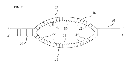
seconds; and a
final extension is conducted at 72 C for about 5 minutes. In some embodiments,
the reaction
mixture comprises genomic DNA, MgC12 and other physiological salts (e.g.,
NaC1), PCR buffer,
0.1-1.0 mM dNTPs, 0.04-1.5 uM of each primer, and 0.5-5.0 units of heat stable
polymerase
(e.g., Taq. polymerase).
[007] Other amplification methods known in the art may also be utilized,
including, for
example, self-sustained sequence replication (3SR), strand-displacement
amplification (SDA);
"branched chain" DNA amplification (Chiron Corp.); ligase chain reaction
(LCR), QB replicase
amplification (QBR), ligation activated transcription (LAT), nucleic acid
sequence-based
amplification (NASBA), repair chain reaction (RCR), and cycling probe reaction
(CPR)
(reviewed, e.g., in The Genesis Report, DX; Vol. 3(4), pp. 2-7 (February
1994)).
[008] Real-time PCR typically relies on the use of fluorescent molecules that
allow
quantification or detection of a PCR product in real time, while other
detection/quantification
chemistries such as electrochemistry are also applicable.
[009] Fluorescent molecules can be DNA binding dyes such as SYBR Green or
fluorescently
labeled primers or probes. There are many fluorescent dyes and probe designs
available for
different applications. The most commonly used DNA-binding dye for real-time
PCR is SYBR
Green I, which binds preferentially to double-stranded DNA (dsDNA) versus
single stranded
CA 02954420 2017-01-05
WO 2016/007914 PCT/US2015/040035
3
DNA. SYBR Green I fluorescence increases up to 1,000-fold when it binds to
dsDNA.
Therefore, fluorescence signal is proportional to the amount of dsDNA present.
[010] The major drawback of DNA-binding dyes is their lack of specificity,
that is, DNA-
binding dyes bind to any dsDNA. As a result, the presence of any nonspecific
products in a real-
time or endpoint PCR reaction will contribute to the overall fluorescence and
affect the accuracy
of quantification or detection. Furthermore, DNA-binding dyes cannot be used
for quantification
or detection in multiplex reactions because fluorescence signals from
different products cannot
be distinguished without the inclusion of a post PCR melting curve analysis to
distinguish the
formation of different products.
[011] In contrast, primer-based and probe-based detection chemistries ensure
that signal is
generated only when the product of interest is amplified. The primer or target-
specific
oligonucleotide probe is typically labeled with a reporter fluorophore, but in
most cases,
fluorescence is quenched when the specific target is not yet amplified or when
not present in the
sample. Usually this is accomplished by attaching a quencher molecule to the
primer or probe,
and devising some mechanism by which the reporter and quencher are separated
when the primer
or probe binds to its specific target.
[012] The principal primer/probe detection chemistries in use today are as
follows:
Hydrolysis (TaqMan) Probe
[013] Hydrolysis assays include a sequence-specific, fluorescently labeled
oligonucleotide
probe, in addition to the sequence-specific primers. Hydrolysis assays exploit
the 5' exonuclease
activity of certain thermostable polymerases, such as Taq or Tth. The
hydrolysis probe is labeled
with a fluorescent reporter at one end and a quencher at the opposite end,
though several
variations on this particular design are in common usage. When the probe is
intact, fluorscence
is quenched due to fluorophore proximity to the quencher. A commonly used
fluorescent
reporter¨quencher pair is fluorescein (FAM), which emits green fluorescence,
and Black Hole
Quencher 1 dye, although this is just one of many dye/quencher combinations in
use.
[014] The amplification reaction includes a combined annealing/extension step
during which
the probe hybridizes to the target and the dsDNA-specific 5' to 3' exonuclease
activity of Taq or
Tth cleaves the oligonucleotide, separating fluorophore from quencher,
resulting in a
CA 02954420 2017-01-05
WO 2016/007914 PCT/US2015/040035
4
fluorescence signal that is proportional to the amount of amplified product in
the sample. A
properly designed Hydrolysis probe can be used in combination with additional
probes of similar
design to determine sequence variations within the amplified target, i.e.
genotype.
Molecular Beacons
[015] Molecular beacons are dye-labeled oligonucleotides (25-40 nt) that form
a hairpin
structure. The 5' and 3' ends have complementary sequences of 5-6 nucleotides
that form the
stem, while the loop is designed to specifically hybridize to a 15-30
nucleotide section of the
target sequence. A fluorescent reporter molecule is attached to one end of the
molecular beacon,
and a quencher is attached to the other end. When the probe is unbound,
hairpin formation
occurs, bringing the reporter and quencher into proximity and fluorescence is
quenched.
[016] If a target sequence is present during the annealing step of an
amplification reaction, the
loop portion of the molecular beacon binds to its target sequence, causing the
stem to denature.
The reporter and quencher are thus separated, quenching is diminished, and the
reporter
fluorescence is detectable. Because fluorescence is emitted from the probe
only when it is bound
to the target, the amount of fluorescence detected is proportional to the
amount of target in the
reaction. Again, a properly designed molecular beacon can be used to
distinguish underlying
sequence variations, i.e. genotypes, within the amplified sequence. Typically,
this is
accomplished with melting curve analysis following PCR.
Dual Hybridization Probes
[017] These assays use two sequence-specific oligonucleotide probes which bind
to adjacent
sequences in the target. The probes are labeled with a pair of dyes that can
engage in
fluorescence resonance energy transfer (FRET). The donor dye is attached to
the 3' end of the
first probe, while the acceptor dye is attached to the 5' end of the second
probe. This order may
be reversed, so long as binding of both oligonucleotides to the target brings
the fluorophores
within FRET range (Forster radius).
[018] During real-time PCR, excitation is performed at a wavelength specific
to the donor dye,
and the reaction is monitored at the emission wavelength of the acceptor dye.
At the annealing
step, the probes hybridize to their target sequences in a head-to-tail
arrangement. This brings the
donor and acceptor dyes into proximity, allowing FRET to occur. The amount of
acceptor
CA 02954420 2017-01-05
WO 2016/007914 PCT/US2015/040035
fluorescence is proportional to the amount of PCR product present.
Hybridization probes enable
a variety of genetic detection and quantification readouts.
Primer/Probe Combinations
[019] These detectors use a sequence specific oligonucleotide primer and a
sequence specific
oligonucleotide probe. The primer and the probe are designed to bind to
adjacent sequences of
the target, usually with the probe complementary to the strand formed by the
primer. The probe
and the primer are labeled with a pair of dyes that can engage in (FRET).
Generally, the donor
dye is attached near the 3' end of the primer, while the acceptor dye is
attached to the 3' end of
the probe, which anneals to the complementary strand synthesized by primer
extension.
[020] As with the dual hybridization probes, during DNA amplification,
excitation is performed
at a wavelength specific to the donor dye, and the reaction is monitored at
the emission
wavelength of the acceptor dye. At the annealing step, the probe and primer
hybridize to their
target sequences in a head-to-tail arrangement. This brings the donor and
acceptor dyes into
proximity, allowing FRET to occur. The increasing amount of acceptor
fluorescence is
proportional to the amount of PCR product present.
Dynamic Flux Amplification
[021] An amplification method described in the art comprises determining the
melting
temperature of the target sequence and setting the upper limit of the thermal
cycle temperature to
maximize the denaturation of the target sequence while minimizing the
denaturation of the non-
target sequences (dynamic flux amplification or DFA). This approach fosters
the creation of a
bubble as the reaction is heated to a temperature approaching the denaturation
temperature of the
target sequence. Assuming the denaturation temperature of the target sequence
is less than the
adjacent sequences, the adjacent sequences will remain annealed, resulting in
a bubble forming
in the DNA strand as the target sequence denatures. Of course, it is probable
that multiple
bubbles form at various points along the DNA sequence that possess a similar
denature
temperature to the target sequence. Nevertheless, the total amount of un-
denatured sequence is
still less than would be the case if the upper temperature was raised to 95 C
or more.
[022] One advantage of controlling the denaturation temperature to create a
nucleic acid bubble
is that it significantly limits the formation of nonspecific product by
preventing the binding of
the primers to sites other than the target sequence, by making such sites
unavailable for
CA 02954420 2017-01-05
WO 2016/007914 PCT/US2015/040035
6
hybridization. This results from the target sequence being favored to denature
relative to non-
target regions of the target genome and thereby significantly reduces the
available sequence that
can serve as non-specific binding sites during the amplification process.
[023] One disadvantage of the aforementioned conventional probe chemistries is
that they are
not compatible with Dynamic Flux Amplification ("DFA") technology. This is due
in part to the
difference in required melting temperatures of the probes used in PCR as
compared to DFA.
PCR utilizes probes that are generally in the 20 ¨ 30 base pair range and
generally possess a Tm
of at least 20 C less than the Tm of the sequence of interest. In contrast,
DFA requires probes
that are within 20 C or less of the Tm of the sequence of interest. Because
DFA normally
operates outside of annealing temperature ranges used in probe technology for
PCR, such probes
as currently practiced are generally not compatible with DFA technology.
[024] It would be desirable if existing PCR primers could be modified to take
advantage of the
narrow temperature range used in DFA or at the very least a thermal cycling
range that is
narrower than those used in conventional PCR and thus obviate the need to
completely redesign
primers in order to obtain an increase in speed. The narrow temperature range
can be used as a
target temperature range in order to identify, design and/or generate specific
primers that have
sufficiently high Tm values when hybridized with the target nucleic acid.
[025] It would be desirable to have an amplification method that significantly
eliminated the
formation of undesirable product by inhibiting the extension of the reaction
beyond the
amplification bubble.
[026] Often the primers with the necessary Tm ranges must be designed de novo.
Thus,
although users of traditional PCR assays may desire increased speed, the cost
of designing,
evaluating and optimizing the primers for DFA necessary to obtain the narrower
cycling range is
frequently prohibitive, locking users into the slower conventional PCR, rather
than taking
advantage of the increased speed possible from dynamic flux amplification.
[027] Thus, there is a need in the art to develop primers and probes, other
reagents, and
methodologies, which are compatible with DFA. Specifically, there is an unmet
need in the art to
develop primers and probes that can be utilized in DFA protocols.
CA 02954420 2017-01-05
WO 2016/007914 PCT/US2015/040035
7
[028] In some aspects, the term "extreme chain reaction" or "XCR" will be
utilized in the
description. The present inventors utilize the term XCR as a synonym for DFA.
Thus, the two
terms are used interchangeably.
Multiplex Detection
[029] The need for, at a minimum, the ability to detect two or more distinct
amplified targets
within a single reaction is a fundamental aspect of modern diagnostic tests.
Although some tests
can be brought to market with separate reaction vessels containing the
necessary test
performance controls, it is cost effective in terms of sample throughput, and
reagent usage, to
incorporate the reaction controls within a single reaction vessel. Effective
utilization of DFA
ideally would involve a means to detect one or more amplified targets
simultaneously.
[030] Another consequence of being able to custom design target denaturation
and primer
annealing temperatures while simultaneously narrowing the thermal cycling
range allows for
amplification of different targets to be carried out in a single reaction
vessel by thermal cycling
the reaction vessel at different temperature ranges in succession.
[031] Probe technology for use with both PCR primers as well as the high Tm
and frequently
longer primers commonly used in DFA have been disclosed in WO 2015/054516
(incorporated
herein in its entirety for all purposes).
SUMMARY OF THE DISCLOSURE
[032] In one aspect of the invention, the disclosure provides oligonucleotide
primers with
increased melting temperatures for more specific amplification of target
nucleic acids.
[033] In one embodiment, an oligonucleotide primer for amplification of a
target nucleic acid
sequence in a polymerase chain reaction (PCR) comprises: a first region,
wherein the first region
is complementary to a strand of the target nucleic acid sequence and is
located at the 3' end of
the primer; and a second region, wherein the second region is located at the
5' end of the primer;
and wherein the Tm of the oligonucleotide primer is increased compared to the
Tm of an
oligonucleotide primer having only the first region.
[034] In another embodiment, the oligonucleotide primer comprises a transition
between the
first and second regions. In yet another embodiment, the transition comprises
a single nucleotide,
CA 02954420 2017-01-05
WO 2016/007914 PCT/US2015/040035
8
a chain of carbons, a multifunctional moiety, modified nucleotides, modified
backbones or a
combination thereof
[035] In one embodiment, the melting temperature (Tm) of the oligonucleotide
primer is within
at least 15 C of the Tm of the target nucleic acid sequence. In another
embodiment, the Tm of
the oligonucleotide primer is within at least 10 C of the Tm of the target
nucleic acid sequence.
In another embodiment, the Tm of the oligonucleotide primer is within at least
5 C of the Tm of
the target nucleic acid sequence. In another embodiment, the Tm of the
oligonucleotide primer is
within at least 2.5 C of the Tm of the target nucleic acid sequence. In
another embodiment, the
Tm of the oligonucleotide primer is equal to the Tm of the target nucleic acid
sequence.
[036] In one embodiment, the second region of the oligonucleotide primer
comprises nucleotide
or backbone modifications to optimize annealing of the oligonucleotide primer
to the target
nucleic acid region.
[037] In one embodiment, the second region is an arbitrary sequence that is
not complementary
to either strand of the target nucleic acid sequence.
[038] In one embodiment, the second region is complementary to a strand of the
target nucleic
acid sequence that is opposite to the strand of the target nucleic acid
sequence that the first
region is complementary to. In another embodiment, the second region comprises
cleavable
chemistries to inhibit cleavage by a polymerase.
[039] In one embodiment, the oligonucleotide primer comprises a sequence of
cytosine
nucleotides adjacent to a first sequence of guanosine nucleotides. In another
embodiment, the
number of nucleotides between the cytosine and guanosine nucleotides is less
than 5. In another
embodiment, the number of nucleotides between the cytosine and guanosine
nucleotides is less
than 4. In another embodiment, the number of nucleotides between the cytosine
and guanosine
nucleotides is less than 3. In another embodiment, the number of nucleotides
between the
cytosine and guanosine nucleotides is less than 2. In another embodiment, the
number of
nucleotides between the cytosine and guanosine nucleotides is 0. In another
embodiment, the
primer can form a Guanosine quadruplex structure.
[040] In one embodiment, the oligonucleotide primer further comprises a second
sequence of
guanosine nucleotides adjacent to the first sequence of guanosine nucleotides.
In another
CA 02954420 2017-01-05
WO 2016/007914 PCT/US2015/040035
9
embodiment, the second sequence of guanosine nucleotides causes the primer to
shift and form a
Guanosine quadruplex structure.
[041] In another aspect of the invention, the disclosure provides for a method
for increasing the
melting temperature (Tm) of an oligonucleotide primer for amplification of a
target nucleic acid
sequence in a polymerase chain reaction (PCR), comprising: identifying a
target nucleic acid
sequence from one or more segments of DNA; designing an oligonucleotide primer
having a first
region and a second region, wherein the first region is complementary to a
strand of the target
nucleic acid sequence and is located at the 3' end of the primer and the
second region is located
at the 5' end of the primer; and wherein the Tm of the oligonucleotide primer
is increased
compared to the Tm of an oligonucleotide primer having only the first region.
[042] In another aspect of the invention, the disclosure provides for a method
for nucleic acid
sequence amplification, comprising: identifying a target nucleic acid sequence
from one or more
segments of DNA comprising target and non-target nucleic acid sequences;
obtaining a first
oligonucleotide primer and a second oligonucleotide primer of the invention;
and amplifying the
target nucleic acid sequence by thermal cycling the target nucleic acid
sequence and the first and
second oligonucleotide primers, wherein thermal cycling comprises: (i)
denaturing the target
nucleic acid; (ii) hybridizing the first oligonucleotide primer to a first
strand and the second
oligonucleotide primer to a second strand of the denatured target nucleic
acid; (iii) extending the
first and second oligonucleotide primers by polymerization with a polymerase
to create two new
strands of the target nucleic acid; (iv) denaturing the two new strands from
the first and second
strands of the target nucleic acid; (v) hybridizing the first oligonucleotide
primer to the first
strand and to one new strand and the second oligonucleotide primer to the
second strand and to
the other new strand of the target nucleic acid; (vi) extending the first and
second oligonucleotide
primers by polymerization with a polymerase to create four additional new
strands of the target
nucleic acid; repeating steps (i) through (vi) to create multiple strands of
the target nucleic acid
that have incorporated the second regions of the first and second
oligonucleotide primers; and
wherein an upper thermal cycle temperature in the thermal cycling is selected
to minimize non-
target denaturation and maximize target denaturation.
[043] In one embodiment of the method for nucleic acid sequence amplification,
the thermal
cycling creates a bubble comprised of denatured target nucleic acid sequence
and adjacent
CA 02954420 2017-01-05
WO 2016/007914 PCT/US2015/040035
annealed non-target nucleic acid sequence. In another embodiment, the
oligonucleotide primers
prevent amplification of the target nucleic acid sequence beyond the bubble.
[044] In another aspect of the invention, the disclosure provides for a method
for amplifying
and detecting two or more target nucleic acid sequences in a sample,
comprising: identifying two
or more target nucleic acid sequences from one or more segments of DNA;
obtaining a pair of
oligonucleotide primers specific for each target nucleic acid sequence,
wherein each pair of
oligonucleotide primers has an annealing curve (TA) that overlaps with a
denaturation curve (TD)
of its target nucleic acid sequence, in such a manner as to minimize the
temperature range
between the higher of the melting temperature of the pair of oligonucleotide
primers and the
melting temperature of its target nucleic acid sequence; amplifying each
target nucleic acid
sequence by thermal cycling each pair of oligonucleotide primers and its
target nucleic acid
sequence within a specific temperature range, wherein the thermal cycling at
different
temperature ranges in succession leads to amplification of the two or more
target nucleic acid
sequences; and detecting the two or more amplified target nucleic acid
sequences.
[045] In one embodiment of the method for amplifying and detecting two or more
target
nucleic acid sequences in a sample, each amplified target nucleic acid
sequence is about 400 bp
or greater. In another embodiment, one or more temperature suitable
polymerases are chosen for
each temperature range.
[046] In one embodiment, one or more of the target nucleic acid sequences is
an internal
control. In another embodiment, the pair of oligonucleotide primers specific
for the internal
control is the same as the pair of oligonucleotide primers specific for a
target nucleic acid
sequence except for mismatches that allow amplification of the internal
control at a different
temperature range than that of the target nucleic acid sequence.
[047] In one embodiment, each pair of oligonucleotide primers is used only at
its own thermal
cycling temperature range. In another embodiment, the thermal cycling at each
temperature
range comprises as many cycles as necessary for amplification of each target
nucleic acid
sequence.
[048] In one embodiment, the thermal cycling comprises cycling at temperature
ranges in
succession, beginning with the lowest temperature range and moving to the
highest temperature
range. In another embodiment, the thermal cycling comprises cycling at
temperature ranges in
CA 02954420 2017-01-05
WO 2016/007914 PCT/US2015/040035
11
succession, beginning with the highest temperature range and moving to the
lowest temperature
range.
[049] In one embodiment, there is overlap between one or more temperature
ranges. In another
embodiment, the thermal cycling comprises temperature ranges of from about 50
C to about
65 C, from about 60 C to about 95 C and from about 90 C to about 105 C. In
another
embodiment, the thermal cycling comprises temperature ranges of from about 45
C to about
72 C and from about 72 C to about 99 C. In another embodiment, the thermal
cycling comprises
temperature ranges of from about 54 C to about 63 C, from about 63 C to about
81 C and from
about 81 C to about 99 C.
[050] In one embodiment of the method for amplifying and detecting two or more
target
nucleic acid sequences in a sample, the detecting comprises using fluorescent
dyes,
electrochemical indicators, target immobilization strategies, or any
combination thereof.
[051] In one embodiment, the amplifying step further comprises thermal cycling
each pair of
oligonucleotide primers, its target nucleic acid sequence and an
oligonucleotide probe
complementary to the target nucleic acid sequence and having a cleavable
sequence. In another
embodiment, each oligonucleotide probe comprises a fluorescent dye and
quencher located
interchangeably on the 5' or 3' end of each probe. In another embodiment, each
oligonucleotide
probe comprises the same fluorescent dye located interchangeably on the 5' or
3' end of each
probe. In another embodiment, the cleavable sequence of each oligonucleotide
probe is cleaved
by polymerization-independent cleavage or by polymerization-dependent cleavage
by a
polymerase. In another embodiment, one or more oligonucleotide probes is a
hybrid
hairpin/cleaved probe.
[052] In one embodiment of the method for amplifying and detecting two or more
target
nucleic acid sequences in a sample, the detecting step comprises detecting a
signal resulting from
cleavage of said probe.
[053] In one embodiment of the method for amplifying and detecting two or more
target
nucleic acid sequences in a sample, the amplifying step further comprising
thermal cycling each
pair of oligonucleotide primers, its target nucleic acid sequence and an
oligonucleotide probe
complementary to the target nucleic acid sequence and having a cleavable
sequence, the two or
more target nucleic acid sequences comprise a Trichomonas sequence and a
Xenorhabdus
CA 02954420 2017-01-05
WO 2016/007914 PCT/US2015/040035
12
nematophila sequence. In another embodiment, the Xenorhabdus nematophila
sequence is a
control sequence. In another embodiment, the pair of oligonucleotide primers
specific for the
Trichomonas sequence comprises SEQ ID NO: 56 and SEQ ID NO: 57. In another
embodiment,
the oligonucleotide probe complementary to the Trichomonas sequence comprises
SEQ ID NO:
59. In another embodiment, the pair of oligonucleotide primers specific for
the Xenorhabdus
nematophila sequence comprises SEQ ID NO: 51 and SEQ ID NO: 52. In another
embodiment,
the oligonucleotide probe complementary to the Xenorhabdus nematophila
sequence comprises
SEQ ID NO: 53. In another embodiment, the thermal cycling comprises
temperature ranges of
from about 89 C to about 74 C and from about 63 C to about 78 C. In another
embodiment, the
thermal cycling from about 89 C to about 74 C amplifies the Trichomonas
sequence. In another
embodiment, the thermal cycling from about 63 C to about 78 C amplifies the
Xenorhabdus
nematophila sequence.
[054] In one embodiment, a method of detecting Trichomonas in cattle
comprises: obtaining a
pair of oligonucleotide primers specific for a Trichomonas target nucleic acid
sequence;
obtaining a pair of oligonucleotide primers specific for a Xenorhabdus
nematophila control
nucleic acid sequence; wherein each pair of oligonucleotide primers has an
annealing curve (TA)
that overlaps with a denaturation curve (TD) of its target nucleic acid
sequence, in such a manner
as to minimize the temperature range between the higher of the melting
temperature of the pair
of oligonucleotide primers and the melting temperature of its target nucleic
acid sequence;
amplifying each nucleic acid sequence by thermal cycling each pair of
oligonucleotide primers
and its target nucleic acid sequence within a specific temperature range,
wherein the thermal
cycling at different temperature ranges in succession leads to amplification
of the Trichomonas
and Xenorhabdus nematophila sequences; and detecting the amplified target
nucleic acid
sequences.
[055] In one embodiment of the method for amplifying and detecting two or more
target
nucleic acid sequences in a sample, one or both of each pair of
oligonucleotide primers
comprises a triplex forming region (TFR). In another embodiment, the TFR
primer creates
strands of triplex forming DNA when the target nucleic acid sequence includes
a sequence
having complementarity with the sequence of the TFR primer. In another
embodiment, the target
nucleic acid sequence includes a natural triplex forming region.
CA 02954420 2017-01-05
WO 2016/007914 PCT/US2015/040035
13
[056] In one embodiment of the method for amplifying and detecting two or more
target
nucleic acid sequences in a sample, one or both of each pair of
oligonucleotide primers further
comprises a label.
[057] In one embodiment, the method further comprises one or more triplex
forming
oligonucleotide (TFO) probes that hybridizes to one or more TFRs, thus forming
a triplex, in a
double stranded DNA sequence that was created during an amplification process
of a target
nucleic acid sequence by one or more TFR primers. In another embodiment, the
TFO probe is
designed to anneal at approximately the same, or lower, temperature than a Tm
of the TFR
primer. In another embodiment, the method further comprises one or more non-
specific DNA
binding dyes that bind with hybridized triplex DNA. In another embodiment, the
method further
comprises one or more quadruplex binding dyes.
[058] In one embodiment, each TFO probe includes a label moiety selected from
the group
consisting of: a fluorescent moiety, radioactive moiety, color moiety,
fluorescent reporter
moiety, fluorescent quenching moiety, one of a pair of fluorescent resonance
energy transfer
moieties, and combinations thereof In another embodiment, one or more of the
TFO probes is a
triplex forming fluorescent probe (TFFP). In another embodiment, one or more
of the TFO
probes is a triplex forming fluorescent probe (TFFP) and the double stranded
DNA has a
receptor dye. In another embodiment, the label moiety is the same for each TFO
probe. In
another embodiment, a cap at the 3' end of the TFO probe inhibits extension
from the 3' end of
the probe.
[059] In one embodiment, the double stranded DNA has a first label and the TFO
probe has a
second label, wherein the first label and second label provide a detectable
emission upon close
association.
[060] In one embodiment, the TFO probe includes a fluorescent dye and
quencher. In another
embodiment, the TFO probe includes a fluorescent dye and quencher in a hairpin
configuration.
[061] Also provided herein are kits comprising any of the aforementioned
oligonucleotides,
primers, probes, and reaction agents.
CA 02954420 2017-01-05
WO 2016/007914 PCT/US2015/040035
14
[062] These and other features, aspects, and advantages of embodiments of the
present
disclosure, will become better understood with regard to the following
description, claims, and
accompanying drawings, explained below.
BRIEF DESCRIPTION OF THE DRAWINGS
[063] FIG. lA and FIG. 1B show graphical representations of a design for
overlapping primer
annealing temperatures and template denaturation temperatures (FIG. 1A) and a
design for non-
overlapping primer annealing temperatures and template denaturation
temperatures (FIG. 1B).
[064] FIG. 2 is an illustration of conventional amplification products by real
time PCR.
[065] FIG. 3 is a graph showing high temperature PCR amplification of the same
template used
in FIG. 2.
[066] FIG. 4 is a graph showing the HTPCR amplification of the same template
material using
different starting material concentrations.
[067] FIG. 5 depicts the creation of a "bubble" 16 as the reaction is heated
to a temperature
approaching the denaturation temperature of the target sequence.
[068] FIG. 6 depicts the bubble 16 having a first primer 32 annealed to DNA
strand 24 at one
end of the bubble. Primer 32 has a first blocking tag 42 that anneals to the
complementary DNA
strand 28. A second primer 38 is annealed to DNA strand 28 at the other end of
the bubble.
Primer 38 has a second blocking tag 46 that anneals to the complementary DNA
strand 24.
[069] FIG. 7 depicts the extension phase of the amplification using the primer
with blocking
tag. The first primer 32 has been extended in the direction of the second
blocking tag 46,
resulting in an extension 50 which cannot readily extend beyond the second
blocking tag 46.
Similarly, the second primer 38 has been extended in the direction of the
first blocking tag 42,
resulting in an extension 54 which cannot readily extend beyond the first
blocking tag 42.
[070] FIG. 8A-8C depicts the second cycle of amplification using the primers
with blocking
tags. FIG. 8A depicts the two extension products from the first cycle of
amplification. The point
at which the tag (either tag 42 or tag 46) transitions to the primer (either
primer 32 or primer 38)
is designated by a T, to denote a transition. FIG. 8B depicts the
amplification of extension 70
CA 02954420 2017-01-05
WO 2016/007914 PCT/US2015/040035
with the annealing of fresh primer 58 comprising tag 66 to the 3' end of
extension 50. FIG. 8C
depicts the amplification of extension 79 with the annealing of primer 32
comprising tag 42 to
the 3' end of extension 54.
[071] FIG. 9 depicts the third cycle of amplification using the primers with
blocking tags. With
the extension 70 product as template, primer 78 comprising tag 74 anneals to
the 3' end of
extension 70 and amplification results in another complete copy of the target
sequence plus the
tags 74 and 66 on both ends of the target sequence.
[072] FIG. 10 depicts the first cycle in an amplification using a primer
comprising a tag with
an arbitrary sequence. A tag 80 is appended to a primer 84. The tag 80 does
not correspond to
any DNA strand adjacent to the target sequence 88 sequence, but rather,
represents a more or less
arbitrary oligonucleotide sequence. In the first cycle, the primer 84 binds to
the target sequence
88 and extends fully across the target sequence 88, creating an
oligonucleotide 94 comprising the
primer 84, the extension 90 and the tag 80. In the first cycle, the tag 80
does not bind to the
target sequence 88.
[073] FIG. 11 depicts the second cycle in an amplification using a primer
comprising a tag with
an arbitrary sequence. In the second cycle, the oligonucleotide 94 binds to a
fresh primer 96 and
tag 98. The fresh tag 98 has no complementary sequence on the oligonucleotide
94 to bind to.
The primer 96 extends all the way to the end of the oligonucleotide 94,
creating a duplicate
oligonucleotide 99 comprising a reproduction of the tag 80, primer 84, and the
extension 90 of
the oligonucleotide 94. This duplicate oligonucleotide comprises a duplicate
of the tag 80 on one
end and its own tag 98 on the opposite end.
[074] FIG. 12 depicts the third cycle in an amplification using a primer
comprising a tag with
an arbitrary sequence. In the third cycle, a fresh tag 100 and primer 104
binds to the duplicate
oligonucleotide 99 (note that fresh primer 104 and tag 100 is equivalent to
primer 84 and tag 80
in sequence). The primer extension 106 extends all the way to the end of the
tag 98, creating a
complete duplicate.
[075] FIG. 13 depicts the initial stage of one mechanism for the formation of
a G-quadruplex.
In this mechanism, a primer 130 is designed to interface with one end of the
target bubble 134,
wherein the bubble comprises principally GC sequences. The primer 130 is
designed with GC
sequences to complement the target's GC sequences.
CA 02954420 2017-01-05
WO 2016/007914 PCT/US2015/040035
16
[076] FIG. 14 depicts unconventional hybridization of Gs to Gs to form
Hoogsteen pairs in
areas comprising high GC content such that G quadruplexes are formed through a
process of
folding the strands to line the Gs up with Gs.
[077] FIG. 15 depicts G quadruplex formation. The displaced C sequence 138 is
not bound to
any complementary sequence in the target and so twists into a folded shape
that serves as a solid
blocker to any extension of the primer past the bubble.
[078] FIG. 16 depicts a sequence of G's 140 that is added internal to the
primer, proximal to
the 3' end and adjacent to the quadruplex forming region of the primer 130.
This sequence of
G's 140 is attracted to the sequence of C's 144 adjacent to it on the first
strand of the target 148.
This attraction gives added impetus to the primer to shift and thus form a G
quadruplex.
[079] FIG. 17 depicts G quadruplex formation. The sequence of G's 140 has
shifted to pair
with the sequence of C's 144 on the first strand of the target 148. The
displaced C sequence 138
is not bound to any complementary sequence in the target and so twists into a
folded shape that
serves as a solid blocker to any extension of the primer past the bubble.
[080] FIG. 18 shows the hybridization of primers (SEQ ID NOs: 22 and 23) to
the
Mycobacterium avium subsp. paratuberculosis str. kl0 sequence (SEQ ID NO: 21)
to form G-
quadruplex structures to block extension beyond the bubble.
[081] FIG. 19 depicts thermal profiles for amplification of high AT nucleic
acid regions first,
then normal nucleic acid regions (regions amplified by traditional PCR
temperature ranges)
second, and then high GC nucleic acid regions third.
[082] FIG. 20 depicts thermal profiles for amplification of high GC nucleic
acid regions first,
then normal nucleic acid regions (regions amplified by traditional PCR
temperature ranges)
second, and then high AT nucleic acid regions third.
[083] FIG. 21 depicts thermal profiles for amplification of nucleic acid
regions between about
45 C and about 72 C, and then for amplification of nucleic acid regions
between about 72 C and
about 99 C.
[084] FIG. 22 depicts thermal profiles for amplification of nucleic acid
regions between about
54 C and about 63 C; for amplification of nucleic acid regions between about
63 C and about
81 C; and for amplification of nucleic acid regions between about 81 C and
about 99 C.
CA 02954420 2017-01-05
WO 2016/007914 PCT/US2015/040035
17
[085] FIG. 23 depicts thermal profiles for amplification of five different
nucleic acid targets
from five different organisms. There are five distinct temperature ranges, one
temperature range
for each of the five targets, starting from low to high.
[086] FIG. 24 depicts thermal profiles for amplification of five different
nucleic acid targets
from five different organisms. There are five distinct temperature ranges, one
temperature range
for each of the five targets, starting from high to low.
[087] FIG. 25 depicts fluorescence history and temperature history of an
amplification
described in Example 3.
[088] FIG. 26 depicts fluorescence history and temperature history of an
amplification
described in Example 4.
[089] FIG. 27 depicts an exemplary thermal profile for amplification of
Trichomonas foetus
target and reaction control template Xenorhabdus nematophila.
[090] FIG. 28 depicts an exemplary thermal profile for amplification of
Trichomonas foetus
target and reaction control template Xenorhabdus nematophila.
[091] FIG. 29 depicts an exemplary thermal profile for amplification of
Trichomonas foetus
target and reaction control template Xenorhabdus nematophila.
[092] FIG. 30 depicts an exemplary thermal profile for amplification of
Trichomonas foetus
target and reaction control template Xenorhabdus nematophila.
[093] FIG. 31A and FIG. 31B depicts an exemplary thermal profile for
amplification of
Trichomonas foetus target and reaction control template Xenorhabdus
nematophila.
[094] FIG. 32 is a general embodiment of cleaved probe technology according to
the
disclosure.
[095] FIG. 33 depicts cleaved probe technology in an embodiment where
bathophenanthroline-
RU II complexes are used as label molecules.
[096] FIG. 34 depicts a Dual Hybridization Probe and Primer combination.
[097] FIG. 35 depicts a primer/probe combination capable of engaging in FRET.
CA 02954420 2017-01-05
WO 2016/007914 PCT/US2015/040035
18
[098] FIG. 36 illustrates forward (top) and reverse (bottom) primers with dye
(squares) spaced
approximately 6-9 nucleotides apart along the length of the primers, but with
sufficient
nucleotides left without dye on the 3' end. When the primers bind to their
complement,
fluorescence quenching is released and thus a detectable signal is created.
[099] FIG. 37 illustrates quenched forward primer-dimer complex (top),
quenched reverse
primer-dimer complex (middle), and primer-dimer complex formed from the
binding together of
the forward and reverse primers (bottom), which is detectable via FRET signal.
Squares
represent dyes.
[100] FIG. 38 illustrates forward primer template formation signal (top), and
reverse primer
template formation signal (middle), and signal generated when both the forward
and reverse
primers produce the targeted template (bottom). Squares represent dye.
[101] FIG. 39 illustrates that correct products with both dye labeled primers
will show the
formation of fluorescent signal from both distinct dyes with equal reaction
formation efficiency,
as they will be linked directly to one another in the formation of
amplification product and could
be monitored in two fluorescent channels simultaneously. Forward primer signal
on left and
reverse primer signal on the right.
[102] FIG. 40 illustrates a data evaluation advantage of the present
disclosure design strategy
where amplified product is formed and both fluorescent signals are generated
by the amplifying
product. Any primer-dimer signals that result in FRET, as the like primers
will be quenched, can
be subtracted from the formed signals to enable a baseline normalization of
the amplification
signals. Forward and reverse primer signals forming sigmoidal curve. Primer-
dimer signal to be
subtracted is illustrated via line at the bottom of the graph.
[103] FIG. 41 illustrates that the triplex forming region (TFR) primer
participates in the
amplification of the target sequence, creating strands of triplex forming DNA
along the length of
and appended to the target sequence.
[104] FIG. 42 illustrates a triplex forming oligonucleotide probe with the 3'
end of the triplex
forming oligonucleotide (TFO) probe being capped.
CA 02954420 2017-01-05
WO 2016/007914 PCT/US2015/040035
19
[105] FIG. 43 depicts a double stranded DNA sequence comprising a Triplex
Forming Region.
The double stranded DNA sequence possesses a receptor dye. The TFR of the TFFP
attaches to
the Triplex Forming Region of the double stranded DNA.
[106] FIG. 44 illustrates that the binding dyes, constrained by covalent
attachment to a
particular location on the TFO probe, in this instance, the end of the TFO
probe, can only bind to
hybridized DNA structures when the TFO probe is bound and thus, puts the TFO
probe in
proximity to the dye attached to the amplified sequence of interest. Thus, a
fluorescent signal
indicates that amplification has occurred.
[107] FIG. 45 depicts that in this embodiment the TFO probe utilizes a hairpin
dye and
quencher configuration.
[108] FIG. 46 illustrates that two or more primers with the same TFR sequence
may be used
along with TFR primers that comprise a sequence complementary to the TFR
sequence.
[109] FIG. 47 depicts an embodiment wherein six primers are divided into three
sets of two
each.
[110] FIG. 48 depicts that the donor dye is attached near the 3' end of the
first primer, while the
acceptor dye is attached near the 3' end of the second primer. At the
annealing step, the primers
hybridize to their target sequences in a near tail-to-tail arrangement, which
brings the dyes into
sufficient proximity for FRET to occur.
DETAILED DESCRIPTION
[111] In the description and tables which follow, a number of terms are used,
in order to
provide a clear and consistent understanding of the specification and claims,
including the scope
to be given such terms, the following definitions are provided.
Definitions
[112] With respect to the use of substantially any plural and/or singular
terms herein, those
having skill in the art can translate from the plural to the singular and/or
from the singular to the
plural as is appropriate to the context and/or application. The various
singular/plural
permutations may be expressly set forth herein for sake of clarity.
CA 02954420 2017-01-05
WO 2016/007914 PCT/US2015/040035
[113] The term "a" or "an" refers to one or more of that entity; for example,
"a primer" refers
to one or more primers or at least one primer. As such, the terms "a" (or
"an"), "one or more"
and "at least one" are used interchangeably herein. In addition, reference to
"an element" by the
indefinite article "a" or "an" does not exclude the possibility that more than
one of the elements
is present, unless the context clearly requires that there is one and only one
of the elements.
[114] The term "adjacent" as used herein refers to the positioning of the
primer with respect to
the probe on its complementary strand of the template nucleic acid in which
the nucleotides may
directly abut one another. Alternatively, for use in the polymerization-
dependent process, as
when the present method is used in the PCR and DFA and detection methods as
taught herein,
the "adjacency" may be anywhere within the sequence to be amplified, anywhere
downstream of
the primer such that primer extension will position the polymerase so that
cleavage of the probe
occurs.
[115] The term "allele" as used herein is any of one or more alternative forms
of a gene which
relate to one trait or characteristic. In a diploid cell or organism, the two
alleles of a given gene
occupy corresponding loci on a pair of homologous chromosomes.
[116] The term "amino acid sequence" as used herein includes an oligopeptide,
peptide,
polypeptide, or protein and fragments thereof that are isolated from, native
to, or naturally
occurring in a plant, or are synthetically made but comprise the nucleic acid
sequence of the
endogenous counterpart.
[117] A "biological sample" described herein can include any biological
material taken from a
subject, including, but not limited to, expectorations (e.g., sputum), blood,
blood cells (e.g.,
lymphocytes), tissue, biopsies, cultured cells, pleural, peritoneal, or
cerebrospinal fluid, sweat,
feces, and urine. In some embodiments, a biological sample from a subject is
treated, e.g., to
culture an infectious microorganism and/or amplify its genetic material,
before being assayed
according to methods provided herein.
[118] The term "bioluminescence" refers to a form of chemiluminescence in
which the light-
emitting compound is one that is found in living organisms. Examples of
bioluminescent
compounds include bacterial luciferase and firefly luciferase.
CA 02954420 2017-01-05
WO 2016/007914 PCT/US2015/040035
21
[119] The term "drug" as used herein can refer to any compound, agent,
treatment modality, or
combination thereof In some preferred aspects, the drug is an antibiotic
compound.
[120] The term "efficiency" as used herein refers to a hallmark of Real-Time
PCR assays. An
ideal qPCR (quantitative PCR) reaction has an efficiency of 100% with a slope
of -3.32, which
correlates with a perfect doubling of PCR product during each cycle. However,
slopes between -
3.1 and -3.6 with efficiencies between 90 and 110% are generally considered
acceptable
(Commission, C. A. (2009). Definition of Minimum Performance Requirements for
Analytical
Methods of GMO Testing European Network of GMO Laboratories (ENGL), (October
2008), 1-
8). Efficiency is established by replicated standard curves. Amplification
efficiency is
determined from the slope of the log-linear portion of the standard curve and
is calculated as E=
(10(-1/slope) -1)*100. (Bustin, S. A., et al. (2009). The MIQE Guidelines:
Minimum I
nformation for Publication of Quantitative Real-Time PCR Experiments. Clinical
Chemistry,
55(4), 1-12. doi:10.1373/clinchem. 2008.112797).
[121] The term "fluorophore" refers to a compound which is capable of
fluorescing, i.e.
absorbing light at one frequency and emitting light at another, generally
lower, frequency.
[122] The term "homogeneous", as used herein applied to multi-step processes,
refers to
methods for carrying out the steps of the process, wherein the need for sample
handling and
manipulation between steps is minimized or eliminated. For example, a
"homogeneous"
amplification/detection assay refers to a coupled amplification and detection
assay wherein the
need for sample handling and manipulation between the amplification and
detection is
minimized or eliminated.
[123] The term "intercalator" refers to an agent or moiety capable of non-
covalent insertion
between stacked base pairs in a nucleic acid double helix.
[124] The term "label" as used herein refers to any atom or molecule which can
be used to
provide a detectable (preferably quantifiable) signal or to interact with a
second label to modify
the detectable signal provided by the second label. The label can be attached
to a nucleic acid or
protein. Labels may be light-emitting compounds which generate a detectable
signal by
fluorescence, chemiluminescence, phosphorescence, or bioluminescence. In the
alternative,
labels may provide signals detectable by radioactivity, electrochemistry,
colorimetry, or by the
absorption of light, producing fluorescence, or may be used to immobilize a
product to an array.
CA 02954420 2017-01-05
WO 2016/007914 PCT/US2015/040035
22
[125] The term "linearity" as used herein refers to a hallmark of optimized
Real-Time PCR
assays and is determined by the R2 value obtained by linear regression
analysis, which should be
> 0.98 (Bustin et al., 2009).
[126] The term "microorganism" as used herein can refer to bacteria, archaea,
fungi, protozoa,
parasites and/or viruses.
[127] The terms "nucleic acid" and "oligonucleotide" refer to primers, probes,
and oligomer
fragments to be detected, and shall be generic to polydeoxyribonucleotides
(containing 2-deoxy-
D-ribose), to polynucleotides (containing D-ribose), and to any other type of
polynucleotide
which contains an N glycoside of a purine or pyrimidine base, or modified
purine or pyridine
base. There is no intended distinction in length between the terms "nucleic
acid" and
"oligonucleotide", and these terms will be used interchangeably These terms
refer only to the
primary structure of the molecule. Thus, these terms include double and single
stranded DNA, as
well as double and single stranded RNA.
[128] The oligonucleotide is not necessarily physically derived from any
existing or natural
sequence but may be generated in any manner, including chemical synthesis, DNA
replication,
reverse transcription, or a combination thereof The terms "oligonucleotide"
intend a
polynucleotide of genomic DNA or RNA, cDNA, semisynthetic, or synthetic origin
which, by
virtue of its origin or manipulation: (1) is not associated with all or a
portion of the
polynucleotide with which it is associated in nature; and/or (2) is linked to
a polynucleotide other
than that to which it is linked in nature; and (3) is not found in nature.
[129] The complement of a nucleic acid sequence as used herein refers to an
oligonucleotide
which, when aligned with the nucleic acid sequence such that the 5' end of one
sequence is
paired with the 3' end of the other, is in "antiparallel association." Certain
bases not commonly
found in natural nucleic acids may be included in the nucleic acids of the
present disclosure and
include, for example, inosine and 7-deasaguanine. Complementarity need not be
perfect; stable
duplexes may contain mismatched base pairs or unmatched bases. Those skilled
in the art of
nucleic acid technology can determine duplex stability empirically considering
a number of
variables including, for example, the length of the oligonucleotide, base
composition and
sequence of the oligonucleotide, ionic strength, and incidence of mismatched
base pairs.
CA 02954420 2017-01-05
WO 2016/007914 PCT/US2015/040035
23
[130] The terms "target nucleic acid(s)" as used herein refers to nucleic
acids derived from an
infectious microorganism, human, mammalians, or plants. In some aspects, a
target nucleic acid
is a nucleic acid of an organism or a microorganism that is assayed according
to a method
provided herein.
[131] The terms "target region", "target sequence", and "target nucleic acid
sequence" refer to
a region of a nucleic acid which is to be detected, quantified, or genotyped.
[132] The term "reference nucleic acid" as used herein refers to a nucleic
acid corresponding to
a target nucleic acid (e.g., representing the same portion of genomic DNA),
that differs from the
target nucleic acid by one or more sequence variations. For example, in some
aspects, a
reference nucleic acid has the sequence of a wild-type microorganism (e.g.,
with respect to
responsiveness to a drug of interest). In further aspects, a reference nucleic
acid has the sequence
of a wild-type human cell, such as a diseased cell, including, e.g., a human
cancer cell.
[133] The term "primer" may refer to more than one primer and refers to an
oligonucleotide,
whether occurring naturally, as in a purified restriction digest, or produced
synthetically, which
is capable of acting as a point of initiation of synthesis along a
complementary strand when
placed under conditions in which synthesis of a primer extension product which
is
complementary to a nucleic acid strand is catalyzed. Such conditions include
the presence of
five different deoxyribonucleoside triphosphates and polymerization-inducing
agents such as
DNA polymerase or reverse transcriptase, in a suitable temperature. The primer
is preferably
single stranded for maximum efficiency in amplification.
[134] The term "probe" refers to an oligonucleotide, typically labeled, that
forms a duplex
structure with a sequence of a target nucleic acid due to complementary base
pairing. The probe
will comprise a "hybridizing region", preferably consisting of 30 or more
nucleotides, and in
some instances, consisting of 50 or more nucleotides, corresponding to a
region of the target
sequence. Ideally, the Tm of the probe will be within 30 degrees or less of
the Tm of the
sequence of interest. "Corresponding" means identical to or complementary to
the designated
nucleic acid. The probe, preferably, does not contain a sequence complementary
to sequence(s)
used to prime the PCR. Generally, the 3' terminus of the probe will be
"blocked" to prohibit
incorporation of the probe into a primer extension product. "Blocking" can be
achieved by using
non-complementary bases or by adding a chemical moiety such as biotin, a
phosphate group, or a
CA 02954420 2017-01-05
WO 2016/007914 PCT/US2015/040035
24
fluorophore to the 3' hydroxyl of the base nucleotide, which may, depending on
the selected
moiety, serve a dual purpose by also acting as a label for subsequent
detection or capture of the
nucleic acid attached to the label. Blocking can also be achieved by removing
the 3'-OH or by
using a nucleotide that lacks a 3'-OH such as dideoxynucleotide.
[135] The term "quenching" refers to a decrease in fluorescence of a first
compound caused by
a second compound, regardless of the mechanism. Quenching typically requires
that the
compounds be in close proximity. As used herein, either the compound or the
fluorescence of
the compound is said to be quenched, and it is understood that both usages
refer to the same
phenomenon.
[136] The terms "responsiveness" and "drug responsiveness" as used herein can
refer to
resistance, sensitivity, susceptibility, tolerance and/or other phenotypic
characteristics of a
microorganism or diseased cell, such as a cancer sell, related to the
therapeutic effect of a drug,
including non-responsiveness. Drug responsiveness can be assessed directly,
according to the
effect of the drug on a targeted microorganism or diseased cell, such as a
cancer cell (e.g., a
bacterial mortality or a cellular mortality), and/or indirectly, according to
the effect of the drug
on one or more aspects of an infectious disease caused by the microorganism
(e.g., prevention,
amelioration, alleviation, and/or elimination of the disease or one or more
symptoms of the
disease). In some preferred aspects, systems and methods are provided herein
for detecting
resistance to one or more drugs, where resistance refers to inheritable
(genetic) resistance.
[137] The terms "sequence-specific oligonucleotide" and "SSO" refer to
oligonucleotide probes
wherein the hybridizing region is exactly complementary to the sequence to be
detected. This is
known as "stringent hybridization." The use of stringent hybridization
conditions under which
the probe will hybridize only to that exactly complementary target sequence
allows for detection
of the specific target sequence. Stringent hybridization conditions are well
known in the art (see,
e.g., Sambrook, et al., 1985, molecular cloning ¨ A Laboratory Manual, Cold
Springs Harbor,
N.Y., incorporated herein by reference). Stringent conditions are sequence
dependent and will be
different in different circumstances.
[138] The term "sequence variation" as used herein, in relation to nucleic
acids, refers to a
difference in the sequence of a nucleic acid relative to the sequence of a
corresponding nucleic
acid (e.g., a sequence representing the same gene or other portion of genomic
DNA). In some
CA 02954420 2017-01-05
WO 2016/007914 PCT/US2015/040035
embodiments, sequence variations detected according to various methods
provided herein are
"Single Nucleotide Polymorphisms" ("SNPS"), resulting from a difference in the
identity of a
single nucleotide between a target nucleic acid and a reference nucleic acid.
In further
embodiments, sequence variations detected according to various methods
provided herein
include "multiple nucleotide Polymorphisms." In some embodiments, the
reference nucleic acid
corresponds to a non-drug resistant phenotype and a drug resistant phenotype
is detected
according to a method provided herein by identifying a sequence variation
between the reference
nucleic acid and a target nucleic acid of a biological sample from a subject
infected with the
microorganisms or diseased cell, such as a drug resistance cancer cell.
[139] The "subject" referred to herein can be any organism capable of hosting
a
microorganism, including but not limited to, experimental animals (e.g., mice,
rats, rabbits, and
the like) and humans. In various embodiments, the subject is a human patient
suffering from an
infectious disease. In other embodiments, the subject is the organism itself,
such as the human
patient.
[140] The term "subsequence" refers herein to a nucleotide sequence contained
within another
sequence.
[141] The Tm is the temperature (e.g., under defined ionic strength and pH) at
which 50% of
the oligonucleotides have dissociated. Relaxing the stringency of the
hybridizing conditions will
allow sequence mismatches to be tolerated; the degree of mismatch tolerated
can be controlled
by suitable adjustment of the hybridization conditions.
[142] The term "variable sequence element" refers to a region of a nucleic
acid (e.g., DNA or
RNA) comprised of a string of adjacent nucleotides that includes at least one
sequence variation
known to be associated with a phenotypic characteristic of interest, such as
resistance,
sensitivity, and/or other aspects of drug responsiveness or propensity for a
particular disease such
as cancer or heart disease, or more mundane phenotypic characteristics such as
eye color or hair
color. For example, a sequence variation associated with drug resistance will
often occur in a
region of a nucleic acid that encodes a site of the corresponding protein that
is a structural and/or
functional determinant of drug responsiveness, such as a drug binding site. A
variable sequence
element including the known variation (and surrounding nucleotides) will
likely encode
structurally and/or functionally related portions of the protein (e.g., a
pocket, fold, or other
CA 02954420 2017-01-05
WO 2016/007914 PCT/US2015/040035
26
structure that comprises the drug blinding site), and additional,
uncharacterized variations within
the variable sequence element will likely be associated with the same
phenotype as the known
variations.
[143] As defined herein, "5'----> 3' nuclease activity" or "5' to 3' nuclease
activity" refers to
that activity of a template specific nucleic acid polymerase including either
a 5' to 3'
exonuclease activity traditionally associated with some DNA polymerase,
whereby nucleotides
are removed from the 5' end of an oligonucleotide in a sequential manner,
(i.e., E. coli DNA
polymerase I has this activity, whereas the Klenow fragment does not), or a 5'
to 3'
endonuclease activity wherein cleavage occurs more than one phosphodiester
bond (nucleotide)
from the 5' end, or both.
[144] The term "reaction mixture" refers to a solution containing reagents
necessary to carry
out the reaction. An "amplification reaction mixture", which refers to a
solution containing
reagents necessary to carry out an amplification reaction, typically contains
oligonucleotides
primers and a DNA polymerase in a suitable buffer. Reaction mixtures for
specific reactions are
well-known in the literature.
[145] A "singleplex reaction" means a reaction where only one product is being
tested for in a
single reaction vessel.
[146] A "duplex reaction" means a reaction where two products are being tested
for in a single
reaction vessel.
[147] A "multiplex reaction" means a reaction where more than two products are
being tested
for in a single reaction vessel.
[148] It will be understood by those within the art that, in general, terms
used herein, and
especially in the appended claims (e.g., bodies of the appended claims) are
generally intended as
"open" terms (e.g., the term "including" should be interpreted as "including
but not limited to,"
the term "having" should be interpreted as "having at least," the term
"includes" should be
interpreted as "includes but is not limited to," etc.). It will be further
understood by those within
the art that if a specific number of an introduced claim recitation is
intended, such an intent will
be explicitly recited in the claim, and in the absence of such recitation no
such intent is present.
For example, as an aid to understanding, the following appended claims may
contain usage of
CA 02954420 2017-01-05
WO 2016/007914 PCT/US2015/040035
27
the introductory phrases "at least one" and "one or more" to introduce claim
recitations.
However, the use of such phrases should not be construed to imply that the
introduction of a
claim recitation by the indefinite articles "a" or "an" limits any particular
claim containing such
introduced claim recitation to embodiments containing only one such
recitation, even when the
same claim includes the introductory phrases "one or more" or "at least one"
and indefinite
articles such as "a" or "an" (e.g., "a" and/or "an" should be interpreted to
mean "at least one" or
"one or more"); the same holds true for the use of definite articles used to
introduce claim
recitations. In addition, even if a specific number of an introduced claim
recitation is explicitly
recited, those skilled in the art will recognize that such recitation should
be interpreted to mean at
least the recited number (e.g., the bare recitation of "two recitations,"
without other modifiers,
means at least two recitations, or two or more recitations).
[149] Furthermore, in those instances where a convention analogous to "at
least one of A, B,
and C, etc." is used, in general such a construction is intended in the sense
one having skill in the
art would understand the convention (e.g., "a system having at least one of A,
B, and C" would
include but not be limited to systems that have A alone, B alone, C alone, A
and B together, A
and C together, B and C together, and/or A, B, and C together, etc.). In those
instances where a
convention analogous to "at least one of A, B, or C, etc." is used, in general
such a construction
is intended in the sense one having skill in the art would understand the
convention (e.g., " a
system having at least one of A, B, or C" would include but not be limited to
systems that have A
alone, B alone, C alone, A and B together, A and C together, B and C together,
and/or A, B, and
C together, etc.). It will be further understood by those within the art that
virtually any
disjunctive word and/or phrase presenting two or more alternative terms,
whether in the
description, claims, or drawings, should be understood to contemplate the
possibilities of
including one of the terms, either of the terms, or both terms. For example,
the phrase "A or B"
will be understood to include the possibilities of "A" or "B" or "A and B."
[150] In addition, where features or aspects of the disclosure are described
in terms of Markush
groups, those skilled in the art will recognize that the disclosure is also
thereby described in
terms of any individual member or subgroup of members of the Markush group.
[151] As will be understood by one skilled in the art, for any and all
purposes, such as in terms
of providing a written description, all ranges disclosed herein also encompass
any and all
CA 02954420 2017-01-05
WO 2016/007914 PCT/US2015/040035
28
possible sub-ranges and combinations of sub-ranges thereof Any listed range
can be easily
recognized as sufficiently describing and enabling the same range being broken
down into at
least equal halves, thirds, quarters, fifths, tenths, etc. As a non-limiting
example, each range
discussed herein can be readily broken down into a lower third, middle third
and upper third, etc.
As will also be understood by one skilled in the art all language such as "up
to," "at least," and
the like include the number recited and refer to ranges which can be
subsequently broken down
into sub-ranges as discussed above. Finally, as will be understood by one
skilled in the art, a
range includes each individual member. Thus, for example, a group having 1-3
cells refers to
groups having 1, 2, or 3 cells. Similarly, a group having 1-5 cells refers to
groups having 1, 2, 3,
4, or 5 cells, and so forth.
Dynamic Flux Amplification
[152] Generally, the present disclosure relates to nucleic acids as well as
the devices, systems,
and methods for using the same in conjunction with a method of DNA
amplification hereinafter
referred to as "Dynamic Flux Amplification" or "DFA." DFA is disclosed in
U57838235, which
is herein incorporated in its entirety for all purposes. Methods are described
for improved
amplification of nucleic acid sequences that comprise utilizing
oligonucleotide primer designs
and target sequence designs in combination to achieve precise temperature
ranges for the
annealing of primers with the target nucleic acid, amplification of the target
nucleic acid, and
denaturation of the amplified target nucleic acid product.
[153] Generally, DFA refers to specific techniques of DNA and RNA
amplification. DFA takes
advantage of the fact that DNA amplification can take place within a fairly
narrow temperature
range. Once the Tm of the sequence of interest is determined, the DNA sample
may be heated to
that temperature or 1 C to 5 C above that temperature. This defines the upper
parameter of the
heating and cooling cycle. The Tm of either the primers or the probes,
(whichever possesses the
lower Tm) defines the lower parameter of the heating and cooling cycle, within
1 C to 5 C.
[154] In practicing DFA, it is generally preferred to use primers with a Tm as
close as possible
to the Tm of the sequence of interest so that the temperature may be cycled
within a narrow
range. The result of this narrow cycling is a dynamic opening and closing of a
duplex between
complementary nucleic acids comprising the sequence of interest as opposed to
the complete, or
nearly complete denaturing of the entire DNA strand. The present existing
primers (e.g,, primers
CA 02954420 2017-01-05
WO 2016/007914 PCT/US2015/040035
29
that were tested) target nucleic acid product that contains fewer nonspecific
products. Thus, the
amplified target nucleic acids products can be overall more specific and
sensitive for quantitative
PCR and genotyping target detection applications as described herein,
[155] The rational design of oligonucleotide primers can include the selection
via calculation,
experiment, or computation of primers that have the desired melting
temperature
(*I'm). The rational design can include selection of a specific primer
sequence with the
appropriate %GC to obtain the desired Tm. Also, the rational design can
include modifications to
the primers that include internucleotide modifications, base modifications,
and nucleotide
modifications.
DFA Primer Design Methodology
[156] In some embodiments, methods are provided for selecting primers for PCR
that flank a
variable sequence element of interest on a target nucleic acid.
[157] In some embodiments, primers are selected to have a Tm with the target
nucleic acid (primer:target Tm) that is within a narrow range of the Tm of the
target nucleic acid
(target:target Tm). The specific, narrow temperature range used for such an
amplification of the
target nucleic acids is dependent on the melting profile of the target nucleic
acid, and thereby the
sequence of the target nucleic acid being amplified. As such, the narrow
temperature range can
be used as a target temperature range in order to identify and/or generate
specific primers that
have sufficiently high Tm values when hybridized with the target nucleic acid.
DFA Primer Design ¨ Overlapping Annealing/Denaturing Curves
[158] Accordingly, the Tm values of the primers can be overlapping within the
temperature
range of annealing and/or denaturing of the target nucleic acid (See, FIG.
1A). FIG. lA can be
contrasted with FIG. 1B to illustrate the design of the primers to have the Tm
within a range of
the Tm of the target nucleic acid. FIG. 113 shows that conventional
amplification with primers
and a target nucleic acid are devoid of having a temperature overlap and
require extreme
temperature variations during amplification, corresponding to denaturation,
annealing and
extension cycles, to produce an amplified product. Such extreme temperature
ranges allow for
the formation of undesired products as depicted in FIG. 2.
DFA Primer Design ¨ Iterative Design
CA 02954420 2017-01-05
WO 2016/007914 PCT/US2015/040035
[159] In some embodiments, an iterative design process is provided to select
and/or optimize
primers for specific target nucleic acid sequences to be amplified and/or
detected.
Advantageously, the iterative method enables the formation of a specific
target nucleic acid by
using a narrow range of thermal conditions where both the target nucleic acid
and the
oligonucleotide primers hybridized with the target nucleic acid are in a
dynamic flux of
annealing and denaturing. Such a dynamic flux of annealing and denaturing can
result in a
specific amplification of the target nucleic acid with a commensurate decrease
in the formation
of nonspecific amplification products. The implications of such iterative
methods for selecting
and/or optimizing primers provides for the use of low cost dyes in lieu of
more expensive custom
oligonucleotide probes (such as those having fluorescent labels) can allow for
quantitative PCR
or high resolution denaturation to be used in analyzing the sequence of the
target nucleic acid.
Also, the iterative method can provide primers that function in the absence of
exquisite thermally
controlled instruments for the formation of amplification products.
[160] That is, the primers can operate within a narrow temperature range in
order to amplify the
target nucleic acid, allowing nucleic acid amplification to be used in a much
broader range of
uses. A number of methods have been described in the art for calculating the
theoretical Tm of
DNA of known sequence, including, e.g,, methods described by Rychlik and
Rhoads, Nucleic
Acids Res. 17:8543-8551 (1989); Sambrook, J. etal., Molecular Cloning: A
Laboratory Manual,
Cold Spring Harbor Laboratory Press, Cold Spring Harbor, N.Y. (1989);
Breslauer et al., Proc
Natl Acad. Sci. 83: 3746-3750 (1986); SantaLucia, J Jr. (1998) "A unified view
of polymer,
dumbbell, and oligonucleotide DNA nearest-neighbor thermodynamics." Proc.
Natl. Acad. Sci.
USA 95, 1460-1465 (Abstract). Mismatches: Allawi, HT & SantaLucia, J Jr.
(1997)
"Thermodynamics and NMR of Internal G=T Mismatches in DNA" Biochemistry 36,
10581-
10594.
[161] Such an iterative process can include identifying an initial target
nucleic acid sequence as
the target amplicon, wherein the target nucleic acid sequence can be
associated with a particular
biological activity, such as possible drug resistance. The target nucleic acid
sequence is then
amplified in order to produce an amplified product, and the Tm value of the
amplified product
(e.g., amplicon) is determined using conventional melting curve analysis. The
melting curve
analysis is then utilized to determine or compute new primers or primer sets
for use in the
amplification of the target nucleic acid,
CA 02954420 2017-01-05
WO 2016/007914 PCT/US2015/040035
31
[162] The determined or computed primers are then designed with primer Tm
values within the
range of the melting peak generated by the melt of the amplified product. The
primers are then
prepared or synthesized to have the designed primer Tm values.
DFA Primer Design ¨ Oligonucleotide Chemical Modification
[163] In some embodiments, primers can be configured to have a Tm that is
within a narrow
range of the Tm of the target nucleic acid by chemically modifying the
oligonucleotides. Well
known oligonucleotide synthesis chemistries may be used to increase the Tm
values of the
primers so they correspond to the temperature range of the Tm of the target
nucleic acid Such
chemistries may use modified bases (e.g., Super G, A, T, C), LNA, or PNA, or
other such
oligonucleotide stabilizing chemistries. Also, additional oligonucleotide
hybridization stabilizing
chemistries may be developed that can be used for this application.
[164] For example, primers synthesized with both conventional phosphodiester
linkage
chemistry, and LNA chemistries have been used to provide primer Tm values
close to the Tm
values of the target nucleic acid sequence. However, it is possible that
certain target nucleic acids
may have Tm values lower than that of the primers, and a hybridization
destabilizing chemistry
may need to be included to decrease the primer Tm values so that the primer Tm
value is within
a range of the Tm values of the target nucleic acid sequence.
DFA Primer Design ¨ Melting Curve Analysis
[165] In some embodiments, methods are provided for refining the design of the
primers to
minimize the temperature range for the specific amplification of the target
nucleic acid sequence.
As such, the target nucleic acid is amplified with standard reaction thermal
cycling conditions to
ensure the target nucleic acid sequence is amplified. The amplification is
monitored using real-
time PCR with a double-stranded DNA binding dye, such as SYBR, LCGreen,
LC:Green+, Eva
dye, or the like.
[166] The amplified target nucleic acid is subjected to a melting curve
analysis to determine the
actual Tm value of the target nucleic acid sequence. The melting peak, which
can be expressed
as -dF/dT, is generated from melting the amplified target nucleic acid and can
have a range
similar to a distribution curve across a defined temperature range. At the low
temperature end,
the amplified target nucleic acid template is partially denatured. At the
uppermost temperature
CA 02954420 2017-01-05
WO 2016/007914 PCT/US2015/040035
32
the entire sample of amplified target nucleic acid is denatured. The
temperature necessary to
denature the target nucleic acid during the amplification procedure is within
this temperature
distribution.
[167] Initially, the uppermost temperature is recommended to ensure more
complete
denaturation. Subsequently, the lowermost temperature of the distribution
curve can be used as
the initial 'I'm for a set of designed primers for use in amplification before
any iterative changes
are made to the primers.
[168] Confirmation of the narrow temperature range that the initial primers
may be used with
can be performed either in serial or in parallel experiments of ever
increasing annealing
temperatures and ever decreasing denaturation temperatures to identify the set
of ideal annealing
and denaturation temperatures for any particular nucleic acid target.
[169] Alternatively, the individual primers can be added to the amplified
template and an
additional melting curve analysis can be performed on the combined primer and
template
melting curves.
[170] In any event, the Tm of the primers can be configured to overlap with a
narrow
temperature range that contains the Tm of the target nucleic acid sequence.
The highest
annealing temperature from these experiments where the target nucleic acid
sequence is
amplified specifically and efficiently can be considered the temperature which
defines the
optimal annealing temperature for the existing primers (e.g. pimers that were
tested). These
same primers or slightly modified primers can then be resynthesized with
additional
hybridization stabilizing chemistries. Modifications to the primers to change
the Tm in the
desired direction so that the primer Tm overlaps with a narrow temperature
range that contains
the Tm of the target nucleic acid sequence. This can be accomplished using
online design tools,
such as the LNA design tool available from Integrated DNA Technologies. Such
design tools can
be used to estimate the number of necessary LNA modifications required to
raise the Tm of the
primer to better overlap with the melting curve of the target nucleic acid
sequence.
[171] In the instance the primer Tm values are greater than the highest
melting temperature of
the target nucleic acid sequence, it may be necessary to redesign the primers
to have a lower Tm.
Alternatively, the quantity of divalent and/or monovalent cation salts or
other destabilizing
agents (e.g., AgCI, DM:SO, etc.) that are used in the amplification protocol
(e.g., PCR) may be
CA 02954420 2017-01-05
WO 2016/007914 PCT/US2015/040035
33
reduced to destabilize the hybridization of these oligonucleotides to the
template. In any event; a
reduction in the primer Tm may be needed in some instances.
DFA Primer Design ¨ GC Content Modification
[172] In some embodiments, the primer Tm can be modified by altering the GC
content of the
primer sequence. By changing the GC content, the primer Tm can be selectively
changed.
Usually, increasing the GC content can increase the Tm, and decreasing the GC
content can
decrease the Tm. However, there are instances that a high GC content is
desired that will overly
increase the Tm. In such instances, destabilizers can be used to enable the
inclusion of high GC
content primers or for the use of high GC content target nucleic acid
sequences. The de-
stabilizers can selectively decrease the temperature of the amplification
procedure. Examples of
destabilizers include DMSO, AgCI, and others.
DFA Thermal Cycling Ranges
[173] In some embodiments, the primers can be prepared so that the target
nucleic acid
amplification or enrichment protocols can be performed at minimized
temperature differences
during the thermal cycling. This allows the thermal cycling to be done within
a narrow
temperature range so as to promote the formation of a specific product.
[174] One range of thermal cycling can be within about 15 C of the target
nucleic acid Tm, or
within 10 C of the target nucleic acid Tm, or within 5 C of the target nucleic
acid Tm, or within
2.5 C of the target nucleic acid Tm., or within 1 C of the target nucleic acid
Tm or even
substantially the same Tm as that of the target nucleic acid Tm.
[175] In some embodiments, the thermal cycling conditions for the
amplification of the target
nucleic acid spans the range of the Tm peak +/- about 1 C to 15 C of the
target nucleic acid
sequence.
[176] In some embodiments, the thermal cycling conditions for the
amplification of the target
nucleic acid spans the range of the Tm peak +/- about 1 C to 10 C of the
target nucleic acid
sequence.
[177] Or, in some embodiments, the thermal cycling conditions for the
amplification of the
target nucleic acid spans the range of the Tm peak +/- about 1 C to 5 C of the
target nucleic acid
sequence.
CA 02954420 2017-01-05
WO 2016/007914 PCT/US2015/040035
34
[178] In some embodiments, the thermal cycling conditions for the
amplification of the target
nucleic acid spans the range of the Tm peak +/- about 5 C to 15 C of the
target nucleic acid
sequence.
[179] In some embodiments, the thermal cycling conditions for the
amplification of the target
nucleic acid spans the range of the Tm peak +/- about 5 C to 10 C of the
target nucleic acid
sequence.
[180] In some embodiments, the thermal cycling conditions for the
amplification of the target
nucleic acid spans the range of the Tm peak +/- about 5 C of the target
nucleic acid sequence.
[181] In some embodiments, the thermal cycling conditions for the
amplification of the target
nucleic acid spans the range of the Tm peak +/- about 2.5 C of the target
nucleic acid sequence.
[182] Such narrow temperature ranges make it possible to amplify specific
target nucleic acids
without thermal cycling between temperatures corresponding to the normal
stages of PCR
amplification (denaturation, annealing, and extension).
[183] Also, it makes it possible to perform amplifications and enrichments in
commercial
temperature-controlled instruments that can be set at selected temperatures or
be varied within
narrow temperature ranges, such as an oven, heating block, or the like.
[184] FIG. 3 illustrates the graph of a narrow temperature range PCR
amplification with the
same target nucleic acid sequence as shown in FIG. 2, but FIG. 3 shows more
specific product
formation and less undesired products are formed.
[185] lEn some embodiments, the temperatures of the thermal cycling can be
selected in a
narrow temperature range to substantially limit amplification to amplifying
the target nucleic
acid sequence. As such, the thermal cycling conditions can be modified to
amplify the target
nucleic acid sequence by modifying the annealing temperature to be
substantially the same as the
lower temperature base of the melting peak for the amplicon. Also, the thermal
cycling
conditions can be modified to amplify the target nucleic acid sequence by
modifying the
annealing temperature to be substantially the same as the higher temperature
base for the melting
peak of the amplicon.
[186] In some embodiments, the primer Tm can be selected so that the
amplification of the
target nucleic acid can be performed at a temperature that ranges between
about 75 C to about
CA 02954420 2017-01-05
WO 2016/007914 PCT/US2015/040035
90 C. Such a temperature range, or narrowed 5 C to 10 C range therein, can be
used for the
amplification of DNA and/or RNA target nucleic acid sequences to reduce the
formation of non-
specific products during the amplification (e.g., PCR) process,
[187] In some embodiments, the primer Tm can be selected so that the
amplification is
performed at isothermal amplification conditions in the Tm range of the target
nucleic acid
sequence to ensure appropriate product formation.
[188] In some embodiments, the present disclosure includes a method of
designing a primer set
having a Tm with a target nucleic acid that is within a narrow range from the
Tm of the target
nucleic acid sequence. As such, the primer set can be designed so that the
primer Tm overlaps
the distribution curve of the Tm of the target nucleic acid sequence. For
example, the primer set
can be used in real-time PCR assays so that the primer Tm overlaps the
distribution curve of the
Tm for the target nucleic acid sequence so that a narrow temperature range can
be used to
amplify the target nucleic acid sequence.
DFA pH Modification
[189] In some embodiments, the conditions of the protocol for amplifying the
target nucleic
acid sequence can be modified to an appropriate pH to increase the specificity
of selectively
amplifying the target nucleic acid over other nucleic acids. As such, the use
of an appropriate pH
can increase the ability to selectively amplify the target nucleic acid
sequence. This can include
the use of an amplification buffer that can enable the activation of
chemically inactivated thermal
stable DNA polymerases. Also, adjusting the pH with selected amplification
buffers can allow
for the amplification protocol to be performed at reduced temperatures, such
as those
temperatures ranges that have been recited herein.
[190] In some embodiments, the pH of the amplification buffer can be adjusted
so as to allow
for the conversion of a chemically inactivated enzyme to the activated state.
As such, an enzyme
may be activated in a slightly acidic condition; however, basic pH values may
be used for some
enzymes. For acid-activated enzymes, standard Tris-based PCR buffers can have
significant
temperature dependence (e.g., reducing by 0.028 pH units per degree C).
Complete activation of
the enzyme (e.g., chemically inactivated thermal stable DNA polymerase) from
the inactivated
state can require the pH to be less than about 7, more preferably less than
about 6.75, and most
preferably less than 6.5.
CA 02954420 2017-01-05
WO 2016/007914 PCT/US2015/040035
36
[191] In some embodiments, the amplification protocol includes the use of
lower pH buffers so
that the amplification can be performed at lower activation temperatures. For
example, for every
C below 95 C, the enzyme activation temperature can be lowered by 0.3 pH
units. However,
limits to this approach are entirely a function of the dye chemistry used for
the real-time
detection of the amplified template (e.g., Fluorescein-based detection has
significantly reduced
fluorescence below pH 7.3).
DFA Modulation of Amplicon Size
[192] In some embodiments, the design of the primers and/or amplification
conditions can be
modulated so as to modulate the size of the target nucleic acid sequence being
amplified. This
can include modulating the design of the primers and/or amplification
conditions so that the size
of the amplicon is significantly larger than that of the combined primers
only. This can include
the amplicon being 1-3 nucleotides longer than the primers, or 2 times larger
than the primers, or
5 times larger than the primers, and more preferably 10 times larger than the
primers.
DFA Arrays
[193] In some embodiments, the primers designed as described herein can be
employed in an
array of amplification procedures with different concentrations of starting
material. That is, the
starting material can be partitioned into an array at varying concentrations,
and the primers can
be used therewith for the narrow temperature amplification protocol as
described herein.
[194] The use of the primers and narrow temperature amplification protocol
with an array of
varying concentrations of starting material can be used for quantification of
the amount of target
nucleic acid in the starting material.
[195] FIG. 4 is a graph that shows the use of the primers and protocol with an
array of varying
concentrations of starting material so that the amount of target material can
be quantified,
Target Nucleic Acid Amplification Enrichment
[196] In some embodiments, methods provided herein include a step of
amplifying or enriching
the target nucleic acid. Such a method can include a procedure substantially
similar to well
known methods of whole genome amplification and whole transcriptome
amplification.
[197] This can include amplifying a genome with a genome library generation
step, which can
be followed by a library amplification step. Also, the library generating step
can utilize the
CA 02954420 2017-01-05
WO 2016/007914 PCT/US2015/040035
37
specific primers or mixtures of the specific primers described herein with a
DNA polymerase or
Reverse Transcriptase. The specific primer mixtures can be designed with the
primers so as to
eliminate ability to self-hybridize and/or hybridize to other primers within a
mixture, but allow
the primers to efficiently and frequently prime the target nucleic acid
sequence, wherein the
primers can be designed as described herein,
[198] In some embodiments, methods are provided for simultaneously determining
a genetic
expression profile for an individual member of a species relative to an entire
standard genome
for the species. The methods can comprise distributing a liquid sample of
genomic material into
an array of reaction chambers of a substrate. The array can comprise a primer
set and a probe for
each target nucleic acid sequence along the entire standard genome. The liquid
sample can
comprise substantially all genetic material of the member. Each of the
reaction chambers can
comprise the primer set and the probe for at least one of the target nucleic
acid sequences and a
polymerase. The methods can further comprise amplifying the liquid sample in
the array,
detecting a signal emitted by at least one of the probes, and identifying the
genetic expression
profile in response to the signal.
[199] Using this directly lysed DNA sample and combining it with reaction
ingredients similar
to those used in whole genome amplification procedures enables the dynamic
opening and
closing of a nucleotide that has been referred to as "breathing" or "flux," of
complementary
nucleic acids. This flux enables access by and binding by, specific primers
and probes only, as
only those regions in flux can be interrogated by the primers or probes. This,
in turn, makes it so
the amplification is wholly specific and subsequently the formation of non-
specific products
(NSP) is substantially eliminated.
[200] The DFA technique has been validated against a variety of DNA templates,
and it has
been determined that DFA works over a broad range of G+C content templates
from 30-66%,
where the DFA technique performed comparably in sensitivity to PCR and without
the formation
of non-specific products (NSPs). DFA has been adapted to the following
detection techniques:
real-time PCR (dsDNA binding dye), gel-electrophoresis, chemiluminescence,
colorimetric, and
ELISA.
Primer and tag for specific amplification of a target nucleic acid
CA 02954420 2017-01-05
WO 2016/007914 PCT/US2015/040035
38
[201] The following relates to a method for the rapid amplification of a
nucleic acid sequence
that significantly inhibits the extension of the reaction beyond the target
sequence, as well as
allows for existing amplification primers to be modified in order to narrow
the thermal cycling
temperature range required to generate a specific target amplicon or
amplicons. As depicted in
FIG. 5, the method implements the creation of a "bubble" 16 as the reaction is
heated to a
temperature approaching the denaturation temperature of the target sequence.
Assuming that the
denaturation temperature, or the initiation of target sequence denaturation at
or near the Tm, of
the target sequence is less than the denaturation temperature of the sequences
20, that are
adjacent to or near the end of the target sequence, the adjacent sequences 20
will remain double
stranded, resulting in a "bubble 16" forming in the DNA strand as the target
sequence denatures.
This bubble will include the target sequence and possible sequences beyond the
target sequence,
depending on the denaturation temperatures of the sequences proximate to the
target sequence.
In the bubble 16, a length of one half strand of DNA 24 has separated from a
length of its
complementary strand of DNA 28 while the adjacent sequences 20 remain annealed
or
substantially more hybridized than the target sequence.
[202] FIG. 6 depicts the bubble 16 having a first primer 32 annealed to the
first strand of DNA
24. A second primer 38 is annealed to the second strand of DNA 28. A first
blocking tag 42 has
been added to the first primer 32. The first blocking tag 42 comprises an
oligonucleotide
sequence that is complementary or substantially similar, as the design of this
tag 42 will contain
variable lengths and potentially nucleotide or backbone modifications to
optimize the annealing
of the tag 42 to the opposite strand 28 target region terminus, to the portion
of the second DNA
strand 28 that is complementary to the portion of the first DNA strand 24 that
is annealed to the
first primer 32. This arrangement causes blocking tag 42 to anneal to the
portion of the second
DNA strand 28 that is complementary to the first DNA strand 24 that is
annealed to the first
primer 32. A second blocking tag 46 has been added to the second primer 38.
The second
blocking tag 46 comprises an oligonucleotide sequence that is complementary to
the portion of
the first DNA strand 24 that is complementary to the portion of the second DNA
strand 28 that is
annealed to the second primer 38. This arrangement causes the second blocking
tag 46 to anneal
to the portion of the first DNA strand 24 that is complementary to the second
DNA strand 28 that
is annealed to the second primer 38. The use of blocking tags in this manner
inhibits the
CA 02954420 2017-01-05
WO 2016/007914 PCT/US2015/040035
39
extension of the reaction beyond the bubble 16 by essentially sealing off the
bubble 16 from the
adjacent sequences 20.
[203] FIG. 7 depicts the extension phase of the reaction. The first primer 32
has been extended
in the direction of the second blocking tag 46. The first primer 32 which
forms the extending
sequence in the direction of the 5' end of the tag 46 from the opposite strand
primer 32 cannot
readily extend beyond the second blocking tag 46. This results in a shorter
extension 50 than
would otherwise exist in the absence of the blocking tag 46. Similarly, the
second primer 38
cannot readily extend beyond the first blocking tag 42, again resulting in a
shorter extension 54
than would otherwise exist. The blocking tags 42 and 46 may comprise cleavable
chemistries in
order to inhibit the tags 42 and 46 from being cleaved by the polymerase as it
extends.
[204] FIG. 8A depicts the two extension products from the first cycle of
amplification. The
point at which the tag (either tag 42 or tag 46) transitions to the primer
(either primer 32 or
primer 38) is designated by a T, to denote a transition. The transition is the
transition between
the primer and the tag. The transition can be a single nucleotide, a chain of
carbons, a
multifunctional moiety (such as Epochs tri-functional linker), or modified
nucleotides or
backbones. The transition can also be a fluorophore, an MGB or any chemical or
combination of
chemicals known to those skilled in the art. The transition can be positioned
at a point that holds
the hydrogen bond together at the end of the bubble. The tag blocked nascent
formed products
from the first cycles of the thermal cycling reaction will have only partial
complementarity with
any fresh primers that anneal in the subsequent cycle until the complete
product with primer and
tag is completely synthesized.
[205] FIG. 8B depicts a fresh primer 58 that has annealed to the end of the
first extension 50.
Because the first extension 50 did not extend all the way to the end of the
bubble, it did not
replicate the complete complement to the fresh primer 58. As a consequence,
only part of the
fresh primer 58 anneals to the first extension 50, resulting in an "overhang
62" of unannealed
primer. The fresh primer 58 comprises a tag 66 (note that fresh primer 58 and
tag 66 is
equivalent to primer 38 and tag 46 in sequence). The fresh primer 58 extends
the length of the
first primer 32 and its tag 42 to form a third extension 70 that forms a
complete complimentary
copy of the first tag 42, the first primer 32 and the first extension 50.
Because this second
extension 70 appends from the fresh primer 58 and its tag 66, it forms a
complete copy of the
target sequence plus the two tags appended to the ends of the primers.
Likewise, the opposite
CA 02954420 2017-01-05
WO 2016/007914 PCT/US2015/040035
strand with extension 54 will hybridize to a fresh primer (32-T-42), and the
extension of primer
32 and tag 42 creates the extension 79 of that strand (FIG. 8C).
[206] FIG. 9 depicts a tag 74 and primer 78 complementary to the sequence of
the third
extension 70. Because the tag 74 and primer 78 have a complete complement in
the third
extension 70, the entire tag and primer 78 anneals to the third extension 70
(note that primer 78
and tag 74 is equivalent to primer 32 and tag 42 in sequence). The primer 78
then extends to
the end of the tag 66 completing another complete copy of the target sequence
plus the tags.
[207] Because the primers do not achieve full extension on cycle 1, the
primers do not have a
complete binding site on cycle 2. This results in the annealing temperature
for the cycle 2
potentially being different and most likely lower. With nucleotide
modifications the Tm of the
first primer 32 may be adjusted to ensure sufficient annealing of its 3' most
portion to the nascent
formed partial product which is truncated in length as a function of the
'blocking' of the sequence
by the presence of the opposite strand tag and, subsequently, will vary in
temperature from the
complete first primer (32). The low temperature or high temperature and the
ramp rate between
temperatures may be adjusted to be completely different to accomplish design
objectives of the
designed test temperature for cycle 2 than the annealing temperature for cycle
1. Once complete
extension of the primer, tag and target sequence is achieved in cycle 2, the
annealing
temperatures of the subsequent cycles may be higher or lower, or the rate of
annealing could be
modified than the annealing temperature would be if the primers were used
without the tags,
though again with nucleotide modifications, the Tm of the fully annealed
oligonucleotides may
be adjusted to limit the effect of the longer annealed sequence. Thus, to
accommodate these
features of the products of the method, it is sufficient to change the
annealing temperature to
allow the formation of amplification product and the matching portion of the
primer sufficient
thermal stability to anneal and initiate the polymerization reaction at the
first few cycles of the
reaction. At a minimum, a single cycle of lowered annealing temperature is
necessary. In this
way, the Tm of the primer can be adjusted in relationship to the Tm of the
target nucleic acid
sequence. The theoretical Tm of the primer can be determined beforehand using
online design
tools, such as the LNA design tool available from Integrated DNA Technologies.
Such design
tools can be used to estimate the number of necessary LNA modifications
required to raise the
Tm of the primer to better overlap with the melting curve of the target
nucleic acid sequence. In
the instance the primer Tm values are greater than the highest melting
temperature of the target
CA 02954420 2017-01-05
WO 2016/007914
PCT/US2015/040035
41
nucleic acid sequence, it may be necessary to redesign the primers to have a
lower Tm. In some
embodiments, the primer Tm can be modified by altering or selecting particular
%GC regions of
a target template to be a primer sequence. By changing the %GC, the primer Tm
can be
selectively changed to fit better within the thermal cycling range for optimal
DFA performance.
Usually, increasing the %GC can increase the Tm, and decreasing the %GC can
decrease the
Tm. However, there are instances that a high %GC is desired that will overly
increase the Tm. In
such instances, destabilizers can be used to enable the inclusion of high %GC
content primers or
for the use of high %GC target nucleic acid sequences. The de-stabilizers can
selectively
decrease the temperature of the amplification procedure. Examples of
destabilizers include
DMSO, AgCI, and others.
[208] Attention to the design of the additional tag appended to the template
may raise the
temperature of the nascent target sequence, the denature temperature of the
template, though it
could be designed to lower the temperature of the nascent target sequence, the
denature
temperature of the template. Overall, the reaction should conform to the
following relationship
between the Tm of the template and the Tm of the primer: The difference
between the Tm of the
template with the tag appended and the Tm of the template without the tag is
less than the
difference between the Tm of the primer with the tag appended and the Tm of
the primer without
the tag. This means that, while the Tm of the template will tend to increase
by some amount as a
result of the addition of the tag, the Tm to the primer will tend to increase
by a larger amount as a
result of the addition of the tag. This results in a net narrowing of the
thermal cycling conditions
for the reaction. An example of one thermal profile is as follows:
[209] 1 or more cycles of 88 C ¨ 75 C (413 C)
[210] 1 or more cycles of 88 C ¨ 70 C (418 C)
[211] 1 or more cycles 89 C ¨ 78 C (411 C)
[212] Hence, a hypothetical cycling temperature would look something like the
following:
Denaturation Temperature
Annealing Temperature
Cycle 1 88 C 75 C
Cycle 2 88 C 70 C
Cycle 3 89 C 78 C
CA 02954420 2017-01-05
WO 2016/007914 PCT/US2015/040035
42
[213] By cycle 3, the denaturation temperature of the template has increased 1
C and the
annealing temperature of the primer with tag has increased 3 C over the
initial annealing
temperature.
[214] See Example 1 for exemplary primers and tags for amplification of a
target sequence.
[215] In another embodiment, depicted in FIG. 10, a tag 80 is appended to a
primer 84. In this
embodiment, the tag 80 does not correspond to any DNA strand adjacent to the
target sequence
88 sequence, but rather, represents a more or less arbitrary oligonucleotide
sequence. The
arbitrary oligonucleotide sequence is designed such that it will not react
with any other
oligonucleotide sequence in the reaction. In the first cycle, the primer 84
binds to the target
sequence 88 and extends fully across the target sequence 88, creating an
oligonucleotide 94
comprising the primer 84, the extension 90 and the tag 80. In the first cycle,
the tag 80 does not
bind to the target sequence 88. In the second cycle, depicted in FIG. 11, the
oligonucleotide 94
binds to a fresh primer 96 and tag 98. The fresh tag 98 has no complementary
sequence on the
oligonucleotide 94 to bind to. The primer 96 extends all the way to the end of
the oligonucleotide
94, creating a duplicate oligonucleotide 99 comprising a reproduction of the
tag 80, primer 84,
and the extension 90 of the oligonucleotide 94. This duplicate oligonucleotide
comprises a
duplicate of the tag 80 on one end and its own tag 98 on the opposite end.
[216] FIG. 12 depicts the third cycle of this method. In the third cycle, a
fresh tag 100 and
primer 104 binds to the duplicate oligonucleotide 99 (note that fresh primer
104 and tag 100 is
equivalent to primer 84 and tag 80 in sequence). The primer extension 106
extends all the way
to the end of the tag 98, creating a complete duplicate.
[217] In another embodiment, the addition of a tag to a primer to promote the
formation of a
bubble structure and simultaneously serve the role of blocking the extension
of the nascent
amplified strand beyond the bubble is accomplished by the incorporation of
naturally occurring
stretches of 3 or more Cystosine residues adjacent, or may have 0 or a few
bases between, to
Guanosine residues.
[218] These stretches can be used to form Guanosine quadruplex structures that
will hold the
DFA bubble together and prevent the elongation of the nascent amplified strand
beyond the
bubble.
CA 02954420 2017-01-05
WO 2016/007914 PCT/US2015/040035
43
[219] G-quadruplexes can form a number of ways, as either separate parallel
strands of G
stretches, as intermolecular dimers, or as intramolecular folds. FIG. 13
depicts the initial stage
of one mechanism for the formation of a G-quadruplex. In this mechanism, a
primer 130 is
designed to interface with one end of the target bubble 134, wherein the
bubble comprises
principally GC sequences. The primer 130 is designed with GC sequences to
complement the
target's GC sequences. However, because of the unconventional hybridization of
Gs to Gs to
form Hoogsteen pairs in areas comprising high GC content, areas with high GC
content tend to
form G quadruplexes through a process of folding the strands to line the Gs up
with Gs as
depicted in FIG.14. This results in the G quadruplex formation depicted in
FIG. 15. The
displaced C sequence 138 is not bound to any complementary sequence in the
target and so
twists into a folded shape that serves as a solid blocker to any extension of
the primer past the
bubble.
[220] In another embodiment, depicted in FIG. 16, a sequence of G's 140 is
added internal to
the primer, proximal to the 3' end and adjacent to the quadruplex forming
region of the primer
130. This sequence of G's 140 is attracted to the sequence of C's 144 adjacent
to it on the first
strand of the target 148. This attraction gives added impetus to the primer to
shift and thus form
the G quadruplex as depicted in FIG. 17.
[221] A method of modifying existing PCR oligonucleotide primers based on
those
embodiments in FIGs. 6-17 could be readily achieved by one skilled in the art
of polymerase
assay design.
Multiplex amplification
[222] PCR has traditionally been carried out in a single fairly wide thermal
cycling range.
Generally, the range is approximately between 60 C and 90 C- 95 C. As a result
of this
practice, primer design in traditional PCR has only been concerned with
designing a set of
primers that correspond to the target sequence, without concern for the
melting or annealing
temperatures of the specific target, or amplicon. However, it has been
observed that primers that
correspond to different targets may possess different denaturation and
annealing profiles
depending on the relative GC and AT concentrations in the sequence in
question. This
observation, in turn, has led to the ability to design primers to thermal
cycle within a specific
temperature range, which, in turn, allows for thermal cycling within ranges
that are narrower
than the traditional 60 C and 90 C- 95 C.
CA 02954420 2017-01-05
WO 2016/007914 PCT/US2015/040035
44
[223] However, another consequence of being able to custom design target
denaturation and
primer annealing temperatures while simultaneously narrowing the thermal
cycling range allows
for amplification of different targets to be carried out in a single reaction
vessel by thermal
cycling the reaction vessel at different temperature ranges in succession.
This ability has many
potential applications, several of which are set forth below.
Next Generation Sequencing
[224] Next Generation Sequencing is the term given to a process that sequences
entire strands
of DNA. Next Generation Sequencing utilizes PCR in its initial phase to
amplify enough sample
DNA to subsequently sequence. The PCR method used to amplify the sample DNA
generally
thermal cycles in a temperature range of between 95 C and 60 C. In order for
Next Generation
Sequencing to perform optimally, the initial PCR amplification process should
ideally amplify
the entire genomic nucleic acid equally. However, conventional PCR cannot
accomplish this.
The PCR method used to amplify the sample DNA generally thermal cycles in a
temperature
range of between 95 C and 60 C. Thus, for example, high % AT (low % GC)
regions generally
amplify most efficiently with annealing temperatures that are substantially
less than 60 C.
Because PCR generally is not conducted below 60 C high % AT (low % GC) regions
experience
low amplification efficiencies with conventional PCR. Further confounding the
target
enrichment, high % AT (low % GC) regions also suffer from low recovery rate
from the gel
electrophoretic sizing processes. High %GC regions, on the other hand,
experience low
amplification efficiencies with conventional PCR due to their propensity to
denature optimally
best at temperatures above 95 C. Conventional PCR generally is not performed
at temperatures
above 95 C. The uneven amplification of various regions is known as enrichment
bias. The
enrichment bias problem cannot be readily solved by simply increasing the
thermal cycling range
to include temperatures above 95 C and below 60 C. Such extreme ranges would
tend to
produce unacceptably high levels of non-specific product and require
substantially greater
genome coverage and data analysis to assemble the sequence information
therein.
[225] The problem of enrichment bias has, with PCR enrichment, therefore
generally been dealt
with by the use of molecular crowding agents that work to produce an average
of the
amplification efficiency for all template regions. Enrichment bias can also be
ameliorated by
using greater concentrations of high % AT (low % GC) and High % GC templates.
Frequently,
CA 02954420 2017-01-05
WO 2016/007914 PCT/US2015/040035
stabilizers such as TMAC are used to stabilize the high % AT (low % GC)
regions. Destabilizers
such as Betaine are used to destabilize the high % GC regions. Additionally,
polymerases with a
higher reaction efficiency such as Kapa Biosystems HiFiTM polymerase may also
be employed to
help smooth out amplification efficiency differences. These compensation
methods are all
somewhat effective in helping to alleviate enrichment bias. However, they all
suffer from the
drawback of unwanted side effects, including adding complexity and expense to
the reaction.
Template enrichment is best suited for PCR of approximately 200 base pair
products or less.
Short amplicons such as those ideally suited for enrichment with PCR require a
greater number
of reads to accurately cover an entire sequencing target region, be it a gene
or an entire genome,
and with such short read lengths the high number of data streams means the
analysis of these
sequencing results require long data processing times.
[226] Thus, it would be useful to have a method for compensating for or
eliminating
enrichment bias that did not rely on adding additional chemistry to the
amplification reaction.
[227] It would also be useful to have a method for compensating for or
eliminating enrichment
bias that also produced greater amplicon length. For example, a process that
creates amplicons
of 400 base pairs in length would require half as many reads than a process
that creates
amplicons of 200 base pairs in length.
[228] The following describes a method for amplifying a genomic sequence that
reduces or
eliminates compensation bias without the need to add additional chemistry to
the reaction. The
method is suitable for generation of amplicons of 400 base pairs or greater,
reducing the number
of reads required and shorter time to process and compile the sequencing data.
[229] The method breaks the amplification process into two or more parts, each
of which
employs distinct thermal profiles. The disclosed method allows for thermal
cycling in the
specific temperature ranges that are most efficient for amplifying the target
sequence(s) or
representative % GC content region with the portion of the thermal profile
designated for such
target sequence or representative % GC content region. Other portions of the
thermal profile may
be designated for the amplification of other target sequence(s) or other % GC
content region.
[230] In one embodiment, depicted in FIG. 19, primers that correspond to areas
with high %
AT (low % GC) content are designed to thermal cycle at temperatures at or
below 60 C. Primers
that correspond to areas with high % GC content are designed to thermal cycle
at temperatures at
CA 02954420 2017-01-05
WO 2016/007914 PCT/US2015/040035
46
or above 95 C. Finally, primers that are designed to thermal cycle within
traditional PCR ranges
are used to amplify the remaining areas.
[231] The DNA would then be thermal cycled within each range in series. One
exemplary
thermal cycling profile for this assay would be: between 45 C and 60 C for as
many cycles as
necessary to generate a result; between 60 C and 95 C for as many cycles as
necessary to
generate a result; and between 95 C and 99 C for as many cycles as necessary
to generate a
result. This profile could vary by temperature and number of cycles, depending
on the primer
designs and the DNA being examined. For example, in one embodiment, the number
of cycles
varies from one temperature range to another. It also may be that specific
temperature suitable
polymerases are chosen for each temperature range of thermal cycling. The
specific order of the
thermal cycling profiles, from low to high, or high to low, can vary depending
on the objectives
and the target regions being enriched, amplified and ultimately sequenced. In
addition, the
method may also comprise a certain degree of overlap between the thermal
cycling ranges. For
example, a set of thermal cycling parameters might comprise 50 C ¨ 65 C, 60 C
¨ 95 C, and
90 C ¨ 105 C.
[232] An alternative thermal cycling profile for this assay depicted in FIG.
20 would be:
between 95 C and 99 C for as many cycles as necessary to generate a result;
between 60 C and
95 C for as many cycles as necessary to generate a result; and between 45 C
and 60 C for as
many cycles as necessary to generate a result. This profile could vary by
temperature and
number of cycles, depending on the primer designs and the DNA being examined.
For example,
in one embodiment, the number of cycles varies from one temperature range to
another. It also
may be that specific temperature suitable polymerases are chosen for each
temperature range of
thermal cycling. The specific order of the thermal cycling profiles, from low
to high, or high to
low, can vary depending on the objectives and the target regions being
enriched, amplified and
ultimately sequenced. In addition, the method may also comprise a certain
degree of overlap
between the thermal cycling ranges. For example, a set of thermal cycling
parameters might
comprise 50 C ¨ 65 C, 60 C ¨ 95 C, and 90 C ¨ 105 C.
[233] In an alternative embodiment, depicted in FIG. 21, one set of primers is
designed to
thermal cycle between about 45 C and about 72 C and another set of primers is
designed to
thermal cycle between about 72 C and about 99 C. The target nucleic acid would
then be
thermal cycled within about 45 C and about 72 C for 40 cycles, or as many
cycles as necessary,
CA 02954420 2017-01-05
WO 2016/007914 PCT/US2015/040035
47
and then thermal cycled between about 72 C and about 99 C for 40 cycles, or as
many cycles as
necessary to generate a result.
[234] In yet another embodiment, depicted in FIG. 22, the primers are designed
to thermal
cycle between about 54 C and about 63 C; between about 63 C and about 81 C;
and between
about 81 C and about 99 C.
Temperature Dependent Multiplexing
[235] The method of using multiple temperature ranges for specific target
primers as part of the
amplification process can also be used to create a partially temperature
dependent method of
target detection and analysis. In one embodiment, depicted in FIG. 23, primers
that correspond
to five different organisms are designed to thermal cycle at five distinct
temperature ranges, one
temperature range for each of the five targets. A fluorescent dye,
electrochemical indicator, or
any other detection chemistry or method (could be a combination of multiple
detection methods)
can be used for each of the five targets. The mixture is then thermal cycled
within each
temperature range in succession. The cycling sequence could go from low to
high (FIG. 23) or
high to low (FIG. 24) or any other order. Readings may be taken during the
amplification at
each temperature range. The temperature ranges at which a positive result is
achieved reveals
which targets are being amplified. Of course, a variety of detection
chemistries can be
combined, either multiple fluorescent dyes, electrochemical indicators, target
immobilization
strategies, or any combination thereof are possible. Embodiments of detection
chemistries that
can be used with temperature dependent multiplexing are described below.
[236] This method allows for simpler multiplexing in terms of designing the
primers and
probes, as well as simplifying the thermal cycler's method for reading the
result. It also provides
an advantage over conventional multiplexing because in conventional
multiplexing, all the
different targets are sought to be amplified at the same time. This means that
all the primers are
active at the same time and can potentially interact with each other. This
phenomenon often
causes design difficulties. With multiple temperature thermal cycling, each
primer set is utilized
only at its own thermal cycling range, and it is not competing with or binding
to other primers.
One thermal cycling sequence that can be used to accomplish multiple
temperature thermal
cycling comprises thermal cycling each temperature range in sequence,
beginning with the
lowest and moving successively to the highest. This pattern of thermal cycling
ensures that at
CA 02954420 2017-01-05
WO 2016/007914 PCT/US2015/040035
48
lower temperatures a heat labile nucleic acid polymerase or reverse
transcriptase is not destroyed
by high heat prior to its being needed for a subsequent amplification
protocol. Thermal cycling
protocols starting with higher temperature targets and working toward lower
temperature targets
and thermal profiles can also be quite suitable for producing amplification of
an internal reaction
control following the completion of a target of interest detection protocol.
[237] In diagnostic testing, it is desirable and often required to include a
control amplification
that verifies correct processing of a sample and/or lack of inhibition of DNA
or RNA
amplification. As an internal control, this amplification can either use a
different pair of primers
and template, or can use the same primers as used for amplification of the
target sequence with a
distinct internal sequence. When the control amplification occurs within the
same reaction
vessel as the target amplification (an internal control), competition between
amplicons can occur,
resulting in decreased sensitivity. Multi-temperature thermal cycling
addresses this problem by
first amplifying the target species within a thermal cycling temperature
range, then after
completion, altering the thermal cycling temperature range to enable
amplification of the internal
control. Such sequential thermal cycling is made possible by the properties of
XCR in which
discrete thermal cycling parameters are optimized for each amplicon.
Similarly, in multiplex
amplification of distinct target species, sensitivity can be maintained using
multiple thermal
cycling profiles, such that amplicons do not compete with each other.
[238] By using primers which have a higher Tm after the initial extension,
narrow thermal
cycling parameters can be maintained after the initial round of extension,
permitting higher
specificity and greater speed of amplification. A further benefit of this
approach is that high
concentration numbers of control template or organism can be present in the
amplification
without loss of sensitivity, increasing reproducibility and decreasing the
time required for the
second (control) thermal cycling period. Finally, an unanticipated effect of
GC-rich primer tails
has been noted (see Example 4): increased amplification efficiency even when
the anneal/extend
temperature remains much higher than the predicted Tm. This observation
suggests the utility of
GC-rich primer tails added to the target amplicon as well as control amplicon:
after initial
round(s) of amplification, the anneal/extend temperature can be raised to
shorten cycling time,
speed up time to Ct and decrease turnaround for a given test that uses multi-
temperature themal
cycling.
Probes and Primers for DFA and temperature dependent multiplexing
CA 02954420 2017-01-05
WO 2016/007914 PCT/US2015/040035
49
[239] A distinct feature of DFA probes and primers is possession of melting
temperatures (Tm)
that are close to the Tm of the target sequence. To satisfy this operating
parameter, the primers
and probes generally must possess higher Tm than those used in PCR
amplification. As a
consequence, common probe designs used for PCR generally cannot work with DFA,
particularly if a real time readout is desired. Hence, the following describes
primer and probe
designs, as well as probe and primer combinations that can be used with DFA
and temperature
dependent multiplexing.
[240] In one or more embodiments, the technology described involves
modifications of existing
probe and primer technology to function with DFA and temperature dependent
multiplexing.
[241] In other embodiments, the technology described minimizes the number of
probes and
primers required for DFA and temperature dependent multiplexing operations.
The
oligonucleotides can be configured and used to limit the number of different
oligonucleotides
present during the reactions.
[242] Many specific probe designs and probe label combinations are discussed
in the literature
and known to those of average skill in the art. These include, but are not
limited to, the
technologies set forth in the following patents: U.S. 5,491,063; U.S.
5,538,848; U.S. 5,571,673;
U.S. 5,573,906, and 5,804,375, which are each incorporated by specific
reference in their
entirety.
i. DFA Cleaved Probe Technology ¨ Considerations
[243] For exemplary reasons, one embodiment of the disclosure is based on
cleaved probe
technology, illustrated generally in FIG. 32 and set forth as follows. Cleaved
probe technology
refers to any of several strategies that may be employed to distinguish the
uncleaved labeled
oligonucleotide from the cleaved fragments thereof In this manner, cleaved
probe technology
permits identification of those nucleic acid samples which contain sequences
complementary to
the upstream and downstream oligonucleotides.
[244] The present DFA cleaved probe technology embodiment is a modification of
existing
cleaved probe technology. For background purposes, cleaved probe technology is
described as
follows. Cleaved probe technology is based on a 5' to 3' nuclease activity
whereby the nucleic
acid polymerase can cleave a mononucleotide or small oligonucleotides from an
oligonucleotide
CA 02954420 2017-01-05
WO 2016/007914 PCT/US2015/040035
(e.g., downstream oligonucleotide) annealed to its target oligonucleotide. In
order for cleavage
to occur efficiently, an upstream oligonucleotide must also be annealed to the
same target
oligonucleotide.
[245] The 3' end of this upstream oligonucleotide provides the initial binding
site for the
nucleic acid polymerase. As soon as the bound polymerase encounters the 5' end
of the
downstream oligonucleotide, the polymerase can cleave mononucleotides or small
oligonucleotides therefrom.
[246] The two oligonucleotides can be designed such that they anneal in close
proximity on the
complementary target nucleic acid such that binding of the nucleic acid
polymerase to the 3' end
of the upstream oligonucleotide automatically puts it in contact with the 5'
end of the
downstream oligonucleotide in a process known as "polymerization-independent
cleavage."
[247] Alternatively, if the two oligonucleotides anneal to more distantly
spaced regions of the
template nucleic acid target, polymerization must occur before the nucleic
acid polymerase
encounters the 5' end of the downstream oligonucleotide. As the polymerization
continues, the
polymerase progressively cleaves mononucleotides or small oligonucleotides
from the 5' end of
the downstream oligonucleotide. This cleaving continues until the remainder of
the downstream
oligonucleotide has been destabilized to the extent that it dissociates from
the template molecule
in a process that is called "polymerization-dependent cleavage."
[248] In practice, the upstream oligonucleotide comprises the primer and the
downstream
oligonucleotide comprises the probe.
[249] The probe contains at least one label which is cleaved by the nuclease
activity. In some
embodiments, the probe comprises an upstream label and a downstream label. The
upstream
label comprises a fluorescent dye or quencher, and the downstream label
comprises a fluorescent
dye or quencher, such that when the probe is in solution, the signal from the
fluorescent dye is
suppressed by the quencher. Thus, when the upstream label comprises a
fluorescent dye, the
downstream label comprises a quencher, and vice versa.
[250] When binding of the probe and primer to the target oligonucleotide
occurs, the
polymerase cleaves either the fluorescent label or the quencher, or both
releasing them into the
solution such that the dye is no longer subject to the quencher and can
fluoresce.
CA 02954420 2017-01-05
WO 2016/007914 PCT/US2015/040035
51
[251] In designing the primer and probe combination to utilize probe
technology polymerase in
the case of DFA, the following factors must be taken into consideration.
[252] First, because one of the central features of DFA is the close proximity
between the Tm
of the primers and the Tm of the sequence of interest, the primers must
frequently be longer than
the primers used in PCR. In embodiments, the primers are frequently 50 base
pairs, more or less.
[253] Second, because the probes must also anneal at approximately the same or
slightly higher
temperatures than the primers, they are frequently longer than the probes used
in PCR. In
embodiments, the probes must also be 50 base pairs or greater.
[254] Third, in order to accommodate probes and primers of this length, the
target sequence
must be longer than that of PCR. These lengths may vary somewhat depending on
the GC
content of the respective probes and primers.
[255] The longer probe length creates a problem in using existing cleaved
probe chemistry with
DFA for the following reason. Quenching generally follows the following
formula: F=1/r3.
[256] Thus, in the case of existing cleaved probe chemistries, the quencher is
generally
sufficiently close radially to the fluorophore that, when the probes are in
solution, quenching
effectively takes place. In the case of DFA probes, the quencher is generally
not sufficiently
close radially to the fluorophore to quench when the probes are in solution.
[257] Thus, in the case of traditional cleaved probe chemistries, it is
impossible to distinguish
between cleaved probes and probes still in solution.
[258] A solution to this problem is herein referred to as "hybrid
hairpin/cleaved probes" or
simply "hybrid probes."
ii. DFA Cleaved Probe Technology ¨ Hybrid Hairpin/Cleaved Probes
[259] Specifically, these hybrid hairpin/cleaved probes are similar to
traditional hairpin probes
in that the oligonucleotide strand comprising the probe contains at least one
pair of
complementary sequences. When the probe is in solution, the complementary
sequences
intramolecularly hybridize to each other, causing the probe to take on a
hairpin like shape and
thereby bringing the quencher into sufficient radial proximity to the
fluorophore to quench the
signal from the fluorophore.
CA 02954420 2017-01-05
WO 2016/007914 PCT/US2015/040035
52
[260] The following comprise exemplary sequences for a DFA oligonucleotide
probe that will
form a hairpin:
Structure 1 Folding bases 1 to 72 of mfoldExamplel (SEQ ID NO: 1)
dG = -2.98 dH = -84.20 dS = -261.87 Tm = 48.4 C
20
. -ACCTCCAATGCC I ACTCC
AAACAT T T
TTTGTAA T
\ ---------- ,
CTCAG
40 50
CCTGT CGATGCGCT
GCCA T
CGGT A
C- - - - ACCCAGATT
70 60
Structure 1 Folding bases 1 to 67 of mfoldExample2 (SEQ ID NO: 2)
dG = -5.79 dH = -129.70 dS = -399.52 Tm = 51.5 C
- - I TT
GCACT CAG \
CGTGG GTC A
\ - TC^ TT
30
.-ACTT CA
GC \
CG G
\ - - - - TT
40 50
ATG G ATAC
G CCTCAT A
C GGGGTA A
T-- G GGAC
CA 02954420 2017-01-05
WO 2016/007914 PCT/US2015/040035
53
Structure 1 Folding bases 1 to 83 of mfoldExample3 (SEQ ID NO: 3)
dG = -3.35 dH = -101.40 dS = -316.14 Tni = 47.6 C
20
. -ATGGACGTGGCTT I T
AGCGTA A
TCGTAT T
\ ----------- A T
30 40 50
. - GAT GGAAAAAT GG TAA GC T
ACGAA \
TGCTT T
\ ------------------- GAT
70
CAAGG GG
CTT \
GAA C
TCGTT AT
Structure 2 Folding bases 1 to 83 of mfoldExample3 (SEQ ID NO: 4)
dG = -2.92 dH = -94.60 dS = -295.60 I'm= 46.9 C
10 20
. -ATGGACGTGGCTT T
AGCGTA A
TCGTAT T
\ ---------------- T
30 40 50 60
GAT G GAAAAAT G G TAAAC GAAG I TCGTCA
CTTTAGT \
GAAATCG A
TCGTT ----------------- A GTTCGG
80 70
Structure 1 Folding bases 1 to 67 of mfoldExample4 (SEQ ID NO: 5)
dG = -13.55 dH = -129.20 dS = -372.88 I'm= 73.3 C
--I TT
GCACT CAG \
CA 02954420 2017-01-05
WO 2016/007914
PCT/US2015/040035
54
CGTGG GTC A
\ - TC^ TT
30
.-ACT CA
TGC \
ACG G
\ - - - TT
40 50
TG G ATAC
G CCTCAT A
C GGGGTA A
T- G GGAC
Structure 1 Folding bases 1 to 38 of LTSOW_SNP2CT_xml (SEQ ID NO: 6)
dG = -2.40 dH = -75.20 dS = -234.73 Tni = 47.2 C
TG- - I TCC TTTC
GCAA CAGGT T
CGTT GTCCA T
AACT ^ T-- TCTT
30 20
Structure 7 Folding bases 1 to 36 of LTSOW_TERT_XM1 75-90 (SEQ ID NO: 7)
dG = -0.79 dH = -30.80 dS = -96.76 T., = 45.2 C
lo
C ------ I A ATCCCC
AG CCC \
TC GGG C
TCCTCCGGTA^ A AGTGGA
30 20
Structure 1 Folding bases 1 to 30 of LTSOW_RNAseP_XM1 (SEQ ID NO: 8)
dG = -2.25 dH = -22.40 dS = -64.97 T., = 71.6 C
10 20
TCAATGGCTGAGGTGAGGTAC I G
CCC \
CA 02954420 2017-01-05
WO 2016/007914 PCT/US2015/040035
GGG C
------------------- ^ A
Structure 1 Folding bases 1 to 42 of LTSOW_CC3_XM1 (SEQ ID NO: 9)
dG = 0.02 dH = -40.30 dS = -130.00 Tm = 36.8 C
10 20
TTTGCT I AGTTCCCCCTGT
CTGAG C
GACTC C
---- ^ CCTTCCACCTCC
30
Structure 1 Folding bases 1 to 45 of LTSOW_CYPD2D_XM1 (SEQ ID NO: 10)
dG = -3.06 dH = -31.50 dS = -91.70 Tm = 70.4 C
10 20
TGCAAGAGTCACCAAAATT I G
GCC A
CGG G
ACCCTACGATTGACCC--- ^ A
40 30
mfold version 3.5
M. Zuker, Rensselaer Polytechnic Institute
[261] However, the hybrid probes differ from traditional hairpin probes in the
following
manner. Unlike traditional hairpin probes, the hybrid comprises sequences on
their ends that are
complementary to the opposite sequences on the DNA strand to which the probe
anneals. This
causes the probe to anneal completely to the target sequence in the same or
similar manner of a
cleaved probe. Thus, like a cleaved probe, the hybrid probe is cleaved as the
polymerase extends
the sequence. This differs from traditional hairpin probes in that the ends of
traditional hairpin
probes, are deliberately designed to not complement their opposite target
sequence. This is done
to allow the polymerase to move under the probe.
[262] In another embodiment, the melting temperature of the primers and probes
may be
increased without significantly increasing oligonucleotide length by
covalently coupling agents
CA 02954420 2017-01-05
WO 2016/007914 PCT/US2015/040035
56
which bind to single ¨ or double ¨ stranded DNA, thereby increasing the Tm. A
class of agents
known as minor groove binders bind and stabilize helical DNA, and have been
exploited as
probes within the limited temperature range. By increasing the Tm, shorter
primers and probes
can function within DFA temperature range. For PCR temperature ranges, an
example of this
approach is the use of minor groove binding agents. Many other classes of
agents, including
those which bind to both single stranded and double stranded DNA, are
contemplated as in the
example below.
[263] (SEQ ID NO: 11)
5LGGCTCTGAGGGGCCATA
264] --CCGAGACTCCCCGGTATCGATCGTAGCTA,G .5'
[
[265] As shown above, the primer or probe has a covalently linked moiety
(labeled as "*") that
binds to adjacent DNA and increases the primer/probe Tm. The stabilizing
moiety can bind to
either ds or ssDNA.
[266] In a further embodiment, oligonucleotide backbone or base modifications
that increase
Tm can also be utilized to move primer/probe Tms into the DFA range without
increasing
oligonucleotide length.
[267] Such modifications include but are not limited to LNA, PNA,
dithiophospate linkages, 2'
sugar modification such as 2'-0-Methyl, 2'-fluoro, base modifications such as
5-halopyridines, 5-
methyl pyrimidines, bases which make additional hydrogen bonds, other purine
base
modifications such as super G, 2-amino purine, and the like.
DFA Probe Technology ¨ FRET Probes
[268] In another embodiment of cleaved probe technology, depicted in FIG. 33,
bathophenanthroline-RU II complexes are used as label molecules. These
complexes can be part
of an interactive pair of label molecules allowing energy transfers from
suitable energy donor
molecules to the Ru complex. Because the efficiency of the energy transfer is
highly dependent
on the distance between the donor and acceptor molecules, such energy transfer
systems are
useful in studying molecule interactions.
CA 02954420 2017-01-05
WO 2016/007914 PCT/US2015/040035
57
[269] A suitable class of acceptor molecules for use as the Ru complexes is
the lumazine
chromophore group of molecules. Using such a combination, energy transfers may
be detected
between the Ru bathophenanthroline complex and the lumazine chromophores where
the Ru
complex is located at a suitable distance from the lumazine chromophore. When
used in
conjunction with polymerase cleaving technology, wherein one of the two labels
is cleaved from
the probe, a change in luminescence may be detected which is useful in
determining whether
amplification of the target sequence has been achieved.
[270] FIG. 34 depicts a Dual Hybridization Probe and Primer combination. This
embodiment
comprises two sequence-specific oligonucleotide probes in addition to two
sequence specific
primers. The probes comprise pairs of dyes that can engage in fluorescence
resonance energy
transfer. (FRET), with a donor dye attached to one probe and an acceptor dye
attached to the
other probe, with both the donor dye and the acceptor dye located such that
when the probes are
attached to the target sequence, they are sufficiently proximate to each other
to engage in FRET.
Both the probes and the primers meet the temperature requirements for XCR.
[271] FIG. 35 depicts a primer/probe combination capable of engaging in FRET.
A sequence-
specific oligonucleotide primer and a sequence-specific oligonucleotide probe
are designed to
bind to adjacent sequences of the target, usually with the probe complementary
to the strand
formed by the primer, such that the probe anneals to the complementary strand
synthesized from
the extended labeled primer.
[272] Designing assays containing probes and primers of these lengths yielded
unexpected
results in that it was generally thought that probes and primers of these
lengths could not be
designed to possess Tm's within the narrow ranges of the Tm of the target
sequence required for
DFA. However, it has been found that probes and primers with adequate Tm
ranges for DFA
can be readily designed.
Primer/Primer XCR Detection Chemistry
[273] Observation from extant probe detection chemistry, such as HybBeacon and
HyGlow
probes, show that the native folding of probes into tight secondary structures
and the
accompanying relative hydrophobicity of fluorescent dyes allows these
fluorescent moieties to
come into close proximity if not actual contact with one another. This is
believed to occur from a
CA 02954420 2017-01-05
WO 2016/007914 PCT/US2015/040035
58
general entropically favored configuration for the folded oligonucleotides and
their attached
dyes.
[274] XCR has demonstrated its ability to amplify DNA or RNA templates at
nearly 10x the
speed of traditional PCR technologies. One substantial limitation to
performing amplifications at
those speeds is the need to incorporate a probe based detection during the
amplification protocol.
The primary source of the additional time required is the need for the
hybridization of the probe,
and in the case of 5' nuclease XCR probes, the additional time required for
the probe to be
cleaved to release quenching of the dye from its quencher.
[275] The following describes a method of detecting fluorescence in real time
amplification
that takes advantage of the HybBeacon and HyGlow technologies, where
fluorescence quenching
is released by the binding of the oligonucleotide to its complement template.
[276] According to this design, in lieu of the fluorescent oligonucleotide
being a probe, the
fluorescent molecules are the primers used to produce the amplification.
[277] The primary advantage being that additional time to allow the
fluorescent probe to bind is
not required, as the primers are inherently annealed and 'stretched' out on
the template thus
releasing the fluorescence quenching upon initiation of the priming complex.
See FIG. 36,
illustrating forward and reverse primers with dye spaced approximately 6-9
nucleotides apart
along the length of the primers, but with sufficient nucleotides left without
dye on the 3' end.
When the primers bind to their complement, fluorescence quenching is released
and thus a
detectable signal is created.
[278] The fluorescent primers serve several purposes in this design. First,
two fluorescent
signals are generated, one for the forward primer binding to the appropriate
template and one for
the reverse primer binding to the opposite strand template.
[279] Should a primer form an extension product, primer-dimer, with another
like primer then
dyes on the primer should be in close enough proximity to prevent the release
of quenching and
thus remain quenched and produce no fluorescent signal from such primer-dimer
complexes.
[280] Should the two differently labeled primers form a primer-dimer complex,
their quenching
will not be relaxed, but rather a FRET complex will be formed, and the signal
indicating the
formation of the primer-dimer complex will be monitorable by excitation at the
higher energy
CA 02954420 2017-01-05
WO 2016/007914 PCT/US2015/040035
59
wavelength with emission at the lower energy wavelength (a standard FRET
signal). See FIG.
37, illustrating quenched forward primer-dimer complex, quenched reverse
primer-dimer
complex, and primer-dimer complex formed from the binding together of the
forward and
reverse primers, which is detectable via FRET signal.
[281] Under certain circumstances, where template dependent non-specific
product is made, it
may be possible for a single primer to initiate the priming of a template.
These products will
produce single fluorescent dye signals. See FIG. 38, illustrating forward
primer template
formation signal, and reverse primer template formation signal, and signal
generated when both
the forward and reverse primers produce the targeted template.
[282] Whereas, correct products with both dye labeled primers will show the
formation of
fluorescent signal from both distinct dyes with equal reaction formation
efficiency as they will be
linked directly to one another in the formation of amplification product and
could be monitored
in two fluorescent channels simultaneously. See FIG. 39.
[283] Another data evaluation advantage of this design strategy is where
amplified product is
formed and both fluorescent signals are generated by the amplifying product.
Any primer-dimer
signals that result in FRET, as the like primers will be quenched, can be
subtracted from the
formed signals to enable a baseline normalization of the amplification
signals. See FIG. 40.
[284] Overall, the advantage of this technique is that it will not limit XCR
speed during the
amplification by no longer needing to wait for the probe to hybridize or for
the probe to be
cleaved.
[285] In addition to XCR, this design is suitable for PCR assays as well;
however, the reason
that such a chemistry has not been implemented and it has been non-obvious is
that PCR suffers
from substantial non-specific product formation and the use of primers only,
as in the case of
double stranded DNA binding dyes like SYBR Green 1, have been largely ignored
as suitable for
diagnostic testing methodology.
Triplex Forming Region Probe Design
[286] In another embodiment, the present disclosure provides a multiplex probe
technology that
is suited for use with DFA or temperature dependent multiplexing.
CA 02954420 2017-01-05
WO 2016/007914 PCT/US2015/040035
[287] This embodiment minimizes the required number of oligos (e.g., primers
and probes) by
eliminating the need for the probes to participate in the amplification
portion of the reaction.
[288] Most current probe technology utilizes individual or multiple
oligonucleotides to probe
for the amplified sequence. The oligonucleotide probes bind to the sequence of
interest in the
course of the DNA amplification.
[289] In contrast, amplification may be detected using triplex forming regions
(TFR) appended
to sequence specific primers. This disclosure then uses a triplex forming
oligonucleotide probe
designed to interact with each specific product at the TFR to produce a unique
color of
fluorescence based on the particular product formed. This reduces the number
of
oligonucleotides present in the reaction.
[290] The triplex forming probe does not participate in the amplification
reaction and hence
does not slow the reaction down in the way existing probe technology has a
tendency to do.
[291] When two different, non-overlapping oligonucleotides anneal to different
regions of the
same linear complementary nucleic acid sequence, and the 3' end of one
oligonucleotide points
toward the 5' end of the other, the former may be called the "upstream"
oligonucleotide and the
latter the "downstream" oligonucleotide.
[292] The practice of the present disclosure will employ, unless otherwise
indicated,
conventional techniques of molecular biology, microbiology, and recombinant
DNA techniques
as explained fully in the literature, as well as the methods disclosed in U.S.
Pat. No. US
7,838,235, incorporated herein by reference in its entirety.
[293] In one embodiment, a triplex forming oligonucleotide probe based
detection chemistry
for nucleic acid amplification products is utilized. According to this method,
a triplex forming
primer is synthesized according to the following method.
[294] An artificial sequence triplex forming region (TFR) is added to the
designed
oligonucleotide to create the triplex forming primer.
[295] As used herein, a triplex forming region, or TFR, refers to particular
DNA sequences that
lend themselves to Hoogsteen, or triplex base pairing, in that a third strand
of DNA binds to the
double stranded TFR to form a triple stranded length of DNA, known as a
triplex.
[296] The following are illustrative examples of sequences that form triplex
forming regions:
CA 02954420 2017-01-05
WO 2016/007914 PCT/US2015/040035
61
[297] 5' ¨ GTGTGGGAAGAGGGGGAXGAGGGGGAGGAGC ¨3' (SEQ ID NO: 32)
[298] 3' ¨ CACACCCCTTCTCCCTXCTCCCCTCCGTCG ¨5' (SEQ ID NO: 33)
[299] 5' ¨ GTGTGGGAAGAGGGGGAXGAGGGGGAGGAGC ¨3' (SEQ ID NO: 34)
[300] 3' ¨ CACACCCCTTCTCCCTXCTCCCCTCCGTCG ¨5' (SEQ ID NO: 35)
[301] In one embodiment, the TFR is located on the 5' end of the designed
primer.
[302] In an alternative embodiment, the TFR is located proximate to the 5' end
of the designed
primer.
[303] In another embodiment, the TFR is located at any location internal to
the designed
primer.
[304] In another embodiment, the TFR is located on the 3' end of the designed
primer.
[305] In another embodiment, the TFR is located proximate to the 3' end of the
designed
primer.
[306] The triplex forming primer can be a segment of DNA or RNA that is
complementary to a
given DNA sequence and that is needed to initiate replication by DNA
polymerase.
[307] The triplex forming oligonucleotide may comprise a Triplex Forming
Oligonucleotide
probe (TFO probe). The TFO probe can be complexed to an appropriate sequence
triplex
forming region of double stranded nucleic-acid sequence and thus, when the TFO
probe is
labeled in some manner, as with a fluorophore, the TFO probe can be used to
identify any
nucleic-acid sequence.
[308] In one embodiment, the primer comprising a Triplex Forming Region (TFR
primer) may
also possess a fluorescent dye.
[309] As depicted in FIG. 41, the TFR primer participates in the amplification
of the target
sequence, creating strands of triplex forming DNA along the length of and
appended to the target
sequence.
[310] Step 1 depicts the single strands of the denatured DNA of the target
sequence, bonded to
the oligonucleotide primer. The oligonucleotide primer comprises a tag end
sequence that does
CA 02954420 2017-01-05
WO 2016/007914 PCT/US2015/040035
62
not match the target sequence. The tag end sequence comprises one or more
triplex forming
sequences.
[311] Step 2 depicts the extension phase of the amplification process. During
this phase, the
primer is extended towards its 3' end to create a target for the next cycle.
[312] Step 3 depicts the extended primer denatured from the target.
[313] Step 4 depicts a primer with no tag annealed to the extended TFR primer.
[314] Step 5 depicts the extension phase of the primer with no tag to create
double stranded
DNA sequence with a Triplex Forming Region.
[315] The following detection method may then be employed to determine whether
the TFO
probe has bonded with the double stranded Triplex Forming Region of the
amplified DNA,
indicating that the DNA possesses the sequence of interest.
[316] As depicted in FIG. 42, a triplex forming oligonucleotide probe is
created in the
following manner. A single stranded TFO is designed to bind to the Triplex
Forming Region in
the target sequence that was created during the amplification process. A dye
capable of engaging
in fluorescence energy transfer (FRET) is attached to the triplex TFO probe.
In this instance, the
dye is the donor dye. The following are two examples of a single stranded DNA
that forms a
triplex with a double stranded DNA with a TFR:
[317] 5' ¨ GGAGGGGGAGAAGGGAGAAGGG ¨3' (SEQ ID NO: 36)
[318] 3' ¨ CCTCCCCCTCTTCCCTCTTCCC ¨5' (SEQ ID NO: 37)
[319] 5' ¨ GGTGGGGGTGTTGGGTGTTGGG ¨3' TFO (SEQ ID NO: 38)
[320] 3'- GGGTTGTGGGTTGTGGGGGTGG ¨5' TFO (SEQ ID NO: 39)
[321] 5' ¨ GTGTGGGAAGAGGGGGAXGAGGGGGAGGAGC ¨3' (SEQ ID NO: 40)
[322] 3' ¨ CACACCCCTTCTCCCTXCTCCCCTCCGTCG ¨5' (SEQ ID NO: 41)
[323] As depicted in FIG. 42, the 3' end of the TFO probe is capped with a
phosphate to
prevent the TFO probe from participating in the amplification process. The TFO
probe may also
be capped by a fluorescent dye, a non-extendable linker, or any other suitable
atom or molecule
known to those of ordinary skill in the art to prevent oligonucleotide
extension during
CA 02954420 2017-01-05
WO 2016/007914 PCT/US2015/040035
63
amplification reactions. The donor dye may be attached at or proximate to the
3' end of the
probe. The donor dye may also be attached at or proximate to the 5' end of the
TFO probe. The
donor dye may also be attached anywhere between the 5' end and the 3' end of
the TFO probe.
When a dye is attached to the TFO probe, it comprises a triplex forming
fluorescent probe
(TFFP).
[324] FIG. 43 depicts a double stranded DNA sequence comprising a Triplex
Forming Region.
The double stranded DNA sequence possesses a receptor dye. The TFR of the TFFP
attaches to
the Triplex Forming Region of the double stranded DNA.
[325] In one embodiment, the TFFP anneals to the amplified DNA only when the
temperature
of the reaction is at or below the annealing temperature of the primers. Thus,
the triplex forming
fluorescent probe does not participate in the reaction. When the TFFP bonds to
the amplified
sequence, and a light is shone on the product, the donor dye on the TFFP
resonates. As the
donor dye resonates, it transfers energy to a receptor dye located on the
double stranded DNA,
causing the receptor dye to fluoresce at a particular wavelength, emitting
light of a color that
corresponds to that wavelength. This indicates that the sequence of interest
was present in the
test sample and has been amplified.
[326] In an alternative embodiment, the receptor dye is attached to the TFFP
and the donor dye
is attached to the amplified double stranded DNA product. In this embodiment,
the acceptor dye
fluoresces when the sequence of interest has been amplified.
[327] In another embodiment, a plurality of primers may be used, with each
primer designed to
bind to and specifically amplify a different sequence of interest. Each primer
has a different
acceptor dye attached to it such that each acceptor dye fluoresces at a
different wavelength. The
triplex forming fluorescent probe will bind to the Triplex Forming Region of
the amplified
product. A donor dye attached to the triplex forming probe will cause the
acceptor dye on the
amplified product to fluoresce a certain color, depending on which product has
been amplified.
In this manner, a plurality of different sequences may be tested for at once.
This embodiment
would allow a plurality of different potential sequences of interest to be
tested for in one reaction
vessel. Testing for a plurality of different potential sequences of interest
in one reaction vessel is
known as multiplexing.
CA 02954420 2017-01-05
WO 2016/007914 PCT/US2015/040035
64
[328] In an alternative embodiment of a multiplex probe combination, the
acceptor dye may be
attached to the TFFP and the donor dye can be attached to the amplified double
stranded DNA
product. In this embodiment, each primer would have a different colored donor
dye, such that
the acceptor dye, attached to the TFO probe, will fluoresce at a different
color, depending on
which primer has amplified the sequence of interest.
[329] In another embodiment, the TFO probe is designed to anneal at
approximately the same
temperature, or at a slightly higher or lower temperature than the Tm of the
primers. This
embodiment allows for the reading of amplification results in real time.
Triplex Forming Region Probe Design With Naturally Occurring TFRs
[330] Another embodiment takes advantage of naturally occurring triplex
forming regions
(TFR) that are located within or adjacent to the sequence of interest itself.
The following are
examples of naturally occurring TFR sequences in double stranded DNA.
[331] 5' TTTTTTCCCGTCC 3' (SEQ ID NO: 42)
[332] 3' AAAAAGGGCAGG 5' (SEQ ID NO: 43)
[333] 5' GGCGAGGGGGGAGCGGG 3' (SEQ ID NO: 44)
[334] 3' CCGCTCCCCCCTCGCCC 5' (SEQ ID NO: 45)
[335] 5' GGAGGTGGGGGAG 3' (SEQ ID NO: 46)
[336] 3' CCTCCACCCCCTC 5' (SEQ ID NO: 47)
[337] 5' GGAGGTGGGGGAG 3' (SEQ ID NO: 48)
[338] 3' CCTCCACCCCCCTC 5' (SEQ ID NO: 49)
[339] 5' GGAGAAGGTGAGGAAGAAGAAGAGGAAGAA 3' (SEQ ID NO: 50)
[340] In this embodiment, the primers are designed to bond with the naturally
occurring triplex
forming regions as well as with the sequence of interest.
[341] In this way, the triplex forming region of the DNA is amplified to
detectable levels as the
sequence of interest is amplified. The primer has a receptor dye attached to
it. The triplex
forming probe is created having a sequence complementary to the naturally
occurring sequence
of interest.
CA 02954420 2017-01-05
WO 2016/007914 PCT/US2015/040035
[342] In another embodiment of a method of multiplexing, each set of primers
designed to test
for a particular sequence of interest comprises its own unique TFR base pair
sequence in addition
to its own unique color dye. In one embodiment, the dye would be a donor dye.
A plurality of
TFO probes is then designed, each set of which comprises a TFR to match a
particular TFR of
one of the amplified products. Each set of probes also comprises its own
unique acceptor dye
color. Which product is amplified determines which probe will bond with it.
Which probe
attaches to the amplified product, and hence, which sequence of interest
exists in the sample, is
determined by the color of the probe's fluorescence when it undergoes FRET
with the dye of the
TFO probe.
[343] In another embodiment, the detection method may comprise the use of
specialty DNA
binding dyes that bind preferentially to triplex DNA structures. In one
embodiment, the dye
comprises Thiazole Orange. In another embodiment, the dye comprises Cyanine 40
dye. In
addition to the dyes set forth herein, any other dyes that bind preferentially
to triplex DNA
structures, known by those of ordinary skill in the art, may be used. These
triplex binding dyes
may be used in combination with dye labeled TFRs, either on the primers or
internal to the
product itself, to produce a FRET based signal that could also indicate the
presence of
specifically formed target sequence(s).
[344] An alternative method involves TFO probe coupling to DNA binding dyes.
These would
include, without limitation: Sybr Green 1; Sybr Gold; Eva Green; LightCycler
Green I;
LightCycler Green II; Toto/Yoyo/Toyo; and other DNA binding dyes that bind to
hybridized
DNA structures known to those of ordinary skill in the art. As depicted in
FIG. 44, the binding
dyes, constrained by covalent attachment to a particular location on the TFO
probe, in this
instance, the end of the TFO probe, can only bind to hybridized DNA structures
when the TFO
probe is bound and thus, puts the TFO probe in proximity to the dye attached
to the amplified
sequence of interest. Thus, a fluorescent signal indicates that amplification
has occurred.
[345] In yet another embodiment, Cy2 or other quadruplex binding dyes known to
those of
ordinary skill in the art may be used.
[346] In another embodiment, the TFO probe may be synthesized with a
fluorescent dye and
quencher located anywhere along its length. As depicted in FIG. 45, this
embodiment utilizes a
hairpin dye and quencher configuration. Upon the binding of the TFO probe to
the sequence of
CA 02954420 2017-01-05
WO 2016/007914 PCT/US2015/040035
66
interest, the hairpin structure of the probe is eliminated, with the result
that the quencher
becomes sufficiently distal from the dye that it is no longer able to suppress
the dye's
fluorescence. This results in a fluorescence of a certain color being emitted
if the sequence of
interest has been amplified. The specific fluorescent signal change is
irrelevant so long as it is
distinguishable from that of any other product in the reaction mix. The total
number of reactions
that can be detected can be distinguished only depends on the instrument
platform that the
reactions are being performed on.
[347] In another embodiment, depicted in FIG. 46, two or more primers with the
same TFR
sequence may be used along with TFR primers that comprise a sequence
complementary to the
TFR sequence. FIG. 46 depicts three TFR primers, each with a different color
dye. In step 2,
the primer comprising the red dye has bound to the sequence of interest and
been amplified. In
step 3, the TFR probe has bound to the TFR in the presence of a binding dye
that binds
preferentially to a triplex. Such a binding dye may comprise Thiazole Orange,
Cyan 40, or any
other triplex binding dye known to those of ordinary skill in the art. The
binding dye engages in
FRET with the attached fluorophore, indicating that the sequence complementary
to the primer
comprising the red dye has been amplified.
[348] In an alternative embodiment, the products may be distinguished by
color, melting
temperature, or a combination of both color and melting temperature of the
triplex products.
FIG. 47 depicts an embodiment wherein six primers are divided into three sets
of two each.
Each set of two primers comprises the same TFR sequence and the three sets
each comprise three
different TFR sequences such that each of the three sets of primers are
distinguishable from the
other two by virtue of having a different melting temperature. The two TFR
sharing primers
within each set each has a different color dye. The method also comprises
using three sets of
probes comprising a sequence that binds the TFR of one of the pairs of
primers. The products
would then be distinguishable based its unique combinations of melting
temperature and color.
These various methods of distinguishing product would enable the use of
Triplex DNA
formation in the detection of amplified product as quantitative, genotyping,
or simply target
detection.
[349] The following is an alternative embodiment of primer and probe
technology designed to
take advantage of the unique characteristics of DFA. In this embodiment, each
primer of a pair of
CA 02954420 2017-01-05
WO 2016/007914 PCT/US2015/040035
67
primers is labeled with a dye that can engage in fluorescence resonance energy
transfer (FRET).
As depicted in FIG. 48, the donor dye is attached near the 3' end of the first
primer, while the
acceptor dye is attached near the 3' end of the second primer. At the
annealing step, the primers
hybridize to their target sequences in a near tail-to-tail arrangement, which
brings the dyes into
sufficient proximity for FRET to occur. The amount of acceptor fluorescence is
proportional to
the amount of DFA product present.
[350] The assay kits of the present disclosure for amplifying and/or detecting
a target sequence
of a DNA sample can include: i) the primers and probes described herein, and
ii) buffer, dNTPs,
and enzymes. Such reactants are present in sufficient quantities to conduct a
plurality of assays.
Further Triplex Forming Region Probe Designs
[351] Triplex forming oligonucleotides provide a unique method for the
detection of
amplification products from either PCR, XCR, RAMP, HDA, NEAR, temperature
dependent
multiplexing or any other amplification technology that results in large
quantities of amplified
double stranded DNA.
[352] We have described the introduction of artificial TFRs either attached
somewhere along
the length of a primer. More interesting though, is the natural occurrence of
triplex forming
regions (TFRs), which is surprisingly abundant. For example, Streptococcus
agalactiae with a 2
million base pair genome, has as many as 29 TFRs of 16 base pairs or longer.
Such relatively
high abundance of such TFRs makes it plausible to utilize such TFRs (either
homopurine or
homopyrimidine stretches) as potential diagnostic markers for the
amplification of the desired
nucleic acid.
[353] The TFO would bind to the amplified double stranded DNA at any point
during the
reaction where the complete double stranded extension has occurred through the
TFR portion of
the product.
[354] Advantageously, such binding events can be monitored at many different
stages of the
amplification and thus will not, like other hybridization chemistries,
obligate the real-time
readout to occur at the lowest temperature of the reaction where the
unextended single stranded
DNA is exposed for probe binding.
CA 02954420 2017-01-05
WO 2016/007914 PCT/US2015/040035
68
[355] One distinct advantage of being able to use higher temperatures for the
fluorescent reads
is that reactions can be sped up to maximal velocity with the commensurate
advantage of not
encouraging non-specific product formation by holding for extended times at
relatively low
reaction temperatures.
Cycling Probe Technology
[356] An alternative method to detect amplification products utilizes Cycling
Probe
Technology to take advantage of the speed of DFA by providing end point
analysis. Cycling
Probe Technology, as described in U.S. 5,660,988 (herein incorporated in its
entirety for all
purposes) is a technology used in the so-called signal amplification method.
As described in
U.S. 5,660,988, a cycling probe binds to a target and is subsequently cleaved
by an enzyme.
Once the cycling probe is cleaved, another cycling probe binds to the target
and is in turn
cleaved. This process can be repeated until all the probes have been used. In
the method
described herein, the cycling probe is designed to have a Tm lower than the
lowest Tm of the
thermal cycling profile for the reaction being run. Once the reaction has been
run, the
temperature of the reaction is lowered to a point that is equal to or lower
than the Tm of the
cycling probe. In the case of a positive result, the cycling probes then bind
and are cleaved in
series until a signal is generated. Alternatively, a probe containing a
ribonucleotide can function
during initial thermal cycling, being cleaved either by an RNAse or by the
exonuclease activity
of a polymerase.
EXAMPLES
Example 1: Amplification of a target sequence using primers with tags
[357] The following primers were created to be used in conjunction with DFA to
amplify a
target sequence.
[358] Salmonella FORw/otag (SEQ ID NO: 12):
CGACGACCCTTCTTTTTCCTCAATACTGAGCGGCTG, Tm 75.8 C @ 4mM Mg and 0.5 M primer
[359] Salmonella REVw/otag (SEQ ID NO: 13):
CA 02954420 2017-01-05
WO 2016/007914 PCT/US2015/040035
69
CGCTGCCGGTATTTGTTATTTTATCGGTGGTTTTAAGCGTACTCTTCTATTTTAAATTCC, Tm 75.2 C
@ 4mM Mg and 0.5 ILIM primer
[360] Salmonella FORw/tag (SEQ ID
NO: 14):
CGTCGCGACGACCCTTCTTTTTCCTCAATACTGAGCGGCTG, tag is underlined
[361] Salmonella REVw/tag (SEQ ID
NO: 15):
CAGCGCGCTGCCGGTATTTGTTATTTTATCGGTGGTTTTAAGCGTACTCTTCTATTTTAAATTCC, tag is
underlined
[362] Target for first primer binding (SEQ ID NO: 16):
CGACGACCCTTCTTTTTCCTCAATACTGAGCGGCTGCTCGCCTTTGCTGGTTTTAGGTTTGGCGGCGCTA
CGTTTTGCTTCACGGAATTTAAAATAGAAGAGTACGCTTAAAACCACCGATAAAATAACAAATACCGGCA
GCG, 86.5 C @ 4mM Mg
[363] Annealing temperature for after first extension:
ACCCTTCTTTTTCCTCAATACTGAGCGGCTG (SEQ ID NO: 17), 73.3 C
CCGGTATTTGTTATTTTATCGGTGGTTTTAAGCGTACTCTTCTATTTTAAATTCC (SEQ ID NO: 18),
73.3 C
[364] Target for second primer binding (SEQ ID NO: 19):
ACCCTTCTTTTTCCTCAATACTGAGCGGCTGCTCGCCTTTGCTGGTTTTAGGTTTGGCGGCGCTACGTTT
TGCTTCACGGAATTTAAAATAGAAGAGTACGCTTAAAACCACCGATAAAATAACAAATACCGG, 85.2 C
@ 4mM Mg
[365] Target for complete primer w/tag binding (SEQ ID NO: 20):
CGTCGCGACGACCCTTCTTTTTCCTCAATACTGAGCGGCTGCTCGCCTTTGCTGGTTTTAGGTT
TGGCGGCGCTACGTTTTGCTTCACGGAATTTAAAATAGAAGAGTACGCTTAAAACCACCGATAA
AATAACAAATACCGGCAGCGCGCTG, 87.8 C @ 4mM Mg
Example 2: Amplification of a target sequence using primers with tags that
form G-
quadruplexes
[366] The following is an example of potential G-quadruplex used in
maintaining the DFA
amplification bubble, and serving to block extension beyond the bubble.
[367] Mycobacterium avium subsp. paratuberculosis str. k10, complete genome,
Sequence
ID: gbIAE016958.11(SEQ ID NO: 21):
CA 02954420 2017-01-05
WO 2016/007914 PCT/US2015/040035
5'-TCGAATCCCTCTCCCCGCCCGGGCGGTACGACGCGCCGAGGAAGCGGTGCACCAGGGCGC
GCTCGGCGGCCGGGTCCTTGAGCGGCCAGCCCCATAACGCCAGGAAGACGCGGATCAGCC
ACTGCGCCGCCAGCGGGTCGTCGTGGCCGGGCCCGAGCATCTCGGCGGCCAGGGCCGTCA-3'
[368] (SEQ ID NO: 22): 5'-TACCGCCCGGGCCCGGGCGGTACGACGCGCCGA-3'
[369] (SEQ ID NO: 23): 5'-GGCCGGGCCCGGGCCCGGCCACGACGACCCGCT-3'
[370] FIG. 18 shows the hybridization of the above primers to the
Mycobacterium avium
sequence to form G-quadruplex structures to block extension beyond the bubble.
Example 3: Temperature dependent multiplexing of target and control sequences
[371] A multi-temperature protocol is followed for development of an internal
control for
amplification of a Mycobacterial target. In this case, the control amplicon
has similar thermal
cycling properties to the target amplicon, 94 C for denaturation and 84 C for
annealing/extension. However, the primers (Mfol275fmut2, Mfol49Ormut2) have
introduced
nucleotide mismatches such that the predicted Tm for the target DNA, a
Mycobacterium
fortuitum sequence (Mfo template), is < 80 C. After an initial round of
extension that erases the
mismatch, however, the predicted Tm returns to 86 C. The initially low
affinity of primers for
template means that, as shown in FIG. 25, there is no amplification evident
for 80 cycles at 94 C
¨ 85 C ¨the thermal cycling profile at which the target Mycobacterial species
will be amplified.
After the target cycling is completed, 80 cycles, the anneal/extend
temperature is downshifted for
five cycles to 75 C to enable priming by the control primers, then returned to
77 C to amplify
the internal control species. In this case, 1 x 10⁷ to 12 x 109 copies of
control template are
quiescent during the initial 80 cycles, and are then activated and amplified
by the second stage of
thermal cycling with a Ct of about 40 cycles.
[372] The thermal cycling conditions are: 95 C ¨ 84 C x 80 cycles, 93 C -72 C
x 5 cycles to
catch, 93 C -77 C x40 cycles.
[373] The input is 1 x 10⁹, 1 x 10⁷ copies M fortuitum synthetic template.
[374] Method: introduce mutations that lower initial Tm and return to high Tm
after initial
extension by polymerase.
[375] Primer Sequences:
CA 02954420 2017-01-05
WO 2016/007914 PCT/US2015/040035
71
[376] Mfo 1275 fmut2 (SEQ ID
NO: 24):
CGTGCACACCCGGCCAAGGTCGTTGCGGCCCAGAG (underlined bases are mismatched to
template), Pre catch Tm =80 C plus effect of 3' mismatch (not predicted by
software); post catch
Tm = 85 C; wild type Tm = 86 C
[377] Mfol49Ormut2 (SEQ ID
NO: 25):
ACGGCGTTTTCGATTGTCGGATCCACCCCGGAGGCCCTGCTCACC (underlined bases
are mismatched to template), pre catch Tm = 80 C; post-catch Tm = 86 C; wild
type Tm = 86 C
[378] Mfo template (SEQ ID
NO: 26):
CGTGCACACCCGGCCGAGGTCGTTGCGGCCGAGATCGACTCGGTCGCCCCGCGCCA
GCGAGTGCCCGCGATCGACGGTGACCAGGGCCTCCGGGCTGGATCCGACAATCGAA
AACGCCGT, Tm= 97 C
[379] Results: control template stays unamplified for 80 cycles, then after
temperature shift, the
control template amplifies in ¨40 cycles.
Example 4: Temperature dependent multiplexing with primers comprising 5' tails
[380] In this example, the internal control primers do not have introduced
mismatches to the
template, but rather have a lower predicted Tm of 70 C. Each of these primers
(SPWXMF mut4
and SPWXMR mut4) bear a 5' GC-rich tail that does not bind to the target
sequence, but when
extended by the polymerase in the first round of amplification, the Tm becomes
76 C. As in
Example 3, these primers do not amplify the template through 50 cycles of 89 C
¨76 C. After
50 cycles, the anneal/extend temperature is reduced to 72 C for 10 cycles then
returned to 76 C,
after which amplification is seen for the control template (see FIG. 26). The
term "catch" refers
to the point at which the amplification product begins to achieve full
extension.
[381] The thermal cycling conditions are: 89 C ¨ 76 C x 50 cycles, 89 C ¨ 72 C
x 10 cycles,
89 C ¨ 76 C x 60 cycles.
[382] The input is Saflager W-34/70 yeast (Spw) genomic DNA diluted 10X.
[383] Method: no mismatched bases, a shortened primer with a 5' tail to
increase Tm after catch
is made.
[384] Primer sequences:
[385] SPWXMF mut4 (SEQ ID NO: 27):
CA 02954420 2017-01-05
WO 2016/007914 PCT/US2015/040035
72
CCA GCC ACC CAC CAA TTC CTG TGC CAG AT (underlined bases are 5' tail), pre-
catch
Tm 70 C, post catch is 77 C
[386] SPWXMR mut4 (SEQ ID NO: 28):
ACC GGA GGT CAC TTT TGA TGG CCA TGG GTC TAT (underlined bases are 5' tail),
Pre
catch 70 C, post catch 76 C
[387] Template (SEQ ID NO: 29):
CTC GTT AGA GGG GCT AAA GCT AAC CCA CCA ATT CCT GTG CCA GAG AAT ATA
TAG GGC GGT GCA TGA ACA ATA GCC GGT AGG TAT GTC AGA AAA CCT CCA
ATG CCA AAC ATT ACT CCT TGA CAC CGC CTA TAT TTA GAC CCA TGG CCA TCA
AAA GTG ACC CGA GCA CCA TCG TTT GTT G, Tm = 87 C
[388] Results: control template stays unamplified for 50 cycles, then after
temperature shift, the
control template amplifies in ¨30 cycles.
Example 5: Temperature dependent multiplexing of target and control sequences
[389] The following comprises a Duplex example with Trichomonas foetus target
and reaction
control template Xenorhabdus nematophila.
[390] A method is described for combining multiple targets in one reaction
vessel with each
target amplified at discrete stages of the thermal profile. In this Example,
the Trichomonas
foetus represents the desired diagnostic target organism, and the control
template Xenorhabdus
nematophila serves as either the extraction control or as an internal process
control. As
designed, the Trichomonas foetus is amplified in the initial stages of the
thermal profile with a
higher temperature design, and following a sufficient number of thermal
cycles, the reaction
transitions to the appropriate conditions to amplify the reaction control
target.
[391] Xenorhabdus nematophila ATCC 19061 (SEQ ID NO: 30):
TTTATTTTTTAGTTATCAATATATCTGAGTTTTATTTTTTAGCTACAGTGTTTTTATTT
GTTTTTTTAGCAGCCTTACTTAACAGTATTTTGTTAATATCAACATATATAAGATATA
TTATTATTTCTTTACTATGCTCAATAACTCTATCTTTACATTTAGATATATTACCATCA
TTTGATTTAATATTTTTCTTGCCTATATTTATTTTTGTTTTTATCTATAAATTTAATCTC
GTCAAAAAAAGACTATAATTATATTGATTAATTTAAGTTTTCAGATGATATAATCAA
CA 02954420 2017-01-05
WO 2016/007914 PCT/US2015/040035
73
ATTTTATTCCAATAATACCAATCATCACTGAAATTGCTTAATTTATACTGAAGATTTG
GTTATGTATTAAATAGTTAATTCTTATCATATACTCCCT
[392] Exemplary forward primer sequence (SEQ ID NO: 31):
[393] TTTTTAGTTATCAATATATCTGAGTTTTATTTTTTAGCTACAGTGTTTTTATTTG
TTTTTTTAGCAGCCTTACTTAACAGTATTTTGTTAATATCAACATATATAAGAT, Tm =
74 C
[394] Exemplary forward primer sequence (SEQ ID NO: 51):
TTTTTAGTTATCAATATATCTGAGTTTTATTTTTTAGC, Tm = 62.2 C
[395] Exemplary reverse primer sequence (SEQ ID NO: 52):
ATCTTATATATGTTGATATTAACAAAATACTGTTAAGT, Tm = 62.3 C
[396] Exemplary probe sequence (SEQ ID NO: 53):
CAGTGTTTTTATTTGTTTTTTTAGCAGCCT, Tm = 65.2 C
[397] Trichomonas foetus 372 (SEQ ID NO: 54):
CGGTAGGTGAACCTGCCGTTGGATCAGTTTCGTTAATAATTACAAACATATTTTTTTA
ATGTCTATAACTATTTATACAAAATTAAACACATAATCTAAAAAATTTAGACCTTAG
GCAATGGATGTCTTGGCTTCTTACACGATGAAGAACGTTGCATAATGCGATAAGCGG
CTGGATTAGCTTTCTTTGCGACAAGTTCGATCTTTGAATGCACATTGCGCGCCGTTTT
AGCTTGCTAGAACACGCATATATGTTACAGTAACCCATATTAATTTAATACCAAATT
CTCTTTTTAAGCAAAAGAGCGAAAAACAAATATGTATTAACAAAAGGGTTCTGTCTC
ATATAGGAAGACCCGCTGAACTGAAGCA
[398] Trichomonas foetus 266 (SEQ ID NO: 55):
AGACCTTAGGCAATGGATGTCTTGGCTTCTTACACGATGAAGAACGTTGCATAATGC
GATAAGCGGCTGGATTAGCTTTCTTTGCGACAAGTTCGATCTTTGAATGCACATTGC
GCGCCGTTTTAGCTTGCTAGAACACGCATATATGTTACAGTAACCCATATTAATTTA
ATACCAAATTCTCTTTTTAAGCAAAAGAGCGAAAAACAAATATGTATTAACAAAAG
GGTTCTGTCTCATATAGGAAGACCCGCTGAACTGAAGCA, Tm = 85.3 C
[399] Exemplary forward sequence (SEQ ID NO: 56):
[400] AGACCTTAGGCAATGGATGTCTTGGCTTCTTACACGATGAAGAACG, Tm =
77.3 C
[401] Exemplary reverse sequence (SEQ ID NO: 57):
TGCTTCAGTTCAGCGGGTCTTCCTATATGAGACAGAACCCTT, Tm = 77.3 C
CA 02954420 2017-01-05
WO 2016/007914 PCT/US2015/040035
74
[402] Exemplary probe sequence (SEQ ID NO: 58):
[403] AAGCGGCTGGATTAGCTTTCTTTGCGACAAGTTCGATCTTTGAATGCACATTG
CGCGCCG, Tm = 83.1 C
[404] Possible thermal profiles for amplification of Trichomonas foetus target
and reaction
control template Xenorhabdus nematophila are depicted in FIGs. 27-31.
Example 6: Temperature dependent multiplexing with Trichomonas primers to
detect
Trichomonas in cattle
[405] The following primers and probes are used in temperature dependent
multiplexing to
detect Trichomonas in cattle.
[406] TFXMF (SEQ ID
NO: 56):
AGAC C TTAGGCAATGGATGT CTTGGC TT CTTACAC GAT GAAGAAC G
[407] TFXMR (SEQ ID NO: 57):
T GCTT CAGTT CAGC GGGT CTT C C TATAT GAGACAGAAC C C TT
[408] TFXMP2 (SEQ ID
NO: 59):
AAGCGGCTGGATTAGCTTTCTTTGCGACAAGTTCGATCTTTGAATGCACATTGCGCG
CGCCG
[409] The samples for amplification come from Bull tissue. The thermal cycling
is from about
89 C to about 74 C to amplify the Trichomonas target. The thermal cycling is
from about 63 C
to about 78 C to amplify the Xenorhabdus nematophila control.
[410] One skilled in the art will appreciate that, for this and other
processes and methods
disclosed herein, the functions performed in the processes and methods may be
implemented in
differing order. Furthermore, the outlined steps and operations are only
provided as examples,
and some of the steps and operations may be optional, combined into fewer
steps and operations,
or expanded into additional steps and operations without detracting from the
essence of the
disclosed embodiments.
[411] The present disclosure is not to be limited in terms of the particular
embodiments
described in this application, which are intended as illustrations of various
aspects. Many
modifications and variations can be made without departing from its spirit and
scope, as will be
CA 02954420 2017-01-05
WO 2016/007914 PCT/US2015/040035
apparent to those skilled in the art. Functionally equivalent methods and
apparatuses within the
scope of the disclosure, in addition to those enumerated herein, will be
apparent to those skilled
in the art from the foregoing descriptions. Such modifications and variations
are intended to fall
within the scope of the appended claims. The present disclosure is to be
limited only by the
terms of the appended claims, along with the full scope of equivalents to
which such claims are
entitled. It is to be understood that this disclosure is not limited to
particular methods, reagents,
compounds compositions or biological systems, which can, of course, vary. It
is also to be
understood that the terminology used herein is for the purpose of describing
particular
embodiments only, and is not intended to be limiting. Applicants hereby
incorporate by reference
U.S. Application No. 14/510,939, filed on October 09, 2014, and entitled:
Multiplex Probes, in
its entirety for all purposes. Further, Applicants hereby incorporate by
reference U.S. Application
No. 12/951,710, filed on November 22, 2010, and entitled: System and Method
for High
Resolution Analysis of Nucleic Acids to Detect Sequence Variations, in its
entirety for all
purposes. Applicants also hereby incorporate by reference U.S. Application No.
12/058,637,
filed on March 28, 2008, and entitled: System and Method for High Resolution
Analysis of
Nucleic Acids to Detect Sequence Variations, which issued as U.S. Pat. No.
7,838,235, on
November 23, 2010, in its entirety for all purposes.
INCORPORATION BY REFERENCE
[412] All references, articles, publications, patents, patent publications,
and patent
applications cited herein are incorporated by reference in their entireties
for all purposes.
However, mention of any reference, article, publication, patent, patent
publication, and
patent application cited herein is not, and should not be taken as an
acknowledgment or any
form of suggestion that they constitute valid prior art or form part of the
common general
knowledge in any country in the world.