Note: Descriptions are shown in the official language in which they were submitted.
1
MANUFACTURE AND CRYOPRESERVATION OF FUCOSYLATED CELLS
FOR THERAPEUTIC USE
CROSS REFERENCE TO RELATED APPLICATIONS
[001] This application claims benefit under 35 USC 119(e) of US Serial No.
62/021,328, filed July
7, 2014.
STATEMENT REGARDING FEDERALLY SPONSORED RESEARCH OR DEVELOPMENT
[002] Not Applicable.
BACKGROUND
[003] Treating cells with an a1,3-fucosyltransferase and fucose donor
increases their ability to
bind to the class of adhesion proteins called selectins. During inflammation,
ischemia or tissue
damage, P-selectin and E-selectin cooperatively mediate leukocyte rolling and
adhesion on vascular
surfaces (reviewed in Zarbock et al. (2011) Blood, 118:6743-51). In most
tissues, P-selectin and E-
selectin are expressed on endothelial cells after stimulation of agonists, but
they are expressed
constitutively on bone marrow endothelial cells.
[004] Selectins use a2,3-sialylated and a1,3-fucosylated glycans such as
sialyl Lewis X (sLeX) on
glycoproteins or glycolipids as ligands. For example, P-selectin binds to the
N-terminal region of P-
selectin glycoprotein ligand-1 (PSGL-1), which contains tyrosine sulfates and
an 0-glycan capped with
sLex. E-selectin binds to one or more different sites on PSGL-1. To interact
with E-selectin, PSGL-1
does not require tyrosine sulfation, but expression of sLex on 0-glycans
enhances binding. E-selectin
also interacts with other ligands. An isoform of CD44 on HSCs has been shown
to bind to E-selectin in
vitro (Dimitroff et al. (2001)1 Cell Biol., 153:1277-1286). Another potential
ligand for E-selectin on
HSCs is E-selectin ligand-1 (ESL-1) (Wild et al. (2001) J Biol Chem.,
276:31602-31612). Each of these
glycoprotein ligands is thought to carry sLeX structures.
[005] Fucose is the terminal carbohydrate in sLeX and ex vivo fucosylation
has been shown to
increase the levels of cell surface sLeX as well as the ability of cells to
extravasate from the vasculature
into the surrounding tissues (Xia et al. (2004) Blood, 104:3091-6;
Date Regue/Date Received 2022-07-07
CA 02954534 2017-01-06
WO 2016/007506 2 PCT/US2015/039370
Sackstein et at. (2008) Nat Med, 14:181-7; Sarkar et al. (2011) Blood,
118:e184-91; Robinson
et at. (2012) Exp Hematol., 40:445-56; US Patent 7,332,334; US 2006/0210558;
US
Application 12/948,489).
[006] The described methods of ex vivo fucosylation to date have involved
treating
cells just prior to intravenous injection into an animal or human. For
example, currently
there is a clinical trial being conducted ("ClinicalTrials.gov" Identifier
NCT01471067) testing
the utility of treating cord blood cells with a1,3-fucosyltransferase VI plus
GDP-fucose prior
to transplant in order to improve the ability of the cord blood cells to home
and engraft into
the bone marrow. In this application, cord blood is fucosylated at the point
of care without
expanding the cell population. The trial involves obtaining cord blood that is
genetically
matched to the recipient from a cord blood bank, thawing the cells and washing
them free
of cryoprotectants, treating with a1,3-fucosyltransferase VI plus GDP-fucose
for 30 minutes
at room temperature, washing the cells again, and infusing them into the
patient through
the intravenous route.
[007] For many applications, however, it is advantageous to expand the
number of
cells prior to treatment. For example, the number of hematopoietic cells in
cord blood is
sufficient to engraft a child after transplantation but not an adult. For this
reason, a number
of attempts have been made to expand the number of engraftable cells by
culturing the
cord blood cells under various conditions prior to transplantation (reviewed
in Dahlberg et
al. (2011) Blood, 117:6083-90; and Delaney et al. (2013) Biol Blood Marrow
Transplant, 19(1
Suppl):574-8).
[008] Despite intensive work, however, there is currently no method for
expansion of
hematopoietic cells that retains all the characteristics of the original cell
population. Both
the cell surface characteristics of the cells, as well as their in vitro and
in vivo potencies, can
change. For example, during ex vivo expansion, adhesion to N-cadherin,
osteopontin and
vascular cell-adhesion molecule-1, ligands present in bone marrow niches, is
rapidly
reduced, which may explain in part the reduced ability of expanded cells to
engraft into the
bone marrow (Kallinikou et al. (2012) Br J Haematol., 158:778-87). It is
therefore clear that
expanded hematopoietic cell populations are different than primary or
unexpanded cell
populations. To date, no studies have looked at whether the loss of adhesion
molecules
during ex vivo expansion affects the ability of the cells to be fucosylated or
whether
fucosylation can rescue the engraftment defects that occur with ex vivo
expansion.
CA 02959534 2017-01-06
WO 2016/007506 3 PCT/US2015/039370
[009] A similar situation exists with mesenchymal stromal cells (MSC). MSC
represent
a small percentage (0.001-0.01% of total nucleated cells) of bone marrow
cells. However,
current therapeutic doses of MSC require doses of 1-5 x 10⁶ MSCs/kg body
weight and
some applications may require even higher cell doses (>5 x 10⁶ MSCs/kg body
weight) to be
effective; it is therefore necessary to develop MSC expansion protocols that
allow for the
generation of up to 5-10 x108 MSCs from a limited starting volume of primary
material.
[010] However, ex vivo expansion of MSC can alter their therapeutic
properties
depending on the conditions used (Menard et al. (2013) Stem Cells Dev.,
22:1789-801). In
addition, a number of cell surface antigens (integrin a6, integrin av,
CD71,CD140b, CCR4,
CD200, CD271, CD349 and CXCR7) are down-regulated with passage of MSC during
passage
under any of five GMP-compliant expansion conditions (Fekete et al. (2012)
PloS One,
7(8):e43255). To date, however, no studies have looked at whether the loss of
these cell
surface antigens during ex vivo expansion affects the ability of the cells to
be fucosylated or
whether fucosylation can improve homing and engraftment of MSC manufactured in
large-
scale expansion cultures.
[011] Manufacture of cells for therapeutic use often involves expansion of
a limited
number of primary cells from either living or cadaveric donors in tissue
culture. Tissues
useful for obtaining such cells include, but are not limited to, cells
isolated from bone
marrow, cord blood, umbilical cord, Wharton's jelly, peripheral blood,
lymphoid tissue,
endometrium, trophoblast-derived tissues, placenta, amniotic fluid, adipose
tissue, muscle,
liver, cartilage, nervous tissue, cardiac tissue, dental pulp tissue,
exfoliated teeth or cells
derived from embryonic stem (ES) cells or induced pluripotent stem (iPS)
cells.
[012] A common method for isolation of cells derived from solid tissues is
to treat the
tissue with proteolytic enzymes such as collagenase that destroy the matrix
holding the cells
in the tissue and release them into the tissue culture medium; alternatively,
mechanical
methods such as sonication can be used.
[013] Optionally, a population of cells may be selected by contacting the
cells with one
or more antibodies to cell surface antigens such as anti-CD34 or anti-STRO1
and separating
the cells by methods known in the art such as fluorescent activated cell
sorting (FACS) or
magnetic bead isolation. Conveniently, the antibodies may be conjugated with
markers,
such as magnetic beads, that allow for direct separation; biotin, which can be
removed with
avidin or streptavidin bound to a support; fluorochromes, which can be used
with a
CA 02959534 2017-01-06
WO 2016/007506 4 PCT/US2015/039370
fluorescence activated cell sorter (FACS), or the like, to allow for ease of
separation of the
particular cell type. Any technique may be employed that is not unduly
detrimental to the
viability of the remaining cells. Rather than using antibodies that bind to
the desired cell
population, it is possible to negatively select by using antibodies that bind
to the undesired
cell populations.
[014] The resulting cells are then either grown in suspension cultures (the
typically
desired method for cells such as hematopoietic, immune or lymphoid cells) or
as attached
cells (the typically desired method for cells that attach to tissue culture
plastic such as MSCs,
adipose stem cells, neuronal stem cells). Attached cells may be grown in
flasks, roller
bottles, cell factories, or on microcarrier beads that are then kept in
suspension in
disposable bags, stirred suspension bioreactors or wave bioreactors. Other
methods known
in the art include, but are not limited to, growing cells in hollow fiber
devices, in bioreactors
that can be rigid-walled stirred-tanks, rotating wall, parallel plates, or
fixed and fluidized bed
reactors; or in automatic cell processing units such as the Aastrom REPLICELC
System
(Aastrom Biosciences, Ann Arbor, MI) (see Rodrigues et al. (2011) Biotechnol
Adv., 29:815-
29 for review of these different methodologies).
[015] The nature of the cells produced during manufacture can differ widely
depending
on the conditions used. For MSC expansion, fetal bovine serum (FBS) is often
included in
the culture medium. As a product obtained after the clotting of whole blood
and release of
platelet and other blood cell products, serum is a pathological fluid not
normally seen in the
body except for wound conditions. As a result, MSCs or other cells
manufactured in the
presence of serum see biologically active factors (e.g., platelet-derived
cytokines and other
products) that they would not normally see in situ under normal homeostatic
conditions.
This is also true for cells grown in human platelet lysate, which can be used
as a substitute
for FBS when cells are produced under cGMP conditions. Cells grown in the
presence of
serum or platelet lysate therefore have properties that are different from
primary cells
obtained from tissues.
[016] Attempts have been made to develop serum-free media to grow MSC and
other
cell types but these present a different set of problems. Cells normally exist
in vivo in a
complex environment in which they constantly receive signals from their
environment.
They may exist attached to extracellular matrix, be in close contact with
other cell types,
and be bathed in a complex proteinaceous fluid particularly to the organ,
blood or lymph in
CA 02959534 2017-01-06
WO 2016/007506 5 PCT/US2015/039370
which they are located. In comparison, existing serum-free media have few
proteins and do
not recapitulate the in situ environment. Moreover, the substrate for attached
cells ¨
usually tissue culture plastic, glass and the like ¨ provide a very different
environment than
cells normally experience in situ. In many cases, cells flatten out to
maximize adherence to
the tissue culture substrate and as a result lose the cuboidal structure they
normally have in
vivo that is important to maintain function.
[017] Regardless of the manufacturing process, therapeutic cells need to
satisfy strict
regulatory guidelines. Since expansion of cells is considered to be more than
minimal
manipulation, cells that are expanded are more strictly regulated than those
that are simply
obtained from a donor and given to a recipient with only minimal manipulation.
In the U.S.,
therapeutic cells must be manufactured in a manner consistent with Current
Good
Manufacturing Practice (cGMP) regulations enforced by the US Food and Drug
Administration (FDA). Cells that have been expanded are considered in the
context of
human cells, tissues, or cellular and tissue-based products (HCT/Ps).
Therefore, cell
production must be in compliance with The Code of Federal Regulation (CFR),
Title 21, Part
1271 and in accordance with current Good Tissue Practice (cGTP) requirements
as described
in 'Current Good Tissue Practice (CGTP) and Additional Requirements for
Manufacturers of
Human Cells, Tissues, and Cellular and Tissue-Based Products (HCT/Ps). In
Europe, expanded
cells are considered as advanced therapy medicinal products (ATMPs), as
defined by the
European Regulation EC 1394/2007. Depending on the source, manufacturing
process and
intended application, expanded cells may be considered somatic-cell therapy
products or
tissue-engineered products. The European Regulation EC 1394/2007 refers to the
European
cGMP guidelines and is in compliance with the 2003/94/EC directive on
medicinal products
for human use as well as directive 2002/98/EC setting standards of quality and
safety for the
collection, testing, processing, storage and distribution of human blood and
blood
components.
[018] The nature of cells grown under cGMP-compliant conditions can differ
substantially from cells grown under laboratory conditions. Under laboratory
conditions,
cells are usually grown in 5 - 10% carbon dioxide (CO2) in tissue culture
medium containing 5
- 10% fetal bovine serum and levels of glucose higher than those usually found
in non-
diabetic individuals in vivo. The medium used under laboratory conditions is
usually one of
the standard laboratory media such as Roswell Park Memorial Institute (RPMI)
1640,
6
Dulbecco's modified Eagle's medium (DMEM) and the like; cells are adapted to
grow in one of these
standard media. Under laboratory conditions, the cells are grown for a period
of time until they begin
to exhaust the nutrients in the tissue culture medium which are then replaced
either by replacing
50%-95% of the medium.
[019] The high oxygen tension used in laboratory conditions can cause
oxidative stress to cells.
Nutrient and metabolite concentrations, which can fluctuate widely under
laboratory conditions, can
also influence cell behavior.
[020] In contrast, cGMP process development optimizes each of these
parameters, as well as
many others, for each cell type (see Rodrigues et al. (supra) for review). The
culture vessels used for
cGMP manufacture are often very different than used under laboratory
conditions and often involve
bioreactors as opposed to tissue culture flasks. The tissue culture medium
components are usually
optimized for each cell type rather than using one off-the-shelf tissue
culture media, and growth
factors and other additives are used that are themselves produced under cGMP
conditions.
Manufacturers that are produced under cGMP conditions generally strive to
eliminate xenogeneic
additives such as FRS that are commonly used under laboratory conditions.
Feeding parameters,
growth factors, and oxygenation are optimized for each cell type during cGMP
process development,
and fluctuations in nutrient and metabolite concentrations are kept within
tight limits. Finally, the
scale of expansion for cGMP processes are often orders of magnitude larger
than occurs under
normal laboratory conditions.
[021] For these reasons, manufacture of therapeutic cells is not simply a
matter of scaling up
laboratory-based methods. Instead, detailed optimization studies must be
conducted at every step
of process development, and observations made under academic laboratory
conditions may not
necessarily apply to cells grown under cGMP-compliant conditions. Further, as
indicated above (e.g.,
Kallinikou et al., Menard et al., and Fekete et al. (supra)), the nature of
cells may change with large-
scale expansion even under cGMP-compliant conditions, which are usually
optimized for cell growth
and not for function. Therefore results obtained with primary cells or with
cells grown under
laboratory conditions may not apply to cells expanded to the extent and under
the conditions used
in large-scale cGMP manufacturing processes.
[022] To date, the optimal methods for fucosylation of expanded cell
populations have not
been determined. In particular, optimal methods for fucoslation have not been
Date recue/ date received 2021-12-23
CA 02959534 2017-01-06
WO 2016/007506 7 PCT/US2015/039370
determined for cells grown under cGMP conditions. Depending on the intended
use, the
fucosylation step can be incorporated into different points during the
manufacture of the
therapeutic cells. For some applications, it is advantageous to manufacture
cells and deliver
them directly to the patient without cryopreservation. Examples of such
applications
include, but are not limited to, ex vivo expansion of hematopoietic stem cells
or immune
cells, mesenchymal stem cells, adipose-derived stem cells, dental pulp-derived
stem cells,
muscle cells, amniotic cells, endometrial cells, neural stem cells and cells
derived from
induced pluripotent stem (iPS) cells, particularly when the cells being given
to the patient
are autologous (i.e., where the cells are derived from the patient or a
genetically identical
individual).
[023] In some
cases it is advantageous to manufacture the cells at a central processing
center. This method involves growing a large batch of cells in vitro,
fucosylating them under
controlled conditions and freezing aliquots for distribution to the clinical
center where they
will be administered. Examples
of such applications include, but are not limited to,
mesenchymal stromal cells (MSC), adipose-derived stem cells, dental pulp-
derived stem
cells, muscle cells, amniotic cells, endometrial cells and neural stem cells
and cells derived
from embryonic stem (ES) cells or induced pluripotent stem (iP5) cells,
particularly when the
cells being given to the patient are allogeneic (i.e., from a donor who is
genetically different
from the recipient). In these cases there are economic, quality control, and
distribution
advantages to being able to grow a large batch of cells, fucosylate them in
bulk, and
cryopreserve them in aliquots prior to distribution to medical centers for
administration to
patients.
Cryopreservation of cells involves adding cryoprotectants to the medium and
using a
controlled rate of freezing, then storing the cells at low temperatures,
usually in liquid
nitrogen freezers. Cryoprotectants are substances used to protect biological
tissue from
freezing damage caused by the formation of ice crystals. Cryoprotectants fall
into two
general categories: permeating cryoprotectants, which can pass through cell
membranes,
and non-permeating cryoprotectants, which do not penetrate the cell membrane
and act by
reducing the hyperosmotic effect present in the freezing procedure. Examples
of
permeating cryoprotectants include, but are not limited to, dimethyl sulfoxide
(Me2S0 or
DMS0), glycerol, sucrose, ethylene glycol, 1,2-propanediol, and any
combinations thereof.
Examples of non-permeating cryoprotectants include, but are not limited to,
hydroxyethyl
=
CA 02959534 2017-01-06
WO 2016/007506 8 PCT/US2015/039370
starch, albumin, sucrose, trehalose, dextrose, polyvinyl pyrrolidone, and any
combinations
thereof.
[024] The most widely used permeative cryoprotectant is DMSO, which is a
hygroscopic polar compound that prevents the formation of ice crystals during
freezing.
DMSO is often used in combination with a non-permeative agent such as
autologous
plasma, serum albumin, and/or hydroxyethyl starch. By using a mixtures of
different
cryoprotectants the toxicity of the solution is decreased, hence rendering the
solution more
effective than single-agent cryoprotectants. For example, the cryopreservation
method that
is most commonly employed for cells includes a freezing medium consisting of 5
- 20%
DMSO in the presence of either animal or human serum. The use of a controlled-
rate
freezing technique at 1 to 2 C/minute and rapid thawing is considered
standard. This can
involve the use of a controlled rate freezer that reduces temperature at that
rate or a
passive cooling device such as a mechanical refrigerator, generally at ¨80 C,
to cool the cells
(so-called dump-freezing) to generate cooling rates similar to those adopted
in controlled
rate freezing.
[025] Rubinstein and colleagues at the New York Blood Center developed an
optimized
protocol for using DMSO to freeze cord blood units (Rubinstein et al. (1995)
PNAS,
92:10119-22). Hetastarch was added to the unit followed by centrifugation to
remove
excess red blood cells and plasma and achieve a uniform final volume of 20 nil
containing
essentially all the stem and progenitor cells (US Patent No. 5,789,147). After
volume
reduction of the cord blood unit, 5 mL of cryopreservation solution (0.85
NaCI, 50% DMSO
[Cryoserv; Research Industries, Salt Lake City, UT] and 5% Dextran 40 [Baxter
Healthcare,
Deerfield, IL]) was added to the cell suspension slowly and with continuous
mixing. Units
were frozen using a controlled-freeze stored in cryogenic tanks within the
liquid phase of
liquid nitrogen. Similar methods are in use for a wide variety of cell types
(reviewed in Hunt
(2011) Transfus Med Hemother, 38:107-123).
[026] Despite such detailed studies of cryopreservation methods to maintain
cell
viability, however, there have been no studies that have investigated the
retention of cell
surface fucosylation after cryopreservation.
[027] The exact cell surface components that are fucosylated after ex vivo
treatment
with an a1,3-fucosyltransferase and fucose donor have not been fully
characterized for any
cell type. It is known for some cells that they involve both glycolipids and
glycoproteins, and
CA 02959534 2017-01-06
WO 2016/007506 9 PCT/US2015/039370
some of the major targets of fucosylation, such as PSGL-1, CD44 and ESL-1,
have been
identified, as described above. However, the full spectrum of proteins and
glycolipids that is
fucosylated after ex vivo treatment has not been well defined for any cell
type.
[028] Faint et al. (1 Immunother. (2011) 34:588-96) disclosed that
cryopreservation of
lymphocytes affects cell surface antigens. These authors observed reduced
levels of CD69, a
transmembrane protein that plays a critical role in lymphocyte egress from
tissues, and the
chennokine receptor CXCR4, a major chemoattractant receptor, increased after
thawing,
whereas levels of CD62L, an adhesion protein, and CXCR3, another
chemoattractant protein,
were reduced. These changes were associated with modulation of the ability of
lymphocytes to migrate across cytokine-stimulated monolayers of endothelium
toward
recombinant CXCL11 and CXCL12. Thus cryopreservation and thawing of
lymphocytes
induces changes in their adhesive phenotype and modulated their ability to
migrate across
endothelial monolayers.
[029] Similarly, Koenigsmann et al. (Bone Marrow Transplantation (1998)
22:1077-
1085) studied adhesion molecules on CD34+ cells before and after
cryopreservation and
found that freezing markedly reduced the fraction of CD34+ cells with L-
selectin (CD62L)
expression from 62 to 11% and also diminished the fluorescence intensity for
the integrin
subunits CD29 and CD49d. Decreases in L-selectin were also observed by Hattori
et al. (Exp.
Hemat. 29 (2001) 114-122).
[030] Campbell et al. (Clin Vaccine lmnnunol. (2009) 16:1648-53) found that
cryopreservation significantly reduced the expression of both PD-1 and PD-L1
on PBMC-
derived CD3+/CD8+ T cells and CD45+/CD14+ monocytes.
[031] Aoyagi et al. (J Craniofac Surg. (2010) 21:666-78) found significant
changes in
expression of the cell surface protein CD271 in MSCs after cryopreservation.
DMSO can
rapidly induce neuronal-like morphology in MSCs and increased expression of
neuronal
markers such as GFAP, nestin, neuronal nuclear antigen (NeuN) and neuron-
specific enolase
(NSE), (Mareschi et al. (2006) Exp Hematol., 34(11):1563-72; and Neuhuber et
al. (2004) J
Neurosci Res., 77:192-204).
[032] While all these studies disclosed that cryopreservation can alter
adhesive
properties and cell surface antigen expression, none looked at whether it
affected the levels
of cell surface fucosylation. Since cryopreservation can alter cell surface
adhesion and other
molecules on a variety of cell types in manners not predictable a priori, and
since the nature
10
of the cell surface components that become fucosylated after treatment with
a1,3-fucosyltransferase
and fucose donor have not been fully defined, the effects of cryopreservation
on cell surface
fucosylation can only be determined empirically. To date, no studies have been
published in either
the scientific or patent literature that address this question.
[033] The extent to which different cell types can be fucosylated after ex
vivo expansion, and
the extent to which cell surface fucosylation is stable to cryopreservation,
can only be empirically
determined for each cell type. As indicated by the previous discussion, ex
vivo expansion and
cryopreservation each can affect the expression and function of a number
cellular adhesion
molecules and other cell surface components; these changes are cell-type
specific and cannot be
predicted a priori. Whereas proteins like L-selectin might recover from the
loss due to
cryopreservation and thawing after short term incubation (Hattori et al.,
supra) this is unlikely to
happen with fucosylation levels after ex vivo fucosylation since there are no
internal stores of
fucosylated proteins to replace the ones that were exposed to the exogenous
enzyme and fucose
donor. The identification of conditions for manufacture and cryopreservation
of cells with increased
fucosylation levels is the subject of the present application.
BRIEF DESCRIPTION OF THE DRAWINGS
[034] Figure 1 graphically illustrates a comparative analysis of the
kinetics of cell surface
fucosylation by FTVI or FTVII of mononuclear cells from thawed human cord
blood.
[035] Figure 2 graphically illustrates a comparative analysis of the
kinetics of cell surface
fucosylation by FTVI or FTVII of human mesenchymal stem cells (MSCs).
[036] Figure 3 graphically illustrates a comparative analysis of the
kinetics of cell surface
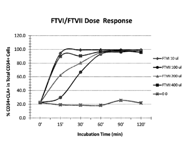
fucosylation of purified cord blood-derived CD34+ cells by FTVI or FTVII.
[037] Figure 4 graphically illustrates a comparative analysis of the
kinetics of cell surface
fucosylation by FTVI or FTVII of fresh cultured neural stem cells (NSCs).
[038] Figures 5 and 6 graphically illustrate a comparative analysis of the
kinetics of cell surface
fucosylation of human thawed cord blood-derived mononuclear cells by FTVI
(Figure 5) or FTVII
(Figure 6).
[039] Figure 7 graphically illustrates an analysis of the effects of FTVI
treatment versus sham
treatment on fucosylation of human endothelial progenitor cells (EPCs).
Date recue/ date received 2021-12-23
CA 02959534 2017-01-06
WO 2016/007506 11 PCT/US2015/039370
[040] Figure 8 graphically illustrates an analysis of the effects of FTVI
treatment on
fucosylation of human amniotic stem cells.
[041] Figure 9 illustrates an analysis of the effects of FTVI treatment on
fucosylation of
human adipose-derived stem cells.
[042] Figure 10 graphically illustrates an analysis of the effects of
fucosylation of
human MSCs either before or after trypsinization.
[043] Figure 11 illustrates the effect of incubating hNK cells with varying
concentrations of FTVI on the Level (%) of Fucosylation. hNK cells were
expanded for 14
days, harvested, washed, and incubated with varying concentrations of FTVI
ranging from 5
lig/mL to 25 g/mL. With the addition of GDP-fucose (final concentration of 1
mM in all
samples), cells were incubated for 30 minutes at room temperature, followed by
analysis of
the extent of fucosylation with CLA-FITC stain in addition to analyzing other
cell surface
markers (CD62L, CD44, CD16, CD56 and PSGL) characteristic of NK cells.
[044] Figure 12 illustrates the effect of incubating control and TZ101-
treated human NK
cells on fluid phase binding to E-selectin chimera. Expanded hNK cells were
incubated
without or with 5, 10, 25, and 50 pg/mL 17101 at 2.5 x 10⁶ NK cells/mL for 30
minutes at
room temperature, washed, and resuspended. 1 pg/106 NK cells was then
incubated with
human or mouse E-selectin/Fc chimeric protein for 30 minutes at 4 C and
stained with CLA,
CD44, human IgG, and Annexin V.
[045] Figure 13 illustrates an examination of the stability of fucosylated
NK Cells at 48
hours following treatment with TZ101. hNK cells were expanded for 18 days,
harvested,
washed, and incubated with FTVI at 25 ii.g/mL. Following the addition of GDP-
fucose (final
concentration of 1 mM), cells were incubated for 30 minutes at room
temperature, followed
by analysis of the extent of fucosylation with CLA-FITC stain at 1 hour and 48
hours after
being maintained in culture media.
[046] Figure 14 illustrates a comparative analysis of cytotoxic potential
of control
versus fucosylated NK cells. hNK cells were expanded for 14 days, harvested,
washed, and
incubated with IL-2 for 24 hours prior to incubation with indicated cell
lines. Toxicity was
measured following the incubation of K562 cells and MM1S cells with either
control or
TZ101-fucosylated hNK cells. Cytotoxicity was monitored at the end of 4 hours
of incubation
with the measurement of chromium release.
CA 02959534 2017-01-06
WO 2016/007506 12 PCT/US2015/039370
[047] Figure 15A illustrates the fucosylation of Regulatory T (Tres) cells.
The left side of
each dot plot shows the isotype control, while the right side shows staining
along with the
expression of the percent CLA positive cells. Treatment with TZ101 (FTVI + GDP-
fucose)
increased the expression of cell surface sLeX units from 8.8% to 62%, as
detected with
HECA-452 anti-CLA antibody stain. Figure 15B illustrates that fucosylated (FT)
Leg cells
maintain their suppressive function. PBMCs from two donors were cultured
together to
generate MLR (D1+D2). Addition of Treg cells or FT-Tõg cells to the donor
mixture (D1+D2) at
a ratio of 1:1 significantly suppressed MLR. Y-axis denotes counts per minute
(CPM)
(Mean SEM, n=3).
[048] Figure 16 illustrates expansion of cytotoxic T cells against CG1 (CG1-
CTL) and
fucosylation thereof. Fucosylation levels were measured using flow cytometry
and anti-CLA
FITC. Non-treated cells exhibited 4% fucosylation, whereas cells treated with
TZ101
exhibited 100% fucosylation.
DETAILED DESCRIPTION
[049] Before explaining at least one embodiment of the inventive concept(s)
in detail
by way of exemplary drawings, experimentation, results, and laboratory
procedures, it is to
be understood that the inventive concept(s) is not limited in its application
to the details of
construction and the arrangement of the components set forth in the following
description
or illustrated in the drawings, experimentation and/or results. The inventive
concept(s) is
capable of other embodiments or of being practiced or carried out in various
ways. As such,
the language used herein is intended to be given the broadest possible scope
and meaning;
and the embodiments are meant to be exemplary - not exhaustive. Also, it is to
be
understood that the phraseology and terminology employed herein is for the
purpose of
description and should not be regarded as limiting.
[050] Unless otherwise defined herein, scientific and technical terms used
in
connection with the presently disclosed and/or claimed inventive concept(s)
shall have the
meanings that are commonly understood by those of ordinary skill in the art.
Further,
unless otherwise required by context, singular terms shall include pluralities
and plural
terms shall include the singular. Generally, nomenclatures utilized in
connection with, and
techniques of, cell and tissue culture, molecular biology, and protein and
oligo- or
polynucleotide chemistry and hybridization described herein are those well
known and
13
commonly used in the art. Standard techniques are used for recombinant DNA,
oligonucleotide
synthesis, and tissue culture and transformation (e.g., electroporation,
lipofection). Enzymatic
reactions and purification techniques are performed according to
manufacturer's specifications or as
commonly accomplished in the art or as described herein. The foregoing
techniques and procedures
are generally performed according to conventional methods well known in the
art and as described
in various general and more specific references that are cited and discussed
throughout the present
specification. See e.g., Sambrook et al. Molecular Cloning: A Laboratory
Manual (2nd ed., Cold Spring
Harbor Laboratory Press, Cold Spring Harbor, N.Y. (1989) and Coligan et al.
Current Protocols in
Immunology (Current Protocols, Wiley Interscience (1994)). The nomenclatures
utilized in
connection with, and the laboratory procedures and techniques of, analytical
chemistry, synthetic
organic chemistry, and medicinal and pharmaceutical chemistry described herein
are those well
known and commonly used in the art. Standard techniques are used for chemical
syntheses, chemical
analyses, pharmaceutical preparation, formulation, and delivery, and treatment
of patients.
[051] All patents, published patent applications, and non-patent
publications mentioned in the
specification are indicative of the level of skill of those skilled in the art
to which this presently
disclosed and/or claimed inventive concept(s) pertains.
[052] All of the compositions and/or methods disclosed and/or claimed
herein can be made
and executed without undue experimentation in light of the present disclosure.
While the
compositions and methods of the inventive concept(s) have been described in
terms of particular,
non-limiting embodiments, it will be apparent to those of skill in the art
that variations may be applied
to the compositions and/or methods and in the steps or in the sequence of
steps of the method
described herein without departing from the concept, spirit and scope of the
presently disclosed
and/or claimed inventive concept(s). All such similar substitutes and
modifications apparent to those
skilled in the art are deemed to be within the spirit, scope and concept of
the inventive concept(s) as
defined by the appended claims.
Date recue/ date received 2021-12-23
CA 02959534 2017-01-06
WO 2016/007506 14 PCT/US2015/039370
[053] As utilized in accordance with the present disclosure, the following
terms, unless
otherwise indicated, shall be understood to have the following meanings:
[054] The use of the word "a" or "an" when used in conjunction with the
term
"comprising" in the claims and/or the specification may mean "one," but it is
also consistent
with the meaning of "one or more," "at least one," and "one or more than one."
The
singular forms "a," "an," and "the" include plural referents unless the
context clearly
indicates otherwise. Thus, for example, reference to "a compound" may refer to
1 or more,
2 or more, 3 or more, 4 or more or greater numbers of compounds. The term
"plurality"
refers to "two or more." The use of the term "or" in the claims is used to
mean "and/or"
unless explicitly indicated to refer to alternatives only or the alternatives
are mutually
exclusive, although the disclosure supports a definition that refers to only
alternatives and
"and/or." Throughout this application, the term "about" is used to indicate
that a value
includes the inherent variation of error for the device, the method being
employed to
determine the value, or the variation that exists among the study subjects.
For example but
not by way of limitation, when the term "about" is utilized, the designated
value may vary
by 20% or 10%, or 5%, or 1%, or 0.1% from the specified value, as
such variations
are appropriate to perform the disclosed methods and as understood by persons
having
ordinary skill in the art. The use of the term "at least one" will be
understood to include
one as well as any quantity more than one, including but not limited to, 2, 3,
4, 5, 10, 15, 20,
30, 40, 50, 100, etc. The term "at least one" may extend up to 100 or 1000 or
more,
depending on the term to which it is attached; in addition, the quantities of
100/1000 are
not to be considered limiting, as higher limits may also produce satisfactory
results. In
addition, the use of the term "at least one of X, Y and Z" will be understood
to include X
alone, Y alone, and Z alone, as well as any combination of X, Y and Z. The use
of ordinal
number terminology (i.e., "first", "second", "third", "fourth", etc.) is
solely for the purpose
of differentiating between two or more items and is not meant to imply any
sequence or
order or importance to one item over another or any order of addition, for
example.
[055] As used in this specification and claim(s), the terms "comprising"
(and any form
of comprising, such as "comprise" and "comprises"), "having" (and any form of
having, such
as "have" and "has"), "including" (and any form of including, such as
"includes" and
"include") or "containing" (and any form of containing, such as "contains" and
"contain")
CA 02959534 2017-01-06
WO 2016/007506 15 PCT/US2015/039370
are inclusive or open-ended and do not exclude additional, unrecited elements
or method
steps.
[056] The term "or combinations thereof" as used herein refers to all
permutations
and combinations of the listed items preceding the term. For example, "A, B,
C, or
combinations thereof" is intended to include at least one of: A, B, C, AB, AC,
BC, or ABC, and
if order is important in a particular context, also BA, CA, CB, CBA, BCA, ACB,
BAC, or CAB.
Continuing with this example, expressly included are combinations that contain
repeats of
one or more item or term, such as BB, AAA, AAB, BBC, AAABCCCC, CBBAAA, CABABB,
and so
forth. The skilled artisan will understand that typically there is no limit on
the number of
items or terms in any combination, unless otherwise apparent from the context.
[057] As used herein, the term "substantially" means that the subsequently
described
event or circumstance completely occurs or that the subsequently described
event or
circumstance occurs to a great extent or degree. For example, the term
"substantially"
means that the subsequently described event or circumstance occurs at least
90% of the
time, or at least 95% of the time, or at least 98% of the time.
[058] As used herein, "Current Good Manufacturing Practice" or "cGMP"
refers to the
Current Good Manufacturing Practice regulations enforced by the US Food and
Drug
Administration (FDA) or equivalent regulatory authorities in non-US countries.
cGMP
regulations provide for systems that assure proper design, monitoring, and
control of
manufacturing processes and facilities. Adherence to the cGMP regulations
assures the
identity, strength, quality, and purity of drug products by requiring that
manufacturers of
medications adequately control manufacturing operations. This includes
establishing strong
quality management systems, obtaining appropriate quality raw materials,
establishing
robust operating procedures, detecting and investigating product quality
deviations, and
maintaining reliable testing laboratories.
[059] As used herein, the term "ex vivo expansion" or "expansion" refers to
a method
of growing a cell population in tissue culture that increases the number of
cells in that
population. Cells that have undergone ex vivo expansion are referred to as
"expanded".
[060] As used herein, the term "fucosylation" refers to the treatment of a
population
of cells with an a1,3-fucosyltransferase and fucose donor under conditions
that increase the
ability of the cells to bind to a selectin or that increase the reactivity of
the cells with an
antibody known in the art to bind to sLeX including, but not limited to, the
HECA-452
CA 02954534 2017-01-06
WO 2016/007506 16 PCT/US2015/039370
monoclonal antibody. Cells that have been treated with an a1,3-
fucosyltransferase and
fucose donor and then exhibit increased binding to selectins or to the HECA-
452
monoclonal antibody or to another antibody specific for sLeX are referred to
as being
"fucosylated". As used herein, "fucosylation" can also refer to the levels of
sLeX present on
a cell population.
[061] As used herein, the term "hematopoeitic stem and progenitor cells" or
"HSPC"
refers to a cell population derived from bone marrow, cord blood or mobilized
peripheral
blood that is used to reconstitute the hematopoietic system of a patient. As
used herein,
the term hematopoeitic stem and progenitor cells" or "HSPC" includes
carlecortemcel-L.
[062] As used herein, the term "mesenchymal stromal cell" or "MSC" refers to
cells that
meet the definition set in 2006 by The International Society for Cellular
Therapy (ISCT): (1)
adherence to plastic, (2) expression of CD73, CD90, and CD105 antigens, while
being CD14,
CD34, CD45, and HLA-DR negative, and (3) ability to differentiate to
osteogenic,
chondrogenic and adipogenic lineage (Dominici et al. (2006) Cytotherapy, 8315-
317). As
used herein, "mesenchymal stromal cell" or "MSC" is synonymous with
"mesenchymal stem
cell," and thus said terms are used interchangeably herein. As used herein,
"MSC" can be
used as either singular or plural. As used herein, "mesenchymal stromal cell"
or "MSC" can
be derived from any tissue including, but not limited to, bone marrow, adipose
tissue,
amniotic fluid, endometrium, trophoblast-derived tissues, cord blood, Wharton
jelly, and
placenta. As used herein, "mesenchymal stromal cell" or "MSC" includes cells
that are CD34
positive upon initial isolation from tissue but satisfy the ISCT criteria
after expansion. As
used herein, "MSC" includes cells that are isolated from tissues using cell
surface markers
selected from the list comprised of NGF-R, PDGF-R, EGF-R, IGF-R, CD29, CD49a,
CD56, CD63,
CD73, CD105, CD106, CD140b, CD146, CD271, MSCA-1, SSEA4, STRO-1 and STRO-3 or
any
combination thereof, and satisfy the ISCT criteria either before or after
expansion. As used
herein, "mesenchymal stromal cell" or "MSC" includes cells described in the
literature as
bone marrow stromal stem cells (BMSSC), marrow-isolated adult multipotent
inducible cells
(MIAMI) cells, multipotent adult progenitor cells (MAPC), mesenchymal adult
stem cells
(MASCS), MULTISTEM (Athersys, Inc., Cleveland, OH), PROCHYMAL (Osiris
Therapeutics,
Inc., Columbia, MD), remestemcel-L, Mesenchymal Precursor Cells (MPCs), Dental
Pulp Stem
Cells (DPSCs), PLX cells, PLX-PAD, ALLOSTEM (Allosource, Centennial, CO),
ASTROSTEM
(Osiris Therapeutics, Inc., Columbia, MD), lxmyelocel-T, MSC-NTF, NurOwnTM
(Brainstorm
CA 02954534 2017-01-06
WO 2016/007506 17 PCT/US2015/039370
Cell Therapeutics Inc., Hackensack, NJ), STEMEDYNETm-MSC (Stemedica Cell
Technologies
Inc., San Diego, CA), STEMPEUCEL (Stem peud ics Research, Bangalore, India),
StempeuceICLI, Stempeucel0A, HiQCell, Hearticellgram-AMI, REVASCOR
(Mesoblast, Inc.,
Melbourne, Australia) CARDIOREL (Reliance Life Sciences, Navi Mumbai, India),
CARTISTEM (Medipost, Rockville, MD), PNEUMOSTEM (Medipost, Rockville, MD),
PROMOSTEM (Medipost, Rockville, MD), Homeo-GH, AC607, PDA001, S8623, CX601,
AC607, Endometrial Regenerative Cells (ERC), adipose-derived stem and
regenerative cells
(ADRCs) obtained with the CELUTION System (Cytori Therapeutics, Inc., San
Diego, CA),
perivascular-derived cells, and pericyte-derived cells. As used herein,
"mesenchymal
stromal cell" or "MSC" includes cells that only satisfy one or more of the
ISCT criteria when
cultured under one set of conditions but satisfy the full set of ISCT criteria
when cultured on
plastic tissue culture flasks in the presence of tissue culture medium
containing 10% fetal
bovine serum.
[063] As used herein, the term "muscle stem cells" refers to a cell
population derived
from muscle, including striated muscle, smooth muscle, cardiac muscle, muscle
satellite
cells or bone marrow cells reprogrammed to form muscle. As used herein, the
term
"muscle stem cells" includes MyoCell (Bioheart, Inc., Sunrise, FL), MyoCell
SDF-1, C3BS-
CQR-1, and CAP-1002.
[064] As used herein, "natural killer cells" or "NK" cells refers to a cell
population that
lacks CD3 and expresses CD56 and/or NKp46.
[065] As used herein, "neural stem cells" or "NSC" refers to a cell
population capable of
differentiating into neural cells or glial cells. As used herein, the term
"neural stem cells"
includes Q-Cells (Q Therapeutics Inc., Salt Lake City, UT), NSI-566, HuCNS-SC
(Stem Cells,
Inc., Newark, CA), and ReN001.
[066] As used herein, "patient" is used broadly to refer to any animal in
need of
therapeutic cells to ameliorate a condition, disease or injury. The animal can
be a mammal,
a bird, a fish, a reptile or any other animal. Some non-limiting examples of
mammals
include humans and other primates, equines such as horses, bovines such as
cows, ovines
such as sheep, caprines such as goats, canines such as dogs, felines such as
cats, rodents
such as mice or rats, and other mammals such as rabbits, Guinea pigs, and
the like.
CA 02954534 2017-01-06
WO 2016/007506 18 PCT/US2015/039370
[067] As used herein, "physiologically balanced salt solution" refers to a
solution or
medium where the concentrations of salts and other components are adjusted
such that the
solution or medium is isotonic with human cells, with osmolarity approximately
280 to 310
mOsmol/L, and is at a physiological pH, approximately pH 7.3 - 7.4. Examples
of
physiologically balanced salt solutions include, but are not limited to,
Hank's basic salt
solution, Alpha Minimum Essential Medium (aMEM), Dulbecco's Minimum Essential
Medium (DMEM), Iscove's Modified Dulbecco's Medium (IMDM) and PlasmaLyte
solutions
such as Plasma Lyte A.
[068] As used herein, "therapeutic cells" refers to an expanded cell
population that
ameliorates a condition, disease, and/or injury in a patient. Therapeutic
cells may be
autologous (i.e., derived from the patient), allogeneic (i.e., derived from an
individual of the
same species that is different than the patient) or xenogeneic (i.e., derived
from a different
species than the patient). Therapeutic cells may be homogenous (i.e.,
consisting of a single
cell type) or heterogenous (i.e., consisting of multiple cell types). The
term "therapeutic
cell" includes both therapeutically active cells as well as progenitor cells
capable of
differentiating into a therapeutically active cell.
[069] Turning now to the presently disclosed and/or claimed inventive
concept(s), one
embodiment thereof relates generally to compositions for and methods of
manufacturing
therapeutic cells that are treated with an a1,3-fucosyltransferase and fucose
donor and
exhibit enhanced migration and engraftment when administered in viva compared
to their
non-fucosylated counterparts.
[070] Embodiments of the presently disclosed and/or claimed inventive
concept(s) also
relate to the commercial provision of the possibility to manufacture and
optionally to
cryopreserve the therapeutic cells under Current Good Manufacturing Practice
(cGMP)
regulations enforced by the United States (US) Food and Drug Administration
(FDA) or the
equivalent regulatory authority in non-US countries. The therapeutic cells are
useful for
treating a variety of diseases and disorders including, but not limited to,
ischernic conditions
(e.g., limb ischemia, congestive heart failure, cardiac ischemia, kidney
ischemia and ESRD,
stroke, and ischemia of the eye), conditions requiring organ or tissue
regeneration (e.g.,
regeneration of liver, pancreas, lung, salivary gland, blood vessel, bone,
skin, cartilage,
tendon, ligament, brain, hair, kidney, muscle, cardiac muscle, nerve, and
limb),
inflammatory diseases (e.g., heart disease, diabetes, spinal cord injury,
rheumatoid arthritis,
CA 02959534 2017-01-06
WO 2016/007506 19 PCT/US2015/039370
osteo-arthritis, inflammation due to hip replacement or revision, Crohn's
disease, and graft
versus host disease) autoimmune diseases (e.g., type 1 diabetes, psoriasis,
systemic lupus,
and multiple sclerosis), a degenerative disease, a congenital disease
hematologic disorders
such as anemia, neutropenia, thrombocytosis, myeloproliferative disorders or
hematologic
neoplasms and cancer such as leukemia and lymphoma.
[071] Embodiments of the presently disclosed and/or claimed inventive
concept(s)
generally relate to compositions and methods of manufacturing and/or storing
fucosylated
cell populations, and more particularly, but not limited to, to therapeutic
cells isolated from
bone marrow, cord blood, umbilical cord, Wharton's jelly, peripheral blood,
lymphoid tissue,
endometrium, trophoblast-derived tissues, placenta, amniotic fluid, adipose
tissue, muscle,
liver, cartilage, nervous tissue, cardiac tissue, dental pulp tissue,
exfoliated teeth, cells
derived from embryonic stem (ES) cells or induced pluripotent stem (iPS)
cells, or any
combination thereof.
[072] In a particular, non-limiting embodiment, the isolated therapeutic
cells are
differentiated embryonic stem cells and/or differentiated induced pluripotent
stem cells.
[073] In particular, one embodiment of the presently disclosed and/or
claimed
inventive concept(s) relates to methods of mass producing such cells, treating
them with an
effective amount of an a1,3-fucosyltransferase and fucose donor (e.g. a1,3-
fucosyltransferase VI or a1,3-fucosyltransferase VII together with the fucose
donor GDP-
fucose), and then optionally cryopreserving them under conditions where the
enhanced
levels of cell surface fucosylation resulting from the enzyme treatment are
retained after
thawing the cells.
[074] The presently disclosed and/or claimed inventive concept(s) can also
be used for
veterinary purposes since there is a parallelism between the mechanisms
involved in
enhanced binding to selectins after fucosylation of selectin ligands between
humans and
animals.
[075] In the methods contemplated herein, the fucosyltransferase may be
selected
from the group comprised of an a1,3-fucosyltransferase III, an a1,3-
fucosyltransferase IV, an
a1,3-fucosyltransferase V, an a1,3-fucosyltransferase VI, an a1,3-
fucosyltransferase VII, an
a1,3-fucosyltransferase IX, an a1,3-fucosyltransferase X, and an a1,3-
fucosyltransferase XI,
or any combination thereof. The fucose donor may be, for example, GDP-fucose.
CA 02959534 2017-01-06
WO 2016/007506 20 PCT/US2015/039370
[076] The presently disclosed and/or claimed inventive concept(s) in one
embodiment
contemplates a method of manufacturing fucosylated therapeutic cells
comprising the steps
of providing a quantity of therapeutic cells in tissue culture or isolating
therapeutic cells,
expanding the therapeutic cells, and fucosylating the quantity or population
of therapeutic
cells by contacting them in vitro with an effective amount of an a1,3-
fucosyltransferase and
a fucose donor. The fucosylated therapeutic cells have enhanced binding to P-
selectin or E-
selectin. The fucosylated therapeutic cells may optionally further be
cryopreserved under
conditions that retain the enhanced binding to P-selectin or E-selectin after
thawing the
cells.
[077] In another non-limiting embodiment, the presently disclosed and/or
claimed
inventive concept(s) includes a method of cryopreserving fucosylated
therapeutic cells. In
the method, therapeutic cells are isolated and fucosylated by contacting them
with an
effective amount of an a1,3-fucosyltransferase and a fucose donor. The
fucosylated
therapeutic cells are then frozen in a therapeutic cell cryopreservation
composition
comprising a physiologically balanced salt solution and a cryoprotectant.
[078] The method may further include the step of expanding the therapeutic
cells prior
to fucosylation. When the cells are expanded, in a particular, non-limiting
embodiment, the
physiologically balanced salt solution in which the cells are frozen may be
the tissue culture
medium in which the cells are expanded. In addition, the physiologically
balanced salt
solution may further contain protein. Non-limiting examples of proteins that
may be
utilized in accordance with the presently disclosed and/or claimed inventive
concept(s)
include fetal bovine serum, horse serum, human serum, human platelet lysate,
bovine
albumin, human albumin, and any combinations thereof.
[079] In a particular, non-limiting embodiment, the freezing step includes
cooling the
therapeutic cells in the cell cryopreservation composition at a rate of about
1 C per minute
from about 37 C to about -80 C to produce a frozen cell suspension, and then
transferring
the frozen cell suspension to storage in the presence of liquid nitrogen. In
addition or
(alternatively), the therapeutic cells may be frozen using a vitrification
method.
[080] In a particular, non-limiting embodiment, adherent cells are first
removed from
the tissue culture plastic or rnicrobead or other substrate on which they are
grown, treated
with an a1,3-fucosyltransferase and a fucose donor and then optionally
cryopreserved. It is
a surprising finding of the presently disclosed and/or claimed inventive
concept(s) that
CA 02959534 2017-01-06
WO 2016/007506 21 PCT/US2015/039370
removal of cells from tissue culture plastic and other substrates by exposing
them to trypsin
followed by fucosylation is a more effective method than fucosylation of cells
while
attached to tissue culture plastic and then removing them with trypsin.
[081] In a particular, non-limiting embodiment, the methods are performed
under
cGMP conditions.
[082] In a particular, non-limiting embodiment, the therapeutic cells of
the presently
disclosed and/or claimed inventive concept(s) are cells isolated from bone
marrow, cord
blood, umbilical cord, Wharton's jelly, peripheral blood, lymphoid tissue,
endometrium,
trophob last-derived tissues, placenta, amniotic fluid, adipose tissue,
muscle, liver, cartilage,
nervous tissue, cardiac tissue, dental pulp tissue, exfoliated teeth, cells
derived from
embryonic stem (ES) cells or induced pluripotent stem (iPS) cells, or any
combination
thereof.
[083] In a particular, non-limiting embodiment, the therapeutic cells of
the presently
disclosed and/or claimed inventive concept(s) are selected from hematopoietic
stem cells,
immune cells, mesenchynnal stem cells, muscle cells, amniotic cells,
endometrial cells,
neural stem cells, natural killer (NK) cells, T cells, B cells, or any
combination thereof. For
example, but not by way of limitation, the therapeutic cells may be T cells
(including but not
limited to, regulatory T cells and cytotoxic T cells (for example, but not by
way of limitation,
CD8+ cytotoxic T cells)), NK cells, B cells, CD38+ cells, neural stem cells,
or any combination
thereof, wherein said cells are fucosylated by fucosyltransferase VII (FT
VII). It is a surprising
finding of the presently disclosed and/or claimed inventive concept(s) that
some cells are
preferentially fucosylated with FT VII instead of FT VI. This is unexpected
given the in vitro
fucose donor specificities of the enzymes ¨ whereas FucT-VI is active on both
neutral and 3'-
sialylated fucose donors, FucT-VII acts on only the 3'-sialylated type 2
chain. A priori, one
would therefore expect that FTVI would fucosylate cells to approximately the
same extent
as FTVII; this was observed for some cells but not for others.
[084] In one embodiment of the presently disclosed and/or claimed inventive
concept(s), hematopoietic cells that have been expanded are mixed with
unexpanded
fucosylated hematopoietic cells. It is a surprising finding of the presently
disclosed and/or
claimed inventive concept(s) that a mixture of fucosylated and non-fucosylated
expanded
hematopoietic cells is more effective than either population used alone.
CA 02954534 2017-01-06
WO 2016/007506 22 PCT/US2015/039370
[085] In one embodiment of the presently disclosed and/or claimed inventive
concept(s), natural killer cells are expanded and then fucosylated. Until the
filing of the
present application there has been neither a description nor suggestion that
natural killer
cells can be fucosylated ex vivo.
[086] Until the presently disclosed and/or claimed inventive concept(s),
there has been
neither a description nor suggestion towards the development of a
cryopreservation
method for fucosylated therapeutic cells. Furthermore, the inventors
surprisingly found that
by following the cryoprotection method of the presently disclosed and/or
claimed inventive
concept(s), therapeutic cells with a high retention of fucosylation are
recovered after
cryopreservation.
[087] The presently disclosed and/or claimed inventive concept(s) in one
embodiment
contemplates a method of treating therapeutic cells comprising the steps of
providing/isolating a quantity or population of therapeutic cells, expanding
the therapeutic
cells in tissue culture, treating the quantity or population of therapeutic
cells in vitro with an
a1,3-fucosyltransferase and a fucose donor, wherein the treated therapeutic
cells have
enhanced binding to P-selectin and E-selectin, and then optionally
cryopreserving the cells.
Furthermore, the therapeutic cells are typically characterized as comprising P-
selectin
glycoprotein ligand-1 (PSGL-1), CD44, and/or other selectin ligands that do
not effectively
bind to P-selectin or E-selectin. The therapeutic cells, in their untreated
state prior to
fucosylation as described herein, have reduced retention in inflamed,
ischemic, or damaged
tissues.
[088] In a particular, non-limiting embodiment of the presently disclosed
and/or
claimed inventive concept(s), the therapeutic cells are derived from the list
comprising bone
marrow, cord blood, umbilical cord, Wharton's jelly, peripheral blood,
lymphoid tissue,
endometrium, trophoblast-derived tissues, placenta, amniotic fluid, adipose
tissue, muscle,
liver, cartilage, nervous tissue, cardiac tissue, dental pulp tissue and
exfoliated teeth, though
they may be derived from cells grown in tissue culture or are cells derived
from embryonic
stem (ES) cells or induced pluripotent stem (iPS) cells. The therapeutic cells
may also be any
combination of the above.
[089] In a particular, non-limiting embodiment of the presently disclosed
and/or
claimed inventive concept(s), the therapeutic cells are expanded under cGMP
conditions.
CA 02959534 2017-01-06
WO 2016/007506 23 PCT/US2015/039370
[090] As noted above, after the fucosylation treatment described herein,
the treated
therapeutic cells have enhanced binding to P-selectin or E-selectin, as
compared to
untreated therapeutic cells. Enhanced binding to P-selectin (or E-selectin) is
defined as at
least 10% of the treated therapeutic cells having fluorescence in a P-selectin
(or E-selectin,
respectively) binding assay which is greater than a predetermined fluorescence
threshold
(as defined below). In another embodiment, at least 25% of the treated
therapeutic cells
exceed the predetermined fluorescence threshold. In another embodiment, at
least 50% of
the treated therapeutic cells exceed the predetermined fluorescence threshold.
In another
embodiment, at least 75% of the treated therapeutic cells exceed the
predetermined
fluorescence threshold. In another embodiment, at least 90% of the treated
therapeutic
cells exceed the predetermined fluorescence threshold. In another embodiment,
at least
95% of the treated therapeutic cells exceed the predetermined fluorescence
threshold.
[091] The presently disclosed and/or claimed inventive concept(s) further
contemplates a therapeutic cell product produced by the method including the
steps of
providing a quantity or population of cells, expanding the cells in tissue
culture, and treating
the quantity of therapeutic cells in vitro with an a1,3-fucosyltransferase and
fucose donor,
wherein the majority of the treated therapeutic cells have enhanced binding to
P-selectin
(or E-selectin) as described herein, and optionally cryopreserving the cells.
The quantity of
cells may be derived from, for example but not by way of limitation, bone
marrow, cord
blood, umbilical cord, Wharton's jelly, peripheral blood, lymphoid tissue,
endometrium,
trophoblast-derived tissues, placenta, amniotic fluid, adipose tissue, muscle,
liver, cartilage,
nervous tissue, cardiac tissue, dental pulp tissue, exfoliated teeth, though
they may be
derived from cells grown in tissue culture or are cells derived from embryonic
stem (ES) cells
or induced pluripotent stem (iPS) cells. The therapeutic cells may also be any
combination
of the above.
[092] The presently disclosed and/or claimed inventive concept(s) in one
embodiment
contemplates a method of treating therapeutic cells comprising providing a
quantity or
population of therapeutic cells which lack or have reduced expression (less
than the normal
level of expression of CD38) of surface protein CD38, and treating the
quantity or population
of therapeutic cells in vitro with an a1,3-fucosyltransferase and a fucose
donor, wherein the
therapeutic cells so treated have enhanced binding to P-selectin or E-selectin
over the
untreated therapeutic cells. Furthermore, the untreated therapeutic cells are
typically
CA 02959534 2017-01-06
WO 2016/007506 24 PCT/US2015/039370
characterized as predominantly comprising PSGL-1, CD44 and/or other selectin
ligands that
do not adequately bind to P-selectin or E-selectin or the therapeutic cells
may lack
expression of any selectin ligands. The PSGL-1 or other selectin ligands that
occur on the
therapeutic cells lack or have reduced numbers of fucosylated glycans, such as
0-glycans,
and may for example, have PSGL-1 which have core-2 0-glycans that comprise
NeuAca2,3Galf31,4GIcNAc but that lack a fucose in a1,3 linkage to the GIcNAc.
The
therapeutic cells, in their untreated state prior to fucosylation, have
reduced homing ability
to bone marrow or to other desired sites that express selectins. In one
particular, non-
limiting embodiment, the therapeutic cells are derived from the list comprised
of bone
marrow, cord blood, umbilical cord, Wharton's jelly, peripheral blood,
lymphoid tissue,
endometrium, trophoblast-derived tissues, placenta, amniotic fluid, adipose
tissue, muscle,
liver, cartilage, nervous tissue, cardiac tissue, dental pulp tissue,
exfoliated teeth, though
they may be derived from cells grown derived from embryonic stem (ES) cells or
induced
pluripotent stem (iPS) cells, as long as they are characterized as needing, or
benefiting from,
further fucosylation to enhance their bone marrow homing ability. In the
methods
contemplated herein, the a1,3-fucosyltransferase may be for example a1,3-
fucosyltransferase IV, a1,3-fucosyltransferase VI, or a1,3-fucosyltransferase
VII. The fucose
donor may be for example GDP-fucose.
[093] The presently disclosed and/or claimed inventive concept(s)
contemplates in one
embodiment a composition of treated therapeutic cells that comprise a cell
population
grown under cGMP-compliant conditions, wherein the treated cells comprise PSGL-
1. or
other selectin ligands that are properly fucosylated (e.g., comprises sialyl
Lewis X) and that
are able to bind to P-selectin (or E-selectin). The treated therapeutic cells
may be disposed
in a pharmaceutically acceptable carrier or vehicle for storage or
administration to a patient.
Optionally, the treated therapeutic cells may be cryopreserved for storage
prior to
administration to a patient.
[094] In a particular, non-limiting embodiment, the therapeutic cells are
selected from
the list comprised of cord blood hematopoietic cells expanded under cGMP-
compliant
conditions, bone marrow-derived cells expanded under cGMP-compliant
conditions, cord
blood-derived cells expanded under cGMP-compliant conditions, mesenchymal
stromal cells
expanded under cGMP-compliant conditions, neural stem cells expanded under
cGMP-
compliant conditions, hepatocytes expanded under cGMP-compliant conditions,
natural
CA 02954534 2017-01-06
WO 2016/007506 25 PCT/US2015/039370
killer cells expanded under cGMP-compliant conditions and T cells expanded
under cGMP-
compliant conditions.
[095] In one embodiment, the therapeutic cells are expanded under cGMP-
compliant
conditions, cryopreserved under conditions that maintain optimal levels of
fucosylation, and
then thawed and fucosylated prior to delivery to a patient.
[096] In one particular, non-limiting embodiment, the therapeutic cells are
expanded
under cGMP-compliant conditions, fucosylated and then cryopreserved under
conditions
that maintain optimal levels of fucosylation after the cells are thawed.
[097] In a particular, non-limiting embodiment, the bone marrow-derived
cells
expanded under cGMP-compliant conditions are selected from the list comprised
of AMR-
001 (Amorcyte, Inc., Allendale, NJ) ALD-301, ALD-201, ALD-401, bone marrow-
derived cells
expanded in the presence of the Notch ligand Delta1 and bone marrow-derived
cells
expanded in the presence of MSC.
[098] In a particular, non-limiting embodiment, the cord blood-derived
cells expanded
under cGMP-compliant conditions are selected from the list comprised of NiCord
(Gam ida
Cell Ltd., Jerusalem, Israel), Hemacord, ProHema, cord blood-derived cells
expanded in the
presence of the Notch ligand Delta1 and cord blood-derived cells expanded in
the presence
of MSC.
[099] In a particular, non-limiting embodiment, the mesenchymal stromal
cells
expanded under cGMP-compliant conditions are selected from the list comprised
of
MULTISTEM (Athersys, Inc., Cleveland, OH), PROCHYMAL (Osiris Therapeutics,
Inc.,
Columbia, MD), remestemcel-L, Mesenchymal Precursor Cells (MPCs), Dental Pulp
Stem
Cells (DPSCs), PLX cells, PLX-PAD, ALLOSTEM (Allosource, Centennial, CO),
ASTROSTEM
(Osiris Therapeutics, Inc., Columbia, MD), lxmyelocel-T, MSC-NTF, NurOwnTM
(Brainstorm
Cell Therapeutics Inc., Hackensack, NJ), STEMEDYNE"-MSC (Stemedica Cell
Technologies
Inc., San Diego, CA), STEMPEUCEL (Stempeudics Research, Bangalore, India),
StempeuceICLI, StempeucelOA, HiQCell, Hearticellgram-AMI, REVASCOR
(Mesoblast, Inc.,
Melbourne, Australia) CARDIOREL (Reliance Life Sciences, Navi Mumbai, India),
CARTISTEM (Medipost, Rockville, MD), PNEUMOSTEM (Medipost, Rockville, MD),
PROMOSTEM (Medipost, Rockville, MD), Homeo-GH, AC607, PDA001, SB623, CX601,
AC607, Endometrial Regenerative Cells (ERC), and adipose-derived stem and
regenerative
CA 02954534 2017-01-06
WO 2016/007506 26 PCT/US2015/039370
cells (ADRCs) obtained with the CELUTION System (Cytori Therapeutics, Inc.,
San Diego,
CA).
[0100] In a particular, non-limiting embodiment, the neural stem cells
expanded under
cGMP-compliant conditions are selected from the list comprised of NSI-566,
HuCNS-SC
(Stem Cells, Inc., Newark, CA), CTX0E03, ReN001, ReN009, STEMEDYNETm-NSC
(Stemedica
Cell Technologies Inc., San Diego, CA), Q-CELLS (0 Therapeutics Inc., Salt
Lake City, UT),
TBX-01, TBX-02, RhinoCyteTM olfactory stem cells (RhinoCyte Inc., Louisville,
KY),
MOTORGRAFT (California Stem Cell, Inc., Irvine, CA), and CellBeadsTM Neuro.
[0101] In a particular, non-limiting embodiment, the cardiac-derived cells
expanded
under cGMP-compliant conditions are cardiac-derived stem cells (CDCs).
[0102] In a particular non-limiting embodiment, the liver cells expanded
under cGMP-
compliant conditions are hpSC-derived hepatocytes, Heterologous Human Adult
Liver
Progenitor Cells (HHALPC), hLEC, and PROMETHERA HepaStem (Promethera
Biosicences
SA/NV, Belgium).
[0103] In one embodiment, the composition of treated therapeutic cells
comprises a
population of human HSPC expanded under cGMP-compliant conditions having
enhanced
binding to P-selectin (or E-selectin). Enhanced binding to P-selectin (or E-
selectin) is defined
as at least 10% of the treated HSPC having fluorescence in a P-selectin
binding assay (or E-
selectin binding assay, respectively) which is greater than a predetermined
fluorescence
threshold. In another embodiment, at least 25% of the treated HSPC exceed the
predetermined fluorescence threshold. In another embodiment, at least 50% of
the treated
HSPC exceed the predetermined fluorescence threshold. In another embodiment,
at least
75% of the treated HSPC exceed the predetermined fluorescence threshold. In
another
embodiment, at least 90% of the treated HSPC exceed the predetermined
fluorescence
threshold. In another embodiment, at least 95% of the treated HSPC exceed the
predetermined fluorescence threshold. The composition of human HSPC may be
disposed in
a pharmaceutically-acceptable carrier or vehicle for storage or for
administration to a
subject.
[0104] In one embodiment, the composition of treated therapeutic cells
comprises a
population of human MSC expanded under cGMP-compliant conditions having
enhanced
binding to P-selectin (or E-selectin). Enhanced binding to P-selectin (or E-
selectin) is defined
as at least 10% of the treated MSC having fluorescence in a P-selectin binding
assay (or E-
CA 02959534 2017-01-06
WO 2016/007506 27 PCT/US2015/039370
selectin binding assay, respectively) which is greater than a predetermined
fluorescence
threshold. In another embodiment, at least 25% of the treated MSC exceed the
predetermined fluorescence threshold. In another embodiment, at least 50% of
the treated
MSC exceed the predetermined fluorescence threshold. In another embodiment, at
least
75% of the treated MSC exceed the predetermined fluorescence threshold. In
another
embodiment, at least 90% of the treated MSC exceed the predetermined
fluorescence
threshold. In another embodiment, at least 95% of the treated MSC exceed the
predetermined fluorescence threshold. The composition of human MSC may be
disposed in
a pharmaceutically-acceptable carrier or vehicle for storage or for
administration to a
subject.
[0105] In one embodiment, the composition of treated therapeutic cells
comprises a
population of human neural stem cells expanded under cGMP-compliant conditions
having
enhanced binding to P-selectin (or E-selectin). Enhanced binding to P-selectin
(or E-selectin)
is defined as at least 10% of the treated neural stem cells having
fluorescence in a P-selectin
binding assay (or E-selectin binding assay, respectively) which is greater
than a
predetermined fluorescence threshold. In another embodiment, at least 25% of
the treated
neural stem cells exceed the predetermined fluorescence threshold. In another
embodiment, at least 50% of the treated neural stem cells exceed the
predetermined
fluorescence threshold. In another embodiment, at least 75% of the treated
neural stem
cells exceed the predetermined fluorescence threshold. In another embodiment,
at least
90% of the treated neural stem cells exceed the predetermined fluorescence
threshold. In
another embodiment, at least 95% of the treated neural stem cells exceed the
predetermined fluorescence threshold. The composition of human neural stem
cells may be
disposed in a pharmaceutically-acceptable carrier or vehicle for storage or
for
administration to a subject.
[0106] In one embodiment, the composition of treated therapeutic cells
comprises a
population of human hepatocytes expanded under cGMP-compliant conditions
having
enhanced binding to P-selectin (or E-selectin). Enhanced binding to P-selectin
(or E-selectin)
is defined as at least 10% of the treated hepatocytes having fluorescence in a
P-selectin
binding assay (or E-selectin binding assay, respectively) which is greater
than a
predetermined fluorescence threshold. In another embodiment, at least 25% of
the treated
hepatocytes exceed the predetermined fluorescence threshold. In another
embodiment, at
CA 02959534 2017-01-06
WO 2016/007506 28 PCT/US2015/039370
least 50% of the treated hepatocytes exceed the predetermined fluorescence
threshold. In
another embodiment, at least 75% of the treated hepatocytes exceed the
predetermined
fluorescence threshold. In another embodiment, at least 90% of the treated
hepatocytes
exceed the predetermined fluorescence threshold. In another embodiment, at
least 95% of
the treated hepatocytes exceed the predetermined fluorescence threshold. The
composition of human hepatocytes may be disposed in a pharmaceutically-
acceptable
carrier or vehicle for storage or for administration to a subject.
[0107] In one embodiment, the composition of treated therapeutic cells
comprises a
population of human NK cells expanded under cGMP-compliant conditions having
enhanced
binding to P-selectin (or E-selectin). Enhanced binding to P-selectin (or E-
selectin) is defined
as at least 10% of the treated NK cells having fluorescence in a P-selectin
binding assay (or E-
selectin binding assay, respectively) which is greater than a predetermined
fluorescence
threshold. In another embodiment, at least 25% of the treated NK cells exceed
the
predetermined fluorescence threshold. In another embodiment, at least 50% of
the treated
NK cells exceed the predetermined fluorescence threshold. In another
embodiment, at least
75% of the treated NK cells exceed the predetermined fluorescence threshold.
In another
embodiment, at least 90% of the treated NK cells exceed the predetermined
fluorescence
threshold. In another embodiment, at least 95% of the treated NK cells exceed
the
predetermined fluorescence threshold. The composition of human NK cells may be
disposed
in a pharmaceutically-acceptable carrier or vehicle for storage or for
administration to a
subject.
[0108] In one embodiment, the composition of treated therapeutic cells
comprises a
population of human T cells (such as, but not limited to, regulatory T cells
and cytotoxic T
cells (for example, but not by way of limitation, CD8+ cytotoxic T cells))
expanded under
cGM P-compliant conditions having enhanced binding to P-selectin (or E-
selectin). Enhanced
binding to P-selectin (or E-selectin) is defined as at least 10% of the
treated T cells having
fluorescence in a P-selectin binding assay (or E-selectin binding assay,
respectively) which is
greater than a predetermined fluorescence threshold. In another embodiment, at
least 25%
of the treated T cells exceed the predetermined fluorescence threshold. In
another
embodiment, at least 50% of the treated T cells exceed the predetermined
fluorescence
threshold. In another embodiment, at least 75% of the treated T cells exceed
the
predetermined fluorescence threshold. In another embodiment, at least 90% of
the treated
CA 02954534 2017-01-06
WO 2016/007506 29 PCT/US2015/039370
I cells exceed the predetermined fluorescence threshold. In another
embodiment, at least
95% of the treated T cells exceed the predetermined fluorescence threshold.
The
composition of human T cells may be disposed in a pharmaceutically-acceptable
carrier or
vehicle for storage or for administration to a subject.
[0109] The predetermined fluorescence threshold in one embodiment is
determined by
first obtaining a sample of therapeutic cells. This control (baseline) sample
of therapeutic
cells is assayed using the P-selectin binding assay (or E-selectin binding
assay) described
elsewhere herein, or by any other P-selectin fluorescence binding assay (or E-
selectin
binding assay, respectively) known in the art or by staining with the HECA-452
antibody. P-
selectin (or E-selectin or HECA-452) binding fluorescence levels are measured
for the
therapeutic cells in the control (baseline) sample. In one embodiment, a
fluorescence value
is selected that exceeds the P-selectin (or E-selectin or HECA-452) binding
fluorescence
levels of at least 95% of the therapeutic cells in the control sample. The
selected
fluorescence value is designated as the predetermined fluorescence threshold
against which
is compared the P-selectin (or E-selectin or HECA-452) binding fluorescence of
the treated
(i.e., fucosylated) therapeutic cells.
[0110] The presently disclosed and/or claimed inventive concept(s) further
contemplates a therapeutic cell product produced by the method of providing a
quantity or
population of therapeutic cells and treating the quantity of therapeutic cells
in vitro with an
a1,3-fucosyltransferase and a fucose donor, wherein the majority of the
treated therapeutic
cells bind to P-selectin (or E-selectin or HECA-452). The quantity of
therapeutic cells may be
derived from bone marrow, but may be derived from cord blood, umbilical cord,
peripheral
blood, lymphoid tissue, adipose tissue, neural tissue, muscle, placenta,
amniotic fluid,
endometrium, liver or they may be derived from cells derived from embryonic
stem (ES)
cells or induced pluripotent stem (iPS) cells. The therapeutic cells may also
be any
combination of the above.
[0111] In general, the presently disclosed and/or claimed inventive
concept(s)
contemplates a method of manufacture of therapeutic cells under cGMP
conditions wherein
non-functional or suboptimally functional PSGL-1 or other selectin ligands
expressed on
therapeutic cells are modified by in vitro a1,3-fucosylation technology to
correct the homing
defect, which improves their use in cell therapy.
30
[0112] As explained previously, therapeutic cells can express PSGL-1 or
other selectin ligands,
yet a significant amount do not bind to P-selectin (or E-selectin) or bind
only low amounts of P-selectin
(or E-selectin, respectively). PSGL-1 is a homodimeric mucin expressed on
almost all leukocytes
including CD34+ cells. To be functional, i.e., able to bind to P-selectin or E-
selectin, PSGL-1 requires
several post-translational modifications leading to formation of an sLex group
thereon, including
a1,3-fucosylation. Insufficient a1,3-fucosylation, for example, results in
impaired ability of naive T
cells to interact with vascular selecting. In the presently disclosed and/or
claimed inventive concept(s)
it has been discovered that the inability of therapeutic cells to bind to the
P-selectin or E-selectin
adhesion molecules can be corrected with ex viva fucosylation after expansion
under cGMP
conditions, either before or after cryopreservation. This is a surprising
finding given the number of
adhesion molecules that are down-regulated with expansion (e.g., Kallinikou et
al., Menard et al.,
Feneke et al. (supra)) and with cryopreservation (e.g., Faint et al.,
Koenigsmann et al., Hattori et al.,
Campbell et al., Aoyagi et al., Mareschi et al., and Neuhuber et al. (supra)).
[0113] Therefore, the basis of the presently disclosed and/or claimed
inventive concept(s) is that
the treatment of therapeutic cells in vitro with an a1,3-fucosyltransferase
and fucose donor (e.g., FT-
VI or FT-VII together with GDP-fucose), which also catalyzes the synthesis of
the sLex structure, will
increase fucosylation of PSGL-1 or other selectin ligands and thereby correct
the homing defect of
the therapeutic cells even after large-scale expansion in cGMP cultures. It is
a further basis of the
presently disclosed and/or claimed inventive concept(s) that fucosylated cells
can be cryopreserved
and retain their fucosylation levels after thaw.
[0114] Fucosyltransferases that are able to transfer fucose in a1,3 linkage
to GIcNAc are well
known in the art. Several are available commercially, for example from R&D
Systems (Minneapolis,
MN). Further, at least eight different types of a1,3-fucosyltransferases
(FTIII-VII) are encoded by the
human genome. These include: the Lewis enzyme (FTIII), which can transfer
fucose either a (1,3) or
a (1,4) to Galf34GIcNAc or Gal[33GIcNAc respectively (Kukowska-Latallo et al.
(1990) Genes Dev.,
4:1288); FTIV, which forms a (1,3) linkages, which does not prefer sialylated
precursors (Goelz, et al.
(1989) Cell, 63:1349; Lowe, et al. (1991) J. Biol. Chem., 266:17467); FTV
(Weston, et al. (1992) J. Biol.
Chem., 267:4152) and FTVI (Weston, et al. (1992) J. Biol. Chem., 267:24575)
which form a(1,3)
linkages, which can
Date recue/ date received 2021-12-23
CA 02959534 2017-01-06
WO 2016/007506 31 PCT/US2015/039370
fucosylate either sialylated or nonsialylated precursors, and FTVII (Sasaki,
et at. (1994) J.
Biol. Chem., 269:14730; Natsuka, et al. (1994) J. Biol. Chem., 269:16789),
which can
fucosylate only sialylated precursors. FTIX preferentially transfers fucose to
the GIcNAc
residue at the nonreducing terminal end of the polylactosamine chain,
resulting in the
terminal Lex structure, whereas the other a1,3FUTs preferentially transfer a
Fuc to the
GIcNAc residue at the penultimate position, resulting in the internal Lex
structure (Nishihara
et al. (1999) FEBS Lett., 462:289-294). FIX and FIX' link alpha-l-fucose onto
conalbumin
glycopeptides and biantennary N-glycan acceptors but not onto short
lactosaminyl acceptor
substrates as do classical monoexonic alpha1,3-fucosyltransferases (Mollicone
et al. (2009) J
Biol Chem., 284:4723-38).
[0115] Sequence information for FTIII is disclosed by GC19M005843; FTIV by
GC11P094277; FTV by GC19M005865; FTVI by GC19M005830; FTVII by GC09M139924,
FTIX
by GC06P096463, FIX by GC08M033286 and FIX! by GC10P075532 (GeneCards
(Weiznnann
Institute of Science, Rehovot, Israel) is a searchable, integrated, database
of human genes
maintained by the Weiznnann Institute that provides concise genomic related
information
on all known and predicted human genes as well as links to other databases).
The presently
disclosed and/or claimed inventive concept(s) further contemplates using
other, non-human
a1,3-fucosyltransferases available and known to those of ordinary skill in the
art, for
example as shown in US Patent Nos. 6,399,337 and 6,461,835.
[0116] Human HSPC can be obtained for treatment with a1,3-
fucosyltransferase, for
example, by separation from the other cells in a source of umbilical cord
blood, peripheral
blood, or bone marrow. Various techniques well known in the art may be
employed to
obtain the HSPC including, but not limited to, density gradient separation,
hypotonic lysis of
red blood cells, centrifugal elutriation or separation with monoclonal
antibodies using
fluorescent-activated cell sorter (FACS) or magnetic bead isolation devices.
Monoclonal
antibodies are particularly useful for identifying markers (surface membrane
proteins)
associated with particular cell lineages and/or stages of differentiation.
Antibodies such as
anti-CD34 or anti-CD133 can be used to isolate HSPC under cGMP-compliant
conditions,
either by FACS or by magnetic bead using an instrument such as the CliniMACS
System
from Miltenyi Biotec Inc. (Bergish Gladbach, Germany). Alternatively, HSPC can
be
separated using a reagent such as ALDEFLUORTM (STEMCELL Technologies, Inc.,
Vancouver,
BC) that is oxidized in cells by aldehyde dehydrogenase (ALDH) into a charged
fluorescent
32
product that accumulates in cells and allows the separation of brightly
fluorescent cells containing
the HSPC by FACS. The separation techniques employed should maximize the
retention of viability
of the fraction to be collected. The particular technique employed will depend
upon efficiency of
separation, cytotoxicity of the methodology, ease and speed of performance,
and necessity for
sophisticated equipment and/or technical skill.
[0117] Once isolated, HSPC can be expanded using a variety of cGMP
expansion protocols known
in the art (see Tung et al. (2010) Best Pract Res Clin Haematol., 23:245-57
for review). Cells can be
grown in tissue culture medium containing a cocktail of factors including, but
not limited to, one or
more from the list of factors comprised of erythropoietin, kit ligand, G-CSF,
GM-CSF, IL-6, IL-11,
thrombopoietin, fit ligand, FGF-1, angiopoietin-like 5, insulin-like growth
factor binding protein 2
(IGFBP2), notch ligand delta 1, PIXY321, prostaglandin E2, aryl hydrocarbon
nuclear receptor protein
antagonists such as SI21, and tetraethylenepentamine (TEPA). Cells can also be
expanded in co-
cultures with MSC, which are thought to exert a favorable environment for the
expansion of HSPC.
Expansion under cGMP conditions can be conducted in tissue culture medium
containing FBS, but it
may be preferable to avoid xenogeneic serum and use serum-free media such as
STEMLINE Medium
(StemLine Therapeutics, Inc., New York, NY), CellGro Medium (MediaTech, Inc.,
Manassas, VA)
QBSF-60, and the like. In certain embodiments, cells are expanded for 5-21
days prior to fucosylation
and infusion into a patient. The fucosylation procedure used is described
below.
[0118] Human MSC can be obtained for treatment with a1,3-
fucosyltransferase, for example, by
separation from the other cells in a source of bone marrow, umbilical cord
blood, Wharton's jelly,
adipose tissue, menstrual fluid, amniotic fluid or placenta. The source of
cells can be autologous,
allogeneic or xenogeneic. MSC can be obtained from cultures of embryonic stem
cells or induced
pluripotent stem cells. Various techniques known in the art may be employed to
obtain the MSC
depending on the source. For MSC derived from sources in which the MSC are
trapped in a matrix,
including but not limited to Wharton's jelly, adipose tissue and placenta, the
MSC can be released by
treatment with proteolytic enzymes including, but not limited to, collagenase,
hyaluronidase, trypsin
and dispaseTM. Once MSC are isolated in a mixture of single cells they can be
separated from the
other cell types by methods known in the art including, but not limited to,
adherence to plastic,
density gradient separation, hypotonic lysis of red blood cells, centrifugal
elutriation,
Date recue/ date received 2021-12-23
CA 02954534 2017-01-06
WO 2016/007506 33 PCT/US2015/039370
binding to non-woven fibers as in the Bone Marrow MSC Separation Device from
Kaneka, or
separation with monoclonal antibodies using a fluorescent-activated cell
sorter (FACS) or
magnetic bead isolation devices such as the CliniMACS System from Miltenyi
Biotec Inc.
(Bergish Gladbach, Germany). Monoclonal antibodies useful for such separation
include, but
are not limited to, anti-NGF-R, anti-PDGF-R, anti-EGF-R, anti-IGF-R, anti-
CD29, anti-CD49a,
anti-CD56, anti-CD63, anti-CD73, anti-CD105, anti-CD106, anti-CD140b, anti-
CD146, anti-
CD271, anti-MSCA-1, anti-SSEA4, anti-STRO-1 and anti-STRO-3. The separation
techniques
employed should maximize the retention of viability of the fraction to be
collected. The
particular technique employed will depend upon efficiency of separation,
cytotoxicity of the
methodology, ease and speed of performance, and necessity for sophisticated
equipment
and/or technical skill.
[0119] Once isolated, MSC are grown in expansion cultures under cGMP
conditions
using methods known in the art. MSC can be grown in medium containing FBS but
can also
be grown in medium containing human platelet lysate instead of FBS or in serum-
free media
such as STEMPRO MSC SFM (Thermo Fisher Scientific Inc., Carlsbad, CA),
STEMLINE
Mesenchymal Stem Cell Expansion Medium (StemLine Therapeutics, Inc., New York,
NY),
CellGro MSC Medium (MediaTech, Inc., Manassas, VA), and the like. Cells are
seeded at
5,000 - 5,000,000/cm2, and non-adherent cells are removed by washing. Cells
are typically
passaged at 50 - 100% confluence after 7 - 28 days. After passage, cells may
be expanded in
tissue culture flasks, cell factories, roller bottles or bioreactors,
including packed bed
bioreactors that use beads, porous structures, fibers, non-woven fibers or
hollow fibers as
the substrate for cell growth. MSC can be safely passaged as many as 25 times
but in
certain embodiments are harvested after 3 - 8 passages, fucosylated, and
either delivered to
the patient or cryopreserved. The methods for fucosylation and for
cryopreservation are
discussed below.
[0120] Human NSC can be obtained for treatment with cx1,3-
fucosyltransferase, for
example, by separation from the other cells in cadaveric brain tissue. The
source of cells can
be autologous, allogeneic or xenogeneic. NSC can be obtained from cultures of
embryonic
stem cells or induced pluripotent stem cells. Various techniques known in the
art may be
employed to obtain the NSC. Mechanical disaggregation can be used and/or the
cells can be
released by treatment with proteolytic enzymes including, but not limited to,
collagenase,
hyaluronidase, trypsin and dispase. Once the NSC are isolated in a mixture of
single cells
CA 02954534 2017-01-06
WO 2016/007506 34 PCT/US2015/039370
they can be separated from the other cell types by methods known in the art
including, but
not limited to, adherence to plastic, density gradient separation, centrifugal
elutriation, or
separation with monoclonal antibodies using panning, a fluorescent-activated
cell sorter
(FACS) or magnetic bead isolation devices such as the CliniMACS6 System from
Miltenyi
Biotec Inc. (Bergish Gladbach, Germany). Monoclonal antibodies useful for such
separation
include, but are not limited to, anti-Integrin a1135, anti-CD15, anti-CD24,
anti-CD33, anti-
CXCR4, anti-EGFR, anti-Notch1 and anti-PSA-NCAM. The separation techniques
employed
should maximize the retention of viability of the fraction to be collected.
The particular
technique employed will depend upon efficiency of separation, cytotoxicity of
the
methodology, ease and speed of performance, and necessity for sophisticated
equipment
and/or technical skill.
[0121] Once isolated, NSCs are grown in expansion cultures under cGMP
conditions
using methods known in the art. NSC can be grown in medium containing FBS but
can also
be grown in medium containing human platelet lysate instead or in serum-free
media such
as STEMPRO MSC SFM (Thermo Fisher Scientific Inc., Carlsbad, CA), STEMLINE
Mesenchymal Stem Cell Expansion Medium (StemLine Therapeutics, Inc., New York,
NY),
CellGro MSC Medium (MediaTech, Inc., Manassas, VA), and the like. Cells are
seeded at
5,000 - 5,000,000/cm2, and non-adherent cells are removed by washing. Cells
are typically
passaged at 50 - 100% confluence after 7 - 28 days. After passage, cells may
be expanded in
tissue culture flasks, cell factories, roller bottles or bioreactors,
including packed bed
bioreactors that use beads, porous structures, fibers, non-woven fibers or
hollow fibers as
the substrate for cell growth. MSC can be safely passaged as many as 25 times
but in
certain embodiments are harvested after 6 - 8 passages, fucosylated and either
delivered to
the patient or cryopreserved. Alternatively, NSC can be conditionally
immortalized as, for
example, with the c-mycERTAm transgene in CTX0E03 cells. The methods for
fucosylation
and for cryopreservation are discussed below.
[0122] The fucosylation process is conducted under cGMP conditions. This
requires that
all reagents used be produced under cGMP conditions. For example, cDNA for
FTVI can be
obtained by methods known in the art, such as using PCR using to amplify the
gene from a
cDNA libray such as the Clontech Quick-Clone II human Lung cDNA library. Once
obtained,
the cDNA can be cloned into a cloning vector such as the Invitrogen PCR-Blunt
Topo PCR
cloning vector. DNA sequencing can be used to verify that the correct sequence
was cloned
CA 02954534 2017-01-06
WO 2016/007506 35 PCT/US2015/039370
by comparing the obtained sequence with DNA databases. The cDNA can then be
cloned
into a vector containing an affinity tag such as the pCDNA 3.1 (+) from
Invitrogen containing
the HPC4 epitope and then subcloned into an expression vector such as the
Lonza pEE14.1
expression vector (Lanza Walkersville, Inc., Walkersville, MD). The Lonza
pEE14.1 uses
glutamine synthetase (GS) for high-level gene amplification, which typically
requires only a
single round of selection for amplification to achieve maximal expression
levels. Cells such
as CHO-K1 cells (ATCC: CCL-61) can be transfected with the construct
containing FTVI cDNA
and a HPC4 tag at its N-terminus. After amplification, clones expressing FT-
VI/HPC4 at high
levels can be selected.
[0123] If CHO
cells are chosen for protein production, then methods known in the art
can be utilized to make master and working cell banks for cGMP production of
protein.
[0124]
Alternative methods of protein production known in the art can be used as well
including, but not limited to, expression in prokaryotes such as bacteria like
E. coli, yeast like
Pichia pastoris, insect cells such as insect cells via baculovirus and other
mammalian cell
lines such as NSO, HEK, and the like. Other affinity tags known in the art can
be used
including, but not limited to, FLAG-tag, V5-tag, c-myc-tag, His-tag, HA-tag
and the like.
Alternatively, proteins can be expressed in the absence of a tag and purified
using various
chromatography techniques known in the art including, but not limited to, ion
exchange, gel
filtration, reverse-phase HPLC and the like. Combinations of affinity
purification and
chromatography can also be used. Similar techniques can be used for cGMP
production of
any a1,3-fucosyltransferase. It is
not necessary to express the full-length a1,3-
fucosyltransferase protein; truncated proteins as well as proteins engineered
by methods
known in the art to improve stability, specificity or activity can also be
used for ex vivo
fucosylation of therapeutic cells as long as they retain enzymatic activity.
The a1,3-
fucosyltransferase protein can be used as a free enzyme in solution or can be
immobilized to
a substrate such as a bead or column in order to facilitate removal of enzyme
from the
therapeutic cells.
EXAMPLES
[0125] Examples
are provided hereinbelow. However, the presently disclosed and/or
claimed inventive concept(s) is to be understood to not be limited in its
application to the
specific experimentation, results and laboratory procedures. Rather, the
Examples are
CA 02959534 2017-01-06
WO 2016/007506 36 PCT/US2015/039370
simply provided as one of various embodiments and are meant to be exemplary,
not
exhaustive.
Example 1
[0126] Different cells types require different fucosylation conditions. As
shown below,
neural stem cells could not be fucosylated with FTVI but were fully
fucosylated with FTVII
(Experiment D); similarly, B (CD19+), T (CD3+ or CD4+), and CD38+ cells were
fucosylated
with FTVII but not FTVI (Example 5, described herein after). Other cell types
were
fucosylated equally with either enzyme. It is not possible to determine a
priori what
enzymes will fucosylate which cell type.
[0127] In order to compare the effects of ex vivo fucosylation on different
cell types,
recombinant FTVI produced in CHO cells was manufactured at Aragen Bioscience
(Morgan
Hill, CA; final concentration 1100 1.1.g/mL) and FTVII produced in a mouse
lymphocyte line
was obtained from Kyowa Hakko Kirin (Japan, final concentration 150 pg/mL).
Frozen
human umbilical cord bloods were purchased from the San Diego Blood Bank.
Human
mesenchymal stem cells and human CD34+ cord blood cells were purchased from
Lonza
(Lonza Walkersville, Inc., Walkersville, MD). Fresh human neural stem cells
were obtained
from the laboratory of Evan Snyder at Sanford/Burnham. Endothelial progenitor
cells (EPCs)
were a gift from Dr. Joyce Bischoff (Vascular Biology Program and Department
of Surgery,
Children's Hospital, Harvard Medical School, Boston, MA). Human amniotic stem
cell lines
were from the laboratory of Shay Soker at Wake Forest University. Human
adipose-derived
stem cells were from the laboratory of Brian Johnstone, Indiana University.
The cells were
grown in EGM-2, 20% heat-inactivated fetal bovine serum, 1% GPS , and all
growth factors in
EGM-2 bullet kit from Lonza (#CC-3162; Lonza Walkersville, Inc., Walkersville,
MD),
excluding hydrocortisone, in a 5% CO2, 37 C incubator. Cells were treated at
106
cells/mILfor 30 minutes at room temperature with 1 mM GDP 13-fucose (EMD
Biosciences,
San Diego, CA.) in Phosphate Buffered Saline (PBS) containing 1% human serum
albumin
(HSA, Baxter Healthcare Corp., Westlake Village, CA.) and in the previously
optimized
concentrations of 100 mU/mL FT-VI, or 75 mg/mL FT-VII. Untreated cells were
incubated as
above except no enzyme was added. Fucosylation levels were determine by flow
cytometry
using HECA-452 antibody (BD Biosciences), a directly conjugated (FITC), rat
IgM antibody
that reacts against a fucosylated (sialyl Lewis X (sLeX)-modified) form of P-
selectin
CA 02959534 2017-01-06
WO 2016/007506 37 PCT/US2015/039370
glycoprotein ligand (PSGL)-1 (C0162), also known as cutaneous lymphocyte
antigen (CLA).
Other antibodies to CD antigens were also obtained from BD Biosciences.
[0128] All of the following experiments have been replicated with similar
if not nearly
identical results in the replicate.
[0129] Figure 1 illustrates a comparative analysis of FTVI (10 I = 11
g/mL) versus FTVII
(100 I=15 g/mL, 200 I = 30 pg/mL and 400 I = 60 g/mL) on the kinetics of
cell surface
fucosylation (%CLA-FITC) using mononuclear cells from thawed human cord blood
incubated
for the indicated time points.
Example 2
[0130] Figure 2 illustrates a comparative analysis of FTVI (11 and 1.1 g)
versus FTVII (15
and 60 g) on the kinetics of fucosylation (%CLA-FITC) using human mesenchymal
stem cells
(MSCs). The same conditions as described for Example 1 were used.
[0131] Figure 2 illustrates that FTVII at both 100 I (15 g) and 400 I (60
jig) was able to
achieve significant fucosylation of mesenchymal stem cells (MSCs) at the early
time point
(15 min), demonstrating that FTVII is more active at fucosylating and
generating CLA sites
than FTVI at 10 I (11 g). By 30 minutes, the differential effect of FTVII
versus FTVI was no
longer observed. Replicate results demonstrate that the maximal effect of
fucosylation on
MSCs was not significantly different between the two isoforms of FT and that
the maximally
achieved percent CLA expression was around 70% - 80%.
[0132] Figure 3 illustrates a comparative analysis of FTVI (11 and 1.1 g)
versus FTVII (15
and 60 prig) on the kinetics of fucosylation (%CLA-FITC) using purified cord
blood-derived
CD34+ cells. The same conditions as described in Example 1 were used.
[0133] Using purified CD34+ cells derived from cord blood, the results in
Figure 3
parallel the results observed with a CB MNC preparation in Figure 1; that is,
maximal %
fucosylation was observed with FTVI at 10 I (11 g) and FTVII at 400 I (60
g) with a time
dependent achieval of maximal effect at lower concentrations of each FT. The
lower dose of
FTVII (100 I, 15 jig) achieved a greater level of fucosylation at the early
time point of 15
minutes (70%) than was observed at the same time point in the M NC preparation
in Figure 1
(30%).
CA 02959534 2017-01-06
WO 2016/007506 38 PCT/US2015/039370
[0134] Figure 4 illustrates a comparative analysis of FTVI (33, 11 and 3.3
lig) versus FTVII
(15, 30 and 60 g) on the kinetics of fucosylation (%CLA-FITC) using fresh
cultured neural
stem cells (NSCs). The same conditions as described in Example 1 were used.
[0135] The results in Figure 4 show no baseline level of fucosylation of a
purified
population of neural stem cells (NSCs). The Figure also shows that FTVI at a
concentration
of 10 I (11 g) and above (30 I, 33 g), which fully fucosylates C034+
cells, was unable to
change the baseline level of fucosylation. Only FTVII at both concentrations
(100 I and 400
I) was able to fucosylate these cells, achieving maximal fucosylation at the
earliest time
point of 15 minutes.
[0136] Figures 5 and 6 illustrate a comparative analysis of FTVI (11 lig;
Figure 5) versus
FTVII (60 g; Figure 6) on the kinetics of fucosylation using human thawed
cord blood-
derived mononuclear cells. The same conditions as described in Example 1 were
used.
[0137] The results above illustrate that FTVI (Figure 5) was able to
fucosylate only select
cells in the mixed population of cells from cord blood. Both B and T
lymphocytes (CD3, CD4,
CD19) were only modestly affected following incubation with FTVI at a dose (10
I, 11 lig)
that fully fucosylates CD34+, C033+, and CD56 cells; similarly, CD38+ cells
were only
minimally fucosyated by FTVI. By contrast FTVII at 400 p.I (60 g; Figure 6)
was able to
achieve nearly 100% fucosylation of all cell types examined, including the
various
lymphocyte subpopulations in a cord blood mononuclear cell (MNC) preparation.
Example 3
[0138] Figure 7 illustrates an analysis of the effects of FTVI treatment
versus sham
treatment on fucosylation of human endothelial progenitor cells (EPCs). The
same
conditions as described in Example 1 were used. All cells were fucosylated by
ex vivo
treatment with FTVI.
[0139] Figure 8 illustrates an analysis of the effects of FTVI treatment on
fucosylation of
human amniotic stem cells. The same conditions as described in Example 1 were
used
except that incubation at 37 C (blue lines) was also tested.
[0140] Figure 9 illustrates an analysis of the effects of FTVI treatment on
fucosylation of
human adipose-derived stem cells. The same conditions as described in Example
1 were
used. As shown in this Figure, greater than 90% of adipose-derived stem cells
were
fucosylated by FTVI.
CA 02959534 2017-01-06
WO 2016/007506 39 PCT/US2015/039370
Example 4
[0141] The experiments in this Example were conducted to test the stability
of
fucosylation of mesenchymal stem cells (MSC) following cryopreservation.
Frozen aliquots
of MSC were obtained from Lonza (Lonza Walkersville, Inc., Walkersville, MD)
and
defrosted. The cells were washed to remove cryoprotectant and then resuspended
in
Hank's basic salt solution (HBSS) plus 1% human serum albumin (HSA). One
aliquot was
used as control and the other fucosylated according to the following
procedure: to MSCs in
800 I of HBSS + 1% HSA containing MSCs, were added 100 I HBSS + 1% HAS, 100
I GDP-
fucose (10 mM stock), and 10 I of FTVI to start reaction, which was
terminated by washing
after 30 minutes. Control cells followed the same protocol but without added
enzyme.
[0142] The cells were incubated at room temperature with gentle mixing for
45 minutes
and then washed by centrifugation. The resulting pellet was resuspended in
HBSS + 1% HAS.
An aliquot of cells was removed for analysis by FACS using CLA-FITC as above
and anti-CD73
as a specific MSC cell surface marker. Propidiuni iodide was used to measure
viability.
[0143] The remaining cells were washed by adding 2 mL HBSS + 1% HSA and
cryopreserved according to the following protocol: (1) the pelleted cells were
in equal
volumes (0.5 mL) of 100% FBS and 20% DMSO; and (2) cells were placed in a -70
C freezer
for two hours, then transferred to liquid nitrogen.
[0144] The next day, the cells were thawed, washed by centrifugation and
resuspended
in 1.0 mL HBSS + 1%HSA and assayed by flow cytometry as above. Results of the
assays are
shown in Table 1.
[0145] As can be seen, fucosylation levels were maintained after
cryopreservation, both
in terms of percent of cells fucosylated and MFI, though there was a loss of
cell viability.
CA 02959534 2017-01-06
WO 2016/007506 40 PCT/US2015/039370
TABLE 1
Before Freezing After freezing
% CIA Positive CLA-FITC MFI % CIA Positive CLA-FITC MFI
Isotype Control 0.4 29.4 2.7 33.8
Control Cells 0.5 38.8 0.6 38.6
_
Fucosylated Cells 98.1 1054.6 96.8 1018.3
Before Freezing After Fucosylation After freezing
Cell count 1.04 x 10⁶/mL 0.95 x 10⁶/mL 0.93 x 10⁶/ml
Viability 96.60% 95.30% 87.50%
Example 5
[0146] In order to compare the effects of fucosylation of human MSC either
before or
after trypsinization, the following experiment was conducted. Human MSC were
expanded
in serum-free media for 11 days, plated in 6-well plates, and allowed to
adhere over several
days. Serum-free media in Plates 1 - 3 was exchanged with the indicated media
(Plate 1:
Media + 10% FBS, Plate 2: serum-free media, Plate 3: HBSS), after which cells
were exposed
to TZ101 under conditions know to achieve maximal fucosylation and MFI. Cells
from Plate
4 were suspended in HBSS before exposure to TZ101 to obtain the maximal level
of
expression of % CLA-FITC and MFI. Results are shown in Table 2 and are the
average of
values from two separate wells.
[0147] As shown in Figure 10, trypsinization prior to fucosylation resulted
in higher MFI
values than fucosylation of adherent cells under any conditions. Fucosylation
of cells in
HBSS resulted in higher MFI than fucosylation of cells in serum-free medium.
CA 02959534 2017-01-06
WO 2016/007506 41 PCT/US2015/039370
TABLE 2
Fucosylation Condition MSC % C1A-F ITC MFI (mean)
Plate 1 Adherent - Media + 10%FBS Control 9 98
Treated 31 205
Plate 2 Adherent - Serum-free Media Control 1 333
Treated 91 944
Plate 3 Adherent¨HBSS Control 1 359
Treated 96 1469
Plate 4 Suspension - HBSS Control 1 350
Treated 100 2208
Example 6
[0148] MSC are grown and fucosylated under cGMP conditions. After written
informed
consent of the donor, 15 mL of bone marrow are harvested from the iliac crest.
The bone
marrow is seeded at 105 nucleated cells/cm2 onto a two level CelISTACK
culture chamber
(1272 cm2, Corning, Acton, MA) in 300 mL of culture medium (aMEM (life
Technologies,
Grand Island, NY) supplemented with 8% human platelet lysate (Mill Creek Life
Sciences,
Rochester, Minnesota). The entire medium is renewed twice weekly until cells
reached
confluence (end of PO). Then, cells are detached using trypsin (Hyclone),
viable cells are
counted, and cells reseeded at 103 cells/cm2 onto five two level CelISTACK
culture
chambers. After two weeks (end of P1), cells are detached by trypsinization,
viable cells
counted, and the process repeated twice (P2 and P3).
[0149] At the end of P3, cells are detached by trypsinization, washed in
Hank's basic salt
solution (HBSS) plus 1% recombinant human serum albumin (HSA) (InVitria, Ft.
Collins, CO),
and resuspended at a concentration of 107/mL in HBSS plus 1% HSA. The cells
are
fucosylated by incubating with recombinant FTVI (0.01 mg/mL) plus 1 mM GDP-
fucose (both
sourced from America Stem Cell, San Diego, CA) for 30 minutes at room
temperature,
washed, and cryopreserved.
CA 02954534 2017-01-06
WO 2016/007506 42 PCT/US2015/039370
[0150] For cryopreservation, a total of 10 ml of MSCs at 107 cells per mL
are mixed with
mL of freeze mix consisting of 10% DMSO, 12% Pentastarch, and 8% Human Serum
Albumin (HSA) in plasmalyte A and transferred into customized 20 mL FEP
cryobags (AFC
Kryosure VP-20f, Gaithersburg, MD). The cells are cryopreserved using a
controlled rate
freezer (Kryosave, Cryo Associates, Gaithersburg, MD) and stored in the vapor
phase of a
liquid nitrogen tank.
Example 7
[0151] Twenty million 100 Gy-irradiated and washed Epstein-Barr virus-
transformed
lymphoblastoid (EBV-LCL) cells were co-cultured with 106 magnetic bead-
purified human
natural killer (hNK) cells in upright 75 cm2 tissue culture flasks in 15 mL of
X-VIVO 20 (Lanza,
Walkersville, MD), supplemented with 10% heat inactivated human AB serum
(Gemini Bio-
Products, West Sacramento, CA), 500 IU/mL rhIL-2 (50 ng/mL, Tecinim, Hoffmann-
La Roche
Inc., Nutley, NJ), and 2 mM GlutaMAX-1 (Invitrogen, Carlsbad, CA) at 37 C and
6.5% CO2.
After five days of culture, half of the culture medium was replaced. Starting
on day 7, NK
cells were diluted to 0.6 x 10⁶ cells/mL with growth medium containing IL-2
every 24 - 72
hours for 14 days.
[0152] The phenotype of the NK cells was assessed by flow cytometry on a
FACSCaliburTM flow cytometer (BD Biosciences, San Jose, CA) with the following
anti-human
monoclonal antibodies: anti-CD56-APC (clone B159), anti-CD16-FITC (clone 3G8),
anti-CD3-
PE (clone UCHT1), anti-CD25-PE (clone M-A251), anti-NKG2D-APC (clone 1D11),
anti-CD244-
PE (2B4, clone 2 69), anti-CD48-FITC (clone T0145), anti-CD11a/LFA-1-PE (clone
G43-25B),
anti-FasL-biotin (clone NOK-1), anti-perforin-FITC (clone 6G9), CD158b-PE
(KIR2DL2/3, clone
CH-L) and anti-CLA (HECA)-FITC antibody; cell viability was determined by
staining with Via-
Probe" (BD Bisociences, San Jose, CA)(7AAD). Intracellular staining was
performed on cells
that were permeabilized and fixed using BD Biosciences' Cytofix/CytopermTM.
Above
antibodies and reagents were purchased from BD Biosciences (San Diego, CA) and
were
used according to manufacturer's specifications. Anti-granzyme A-FITC (clone
CB9), anti-
granzyme B-PE (clone GB11), and anti-TRAIL-PE (clone RIK-2) were purchased
from Abcam
Inc. (Cambridge, MA). Anti-NKG2A-APC (CD94/CD159a, clone 131411) and anti-
NKG2C-PE
(CD94/CD159c, clone 134591) were purchased from R&D Systems (Minneapolis, MN).
Anti-
CA 02959534 2017-01-06
WO 2016/007506 43 PCT/US2015/039370
KIR3DL1-PE (clone DX9) was obtained from BioLegend Inc. (San Diego, CA). Cells
were also
stained with their corresponding isotype-matched control monoclonal
antibodies.
[0153] The results in Figure 11 show the fucosylation levels of human
expanded hNK
cells after incubation with TZ101 with varying concentrations of FTVI. While
the maximal %
of cells that achieved fucosylation (as reflected by reactivity with CLA-FITC)
was observed at
the lowest dose of FTVI (5 pg/mL) examined, maximal M Fl was achieved at the
higher FTVI
doses of 20 -25 pg/mL (MFI for control = 36, at 5 pg/mL FTVI = 2033, at 10
pg/mL FTVI
2097, at 15 pg/mL FTVI = 1943, at 20 pg/mL FTVI = 2205, and at 25 p.g/mL FTVI
= 2116).
Other observations from the present Example include that there was no change
in the
phenotype, as reflected by stable levels of CD16 and CD56 staining, barely
detectable levels
of L-selectin, and high baseline levels of CD44 and PSGL on NK cells.
[0154] Fluid Phase Binding of NK Cells to E-selectin is Enhanced by
Preincubation of NK Cells
with TZ101. The effect of pretreatment of expanded hNK cells with TZ101 on the
fluid-
phase binding to E-selectin chimera was examined. Figure 12 illustrates a dose-
response
effect of FTVI on the binding of E-selectin to NK cells, with maximal binding
achieved
following incubation at a FTVI dose of 25 pg/mL, which correlates with the MFI
results.
Thus, all further studies were conducted using FTVI at 25 pg/mL.
[0155] Stability of Fucosylation Achieved Following Incubation at Room
Temperature. The
results in Figure 13 show that hNK cells retain their fucosylation levels
following incubation
for 48 hours in tissue culture (FTVI at 25 pginnL and GDP-fucose at 1mM).
Furthermore, the
data demonstrate that CD44 on NK cells may be the predominate site of action
of FTVI in
the enzymatically-mediated transfer of fucose to the tetrasaccharide, siLeX
moiety
decorating this cell surface glycoprotein.
[0156] Cytotoxic Potency of NK Cells Maintained following Fucosylation. The
results in
Figure 14 demonstrate that fucosylated hNK Cells exhibit a cytotoxic profile
in vitro that is
similar to what is observed with control cells. In particular, incubation of
either control or
TZ101-treated (10 pg/mL or 25 pg/mL FTVI) NK cells, pre-activated by exposure
to IL-2,
exhibited potent cytotoxic effects against both K562 and MM1S (multiple
myeloma) cells.
Example 8
[0157] NK cells are manufactured and fucosylated under current good
manufacturing
practice (cGMP) conditions. All reagents used, including FTVI, are cGMP grade.
12 - 24 x 106
CA 02954534 2017-01-06
WO 2016/007506 44 PCT/US2015/039370
magnetic bead-purified NK cells are combined with 120 - 240 x 106 irradiated
EBV-TM-LCL
cells in 100 - 140 mL of medium containing rhIL-2 obtained from CellGenix Inc.
(Portsmouth,
New Hampshire) in Baxter 180 cm2 300 mL bags (Fenwal Lifecell, Baxter
Healthcare
Corporation, Deerfield, IL). Four to five days after the initiation of the
culture, half of the
medium is replaced. Two days later, the concentration of NK cells is adjusted
to 106 cells/mL
using growth medium containing IL-2. Expanding cells are counted and diluted
every 24 - 72
hours until day 28. A portion of the cells is cryopreserved in PlasmaLyte A
medium (Baxter)
supplemented with 4% human serum albumin (HSA, Talecris Biotherapeutics, Inc.,
Research
Triangle Park, NC), 6% pentastarch (Hypoxyethylstarch, NIH PDS), 10 pg/mL
DNase I
(Pulmozyme, Genentech, Inc., South San Francisco, CA), 15 Wail heparin
(Abraxis
Pharmaceutical Products, IL), and 5% DMSO at 20 - 50 x 106 cells/mL per vial
using a
controlled-rate freezer followed by transfer to the vapor phase of a liquid
nitrogen tank.
After two weeks, the cells are thawed using thawing medium containing X-VIVO
20, 10%
human AB serum, 4% HSA, and 10 U/mL heparin. Cells are thawed at 37 C, slowly
diluted
with 10 mL of thawing medium, and left at room temperature for 1- 2 hours
before being
centrifuged to avoid cell breakage. Thawed cells are tested for fucosylation
levels two hours
following thawing, gating on viable cells using 7AAD staining. Fucosylation
levels (as
measured by MFI) should be observed to be 10% of levels observed prior to
cryopreservation.
Example 9
[0158] Regulatory T cells (Tregs) were expanded from cord blood (CB).
Cryopreserved CB
units were thawed and washed in CliniMACS buffer (Miltenyi Biotec, Bergish
Gladbach,
Germany) containing 0.5% HSA (Baxter Healthcare, Westlake Village, CA) to
yield CB
mononuclear cells (MNC). CB MNC were then subjected to CD25+ cell enrichment
using
magnetic activated cell sorting (MACS) according to manufacturer's
instructions (Miltenyi
Biotec, Bergish Gladbach, Germany). Positively selected cells were co-cultured
with CD3/28
co-expressing Dynabeads (ClinExVivoTM CD3/CD28, Invitrogen Dynal AS, Oslo,
Norway) in a
1 cell: 3 bead ratio and re-suspended at 1 x 10⁶ cells/mL in X-VIVO 15 medium
(Cambrex
BioScience, Walkersville, MD) supplemented with 10% human AB serum (Gemini Bio-
Products, Sacramento, CA), 2 mM L-glutamine (Sigma, St. Louis, MO), 1%
Penicillin-
Streptomycin (Gibco/Invitrogen, Grand Island, NY)] and 200 IIU/mL interleukin
(IL)-2
= CA 02954534 2017-01-06
WO 2016/007506 45 PCT/US2015/039370
(CHIRON Corporation, Emeryville, CAL in tissue culture flasks at 37 C in a 5%
CO2-in-air
atmosphere.
[0159] The CB-derived CO25+ enriched 1-cells were maintained at 1 x 10⁶
cells/mL by the
addition of fresh medium and IL-2 (maintaining 200 IU/mL) every 48 - 72 hours.
The average
number of CD25+ cells isolated from one CB was 0.78 x 10⁶; after two weeks
expansion, up
to 400 x 106 Treg cells could be obtained.
[0160] Fucosylation was characterized by the presence of sleX residues, as
assessed by flow
cytometry with antibody HECA-452 (BD Biosciences, San Jose, CA). A portion of
the cells
were removed pre- and post-fucosylation for flow staining with CIA, CD4,
CD127, and CD25
antibodies.
[0161] Ex vivo fucosylation of expanded Treg cells was performed on day 11
when the
cultured cells were harvested and washed in PBS 1%HSA. The cells were then
incubated
with TZ101 (10 1,1g/mL of FTVI + 1 mM GDP-fucose) for 30 minutes at room
temperature
with occasional mixing, then washed and resuspended in PBS. A portion of cells
was
removed pre- and post-fucosylation for flow staining with CLA, CD4, CD127, and
CD25
antibodies. The results are shown in Figure 15A and demonstrate that FTVI
increased
fucosylation levels on Treg cells from 8.8% to 62%. In addition, fucosylated
Tress were able to
suppress in vitro allo-mixed lymphocyte reaction (MLR) (Figure 15B).
Example 10
[0162] Regulatory T cells (Tregs) are expanded from cord blood (CB) and then
fucosylated, all
under cGMP conditions. Cryopreserved CB units are thawed in a 37 C sterile
saline bath
using 10% dextran 40/5% human serum albumin as a wash solution. A
MgC12/rHuDNAse/sodium citrate cocktail is used to prevent clumping prior to
the
immunomagnetic selection. Enrichment of CD25+ Treg cells is accomplished by
positive
selection with directly conjugated anti-CD25 magnetic microbeads (Miltenyi
Biotec, Bergish
Gladbach, Germany) and a CliniMACS device (Miltenyi). After the column
selection, CD25+
cells are suspended at a concentration of approximately 1 x 10⁶ cells/mt. in X-
VIVO 15
(Cambrex BioScience, Walkersville, Maryland, USA) supplemented with 10% human
AB
serum, heat-inactivated 1-glutamine (2 mM; Valley Biomedical Products and
Services, Inc.,
Winchester, VA), and 2.5 mi. penicillin/gentamicin (10 mg/mL) in a tissue
culture flask
(37*C/5% CO2). The resultant population is characterized for purity by using
flow cytometry.
CA 02959534 2017-01-06
WO 2016/007506 46 PCT/US2015/039370
Isolated cells are subsequently cultured with anti-CD3/anti-CD28 monoclonal
antibody
(mAb)-coated Dynabeads (lnvitrogen) at a 3:1 bead to cell ratio for 14 1
days. On day 0,
cultures are supplemented with 200 IU/mL IL-2 (Proleukin, Chiron Corporation,
Emeryville,
CA). Cells are maintained at a density of 1.0 x 10⁶ viable nucleated cells/mL
by splitting every
48 - 72 hours for 14 days until harvesting. All products that pass lot release
criteria include:
7AAD viability 70%, CD4+CD25+ purity 60%, less than 10% CD4-/CD8+ cells, anti-
CD3/anti-
CD28 mAB bead count <100 per 3 x 10⁶ cells, gram stain with no organisms', and
endotoxin
<5 EU/kg. Fucosylation is conducted using FTVI at a concentration shown to be
optimal for
cGMP expanded Tregs plus GDP-fucose at 1 mM for 30 minutes at room
temperature. A
portion of the cells is suspended in RPM! 1640 supplemented with pyruvate
(0.02 mM),
penicillin (100 U/mL), streptomycin (100 mg/ mL), 20% human pooled serum
(HPS), and 15%
dimethylsulfoxide, and cryopreserved using a controlled-rate freezer followed
by transfer to
the vapor phase of a liquid nitrogen tank. After two weeks the cells are
quickly thawed in a
37 C water bath and washed twice before use. Thawed cells are tested for
fucosylation
levels two hours following thawing, gating on viable cells using 7AAD
staining. Fucosylation
levels (as measured by MFI) should be observed to be 10% of levels observed
prior to
cryopreservation.
Example 11
[0163] Cytotoxic T cells were expanded against CG1 peptide (amino acid
sequence
FLLPTGAEA; SEQ ID NO:1) that binds HLA-A2. Dendritic cells (DC) were generated
from HLA-
A*0201 healthy donor monocytes by adherence and immunostimulation and then co-
cultured with PBMC from the same healthy donor. After an adherence step at 37
C, cells
remaining in suspension (lymphocytes) were removed and pulsed with 40 pg/mL of
CG1
peptide followed by stimulation with IL-7 (10 ng/mL) and IL-2 (10 ng/mL) for 5
days.
Adherent cells from the initial step were matured into monocyte-derived DC by
addition of
GM-CSF (100 ng/mL), ft-4 (50 ng/mL), and TNF-a (25 ng/mL). After 5 days, DC
were
detached and pulsed with appropriate peptides at 40 g/mL and subsequently
combined
with the remainder of autologous lymphocyte population. Co-cultures were then
re-
stimulated with IL-7 (10 ng/mL) and IL-2 (25 ng/mL) for 7 days to allow for
CTL proliferation.
On day 12, cells were harvested and analyzed by dextramer staining and in
vitro cytotoxicity
assays to confirm CTL expansion and specificity. The cells were then either
fucosylated with
CA 02959534 2017-01-06
WO 2016/007506 47 PCT/US2015/039370
TZ102 (1 mM GDP-fucose plus 75 pg/mL FTVII) by incubation at room temperature
for 30
minutes and then washed ("FTVII treated") or given a mock incubation in the
absence of
FTVII enzyme ("untreated"). Fucosylation levels were determined by flow
cytometry using
the HECA-452 antibody (BD Biosciences) autologous lymphocyte population. Co-
cultures
were then re-stimulated with IL-7 (10 ng/mL) and IL-2 (25 ng/mL) for 7 days to
allow for CTL
proliferation. On day 12, cells were harvested and analyzed by dextramer
staining and in
vitro cytotoxicity assays to confirm CTL expansion and specificity. The cells
were then either
fucosylated with TZ102 (1 mM GDP-fucose plus 75 p.g/mL FTVII) by incubation at
room
temperature for 30 minutes and then washed ("FTVII treated") or given a mock
incubation
in the absence of FTVII enzyme ("untreated"). Fucosylation levels were
determined by flow
cytometry using the anti-CLA-FITC (HECA-452) antibody (BD Biosciences). As can
be seen in
Figure 16, virtually 100% of the cells were fucosylated by treatment with
TZ102.
Example 12
[0164] Cytotoxic T cells are prepared and fucosylated under cGMP conditions
using the
methodology described in Example 11. All reagents are cGMP grade including CG1
peptide
and FTVII. Fucosylation levels are measured using anti-CLA-FITC as described
in Example 11.
A portion of the cells is suspended in RPM! 1640 supplemented with pyruvate
(0.02 mM),
penicillin (100 U/mL), streptomycin (100 mg/mL), 20% human pooled serum (HPS),
and 15%
dinnethylsulfoxide, and cryopreserved using a controlled-rate freezer followed
by transfer to
the vapor phase of a liquid nitrogen tank. After two weeks the cells are
quickly thawed in a
37 C water bath and washed twice before use. Thawed cells are tested for
fucosylation
levels two hours following thawing, gating on viable cells using 7AAD
staining. Fucosylation
levels as measured by MFI should be observed to be 10% of levels observed
prior to
cryopreservation.
[0165] Although the presently disclosed and/or claimed inventive concept(s)
and the
advantages thereof have been described in detail, it should be understood that
various
changes, substitutions and alterations can be made herein without departing
from the spirit
and scope of the presently disclosed and/or claimed inventive concept(s) as
defined in the
present disclosure. Moreover, the scope of the present application is not
intended to be
limited to the particular, non-limiting embodiments of the processes,
compositions of
matter, means, methods and steps described in the specification. As one of
ordinary skill in
CA 02954534 2017-01-06
WO 2016/007506 48 PCT/US2015/039370
the art will readily appreciate from the disclosure of the presently disclosed
and/or claimed
inventive concept(s), processes, compositions of matter, means, methods, or
steps,
presently existing or later to be developed that perform substantially the
same function or
achieve substantially the same result as the corresponding embodiments
described herein
may be utilized according to the presently disclosed and/or claimed inventive
concept(s).
Accordingly, the presently disclosed and/or claimed inventive concept(s) is
intended to
include within their scope all such processes, compositions of matter, means,
methods, or
steps.