Note: Descriptions are shown in the official language in which they were submitted.
CA 02954920 2017-01-11
WO 2016/011070 PCT/US2015/040439
A PROTEIN TAGGING SYSTEM FOR IN VIVO SINGLE MOLECULE
IMAGING AND CONTROL OF GENE TRANSCRIPTION
STATEMENT AS TO RIGHTS TO INVENTIONS MADE UNDER
FEDERALLY SPONSORED RESEARCH AND DEVELOPMENT
[0001] This invention was made with government support under grant nos. P50
GM102706, RO1 DA036858, OD017887 and R37 GM038499 awarded by the National
Institutes of Health. The government has certain rights in the invention.
CROSS-REFERENCES TO RELATED APPLICATIONS
[0002] This application claims priority to U.S. Provisional Application No.
62/024,241,
filed on July 14, 2014, the contents of which are hereby incorporated by
reference in the
entirety for all purposes.
REFERENCE TO SUBMISSION OF A SEQUENCE LISTING
[0003] This application includes a Sequence Listing as a text file named "SEQ
81906-
950428 5T25" created July 14, 2015 and containing 429,403 bytes. The material
contained
in this text file is incorporated by reference in its entirety for all
purposes.
BACKGROUND OF THE INVENTION
[0004] Methods and compositions for imaging and detection of proteins in cells
or cellular
extract are useful in a wide array of research and diagnostic techniques.
Similarly, methods
and compositions for transcriptional regulation (e.g., activation or
inhibition) of genetic
elements in a cell or cellular extract are useful in a wide array of research,
diagnostic, and
clinical techniques. Generally, however, such methods can fail to provide
sufficient
sensitivity and/or specificity.
1
CA 02954920 2017-01-11
WO 2016/011070 PCT/US2015/040439
BRIEF SUMMARY OF THE INVENTION
[0005] In some embodiments, the present invention provides a composition for
recruiting
one or more effector domains to a polypeptide of interest in a cell or cell
extract, the
composition comprising: the polypeptide of interest fused to a multimerized
epitope; and an
affinity agent fusion protein, wherein the affinity agent fusion protein
comprises: an affinity
domain that specifically binds the epitope; and an effector domain. In some
cases, the
polypeptide of interest comprises dCas9 (SEQ ID NO:9). In some cases, the
multimerized
epitope comprises SEQ ID NO: 10, 11, or 12.
[0006] In some cases, the effector domain is an enzyme (e.g., a nuclease, a
methylase, a
demethylase, an acetylase, a deacetylase, a kinase, a phosphatase, a
ubiquitinase, a
deubiquitinase, a luciferase, or a peroxidase), a fluorescent protein (e.g., a
green fluorescent
protein), a transcriptional enhancer, a transcriptional activator, or a
transcriptional repressor.
In some cases, the multimerized epitope contains multiple copies of an epitope
of at least 5
amino acids in length. In some cases, the multimerized epitope contains at
least 3, 4, 5, 6, 7,
8,9, 10, 11, 12, 13, 14, 15, 16, 17, 18, 19, 20, 21, 22, 23, 24, or more
copies of the epitope.
Each epitope of the multimerized epitope can be separated by a linker. In some
cases, the
linker is at least 5 amino acids in length. In some cases, the multimerized
epitope comprises
SEQ ID NO:1 or 2 and SEQ ID NO:2 or 3. In some cases, the multimerized epitope
comprises: at least one copy of SEQ ID NO:3 or 4; and: at least two copies of
SEQ ID NO:1;
at least two copies of SEQ ID NO:2; or at least one copy of SEQ ID NO:1 and at
least one
copy of SEQ ID NO:2.
[0007] In some cases, wherein the affinity domain is an antibody or a single-
chain antibody
that specifically binds the epitope. In some cases, the antibody or single-
chain antibody is
stable under the reducing conditions of a cell or cellular extract. In some
cases, the affinity
domain comprises a single chain antibody of SEQ ID NO:5. In some cases the
effector
domain comprises a fluorophore. For example, the effector domain can be a
fluorescent
protein. In some cases, the affinity domain is a single-chain antibody fused
to a solubility
enhancing domain. For example, the solubility enhancing domain can be a GB1
polypeptide
(SEQ ID NO:6). In some cases, the solubility enhancing domain is a solubility
enhanced
effector domain. For example, the solubility enhanced effector domain can be
superfolder-
GFP (SEQ ID NO:7). In some cases, the affinity domain is fused to an N-
terminal solubility
enhancing domain and a C-terminal solubility enhancing domain. In some cases,
the N-
terminal solubility enhancing domain is a GB1 polypeptide (SEQ ID NO:6) and
the C-
2
CA 02954920 2017-01-11
WO 2016/011070 PCT/US2015/040439
terminal solubility enhancing domain is superfolder-GFP (SEQ ID NO:7). In some
cases, the
N-terminal solubility enhancing domain is superfolder-GFP (SEQ ID NO:7) and
the C-
terminal solubility enhancing domain is a GB1 polypeptide (SEQ ID NO:6). In
some cases,
the affinity agent fusion protein comprises the amino acid sequence of SEQ ID
NO:8.
[0008] In some embodiments, the present invention provides a cell or cell
extract
comprising any one of the foregoing compositions. In some embodiments, the
present
invention provides an isolated polynucleotide encoding SEQ ID NO:5 or SEQ ID
NO:8.
[0009] In some embodiments, the present invention provides an isolated
polynucleotide
encoding a polypeptide of interest fused to a multimerized epitope, wherein
the multimerized
epitope contains multiple copies of an epitope of at least 5 amino acids in
length. In some
cases, the multimerized epitope contains at least 3, 4, 5, 6, 7, 8, 9, 10, 11,
12, 13, 14, 15, 16,
17, 18, 19, 20, 21, 22, 23, 24, or more copies of the epitope. In some cases,
each epitope of
the multimerized epitope is separated by a linker. In some cases, the
multimerized epitope
comprises SEQ ID NO:1 or 2 and SEQ ID NO:3 or 4. In some cases, the
multimerized
epitope comprises: at least one copy of SEQ ID NO:3 or 4; and: at least two
copies of SEQ
ID NO:1; at least two copies of SEQ ID NO:2; or at least one copy of SEQ ID
NO:1 and at
least one copy of SEQ ID NO:2.
[0010] In some embodiments, the present invention provides one or more
expression
cassettes, the expression cassettes containing one or more promoters (e.g.,
heterologous
promoters) operably linked to one or more polynucleotides encoding: (i) any
one of the
foregoing polypeptides fused to a multimerized epitope; and/or (ii) any one of
the foregoing
affinity agent fusion proteins.
[0011] In some embodiments, the present invention provides a host cell
transformed with
one or more expression cassettes, the expression cassettes encoding: (i) any
one of the
foregoing polypeptides fused to a multimerized epitope; and/or (ii) any one of
the foregoing
affinity agent fusion proteins. In some cases, one or more of the one or more
of the
expression cassettes of the host cell are inducible. In some cases, the host
cell comprises a
tet-transactivator, and the host cell further comprises a tet-inducible
expression cassette.
[0012] In some embodiments, the present invention provides a kit comprising:
(i) an
expression cassette comprising a heterologous promoter operably linked to a
polynucleotide
encoding an affinity agent fusion protein, wherein the affinity agent fusion
protein comprises:
an affinity domain that specifically binds the epitope; and a effector domain;
and/or (ii) an
3
CA 02954920 2017-01-11
WO 2016/011070 PCT/US2015/040439
expression cassette encoding: (a) a heterologous promoter, a cloning site, and
a multimerized
epitope, wherein the cloning site is configured to allow cloning of a
polypeptide of interest
operably linked to the promoter and fused to the multimerized epitope; or (b)
a heterologous
promoter operably linked to a polypeptide of interest fused to a multimerized
epitope.
[0013] In some cases, the effector domain is an enzyme (e.g., a nuclease, a
methylase, a
demethylase, an acetylase, a deacetylase, a kinase, a phosphatase, a
ubiquitinase, a
deubiquitinase, a luciferase, or a peroxidase), a fluorescent protein (e.g., a
green fluorescent
protein), a transcriptional enhancer, a transcriptional activator, or a
transcriptional repressor.
In some cases, the affinity domain comprises the single chain antibody of SEQ
ID NO:5. In
some cases, the affinity agent fusion protein comprises the amino acid
sequence of SEQ ID
NO:8. In some cases, the multimerized epitope contains at least 3, 4, 5, 6, 7,
8, 9, 10, 11, 12,
13, 14, 15, 16, 17, 18, 19, 20, 21, 22, 23, 24, or more copies of the epitope.
In some cases,
each epitope of the multimerized epitope is separated by a linker. In some
cases, the linker is
at least 5 amino acids in length. In some cases, the multimerized epitope
comprises SEQ ID
NO:1 or 2 and SEQ ID NO:3 or 4. In some cases, the multimerized epitope
comprises: at
least one copy of SEQ ID NO:3 or 4; and: at least two copies of SEQ ID NO:1;
at least two
copies of SEQ ID NO:2; or at least one copy of SEQ ID NO:1 and at least one
copy of SEQ
ID NO:2.
[0014] In some cases, the kit comprises an expression cassette encoding a
small guide RNA
(sgRNA) or an sgRNA scaffold. In some cases, the expression cassette encoding
an sgRNA
scaffold comprises from 5' to 3': a 5' promoter; a cloning site; a 5' hairpin
region; a 3'
hairpin region; and a transcription termination region, wherein the cloning
site is configured
to operably link a binding region to the 5' promoter and the 3' regions, when
the binding
region is cloned into the cloning site.
[0015] In some embodiments, the present invention provides, a method for
recruiting one
or more effector domains to a polypeptide of interest in a cell or cell
extract, the method
comprising: contacting the cell or cell extract with any one of the foregoing
compositions for
recruiting one or more effector domains under conditions suitable to permit
binding of
multiple copies of the affinity agent fusion protein to the multimerized
epitope fused to the
polypeptide of interest, thereby bringing multiple copies of the effector
domain in proximity
to the polypeptide of interest.
4
CA 02954920 2017-01-11
WO 2016/011070 PCT/US2015/040439
[0016] In some cases, the method comprises detecting the effector domain. In
some cases,
the detecting comprises directing incident light into the cell or cell
extract, thereby inducing
fluorescence from the effector domain and detecting the fluorescence. In some
cases, the
detecting comprises measuring upregulation or downregulation of transcription
at or near a
target binding site of the sgRNA. In some cases, the method comprises binding
at least 3, 4,
5, 6, 7, 8, 9, 10, 11, 12, 13, 14, 15, 16, 17, 18, 19, 20, 21, 22, 23, 24, or
more copies of the
affinity agent fusion protein to the multimerized epitope, thereby binding
said number of
copies of the effector domain to the polypeptide of interest. In some cases,
the method
comprises single molecule detection of the polypeptide of interest.
[0017] In some embodiments, the present invention provides a composition for
site-specific
transcriptional activation of a genetic element comprising: a dCas9 domain
fused to a
multimerized epitope; and an affinity agent fusion protein, wherein the
affinity agent fusion
protein comprises: an affinity domain that specifically binds the epitope; and
a transcriptional
activator domain.
[0018] In some cases, the multimerized epitope contains multiple copies of an
epitope of at
least 5 amino acids in length. In some cases, wherein the multimerized epitope
contains at
least 3, 4, 5, 6, 7, 8, 9, 10, 11, 12, 13, 14, 15, 16, 17, 18, 19, 20, 21, 22,
23, 24, or more copies
of the epitope. In some cases, each epitope of the multimerized epitope is
separated by a
linker of at least 5 amino acids in length. In some cases, the linker is at
least 5 amino acids in
length. In some cases, the multimerized epitope comprises SEQ ID NO:1 or 2 and
SEQ ID
NO:3 or 4. In some cases, the multimerized epitope comprises: at least one
copy of SEQ ID
NO:3 or 4; and: at least two copies of SEQ ID NO :1; at least two copies of
SEQ ID NO:2; or
at least one copy of SEQ ID NO:1 and at least one copy of SEQ ID NO:2.
[0019] In some cases, the dCas9 fused to a multimerized epitope comprises the
amino acid
sequence of SEQ ID NO:9. In some cases, the dCas9 fused to a multimerized
epitope
comprises the amino acid sequence of SEQ ID NO:9 and the amino acid sequence
of SEQ ID
NO:10, 11, or 12. In some cases, the dCas9 fused to a multimerized epitope
comprises the
amino acid sequence of SEQ ID NO:13.
[0020] In some cases, the affinity domain is an antibody or a single-chain
antibody that
specifically binds the epitope. In some cases, the antibody or single-chain
antibody is stable
under the reducing conditions of a cell or a cellular extract. In some cases,
the transcriptional
activator domain comprises a VP16 domain. In some cases, the transcriptional
activator
5
CA 02954920 2017-01-11
WO 2016/011070 PCT/US2015/040439
domain comprises at least 2, 3, 4, or more VP16 domains. In some cases, the
affinity domain
is a single-chain antibody fused to solubility enhancing domain. In some
cases, the solubility
enhancing domain is a GB1 polypeptide (SEQ ID NO:6). In some cases, the
affinity agent
fusion protein comprises SEQ ID NO:5. In some cases, the composition further
comprises a
small guide RNA (sgRNA).
[0021] In some embodiments, the present invention provides one or more
expression
cassettes, the expression cassettes containing one or more promoters (e.g.,
heterologous
promoters) operably linked to one or more polynucleotides encoding: (i) an
sgRNA; (ii) a
dCas9 fused to a multimerized epitope; and/or (iii) an affinity agent fusion
protein of any one
of the foregoing affinity agent fusion protein compositions.
[0022] In some embodiments, the present invention provides a host cell
transformed with
one or more expression cassettes, the expression cassettes encoding: (i) an
sgRNA; (ii) a
dCas9 fused to a multimerized epitope; and/or (iii) an affinity agent fusion
protein of any one
of the foregoing affinity agent fusion protein compositions. In some cases,
one or more of
the expression cassettes are inducible. In some cases, the host cell comprises
a tet-
transactivator, and the host cell further comprises a tet-inducible expression
cassette encoding
dCas9 fused to a multimerized epitope.
[0023] In some embodiments, the present invention provides a kit for
activating
transcription of a genetic element, the kit comprising one or more expression
cassettes
encoding: (i) a small guide RNA (sgRNA) or an sgRNA scaffold; (ii) a dCas9
fused to a
multimerized epitope; and/or (iii) an affinity agent fusion protein of any one
of the foregoing
affinity agent fusion protein compositions. In some cases, the kit comprises
an expression
cassette encoding a small guide RNA (sgRNA) or an sgRNA scaffold. In some
cases, the
expression cassette encoding an sgRNA scaffold comprises from 5' to 3': a 5'
promoter; a
cloning site; a 5' hairpin region; a 3' hairpin region; and a transcription
termination region,
wherein the cloning site is configured to operably link a binding region to
the 5' promoter
and the 3' regions, when the binding region is cloned into the cloning site.
[0024] In some embodiments, the present invention provides a method of site-
specific
transcriptional activation of a genetic element in a cell or cell extract
comprising: contacting
the cell or cell extract with any one of the foregoing compositions containing
dCas9 fused to
a multimerized epitope, wherein the composition further comprises a small
guide RNA
(sgRNA) that specifically binds the genetic element, or a region proximal to
the genetic
6
CA 02954920 2017-01-11
WO 2016/011070 PCT/US2015/040439
element, under conditions suitable to permit the binding of the sgRNA to the
genetic element
or region, the binding of the sgRNA to the dCas9 domain fused to the
multimerized epitope,
and the binding of multiple copies of the affinity agent fusion protein to the
multimerized
epitope, thereby bringing multiple copies of the transcriptional activator
domain in proximity
to the genetic element. In some cases, the method comprises binding at least
3, 4, 5, 6, 7, 8,
9, 10, 11, 12, 13, 14, 15, 16, 17, 18, 19, 20, 21, 22, 23, 24, or more copies
of the affinity agent
fusion protein to the multimerized epitope, thereby bringing said number of
copies of the
transcription activator domain in proximity to the genetic element.
[0025] In some embodiments, the present invention provides a composition
comprising
dCas9 fused to a multimerized effector domain. In some cases, the multimerized
effector
domain comprises two or more (e.g., 2, 3, 4, 5, 6, 7, 8, 9, 10 or more) copies
of an effector
domain. In some cases, the effector domain is an enzyme (e.g., a nuclease, a
methylase, a
demethylase, an acetylase, a deacetylase, a kinase, a phosphatase, a
ubiquitinase, a
deubiquitinase, a luciferase, or a peroxidase), a fluorescent protein (e.g., a
green fluorescent
protein), a transcriptional enhancer, a transcriptional activator, or a
transcriptional repressor.
[0026] In some embodiments, the present invention provides a kit comprising
one or more
expression cassettes encoding: (i) a dCas9 fused to a multimerized effector
domain of any
one of foregoing compositions; and optionally (ii) a small guide RNA (sgRNA)
or an sgRNA
scaffold.
[0027] In some embodiments, the present invention provides a method for site-
specific
recruitment of effector domains to a genetic element in a cell or cell extract
comprising:
contacting the cell or cell extract with any one of the foregoing compositions
containing
dCas9 fused to a multimerized effector domain, wherein the composition further
comprises a
small guide RNA (sgRNA) that specifically binds the genetic element, or a
region proximal
to the genetic element, under conditions suitable to permit the binding of the
sgRNA to the
genetic element or region, and the binding of the sgRNA to the dCas9 domain
fused to the
multimerized effector domain, thereby bringing multiple copies of the effector
domain in
proximity to the genetic element.
BRIEF DESCRIPTION OF THE DRAWINGS
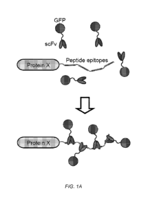
[0028] Figure 1. Identification of an antibody-peptide pair that binds tightly
in vivo.
[0029] A) Schematic of the antibody-peptide labeling strategy. A protein of
interest
(protein X) is tagged with 4-24 copies of a short peptide (peptide epitopes),
and is co-
7
CA 02954920 2017-01-11
WO 2016/011070 PCT/US2015/040439
expressed with the single chain antibody tagged with GFP that recognizes the
short peptide
and can be recruited in multiple copies. B). A schematic of an experiment in
which the
mitochondrial targeting domain of mitoNEET (mito) is fused to mCherry and 4
tandem
copies of a peptide, which binds to mitochondria and labels them with a red
fluorescent
protein. The matching antibodies are tagged with GFP and expressed in the same
cell. If
binding occurs between antibody and peptide, then GFP labeling of the
mitochondria should
be observed. C) Indicated GFP-tagged antibodies are co-expressed with
mitochondrial-
targeted, mCherry-tagged 4xpep arrays in U2OS cells, and cells were imaged
using spinning
disk confocal microscopy. The GCN4 and V1 antibody-GFP fusions succeed in
recognizing
their corresponding peptide arrays on the mitochondria but the C4 antibody-GFP
fusion does
not. D) As a control, scFv-GCN4-GFP is co-expressed with a mito-mCherry
plasmid in
which the GCN4 peptides have been swapped for the FKBP protein, which does not
bind the
antibody. Scale bars, 10 gm.
[0030] Figure 2 Mitoneet N-terminal domain targets proteins to the
mitochondria
[0031] U205 cells were transfected with a construct encoding the N-terminus of
mitoNEET fused to GFP and incubated with mitotracker to stain mitochondria.
Scale bars, 10
gm.
[0032] Figure 3. Characterization of the off-rate and stoichiometry of the
binding
interaction between the scFv-GCN4 antibody and the GCN4 peptide array in vivo.
[0033] A) Mito-mCherry-24xGCN4pep was co-transfected in U205 cells along with
scFv-
GCN4-GFP and their co-localization on mitochondria in a single cell is shown
at time -10
sec. At 0 sec, the GFP signal from half of this cell was photobleached, and
fluorescence
recovery was followed by time-lapse microscopy. Scale bar, 5 gm. B) The
fluorescence
recovery after photob leaching was quantified (shown is an average of FRAP
recovery curves
from 6 cells). A small amount of recovery is observed in the first 10 sec,
which may be due to
recovery of unbound GFP-tagged antibody which is freely diffusing in the
cytoplasm in the
vicinity of the mitochondria. C-E) Indicated constructs were transfected in
U205 cells and
images were acquired 24 hr after transfection with equivalent image
acquisition settings.
Representative images are shown in C). Note that the GFP signal intensity in
the mito-
mCherry-24xGCN4pep + scFv-GCN4-GFP is highly saturated when the same scaling
is used
as in the other panels. Bottom row shows a zoom of a region of interest:
dynamic scaling was
different for the GFP and mCherry signals, so that both could be observed.
Scale bars, 10 gm.
8
CA 02954920 2017-01-11
WO 2016/011070 PCT/US2015/040439
D-E) Quantifications of the GFP:mCherry fluorescence intensity ratio on
mitochondria after
normalization (The average GFP:mCherry ratio for the sfGFP-linker-mCherry
fusion protein
was set to 1, see methods section). Each dot represents a single cell and
dashed lines indicates
the average value. All scale bars, 10 gm.
[0034] Figure 4. Optimizing the GCN4 antibody-peptide pair
[0035] A) HEK293 cells were transfected with the indicated constructs and 24
hr after
transfection, images were acquired using spinning disk confocal microscopy.
Maximum
intensity Z-projections are shown. All scale bars, 10 gm. B) U2OS cells were
transfected
with a sfGFP-linker-mCherry fusion protein and images were acquired on a
spinning disk
confocal microscope. GFP and mCherry fluorescence intensities for single cells
were
quantified and values were plotted after background subtraction.
[0036] Figure 5. sunGFP allows long-term single molecule fluorescence imaging
in the
cytoplasm.
[0037] A-H) U2OS cells were transfected with indicated SunTag constructs, all
containing
24 copies of the GCN4 peptide, and were imaged by spinning disk confocal
microscopy 24 hr
after transfection. To decrease cytoplasmic background fluorescence of unbound
scFv-
GCN4-GFP, a nuclear localization signal was added to the scFv-GCN4-GFP to
shuttle
unbound antibody from the cytoplasm to the nucleus. A) A representative image
of
SunTag24x-IFP-CAAX-GFP is shown (top), as well as the fluorescence intensities
quantification of the foci (bottom). Dotted line marks the outline of the
cell. Scale bar, 10
gm. B) Cells expressing K560-SunTag24x¨GFP were followed by spinning disk
confocal
microscopy (image acquisition every 200 ms). Movement is revealed by a maximum
intensity projection of 50 time-points (left) and a kymograph (right). Scale
bar, 10 gm. C-D)
Cells expressing both EB3-tdTomato and K560-SunTag24x¨GFP were imaged and
moving
particles were tracked manually. Tracks indicate movement towards the cell
interior and
periphery (C). Scale bar, 5 gm. Dots in (D) represent fraction of movement
towards the
interior from individual cells with between 5-20 moving particles scored per
cell. The mean
and standard deviation is indicated. (E-F) Cells expressing Kifl 8b-
SunTag24x¨GFP were
imaged with a 250 ms time interval. Images in (E) show a maximum intensity
projection (50
time-points (left)) and a kymograph (right). Speeds of moving molecules were
quantified
from 10 different cells (F). (G-H) Cells expressing both mCherry-a-tubulin and
K560rig-
SunTag24xD were imaged with a 600 ms time interval. The entire cell is shown
in (G), while
9
CA 02954920 2017-01-11
WO 2016/011070 PCT/US2015/040439
H shows stills of a time series from the same cell. Open circles track two
foci on the same
microtubule, which is indicated by the dashed line. Asterisks indicate
stationary foci. Scale
bars, 10 and 2 gm (G and H), respectively.
[0038] Figure 6. Single molecule imaging using the SunTag.
[0039] A) Representative images of cells expressing either scFv-GCN4-GFP alone
or
together with IFP-SunTag24x are show. Bottom panels are enlargements of boxed
areas. B-C)
Run length (B) and speed (E) of K560-SunTag24x were calculated in at least 10
different cells.
[0040] Figure 7. An optimized peptide array for high expression.
[0041] A) Indicated constructs were transfected in HEK293 cells and imaged 24
hr after
transfection using wide-field microscopy. All images were acquired using
identical
acquisition parameters. B) Sequence of the first and second generation GCN4
peptide. C-D)
Indicated constructs were transfected in HEK293 (C) or U205 (D) cells and
imaged 24 hr
after transfection using wide-field (C) or spinning disk confocal (D)
microscopy. E) U205
cells were transfected with scFv-GCN4-GFP together with mito-mCherry-SunTagiox
v4. 24 hr
after transfection, GFP signal on mitochondria was photobleached and
fluorescence recovery
was determined over time. The graph represents an average of 6 cells. The
results are
overlayed with the fluorescence recovery measurements shown in fig. 3B. Cells
expressing
K560-SunTag24x v4¨GFP were followed by time-lapse microscopy (acquisition at
100 msec
intervals); a maximum intensity projection of 25 time-points (left) or a
kymograph (right) is
shown. Scale bars in A and C, 50 gm, scale bars in D, 10 gm.
[0042] Figure 8. dCas9-SunTag allows genetic rewiring of cells through
activation of
endogenous genes.
[0043] A) Schematic of gene activation by dCas9-VP64 and dCas9-SunTag-VP64.
dCas9
binds to a gene promoter through its sequence specific sgRNA. Direct fusion of
VP64 to
dCas9 (top) results in a single VP64 domain at the promoter which weakly
activates
transcription of the downstream gene. In contrast, recruitment of many VP64
domains using
the SunTag potently activates transcription of the gene (bottom). (B-D) K562
cells stably
expressing dCas9-VP64 or dCas9-SunTagiox-VP64 were infected with lentiviral
particles
encoding indicated sgRNAs, as well as BFP and a puromycin resistance gene and
selected
with 0.7 gg/ml puromycin for 3 days. B) Cells were stained for CXCR4 using a
directly
labeled a-CXCR4 antibody and fluorescence analyzed by FACS. C) Levels of
CXCR4,
CA 02954920 2017-01-11
WO 2016/011070 PCT/US2015/040439
analyzed as indicated in panel B, were determined with several sgRNAs. (D)
Trans-well
migration assays were performed with the same set of sgRNAs as in panel C (see
methods).
(E) dCas9-VP64 or dCas9-SunTagiox-VP64 induced transcription of CDKN1B with
several
sgRNAs. mRNA levels were quantified by qPCR. (F) Growth competition assays
were
performed by infecting around 30% of cells with indicated sgRNA/BFP, as well
as a control
sgRNA. Two days after infection the percentage of BFP positive cells was
determined for
each population. Cells were then grown for 2 weeks and the percentage of BFP
positive cells
was determined again. From the decrease in BFP/sgRNA positive cells over time,
combined
with the cell doubling time (which was determined in parallel to be on average
27 hr) the
percentage growth reduction was determined. Note that the control sgRNA did
not affect the
doubling time of cells. Graphs in B, D, and F are averages of three
independent experiments.
Graph in E is average of two biological replicates, each with two or three
technical replicates.
Error bars indicated standard error of the mean (SEM).
[0044] Figure 9. dCas9-SunTag can recruit many copies of scFv-GCN4-GFP to a
genomic locus.
[0045] A-B) HEK293 cells were transfected with dCas9-SunTag24x, scFv-GCN4-GFP
and
indicated sgRNAs. 24 hr after transfection, cells were imaged by spinning disk
confocal
microscopy. Images are maximum intensity projections of Z-stacks (A).
Intensities of
individual telomere foci was measured in ImageJ and telomere fluorescence was
calculated
by subtraction of diffuse nuclear background. Vertical set of dots in (B)
represents individual
telomere intensities in a single cell. Scale bars, 5 gm.
DEFINITIONS
[0046] As used in this specification and the appended claims, the singular
forms "a," "an,"
and "the" include plural reference unless the context clearly dictates
otherwise.
[0047] The term "nucleic acid" or "polynucleotide" refers to deoxyribonucleic
acids (DNA)
or ribonucleic acids (RNA) and polymers thereof in either single- or double-
stranded form.
Unless specifically limited, the term encompasses nucleic acids containing
known analogues
of natural nucleotides that have similar binding properties as the reference
nucleic acid and
are metabolized in a manner similar to naturally occurring nucleotides. Unless
otherwise
indicated, a particular nucleic acid sequence also implicitly encompasses
conservatively
modified variants thereof (e.g., degenerate codon substitutions), alleles,
orthologs, SNPs, and
11
CA 02954920 2017-01-11
WO 2016/011070 PCT/US2015/040439
complementary sequences as well as the sequence explicitly indicated.
Specifically,
degenerate codon substitutions may be achieved by generating sequences in
which the third
position of one or more selected (or all) codons is substituted with mixed-
base and/or
deoxyinosine residues (Batzer et at., Nucleic Acid Res. 19:5081 (1991);
Ohtsuka et at., J.
Biol. Chem. 260:2605-2608 (1985); and Rossolini et at., Mol. Cell. Probes 8:91-
98 (1994)).
The term nucleic acid is used interchangeably with gene, cDNA, and mRNA
encoded by a
gene.
[0048] The term "gene" means the segment of DNA involved in producing a
polypeptide
chain. It may include regions preceding and following the coding region
(leader and trailer)
as well as intervening sequences (introns) between individual coding segments
(exons).
[0049] A "promoter" is defined as an array of nucleic acid control sequences
that direct
transcription of a nucleic acid. As used herein, a promoter includes necessary
nucleic acid
sequences near the start site of transcription, such as, in the case of a
polymerase II type
promoter, a TATA element. A promoter also optionally includes distal enhancer
or repressor
elements, which can be located as much as several thousand base pairs from the
start site of
transcription.
[0050] An "expression cassette" is a nucleic acid construct, generated
recombinantly or
synthetically, with a series of specified nucleic acid elements that permit
transcription of a
particular polynucleotide sequence in a host cell. An expression cassette may
be part of a
plasmid, viral genome, or nucleic acid fragment. Typically, an expression
cassette includes a
polynucleotide to be transcribed, operably linked to a promoter. The promoter
can be a
heterologous promoter. In the context of promoters operably linked to a
polynucleotide, a
"heterologous promoter" refers to a promoter that would not be so operably
linked to the
same polynucleotide as a product of nature (i.e., in a wild-type organism).
[0051] A "reporter gene" encodes proteins that are readily detectable due to
their
biochemical characteristics, such as enzymatic activity or chemifluorescent
features. One
specific example of such a reporter is green fluorescent protein. Fluorescence
generated from
this protein can be detected with various commercially-available fluorescent
detection
systems. Other reporters can be detected by staining. The reporter can also be
an enzyme that
generates a detectable signal when contacted with an appropriate substrate.
The reporter can
be an enzyme that catalyzes the formation of a detectable product. Suitable
enzymes include,
but are not limited to, proteases, nucleases, lipases, phosphatases and
hydrolases. The
12
CA 02954920 2017-01-11
WO 2016/011070 PCT/US2015/040439
reporter can encode an enzyme whose substrates are substantially impermeable
to eukaryotic
plasma membranes, thus making it possible to tightly control signal formation.
Specific
examples of suitable reporter genes that encode enzymes include, but are not
limited to, CAT
(chloramphenicol acetyl transferase; Alton and Vapnek (1979) Nature 282: 864-
869);
luciferase (lux); f3-galactosidase; LacZ; 13.-glucuronidase; and alkaline
phosphatase (Toh, et
al. (1980) Eur. J. Biochem. 182: 231-238; and Hall et al. (1983) J. Mol. Appl.
Gen. 2: 101),
each of which are incorporated by reference herein in its entirety. Other
suitable reporters
include those that encode for a particular epitope that can be detected with a
labeled antibody
that specifically recognizes the epitope.
[0052] The term "amino acid" refers to naturally occurring and synthetic amino
acids, as
well as amino acid analogs and amino acid mimetics that function in a manner
similar to the
naturally occurring amino acids. Naturally occurring amino acids are those
encoded by the
genetic code, as well as those amino acids that are later modified, e.g.,
hydroxyproline, y-
carboxyglutamate, and 0-phosphoserine. Amino acid analogs refers to compounds
that have
the same basic chemical structure as a naturally occurring amino acid, i.e.,
an a carbon that is
bound to a hydrogen, a carboxyl group, an amino group, and an R group, e.g.,
homoserine,
norleucine, methionine sulfoxide, methionine methyl sulfonium. Such analogs
have modified
R groups (e.g., norleucine) or modified peptide backbones, but retain the same
basic chemical
structure as a naturally occurring amino acid. "Amino acid mimetics" refers to
chemical
compounds having a structure that is different from the general chemical
structure of an
amino acid, but that functions in a manner similar to a naturally occurring
amino acid.
[0053] There are various known methods in the art that permit the
incorporation of an
unnatural amino acid derivative or analog into a polypeptide chain in a site-
specific manner,
see, e.g., WO 02/086075.
[0054] Amino acids may be referred to herein by either the commonly known
three letter
symbols or by the one-letter symbols recommended by the IUPAC-IUB Biochemical
Nomenclature Commission. Nucleotides, likewise, may be referred to by their
commonly
accepted single-letter codes.
[0055] "Polypeptide," "peptide," and "protein" are used interchangeably herein
to refer to a
polymer of amino acid residues. All three terms apply to amino acid polymers
in which one
or more amino acid residue is an artificial chemical mimetic of a
corresponding naturally
occurring amino acid, as well as to naturally occurring amino acid polymers
and non-
13
CA 02954920 2017-01-11
WO 2016/011070 PCT/US2015/040439
naturally occurring amino acid polymers. As used herein, the terms encompass
amino acid
chains of any length, including full-length proteins, wherein the amino acid
residues are
linked by covalent peptide bonds.
[0056] "Conservatively modified variants" applies to both amino acid and
nucleic acid
sequences. With respect to particular nucleic acid sequences, "conservatively
modified
variants" refers to those nucleic acids that encode identical or essentially
identical amino acid
sequences, or where the nucleic acid does not encode an amino acid sequence,
to essentially
identical sequences. Because of the degeneracy of the genetic code, a large
number of
functionally identical nucleic acids encode any given protein. For instance,
the codons GCA,
GCC, GCG and GCU all encode the amino acid alanine. Thus, at every position
where an
alanine is specified by a codon, the codon can be altered to any of the
corresponding codons
described without altering the encoded polypeptide. Such nucleic acid
variations are "silent
variations," which are one species of conservatively modified variations.
Every nucleic acid
sequence herein that encodes a polypeptide also describes every possible
silent variation of
the nucleic acid. One of skill will recognize that each codon in a nucleic
acid (except AUG,
which is ordinarily the only codon for methionine, and TGG, which is
ordinarily the only
codon for tryptophan) can be modified to yield a functionally identical
molecule.
Accordingly, each silent variation of a nucleic acid that encodes a
polypeptide is implicit in
each described sequence.
[0057] As to amino acid sequences, one of skill will recognize that individual
substitutions,
deletions or additions to a nucleic acid, peptide, polypeptide, or protein
sequence which
alters, adds or deletes a single amino acid or a small percentage of amino
acids in the encoded
sequence is a "conservatively modified variant" where the alteration results
in the substitution
of an amino acid with a chemically similar amino acid. Conservative
substitution tables
providing functionally similar amino acids are well known in the art. Such
conservatively
modified variants are in addition to and do not exclude polymorphic variants,
interspecies
homologs, and alleles of the invention. In some cases, conservatively modified
variants of
Cas9 or sgRNA can have an increased stability, assembly, or activity as
described herein.
[0058] The following eight groups each contain amino acids that are
conservative
substitutions for one another:
1) Alanine (A), Glycine (G);
2) Aspartic acid (D), Glutamic acid (E);
3) Asp aragine (N), Glutamine (Q);
14
CA 02954920 2017-01-11
WO 2016/011070 PCT/US2015/040439
4) Arginine (R), Lysine (K);
5) Isoleucine (I), Leucine (L), Methionine (M), Valine (V);
6) Phenylalanine (F), Tyrosine (Y), Tryptophan (W);
7) Serine (S), Threonine (T); and
8) Cysteine (C), Methionine (M)
(see, e.g., Creighton, Proteins, W. H. Freeman and Co., N. Y. (1984)).
[0059] Amino acids may be referred to herein by either their commonly known
three letter
symbols or by the one-letter symbols recommended by the IUPAC-IUB Biochemical
Nomenclature Commission. Nucleotides, likewise, may be referred to by their
commonly
accepted single-letter codes.
[0060] In the present application, amino acid residues are numbered according
to their
relative positions from the left most residue, which is numbered 1, in an
unmodified wild-
type polypeptide sequence.
[0061] As used in herein, the terms "identical" or percent "identity," in the
context of
describing two or more polynucleotide or amino acid sequences, refer to two or
more
sequences or subsequences that are the same or have a specified percentage of
amino acid
residues or nucleotides that are the same. For example, a core small guide RNA
(sgRNA)
sequence responsible for assembly and activity of a sgRNA:nuclease complex has
at least
80% identity, preferably 85%, 90%, 91%, 92%, 93, 94%, 95%, 96%, 97%, 98%, 99%,
or
100% identity, to a reference sequence, e.g., one of SEQ ID NOs:42-45), when
compared and
aligned for maximum correspondence over a comparison window, or designated
region as
measured using one of the following sequence comparison algorithms or by
manual
alignment and visual inspection. As another example, a Cas9 sequence
responsible for
assembly and activity of a sgRNA:nuclease complex has at least 80% identity,
preferably
85%, 90%, 91%, 92%, 93, 94%, 95%, 96%, 97%, 98%, 99%, or 100% identity, to a
reference
sequence, e.g., one of SEQ ID NOs:46-50), when compared and aligned for
maximum
correspondence over a comparison window, or designated region as measured
using one of
the following sequence comparison algorithms or by manual alignment and visual
inspection.
Such sequences are then said to be "substantially identical." With regard to
polynucleotide
sequences, this definition also refers to the complement of a test sequence.
With regard to
amino acid sequences, preferably, the identity exists over a region that is at
least about 50
CA 02954920 2017-01-11
WO 2016/011070 PCT/US2015/040439
amino acids or nucleotides in length, or more preferably over a region that is
75-100 amino
acids or nucleotides in length.
[0062] For sequence comparison, typically one sequence acts as a reference
sequence, to
which test sequences are compared. When using a sequence comparison algorithm,
test and
reference sequences are entered into a computer, subsequence coordinates are
designated, if
necessary, and sequence algorithm program parameters are designated. Default
program
parameters can be used, or alternative parameters can be designated. The
sequence
comparison algorithm then calculates the percent sequence identities for the
test sequences
relative to the reference sequence, based on the program parameters. For
sequence
comparison of nucleic acids and proteins, the BLAST and BLAST 2.0 algorithms
and the
default parameters discussed below are used.
[0063] A "comparison window", as used herein, includes reference to a segment
of any one
of the number of contiguous positions selected from the group consisting of
from 20 to 600,
usually about 50 to about 200, more usually about 100 to about 150 in which a
sequence may
be compared to a reference sequence of the same number of contiguous positions
after the
two sequences are optimally aligned. Methods of alignment of sequences for
comparison are
well-known in the art. Optimal alignment of sequences for comparison can be
conducted,
e.g., by the local homology algorithm of Smith & Waterman, Adv. Appl. Math.
2:482 (1981),
by the homology alignment algorithm of Needleman & Wunsch, J. Mol. Biol.
48:443 (1970),
by the search for similarity method of Pearson & Lipman, Proc. Nat'l. Acad.
Sci. USA
85:2444 (1988), by computerized implementations of these algorithms (GAP,
BESTFIT,
FASTA, and TFASTA in the Wisconsin Genetics Software Package, Genetics
Computer
Group, 575 Science Dr., Madison, WI), or by manual alignment and visual
inspection (see,
e.g., Current Protocols in Molecular Biology (Ausubel et at., eds. 1995
supplement)).
[0064] Examples of algorithms that are suitable for determining percent
sequence identity
and sequence similarity are the BLAST and BLAST 2.0 algorithms, which are
described in
Altschul et at., (1990) J. Mol. Biol. 215: 403-410 and Altschul et at. (1977)
Nucleic Acids
Res. 25: 3389-3402, respectively. Software for performing BLAST analyses is
publicly
available at the National Center for Biotechnology Information website,
ncbi.nlm.nih.gov.
The algorithm involves first identifying high scoring sequence pairs (HSPs) by
identifying
short words of length W in the query sequence, which either match or satisfy
some positive-
valued threshold score T when aligned with a word of the same length in a
database
sequence. T is referred to as the neighborhood word score threshold (Altschul
et al, supra).
16
CA 02954920 2017-01-11
WO 2016/011070 PCT/US2015/040439
These initial neighborhood word hits acts as seeds for initiating searches to
find longer HSPs
containing them. The word hits are then extended in both directions along each
sequence for
as far as the cumulative alignment score can be increased. Cumulative scores
are calculated
using, for nucleotide sequences, the parameters M (reward score for a pair of
matching
residues; always >0) and N (penalty score for mismatching residues; always
<0). For amino
acid sequences, a scoring matrix is used to calculate the cumulative score.
Extension of the
word hits in each direction are halted when: the cumulative alignment score
falls off by the
quantity X from its maximum achieved value; the cumulative score goes to zero
or below,
due to the accumulation of one or more negative-scoring residue alignments; or
the end of
either sequence is reached. The BLAST algorithm parameters W, T, and X
determine the
sensitivity and speed of the alignment. The BLASTN program (for nucleotide
sequences)
uses as defaults a word size (W) of 28, an expectation (E) of 10, M=1, N=-2,
and a
comparison of both strands. For amino acid sequences, the BLASTP program uses
as
defaults a word size (W) of 3, an expectation (E) of 10, and the BLOSUM62
scoring matrix
(see Henikoff & Henikoff, Proc. Natl. Acad. Sci. USA 89:10915 (1989)).
[0065] The BLAST algorithm also performs a statistical analysis of the
similarity between
two sequences (see, e.g., Karlin & Altschul, Proc. Nat'l. Acad. Sci. USA
90:5873-5787
(1993)). One measure of similarity provided by the BLAST algorithm is the
smallest sum
probability (P(N)), which provides an indication of the probability by which a
match between
two nucleotide or amino acid sequences would occur by chance. For example, a
nucleic acid
is considered similar to a reference sequence if the smallest sum probability
in a comparison
of the test nucleic acid to the reference nucleic acid is less than about 0.2,
more preferably
less than about 0.01, and most preferably less than about 0.001.
[0066] An indication that two nucleic acid sequences or polypeptides are
substantially
identical is that the polypeptide encoded by the first nucleic acid is
immunologically cross
reactive with the antibodies raised against the polypeptide encoded by the
second nucleic
acid, as described below. Thus, a polypeptide is typically substantially
identical to a second
polypeptide, for example, where the two peptides differ only by conservative
substitutions.
Another indication that two nucleic acid sequences are substantially identical
is that the two
molecules or their complements hybridize to each other under stringent
conditions, as
described below. Yet another indication that two nucleic acid sequences are
substantially
identical is that the same primers can be used to amplify the sequence. Yet
another indication
17
CA 02954920 2017-01-11
WO 2016/011070 PCT/US2015/040439
that two polypeptides are substantially identical is that the two polypeptides
retain identical
or substantially similar activity.
[0067] A "translocation sequence" or "transduction sequence" refers to a
peptide or
protein (or active fragment or domain thereof) sequence that directs the
movement of a
protein from one cellular compartment to another, or from the extracellular
space through the
cell or plasma membrane into the cell. Translocation sequences that direct the
movement of a
protein from the extracellular space through the cell or plasma membrane into
the cell are
"cell penetration peptides." Translocation sequences that localize to the
nucleus of a cell
are termed "nuclear localization" sequences, signals, domains, peptides, or
the like.
Examples of translocation sequences include, without limitation, the TAT
transduction
domain (see, e.g., S. Schwarze et al., Science 285 (Sep. 3, 1999); penetratins
or penetratin
peptides (D. Derossi et al., Trends in Cell Biol. 8, 84-87); Herpes simplex
virus type 1 VP22
(A. Phelan et al., Nature Biotech. 16, 440-443 (1998), and polycationic (e.g.,
poly-arginine)
peptides (Cell Mol. Life Sci. 62 (2005) 1839-1849). Further translocation
sequences are
known in the art. Translocation peptides can be fused (e.g. at the amino or
carboxy
terminus), conjugated, or coupled to a compound of the present invention, to,
among other
things, produce a conjugate compound that may easily pass into target cells,
or through the
blood brain barrier and into target cells.
[0068] The "CRISPR/Cas" system refers to a widespread class of bacterial
systems for
defense against foreign nucleic acid. CRISPR/Cas systems are found in a wide
range of
eubacterial and archaeal organisms. CRISPR/Cas systems include type I, II, and
III sub-
types. Wild-type type II CRISPR/Cas systems utilize the RNA-mediated
nuclease,Cas9 in
complex with guide and activating RNA to recognize and cleave foreign nucleic
acid.
[0069] Cas9 homologs are found in a wide variety of eubacteria, including, but
not limited to
bacteria of the following taxonomic groups: Actinobacteria, Aquificae,
Bacteroidetes-
Chlorobi, Chlamydiae-Verrucomicrobia, Chlroflexi, Cyanobacteria, Firmicutes,
Proteobacteria, Spirochaetes, and Thermotogae. An exemplary Cas9 protein is
the
Streptococcus pyo genes Cas9 protein. Additional Cas9 proteins and homologs
thereof are
described in, e.g., Chylinksi, et at., RNA Biol. 2013 May 1; 10(5): 726-737 ;
Nat. Rev.
Microbiol. 2011 June; 9(6): 467-477; Hou, et at., Proc Natl Acad Sci U S A.
2013 Sep
24;110(39):15644-9; Sampson et at., Nature. 2013 May 9;497(7448):254-7; and
Jinek, et at.,
Science. 2012 Aug 17;337(6096):816-21.
18
CA 02954920 2017-01-11
WO 2016/011070 PCT/US2015/040439
[0070] As used herein, "activity" in the context of CRISPR/Cas activity, Cas9
activity,
sgRNA activity, sgRNA:nuclease activity and the like refers to the ability to
bind to a target
genetic element and recruit effector domains to a region at or near the target
genetic element.
Such activity can be measured in a variety of ways as known in the art. For
example,
expression, activity, or level of a reporter gene, or expression or activity
of a gene encoded by
the genetic element can be measured. As another example, a signal (e.g., a
fluorescent
signal) provided by a recruited effector domain (e.g., a recruited fluorescent
protein) can be
detected.
[0071] As used herein, the term "effector domain" refers to a polypeptide that
provides an
effector function. Exemplary effector functions include, but are not limited
to, enzymatic
activity (e.g., nuclease, methylase, demethylase, acetylase, deacetylase,
kinase, phosphatase,
ubiquitinase, deubiquitinase, luciferase, or peroxidase activity),
fluorescence, binding and
recruitment of additional polypeptides or organic molecules, or
transcriptional modulation
(e.g., activation, enhancement, or repression). Thus, exemplary effector
domains include, but
are not limited to enzymes (e.g., nucleases, methylases, demethylases,
acetylases,
deacetylases, kinases, phosphatases, ubiquitinases, deubiquitinases,
luciferases, or
peroxidases), adaptor proteins, fluorescent proteins (e.g., green fluorescent
protein),
transcriptional enhancers, transcriptional activators, or transcriptional
repressors. Adaptor
protein effector domains can function to bind, and thus recruit other
polypeptides, organic
molecules, etc.
DETAILED DESCRIPTION OF THE INVENTION
I. Introduction
[0072] Recruitment of multiple copies of a protein to a target substrate (e.g.
DNA, RNA, or
protein) is used to amplify signals in biological systems. For example,
recruitment of multiple
copies of a transcription factor to a single gene promoter can dramatically
enhance
transcriptional activation of the target gene (Anderson and Freytag, 1991;
Chen et at., 1992;
Pettersson and Schafther, 1990). Similarly, the recruitment of multiple copies
of an RNA
binding protein to an mRNA can result in potent regulation of translation
(Pillai et at., 2004;
Pique et at., 2008). Protein localization and interactions also can be
modulated by the copy
number of interaction sites within a polypeptide sequence. For example, many
nuclear
proteins contain multiple nuclear localization signal (NLS) sequences, which
control
19
CA 02954920 2017-01-11
WO 2016/011070 PCT/US2015/040439
robustness of nuclear import (Luo et at., 2004). Similarly, in receptor-
mediated signaling,
multimerization of receptors in response to ligand binding helps to elicit a
downstream
response (Boniface et at., 1998). Downstream of the receptors, adapter
proteins with
multiple 5H2/5H3 domains can generate multivalent interactions of interacting
signaling
molecules (Li et at., 2012), which is thought to facilitate the signaling
response
[0073] Protein multimerization also has been widely used in synthetic biology.
A
commonly used method to study RNA localization, even at the single molecule
level, is to
insert many copies of the M52 binding aptamer (as many as 24), which then
recruit many
MS2-GFP fusion proteins (Bertrand et at., 1998; Fusco et at., 2003).
Similarly, the activity of
a RNA-binding protein can be studied by artificially tethering it to an RNA in
multiple copies
using the M52 system (Coller and Wickens, 2007). Similar multimerization
approaches have
also been used to fluorescently label a specific region of a chromosome. For
example, the
Lac operon can be inserted into a chromosomal locus in many tandem repeats
and then
visualized by the recruitment of many copies of GFP-LacI (Gordon et at.,
1997). More
recently, several studies have shown that GFP-tagged engineered DNA-binding
proteins, like
TALEs or the CRISPR effector protein Cas9, can also be used to fluorescently
label an
endogenous DNA sequence when its binding site is present in many tandem
repeats in the
DNA (Chen et at., 2013; Ma et at., 2013; Miyanari et at., 2013). Furthermore,
as with native
transcriptional regulation, a gene can be artificially activated when a
binding site for a
synthetic transcription factor is placed upstream of a gene in multiple
copies; this principle is
employed in the "tet-on" system for inducible transgene expression (Huang et
at., 1999;
Sadowski et at., 1988). Taken together, these studies demonstrate the power of
introducing
multiple copies of protein binding sites within RNA or DNA for the purpose of
signal
amplification.
[0074] Despite the success of multimerizing nucleic acid based motifs within
RNA and
DNA for protein recruitment, no comparable and generic system exists for
controlling copy
number of protein-protein interactions. For fluorescence imaging, the fusion
of 3 copies of
GFP to a protein of interest has been used to increase signal intensity, but a
further increase in
the copy number of fluorescent proteins is challenging due to their size (-25
kDa) and
bacterial recombination when constructing DNA plasmids encoding such proteins.
Here, we
describe a new synthetic system for recruiting as many as 24 copies of a
protein to a target
polypeptide chain. We demonstrate that this approach can be used to create
bright
fluorescent signals for single molecule protein imaging in living cells,
through the
CA 02954920 2017-01-11
WO 2016/011070 PCT/US2015/040439
recruitment of 24 copies of GFP to a target protein. We also demonstrate that
the system can
be used to modulate gene expression through the recruitment of multiple copies
of gene
regulatory effector domains to a modified CRISPR/Cas9 protein targeted to
specific
sequences in the genome. The ability to multimerize proteins in a controlled
fashion on a
polypeptide backbone will likely have many additional uses in biotechnology.
II. Compositions
[0075] Described herein are compositions useful as components of a system for
recruiting
one or more effector domains to a polypeptide of interest. The components can
be used to
target the effector domains to the polypeptide of interest, or a binding
partner of the
polypeptide of interest. Thus, for example, the components can be used to
target the effector
domains to a region of interest such as a genomic region, an intracellular
compartment (e.g.,
nucleus, cytoplasm, endoplasmic reticulum, etc.), or a membrane (e.g.,
cytoplasmic, nuclear,
or mitochondrial, etc.). The polypeptide of interest can be any natural,
recombinant, or
synthetic polypeptide. The components include epitopes, multimerized epitopes,
affinity
agents, Cas9 domains (including dCas9 domains), sgRNAs, and effector domains.
A. Epitopes and Multimerized Epitopes
[0076] Described herein are epitopes and multimerized epitopes for recruiting
affinity
agents to a polypeptide of interest. Typically, the epitopes are fused to the
polypeptide of
interest. The epitopes can be fused to one or more of the N-terminus of the
polypeptide of
interest, the C-terminus of the polypeptide of interest, or inserted into the
polypeptide of
interest. For example, the epitopes can be inserted into a region of the
polypeptide of interest
that is solvent accessible when the polypeptide is in a folded conformation.
Such regions
include, but are not limited to protein surface loops or linker regions
between discrete protein
domains. A polypeptide of interest can be fused to an epitope, multiple copies
of an epitope,
more than one different epitope, or multiple copies of more than one different
epitope as
further described herein.
[0077] The epitopes can be any polypeptide sequence that is specifically
recognized by an
affinity agent. Such epitopes include, but are not limited to the c-Myc
affinity tag, an HA
affinity tag, a His affinity tag, an S affinity tag, a methionine-His affinity
tag, an RGD-His
affinity tag, a 7x His tag, a FLAG octapeptide, a strep tag or strep tag II, a
V5 tag, or a VSV-
G epitope. An exemplary epitope includes, but is not limited to, a GCN4
epitope (e.g., SEQ
ID NOs:1 or 2).
21
CA 02954920 2017-01-11
WO 2016/011070 PCT/US2015/040439
[0078] Epitopes, such as the epitopes described herein can be multimerized.
For example,
the a polypeptide of interest can be fused to a multimerized epitope
containing 2, 3, 4, 5, 6, 7,
8,9, 10, 11, 12, 13, 14, 15, 16, 17, 18, 19, 20, 21, 22, 23, 24, or more
copies of an epitope. In
some cases, the polypeptide of interest is fused to a first epitope or
multimerized epitope. In
some cases, the polypeptide of interest is fused to a first epitope or
multimerized epitope and
a second epitope or multimerized epitope. Multimerized epitopes include, but
are not limited
to multimerized epitopes containing 2, 3, 4, 5, 6, 7, 8, 9, 10, 11, 12, 13,
14, 15, 16, 17, 18, 19,
20, 21, 22, 23, 24, or more copies of a GCN4 epitope. An exemplary epitopes
include, but
are not limited to, a 24xGNC4 epitope (e.g., SEQ ID NOs:10 or 11) or a 10xGCN4
epitope
(e.g., SEQ ID NO:12)
[0079] The individual epitopes of a multimerized epitope can be separated by a
linker
region. Suitable linker regions are known in the art. In some cases, the
linker is configured
to allow the binding of affinity agents to adjacent epitopes without, or
without substantial,
steric hindrance. In some cases, the linker sequences are configured to
provide an
unstructured or linear region of the polypeptide. For example, the linker
sequence can
comprise one or more glycines and/or serines. The linker sequences can be at
least about 2,
3, 4, 5, 6, 7, 8, 9, 10 or more amino acids in length. In some cases, the
linker sequences are,
or comprise, one or more of the linkers disclosed on the world wide web at
parts.igem.org/Protein domains/Linker. Exemplary linkers include, but are not
limited to,
SEQ ID NOs:3 or 4.
[0080] Also described herein are expression cassettes and vectors for
producing one or
more epitopes or multimerized epitopes described herein (e.g., a polypeptide
of interest fused
to an epitope or multimerized epitope) in a host cell. The expression
cassettes can contain a
promoter (e.g., a heterologous promoter) operably linked to a polynucleotide
encoding an
epitope or multimerized epitope. The promoter can be inducible or
constitutive. The
promoter can be tissue specific. In some cases, the promoter is a strong
promoter. For
example, the promoter can be a CMV promoter, an SFFV long terminal repeat
promoter, or
the human elongation factor 1 promoter (EF1A). In some cases, the
polynucleotide encoding
the epitope or multimerized epitope of the expression cassette further encodes
the polypeptide
of interest. In some cases, an expression cassette is provided for cloning a
polynucleotide
encoding a polypeptide of interest in frame with an epitope or multimerized
epitope. The
expression cassette can include one or more localization sequences. In some
cases, the
polypeptide of interest provides a localization function. The expression
cassette can be in a
22
CA 02954920 2017-01-11
WO 2016/011070 PCT/US2015/040439
vector, such as a plasmid, a viral vector, a lentiviral vector, etc. In some
cases, the expression
cassette is in a host cell. The expression cassette can be episomal or
integrated in the host
cell.
B. Affinity Agents
[0081] Described herein are affinity agents for recruiting effector functions
to a
polypeptide fused to an epitope or multimerized epitope. A wide variety of
affinity agents
can be utilized. Generally, the affinity agent is stable under the reducing
conditions present
in the intracellular environment of the cell. Additionally, the affinity agent
should
specifically bind to its corresponding epitope with minimal cross-reactivity.
In some cases,
the affinity agent is an antibody, such as an scFv. In some cases, the
affinity agent is an
antibody (e.g., scFv) that has been optimized for stability in the
intracellular environment.
For example, the affinity agent (e.g., scFv) can be an intrabody (see, e.g.,
Lo et at., Handb.
Exp. Pharm. 2008;(181):343-73). An exemplary affinity agent comprises the anti-
GCN4
scFv domain of SEQ ID NO:5. In some cases, the affinity agent comprises an
affinity
domain (e.g., an anti-GCN4 scFv domain such as SEQ ID NO:5) and a linker
(e.g., a linker
such as SEQ ID NO :58), wherein the linker links the affinity domain to an
effector domain.
[0082] The affinity agent can contain one or more solubility enhancing
domains. For
example, the affinity agent can be fused at the N- and/or C-terminus to a
highly soluble,
and/or a highly stable, polypeptide. Exemplary solubility enhancing domains
include,
without limitation, superfolder GFP (Pedelacq et at., Nat Biotechnol. 2006
Jan; 24(1):79-88),
maltose binding protein, albumin, hen egg white lysozyme, glutathione S-
transferase, the
protein G B1 domain (SEQ ID NO:6), protein D, the Z domain of protein A,
thioredoxin,
bacterioferritin, DhaA, HaloTag, and GrpE.
[0083] The affinity agent can be fused (e.g., at the N- or C-terminus) to one
or more
effector domains. Such effector domains include, but are not limited to
enzymes (e.g.,
nucleases, methylases, demethylases, acetylases, deacetylases, kinases,
phosphatases,
ubiquitinases, deubiquitinases, luciferases, or peroxidases), fluorescent
proteins (e.g., green
fluorescent protein), transcriptional enhancers, transcriptional activators,
or transcriptional
repressors. An exemplary effector domain is fluorescent protein such as green
fluorescent
protein (GFP). In some cases, the effector domain is optimized for expression
(e.g., codon
optimized) or stability. For example, the fluorescent effector domain can be
superfolder
green fluorescent protein (superfolder GFP (sfGFP), SEQ ID NO:7).
23
CA 02954920 2017-01-11
WO 2016/011070 PCT/US2015/040439
[0084] In some embodiments, the affinity agent effector domain comprises a
transcriptional
modulator domain. For example, the affinity agent can contain an affinity
domain (e.g., an
scFv domain) and a transcriptional modulator (e.g., transcriptional activator
or repressor)
domain. In some cases, the affinity agent contains an affinity domain fused to
one or more
copies of a Herpes Simplex Virus Viral Protein 16 (VP16) domain, or a portion
thereof In
some cases, the affinity agent contains an anti-GCN4 affinity domain fused to
one or more
(e.g., at least 2, 3, 4, or more) copies of a VP16 domain. A polypeptide
containing 4 copies
of the Herpes Simplex Virus Viral Protein 16 (VP16) domain is known as a VP64
domain.
An exemplary affinity agent fused to a VP64 domain is an anti-GCN4 antibody
fused to
sfGFP and VP64 (e.g., SEQ ID NO:16).
[0085] Also described herein are expression cassettes and vectors for
producing one or
more affinity agents described herein in a host cell. The expression cassettes
can contain a
promoter (e.g., a heterologous promoter) operably linked to a polynucleotide
encoding an
affinity agent. The promoter can be inducible or constitutive. The promoter
can be tissue
specific. In some cases, the promoter is a strong promoter. For example, the
promoter can be
a CMV promoter, an SFFV long terminal repeat promoter, or the human elongation
factor 1
promoter (EF1A). In some cases, the polynucleotide encoding an affinity agent
of the
expression cassette further encodes one or two localization sequences (e.g.,
nuclear
localization sequences) to ensure that the affinity agent localizes at or near
the polypeptide of
interest fused to the epitope or multimerized epitope. For example, the
polynucleotide can
encode an affinity agent having one or more localization sequences at the N-
and/or C-
terminus. The expression cassette can be in a vector, such as a plasmid, a
viral vector, a
lentiviral vector, etc. In some cases, the expression cassette is in a host
cell. The expression
cassette can be episomal or integrated in the host cell.
C. Cas9
[0086] Described herein are guide RNA dependent nucleases and derivatives
thereof In
some embodiments, the guide RNA dependent nucleases can serve as a polypeptide
of
interest fused to an epitope or multimerized epitope. In some embodiments, the
guide RNA
dependent nucleases can serve as a polypeptide of interest fused to a
multimerized effector
domain. In some cases, the sgRNA-mediated nuclease is a Cas9 protein. For
example, the
sgRNA-mediated nuclease can be a type I, II, or III Cas9 protein. In some
cases, the sgRNA-
mediated nuclease can be a modified Cas9 protein. Cas9 proteins can be
modified by any
method known in the art. For example, the Cas9 protein can be codon optimized
for
24
CA 02954920 2017-01-11
WO 2016/011070 PCT/US2015/040439
expression in host cell or an in vitro expression system. Additionally, or
alternatively, the
Cas9 protein can be engineered for stability, enhanced target binding, or
reduced aggregation.
[0087] The Cas9 can be a nuclease defective Cas9 (i.e., dCas9). For example,
certain Cas9
mutations can provide a nuclease that does not cleave or nick, or does not
substantially cleave
or nick the target sequence. Exemplary mutations that reduce or eliminate
nuclease activity
include one or more mutations in the following locations: D10, G12, G17, E762,
H840,
N854, N863, H982, H983, A984, D986, or A987, or a mutation in a corresponding
location
in a Cas9 homologue or ortholog. The mutation(s) can include substitution with
any natural
(e.g., alanine) or non-natural amino acid, or deletion. An exemplary nuclease
defective
dCas9 protein is Cas9D10A&H840A (Jinek, et at., Science. 2012 Aug
17;337(6096):816-21;
Qi, et at., Cell. 2013 Feb 28;152(5):1173-83).
[0088] dCas9 proteins that do not cleave or nick the target sequence can be
utilized in
combination with an sgRNA, such as one or more of the sgRNAs described herein,
to form a
complex that is useful for targeting, detection, or transcriptional modulation
of target nucleic
acids as further explained below. The dCas9 can be targeted to one or more
genetic elements
by virtue of the binding regions encoded on one or more sgRNAs. Recruitment of
dCas9 can
therefore provide recruitment of additional effector domains as provided by
polypeptides
fused to the dCas9 domain. For example, a polypeptide comprising an effector
domain can
be fused to the N and/or C-terminus of a dCas9 domain. In some cases, the
polypeptide
encodes a transcriptional activator or repressor. In other cases, the
polypeptide encodes an
epitope or multimerized epitope fusion that can be used to recruit one or more
copies of an
affinity agent. In some cases, the affinity agent is fused to one or more
copies of an effector
domain, such as an enzyme (e.g., a nuclease, a methylase, a demethylase, an
acetylase, a
deacetylase, a kinase, a phosphatase, a ubiquitinase, a deubiquitinase, a
luciferase, or a
peroxidase), a fluorescent protein (e.g., a green fluorescent protein), a
transcriptional
enhancer, a transcriptional activator, or a transcriptional repressor.
[0089] In one embodiment, the dCas9 is a transcriptional activator and
comprises a dCas9
domain and a multimerized transcriptional activator domain. In some cases, the
dCas9
domain is fused to two or more copies of a p65 activation domain (p65AD). In
some cases,
the dCas9 domain transcriptional activator comprises a dCas9 domain fused to
two or more
copies of a VP16 or VP64 activation domain. In some cases, the dCas9 domain is
fused to at
CA 02954920 2017-01-11
WO 2016/011070 PCT/US2015/040439
least one copy of a first activation domain (e.g., p65AD) and at least one
copy of a second
activation domain (e.g., VP16 or VP64).
[0090] In some embodiments, the dCas9 is a transcriptional repressor and
comprises a
dCas9 domain and a multimerized transcriptional repressor domain. In some
cases, the
dCas9 domain is fused to two or more copies of a Kriippel associated box
(KRAB) repressor
domain. In some cases, the dCas9 domain is fused to two or more copies of a
chromoshadow domain (CSD) repressor. In some cases, the dCas9 is fused to at
least one
copy of a first repressor domain (e.g., a KRAB domain) and at least one copy
of a second
repressor domain (e.g., a CSD domain).
[0091] In some embodiments, the dCas9 transcriptional modulator is a dCas9
domain fused
to an epitope fusion polypeptide. The epitope fusion polypeptide can contain
one or more
copies (e.g., 2, 3, 4, 5, 6, 7, 8, 9, 10, 11, 12, 13, 14, 15, 16, 17, 18, 19,
20 21, 22, 23, 24, or
more copies) of an epitope. In some cases, the epitope fusion polypeptide
contains multiple
copies of an epitope separated by one or more linker sequences.
[0092] The amino acid sequence of the epitope can be any sequence that is
specifically
recognized by a corresponding affinity agent. Thus, the dCas9 domain fused to
the epitope
fusion polypeptide will recruit one or more copies of the corresponding fusion
agent. This
can result in an amplification of any signal or effector function provided by
the affinity agent.
For example, the affinity agent can be a fusion protein comprising an affinity
domain and a
transcriptional modulation domain. The dCas9 epitope fusion can form a complex
with an
sgRNA specific for a target genetic element and recruit multiple copies of the
transcriptional
modulation domain via the affinity domain to the targeted genetic element. As
another
example, the affinity agent can be a fusion protein comprising an affinity
domain and a
fluorescent protein. The dCas9 epitope fusion can form a complex with an sgRNA
specific
for a target genetic element and recruit multiple copies of the fluorescent
protein via the
affinity domain to the targeted genetic element.
[0093] In some cases, the dCas9 domain fused to an epitope fusion polypeptide
contains
one or more copies of a GCN4 epitope. In some cases, the epitope fusion
polypeptide
contains multiple copies of a GCN4 epitope separated by one or more copies of
one or more
linker sequences. In some cases, the linker is configured to allow the binding
of affinity
agents to adjacent GCN4 epitopes without, or without substantial, steric
hindrance. An
exemplary dCas9 fused to a GCN4 epitope fusion domain is or comprises SEQ ID
NO:13. In
26
CA 02954920 2017-01-11
WO 2016/011070 PCT/US2015/040439
some cases, the dCas9 fused to a GCN4 epitope fusion domain is at least about
90%, 95%, or
99% identical, or identical, to SEQ ID NO:13.
[0094] In some embodiments, the epitope fusion polypeptide contains one or
more copies
of two or more different epitopes. In such cases, the dCas9 can recruit
multiple different
effector functions. For example, the epitope fusion polypeptide can contain a
first epitope
that recruits an affinity agent fused to a transcriptional activator. The
epitope fusion
polypeptide can further contain a second epitope that recruits an affinity
agent fused to
different effector function (e.g., a different transcriptional activator, a
chromatin modifier, or
a regulator of DNA methylation). For example, the epitope fusion polypeptide
can recruit a
p65 activation domain (p65AD) and a VP64 activation domain, or a VP64
activation domain
and a regulator of histone or DNA methylation. In some cases, the epitope
fusion
polypeptide containing one or more copies of two or more different epitopes
can be used to
enhance the specificity of a CRISPR/Cas interaction. For example, one epitope
can recruit an
affinity agent fused to one half of an obligate dimer effector domain, while
the other epitope
recruits an affinity agent fused to the other half of the obligate dimer
effector domain. In
some cases, the obligate dimer can be a transcription factor, a
transcriptional activator, a
transcriptional repressor, a fluorescent protein (e.g., GFP), a recombinase
(e.g., CRE
recombinase), a luciferase, thymidine kinase, TEV protease, or dihydrofolate
reductase.
[0095] Also described herein are expression cassettes and vectors for
producing a small
guide RNA-mediated nuclease (e.g., Cas9 or dCas9), including Cas9 or dCas9
fusion
proteins, in a host cell. The expression cassettes can contain a promoter
(e.g., a heterologous
promoter) operably linked to a polynucleotide encoding Cas9 or dCas9. The
promoter can be
inducible or constitutive. The promoter can be tissue specific. In some cases,
the promoter is
a weak mammalian promoter as compared to the human elongation factor 1
promoter
(EF1A). In some cases, the weak mammalian promoter is a ubiquitin C promoter,
a vav
promoter, or a phosphoglycerate kinase 1 promoter (PGK). In some cases, the
weak
mammalian promoter is a TetOn promoter in the absence of an inducer. In some
cases, when
a TetOn promoter is utilized, the host cell is also contacted with a
tetracycline transactivator.
[0096] In some embodiments, the strength of the selected small guide RNA-
mediated
nuclease promoter is selected to express an amount of small guide RNA-mediated
nuclease
(e.g., Cas9 or dCas9) that is proportional to the amount of sgRNA or amount of
sgRNA
expression. In some embodiments, the strength of the selected promoter is
selected to
27
CA 02954920 2017-01-11
WO 2016/011070 PCT/US2015/040439
express an amount of small guide RNA-mediated nuclease epitope fusion protein
that
expresses an amount of epitopes that is proportional to the amount of
corresponding affinity
agent. For example, if a dCas9 epitope fusion protein contains ten copies of
an epitope, then
the dCas9 promoter can be selected to express 1/10th the amount of dCas9 as
compared to
corresponding affinity agent (or less). In some cases, the a weak promoter can
be selected to
reduce cytotoxicity induced by expression of the Cas9 or dCas9 gene.
[0097] In some cases, the polynucleotide encoding a small guide RNA-mediated
nuclease
of the expression cassette further encodes one or two localization sequences.
For example,
the polynucleotide can encode a Cas9 or dCas9 protein having a nuclear
localization
sequence at the N- and/or C-terminus. The expression cassette can be in a
vector, such as a
plasmid, a viral vector, a lentiviral vector, etc. In some cases, the
expression cassette is in a
host cell. The expression cassette can be episomal or integrated in the host
cell.
D. sgRNAs
[0098] Described herein are small guide RNAs (sgRNAs). The sgRNAs can contain
from
5' to 3': a binding region, a 5' hairpin region, a 3' hairpin region, and a
transcription
termination sequence. The sgRNA can be configured to form a stable and active
complex
with a small guide RNA-mediated nuclease (e.g., Cas9 or dCas9). In some cases,
the sgRNA
is optimized to enhance expression of a polynucleotide encoding the sgRNA in a
host cell.
[0099] The 5' hairpin region can be between about 15 and about 50 nucleotides
in length
(e.g., about 15, 16, 17, 18, 19, 20, 21, 22, 23, 24, 25, 26, 27, 28, 29, 30,
31, 32, 33, 34, 35, 36,
37, 38, 39, 40, 41, 42, 43, 44, 45, 46, 47, 48, 49, or about 50 nucleotides in
length). In some
cases, the 5' hairpin region is between about 30-45 nucleotides in length
(e.g., about 31, 32,
33, 34, 35, 36, 37, 38, 39, 40, 41, 42, 43, 44, or 45 nucleotides in length).
In some cases, the
5' hairpin region is, or is at least about, 31 nucleotides in length (e.g., is
at least about 31, 32,
33, 34, 35, 36, 37, 38, 39, 40, 41, 42, 43, 44, or 45 nucleotides in length).
In some cases, the
5' hairpin region contains one or more loops or bulges, each loop or bulge of
about 1, 2, 3, 4,
5, 6, 7, 8, 9, or 10 nucleotides. In some cases, the 5' hairpin region
contains a stem of
between about 10 and 30 complementary base pairs (e.g., 11, 12, 13, 14, 15,
16, 17, 18, 19,
20, 21, 22, 23, 24, 25, 26, 27, 28, 29, or 30 complementary base pairs).
[0100] In some embodiments, the 5' hairpin region can contain protein-binding,
or small
molecule-binding structures. In some cases, the 5' hairpin function (e.g.,
interacting or
assembling with a sgRNA-mediated nuclease) can be conditionally activated by
drugs,
28
CA 02954920 2017-01-11
WO 2016/011070 PCT/US2015/040439
growth factors, small molecule ligands, or a protein that binds to the protein-
binding structure
of the 5' stem-loop. In some embodiments, the 5' hairpin region can contain
non-natural
nucleotides. For example, non-natural nucleotides can be incorporated to
enhance protein-
RNA interaction, or to increase the thermal stability or resistance to
degradation of the
sgRNA.
[0101] The sgRNA can contain an intervening sequence between the 5' and 3'
hairpin
regions. The intervening sequence between the 5' and 3' hairpin regions can be
between
about 0 to about 50 nucleotides in length, preferably between about 10 and
about 50
nucleotides in length (e.g., at a length of, or about a length of 10, 11, 12,
13, 14, 15, 16, 17,
18, 19, 20, 21, 22, 23, 24, 25, 26, 27, 28, 29, 30, 31, 32, 33, 34, 35, 36,
37, 38, 39, 40, 41, 42,
43, 44, 45, 46, 47, 48, 49, or 50 nucleotides). In some cases, the intervening
sequence is
designed to be linear, unstructured, substantially linear, or substantially
unstructured. In
some embodiments, the intervening sequence can contain non-natural
nucleotides. For
example, non-natural nucleotides can be incorporated to enhance protein-RNA
interaction or
to increase the activity of the sgRNA:nuclease complex. As another example,
natural
nucleotides can be incorporated to enhance the thermal stability or resistance
to degradation
of the sgRNA.
[0102] The 3' hairpin region can contain an about 3, 4, 5, 6, 7, or 8
nucleotide loop and an
about 4, 5, 6, 7, 8, 9, 10, 11, 12, 13, 14, 15, 16, 17, 18, 19, 20, 21, 22,
23, 24, or 25 nucleotide
or longer stem. In some cases, the 3' hairpin region can contain a protein-
binding, small
molecule-binding, hormone-binding, or metabolite-binding structure that can
conditionally
stabilize the secondary and/or tertiary structure of the sgRNA. In some
embodiments, the 3'
hairpin region can contain non-natural nucleotides. For example, non-natural
nucleotides can
be incorporated to enhance protein-RNA interaction or to increase the activity
of the
sgRNA:nuclease complex. As another example, natural nucleotides can be
incorporated to
enhance the thermal stability or resistance to degradation of the sgRNA.
[0103] In some embodiments, the sgRNA includes a termination structure at its
3' end. In
some cases, the sgRNA includes an additional 3' hairpin region, e.g., before
the termination
and after a first 3' hairpin region, that can interact with proteins, small-
molecules, hormones,
etc., for stabilization or additional functionality, such as conditional
stabilization or
conditional regulation of sgRNA:nuclease assembly or activity.
29
CA 02954920 2017-01-11
WO 2016/011070 PCT/US2015/040439
[0104] In some embodiments, the sgRNA forms an sgRNA:Cas9 or dCas9 complex
that
has increased stability and/or activity as compared to previously known sgRNAs
or an
sgRNA substantially identical to a previously known sgRNA. In some cases, the
sgRNA
forms an sgRNA:Cas9 or dCas9 complex that has increased stability and/or
activity as
compared to as an sgRNA encoded by:
SEQ ID NO:42 [N]5_
iooGUUUUAGAGCUAGAAAUAGCAAGUUAAAAUAAGGCUAGUCC
GUUAUCAACUUGAAAAAGUGGCACCGAGUCGGUGCUUUUUUU, where [N]
represents a target specific binding region of between about 5-100 nucleotides
(e.g., about 5,
10, 15, 20, 15, 30, 35, 40, 45, 50, 55, 60, 70, 80, or 90 nucleotides) that is
complementary or
substantially complementary to the target genetic element. In some
embodiments, the
binding region of the sgRNA is, or is about, 10, 11, 12, 13, 14, 15, 16, 17,
18, 19, 20, 21, 22,
23, 24, 25, 26, 27, 28, 29, 30, 31, 32, 33, 34, 35, 36, 37, 38, 39 or 40 or
more nucleotides in
length. In some cases, the binding region of the sgRNA is between about 19 and
about 21
nucleotides in length.
[0105] Generally, the binding region is designed to complement or
substantially
complement the target genetic element or elements. In some cases, the binding
region can
incorporate wobble or degenerate bases to bind multiple genetic elements. In
some cases, the
binding region can be altered to increase stability. For example, non-natural
nucleotides, can
be incorporated to increase RNA resistance to degradation. In some cases, the
binding region
can be altered or designed to avoid or reduce secondary structure formation in
the binding
region. In some cases, the binding region can be designed to optimize G-C
content. In some
cases, G-C content is preferably between about 40% and about 60% (e.g., 40%,
45%, 50%,
55%, 60%). In some cases, the binding region, can be selected to begin with a
sequence that
facilitates efficient transcription of the sgRNA. For example, the binding
region can begin at
the 5' end with a G nucleotide. In some cases, the binding region can contain
modified
nucleotides such as, without limitation, methylated or phosphorylated
nucleotides.
[0106] In some cases, the sgRNAs described herein form an sgRNA:nuclease
complex with
enhanced stability or activity as compared to SEQ ID NO:42, or an sgRNA 90,
95, 96, 97, 98,
or 99% or more identical to SEQ ID NO:42. In some cases, the optimized sgRNAs
described
herein form an sgRNA:nuclease complex with enhanced stability or activity as
compared to
SEQ ID NO:42, or an sgRNA with fewer than 5, 4, 3, or 2 nucleotide
substitutions, additions,
CA 02954920 2017-01-11
WO 2016/011070 PCT/US2015/040439
or deletions of SEQ ID NO :42. As used herein, identity of an sgRNA to another
sgRNA,
such as an sgRNA to SEQ ID NO:42 is determined with reference to the identity
to the
nucleotide sequences outside of the binding region. For example, two sgRNAs
with 0%
identity inside the binding region and 100% identity outside the binding
region are 100%
identical to each other. Similarly, as used herein, the number of
substitutions, additions, or
deletions of an sgRNA as compared to another, such as an sgRNA compared to SEQ
ID
NO :42 is determined with reference to the nucleotide sequences outside of the
binding
region. For example, two sgRNAs with multiple additions, substitutions, and/or
deletions
inside the binding region and 100% identity outside the binding region are
considered to
contain 0 nucleotide substitutions, additions, or deletions.
[0107] In some embodiments, the sgRNA can be optimized for expression by
substituting,
deleting, or adding one or more nucleotides. In some cases, a nucleotide
sequence that
provides inefficient transcription from an encoding template nucleic acid can
be deleted or
substituted. For example, in some cases, the sgRNA is transcribed from a
nucleic acid
operably linked to an RNA polymerase III promoter. In such cases, sgRNA
sequences that
result in inefficient transcription by RNA polymerase III, such as those
described in Nielsen
et at., Science. 2013 Jun 28;340(6140):1577-80, can be deleted or substituted.
For example,
one or more consecutive uracils can be deleted or substituted from the sgRNA
sequence. In
some cases, the consecutive uracils are present in the stem portion of a stem-
loop structure.
In such cases, one or more of the consecutive uracils can be substituted by
exchanging the
uracil and its complementary base. For example, if the uracil is hydrogen
bonded to a
corresponding adenine, the sgRNA sequence can be altered to exchange the
adenine and
uracil. This "A-U flip" can retain the overall structure and function of the
sgRNA molecule
while improving expression by reducing the number of consecutive uracil
nucleotides. In
some cases, the sgRNA containing an A-U flip is encoded by:
SEQ ID NO:43 [N]5_
iooGUUUAAGAGCUAGAAAUAGCAAGUUUAAAUAAGGCUAGUCC
GUUAUCAACUUGAAAAAGUGGCACCGAGUCGGUGCU , where the A-
U
flipped nucleotides are underlined. In some cases, the optimized sgRNA is at
least 90, 91,
92, 93, 94, 95, 96, 97, 98, or 99% identical or more to SEQ ID NO:43, or
contains fewer than
10, 9, 8, 7, 6, 5, 4, 3, or 2 nucleotide additions, deletions, or
substitutions compared to SEQ
ID NO:43. Alternatively, the A-U pair can be replaced by a G-C, C-G, A-C, G-U
pair. In
some cases, the sgRNA is designed so that, with the exclusion of the
transcription terminator
31
CA 02954920 2017-01-11
WO 2016/011070 PCT/US2015/040439
sequence, it does not contain any run of four or more consecutive nucleotides
of the same
type (e.g., four or more consecutive U nucleotides; four or more consecutive A
nucleotides;
four or more consecutive G nucleotides; four or more consecutive C
nucleotides; or a
combination thereof).
[0108] In some embodiments, the sgRNA can be optimized for stability.
Stability can be
enhanced by optimizing the stability of the sgRNA:nuclease interaction,
optimizing assembly
of the sgRNA:nuclease complex, removing or altering RNA destabilizing sequence
elements,
or adding RNA stabilizing sequence elements. In some embodiments, the sgRNA
contains a
5' stem-loop structure proximal to, or adjacent to, the binding region that
interacts with the
sgRNA-mediated nuclease. Optimization of the 5' stem-loop structure can
provide enhanced
stability or assembly of the sgRNA:nuclease complex. In some cases, the 5'
stem-loop
structure is optimized by increasing the length of the stem portion of the
stem-loop structure.
An exemplary sgRNA containing an optimized 5' stem-loop structure is encoded
by:
SEQ ID NO:44 [N]5_
100GUUUUAGAGCUAUGCUGGAAACAGCAUAGCAAGUUAAAAU
AAGGCUAGUCCGUUAUCAACUUGAAAAAGUGGCACCGAGUCGGUGCUUUUUIJ
U, where the nucleotides contributing to the elongated stem portion of the 5'
stem-loop
structure are underlined. In some cases, the optimized sgRNA is at least 90,
91, 92, 93, 94,
95, 96, 97, 98, or 99% identical or more to SEQ ID NO:44, or contains fewer
than 10, 9, 8, 7,
6, 5, 4, 3, or 2 nucleotide additions, deletions, or substitutions compared to
SEQ ID NO:44.
[0109] In some embodiments, the 5' stem-loop optimization is combined with
mutations for
increased transcription to provide an optimized sgRNA. For example, an A-U
flip and an
elongated stem loop can be combined to provide an optimized sgRNA. An
exemplary
sgRNA containing an A-U flip and an elongated 5' stem-loop is encoded by:
SEQ ID NO: 45 [N]5_
iooGUUUAAGAGCUAUGCUGGAAACAGCAUAGCAAGUUUAAAU
AAGGCUAGUCCGUUAUCAACUUGAAAAAGUGGCACCGAGUCGGUGCUUUUUIJ
U, where the A-U flipped nucleotides and the nucleotides contributing to the
elongated stem
portion of the 5' stem-loop structure are underlined. In some cases, the
optimized sgRNA is
at least 90, 91, 92, 93, 94, 95, 96, 97, 98, or 99% identical or more to SEQ
ID NO:45, or
contains fewer than 10, 9, 8, 7, 6, 5, 4, 3, or 2 nucleotide additions,
deletions, or substitutions
compared to SEQ ID NO:45.
32
CA 02954920 2017-01-11
WO 2016/011070 PCT/US2015/040439
[0110] sgRNAs can be modified by methods known in the art. In some cases, the
modifications can include, but are not limited to, the addition of one or more
of the following
sequence elements: a 5' cap (e.g., a 7-methylguanylate cap); a 3'
polyadenylated tail; a
riboswitch sequence; a stability control sequence; a hairpin; a subcellular
localization
sequence; a detection sequence or label; or a binding site for one or more
proteins.
Modifications can also include the introduction of non-natural nucleotides
including, but not
limited to, one or more of the following: fluorescent nucleotides and
methylated nucleotides.
[0111] Also described herein are expression cassettes and vectors for
producing sgRNAs in
a host cell. The expression cassettes can contain a promoter (e.g., a
heterologous promoter)
operably linked to a polynucleotide encoding an sgRNA. The promoter can be
inducible or
constitutive. The promoter can be tissue specific. In some cases, the promoter
is a U6, H1,
or spleen focus-forming virus (SFFV) long terminal repeat promoter. In some
cases, the
promoter is a weak mammalian promoter as compared to the human elongation
factor 1
promoter (EF1A). In some cases, the weak mammalian promoter is a ubiquitin C
promoter
or a phosphoglycerate kinase 1 promoter (PGK). In some cases, the weak
mammalian
promoter is a TetOn promoter in the absence of an inducer. In some cases, when
a TetOn
promoter is utilized, the host cell is also contacted with a tetracycline
transactivator. In some
embodiments, the strength of the selected sgRNA promoter is selected to
express an amount
of sgRNA that is proportional to an amount of Cas9 or dCas9. The expression
cassette can be
in a vector, such as a plasmid, a viral vector, a lentiviral vector, etc. In
some cases, the
expression cassette is in a host cell. The sgRNA expression cassette can be
episomal or
integrated in the host cell.
E. Effector Domains
[0112] Described herein are effector domains for recruitment to a polypeptide
of interest or
a genetic target of interest. One or more effector domains, or one or more
copies of an
effector domain, can be fused to an affinity agent and recruited to a
polypeptide of interest
that is fused to an epitope or multimerized epitope recognized by the affinity
agent.
Alternatively, one or more effector domains, or one or more copies of an
effector domain can
be fused to a small guide RNA-mediated nuclease (e.g., dCas9 or Cas9) and
recruited to an
sgRNA that specifically binds to a genetic target of interest. Effector
domains can be any
polypeptide that provides a desired effector function. Exemplary effector
domains include,
but are not limited to enzymes, adaptor proteins, fluorescent proteins,
transcriptional
activators, and transcriptional repressors.
33
CA 02954920 2017-01-11
WO 2016/011070 PCT/US2015/040439
III. Methods
[0113] Described herein are methods for recruiting effector domains to a
polypeptide of
interest. The recruitment can be performed in vivo, e.g., in a cell, or in
vitro, e.g., in a cell
extract. In one embodiment, the recruitment is performed in a cultured cell.
In some
embodiments, the recruitment is performed by contacting a cell (e.g., a cell
in culture or a cell
in an organism) or cell extract with a composition containing a polypeptide of
interest fused
to an epitope or multimerized epitope; and an affinity agent fusion protein,
wherein the
affinity agent fusion protein contains an affinity domain that specifcally
binds one or more
epitopes that are fused to the polypeptide of interest, and one or more
effector domains or one
or more copies of an effector domain. The method can include recruiting 2, 3,
4, 5, 6, 7, 8, 9,
10, 11, 12, 13, 14, 15, 16, 17, 18, 19, 20, 21, 22, 23, 24, or more affinity
agents, and their
fused effector domains to the epitope or multimerized epitope, and thus the
polypeptide of
interest.
[0114] The contacting can be performed by contacting the cell or cell extract
with one or
more expression cassettes that contain a promoter operably linked to a
polynucleotide that
encodes one or more components of the composition. In some cases, each
component of the
composition is encoded in a polynucleotide in a separate expresssion cassette.
In some cases,
an expression cassette can contain one or more polynucleotides that encode
multiple
components of the composition. In some cases, one or more of the expression
cassettes are in
a vector, such as a lentiviral vector. For example, a cell or population of
cells can be
transiently or stably transfected with a vector (e.g., lentiviral vector)
containing an expression
cassette having a promoter operably linked to a polynucleotide encoding a
polypeptide of
interest (e.g., dCas9 or any other polypeptide of interest) fused to, e.g., a
multimerized
epitope or a multimerized effector domain. The cell or population of cells can
optionally be
subject to a selection step to select against a cell that has not been
transfected. Stably or
transiently transfected cells can be transfected with a second vector (e.g.,
lentiviral vector)
containing an expression cassette with a promoter operably linked to a
polynucleotide
encoding an affinity agent that specifically binds to the multimerized epitope
and is fused to
an effector domain. Alternatively, the second vector can contain an expression
cassette with
a promoter operably linked to a polynucleotide encoding an sgRNA. One of skill
in the art
can appreciate that expression vectors described herein can be used in any
order, or
simultaneously to contact a cell or cell extract with a polypeptide of
interest fused to an
epitope or multimerized epitope. For example a cell can be first transfected
with an
34
CA 02954920 2017-01-11
WO 2016/011070 PCT/US2015/040439
expression vector with a promoter operably linked to a polynucleotide encoding
an sgRNA
and then transfected with an expression vector with a promoter operably linked
to a
polynucleotide encoding a dCas9 fused to a multimerized epitope or
multimerized effector
domain.
[0115] Recruitment of effector domains to the polypeptide of interest can be
detected by a
variety of methods known in the art. In some cases, the effector domain is a
fluorescent
protein, and the method includes directing incident excitation light onto the
cell or cell extract
and detection of emission light from the cell or cell extract to detect
recruitment of the
fluorescent protein to the polypeptide of interest. In other cases, the
effector domain is a
transcriptional modulator and recruitment can be detected by a change in
expression of a
target genetic element or a change in cellular phenotype.
IV. Kits
[0116] Also described herein are kits for performing methods described herein
or obtaining
or using a composition described herein. Such kits can include one or more
polynucleotides
encoding one or more compositions described herein (e.g., an sgRNA, a dCas9,
an epitope or
multimerized epitope, an affinity agent, one or more effector domains or
multimerized
effector domains), or portions thereof The polynucleotides can be provided as
expression
cassettes with promoters operably linked to one or more of the foregoing
polynucleotides.
The expression cassettes can be provided in one or more vectors for
transfecting a host cell.
In some embodiments, the kits provide a host cell transfected with one or more
polynucleotides encoding one or more compositions described herein.
[0117] For example, a kit can contain a vector containing an expression
cassette with a
promoter operably linked to a polynucleotide encoding an sgRNA scaffold and a
cloning
region. A binding region of the sgRNA can be cloned into the cloning region,
thereby
generating a polynucleotide encoding an sgRNA that targets a desired genetic
element.
Alternatively, or in addition, the kit can contain an expression cassette with
a promoter
operably linked to a polynucleotide encoding an sgRNA. As another example, a
kit can
contain a vector containing an expression cassette with a promoter operably
linked to a
polynucleotide encoding a cloning region and an epitope or multimerized
epitope or effector
domain or multimerized effector domain. A polypeptide of interest or an
affinity domain can
be cloned into the cloning region thereby fusing the polypeptide of interest
or affinity domain
to the epitope, multimerized epitope, effector domain, or multimerized
effector domain.
CA 02954920 2017-01-11
WO 2016/011070 PCT/US2015/040439
[0118] In one embodiment, the kit contains (i) an expression cassette with a
heterologous
promoter operably linked to a polynucleotide encoding an affinity agent fusion
protein,
wherein the affinity agent fusion protein comprises: an affinity domain that
specifically binds
the epitope; and a effector domain; and/or (ii) an expression cassette
encoding: (a) a
heterologous promoter, a cloning site, and a multimerized epitope, wherein the
cloning site is
configured to allow cloning of a polypeptide of interest operably linked to
the promoter and
fused to the multimerized epitope; or (b) a heterologous promoter operably
linked to a
polypeptide of interest fused to a multimerized epitope.
[0119] All patents, patent applications, and other publications, including
GenBank
Accession Numbers, cited in this application are incorporated by reference in
the entirety for
all purposes.
EXAMPLES
[0120] The following examples are provided by way of illustration only and not
by way of
limitation. Those of skill in the art will readily recognize a variety of non-
critical parameters
that could be changed or modified to yield essentially the same or similar
results.
Example 1
Introduction
[0121] Signal amplification is important for many biological processes as well
as
bioengineering applications. Outputs from transcriptional and signaling
pathways can be
amplified by recruiting multiple copies of regulatory proteins to a site of
action. Taking
advantage of this principle, we have developed a novel protein scaffold (a
repeating peptide
array termed SunTag) that can recruit multiple copies of an antibody-fusion
protein. We show
that the SunTag can be used to recruit a variety of proteins to the protein
scaffold, including
GFP, which allows tagging of a single protein molecule with up to 24 copies of
GFP, thereby
enabling long-term imaging of single protein molecules in living cells. We
also used the
SunTag to create a potent synthetic transcription factor by recruiting
multiple copies of a
transcriptional activation domain to a modified CRISPR/Cas9 protein and
demonstrate strong
activation of endogenous gene expression with this system. Thus, SunTag
provides a versatile
platform for multimerizing proteins on a target protein scaffold and is likely
to have many
potential applications in imaging and in controlling biological outputs.
36
CA 02954920 2017-01-11
WO 2016/011070 PCT/US2015/040439
Materials and Methods
Cell culture, transfection and viral infection
[0122] HEK293 and U2OS cells were grown in DMEM supplemented with 10% FCS and
Pen/Strep. K562 cells were grown in RPMI containing 25 mM HEPES supplemented
with
10% FCS and Pen/Strep. HEK293 and U2OS cells were transfected with PEI (Sigma)
and
Fugene 6 (Roche), respectively. To generate lentivirus, HEK293 cells were
plated in 6-well
plates, and 24 hr after plating, cells were transfected with lentiviral
packaging plasmids. 24 hr
after transfection, the cell culture medium was replaced, and 72 hr after
transfection the cell
medium containing lentiviral particles was harvested and either used directly
to infect cells or
frozen at -80 C. To generate K562 cells stably expressing dCas9-SunTagiox v4
and scFv-
GCN4-GFP-NLS-VP64, cells were infected with freshly harvested lentivirus
diluted 1:3 in
RPMI cell culture medium and incubated for 24 hr in virus-containing medium.
Our initial
experiments with the polyclonal K562 cell line expressing dCas9-SunTagiox v4
and scFv-
GCN4-GFP-NLS-VP64 generated in this way revealed that only ¨40% of cells
showed
robust transcriptional activation, possibly due to cell-to-cell variation in
transgene expression
level. We therefore plated the K562 cells expressing dCas9-SunTagiox v4 and
scFv-GCN4-
GFP-VP64 at one cell per well in a 96-well plate and isolated several
monoclonal cell lines
that showed uniform transcriptional activation. One clone (E3) was selected
for further
experiments. For all experiments involving transcriptional activation, K562
cells expressing
dCas9-SunTagiox v4 and scFv-GCN4-GFP-VP64 were infected with lentivirus
encoding for a
gene-specific sgRNA together with a puromycin resistance gene and either BFP
or mCherry
at an multiplicity of infection (MOI) of less than one, so most cells received
a single
lentivirus. Cells were then treated with 11.1g/m1 puromycin for 3 days to
select for cells that
expressed an sgRNA.
Plasmids and cloning
[0123] Sequences of all constructs used in this paper are provided in the
sequence listing as
SEQ ID NOs:14-41 and 56-57.
Microscopy
[0124] Cells were grown in 96-well glass bottom dishes and were imaged on an
inverted
Nikon TI spinning disk confocal microscope with the Nikon Perfect Focus system
which was
operated by Micro-Manager software (Edelstein et al., 2010). Epifluorescence
images were
37
CA 02954920 2017-01-11
WO 2016/011070 PCT/US2015/040439
acquired using widefield epifluorescence illumination using a 20x air
objective combined
with a Hamamatsu CMOS Flash 4.0 camera. All other images were obtained using
spinning
disk microscopy and were acquired using a 100x 1.45NA oil objective combined
with an
EM-CCD camera (Andor). For time-lapse microscopy cells were grown in DMEM:F12
medium without phenol red, supplemented with 20 mM HEPES to maintain correct
pH in the
absence of added CO2 and were imaged in a thermally-controlled chamber heated
to 37 C.
For single molecule imaging of the SunTag, 2x2 pixel binning was applied,
resulting in a
pixel size of 166 nm. For photoleaching experiments, a single point was
illuminated for 500
ms using a dedicated 488 nm photobleaching laser which was run at 5 mW. Image
acquisition
before and after photobleaching was performed using spinning disk confocal
microscopy as
described above. Fluorescence intensities of GFP before and after
photobleaching were
determined for each time point and corrected for cellular background
fluorescence signal.
Quantitative image analysis
[0125] To determine the number of antibodies bound to a single peptide array,
a sfGFP-
mCherry fusion protein was created, in which sfGFP and mCherry were separated
by a long
linker to prevent energy transfer between the two fluorophores. Image
acquisition parameters
were chosen so that GFP and mCherry fluorescence intensities were
approximately equal.
Imaging of the mito-mCherry-peptide arrays with GFP-tagged antibody and the
sfGFP-
mCherry fusion protein was performed on the same day using the same
acquisition
parameters to allow a quantitative comparison. In all cases, background
fluorescence was
subtracted first. The sfGFP:mCherry fluorescence intensity ratio for the sfGFP-
mCherry
fusion protein of all cells was averaged and was set to 1. The GFP :mCherry
ratio of
individual cells was then normalized to this average.
[0126] To measure spot fluorescence intensities of either single SunTag foci
associated
with the cell membrane or of individual telomeres, a circular region of
interest (ROI) was
generated with a diameter of 0.5 gm. The ROI was centered over the individual
fluorescent
foci and the average fluorescence intensity of the ROI was measured. For a
background
measurement, the same ROI was positioned in five different areas of the cell
(or the nucleus
in the case of the telomere measurements) that did not contain any fluorescent
foci and the
average intensity of those measurements was used as a background value that
was subtracted
from the foci intensities.
38
CA 02954920 2017-01-11
WO 2016/011070 PCT/US2015/040439
[0127] To determine kinesin run lengths and speeds, maximal intensity
projections were
generated of the single color time-series to identify kinesin runs. Kymographs
were then
created along the motor trajectories in these maximal intensity projections
and the run length
and speed were then calculated from the length and angle of the bright
fluorescence lines then
were apparent in the kymographs.
[0128] In experiments in which the fraction of inward and outward moving
particles was
determined, a line was drawn halfway in between the cell nucleus and the most
distal part of
the cell and the number of particles that crossed the line, either moving
towards the nucleus,
or moving towards the cell periphery was scored.
Quantification of protein and mRNA levels
[0129] To determine the levels of CXCR4 and CDKN1B transcriptional activation,
K562
cells stably expressing either dCas9-VP64-BFP or dCas9-SunTagiox v4 together
with scFv-
GCN4-GFP-VP64 were infected with lentivirus encoding individual sgRNAs
targeting the
upstream region of the CXCR4 and CDKN1B transcripts, as well as BFP and a
puromycin
resistance gene. Cells were then selected with 1 iug/m1puromycin for 3 days.
Measurements
of CXCR4 protein levels was then performed by FACS as described previously
(Gilbert et
al., 2013). For the measurement of CDKN1B mRNA levels, total RNA was isolated
with
Trizol (Ambion) and cDNA was synthesized using the Superscript cDNA synthesis
kit VILO
(Life Technologies). qPCR was then performed using the following CDKN1B
specific
primers: Fw GAGTGGCAAGAGGTGGAGAA (SEQ ID NO:46) and Rev
GCGTGTCCTCAGAGTTAGCC (SEQ ID NO:47) as described previously (Gilbert et al.,
2013). sgRNA sequences used in this study are: Control TTCTCTTGCTGAAAGCTCGA
(SEQ ID NO:48), CXCR4 #1 GCCTCTGGGAGGTCCTGTCCGGCTC (SEQ ID NO:49),
CXCR4 #2 GCGGGTGGTCGGTAGTGAGTC (SEQ ID NO:50), CXCR4 #3
GCAGACGCGAGGAAGGAGGGCGC (SEQ ID NO:51), CDKN1B #1
AAGGTCGCCGGCAGCTCGCT (SEQ ID NO:52), CDKN1B #2
GAAGCCGGGACCTGGACCAG (SEQ ID NO:53), CDKN1B #3
CTGCGTTGGCGGGTTCGCCG (SEQ ID NO:54), CDKN1B #4
GGGCCCGGCGCTGCGTTGG (SEQ ID NO:55).
Transwell migration
[0130] Recombinant human SDF-lalpha (Peprotech) was used as a chemoattractant
for the
migration assay. K562 cells were cultured in RPMI-1640 with 2% serum for 16
hr. 75,000
39
CA 02954920 2017-01-11
WO 2016/011070 PCT/US2015/040439
cells were counted and resuspended in RPMI-1640 with 2% serum and added to the
upper
chamber of 24-well Transwell inserts (8-micron pore size polyethylene
terephthalate,
Millipore), and 200 ng/mL SDF-la was added to the lower chamber. The number of
K562
cells that migrated to the lower chamber was quantified after 5 hr by flow
cytometry on a BD
Bioscience LSR-II flow cytometer. Results are displayed as the fold change in
directional
migrating cells over control cell migration.
K562 growth rate measurements
[0131] K562 cells stably expressing either dCas9-VP64-BFP alone or dCas9-
SunTagiox v4
together with scFv-GCN4-GFP-VP64 were infected with lentivirus encoding
indicated
sgRNAs together with BFP at an MOI of approximately 0.3. Three days after
infection, the
fraction of BFP positive cells was determined by FACS for each sample. Cells
were then
grown for two weeks, after which the fraction of BFP positive cells was re-
measured. In cells
infected with a control sgRNA, the fraction of BFP positive cells remained
constant over
time, indicating that infection with a lentivirus encoding control sgRNA and
BFP did not
reduce cell proliferation rate as compared to the uninfected cells within the
same dish. In
contrast, in dCas9-SunTagiox v4-VP64 expressing cells infected with 3/4 sgRNAs
targeting
CDKN1B, the fraction of the BFP positive cells was substantially reduced over
time,
indicating they had a reduced growth rate compared to uninfected cells in the
same dish. In
parallel the cell doubling time of uninfected cells was determined. Using the
cell doubling
time and the fraction of BFP positive cells at day 3 and day 14, the growth
rate of BFP
positive cells was determined compared to uninfected control cells.
Results
Development of Sun Tag, a system for recruiting multiple protein copies to a
polypeptide
scaffold
[0132] Protein multimerization on a single RNA or DNA template is made
possible by
identifying protein domains that bind with high affinity to a relatively short
nucleic acid
motif. We therefore sought a protein-based system with similar properties,
specifically a
protein that can bind tightly to a short peptide sequence. Antibodies are
capable of binding to
short, unstructured peptide sequences with high affinity and specificity, and,
importantly,
peptide epitopes can be designed that differ from naturally occurring
sequences in the
genome. Furthermore, while antibodies generally do not fold properly in the
cytoplasm,
single chain variable fragment (scFv) antibodies, in which the epitope binding
regions of the
CA 02954920 2017-01-11
WO 2016/011070 PCT/US2015/040439
light and heavy chains of the antibody are fused to form a single polypeptide,
have been
successfully expressed in soluble form in cells (Colby et at., 2004a; Lecerf
et at., 2001; Worn
et at., 2000).
[0133] We expressed three previously developed single-chain antibodies fused
to GFP in
U2OS cells to see if they would recognize their cognate peptide (multimerized
in 4 tandem
copies) fused to the cytoplasmic side of the mitochondrial protein mitoNEET
(referred to
here as Mito) (Colca et at., 2004). We then assayed by fluorescence microscopy
whether the
antibody-GFP fusion proteins would be recruited to the mitochondria, which
would indicate
binding between antibody and peptide (Fig. lA and Fig. 2A). The three antibody-
peptide
tested were: 1) A single chain variable fragment (scFv) antibody, developed
using in vitro
evolution, which binds with very high affinity to a 22 amino acid monomeric
fragment of the
yeast transcription factor GCN4 (scFv-GCN4) (Hanes et at., 1998), 2) V1 12.3-
Htt, an
antibody light chain domain, that binds to a 20 amino acid fragment of the N-
terminus of
huntingtin (Colby et at., 2004a; Colby et at., 2004b) and 3) scFv-C4-Htt, a
single chain
variable fragment antibody that binds to the N-terminal 17 amino acids of
huntintin (Lecerf et
at., 2001). The GFP-tagged GCN4 antibody-peptide and the V1 12.3-Htt antibody-
peptide
pairs, but not the scFv-C4-Htt pair, were recruited to mitochondria,
indicating that these
antibodies were binding to their cognate peptides in vivo (Fig. 1B-C).
However, expression of
the Htt peptide-Mito fusion, even without the antibody-GFP being expressed,
disrupted
mitochondrial organization (Fig. 1B). This effect was likely due to
aggregation of the Htt
4xpep, as expression of the 4xHtt peptide lacking the mitochondrial targeting
domain resulted
in large perinuclear aggregates (data not shown), making this antibody-peptide
pair
unsuitable. In contrast, the GCN4 peptide showed no detectable aggregation and
the scFv-
GCN4-GFP was not recruited to mitochondria in the absence of its cognate
peptides,
confirming the specificity of the interaction (Fig. 1C). Thus, we focused our
further efforts on
the GCN4 antibody-peptide pair
[0134] The GCN4 antibody was optimized to allow intracellular expression in
yeast (Worn
et at., 2000). In human cells however, we still observed some protein
aggregates of scFv-
GCN4-GFP at high expression levels (Fig. 4A). To improve scFv-GCN4 stability,
we added a
variety of N-and C-terminal fusion proteins known to enhance protein
solubility, and found
that fusion of superfolder-GFP (sfGFP) along with the small solubility tag GB1
to the C-
terminus of the GCN4 antibody almost completely eliminated protein
aggregation, even at
41
CA 02954920 2017-01-11
WO 2016/011070 PCT/US2015/040439
very high expression levels (Fig. 4A). Thus, we performed all further
experiments with scFv-
GCN4-sfGFP-GB1 (hereafter referred to as scFv-GCN4-GFP).
[0135] Very tight binding of the antibody-peptide pair in vivo is critical for
the formation of
multimers on a protein scaffold backbone. To determine the dissociation rate
of the GCN4
antibody-peptide interaction, we performed fluorescence recovery after
photobleaching
(FRAP) experiments on scFv-GCN4-GFP bound to the mitochondrial-localized mito-
mCherry-4xGCN4pep. After photobleaching, very slow GFP recovery was observed
(half-
life of ¨5-10 min (Fig. 3A-B)), indicating that the antibody bound very
tightly to the peptide.
We next sought to optimize the spacing of the scFv-GCN4 binding sites within
the protein
scaffold so that they could be saturated by scFv-GCN4, since steric hindrance
of neighboring
peptide binding sites was a concern. We varied the spacing between neighboring
GCN4
peptides and quantified the antibody occupancy on the peptide array using the
mitochondrial
localization assay described above combined with quantitative fluorescence
microscopy. The
ratio of GFP fluorescence (from the scFv-GCN4-GFP antibody) to mCherry
fluorescence
(present in one copy on the mito-4xGCN4pep scaffold) on the mitochondria
provided a
measure of the number of antibodies recruited to the protein scaffold. This
ratio was
normalized to the GFP-mCherry ratio of a control protein in which GFP and
mCherry were
directly fused (Fig 3. A-C and 4B). We compared a short (GGSGG; SEQ ID NO:3)
and long
(GGSGGSGGTGGTGG; SEQ ID NO:59) linker and found an average GFP:mCherry molar
ratio of 3.4 and 2.9, respectively (Fig. 3C-D). This experiment indicates that
a spacer as short
as five amino acids sufficiently separates peptides to allow binding of
antibodies to
neighboring peptides. Importantly, in a peptide array containing 24 tandem
copies of the
peptide, separated by 5 a.a. linkers, we found an average GFP:mCherry molar
ratio of ¨24
(Fig. 3C,E). These results show that full antibody occupancy can be achieved
with as many as
24 copies of a 22 a.a peptide binding site, separated by a 5 residue linker,
fused to the parent
polypeptide chain (an 24x peptide tag is thus ¨70 kDa). Taken together, these
results show
that this optimized GCN4 antibody-peptide pair meets all the requirements for
an effective
system for recruiting many copies of a protein to a polypeptide scaffold. As
the GCN4
antibody-peptide pair allows ultra-bright fluorescent labeling of molecules,
we named the
tagging system SUperNova (SunTag) after the very bright stellar explosion.
42
CA 02954920 2017-01-11
WO 2016/011070 PCT/US2015/040439
Single molecule imaging in living cells using SunTag
[0136] Single molecule imaging is a powerful emerging tool in biology; in our
first
application of the SunTag, we tested whether SunTag24x (24 copies of the
peptide binding
site) could be used for single molecule imaging in living cells. We first
fused a cytoplasmic
protein, infrared fluorescent protein (IFP), to the C-terminus of the
SunTag24x (SunTag24x-
IFP) and added a plasma membrane targeting domain (CAAX) to SunTag24x-IFP
(SunTag24x-
IFP-CAAX) and co-expressed the scFv-GCN4-GFP antibody (referred to as
SunTag24x-IFP-
CAAX-GFP) which resulted in localization to the plasma membrane. By spinning
disk
microscopy, individual fluorescent punctae could be visualized diffusing in
the plane of the
membrane (Fig. 5A); their intensities were very homogeneous (Fig. 5A-B),
suggesting that
they are single polypeptides and not a mixture of aggregates. Single GFP
molecules at the
plasma membrane are routinely imaged by total internal fluorescence (TIRF)
microscopy, but
these molecules bleach in several seconds. In contrast, with multiple GFP
copies bound to a
single SunTag24x-IFP-CAAX-GFP, we could still observe single molecules at the
plasma
membrane after several minutes of continuous imaging.
[0137] Single molecule imaging in the interior of the cell is more difficult
than at the
plasma membrane due to lower signal to background and the inability to use
TIRF
microscopy. We therefore tested whether SunTag could be used to image single
molecules
deep inside the cell. We imaged U205 cells expressing low levels of the
mitoNEET
mitochondrial targeting domain fused to the SunTag24x together with scFv-GCN4-
GFP using
spinning disk confocal microscopy. Bright punctae of uniform fluorescence
intensity were
observed that colocalized with mitochondria and showed very rapid diffusion in
the
mitochondrial membrane. Similarly, when the SunTag24x was fused to a
cytoplasmic protein
(infrared fluorescent protein IFP-SunTag24x-GFP) or a nuclear protein (NLS-IFP-
SunTag24x-
GFP), bright foci were observed that rapidly diffused in the cytoplasm or
nucleus,
respectively (Fig. 6A). Together, these results show that the SunTag24x can be
used to image
single protein molecules in different regions of the cell.
[0138] We next tested whether the SunTag could be used to make single molecule
measurements of cytoskeletal motors moving in vivo. Previous studies have
imaged single
motor proteins fused to three copies of GFP using TIRF microscopy (Cai et al.,
2009), but the
signal is relatively weak and imaging by TIRF microscopy is limited to
molecules that are
very close to the glass surface (<200 nm). We first fused SunTag24x to a
truncated version of
43
CA 02954920 2017-01-11
WO 2016/011070 PCT/US2015/040439
kinesin-1 (termed K560), which is a processive motor that lacks its cargo
binding domain
(Friedman and Vale, 1999). Spinning disk confocal imaging (10 frames/sec) of
K560-
SunTag24x-GFP revealed bright foci moving unidirectionally throughout the cell
with an
average speed of 1.29 0.24 um/s (Fig. 5B and 6B). Due to the very low
photobleaching, we
were able to accurately measure run lengths of single K560-SunTag24x-GFP
molecules,
revealing an average run length of 1.28 0.63 gm (Fig. 6C), which is
consistent with
previous measurements (Cai et at., 2009; Courty et at., 2006). These results
show that the
SunTag allows long-term single molecule imaging of function cytoskeletal motor
proteins in
vivo.
[0139] Interestingly, when we imaged motility of K560-SunTag24x-GFP (which
moves
exclusively towards plus-ends of microtubules), we found that a substantial
fraction of K560-
SunTag24x-GFP motors moved towards the cell interior, indicating that the
microtubule tracks
for these motors have their plus-ends directed inwards (Fig. 5C-D). This was
surprising, as
microtubules are generally thought to be oriented with their plus-ends
outwards. Indeed, in
these same cells, when microtubule polarity was assessed using a conventional
method of
visualizing EB3-GFP, which tracks along the growing plus ends of microtubule,
then
microtubules plus-ends were found to be oriented almost exclusively towards
the cell
periphery (Fig. 5C-D). These results reveal that cells contain a subpopulation
of
microtubules that have inverted polarity and are not growing and hence not
interacting with
EB3. Furthermore, these results show that K560-SunTag24x-GFP can be used as a
general
tool to dissect microtubule polarity in vivo.
[0140] We next sought to test whether the SunTag could be used to study
cytoskeletal
motors whose motility have not been characterized. KIF18b is a member of the
kinesin
superfamily which has been shown to track with growing microtubule plus-ends
and regulate
their dynamics (Stout et at., 2011; Tanenbaum et at., 2011). However, it is
currently unclear
how Kifl8b tracks the growing plus-ends. Robust accumulation at microtubule
plus-ends of
Kifl8b requires both direct binding to the microtubule plus-end tracking
protein EB1, as well
as Kifl8b's motor domain (Akhmanova and Steinmetz, 2008; Stout et at., 2011;
Tanenbaum
et at., 2011), suggesting Kifl8b may be initially recruited to plus-ends by
EB1 and and
subsequently individual molecules of Kifl8b remain at the tip of the growing
microtubule by
transporting itself along the microtubule at a rate equal to the speed of
microtubule growth.
However, while KIF18b motility has not been directly measured, all the
homologs of Kifl8b
were found to move at rates that are far too slow to keep up with microtubule
growth in vitro
44
CA 02954920 2017-01-11
WO 2016/011070 PCT/US2015/040439
(<100 nm/s), arguing against this model. To analyze Kifl 8b's motility in
vivo, we expressed
full length KIF18b with a C-terminal SunTag24x in U2OS cells. Surprisingly,
and unlike what
was reported for its homologs, single KIF18b-SunTag24x-GFP molecules moved
highly
processively and at fast speeds (635 163 nm/s; mean s.d.) (Fig. 5E-F),
demonstrating that
individual molecules of Kifl 8b are sufficiently fast and processive to remain
at the tip of
microtubules as they grow through its own plus-end directed motility,
explaining the
requirement of Kifl8b's motor domain for its ability to track growing
microtubule plus-ends.
Taken together, our results for kinesin-1 and KIF18b show that the SunTag is a
versatile tool
for imaging single molecule motility in living cells.
[0141] We also tested whether the SunTag could be used to image single
cytoskeletal
filament dynamics in dense networks using fluorescence speckle microscopy
(FSM). FSM
visualizes and tracks identifiable fluorescent "speckles" that arise from the
stochastic
variations in the incorporation of fluorescently-labeled actin or tubulin
monomers into
complex cytoskeletal networks (Waterman-Storer et at., 1998). However, due to
the
stochastic nature of the labeling in traditional FSM, signal-to-noise is
generally suboptimal
and fluorescent speckles can contain fluorescently labeled monomers that are
present in
different filaments. Therefore, a FSM strategy that allows very bright
labeling of single
filaments would be would a great improvement. We examined whether we could
follow the
movements of microtubules in living cells by creating positional marks using
single
SunTagged molecules. For this purpose, we fused SunTag24x to a K560 ATP
hydrolysis
blocked, rigor mutant (K560rig) that binds tightly to microtubules but does
not translocate
along them (Rice et at., 1999). As K560rig-SunTag24x-GFP binds statically to a
microtubules,
a movement of a K560rig-SunTag24x-GFP foci reveals the translocation of the
entire
microtubule. Expression of K560rig-SunTag24x-GFP at low levels resulted in
sparse labeling
of the microtubule network (visualized by a-tubulin-mCherry), in which
individual K560rig-
SunTag24x-GFP molecules could be observed colocalizing with microtubules (Fig.
5G-H).
While the microtubule network appeared largely static when imaging the
microtubules
directly with mCherry-tuulin, imaging of K560rig-SunTag24x-GFP revealed many
microtubules undergoing translocation events in cells (Fig. 5H). As many
microtubules had
two or more K560rig-SunTag24x-GFP molecules bound, changes in angle of the
microtubule
axis also could be observed (Fig. 5H). These results reveal that the SunTag
provides a
powerful tool to study movements of individual microtubule filaments in dense
microtubule
networks in living cells.
CA 02954920 2017-01-11
WO 2016/011070 PCT/US2015/040439
Optimizing protein expression levels of the Sun Tag
[0142] The first generation construct of SunTag24x described in the previous
sections was
expressed at extremely low levels, usually only a few hundred protein copies
per cell (based
on the number of foci observed when the SunTag24x is co-expressed with scFv-
GCN4-GFP).
Indeed, when SunTag24x peptide array was fused directly to sfGFP and
transfected in
HEK293 cells, the GFP signal was extremely low compared to sfGFP expressed
alone (Fig.
7A). While such low level expression is ideal for single molecule imaging,
other applications
for controlled protein multimerization could benefit from higher expression.
The very low
expression level of the SunTag24x may be due to either a problem with the mRNA
(poor
synthesis, stability or translation) or an instability of the peptide array
after its translation. To
distinguish between these possibilities, we inserted a viral P2A ribosome
skipping sequence
in between the 24xGCN4 peptide array and GFP, which allows synthesis of two
distinct
proteins (i.e. 24xGCN4 peptide array and GFP) from the same mRNA (Kim et al.,
2011).
Insertion of the P2A site in between 24xGCN4 peptide and GFP dramatically
increased GFP
expression (Fig. 7A), indicating that the mRNA is present and efficiently
translated. This
result strongly suggests that poor protein stability explains the low
expression of the
24xGCN4 peptide array.
[0143] The GCN4 peptide contains many hydrophobic residues (Fig. 7B) and is
largely
unstructured in solution (Berger et al., 1999); thus, the poor expression of
the peptide array
could be due to its unstructured and hydrophobic nature. To test this idea, we
designed
several modified peptide sequence that were predicted to increase a-helical
propensity and
reduce hydrophobicity. One of these optimized peptides (v4, Fig. 7B) was
expressed
moderately well as a 24x peptide array although somewhat higher expression was
achieved
with a 10x peptide array (Fig. 7C). Importantly, the GCN4 v4 peptide array
still bound the
antibody with similar affinity as the original peptide (Fig. 4D-E).
Furthermore, robust single
molecule motility could be observed when K560 was tagged with the optimized v4
24x
peptide array, suggesting that the optimized v4 peptide array did not
interfere with protein
function. Together, these results identify a new version of the peptide array
that can be used
for both single molecule imaging as well as applications requiring higher
expression.
Activation of gene transcription using Cas9-SunTag
[0144] Since the SunTag system can be used to amplify a fluorescence signal,
we
wondered whether it also could be used to amplify other outputs from
biological systems.
46
CA 02954920 2017-01-11
WO 2016/011070 PCT/US2015/040439
Gene transcription is enhanced by recruiting multiple copies of
transcriptional activators to
endogenous or artificial gene promoters (Anderson and Freytag, 1991; Chen et
at., 1992;
Pettersson and Schafther, 1990). Thus, we thought that activation of gene
transcription might
also be achieved by recruiting multiple copies of a synthetic transcriptional
activator to a
gene. Recently, a highly versatile, synthetic transcriptional activator was
developed by fusing
the herpes virus transcriptional activation domain VP16 (or 4 copies of VP16,
termed VP64)
to a nuclease-deficient mutant of the CRISPR effector protein Cas9 (dCas9),
which can be
targeted to any sequence in the genome using sequence specific small guide
RNAs (sgRNAs)
(Cheng et at., 2013; Farzadfard et at., 2013; Gilbert et at., 2013; Hu et at.,
2014; Kearns et
at., 2014; Maeder et at., 2013; Mali et at., 2013; Perez-Pinera et at., 2013).
While targeting of
dCas9-VP64 was able to increase transcription of the targeted gene, the level
of gene
activation using dCas9-VP64 was generally very low, most often less than 50%
(Cheng et at.,
2013; Hu et at., 2014; Mali et at., 2013; Perez-Pinera et at., 2013), thus
severely limiting the
potential use of this system. Intriguingly, several studies found that
recruitment of multiple
copies of dCas9-VP64 to a single promoter, using multiple non-overlapping
sgRNAs could
enhance transcriptional activation (Cheng et at., 2013; Hu et at., 2014;
Maeder et at., 2013;
Mali et at., 2013; Perez-Pinera et at., 2013), consistent with the fact that
multiple
transcriptional activators are required to stimulate robust transcription. We
therefore
wondered whether recruitment of multiple VP64 domains to a single molecule of
dCas9
using the SunTag would enhance the ability of dCas9 to activate endogenous
transcription
(See Fig. 8A).
[0145] To test whether dCas9 could be tagged with the SunTag, dCas9-SunTag24x
v4 was
co-expressed with scFv-GCN4-GFP and targeted to telomeres using a telomere-
specific
sgRNA. When examined by fluorescence microscopy, very bright dots were
observed in the
nucleus, similar to previous work with dCas9 directly labeled with GFP (dCas9-
GFP) (Chen
et at., 2013) (Fig. 9A). Comparison of dCas9-SunTag24x v4-GFP with dCas9-GFP,
showed
that telomere labeling was ¨20-fold brighter when dCas9 was labeled with the
SunTag
compared to dCas9 directly fused to GFP, consistent with the recruitment of
¨24 copies of
GFP to a single dCas9 molecule (Fig. 9A-B). As a control, in the absence of
the sgRNA
targeting the telomere, nuclear GFP fluorescence was diffuse (Fig. 9A). Thus,
dCas9-SunTag
can efficiently recruit multiple proteins to a single genomic locus and can be
used for very
bright labeling of telomeres.
47
CA 02954920 2017-01-11
WO 2016/011070 PCT/US2015/040439
[0146] Next, scFv-GCN4-GFP was fused to VP64 to test whether recruitment of
multiple
VP64 domains to a promoter would enhance transcription of the downstream gene.
K562 cell
lines were generated expressing either dCas9-VP64 (Gilbert et at., 2013) alone
or co-
expressing dCas9iox v4 with GCN4-sfGFP-NLS-VP64 (hereafter referred to as
dCas9-
SunTag-VP64). dCas9-SunTagiox v4 was used for these experiments, as we found
similar
maximal activation and less cell-to-cell variation in gene expression than the
dCas9-
SunTag24x v4 (see also Fig. 7C). As a target gene, we selected CXCR4, a
transmembrane
receptor known to stimulate cell migration, which is normally poorly expressed
in K562
cells. dCas9-VP64 and dCas9-SunTagiox v4-VP64 expressing cells were infected
with a
lentivirus that encoded either a control sgRNA or an sgRNA targeting CXCR4
(sgCXCR4;
three different sgRNA were tested). Five days after lentivirus infection, the
levels of CXCR4
protein were determined. We found little or no activation of CXCR4 expression
using dCas9-
VP64 with the three sgRNAs tested (Fig. 8B-C), consistent with previous
studies. In contrast,
strong activation (10-50-fold) was observed with all three CXCR4 sgRNAs using
dCas9-
SunTagiox v4-VP64 (Fig. 8B-C). These results show that robust transcriptional
activation can
be achieved by SunTag-dependent multimerization of transcriptional activation
domains at an
endogenous gene promoter.
[0147] We next wished to test whether transcriptional regulation using dCas9-
SunTagiox v4-VP64 could induce a biological response. CXCR4 is a chemokine
receptor
which can stimulate cell migration in response to activation by SDFla (Brenner
et at., 2004).
We tested whether activation of CXCR4 in K562 could induce migration in
response to SDF1
using a transwell migration assay. We found that activating CXCR4 expression
using dCas9-
SunTagiox v4-VP64 dramatically stimulated cell migration by an order of
magnitude (Figure
8D). In contrast, very weak (<2-fold) enhancement of cell migration was
observed using
CXCR4 activation by dCas9-VP64 (data not shown). This result indicates that
dCas9-
SunTagiox v4-VP64-dependent gene activation is sufficiently potent to affect
the behavior of
these cells. Surprisingly, cells expressing the highest level of CXCR4 showed
less cell
migration, suggesting there may be an optimum level of CXCR4 expression for
stimulation
of cell migration (compare Fig. 8B with 8D).
[0148] CXCR4 is normally expressed at very low levels in K562 cells, so we
tested
whether the expression of a well-expressed gene, the cell cycle inhibitor
CDKN1B (also
known as p27kipl), could also be increased using SunTag-dependent
transcriptional
activation. Four different sgRNAs were designed that target CDKN1B, and their
effects on
48
CA 02954920 2017-01-11
WO 2016/011070 PCT/US2015/040439
CDKN1B mRNA expression level were determined in both dCas9-VP64 and dCas9-
SunTag-
VP64 cells. Very little activation of CDKN1B transcription was observed using
dCas9-VP64
(28% increase in mRNA at best) (Fig. 8E), while 3/4 sgRNAs robustly activated
CDKN1B in
dCas9-SunTagiox v4-VP64 cells (330% for the best sgRNA) (Fig. 8E).
Furthermore, as
expected for increased levels of the cell cycle inhibitor CDKN1B, activation
of CDKN1B
with dCas9-SunTagiox v4-VP64 significantly reduced cell growth (Fig. 8F). In
contrast,
activation of CDKN1B with dCas9-VP64 had little impact on cell growth (Fig.
8F). Taken
together, these results show that the SunTag-dependent signal amplification
robustly
enhances transcriptional activation by dCas9-VP64 and allows functional re-
engineering of
cell behavior through precise control of gene expression.
Discussion
[0149] Amplification of biological signal is crucial for many biological
processes as well as
for bioengineering. Here, we have developed a versatile protein tagging
system, the SunTag,
which can be used to increase fluorescence of genetically-encoded proteins as
well as amplify
gene expression. The SunTag system provides a proof-of-concept of the power of
controlled
protein multimerization, and could form the basis for developing other protein
multimerization strategies.
Imaging applications of the SunTag
[0150] SunTag represents the brightest genetically-encoded fluorescent tagging
system
available and has several major advantages over existing imaging methods.
First, due to its
extremely high signal, a low expression level of SunTag-proteins is sufficient
for imaging
and thus avoids potential problems associated with protein overexpression. For
example, we
have found that overexpression of GFP-mitoNEET is detrimental to mitochondrial
function
(data not shown). However, we have achieved very bright images of mitochondria
with much
lower expression of mitoNEET-SunTag than can be achieved by single copy GFP
tagging.
Second, bright labeling of both organelles and single molecules allows imaging
with much
lower light illumination, which reduces photobleaching and minimizes
phototoxicity,
allowing long-term tracking . Third, automated tracking algorithms are very
sensitive to
signal-to-noise ratios, and bright labeling using the SunTag will likely be
beneficial for such
analyses, especially for single molecule tracking in vivo. Fourth, the SunTag
allows single
molecule imaging deep inside the cytoplasm and nucleus. In contrast, single
molecule
imaging of GFP in TIRF microscopy is only applicable to molecules that are
located very
49
CA 02954920 2017-01-11
WO 2016/011070 PCT/US2015/040439
close to the cell membrane (for examples, see (Cai et at., 2009; Douglass and
Vale, 2005)).
Finally, our analysis of microtubule translocation in the cytoplasm provides a
proof-of-
concept that the SunTag, when expressed at low levels to sparsely label dense
or complex
structures, can be used follow the movement of individual cytoskeletal
filaments. Because
SunTag speckles are brighter and more homogeneous and labels only a single
filament, this
method might have advantages over traditional FSM, which relies on stochastic
fluctuations
in fluorophore distribution (Waterman-Storer et at., 1998).
[0151] We also show that SunTag is a powerful single molecule reporter of
intracellular
processes. For example, analysis of K560-SunTag movements revealed a stable
subset of
microtubules with reversed polarity, which was not evident from tracking
growing
microtubules with EB3-GFP. The K560rig-SunTag allowed visualization of
microtubule
movement in dense microtubule networks. These applications could be especially
powerful
during mitosis, when the high microtubule density in the mitotic spindle makes
analysis of
single microtubules very difficult. Similarly, labeling of genomic loci by
dCas9 by the
SunTag allows much brighter labeling of genomic loci than dCas9 directly fused
to GFP (Fig.
9) (Chen et at., 2013). SunTag potentially could be used to image non-
repetitive DNA loci as
well using single dCas9 molecules; however, our preliminary attempts to
observe single
dCas9-SunTag24x molecules binding to a non-repetitive DNA sequence have been
unsuccessful, possibly due to the large amount of unbound dCas9 in the
nucleus, which
obscured detection of the bound molecule. Overall, these results show that the
SunTag is a
versatile tool for single molecule imaging and very bright labeling of
intracellular structures
and organelles.
Using SunTag to Engineer Gene Transcription and Cell Behavior
[0152] The second application of the SunTag, for which we provide a proof-of-
concept, is
the amplification of biological signaling pathways. Transcriptional regulation
is a powerful
example, as transcriptional output is strongly dependent on the number of
transcriptional
activators recruited to the gene promoter (Anderson and Freytag, 1991; Chen et
at., 1992;
Pettersson and Schaffner, 1990). Indeed, previous attempts to activate
transcription of
endogenous genes using a single dCas9 or TALE fused to the transcriptional
activation
domain VP64 generally resulted in very weak or no transcriptional activation.
However,
several studies showed that robust gene activation was possible when multiple
sgRNAs
targeting the same promoter were co-expressed, in effect targeting multiple
copies of dCas9-
CA 02954920 2017-01-11
WO 2016/011070 PCT/US2015/040439
VP64 to the promoter (Cheng et at., 2013; Hu et at., 2014; Maeder et at.,
2013; Mali et at.,
2013; Perez-Pinera et at., 2013). In contrast, our results demonstrate that
the dCas9-SunTag
transcriptional system can robustly activate the expression of a gene using a
single sgRNA,
which not only simplifies single gene activation, but also opens the
possibilities of activation
of multiple genes simultaneously, potentially allowing complex genetic re-
wiring of cells or
organisms. For example, generation of induced pluripotent stem cells (iPS)
requires
expression of four proteins (Takahashi and Yamanaka, 2006), and it will be
very interesting
to test whether such iPS cells can be generated through activation of the
endogenous genes
using the SunTag, rather than through gene overexpression with transfected
plasmids.
[0153] The ability to upregulate gene expression using dCas9-SunTag with a
single sgRNA
opens the door to large scale genetic screens to uncover phenotypes that
result from increased
gene expression. This application will be especially important for
understanding the effects of
gene upregulation in cancer. In addition, large scale activation screens could
be used to
identify proteins that promote induced pluripotency (Takahashi and Yamanaka,
2006) or,
conversely, promote differentiation to a specific lineage.
[0154] Here, we have applied the SunTag to transcriptional activation, but a
similar
approach could be used to enhance dCas9-dependent transcriptional silencing.
Previous work
found that the fusion of dCas9 to a transcriptional silencing domain was able
to inhibit gene-
specific transcription (Gilbert et at., 2013), but in most cases residual
transcription was still
observed. Possibly recruitment of many transcriptional silencing domains to a
single
promoter could enhance gene silencing and could be a powerful tool for loss-of-
function
studies. This could provide a parallel approach to gene knockout that is
possible through the
nuclease activity of wildtype Cas9, and could be especially useful to study
essential genes
and non-coding RNAs, which are both more difficult to study using Cas9-
dependent DNA
cleavage. In addition, multiple types of transcriptional activators or
repressors could be
recruited to a single scaffold, which may provide maximal or enhanced
transcriptional
activation or repression.
References
Akhmanova, A., and Steinmetz, M.O. (2008). Tracking the ends: a dynamic
protein network
controls the fate of microtubule tips. Nature reviews Molecular cell biology
9, 309-322.
Anderson, G.M., and Freytag, S.O. (1991). Synergistic activation of a human
promoter in
vivo by transcription factor Spl. Molecular and cellular biology 11, 1935-
1943.
51
CA 02954920 2017-01-11
WO 2016/011070 PCT/US2015/040439
Berger, C., Weber-Bornhauser, S., Eggenberger, J., Hanes, J., Pluckthun, A.,
and Bosshard,
H.R. (1999). Antigen recognition by conformational selection. FEBS letters
450, 149-153.
Bertrand, E., Chartrand, P., Schaefer, M., Shenoy, S.M., Singer, R.H., and
Long, R.M.
(1998). Localization of ASH1 mRNA particles in living yeast. Molecular cell 2,
437-445.
Binz, H.K., Amstutz, P., Kohl, A., Stumpp, M.T., Briand, C., Forrer, P.,
Grafter, M.G., and
Pluckthun, A. (2004). High-affinity binders selected from designed ankyrin
repeat protein
libraries. Nature biotechnology 22, 575-582.
Boniface, J.J., Rabinowitz, J.D., Wulfing, C., Hampl, J., Reich, Z., Altman,
J.D., Kantor,
R.M., Beeson, C., McConnell, H.M., and Davis, M.M. (1998). Initiation of
signal
transduction through the T cell receptor requires the multivalent engagement
of peptide/MHC
ligands [corrected]. Immunity 9, 459-466.
Brenner, S., Whiting-Theobald, N., Kawai, T., Linton, G.F., Rudikoff, A.G.,
Choi, U., Ryser,
M.F., Murphy, P.M., Sechler, J.M., and Malech, H.L. (2004). CXCR4-transgene
expression
significantly improves marrow engraftment of cultured hematopoietic stem
cells. Stem Cells
22,1128-1133.
Cai, D., McEwen, D.P., Martens, J.R., Meyhofer, E., and Verhey, K.J. (2009).
Single
molecule imaging reveals differences in microtubule track selection between
Kinesin motors.
PLoS biology 7, e1000216.
Chen, B., Gilbert, L.A., Cimini, B.A., Schnitzbauer, J., Zhang, W., Li, G.W.,
Park, J.,
Blackburn, E.H., Weissman, J.S., Qi, L.S., et at. (2013). Dynamic imaging of
genomic loci in
living human cells by an optimized CRISPR/Cas system. Cell /55, 1479-1491.
Chen, X., Azizkhan, J.C., and Lee, D.C. (1992). The binding of transcription
factor Spl to
multiple sites is required for maximal expression from the rat transforming
growth factor
alpha promoter. Oncogene 7, 1805-1815.
Cheng, A.W., Wang, H., Yang, H., Shi, L., Katz, Y., Theunissen, T.W.,
Rangarajan, S.,
Shivalila, C.S., Dadon, D.B., and Jaenisch, R. (2013). Multiplexed activation
of endogenous
genes by CRISPR-on, an RNA-guided transcriptional activator system. Cell
research 23,
1163-1171.
Colby, D.W., Chu, Y., Cassady, J.P., Duennwald, M., Zazulak, H., Webster,
J.M., Messer,
A., Lindquist, S., Ingram, V.M., and Wittrup, K.D. (2004a). Potent inhibition
of huntingtin
52
CA 02954920 2017-01-11
WO 2016/011070 PCT/US2015/040439
aggregation and cytotoxicity by a disulfide bond-free single-domain
intracellular antibody.
Proceedings of the National Academy of Sciences of the United States of
America 101,
17616-17621.
Colby, D.W., Garg, P., Holden, T., Chao, G., Webster, J.M., Messer, A.,
Ingram, V.M., and
Wittrup, K.D. (2004b). Development of a human light chain variable domain
(V(L))
intracellular antibody specific for the amino terminus of huntingtin via yeast
surface display.
Journal of molecular biology 342, 901-912.
Colca, J.R., McDonald, W.G., Waldon, D.J., Leone, J.W., Lull, J.M., Bannow,
C.A., Lund,
E.T., and Mathews, W.R. (2004). Identification of a novel mitochondrial
protein
("mitoNEET") cross-linked specifically by a thiazolidinedione photoprobe.
American journal
of physiology Endocrinology and metabolism 286, E252-260.
Coller, J., and Wickens, M. (2007). Tethered function assays: an adaptable
approach to study
RNA regulatory proteins. Methods in enzymology 429, 299-321.
Courty, S., Luccardini, C., Bellaiche, Y., Cappello, G., and Dahan, M. (2006).
Tracking
individual kinesin motors in living cells using single quantum-dot imaging.
Nano letters 6,
1491-1495.
Douglass, A.D., and Vale, R.D. (2005). Single-molecule microscopy reveals
plasma
membrane microdomains created by protein-protein networks that exclude or trap
signaling
molecules in T cells. Cell 121, 937-950.
Edelstein, A., Amodaj, N., Hoover, K., Vale, R., and Stuurman, N. (2010).
Computer control
of microscopes using microManager. Current protocols in molecular biology /
edited by
Frederick M Ausubel [et al] Chapter 14, Unit14 20.
Farzadfard, F., Perli, S.D., and Lu, T.K. (2013). Tunable and Multifunctional
Eukaryotic
Transcription Factors Based on CRISPR/Cas. ACS synthetic biology 2, 604-613.
Friedman, D.S., and Vale, R.D. (1999). Single-molecule analysis of kinesin
motility reveals
regulation by the cargo-binding tail domain. Nature cell biology 1, 293-297.
Fusco, D., Accornero, N., Lavoie, B., Shenoy, S.M., Blanchard, J.M., Singer,
R.H., and
Bertrand, E. (2003). Single mRNA molecules demonstrate probabilistic movement
in living
mammalian cells. Current biology : CB 13, 161-167.
53
CA 02954920 2017-01-11
WO 2016/011070 PCT/US2015/040439
Gilbert, L.A., Larson, M.H., Morsut, L., Liu, Z., Brar, G.A., Torres, S.E.,
Stern-Ginossar, N.,
Brandman, 0., Whitehead, E.H., Doudna, J.A., et at. (2013). CRISPR-mediated
modular
RNA-guided regulation of transcription in eukaryotes. Cell 154, 442-451.
Gordon, G.S., Sitnikov, D., Webb, C.D., Teleman, A., Straight, A., Losick, R.,
Murray,
A.W., and Wright, A. (1997). Chromosome and low copy plasmid segregation in E.
coli:
visual evidence for distinct mechanisms. Cell 90, 1113-1121.
Hanes, J., Jermutus, L., Weber-Bornhauser, S., Bosshard, H.R., and Pluckthun,
A. (1998).
Ribosome display efficiently selects and evolves high-affinity antibodies in
vitro from
immune libraries. Proceedings of the National Academy of Sciences of the
United States of
America 95, 14130-14135.
Hu, J., Lei, Y., Wong, W.K., Liu, S., Lee, K.C., He, X., You, W., Zhou, R.,
Guo, J.T., Chen,
X., et at. (2014). Direct activation of human and mouse Oct4 genes using
engineered TALE
and Cas9 transcription factors. Nucleic acids research 42, 4375-4390.
Huang, C.J., Spinella, F., Nazarian, R., Lee, M.M., Dopp, J.M., and de Vellis,
J. (1999).
Expression of green fluorescent protein in oligodendrocytes in a time- and
level-controllable
fashion with a tetracycline-regulated system. Mol Med 5, 129-137.
Kearns, N.A., Genga, R.M., Enuameh, M.S., Garber, M., Wolfe, S.A., and Maehr,
R. (2014).
Cas9 effector-mediated regulation of transcription and differentiation in
human pluripotent
stem cells. Development 141, 219-223.
Kim, J.H., Lee, S.R., Li, L.H., Park, H.J., Park, J.H., Lee, K.Y., Kim, M.K.,
Shin, B.A., and
Choi, S.Y. (2011). High cleavage efficiency of a 2A peptide derived from
porcine
teschovirus-1 in human cell lines, zebrafish and mice. PloS one 6, e18556.
Lecerf, J.M., Shirley, T.L., Zhu, Q., Kazantsev, A., Amersdorfer, P., Housman,
D.E., Messer,
A., and Huston, J.S. (2001). Human single-chain Fv intrabodies counteract in
situ huntingtin
aggregation in cellular models of Huntington's disease. Proceedings of the
National Academy
of Sciences of the United States of America 98, 4764-4769.
Li, P., Banjade, S., Cheng, H.C., Kim, S., Chen, B., Guo, L., Llaguno, M.,
Hollingsworth,
J.V., King, D.S., Banani, S.F., et at. (2012). Phase transitions in the
assembly of multivalent
signalling proteins. Nature 483, 336-340.
54
CA 02954920 2017-01-11
WO 2016/011070 PCT/US2015/040439
Luo, M., Pang, C.W., Gerken, A.E., and Brock, T.G. (2004). Multiple nuclear
localization
sequences allow modulation of 5-lipoxygenase nuclear import. Traffic 5, 847-
854.
Ma, H., Reyes-Gutierrez, P., and Pederson, T. (2013). Visualization of
repetitive DNA
sequences in human chromosomes with transcription activator-like effectors.
Proceedings of
__ the National Academy of Sciences of the United States of America 110, 21048-
21053.
Maeder, M.L., Linder, S.J., Cascio, V.M., Fu, Y., Ho, Q.H., and Joung, J.K.
(2013). CRISPR
RNA-guided activation of endogenous human genes. Nature methods 10, 977-979.
Mali, P., Aach, J., Stranges, P.B., Esvelt, K.M., Moosburner, M., Kosuri, S.,
Yang, L., and
Church, G.M. (2013). CAS9 transcriptional activators for target specificity
screening and
__ paired nickases for cooperative genome engineering. Nature biotechnology
31, 833-838.
Miyanari, Y., Ziegler-Birling, C., and Torres-Padilla, M.E. (2013). Live
visualization of
chromatin dynamics with fluorescent TALEs. Nature structural & molecular
biology 20,
1321-1324.
Perez-Pinera, P., Kocak, D.D., Vockley, C.M., Adler, A.F., Kabadi, A.M.,
Polstein, L.R.,
__ Thakore, P.I., Glass, K.A., Ousterout, D.G., Leong, K.W., et at. (2013).
RNA-guided gene
activation by CRISPR-Cas9-based transcription factors. Nature methods 10, 973-
976.
Pettersson, M., and Schaffner, W. (1990). Synergistic activation of
transcription by multiple
binding sites for NF-kappa B even in absence of co-operative factor binding to
DNA. Journal
of molecular biology 214, 373-380.
__ Pillai, R.S., Artus, C.G., and Filipowicz, W. (2004). Tethering of human
Ago proteins to
mRNA mimics the miRNA-mediated repression of protein synthesis. RNA 10,1518-
1525.
Pique, M., Lopez, J.M., Foissac, S., Guigo, R., and Mendez, R. (2008). A
combinatorial code
for CPE-mediated translational control. Cell 132, 434-448.
Rice, S., Lin, A.W., Safer, D., Hart, C.L., Naber, N., Carragher, B.O., Cain,
S.M.,
__ Pechatnikova, E., Wilson-Kubalek, E.M., Whittaker, M., et at. (1999). A
structural change in
the kinesin motor protein that drives motility. Nature 402, 778-784.
Sadowski, I., Ma, J., Triezenberg, S., and Ptashne, M. (1988). GAL4-VP16 is an
unusually
potent transcriptional activator. Nature 335, 563-564.
CA 02954920 2017-01-11
WO 2016/011070 PCT/US2015/040439
Stout, J.R., Yount, A.L., Powers, J.A., Leblanc, C., Ems-McClung, S.C., and
Walczak, C.E.
(2011). Kifl8B interacts with EB1 and controls astral microtubule length
during mitosis.
Molecular biology of the cell 22, 3070-3080.
Takahashi, K., and Yamanaka, S. (2006). Induction of pluripotent stem cells
from mouse
embryonic and adult fibroblast cultures by defined factors. Cell 126, 663-676.
Tanenbaum, M.E., Macurek, L., van der Vaart, B., Galli, M., Akhmanova, A., and
Medema,
R.H. (2011). A complex of Kifl8b and MCAK promotes microtubule
depolymerization and
is negatively regulated by Aurora kinases. Current biology: CB 21, 1356-1365.
Waterman-Storer, C.M., Desai, A., Bulinski, J.C., and Salmon, E.D. (1998).
Fluorescent
speckle microscopy, a method to visualize the dynamics of protein assemblies
in living cells.
Current biology : CB 8, 1227-1230.
Worn, A., Auf der Maur, A., Escher, D., Honegger, A., Barberis, A., and
Pluckthun, A.
(2000). Correlation between in vitro stability and in vivo performance of anti-
GCN4
intrabodies as cytoplasmic inhibitors. The Journal of biological chemistry
275, 2795-2803.
Wozniak, M.J., Bola, B., Brownhill, K., Yang, Y.C., Levakova, V., and Allan,
V.J. (2009).
Role of kinesin-1 and cytoplasmic dynein in endoplasmic reticulum movement in
VERO
cells. Journal of cell science 122, 1979-1989.
56