Note: Descriptions are shown in the official language in which they were submitted.
CA 02956161 2017-01-23
WO 2016/014839 PCT/US2015/041809
METHODS AND COMPOSITIONS RELATED TO ANTIBODY: FRAGMENTS THAT
BIND TO TUMOR-ASSOCIATED GLYCOPROTEIN 72 (TAG-72)
CROSS REFERENCE TO RELATED APPLICATIONS
This application claims the benefit of U.S. Provisional Patent Application
Serial No.
62/028,003, filed July 23, 2014, the disclosure of which is expressly
incorporated herein by
reference.
FIELD OF THE DISCLOSURE
'I'his disclosure relates gen.erall.y to antibodies, more particularly, to
antibodies that are
suitable for the treatment of cancer.
BACKGROUND
Greater than 1.0% of all deaths are caused by cancer; therefore, it is
i.m.perative that
scientific research improves and innovates the state-of-the-art in prevention,
diagnosis, imaging,
therapeutics, and surgery (Jem.al 2011). Traditional cancer imaging techniques
rely on computed.
tomography (CT) and positron emission tomography (PET). Both methods suffer
from poor
resolution and weak signal-to-noise ratios. Radioimmunoguided detection and
surgery (RIGS) is
a powerful modality for accurately mapping the surfaces of cancerous tissue,
but the current
catalog of cancer-binding antibodies are not ideal for these applications (Sun
2007).
Several generations of monoclonal antibodies have been developed against the
sialyl-Tn
epitope. The first two, B72.3 (Thor 1986 and Thor 1987) and CC49 (Muraro 1988;
Colcher
1988), entered clinical trials for radioimmunoguided surgery (RIGS), but a
significant fraction of
patients developed human anti-mouse antibodies (HAMA) (Dvigi 1995). In
response, a
humanized variant of CC49 was constructed (AKA) (Yoon 2006; Kashmiri 1995).
None of the
21 patients experience HAMA when the procedure was performed with AKA, but the
third
generation antibody lost more than two-fold of its binding affinity. In 2008,
Yoon et al.
constructed a Fab library at CDR3 of AKA (Yoon 2006). This study yielded a Fab
with
improved binding that was later converted to a full-length IgG named 3E8.
The latest generation of sialyl-Tn IgGs (tumor-associated antigens) are
nonimmunogenic
and bind the sialyl-In epitope with rem.arkable affinity. However, in order to
satisfy ali the
requirements for imaging, these full-length antibodies require reduction to
the smaller scFv
scaffold. This remaining step is nontrivial, which likely describes why these
imaging agents are
not common place in hospitals. The variable domains are stabil.ized by the
constant domains
which are void in the truncated scFv. Independently, the VH and VL domains are
only weakly
1
CA 02956161 2017-01-23
WO 2016/014839 PCT/US2015/041809
associated by noncovalent interactions (and possibly disulfide bonding), thus
an amino acid
linker is required to assemble the full antigen binding site. Often, these
engineered proteins
suffer from loss of affinity, heterogeneity in quaternary structure, and
diminished stability. What
is needed in the art is a dramatically stabilized 3E8 antibody fragment, such
as those disclosed
herein.
SUMMARY
Disclosed herein is an antibody fragment which specifically bind tumor-
associated
glycoprotein 72 (TAG-72). Disclosed herein are antibody fragments which bind
the sialyl-Tn
epitope of TAG-72. Exam.ples include those found in SEQ ED NOS SEQ ID NO: 13,
15, 17, 19,
21, 23, 25, 27, 29, 31, 33, 35, 37, 39, 41, 43, or 45. The antibody fragment
can comprise a heavy
chain variable region comprising SEQ ID NO: 10, and a light chain variable
region com.prising
SEQ ID NO: 11.
Also disclosed are nucleic acid sequences corresponding to the antibody
fragments
discl.osed herein which specifically bind TAG-72. For exampl.e, disclosed are
nucleic acid
sequences SEQ ID NOS: 12, 14, 16, 18, 20, 22, 24, 26, 28, 30, 32, 34, 36, 38,
40, 42, and 44.
Disclosed are compositions comprising the antibody fragments disclosed herein
which
specifically bind TAG-72 and a pharmaceutically acceptable carrier. Disclosed
are compositions
suitable for the treatment of cancer comprising a therapeutically effective
amount of an antibody
fragment which specifically binds TAG-72.
Further disclosed is a composition suitable for the in vivo or in vitro
detection of cancer
comprising a diagnostically effective amount of an antibody fragment which
specifically binds
TAG-72.
Disclosed is a method for in vivo treatment of a mammal having a TAG-72-
expressing
cancer comprising a step of administering to the mammal a therapeutically
effective amount of a
composition com.prisi.ng an antibody fragment which specifically binds TAG-72.
Disclosed is a method for in vitro irnmunodetection of TAG-72-expressing
cancer cells
comprising a step of contacting the cancer cells with a composition suitable
in vitro detection of
cancer comprising a diagnostically effective amount of an antibody fragment
which specifically
binds TAG- 72.
Also disclosed is a method for in vivo immunodetection of TAG-72-expressing
cancer
cells comprising a step of contacting the cancer cells with a composition
suitable for in vitro
detection of cancer comprising a diagnostically effective amount of an
antibody fragment of
TA.G-72.
2
CA 02956161 2017-01-23
WO 2016/014839 PCT/US2015/041809
Disclosed herein is a method of in vivo treatment of cancer com.prising the
steps of: (a)
intravenously administering a radionuclide-labeled antibody fragment which
specifically binds
TAG-72; (b) detecting tumor cells using a radionuclide activity probe; and (c)
removing the
detected tumor cells by surgical excision.
Disclosed herein are kits comprising an antibody fragment which specifical.ly
binds
TAG-72 and instructions for its use.
Disclosed are method of making an antibody fragment which specifically binds
TAG-72,
comprising: (a) culturing an isolated cell under conditions such that said
antibody fragment is
expressed; and (b) recovering said antibody fragment from the cell.
Also disclosed are methods of treating cancer comprising administering to a
subject in
need thereof a composition comprising an antibody fragment which specifically
binds TAG-72,
wherein the effector moiety is a chemotherapeutic agent.
Disclosed is a method for prognosing recurrence of cancer in a subject
previously treated
for the cancer, the method comprising: (a) isolating a biological sample
comprising cells from a
subject with a cancer; (b) contacting the biological sample with a composition
comprising an
antibody fragment under conditions sufficient for the composition to bind to
an epitope present
on a tumor and/or a cancer cell, if present, in the biological sample; and (c)
identifyi.ng in the
biological sample one or more cells that bind to the composition comprising an
antibody
fragment which specifically binds TAG-72, whereby recurrence of a cancer is
prognosed in the
subject.
DESCRIPTION OF DRAWINGS
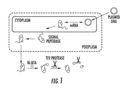
Figure 1 shows a schem.atic describing the production, export and purification
of scFv.
Signal peptide with TEv cleavage site shown. Note that the 6xHis tag and TEv
protease are
removed by a second Ni-NTA agarose column.
Figure 2 shows sampl.e purification of 3E8.scFv from pCOLD IV. Lane 1:
Sphereoplasts,
Lane 2: Periplasmic fraction after Ni-NTA binding, Lane 3: Wash, Lane 4:
Eluted 6xHis-TEV-
3E8.scFv, Lane 5: 3E8.scFv after TEv protease cleavage to remove 6xHi.s-tag,
Lane 6: Ni-NTA
purified protein after removal of TEV protease and 6xHis-tag.
Figure 3A. and 3B show purification of 3E8.scFv.Cys from its proteolytic
fragment using
cation exchange chromatography. The two species eluted from a Resource S
column at 450 and
600 m.M NaC1, with the authentic product eluting first. This was confirmed by
SDS-PA.GE.
Lanes 1 and 12 are USB ladder, Lane 2: 3E8.scFv.Cys prior to ion exchange
chromatography,
3
CA 02956161 2017-01-23
WO 2016/014839 PCT/US2015/041809
Lanes 3-4: Fractions 1 and 2, Lanes 5-8: Fractions 3-6, Lanes 9-11: Fractions
7-9. The desired
product is indicated with an asterisk.
Figure 4 shows optimization of peiiplasm extraction. The osmotic (0) and
peri.plasmic
(P) fractions are compared across nine purification methods. The modified
lysozyme procedure
yiel.ded the best results. The faint band above the desired product is scFv
with PelB leader
sequence. When digested with TEV protease, both protein bands resolve to a
single species. All
samples purified are 3E8.scFv from pCOLD IV in DH10B.
Figure 5 shows gel filtration of antibody fragments. CC49.scFv is a
heterogeneous
sample that contains both monomer and dimer. Additionally, the fragment elutes
as a slightly
larger protein than its calculated molecular weight, showing some degree of
unfolding or
expansion. The 3E8.scFv elutes as a single species with molecular weight
corresponding to a
weli folded monomer.
Figure 6 shows the stability of antibodies and fragments. A. Aggregation
propensity is
measured with increasing temperature. 3E8.scFv is intermediate in stability
between the less
stable CC49.scFv and the more stable 3E8.IgG. B. HTTS shows similar results to
those reported
by DSLS, with a second unfolding transition for 3E8.1gG. The binding domains
of 3E8.scFv and
3E8.1gG both unfol.d at 66 C.
Figure 7 shows antibody and fragment binding. A. Dot blot assay shows that
both
CC49.scFv and 3E8.scFv bind BSM (sial.yl-Tn), but not BSA. B. Inhibition assay
with
fluorescent IgG and nonlabeled scFv shows that the scFv binds --16-fold less
strongly than the
bivalentigG. (-)* was performed with 0.251AM IgG with no BSM. (-)** was
perform.ed using
free fluorescein in the absence of antibody. C. SPR sensogams for each
variant.
Figure 8 shows dot blot assay using horseradish peroxidase as the reporter.
Here, the
say specifically binds the section of nitrocellulose that was blotted with
mucin containing the
TAG-72 epitope. The brown color is the result of the chemical reaction
catalyzed by horseradish
peroxidase, which is linked to the say by the biotin-streptavidin interaction.
Figure 9 shows irnmunohistochemical staining of human colon cancer. The scFv
intensely stains the extracellular mucin and the intracellular vacuoles
containing the TAG-72
epitope.
Figure 10 shows NHS-PEGylation of 3E8.scFv. Lane 1: USB ladder, Lanes 2 and 3:
Unmodified antibody fragment, Lane 4: Reaction with 5-fold molar excess of
PEG, Lane 5:
Reaction with 20-fold molar excess of PEG.
4
CA 02956161 2017-01-23
WO 2016/014839 PCT/US2015/041809
Figure 11 shows fluorescent labeling of antibody fragments with and without 6x-
His
tags. The samples with hexahistidine tags (3E8H6d and CC49H6d) generate more
intense signals
due to increased concentrations of antibody fragment.
Figure 12 shows the specific PEGylation of 3E8.scFv.Cys. The antibody fragment
was
labeled specifically at the C-terminai cysteine residue using maleimide
chemistry. The scFv was
nearly quantitatively PEGylated. Lane 1: partially purified, reduced
3E8.scFv.Cys, Lane 2:
PEGylated 3E8.scFv.Cys. Lane 3: unrelated. Lane 4: ladder.
Figure 13 shows the biophysical characterization of 3E8.scFv.
Figure 14 shows binding studies for 3E8.scFv. An Estimated 50% bound at 4 1.1M
i.e.
3E8.scFv is roughly a 16-fol.d worse binder than 3E8.11gG (KD = 0.65 nM) so
it's estimated KD
= 10.4 nM.
Figure 15 shows surface plasmon resonance. 3E8.scFv has a measured Kd of 16.4
nM,
which indicates very tight binding to the proper substrate.
Figure 16 shows immunohistochemistry. Biotin attached to 3E8.scFv via lysines
can be
coupled to a chromogenic enzyme complex which produces a brown product. Using
this scheme,
one can visual.ize 3E8.scFv bound to its epitope. Histologists can use this
technique to anal.yze
surgicai specimens to determ.ine the success of surgical. procedures.
Figure 17 shows PEGylation results. PEGylations of T4L with 2 kDa PEG is
shown.
Polydispersed PEGs result in smear of PEGylated products; discrete PEGs result
in I.adder of
distinct PEGylated products.
Figure 18 shows Sialyl-Tn Disaccharide and TAG-72 protein.
Figure 19 shows antibody scaffolds depicted in gray bound to antigen (black
stars). Also
shown is the schematic for the scFv gene.At 60 degrees, both the IgG and say
of 3E8 are well.
folded. At 70 degrees, the scFv is completely unfolded, as is the binding site
of the IgG. The
constant domains of the IgG prevent aggregation of the IgG at this
temperature. By 85 degrees,
both molecules are unfolded and aggregated.
Figure 20 shows the ScFv structure. A. CD wavelength scan of scFvs is
consistent with
the immunoglobulin domain fold. B. Gel filtration shows a single monomeric
species for
3E8.scFv, but CC49 is slightly expanded and exists as a say.
Figure 21 shows that mucin is a large glycoprotein expressed and secreted from
healthy
and diseased cells. The mucin of adenocarcinomas has been shown to overexpress
the
disaccharide, Sialyl-Tn. This epitope is targeted with antibodies and antibody
fragments.
Figures 22A and 22B show PEGylation. Figure 22A: Model of 3E8.scFv - The
complementary determining regions are the loops responsible for binding the
antigen. The C-
CA 02956161 2017-01-23
WO 2016/014839 PCT/US2015/041809
terminal cysteine is shown opposite the binding site. Figure 22B: PEGylation
increases the
hydrodynamic radius of proteins, can reduce immunogenicity, decrease
aggregation, and protect
the antibody fragment from serum. proteases.
Figures 23 shows crude modeling of 3E8.scFv reveals four lysines within the
CDRs
responsible for antigen binding.
Figure 24 shows PEGylated 3E8cys.scFv. Modified antibody fragments are shown
on an
SDS-PAGE gel. The 40 kD samples are loaded at 2x and lx concentrations. PEG
polymers do
not strongl.y interact with SUS; therefore, their apparent masses are
anomalous by SUS-PAGE
compared to protein ladders. A linear and branched 1.8 kD PEG are shown.
Figure 25 shows repl.icates of 3E8cys.scFv + Y-40 kD binding to TAG-72
immobilized
on nitrocellulose paper. The antigen was spotted on the corner of the paper
indicated by pencil
mark. Far right is the negative control, where all experimental steps were
performed, but the
paper was incubated with buffer in place of antibody fragment.
Figure 26 shows correlation between serum half-lives and microPET/CT imaging.
Blood
radioactivity (%ID) of each individual mouse is plotted against its normalized
tumor intensity in
PET imaging. Upper panel, microPET/CT i.m.aging at 5 h; bottom panel,
microPET/CT imaging
at 24 h. 5 and 24h are the appropriate time points for a 1231-SPECT/CT
radiopharmaceutical..
Figure 27 shows 3E8cys.scFv was conjugated with a linear 30 Id3 PEG. Three
antibody
fragment aliquots were PEGylated and purified. Sample C provided the highest
yield and purity,
therefore, it was used for tumor imaging.
Figure 28 shows the serum half-lives can be tuned with polyethylene glycol
(PEG)
conjugation. PEGylated antibody fragments are shown using lysines (NHS-ester
chemistry) and
cysteines (maleimid.e chemistry).
Figures 29A-C show proof-of-concept surgical resection with intraoperative
imaging via
the 1231-1abe1ed. antibody fragment. A. Image taken prior to surgery (tumors
arrowed). B. A
second image is taken to assess the surgical procedure. Note residual tumor
remains on the right
flank. C. Image taken after complete removal of cancerous tissue.
Figure 30 shows response units for various concentrations of
3E8HL(GGGGS)3scFv.
Figure 31 shows the thermal denaturation of TIM variants performed at 25uM
protein
with 5x SYPRO Orange dye. The melts were assayed using a Bio-Rad C10000
thermal cycl.er
with a ramp rate of 1 C min-1 at 0.2 C intervals. The data was exported into
Microsoft Excel
2013. The 77/2 was calculated as the temperature with the max immn sl.ope as
determined from a
5' window around each point.
Figure 32 shows the plasmid insert for 3E8.G4S.
6
CA 02956161 2017-01-23
WO 2016/014839 PCT/US2015/041809
Figure 33 shows two T7 expression strains tested, BLR(DE3) and HMS174(DE3).
BLR(DE3) is a BL21(DE3) derivative that is recA-, resulting in improved genome
and plasmid
stability. HMS174(DE3) is a K12 bacteria that also contains a mutations to the
recA gene.
Figure 34 shows bioreactor optimization conditions. The two primary conditions
that
were optimized were dissolved oxygen percentage and source, as well as
agitation speed. The
standard Rushton impeller was used, but did not include baffles. Initially,
baffles were used and
set the agitation speed to 100 rpm based on previous experience with shake
flasks - baffled flasks
at 225 rpm led to cellular lysis.
Figure 35 shows the role of induction point and the expression time course for
3E8.G4S.
The sample was prepared identically as before, but the cul.ture was allowed to
reach 0D600 =
45, before added 0.5 rnM IPTG, shifting the temperature to 20 C, reducing
agitation to 500 rpm,
and adding supplemental oxygen to maintain d02 at 30 %. This culture
maintained an optical
density of 45-50 for nearly 24 hours post induction when the experiment was
terminated. At 0.5,
1, 1.5, 2, 3, 4, 6, and 8 hours 50 mi., of cul.ture were harvested and snap
frozen for further studies.
Each cell pellet was resuspended to an 0D600 = 46, and 10 mL of normalized
cultures were
purified in parallel.
Figure 36 shows that the 3E8.C4S was expressed with a cleavable hexahistidine
tag and
immobilized metal affinity chromatography (IMAC). After elution from the Ni-
NTA column,
the protein was incubated at room temperature overnight with the cysteine
protease from
Tobacco Etch Virus (TEV) and 1 rnM DTT. A second Ni-NTA column is used to
remove the
hexahistidine tag, His-tagged-TEV protease. A cation exchange step is used to
remove several
protein contaminants. By SDS-PAGE, the desired product is nearly homogeneous.
Figure 37 shows purification strategies were employed that remove reliance on
TEV
protease and hexahistidine tags for licensing and costs. As a result two new
variantsv were
cloned and characterized that remove the hexahistidine tag and TEV protease
recognition
sequence. The original 3E8.G4S leaves a small GSSG linker at the N-terminus.
To test the
inwortance of the 1.inker, two variants were tested - one that begins GSSG-
QVQ..., and a second
that begins with the native QVQ...
Figure 38 shows FPLC Procedure (1 L expression from shake flask): The Protein
L
column is first equilibrated with 5 column volum.es (CV) of Protein L binding
buffer. Next, the
sample is loaded at 1 tfiL min-1 before washing with 10 CVs Protein L binding
buffer (1 mL
min-1. washing). (Figure 38). Next, the sampl.e is eluted with Protein L
el.ution buffer (100 mM
glycine pH 3). The SDS-PAGE gel shown to the right shows the flowthrough,
wash, and elution
of the hexahistidine tagged 3E8.G45. Note that the desired antibody fragment
is arrowed and that
7
CA 02956161 2017-01-23
WO 2016/014839 PCT/US2015/041809
several proteolytic products or copurifying contaminates are bound and eluted
from the Protein L
column. These bands are able to be removed by ion exchange chromatography.
Figure 39 shows a final ion exchange column was used to remove bacterial
endotoxins.
When 3E8.G4S is exchanged into 20 rriM Tris at pH 8 or 9. This sample was
applied to a
Resource Q column where little to no binding was observed. The flowthrough was
collected,
which is expected to be depleted of both endotoxin and nucleic acids. The
primary data for these
experiments are shown in the below graphs. Note that the elutions from
Resource S and
flowthroughs from Resource Q were run on SUS-PAGE gel to confirm. the protein
identity.
Figure 40 shows various optimization conditions.
Figure 41 shows a PET scan of a 3E8.G4S mouse with 124I-diabody at 24 hours.
Figure 42 shows blood curves for 3E8 antibody fragment.
Figure 43 shows average percentage of ID/g and sd in blood, lung, heart,
liver, spleen,
pancreas, GI, kidney, muscle, skin, tail, and carcass of mouse.
Figure 44 shows construction and IMAC purification of 3E8.scFv and 3E8*.G4S.
Top:
The open reading frames are shown for the antibody fragments. Bottom: The
final purified scFv
appears in the second flow-through and wash, by SDS-PAGE. Far Right: SDS-PA.GE
of ladder
and purified 3E8*.G4S from C43(DE3).
Figure 45 shows an example sensorgraph of 3E8.scFv and dissociation constants
for all
studied variants. All variants studied to date exhibit low nanomolar binding
to TAG-72. The
two 3E8*.G45 entries are the same protein sequence. The first expression and
purification was
performed in the laboratories of ABT. The Magliery Lab (um) repeated the
procedure in
C43(DE3) E. coll. Both purification yielded similar nanomolar KDs.
Figure 46 shows pharmacokin.etics of antibody fragments. Blood curves of
3E8*.G4S,
3E8.scFv, and 3E8cys.scFv + 11451 are shown. The 3E8*.G4S exhibits the longest
serum half-
life of the three molecules studied. Note that 3E8cys.scFv + 10484 shown as
counts/4 rather
than IDValg.
Figure 47 shows Biodistribution data for each compound. PEGylation with the 40
kD
PEGs dramatically affected the pharmacokinetic properties and biodistribution.
Figure 48 shows microSPECT/CT imaging of xenograft mice.
Figure 49 shows n.eoprobe 3000 data.
Figure 50 shows two new variants that remove the hexahistidine tag and TEV
protease
recognition sequence. The N-terminus begins with the PelB leader sequence to
direct the
disulfide-containing protein to the periplasm. Upon leader sequence cleavage
by the endogenous
signal peptidase from. E. coil, the final proteins begins with amino acids,
QVQ.
8
CA 02956161 2017-01-23
WO 2016/014839 PCT/US2015/041809
Figure 51 shows the purification steps used to extract 3E8.G4S from the
cleared lysate is
Protein L chromatography. When the hexahistidine tag and TEV protease
recognition sequence
was removed from. the open reading frame (QVQ variant), the copurifying bands
were absent.
Protein L has produced nearly quantitative purification and near-homogeneity
by SDS-PAGE
(>95 %).
Figure 52 shows twelve individual transformants were selected, expressed,
purified, and
characterized by Superdex 75. All preparations show diabody, triabody, and
tetrabody features
with less than 10% separating the extremes (+/- 5 % difference from the
average).
Figure 53 shows full-length antibodies have long serum lifetimes due to their
large size
(-160kD) and cellular uptake via neonatal 17, receptors. Shown are full-length
:IgG labeled with
1251, and iodine half-lives.
Figure 54 shows blood curves. 3E8.G4S radiolabels weli with iodine using the
standard
Iodogen method (>70%, and as high as 95%).
Figure 55 shows the biodistribution of 3E8.G4S in mice. The approxi.m.ate
tumor:tissue
ratios of blood (9:1), liver (20:1), kidneys (7:1), GI (29:1) are shown. Based
on these analyses,
3E8.G45 is ideal for imaging of a broad range of adenocarcinom.as, as no
tissue significantly
accumulates 3E8.G4S.
Figure 56 shows xenograft mice implanted with human colon adenocarcinoma
tumors
(LS-174T cells), imaged with 18FDG, full-length 3E8 1gG, and a 3E8 antibody
fragment -
specifically a component of 3E8.G4S. As expected, the 18FDG mouse shows
nonspecific uptake
of the radiotracer and poor labeling of the two implanted tumors on the left
and right flanks. 4
mice imaged at 48 hours with 3E8.G4S are also shown.
DETAILED DESCRIPTION
The materials, compositions, and methods described herein can be understood
more
readily by reference to the following detailed descriptions of specific
aspects of the disclosed
subject matter and the Examples and Figure included herein.
Before the present materials, compositions, and methods are disclosed and
described, it is
to be understood that the aspects described below are not limited to specific
synthetic methods or
specific reagents, as such may, of course, vary. It is also to be understood
that the terminology
used herein is for the purpose of describing particular aspects only and is
not intended to be
limiting.
9
CA 02956161 2017-01-23
WO 2016/014839 PCT/US2015/041809
A.Iso, throughout this specification, various publications are referenced. The
disclosures
of these publications in their entireties are hereby incorporated by reference
into this application
in order to more fully describe the state of the art to which the disclosed
matter pertains. The
references disclosed are also individually and specifically incorporated by
reference herein for
the material contained in them that is discussed in the sentence in which the
reference is relied
upon.
Definitions
Unless defined otherwise, all technical and scientific terms used herein have
the same
meaning as commonly understood by one of ordinary skill in the art. Methods
and materials
similar or equivalent to those described herein can be used in the practice or
testing of the
present disclosure. In this specification and in the claims that fol.low,
reference wili be m.ade to a
number of terms, which shall be defined to have the following meanings:
Throughout the specification and claims the word "comprise" and other forms of
the
word, such as "comprising" and "comprises," means including but not limited
to, and is not
intended to exclude, for example, other additives, components, integers, or
steps.
As used in the description and the appended claims, the singular forms "a,"
"an," and
"the" include plural referents unless the context clearly dictates otherwise.
Thus, for example,
reference to "an antibody" includes mixtures of two or more such antibodies;
reference to "the
composition" includes mixtures of two or more such compositions, and the like.
"Optional" or "optionally" means that the subsequently described event or
circumstance
can or cannot occur, and that the description includes instances where the
event or circumstance
occurs and instances where it does not.
Unless otherwise indicated, all numbers expressing quantities of ingredients,
reaction
conditions, and so forth used in the specification and claims are to be
understood as being
modified in ali instances by the term "about". The term "about", as used
herein when referring to
a measurable value such as an amount of mass, weight, time, volume,
concentration, or
percentage, is meant to encompass variations of in some embodiments 20%, in
some
embodiments .10%, in some embodiments - 5%, in some embodiments 1 %, in some
embodi.m.ents 0.5%, and in some embodiments 4.1 % from the specified amount,
as such
variations are appropriate to perform the disclosed methods andlor employ the
disclosed
compositions. Accordingly, unless indicated to the contrary, the n.um.erical
parameters set forth
in this specification and attached claims are approximations that can vary
depending upon the
desired properties sought to be obtained by the presently disclosed subject
m.after.
CA 02956161 2017-01-23
WO 2016/014839 PCT/US2015/041809
As used herein, the term "and/or" when used in the context of a list of
entities, refers to
the entities being present singly or in combination. Thus, for example, the
phrase "A, B, C,
and/or D" includes A, B, C, and D individually, but also includes any and all
combinations and.
subcombinations of A, B, C, and D.
With respect to the terms "comprising", "consisting of, and "consisting
essentially of,
where one of these three terms is used herein, the presently disclosed and
claimed subject matter
can include the use of either of the other two terms. For example, in some
embodiments, the
presentl.y disclosed subject matter relates to compositions comprising
antibodies. It would be
understood by one of ordinary skill in the art after review of the instant
disclosure that the
presently di.scl.osed subject matter thus encompasses compositions that
consist essentially of the
antibodies of the presently disclosed subject matter, as well as compositions
that consist of the
antibodies of the presently disclosed subject matter.
The term "subject" as used herein refers to a member of any invertebrate or
vertebrate
species. Accordingly, the term "subject" is intended to encompass in some
embodiments any
member of the Kingdom Animalia including, but not limited to the phylum.
Chordata (e.g.,
members of Classes Osteichythyes (bony fish), Amphibia (amphibians), Reptilia
(reptiles), Ayes
(birds), and Mammalia (mammals), and all Orders and Families encompassed
therein.
The compositions and methods of the presently disclosed subject matter are
particularly
useful for warm-blooded vertebrates. Thus, in some embodiments the presently
disclosed subject
matter concerns mammals and birds. More particularly provided are compositions
and methods
derived from and/or for use in mammals such as humans and other primates, as
well as those
mammals of importance due to being endangered (such as Siberian tigers), of
economic
importance (animals raised on farms for consumption by humans) and/or social
importance
(animals kept as pets or in zoos) to hum.ans, for instance, carnivores other
than humans (such as
cats and dogs), swine (pigs, hogs, and wild boars), ruminants (such as cattle,
oxen, sheep,
giraffes, deer, goats, bison, and camels), rodents (such as mice, rats, and
rabbits), marsupials,
and horses. Also provided is the use of the disclosed methods and compositions
on birds,
including those kinds of birds that are endangered, kept in zoos, as well as
fowl, and more
particularly domesticated fowl, e.g., poultry, such as turkeys, chickens,
ducks, geese, guinea
fowl, and the like, as they are also of economic importance to humans. Thus,
also provided is the
use of the disclosed methods and compositions on livestock, including but not
limited to
domesticated swine (pigs and hogs), ruminants, horses, poultry, and the like.
11
CA 02956161 2017-01-23
WO 2016/014839 PCT/US2015/041809
Simil.arly, ali genes, gene names, and gene products disclosed herein are
intended to
correspond to homologs and/or orthologs from any species for which the
compositions and
methods disclosed herein are applicabl.e. Thus, the terms include, but are not
limited to genes and
gene products from humans and mice. It is understood that when a gene or gene
product from a
particular species is disclosed, this disclosure is intended to be exemplary
only, and is not to be
interpreted as a limitation unless the context in which it appears clearly
indicates. Thus, for
example, for the genes presented in GENBANK Accession Nos: AAA60019 and
NP...004976,
the human amino acid sequences disclosed are intended to encompass homologous
genes and
gene products from other animals including, but not limited to other mammals,
fish, amphibians,
reptiles, and birds. A.lso encompassed are any and all nucleotide sequences
that encode the
disclosed amino acid sequences, including but not limited to those disclosed
in the corresponding
GENBANK entries (i.e., J05582.1 and NM_004985, respectively).
The terms "cancer" and "tumor" are used interchangeably herein and can refer
to both
primary and metastasized solid tumors and carcinomas of any tissue in a
subject, including but
not limited to breast; colon; rectum; lung; oropharynx; hypopharynx;
esophagus; stomach;
pancreas; liver; gallbladder; bile ducts; small intestine; urinary tract
including kidney, bladder,
and urothelium; female genital tract including cervix, uterus, ovaries (e.g. ,
ch.oriocarcinoma and
gestational trophoblastic disease); male genital tract including prostate,
seminal vesicles, testes
and germ cell tumors; endocrine glands including thyroid, adrenal, and
pituitary; skin (e.g.,
hemangiomas and melanomas), bone or soft tissues; blood vessels (e.g. ,
Kaposi's sarcoma);
brain, nerves, eyes, and meninges (e.g. , astrocytomas, gliomas,
glioblastomas, retinoblastom.as,
neuromas, neuroblastomas, Schwannomas and meningiomas). As used herein, the
terms "cancer
and "tumor" are al.so intended to refer to multicellular tumors as well as
individual neoplasti.c or
preneoplastic cells. In some embodiments, a cancer or a tumor comprises a
cancer or tumor of an
epithelial tissue such as, but not limited to a carcinoma. In some
embodiments, a tumor is an
adenocarcinoma, which in some embodiments is an adenocarcinoma of the
pancreas, breast,
ovary, colon, or rectum, and/or a metastatic cell derived therefrom.
As used herein in the context of molecules, the term "effector" refers to any
molecule or
combination of molecules whose activity it is desired to deliver/into and/or
localize at a cell.
Effectors include, but are not li.m.ited to labels, cytotoxins, enzymes,
growth factors, transcription
factors, drugs, etc.
As used herein in the context of cells of the imm.une system, the term
"effector" refers to
an immune system cell that can be induced to perform a specific function
associated with an
12
CA 02956161 2017-01-23
WO 2016/014839 PCT/US2015/041809
immune response to a stimulus. Exemplary effector cells include, but are not
limited to natural.
killer (NK) cells and cytotoxic T cells (Tc cells).
As used herein, the term "expression vector" refers to a DNA sequence capable
of
directing expression of a particular nucleotide sequence in an appropriate
host cell, comprising a
promoter operatively linked to the nucleotide sequence of interest which is
operatively linked to
termination signals. It also typically comprises sequences required for proper
translation of the
nucleotide sequence. The construct comprising the nucleotide sequence of
interest can be
chimeric. The construct can also be one that is naturally occurring but has
been obtained in a
recombinant form useful for heterologous expression.
As used herein, the term "hybridoma" refers to a cell or cell line that is
produced in the
laboratory from the fusion of an antibody-producing lymphocyte and a non-
antibody-producing
cancer cell, usually a myeloma or lymphoma cell. As would be known to those of
one of
ordinary skill in the art, a hybridoma can proliferate and produce a
continuous supply of a
specific monoclonal antibody. Methods for generating hybridomas are known in
the art (see e.g.,
Harlow & Lane, 1988).
As used herein, the terms "operatively linked" and "operably linked" refer to
transcriptional regulatory elements (such as, but not limited to promoter
sequences, transcription
terminator sequences, etc.) that are connected to a nucleotide sequence (for
example, a coding
sequence or open reading frame) in such a way that the transcription of the
nucleotide sequence
is controlled and regulated by that transcriptional regulatory element.
Similarly, a nucleotide
sequence is said to be under the "transcriptional control" of a promoter to
which it is operably
linked. Techniques for operatively linking a promoter region to a nucleotide
sequence are known
in the art.
As used herein, the term "prodrug" refers to an analog and/or a precursor of a
drug (e.g.,
a cytotoxic agent) that substantially lacks the biological activity of the
drug (e.g., a cytotoxic
activity) until subjected to an activation step. Activation steps can include
enzymatic cleavage,
chemical activation steps such as exposure to a reductant, and/or physical
activation steps such
as photolysis. In some embodiments, activation occurs in vivo within the body
of a subject,
As used herein, the terms "antibody" and "antibodies" refer to proteins
comprising one or
more polypeptides substantially encoded by imm.unogl.obulin genes or fragments
of
immunoglobulin genes. Inununoglobulin genes typically include the kappa (x),
lambda (X), alpha
(a), gamma (y), delta (6), epsilon (c), and mu (g) constant region genes, as
well as myriad
immunoglobulin variable region genes. Light chains are classified as either lc
or X. In mammals,
heavy chains are cl.assified as y, IA, a, 6, or c, which in turn. define the
immunoglobulin classes,
13
CA 02956161 2017-01-23
WO 2016/014839 PCT/US2015/041809
1gG, IgM, IgA, IgD, and IgE, respectively. Other species have other light and
heavy chain genes
(e.g., certain avians produced what is referred to as IgY, which is an
immunoglobulin type that
hens deposit in the yolks of their eggs), which are similarly encompassed by
the presently
disclosed subject matter. In some embodiments, the term "antibody" refers to
an antibody that
binds specifically to an epitope that is present on a tumor antigen.
The term "antibody fragment" refers to any derivative of an antibody which is
less than
full-length. In exemplary embodiments, the antibody fragment retains at least
a significant
portion of the full-length antibody's specific binding ability. Examples of
antibody fragments
include, but are not limited to, Fab, Fab', F(a13`) , scFv, Fv, diabody,
tribody, tetrabody, Fd
fragments, or mixtures thereof. The antibody fragment may be produced by any
means. For
instance, the antibody fragment may be enzymatically or chemically produced by
fragmentation
of an. intact antibody, it may be recombinantly produced from a gene encoding
the partiai
antibody sequence, or it may be wholly or partially synthetically produced.
The antibody
fragment may optionally be a single chain antibody fragment. Alternatively,
the fragment may
comprise multiple chains which are linked together, for instance, by disulfide
linkages. The
fragment may also optionally be a multimolecular complex.
A typical. immunoglobulin (antibody) structural unit is known to comprise a
tetramer.
Each tetramer is composed of two identical pairs of polypeptide chains, each
pair having one
"light" chain (average molecular weight of about 25 kiloDalton (kDa)) and one
"heavy" chain
(average molecular weight of about 50-70 kDa). The two identical pairs of
polypeptide chains
are hel.d together in di.m.eric form. by disulfide bonds that are present
within the heavy chain
region. The N-terminus of each chain defines a variable region of about 100 to
110 or more
amino acids prim.arily responsible for antigen recognition (sometimes referred
to as the
"paratope"). The terms variable light chain (VL) and variable heavy chain (VH)
refer to these
light and heavy chains, respectively.
Antibodies typically exist as intact immunoglobulins or as a number of well-
characterized fragments that can be produced by digestion with various
peptidases. For example,
digestion of an antibody molecule with papain cleaves the antibody at a
position N-terminal to
the disulfide bonds. This produces three fragments: two identical "Fab"
fragments, which have a
light chain and the N-terminus of the heavy chain, and an "Fc" fragment that
includes the C-
terminus of the heavy chains held together by the disulfide bonds. Pepsin, on
the other hand,
digests an antibody C-terminal to the disulfide bond in the hinge region to
produce a fragment
known as the "F(ab)12" fragment, which is a dimer of the Fab fragments joined
by the disulfide
bond. The F(ab)12 fragment can be reduced under mild conditions to break the
disulfide linkage
14
CA 02956161 2017-01-23
WO 2016/014839 PCT/US2015/041809
in the hinge region, thereby converting the F(ab)2 dimer into two "Fab'"
monorners. The Fab'
monomer is essentially an Fab fragment with part of the hinge region (see
e.g., Paul, 1993, for a
more detailed description of other antibody fragments). With respect to these
various fragments,
Fab, F(abt)2, and Fab' fragments include at least one intact antigen binding
domain (paratope),
and thus are capable of binding to antigens.
While various antibody fragments are defined in terms of the digestion of an
intact
antibody, one of skill will appreciate that various of these fragments
(including, but not limited
to Fab' fragments) can be synthesized de novo either chemically or by
utilizing recombinant
DNA methodology. Thus, the term "antibody" as used herein also includes
antibody fragments
produced by the modification of whole antibodies and/or synthesized de novo
using recombinant
DNA methodologies. In some embodiments, the term "antibody" comprises a
fragment that has
at I.east one antigen binding domain (paratope).
Antibodies can be polyclonal or monoclonal. As used herein, the term
"polyclonal" refers
to antibodies that are present together in a given collection of antibodies
and that are derived
from different antibody-producing cells (e.g., B cells). Exemplary polyclonal
antibodies include,
but are not li.m.ited to those antibodies that bind to a particular antigen
and that are found in the
blood of an animal after that animal has produced an imm.une response against
the antigen.
However, it is understood that a polyclonal preparation of antibodies can also
be prepared
artificially by m.ixing at least non-identical two antibodies. Thus,
polyclonal antibodies typically
include different antibodies that are directed against (i.e., bind to) the
same and/or different
epitopes (sometimes referred to as an "antigenic determinant" or just
"determinant") of any
given antigen.
As used herein, the term "monoclonal" refers to a single antibody species
and/or a
substantially homogeneous population of a single antibody species. Stated
another way,
"monoclonal" refers to individual antibodies or populations of individual
antibodies in which the
antibodies are identical in specificity and affinity except for possible
naturally occurring
mutations that can be present in minor amounts. Typically, a monoclonal
antibody (mAb or
moAb) is generated by a single B cell or a progeny cell thereof (although the
presently disclosed
subject matter also encompasses "monoclonal." antibodies that are produced by
molecular
biological techniques as described herein.). Monoclonal antibodies (mAbs or
moAbs) are highl.y
specific, typically being directed against a single antigenic site.
Furthermore, in contrast to
polyclonal antibody preparations, a given mAb is typically directed against a
single epitope on
the antigen.
CA 02956161 2017-01-23
WO 2016/014839 PCT/US2015/041809
In addition to their specificity, mAbs can be advantageous for some purposes
in that they
can be synthesized uncontaminated by other antibodies. The modifier
"monoclonal" is not to be
construed as requiring production of the antibody by any particular method,
however. For
example, in some embodiments, the mAbs of the presently disclosed subject
matter are prepared
using the hybridoma methodology first described by Kohler et al., 1975, and in
some
embodiments are made using recombinant DNA methods in prokaryotic or
eukaryotic cells (see
e.g. , U.S. Patent No. 4,816,567, the entire contents of which are
incorporated herein by
reference). mAbs can also be isolated from phage antibody libraries.
The antibodies, fragments, and derivatives of the presently disclosed subject
matter can
also include chimeric antibodies. As used herein in the context of antibodies,
the term.
"chimeric", and grammatical variants thereof, refers to antibody derivatives
that have constant
regions derived substantially or exclusively from antibody constant regions
from one species and
variable regions derived substantially or exclusively from the sequence of the
variable region
from. another species.
The variable region allows an antibody to selectively recognize and
specifically bind
epitopes on antigens. That is, the VI, domain and VII domain., or subsets of
the complementarily
determining regions (CDR.$) within these variable domains, of an antibody
combine to form the
variable region that defines a three dimensional antigen binding site. This
quaternary antibody
structure forms the antigen binding site present at the end of each arm of the
antibody. More
specifically, the antigen binding site is defined by three CDRs on each of the
VH and VL chains.
In some instances (e.g., certain immunoglobulin molecules derived from
camel.id species or
engineered based on camelid immunoglobulins), a complete immunoglobulin
molecule can
consist of heavy chains only with no light chains.
In naturally occurring antibodies, there are six CDRs present in each antigen
binding
domain that are short, non-contiguous sequences amino acids that are
specifical.ly positioned
to form the antigen binding domain as the antibody assumes its three
dimensional configuration
in an aqueous environment. The remainder of the amino acids in the antigen
binding domains,
referred to as "framework" regions, show less inter-molecular variability. The
framework
regions largely adopt a I3-sheet conformation and the CDRs form loops that
connect, and in some
cases form part of, the 13-sheet structure. Thus, framework regions act to
forrn a scaffold that
provides for positioning the CDRs in correct orientation by inter-chain, non-
covalent
interactions. The antigen binding domain formed by the positioned CDRs defines
a surface
complementary to the epitope on the irrununoreactive antigen. This
complementary surface
promotes the non-covalent binding of the antibody to its cognate epitope. The
amino acids
16
CA 02956161 2017-01-23
WO 2016/014839 PCT/US2015/041809
comprising the CDRs and the framework regions, respectively, can be readily
identified for any
given heavy or light chain variable domain by one of ordinary skill in the
art, since they have
been preci.sel.y defined (see e.g., Chothia & Lesk, 1987; Kabat et al., 1991 ;
Martin, 1996;
Johnson & Wu, 2000).
A. particular kind of chimeric antibody is a "humanized" antibody, in which
the
antibodies are produced by substituting the CDRs of, for example, a mouse
antibody, for the
CDRs of a human antibody (see e.g., PCT International Patent Application
Publication No. WO
1992/22653). Thus, in some embodiments, a humanized antibody has constant
regions and
variable regions other than the CDRs that are derived substantially or
exclusively from the
corresponding regions of a human antibody, and CDRs that are derived
substantially or
exclusively from a mammal other than a human.
Fv fragments correspond to the variable fragments at the N-termini of
immunoglobulin
heavy and light chains. Fv fragments appear to have lower interaction energy
of their two chains
than Fab fragm.ents. To stabilize the association of the VII and VL domains,
they can be linked
with peptides (see e.g., Bird et al., 1988; Huston et al., 1988), disulfide
bridges (see e.g.,
Glockshuber et al., 1990), and/or "knob in hole" mutations (see e.g., Zhu et
al., 1997). ScFv
fragments can be produced by m.ethods well known to those skilled in the art
(see e.g., Whitlow
et al., 1991; Huston et al., 1993).
A "single-chain variable fragment" (scFv) is a fusion protein of the variable
regions of
the heavy (VH) and light chains (VL) of immunoglobulins, connected with a
short linker
peptide. The linker can be rich in glyci.ne for flexibility, as well as serine
or threonin.e for
solubility, and can either connect the N-terminus of the VH with the C-
terminus of the VL, or
vice versa. This protein retains the specificity of the original
immunoglobu.lin, despite removal
of the constant regions and the introduction of the linker. scFv can be
produced in bacterial cells
such as E. coli or in eukaryotic cell.s.
Methods and Compositions
scFvs, Diabodies, Tribodies, and Tetrabodies, and Nucleic Acids Thereof
Antibodies recognizing TAG-72 provide a novei approach for the imaging,
detection, and
treatment of cancer. Antibody fragments that binds specifically to the sialyl-
Tn surface adhesin
of TAG-72 is disclosed herein. The antibody fragment derivatives of the
present invention are
advantageously useful over other antibody and antibody fragments known in the
art because they
are easy to express in large quantities, can penetrate tissues easily and lack
the constant domains
17
CA 02956161 2017-01-23
WO 2016/014839 PCT/US2015/041809
that promote often unwanted and u.sual.ly superfluous effector functions.
ScFvs are monovalent
because the heavy and light chains are joined by a flexible peptide linker,
which allows the two
domains to fold and interact with each other. By using antibody fragments,
such as diabodies,
wherein the linking peptide is shortened thereby forcing the heavy and light
chain variable
dom.ains to interact to form a dimer, the drawback of using scFvs is overcome.
Further, as a
consequence of this interaction, the antibody fragment is bivalent like the
parent
immunoglobulin, and therefore has increased binding avidity.
Recombinant antibody fragments can be engineered to assemble into stable
multimeric
oligomers of high binding avidity and specificity (Kortt, et al. (2001)
Biomol. Eng. 18:95-108).
A scFv molecule joined by a linker of 3-12 residues cannot fol.d into a
functional Fv domain and
instead associates with a second scFv molecule to form a bivalent dimer
(diabody, approx. 60
kDa). For the cross-linking of cell surface antigens at least two binding
moieties are necessary.
The diabody is the smallest bivalent antibody molecule able to fulfill this
requisite.
As used herein, the term "diabody" refers to an engineered antibody construct
prepared
by isolating the binding domains (both heavy and light chain) of a binding
antibody, and
supplying a linking moiety which joins or operably links the heavy and light
chains on the same
polypeptide chain thereby preserving the binding function as described in
detail by Holliger et al.
(1993) Proc. Natl. Acad. Sci. USA 90:6444 and reviewed by Poljak (1994)
Structure 2:1121-
1123. This forms, in essence, a radically abbreviated antibody, having only
the variable domain
necessary for binding the antigen. By using a linker that is too short to
allow pairing between the
two domains on the sam.e chain, the domains are forced to pair with the
com.plementary domains
of another chain and create two antigen-binding sites. These dimeric antibody
fragments, or
d.iabodies, are bivalent and bispecific. It should be clear that any method to
generate diabodi.es,
or other types of multimers, as for example described by Holliger, et al.
(1993) supra, Poljak
(1994) supra, Zhu, et al. (1996) Biotechnology 14:192-196, and U.S. Pat. No.
6,492,123, herein
incorporated by reference, can be used. Once generated, the binding
specificity can be
determined by, for example, equilibrium methods (e.g., enzyme-linked
immunoabsorbent assay
(ELISA) or radioirnmunoassay (RIA)), or kinetics (e.g. BIACORETM analysis).
Alternatively,
the diabody can be subjected to other biological activity assays, e.g.,
bacterial aggregation or
colonization assays, in order to evaluate its potency or pharmacologicai
activity and potential
efficacy as a therapeutic agent. Such assays are disclosed herein and are well-
known in the art.
The term "diabody," as used herein, can al.so generally refer to an antibody
fragment that can
comprise a mixture of diabodies, tribodies, tetrabodies, or other antibody
fragments known in the
art.
18
CA 02956161 2017-01-23
WO 2016/014839 PCT/US2015/041809
The generation of antibody fragments containing the human variable domains is
described further in the Examples section of the present application.
Disclosed herein are antibody fragments which specifically bind tumor-
associated
glycoprotein 72 (TAG-72). Even more specifically, they can bind the sialyl-Tn
epitope of TAG-
72. These highly stable, high-affinity, bacterially-expressible antibody
fragments are capable of
specifically binding to a sialyl-Tn glycoform epitope found in TAG-72, a mucin-
like
glycoprotein found in human adenocarcinomas. This epitope is rarely expressed
in the
microenvironment of healthy tissue and thus provides a specific target for
imaging and detection.
Radiolabeled antibodies that specifically bind Sialyl-Tn allow one to image at
the molecular
level and provide the ability to improve patient care. Various molecules
B72.3, CC49,
huCC49, 3E8-demonstrate the utility of anti-TAG-72 antibodies in cancer
diagnosis and
imaging.
3E8.G4S, an antibody fragment that incorporates structural and binding site
components
from a CC49 scFv and the 3E8 antibody, as well as other sequence features for
bacterial
expression and purification, are described herein. Also described herein are
the DNA sequences,
protein sequences, and method of expression in and purification from
Escherichia coll.
The antibody fragments disclosed herein have the following properties: tight
and specific
binding to the cancer epitope, sialyl-Tn (Thor 1986; Thor 1987), enhanced
stability for longer
shelf life, performance during application, resistance to serum proteases;
improved expression
and purification from bacteria; amenability to further engineering; reduced
immunogenicity; and
increased tissue penetrance over full-length antibodies (lIgG) and fragment
antigen binding (Fab)
domains (Yokota 1992) Several of these properties exist in one or more sialyl-
Tn binding
proteins, but to date, no single molecule combines all desired features
(Colcher 1999; Yoon
2006).
Specifically, the antibody fragments disclosed herein can have a shelf life of
1, 2, 3, 4, 5,
6, 7, 8, 9, 10, 11, or 12 weeks, or 3, 4, 5, 6, 7, 8, 9, 10, 11, or 12 months,
or 1 2, 3, 4, 5, 6, 7, 8, 9,
or 10 years more than a full-length antibody (IgG) or Fab domain. The antibody
fragments
disclosed herein can be 2, 3, 4, 5, 6, 7, 8, 9, or 10 times, or any amount
smaller, larger or in
between, more resistance to serum proteases. They can have 2, 3, 4, 5, 6, 7,
8, 9, or 10 times, or
any amount smaller, larger or in between, reduced immunogenicity when compared
with a full
length IgG or Fab domain. They can have 2, 3, 4, 5, 6, 7, 8, 9, or 10 times,
or any amount
smaller, larger or in between, increased tissue penetrance compared with a
full length IgG or Fab
domain. They can have 1, 2, or 3 or more of these characteristics.
19
CA 02956161 2017-01-23
WO 2016/014839 PCT/US2015/041809
To gen.erate a cancer detection and imaging agent with the above features,
antibody
fragments have been engineered (SEQ ID NOS 13, 15, 17, 19, 21, 23, 25, 27, 29,
31, 33, 35, 37,
39, 41, 43, and 45 are examples). Ad.ditional.ly, the compactness of antibody
fragments, such as
diabodies or mixtures thereof, and lack of cellular uptake improve tissue
perietrance and provide
more flexible serum. half-lives. The clearance rates are faster than IgGs
which is desired when
using harmful radionuclides, but can be extended by PEGylation to complement a
wider pairing
of isotopes (Yang 2003). The 3E8-inspired antibody fragments disclosed herein
are humanized
for reduced imm.unogenicity, expresses well in bacteria, are 1, 2, 3, 4, 5, 6,
7, 8, 9, 10, 1.1, 12, 13,
14, 15, 16, 17, 18, 19, or 20 C more stable than the clinically tested
CC49.scFv, and bind the
sialyl-Tn antigen with low nanomolar affinity.
The antibody fragments disclosed herein can be made in a variety of ways, as
one of skill
in the art wil.1 appreciate. In its most essential form, the antibody fragment
can comprise a heavy
chain variable region comprising SEQ ID NO: 10, and a light chain variable
region comprising
SEQ ID NO: 11, or a fragment of SEQ ID NO: 10 and 11. For example, an antibody
fragment
such as a diabody can be produced which has 60, 70, 80, 90, 91, 92, 93, 94,
95, 96, 97, 98, or
99% identity to SEQ ID NO: 10, and 60, 70, 80, 90, 91, 92, 93, 94, 95, 96, 97,
98, or 99%
identity to SEQ ID NO: 11. The antibody fragments can be functionally
equivalent to those
found in SEQ ID NOS 10 and 11.
The linker between the heavy and light chains can be 2, 3, 4, 5, 6, 8, 9, or
10 amino acids
long.
The antibody fragments can have an antigen binding affinity for sialyl-In
which is at
least 25% that of 3E8. 3E8 has shown an anti-tumor therapeutic effect in
athymic mice bearing
human colon adenocarcinom.a xenografts (Yoon 2006).
The presently disclosed subject matter includes functional equivalents of the
antibodies
of the presently disclosed subject matter. As used herein, the phrase
"functional equivalent" as it
refers to an antibody refers to a molecule that has binding characteristics
that are comparable to
those of a given antibody. In some embodiments, chimerized, humanized, and
single chain
antibodies, as well as fragments thereof, are considered functional
equivalents of the
corresponding antibodies upon which they are based.
Functional equivalents also include polypeptides with amino acid sequences
substantially
the same as the amino acid sequence of the variable or hypervariable regions
of the antibodies of
the presently disclosed subject matter. As used herein with respect to nucleic
acid and/or amino
acid sequences, the phrase "substantially the same" refers to a biosequence
with in some
embodiments at least 80%, in som.e embodiments at least 85%, in some
embodiments at least
CA 02956161 2017-01-23
WO 2016/014839 PCT/US2015/041809
about 90%, in some embodiments at least 91%, in some embodiments at least 92%,
in some
embodiments at least 93%, in some embodiments at least 94%, in some
embodiments at least
95%, in some embodiments at least 96%, in some embodiments at least 97%, in
some
embodiments at least 98%, and in some embodiments at least about 99% sequence
identity to
another nucleic acid andfor amino acid sequence, as determined by the PASTA
search method in
accordance with Pearson & Lipman, 1988. In some embodiments, the percent
identity
calculation is performed over the full length of the nucleic acid and/or amino
acid sequence of an
antibody of the presently disclosed subject matter.
Specifically disclosed herein is an amino acid sequence comprising 90%
identity to SEQ
III) NOS 13, 15, 17, 19, 21, 23, 25, 27, 29, 31, 33, 35, 37, 39, 41, 43, or
45, Further disclosed is a
nucleic acid sequence from which may be expressed an antibody fragment, such
as the antibody
fragments disclosed herein. Also disclosed is nucleic acid sequence from which
may be
expressed from the antibody fragments of the present invention. Disclosed
herein is a nucleic
acid sequence comprising 90% identity to SEQ 11) NO: 12, 14, 16, 18, 20, 22,
24, 26, 28, 30, 32,
34, 36, 38, 40, 42, or 44. Also disclosed is a vector comprising the nucleic
acids disclosed
herein. Vectors include, but are not limited to, a bare nucleic acid segment,
a carrier-associated
nucleic acid segment, a nucleoprotein, a plasm id, a virus, a viroid, or a
transposable element.
Also disclosed is a cell that produces the antibody fragments of the present
invention.
Treatment Methods
Disclosed herein are compositions comprising an antibody fragm.ent of the
present
invention, and a pharmaceutically acceptable carrier. For example, disclosed
are compositions
u.sefui fir the treatment of cancer comprising a therapeutically effective
amount of an antibody
fragment, such as a diabody. For instance, the diabody can be, directly or
indirectly, associated
with or linked to an effector moiety having therapeutic activity, and the
composition is suitable
for the treatment of cancer. The effector moiety can be a radionuclide,
therapeutic enzyme, anti-
cancer drug, cytokine, cytotoxin, or anti-proliferative agent.
Disclosed herein is a method fir in vivo treatment of a mammal having a TAG-72-
expressing cancer comprising a step of administering to the mammal a
therapeutically effective
amount of a composition comprising an antibody fragment of the present
invention.
Also disclosed is a method for suppressing tumor growth in a subject, the
method
comprising administering to a subject bearing a tumor an effective amount of
an antibody
fragment composition, wherein the antibody fragment is coupled to an anti-
tumor composition.
By "suppressing tumor growth" is meant that a tumor grows less than one which
is not treated (a
21
CA 02956161 2017-01-23
WO 2016/014839 PCT/US2015/041809
control). For example, suppressed tumor growth can mean that the tumor being
treated grows 1,
2, 3, 4, 5, 6, 7, 8, 9, 10, 11, 12, 13, 14, 15, 16, 17, 18, 19, 20, 21, 22,
23, 24, 25, 26, 27, 28, 29,
30, 40, 50, 60, 70, 80, 90, or 1.00% less than the m.easured growth of a
control over the same
period of time.
Administration
The antibody fragments of the invention may be administered to a mammal in
accordance with the aforem.entioned m.ethods of treatment in an amount
sufficient to produce
such effect to a therapeutic, prophylactic, or diagnostic effect. Such
antibodies of the invention
can be administered to such mammai in a conventional dosage form prepared by
combining the
antibody of the invention with a conventionai pharmaceutically acceptable
carrier or vehicle,
diluent, and/or excipi.en.t according to known techniques to form a
suspension, injectable
sol.ution., or other formulation. It wil.1 be recognized by one of skill in
the art that the form and
character of the pharmaceutically acceptable carrier or diluent is dictated by
the amount of active
ingredient with which it is to be combined, the route of administration and
other well-known
variables.
Pharmaceutical.ly acceptable formulations may include, e.g., a suitable
solvent,
preservatives such as benzyl alcohol if desired, and a buffer. Useful solvent
may include, e.g.,
water, aqueous alcohols, gl.ycols, and phosphate and carbonate esters. Such
aqueous solutions
contain no more than 50% by volume of organic solvent. Suspension-type
formulations may
include a liquid suspending medium as a carrier, e.g., aqueous
polyvinylpyrrolidon.e, inert oils
such as vegetable oils or highly refined mineral oils, or aqueous cellulose
ethers such as aqueous
carboxymethylcellulose. A thickener such as gelatin or an alginate may also be
present, one or
more naturai or synthetic surfactants or antifoam agents may be used, and one
or more
suspending agents such as sorbitol or another sugar may be employed therein.
Such formations
may contain one or more adjuvants.
The route of administration of the antibody fragment of the invention may be
oral,
parenteral, by inhalation or topical. The term parenteral as used herein
includes intravenous,
intramuscular, subcutaneous, rectal, vaginal or intraperitoneal
administration. The subcutaneous,
intravenous and intramuscular forms of parenteral administration are generally
preferred. The
daily parenteral and oral dosage regimens for employing humanized antibodies
of the invention
prophylactically or therapeutically will generally be in the range of about
0.005 to 100, but
preferably about 0.5 to 10, milligrams per kilogram body weight per day.
The antibody fragment of the invention may also be administered by inhalation.
By
"inhal.ation" is meant intranasal and oral inhalation administration.
Appropriate dosage forms for
22
CA 02956161 2017-01-23
WO 2016/014839 PCT/US2015/041809
such administration, such as an aerosoi form.ulation or a metered dose
inhal.er, m.ay be prepared
by conventional techniques. The preferred dosage amount of a compound of the
invention to be
em.ployed is generally within the range of about 0.1 to 1000 milligrams,
preferably about 10 to
100 milligrams/kilogram body weight.
The antibody fragment of the invention may also be administered topically. By
topical
administration is meant non-systemic administration. This includes the
administration of a
humanized antibody (or humanized antibody fragment) formulation of the
invention externally
to the epidermis or to the buccal cavity, and instillation of such an antibody
into the ear, eye, or
nose, and wherever it does not significantly enter the bloodstream. By
systemic administration is
meant oral, intravenous, intraperitoneal, subcutaneous, and intramuscul.ar
administration. The
amount of an antibody required for therapeutic, prophylactic, or diagnostic
effect will, of course,
vary with the antibody chosen, the nature and severity of the condition being
treated and the
animal undergoing treatment, and is ultimately at the discretion of the
physician. A suitable
topical dose of an antibody of the invention will generally be within the
range of about 1 to 100
milligrams per kilogram body weight daily.
Formulations
While it is possible for an antibody fragment to be administered alone, it is
preferable to
present it as a pharm.aceutical. form.u.lation. The active ingredient may
comprise, for topicai
administration, from 0.001% to 10% w/w, e.g., from 1% to 2% by weight of the
formulation,
although it may comprise as much as 10% w/w but preferably not in excess of 5%
w/w and more
preferably from 0.1% to 1% w/w of the formulation. The topical formulations of
the present
invention, comprise an active ingredient together with one or more acceptable
carrier(s) therefor
and optionall.y any other therapeutic ingredients(s). The carrier(s) must be
"acceptable" in the
sense of being compatible with the other ingredients of the formulation and
not deleterious to the
recipient thereof.
Formulations suitable for topical administration include liquid or semi-liquid
preparations suitable for penetration through the skin to the site of where
treatment is required,
such as liniments, lotions, creams, ointments or pastes, and drops suitable
for administration to
the eye, ear, or nose. Drops according to the present invention may comprise
sterile aqueous or
oily solutions or suspensions and may be prepared by dissolving the active
ingredient in a
suitable aqueous solution of a bactericidal and/or fungicidal agent and/or any
other suitable
preservative, and preferably including a surface active agent. The resulting
solution may then be
clarified and sterilized by filtration and transferred to the container by an
aseptic technique.
Examples of bactericidal and fungicidal agents suitable for inclusion in the
drops are
23
CA 02956161 2017-01-23
WO 2016/014839 PCT/US2015/041809
phen.ylmercuric nitrate or acetate (0.002%), benzalkonium chloride (0.01%) and
chlorhexidi.ne
acetate (0.01%). Suitable solvents for the preparation of an oily solution
include glycerol, diluted
alcohol and propyl.en.e glycol.
Lotions according to the present invention include those suitable for
application to the
skin or eye. An eye lotion may comprise a sterile aqueous solution optional.ly
containing a
bactericide and may be prepared by methods similar to those for the
preparation of drops.
Lotions or liniments for application to the skin may also include an agent to
hasten drying and to
cooi the skin, such as an al.cohol or acetone, and/or a moisturizer such as
glycerol or an oil such
as castor oil or arachis oil.
Creams, ointments or pastes according to the present invention are semi.-solid
formulations of the active ingredient for external application. They may be
made by mixing the
active ingredient in finely-divided or powdered form, alone or in solution or
suspension in an
aqueous or non-aqueous fluid, with the aid of suitable machinery, with a
greasy or non-greasy
basis. The basis may comprise hydrocarbons such as hard, soft or liquid
paraffin, glycerol,
beeswax, a metallic soap; a mucilage; an oil of natural origin such as almond,
corn, arachis,
castor or olive oil; wool fat or its derivatives, or a fatty acid such as
stearic or oleic acid together
with an alcohol such as propylene glycol or macrogels. The formulation may
incorporate any
suitable surface active agent such as an anionic, cationic or non-ionic
surface active such as
sorbitan esters or polyoxyethylene derivatives thereof. Suspending agents such
as natural gums,
cellulose derivatives or inorganic materials such as silicaceous silicas, and
other ingredients such
as lanolin, may also be included.
Kits according to the present invention include antibody fragments as
disclosed herein,
and instructions for their use. Frozen or lyophilized humanized antibody
fragments to be
reconstituted, respectively, by thawing (optionally followed by further
dilution) or by suspension
in a (preferably buffered) liquid vehicle can also be used in these kits. The
kits may also include
buffer and/or excipient solutions (in liquid or frozen form)¨or buffer and/or
excipient powder
preparations to be reconstituted with water¨for the purpose of mixing with the
humanized
antibodies or humanized antibody fragments to produce a formulation suitable
for
administration. Thus, preferably the kits containing the humanized antibodies
or humanized
antibody fragments are frozen, lyophilized, pre-diluted, or pre-mixed at such
a concentration that
the addition of a predetermined amount of heat, of water, or of a solution
provided in the kit will
result in a formulation of sufficient concentration and pH as to be effective
for in vivo or in vitro
use in the treatment or diagnosis of cancer. Preferably, such a kit will also
comprise instructions
for reconstituting and using the hum.anized antibody or humanized antibody
fragment
24
CA 02956161 2017-01-23
WO 2016/014839 PCT/US2015/041809
composition to treat or detect cancer. The kit may also comprise two or more
compon.en.t parts
for the reconstituted active composition. For example, a second component
part¨in addition to
the humanized antibodies or humanized antibody fragments¨may be bifunctional
chelant,
bifunctional chelate, or a therapeutic agent such as a radionuclide, which
when mixed with the
hum.anized antibodies or humanized antibody fragments forms a conjugated
system therewith.
The above-noted buffers, excipients, and other component parts can be sold
separately or
together with the kit.
It wili be recognized by one of skill in the art that the optimal quantity and
spacing of
individual dosages of a humanized antibody or humanized antibody fragment of
the invention
wi.II be determined by the nature and extent of the condition being treated,
the form, route and
site of administration, and the particular animal being treated, and that such
optima can be
determined by conventional techniques. It will also be appreciated by one of
skill in the art that
the optimal course of treatment, i.e., the number of doses of an antibody or
fragment thereof of
the invention given per day for a defined number of days, can be ascertained
by those skil.led in
the art using conventional course of treatment determination tests.
Active Agents
The compositions of the presently disclosed subject matter can comprise an
active agent,
wherein the active agent comprises a therapeutic moiety, a diagnostic moiety,
and/or a
biologically active moiety. As used herein, the phrase "active agent" thus
refers to a component
of the presently disclosed compositions that provides a therapeutic benefit to
a subject, permits
visualization of cells or tissues in which the compositions of the presently
disclosed subject
matter accumulate, detection of epitopes to which the presently disclosed
antibody fragments
bind, and/or enhances any of these activi.ties. :In some embodi.m.ents, an
active agent of the
presently disclosed subject matter is selected from the group consisting of a
radioactive molecule
(including, but not li.m.ited to radionuclides and radioisotopes), a
sensitizer molecule, an imaging
agent or other detectable agent, a toxin, a cytotoxin, an anti-angiogenic
agent, an anti-tumor
agent, a chemotherapeutic agent, an immunom.odulator, a cytokin.e, a reporter
group, and
combinations thereof. It is understood that these categories are not intended
to be mutually
exclusive, as some radioactive molecules, for exampl.e, are also
chemotherapeutic agents, some
irnmunomodulators are cytokines, etc.
In some embodiments, an active agent comprises a chemotherapeutic. Various
chemotherapeutics are known to one of ordinary skill in the art, and include,
but are not limited
to alkylating agents such as nitrogen mustards (e.g. , Chlorambucil,
Cyclophosphamide,
.lsofamide, Mechlorethamine, Melphalan, liracil mustard), aziridines (e.g. ,
Thiotepa),
CA 02956161 2017-01-23
WO 2016/014839 PCT/US2015/041809
methanesulfonate esters (e.g. , Busulfan), nitroso ureas (e.g. , Carmustine,
:Lornustine,
Streptozocin), platinum complexes (e.g. , Cisplatin, Carboplatin), and
bioreductive alkylators
(e.g. , Mitomycin C, Procarbazine); DNA. strand breaking agents (e.g. ,
Bl.eomycin); DNA
topoisomerase I inhibitors (e.g., camptothecin and derivatives thereof
including, but not limited
to 10- hydroxycamptothecin), DNA topoisomerase II inhibitors (e.g., Amsacrine,
Dactinomycin,
Daunorubicin, Doxon.tbicin, Idarubicin, Mitoxantrone, Etoposide, Teniposide,
Podophyllotoxin);
DNA minor p=oove binders (e.g., Plicamycin); anti-metabolites such as folate
antagonists (e.g.,
Methotrexate and trimetrexate), pyrimidine antagonists (e.g., Fluorouracil,
Fluorodeoxyuridine,
CB3717, Azacytidine, Cytarabine, Floxuridine), purine antagonists (e.g.,
Mercaptopurine, 6-
Thioguanine, Fludarabine, Pentostatin), sugar modified analogs (e.g.,
Cyctrabine, Fludarabine),
and ribonucleotide reductase inhibitors (e.g., Hydroxyurea); tubulin
interactive agents (e.g.,
Vincristine, Vinblasti.ne, Paclitaxel); adrenal corticosteroids (e.g.,
Prednisone, Dexamethasone,
Methylprednisolone, Prednisolone); hormonal blocking agents such as estrogens
and related
compounds (e.g., Ethin.y1 Estradi.ol, Diethylstilbesterol., Chlorotrianisene,
Idenestrol), progestins
(e.g., Hydroxyprogesterone caproate, Medroxyprogesterone, Megestrol),
androgens (e.g.,
Testosterone, Testosterone propionate; Fluoxymesterone, Methyltestosterone),
leutinizin.g
hormone releasing hormone agents and/or gonadotropin-releasing hormone
antagonists (e.g.,
Leuprolide acetate; Goserelin acetate), anti-estrogenic agents (e.g.,
Tamoxifen), anti-androgen
agents (e.g., Flutamide), and anti-adrenal agents (e.g., Mitotane,
Aminoglutethimide). Other
chemotherapeutics include, but are not limited to Taxol, retinoic acid and
derivatives thereof
(e.g., 13-cis-retinoic acid, all-trans-retinoic acid, and 9-cis-retinoic
acid), sul.fathiazole,
mitomycin C, mycophenolic acid, sulfadiethoxane, and gerricitabine (4-amino-1 -
(2-deoxy-2,2-
difluoro- -D-eryi/7ro-pentofuranosyl)pyhmidin-2(1 H)-on-2',2'-difluoro-2'-
deoxycytidine).
The subject antibody fragments may also be administered in combination with
other anti-
cancer agents, e.g., other antibodies or drugs. Also, the subject humanized
antibody fragments
may be directly or indirectly attached to effector having therapeutic
activity. Suitable effector
moieties include by way of example cytokines (IL-2, TNF, interferons, colony
stimulating
factors, IL-1, etc.), cytotoxins (Pseudomonas exotoxin, ricin, abrin, etc.),
radionuclides, such as
90y, 131-,
I 99mTC,"in, , 125*I among others, drugs (m.ethotrexate, d.aunorubicin,
doxorubici.n, etc.),
immunomodulators, therapeutic enzymes (e.g., beta-galactosidase), anti-
proliferative agents, etc.
The attachment of antibodies to desired effectors is well known. See, e.g.,
U.S. Pat. No.
5,435,990 to Chen.g et al. Moreover, bifunctional linkers for facilitating
such attachment are well
known and widely available. Also, chelators (chelants and chelates) providing
for attachment of
radionuclides are well known and available.
26
CA 02956161 2017-01-23
WO 2016/014839 PCT/US2015/041809
The compositions of -the presently disclosed subject matter can further
comprise a drug carrier to
facilitate drug preparation and administration. Any suitable drug delivery
vehicle or carrier can
be used, including hut not limited to a gene therapy vector (e.g., a viral
vector or a plasmid),
microcapsule, for example a mierosphere or a nanosphere (TIanome et al. ,
1994; Haliahan et al.,
2001 b; Saltzman & Fung, 1997), a pc.Ttide (U.S. Patent Nos. 6,127,339 and
5,574,172), a
glycosaminoglycan (U.S. Patent No. 6,106,866), a fatty acid (-U.S. Patent No.
5,994,392), a fatty
emulsion (U.S. Patent No. 5,651 ,991 ), a lipid or lipid derivative (U.S.
Patent No. 5,786,387),
collagen (U.S. Patent No. 5,922,356), a polysaccharide or derivative thereof
(U.S. Patent No.
5,688,931 ), a nanosuspension (US, Patent No. 5,858,410), a polymeric micelle
or conjugate
(Goldman et A, 1997; U.S. Patent Nos. 4,551 ,482; 5,714,166; 5,510,103;
5,490,840; and
5,855,900), and a polysome (U.S. Patent No. 5,922,545).
The disclosed antibody fragments can also be coupled to drugs or drug carriers
using
methods known in the art, including but not limited to carbodiimide
conjugation, esterification,
sodium 'period.ate oxidation followed by reductive alkylation, and
glutaraldehyde crosslinking
(see e.g,, U.S. Patent No. 6,071 ,890; and European Patent No. 0 439 095).
Detection iidethods
Disclosed are compositions suitable for the in vivo or in vitro detection of
cancer
comprising a diagnostically effective amount of an antibody fragment disclosed
herein. The
antibody fragment can be, directly or indirectly, associated with or linked to
a detectable
and the composition can be suitable for detection of cancer. Al.so disclosed
is a _method for in
vitro immunodetection of TAG-72-expressing cancer cells comprising a step of
contacting the
cancer cells with a composition comprising an antibody fragment of -the
present invention. The
antibody fragnent can be bound to a solid support, for example.
Also disclosed is a method of in vivo immunodetection of TAG-72-expressing
cancer
cells in a mammal comprising a step of administering to the mammal a
diagnostically effective
amount of a composition comprising the antibody fragment of the present
invention.
For diagnostic applications, a detectable amount of a composition of the
presently
disclosed subject matter is administered to a subject. A "detectable amount",
as used herein to
refer to a composition, refers to a dose of such a composition that the
presence of the
composition can be determined in vivo or in vitro. A detectable amount will
vary according to a
variety of factors, including but not limited to chemical features of the
coniposition being
labeled, the detectable -label, the labeling methods, the method of imaging
and parameters related
thereto, metabolism of the labeled drug in the subject, the stability of the
label (including, but not
CA 02956161 2017-01-23
WO 2016/014839 PCT/US2015/041809
limited to the half-life of a radionuclide label), the time elapsed following
administration of the
composition prior to imaging, the route of administration, the physical
condition and prior
medical history of the subject, and the size and longevity of the tumor or
suspected turn.or. Thus,
a detectable amount can vary and can be tailored to a particular application.
After study of the
present disclosure, .it is within the skill of one in the art to determine
such a detectable amount.
As used herein, the terms "detectable moiety", "detectable -label.", and
"detectable agent"
refer to any molecule that can be detected by any moiety that can be added to
an antibody
fragment that allows for the detection of the antibody fragment in vitro
and/or in vivo.
Representative detectable moieties include, but are not limited to,
chromophores, fluorescent
moieties, enzymes, antigens, groups with specific reactivity, chemiluminescent
moieties, and
electrochemically detectable moieties, etc. In some embodiments, the
antibodies are biotin.2,,,,lated.
Detection and imaging of the antibody fragment is -tunable, such that imaging
can. be
performed in under 1, 2, 4, 6, 12, or 18, 24, 36, or 48 hours, or any amount
below, above, or
between this amount. It has been demonstrated that PEGsllarger fragments
increase serum half-
life by 2, 3, 4, 5, 6, 7, 8, 9, 10, 15, 20, 25, 30, 35, 40, 45, 50, 55, 60,
65, 70, 75, 80, 85, 90, 95, or
100%, or 2, 3, 4, 5, 6, 7, 8, 9, or 10 or more times compared to a smaller
fragment. This allows
for imaging at different time points. For therapeutic purposes, it allows for
an increase in the
therapeutic window.
Detectable Moieties
In some embodiments, a detectable moiety comprises a fluorophore. Any
fluorophore can
be ern.ployed. with the compositions of the presently disclosed subject
matter, provided that the
conjugation of fluorophore results in a composition that is detectable either
in vivo (e.g., after
administration to a subject) and/or in vitro, and further does -not negatively
impact the ability of
the antibody fragment to bind to its epitope. Representative fluorophores
include, but are not
limited to 7-dimethylaminocoumarin-3-carboxylic acid, d.ansyl chloride,
nitrobenzodiazola.mine
(NE31)), dabsyl chloride, cinnamic acid, fluorescein carboxyl.ic acid, Nile
I31-u.e,
tcAramethylcarboxyrhodamine, tetraethyisulfohodamine, 5-earboxy-X-rhod.amine
(5-ROX), and
6-carboxy-X-rhod.amine (6-ROX). It is understood that these representative
fluoroph.ores are
exemplary only, and additional fluorophores can also be enipioyed. For
example, there the
ALEXA. FLUOR dye series includes at least 19 different dyes that. are
characterized by
different emission spectra. These dyes include ALEXA FLUOR 350, 405, 430,
488, 500, 514,
532, 546, 555, 568, 594, 610, 633, 635, 647, 660, 680, 700, and 750
(availabic. from. Imitrogen
Corp., Carlsbad, California, United States of America), and the choice of
which dye to employ
can be made by the skilled artisan after consideration of the instant
specification based on
28
CA 02956161 2017-01-23
WO 2016/014839 PCT/US2015/041809
criteria including, but not limited to the chemical compositions of the
specific ALEXA
FLUOR , whether multiple detectable moieties are to be employed and the
emission spectra of
each, the detection technique to be employed, etc.
in some embodiments, a detectable moiety comprises a cyanine dye. Non-limiting
examples of cyanine dyes that can be conjugated to the antibody fragments of
the presently
disclosed subject matter include the succinimide esters Cy5, Cy5 .5, and Cy7,
supplied by
Amersham Biosciences (Piscataway, New Jersey, United States of America).
In some embodiments, a detectable moiety comprises a near infrared (NIR) dye.
Non-
limiting examples of near infrared dyes that can be conjugated to the antibody
fragment of the
presently disclosed subject matter include NIR641, NIR664, NIT7000, and
NIT782.
In some embodiments, the biotinylated antibodies are detected using a
secondary
antibody that comprises an avidin or streptavidin group and is also conjugated
to a fluorescent
label including, but not limited to Cy3, Cy5, Cy7, and any of the ALEXA FLUOR
series of
fluorescent labels available from INVITROGENTm (Carlsbad, California, United
States of
America). In some embodiments, the antibody frag-ment is directly labeled with
a fluorescent
label and cells that bind to the antibody fragment are separated by
fluorescence-activated cell
sorting. Additional detection strategies are known to the skil.led artisan.
For diagnostic applications (including but not limited to detection
applications and
imaging applications), the antibodies of the presently disclosed subject
matter can be labeled
with a detectable moiety. The detectable moiety can be any one that is capable
of producing,
either directly or indirectly, a detectable signal. For example, a detectable
moiety can be a
radioisotope, such as but not limited to 3H, 14c, 32p, 35s, 1251, or - - 3
I; a fluorescent or
chemiluminescent compound such as but not limited to fluorescein
isothiocyanate, rhodamine, or
luciferin.; or an enzyme, such as but not limited to alkaline phosphatase, 0-
ga1actosidase, or
horseradish peroxidase.
The presently disclosed subject matter further provides methods for diagnosing
a tumor,
wherein a tumor sample or biopsy is evaluated in vitro. In some embodiments, a
targeting ligand
of the presently disclosed subject matter comprises a detectable label such as
a fluorescent label.,
an epitope tag, or a radioactive label, each described briefly herein bel.ow.
Detection of an Epitope Tag
If an epitope label has been used, a protein or compound that binds the
epitope can be
used to detect the epitope. A representative epitope label is biotin, which
can be detected by
binding of an avidin-conjugated fluorophore, for exam.ple avidin-FITC.
Alternatively, the label
can be detected by binding of an avidin-horseradish peroxidase (HRP)
streptavidin conjugate,
29
CA 02956161 2017-01-23
WO 2016/014839 PCT/US2015/041809
followed by colorimetric detection of an IIRP enzymatic product. The
production of a
colorimetric or luminescent product/conjugate is measurable using a
spectrophotometer or
luminom.eter, respectively.
Autoradiographic Detection
In the case of a radioactive label (e.g., 1311 or 99mTc) detection can be
accomplished by
conventional autoradiograph.y or by using a phosphorimager as is known to one
of skill in the
art. A preferred a.utoradiographic method employs photostimulable luminescence
imaging plates
(Fuji Medical Systems of Stamford, Connecticut, United States of America).
Briefly,
photostimulable luminescence is the quantity of light emitted from irradiated
phosphorous plates
following stimulation wi.th a laser during scanning. The luminescent response
of the plates is
linearly proportional to the activity.
Any method known in the art for conjugating an antibody to a detectable moiety
can be
employed.
Immunohistochemistty
Disclosed herein are m.ethods of using immunohistochemistry (IHC) utilizing
the
antibody fragments disclosed herein to detect cancer. IHC detects target
molecules through
antigen-antibody complexes in a pathologicai specimen using enzyme-linked
antigens or
antibodies. The presence of the target molecule can then detected via an
enzyme immunoassay.
A. multitude of benefits are realized with IHC versus traditional
imm.unofluorescence. For
example, unlike irnmunofluorescence, IHC can be used with commonly used
formalin-fixed
paraffin-embedded tissue speci.m.ens. Pathological specimens, including
histological tissue
sections and/or other biological preparations such as tissue culture cells and
PAP smears, are
commonly used in diagnostic pathology and can be easily screened via IHC.
Further, IHC
staining is permanent and preserves cell m.orphology. A comparison of the cell
m.orphology and
antigen proliferation on two different slides can be useful in monitoring the
progression of a
disease.
Once a labeled antibody has been attached, either directly or indirectly, to
the specimen,
a substrate, specific for the enzyme, is added to the specimen. When the
substrate is added, the
enzyme label converts the substrate causing a color change that can be seen
with light
microscopy. The presence of a color change indicates the presence of the
target m.olecule and
allows an observer to determine, assess, and diagnose the disease level and
severity.
In vivo Imaging
The antibody fragments of the presently disclosed subject matter also are
useful for in
vivo imaging, wherein an antibody labeled with a detectable moiety such as a
radio-opaque agent
and/or a radioisotope is administered to a subject, in some embodi.m.ents via
intravenous
CA 02956161 2017-01-23
WO 2016/014839 PCT/US2015/041809
administration, and the presence and location of the I.abeled antibody in the
host is assayed. This
imaging technique can be useful in the staging and treatment of malignancies.
Therefore, disclosed is a method of in vivo treatm.ent of cancer comprising
the steps of:
(a) intravenously administering a radionuclide-labeled antibody fragment; (b)
thereafter
detecting tumor cells using a radionuclide activity probe; and (c) thereafter
removing the
detected tumor cells by surgical excision.
Thus, in some embodiments, a composition of the presently disclosed subject
matter
comprises a label that can be detected in vivo. The term "in vivo" as used
herein to describe
imaging or detection methods, refers to generally non-invasive methods such as
scintigraphic
methods, magnetic resonance imaging, ultrasound, or fluorescence, each
described briefly herein
below. The term "non-invasive methods" does not exclude methods employing
administration of
a contrast agent to facilitate in vivo imaging.
In some embodiments, the detectable moiety can be conjugated or otherwise
associated
with the antibody fragment of the presently disclosed subject matter, a
therapeutic, a diagnostic
agent, a drug carrier, or combinations thereof as set forth in more detail.
herei.nabove. Following
administration of the labeled composition to a subject, and after a time
sufficient for binding, the
biodistribution of the composition can be visualized. The term "time
sufficient for binding"
refers to a temporal duration that permits binding of the labeled agent to a
radiation-induced
target molecule.
Scintigraphic Imaging
Scintigraphic i.m.aging methods include SPECT (Single Photon Em.ission
Computed
Tomography), PET (Positron Emission Tomography), gamma cam.era imaging, and
rectil.in.ear
scanning. A gamma camera and a rectilinear scanner each represent instruments
that detect
radioactivity in a single plane. Most SPECT systems are based on the use of
one or more gamma
cameras that are rotated about the subject of analysis, and thus integrate
radioactivity in more
than one dimension. PET systems comprise an array of detectors in a ring that
also detect
radioactivity in multiple dimensions.
:Im.aging instruments suitable for practicing the detection and/or imaging
methods of the
presently disclosed subject matter, and instruction for using the same, are
readily available from
commercial sources. For example, a SPECT scanner can be used with a CT
scanner, with
coregistration of images. As in PET/CT, this all.ows location of tumors or
tissues which may be
seen on SPECT scintigraphy, but are difficult to precisely locate with regard
to other anatomical
structures. Both PET and SPECT systems are offered by ADAC of :Milpitas,
California, United
States of America, and Siemens of Hoffinan Estates, Illinois, United States of
America. Related
31
CA 02956161 2017-01-23
WO 2016/014839 PCT/US2015/041809
devices for scin.tigraphic imaging can also be used, such as a radio-imaging
device that includes
a plurality of sensors with collimating structures having a common source
focus.
When scintigraphic imaging is employed, the detectable label comprises in some
embodiments a radionuclide label, in some embodiments a radionuclide label
selected from the
group consisting of t8F, 64cu, 65cu,
67Ga, 680a, 77Br, 8 mBr, 951t11, 971ZU, 103Ru, 105K¨u, 99mTC,
107Hg, 203Hg, 1.231, 1241, 1251, 1 131 -,
.I 1331, ''In, "3911, 99mRe, 1 5Re, 1 1Re, 'Re, 188Re, 121mTe,
122nrre, 125mrre, 16.51-m, 167Tm, 168Tm, and nitride or oxide forms derived
there from. In some
em.bodiments, the radionuclide label comprises 1311 or 99n.
Methods for radionuclide labeling of a molecule so as to be used in accordance
with the
disclosed methods are known in the art. For example, a targeting molecul.e can
be derivatized so
that a radioisotope can be bound directly to it. Alternatively, a linker can
be added that to enable
conjugation. Representative linkers include diethylenetriamine pentaacetate
(DTPA)-
isothiocyanate, succinimidyl 6-hydrazinium nicotinate hydrochloride (SHNH),
and
hexamethylpropylene amine oxime (U.S. Patent No. 6,024,938). Additional
methods can be
found in U.S. Patent No. 6,080,384.
When the labeling moiety is a radionuclide, stabilizers to prevent or minimize
radiolytic
dam.age, such as ascorbic acid, gentisic acid, or other appropriate
antioxidants, can be added to
the composition comprising the labeled targeting molecule.
.Magnetic Resonance Imaging ((RI.)
Magnetic resonance image-based techniques create images based on the relative
relaxation rates of water protons in unique chemical environments. As used
herein, the term
"magnetic resonance imaging" refers to magnetic source techniques including
convention
magnetic resonance imaging, magnetization transfer imaging (MTI), proton
magnetic resonance
spectroscopy (M RS), diffusion-weighted imaging (DWI) and functional MR
imaging.
Contrast agents for magnetic source imaging include but are not limited to
paramagnetic
or superparamagnetic ions, iron oxide particles, and water-soluble contrast
agents. Paramagnetic
and superparamagnetic ions can be selected from the group of metals including
iron, copper,
manganese, chromium, erbium, europium, dysprosium., hol.m.ium and gadolinium.
Preferred
metals are iron, manganese and gadolinium; most preferred is gadolinium.
Those skilled in the art of diagnostic labeling recognize that metal ions can
be bound by
chelating moieties, which in turn can be conjugated to a therapeutic agent in
accordance with the
methods of the presently disclosed subject matter. For example, gadolinium
ions are chelated by
diethylenetriaminepentaacetic acid (DTPA). Lanthanide ions are ch.elated by
32
CA 02956161 2017-01-23
WO 2016/014839 PCT/US2015/041809
tetraazacyclododocane compounds. See U.S. Patent Nos. 5,738,837 and 5,707,605.
Alternatively, a contrast agent can be carried in a Liposome.
Im.ages derived used a magnetic source can be acquired using, for example, a
superconducting quantum interference device magnetometer (SQUID, available
with instruction
from Quantum Design of San Diego, California, United States of America; see
also U.S. Patent
No. 5,738,837).
Ultrasound
Ultrasound imaging can be used to obtain quantitative and structural
information of a
target tissue, including a tumor. Administration of a contrast agent, such as
gas microbubbles,
can enhance visualization of the target tissue during an ultrasound
examination. In some
embodiments, the contrast agent can be selectively targeted to the target
tissue of interest, for
example by using a peptide for guided drug delivery (e.g., radiation guided
drug delivery) as
disclosed herein. Representative agents for providing microbubbles in vivo
include but are not
limited to gas-filled lipophilic or lipid¨ based bubbles (e.g., U.S. Patent
Nos. 6,245,318;
6,231,834; 6,221,018; and 5,088,499). In addition, gas or liquid can be
entrapped in porous
inorganic particles that facilitate microbubble release upon delivery to a
subject (U.S. Patent
Nos. 6,254,852 and 5,147,631).
Gases, liquids, and combinations thereof suitable for use with the presently
disclosed
subject matter include air; nitrogen; oxygen; is carbon dioxide; hydrogen;
nitrous oxide; an inert
gas such as helium, argon, xenon or krypton; a sulfur fluoride such as sulfur
hexafluoride,
disulfur decafluoride or trifluorom.ethylsu.lfur pentafluoride; selenium
hexafluoride; an optionally
halogenated silane such as tetramethylsilane; a low molecular weight
hydrocarbon (e.g.
containing up to 7 carbon atoms), for exampl.e an alkane such as methane,
ethane, a propane, a
butane or a pentane, a cycloalkane such as cyclobutane or cyclopentane, an
alkene such as
propene or a butene, or an alkyne such as acetylene; an ether; a ketone; an
ester; a halogenated
low molecular weight hydrocarbon (e.g. containing up to 7 carbon atoms); or a
mixture of any of
the foregoing. Halogenated hydrocarbon gases can show extended longevity, and
thus are
preferred for some appl.ications. Representative gases of this group include
decafluorobutane,
octafluorocyclobutane, decafluoroisobutane, octafluoropropane,
octafluorocyclopropane,
dodecafluoropentane, decafluorocyclopentane, decafluoroisopentane,
perfluoropexane,
perfluorocyclohexane, perfluoroisohexane, sulfur hexafluoride, and
perfluorooctaines,
perfluorononanes; perfluorodecanes, optionally brominated.
Attachment of targeting ligands to lipophilic bubbles can be accomplished via
chemicai
crosslinking agents in accordance with standard protein-polymer or protein-
lipid attachment
33
CA 02956161 2017-01-23
WO 2016/014839 PCT/US2015/041809
methods (e.g., via carbod.iimi.de (EDC) or thi.opropionate (SPDP)). To improve
targeting
efficiency, large gas-filled bubbles can be coupled to a targeting ligand
using a flexible spacer
arm., such as a branched or linear synthetic polymer (U.S. Patent No.
6,245,318). A. targeting
ligand can be attached to the porous inorganic particles by coating,
adsorbing, layering, or
reacting the outside surface of the particle with the targeting ligand (U.S.
Patent No. 6,254,852).
Fhtorescence Imaging
Non-invasive imaging methods can also comprise detection of a fluorescent
label. A drug
comprising a lipophilic component (therapeutic agent, diagnostic agent,
vector, or drug carrier)
can be labeled with any one of a variety of lipophilic dyes that are suitable
for in vivo imaging.
Representative label.s include but are not limited to carbocyanine and
aminostyryl dyes,
preferably long chain dialkyl carbocyanines (e.g., Dil, DiO, and DiD available
from Molecular
Probes Inc. of Eugene, Oregon, United States of America) and
dialkylaminostyryl dyes.
Lipophilic fluorescent labels can be incorporated using methods known to one
of skill in the art.
For example VYBRANTTm celi labeling solutions are effective for label.in.g of
cul.tured cells of
other lipophilic components (Molecular Probes Inc. of Eugene, Oregon, United
States of
America).
A fluorescent labei can also comprise sulfonated cyanine dyes, including Cy5.5
and Cy5
(available from Amersham of Arlington Heights, Illinois, United States of
America), IRD41 and
IRD700 (available from Li-Cor, Inc. of Lincoln, Nebraska), NIR.-1 (available
from. Dejindo of
Kumamoto, Japan), and LaJolla Blue.
In addition, a fluorescent label can comprise an organic chel.ate derived from
lanthanide
ions, for example fluorescent chelates of terbium and europium (U.S. Patent
No. 5,928,627).
Such I.abels can be conjugated or covalently linked to a drug as disclosed
therein.
For in vivo detection of a fl.uorescent label, an image is created using
emission and
absorbance spectra that are appropriate for the particular label used. The
image can be
visualized, for example, by diffuse optical. spectroscopy. A.ddifionai methods
and imaging
systems are described in U.S. Patent Nos. 5,865,754; 6,083,486; and 6,246,901,
among other
places.
Radioimmunoguided System (RIGS)
Another preferred application of the antibody fragments is in the
Radioimmunoguided
System . This technique, also known as the RIGS System involves the
intravenous
administration of a radiolabeled monoclonal antibody or its fragment prior to
surgery. After
all.owing for tumor uptake and blood clearance of radioactivity, the patient
is taken to the
operating room where surgical exploration is effected with the aid of a hand-
held gamma activity
34
CA 02956161 2017-01-23
WO 2016/014839 PCT/US2015/041809
probe, e.g., Neoprobe01.000. This helps the surgeon identify the tumor
metastases and improve
the complications of excision. The RIGS system is advantageous because it
allows for the
detection of tumors not otherwise detectable by visual inspection and/or
palpation. See, O'Dwyer
et al, Arch. Surg., 121:1 391-1394 (1986). This technique is described in
detail in Hinkle et al,
Antibody, Immunoconjugates and Radiopharmacouticals, 4 :(3)339-358 (1991)
(citing numerous
references describing this technique). This reference also discloses the use
of this technique with
the CC49 monoclonal antibody itself. This technique is particularly useful for
cancers of the
colon, breast, pancreas, and ovaries.
In some embodiments, the antibody fragments of the presently disclosed subject
matter
are employed for in vivo i.m.aging of tumors, wherein a composition of the
presently disclosed
subject matter that has been labeled with an imaging moiety such as a radio-
opaque agent, a
radioisotope, or other imaging agent is administered to a subject, and the
presence and location
of the detectibly-labeled composition in the subject is assayed. This imaging
technique can be
u.sefui in the staging and treatment of malignancies. In some embodiments, an
antibody is
labeled with any moiety that is detectable in situ in a subject, for exam.ple
by nuclear magnetic
resonance, radiology, or other detection methods known in the art.
As such, the presentl.y disclosed subject matter also provides methods for
detecting
tumors in subjects. In some embodiments, the presently disclosed methods
comprise (a)
administering to the subject a composition comprising the antibody fragment of
the presently
disclosed subject matter conjugated to a detectable label; and (b) detecting
the detectable label to
thereby detect the tumor.
Methods for Predicting the Recurrence and/or Progression of Cancer in a
Subject
In some embodiments, the presently disclosed subject matter also provides
methods for
predicting the recurrence of cancer in a subject. In some embodiments, the
methods comprise (a)
isolating a biological sample comprising cells from a subject with a cancer;
(b) contacting the
biological sample with an antibody fragm.ent of the presently disclosed
subject matter; and (c)
identifying in the biological sample one or more cells that bind to the
antibody fragment of the
presentl.y disclosed subject matter, whereby the recurrence of a cancer is
predicted in the subject.
With respect to these methods, the identification of cells that bind to the
antibody fragment of
the presently disclosed subject matter can be indicative of a recurrence of a
subject's cancer
when the subject had previously been negative for such circulating cells. In
some embodiments,
the presence of cel.ls that bind to the one or more of the antibody fragments
of the presently
disclosed subject matter indicates that the subject is at enhanced risk of
metastatic disease
relative to a subject that is negative for such cells.
CA 02956161 2017-01-23
WO 2016/014839 PCT/US2015/041809
Methods for Prognosing Progression of Cancer
The presently disclosed subject matter also provides methods for proposing
progression
of a cancer in subjects. In some embodiments, the methods comprise isol.ating
a biological
sample comprising cells from a subject with a cancer; contacting the
biological sample with the
antibody fragment of the presently disclosed subject matter under conditions
sufficient for the
diabody to bind to an epitope present on a tumor and/or a cancer cell, if
present, in the biological
sample; and identifying in the biological sample one or more cells that bind
to the antibody
fragment, whereby progression of a cancer is proposed in the subject. :In some
embodiments,
the biological sample comprises a blood sample, a lymph sample, or a fraction
thereof. In some
embodi.m.ents, the cancer is an adenocarcinoma or colon cancer.
As used herein, the phrase "prognosing progression of a cancer" refers to
evaluating
indici.a of a cancer disease at a given tim.e point and comparing the same to
the indici.a of the
cancer disease taken at an earlier time point, wherein the comparison is
indicative of a
progression of the cancer in the subject. In some embodiments, progression of
the cancer
comprises metastasis of the cancer in the subject.
Other Uses
The antibodies of the presently disclosed subject matter can also be employed
in various
assay methods, such as but not limited to competitive binding assays, direct
and indirect
sandwich assays, and immunoprecipitation. assays (see e.g., Zola, 1987; Harlow
& Lane, 1988).
The antibodies of the presently disclosed subject matter also are useful as
affinity
purification agents. In this process, one or more antibodies are immobilized
on a suitable support
(such as, but not limited to a Sephadex resin or filter paper) using methods
well known in the art.
See e.g., Harlow & Lane, 1988.
Making Antibody Fragments
Also disclosed are methods of making antibody fragments, such as diabodies,
tribodies,
tetrabodies, or m.ixtures thereof, comprising: (a) culturing an isolated ce1.1
comprising a vector
comprising a nucleic acid sequence encoding the antibody fragment as disclosed
herein, under
conditions such that said antibody fragment is expressed; and (b) recovering
said antibody
fragment from the cell.
As disclosed herein, the antibody fragments disclosed herein can be made by a
variety of
methods. Importantly, a VH and VL domain are present, and they are linked
together. The VH
and VI, domains can comprise SEQ ID NOS 10 and 11, for exam.ple.
Having gen.erall.y described the invention, the same wi.11 be more readily
understood by
reference to the following examples, which are provided by way of illustration
and are not
intended as li.m.iting.
36
CA 02956161 2017-01-23
WO 2016/014839 PCT/US2015/041809
Without further description, it is believed that one of ordinary skili in the
art can, using
the preceding description and the following illustrative examples, make and
utilize the
alterations detected in the present invention and practice the cl.aimed
methods. The following
working examples therefore, specifically point out preferred embodiments of
the present
invention, and are not to be construed as limiting in any way the remainder of
the disclosure.
EXAMPLES
Example 1: Sequences and Purification Methods for Stable, High-Affinity Single-
Chain
Antibody Fragments that Bind to the Human Adenocarcinoma marker TAG-72
Protein Sequences for .3E8.scFv and 3E8.scFv.Cys
3E8.scFv
MKYLLPTAAA.GLIALAAQPAMAAHMIHHEIGSSGGGENLYNGSSGDIVMTQSP
DSLAVSLGERATINCKSSQSVLYSSNNKNYLAWYQQKPGQPPKWYWASTRESGVPDR
FSGSGSGIDFTLTISSLQAEDVAVYYCQQYYSYPLTFGGGTKVEIKLSADDAKKDAAKK
DDAKKDDAKKDLQVQLVQSGAEVKKPGASVKVSCICASGYTFIDHAIHWVRQAPGQR
LEWMGYFSPGNDDFKYSQKFQGRVTITADKSASTAYMELSSLRSEDTAVYYCARSWIM
QYWGQGTLVTVSS (SEQ ID NO: 1.)
3E8.scFv.Cys
MKYLLPTAAAGLLLLAAOPAMAAHHHHHHGSSGGGENLYFQGSSGDWMTQSP
DSLAVSLGERATINCKSSQSVLYSSNNKNYIA.WYQQKPGQPPK.LLIYWASTRESGVPDR
FSGSGSGTDFTLTISSLQAEDVAVYYCQQYYSYPLTFGGGTKVEIKLSADDAKKDAAKK
DDAKKDDAKKDLQVQLVQSGAEVKKPGASVKVSCKASGYTFT'DH.AIHWVRQAPGQR
LEWMGYFSPGNDDFKYSQKFQGRVTITADKSASTAYMELSSLRSEDTAVYYCARSWIM
QYWGQGTLVTVSSC (SEQ. ID NO: 2)
The sequences include the pelB leader sequence for periplasmic export with the
signal
peptidase sequence underlined (MKYLLPTAAAGLILLAAQPAMA. (SEQ ID NO: 3)), a
cleavable 6xHis tag with TEV protease recognition sequence
(AHHHHHHGSSGGGENLYFQ
(SEQIID NO: 4)), a short linker (GSSG (SEQ ID NO: 5)), the VL domain derived
from 3E8, a
linker known as 205C (LSADDAKKDAAKKDDAICKDDAKKDL (SEQ ID NO: 6)) derived
from. a CC49 scFv, and the VII domain derived from. 3E8.
37
CA 02956161 2017-01-23
WO 2016/014839 PCT/US2015/041809
DATA Sequences jiff Expression of 3E8.seFv and 3E8.seFv.Cys
3E8.sav (SEQ ID NO: 7)
5'CATATGAAATATCTGTTACCTACTGCTGCTGCGGGCCTGCTATTATTAGCGG
CACAACCAGCAATGGCGGCGCATCATCATCATCATCATGGGTCCTCGGGCGGTGGC
GAAAATCTGTATITTCAGGGTAGCAGCGGCGNT AITGTGATGACCCAGAGCCCGGA
TAGTTTGGCCGTTAGCCTGGGCGAACGTGCGACGATTAATTGCAAGAGCAGCCAGA
GCGTGCTITACAGCAGCAA.0 AATAA.GAATTACCTGGCGTGGT ATCAGCAAAAACCC
GGCCAGCCGCCGAAACTITTGATTTATTGGGCGAGCACCCGTGAAAGCGGCGTGCC
GGATCGITTCTCGGGCTCA.GGC A GCG GGACCGAMT.ACGCTGACCATC.AGCA.GCCT
TCAGGCGGAGGATGTCGCGGTGTACTACTGCCAGCAGTATTACAGCTATCCGTTGAC
CTTTG GG GGAG GCA.CCAAA.GTG GA GA TCAAACTGAG CGCGGATGATGCTAAG AAA
GATGCGGCGAAGAAGGACGATGCGAAAAAAGACGACGCAAAAAAGGATCTGCAGG
TGCAGCTGGTGC.AGICGGGTGCGGAAGTG.AAGAAACCTGGG GCGTCGGTGAAA GTG
A GCTGCAAAGCGAGCGGCTNT ACCTITA.CCGATC ATGCGNITCATTGGGTGCGTCAA
GCGCCAGGCCAGCGTCTGGAATGGATGGGCTATTTTTCCCCAGGCAACGATGATTTC
AA GTATTCCCAGAA arra; AAGGGCGCGIGACCATTA.CCGCCGATAAAA.GCGCAAG
CACCGCGTATATGGAGCTGTCCAGCCTGCGTAGCGAAGATACAGCGGTTTACTATTG
CGC ACGGAGCTGGNITATGCAATACTGGGGCCAGGGCACCCTGGTGACCGTGAGCA
GCTAAGGATCC3'
3E8.scFv.Cys (SEQ ID NO: 8)
5'CATATGAAATATCTGTTACCTACTGCTGCTGCGGGCCTGCTATTAT'TAGCGG
CACAACCAGCAATGGCGGCGCATCATCATCATCATCATGGGTCCTCGGGCGGTG
GCGAAANICTGTATTITCA.GGGTAGCA.GCGGCGATKITGTGAIGA.CCCAGAGCC
CGGATAGITTGGCCGTTAGCCTGGGCGAACGTGCGACGATTAATTGCAAGAGCA
GCCAGAGCGIGMT ACAGCAGCAACANT AAGAATTA.CCTGGCGTGGTATCAGC
AAAAACCCGGCCAGCCGCCGAAACTTTTGATTTATTGGGCGAGCACCCGTGAAA
GCGGCGTGCCGGATCGTTTCTCGGGCTCA.GGC AGCGGGACCGATFITACGCTGA
CCATCAGCAGCCTTCAGGCGGAGGATGTCGCGGTGTACTACTGCCAGCAGTATT
ACAGCTATCCGTTGA.CCITIGGGGGA.GGC.ACCAAAGTGGAGATCAAACTGA.GCG
CGGATGATGCTAAGAAAGATGCGGCGAAGAAGGACGATGCGAAAAAAGACGAC
GCAAAAAAGGATCTGCAGGTGCAGCTGGTGCA.GTCGGGTGCGGAAGTG.AAGAA
38
CA 02956161 2017-01-23
WO 2016/014839 PCT/US2015/041809
.ACCTGGGGCGTCGGTGAAAGTG.AGCTGCAAAGCGAGCGGCTAT.ACC'TTTA.CCGA
TCATGCGATTCATTGGGTGCGTCAAGCGCCAGGCCAGCGTCTGGAATGGATGGG
CTATTTTTCCCCAGGCAACG.ATGATTTC.AAGTATTCCC.AG.AAGTTCCAAGGGCGC
GTGACCATTACCGCCGATAAAAGCGCAAGCACCGCGTATATGGAGCTGTCCAGC
CTGCGTAGCGAAGATACAGCGGTFIACTATTGCGCACGGAGCTGGATTATGCAA.
TACTGGGGCCAGGGCACCCTGGTGACCGTGAGCAGCTGTTAAGGATCC3'
This sequence has been subcloned into plasmids pCOLD I.V (under the control of
the
cspA promoter) and pHLIC (under the control of the T7 promoter), in both cases
between NdeI
and Bamlil restriction sites. Expression of 3E8.scFv from pCOLD 1V and pHLIC
and
3E8.scFv.Cys from pCOLD IV and pHLIC has been demonstrated.
Method of Expression and Purification
Both scFvs are produced from bacterial expression with export to the
periplasm, IM.AC
purification, and proteolytic cleavage of the 6xHis tag (Fig. 1).
Expression from pCOLD IV: The ampicillin resistant plasmids (3E8.scFv or
3E8.scFv.Cys) were transformed into DH1OB for cold-shock expression. Cells
were grown at
37 C in 2xYT shake flasks to 0D600 = 0.7-1Ø At mid-log phase the flasks were
plunged into
ice water for 10 minutes. Next, the cell.s were induced with 0.2 mM 1PTG and
moved to 4 C for
20 minutes. After cold shock, the flasks were returned to the shaker and grown
for ¨16 hours at
16 C.
Expression from. pi-11,1C: The ampicillin resistant plasmids were transformed
into DE3
(successful expression achieved in BL21 (DE3), C41 (DE3), C43 (DE3), C43 (DE3)
pLysS, T7
Express LysY (NEB), T7 Express LysY/Iq (NEB)) bacterial strains for cold-shock
expression.
Cells were grown at 37 C in 2xYT shake flasks to 0D600 = ¨1.0-1.5. At late-
log phase the
flasks were plunged into ice water for 10 minutes. Next, the cells were
induced with 0.05 mM
1PTG and moved to 4 C for 20 minutes. After col.d shock, the flasks were
returned to the shaker
and grown for about 16 hours at 16 C.
Purification from Bacteria: Cells were harvested by centrifugation at 8,000 g
and
resuspended (40 mL/1 L culture) in 30 m1v1 Tris=HC1, 20 % sucrose, pH 8.
Spheroplasts from 1
L of culture were isolated by adding 30m.g lysozyme, 0.05 mg RNase (Pierce),
100 U DNase
(Fisher), and 2 mM MgC12. The suspension is mixed at 4 C with a magnetic stir
bar for 20
minutes before dilution with 80 mi., of ice col.d water. The diluted sam.ple
is stirred for another
39
CA 02956161 2017-01-23
WO 2016/014839 PCT/US2015/041809
30 minutes at 4 C before centrifugation at 8,000 g. The antibody fragment is
purified from. the
supernatant by immobilized metal affinity chromatography (IMAC). For each
liter of culture, 1
ml, of 50 % Ni.-NTA. agarose (Thermo) is added to a pre-fitted column (Bio-
R.ad). Next, the
supernatant is passed through the resin and the bound material is washed (50
mM Tris=HCI, 300
mM NaCI, 20 mM imidazole pH 8.0) before elution (50 mM Tris=FICI, 300 mM NaCI,
250 mM
irnidazole pH 8.0). The 6xHis-TEV-3E8.scFv is digested overnight with 6xHis-
tagged TEV
protease with 1 mM DTT. After cleavage, the sample is dialyzed into 50 mM
potassium
phosphate, 300 mM NaCI, pH 8. The hexahistidine tag and 6xHis-fused TEV
protease are
removed by a second Ni-NTA column. Concentration and purity are assayed by SDS-
PAGE and
absorbance at 280 nm.
Purification of 3E8.scFv.Cys: The C-terminal cysteine variant is purified
identically to
3E8.scIFy with the following modifications. (1) All sol.ution.s are
supplemented with 1 niM TCEP
to prevent undesired disulfide bonds between the C-terminal cysteine residues.
(2) The
3E8.scFv.Cys co-purifies with a degradation product. To remove this protein,
the 6x1-iis-
3E8.scFv.Cys was dialyzed into 50 mM acetate pH 5, 15 mM NaCI, 1 mM TCEP and
ion
exchange chromatography was performed with Resource S column (GE). The protein
is eluted
with increasing concentrations of NaCl in 50 mM acetate pH 5, 1 mM TCEP. The
full-len.gth
scFv elutes at 450 mM NaC1 and is easily separated from the contaminant which
elutes at 600
mM NaCI. Post elution, the desired fractions are dialyzed into 50 mM potassium
phosphate, 300
mM NaC1, pH 8 and TEV digested overnight. The 6xHis-tag and TEV protease are
removed by a
second Ni-NTA column.
Addition of extra alanine following the signal peptidase cleavage site
Initial purification of 3E8.scFv resul.ted in poor yiel.ds with the m.ajoiity
of the antibody
fragment residing in the insoluble fraction. It appeared that the am.ino acid
sequence of SEQ. ID
NO: 1 was a poor substrate for signal peptidase. To improve the cleavage
reaction, a second
alanine codon was inserted into the DNA sequence. The resulting protein
product,
MKYLLPTAAAGULLAAQPAMAAHHHHHHGSSGGGENIXFQGSSGDIV (SEQ ID NO:
9), increases the fraction of soluble (membrane-liberated) antibody fragm.ent.
Optimization of Periplasm Extraction
The purification methodology reported here is the result of empirical
optimization that
significantly deviates from standard periplasmic purification protocols. The
most common
CA 02956161 2017-01-23
WO 2016/014839 PCT/US2015/041809
approach is to resuspend the cells in TSE buffer (Tris-Sucrose- EDTA). In this
protocol, after
incubation in TSE, the cells are harvested from the osmotic fraction by
centrifugation and
resuspended in water supplemented with magnesium. A.fter incubation in water,
the sample is
centrifuged to separate the periplasmic fraction and the cells. The
periplasmic fraction is then
dialyzed to remove residual EDTA before IMAC. This process generates
excessively large
volumes that complicate dialysis steps, or require concentration. In addition,
some or all protein
may be lost to the osmotic fraction. 3E8.scFv was purified in poor yield when
executing this
standard protocol.
To improve yield, the purification procedure was optimized and the amount of
protein
recovered from the osmotic and periplasmic fractions was quantified. It is
thought that dialysis is
necessary to remove residual EDTA before applying the protein to the Ni-NTA
column. In fact,
when the dialysis step was omitted, the amount of recovered protein decreased
in both the
osmotic and periplasmic fraction. It was then questioned whether or not the
EDTA itself was
necessary. EDTA chelates divalent cations resul.ting in membrane
destabilization. When the
procedure was repeated in the absence of EDTA, the dialysis step was no longer
necessary. Here,
an increased recovery in the osmotic fraction was obtained, but minimal
m.aterial was isolated
from the periplasmic fraction. Next, it was hypothesized that lysozyme could
destabilize the
membrane in lieu of EDTA. Once again, no dialysis was required and increased
yields were seen
in both fractions. Finally, the lysozym.e protocol was modified by omitting
the centrifugation and
harvest step between Tris-Sucrose and water. This generated pure protein in
the highest yield.
Effect of.Expression Vessel
Early preparations of periplasmic scFvs resulted in slow growth and
significant cell lysis.
To deter cellular lysi.ng, the protocol was switched from aeration baffled
flasks to standard
Erlenmeyer flasks and decreased shaking from 200 rpm to 100 rpm. These
adaptions led to
higher 0.D.600 values with minimal lysing.
Physical Properties
Oligomeric state
The CC49 scFv from Pavlinkova (1999) was reported to be a mixture of monomer
and
d.imer. The qu.atemaly structure of both scFvs by gei filtration
chrom.atography was assayed.
3E8.scFv has a molecular weight of 28 kDa, and elutes as a single species with
a calculated
molecular weight of 25 kDa. The engineered scFv of 3E8 is monomeric with no
visible d.imer or
41
CA 02956161 2017-01-23
WO 2016/014839 PCT/US2015/041809
higher ol.igom.er formation. CC49.scFv elutes earlier with a calculated
m.olecular weight of 31
kDa, which suggests some degree of unfolding/expansion. Additionally, the CC49
chromatogram has a small.er second peak with calculated molecular mass of 64
kDa,
corresponding to some dimer formation. The CC49.scFv exists as a heterogeneous
mixture, and
may be slightl.y expanded or unfolded.
Stability
The full-length IgG and both scFvs were assayed for stability to aggregation
by
Differential Static Light Scattering (DSLS) and High-Throughput Thermal.
Scanning (WITS).
DSLS measures the diffraction of 600 nm light with increasing temperature. As
proteins unfold
and aggregate, the precipitation products diffract light leading to high
0.D.600 values.
CC49.scFv undergoes a single cooperative transition with Tagg = 54.0 C
(temperature where
half the protein is aggregated). A similar transition is seen in 3E8.scFv, but
the engineered
variant is --12 C more stable (66.0 C). The full-length antibody, 3E8.IgG is
an additional 21 C
more stable than its truncated relative. These results show that the 3E8.scFv
is significantly more
stable to aggregation than CC49.scFv, but more aggregation-prone than the
corresponding IgG.
A second technique for measuring protein stability is based on hydrophobic dye
binding
of thermally denatured intermediates (FITTS). Here, it is reported that THTTS
values
(temperature where half the protein is unfolded) that are highly concordant to
the Tagg values
shown for both scFvs (55.4 C - CC49.scFv and 66.0 C 3E8.scFv). The full-length
IgG exhibits
two unfolding transitions - one at 66.2 C, and a second at 83.6 'C. The first
transition overlaps
the unfolding event seen for 3E8.scFv and can describe the unfolding variable
domains. The
second transition therefore corresponds to the unfolding of constant domains.
These data taken
together with the DSLS val.ues, show that the increased stabil.ity of the
constant domains prevent
the IgG from aggregating, but both the scFv of 3E8 and the IgG are inactivated
at 66 'C.
Therefore a single chain variable fragm.ent has been su.ccessful.ly produced
that is dramati.call.y
more stable than CC49.scFv and equal to the stability of 3E8.IgG.
Binding
Fluorescence Dot Blot
Bovine submaxillary mucin is positive for the TAG-72 epitope, sialyl-Tn. To
qualitatively assay binding, BSM was spotted on a nitrocellulose membrane and
then blocked
with bovine serum albumin (BSA). The antibodies and fragments were labeled
nonspecifically at
lysines with the NHS-ester of fluorescein, and then were added to the dot
blots. After gentle
washing the sam.ples were imaged using a Typhoon phosphorimager. The darker
circle indicates
42
CA 02956161 2017-01-23
WO 2016/014839 PCT/US2015/041809
a positive result for sialyl-Tn binding and was seen for both CC49.scFv and
our engineered
variant, 3E8.scFv (Fig. 7A).
Competition Dot Blot
A. simil.ar dot blot experiment was performed using constant concentrations of
BSM and
fluorescein-labeled 3E8 IgG. The assays were performed with increasing
concentrations of
unlabeled 3E8.scFv. If the scFv and IgG recognize the same epitope in BSM, and
the scFv
affinity is comparable to the IgG, one should see diminished fluorescence at
increasing
concentrations of scFv. Two negative controls were performed in parallel.
First, the
nitrocellulose membrane was prepared using only BSA. to show that the
antibodies do not bind
nitrocellulose or BSA nonspecifically. Second, free fluorescein was added to
the BSM dots to
show that the interaction is not mediated by the fluorophore. As shown in Fig.
7B, 3E8 IgG
binds strongly until ¨2 11/1 competing 3E8.scFv. By 4 M scFv about half of
the IgG is
displaced and by 8 p.M the dot blot resembles the negative control. This
anal.ysis estimates that
3E8.scFv binds approximately 16-fold weaker than 3E8.IgG and both bind the
same epitope. The
slight loss in affi.nity is expected since the native IgG is bivalent versus
the monovalent scFv.
Surface Pla.smon Resonance
To further confirm the binding data, surface plasmon resonance was performed
on 3E8
CC49.scFv, and 3E8.scFv (Fig. 7C). The 3E8 IgG has been previously reported to
bind the
sialyl-Tn epitope with a KD of --1 nM (Yoon 2006). The commercially prepared
3E8 IgG was
assayed by SPR and determined the affinity to be si.m.ilar, 4 2 nM.
CC49.scFv binds in the
mid-nanomolar range with a dissociation constant of 30 8 riM. 3E8.scFv bound
2-fold more
tightly than CC49.scFv and only 4-fold more weakly than the bivalent IgG. A.t
16 4 n:M,
3E8.scFv binds better than clinically tested CC49 IgG and scFv variants of
CC49, and has more
desirabl.e biophysical properties than full-len.gth antibodies.
IHC
3E8.scFv was nonspecifically biotinylated (using NHS-biotin) to investigate
its
candidacy for immunohistochemistry (IC), and to validate its ability to bind
sialy1.-Tn in human
tissue. Generall.y, antibody was incubated with tissue before gentle washing
and addition of a
biotinylated secondary antibody. Next, streptavidin-linked horseradish
peroxidase (HRPO) was
added to the tissue in the presence of 3,3'-diaminobenzidine
tetrahydrochloride (DAB). The
oxidation of DAB results in a chromogenic product that stains localized
tissue. The fragment
43
CA 02956161 2017-01-23
WO 2016/014839 PCT/US2015/041809
was directly labeled with biotin at surface lysines. Before staining human
tissue, a nitrocellulose
dot blot analogous to Fig. 8A was performed successfully.
Diseased colon was obtained from. surgical resection and embedded in paraffin
before
sectioning. The sample was stained with the commercial B72.3 kit (Biocare
Medical) and
3E8.scFv. Both samples intensely stained the extracellular m.ucin, as well as
mucin-filled
intracellular vesicles (Fig. 9). Nonspecific binding in the two colon
specimens tested was not
detected.
Paraffin-embedded tissue was cut at 4 gm. and sections were placed on
positively-
charged slides. Slides were then placed at 60 C for one hour, cooled,
deparaffinized and
rehydrated through xylen.e and graded ethanol solutions to water. All slides
were quenched for 5
minutes in 3 % hydrogen peroxide to block endogenous peroxidase. Antigen
retrieval was
performed by Heat-Induced Epitope Retrieval. (HIER) where slides are incubated
in Target
Retrieval Solution pH 6 (Dako) for 25 minutes at 96 C. Slides were stained
with 5 gM scFv
using a Dako Au.tostainer Immun.ostaining System at room temperature. Slides
were
counterstained in Richard-Allan hematoxylin, dehydrated through graded ethanol
solution,
cleared with xylene and coverslipped.
Conjugation of 3 E8.scPli and 3E8.scFv.C'ys
NHS-PEG
3E8.scFv was nonspecifically PEGyl.ated at surface lysines using NHS-ester
chemistry. A.
discrete PEG with molecule weight of 1.810 (Quanta BioDesign - 10910) was
reacted with the
antibody fragment at 0, 5x, and 20x molar excess. The reaction proceeded in
phosphate buffered
saline for 1 hour at room temperature before quenching with ethanolamine.
Unreacted PEG was
removed by dialysis. Conjugation of a deuterated PEG for detection by Raman or
R.
spectroscopy was also demonstrated.
NHS-Fluorescein
3E8.scFv was nonspecifically labeled with fluorescein at surface lysines using
NHS-ester
chemistry. A NHS-fluorescein (Pierce - 46410) was reacted with the antibody
fragment at 20x
molar excess. The reaction proceeded in phosphate buffered saline for 2 hour
at 4 C. Unreacted
fluorophore was removed by dialysis.
NHS-Biotin
3E8.scFv was nonspecifically labeled with biotin at surface lysines using NHS-
ester
chemistry. A NHS-biotin (Sigma - H1759) was reacted with the antibody fragment
at 5x mol.ar
excess. The reaction proceeded in phosphate buffered saline for 1 hour at room
temperature.
Unreacted biotin was removed by dialysis.
44
CA 02956161 2017-01-23
WO 2016/014839 PCT/US2015/041809
Makimide-PEG
3E8.scFv.Cys was specifically PEGylated at the C-terminal cysteine using
maleimide
chemistry. A discrete PEG with m.olecule weight of 2.7 kD (Quanta BioDesign -
10931) was
reacted with the antibody fragment at 20-fold molar excess. The reaction
proceeded in phosphate
buffered saline for 1 hour at room temperature. Unreacted PEG was removed by
dialysis.
Example 2: Improving Therapeutic Protein through PEGylation: Cancer imaging
Antibodies
A modem cancer-imaging system, radioimmunoguided surgery (RIGS), utilizes
radionuclide-labeled antibodies that bind to an epitope present only on
certain cancer cells.
Studies on the covalent attachment of polyethylene glycol molecules (PEGs) to
proteins indicate
that PEGylati.on can improve therapeutic effectiveness. Disclosed herein are
the effects of
PEGylafion (using different types of PEG) with the end-goal of improving
3E8.scFv as a RIGS
antibody. Described herein is what PEGs actually do to the protein to which
they are attached.
Differentiation between two models, PEG-protein interaction: polymer-like
beads near the
attachment point vs. wrapping around the protein like thread, is examined. A
model protein (T4
lysozyme, T4L) is used to observe the general behavior of PEGylated proteins
and to compare
the different types of PEGs. An analysis of the effects of PEGylation with SDS
polyacrylamide
gel electrophoresis (PAGE) and a lysozyme activity assay is analyzed. Circular
dichroism is used
to do an in depth analysis to measure folding; gel filtration chromatography
to measure size;
differential static light-scattering and high-throughput thermal scanning to
measure stability; and
analytical ultracentrifugation and small-angle x-ray scattering to measure
size/shape.
A. PEGylation procedure that attaches activated PEGs to T41., or 3E8.sav has
been
developed. SDS-PAGE analysis indicates proteins with integer numbers of
attached PEGs. The
activity of T4I., and the binding of 3E8.sav (PEGylated and unPEGylated) has
been assessed
using a fluorescence-based activi.ty assay and surface plasmon resonance and
immunohistochemistry binding assays, respectively.
The clinical applications of RIGS include that RIGS can be performed during
surgery,
eliminating hours of pre- and post-operative imaging. Furthermore, one can
tune serum half-life
of PEGylated proteins by changing the amount of :PEGylation.. One can also
tune half-life of
radionuclide to match the half-life of the antibody.
Traditional cancer imaging includes CT and PET scans, and is not very
sensitive or
specific. Modern cancer imaging uses Radioimmunoguided Surgery (RIGS), which
uses radio-
I.abeled antibodies raised against a disaccharide (sialyl-TN) present on tumor-
associated
CA 02956161 2017-01-23
WO 2016/014839 PCT/US2015/041809
glycoprotein (TAG-72) which is present on the surface of many cancer cells. It
is very sensitive
and very specific. The antibody currently used is CC49. 3E8 is a good binder,
and can be
modified into a single chain variable fragment (say) as disclosed herein.
It has been shown that attaching PEGs to proteins can improve the therapeutic
properties
of the protein. It can be attached to proteins (like the model protein, called
T4L), at lysine
residues using NHS-ester chemistry; at cysteine residues using maleimide
chemistry. When
attached to proteins, PEGs: are non- immunogenic, decrease aggregation and
proteolysis, and
increase serum half-lives.
PEGs exist as polydisperse mixtures of molecules, discrete (homogenous)
molecules
(dPEGs from. Quanta Biodesign), and linear and branched molecules, as well as
neutral and
charged molecules. 3E8.scFv is a correctly folded monomer. It is as stable as
the binding domain
of 3E8.1gG. It has a KD = 16.4 nM and binds correctly. PEGylated 3E8.scFv
stili binds to
correct tissue. PEGylated T4L can be PEGylated with integer numbers of PEGs at
lysine
residues.
Example 3: Design and Biophysical Characterization of a Stabilized Single
Chain Variable
Antibody Fragment that I3inds Tumor Associated Glycoprotein-72
Results
construction and Purification
A detection and imaging agent for adenocarcinomas that express the TAG-72
epitope,
sialyl-Tn, was discovered. Disclosed herein is a say inspired by 3E8 with
carefully chosen
linker sequences and improved expression and purification protocols. The
variable light domain
(VL) is fused to the variable heavy domain (VH) by the 205C linker sequence
(Denzin 1991).
The scFv is produced as a cleavable hexahistidine fusion and trafficked to the
periplasm. to
enhance folding (Fig. 18). Finally, the full-length gene is subcloned into the
pCOLD expression
vector to make use of the cold-shock chaperone system (Takara Bio, Inc.).
The scFvs are purified from the periplasmic fraction using lysozyme digestion
and
osmotic shock. The modified protocol is preferred to standard procedures for
its lack of large
volumes, removal of dialysis steps, and compatibility with Ni-NTA
purification. EDTA has
been shown to destabilize the membrane by &elation of divalent calcium, but
must be removed
before nickel binding (Prachayasittkul 2007). Instead, the outer membrane is
disturbed by mild
lysozym.e digestion. After osmotic shock, the periplasmic fraction can be
directly bound to Ni-
NTA agarose and purified by standard means. Addition of TEv protease and a
second IMAC
step yields native single chain variable fragments.
46
CA 02956161 2017-01-23
WO 2016/014839 PCT/US2015/041809
Under cold-shock conditions we are able to express and purify >2 mg LI of
3E8.scFv in
shake flasks, with the ability to increase production through fermentation. We
also constructed a
literature reported CC49.scFv as a control that under the same conditions
yields ¨1 m.g
(Pavlinkova 1999).
Structure
Full-length IgGs consist of four polypeptide chains including two heavy (-50
kDa) and
two light (-25 kDa) chains. The two heavy chains interact with each other and
with the light
chain both through noncovalent contacts and disulfide bonds. Antigen binding
is achieved using
the variable loops in the N-term.inal dom.ains of the heavy and light chains,
both of which belong
to the immunoglobulin fold. An immunoglobulin fold is comprised of 7-9
antiparallel 13-strands
(Bork 1994). These secondary structures form two 13-sheets with Greek key
architecture. To
stabilize the VII and VL interaction in scFvs, an amino acid linker is used to
connect the C-
terminus of the VL domain to the N-terminus of VH domain. To assess the gross
structural
features of 3E8.scFv and CC49.scFv circular dichroism (CD) (Fig. 20a) was
performed.
CC49.scFv has the expected minima around 215-220 nm for 13-strands. 3E8.scFv
has a similar
shape spectra, but also exhibits a large positive peak around 205 nm
consistent with
irnmunoglobulin domains. The 205C linker is predicted to possess some coiled
structure which
can lead enhanced signal at 222 nm.
Next the quaternary structure of both scFvs by gel filtration were assayed.
3E8.scFv has
a m.olecular weight of 28 kDa, and elutes as a single species with a
calculated molecular weight
of 25 kDa (Fig. 20b). The engineered scFv of 3E8 is monomeric with no visible
dimer or timer
formation, nor aggregation. CC49.scFv elutes earlier with a calculated
molecular weight of 31
kDa, which is 3 kDa greater than the predicted mass which shows some degree of
unfolding or
expansion.
Binding
Up regulation of metabolic genes in cancer cells leads to an increase in
sialyl-Tn
disaccharide display on mucin (Kjeldsen 1988). Molecules targeted to this
epitope provide a
powerful means for distinguishing cancerous and healthy tissue. Bovine
submaxillary mucin is
positive for the TA.G-72 epi.tope, sialyl-Tn. To qualitatively assay binding,
BSM was dotted on
a nitrocellulose membrane and then blocked with bovine serum albumin (BSA).
The antibodies
and frag-ments were labeled nonspecifically at lysi.nes with fluorescein, and
then were added to
47
CA 02956161 2017-01-23
WO 2016/014839 PCT/US2015/041809
the dot blots. After gentle washing the samples were imaged using a Typhoon
phosphorimager.
The darker circle indicates a positive result for sialyl-Tn binding and was
seen for both our
positive control, CC49.scFv, and our engineered variant, 3E8.scFv (Fig. 7a).
Next, a similar dot blot experiment was performed using constant
concentrations of BSM
and fluorescently-labeled 3E8.IgG. The assays were performed with increasing
concentration of
nonlabeled 3E8.scFv. If the scFv and IgG recognize the same epitope in BSM,
and the say
affinity is comparable to the IgG, one should see diminished fluorescence at
increasing
concentrations of scFv. Two negative controls were performed in paral.lel.
First, the
nitrocellulose membrane was prepared using only BSA to show that the
antibodies do not bind
nitrocellulose or BSA nonspecifically. Second, free fluorescein was added to
the BSM dots to
show that the interaction is not mediated by the fluorophore. As shown in
figure 7b, 3E8.IgG
binds strongly until ¨2 JIM competing 3E8.scFv. By 4 JIM scFv about half of
the 1gG is
displaced and by 8 M the dot blot resembles the negative control. This
analysis estimates that
3E8.scFv binds approximately 16-fold worse than 3E8.1gG and both bind the same
epitope. The
slight loss in affinity is expected since the native IgG is bivalent verse the
monovalent scFv.
To further confirm the binding data, surface plasmon resonance on 3E8.1gG,
CC49.scFv,
and 3E8.scFv (Fig. 7c) was determined. The 3E8.1gG has been previously
reported to bind the
sialyl-Tn epitope with a KA of ¨1 nM7. 3E8.IgG was assayed by SPR and it was
determined that
the affinity to be similar, 4 2 nM. Next the affinity of the scFv of CC49
was measured.
CC49.scFv binds in the mid-nanomolar range with a dissociation constant of 30
8 nM.
Final.ly, SPR was conducted on 3E8.scFv and found it to bind 2-fol.d better
than CC49.scFv and
only 4-fold weaker than the bivalent IgG. At 16 4 nM, 3E8.scFv binds better
than clinically
tested CC49.IgG and scFv variants of CC49, and has more desirable biophysical
properties than
full-length antibodies.
Immunohistochemistry
The single chain variable fragment of 3E8 was nonspecifically biotinylated to
investigate
its candidacy for immunohistochemistry (1HC), and to validate its ability to
bind sialyl-In in
human tissue. Generally, an antibody is incubated with tissue before gentle
washing and
addition of a biotin.ylated secondary antibody. Next, streptavidin.-linked
horseradish peroxidase
(HRPO) is added to the tissue in the presence of 3,3'-diaminobenzidine
tetrahydrochloride
(Dab). The oxidation of Dab results in a chromogenic product that stains
localized tissue.
Because the scFv lacks the constant domain where the secondary antibody binds,
our fragment
48
CA 02956161 2017-01-23
WO 2016/014839 PCT/US2015/041809
was directly labeled with biotin at surface lysin.es. Before staining hum.an
tissue, a nitrocellulose
dot blot analogous was performed successfully.
Diseased colon was obtained from. surgical resection and embedded in paraffin
before
sectioning. The sample was stained with the commercial B72.3 kit and 3E8.scFv.
Both samples
intensely stained the extracellular m.ucin, as well as mucin-filled
intracellular vesicles (Fig 9).
Stability
Engineering a single chain variable fragment with enhanced stability and
aggregation
resistance was the goal. The full-length IgG and both scFvs were assayed for
stability by
Differential Static Light Scattering (DSLS) (Senisterra 2009) and High-
Throughput Therm.al
Scanning (HTTS) (Lavinder 2009). DSLS measures the diffraction of 600 nm light
with
increasing temperature (Fig. 6a). As proteins unfold and aggregate, the
precipitation products
diffract light leading to high 0.D.600 values. CC49.scFv undergoes a single
cooperative
transition with Tagg=54.0 C (temperature where hal.f the protein is
aggregated). A similar
transition is seen in 3E8.scFv, but the engineered variant is ¨12 C more
stable (66.0 C). The
full-length antibody, 3E8.IgG is an additional 21 C more stable than its
truncated relative.
These results al.one show that the 3E8.scFv is significantly more stable than
CC49.scFv.
A second technique for measuring protein stability is based on hydrophobic dye
binding
of thermally denatured intermediates (HTTS). Here, it is shown that Twris
values (temperature
where half the protein is unfolded) that are highly concordant to the Tagg
values shown for both
scFvs (55.4"C-CC49.scFv and 66.0 C-3E8.11gG). The full-length IgG exhibits
two unfolding
transitions - one at 66.2 C, and a second at 83.6 C (Fig. 6b). The first
transition overlaps the
unfolding event seen for 3E8.scFv and can describe the unfolding variable
domains. The second
transition therefore corresponds to the unfolding of constant domains. This
data taken together
with the DSLS values, show that the increased stability of the constant
domains prevent the IgG
from aggregating, but both the scFv of 3E8 and the IgG are inactivated at 66
"C. Therefore a
single chain variable fragment that is dramatically more stable than CC49.scFv
and equal to the
stability of 3E8.11gG has been produced.
Materials and Methods
Design and Construction
The say genes were designed as VL-linker-VI fusions. The genes encode the PelB
leader sequence for periplasmic trafficking, a hexahistidine tag for
purification, and the
49
CA 02956161 2017-01-23
WO 2016/014839 PCT/US2015/041809
recognition site for Tobacco Etch Vi.rus (TEV) protease to remove the
purification tag. The full-
length DNA genes were ordered from Genewiz, Inc. (South Plainfield, NJ) and
subcloned into
pCOLD IV (T'akara Bio, Japan). The monoclonal antibody, 3E8.:IgG was received
as a gift from.
Enlyton, Ltd. (Columbus, OH).
Expression and Purification
The ampicillin resistant plasmids were transformed into DH1013 for cold-shock
expression. Cells were grown at 37 'V in 2xYT shake fl.asks to 0.D.600 = ¨0.7.
At mid-log
phase the flasks were plunged into ice water for 10 minutes. Next, the cells
were induced with
0.3 mM IPTG and moved to 4 "C for 30 minutes. A.fter cold shock, the flasks
were returned to
the shaker and grown for ¨16 hours at 16 'C.
Cells were harvested by centrifugation at 8000 g and resuspended (40 mill L
culture) in
30 mM Tris=HC1, 20 % sucrose, pH 8. Spheroplasts from 1 L of culture were
generated by
adding 30 mg lysozyme, 0.05 mg RNase (Pierce), 100 Units DNase (Fisher), and
1.5 mM
MgC12. The suspension was mixed at 4 C with a magnetic stir bar for 30
minutes before
dilution with 160 mL of ice cold water. The diluted sample was stirred for
another 30 minutes at
4 C before centrifugation at 8000 g. The supernatant was decanted and
prepared for Ni-NTA
binding by adding 4 mL of 0.5 M imidazole and 1 mL of 50 % Ni-NTA agarose
(Qiagen). After
1 hour of nickel. binding at 4 "C the periplasmic fraction was poured into a
prefritted column
(Bio-Rad) and washed (50 mM Tris-HCI, 300 mM NaC1, 20 mM imidazole pH 8.0)
before
elution (50 mM Tris=HCI, 300 mM NaCI, 250 mM imidazole pH 8.0). The 6xHis-TEV-
scFvs
were digested overnight with 6xHis-TEV protease with 5 mM DTI'. After
cleavage, the sample
was dialyzed into 50 mM potassium phosphate, 300 mM NaCI, pH 8. The
hexahistidine tag and
6xHis-TEV were removed by a second Ni-NTA column. Protein concentration and
purity were
assayed by SDS-PAGE and absorbance at 280 nm.
Circular Dichroism
Spectra were recorded on a Jasco J-185 spectrometer at 10 KM protein in 10 mM
HEPES,
150 mM NaCI, 3.404 EDTA, 0.005 % surfactant P20 (GE Healthcare). Wavelength
scans
were collected in triplicate from 190 to 275 nm with 2 second integration at
100 nm min-t
scanning speed. Data collected with HT voltage greater than 600 V were
discarded.
CA 02956161 2017-01-23
WO 2016/014839 PCT/US2015/041809
Gel Filtration
Size-exclusion chromatography was performed on a GE Pharmacia AKTA Purifier.
Antibody fragments were injected at 10 IuM and eluted from a Superdex 75
10/300 column (GE
Amersham) with 50 mM Tris=HCI, 100 mM NaC1, pH 8 at 0.4 mL
Molecular weights for
scFvs were calculated based on fits from known standards: aprotinin (6.5 kDa-
14.6 mL),
cytochrome c (12.5 kDa-12.9 mL), carbonic anhydrase (29.0 kDa-11.21 mL), and
bovine serum
albumin (66.5 kDa-9.2 mL).
Stability
The stability of the full-length antibody and scFvs were determined by High-
Throughput
Thermal Scanning (HTTS). Here, 5 1.1M protein was incubated with 5x SYPRO
Orange dye
(Invitrogen). The melts were assayed using a Bio-Rad C1000 thermal cycler with
a ramp rate of
1 "C min' at 0.2 C intervals. The data were exported to Microsoft Excel 2010.
The THTTS'S
were calculated as the temperature with the maximum slope as determined from a
5 C window
around each point.
The temperature of aggregation was assayed by Differential Static Light
Scattering
(DSLS) using the absorbance feature of the Jasco J-185 spectrometer at 1011M
protein in 10 mM
HEPES, 150 mM NaCI, 3.4 11M EDTA, 0.005 % surfactant P20 (GE Healthcare). Data
were
collected in 1 "C steps with 6 second temperature equilibration, 1 'V min-1
ramping, and 2
second integration. All melts were exported to Microsoft Excel 2010. The
Tmxi,s were
calculated as the temperature with the maximum slope as determined from a 5 C
window around
each point.
Labeling
The antibody and fragments were nonspecifically labeled at surface lysines
with a 20
molar excess of NHS-fluorescein (53209-Pierce). Labeling was confirmed with an
Olis DM-45P
fluorimeter. ScFv was labeled at surface exposed lysines with a 3 molar excess
of NHS-biotin
(H1759-Sigma). Excess labeling reagent was removed by dialysis into 50 mM
potassium
phosphate, 300 mM NaC1, pH 8.
Binding
Qualitative binding to sialyl-Tn was assayed by dot-blotting. First, 2 IAL of
5 mg mL-1
bovine submaxillary mucin (M3895-Sigma) was spotted on a nitrocellulose
membrane. The
membrane was allowed to dry overnight before blocking with 5 mg mL-1 BSA.
After overnight
51
CA 02956161 2017-01-23
WO 2016/014839 PCT/US2015/041809
incubation, excess BSA was removed by washing before addition of fluorescein-
labeled says.
The scFvs were incubated for 3 hours at room temperature before washing.
Fluorescence was
observed using a Typhoon phosphorimager with 488 nm excitation.
The epitope binding of the 3E8.scFv was assayed for its ability to inhibit
3E8.IgG
binding. Here, 0.25 pM labeled 3E8.IgG competed with increasing concentrations
of nonlabeled
3E8.scFv (0 to 8 M). The loss of fluorescence was observed using the Typhoon
phosphorimager with 488 nm excitation.
The binding parameters were evaluated using surface plasmon. resonance (SPR).
BSM
was irnmobilized on a CM5 dextran sensor chip using the GE Pharmacia amine
coupling kit
(BR-1000-50). Scouting conditions were 200 lig BSM in 100 mM sodium
acetate, pH 4
for channel 2, and 10 gg mL-1 BSA in 100 mM sodium acetate, pH 4 for channel
1. The
immobilization protocol continued until. 700 RU were obtained. Binding was
measured as 2-1.
Antibody and fragments were dialyzed into 10 rriM HEPES, 150 mM NaCI, 3.4 JAM
EDTA,
0.005% surfactant P20 (GE Healthcare) and were assayed from. 0-400 n:M.
Samples were bound
for 120 seconds at 10111, min-1 and dissociation was measured for 180 seconds.
The CM5 chip
was regenerated between trials with 6 M guanidine and 200 mM acetic acid with
no loss in
activity. The average binding parameters were evaluated using the
BIAevaluation. software.
Immunohistochemistry
Paraffin-embedded tissue was cut at 4 gm and sections were placed on
positively-charged
slides. Slides were then placed at 60 C for one hour, cooled, deparaffinized
and rehydrated
through xylene and graded ethanol solutions to water. All slides were quenched
for 5 minutes in
3% hydrogen peroxide to block endogenous peroxidase. Antigen retrieval was
performed by
Heat-Induced Epitope Retrieval (HIER) where slides are incubated in Target
Retrieval Solution
pH 6 (Dako) for 25 minutes at 96 C. Slides were stained with 5 i.tM. scFv
using a Dako
Autostainer Immunostaining System at room temperature. Slides were
counterstained in Richard
Allen hematoxyl.in, dehydrated through graded ethanol solution, cleared with
xylene, and
coverslipped.
Example 4: Single Chain Variable Fragments of 3E8, PEGylation, and Conjugation
to
3E8cys.scFv
Design and Cloning of Antibody Fragments
A first-generation single-chain variable fragment (scFv) based on the affinity-
optimized,
humanized antibody, 3E8 (Yoon 2006). The parent antibody has been reported to
bind to the
52
CA 02956161 2017-01-23
WO 2016/014839 PCT/US2015/041809
sialyl-Tn epitope that is found in the tumor-associated glycoprotein-72 (TAG-
72) with a
dissociation constant in the range of 5-10 riM (Thor 1986; Thor 1987; Muraro
1988; Co[cher
1988). say fragments are monomeric and dim.eric fusions, respectivel.y, of the
binding dom.ains
of the antibody, and have molecular weights in the range of 25 and 50 IcD,
respectively (Bird
1988; Kortt 2001). In contrast to full-length IgGs and Fab fragments, these
smaller fragments
can be expressed in bacteria (Sandhu 1992; Pini 2000).
The variable domains of 3E8 were connected by the 205C linker with the
following
orientation: V1-205C-Vi1 Denzin, 1991). The sequence was appended with the
PelB leader
sequence to direct the protein to the periplasm of E. call, a hexahistidine
tag for purification, and
TEV protease site for subsequent removal of the hexahistidine tag. The full-
length open reading
frame was cloned into the pHLIC plasmid for overexpression (Durani 2012). A
second
construct, 3E8cys.scFv, was generated by PCR. of the parent construct with
mutagenic primers.
This variant places a single cysteine at the C-terminus of 3E8.scFv to allow
site-specific
PEGylation via maleimi.de chemistry. Both constructs were confirmed by DNA
sequencing.
Expression and Purification of Antibody Fragments
The constructs were transformed into C43(DE3) Escherichia coli for
overexpression
under the T7 promoter (Miroux 1996). Cultures were grown at 37 C to OD600=1.0
before cold
shock and induction with 0.05 mM IPTG. The cells continued to express protein
overnight at
16 C. The next morning, the cells were harvested, resuspended, and lysed by an
Avestin
Em.ulsi.flex. The soluble fraction was recovered by centrifugation and
purified by immobilized
metal affinity chromatography (IMAC). The hexahistidine tags were cleaved by
TEV protease
and further purified by a second IMAC step (Fig. 20), to better than 90%
purity. An additionai
ion exchange column was required to purify 3E8cys.scFv to near homogeneity due
to the
presence of what appears to be a proteolytic contaminant. Here, a R.esource S
cation exchange
column separated the desired product from the 17 kD contaminant (Fig. 4).
Under these
conditions ¨2 mg Li of each scFv variant was purified, and have generated >10
mg from a
single purification of material from 6 L of media in shake flasks.
Selection of PEGs and Conjugation
PEGylation has been shown to increase the serum half-lives of antibody
fragments by
raising the hydrodynamic radius of the molecule, decreasing kidney filtration
(Chen 2011;
Veronese 2008). In addition, PEGylation has been reported to decrease
proteolysis,
immunogenicity, and aggregation. PEGs can be attached to proteins at lysi.nes
and cysteines via
53
CA 02956161 2017-01-23
WO 2016/014839 PCT/US2015/041809
NS-ester or maleimide activated PEGs, respectively. Crude modeling of 3E8.scFv
reveals four
lysines within the CDRs responsible for antigen binding (Fig. 23A).
3E8cys.scFv was
engineered, which adds a C-terminal. cysteine. This cysteine is expected to be
the only free thiol
within the antibody fragment, allowing for the site-specifically PEGylation of
the scFv at a site
distal to the antigen interaction surface.
PEGylate 3E8cys.scFv was chose for PEGylation with Quanta BioDesign's 10484
and
11451 PEGs. Both PEGs are discrete - meaning each molecule has an exact number
of repeating
polymer units ((-CH2-CH2-0)n), as opposed to polydisperse PEGs which contain a
distribution of
numbers of monomeric units. The first PEG (10484) has a molecular weight of
exactly 8,323.78
Da. It is a neutral PEG with three large arms each containing three further
branches. The second
PEG (11451) has a molecular weight of exactly 4,473.17 Da. It has three
branches each
terminating in a negatively charged carboxylic acid. To explore the effects of
even larger PEGs
linear and Y-shaped (branched) 40 kD PEGs were obtained from JenKem Technology
USA.
These large PEGs are polydisperse, but the polydispersity index for these
molecules is 1.03,
meaning that the PEG molecules have masses tightly distributed around 40,000
Da. All PEGs
were activated with maleimide functional groups (Figure 24 and Table 1).
Table 1:
iVtass (kt)) Type Shape Charge
10484 8.3 discrete multiple branches neutral
11451 4.5 discrete branched -3
¨40 polydisperse linear neut ral
40-Y ¨40 polydisperse branched (Y=shaped) neutral
The protein samples were dialyzed into phosphate buffered saline (PBS) with 1
mM
TCEP to keep the C-terminal cysteines reduced for PEGylation. The reactions
proceeded to near
completion overnight in the presence of-'SO-fold molar excess PEG. Unreacted
PEG and
unmodified 3E8cys.scFv were removed by ion exchange chromatography followed by
dialysis
into PBS. Between 150-350 pg leach compound was generated.
Binding of 3E8.scFr and PEGylated Antibody Fragments
The purified antibody fragments were analyzed for binding to immobilized
bovine
submaxillary mucin (BSM), which is positive for the Sialyl-Tn epitope, by
surface plasmon
54
CA 02956161 2017-01-23
WO 2016/014839 PCT/US2015/041809
resonance (SPR) (Goel 2000). From this data, the rate of association (Ica),
rate of dissociation
(Ica), and the equilibrium dissociation constant (KD) can be determined.
The scFv of 3E8 binds the Sialyl-Tn epi.tope with low nanornolar affinity (12
nM). In
fact, the scFv binds nearly as well as the bivalent IgG (4 nM). The three non-
PEGylated
constructs all bind the antigen better than clinically investigated CC49 IgG (-
30 nM)15.
3E8cys.sav shows slightly lower apparent binding affinity (3-fold higher KD),
but on par with
CC49. The binding of 3E8cys.scFv is slightly improved when PEGylated with
10484 and
11451; when PEGylated with. the larger 40 kD PEGs, the binding is unperturbed.
In addition, binding of these molecules has been validated by dot blots
against bovine
submaxillary mucin (Fig. 25). Here, nitrocellulose paper is spotted with mucin
(TAG-72
positive) and blocked with BSA. The proteins are labeled with biotin for
binding to streptavidin-
horseradish peroxidase (HRP), and HRP development chemistry. A brown color
indicates
localization of the antibody fragment to TAG-72.
3E8.scFv's ability to target cancerous tissue by immunohistochemical (ITIC)
staining of
colon tissue has been shown. Here, resected tissue was acquired from a patient
with advanced
colon cancer. The paraffin-embedded tissue was cut at 4 1.1111 and sections
were placed on
positively-charged slides. 3E8.scFv was nonspecifically labeled at lysines
with N HS-biotin and
applied to the tissue with a Dako Immunostaining System. Next, streptavidin-
linked HRP is
added to the tissue in the presence of DAB. The oxidation of DAB by HRP
results in a col.ored
product that stains the localized tissue brown. Figure 9 shows intense
staining of the cancerous
extracel.lular mucin as wel.1 as intracellular vesicles containing TAG-72. The
scFv of 3E8
targeted the same sites as clinically-used B72.3.
Stability of Antibody Fragments
Antibody fragments with increased stabil.ity can have more favorable
pharmacokin.etic
properties and to resist aggregation. The fa-length 3E8.IgG and scFvs based on
3E8 and CC49
were assayed for stability by Differential Static Light Scattering (DSLS) and
Differential
Scanning Fluorimetry (DSPHTTS) (Senisterra 2009; Lavinder 2009). DSLS measures
the
scattering of 600 nm light with increasing temperature (Fig. 10a), which is
related to
aggregation. CC49.scFv undergoes a single cooperative transition with
Tags=54.0 C
(temperature where half the protein is aggregated). A similar transition is
seen in 3E8.scFv, but
the engineered variant is ¨12 C more stable (66.0"C). The full-length
antibody, 3E8.IgG is an
additional 21 C more stable to aggregation than its truncated relative. These
results alone show
that the 3E8.scFv is significantly more stable than CC49.scFv.
CA 02956161 2017-01-23
WO 2016/014839 PCT/US2015/041809
A. second technique for measuring protein stability is based on hydrophobic
dye binding
of thermally denatured intermediates (DSF/HTTS). Here, we report TDsF values
(temperature
where half the protein is unfolded) that are highly concordant to the Tagg
values shown for both
scFvs (55.4 C-CC49.scFv and 66.0 C-3E8.scFv). The full-length IgG exhibits two
unfolding
transitions - one at 66.2 C, and a second at 83.6 C (Fig. 10b). The first
transition overlaps the
unfolding event seen for 3E8.scFv and therefore likely describes the unfolding
variable domains.
The second transition would therefore correspond to the unfolding of constant
domains. These
data taken together with the DSLS values suggest that the increased stability
of the constant
domains prevent the IgG from aggegating, but both the scFv of 3E8 and the IgG
are inactivated
at 66 C. Therefore a produced a single chain variable fragment has been
produced that is
dramatically more stable than CC49.scFv and equal to the functional stability
of 3E8.IgG.
Radiolabeling and Pharmacokinetic Properties
The antibody fragments were first rad.iolabeled using the standard Iodogen.
method
(Bailey 1996; Paus 1982). The free radioactive iodine was separated from
labeled antibody by
size excl.usion chromatography and the fractions with the highest
radioactivity were used for
animal blood curves.
Radiolabeling
In a typical radiolabelin.g experiment, ¨50 100 j.i.g; of protein was
transferred to an
lodogen tube (Pierce, Rockford, IL) containing 1001.L1L phosphate buffer
(0.1.M, pH 7.4)
followed by addition of known amounts of1251Na (Perkin Elmer, Waltham MA) or
1231Na
(Nordion, Ottowa, Ontario, Canada) in 0.02 M NaOH. An additional 50 pi of
phosphate buffer
(0.1M, pH 7.4) was then added to the mixture, it was covered with parafilm,
and the mixture was
incubated at room temperature for 30 to 45 min with occasional swirling. The
labeled protein
was loaded onto a Sephadex G-25 (PD-10) size-exclusion column and eluted with
Phosphate
Buffer Sal.in.e (PBS) to separate labeled protein from free 125/1231. Several
fractions, containing
approximately 10 drops, were collected and the fractions containing the
highest radioactivity
were collected and pooled in a pre-weighed plastic vial.
The amount of radioactivity was determined using a dose calibrator. The
percent yiel.d of
radiolabeling was calculated by dividing the total radioactivity of the pooled
samples by the
amount of radi.oactivi.ty added to the lodogen. tube. Purity of the sampl.es
was determined by Thin
Layer Chromatography (TLC) strips (Whatman) eluted with 85% methanol: 15%
water mixture.
The bound protein did not migrate and unbound iodide moved to the solvent
front.
56
CA 02956161 2017-01-23
WO 2016/014839 PCT/US2015/041809
Biodistribution and Pharmacokinetics
CC49 12"I-Fab'-dPEG molecules were studied using dPEG structures similar or
identical
to those used in this current study. The same tumors (LS-174T) and same 'I'A.G-
72 target were
used. In the Fab'-dPEG study of 4 different molecules, we measured the blood
clearance curves
from 1 to 24 hours and whole body microPET/CT imaging. A solid correlation
between blood
radioactivity at 5 and 24 hours and tumor intensity was found in microPET/CT
images at both 5
and 24 hours post administration (Fig. 25). These time points (5 and 24 hours)
are appropriate
imaging times for the 123I-SPECTICT imaging. The mouse blood curves from 1-24
hours were
therefore good quantitative indicators of early time tumor uptake and
retention. The full tissue
biodistribution data are collected at 24 hours.
For evaluating bl.00d clearance, at 0.5, 1, 3, 6, and 24 h, mice were
anesthetized with
isoflurane, and leg skin was sterilized with a 70% ethanol pad. The saphenous
vein was
punctured using a 25G syringe needle and 5-10 jtL of blood was collected using
a capillary tube.
The radioactivity of the blood samples was counted using the Wiz gamma-counter
and %ID/g
was calculated using the same methods mentioned above. A blood factor of 78
mL/kg was used
to calcul.ate % ID for each mouse based on the individual weight of the mouse.
Mean %ID was
determined for each dose group at each time point.
Mice were sacrificed after the 24 h time point. Organs and tissues were
dissected,
including heart, lungs, spleen, liver, kidneys, pancreas, gastrointestinal
tract (GI), muscle, skin,
blood, tail., and carcass. Organs and tissues were then weighed, and
radioactivity was counted
using a gamma-counter (Perkin Elmer Wizard II, Model 2480, Waltham, MA).
It has now been shown that 3E8.scFv can be tuned from very short to very long
serum
half-lives by modulating the length and type of PEG polymer.
Pharmacokinetics Results
Inclusion of a 30 kD Linear PEG
The unmodified scFv fil.tered rapidly, and conjugation to 11451 and 10484 did
not extend
the serum half-lives of the 25 kD antibody fragment. Two 40 kD conjugates were
studied that
increase the total m.ass to 65 kD, which approxi.m.ates the cut-off mass for
first-pass renal
clearance. These two molecules exhibited dramatic increases in serum
residency. In fact, at 24
hours 8 and 18% serum activity were recorded for the linear and Y-shaped PEGs,
respectively.
Animal Tumor .Model
Animal studies were conducted in compliance with animal protocols approved at
The
Ohio State University Laboratory Animal Resource. Human colon ad.enocarcinoma
cell line LS-
57
CA 02956161 2017-01-23
WO 2016/014839 PCT/US2015/041809
174T was obtained from. American Type Culture Collection (Manassas, VA) and
maintained in
McCOY's 5A (Invitrogen, Carlsbad, CA) supplemented with 10% FBS (Invitrogen)
and 1%
penicillin and streptom.ycin (Invitrogen) at 37 C with. 5% CO2. LS-174T celi
line was passaged
twice a week after being washed in PBS and trypsinization. For each antibody
fragment, seven
4-6 week-old female athymic nu/nu. mice (Charles River Laboratories, MA.) were
subcutaneously
injected with 6 x 10⁶ LS-174T cells in 100 pi of PBS on the right and left
flanks. Tumors were
allowed to grow for 10 days and five mice with proper sizes on both flanks
were recruited in the
following studies. Potassium iodine (UPSHER-SMITH, MN) was in ClearH20
HydroGels at
290 mg/L 24 hours prior to injection to block thyroid uptake of metabolized
iodine.
Imaging with MicroSPECT/CT
123I-labeled 3E8 fragment proteins were injected at 1.8 mCi for 3E8cys.scFv +
30 kD
conjugate, per mouse via tail in 200 I., of PBS. N 4 mice were imaged. Micro-
SPECT/CT
(Inveon, Siemens Preclinical, Knoxville, TN) imaging of mice was carried out
at 5-7 and 20 h
post-injection IV. Animals were anesthetized using isoflurane inhalation with
5% of dial
vaporizer for induction and 1.5-2% for maintenance. The process of
microSPECT/CT scanning
lasted one hour at the 5 h time point and two hours at the 20 h time point.
The difference was
due to the decay of the 1231 over one half life (13 h) and therefore much
lower count rates at 20 h.
The CT scans lasted three minutes. The microSPECT/CT images were reconstructed
with the
OSEM3D algorithm to try to lower possible artifact due to bladder contents and
I.ow localization
counts. The discrepancy in threshold was due to a low signal to noise ratio
(SNR) and
differences in agent performance. These levels gave the best range across ali
samples to show
clearance and background in the surrounding tissues. Large amounts of uptake
in the stomach
and bladder contents contributed to poor visualization. This however, did not
often interfere
with the region of interest (ROI) evaluations of the tumors. In all instances,
the cr scan was
used to assist in the determination of the tumor volume. The fasciae and
subcutaneous fat gave
enough tissue contrast to separate the xenograft in the CT volumes.
Example 5: Antibody Fragments that Bind TAG-72
Disclosed herein is a series of VH-linker-VL and VL-linker-VH molecules based
on the
sequence of the 3E8 antibody that binds with high. affinity and specificity to
the sialyl-Tn epi.tope
found in TAG-72 in adenocarcinomas in humans. The linkers vary from a single
glycine
residue, to short Gly-Ser containing linkers, to longer repeats of Gly-Ser
sequences, to the 205C
58
CA 02956161 2017-01-23
WO 2016/014839 PCT/US2015/041809
linker 3E8.scFv. The molecules were all expressed, purified, and characterized
as shown herein.
Briefly, the same purification scheme as used for 3E8.scFv was initially
employed, with IMAC
purification of 6xHis tagged material, cleavage by TEV protease, and re-
purification by IMA.C.
The molecules were examined for stability by differential scanning fluorimetry
(DSF),
for oligomeric state by gel filtration chromatography, and for binding by
surface plasmon
resonance (SPR). The key finding for this disclosure is that 3E8HL(GGGGS) was
found to be
nearly oligomerically pure, it was the most stable of the molecules tested,
and its affinity for
TAG-72 (2.6 nM) is as good as reported for any TAG-72 binder. This is referred
to throughout
as the 3E8.04S. Since 3E8.G45 is derived from a humanized antibody (3E8), it
is not likely to
elicit a human immune response.
It has been demonstrated that 3E8.G4S is useful not only for imaging
adenocarcinomas in
xenograft mice, but radioiodination of the antibody fragment. Imaging and
biodistribution
studies show that the molecule is cleared in hours from serum, targets tumor
efficiently, and does
not accumulate in other organs. Various molecules¨B72.3, CC49, hu.CC49,
3E8¨demonstrate
the utility of anti-TAG-72 antibodies in cancer diagnosis and imaging. Various
antibody
fragments, including Fabs, domain-deleted Fabs, diabodies, and says, have been
reported based
on the previously nam.ed antibodies. GMP-like expression as near-process
amounts for
production of a biologic has been demonstrated from bacteria. 3E8.G4S is
therefore an advanced
lab prototype.
SEQUENCES, LABORATORY PRODUCTION, AND CHARACTERIZATION
Nomenclature
To fully describe an antibody fragment one must include a description of the
variable
domain orientation (VH-VL or VL-VH), the am.ino acid linker sequence, other
features
maintained in the final purified protein product (e.g. a C-terminal cysteine
or hexahistidine tag),
and the quaternary structure (scFv, diabody, triabody, multi.m.er, etc.). The
constructs enclosed in
this document are written as 3E8w(X) y.z, where W describes the orientation
(LH for VL-VH
and HL for VH-VL), X describes the linker sequence, Y explains additional
features, and Z
reports the major quaternary species. The molecule called 3E8HL(GGGGS) (SEQ ID
NO: 32) is
referred to herein as the 3E8.G4S.
Cloning
Quaternary variants:
3E8LH(G).multimer (SEQ ID NOS: 18 and 19), 3E8LH(GG).multimer (SEQ ID NOS:
20 and 21), 3E8L1-I(GGG).multimer (SEQ ID NOS: 22 and 23), 3E8LH(GGGG), (SEQ
ID
NOS: 24 and 25), 3E8LH(GSSG), (SEQ ID NOS: 26 and 27), 3E8LH(GGGGS), (SEQ ID
NOS:
28 and 29), and 3E8L1-I(SGGGG), (SEQ ID NOS: 30 and 31) open reading frames
were
59
CA 02956161 2017-01-23
WO 2016/014839 PCT/US2015/041809
generated from overlapping polymerase chain reactions using 3E8.sav as the
template. Full-
length genes were digested with restriction endonucleases Ndel and BarnHI
before ligation into
overexpression vector, pCOLD :IV (Takara Bi.o.). Each DNA clone was verified
by analytical
restriction endonuclease digest and DNA sequencing at Genewiz Inc. Each
variant was
subcloned into the T7 expression plasmid, pHLIC. First, the entire gene
sequences was digested
from pCOLD with Ndei and BamHI. Next, the gene was ligated into pHLIC which
had
previously been digested with the same restriction endonucleases. Each DNA
clone was verified
by analytical restriction endonuclease digest and DNA sequencing at Genewiz
In addition, two other quaternary variants were constructed, 3E8HL(GGGGS) (SEQ
ID
NO: 32) and 3E8HL(GGGGS)his6 (SEQ ID NO: 12). The desired protein sequences
were
reverse translated into nucleotide sequences and appended with restriction
sites CATATG and
GGATCC for subcloning into pCOLD IV and pHLIC at Ndell and BamHI,
respectively. The
open reading frames were synthesized by Genewiz Inc. Sequence confirmed
products were
digested and ligated into expression vectors. Clones were confirmed by
analytical restriction
endonuclease reactions and DNA sequencing at Genewiz Inc.
3E8TIL(205C).scFv (SEQ ID NOS: 34 and 35), 3E8111,(GGGGS)4.sav (SEQ ID NOS:
36 and 37), 3E8HL(GGGGS)3.scFv (SEQ ID NOS: 38 and 39), 3E8H L(GSSG), (SEQ ID
NOS:
42 and 43), 3E8HL(GGGGS), (SEQ ID NOS: 32 and 33), and 3E8HL(SGGGG), (SEQ ID
NOS: 30 and 31) open reading frames were generated from overlapping
pol.ymerase chain
reactions using 3E8HL(GGGGS), as the template. 3E8LH(GGGGS)4.scFv and
3E8LH(GGGGS)3.scFv open reading frames were generated from overlapping
polymerase chain
reactions using 3E8.scFv as the template. Full-length genes were digested with
restriction
endonucleases Ndel and Baran before ligation into overexpression vector,
pHLIC. Each DNA.
clone was verified by analytical restriction endonuclease digest and DNA
sequencing at Genewiz
Inc.
Orientation variants:
Expression
pHLIC variants were transformed into C43(DE3) E. coif and plated on LB agar
supplemented with ampicillin. Single colonies are selected the next day to
inoculate seed
cultures (25 mL 2xYT with ampi.cill.in. per 1 L desired expression media).
After the seeds reach
saturation they are used to inoculate 1 L of 2xYT with ampicillin in 4 L
nonbaffled flasks.
Expression cultures are grown to O.D.600 = ¨0.8 at 37 C before cold shock.
Here, cold shock is
described as 1 hour at 4 C, or submerged in ice water for approximately 20
minutes. Half way
through the col.d shock procedure, the cultures are induced with 0.05 mM IPTG.
Upon
CA 02956161 2017-01-23
WO 2016/014839 PCT/US2015/041809
completion of cold. shock, the flasks are returned to the shaker/incubator
overnight (16 hrs) at
16 C.
Purification
Post expression cells are harvested at 5000 g by centrifugation. Pellets are
either frozen at
-80 C, or immediately purified. To purify, cell_ pellets are resuspended in 25
triL lysis buffer (50
triM Tris.HC1, 300 mM NaCI, 10 mM imidazoie pH 8.0) for each 1 L of
expression. media. The
following reagents were added to the resuspended cells to facilitate lysis and
purification: 5 mM
MgC12, 0.5 triM CaC12, 2 U mi,-1 DNase (Pierce), 200 ng RNase (Fisher), 0,5
mg mL-1
lysozyme (Fisher), and 0.1 % Triton-X-100. The samples were incubated at 4 C
for 30 minutes
with gentle stirring. Next, the cel.ls were mechanically lysed by an A.vestin
Emulsiflex C2
between 15,000 and 20,000 psi. Centrifugation at 15,000 g was pertbrmed to
separate the soluble
and insoltible fractions. The soluble fraction was bound to 1 mL of 50 % Ni-
NTA resin
(Thenno) for each 1 L of expressed media for at least 1 hour. After nickel
binding, the
sarnplewas added to a prefritted column and washed (50 mM Tris-HCI, 300 mM
Na.C1, 20 rnM
imidazole pH 8.0) before elution (50 ml\,1 Tris.HC1, 300 mM -NaC1, 250 mM
imidazole pH 8.0).
The eluted fraction was treated with I triM [)TT and -the cysteine protease
from Tobacco
Etch Virus ("TEV) to cleave the hexahistidine fusion. The reaction was
performed at rootn
temperature overnight (16 hours), before a second aliquot of TEV and DTT were
added for an
additional 4 hours, The cleaved sample was dialyzed into lysis buffer and
incubated with 50%
Ni-NTA agarose for at least 1 hour before application to a second prefritted.
column. The flow
through was collected and dialyzed into phosphate buffered saline (PBS) for
characterization.
Concentration and purity were assayed by SDS-PAGE against known standards.
Characterization
Binding (S'orface Plasmon Resonance)
The binding constants were determined using a Biacore T100. :Protein
concentrations
were run at 0, 5, 10, 25, 50, 75, 100, 150, and 2000,1 with 75nM run in
duplicate (figure 30).
Association and dissociation times of protein dilutions were 120 and 180
seconds respectively.
Construct and KD (nM)
3E8LH(G).muitimer 3.7
3E8LH(GG).m.ultimer 4,6
3E8LH(GGG),multimer 5,5
3E8LH(GG(jG).diabody 14,9
3E8LH(GSSG).diabody 11.9
3E8LH(GGGGS).diabody 14,0
61
CA 02956161 2017-01-23
WO 2016/014839 PCT/US2015/041809
3E8LH(SGGGG).diabody 11.8
3E8LH(GGGGS)4.scFv 11.0
3E811-1(GGGGS)3.scFv 7
3E8HL(GGGG).diabody 5.3
3E8HL(GSSG).diabody 5.6
3E8HL(GGGGS).diabody 2.6
3E8HL(GGGGS)his6.diabody 11.1
3E8HL(SGGGG).diabody 5.5
3E8HL(GGGGS)4.scFv 5
3E8HL(GGGGS)3.scFv 5
3E8HL(205c).scFv 7.8
Stability (Differential Scanning Fluorimetry)
The thermal denaturation of TIM variants was performed at 25uM protein with 5x
SYPRO Orange dye. The melts were assayed using a Bio-Rad C10000 thermal cyler
with a ramp
rate of PC min-1 at 0.2 C intervals. The data was exported into Microsoft
Excel. 2013. The TI/2
was calculated as the temperature with the maximum. slope as determined from a
5' wi.ndow
around each point (Figure 31).
Quaternary Structure (Gel Filtration Chromatography)
Oligomeric state
The quaternary structure of all scFv, diabody, and mul.timer constructs were
assayed by
gel filtration chromatography. 3E8.scFv constructs have molecular weights
ranging from 26.5 to
28.3kDa and elute mostly as monomeric species with some dimeric species.
3E8.G4S constructs
have molecular weights ranging from 25.8 to 27.1k Da and elute as a single
dimeric species.
3E8.Multimer constructs have molecular weights ranging from 25.6 to 25.7 kDa.
SEQUENCE DATA AND GMP-LIKE PRODUCTION OF 3E8.64S
The protein is an antibody fragment that binds the disaccharide, Sialyl-Tn,
which is
overexpressed in a broad range of human adenocarcinomas.
H. Composition & Materials
A. Protein sequence
MKYLLPTAAAGIILLAAQPAMA/OVOLVOSGAEVKI(PGASVKVSCKASGYTFTDHAIH
WVR.QAPGQRLEWMGYFSPGNDDFKYSQKFQGR VTITA.DKSSSTAYMELSSLRSEDTAVYYC
62
CA 02956161 2017-01-23
WO 2016/014839 PCT/US2015/041809
ARSWIMQYWGQG TLVTVSSGGGGSDIVMTQSPDSLAVSLGERATINCKSSQSVLYSSNNKNYL
AWYQQKPGQPPKWYWASTRESGVPDRFSGSGSGTDFTLTISSLQAEDVAVYYCQQYYSYPLT
.FGGGTKVEIK* (SEQ ID NO: 46)
The primary structure of 3E8.G4S is shown with the variable heavy domain
underlined,
GGGGS linker in bold, and the variable light domain italics. The protein is
expressed with a
PelB leader sequence for export (SEQ ID NO: 3) to the periplasm. The
endogenous signal
peptidase from E. coil cleaves the leader sequence at the "/" between the
alanine and gl.utamine
leaving the final product "QVQ..." The product contains no additional post-
translational
modifications. The final product contains 233 amino acid residues. The
following properties are
calculated based on the amino acid sequence of 3E8.G4S:
Number of residues 233
Molecular weight 25,610.5 Da
p18.36
Charge at (pH) +22.9(3), +18.0(4), +8.6(5), +4.9(6), +3.3(7), +1.3(8), -
3.6(9), -16.4(10)
B. DNA sequence
S'ATGAAATACTTGTTACCAACGGCAGCTGCTGGACTACTCCTATTGGCGGCTCAACC
TGCTATGGCTCAAGTACAACTAGTACAGTCGGGGGCGGAAGTCAAGAAACCGGGIG
CAAGCGTGAAGGTGAGTTGTAAAGCATCTGGTTATACATTCACAGACCATGCGATA
CATTGGGTCAGACAAGCA.CCGGGGCAACUTCIGGAATGGATGGGGTACTITAGCCC
TGGGAATGACGATTTCAAGTACTCTCAAAAATTTCAAGGCCGGGTTACAATCACCGC
CGA.TAAATC ATCGTC AACA.GCCTATATGG A GCMCGTCCTTGAGATCTGA.GG.ATA.0
GGCTGITTACTATTGCGCGAGATCTTGGATAATGCAGTATTGGGGACAAGGTACCCT
CGTAACTGTGTCATCTGGCGGAGGTGGCTCCGACATTGTGATGACACAG.AGICCTGA
CTCATTAGCGGTTTCCTTGGGGGAACGGGCAACTATTAACTGTAAGTCCAGTCAATC
GGTCCTGTACTCGTCAAATAACAAGAATTATTTAGCTTGGTACCAGCAAAAGCCTGG
GCAGCCGCCTAAACTTTTGATCTACTGGGCGAGCACTAGAGAGTCCGGAGTACCAG
ACCGCTTT.AGIGGGTCA.GGTTCTGGAACGGATITTACCCTCACT.ATITCGAGCTTA.0
A GGCGGAA.GATGT AGCTGTCTATTACTGCCAGC AGIATTATA GCTATCCACITA car
TCGGCGGTGGCACCAAGGTTGAAATAAAATAA3' (SEQ ID NO: 47)
C. Plasmid features
63
CA 02956161 2017-01-23
WO 2016/014839 PCT/US2015/041809
3E8.G4S is cl.oned into the T7 overexpression vector, pHL1CK (Durani,
Sullivan, and
Magliery (2012) Protein Expression and Purffication 85, 9-17) (Figure 32). The
plasmid is
related to pHLIC, but substitutes the 13-1actamase gene for ampicil.lin
resistance with the
neomycin phosphotransferase II gene for kanamycin resistance. Both vectors are
derivatives of
pET1.1a. The open reading frame for 3E8.G4S is placed downstream of the T7
promoter between
the NdeI and BarnHI restriction endonuclease sites. Finally, the plasmid
harbors the ColE1
origin for high copy number. Complete Plasmid DNA sequence is found in SEQ 113
NO: 48.
Materials
DNA: Plasmid miniprep of 3E8.G4S pHLICK
Transformed strain: 3E8.G4S pHLICK in DH1013
Transformed strain: 3E8.G4S pHLICK in BLR(DE3)
All constructs confirmed by DNA. sequencing at Genewiz Inc.
Expression
Strain and plasmid selection
Antibody fragments of 3E8 were originally cl.oned into pC01.D IV (Takara Bio
Inc.) for
cold shock expression under the cspA promoter. When transformed and screened
in DM op,
XL1-Blue, and NEB Express, similar yields were obtained (-0.05 mg L-1). Here,
single
transformants were used to inoculate 20 mt of 2xYT (16 g Bacto Tryptone, 10 g
Bacto Yeast
Extract, 5 g NaC1) supplemented with 1 % glucose and appropriate antibiotic.
The seed cultures
were grown overnight at 37 "C with rigorous shaking (225 rpm). The next day,
the 20 mt
saturated seeds were diluted into 1 L of 2xYT in a 2 L baffled flask and grown
at 37 C. and 225
rpm to 0D600=0.75. At mid-log the flasks were plunged into ice water for 10
minutes and
treated with 0.1 mM IPTG to induce the tandem cspA/lac promoter. After cold
shock induction,
the cul.tures were returned to the shaker incubator and grown overnight at
1"C. This protocol and
plasmid produced low yields with inconsistent results. As a result, the
antibody fragment genes
were subcloned into pHLIC for T7 expression.
The above expression protocol was replicated for pHLIC transformed into
BL21(DE3),
C41.(DE3), C43(DE3), T7 Express, T7 Express lq, T7 Express :LysY, T7 Express
LysYliq, and
Novablue(DE3). From. this study, it was determined that the best expression
was obtained from
Lucigen's Overexpress C43(DE3). This is a BL21(DE3) derivative originally
selected for
overexpression of toxic variants including membrane proteins.
Two additional T7 expression strains were tested, BLR(DE3) and HMS174(DE3).
BLR(DE3) is a BL21(DE3) derivative that is recA-, resulting in improved genome
and plasmid
64
CA 02956161 2017-01-23
WO 2016/014839 PCT/US2015/041809
stability. HMS174(DE3) is a K12 bacteria that also contains a m.utations to
the recA. gene.
(Figure 33). When both strains were screened for T7 overexpression with pHLIC,
BLR(DE3)
consistently outperformed HMS174(DE3) and generated results comparable to
C43(DE3).
Therefore, 3E8.G4S was optimized with pHLICK in BLR(DE3).
Shake flask
The following parameters have been optimized for 3E8.G4S expression in shake
flasks:
IPTG concentration, temperature, time of induction, shaker speed, flask
choice, and glucose
supplementation.
i. IPTG concentration. Our lab has expressed antibody fragments of 3E8 under
various
concentrations of IPTG including 1 mM, 0.1 mM, and 0.05 mM. It has been
determined that 0.05
mM IPTG yiel.ds the highest overall expression and consistency.
ii. Temperature. Flasks are incubated at 37 C from inoculation to induction.
At mid- to
late-log phase the flasks are removed from the shaker incubator and cold
shocked to induce
chaperone expression. This is accomplished by either plunging the cultures in
ice water for 10
minutes, or incubating the flasks for 30-60 minutes at 4 C. Post cold shock
the flasks are
returned to the shaker incubator and grown at 16 C overnight.
iii. Time of induction. Induction originally occurred at 0D600 = 0.6-0.75, but
it was
concluded that higher yields were obtained with I.ater induction. Currently,
induction takes place
at OD600 = 1.0-1.5.
iv. Shaker speed and flask choice. Severai expressions of 3E8 antibody
fragments
resulted in reduced optical densities from cellular lysis after induction with
IPTG. Quality
control confirmed that these cultures were not afflicted with phage. It was
hypothesized that
cellular lysis was the result of membrane instability caused by periplasmic
export of our
overexpressed protein. Proteins were expressed in nonbaffled flasks at reduced
speeds (125
rpm). To counterbalance the loss of oxygenation, 1 L of media is prepared in 4
L flasks. These
adaptions improved reproducibility, but did not negatively affect yield. As a
result, nonbaffled
flasks were used at 225 rpms to induction, then the speed is reduced to 125
after addition of
IPTG.
v. Glucose .supplementation. In order to li.m.it leaky expression of the T7
RNA.
polymerase,
transformants are plated on LB agar (10 g Bacto Tryptone, 5 g Bacto Yeast
Extract, 5 g NaCI., 15
g Bacto Agar per liter) supplemented with appropriate antibiotics and 1%
glucose. Seed cultures
of 2xYT are inoculated with a single colony, appropriate antibiotic, and 1%
glucose. Expression
CA 02956161 2017-01-23
WO 2016/014839 PCT/US2015/041809
flasks are not supplemented with glucose, and saturated seed cultures
containing 1 % glucose are
diluted at least 40-fold into expression media.
In summ.ary, 3E8.G4S is expressed in BLR(DE3) from the overexpression plasmid,
pHLICK. Transformants are plated on LB agar supplemented with kanamycin and 1
A) glucose,
and grown overnight at 37 C. The next day, a single colony is used to
inoculate 20 mi., (per 1 L
desiredexpression) of 2xYT with kanamycin and 1% glucose. The seed culture is
grown
overnight at 37 C at 225 rpm. On the third day, the 20 inL saturated seed is
diluted into 1 L of
2xYT with kanamycin in a 4 L nonbaffled flask. The culture is grown at 37 C
and 225 rpm to
0D600 = 1.0-1.5. At this stage, the flasks are plunged into ice water for 10
minutes or moved to
the 4 C cold room for 30-60 minutes. Half way through the cold shock
procedure, IPTG is added
to a final concentration of 0.05 mM (50 of 1 M IPTG per 1 L media). At the
conclusion of
the cold shock, the cultures are returned to the shaker/incubator at 16 C and
125 rpm for
approximately 16 hours. These procedures yield ¨0.25 mg L-1.
Batch-fed fermentation
All fermentation optimization has taken place in a New Brunswick Bioflo 110.
This
bioreactor possesses a 4 L vessel and expressions were performed at the 3 L
scale. Furthermore,
all expressions to date, were performed on 3E8.G4S pHLICK in BLR(DE3).
i. Seed cultures. A single transformant was selected from solid media to
inoculate a pre-
seed culture of 20 inL 2xYT supplemented with appropriate antibiotic and 1%
glucose (37 C,
225rpm, overnight). Next, 10 m L of the saturated pre-seed was used to
inoculate the seed
culture. The seed culture contains 250 rriL 2xYT, appropriate antibiotic, and
1% glucose in a 500
rnL flask (baffled or nonbaffled). This culture is grown to saturation at 37 C
and 225 rpm. The
entire saturated seed culture is used to inoculate the 3 L expression culture
(12-fold dilution).
ii. Media. The following chemical media (animal free) was used a base for
initial
fermentation trials (all values are per 1 L):
4 g K2HPO4
1 g KH2PO4
1g NH4C1
2.4 g K2SO4
20 g Bacto Casamino Acids
3 g Bacto Yeast Extract
40 g Glycerol
rnL Trace elements*
66
CA 02956161 2017-01-23
WO 2016/014839 PCT/US2015/041809
pH 7
*Trace element solution (all values are per 1 L)
g EDTA
0.5 g FeC13=6H20
0.05 g ZnO
0.01 g CuC12=2H20
0.01 g Co(NO3)2=6H20
0.01 g (NH4)6Mo7024404H20
pH 7
Bioreactor conditions. The following conditions are summarized from several
trials
(37 C to induction, 20 C after addition of IPTG). The two primary conditions
that were
optimized were dissolved oxygen percentage and source, as well as agitation
speed. The standard
Rushton impeller was used, but did not include baffles. Initially, baffles
were used and set the
agitation speed to 100 rpm based on previous experience with shake flasks -
baffled flasks at 225
rpm led to cellular lysis. (Figure 34). When agitation speeds were increased
to 300 rpm, the
optical densities reached ¨7 using the base media described above. Cultures
grown at 300 rpm
could be coaxed to 0D600> 10 by the addition of additional sterile filtered
carbon courses
(glucose, glycerol, Bacto Casamino Acids, etc.). A richer media was conducted
based on the
above composition.
Modifications included 30 g Bacto Casamino Acids, 5 g Bacto Tryptone, 60 g
glycerol,
and 1% glucose per 1L media. :In addition, agitation speeds were increased to
1000 rpm prior to
induction. Under these conditions optical densities greater than 15 were
obtained. At this optical
density, induction took place with 0.5 mM IPTG and the temperature and
agitation speed were
reduced to 20 C and 500 rpm, respectively. This induced culture quickly
depleted d02 and
growth stagnated at 0D600 < 20. Supplemental oxygen was added to d02 = 30 %
and cell
densities increased to nearly 50 during overnight expression. At 24 hours, the
cells were
harvested and purified. Yields of 10 mg L-1 were achieved.
Next, the role of induction point and the expression time course for 3E8.G4S
was
determined. The sample was prepared identically as before, but the culture was
allowed to reach
0D600 = 45, before added 0.5 m:M IPTG, shifting the temperature to 20 C,
reducing agitation
to 500 rpm, and adding supplemental oxygen to maintain d02 at 30 %. This
culture maintained
an optical density of 45-50 for nearly 24 hours post induction when the
experiment was
terminated. At 0.5, 1, 1.5, 2, 3, 4, 6, and 8 hours 50 mL of culture were
harvested and snap
67
CA 02956161 2017-01-23
WO 2016/014839 PCT/US2015/041809
frozen for further studies (Figure 35). Each cell pellet was resuspended to an
()DSO() =. 46, and
mL of normalized cultures were purified in parallel.
The results shows that under these conditions the highest yields can be
obtained when the
cultures are harvested between 2 and 3 hours of induction. The four hour time
point appears to
be an anomaly (disrupted Gaussian distribution), likely due to inconsistent
purification. From.
this experiment it was calculated that 33 mg L-1 (100 mg total) would have
been obtained if the
entire culture was harvested and purified between 2-3 hours.
iv. Additional parameters. pH was maintained at 7 by base titration with
ammonium
hydroxide and acid titration with phosphoric acid. Temperature was maintained
by a heating
blanket and circulating cold water through closed-loop coils.
Purification
Overview
3E8.G4S was expressed with a cleavable hexahistidine tag and irnmobilized
metal affinity
chromatography (IMAC). After el.ution from the Ni-NTA column, the protein was
incubated at
room temperature overnight with the cysteine protease from Tobacco Etch Virus
(TEV) and 1
mM DTI'. A. second Ni-NT.A column is used to remove the hexahistidine tag, His-
tagged-TEV
protease. A cation exchange step is used to remove severai protein
contaminants. By SUS-
PAGE, the desired product is nearly homogeneous. (Figure 36).
Purification strategies were employed that remove reliance on TEV protease and
hexahistidine tags for licensing and costs. As a result two new variants were
cloned and
characterized that remove the hexahistidine tag and TEV protease recognition
sequence. The
original 3E8.04S leaves a small GSSG linker at the N-terminus. To test the
importance of the
linker, two variants were tested - one that begins GSSG-QVQ..., and a second
that begins with
the native QVQ... (Figure 37).
Note: The GSSG linker did not affect the folding, stability, function, or
expression of
3E8.G4S. As a result, the bottom-most construct shown above. Here, the N-
terminus begins with
the PelB leader sequence to direct the disulfide-containing protein to the
periplasm. Upon leader
sequence cleavage by the endogenous signal peptidase from E. coif, the final
proteins begins
with amino acids, QVQ.
Harvesting and Storage
After overnight growth, the cells are harvested by centrifugation. Here, the
expression is
spun at 5000 g for 10 minutes using a Sorvall RC6+ centrifuge. The m.edia is
decanted and the
cell paste is transferred to specimen cups for storage at -80 C. Cell pastes
have been stored at
this temperature for six months with no loss in activity.
68
CA 02956161 2017-01-23
WO 2016/014839 PCT/US2015/041809
Cellular Lysis and Clarification
The cell pastes are removed from the -80 C freezer and thawed on ice for
approximately
20-30 minutes. Next, the cel.ls are resuspended in Protein L binding buffer
(20 mM sodium
phosphate, 150 mM sodium chloride, pH 7.2). For shake flask expression, the
cell pastes are
resuspended in 20-25 mL binding buffer per 1 L expression. We have not scaled
this step for full
3 L fermentations reaching 0D600 = ¨40. The cells are resuspended to
homogeneity before the
sequential addition of 75 pL 2 M MgC12, 150pL CaC12, 10 !AL RNase A (10 mg mL-
1, USB
Cat. #: 78020Y), 5 gL DNase (10 Units pL-1, Roche Cat. #: 04716728001), 300 gL
10 % Triton-
X-100 -- all values per 30 mL resuspension.
Final Concentrations
MgC12 5 mM
CaCl2 0.5 mM
RNase 3.3 p.g mL-1
DNase 1.6 Units mL-1
Triton X-100 0.1 %
The 3E8.G4S is exported to periplasm of BLR(DE3). Cellular lysis was used,
where the
cytoplasm. and periplasm are isolated together. This procedure increased
yield, but did not affect
activity. After the addition of the listed supplements, the cells are
mechanically lysed with an
Avesti.n Emulsiflex C3. Here, the resuspension is applied to the system and
cycled through once
with no pressure differential. Upon completion of this step, the cells are
then lysed by alternating
the pressure between 0 and 1.5,000-20,000 psi. The entire resuspension is
circulated through the
apparatus 3-4 times to ensure complete lysis and homogeneity. Next, the cell
lysis is transferred
to SS34 tubes and spun at 15,000 g for 45 minutes in a Sorvall RC6-1-
centrifuge to separate the
soluble and insoluble cell fractions. After the 45 minute spin, the
supernatant is transferred to a
second SS34 tube and spun for an additional 45 minutes at 15,000 g. The
supernatant represents
the cleared lysate.
Protein L Chromatography
The first purification step to extract 3E8.G4S from the cleared lysate is
Protein L
chromatography. The purification procedures were optimized using a 1 mL GE
Healthcare
HiTrap Protein L column (Cat #: 29-0486-65). The cl.eared lysate was diluted
1:1 in Protein L
binding buffer (20 mM sodium phosphate, 150 mM sodium chloride, pH 7.2), or
dialyzes the
cleared lysate into Protein. L binding buffer (4 x 1L exchanges for 30 minutes
each - assume ¨25
mL sample). These two procedures bound the column nearly quantitatively.
69
CA 02956161 2017-01-23
WO 2016/014839 PCT/US2015/041809
FPLC Procedure (l L expression from shake flask): The Protein L column is
first
equilibrated with 5 column volumes (CV) of Protein L binding buffer. Next, the
sample is loaded
at 1 miL min.-1 before washing with 10 CVs Protein L binding buffer (1 mi.,
min.-1 washing).
(Figure 38). Next, the sample is eluted with Protein L elution buffer (100 mM
glycine pH 3). The
SDS-PAGE gel shown to the right shows the flowthrough, wash, and elution of
the hexah.istidine
tagged 3E8.G4S. Note that the desired antibody fragment is arrowed and that
several proteolytic
products or copurifying contaminates are bound and eluted from the Protein L
column. These
bands are able to be removed by ion exchange chromatography. Fortunately, when
hexahi.sti.din.e
tag was removed and TEV protease recognition sequence from the open reading
frame, the
copurifying bands are absent (see gel to right, bel.ow). Protein L has
produced nearl.y quantitative
purification and near-homogeneity by SDSPAGE (>95 %).
Ion Exchange Chromatography
Protein L purification was followed with ion exchange as polishing steps and
to remove
contaminant nucleic acid and endotoxins. Based on Scripp's Protein Calculator
V3.3, the pH of
3E8.G4S is 8.4, and therefore more amenable to binding negatively charged ion
exchangers.
Here, is research-scale optimization is shown for 3E8.binding binding and
elution from 1 mL
Resource Q (GE Cat. #: 17-1177-01) and Resource S (GE Cat. #: 17-1178-01)
columns.
Traditionally, 3E8.G4S is exchanged into Resource S binding buffer (50 mM
potassium
acetate, 15 mM sodium chloride, pH 5) via size exclusion chromatography (PD10,
GE Cat. #:
17-0851-01) prior to Resource S column loading. After the column is
equilibrated with 5 CV
Resource S binding buffer, the sample is added at 1 mL min-1. The column is
washed with 2 CV
Resource S binding buffer before elution. A gradient from Resource S binding
buffer to
Resource S el.ution. buffer (50 mM potassium acetate, 1. M NaCI, pH 5) was
performed.. The
3E8.G4S generally elutes at 300 mM sodium chloride (-30% elution buffer).
Under these
conditions, binding and subsequent elution are nearly quantitative.
To test the feasibility of tandem purification between Protein L
chromatography and
Resource S ion exchange, the antibody fragment was buffer exchanged into
Protein L elution
buffer and applied to the Resource S column. Here, the sample bound very
tightly and eluted
with a broad profile between 600- 800 mM sodium chloride (60-70 % 100 mM
glycine, 1 M
sodium chloride pH 3). The additionai drop in pH from 5 to 3 likely dictates
the enhanced
binding to the Resource S column. It may be beneficial to adjust the pH
proceeding the Protein L
elution with concentrated buffer of approximately pH 5.
Nucleic acids and bacterial endotoxins are known to tightly associate with
positively
charged ion resins. A final ion exchange column was used to remove these
contaminants. When
CA 02956161 2017-01-23
WO 2016/014839 PCT/US2015/041809
3E8,G4S is exchanged into 20 rn_M Tris at pH 8 or 9. This sample was applied
to a Resource Q
column where little to no binding was observed. The flowthrough was collected,
which is
expected to be depleted of both endotoxin and nucleic acids. The primary data
for these
experiments are shown in the below graphs. Note that the elutions from
Resource S and
flowthroughs from Resource Q were run on SDS-PAGE gel to confirm the protein
identity.
(Figure 391).
Low SensitKity Nucleic Acid Analysis
To assay for gross nucleic acid contamination, several samples were analyzed
by DNA
gel electrophoresis and ethidium bromide staining, as well as absorbance at
260 nm. An aliquot
of the following samples were transferred to phosphate buffered saline (PE3S)
and normalized for
3E8.G4S concentration by gel quantification and A280: Protein L elution pre
ion exchange,
Protein L elution post ion exchange from. Resource S (potassium acetate),
Protein L elution post
ion exchange from Resource S (glycine), Protein L elution post ion exchange
from Resource Q
(Tris pH 8), ProteinL elution post ion. exchange from Resource Q (Tris pH 9).
Each sample was
tested by A280, A260, and ethidium 'bromide staining in agarose gel. No
observable staining was
seen with c.qhidiurn bromide, and no shoulders were detected by UV-Vis
spectrophotometry.
(Figure 40).
Process Flow
Based on the current optimizations the following process flow can be used.
1. Expression by fermentation
2. Cell harvesting by centrifugation
3. esuspension in Protein L binding buffer
4. Cellular lysis by automated pressure differential (Avestin Emulsiflex or
similar)
5. Removal of insoluble material =by centrifugation
6. Protein L chromatography
7. Adjust elution to pH 5 with concentrated buffer
8. Cation exchange chromatography
9. Adjust elution to pH 8-9 with concentrated buffer
_10. Ion exchange chromatography
11. Transfer to storage buffer
Activity Assay
A 96-well plate assay for quality assuring activity (binding) can be used. It
will be based.
on a competitive EL1SA with commercially available B72.3. The binding of
3E8.G4S is
quantitated by surface plasmon resona.nce (SPR).
71
CA 02956161 2017-01-23
WO 2016/014839 PCT/US2015/041809
Example 6: Antibody Fragments
Table 2. Radiolabeled Protein, Amount, Radiolabeling Yield, Radiochemical
Purity,
R.adioconcentration and Specific Activity of Radiolabeled Protein
Radiolabeled Protein Vol,mL %Yield % RCP Radioconc, SpActivity,
mCi/mL mCi/mg
1251-3E8*.G4S 1.0 88.3 100.0 0.65 10.85
1251-3E8.scFv 0.6 34.4 98.9 0.58 9.60
1251-3E8cys.scFv+11451 1.1 68.6 98.9 0.37 4.36
1251-3E8cys.scFv+ 10484 1.0 48.3 98.9 0.31 4.22
125I-3E8cys.scFv+L-40kD 0.9 74.9 98.1 0.40 11.18
125I-3E8cys.scFv+Y-401cD 0.7 60.6 96.2 0.16 11.65
1231_3E8*.G4s 0.7 25.1 92.9 5.92 122.1
123I-3E8cys.scFv + 10 kD 0.7 35.0 90.6 9.85 104.5
Prel.iminary analysis of the distribution data shows that about 5% ID/g is
observed at 3
hours with the antibody fragment and at about 1 hour with both the naked scFv
and the 11451-
conjugated scFv. Thus, the smaller scFv appears to be clearing more quickly
than the antibody
fragment, as expected, but addition of the small, discrete PEGs did not
dramatically increase the
serum residence times of the scFv. Semilog plots of the blood concentration
data were
inconclusive. The naked say and the 40 kDa PEGylated scFv showed only a single
linear plot,
while the rest yielded two compartments of fairly similar slope, making
comparisons of any rate
data not meaningful. The wide range of blood retention and the ordering of
compounds is well
visualized in the blood curve %ID vs. time plots in Figure 46.
The two 40 kD PEGs, one linear and one branched, greatly extended the bl.00d
clearance,
well beyond that of the non-PEGylated antibody fragment. The serum half-lives
of the 40 kD
PEGs nearl.y match those obtained for full-length CC49 IgG. Tumor uptake and
retention should
be very strong in these derivatives, based on our known correlation between
blood clearance and
tumor signal; however, the tissue biodistribution at 24 hours (the next day
imaging mode for 1231-
SPECT/CT imaging) indicates that significant blood background remains which is
likely to
compromise the images.
MicroSPECT/CT scanning indicated a significant uptake of each tested molecule
in LS-
174T human colon carcinoma xenografts in nude mice. The main background
tissues for both
molecules were stomach and bladder. microSPECT/CT scanning at 5 and 20 h is
shown in
72
CA 02956161 2017-01-23
WO 2016/014839 PCT/US2015/041809
Figure 48. Less bac.kgroun.d was observed for 3E8.G4S at 20 h, but a higher
amount of total
signal was observed in the tumors for 3E8cys.scFv + 30 kD conjugate at the
same time point.
This is consistent with the biodistribution data.
Biodistribution in Xenograft Mice
The biodistributions of the '23I-labeled 3E8.G4S and 3E8.scFv + 30 kD PEG in
xenograft
mice were determined at 22 h as described above for the 1251-labeled molecules
with normal
mice. The antibody fragment targets the tumor with good specificity at 22 h,
with little material
left in the blood and significant materiai foun.d only in the kidneys (besides
the tumor). A higher
percentage of the initial dose of the 30 kD scFv conjugate was found in the
tumor compared to
the antibody fragment, but significant amounts of PEGyl.ated scFv remained in
the blood,
kidneys, lungs and heart.
Table 3. Comparison of biodistribution of '231-antibody fragment and '231-30kD
in nude mice at
22 h (n=2)
%ID/organ %ID/g
l 1 23-diabody 11 23-30k1) l 123-diabody 1123-30k1)
blood 0.52 0.02 9.87 0.28 0.32 0.03 5.98 0.00
lungs 0.07 0.03 0.57 0.08 0.48 0.30 2.96 0.07
heart 0.01 0.00 0.28 0.04 0.12 0.02 2.26 0.33
liver __ 0.64 0.00 1.86 0.00 0.52 0.01 1.38 0.23
spleen 0.04 0.00 0.15 0.01 0.44 0.07 1.79 0.11
pancreas 0.01 0.00 0.13 0.00 0.07 0.01 0.89 0.13
GI 0.43 0.18 6.36 1.96 0.12 0.05 .1.64 0.51
Kidneys 0.37 0.05 1.13 0.04 1.27 0.06 3.21 0.43
muscle 0.06 0.01 0.66 0.28
skin 0.16 0.01 1.71 0.27
tumor ().96 1.14 4.28 1.81 1.78 1.73 5.36 0.02
carcass 0.18 0.06 1.19 0.03
Probe Data
In addition to SPECT/CT imaging, radioactivity was recorded using a handheld
probe.
At early time points the Neoprobe 2000 poorly differentiates between
background and tumor.
This is expected as the antibody fragments have not had time to localize to
the diseased tissue
and are actively circulating in the blood serum. The signal-to-noise ratios
improve with time and
by 22 hours there is a three-fold enhancement in tumor signal over background
for 3E8.G4S.
At least 3 mg of the following three antibody fragments were produced: (1)
HuCC49
diabody; (2) 3E8.G4S; (3) 3E8 scFv single chain. The proteins are at least 95
% pure and
demonstrate binding to the TAG-72 antigen.
73
CA 02956161 2017-01-23
WO 2016/014839 PCT/US2015/041809
Table 4:
Yield (pued folded protein) Purity
3E8.scf:v 2 mg L-' > 95 %
3E8cys.scRI 1.5 mg Li > 95 %
3E8*Aiabody 1.0 mgt..4
The production of conjugates of the 3E8 scEv single chain antibody fragment
with three
different dPLG molecules was accomplished. At least 100 [tg of each of the
conjugates are
recovered, with a purity of? 95 %. The protein conjugates demonstrate binding
to the TAG-72
antigen in vitro.
TABLE 5:
ass supp1ied far 1251-labeling Purity KD
(SPR)
(purified conjugate)
3E8.scFv 350 plg syi % 12 nM
368*.diabody 200 pg > 95 % 6 nM
3E8cys.scFv + 10484 20O .g > 95 % 15
n114
3E8cys.sav a- 11451 15014 >95 % 17 nM
3E8cys.scFv +1-40 ko 150 g >95 % 41
3E8cys.scEv + Y-40 kD 150 ug >95 % 44 nM
4
3E8cys,scEv + L-30 kD 150 ug >95 % TBD
Example 7: Purification of 3E8.G4S
3E8.G4S was expressed with a cleavable hexahistidine tag and immobilized metal
affinity
chromatography (IMA.C). After elution from the Ni-NTA column, the protein was
incubated at
room temperature overnight with the cysteine protease from Tobacco Etch Virus
(TEN) and 1
rnM DTI. A second Ni-NTA column is used to remove the hexahistidine tag, His-
tagged-TEV
protease. A cation exchange step is used to remove several protein
contaminants. By SDS-
PAGE, the desired product is n.early homogeneous (Figure 36).
Two new variants were cloned that remove the hexahistidine tag and TEN
protease
recognition sequence. The original 3E8.G4S leaves a small GSSG linker at the N-
terminus. One
variant begins GSSG-QVQ..., and th.e second that begins with. the native QVQ..
The GSSG
linker did not affect the folding, stability, function, or expression of
3E8,G4S. As shown in
74
CA 02956161 2017-01-23
WO 2016/014839 PCT/US2015/041809
Figure 50, the N-terminus begins with the PelB leader sequence to direct the
disulfide-containing
protein to the periplasm. Upon leader sequence cleavage by the endogenous
signal peptidase
from E. coli, the finai proteins begins with amino acids, QVQ.
The first purification step to extract 3E8.G4S from the cleared lysate is
Protein L
chromatography. The purification procedure involves using a 1 m1, or 5 ml, GE
Healthcare
HiTrap Protein L column. The cleared lysate can be diluted 1:1 in Protein L
binding buffer (20
m.M sodium phosphate, 150 !TIM sodium chloride, pH 7.2), or dialyzes the
cleared lysate into
Protein L binding buffer (4 x IL exchanges for 30 minutes each - assume --25
m.L sample.).
These two procedures bound the column nearly quantitatively.
FPLC Procedure (assume I L expression from shake flask): The Protein L column
was
first equilibrated with 5 column volumes (CV) of Protein L binding buffer.
Next, the sample
was loaded at 1 mL min-1 before washing with 10 CVs Protein L binding buffer
(1 mL min-1
washing). Next, the sample was eluted with Protein L elution buffer (100 rnM
glycine pH 3).
The SDS-PAGE gei shown in Figure 51 shows the flow-through., wash, and elution
of the
hexahistidine tagged 3E8.G4S. Note that the desired antibody fragment is
arrowed and that
several proteolytic products or copurifyin.g contaminates are bound and eluted
from the Protein L
column. These bands are able to be removed by ion exchange chromatography.
When the
hexahistidine tag and TEV protease recognition sequence was removed from the
open reading
frame (QVQ variant), the copurifying bands were absent (Figure 51). Protein L
has produced
nearly quantitative purification and near-homogeneity by SDS-PAGE (>95 %).
Example 8: Biophysical Characterization
Quaternaty Structure. The length of the amino acid linker that connects the
variable
heavy domain and variable light domain of antibody fragments determines the
quaternary
structure of the protein. In general, I.onger linker lengths lead to scFvs,
while those shorter than
¨12 amino acids lead to diabodies, triabodies, tetrabodies, etc. Based on
animal studies (PK,
BioD, and imaging), in addition to biophysical studies, the best 3E8 linker
for imaging and
diagnostics with 1231 and 121 is GGGGS (G4S).
To determine the quaternary structure of 3E8.G4S, gel filtration (GE Superdex
75 media)
and analytical ultracentrifugation (Beckman) was performed. By gel filtration,
two peaks were
seen with a higher molecular weight shoulder suggesting three species. The
same observation
was reported by analytical ultracentrifugation (AUC). Here, AUC was performed
at 1 mg ml.õ4.
Fits were performed using Beckman's software and confirmed with SedFit. The
absorbance and
interference data from AUC was fit to determine the molecular weight and
distribution of the
CA 02956161 2017-01-23
WO 2016/014839 PCT/US2015/041809
three species. 3E8.04S exists as a stable formulation of diabody (50 kD),
triabody (75 kD), and
tetrabody (100 kD). The distribution is 50 % diabody, 35-40 % triabody, and 10-
15 % tetrabody
(Figure 52). No molecular weight species of larger (including aggregates) or
smaller
composition were identified by gel filtration of analytical
ultracentriffigation. .
To test the consistency of the m.ateri.al produced by E. coil, twelve
individual
transformants were selected, expressed, purified, and characterized by
Superdex 75 (Figure 52).
All preparations show diabody, triabody, and tetrabody features with less than
10 % separating
the extremes (+/- 5 % difference from the average).
Example 9: Molecule Characterization
Pharmacokinetics
3E8.04S exhibited ideal pharmacokinetics as shown in Figures 53 and 54. A.t 24
hours
and longer, less than 5 % background is identified in serum, and unlike scFv,
there is adequate
circulation time to ensure interaction and binding with tumors. All blood
curves were performed
in healthy mice. Note that 3E8.G4S radi.olabels well with iodine using the
standard lodogen.
method (>70%, and as high as 95%).
Biodistribution
To better understand the biodistribution of 3E8.G4S in mice, the animals were
sacrificed
at 72 hours and organ systems were isolated for further analysis. As can be
seen in Figure 55, no
tissue significantly accumulates 3E8.G4S.
To better understand the biodistribution of the molecule, necropsy studies of
mice were
performed at two additional time points. Note that the 48 hour time point was
in nude xenograft
mice. For these animals the average tumor uptake was 7 % ID/g. The approximate
tumor:tissue
ratios of blood (9:1), liver (20:1), kidneys (7:1), GI (29:1) are shown in
Figure 55. Based on
these anal.yses, 3E8.G4S is ideal for imaging of a broad range of
adenocarcinomas.
Imaging, 124I-3E8.G4S via PET
The current state-of-the-art in cancer imaging is 18FDG. This imaging modality
suffers
from high background, numerous false positives, and poor tumor: background
ratios for
handheld probes. Xen.ograft mice implanted with human colon adenocarcinoma
tumors (1.S-
174T cells) were imaged with 18FDG, full-length 3E8 IgG, and a 3E8 antibody
fragm.ent -
specifically a component of 3E8.G4S. As expected, the 18FDG mouse shows
nonspecific uptake
of the radiotracer and poor labeling of the two implanted tumors on the left
and right flanks. The
full-length IgG labels the tumors well, but at 24 hours still resides in the
serum leading to high
76
CA 02956161 2017-01-23
WO 2016/014839 PCT/US2015/041809
background. The smaller fragment of 3E8 is ideal. It strongly labels both
tumors including a
small (1 mm) tumor on the left flank, and the background is minimal. In Figure
56, 4 mice
imaged at 48 hours with 3E8.G4S are shown. These animals have single tumors on
the right
flanks. Here, the only background seen is a minimal amount in the bladder and
thyroid. Human
patients have the thyroids blocked with cold iodine and catherization empty
the radioactivity
from the bladder prior to imaging and surgery. 3E8.G4S exhibits the higher
%ID/g for tumor
than previously characterized diabodies.
Biophysical Characterization
Quaternary Structure. The length of the amino acid linker that connects the
variable
heavy domain and variable light domain of antibody fragments determines the
quaternary
structure of the protein. In general, longer linker lengths lead to scFvs,
while those shorter than
=-12 amino acids lead to diabodies, triabodies, tetrabodies, etc. Based on
animal studies (PK,
BioD, and imaging), in addition to biophysical studies, the best 3E8 linker
for imaging and
diagnostics with 1231 and 1241 is GGGGS (GIS).
To determine the quaternary structure of 3E8.G4S, gel filtration (GE Superdex
75 media)
and analytical ultracentrifugation (Beckman) was performed. By gel filtration,
two peaks are
seen with a higher molecular weight shoulder showing three species (Figure
57). The same
observation was reported by analytical ultracentrifugation (AUC). The
absorbance and
interference data from AUC was fit to determine the molecular weight and
distribution of the
three species. 3E8.G4S exists as a stable formulation of diabody (50 kD),
triabody (75 kD), and
tetrabody (100 kD). The distribution is 50 % diabody, 35-40 % triabody, and 10-
15 % tetrabody.
No molecular weight species of larger (including aggregates) or smaller
composition were
identified by gei filtration of analytical. ultracentrifugation.
To better understand the 3E8.G4S formulation, the low (-50 kD) and high (>75
kD)
molecular weight species were isolated by gel filtration chromatography. Note
that this
separation is not amenable to process-scale production. The full formulation
and components
were studied individually. The pharmacokinetics, biodistribution, and imaging
of the diabody
have been previously shown above (High MW species of 3E8.G4S and Low MW
species of
3E8.G4S in graphs). As expected, the High MW species produce longer serum half-
lives than
the full formulation and Low MW species. As a consequence, the
biodistributions are slightly
higher as well. It is important to note that the larger molecular weight
species do not aggregate
or accumulate in the liver. The lower molecular weight species are rapidly
excreted from the
serum, but still interact with tumor; however, the VaID/g is less than the
full formulation. The
full formulation and separated components were studied by gel filtration at
days 0, 71, and 107.
77
CA 02956161 2017-01-23
WO 2016/014839 PCT/US2015/041809
The distribution of the fuli formulation did not change with time, and the
separated components
maintained their identity post fractionation.
To test the consistency of the materiai produced by E. coil, twelve individual
transformants were selected, expressed, purified, and characterized by
Superdex 75 (bottom
graph above). .Ali preparations show diabod.y, triabody, and tetrabody
features with less than 10
% separating the extremes (+/- 5 % difference from the average).
Binding. The binding activity of 3E8.G4S and its components were determined by
surface plasmon resonance (SPR). Here, bovine submaxillary mucin (TAG-72+),
was adhered
to a CM5 sensor chip, and antibody fragment was flowed over the immobilized
antigen at
varying concentrations. We cal.culated a binding constant (KO of 3.6 nM for
the 3E8.G4S, and
1.6 nM and 9 nM for the High MW species and Low MW species, respectively. All
components
of the formulation are active ingredients with si.ngl.e-digit nM binding
constant. Note that 3.6
nM binds approximately 10 fold better than CC49 and 100-fold better than
B72.3. CC49 and
B72.3 are full-length antibodies that have been shown previously to bind T.AG-
72. 3E8 binds
significantly tighter and is more thermally stable than these molecules.
Although the subject matter has been described in language specific to
structural features
and/or methodological acts, it is to be understood that the subject matter
defined in the appended
claims is not necessarily I.imited to the specific features or acts described
above. Rather, the
specific features and acts described above are disclosed as example forms of
implementing the
claims.
78
CA 02956161 2017-01-23
WO 2016/014839 PCT/US2015/041809
REFERENCES
1. Jemal, A., Bray, F., Center, M. M., Ferlay, J., Ward, E. & Forman, D.
(2011). Global cancer
statistics. CA Cancer J Clin 61, 69-90.
2. Sun, D., Bloornston, M., Hinkle, G., Al-Saif, O. H., I-Iall, N. C.,
Povoski, S. P., Arnold, M. W. &
Martin, E. W., Jr. (2007). Radioimmunoguided surgery (RIGS), PET/CT image-
guided surgery,
and fluorescence image-guided surgery: past, present, and future. J Surg Oncol
96, 297-308.
3. Thor, A., Ohuchi, N., Szpak, C. A., Johnston, W. W. & Schiom, J. (1986).
Distribution of
oncofetal antigen tumor-associated glycoprotein-72 defined by monoclonal
antibody B72.3.
Cancer Res 46, 3118-24.
4. Thor, A., Viglione, M. J., Muraro, R., Ohuchi, N., Schlom, J. &
Gorstein, F. (1987). Monoclonal
antibody B72.3 reactivity with human endometrium: a study of normal and
malignant tissues. int
J Gynecol Pathol 6, 235-47.
5. Yokota, T., Milenic, D. E., Whitlow, M. & Schlom, J. (1992). Rapid tumor
penetration of a
single-chain Fv and comparison with other immunoglobulin forms. Cancer Res 52,
3402-8.
6. Colcher, D., Pavlinkova, G., Beresford, G., Booth, B. J. & Batra, S. K.
(1999). Single-chain
antibodies in pancreatic cancer. Ann N YAcad Sci 880, 263-80.
7. Yoon, S. O., Lee, T. S., Kim, S. j., Jang, M. H., Kang, Y. J., Park, J.
H., Kim, K. S., Lee, H. S.,
Ryu, C. J., Gonzales, N. R., Kashmiri, S. V., Lim, S. M., Choi, C. W. & Hong,
H. J. (2006).
Construction, affinity maturation, and biological characterization of an anti-
tumor-associated
glycoprotein-72 humanized antibody../Bio/ Chem 281, 6985-92.
8. Bird, R. E., Hardman, K. D., Jacobson, J. W., Johnson, S., Kaufman, B.
M., Lee, S. M., Lee, T.,
Pope, S. H., Riordan, G. S. & Whitlow, M. (1988). Single-chain antigen-binding
proteins.
Science 242, 423-6.
9. Sandhu, J. S. (1992). Protein engineering of antibodies. Crit Rev
Biotechnol 12, 437-62.
10. Pini, A. & Bracci, L. (2000). Phage display of antibody fragments.
C'urr Protein Pept Sci 1, 155-
69.
11. Yang, K., Basu, A., Wang, M., Chintala, R., Hsieh, M. C., Liu, S., Hua,
J., Zhang, Z., Zhou, J.,
Li, M., Phyu, H., Petti, G., Mendez, M., Janjua, H., Peng, P., Lortgley, C.,
Borowski, V., Mehlig,
M. & Filpula, D. (2003). Tailoring structure-function and pharmacokirtetic
properties of single-
chain Fv proteins by site-specific PEGylation. Protein Eng 16, 761-70.
12. Muraro, R., Kuroki, M., Wunderlich, D., Poole, D. J., Colcher, D.,
Thor, A., Greiner, J. W.,
Simpson, J. F., Molinolo, A., Noguchi, P. & et al. (1988). Generation and
characterization of
B72.3 second generation monoclonal antibodies reactive with the tumor-
associated glycoprotein
72 antigen. Cancer Res 48, 4588-96.
13. Colder, D., Minelli, M. F., Roselli, M., Muraro, R., Simpson-Milertic,
D. & Schlom, J. (1988).
Radioimmurtolocalization of human carcinoma xenografts with B72.3 second
generation
monoclonal antibodies. Cancer Res 48, 4597-603.
14. Divgi, C. R., Scott, A. M., Dantis, L., Capitelli, P., Siler, K.,
Hilton, S., Finn, R. D., Kemeny, N.,
Keiser', D., Kostakoglu, L. & et al. (1995). Phase I radioimmunotherapy trial
with iodine-131-
CC49 in metastatic colon carcinoma. J Arucl Med 36, 586-92.
=15. Kashmiri, S. V., Shu, L., Padlart, E. A., Milertic, D. E., Seldom, J.
& Hand, P. H. (1995).
Generation, characterization, and in vivo studies of humanized anticarcinoma
antibody CC49.
Hybridoma 14, 461-73.
16. Denzin, L. K., Whitlow, M. & Voss, E. W., Jr. (1991). Single-chain site-
specific mutations of
fluorescein-amino acid contact residues in high affinity monoclonal antibody 4-
4-20../ Biol Chem
266, 14095-103.
17. Prachayasittikul, V., Isarankura-Na-Ayudhya, C., Tantimongeolwat, T.,
Narttasenamat, C. &
Galla, H. J. (2007). EDTA-induced membrane fluidization and destabilization:
biophysical
studies on artificial lipid membranes. Acta Biochim Biophys Sin (Shanghai) 39,
901-13.
18. Pavlinkova, G., Beresford, G. W., Booth, B. J., Batra, S. K. & Colcher,
D. (1999).
Pharmacokinetics and biodistribution of engineered single-chain antibody
constructs of IvIAb
CC49 in colon carcinoma xenografts. JNucl Med 40, 1536-46.
19. Bork, P., Holm, L. & Sander, C. (1994). The immunoglobulin fold.
Structural classification,
sequence patterns and common core. J Mol Biol 242, 309-20.
79
CA 02956161 2017-01-23
WO 2016/014839 PCT/US2015/041809
20. Kjeldsen, T., Clausen, H., Hirohashi, S., Ogawa, T., lijima, H. &
Hakomori, S. (1988).
Preparation and characterization of monoclonal antibodies directed to the
tumor-associated ()-
linked sialosy1-2----6 alpha-N-acetylgalactosaminyl (sialosyl-Tn) epitope.
Cancer Res 48, 2214-
20.
21. Senisterra, G. A. & Finerty, P. J., Jr. (2009). High throughput methods
of assessing protein
stability and aggregation. Mol Biosyst 5, 217-23.
22. Lavinder, J. J., Hari, S. B., Sullivan, B. J. & Magliery, T. J. (2009).
High-throughput thermal
scanning: a general, rapid dye-binding thermal shift screen for protein
engineering. .1 Am Chem
Soc 131, 3794-5.
23. Kortt, A.A., Dolezal, O., Power, B.E., Hudson, P.J. (2001). Dimetic and
trimeric antibodies:
high avidity scFv for cancer targeting. Biomol Eng 18, 95-108.
24. Pini, A. & Bracci, L. (2000). Phage display of antibody fragments. Curr
Protein Pept Sci 1, 155-
69.
25. Dural* V., Sullivan, B.J., Magliery, T.J. (2012) Simplifying protein
expression with ligation-
free, traceless and tag-switching plasmids. Protein Expr Purif 85, 9-17.
26. Miroux, B., Walker, J.E. (1996) Over-production of proteins in
Escherichia coli: mutant hosts
that allow synthesis of some membrane proteins and globular proteins at high
levels. J Mol Biol
260, 289-98.
27. Chen, C., Constantinou, A., Deonarain, M. (2011) Modulating antibody
pharmacokinetics using
hydrophilic polymers. Exper Opin Drug Deliv 8, 1221-36.
28. Veronese, F.M., Mero, A. (2008) The impact of PEGylation on biological
therapies. BioDrugs
22, 315-29.
29. Goel, A., Colcher, D., Baranowska-Kortylewicz, et al. (2000)
Genetically engineered tetravalent
single-chain Fv of the pancarcinoma monoclonal antibody CC49: Improved
biodistribufion and
potential for therapeutic application. Cancer Res 60, 6964-71.
30. Bailey, G. S. (1996) The Iodogen M.ethod for Radiolabeling Protein,
Humana Press Inc., Totowa,
NJ.
31. Paus, E. B., O.; Nustad,K. (1982) Radiolodination of proteins with the
Iodogen method,
International Atomic Energy Agency, Vienna.
32. Maddalena, M. E., Fox, J., Chen, J., Feng, W., Cagnolini, A., Linder,
K. E., Tweedle, M. F.,
Nunn, A. D., and Lantry, L. E. (2009) 177Lu-AMBA biodistribution,
radiotherapeutic efficacy,
imaging, and autoradiography in prostate cancer models with low GRP-R
expression. Journal of
nuclear medicine: official publication, Society of Nuclear Medicine 50, 2017-
24
33. Wedeking, P., and Tweedle, M. (1988) Comparison of the Biodistribution
of Gd-153-Labeled
Gd(Dtpa)2-, Gd(Dota)-, and Gd(Acetate)N in Mice. Nuclear medicine and biology
15, 395-402.