Note: Descriptions are shown in the official language in which they were submitted.
CA 02956502 2017-01-27
IMPLANTABLE INTRALUMENAL DEVICE
TECHNICAL FIELD
[0001] This document relates to implantable intralumenal medical devices. For
example, this document relates to stent graft devices that can be implanted in
bodily
cavities, organs, and vessels.
BACKGROUND
[0002] In numerous locations of the human anatomy, a primary conduit is
connected
with one or more secondary conduits that branch off from the primary conduit.
In
some cases the secondary branches conduct fluid into the primary conduit,
while in
other cases the secondary branches conduct fluid away from the primary
conduit.
[0003] The human vasculature includes many examples of primary conduits that
have secondary branches. One example of a primary conduit is the aorta. In the
aortic arch region, three arteries branch off from the aorta. Those three
arteries are
the brachiocephalic artery, the left common carotid artery, and the left
subclavian
artery, and they conduct fluid away from the aorta.
[0004] The ductal system of the pancreas provides another example of a primary
conduit with secondary branches. The main pancreatic duct receives enzymes
that
flow into the duct from the side branches.
[0005] The left and right intrahepatic ducts of the liver provide yet another
example of
primary conduits with secondary branches. The intrahepatic ducts receive bile
that
flows into the common hepatic duct
CA 02956502 2017-01-27
[0006] Conduits within the human body can experience a variety of problems.
For
example, conduits can have strictures that cause the conduit to become
occluded.
In some cases, plaque or embolic material can create an occlusion. In the
pancreas
and liver, for example, stones and other conditions can occlude the
pancreatic, bile,
and hepatic ducts.
[0007] An aneurysm, another potential problematic condition associated with
body
conduits, is a weakening of the wall of a conduit that causes a bulge in the
wall as a
result of pressure within the conduit. The bulged wall may burst if the
pressure is
not relieved. For example, arteries such as the aortic arch can experience
aneurysms.
[0008] Implantable stent graft devices can be used to treat various problems
afflicting
conduits. In general, a stent graft is a tubular device which is composed of a
membrane supported by a frame. For example, stent grafts can be installed in
the
location of a stricture to create an open passageway for fluid flow. Stent
grafts can
also treat aneurysms by providing a conduit liner to relieve the pressure on
the
weakened wall of an aneurysm.
[0009] When stent grafts are installed in conduits that have branches, the
membranous wall covering of the stent graft has the potential to block the
fluid flow
between the conduit and the branches. Therefore, provisions that allow fluid
flow
between a conduit containing a stent graft and the conduit's branches are
desirable.
For example, in some cases, stent grafts can include discrete flow path sites
in the
membranous wall covering of the stent graft (e.g., fenestrations, tubes,
channels,
etc.). The discrete flow paths are intended to be located in areas on the wall
of the
2
CA 02956502 2017-01-27
stent graft that are in alignment with the anastomoses of the branches.
However,
such alignment can be challenging to achieve on a consistent basis.
[0010] The anatomical configuration of conduit networks, such as the
vasculature or
the pancreatic, hepatic, and biliary ductal systems, can be unique in every
person.
That is, the branches from the primary conduits, or the bifurcation of two
primary
conduits, are likely to be in different locations, and be different sizes,
from one
person to the next.
SUMMARY
[0011] This document provides implantable intralumenal medical devices. For
example, this document provides stent graft devices that can be implanted in
bodily
conduits. In some embodiments, the stent graft devices provided herein are
implantable in bodily conduits that have side branches, and the stent graft
devices
are operable to allow the flow of fluids between the conduit and the side
branches.
[0012] In general, one aspect of this document features an implantable
intralumenal
device with resistance to tissue ingrowth. The device comprises a tubular
member
defining a lumen having an inner surface, an outer surface and a wall
extending
therebetween defined by a plurality of spaced apart circumferential support
elements. The device also comprises a covering disposed on at least one of the
surfaces of the tubular member. The covering includes a plurality of compliant
channels therein, with a first opening, a length, and a second opening. At
least the
first opening of the compliant channels is located between the spaced apart
support
3
CA 02956502 2017-01-27
elements. The length of the compliant channels is sufficient to impede tissue
ingrowth.
[0013] In various implementations, the length of the compliant channels of the
implantable intralumenal device may be greater than about 2mm. The length of
the
compliant channels of the implantable intralumenal device may be greater than
about 5mm. The length of the compliant channels of the implantable
intralumenal
device may be greater than about 10 mm.
[0014] In a second general aspect, a tubular intralumenal device comprises a
main
body defining a lumen. The main body comprises an inner surface, an outer
surface, and a wall extending therebetween. The wall is defined by at least
two
circumferential support elements that are spaced longitudinally apart at a
first
predetermined length. The device also comprises at least a first biocompatible
flexible membrane disposed on a surface of the tubular member, wherein the
membrane has a proximal edge fixed to a first proximal support element and a
free
distal edge extending longitudinally to a second predetermined length. The
second
predetermined length is greater than said first predetermined length. The
flexible
membrane defines a compliant channel which allows for fluid communication
between the inner surface and the outer surface of the main body.
[0015] In various implementations, the free distal edge may be oriented to
extend
longitudinally within an inner circumference of an adjacent distal support
element.
The free distal edge may be oriented to extend longitudinally about the
periphery of
an adjacent distal support element. The spaced apart support elements may be
4
CA 02956502 2017-01-27
independent ring-like stents. The spaced apart support elements may be
individual
windings of a helically wound wire.
[0016] In a third general aspect, an intralumenal stent graft with resistance
to tissue
ingrowth, which allows for fluid communication between a defined lumen and
surrounding tissues at multiple points along its length comprises a helically
wound
wire. The stent graft also comprises at least one biocompatible flexible tape
material
having a first edge, a second edge and a distance therebetween. The first edge
of
the tape material is fixed to at least a first proximal winding of the
helically wound
wire and the second edge of the tape material is oriented to extend through an
inner
circumference of at least one distal adjacent winding of the helically wound
wire.
[0017] In a fourth general aspect, an implantable intralumenal device
comprises an
elongate tubular member with a longitudinal axis. The elongate tubular member
comprises a plurality of discrete substantially cylindrical segments, wherein
each
cylindrical segment comprises a substantially cylindrical membranous wall with
first
and second open ends and one or more annular reinforcement members fixedly
attached to the membranous wall, Each cylindrical segment has an axis, and the
cylindrical segments are arranged adjacently such that a combination of the
axes of
the cylindrical segments coincide with the longitudinal axis of the elongate
tubular
member, and the membranous walls of adjacent cylindrical segments
longitudinally
overlap by a distance. The device also comprises an elongate axial
reinforcement
member. The elongate axial reinforcement member is fixedly attached to each of
the cylindrical segments.
CA 02956502 2017-01-27
[0018] In various implementations, the annular reinforcement members may have
a
width measured in a direction parallel to the longitudinal axis of the
elongate tubular
member, and the distance of the overlap may be greater than the width of the
reinforcement members.
[0019] In a fifth general aspect, an implantable medical device comprises an
elongate tubular member with a longitudinal axis. The elongate tubular member
comprises a helically arranged membranous strip and a helically arranged
support
member fixedly attached to the helically arranged membranous strip. The
helically
arranged membranous strip and the helically arranged support member comprise a
plurality of turns. The membranous strip has first and second side regions
along
opposite lengthwise sides. The first and second side regions that correspond
to
adjacent turns overlap by a distance. The device also comprises an elongate
axial
reinforcement member. The elongate axial reinforcement member is fixedly
attached to each of the plurality of turns.
[0020] In a sixth general aspect, a method for fabricating a stent graft
device
comprises arranging a membranous material on a mandrel; attaching a plurality
of
annular support members onto the membranous material; cutting the membranous
material to create a plurality of discrete substantially cylindrical segments,
wherein
each cylindrical segment comprises a substantially cylindrical membranous wall
with
first and second open ends and one or more annular support members attached to
the membranous wall; arranging the plurality of cylindrical segments so that
the
membranous walls of adjacent cylindrical segments longitudinally overlap by a
distance; and applying one or more elongate axial reinforcement members,
wherein
6
CA 02956502 2017-01-27
the one or more elongate axial reinforcement members are fixedly attached to
each
of the cylindrical segments.
[0021] In a seventh general aspect, a method for fabricating a stent graft
device
comprises arranging a membranous material on a mandrel; attaching a helically
arranged support member onto the membranous material; cutting the membranous
material along an edge of the helically arranged support member to create a
helical
membranous strip, wherein the helical membranous strip comprises a plurality
of
turns, and wherein the helical membranous strip has first and second side
regions
along opposite lengthwise sides; arranging the helical membranous strip to
comprise
a plurality of turns, wherein the first and second side regions that
correspond to
adjacent turns overlap by a distance; and applying one or more elongate axial
reinforcement members, wherein the one or more elongate axial reinforcement
member are fixedly attached to each of the turns.
[0022] In an eighth general aspect, a method for fabricating a stent graft
device
comprises providing a plurality of discrete substantially cylindrical
segments, wherein
each cylindrical segment comprises a substantially cylindrical membranous wall
with
first and second open ends and one or more annular support members attached to
the membranous wall; arranging the plurality of cylindrical segments so that
the
membranous walls of adjacent cylindrical segments longitudinally overlap by a
distance; and applying one or more elongate axial reinforcement members,
wherein
the one or more elongate axial reinforcement members are fixedly attached to
each
of the cylindrical segments.
7
CA 02956502 2017-01-27
[0023] In a ninth general aspect, a method of using a stent graft device to
treat a
human comprises providing a stent graft device. The stent graft device
comprises
an elongate tubular member with a longitudinal axis. The elongate tubular
member
comprises a plurality of discrete substantially cylindrical segments. Each
cylindrical
segment comprises a substantially cylindrical membranous wall with first and
second
open ends and one or more annular reinforcement members fixedly attached to
the
membranous wall. Each cylindrical segment has an axis. The cylindrical
segments
are arranged adjacently such that a combination of the axes of the cylindrical
segments coincide with the longitudinal axis of the elongate tubular member
and the
membranous walls of adjacent cylindrical segments longitudinally overlap by a
distance. The stent graft device also comprises an elongate axial
reinforcement
member. The elongate axial reinforcement member is fixedly attached to each of
the cylindrical segments. The method also comprises delivering the stent graft
device to a treatment site in the human and implanting the stent graft device
at the
treatment site in the human.
[0024] In a tenth general aspect, a method of using a stent graft device to
treat a
human comprises providing a stent graft device. The stent graft device
comprises a
helically arranged membranous strip and a helically arranged support member
fixedly attached to the helically arranged membranous strip. The helically
arranged
membranous strip and the helically arranged support member comprise a
plurality of
turns. The membranous strip has first and second side regions along opposite
lengthwise sides. The first and second side regions that correspond to
adjacent
turns overlap by a distance. The stent graft device also comprises an elongate
axial
8
CA 02956502 2017-01-27
reinforcement member. The elongate axial reinforcement member is fixedly
attached to each of the plurality of turn. The method also comprises
delivering the
stent graft device to a treatment site in the human and implanting the stent
graft
device at the treatment site in the human.
[0025] Particular embodiments of the subject matter described in this
specification
can be implemented so as to realize one or more of the following advantages.
The
stent graft devices provided herein are suitable for implantation in bodily
conduits
including conduits that have side branches. The stent graft devices can
operably
allow the flow of fluids between a conduit and side branches of the conduit.
The
stent graft devices can allow the flow of fluids between a conduit and one or
more
side branches along substantially the entire length of the stent graft device.
The
stent graft devices can allow the flow of fluids between a conduit and one or
more
side branches of the conduit without requiring alignment of portions of the
stent graft
device with the anastomoses of the side branches. In some embodiments, the
stent
grafts are configured to facilitate fluid flow from a conduit towards one or
more side
branches. In some embodiments, the stent grafts are configured to facilitate
fluid
flow from one or more side branches towards the conduit. In some embodiments,
the stent graft devices provided herein are configured to inhibit tissue
encapsulation,
so as to facilitate removal of the device from the conduit after a period of
time, and to
prevent potential blockage of the conduit or side vessels caused by ingrowth.
The
stent grafts are configured to have greater structural integrity than stent
grafts that
facilitate flow between a conduit and side branches of the conduit by having a
series
of fenestrations in the wall of the stent graft.
9
CA 02956502 2017-01-27
[0026] The details of one or more embodiments of the subject matter of this
specification are set forth in the accompanying drawings and the description
below.
Other features, aspects, and advantages of the subject matter will become
apparent
from the description, the drawings, and the claims.
BRIEF DESCRIPTION OF THE DRAWINGS
[0027] Figures 1A and 1B illustrate schematic side views of example
embodiments
of stent graft devices that can be deployed within a bodily conduit.
[0028] Figures 2A and 2B illustrate schematic side views of additional example
embodiments of stent graft devices that can be deployed within a bodily
conduit.
[0029] Figure 3A illustrates a pancreas with an example intralumenal stent
graft
device deployed in the pancreatic duct.
[0030] Figure 3B illustrates a pancreas with an example intralumenal stent
graft
device deployed transpapillary and with sections in the pancreatic and common
bile
ducts.
[0031] Figure 3C illustrates a liver with an example intralumenal stent graft
device
deployed in the intrahepatic ductal system.
[0032] Figure 4 illustrates a portion of an aorta with an example intralumenal
stent
graft device deployed within the aortic arch, and an example secondary stent
graft
device deployed within a branch artery.
[0033] Figure 5 is a schematic illustration of an example process for
fabricating an
intralumenal stent graft device.
CA 02956502 2017-01-27
[0034] Figure 6 is schematic illustration of another example process for
fabricating
an intralumenal stent graft device.
[0035] Figure 7 depicts a flowchart of an example process for fabricating an
intralumenal stent graft device.
[0036] Figure 8 depicts a flowchart of another example process for fabricating
an
intralumenal stent graft device.
[0037] Like reference numbers and designations in the various drawings
indicate like
elements.
DETAILED DESCRIPTION
[0038] This document provides implantable intralumenal medical devices. For
example, this document provides stent graft devices that can be implanted in
bodily
conduits. In some embodiments, the stent graft devices provided herein are
suited
for implantation in bodily conduits that have side branches. In some
embodiments,
the stent graft devices provided herein operably allow the flow of fluids
between the
primary conduit and the side branches through flow channels disposed at the
peripheral wall of the stent graft devices.
[0039] With reference to Figure 1A, an example stent graft device 10 includes
multiple tubular segments 40, 42, 44, 46, and 48. Each tubular segment 40, 42,
44,
46, and 48 includes an individual annular stent member 20, 22, 24, 26, and 28,
respectively, and a tubular membrane 30, 32, 34, 36, and 38, respectively.
Adjacent
segments of the tubular segments 40, 42, 44, 46, and 48 are partially nested
within
each other and are connected to one another by one or more axial reinforcement
11
CA 02956502 2017-01-27
members 50. While the example stent graft 10 is composed of five (5) tubular
segments 40, 42, 44, 46, and 48, some embodiments of the stent graft devices
provided herein have fewer than five (5) segments (e.g., four (4), three (3),
or two
(2)). Some embodiments of the stent graft devices provided herein have more
than
five (5) segments (e.g., six (6), seven (7), eight (8), nine (9), ten (10), or
more).
Stent graft devices having any appropriate number of segments are envisioned
within the scope of this document.
[0040] Stent graft 10 includes a first end 12 and a second end 14. Stent graft
10 is
configured to conduct fluid flow between the first end 12 and the second end
14. As
used herein, fluid flow within the lumen of a stent graft and between the
first and
second ends of the stent graft may be referred to as "axial" flow.
[0041] Connecting the first end 12 and the second end 14 is a substantially
cylindrical tunnel. The peripheral wall of the tunnel is defined by the
annular stents
20, 22, 24, 26, and 28, and the tubular membranes 30, 32, 34, 36, and 38.
[0042] Stent graft device 10 is also configured to facilitate flow through the
peripheral
wall of stent graft device 10, from the exterior to the interior of stent
graft device 10.
Said differently, in some embodiments, stent graft device 10 is configured to
facilitate inward radial flow.
[0043] As used herein, "radial" flow refers to any fluid flow between the
exterior and
interior of the stent graft that is conducted through flow channels disposed
at the
peripheral wall of the stent grafts provided herein. Such radial flow is to be
distinguished from axial flow as described above. While the term radial flow
is used,
it is not intended to be limiting in terms of the specific geometry or angle
of the fluid
12
CA 02956502 2017-01-27
flow path. That is, any flow between the interior and exterior (in either
direction)
through the peripheral wall of the stent grafts provided herein may be
described
herein as radial flow, even if a portion of such flow may be substantially
parallel to
the axis of the stent graft. The radial flow capabilities of the stent grafts
provided
herein can facilitate flow between one or more side branches and a primary
conduit
containing a stent graft, as will be described further below.
[0044] In some embodiments, axial reinforcement members can function like a
"backbone" of the stent graft devices provided herein. That is, axial
reinforcement
members can help the stent graft maintain a desired physical configuration.
For
example, axial reinforcement member 50 links together segments 40, 42, 44, 46,
and 48, and assists in defining the spacing between the segments. Axial
reinforcement member 50 defines the overall length of example stent graft
device
10.
[0045] In some embodiments, an axial reinforcement member is adhered to
portions
of the outer wall surface of the stent graft device. In some embodiments, an
axial
reinforcement member is adhered to the inner wall surface of the stent graft
device.
In some embodiments, an axial reinforcement member is adhered to both the
inner
and outer wall surfaces of the stent graft device. In some embodiments, the
axial
reinforcement members are strips of biocompatible membrane material that are
adhered to portions of the stents and membranes of the segments. In some
embodiments, other materials, such as metallic or polymeric wires, can be used
for
the axial reinforcement member.
13
CA 02956502 2017-01-27
[0046] In some embodiments, tubular membrane segments can be linked together
by having discrete bondable areas on the tubular membranes 30, 32, 34, 36, and
38.
The discrete bondable areas adhere portions of adjacent tubular membrane
segments together. In those embodiments, an additional axial reinforcement
member may not be needed. In some embodiments, a combination of discrete
bondable areas and additional axial reinforcement members are used to link
adjacent tubular membrane segments.
[0047] Axial reinforcement members can have any suitable width. For example,
in
some embodiments axial reinforcement members made from membranous material
can be about 'VI" wide. Membranous axial reinforcement members with any other
suitable width are also envisioned. Any suitable quantity of axial
reinforcement
members can be included in a stent graft device. For example, in some
embodiments, one (1) axial reinforcement member is included. In some
embodiments, two (2) axial reinforcement members are included. In some
embodiments, three (3) or more axial reinforcement members are included. In
some
implementations where more than one axial reinforcement member is used, the
axial
reinforcement members may be approximately equally spaced around a
circumference of the device, for example. In some implementations where more
than one axial reinforcement member is used, the axial reinforcement members
are
not equally spaced around a circumference of the device.
[0048] In some embodiments, the tubular membranes 30, 32, 34, 36, and 38 are
comprised of a membranous material that inhibits or reduces passage of blood
and
other bodily fluids. In some embodiments, the tubular membranes 30, 32, 34,
36,
14
CA 02956502 2017-01-27
and 38 have a material composition and configuration that inhibits or prevents
tissue
ingrowth to the membrane. In some embodiments, the tubular membranes 30, 32,
34, 36, and 38, or portions thereof, have a microporous structure that
provides a
tissue ingrowth scaffold for durable occlusion and supplemental anchoring
strength
of the stent graft device. Some embodiments of the tubular membranes 30, 32,
34,
36, and 38 comprise a fluoropolymer, such as an expanded
polytetrafluoroethylene
(ePTFE) polymer. In some embodiments, the tubular membranes 30, 32, 34, 36,
and 38 comprise a polyester, a silicone, a urethane, or another biocompatible
polymer, or combinations and subcombinations thereof. In some embodiments, the
tubular membranes 30, 32, 34, 36, and 38 may be formed of a copolymer. In some
embodiments, a first portion of the tubular membranes 30, 32, 34, 36, and 38
is
formed of a first material and a second portion of the tubular membranes 30,
32, 34,
36, ond 38 is formed of a second material. For example, the portion of the
tubular
membranes 30, 32, 34, 36, and 38 near the stent members 20, 22, 24, 26, and 28
may be formed of a first material, and the remainder of the tubular membranes
30,
32, 34, 36, and 38 may be formed of a second material. In some embodiments,
portions of the membrane have one or more radiopaque markers attached thereto
to
enhance in vivo radiographic visualization.
[0049] In general, the stent members of a stent graft device provide a
structural
framework for the stent graft device. Whereas the membranous covering of a
stent
graft by itself may tend to be relatively flaccid, the stent members can
provide
desired structural strength and rigidity to the stent graft device. The stent
members
CA 02956502 2017-01-27
can provide structure that is useful during the deployment process. In
general, the
stent graft devices provided herein can be deployed using transcatheter
techniques.
[0050] Stent members can be attached to membranous coverings in a variety of
suitable manners well known to those of ordinary skill in the art. For
example, in
some embodiments, the stent members are sewn to the membranous covering. In
some embodiments, the stent members are glued to the membranous covering. In
some embodiments, the stent members are sandwiched between layers of
membranous covering.
[0051] In some embodiments, portions of the stent members have one or more
radiopaque markers attached thereto to enhance in vivo radiographic
visualization.
In some embodiments, the materials of the stent members themselves are
constructed to enhance in vivo radiographic visualization of the stent
members. For
example, in some embodiments the stent members can be at least partially
hollow
and radiopaque material can be inserted within the hollow portions of the
stent
members.
[0052] In some embodiments, the stent members are self-expanding to thereby
intrinsically provide radial force that can bear against the wall of a bodily
lumen or
cavity. Self-expanding stent members are often comprised of super elastic
shape-
memory Nitinol (NiTi) material. In some embodiments, a secondary device such
as
a balloon is used to provide a temporary supplemental radial force to help
expand
the stent members into contact with the wall of a bodily lumen or cavity and
to
expand a constricted area of the lumen or cavity. Such stent members may be
comprised of stainless steel or other materials. Stent members can be
fabricated in
16
CA 02956502 2017-01-27
various manners, such as by forming a wire, or by laser cutting a tube, and
the like.
These and all other variations of stent member types, material compositions,
material treatments, configurations, fabrication techniques, and methods for
attaching stents to membranous coverings are envisioned and within the scope
of
the stent graft devices provided herein.
[0053] Stent members 20, 22, 24, 26, and 28 of example stent graft 10 are
depicted
as NiTi wire rings that have been heat-set into a sinusoidal wave pattern.
Each
segment, 40, 42, 44, 46, and 48 includes an individual stent member 20, 22,
24, 26,
and 28, respectively.
[0054] With the exception of segment 48, which serves as a unique end segment,
the stent members 20, 22, 24, and 26 are located asymmetrically in relation to
the
segmented tubular membranes 30, 32, 34, and 36. That is, stent members 20, 22,
24, and 26 are located off-center and nearer to one of the edges of their
respective
membranes 30, 32, 34, and 36. As a result of the asymmetrical location of the
stent
members 20, 22, 24, and 26, one end portion of each membrane 30, 32, 34, and
36
is supported by a stent member, while the other end portion of each membrane
30,
32, 34, and 36 is not supported by a stent member. Therefore, one end portion
of
each segment 40, 42, 44, and 46 is supported by a stent member, but the other
end
portion of each segment 40, 42, 44, and 46 is unsupported and relatively
flaccid,
compared to the supported end portion.
[0055] Segment 40 can be used to illustrate the previous point. Segment 40
includes a supported edge portion 52 and an unsupported edge portion 54. The
supported edge portion 52 is supported by stent member 20, whereas the
17
CA 02956502 2017-01-27
unsupported edge portion 54 has no such supplemental support from a stent
member. Instead, unsupported edge portion 54 is comprised of tubular membrane
30 without supplemental support from a stent member. Unsupported edges may
also be referred to herein as "free" edges, and the unsupported edge portions
of the
membrane may be referred to herein as "flaps" or "tails." Unsupported edge
portion
54 is relatively flaccid and compliant as compared to the supported edge
portion 52.
That is, unsupported edge portion 54 exhibits the flexibility and compliance
of the
unsupported tubular membrane 30, and therefore unsupported edge portion 54 may
provide relatively little resistance to being deflected in an inward radial
direction, for
example.
[0056] The resistance of the unsupported edge portions to deflection, or
flexibility,
can be engineered by manipulating one or more stent graft design parameters.
For
example, design parameters such as the material composition of the membrane,
the
thickness of the membrane, the length of the segment, the diameter of the
segment,
the number of axial reinforcement members, the length of the stent members,
the
flexibility of the stent members, and the like, can have an effect on the
flexibility of an
unsupported edge portion. Those design parameters can be selected and
established so as to create a stent graft with the desired characteristics for
the
flexibility of the unsupported edge portions. As will be described further
below, the
flexibility of the unsupported edge portions is a feature that facilitates or
regulates
radial flow between the exterior and interior of the stent graft, e.g., the
flow that
occurs between a side branch and primary conduit where a stent graft is
placed.
18
CA 02956502 2017-01-27
[0057] Still referring to Figure 1A, unsupported edge portion 54 of segment 40
is
nested within the supported edge portion 56 of segment 42. Since unsupported
edge portion 54 is relatively flaccid, whereas supported edge portion 56 is
more
rigid, a fluid flow path or channel exists between the unsupported edge
portion 54
and the supported edge portion 56. The configuration of example stent graft 10
facilitates radial flow in the direction from the exterior of the stent graft
10 to the
interior of the stent graft 10, as represented by flow arrows 60. In general,
the fluid
flow path may exist generally around the circumference of the device, for
example in
the overlap areas between the one or more axial reinforcement members 50. In
some embodiments, when the fluid pressure at the exterior of the stent graft
10 is
higher than the fluid pressure within the interior of the stent graft 10, the
pressure
differential can cause the unsupported edge portion 54 to be deflected in an
inward
radial direction, while the supported edge portion 56 remains substantially
stationary.
In that case, fluid flow can occur in a flow channel between the outer
periphery of
unsupported edge portion 54 and the inner periphery of supported edge portion
56.
Such flow can be directed from the exterior of the stent graft 10 to the
interior of
stent graft 10. Such flow can be described as inward radial flow through a
flow
channel within the peripheral wall of stent graft 10. In some embodiments,
inward
radial flow can occur through the flow channels existing between each of the
adjacent segments of the stent graft device 10. The amount of differential
pressure
required to induce deflection of the unsupported edge 54 can depend upon
various
stent graft design parameters, as described above. In some embodiments, the
unsupported edge 54 can be optimized to inhibit outward radial flow. For
example,
19
CA 02956502 2017-01-27
the amount that an unsupported edge overlaps a supported edge can be selected
to
inhibit outward radial flow.
[0058] While in some implementations the stent graft device is implanted to
remain
indefinitely, in some implementations it is desirable to implant the stent
graft for a
temporary period of time. For example, in some applications, it is desirable
to
implant a stent graft for a period of about one (1) year to remodel a conduit,
and then
to remove the stent graft. For example, as described further below, treatment
of
chronic pancreatitis or intrahepatic strictures using a stent graft are
applications for
which it is desirable to implant a stent graft for a finite period of time. In
some
applications, the desired finite period of time can be more than or less than
one (1)
year. In some cases, the clinician implanting the stent graft may not have a
pre-
conceived period of time that the stent graft is intended to be implanted.
[0059] For implementations where the stent graft is to be later removed, it
may in
some embodiments be desirable to configure the stent graft to inhibit or
reduce
tissue encapsulation of the device, including inhibition or reduction of
tissue
ingrowth, tissue bridging, and/or endothelialization. Inhibition of
encapsulation can
help facilitate the removal process. One of the design parameters of the stent
grafts
provided herein that can affect tissue encapsulation is the configuration of
the flow
channels that exist between the supported edge portions and the unsupported
edge
portions of the membranous covering. Minimizing or inhibiting tissue
encapsulation
may be desirable as well to minimize a risk of occlusion or blockage of a
fluid flow
path caused by excess tissue ingrowth, whether or not the device is intended
to be
later removed.
CA 02956502 2017-01-27
[0060] In general, openings in the wall of traditional stent grafts can have
the
potential, in some scenarios, to allow tissue encapsulation. To understand
this
better, consider bare metal stents as an example. Bare metal stents (stents
with
substantial wall openings because of having no membranous covering) are, in
some
cases, generally associated with substantial epithelial hyperplasia and
endothelialization. Bare metal stents can allow tissue to grow and engulf or
entangle
portions of the bare stent framework, in some cases. That propensity for
tissue
encapsulation is at least partially attributable to the fact that tissue has
little distance
to travel to bridge the bare stent's frame members, i.e., to engulf portions
of the stent
frame.
[0061] The flow channels of the stent graft devices provided herein can be
configured to inhibit or reduce tissue encapsulation, despite providing
openings in
the wall of the stent graft to permit fluid flow. For example, in some
embodiments,
configuring flow channels that are longer, rather than shorter, can inhibit or
reduce
tissue encapsulation because longer flow channels may require tissue to grow a
greater distance to engulf a stent graft device. The size of the flow channel
openings can also be configured to inhibit or reduce tissue encapsulation of
the stent
graft devices provided herein. For instance, the use of smaller openings
rather than
larger openings may inhibit or reduce tissue encapsulation. In some
embodiments,
the use of membranous materials with a known low foreign body response (e.g.,
ePTFE) can also inhibit or reduce tissue encapsulation.
[0062] In some embodiments, the lengths of the flow channels of the stent
grafts
provided herein are established by the distance that the adjacent segments
nest or
21
CA 02956502 2017-01-27
overlap with each other. That is, the unsupported edge portions of a segment
(or a
wind, in reference to Figures 2A and 2B, described below) can be configured to
overlap the supported edge portions of the adjacent segment by a particular
distance. For example, in example stent graft 10, the edge of unsupported edge
portion 54 of segment 40 extends just beyond the stent member 22 of segment
42.
The distance that the unsupported edge portion overlaps with an adjacent
segment
can be configured to be any suitable distance. For example, in some
embodiments,
the edge of the unsupported edge portion extends beyond the stent member of
the
adjacent segment. In some embodiments, the edge of the unsupported edge
portion
extends to about the farthest end of the stent member of the adjacent segment.
In
some embodiments, the unsupported edge extends to a distance between the ends
of the stent member of the adjacent segment.
[0063] With reference to Figure 1B, an example stent graft device 100 includes
multiple tubular segments 140, 142, 144, 146, and 148. Each tubular segment
140,
142, 144, 146, and 148 includes at least one individual annular stent member
120,
122, 124, 126, 128 and 129, respectively, and a tubular membrane 130, 132,
134,
136, and 138, respectively. Unique end segment 148 includes two (2) annular
stent
members 128 and 129.
[0064] Adjacent segments of the tubular segments 140, 142, 144, 146, and 148
are
partially nested within each other and are connected to one another by one or
more
axial reinforcement members 150. While example stent graft 100 is composed of
five (5) segments 140, 142, 144, 146, and 148, some embodiments have fewer
than
five (5) segments (e.g., four (4), three (3), or two (2)). Some embodiments
have
22
CA 02956502 2017-01-27
more than five (5) segments (e.g., six (6), seven (7), eight (8), nine (9),
ten (10), or
more). Stent grafts having any appropriate number of segments are envisioned
within the scope of this document.
[0065] Stent graft 100 includes a first end 112 and a second end 114.
Connecting
the first end 112 and the second end 114 is a substantially cylindrical
tunnel. The
peripheral wall of the tunnel is defined by the annular stents 120, 122, 124,
126, 128,
and 129, and the tubular membranes 130, 132, 134, 136, and 138. Stent graft
100
is configured to conduct axial fluid flow within the tunnel (or lumen) between
the first
end 112 and the second end 114, in either direction.
[0066] Stent graft device 100 is also configured to facilitate flow through
flow,
channels at the peripheral wall of stent graft device 100 from the interior to
the
exterior of the stent graft device 100. Said differently, stent graft device
100 is
configured to facilitate outward radial flow. The radial flow capability of
stent graft
100 can, for example, facilitate flow between a primary conduit containing the
stent
graft 100 and one or more side branches or ducts with anastomoses intersecting
with the conduit containing stent graft 100. In some embodiments, the flow
channels
at the peripheral wall can be optimized to inhibit inward radial flow. For
example, the
amount that an unsupported edge overlaps a supported edge can be selected to
inhibit inward radial flow.
[0067] Stent graft 100 includes one or more axial reinforcement members 150.
Axial
reinforcement members 150 link segments 140, 142, 144, 146, and 148 together,
and assist in defining the desired spacing between the segments. Axial
23
CA 02956502 2017-01-27
reinforcement members 150 define the overall length of example stent graft
device
100.
[0068] Tubular membranes 130, 132, 134, 136, and 138 are comprised of a
membranous material as described above in reference to tubular membranes 30,
32, 34, 36, and 38 of example stent graft 10.
[0069] In some embodiments, stent members 120, 122, 124, 126, 128, and 129 of
example stent graft 100 are equivalent to stent members 20, 22, 24, 26, and
28, as
described above in reference to example stent graft 10. Each segment, 140,
142,
144, 146, and 148 includes at least one individual stent member 120, 122, 124,
126,
and 128, respectively. End segment 148 includes two (2) annular stent members
128 and 129.
[0070] With the exception of end segment 148, which serves as a unique end
segment, the stent members 120, 122, 124, and 126 are located asymmetrically
in
relation to the segmented tubular membranes 130, 132, 134, and 136. That is,
stent
members 120, 122, 124, and 126 are located off-center and nearer to one of the
edges of their respective membranes 130, 132, 134, and 136. As a result of the
asymmetrical location of the stent members 120, 122, 124, and 126, one edge
portion of each membrane 130, 132, 134, and 136 is supported by a stent
member,
while the other edge portion of each membrane 130, 132, 134, and 136 is not
supported by a stent member. Therefore, one edge portion of each segment 140,
142, 144, and 146 is supported by a stent member, but the other edge portion
of
each segment 140, 142, 144, and 146 is unsupported and relatively flaccid,
compared to the supported edge portion,
24
CA 02956502 2017-01-27
[0071] Segment 140 can be used to illustrate the previous point, Segment 140
includes a supported edge portion 152 and an unsupported edge portion 154. The
supported edge portion 152 is supported by stent member 120, whereas the
unsupported edge portion 154 has no such supplemental support from a stent
member. Instead, unsupported edge portion 154 is comprised of tubular membrane
130 without supplemental support from a stent member. As such, unsupported
edge
portion 154 is relatively flaccid and compliant, as compared to the supported
edge
portion 152. That is, unsupported edge portion 154 exhibits the flexibility of
the
unsupported tubular membrane 130, and therefore unsupported edge portion 154
may provide relatively little resistance to being deflected in an outward
radial
direction.
[0072] The unsupported edge portion 154 of segment 140 is nested over the
outer
periphery of the supported edge portion 156 of segment 142. Since unsupported
edge portion 154 is relatively flaccid, whereas supported edge portion 156 is
more
rigid, a fluid flow channel exists between them. The configuration of example
stent
graft 100 can facilitate radial flow in the direction from the interior of the
stent graft
100 to the exterior of the stent graft 100, as represented by flow arrows 160.
In
general, the fluid flow path may exist generally around the circumference of
the
device, for example in the overlap areas between the one or more axial
reinforcement members 150. In some embodiments, when the fluid pressure within
the interior of the stent graft 100 is higher than the fluid pressure at the
exterior of
the stent graft 100, the pressure differential can cause the unsupported edge
portion
154 to be deflected in an outward radial direction, while the supported edge
portion
CA 02956502 2017-01-27
156 remains substantially stationary. In that case, fluid flow can occur in a
flow
channel between the inner periphery of unsupported edge portion 154 and the
outer
periphery of supported edge portion 156. Such flow is directed from the
interior of
the stent graft 100 to the exterior of stent graft 100, and can be described
as
outward radial flow through a flow channel within the peripheral wall of stent
graft
100. Outward radial flow can occur through the flow channels existing between
each of the adjacent segments of the stent graft device 100, in some
embodiments.
[0073] With reference to Figure 2A, an example stent graft device 200 includes
a
continuous helical stent member 220, a continuous helical membranous covering
230, and one or more axial reinforcement members 250. The one or more axial
reinforcement members 250 may be equivalent to the axial reinforcement members
50 and 150 described above in reference to Figures 1A and 1B.
[0074] Stent graft 200 includes a first end 212 and a second end 214. Between
the
first end 212 and the second end 214 is a substantially cylindrical tunnel.
The
peripheral wall of the tunnel is defined by the continuous helical stent
member 220
and the continuous helical membranous covering 230. Stent graft 200 is
configured
to conduct fluid flow axially within the tunnel (or lumen) from the first end
212 toward
the second end 214.
[0075] Stent graft device 200 is also configured to facilitate flow through
flow
channels at the peripheral wall of stent graft device 200 from the exterior of
the stent
graft device 200 to the interior of the stent graft device 200. Said
differently, stent
graft device 200 is configured to facilitate inward radial flow. The radial
flow
capability of stent graft 200 can, for example, facilitate flow between one or
more
26
CA 02956502 2017-01-27
side branches or ducts with anastomoses intersecting with stent graft 200 and
a
primary conduit containing the stent graft 200.
[0076] In contrast to the stent graft devices 10 and 100 described above, the
stent
frame of example stent graft device 200 is not comprised of multiple
individual
annular stent rings. Rather, the stent frame of example stent graft device 200
is a
single continuous helically wound or arranged stent member 220. The stent
frame
member of example stent graft device 200 is depicted as a single wire formed
in a
sinusoidal wave pattern, but any suitable configuration of a stent frame
member is
envisioned as within the scope of the devices discussed herein.
[0077] In contrast to the stent graft devices 10 and 100 described above, the
membrane of example stent graft device 200 is not comprised of multiple
individual
tubular segments. Rather, the membrane of example stent graft device 200 is a
continuous helically wound or arranged membranous covering 230. The continuous
helical membranous covering 230 is wound or arranged in a helical
configuration.
For example, example stent graft device 200 has about five (5) winds. Stent
grafts
having any suitable number of winds are envisioned as within the scope of this
document (e.g., two (2), three (2), four (4), six (6), seven (7), eight (8),
nine (9), ten
(10), or more).
[0078] The continuous helical stent member 220 and the continuous helical
membranous covering 230 can be attached to each other as described above, In
some embodiments, the continuous helical stent member 220 is attached so as to
be approximately abutting an edge of the continuous helical membranous
covering
230, i.e., in an asymmetrical manner. As a result of the asymmetrical
placement of
27
CA 02956502 2017-01-27
the continuous helical stent member 220 on the continuous helical membranous
covering 230, one edge portion of the continuous helical membranous covering
230
is supported by a stent member but the other edge portion of continuous
helical
membranous covering 230 is unsupported by a stent member. For example,
continuous helical membranous covering 230 includes a supported edge 252 and
an
unsupported edge 254. In order to keep Figure 2A uncluttered and easier to
understand, the literal edge of the unsupported edge 254 is not shown, The
unsupported edge 254 of each wind is nested within the supported edge 252 of
the
adjacent wind. As described above in reference to example stent grafts 10 and
100,
the overlap distance of the unsupported edge 264 with the supported edge 252
can
be any suitable distance including beyond the edge of the stent member 220.
Longer overlaps can tend to reduce the potential for endothelialization,
tissue
ingrowth, or tissue bridging in some implementations.
[0079] As described above, supported edge 252 may be relatively rigid, while
unsupported edge 254 may be relatively flaccid. Since unsupported edge 254 is
relatively flaccid, whereas supported edge 252 is more rigid, a fluid flow
channel
exists between them. The configuration of example stent graft 200 can
facilitate
radial flow in the direction from the exterior of the stent graft 200 to the
interior of the
stent graft 200, as represented by flow arrows 260. In general, the fluid flow
path
may exist generally helically around the circumference of the device in the
overlap
areas, for example in the areas between the one or more axial reinforcement
members 250. In some embodiments, when the fluid pressure at the exterior of
the
stent graft 200 is higher than the fluid pressure within the interior of the
stent graft
28
CA 02956502 2017-01-27
200, the pressure differential causes the unsupported edge 254 to be deflected
in an
inward radial direction, while the supported edge 252 remains substantially
stationary. In that case, fluid flow can occur in a flow channel between the
outer
periphery of unsupported edge 264 and the inner periphery of supported edge
252.
Such flow can be directed from the exterior of stent graft 200 to the interior
of stent
graft 200, and can be described as inward radial flow through a flow channel
within
the peripheral wall of stent graft 200. Inward radial flow can occur through
the flow
channels existing between each of the adjacent winds of the stent graft device
200,
in some embodiments.
[0080] With reference to Figure 2B, an example stent graft device 270 includes
a
continuous helical stent member 280, a continuous helical membranous covering
290, and one or more axial reinforcement members 272. The one or more axial
reinforcement members 272 may be equivalent to the axial reinforcement members
50, 150, and 250 described above in reference to Figures 1A, 1B, and 2A.
[0081] Stent graft 270 includes a first end 282 and a second end 284. Between
the
first end 282 and the second end 284 is a substantially cylindrical tunnel.
The
peripheral wall of the tunnel is defined by the continuous helical stent
member 280
and the continuous helical membranous covering 290. Stent graft 270 is
configured
to conduct fluid flow axially through the tunnel (or lumen) between the first
end 282
and the second end 284, in either direction.
[0082] Stent graft device 270 is also configured to facilitate flow through
flow
channels at the peripheral wall of stent graft device 270, from the interior
of the stent
graft device 270 to the exterior of the stent graft device 270. Said
differently, stent
29
CA 02956502 2017-01-27
graft device 270 is configured to facilitate outward radial flow. The radial
flow
capability of stent graft 270 can, for example, facilitate flow between a
primary
conduit containing the stent graft 270 and one or more side branches with
anastomoses intersecting with stent graft 270.
[0083] In contrast to the stent graft devices 10 and 100 described above, the
stent
frame of example stent graft device 270 is not comprised of multiple
individual
annular stent rings. Rather, the stent frame of example stent graft device 270
is a
single continuous helically wound or arranged stent member 280. The stent
frame
member of example stent graft device 270 is depicted as a single wire formed
in a
sinusoidal wave pattern, but any suitable configuration of a stent frame
member can
be incorporated.
[0084] In contrast to the stent graft devices 10 and 100 described above, the
membrane of example stent graft device 270 is not comprised of multiple
individual
segments. Rather, the membrane of example stent graft device 270 is a
continuous
helically wound or arranged membranous covering 290. The continuous helically
membranous covering 290 is wound or arranged in a helical configuration. For
example, example stent graft device 270 has about five (5) winds. Stent grafts
having any suitable number of winds are envisioned as within the scope of this
document (e.g., two (2), three (2), four (4), six (6), seven (7), eight (8),
nine (9), ten
(10), or more).
[0085] The continuous helical stent member 280 and the continuous helical
membranous covering 290 can be attached to each other as described above. In
some embodiments, the continuous helical stent member 280 is attached so as to
CA 02956502 2017-01-27
be approximately abutting an edge of the continuous helical membranous
covering
290 in an asymmetrical manner. As a result of the asymmetrical placement of
the
continuous helical stent member 280 on the continuous helical membranous
covering 290, one edge of the continuous helical membranous covering 290 is
supported by a stent member but the other edge of continuous helical
membranous
covering 290 is unsupported by a stent member. For example, continuous helical
membranous covering 290 includes a supported edge 292 and an unsupported edge
294. In order to keep Figure 2B uncluttered and easier to understand, the
literal
edge of the supported edge 292 is not shown. The supported edge 292 of each
wind is nested within the unsupported edge 294 of the adjacent wind. As
described
in reference to example stent grafts 10 and 100, the overlap distance of the
unsupported edge 294 with the supported edge 292 can be any suitable distance,
including beyond the edge of the stent member 280. Longer overlaps can tend to
reduce the potential for endothelialization or tissue ingrowth, in some
implementations.
[0086] As described above, supported edge 292 may be relatively rigid while
unsupported edge 294 may be relatively flaccid. Since unsupported edge 294 is
relatively flaccid, whereas supported edge 292 is more rigid, a fluid flow
channel
exists between them. The configuration of example stent graft 270 facilitates
radial
flow in the direction from the interior of the stent graft 270 to the exterior
of the stent
graft 270, as represented by flow arrows 296. In general, the fluid flow path
may
exist generally helically around the circumference of the device in the
overlap areas,
for example in the areas between the one or more axial reinforcement members
31
CA 02956502 2017-01-27
272. In some embodiments, when the fluid pressure in the interior of the stent
graft
270 is higher than the fluid pressure at the exterior of the stent graft 270,
the
pressure differential causes the unsupported edge 294 to be deflected in an
outward
radial direction, while the supported edge 292 remains substantially
stationary. In
that case, fluid flow can occur in a flow channel between outer periphery of
supported edge 292 and the inner periphery of unsupported edge 294. Such flow
can be directed from the interior of the stent graft 270 to the exterior of
stent graft
270, and can be described as outward radial flow through a flow channel within
the
peripheral wall of stent graft 270. Outward radial flow can occur through the
flow
channels existing between each of the adjacent winds of the stent graft device
270,
in some embodiments,
[0087] With reference to Figure 3A, a human pancreas 300 with an example
intralumenal stent graft device 310 deployed in a main pancreatic duct 302 is
depicted. The pancreatic ductal system includes, in addition to the main
pancreatic
duct 302, multiple side branches 304.
[0088] Figure 3A depicts an example implementation of some embodiments of the
stent graft devices provided herein, That is, some embodiments of the stent
graft
devices provided herein can be used as an interventional treatment for
pancreatitis,
i.e., to facilitate patency of the main pancreatic duct. In doing so, the
stent graft
devices provided herein can also facilitate flow of pancreatic enzymes and
juices
from the side branches 304 into the main pancreatic duct 302.
[0089] Pancreatitis can result when digestive enzymes generated in the
pancreas
are prevented, as by a stricture, from flowing through the pancreatic ductal
system
32
CA 02956502 2017-01-27
and into the duodenum portion of the small intestine. Pancreatic damage can
occur
as a result of cellular necrosis and apoptosis mechanisms that are triggered
following activation of co-localized digestive enzymes before secretion from
the
pancreas. Blockage of the pancreatic ductal system can be a result of stones,
fibrotic tissue, or other strictures in the main pancreatic duct,
[0090] Some embodiments of the stent grafts provided herein are suited to
treating
strictures in the main pancreatic duct. That is, the stent grafts provided
herein can
be implanted to open up a flow path through the main pancreatic duct. The
stent
grafts provided herein can also facilitate flow from side branches of the
pancreatic
ductal system into the main pancreatic duct. In addition, some embodiments of
the
stent grafts provided herein are suitable for later removal, and are resistive
to
endothelialization or tissue ingrowth. Such a feature can be beneficial
because
stents that are left in the main pancreatic duct can become occluded, for
example,
due to tissue encapsulation or clogging, thereby blocking flow and requiring
removal.
[0091] The treatment of main pancreatic duct strictures due to chronic
pancreatitis
by deploying a stent graft in the main pancreatic duct can be a suitable
implementation of stent graft embodiments that include radial inflow
capability. As
shown in the enlarged view, pancreatic enzymes flow from the side branches 304
into the main pancreatic duct 302, as depicted by arrows 312. Stent graft
embodiments with radial inflow capability can facilitate the flow from the
side
branches 304 into the main pancreatic duct 302. For example, the stent graft
embodiments 10 and 200, described above in reference to Figures 1A and 2A,
include such radial inflow capability.
33
CA 02956502 2017-01-27
[0092] In some embodiments, the radial inflow or outflow capabilities of the
stent
grafts provided herein can exist along substantially the entire axial length
of the stent
graft device body. Such a feature can be desirable because the side branch
anatomies of human patients can vary significantly, and the stent graft
embodiments
provided herein can thereby accommodate variation in side branch anatomies.
That
is, since radial inflow or outflow can occur along the entire axial length of
the stent
graft device body, it may generally not matter where the anastomoses of the
side
branches are in relation to the primary conduit, or in relation to particular
portions of
the stent graft device body. Hence, the stent graft devices provided herein
may
provide versatility for use in a wide variety of patients, without
customization of the
stent graft device to accommodate differing ductal system anatomies.
[0093] With reference to Figure 3B, a human pancreas 300 with an example
intralumenal stent graft device 330 deployed in a main pancreatic duct 302
across
the major papilla 308 and into the duodenal intestine 320 is depicted. Figure
3B
depicts another example implementation of some embodiments of the stent graft
devices provided herein. That is, some embodiments of the stent graft devices
provided herein can be used as an interventional treatment for strictures due
to
chronic pancreatitis, i.e., to facilitate patency of the major papilla and
main
pancreatic duct of the pancreas. In doing so, the stent graft devices provided
herein
can also facilitate radial inflow of bile from the common bile duct 306 into
the main
pancreatic duct 302. For example, stent graft embodiments 10 and 200 described
above in reference to Figures 1A and 2A, which facilitate radial inflow, may
be
appropriate configurations for this implementation. In some implementations,
it may
34
CA 02956502 2017-01-27
be desirable for a portion of the stent graft 330 to protrude from the major
papilla 308
into the duodenal intestine 320. In some implementations, some embodiments of
the stent graft devices provided herein are deployed within the bile duct 306.
[0094] With reference to Figure 3C, a human liver 340 with an example
intralumenal
stent graft device 350 deployed in the intrahepatic ductal system 342 is
depicted.
Some embodiments of the stent graft devices provided herein can be used as an
interventional treatment for intrahepatic biliary strictures, i.e., to
facilitate patency of
the common hepatic duct 306 and/or the intrahepatic ductal system 342 of the
liver
340. In doing so, the stent graft devices provided herein can also facilitate
radial
inflow of bile from the intrahepatic ductal system 342 into the common hepatic
duct
306. For example, stent graft embodiments 10 and 200 described above in
reference to Figures 1A and 2A, which facilitate radial inflow, may be
appropriate
configurations for this implementation, [0095] With reference to Figure 4, a
portion
of a human aorta 400 including an aortic arch 402 with an example intralumenal
stent graft device 420 installed therein is depicted. The aortic arch 402 is
depicted
as having an aneurysm 410. This example implementation of the stent graft
devices
provided herein represents the treatment of an aneurysm in the wall of a
vessel.
[0096] The aortic arch 402 has secondary arteries 404, 406, and 408 branching
off
from the aortic arch 402. An example secondary stent graft device 430 is
depicted
in the middle secondary artery 406. This illustrates the capability of some
embodiments of the stent graft devices provided herein to allow one or more
other
devices to be deployed through or within the flow channels in the wall of the
stent
graft devices provided herein. In addition to using the flow channels to
deploy a
CA 02956502 2017-01-27
secondary stent 430, other usages are envisioned. For example, catheters can
be
routed through the flow channels to deploy other devices or to perform various
treatments within or via the side branches.
[0097] In some implementations, it can be desirable to allow radial flow
through
some portions of the wall of the stent graft but not through other portions of
the wall
of the stent graft. For example, in reference to stent graft device 420, it
may be
desirable to allow radial flow through the wall to supply the secondary
arteries 404,
406, and 408, but it may not be desirable to allow radial flow through the
wall in the
area of the aneurysm 410. Some embodiments of the stent graft devices provided
herein can be configured to allow radial flow through portions of the stent
graft wall
while restricting radial flow through other portions of the stent graft wall.
In some
embodiments, this localized restricting capability can be created during
device
construction, or by the doctor just prior to implantation, or after deployment
of the
device. In some implementations, it is desirable to allow radial inflow
through some
portions of the wall of the stent graft, and to allow radial outflow through
other
portions of the wall. Some implementations of the stent graft devices provided
herein can be configured to allow radial inflow through some portions of the
wall of
the stent graft, and to allow radial outflow through other portions of the
wall.
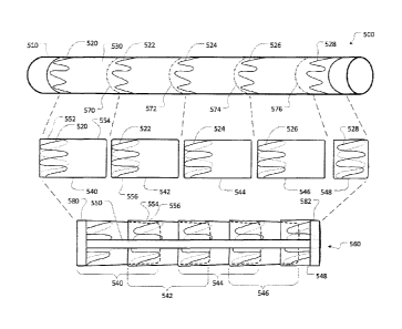
[0098] With reference to Figure 5, an exemplary process 500 for fabricating an
intralumenal stent graft device 560 is schematically illustrated. The
progressive
steps of process 500 are illustrated generally, beginning with the view of the
top of
the sheet, continuing with the view in the middle, and ending with the
finished stent
graft 560 at the bottom of the sheet. Process 500 is provided as an exemplary
36
CA 02956502 2017-01-27
process for fabricating an intralumenal stent graft device that has multiple
discrete
tubular segments such as stent graft embodiments 10 and 100, described above
in
reference to Figures 1A and 1B. However, other processes, sub-processes, and
techniques for fabricating an intralumenal stent graft device with multiple
discrete
tubular segments are also envisioned within the scope of this document.
Process
500 will be described as fabricating a stent graft device 560 from certain
exemplary
types of materials. However, the use of other types of materials to fabricate
stent
graft devices with multiple discrete tubular segments is also envisioned
within the
scope of this document. Although an intralumenal stent graft device with five
(5)
segments is used to illustrate process 500, a stent graft device with
virtually any
number of tubular segments can be fabricated using process 500.
[0099] As shown in the view at the top of Figure 5, a membrane 530 with a
plurality
of attached stent members 520, 522, 524, 526, and 528 is formed to surround a
cylindrical mandrel 510. The mandrel 510 is used as a form from which to build
up a
stent graft 560. The mandrel 510 can be comprised of any suitable mandrel
material, e.g., stainless steel, tool steel, or aluminum. The diameter of
mandrel 510
substantially determines the inner diameter of the stent graft 560. As such,
an
appropriately sized mandrel 510 should be selected in accordance with the size
of
the stent graft desired. For example, a smaller diameter mandrel should be
used to
form a small stent graft for a pancreatic duct implementation, as compared to
a
larger diameter mandrel for forming a larger stent graft for an aortic arch
implementation. The length of mandrel 510 will be at least as long as the
desired
37
CA 02956502 2017-01-27
length of the stent graft to be fabricated, and the mandrel 510 may be
substantially
longer than the stent graft to be fabricated.
[0100] In some embodiments of process 500, a cushion tube (not shown) is
included
as a liner over the mandrel 510 surface. The cushion tube can be a suitable
compressible material, e.g., an ePTFE tube or tape wrap. In some embodiments,
a
thin, heat resistant, non-stick liner made from a material such as a Kapton
is
wrapped over the cushion tube.
[0101] A base layer of membrane 530 is wrapped around mandrel 510 over the
cushion tube and non-stick liner. In some embodiments, a film-like, ePTFE
membrane material is used. Other suitable materials, such as woven or knitted
polyester, and the like, can also be used. In some embodiments, the ePTFE
membrane 530 has a surface layer of fluorinated ethylene propylene (FEP)
material
on one side of the ePTFE membrane 530. The side of the membrane 530 with the
FEP layer is oriented outward, i.e., away from the mandrel 510. FEP is a heat
activated adhesive that, as described further below, can be used to bond
layers of
membrane. In some embodiments, the ePTFE membrane does not include a FEP
layer. In such cases, a separate FEP film can be wrapped onto the ePTFE
membrane.
[0102] In some embodiments, a second layer of ePTFE membrane 530 is wrapped
onto the ePTFE and FEP already on the mandrel 510. In some embodiments, the
second layer of ePTFE membrane 530 is a spiral wrap with about a fifty percent
(50%) overlap. The second layer of ePTFE membrane 530 can also have a FEP
layer on one side of the membrane 530. The side with the FEP layer should be
38
CA 02956502 2017-01-27
oriented down onto the first layer of membrane 530, i.e., no FEP should be
exposed
in the area of the channel flaps after the addition of the second layer of
ePTFE
membrane 530. In some embodiments, the first two (2) layers of ePTFE membrane
530 make up the base membrane 530. In some embodiments, other constructions
can make up the base membrane. For example, in some embodiments, more than
two (2) layers of ePTFE membrane are included. In some embodiments, only one
(1) layer of ePTFE membrane is included.
[0103] Stent members 520, 522, 524, 526, and 528 are added on top of the
layers of
membrane 530. In this embodiment, ring-like annular stent members are used. In
some embodiments, stent members are wrapped around the membrane in another
configuration, such as helically as described below in reference to Figure 6.
The
annular stent members 520, 522, 524, 526, and 528 are to be placed on the
mandrel
510 at locations in relation to the membrane 530 such that the desired axial
lengths
of the unsupported membrane (the flap length) will be created.
[0104] In some embodiments, a layer of ePTFE with FEP (oriented downward) is
added over the stent members 520, 522, 524, 526, and 528. In some embodiments,
this additional ePTFE is only wrapped over the individual stent members 520,
522,
524, 526, and 528, and is not wrapped over the entire length of the membrane
530.
That is, each discrete stent member 520, 522, 524, 526, and 528 can be wrapped
individually by a strand of ePTFE with FEP. The strands of ePTFE with FEP can
be
a little wider than the individual stent members 520, 522, 524, 526, and 528,
so that
the stent members 520, 522, 524, 526, and 528 will be fully laminated within
the
39
CA 02956502 2017-01-27
membrane material. In some embodiments, the additional ePTFE is wrapped over
the entire length of the membrane 530.
[0105] A hot iron or other heat source is applied to all areas of the strands
of ePTFE
with FEP that cover the stent members 520, 522, 524, 526, and 528. The hot
iron
can be used to trace around the stent members 520, 522, 524, 526, and 528. The
hot iron, with a temperature of about 670-720 F, for example, will activate
the FEP
and cause the strands of ePTFE to bond to the stent members 520, 522, 524,
526,
and 528 and to the base membrane 530. The use of the hot iron causes the stent
members 520, 522, 524, 526, and 528 to become firmly laminated between the
strands of ePTFE and the base membrane 530, such that substantially all
portions of
the stent members 520, 522, 524, 526, and 528 are covered by ePTFE material.
[0106] In some embodiments, the mandrel 510, membrane 530, and stent members
520, 522, 524, 526, and 528 are then heated in an oven to activate the FEP
adhesive, e.g., the FEP between the first two layers of membrane 530. Any
suitable
time and temperature profile can be used. For example, in some embodiments of
process 500, the heating takes place at about 320 C for about twelve (12)
minutes.
[0107] After heating, and subsequent cooling, the non-stick liner can be
removed
from the mandrel 510. The membrane 530 with the stent members 520, 522, 524,
526, and 528 can also be removed from the mandrel 510.
[0108] In some embodiments, the membrane 530 is circumferentially cut at lines
570, 572, 574, and 576 to create discrete cylindrical segments 540, 542, 544,
546,
and 548. The cutting is performed so as to create discrete cylindrical
segments 540,
542, 544, and 546 with stent members 520, 522, 524, and 526 that are
CA 02956502 2017-01-27
asymmetrically located on the discrete cylindrical segments 540, 542, 544, and
546
(see middle view of Figure 5). In this example, the end segment 548 is unique,
and
its stent member 528 may be located in a suitable location that is different
than the
other discrete cylindrical segments 540, 542, 544, and 546. The asymmetrical
location of the stent members 520, 522, 524, and 526 causes the discrete
cylindrical
segments 540, 542, 544, and 546 to each have a supported edge portion and an
unsupported edge portion (a flap or tail), as described above in reference to
stent
graft embodiments 10 and 100.
[0109] Segment 540 can be used to illustrate the previous point. Segment 540
includes a supported edge portion 552 and an unsupported edge portion 554. The
supported edge portion 552 is supported by stent member 520, whereas the
unsupported edge portion 554 has no such supplemental support from a stent
member. Instead, unsupported edge portion 554 is comprised of tubular membrane
530 without supplemental support from a stent member.
[0110] The discrete cylindrical segments 540, 542, 544, 546, and 548 are then
placed again on mandrel 510 (or on a different mandrel), in some examples with
a
cushion tube and non-stick liner, and configured in relation to each other
(nested
together) as desired. That is, the tails of cylindrical segments are placed
interior of,
or exterior of, the supported edge of an adjacent cylindrical segment. As
shown in
the bottom view of Figure 5, in some embodiments, the tails are placed
interior of the
supported edge portion of an adjacent cylindrical segment. For example, the
tail 554
of cylindrical segment 540 is located within the supported edge portion 556 of
the
adjacent cylindrical segment 542. In some embodiments, the tails are placed
over
41
CA 02956502 2017-01-27
the exterior of the supported edge portion of an adjacent cylindrical segment
(see,
e.g., stent graft 100 of Figure 1B). In some embodiments, a combination of
interior
and exterior placements of the tails in relation to the supported edges of the
adjacent
cylindrical segments can be created. The configuration of the tails in
relation to the
adjacent cylindrical segment can effect whether that portion of the stent
graft device
is configured for inward radial flow or outward radial flow.
[0111] One or more axial reinforcement members 550 are attached to the nested
cylindrical segments 540, 542, 544, 546, and 548. In some embodiments, the
axial
reinforcement members 550 are strips of ePTFE that have a FEP layer on one
side.
In such embodiments, the strips of ePTFE with a FEP layer are attached to the
cylindrical segments 540, 542, 544, 546, and 548 by applying a hot iron on the
surface of the ePTFE strip. The heat from the hot iron will activate the FEP
to cause
the ePTFE strip to adhere to the cylindrical segments 540, 542, 544, 546, and
548.
The axial reinforcement members 550 can be of any suitable width. In some
embodiments, the axial reinforcement members 550 are about 1/4" wide. Any
suitable number of axial reinforcement members 550 can be used. In some
embodiments, one (1), two (2), three (3), or more than three (3) axial
reinforcement
members 550 are used.
[0112] In some embodiments, one or both of the ends of stent graft 560 are
reinforced by the addition of circumferential end reinforcement members 580
and
582, for example. In some embodiments, the end reinforcement members 580 and
582 are strips of ePTFE that have a FEP layer on one side. In such
embodiments,
end reinforcement members 580 and 582 are attached to the end cylindrical
42
CA 02956502 2017-01-27
segments 540 and 548 by applying a hot iron on the surface of the ePTFE strip.
The
heat from the hot iron, for example at a temperature of about 670-720 F, will
activate
the FEP to cause the ePTFE strip to adhere to the cylindrical segments 540 and
548. The end reinforcement members 580 and 582 can be of any suitable width.
In
some embodiments, the end reinforcement members 580 and 582 are about IA"
wide. In some embodiments the end reinforcement members 580 and 582 are
wrapped about a single circumference around cylindrical segments 540 and 548.
In
some embodiments, two (2) or more wraps of end reinforcement members 580 and
582 are made around cylindrical segments 540 and 548.
[0113] The stent graft 560 on the mandrel 510 can then be heated in an oven to
ensure all FEP adhesive has been activated. Any suitable time and temperature
profile can be used. For example, in some embodiments of process 500, the
heating can take place at about 320 C for about twelve (12) minutes.
[0114] The non-stick liner and the stent graft 560 can then be removed from
the
mandrel 510. The flow channels between the tails and the supported edges can
be
checked to ensure that the channels are operable to be opened as desired. If
any
flow channels are adhered together they can be gently separated using an
appropriate tool, e.g., one of the tips of a pair of tweezers.
[0115] With reference to Figure 6, an exemplary process 600 for fabricating an
intralumenal stent graft device 660 is schematically illustrated. The
progressive
steps of process 600 are illustrated generally, beginning with the view of the
top of
the sheet, continuing with the view in the middle, and concluding with the
finished
stent graft 660 at the bottom of the sheet. Process 600 is provided as an
example
43
CA 02956502 2017-01-27
process for fabricating an intralumenal stent with a helically arranged
membranous
strip and a helically arranged support member attached to the helically
arranged
membranous strip, such as, for example, stent graft embodiments 200 and 270 as
described above in reference to Figures 2A and 2B. The helically arranged
membranous strip and the helically arranged support member are configured to
comprise a plurality of turns or winds. However, other processes, sub-
processes,
and techniques for fabricating an intralumenal stent comprising a helically
arranged
membranous strip are also envisioned within the scope of this document.
Process
600 will be described as fabricating a stent graft device 660 from certain
exemplary
types of materials. However, the use of other types of materials to fabricate
stent
graft devices with a helically arranged membranous strip is also envisioned
within
the scope of this document. Although an intralumenal stent graft device with
five (5)
turns (or winds) is used to describe process 600, a stent graft device with
virtually
any number of turns can be fabricated using process 600.
[0116] As shown in the view at the top of Figure 6, a membrane 630 with a
helically
arranged support member 620 is formed to surround a cylindrical mandrel 610.
The
mandrel 610 is used as a form from which to build up a stent graft 660. The
mandrel
610 can be comprised of any suitable mandrel material, e.g., stainless steel,
tool
steel or aluminum. For process 600, the diameter of mandrel 610 is oversized
in
comparison to the desired final inner diameter of the stent graft 660. For
example,
to fabricate a stent graft 660 with a final inner diameter of about ten (10)
millimeters,
a mandrel 610 with a diameter of about thirteen (13) millimeters can be used.
As
such, an appropriately oversized mandrel 610 should be selected in accordance
with
44
CA 02956502 2017-01-27
the final inner diameter of the stent graft 660 desired. The length of mandrel
610 will
be longer than the desired length of the stent graft to be fabricated, and the
mandrel
610 may be substantially longer than the stent graft to be fabricated.
[0117] In some embodiments of process 600, a cushion tube (not shown) is
included
as a liner over the mandrel 610 surface. The cushion tube can be a suitable
compressible material, e.g., an ePTFE tube or tape wrap. In some embodiments,
a
thin, heat resistant, non-stick liner made from a material such as a Kapton
is
wrapped over the cushion tube.
[0118] A base layer of membrane 630 is wrapped around mandrel 610 over the
cushion tube and non-stick liner. In some embodiments, a film-like, ePTFE
membrane material is used. Other suitable materials, such as woven or knitted
polyester, and the like, can also be used. In some embodiments, the ePTFE
membrane 630 has a surface layer of fluorinated ethylene propylene (FEP)
material
on one side of the ePTFE membrane. The side of the membrane 630 with the FEP
layer is oriented outward, i.e., away from the mandrel 610. The FEP is a heat
activated adhesive that, as described further below, can be used to bond
layers of
membrane, In some embodiments, the ePTFE membrane does not include a FEP
layer. In such cases, a separate FEP film can be wrapped onto the ePTFE
membrane.
[0119] In some embodiments, a second layer of ePTFE membrane 630 is wrapped
onto the ePTFE and FEP already on the mandrel 610. In some embodiments, the
second layer of ePTFE membrane 630 is spiral wrap with about a fifty percent
(50%)
overlap. The second layer of ePTFE membrane 630 can also have a FEP layer on
CA 02956502 2017-01-27
one side of the membrane 630. The side with the FEP layer should be oriented
down onto the first layer of membrane 630, i.e,, no FEP should be exposed in
the
area of the channel flaps after the addition of the second layer of ePTFE
membrane
630. In some embodiments, the first two (2) layers of ePTFE membrane 630 make
up the base membrane 630. In some embodiments, other constructions can make
up the base membrane. For example, in some embodiments, more than two (2)
layers of ePTFE membrane are included. In some embodiments, only one (1) layer
of ePTFE membrane is included.
[0120] Stent member 620 is added on top of the layers of membrane 630. In this
exemplary embodiment, a single helically arranged stent member is used. The
stent
member 620 is helically wound on the mandrel 610 with a spacing between turns
of
stent members 620 that is greater than the desired spacing between the turns
of
stent members 620 in the final stent graft 660. For example, in some
embodiments,
a spacing of about ten (10) millimeters between the turns of stent members 620
is
made on the mandrel 610, and a spacing of about two (2) millimeters between
the
turns of stent members 620 is made in the final product.
[0121] In some embodiments, a layer of ePTFE with FEP (oriented downward) is
added over the stent member 620. In some embodiments, this additional ePTFE is
only helically wrapped over the stent member 620, and is not wrapped over the
entire length of the membrane 630. The strand of ePTFE with FEP may be a
little
wider than the stent member 620 so that the stent member 620 will be fully
laminated within the membrane material. In some embodiments, the additional
ePTFE is wrapped over the entire length of the membrane 630.
46
CA 02956502 2017-01-27
[0122] A hot iron or other heat source is applied to all areas of the strand
of ePTFE
with FEP that covers the stent members 620. The hot iron can be used to trace
around the stent member 620. The hot iron, with a temperature of about 670-720
F,
for example, will activate the FEP and cause the strand of ePTFE to bond to
the
stent member 620 and to the base membrane 630. The use of the hot iron causes
the stent member 620 to become firmly laminated between the strand of ePTFE
and
the base membrane 630, such that substantially all portions of the stent
member 620
are covered by ePTFE material.
[0123] In some embodiments, the mandrel 610, membrane 630, and stent member
620 are then heated in an oven to activate the FEP adhesive, e.g., the FEP
between
the first two layers of membrane 630. Any suitable time and temperature
profile can
be used. For example, in some embodiments of process 600, the heating takes
place at about 320 C for about twelve (12) minutes.
[0124] After heating, and subsequent cooling, the non-stick liner can be
removed
from the mandrel 610. The membrane 630 with the stent member 620 can also be
removed from the mandrel 610.
[0125] In some embodiments, the membrane 630 is cut in a helical pattern along
line
670. The cutting is performed so as to create a helical strip of membrane 630
with
stent member 620 asymmetrically located on the helical strip of membrane 630
(see
middle view of Figure 6), The asymmetrical location of the stent member 620
will
cause the final configuration of stent graft 660 to have a supported edge and
an
unsupported edge at each turn, as described above in reference to stent graft
embodiments 200 and 270. That is, the helical strip of membrane 630 has
47
CA 02956502 2017-01-27
lengthwise side regions (or margins), and one of the side regions is supported
by
stent member 620 while the other side region is unsupported.
[0126] The helical strip of membrane 630 with stent member 620 is then placed
on
an undersized mandrel, in some cases with a cushion tube and non-stick liner.
For
example, for a stent graft with about a ten (10) millimeter final inner
diameter, a
mandrel with about an eight (8) millimeter diameter can be used.
[0127] The turns of the helical strip of membrane 630 are then configured in
relation
to each other (nested together) as desired. That is, the unsupported side
region
(tails) of the turns are placed interior of, or the exterior of, the supported
side region
of adjacent turns. As shown in the bottom view of Figure 6, in some
embodiments,
the tails are placed interior of the supported side region of an adjacent
cylindrical
segment. In some embodiments, the tails are placed over the exterior of the
supported side region of an adjacent cylindrical segment (see, e.g., stent
graft 270 of
Figure 2B). The configuration of the tails in relation to the adjacent
cylindrical
segment can effect whether that portion of the stent graft device is
configured for
inward radial flow or outward radial flow.
[0128] In some embodiments, one or more axial reinforcement members 650 are
attached to the helical strip of membrane 630 with stent member 620. In some
embodiments, the axial reinforcement members 650 are strips of ePTFE that have
a
FEP layer on one side. In such embodiments, the strips of ePTFE with a FEP
layer
are attached to the turns of the helical strip of membrane 630 with stent
member 620
by applying a hot iron on the surface of the ePTFE strip. The heat from the
hot iron
will activate the FEP to cause the ePTFE strip to adhere to the helical strip
of
48
CA 02956502 2017-01-27
membrane 630 with stent member 620. The axial reinforcement members 650 can
be of any suitable width. In some embodiments, the axial reinforcement members
650 are about 1/4" wide. Any suitable number of axial reinforcement members
650
can be used. In some embodiments, one (1), two (2), three (3), or more than
three
(3) axial reinforcement members 650 are used.
[0129] In some embodiments, one or both of the ends of stent graft 660 are
reinforced by the addition of circumferential end reinforcement members 680
and
682, for example. In some embodiments, the end reinforcement members 680 and
682 are strips of ePTFE that have a FEP layer on one side. In such
embodiments,
end reinforcement members 680 and 682 are attached to the ends of the helical
strip
of membrane 630 with stent member 620 by applying a hot iron on the surface of
the
ePTFE strip. The heat from the hot iron, for example at a temperature of about
670-
720 F, will activate the FEP to cause the ePTFE strip to adhere to the
membrane
630. The end reinforcement members 680 and 682 can be of any suitable width.
In
some embodiments, the end reinforcement members 680 and 682 are about 1/4"
wide. In some embodiments the end reinforcement members 680 and 682 are
wrapped about a single circumference around membrane 630. In some
embodiments, two (2) or more wraps of end reinforcement members 680 and 682
are made around the membrane 630.
[0130] The stent graft 660 on the mandrel can then be heated in an oven to
ensure
all FEP adhesive has been activated. Any suitable time and temperature profile
can
be used. For example, in some embodiments of process 600, the heating can take
place at about 320 C for about twelve (12) minutes.
49
CA 02956502 2017-01-27
[0131] The non-stick liner and the stent graft 660 can then be removed from
the
mandrel. The flow channels between the tails and the supported edges can be
checked to ensure that the channels are operable to be opened as desired. If
any
flow channels are adhered together they can be gently separated using an
appropriate tool, e.g., one of the tips of a pair of tweezers.
[0132] Figure 7 is a flowchart of an exemplary process 700 for fabricating a
stent
graft device with discrete cylindrical segments arranged in a nested
configuration as
provided herein. For example, process 700 can be used to fabricate stent graft
embodiments 10 and 100 of Figures 1A and 1B. Process 700 also corresponds to
some embodiments of the process depicted in Figure 5, for example.
[0133] At operation 710, membranous material is arranged on a mandrel. The
mandrel can be sized corresponding to an inner diameter of the stent graft to
be
fabricated. As described above, in some embodiments ePTFE is used for the
membranous material. In some embodiments, a FEP layer is included on one
surface of the ePTFE. In some embodiments, two (2) or more layers of film
material
comprise the membranous material as a laminate. In some embodiments, woven or
knitted membranes are used.
[0134] At operation 720, a plurality of individual ring-like annular support
members
are arranged over the membranous material. In some embodiments, the individual
ring-like annular support members are stent members. In some embodiments, the
stent members are formed wires or laser cut lattice rings. The stent members
are
placed over the membranous material in locations that will result in the
desired
asymmetrical stent placement configuration as described above in reference to
CA 02956502 2017-01-27
Figures 1A and 1B. Strips of membrane material can be placed over the support
members and laminated to the membranous material so as to attach and laminate
the stent members onto the membranous material. In some embodiments a hot iron
can be used to adhere the strips of membrane material to the membranous
material
to thereby laminate the support members with membranous material.
[0135] In some embodiments, the mandrel with the partially completed stent
graft
device is then heated in an oven to activate the FEP. The activation of FEP
bonds
the layers of membranous material together.
[0136] At operation 730, after removing the partially completed stent graft
from the
mandrel, the base membrane can be cut to produce a plurality of cylindrical
segments. The cuts are made in locations on the base membrane near the edges
of
stent members. The locations of the stent members are thereby located axially
asymmetrical on the segments. That is, one edge of the cylindrical segments
has
support from a stent member but the other edge does not (it is the tail
portion).
[0137] At operation 740 the plurality of cylindrical segments are again placed
on the
mandrel, or another mandrel, and arranged in a nested configuration in
accordance
with the type of stent graft device desired, such as a radial inflow stent
graft device
or a radial outflow stent graft device. If a radial inflow stent graft is
desired, the tails
of the cylindrical segments are placed interior of (i.e., closer to the
mandrel) the
supported edges of the adjacent cylindrical segments. If a radial outflow
stent graft
is desired, the tails of the cylindrical segments are placed exterior of
(i.e., further
from the mandrel) the supported edges of the adjacent cylindrical segments.
51
CA 02956502 2017-01-27
[0138] At operation 750, reinforcing members are applied to the cylindrical
segments
that are arranged in the nested configuration. One or more axial reinforcement
members can be applied. In some embodiments, end reinforcement members can
also be applied to one or both ends of the stent graft device. In some
embodiments,
the reinforcement members are strips of ePTFE membrane with a FEP layer. In
some embodiments, the strips are about 1/4" wide. The reinforcement members
may
be of any suitable width.
[0139] In some embodiments, the mandrel with the completed stent graft device
is
once again heated in an oven to activate the FEP. The activation of FEP bonds
the
layers of membranous material together to create a completed stent graft
device.
[0140] Figure 8 is a flowchart of an example process 800 for fabricating a
stent graft
device with a helically arranged membrane, wherein the turns of the helix
overlap to
create a nested configuration. For example, process 800 can be used to
fabricate
stent graft embodiments 200 and 270 of Figures 2A and 2B. Process 800 also
corresponds to some embodiments of the process depicted in Figure 6, for
example.
[0141] At operation 810, membranous material is arranged on a mandrel. In some
embodiments, the mandrel is over-sized for the inner diameter of the stent
graft to
be fabricated. For example, to fabricate a stent graft with a final inner
diameter of
about ten (10) millimeters, a mandrel with a diameter of about thirteen (13)
millimeters can be selected. As described above, in some embodiments ePTFE is
used for the membranous material. In some embodiments, a FEP layer is included
on one surface of the ePTFE. In some embodiments, two (2) or more layers of
film
52
CA 02956502 2017-01-27
material can comprise the membranous material as a laminate. In some
embodiments, woven or knitted membranes are used.
[0142] At operation 820, a single continuous support member is helically
arranged
over the membranous material. In some embodiments, the helically arranged
support member is a stent member. In some embodiments, the stent member is
made of a formed wire or a laser cut lattice strip. The stent member is placed
over
the membranous material in a location that will result in the desired
asymmetrical
stent placement configuration, as described above in reference to Figures 2A
and
2B. A strip of membrane material can be placed over the support member and
laminated to the base membrane, so as to attach and laminate the stent member
within the membranous material. In some embodiments a hot iron can be used to
adhere the strip of membranous material to the base material to thereby
laminate
the support member within membranous material.
[0143] In some embodiments, the mandrel with the partially completed stent
graft
device is then heated in an oven to activate the FER The activation of FEP
bonds
the layers of membrane material together.
[0144] At operation 830, after removing the partially completed stent graft
from the
mandrel, the base membrane can be cut to produce a helical strip of membranous
material with an asymmetrically located support member. The helical cut is
made on
the base membrane near the edges of the stent member. The stent member is
thereby located asymmetrically on the helical strip of membranous material.
[0145] At operation 840, in some embodiments, the plurality of cylindrical
segments
are placed on an undersized mandrel. For example, for a stent graft with about
a
53
CA 02956502 2017-01-27
ten (10) millimeter final inner diameter, a mandrel with about an eight (8)
millimeter
diameter can be used. The turns of the helical strip of membranous material
are
then arranged in a nested configuration in accordance with the type of stent
graft
device desired, such as a radial inflow stent graft device or a radial outflow
stent
graft device. If a radial inflow stent graft device is desired, the tails of
the turns are
placed interior of (i.e., closer to the mandrel) the supported edge of the
adjacent
turn. If a radial outflow stent graft is desired, the tails of the turns are
placed exterior
of (i.e., further from the mandrel) the supported edge of the adjacent turn.
[0146] At operation 850, reinforcing members are applied to the cylindrical
segments
that are arranged in the nested configuration. One or more axial reinforcement
members can be applied. In some embodiments, end reinforcement members can
be applied to one or both ends of the stent graft device. In some embodiments,
the
reinforcement members are strips of ePTFE membrane with a FEP layer. In some
embodiments, the strips are about 1/4" wide. The reinforcement members may be
of
any suitable width.
[0147] In some embodiments, the mandrel with the completed stent graft device
is
once again heated in an oven to activate the FEP. The activation of FEP bonds
the
layers of membrane material together to create a completed stent graft device.
[0148] While this specification contains many specific implementation details,
these
should not be construed as limitations on the scope of any devices, methods,
and
systems discussed herein, but rather as descriptions of features that may be
specific
to particular embodiments. Certain features that are described in this
specification in
the context of separate embodiments can also' be implemented in combination in
a
54
CA 02956502 2017-01-27
single embodiment. Conversely, various features that are described in the
context
of a single embodiment can also be implemented in multiple embodiments
separately or in any suitable subcombination. Moreover, although features may
be
described above as acting in certain combinations and even initially claimed
as
such, one or more features from a claimed combination can in some cases be
excised from the combination, and the claimed combination may be directed to a
subcombination or variation of a subcombination.
[0149] Particular embodiments of the subject matter have been described. Other
embodiments are within the scope of the following claims.