Note: Descriptions are shown in the official language in which they were submitted.
VASCULAR DEVICE MARKER ATTACHMENT
BACKGROUND OF THE DISCLOSURE
[00011 Blood vessels can become occluded by emboli, e.g., thrombi. For
example,
intracranial arteries can become occluded by thromboembolisms. Disruption of
blood flow by
the occlusion can prevent oxygen and nutrients from being delivered to tissues
downstream
of the occlusion. Deprivation of oxygen and nutrients to tissue distal to an
occlusion can
impair proper function of the tissue, and may result in cellular death.
Cellular death
increases with duration of the occlusion.
SUMMARY OF THE DISCLOSURE
[0002] Markers can be used to assist an operator in determining the
location and/or
orientation of a medical device within a blood vessel. Some aspects of the
subject
technology relate to attachment of a marker to a thrombectomy or other medical
device. Some
aspects of the subject technology relate to the positioning of one or more
markers on a
thrombectomy device. Some aspects of the subject technology relate to the use
of markers in
methods for removing thrombus from a blood vessel.
[0003] The subject technology is illustrated, for example, according to
various
aspects described below. Various examples of aspects of the subject technology
are described
as numbered clauses (1, 2, 3, etc.) for convenience. These are provided as
examples and do
not limit the subject technology. It is noted that any of the dependent
clauses may be
combined in any combination, and placed into a respective independent clause,
e.g., clause
1, 12, 16, or 24. The other clauses can be presented in a similar manner.
1. A medical device comprising:
an elongate manipulation member; and
a thrombectomy device connected to the elongate manipulation
member, the thrombectomy device having a first configuration and a second
configuration, the thrombectomy device being expandable from the first
configuration to the second configuration, wherein the thrombectomy device
comprises a plurality of arcuate marker-mounting projections each attached to
a portion of the thrombectomy device configured to contact a thrombus
- 1 -
CA 2957130 2018-08-13
and arranged such that any laterally aligned arcuate marker-mounting
projections are disposed laterally farther from each other when the
thrombectomy device is in the second configuration than they are when the
thrombectomy device is in the first configuration; and
a plurality of markers, each marker being attached to one of the arcuate
marker-mounting projections.
2. The medical device of clause 1, wherein each of the arcuate marker-
mounting projections comprises a concave surface.
3. The medical device of clause 2, wherein the concave surface faces away
from
the portion of the thrombectorny device configured to contact the thrombus to
which the arcuate marker-mounting projection is attached.
4. The medical device of clause 2, wherein each of the arcuate marker-
mounting projections comprises a convex surface opposite the concave surface.
5. The medical device of clause 4, wherein the convex surface is parallel
to
the concave surface.
6. The medical device of clause 1, wherein the thrombeetomy device
comprises a plurality of struts forming a plurality of cells, and each of the
arcuate
marker-mounting projections extends from one of the struts.
7. The medical device of clause 6, wherein each of the arcuate marker-
mounting projections is separated from all of the other arcuate marker-
mounting
projections by at least one strut length.
The medical device of clause 1, wherein at least one of the marker-
mounting projections is disposed at a proximal end of a working length of the
thrombectomy device.
9. The medical device of clause 8, wherein the least one of the marker-
mounting projection is within 5 millimeters of the proximal end of the working
length.
10. The medical device of clause 1, wherein a group of marker-mounting
projections is disposed at a proximal end of a working length of the
thrombectomy
device.
- 2 -
CA 2957130 2018-08-13
11. The medical device of clauses 1 to 10, wherein the arcuate marker-
mounting
projections are cantilevered from the portion of the thrombectomy device to
which
they are attached.
12. A medical device comprising:
an elongate manipulation member;
a thrombectomy device connected to the elongate manipulation
member; an arcuate marker-mounting projection extending from a
portion of the
thrombectomy device configured to contact a thrombus; and
a marker coupled to, and extending around, the arcuate marker-
mounting projection with the marker and the arcuate marker-motmting
projection contacting each other at three discrete locations.
13. The medical device of clause 12, wherein the marker and the marker-
mounting projection contact each other at one location on a convex side of the
arcuate marker- mounting projection, and the marker and the arcuate marker-
mounting
projection contact each other at two locations on a concave side of the marker-
mounting projection.
14. The medical device of clause 13, wherein the one contact location on
the
convex side is between the two contact locations on the concave side.
15. The medical device of clause 13, wherein the convex side of the arcuate
marker- mounting projection and the concave side of the marker-mounting
projection
are parallel.
16. A method for engaging a thrombus, the method comprising:
(a) advancing a thrombectomy device, using an 'elongate manipulation
member, to a location radially adjacent to a thrombus in a blood vessel,
the thrombectomy device comprising a working length and a non-working
length, the non-working length disposed between and separating the working
length and a connection between the thrombectomy device and the elongate
manipulation member, the working length having a proximal end and a distal
end with a proximal marker disposed at the proximal end, and a distal marker,
discrete from the proximal marker, disposed at the distal end;
- 3 -
CA 2957130 2018-08-13
(b) positioning the thrombectomy device relative to the thrombus such
that the proximal marker is proximal to or longitudinally aligned with a
proximal end of the thrombus and the distal marker is distal to or
longitudinally
aligned with a distal end of the thrombus; and
(c), after (b), expanding the thrombectomy device into the thrombus.
17. The
method of clause 16, wherein a plurality of proximal markers are disposed
at the proximal end of the working length and a plurality of distal markers
are
disposed at
- 3a -
CA 2957130 2018-08-13
CA 02957130 2017-02-06
the distal end of the working length, and wherein positioning the thrombectomy
device
relative to the thrombus comprises positioning all of the proximal markers
proximal to or
longitudinally aligned with a proximal end of the thrombus and all of the
distal markers
distal to or longitudinally aligned with a distal end of the thrombus.
18. The method of clause 17, wherein a plurality of intermediate markers is
attached
to the thrombectomy device between the plurality of proximal markers and the
plurality
of distal markers, the method further comprising determining whether a maximum
marker separation of the plurality of intermediate markers is less than a
maximum marker
separation of either the plurality of proximal markers or the plurality of
distal markers.
19. The method of clause 18 wherein, when the maximum marker separation of
the
plurality of intermediate markers is not less than the maximum marker
separation of
either the plurality of proximal markers or the plurality of distal markers,
the method
further comprises:
collapsing the thrombectomy device;
repositioning the thrombectomy device relative to the thrombus such that the
proximal marker is proximal to or longitudinally aligned with a proximal end
of the
thrombus and the distal marker is distal to or longitudinally aligned with a
distal end of
the thrombus; and
re-expanding the thrombectomy device into the thrombus.
20. The method of clause 18 further comprising determining a state of
expansion of
the working length by observing the proximal, intermediate and distal
pluralities of
markers.
21. The method of clause 16, wherein the proximal marker is located within
5
millimeters of the proximal end of the working length.
22. The method of clause 21, wherein the thrombectomy device includes a
plurality
of cells, and wherein the proximal marker grouping is located within one cell-
length of
the proximal end of the working length.
23. The method of clause 21, wherein the thrombectomy device includes a
generally
cylindrical structure having a roll-up configuration.
-4-
CA 02957130 2017-02-06
24. A method for engaging a thrombus, the method comprising:
(a) advancing a thrombectomy device, using an elongate manipulation
member, to a location radially adjacent to a thrombus in a blood vessel, the
thrombectomy device comprising a working length having a proximal end and a
distal end with a proximal marker disposed at the proximal end;
(b) positioning the thrombectomy device relative to the thrombus such that
the proximal marker is proximal to or longitudinally aligned with a proximal
end
of the thrombus; and
(c), after (b), expanding the thrombectomy device into the thrombus.
25. The method of clause 24, wherein the thrombectomy device further
comprises a
non-working length, the non-working length disposed between and separating the
working length and a connection between the thrombectomy device and the
elongate
manipulation member, the proximal marker being located distal of the
connection.
26. The method of clause 24, further comprising imaging the proximal end of
the
working length distinctly from the connection with the proximal marker.
27. The method of clause 24, wherein the thrombectomy device further
comprises a
distal marker, discrete from the proximal marker, disposed at the distal end
of the
working length.
28. The method of clause 27, further comprising positioning the
thrombectomy
device such that the distal marker is distal to or longitudinally aligned with
a distal end of
the thrombus.
29. The method of clause 27, wherein the thrombectomy device has a body
comprising a plurality of struts, and the distal and proximal markers are more
radiopaque
than the body.
30. The method of clause 24, wherein a plurality of proximal markers are
disposed at
the proximal end of the working length, and wherein positioning the
thrombectomy
device relative to the thrombus comprises positioning all of the proximal
markers
proximal to or longitudinally aligned with a proximal end of the thrombus.
31. The method of clause 30, wherein a plurality of intermediate markers is
attached
to the thrombectomy device distal of the plurality of proximal markers, the
method
-5-
further comprising determining whether a maximum marker separation of the
plurality
of intermediate markers is less than a maximum marker separation of the
plurality of
proximal markers.
32. The method of clause 31, wherein, when the maximum marker
separation of
the plurality of intermediate markers is not less than the maximum marker
separation of
the plurality of proximal markers, the method further comprises:
collapsing the thrombectomy device;
repositioning the thrombectomy device relative to the thrombus such that
the proximal marker is proximal to or longitudinally aligned with a proximal
end
of the thrombus; and
re-expanding the thrombectomy device into the thrombus.
[0003a] According to another aspect, there is provided a medical device
comprising: an
elongate manipulation member; and a thrombectomy device coupled to the
elongate manipulation
member, the thrombectomy device extending between a first end and a second end
and expandable
from a first configuration to a second configuration, the thrombectomy device
comprising (i) a
plurality of arcuate marker-mounting projections, each attached to a portion
of the thrombectomy
device configured to contact a thrombus, (ii) a plurality of struts forming a
plurality of cells, each
of the projections extending from one of the struts disposed between the first
and second ends of
the thrombectomy device, and (iii) a plurality of markers, each marker being
attached to one of the
arcuate marker-mounting projections, wherein at least some of the marker-
mounting projections
are laterally aligned in the absence of external forces on the thrombectomy
device, and wherein at
least some of the laterally aligned marker-mounting projections are arranged
such that they are
disposed laterally farther from each other when the thrombectomy device is in
the second
configuration than they are when the thrombectomy device is in the first
configuration.
10003b] According to another aspect, there is provided a medical device
comprising: an
elongate manipulation member; a thrombectomy device connected to the elongate
manipulation
member, the thrombectomy device having a plurality of struts forming a
plurality of cells; an
arcuate marker-mounting projection extending from one of the struts disposed
along a portion of
the thrombectomy device configured to contact a thrombus; and a marker coupled
to and extending
- 6 -
Date recue/Date Received 2020-08-28
around the projection, and wherein the marker and the projection contact each
other at three
discrete locations.
[0003c] According to another aspect, there is provided a medical device
comprising: an
elongate manipulation member; a thrombectomy device connected to the elongate
manipulation
member, the thrombectomy device having a first configuration and a second
configuration, the
thrombectomy device being expandable from the first configuration to the
second configuration,
the thrombectomy device comprising a plurality of arcuate marker-mounting
projections each
attached to a portion of the thrombectomy device configured to contact a
thrombus, wherein at
least some of the marker-mounting projections are laterally aligned in the
absence of external
forces on the thrombectomy device, and wherein at least some of the laterally
aligned marker-
mounting projections are arranged such that they are disposed laterally
farther from each other
when the thrombectomy device is in the second configuration than they are when
the
thrombectomy device is in the first configuration; and a plurality of
generally cylindrical markers,
each marker being attached to a marker-mounting projection, wherein at least
some of the markers
contact at least some of the marker-mounting projections at (i) one location
on a convex side of
the at least some marker-mounting projections, and (ii) two locations on a
concave side of the at
least some marker-mounting projections.
[0003d] According to another aspect, there is provided a medical device
comprising: an
elongate manipulation member; a thrombectomy device connected to the elongate
manipulation
member; an arcuate marker-mounting projection extending from a portion of the
thrombectomy
device configured to contact a thrombus; and a generally cylindrical marker
coupled to, and
extending around, the marker-mounting projection with the marker and the
marker-mounting
projection contacting each other at three discrete locations, wherein the
marker and the marker-
mounting projection contact each other at one location on a convex side of the
arcuate marker-
mounting projection, and the marker and the marker-mounting projection contact
each other at two
locations on a concave side of the marker-mounting projection.
[0004] Additional features and advantages of the subject technology will
be set forth
in the description below, and in part will be apparent from the description,
or may be learned
by practice of the subject technology. The advantages of the subject
technology will be realized
and attained by the structure particularly pointed out in the written
description and claims hereof
as well as the appended drawings.
- 6a -
Date recue/Date Received 2020-08-28
[0005] It is to be understood that both the foregoing general description
and the
following detailed description are exemplifying and explanatory and are
intended to provide
further explanation of the subject technology as claimed.
BRIEF DESCRIPTION OF THE DRAWINGS
[0006] The accompanying drawings, which are included to provide further
understanding of the subject technology and are incorporated in and constitute
a part of this
description, illustrate aspects of the subject technology and, together with
the specification, serve
to explain principles of the subject technology.
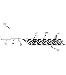
[0007] Figure 1 illustrates a medical device including a thrombectomy
device, according
to an embodiment.
[0008] Figure 2 is a schematic illustration of overlap configurations of
the
thrombectomy device of Figure 1.
[0009] Figure 3 illustrates an exemplifying thrombectomy device in an
unrolled state.
- 6b -
Date recue/Date Received 2020-08-28
CA 02957130 2017-02-06
[0010] Figure 4 illustrates another exemplifying thrombectomy device in an
unrolled
state.
[0011] Figure 5 is an enlarged view of a marker-mounting projection of the
thrombectomy device shown in the area 5-5 of Figure 4.
[0012] Figure 6 illustrates the marker-mounting projection of Figure 5
together with a
marker.
[0013] Figure 7 is a schematic representation of a fluoroscopic image of an
arrangement
of markers when a thrombectomy device to which they are attached is in an
unexpanded state.
[00141 Figure 8 is a schematic representation of a fluoroscopic image of
the markers of
Figure 7 when the thrombectomy device to which they are attached is in a fully
expanded state.
[0015] Figure 9 is a schematic representation of a fluoroscopic image of
the markers of
Figure 7 when the thrombectomy device to which they are attached is partially
expanded within
a vessel and in contact with a thrombus.
[0016] Figures 10-13 are cross-sectional views of a blood vessel and
illustrate a
processes of advancing and positioning a medical device, according to some
embodiments_
[0017] Figures 14-16 are cross-sectional views of the blood vessel shown in
Figures
10-13, and illustrate various clot positions and thrombectomy device
configurations, according to
some embodiments.
[0018] Figures 17-20 are cross-sectional views of a blood vessel and
illustrate uses of a
medical device according to some embodiments.
DETAILED DESCRIPTION OF THE SUBJECT TECHNOLOGY
[0019] The detailed description set forth below is intended as a
description of various
configurations of the subject technology and is not intended to represent the
only configurations
in which the subject technology may be practiced. The appended drawings are
incorporated
herein and constitute a part of the detailed description. The detailed
description includes specific
details for the purpose of providing a thorough understanding of the subject
technology.
However, the subject technology may be practiced without these specific
details. In some
instances, well-known structures and components are shown schematically to
avoid obscuring
the concepts of the subject technology.
-7-
CA 02957130 2017-02-06
[0020] Figure 1 depicts an exemplifying medical device 100 according to
some
embodiments of the subject technology. As illustrated in Figure 1, the medical
device 100 can
comprise a vascular device or thrombectomy device 102 and a manipulation
member 104.
Figure 1 also illustrates markers 150 attached to the thrombectomy device 102.
A proximal end
portion of the thrombectomy device 102 and a distal end portion of the
manipulation member
104 can be joined at a connection 106. The manipulation member 104 can extend
through a
catheter 107 such that an operator can manipulate the thrombectomy device 102,
positioned
within and/or distal to a distal end of the catheter 107, using the
manipulation member 104 at a
location proximal to a proximal end of the catheter 107.
[0021] The manipulation member 104 can be an elongate manipulation member.
The
manipulation member 104 can have a length sufficient to extend from a location
outside the
patient's body through the vaseulature to a treatment site within the
patient's body. For example,
the manipulation member can have a length of at least 100 cm, at least 130 cm,
or at least 150
cm. The manipulation member 104 can be monolithic or formed of multiple joined
components.
In some embodiments, the manipulation member 104 can comprise a combination of
wire(s),
coil(s), and/or tube(s).
[0022] The thrombectomy device 102 and the manipulation member 104 can be
attached
together at the connection 106. In some embodiments, the thrombectomy device
102 and the
manipulation member 104 can be substantially permanently attached together at
the connection
106. That is, the thrombectomy device 102 and the manipulation member 104 can
be attached
together in a manner such that, under the expected use conditions of the
medical device 100, the
endovascular device and the manipulation member would not become separated,
whether
deliberately or unintentionally, from one another without damage to or
destruction of at least a
portion of the connection 106. In some embodiments, the thrombectomy device
102 and the
manipulation member 104 can be permanently or releasably attached together at
the connection
106.
[0023] In some embodiments, the connection 106 can comprise a marker. The
marker of
the connection can comprise a radiopaque material such as platinum, iridium,
tantalum, gold,
alloys thereof or bismuth and tungsten-doped polymers, among other materials.
The connection
marker can be more radiopaque than a body of the vascular or thrombectomy
device 102. The
-8-
CA 02957130 2017-02-06
connection marker can be visible under fluoroscopy, CAT scans, X-Rays, MRI,
ultrasound
technology or other types of imaging. The connection marker can include an
interior channel, an
interior recess or another mounting feature. Further, the connection marker
can comprise a band
or substantially cylindrical shape with an open or closed circumference, a
coil, or other form.
[0024] It optionally may be advantageous to have a connection mechanism
that permits
intentional release of the thrombectomy device 102. For example, during a
blood flow
restoration procedure, it may prove difficult and/or dangerous to fully
retrieve a thrombus due to
a complicated vasculature or the risk of damaging a lumen wall. Leaving the
thrombectomy
device 102 inside the patient may prove to be the only option available to a
surgeon or other
medical personnel, or it may be a goal of the procedure, such as when the
thrombectomy device
102 is deployed across an aneurysm (e.g., as an aneurysm bridge to retain
coils or other materials
in an aneurysm). In other circumstances the thrombectomy device 102 may
include drug-eluting
capabilities, and/or may be coated with a particular type of drug that
facilitates thrombus
dissolution. It may be advantageous in such circumstances to release the
thrombectomy device
102 and allow the thrombectomy device 102 to anchor the thrombus against the
lumen wall
while the thrombus is dissolved by the drug. In some embodiments, the medical
device 100 can
comprise a portion, located proximally or distally of the connection 106, that
is configured for
selective detachment of the thrombectomy device 102 from the manipulation
member 104. For
example, such a portion can comprise an electrolytically severable or
mechanically detachable
segment of the manipulation member. In some embodiments, the medical device
100 can be
devoid of any feature that would permit selective detachment of the
thrombectomy device 102
from the manipulation member 104.
[0025] As illustrated in Figures 1 and 2, the thrombectomy device 102 can
have a tubular
or generally cylindrical shape in the absence of external forces in some
embodiments. However,
in some embodiments, the thrombectomy device can have a shape that is neither
tubular nor
cylindrical. In some embodiments, the thrombectomy device can have open
proximal and distal
ends, for example as illustrated in Figures 1 and 2, while in other
embodiments, the
thrombectomy device can have closed proximal and/or distal ends. In some
embodiments, the
thrombectomy device can comprise a series of structures (e.g., a longitudinal
series of structures)
each having proximal and distal ends that are open or closed. ln some
embodiments, the
-9-
CA 02957130 2017-02-06
thrombectomy device can comprise a tubular or cylindrical structure disposed
within, around, or
radially overlapping such a series of structures or one or more other tubular
or cylindrical
structure(s). The thrombectomy device 102 can be self-expanding, e.g. by super-
elasticity or
shape memory, or expandable in response to forces applied on the expandable
member, e.g. by a
balloon.
100261 As shown in Figures 1 and 2, the thrombectomy device 102 in some
embodiments
can be curled, rolled, or otherwise formed such that a first edge 124 and a
second edge 126
overlap one another, or form a gap between each other, when the thrombectomy
device 102 is in
a volume-reduced foint. In a volume-reduced form, or an unexpanded form, the
thrombectomy
device 102 illustrated in Figures 1 and 2 can overlap itself to facilitate
introduction of the
thrombectomy device 102 into and through the catheter 107. Figure 2 is a
schematic illustration
of overlap configurations (e.g., various amounts of overlap) of the
thrombectomy device of
Figure 1. Figure 2 illustrates various amounts of overlap of the thrombectomy
device 102,
forming zones of overlap 128. The thrombectomy device 102 can assume various
diameters A1,
A2, etc., depending on the degree of the overlap (e.g. represented by angle
al, a2. etc.). The
extent of any overlap of a frame 108 of the vascular or thrombectomy device
can depend upon a
degree of the frame's expansion. Expansion within a vessel can be limited, at
least in part, by
the vessel's size, and the amount and the properties of any thrombus present.
For example, a
greater overlap of the edges 124, 126 can occur in narrower vessels, whereas
in wider vessels the
overlap can be smaller, or even an "underlap" may occur, in which case the
edges 124 and 126
are separated by an open gap or space within the vessel. Advantageously, the
presence of an
overlap or "roll-up" configuration allows the thrombectomy device to be
expanded or
compressed in diameter with little or no change in length (e.g. foreshortening
during expansion),
in comparison to a similar device that lacks the overlap or roll-up
configuration. This is because
the expansion or compression can result from a decrease or increase in degree
of overlap (see
Figure 2) rather than wholly from deformation of the struts 114 and cells 116
(Figure 3), which
deformation can decrease or increase the length of the device when
transitioning to the expanded
or compressed state.
[0027] In some embodiments, the thrombectomy device 102 is
circumferentially
continuous (e.g., framing a circumferentially continuous tubular or
cylindrical shape), lacking
-10-
CA 02957130 2017-02-06
first and second edges 124, 126 and having no overlap or gap in a volume-
reduced form and
expanded form. Regardless of whether the thrombectomy device is
circumferentially
continuous, the thrombectomy device 102 can have a central longitudinal axis
both while in a
volume-reduced form and when fully or partially expanded. In some embodiments,
the
thrombectomy device 102 can be self-expandable, and can expand toward a fully
expanded
configuration upon release from the catheter 107. Upon expansion, the
thrombectomy device
102 can expand towards an inner wall of a vessel, towards an occlusive or
partially-occlusive
thrombus, clot or embolus within a vessel, or both.
[0028] The thrombectomy device 102 can be oversized relative to the
interior of a vessel
in which it is to be used, or the thrombectomy device 102 can occupy a larger
volume when
allowed to expand outside a vessel than when allowed to expand inside a
vessel. In other words,
the vessel may prevent a complete expansion of some or all of the thrombectomy
device 102.
[0029] Upon thrombectomy device 102 expansion into an expanded
configuration,
portions of the thrombectomy device can to penetrate into a thrombus, capture
a thrombus, or
both. In some embodiments, the thrombectomy device 102 can capture the
thrombus with an
exterior, or radial exterior, of the expanded thrombectomy device 102.
Additionally or
alternatively, in some embodiments, the thrombectomy device 102 may contact,
interlock,
capture or engage with a portion of the thrombus with an interior, or radial
interior, of the
expanded thrombectomy device 102.
[0030] The thrombectomy device can comprise a working length and anon-
working
length. The portion of the thrombectomy device 102 in the working length is
configured to
interlock, capture or engage a thrombus. The portion of the thrombectomy
device in the non-
working length may contact thrombotic material in use, but is configured to
perform a function
that renders it ineffective or less effective other than the working length
for interlocking,
capturing or engaging with a thrombus. In some embodiments, the non-working
length is
disposed between the working length and the connection 106 to the manipulation
member 104.
[0031] In some embodiments, the working length of the thrombectomy device
102 can
comprise a repeating pattern of structural features. For example, a working
portion of the
thrombectomy device 102 illustrated in Figures 1 and 2 comprises a matrix of
cells. Nonetheless,
in some embodiments the repeating pattern of structural features can have
other forms.
-11-
CA 02957130 2017-02-06
[0032] Figure 3 illustrates an exemplifying the vascular device or
thrombectomy device
102 in a flat configuration to facilitate understanding of various features
present in some
thrombectomy devices according to various embodiments. The thrombectomy device
102
illustrated in Figure 3 includes a working length 144 and a non-working length
145. As
illustrated in Figure 3, for example, the non-working length 145 is disposed
between the working
length 144 and the connection 106 to the manipulation member 104.
[0033] As illustrated in Figure 3, in some embodiments, the thrombectomy
device can
comprise a frame or body 108 having a plurality of struts 114 and a plurality
of cells 116,
forming a mesh. Groups of longitudinally and serially interconnected struts
114 can form
undulating members 118 that extend in a generally longitudinal direction. The
struts 114 can be
connected to each other by joints 120. While the struts are shown having a
particular undulating
or sinuous configurations, in some embodiments the struts can have other
configurations. The
frame of the thrombectomy device can have a generally tubular or generally
cylindrical shape in
some embodiments, while in others the frame can have a shape that is neither
tubular nor
cylindrical
[0034] The working length 144 of the thrombectomy device illustrated in
Figure 3
comprises some of the cells 116. In embodiments wherein the thrombectomy
device 102
comprises cells, the cells 116 in the working length and the portion of the
thrombectomy device
that form them can be sized and shaped such that they penetrate into a
thrombus, capture a
thrombus, or both upon expansion of the working length into a thrombus. In
some embodiments,
the portion of the thrombectomy device 102 in the working length can capture
the thrombus with
the individual cells 116 and/or with an exterior, or radial exterior, of the
expanded thrombectomy
device 102. Additionally or alternatively, in some embodiments, the portion of
the
thrombectomy device 102 in the working length may contact, interlock, capture
or engage with a
portion of the thrombus with individual cells 116 and/or an interior, or
radial interior, of the
expanded thrombectomy device 102.
[0035] As illustrated in Figure 3, for example, the non-working length can
comprise a
tapered proximal portion 122 of the thrombectomy device 102. The proximal
portion 122 of the
thrombectomy device 102 can be tapered toward a proximal end 110 of the
thrombectomy device
102. In some embodiments, the taper of the proximal, non-working portion 122
can
-12-
CA 02957130 2017-02-06
advantageously facilitate retraction and repositioning of the medical device
100 and
thrombectomy device 102. For example, in some embodiments, the non-working
length 145
facilitates a retraction of the thrombectomy device 102 into the catheter 107.
[0036] In some embodiments, the tapered proximal, non-working portion 122
can be
additionally or alternatively designed to generally not contact the vessel
wall during a blood flow
restoration procedure, and to generally not interfere with the flow of blood
within a vessel.
[0037] The taper of proximal portion 122 can be at various angles relative
to the
manipulation member 104 or the longitudinal axis of the thrombectomy device
102. For
example, in some embodiments, the taper can have an angle of approximately 45
degrees relative
to the manipulation member, though other angles are also possible, and within
the scope of the
present disclosure.
[00381 The thrombectomy device 102 can comprise a first edge 124 and a
second edge
126. The first edge 124 and second edge 126 can be formed, for example, from
cutting a sheet or
a tube. While the first and second edges are shown as having an undulating, or
sinuous
configuration, in some embodiments the first and second edges can have a
straight, or linear
configuration, or other configuration. In some embodiments, the edges 124, 126
can be curved,
straight, or a combination thereof along the tapered proximal portion 122.
[0039] Each cell 116 of the thrombectomy device 102 can have a maximum
length
(labeled "L" in Figure 3), as measured along a longitudinal axis of the
thrombectomy device 102,
and a maximum width W, as measured along a direction generally perpendicular
to the length
(labeled "W" in Figure 3). Figure 3 illustrates an embodiment of the
thrombectomy device 102
having a pattern 130 of cells 116 of substantially uniform dimensions and
struts 114 of
substantially uniform dimensions. Nonetheless, in some embodiments, cell size
and dimensions
can vary along the length and wide of the frame 108, as can the individual
filament thicknesses
and widths.
[0040] Figure 3 also illustrates a plurality of marker-mounting projections
148. Each
marker-mounting projection 148 can be attached to a portion of the
thrombectomy device 102
that may contact thrombus during use of the thrombectomy device. In some
embodiments, the
marker-mounting projections 148 can be attached to portions of the
thrombectomy device 102 in
the working length 144, for example as illustrated in Figure 3. In embodiments
wherein the
-13-
CA 02957130 2017-02-06
thrombectomy device comprises struts 114, the marker-mounting projection(s)
148 can be
attached to a strut 114. The marker-mounting projection 148 can be disposed
within a cell 116,
if present, or on another surface of the thrombectomy device 102. In some
embodiments, a
plurality of marker-mounting projections 148 can be attached respectively to a
plurality of struts
114. In some embodiments, some or all of the marker-mounting projections 148
can each be
attached to and/or at only a single strut 114. In some embodiments, the marker-
mounting
projection 148 can be attached to and/or at a joint 120. In some embodiments,
the marker-
mounting projections 148 can be separated from all other marker-mounting
projections 148 by a
distance, tor example at least 2mm or at least 3 mm, in a fully expanded
configuration of the
thrombectomy device 102. In some embodiments, the marker-mounting projections
148 can be
separated from all other marker-mounting projections 148 by one cell width or
one strut length
(e.g, an entire length of a strut separates the adjacent marker-
mountingprojections).
[0041] One or
more marker-mounting projections 148 can be located at some or all of a
proximal end 146 of the working length 144, a distal end 147 of the working
length 144, or an
intermediate area 149 of the working length 144 between the proximal end 146
and the distal end
147. The working length 144 can extend continuously or intermittently between
the proximal end
146 and the distal end 147.
[0042] In
some embodiments, the proximal end of the working length can be at a
proximalmost location where the thrombectomy device forms a complete
circumference. In some
embodiments, the proximal end of the working length can be at a proximalmost
location where
the thrombectomy device has its greatest transverse dimension in a fully
expanded state. In some
embodiments, the proximal end of the working length can be at a proximalmost
location where
the thrombectomy device has a peak, crown, or crest in transverse dimension in
a fully expanded
state.
[0043] In
some embodiments, the distal end of the working length can be at a distalmost
location where the thrombectomy device forms a complete circumference. In
some
embodiments, the distal end of the working length can be at a distalmost
location where the
thrombectomy device has its greatest transverse dimension in a fully expanded
state. In some
embodiments, the distal end of the working length can be at a distalmost
location where the
-14-
CA 02957130 2017-02-06
thrombeetomy device has a peak, crown, or crest in transverse dimension in a
fully expanded
state.
[0044] In some embodiments, a marker-mounting projection 148 located at the
proximal
end 146 can be disposed within 5 mm, within 4 mm, within 3 mm, within 2 mm, or
within 1 mm,
proximally or distally, of the proximal end 146. In some embodiments, a marker-
mounting
projection 148 located at the proximal end 146 can be disposed within the
length of one cell or
one strut, proximally or distally, of the proximal end 146.
[0045] In some embodiments, a marker-mounting projection 148 located at the
distal end
147 can be disposed within 5 mm, within 4 mm, within 3 mm, within 2 mm, or
within 1 mm,
proximally or distally, of the distal end 147. In some embodiments, a marker-
mounting
projection 148 located at the distal end 147 can be disposed within the length
of one cell or one
strut, proximally or distally, of the distal end 147.
[0046] A plurality or group of marker-mounting projections 148 can be
located at some
or all of the proximal end 146, the distal end 147, or the intermediate area
149. In some
embodiments, the plurality or group of marker-mounting projections 148 at each
of these
locations (if present) have a common pattern. For example, the projections 148
in the plurality or
group at the proximal end 146 can have the same arrangement relative to each
other as do the
projections 148 in the plurality or group at the distal end 147. The
projections 148 in the plurality
or group at intermediate area 149 (if present) can have the same arrangement
relative to each
other as do the projections 148 in the plurality or group at each of the
proximal end 146 and the
distal end 147, for example as illustrated in Figure 3. In some embodiments,
the marker-
mounting projections 148 of such a plurality or group of marker-mounting
projections 148 can
be disposed farther from each other when the thrombectomy device 102 is in an
expanded
configuration that they are when the thrombectomy device 102 is in an
unexpanded or less
expanded configuration.
[0047] In some embodiments, the vascular or thrombectomy device 102 can
comprise
one or more distally extending tips extending from a distal end of the
thrombectomy device. For
example, the device illustrated in Figure 3 is shown comprising four elongate,
distally extending
tips 154 extending from a distal end of the thrombectomy device 102. In some
embodiments
wherein the thrombectomy device comprises struts, these distal tips 154 can
extend from a
-15-
CA 02957130 2017-02-06
distalmost row of struts, for example as illustrated in Figure 3. In some
embodiments, one or
more markers 150 can be attached to the distal tips 154, if present. In some
embodiments
wherein one or more markers 150 are attached to the distal tips, the marker(s)
150 on the distal
tips 154 can be positioned at the distal end 147 of the working length 144,
for example as
illustrated in Figure 3.Figure 4 illustrates another exemplifying the
thrombectomy device 102 in
a flat configuration to facilitate understanding of various features present
in some thrombectomy
devices according to various embodiments. Figure 4 shows sets of marker-
mounting projections
148 arranged or laterally aligned such that they lie along straight lines R1,
R2, R3 that are
parallel to a longitudinal axis of the thrombectomy device 102 in the absence
of external forces
on the thrombectomy device. However, when external forces are applied, the
lines R1, R2, R3
through one or more of the sets of marker-mounting projections may not be
straight and/or may
not be parallel to the longitudinal axis of the thrombectomy device.
[0048] In some embodiments, the markers in a laterally aligned set or
longitudinally
grouped set can be separate and/or spaced from each other and from the markers
in other sets
and/or groups.
[0049] Figures 5 and 6 are enlarged views of a marker-mounting projection
148 shown in
the area 5-5 of Figure 4. In some embodiments, the marker-mounting projection
148 has an
arcuate, bowed or curved shape, and such a shape can span an entirety of the
length of the
marker-mounting projection 148 that receives a marker 150. In some
embodiments, the marker-
mounting projection 148 can be cantilevered from the portion of the
thrombectomy device to
which it is attached (e.g., from a strut 114, if present).
[0050] As illustrated in Figures 5 and 6, the marker-mounting projection
148 can include
a concave surface 160 and a convex surface 162. However, some marker-mounting
projections
can have a concave surface 160 or a convex surface 162 (either without the
other), or neither. In
some embodiments, the concave surface 160 faces away from a portion of the
thrombectomy
device 102 to which to is attached (e.g., from a strut 114, if present). In
some embodiments, the
convex surface 162 is arranged opposite from the concave surface 160, facing
toward a portion
of the thrombectomy device 102 to which to is attached (e.g., toward a strut
114, if present), or
both.
-16-
CA 02957130 2017-02-06
[0051] In some embodiments, the concave surface 160 and the convex surface
162 are
parallel to each other over some or all of the length of the marker-mounting
projection 148 that
receives a marker 150. The marker-mounting projection 148 can comprise a
constant cross-
sectional area along its length and/or a constant width along the length of
the marker-mounting
projection 148 that receives a marker 150. In some embodiments, the concave
surface 160, the
convex surface 162, or both includes a constant curvature or radius along the
length of the
marker-mounting projection 148 that receives a marker 150. In some
embodiments, the marker-
mounting projection 148 includes a rounded distal end 163.
[0052] Figure 6 illustrates a marker 150 on the marker-mounting projection
148. The
marker 150 can comprise a radiopaque material such as platinum, iridium,
tantalum, gold, alloys
thereof or bismuth and tungsten-doped polymers, among other materials. The
marker 150 can be
more radiopaque than a body of the vascular or thrombectomy device 102. The
marker 150 can
be visible under fluoroscopy, CAT scans, X-Rays, MRI, ultrasound technology or
other types of
imaging. The marker 150 can include an interior channel, an interior recess or
another mounting
feature_ Further, the marker 150 can comprise a hand or substantially
cylindrical shape with an
open or closed circumference, a coil, or another form that mounts around a
marker-mounting
projection 148.
100531 The marker 150 can directly attach to the marker-mounting projection
148
through direct contact between the marker 150 and the marker-mounting
projection 148. In
some embodiments, adhesives, welding, soldering, friction or mechanical
fastening (e.g.,
crimping) directly attach the marker 150 to marker-mounting projection 148. In
some
embodiments, the marker 150 extends completely around the marker-mounting
projection 148
when the marker 150 is mounted, or directly attached, to the marker-mounting
projection 148. In
another embodiment, the marker 150 extends partially (e.g., at least three
quarters of the
perimeter) around the marker-mounting projection 148 when the marker 150 is
mounted, or
directly attached, to the marker-mounting projection.
[0054] The marker-mounting projection 148 can extend generally parallel to
a segment
of the thrombectomy device (e.g., a strut) adjacent to the marker 150, for
example as illustrated
in Figure 5, or such that a marker when mounted to the marker-mounting
projection 148 is
-17-
CA 02957130 2017-02-06
parallel to a segment of the thrombectomy device (e.g., a strut) adjacent to
the marker 150, for
example as illustrated in Figure 6.
[0055] The marker 150 and the marker-mounting projection 148 can contact
each other at
three discrete locations when the marker 150 is directly attached to the
marker mounting
projection 148. In some embodiments, the marker 150 and the marker-mounting
projection 148
contact each other at more or fewer than three locations when the marker 150
is directly attached
to the marker mounting projection 148. In other embodiments, the marker 150
and the marker-
mounting projection 148 contact each other at two locations on the concave
surface 160 and at
one location on the convex surface 162 when the marker 150 is directly
attached to the marker-
mounting projection 148. In one embodiment, the contact location of the marker
150 and the
convex surface 162 is located between the contact locations of the marker 150
and the concave
surface 160.
[0056] In some embodiments, an arcuate marker-mounting projection can have
greater
marker retention strength, better withstand electropolishing, or both compared
to marker-
mounting projection having a straight configuration.
[0057] Figures 7-9 are schematic representations of fluoroscopic images of
an
arrangement of markers when a thrombectomy device to which they are attached
is in various
states. Figure 7 illustrates an arrangement of markers 150 when the
thrombectomy device 102 is
in an unexpanded state within a catheter 107 (see Figure 1). As shown in
Figure 7, the markers
150 in a proximal marker group 151, which can be located at the working length
proximal end
146 and mounted on marker-mounting projections 148, markers 150 in a distal
marker group
152, which can be located at the working length distal end 147 and mounted on
marker-mounting
projections 148, and markers 150 in an intermediate marker group 153, which
can be located at
the working length intermediate area 149 and mounted on marker-mounting
projections 148, can
be in close lateral proximity to the other markers 150 in the respective
marker group. In some
embodiments, a portion of a length of the thrombectomy device 102 between the
proximal
marker group 151 and the distal marker group 152, or between the working
length proximal 146
and distal 147 ends, has no marker 150.
[0058] Figure 8 illustrates the markers of Figure 7 when the thrombectomy
device 102 is
in a fully expanded state. As shown in Figure 8, the markers 150 in the
proximal marker group
-18-
CA 02957130 2017-02-06
151, the markers 150 in the distal marker group 152, and the markers 150 in
the intermediate
marker group 153 can be located farther laterally from the other markers 150
in a respective
marker group in this state than they are when the thrombectomy device 102 is
in an unexpanded
state. Figure 8 also shows the markers having substantially the same pattern
and/or spacing
relative to each other in each of the proximal marker group 151, the distal
marker group 152, and
the intermediate marker group 153.
[0059] Figure 9 illustrates the markers of Figure 7 when the thrombectomy
device 102 is
in a partially expanded state within a vessel and in contact with a thrombus.
As shown in
Figure 9, markers 150 in the intermediate marker group 153 are not spaced as
far from each other
laterally as are the markers 150 in the proximal marker group 151 from each
other or the markers
150 in the distal marker group 152 are from each other. Such an arrangement
can occur when
the thrombectomy device 102 is in an expanded state in the presence of a
thrombus 165 that
inhibits or prevents expansion of a region of the thrombectomy device.
[0060] Methods for engaging and removing a thrombus 165 will now be
discussed with
reference to Figures 10-70 Referring to Figure 10, the medical device 100 may
he inserted into
an anatomical vessel 172 by first inserting a guide wire 174 into the
anatomical vessel 172. The
illustrated anatomical vessel is an intracranial blood vessel. In some
embodiments, the medical
device is introduced into a segment of cerebral blood vessel distal to the
carotid siphon. The
inserted medical device 100 can be any embodiment of the medical device 100
disclosed herein,
including any of the thrombectomy devices 102, elongate members 104, or
connections 106.
The guide wire 174 can be advanced through a guide catheter 164 (see Figure
18), which
optionally includes a balloon near the guide catheter's distal end, and/or a
catheter 107 to the
treatment site, adjacent the thrombus 165. Referring to Figure 11, the guide
wire 174 is
advanced distally through the thrombus 165. Once the guide wire 174 is in
position, the catheter
107 is advanced over the guide wire 174, through a distal end of the guide
catheter, toward the
thrombus 165 in the anatomical vessel 172. Referring to Figure 12, the
catheter 107 is advanced
distally through the thrombus 165. The guide wire 174 is then withdrawn
proximally.
[0061] Referring to Figure 13, the medical device 100 is advanced through
the catheter
107. The medical device 100 is advanced through the catheter 107 by the
manipulation member
104 coupled to the thrombectomy device 102 (e.g., at the proximal end of the
thrombectomy
-19-
CA 02957130 2017-02-06
device). The catheter 107 prevents expansion of the thrombectomy device 102
and thus
maintains the thrombectomy device 102 in a compressed, volume-reduced
configuration as the
thrombectomy device 102 is advanced to the treatment site. The thrombectomy
device 102 is
advanced or otherwise moved to position (i) the proximal marker or marker
group 151 proximal
to a proximal end 170 of the thrombus 165, and (ii) the distal marker or
marker group 152 distal
to a distal end 171 of the thrombus 165. If an intermediate marker or marker
group 153 is
present, the thrombectomy device 102 is advanced or otherwise moved to
position the
intermediate marker or marker group 153 between the proximal and distal ends
of (e.g., within)
the thrombus.
[0062] Turning to Figure 14, the catheter 107 is then withdrawn proximally
relative to
the thrombectomy device 102 to expose the thrombectomy device 102. If the
thrombectomy
device 102 is self-expanding, retraction of the catheter 107 can permit the
thrombectomy device
102 to expand to an expanded state. Figure 14 illustrates the markers 150 of
the proximal marker
group 151 and the distal marker group 152 as more expanded than are the
markers 150 of the
intermediate 153 marker group, which appear in a less expanded distribution.
Such an
arrangement can result when the thrombectomy device 102 is expanded while all
markers 150 of
the proximal marker group 151 are located proximal to the thrombus 165, or to
a proximal end of
the thrombus 170, and all markers 150 of the distal marker group 152 are
located distal to the
thrombus 165, or to a distal end of the thrombus 171. When such a marker 150
arrangement is
observed, an operator may check, by injecting contrast solution through the
catheter 107 or guide
catheter 164, for perfusion of the distal vasculature through the thrombus 165
via a flow channel
(if any) opened in the thrombus 165 by the expansion of the thrombectomy
device 102, allow the
thrombectomy device 102 to continue expanding into the thrombus 165 and/or
proceed to
withdraw the thrombectomy device 102, as illustrated in Figure 17.
[0063] Figure 15 illustrates another configuration of the thrombectomy
device 102,
wherein the markers 150 of the distal marker group 152 are more expanded than
are the markers
150 of the proximal marker group 151 or the marker group intermediate 153,
which each appear
in a less expanded state. Such an arrangement can result when the thrombectomy
device 102 is
expanded while all markers in the proximal marker group 151 and the
intermediate marker group
153 are located radially adjacent to the thrombus 165 within the blood vessel
and all markers in
-20-
CA 02957130 2017-02-06
the distal marker group 152 are located distal to the thrombus 165 or a distal
end 171 of the
thrombus. When such a marker 150 arrangement is observed, an operator may
check for
perfusion of the distal vasculature through the thrombus 165, allow the
thrombectomy device
102 to continue expanding into the thrombus 165 and/or proceed to withdraw the
thrombectomy
device 102, as illustrated in Figure 17.
[0064] Figure 16 illustrates another configuration of the thrombectomy
device 102,
wherein the markers 150 of the proximal marker group 151 are more expanded
than are the
markers 150 of the distal marker group 152 or intermediate marker group 153,
which appear in
less expanded state. Such an arrangement can result when the thrombectomy
device 102 is
expanded while the markers in the distal marker group 152 are not located
distal to the thrombus
165 or a distal end 171 of the thrombus or when the thrombus migrates distally
during or after
expansion of the thrombectomy device 102.
[0065] When a marker 150 arrangement as illustrated in Figures 16 is
observed or
determined, or when a maximum marker 150 separation of the intermediate marker
group 153 is
observed as greater than a maximum marker 150 separation of either the
proximal marker group
151 or distal marker group 152, an operator may elect to collapse the
thrombectomy device 102
into less unexpanded state, for example by advancing the catheter 107 over the
thrombectomy
device 102. The operator can then again position the thrombectomy device 102
relative to the
thrombus and expand the thrombectomy device as described above.
[0066] With the proximal marker or marker group 151 located at the working
length
proximal end 146, the operator can more accurately and/or confidently position
the
thrombectomy device relative to a thrombus prior to expansion, thereby
facilitating utilization of
the working length of the thrombectomy device. In some embodiments,
positioning the
thrombectomy device 102 with reference to the proximal marker or marker group
151 located at
the working length proximal end 146 can facilitate or promote a successful
removal of the
thrombus or clot 165, by achieving a more secure contact, interlock or
engagement between the
thrombectomy device 102 and the thrombus or clot 165. Further, a comparison of
the relative
extent of marker group expansion can provide information to an operator that
assists in
determining whether and how (e.g., which direction) to reposition the
thrombectomy device 102.
-21-
CA 02957130 2017-02-06
[0067]
Accordingly, during a revascularization procedure, the user can use the
proximal
marker group 151 and/or the distal marker group 152 to properly locate the
thrombectomy device
102 longitudinally relative to the thrombus 165 before expanding the device
102 into the
thrombus. At appropriate time(s) in the procedure, the user can establish the
location of the
thrombus on an image of the treatment location (such as a fluoroscopic image
or other suitable
image as disclosed herein) by injecting contrast media into the target vessel
172 and observing
the effect of the thrombus on the flow of the contrast media in the vessel.
Once the catheter 107
is positioned in the thrombus 165 as shown in Figure 12, the user can advance
the thrombectomy
device 102 toward the distal end of the catheter and observe in the image of
the treatment
location the position of the proximal marker group 151 and/or the distal
marker group 152
relative to the thrombus 165 (e.g. relative to the proximal end 170 and/or the
distal end 171
thereof). This can be done while the thrombectomy device 102 is still in the
catheter to enable
adjustment of the position of the thrombectomy device 102 prior to expansion;
Figure 7 depicts
an example of a fluoroscopic image that the user might observe with the
proximal marker group
151 and the distal marker group 152 clearly visible due to their radiopacity.
The user can also
observe from such an image that the entire device 102 is still in the catheter
107 due to the
closely "packed" state of the marker groups 151, 152, 153 (and as well that
the distal end of
device 102 is near the distal tip of the catheter 107 as may be facilitated by
a catheter tip marker
173 (see Figures 7-9). With the location of the proximal end of the working
length 144 indicated
in the image by the proximal marker group 151 (and, optionally, the distal end
of the working
length 144 indicated in the image by the distal marker group 152), the user
can determine
whether the proximal end of the working length 144 is positioned proximal of
or longitudinally
aligned with the proximal end 170 of the thrombus (and, optionally, whether
the distal end of the
working length 144 is positioned distal of or longitudinally aligned with the
distal end 171 of the
thrombus). Based on this observation, the user can either confirm that the
working length 144 of
the device 102 is aligned with (or spans the entirety of) the length of the
thrombus 165; if either
or both is the case the user can leave the device in its current longitudinal
position relative to the
thrombus; if not, the user can adjust the longitudinal position of the device
102 until it is
correctly positioned relative to the thrombus 165 as described above. Once the
user has
confiimed the correct positioning of the device 102 in this manner, the user
can proceed to
-22-
CA 02957130 2017-02-06
expand the device 102 into the thrombus 165, e.g. as described elsewhere
herein, and remove
some or all of the thrombus from the vessel 172. Advantageously, as mentioned
above, when the
thrombectomy device 102 is of an overlap or roll-up configuration, relatively
little or no change
in the length of the device 102 will occur during expansion, and accordingly
the positions of the
markers relative to the thrombus (and to each other) will not change
significantly or at all as the
device expands. This in turn facilitates accurate placement of the expanded
device 102 in and
relative to the thrombus 165.
[0068] Referring to Figures 17 and 18, once the user is satisfied that the
device 102 has
been properly located longitudinally relative to the thrombus 165 and expanded
into it, the
thrombectomy device 102 can be withdrawn proximally, along with the thrombus
165. As
illustrated in Figure 18, the thrombectomy device 102 can be withdrawn
proximally, along with
the thrombus 165, into the guide catheter 164.
[0069] Referring to Figures 18 and 19, in embodiments wherein the guide
catheter 164
comprises a balloon 168, the balloon optionally can be inflated to occlude
flow during retraction
of the thrombus 165 toward the guide catheter. Referring to Figure lg, the
thrombectomy device
102 is withdrawn proximally to the guide catheter 164. The guide catheter 164
causes the frame
108 to collapse, with the thrombus 165 engaged therein. The thrombus 165 is
thus retrieved and
removed from the anatomical vessel 172. Referring to Figure 20, if retrieval
of the
thrombectomy device 102 is determined to be undesirable, e.g., to avoid
damaging the vessel
172, and the thrombectomy device 102 is detachably or releasably connected to
the manipulation
member 104, the thrombectotny device 102 can be detached from the manipulation
member 104
and can remain in the vessel 172.
[0070] Additionally, while the thrombectomy device 102 described above has
been
described in the context of use during a thrombectomy or blood flow
restoration procedure, the
thrombectomy device 102 can also, or alternatively, be used as an implantable
member (e.g.
stent). For example, the thrombectomy device 102 can be released through the
connection 106 at
a stenosis, aneurysm, or other appropriate location in a vessel. The
thrombectomy device 102
can expand and engage a vessel wall so as to hold the vessel wall open and/or
act as an occluding
member. While the filament thicknesses, widths, cell sizes, and forces
described above can be
optimized for an thrombectomy device 102 for flow restoration, these values
can also be
-23-
optimized for an thrombectomy device 102 for use as an implantable member. In
some
embodiments the same values can be used for both flow restoration and use as
an implantable
member.
[0071]
Also, while use of the thrombectomy device 102 described above with use of
a catheter 107, the catheter 107 can be omitted in some embodiments.
[0072]
The foregoing description is provided to enable a person skilled in the art
to practice the various configurations described herein. While the subject
technology has
been particularly described with reference to the various figures and
configurations, it should
be understood that these are for illustration purposes only and should not be
taken as limiting
the scope of the subject technology.
[0073]
There may be many other ways to implement the subject technology. Various
modifications to these configurations will be readily apparent to those
skilled in the art, and
generic principles defined herein may be applied to other configurations.
Thus, many changes
and modifications may be made to the subject technology, by one having
ordinary skill in the
art, without departing from the scope of the subject technology.
[0074] It
is understood that the specific order or hierarchy of steps in the processes
disclosed is an illustration of exemplifying approaches. Based upon design
preferences, it is
understood that the specific order or hierarchy of steps in the processes may
be rearranged.
Some of the steps may be performed simultaneously. The accompanying method
claims present
elements of the various steps in a sample order, and are not meant to be
limited to the specific
order or hierarchy presented.
[0075] A
phrase such as "an aspect" does not imply that such aspect is essential to the
subject technology or that such aspect applies to all configurations of the
subject technology. A
disclosure relating to an aspect may apply to all configurations, or one or
more
configurations. An aspect may provide one or more examples of the disclosure.
A phrase such
as "an aspect" may refer to one or more aspects and vice versa. A phrase such
as "an
embodiment" does not imply that such embodiment is essential to the subject
technology or that
such embodiment applies to all configurations of the subject technology. A
disclosure relating
to an embodiment may apply to all embodiments, or one or more embodiments. An
embodiment may provide one or more examples of the disclosure. A phrase such
"an
embodiment" may refer to one or more embodiments and vice versa. A phrase such
as "a
- 24 -
Date recue/Date Received 2020-08-28
configuration" does not imply that such configuration is essential to the
subject technology or
that such configuration applies to all configurations of the subject
technology. A disclosure
relating to a configuration may apply to all configurations, or one or more
configurations. A
configuration may provide one or more examples of the disclosure. A phrase
such as "a
configuration" may refer to one or more configurations and vice versa.
[0076] Furthermore, to the extent that the term "include," "have," or the
like is used
in the description or the claims, such term is intended to be inclusive in a
manner similar to
the term "comprise" as "comprise" is interpreted when employed as a
transitional word in a
claim.
[0077] A reference to an element in the singular is not intended to mean
"one and
only one" unless specifically stated, but rather "one or more." The term
"some" refers to one or
more.
[0078] All structural and functional equivalents to the elements of the
various
configurations described throughout this disclosure that are known to those of
ordinary skill in
the art are intended to be encompassed by the subject technology. Moreover,
nothing disclosed
herein is intended to be dedicated to the public regardless of whether such
disclosure is
explicitly recited in the above description.
[0079] While certain aspects and embodiments of the subject technology
have been
described, these have been presented by way of example only, and are not
intended to limit
the scope of the subject technology. Indeed, the novel methods and systems
described herein
may be embodied in a variety of other forms without departing from the spirit
thereof. The
accompanying claims and their equivalents are intended to cover such forms or
modifications
as would fall within the scope and spirit of the subject technology.
- 25 -
Date recue/Date Received 2020-08-28