Note: Descriptions are shown in the official language in which they were submitted.
CA 02957220 2017-02-02
WO 2016/022759
PCT/US2015/043940
MEDICAL DEVICES AND METHODS OF PLACEMENT
FIELD OF THE INVENTION
The invention provides various medical devices, each with a camera
placed in a camera tube, which allows for one camera to provide continuous
visualization for each device during and after placement in a patient. A sound
device, such as a microphone, is incorporated in many devices and provides
continuous monitoring of breath and heart sounds in a patient. The camera and
microphone do not contact the patient's tissues and thus, do not require
sterilization.
The continuous visualization and sound monitoring of the patient are in real
time and
enable remote monitoring as well. Methods for rapid and accurate placement of
a
medical device in a patient are provided as well.
BACKGROUND
Various devices are available to stabilize a patient and facilitate his
breathing, feeding and medication delivery. Such devices are used in patients
during surgical procedures, after certain traumas including spinal cord
injuries, and in
patients suffering from certain medical conditions including advanced
Alzheimer
disease. These devices include endotracheal tubes, airway devices, feeding
tubes,
oral airways, nasal cannulas and the like.
Because human anatomy varies significantly from a patient to a
patient, properly placing a medical device in a patient's trachea requires a
significant
skill and is a task laced with inherent risk. The task becomes even more
complicated
because the insertion procedure may have to be performed immediately at an
accident site, on pediatric patients, in a nursing home, on a battlefield or
at a natural
disaster site where many patients have to be attended at the same time.
The process of placing a breathing tube in a patient is called intubation.
Devices such as laryngoscopes, videolaryngoscopes, fiberoptic scopes, as well
as
other proprietary videoscopes have been developed. These devices provide
1
Date recue / Date received 2021-12-16
CA 02957220 2017-02-02
WO 2016/022759
PCT/1JS2015/043940
accuracy for initial placement, but do not provide continuous visualization or
mobility
of the image after a medical device has been placed in a patient. Newer
devices,
such as Vivasight SL or DL endotracheal tubes, provide continuous
visualization, but
are costly because they depend on a single use of disposable cameras and they
are
not transferrable from one medical device to another. The Totaltrack VLM
supraglottic airway has a proprietary reusable camera for only its one device,
and it
cannot be transferred to other medical devices.
Thus, there remains the need for improved devices which can be easily
monitored remotely by a qualified personal during placement and after
placement for
an adverse reaction. After a medical device has been placed in a patient, the
need
remains to monitor in real time the patient's possible adverse reactions such
as for
example, aspiration, airway secretion, apnea, etc.
SUMMARY OF THE INVENTION
At least some of these needs are addressed by present medical
devices which are equipped with a portable universal visualization device in
which a
camera is contained within a separate camera tube and which transmits
information
that can be accessed and monitored remotely and simultaneously from several
patients in real time.
One embodiment provides a medical visualization device which
comprises a camera tube with a distal end and a proximal end. The distal end
is
sealed with a transparent material and a proximal end has an opening. A camera
with a wire is placed inside of the camera tube. The camera can be placed
inside of
the camera tube and it can be retracted from the camera tube on demand. The
camera can be reused in various devices without sterilization. The camera can
transmit images to a remote location wirelessly. In some embodiments, the
camera
tube comprises a fiber optic material. The visualization device can be
equipped with
at least one of the following: a light source, a stylet, a bougie and a sound-
and
temperature-monitoring device which can transmit the information to a remote
location wirelessly. The visualization device can transmit images, sounds and
other
data to any number of remotely located monitoring devices and/or data storage
devices. Such devices include, but are not limited to, a wireless portable
device,
2
CA 02957220 2017-02-02
WO 2016/022759 PCT/1JS2015/043940
smart phone, tablet, watch, cell phone, hand-held wireless device, computer,
remote
data server, radio, television, walkie-talkie and the like.
A further embodiment provides a method of continuous monitoring of a
patient's at least one internal organ, the method comprising placing in the
patient the
visualization device with the camera in the sealed camera tube, causing the
camera
to transmit images of the internal organ in real time through the transparent
material
at the distal end of the camera tube, and analyzing the transmitted images. In
some
embodiments, the images are transmitted wirelessly to at least one remote
location.
Various internal organs can be monitored by this method, including
nasopharynx, pharynx, hypopharynx, supraglottic structures, airway, trachea,
vocal
cords, stomach, and vagina.
In some embodiments, the length of the camera tube in the
visualization device is adjustable and it can be adjusted to the length of at
least one
of the following devices: an endotracheal tube, a supraglottic airway, airway
device,
oral airway, dilator, tracheostomy device, intubating oral airway, esophageal
stethoscope, nasal cannula, feeding tube, suction tube and endotracheal
changing
tube.
Further embodiments provide a method for placing a medical device in
a patient in which the medical device is equipped with the visualization
device and a
bougie. The medical device is inserted in the patient and the placement of the
device is guided with the bougie under continuous visualization.
A kit for monitoring a patient's internal organ in real time is also
provided. The kit comprises a camera tube with the adjustable length and with
at
least one ring attached externally to the camera tube, wherein the camera tube
has a
distal end and a proximal end and wherein the distal end of the camera tube is
sealed with a transparent material; and a reusable camera which can be placed
and
removed from the camera tube and which can transmit images wirelessly to at
least
one remote location. This real time information obtained with the
visualization device
can be transferred or stored to multiple distant monitoring sites.
Also provided is a medical device comprising a visualization device
sealed to, attached to or otherwise combined with at least one of the
following
3
CA 02957220 2017-02-02
WO 2016/022759
PCT/1JS2015/043940
second devices: an endotracheal tube, a supraglottic airway device, a
ventilator
adaptive cap, a dilator, a tracheostomy device, a nasal trumpet, a an oral
airway, an
esophageal stethoscope, a laryngoscope, a speculum, a nasal cannula, a feeding
tube, a suction tube, a suction catheter, and an endotracheal changing tube;
and
wherein the visualization device comprises a camera tube with a distal end and
proximal end, the distal end being sealed with a transparent material and a
camera
being placed inside of the camera tube through an opening at the proximal end.
These medical devices can be further equipped with at least one of the
following a
bougie, a flexible stylet and a sound- and temperature-monitoring device. In
some
embodiments, the visualization device is sealed, attached or otherwise
connected
externally to the second device. In other embodiments, the visualization
device can
be placed inside of the second device. Various endotracheal tubes equipped
with
the visualization device are contemplated as well, including an endotracheal
tube
which comprises a sleeve through which the visualization device can be
inserted, an
.. endotracheal tube into which the visualization device is placed internally
through a
ventilator adaptive cap and an endotracheal tube to which the visualization
device is
attached externally.
Further embodiments provide an oral airway device comprising a tubal
body with a central lumen and a visualization device attached to the tubal
body,
wherein the diameter of the lumen is such that an endotracheal tube can be
placed
insider the lumen and wherein the visualization device comprises a camera tube
sealed at the distal end with a transparent material and a camera placed
inside the
tube through the opening at the proximal end, end wherein the camera tube is
positioned along the tubal body. The visualization device can be attached to
the
tubal body either internally or externally. The oral airway device can further
comprise a removable handle which can be connected to the oral airway device
with
a holder.
Further embodiments provide an oral airway device with a rotating
central passageway made of two half-cylinders, a first external half-cylinder
and
second internal half-cylinder, wherein the second half-cylinder fits inside
the first half
-cylinder and can glide inside the first half-cylinder along the proximal-
distal axis of
the first half-cylinder and wherein the second half-cylinder can also rotate
inside the
first half-cylinder and thereby create a completely enclosed central
passageway or
4
CA 02957220 2017-02-02
WO 2016/022759
PCMJS2015/043940
only partially enclosed central passageway with a lateral opening, and wherein
the
first half-cylinder and the second half-cylinder can be completely separated
from
each other.
Other embodiments provide a supraglottic ventilating tube with camera,
comprising a ventilating tube with the distal end and the proximal end and
equipped
with a visualization device comprising a camera tube attached externally along
the
ventilating tube, and a camera which can be placed inside the camera tube,
wherein
an inflatable cuff which wraps around the ventilating tube and the camera tube
being
positioned under the cuff.
Methods for intubating and extubating a patient are also provided in
which an endotracheal tube or ventilating tube is loaded onto the second half-
cylinder of the oral airway device which is then assembled with the first half-
cylinder
of the oral airway device and the assembly is inserted into a patient under
continuous visualization and monitoring.
Another embodiment provides a tubeless intubating device, comprising
an ellipsoid body attached to a handle and a visualization device attached to
the
intubating device, wherein the visualization device comprises a camera tube
and a
camera which can be placed and removed from the camera tube and wherein the
ellipsoid body comprises a lumen and canal which opens beneath the handle.
Other embodiments provide a sliding endotracheal cuff device,
comprising a tube with the distal end and the proximal end, a rail attached
externally
on the tube along the proximal-distal axis, wherein the rail has a groove
which opens
inside the tube, wherein the device further comprises a cuff which wraps
around the
tube externally at the distal portion of the tube, and wherein the device
further
comprises a camera tube attached externally to the tube along the proximal-
distal
axis, and a camera which can be positioned inside and removed from the camera
tube.
Further embodiments include an assembly in which an oral airway
device is inserted inside of a carrier which comprises a tubal body with a
lumen and
.. a first balloon which caps the distal end of the carrier. The carrier has a
lumen
opening proximal to the first balloon and the carrier has a second balloon
circumventing the tubal body of the carrier proximally to the lumen. The
carrier may
5
CA 02957220 2017-02-02
WO 2016/022759
PCMJS2015/043940
further optionally comprise a third balloon circumventing the body of the
carrier
proximally to the second balloon. The balloons can be inflated with an
inflating
means. Methods of intubating and extubating a patient with the carrier
assembly are
provided as well.
BRIEF DESCRIPTION OF THE DRAWINGS
Figs. 1A, 1B and 1C depict a side view of an embodiment for a
visualization device as shown in Fig. 1A which can be further equipped with a
stylet
as shown in Figs. 1B and 1C.
Fig. 2 depicts a side view of an embodiment for an endotracheal device
equipped with a visualization device.
Fig. 3 depicts a side view of an alternative embodiment for an
endotracheal device equipped with a visualization device.
Figs. 4A and 4B depict two embodiments showing a visualization
device attached to a built-in ventilator adaptive cap. Fig. 4A is an
embodiment
without a light source and Fig. 4B is an embodiment with a light source.
Figs. 5A, 5B and 5C depict side views of further embodiments of a
visualization device attached to a built-in ventilator adaptive cap and
delivered
through a sliding sleeve (Figs. 5A and 5B), with further embodiment in Fig. 5C
which
includes a bougie or a flexible stylet as shown in the insert.
Figs. 6A and 6B depict side views of an embodiment for an
endotracheal device equipped with a visualization device delivered through a
sliding
sleeve.
Figs. 7A and 7B depict side views of an alternative embodiment for an
endotracheal device equipped with a visualization device and delivered through
a
sliding sleeve.
Fig. 8 is a side view of an embodiment for an endotracheal device
equipped with a bougie.
Figs. 9A, 9B and 9C are a side view of a visualization device equipped
with rings. Fig. 9A is a side view of a visualization device equipped with two
rings.
6
CA 02957220 2017-02-02
WO 2016/022759
PCMJS2015/043940
Fig. 9B is a side view of the visualization device as shown in Fig. 9A, but
equipped
further with a bougie. Fig. 9C is a side view of the visualization device as
shown in
Fig.9A, but equipped further with a light source. Fig. 9D is a cross-sectional
view
through the visualization device of Fig. 9A showing a ring connected to the
camera
tube. Fig. 9E is a cross-sectional view through the visualization device of
Fig. 9A
showing an adjustable sliding ring with a clasp connected to the camera tube.
Fig.
9F is a side view of an endotracheal tube to which the visualization device of
Fig. 9A
is connected with two sliding rings.
Fig. 10 is a side view of a supraglottic airway device equipped with a
visualization device. An endotracheal device of Fig. 3 is shown as an insert
on the
left and a flexible guided stylet is shown as an insert on the right.
Figs. 11A and 11B depict a side view of an alternative airway device
with a visualization device. Fig. 11A is a side view of the airway device and
Fig. 11B
is the device as shown in Fig. 11A, but with a ventilator adaptive cap.
Figs. 12A, 12B and 12C depict an oral airway intubating device with a
visualization device. Fig.12A is a side view of the oral airway device, while
Figs. 12B
and 12C are cross-sections through the airway device in its full cylinder form
(Fig.
12B) and in its half-cylinder form in which one half-cylinder is retracted
into the other
half-cylinder (Fig. 12C).
Fig. 13 depicts a side view of a dilator with a visualization device.
Figs. 14A and 14B depict a side view of a tracheostomy device with a
visualization device. Fig. 14A depicts an embodiment in which the
visualization
device is attached externally to a tracheostomy tube, while Fig. 14B depicts
an
embodiment in which the visualization device is attached inside of the
tracheostomy
tube.
Fig. 15 depicts a side view of a nasal trumpet with a visualization
device.
Figs. 16A-16C depict a side view of an oral airway equipped with a
visualization device and Fig. 16D depicts an intubating oral airway also
equipped
with a visualization device. Fig. 16A shows a visualization device being
positioned
inside of the oral airway. Fig.16B is the same as Fig.16A, but includes a
light source
7
CA 02957220 2017-02-02
WO 2016/022759
PCMJS2015/043940
for the visualization device. Fig. 16C is the same as Fig. 16A, but includes a
whistle.
Fig. 16D is an intubating oral airway device with a main lumen into which an
endotracheal tube can be placed. Fig. 16E is an intubating/extubating oral
airway
device with a main lumen as shown in Fig. 160 and into which an endotracheal
tube
has been placed.
Figs. 16F and 16G are embodiments showing a portion of an
intubating/extubating oral airway device with a detachable handle which can be
attached to a holder on an intubating/extubating oral airway device.
Figs. 16H and 16J-16P depict an intubating/extubating oral airway
device with a rotating central passageway. Figs. 16H and 16J depict the
capacity of
intubating/extubating oral airway device with a rotating central passageway to
extend
distally. The embodiment of Figs. 16H and 16J has a camera tube with camera
placed externally on the intubating/extubating oral airway, while the
embodiment of
Fig. 16K provides the intubating/extubating oral airway in which the camera
tube is
placed inside of the rotating central passageway. Figs. 16L-16N demonstrate
further
how the inner half-cylinder can rotate in the intubating/extubating oral
airway to
create a fully enclosed central passageway. Figs. 160-16P depict insertion of
an
endotracheal tube inside the rotating central passageway of the
intubating/extubating
oral airway.
Figs. 17A and 17B depict a side view for a supraglottic airway device
equipped with a visualization device and working tube and bougie tube.
Figs. 18A-18B depict a side view for a one-piece laryngoscope with a
visualization device. Fig. 18A shows a laryngoscope with a visualization
device, and
Fig. 18B is the same, except it is further equipped with a bougie and the
visualization
device is equipped with a light source.
Fig. 19 depicts a side view of a speculum with a visualization device.
Figs. 20A-F depict a nasal cannula with a visualization device. Fig.
20A depict positioning of the cannula on a patient's head and Fig. 20B is the
same
as in Fig. 20A, except the visualization device is equipped with a light
source. Fig.
20C is a side view of the cannula of Fig. 20A, and Fig. 200 is a side view of
the
cannula of Fig. 20B. Fig. 20E is a cross-sectional view of a patient's head
with the
8
CA 02957220 2017-02-02
WO 2016/022759
PCMJS2015/043940
cannula of Fig. 20A inserted. Fig. 20F is a further embodiment in which a
nasal
cannula with a visualization device as shown in Fig. 20A is further combined
with an
external stethoscope.
Fig. 21 is a feeding tube equipped with a visualization device.
Fig. 22A, 22B, 22C depict various embodiments of a suction tube
equipped with a visualization device. Fig. 22A depicts a visualization device
positioned inside of the suction tube. Fig. 22B the same as in 22A, but
equipped
further with a bougie, and Fig. 22C is the same as Fig. 22B, but showing the
bougie
protruding from the distal end of the suction tube.
Fig. 23 depicts a suction catheter equipped with a visualization device.
Fig. 24 depicts an endotracheal changing tube equipped with a
visualization device.
Figs. 25A-25D depict a supraglottic ventilating tube with camera. Fig.
25A is a side view of the supraglottic ventilating tube with camera. Fig. 25B
is an
enlarged view of the supraglottic ventilating tube distal tip with a cuff.
Figs. 25C and
25D depict insertion of the supraglottic ventilating tube into an
intubating/extubating
oral airway.
Figs. 26A-26J depict a tubeless intubating device. Fig. 26A depicts the
upper surface of the tubeless intubating device. Fig. 26B depicts the bottom
surface
of the tubeless intubating device. Figs. 26C-26F depict loading the tubeless
intubating device with an endotracheal tube for insertion into a patient. Fig.
26G
depicts the upper surface of the tubeless intubating device without a cuff,
while Fig.
26H depicts the bottom surface of the tubeless intubating device of Fig. 26G.
Figs.
261 and 26J depict loading of the device Fig. 26G with a supraglottic airway.
Figs. 27A-27G depict a sliding endotracheal cuff (Figs. 27A, 27C-27G)
and loading an endotracheal tube into the sliding cuff (Fig. 27B).
Figs. 28A-28B depict an endotracheal tube with a visualization device
which can slide along the endotracheal tube and be removed from the
endotracheal
tube.
9
CA 02957220 2017-02-02
WO 2016/022759
PCMJS2015/043940
Figs. 29A-29C depict a sliding camera tube with rail and placing of the
camera tube into a laryngoscope.
Figs. 30A-30B depict a supraglottic airway device with built in
endoscope guide (Fig. 30A) and insertion of the device into a patient (Fig.
30B).
Figs. 31A and 31B depict an assembly of a naso-gastric tube with a
visualization device.
Fig. 32 depicts an oral airway embodiment.
Figs. 33A-33C depict further embodiments of an endotracheal tube
with an externally attached camera tube. Fig. 33A depicts an embodiment with a
suction tube, Fig. 33B with a medication dispensing device and Fig. 33C with
biopsy
forceps.
Figs. 34A-34H depict various embodiments for an oral airway device.
Fig. 34A depicts an embodiment with a separate lumen for an esophageal
blocker.
An endotracheal tube which can be placed into the oral airway device is also
shown.
Fig. 34B is the same embodiment as in 34A, but with a ventilator cap instead
of an
endotracheal tube. Fig. 34C is an oral airway device without a balloon with an
endotracheal tube also shown. Fig. 34D is the same embodiment as in 34C, but
with
a ventilator cap instead of an endotracheal tube. Figs. 34E-34H depict an
expendable oral airway device with a carrier. Fig. 34E shows placement of an
oral
airway device into a carrier. Fig. 34F is an enlarged view of the carrier from
Fig.
34E. Figs. 34G and 34H depict an oral airway device positioned inside of the
carrier,
with Fig. 34G showing the oral airway device positioned fully inside the
carrier, while
Fig. 34H showing the oral airway device expending from the carrier.
Figs. 35A-35B are further embodiments of an oral airway device. Fig.
35A depicts an oral airway device positioned in a patient, while Fig. 35B
provides an
embodiment of an oral airway device with a side opening.
Figs. 36A, 36B, 36C depict a nasopharyngeal airway device. Fig. 36A
is an embodiment with two balloons, while Fig. 36B is an embodiment with three
balloons. Fig. 36C shows positioning of the three balloon nasopharyngeal
airway
device in a patient.
CA 02957220 2017-02-02
WO 2016/022759 PCMJS2015/043940
DETAILED DESCRIPTION
The present invention provides improved medical devices equipped
with a visualization device for intubation, ventilation, feeding and
monitoring of a
patient. The present invention also provides methods for rapid and accurate
placement of a medical device in a patient and remote continuous real-time
monitoring of the patient after the placement.
These medical devices are equipped with a visualization device in
which a camera is placed in a separate sealed camera tube. As the camera does
not come in contact with a patient, there is no need to sterilize the camera
and the
same camera can be reused in many applications. Thus, the same camera can be
switched between different medical devices which monitor internal organs such
as
medical devices that are placed in patient's airway, larynx, gastrointestinal
tract,
chest or vaginal cavity. In some embodiments, the camera is disposable.
One embodiment provides a visualization device as shown in Fig. 1A
and its further embodiments as shown in Figs. 1B and 1C. A visualization
device,
generally 10, in Fig. 1A comprises a camera tube 12 with a distal end 14 and a
proximal end 16. The camera tube 12 can be a plastic tubing. In some
embodiments, the camera tube 12 may comprise a fiber-optic material. The
camera
tube 12 is sealed at the distal end 14 with a transparent material 17. The
diameter of
camera tube 12 is designed in such a way that a camera 18 with wire 20 can be
inserted inside of the camera tube 12 through an opening at the proximal end
16 and
moved down the camera tube 12 toward the distal end 14, so that the camera 18
transmits continuously images obtained through the transparent material 17.
The
length of the camera tube 12 can vary and it can be adjusted dependent on the
length of a medical device with which the visualization device is to be used.
For
example, the length of the camera tube 12 may be longer when the visualization
device 10 is used with a feeding tube in comparison to the length of the
camera tube
12 when the visualization device 10 is used with an endotracheal tube. In some
embodiments, the visualization device obtains images and transmits wirelessly,
broadcasts or records this information to at least one device positioned at a
remote
location.
Because the camera tube 12 is sealed at the distal end 14 with the
transparent material 17, the camera 18 does not come in contact with patient's
11
CA 02957220 2017-02-02
WO 2016/022759
PCMJS2015/043940
tissues or fluids and therefore, the camera 18 does not have to be sterilized
or to be
disposable, and it can be reused in further applications. However, the camera
18
can be disposable in some applications. The camera 18 can be loaded with a
chip
and equipped to obtain and transmit digital images in real time. The camera 18
is
further connected by an electric wire 20 to an image receiving and processing
device
(not shown) such as a computer equipped with a monitor or a computer network.
The camera 18 may also be in communication wirelessly with an image-receiving
device located at any location, including multiple locations and remote
locations.
Because the length and diameter of the camera tube 12 can be adjusted based on
patient's needs, the visualization device 10 is suitable for a broad variety
of patients,
including pediatric patients and adult patients with abnormal anatomy or
trauma.
As the visualization device 10 is bendable and flexible, the visualization
device 10 is easy to insert in a patient and remove from the patient. The
camera 18
may have its own light source. As the visualization device 10 transmits images
from
a patient in real time, it can be used for guiding a medical device for proper
placement. Thus, some embodiments are concerned with methods for rapid and
accurate placement of a medical device inside of a patient, including a method
for
guided and rapid placement into patient's airway, larynx, gastrointestinal
tract, chest
or vaginal cavity under continuous visualization.
As shown in embodiment of FIG. 1B, the visualization device 10 can be
further equipped with a stylet 22 which can be sealed onto or otherwise
attached to
the camera tube 12 externally on at least one side of the camera tube 12 along
the
proximal-to-distal (16-14) axis of the camera tube 12. The stylet 22 can be
made of
metal wire or some other sturdy material with the purpose to keep the
otherwise
flexible visualization device 10 in a particular shape. In some embodiments,
the
stylet 22 can be of the same length as the camera tube 12. In other
embodiments,
the stylet 22 is shorter than the camera tube 12 such that at a least a
portion of the
camera tube 12 on either the proximal end 16 or distal end 14, or on the both
ends
16 and 14 is not in contact with the stylet 22. As shown in Fig. 1C, the
stylet 22 can
be bent into various shapes and it retains the shape into which it has been
bent,
which permits for visualization device 10, which is otherwise flexible, to
retain a
particular shape.
In alternative embodiments, the visualization device 10 can be
equipped with a bougie which can be attached to the camera tube12 externally
on at
12
CA 02957220 2017-02-02
WO 2016/022759
PCT/1JS2015/043940
least one side of the camera lumen 12 along the proximal-distal (16-14) axis
of the
visualization device 10.
The bougie can be made of various materials, including plastic material
which is bendable. As the bougie is bendable, but keeps a shape into which it
is
bent, the bougie is suitable for guiding the visualization device 10 inside of
a patient.
In some embodiments, the bougie can be of the same length as the camera tube
12.
In other embodiments, the bougie can be made shorter or longer than the camera
tube 12 such that only a portion of the camera tube 12 is in contact with the
bougie.
In some embodiments, the bougie protrudes on at least the distal end 14.
The visualization device 10 can be further equipped with a portable
light source (not shown) which can be either built-in the camera 18 or it can
be built-
in the camera tube 12. In alternative, a light source can remain outside the
camera
tube 12 on the proximal end 16, but still be placed such that the light source
sheds
light inside of the camera tube 12.
In embodiments of Figs. 1A-1C, the camera tube 12 can be disposable,
while the camera 18 is reusable without the need of sterilization. However,
the
camera 18 can be also disposable in at least some embodiments.
During placement in a patient, a visualization device 10 either alone or
in combination with another medical device is positioned such that it is
inserted with
its distal end 14 in the patient under continuous visualization with the
camera 18.
Any of the visualization devices 10 described above can be attached,
sealed or otherwise connected to a disposable or non-disposable medical device
either externally or internally and as described in more detail below. Various
medical
devices for pediatric and adult patients can be built such that the camera
device tube
12 is sealed or attached to the medical device during manufacturing. In some
embodiments, the visualization device 10 can slide or glide along the medical
device
to which the visualization device 10 is attached. For example, the camera tube
12 of
the visualization device 10 can be equipped with a set of rings, a rail or a
half-
cylinder which will allow the camera tube 12 to slide or glide along the
medical
device to which the visualization device 10 is attached.
In other embodiments, the visualization device 10 can be sold as a kit
which can be attached by a medical practitioner to a pre-made medical device
for
pediatric and adult patients, based on a particular patient's individual
needs. The
length of the camera tube 12 can vary such that the camera tube 12 is of the
same
13
CA 02957220 2017-02-02
WO 2016/022759
PCMJS2015/043940
or similar length with a medical device to which the visualization device 10
is sealed,
attached or otherwise connected to.
Having the ability to verify placement for a medical device in real time
from near and far allows several experts to assist and verify placement. This
is
accomplished by equipping the medical device with the visualization device 10.
In
some embodiments, a method is provided in which the visualization device 10 is
used for placing a medical device in a patient in ambulances, on battlefields,
in
nursing homes or hospitals. The visualization device 10 provides the ability
to
monitor in real time a patient. Because the visualization device 10 may
interact with
a plethora of devices disposable and otherwise, the use of the device 10 on
various
medical devices provides for a method in which a medical practitioner can
customize
a proper device for each patient or situation. Having the same camera
equipment
that can interact with various medical devices provides economy of scale such
that
even the smallest of organizations can have all the proper vigilance and
technology.
At least in some embodiments the visualization device 10 can be used
in assembly with at least one medical device as described in more detail
below. A
method in which the visualization device 10 is used on an airway device allows
continuous visualization of any of the following in a patient in real time:
nasopharynx,
pharynx/hypo pharynx, supraglottic structures, airway, internal organ anatomy,
vocal
cords during normal and abnormal ventilation. This method also allows
detection of
abnormal anatomy and abnormal vocal cord movements.
Referring to FIG. 2, this embodiment provides an endotracheal device,
generally 30. The endotracheal device 30 comprises an endotracheal tube 32
with a
distal end 32A and a proximal end 32B. The visualization device 10 is sealed
or
otherwise attached externally on at least one side of the endotracheal tube
32, along
the proximal-distal (32B-32A) axis of the endotracheal tube 32. The
visualization
device 10 comprises essentially of all elements as shown in Fig. 1A, with the
camera
18 inserted inside of the camera tube 12 through an opening at the proximal
end 16
of the camera tube 12, all the way down to the distal end 14 and the opening
of the
distal end 14 being sealed with the transparent material 17. Because the
camera 18
is positioned inside of the sealed camera tube 12, the camera 18 does not come
into
contact with a patient and the camera 18 does not need to be sterilized and
can be
reused in multiple applications. Thus, the camera 12 does not have to be
disposable
14
CA 02957220 2017-02-02
WO 2016/022759
PCMJS2015/043940
or to be sterilized before further applications. However, the camera 18 can be
disposable in at least some applications.
As the camera 18 is contained inside of the separate camera tube 12
which is positioned externally on the endotracheal tube 32, a diameter of the
camera
tube 12 is not limited by a diameter of the endotracheal tube 32. Thus, the
diameter
of the camera tube 12 can be larger or smaller than the diameter of the
endotracheal
tube 32.
Thus, the visualization device 10 can be used on endotracheal devices
for pediatric patients and patients with abnormal anatomy. In some
embodiments,
the visualization device 10 has a diameter larger than that of the
endotracheal tube
32.
The camera 18 is connected by electric wire 20 to an external device
such as a computer and monitor (not shown). At least in some embodiments, the
visualization device 10 is further equipped with a light source 21. The light
source 21
can be kept outside of the camera tube 12, but in proximity with the proximal
end 16
of the visualization tube 12 so that the light source 21 sheds light inside of
the
camera tube 12. In alternative embodiments, the light source 21 can be built-
in the
camera tube 12 or in further embodiments, the light source 21 can be built-in
the
camera 18.
At least in some applications, the camera 18 is a digital camera
equipped with a chip and it collects and transmits images continuously. The
camera
18 can be connected wirelessly or hard-wired with a computer network (not
shown)
which collects and analyzes images obtained by the camera 18. This arrangement
permits for remote, continuous and real time monitoring of the endotracheal
device
30 during placement and after-placement in a patient. Thus, an accurate and
rapid
placement of the endotracheal device 30 can be achieved. Further and because
the
visualization device 10 continues to acquire images after the endotracheal
device 30
is placed inside of a patient, the patient can be monitored in real time for
adverse
reactions such as bleeding, airway obstruction, shifting or malfunctioning,
etc. of the
endotracheal device 30 and other reactions. The endotracheal device 30 may
continue to transmit images and information for as long as it remains in a
patient.
In some embodiments, the endotracheal tube 32 is further fitted with a
cuff 34 at its distal end 32A. In other embodiments, the endotracheal tube 32
is not
fitted with the cuff 34. The cuff 34 can be inflated with a device 36 after
the
CA 02957220 2017-02-02
WO 2016/022759
PCMJS2015/043940
endotracheal device 30 is placed in a patient and its proper positioning
inside of the
patient is verified by images obtained with the visualization device 10.
The endotracheal device 30 can be further equipped with a sound-
monitoring device 38 which is sealed onto or otherwise attached externally on
one
side of the endotracheal tube 32 along the proximal-distal axis (32B-32A) of
the
endotracheal tube 32. The sound-monitoring device 38 can be a microphone
placed
inside of a plastic tube 40. It monitors heart beats and breathing tones and
can be
connected by wire or wirelessly to a remote device which collects and monitors
patient's vital signals. In the embodiment of Fig. 2, the visualization device
10 is
.. placed proximally to the cuff 34 and externally to the endotracheal tube
32. It will be
understood that the endotracheal device 30 can be built with any endotracheal
tube
32, including single-lumen and double-lumen tubes. The endotracheal device 30
can
be used for either pediatric or adult patients. The endotracheal device 30 can
be
made in various sizes.
In another embodiment and as shown in FIG. 3, an endotracheal
device, generally 50, comprises an endotracheal tube 52 with a distal end 52A
and a
proximal end 52B, and a visualization device 10 placed inside of the
endotracheal
tube 52 through an opening in the proximal end 52B. In this embodiment, the
visualization device 10 is attached to a built-in ventilator adaptable cap 68
which
connects the endotracheal device 50 to a ventilator (not shown) through an
outlet 70.
The built-in ventilator adaptable cap 68 comprises an opening 72 through the
cap 68.
The visualization device 10 is passed through the opening 72 and is placed
inside of
the endotracheal tube 52. The built-in ventilator adaptable cap 68 is then
connected
with the endotracheal tube 52 at the proximal end 52B of the endotracheal tube
52.
The visualization device 10 is the same as the visualization device 10
of FIG. 1A and it comprises a camera tube 12 with a sealed distal end 14 and
an
open proximal end 16. A camera 18 is placed inside of the camera tube 12
through
the proximal end 16 of the camera tube 12. The camera 18 is connected by
electrical wire 20 to an image-monitoring device (not shown). In some
embodiments,
the camera 18 is connected wireless to an image-monitoring device (not shown).
The camera 18 collects images continuously and in real time through a
transparent
material 17 with which the distal end 14 of the camera tube 12 is sealed. The
images can be transmitted to a remote location.
16
CA 02957220 2017-02-02
WO 2016/022759
PCMJS2015/043940
The endotracheal tube 52 can be optionally equipped with a cuff 64 at
the distal end 52A such that the cuff 64 wraps around the endotracheal tube 52
and
the cuff 64 can be inflated with a device 65, once the endotracheal device 50
is
properly placed inside of a patient's airway. As can be seen from FIG. 3, the
distal
end 14 of the visualization device 10 extends distally from the distal end 52A
of the
endotracheal tube 52 and below the cuff 64 such that even when the cuff 64 is
inflated with a device 65 after placement in a patient, the visualization
device 10 can
still record images inside of a patient's body and below the cuff 64. Further,
the
endotracheal device 50 may have an elliptical opening 67 at the distal end 52A
and
the visualization device 10 can be positioned inside of the endotracheal tube
52 such
that the distal end 14 of the visualization device 10 aligns with or is in
close proximity
with the elliptical opening 67 of the endotracheal tube 52.
Referring to FIGS. 4A and 4B, further embodiments provide a
visualization device 10 assembled with a built-in ventilator adaptable cap 68
which
connects to a ventilator (not shown) by an outlet 70. The visualization device
10 is
inserted through an opening 72 in the built-in ventilator adaptable cap 68 as
shown
in FIGS. 4A and 4B. As shown in FIG. 4B, the visualization device 10 can be
further
equipped with a light source 74 which can be a part of the camera tube 12 or
it can
be built in the camera 18, or it can remain outside the built-in ventilator
adaptable
cap 68. The visualization device 10 is assembled with the built-in ventilator
adaptable cap 68 as shown in FIGS. 4A and 4B and can be then used in an
endotracheal tube as described in connection with FIG. 3 or in a supraglottic
device
or with a laryngeal mask or with any other medical device to which a built-in
ventilator adaptable cap 68 can be attached. As shown in FIGS. 4A and 4B, the
camera tube 12 has a distal end 14 and a proximal end 16. The camera 18 is
placed
inside of the tube 12 through an opening in the proximal end 16 and moved all
the
way down to the distal end 14 which is sealed with a transparent material 17.
The
camera 18 collects images through the transparent material 17 and transmits
the
images in real time to a monitoring device which can be located remotely.
Further embodiments for a built-in ventilator adaptable cap 68
equipped with a visualization device 10 are shown in Figs 5A, 5B and 5C. As
can be
appreciated from FIG. 5A, the visualization device 10 comprises the camera 18
inside of the camera tube 12. The visualization device 10 is inserted through
the
ventilator adaptable cap 68. As shown in the embodiment of FIG. 5A, a plastic
clear
17
CA 02957220 2017-02-02
WO 2016/022759
PCMJS2015/043940
sleeve 76 can be attached over the adaptable cap 68 such that the sleeve 76
can
slide up and down as shown in FIGS 5A and 5B in the proximal-distal direction,
which allows the visualization device 10 to remain sterile during insertion
and
removal. As the visualization device 10 is inserted and removed through the
sleeve
76, the visualization device 10 remains sterile and free of contamination. The
sleeve
76 is long enough to maintain the whole visualization device 10 outside the
ventilation cap and remain sterile. A further embodiment is shown in FIG. 5C
in
which the visualization device 10 is inserted through the sleeve 76 as shown
in
FIGS. 5A and 5B, except a bougie 78 is added through a bougie tube 80.
The bougie 78 can be replaced with a flexible guided stylet 82 as
shown in the insert to FIG. 5C which rotates and guides a stylet inside of a
patient,
which is protected from patient's tissues. If the tube 80 is used with a
stylet, then the
tube 80 has to be sealed at the distal end. Additional tubes can be attached
and
placed through the sleeve 76. Such tubes include, but are not limited to a
suctions
tube and a tool tube which can be used for delivering biopsy forceps and other
tools.
The assembly of the built-in ventilator adaptable cap 68 and visualization
device 10
with the sleeve 76 can be used with any medical device to which a built-in
ventilator
adaptable cap can be attached, including an endotracheal tube as described in
connection with FIG. 3, a supraglottic device or with a laryngeal mask airway.
If an
embodiment with a bougie or stylet is used as described in connection with
Fig.5C,
the bougie 78 can protrude distally or slide independently from a medical
device and
guide the medical device movement inside of a patient during placement under
visualization with the visualization device 10.
Further embodiments for an endotracheal device equipped with a
visualization device, generally 84, are shown in Figs 6A and 6B. As can be
appreciated from Fig. 6A, the visualization device 10 which comprises the
camera 18
inside of the camera tube 12 can be inserted inside of an endotracheal tube 86
through an opening 87 on one side of the endotracheal tube 86. As shown in the
embodiment of Fig. 6A, a plastic sleeve 92 can be attached over the opening 87
such that the sleeve 92 can slide up and down as shown in Figs 6A and 6B,
which
facilitates keeping the visualization device 10 sterile while it is moved in
or out of the
endotracheal tube 86. As the visualization device 10 is inserted and removed
from
the endotracheal tube 86 through the sleeve 92, the visualization device 10
remains
sterile and free of contamination. The visualization device 10 can be removed
18
CA 02957220 2017-02-02
WO 2016/022759
PCMJS2015/043940
entirely from the endotracheal tube 86 through the sleeve 92 and remain
sterile. The
endotracheal tube 86 may be equipped with a cuff 88 positioned near the distal
end
86A. The visualization device 10 can move inside the endotracheal tube 86
along
the proximal-distal (86B-86A) axis such that the visualization device 10 is
distal to
the cuff 88 or the visualization device 10 can protrude outside the
endotracheal tube
86 distally as shown in Fig. 6B. This permits for obtaining images from a
patient with
the visualization device 10 after the cuff 88 is inflated with a device 90 and
obtaining
the images from the area in a patient's body which is distal to the cuff 88.
This distal
to the cuff 88 area is available for monitoring after the cuff 88 is inflated
because of
the visualization device 10 in which the camera 18 collects images through the
transparent material 17 at the distal end 14.
In this embodiment, the visualization device can slide up and down
inside of an endotracheal tube, which permits advancement and retraction of
the
camera tube 12 while maintaining sterility of an endotracheal tube into which
the
.. visualization device 10 can be inserted as described above. The camera 18
can be
easily advanced inside of the camera tube 12 and provide inspection of the
endotracheal tube through its length as well as distal to the tip of the
endotracheal
tube.
Further embodiments for an endotracheal device, generally 100,
.. equipped with a visualization device 10 are shown in Figs 7A and 7B.
Additional
tubes can be attached to the tube 10 or be placed adjacent to the tube 10.
Such
tubes include, but are not limited to a suction tube, a tube for delivering
instruments
such as forceps, a bougie or flexible stylet. As can be appreciated from Fig.
7A, the
visualization device 10 comprises the camera 18 inside of the camera tube 12
positioned externally on the endotracheal tube 102 and along the proximal-
distal
(102B-102A) axis. As shown in the embodiment of Fig. 7A, a plastic sleeve 108
can
be attached to the endotracheal tube 102 such that the sleeve 108 can slide up
and
down outside the endotracheal tube 102 as shown in Figs 7A and 7B, which
facilitates the movement of the visualization device 10 along the proximal-
distal
.. (102B-102A) axis of the endotracheal tube 102. As the visualization device
10 is
inserted and removed through the sleeve 108, the visualization device 10
remains
sterile and free of contamination. The endotracheal tube 102 may be equipped
with
a cuff 104 wrapped around the endotracheal tube 102 near its distal end 102A.
The
visualization device 10 moves outside the endotracheal tube 102 along the
proximal-
19
CA 02957220 2017-02-02
WO 2016/022759
PCMJS2015/043940
distal axis 102B-102A such that the visualization device 10 can be proximal to
the
cuff 104. This also permits for obtaining images from a patient with the
visualization
device 10 after the cuff 88 is inflated with a device 106. The camera tube 12
can
slide proximal or distal of the cuff 104. Thus, at least in some embodiments,
the
camera tube 12 would be into a sealed tunnel.
Fig. 8 depicts a further embodiment of an endotracheal device,
generally 110. The device 110 can be equipped with the visualization device 10
and
the sound tube 40 described in connection with Fig. 2 (not shown). The
endotracheal device is further equipped with a bougie 116 which can slide up
and
down along the proximal-distal (116B-116A) axis inside of a tube 118 which is
attached externally to the endotracheal tube 111. The endotracheal tube 111 is
equipped with a cuff 112 located in proximity to a distal end 110A of the
tube. The
cuff 112 can be inflated with a device 114 after the endotracheal device 110
is
placed inside of a patient. The visualization device 10 can be sealed or
attached to
the endotracheal tube 111 either outside or inside as described above in
connection
with embodiments provided by FIGS. 2, 3, 4A, 4B, 5A, 5B, 6A, 6B, 7A and 7B.
The
bougie 116 guides the movement of the endotracheal device 111 during placement
in a patient under visualization with the visualization device 10 and permits
guided
sliding down the medical device over the bougie 116 inside of the patient's
airway.
Further embodiments of a visualization device, generally 120, are
shown in FIGS. 9A-9C. As shown in Fig. 9A, the camera tube 12 can be equipped
with at least one, and preferably two external rings 122 which are sealed or
otherwise connected by means 124 to the camera tube 12. In some embodiments,
one ring 122 is positioned at about 1/3 of the camera tube length from the
proximal
end 16 and the other ring is positioned at about 2/3 of the camera tube length
from
the proximal end. While in the embodiment of Fig.9A, the camera tube 12 is
equipped with two rings 122, other embodiments include those in which more
than 2
rings are used or only one ring is used. The positioning of the rings along
the
proximal-distal (16-14) axis of the camera tube 12 can also vary. Other
modalities
include a clasp or a plastic band to hold the camera tube 12.
As in all other embodiments, the camera tube 12 has a distal end 14
sealed with a transparent material 17 and a proximal end 16 with an opening
through
which a camera 18 is inserted into the camera tube 12. As shown in Fig. 9B,
the
visualization device 120 can be further equipped with a tube 118 sealed or
otherwise
CA 02957220 2017-02-02
WO 2016/022759
PCT/1JS2015/043940
attached externally along the proximal-distal (16-14) axis of the camera tube
12. A
bougie 116 is placed inside of the tube 118 such that a distal end 116A of the
bougie
116 protrudes distally over the camera tube 12, while its proximal end 116B
extends
outside the visualization device 120 proximally and can be used by a medical
provider to rotate the distal end 116A and in this way guide the movement of
the
visualization device 120 along with a medical device to which it is attached.
As shown in Fig. 9C, the visualization device 120 can be further
equipped with a light source 21 which can be either built in the camera tube
12, built
in the camera 18 or it can be kept outside the visualization device 120 and
outside
the patient's body. The visualization device 120 is attached to a medical
device with
the rings 122, and this permits for customized positioning of the
visualization device
120 as it can slide up and down along a proximal-distal axis of a medical
device.
As shown in a cross-sectional view in Fig. 9D, the ring 122 can be of
any diameter in order to fit on a medical device of choice. As shown further
in Fig.
9E, at least in some embodiments the ring 122 may have a clasp 126 such that
the
diameter of the ring 122 can be adjusted according to a diameter of a medical
device
to which the visualization device 120 is attached with the rings 122.
An embodiment as shown in Fig. 9F provides an assembly, generally
128, in which the visualization device 120 is attached with the rings 122 to
an
endotracheal tube 52. The rings 122 can slide up and down along the proximal-
distal (52B-52A) axis of the endotracheal tube 52, and in this way the
position of the
visualization device 120 can be adjusted with respect to the endotracheal tube
52.
Further, the rings 122 can rotate around the endotracheal tube 52, which
permits
altering the positioning of the camera device 120 if images are needed from a
different area inside of a patient.
Because the rings 122 can be adjustable, the visualization device 120
can be used with an endotracheal tube of any size, including those for
pediatric
patients. Further, the visualization device 120 with at least two rings
connected
externally to the camera tube 12 can be provided as a kit, and a medical
practitioner
can assemble the visualization device with any conventional endotracheal tube
or
any other conventional medical device for which visualization and monitoring
are
needed at the time of treatment.
Further embodiments provide an intubation method in which an
endotracheal tube, including any of the endotracheal tubes described above and
21
CA 02957220 2017-02-02
WO 2016/022759
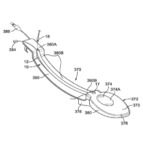
PCMJS2015/043940
equipped with the visualization device as described above, is placed in
patient's
airway and positioned under the patient's vocal cords under constant
visualization by
the visualization device 10.
Referring to Fig. 10, it depicts a side view of a supraglottic airway
.. device, generally 130. Any standard endotracheal tube known in the art and
an
endotracheal device of Fig. 3 is shown in the insert on the left of Fig. 10
can be used
in combination with the supraglottic airway device 130.
The supraglottic airway device 130 comprises a supraglottic tubal body
131 with a distal end 131A and a proximal end 131B and a lumen 146. The
supraglottic airway device 130 includes a designated intubation tube 133 which
is
inserted into the lumen 146 and into which an endotracheal device 50 can be
placed
as shown in Fig. 10. The distal end 133A of the intubation tube 133 ends with
an
elliptical opening 140 which is located distally from a cuff 132 which can be
inflated
with a device 134. The intubation tube 133 has a plurality of holes 148
distributed
throughout its body to allow ventilation from outlet 144 through tubal body
131.
While a standard endotracheal device, including an endotracheal
device 50, may be equipped with a visualization device, the supraglottic
airway
device 130 comprises its own visualization device 10 which is placed in the
lumen
146. The visualization device 10 comprises a camera tube 12 with a distal end
14
and a proximal end 16. The distal end 14 is sealed with a transparent material
17.
The camera tube 12 is sealed or otherwise attached externally to the
intubation tube
133 along the proximal-distal (131B-131A) axis. The supraglottic device 130
can be
further equipped with a bougie 116 which is located inside of the tube 118.
The tube
118 is placed inside of the lumen 146 and such that the distal end 116A of the
bougie 116 protrudes from the tube 118 and outside the supraglottic tubal body
131
through an elliptical opening 142 which is located on the supraglottic tubal
body 131
slightly proximally from the distal end 131A. The elliptic opening 142 of the
supraglottic tubal body 131 overlaps partially with the elliptic opening 140
of the
intubation tube 133. The bougie tube has its own opening through 140.
At the distal end 131A, the tubal body 131 is capped with a balloon 136
which can be inflated with a device 138. In some embodiments, the bougie 116
can
be replaced with a flexible guided stylet 82 shown on the right of Fig. 10.
In addition to the visualization device 10, the supraglottic device 130
can be also equipped with a sound- and temperature-monitoring device 38 which
is
22
CA 02957220 2017-02-02
WO 2016/022759 PCMJS2015/043940
located inside a tube 40 which is sealed or otherwise attached externally to
the tubal
body 131 along the proximal-distal (131B-131A) axis. The sound device 38 can
monitor patient's heart beat and breathing after the supraglottic device 130
is placed
inside of the patient. On its proximal end 131B, the tubal body 131 may be
connected to a ventilator (not shown) though an outlet 144. Because the
supraglottic
device 130 can ventilate in a closed circuit through the tubal body 131, an
endotracheal tube 50 can be placed inside of the intubation tube 133 without
the
need to stop ventilation and therefore, the supraglottic device 130 provides
continuous ventilation, continuous visualization in real time through the
visualization
device 10 and continuous sound and temperature monitoring by the sound
monitoring device 38 with a temperature probe. This real time information can
be
transferred or stored to multiple distant monitoring sites.
Other advantages for the supraglottic airway device include the ability
to intubate, extubate and to easily reintubate if needed under continuous
ventilation
and the ability to continuously visualize vocal cords and supraglottic
structures. The
device 130 is suitable for applications in children and adults. Further, the
device 130
is equipped with the cuff 132 for blocking the pharynx and the balloon 136
which
blocks the esophagus after the device 130 is placed in a patient. Furthermore,
an
endotracheal tube can be placed just proximal to the vocal cords in the tubal
body
133. This permits ventilation through 144 and tubal body 131 uninterrupted.
Referring to Figs. 11A and 11B, an alternative embodiment for an
airway device, generally 150, is provided. This device can be used in
pediatric and
adult patients as it is adoptable to different sizes. It provides continuous
visualization
of supraglottic structures and it can be advanced, retracted, or rotated, side
to side to
provide direct visualization of vocal cords. As can be appreciated from Figs.
11A
and 11B, the airway device 150 comprises a tubal body 152 with a distal end
152A
and a proximal end 152B and a lumen 153. The tubal body 152 may be connected
to a ventilator through an outlet 154. A visualization device 10 is sealed or
otherwise
attached inside of the tubal body 152 along the proximal-distal (152B-152A)
axis on
at least one side. The visualization device 10 comprises a camera tube 12 with
a
distal end 14 and a proximal end 16. The camera tube 12 is sealed at the
distal end
14 with a transparent material 17. The proximal end 16 of the camera tube 12
remains open and a camera 18 is inserted in the camera tube 12 through the
proximal end 16. The camera 18 does not come in contact with a patient's body
and
23
CA 02957220 2017-02-02
WO 2016/022759
PCMJS2015/043940
it does not have to be sterilized, it does not have to be disposable, although
it may
be disposable in at least some applications. The visualization device 10 can
be
further equipped with a light source which can be built in the camera tube 12
or be a
part of the camera 18. In alternative, a light source may be left outside the
camera
tube 12, but still shed enough light inside of the camera tube 12 for the
camera 18 to
obtain images inside of a patient's body.
An intubation tube 156 is placed inside of the lumen 153 of the tubal
body 152 along the proximal-distal (152B-152A) axis. The intubation tube 156
shares a lumen 119 with a bougie 116 which is inserted inside the lumen 119
along
the proximal-distal (152B-152A) axis such that a distal end 116A of the bougie
116
may protrude outside the tubal body 152 at the distal end 152A and proximal
end
116B may protrude outside the tubal body 152 and the proximal end 152B can be
used by a medical practitioner to guide the movement of the airway device 150
with
the bougie 116 during placement in a patient, including advancing the bougie
116
through patient's vocal cords under direct visualization by camera 18. The
intubation
tube 156 has a plurality of holes 157 distributed along the intubation tube
156.
At least in some embodiments, the airway device 150 is further
equipped with a sound- and temperature-monitoring device 38 which can be
inserted
in a tube 40 which is sealed or otherwise attached inside of the tubal b0dy152
along
the proximal-distal (152B-152A) axis such as the distal end of the sound-
monitoring
device 38 is positioned at or near the distal end 152A of the tubal body 152,
which is
also equipped with a cuff 158 along the perimeter of the tubal body 152 at the
distal
end 152A. The intubation tube 156 is designed such that at least in some
embodiments the intubation tube 156 has a ramp 160 at the distal end 152A of
the
airway device 150. A standard endotracheal tube, including those described in
various embodiments above, can be placed inside of the lumen 119 in the
intubation
tube 156 for positioning in a patient.
As shown in Fig. 11B, a ventilator adaptable cap 68 and a lid 69 are
attached to the tubal body 152 at the proximal end 152B. The endotracheal tube
is
.. inserted into the device 150 through the cap 68. Using the cap 68 with the
lid 69 on
the airway device 150 is preferred when ventilation is accomplished through an
outlet 154.
Yet another embodiment for an oral airway device, generally 170, is
provided as shown in Figs. 12 A, 12B and 12C. As can be appreciated from Fig.
24
CA 02957220 2017-02-02
WO 2016/022759
PCMJS2015/043940
12A, the airway device 170 comprises a tubal body 172 with a distal end 172A
and a
proximal end 172B. The tubal body 172 ends with two ramps 174 and 176 at the
distal end 172B. As can be appreciated from a side view in Fig. 12A and cross-
sectional views of the tubal body 172 in Figs. 12B and 12C, the tubal body 172
is
made of two half-cylinders 178 and 180. The half-cylinder 178 is slightly
smaller in
diameter than the half-cylinder 180. The tubal body 172 can be present in one
of the
two forms: as a full cylinder shown in Fig. 12B or as a half-cylinder as shown
in Fig.
12C. The half-cylinder 178 and the half-cylinder 180 are connected by means
such
that the half-cylinder 178 can rotate and retract into the half-cylinder 180.
The half-
cylinder form of Fig. 12C is achieved by the half-cylinder 178 rotating at
about 180
degrees and aligning with the half-cylinder 180 such that the half-cylinder
178 is
located inside of the half-cylinder 180 as shown in Fig. 12C.
A visualization device, generally 10, is sealed or otherwise attached
externally to the half-cylinder 180 along the proximal-distal (172B-172A)
axis. The
visualization device 10 comprises of a camera tube 12 with a distal end 14 and
a
proximal end 16. The distal end 14 is sealed with a transparent material 17. A
camera 18 is placed through an opening at the proximal end 16 into the camera
tube
12 and is moved inside the camera tube 12 to the distal end 14. Similarly to
all other
embodiments, the camera 18 does not come in contact with a patient's body, and
it
does not have to be disposable, does not have to be sterilized and it can be
reused
in multiple devices. The camera 18 is connected with wire 20 to at least one
monitoring device and it transmits images in real time. The camera 18 can be
connected wirelessly to at least one monitoring device which can be positioned
at
some remote location. A light source can be added as described in connection
with
the visualization device in other applications.
The half-cylinder 180 ends in two ramps 174 and 176 at the distal end
172A. The ramp 174 is smaller in size than the ramp 176 and the two ramps are
superimposed over each other such as the smaller ramp 174 is proximal to a
lumen
182 created by half-cylinders 178 and 180 when they are in the full-cylinder
form as
shown in Fig. 12B, while the ramp 176 is distal to the lumen 182. The ramps
174
and 176 are flexible and absorb the shock from sliding and releasing an
endotracheal tube which can be delivered into a patient by the oral airway
device
170. The ramps also facilitate the removal of the oral airway device 170 after
the
endotracheal tube is placed inside of the patient.
CA 02957220 2017-02-02
WO 2016/022759 PCMJS2015/043940
As shown in Fig. 12A, the oral airway intubating device 170 can be
further equipped with a bougie 160 which can be inserted into a tube 118 along
the
proximal-distal (172B-172A) axis such that a distal end 116A of the bougie 116
protrudes distally from the oral airway device 170 and a proximal end 116B
protrudes outside the oral airway device proximately and can be used to
manipulate
the distal end 116A of the bougie 116 such that it guides the movement of the
airway
device 170 during placement in a patient. The bougie tube 118 is located on
the
smaller half-cylinder 178 and it shares the lumen 182 with the tubal body 172.
A further embodiment provides a dilator with a visualization device,
generally 190 in Fig. 13. As can be appreciated from Fig. 13, the dilator 190
comprises a tubal body 192 with a proximal end 192B and a distal end 192A. A
certain distal portion of the tubal body 192 is tapered into a conical shape
192C such
that the opening at the distal end 192A of the tubal body 192 is significantly
smaller
in diameter in comparison to an opening at the proximal end 192B. A
visualization
device 10 is positioned inside of a lumen 195 of the tubal body 192 and along
the
proximal-distal (192B-192A) axis. The visualization device 10 may be sealed or
otherwise attached inside of the tubal body 192. The visualization device 10
is
essentially the same device as shown in Fig. 1A, and it comprises a camera
tube 12
with a proximal end 16 and a distal end 14. The distal end 14 of the camera
tube 12
is in close proximity with the distal end 192A of the tubal body 192. A camera
18
which can be either disposable or reusable is placed inside of the camera tube
12
through an opening at the proximal end 16 and all the way down to the distal
end 14
of the camera tube 12, which is sealed with a transparent material 17. Just
like other
embodiments, the visualization device 10 can be equipped with a light source
located outside of the dilator 194 or built in the camera tube 12. In some
embodiments, the light source can be built in the camera 18.
As shown in Fig. 13, the camera 18 is connected by electrical wire 20
to a monitoring device (not shown). In some embodiments, the camera 18 can be
in
communication with a monitoring device wirelessly. A guide wire at the
proximal end
194A is positioned inside of the lumen 195 of the tubal body 192. A proximate
end
194B of the guide wire 194 protrudes outside of the tubal body 192 at the
proximal
end 192B. The visualization device 10 verifies appropriate placement of the
dilator
device 190 and allows mobility of continuous visualization as dilation
proceeds. The
dilator device 190 is especially well suited for use with the Seldinger
technique.
26
CA 02957220 2017-02-02
WO 2016/022759 PCMJS2015/043940
Further embodiments provide various tracheostomy tubes equipped
with a visualization device. Fig. 14A depicts a side view of an embodiment for
a
tracheostomy device, generally 200. The device 200 comprises a tubal body 202
with a distal end 202A and a proximal end 202B. An inflatable cuff 204 is
wrapped
around the tubal body 200 in some proximity to the distal end 202A, but never
at the
very distal end 202A. The cuff 204 can be inflated with a device 206 after
proper
placement of the device 200 in a patient. At the proximal end 202B, the tubal
body
202 protrudes through a plastic plate 208 such that some portion of the tubal
body
202 is proximal to the plastic plate and will remain outside of a patient's
neck after
the device 202 is positioned in the patient. The plastic plate 208 may be oval
in
shape with the tubal body 202 protruding from the plate in the middle of the
oval
plastic plate 208. The plastic plate 208 may have two openings 209, one on
each
side of the plate such that the device 200 can be secured around patient's
neck with
some bandage by tying the device 200 through the openings 209 around patient's
neck.
In the embodiment of Fig. 14A, the visualization device 10 is sealed or
otherwise attached to the tubal body 202 externally. The visualization device
10
comprises a camera tube 12 which is sealed or otherwise attached externally
along
the proximal-distal (202B-202A) axis to the tubal body 202. The camera tube 12
is
.. placed under the cuff 204 such that the cuff 204 wraps over the camera tube
12 and
a distal end 14 of the camera tube 12 is distal to the cuff 204. The distal
end 14 is
sealed with a transparent material 17. A proximal end 16 of the camera tube 12
protrudes through the plastic plate 208 and remains outside of patient's neck.
A
camera 18 can be placed inside of the camera tube 12 through an opening in the
proximal end 16. The camera 18 is not disposable, does not need to be
sterilized
and can be easily removed from the camera tube 12. The camera 18 is connected
by electrical wire 20 to a monitoring device. In further embodiments, the
camera 18
can be in communication with a monitoring device wirelessly. A light source
can be
added to the visualization device 10 as was described in other embodiments
above.
Fig. 14B depicts another embodiment for a tracheosomy device,
generally 210. In this embodiment, the device 210 comprises of the same tubal
body
202, cuff 204, plate 208 and other components as was discussed in connection
with
the device 200. However, unlike the device 200, a visualization device 10 is
placed
inside of a lumen 203 of the tubal body 202. The visualization device 10
comprises
27
CA 02957220 2017-02-02
WO 2016/022759
PCMJS2015/043940
a camera tube 12 with a distal end 14 and a proximal end 16. The camera tube
12
may be sealed or otherwise attached internally to the tubal body 202 along the
proximal-distal (202B-202A) axis such as the distal end 14 of the camera tube
12 is
in close proximity with the distal end 202A of the tubal body 202. The distal
end 14
is sealed with a transparent material 17. A camera 18 is placed inside of the
camera
tube 12 through an opening at the proximal end 16 which remains outside of the
patient's neck after the device 210 is placed in the patient. The camera 18 is
connected by electrical wire 20 to a monitoring device. In other embodiments,
the
camera 18 communicates with a monitoring device wirelessly. In some
embodiments, the visualization device 10 comprises a light source.
A further embodiment provides a nasal trumpet with a visualization
device, generally 220 in Fig. 15. The trumpet 220 comprises a tubal body 222
with a
proximal end 222B and a distal end 222A. Two fasteners 224 are attached at the
proximal end 222B of the tubal body 222. After placing the trumpet 220 in a
patient,
.. the proximal portion of the tubal body 222 with the fasteners 224 remains
outside of
the patient, and the fasteners 224 can be used to secure the trumpet 220
around the
patient's head.
A visualization device 10 is sealed or otherwise attached to the tubal
body 222 externally along the proximal-distal (222B-222A) axis. The
visualization
device 10 comprises a camera tube 12 with a proximal end 16 and a distal end
14.
The distal end is in near proximity with the distal end 222A of the tubal body
222.
The distal end 14 is sealed with a transparent material 17. A camera 18 is
placed
inside of the camera tube 12 through an opening at the proximal end 16. The
camera 18 is moved all the way to the distal end 14 and collects images in
real time
inside of a patient's body during placement of the device 220 as well as after
the
device 220 has been properly placed and secured. As in other embodiments, the
camera 18 does not come in contact with patient's body, does not have to be
sterilized and can be reused in multiple devices or in different patients. The
camera
18 communicates with a monitoring device (not shown) either with electrical
wire 20
or wirelessly, or both.
Further embodiments provide various oral airways as shown in Figs.
16A, 16B, 16C, and 16D. Referring to Fig. 16A, an oral airway with a
visualization
device, generally 230, comprises a tubal body 232 with a lumen 233. The tubal
body
is slightly curved in a hook-like shape along the proximal-distal (232B-232A)
axis. A
28
CA 02957220 2017-02-02
WO 2016/022759
PCMJS2015/043940
visualization device 10 is placed inside of the lumen 233 of the tubal body
232. The
visualization device 10 comprises a camera tube 12 and a camera 18. The camera
tube 12 may be sealed or otherwise attached internally to the tubal body 232
inside
of the lumen 233 and along the proximal-distal (232B-232A) axis. The camera
tube
12 has a proximal end 16 and a distal end 14. The distal end 14 is in close
proximity
with the distal end 232A of the tubal body 232. The distal end 14 is sealed
with a
transparent material 17.
The camera tube 12 has an opening at the proximal end 16 through
which the camera 18 is inserted into the camera tube 12 all the way to the
distal end
14. The camera 18 communicates with a monitoring device either wirelessly or
by
electrical wire 20. The embodiment shown in Fig. 16B is the same as in Fig.
16A,
except a light source 21 is added to the visualization device 10. The light
source 21
may remain outside of the camera tube 12 or it may be built in the camera tube
12 or
it may be a part of the camera 18.
The embodiment shown in Fig. 16C is the same as that of the Fig. 16A,
except two whistles 234A and 234B are added inside of the lumen 233 of the
tubal
body 232. The whistle 234B is located at the proximal end of the tubal body
232 and
it produces a sound when a patient breathes in. The whistle 234A is located at
the
distal end of the tubal body 232 and it produces a sound when the patient
breathes
out.
Further embodiments include an oral airway as shown in Figs. 16A-
16C, but further equipped with a sound and temperature monitoring device which
is
also placed inside of the lumen 233 and transmits information to a monitoring
device
which can be positioned at a remote location.
The embodiment of Fig.16D is an intubating airway device with a
visualization device, generally 230. The intubating airway device 230
comprises a
tubal body 232 with a lumen 233 in which an endotracheal tube can be placed. A
visualization device 10 comprises a camera tube 12 with a distal end 14 and a
proximal end 16. The distal end 14 is sealed with a transparent material 17. A
camera 18 is placed inside of the camera tube 12 through an opening at the
proximal
end 16. The camera tube 12 is placed inside the lumen 233 of the tubal body
232.
A bougie 116 is added inside of the tubal body 232 such that the
bougie 116 is inserted in a tube 118 which shares the lumen with the lumen 233
along the proximal-distal (232B-232A) axis. A portion 116A of the bougie 116
29
CA 02957220 2017-02-02
WO 2016/022759
PCT/1JS2015/043940
protrudes outside the distal end 232A of the tubal body 232. A portion 116B of
the
bougie 116 protrudes outside the tubal body 232 from the proximal end 232B and
over vocal cords. An endotracheal tube can be positioned inside the lumen 233
and
the bougie 116 is used under constant visualization from the camera 10 to
guide the
placement of the endotracheal tube through patient's vocal cords. The distal
end 14
of the camera tube 12 is in proximity with the distal portion 116A of the
bougie 116
and therefore, the guided placement takes place under constant visualization.
Fig. 16E is the same intubating airway device with a visualization
device, 230 of Fig. 16D, but with an endotracheal tube 234 inserted inside of
the
lumen 233 of the tubal body 232 of the intubating airway device 230. As can be
appreciated from Fig. 16E, any endotracheal device can be easily inserted and
removed by sliding through the lumen 233. Thus, the intubating airway device
230
can be used to intubate, extubate and reintu bate under continuous
visualization.
The device provides continued visualization during intubation and extubation
as well
as during ventilation via the camera 18 in the camera tube 12. Thus, the
device 230
can be used for intubating and extubating without lifting the patient's
mandible,
tongue or soft tissues of the oral airways and this method is one of the
embodiments.
Figs. 16F and 16G provide a further embodiment for an oral airway
intubating/extubating device. As can be seen from the figures, a handle 236
can be
connected to the proximal end 232B of the oral airway intubating/extubating
device
230. The handle 236 can be made of any suitable sturdy material such as for
example, plastic, wood or metal. The handle 236 can be of any shape and size
that
would provide a sufficient grip for a hand of a medical provider. In some
embodiments, the handle 236 is a cylinder. In other embodiments, it can be
made in
any other shape suitable for gripping by a hand. On one end, the handle 236
has a
means 237 for attaching to a holder 238 which is connected the proximal
surface
232C of the oral airway intubating/extubating device 230, as shown in Fig.
16G. The
means 237 can be made in any shape suitable for connection to the holder 238.
In
some embodiments, the means 237 can be in a shape of a cylinder as shown in
Fig.
16F. In some embodiments, the holder 238 can be in a shape of a half-cylinder
slightly bigger in diameter than the means 237 as shown in Fig. 16F such as
that the
means 237 fits tightly inside the holder 238.
When the handle 236 is connected to the holder 238, a medical service
provider can use the handle 236 to lift the patient's mandible and tongue.
Thus,
CA 02957220 2017-02-02
WO 2016/022759 PCT/1JS2015/043940
intubation/extubation can be easily accomplished on any patient, including
patients
who are unconscious and by any medical service provider, including those who
are
of small physical statute. Because the handle 236 can be disconnected from the
holder 238, the handle 236 can be removed after it is no longer in use. It can
be
reconnected with the holder 238 later if needed for further manipulations of
the
patient's lower jaw and/or tongue.
Figs. 16 H and 16J-16P provide a further embodiment for an oral airway
intubating/extubating device with a rotating central passageway, generally
350. The
oral airway intubating/extubating device with a rotating central passageway
comprises of two half-cylinders, an inner half-cylinder 351 and outer half-
cylinder
352. The half-cylinder 351 is smaller in diameter and fits inside the larger
half-
cylinder 352 such as the inner half-cylinder 351 can slide proximally and
distally
inside the outer half-cylinder 352, as shown in Fig. 16J. At least in some
applications, the inner half-cylinder 351 can be removed completely from the
device
350, such as only the outer half-cylinder 352 remains inserted in a patient,
as shown
in Fig. 16H.
As can be appreciated from Fig. 16J, the inner half-cylinder 351 may
be longer than the outer half-cylinder 352 and the inner half-cylinder 351 can
slide
distally and proximally inside the outer half-cylinder 352. As can be
appreciated from
Figs. 16H and 16J, a camera tube 12 can be attached externally along the outer
half-
cylinder 352 in some embodiments and a camera 18 can be placed inside the
camera tube 12 and provide continuous visualization during intubation and
extubation.
In other embodiments, the camera tube 12 can be positioned inside the
inner half-cylinder 351, as shown in Fig. 16K such that the camera 18 provides
continuous visualization during intubation and extubation.
As can be appreciated from Figs. 16L-16N, the inner half-cylinder 351
can rotate inside the outer half-cylinder 352 such that the two half-cylinders
may
create a completely enclosed passageway as shown in Fig. 16N or the half-
cylinders
may create a passageway which is not fully enclosed and remains open on at
least
one side as shown in Fig. 16L. In some embodiments, the inner half-cylinder
351
may have at least one retractable extension 353 which when extended outside
the
half-cylinder 351 locks the half-cylinder 351 in a position on the half-
cylinder 352 and
31
CA 02957220 2017-02-02
WO 2016/022759
PCT/1JS2015/043940
prevents the half-cylinder 351 from sliding further distally along the outer
half-
cylinder 352.
Figs. 160 and 16P depict two different ways of inserting an
endotracheal tube 354 inside the oral airway intubating/extubating device with
a
rotating central passageway 350. As shown in Fig. 160, the endotracheal tube
354
can be conveniently placed inside the rotating central passageway of the oral
airway
intubating/extubating device 350. In alternative and as shown in Fig. 16P, the
endotracheal tube 354 can be first placed inside the inner half-cylinder 351
which is
then inserted insider the outer cylinder 352. This assembly permits
flexibility and
makes insertion of endotracheal tubes of various sizes, including small
pediatric
endotracheal tubes, very accurate and under constant visualization of a
camera.
Further and because the inner half-cylinder 351 can slide along the proximal-
distal
axis, the intubation can be accurate and customized for a particular patient
to fit the
patient's size and anatomy. Alternatively, the device 250 may be placed over
the
endotracheal tube 354 which is already in place in a patient in order to
provide
constant visualization and a conduit for extubation and possible reintubation.
Further embodiments provide a supraglottic airway device with a
visualization device. Referring to Fig. 17A, it depicts a supraglottic airway
device
with a visualization device, generally 240. It comprises a body 242 with a
lumen 243
into which a sound-monitoring device 246 is placed. The body 242 may have a
cylinder-like shape tapered at the distal end, and with the distal end the
body 242
protruding with a tongue-like tip 248. At least a portion of the body 242
surface is
slightly curved toward the lumen 243 and creates a surface 249. The
visualization
device, generally 10, comprises a camera tube 12 with a distal end 14 which is
sealed with a transparent material 17, and a camera 18 which is placed inside
of the
camera tube 12 through an opening at the proximal end of the camera tube 12.
The
camera tube is positioned externally on surface 249 along the distal-proximal
axis of
the body 242. The camera tube 12 is connected with the surface 249 by a
sliding
means 245 such that the camera tube 12 can slide along the 242A-242B axis on
the
surface 249.
The camera 18 is placed inside of the camera tube 12 and because the
camera tube 12 is sealed at the distal end 14, the camera 18 does not come in
contact with a patient and the camera 18 does not need to be sterilized and it
can be
reused in other applications. The camera 18 is connected by electric wire 20
or
32
CA 02957220 2017-02-02
WO 2016/022759
PCMJS2015/043940
wirelessly to a monitoring device. The camera 18 is not disposable and can be
reused in other applications.
The device 240 is further equipped with a bougie tube 244 which is
also located on the surface 249 and is connected to the surface 249 with a
sliding
means 245 such that the bougie tube 244 can slide along the 242A-242B axis.
Fig.
17B provides an alternative embodiment for the device 240, in which the sound
and
temperature monitoring device 246 can protrude through an opening at the 248
tip.
A further embodiment provides a one-piece video-laryngoscope with a
visualization device, generally 250, as shown in Figs. 18A and 18B. The video-
laryngoscope 250 comprises a tubal body 252 which has a proximal end 252B and
a
distal end 252A. The tubal body 252 extends with a scoop-like portion 254 at
the
distal end 252A. The scoop-like portion 254 curves horizontally such that the
distal
end 254A of the scoop-like portion 254 is nearly parallel to the proximal end
254B of
the scoop-like portion. The tubal body 252 has an opening 256 near its distal
end
252A. A visualization device 10 which comprises a camera 18 placed inside of a
camera tube 12 is placed through the opening 256 such that the distal end 14
of the
camera tube 12 may be in proximity with the proximal end 254A of the scoop-
like
portion 254. The device can be easily inserted to an upper esophagus and
visualize
vocal cords.
However, the position of the distal end 14 can be adjusted as needed
by sliding the camera tube 12 through the opening 256. The distal end 14 is
sealed
with a transparent material 17 such that the camera 18 does not come in
contact
with a patient's body and therefore, the camera 18 does not need to be
sterilized and
it can be reused in multiple applications. The camera 18 is inserted into the
camera
tube 12 through an opening at the proximal end 16. The camera 18 is connected
to
at least one monitoring device either by electrical wire 20 or wirelessly.
The embodiment of Fig. 18B is the same as that of Fig. 18A, except a
light source 21 is added to the visualization device 10 as described in
connection
with the light source 21 in other medical devices above. A bougie 116 in a
tube 118
is also added through the opening 256, and the distal end 116A of the bougie
116
can be manipulated at the proximal end 116B such that the placement of the
device
250 is guided under continuous visualization with the camera 18.
A further embodiment includes a vaginal speculum with visualization
device, generally 260 as shown in Fig. 19. Any speculum 261, including
disposable,
33
CA 02957220 2017-02-02
WO 2016/022759 PCMJS2015/043940
generally known and used for a pelvic exam can be equipped with a
visualization
device, generally 10, which comprises a camera 18 placed inside of a camera
tube
12. The camera tube 12 can be attached to the speculum 261 or to some other
instrument. The camera 18 is placed in the camera tube 12 through an opening
at a
proximal end 16 and is moved all the way toward the distal end 14 which is
sealed
with a transparent material 17. The camera 18 connected to at least one
monitoring
device by electric wire 20 or the camera 18 can be connected wirelessly.
Further embodiments relate to various tubing equipped with a
visualization device shown in Fig. 1A and as described in more detail below.
Figs. 20A-20F refer to various embodiments for a nasal cannula with a
visualization device, generally 270. Fig. 20A shows the positioning of a nasal
cannula 272 on patient's head with a visualization device 10 added to one of
the two
nostril tubes. The nasal cannula 272 can be any nasal cannula known in the art
and
used by medical practitioners. The visualization device 10 is as described in
connection with Fig. 1A and comprises a camera 18 inserted inside of a camera
tube
12. The camera tube 12 is sealed or otherwise attached externally along at
least
one nostril tube 274 of the nasal cannula 272 as shown in more detail in Fig.
20C.
This nasal cannula with the visualization device 10 provides continuous
visualization
of vocal cords, upper esophagus. The cannula can be used to determine whether
vocal cords are moving correctly, if there is any abnormal anatomy and the
color of
the patient's tissues.
As shown in Fig. 20E, the nasal cannula with the visualization device
can be properly positioned through patient's nostrils as the positioning is
guided and
constantly visualized with the camera device 10. The distal end 14 of the
camera
tube 12 aligns with the distal end of at least one nostril tube 274. As shown
in Fig.
20F, the nasal cannula with the visualization device can be further equipped
with an
external stethoscope 275, which can be placed on patient's chest externally
and
monitors breathing and heart-beat sounds.
Figs. 20B and 20D show the same embodiment as in Figs. 20A and
.. 20C, except the visualization device 10 is equipped with a light source 21
as was
described in connection with the light source 21 in other embodiments.
A further embodiment includes a feeding tube with a visualization
device, generally 280, as shown in Fig.21. A visualization device, generally
10, is
sealed or otherwise attached externally along the proximal-distal (282A-282B)
axis of
34
CA 02957220 2017-02-02
WO 2016/022759
PCMJS2015/043940
a feeding tube 282. The visualization device 10 is essentially the same as
described
in connection with Fig. 1A and other embodiments above. It comprises a camera
tube 12 with a proximal end 16 and a distal end 14. A camera 18 with wire 20
is
inserted into the camera tube 12 through an opening at the proximal end 16 and
is
slid all the way to the distal end 14 which is sealed with a transparent
material 17.
The camera 18 does not come in contact with a patient's body and can be reused
in
multiple devices. Any feeding tubes known in the art can be used in this
embodiment, including a feeding tube with a stylet 284 as shown in Fig. 21.
The
feeding tube 282 can be equipped with an adaptor 283 at the proximal end 282A.
The feeding tube 282 may further comprise a plurality of holes 285 at the
distal end
282B for food distribution.
Further embodiments provide various suction tubes equipped with a
visualization device, generally 290, as shown in Figs. 22A, 22B and 22C. Any
suction tube including but not limited to the nasal gastric tubes known in the
art can
be used and in general a suction tube 292 with an adaptor 293 at a proximal
end
292B is suitable, as shown in Fig. 22A. A visualization device, generally 10,
comprises a camera tube 12 and a camera 18 with wire 20. The camera 18 is
inserted into the camera tube 12 through an opening at a proximal end 16 and
is slid
all the way to the distal end 14 of the camera tube 12. The distal end 14 is
sealed
with a transparent material 17. The camera 18 can transmit information to a
remote
location.
The camera tube 12 is placed inside of the suction tube 292 through an
opening 294 at the proximal end 292B of the suction tube 292. The camera tube
12
is then aligned with the length of the suction tube 292 such that the distal
end 14 of
the camera tube 12 is in close proximity with the distal end 292A of the
suction tube
292.
Figs. 22B and 22C is a further embodiment of a suction tube with a
visualization device, generally 290 as shown in Fig. 22A, but it is further
equipped
with a bougie 116 placed inside of a tube 118 which is placed inside of the
suction
tube 292 through an opening 295. A distal end 116A of the bougie 116 can
protrude
outside the distal end 292A of the suction tube 292 and can be manipulated by
a
medical practitioner with a proximal end 116B which protrudes outside a
patient such
as the placement of the suction tube 292 is guided under constant
visualization with
the camera 18 through the distal end 14 of the camera tube 12. The bougie 116
CA 02957220 2017-02-02
WO 2016/022759
PCT/1JS2015/043940
under constant visualization from the camera 12 permits rapid and accurate
placement of the device 290 in a patient. The bougie 116 can be used to guide
the
placement of the device 290 and moving it left or right in the trachea.
Referring to Fig. 23, this embodiment provides a suction catheter with a
visualization device, generally 300. The suction catheter 302 is not flexible
and can
be any suction catheter known in the art. A visualization device, generally
10, is
positioned inside the suction catheter 302 through an opening 303 which is in
near
proximity with a proximal end 302B of the suction catheter 302. The
visualization
device 10 comprises a camera 18 with wire 20 which is placed inside of a
camera
tube 12 through an opening at a proximal end 16 of the camera tube 12 and then
the
camera 18 is slid to the distal end 14 which is sealed with a transparent
material 17.
The distal end 14 of the camera tube 12 is aligned with the distal end 302A of
the
suction catheter 302, while the proximal end 16 of the camera tube 12
protrudes
outside the patient's body such as the camera 18 can be pulled out from the
camera
tube 12 as needed. In other embodiments, the suction tube is placed externally
and
this combination can work with suction caps.
Referring to Fig. 24, this embodiment provides an endotracheal
changing tube with a visualization device, generally 310. An endotracheal
changing
tube can be any endotracheal changing tube as known in the art. A
visualization
device, generally 10, comprises a camera 18 with wire 20 which is placed
inside of a
camera tube 12 through an opening at a proximal end 16 of the camera tube 12
and
slid all the way to a distal end 14 of the camera tube 12. The distal end 14
is sealed
with a transparent material 17. The visualization device 10 is placed inside
of the
endotracheal changing tube 312 through an opening 313 in the changing tube 312
such as the camera tube 12 is aligned with the changing tube 312 along the
proximal-distal (312B-312A) axis, and the distal end 14 of the camera tube 12
is in
close proximity with the distal end 312A of the changing tube 312. In other
embodiments, the camera tube 12 can be placed outside of the endotracheal
changing tube or it can be fitted externally onto an endotracheal changing
tube
known in the art.
Further embodiments include a supraglottic ventilating tube with
camera, generally 360 as shown in Figs. 25A-25D. As can be appreciated from
Fig.
25A, the device 360 comprises a tube 361 with the distal end 361A and the
proximal
end 361B. The device 360 is equipped with a visualization device 10 as
described in
36
CA 02957220 2017-02-02
WO 2016/022759 PCMJS2015/043940
connection with Figs. 1A, 1B and 1C and which comprises a camera tube 12
attached externally to the device 360. A disposable camera 18 can be inserted
into
the camera tube 12 along with a light source 362. In proximity to the distal
end
361A, the device 360 comprises an inflatable cuff 364 which wraps around the
tube
361. The cuff 364 can be inflated with a means 366 after the esophageal camera
tube is positioned in a patient.
As can be appreciated from Fig. 25B, the camera tube 18 is positioned
under the cuff 364 such that the cuff 364 wraps over the camera tube 12. The
camera tube 12 can slide along the tube 361 such that images with the camera
18
can be taken either proximally or distally from the cuff 364 after the cuff
364 is
inflated in the patient.
As shown in Figs. 250 and 25D, the device 360 can be placed inside
the intubating/extubating oral airway device 350, including the
intubating/extubating
oral airway device 350 shown in Figs. 16H-16K. As can be appreciated from Fig.
25D, the device 360 can be easily inserted into a patient with help of the
intubating/extubating oral airway device 350. Supraglottic ventilating tube
can be
positioned under the direct and continuous visualization into patient's
hypopharynx.
The device 350 can be removed after the insertion is completed.
Further embodiments provide a tubeless intubating device shown in
Figs. 26A-26J, generally 370. The tubeless intubating device 370 has the upper
surface shown in Fig. 26A and the bottom surface shown in Fig. 26B. The
tubeless
intubating device 370 comprises an ellipsoid body 372 which has an upper oval
surface 373 with a lumen opening 374A on the upper oval surface 373 of the
ellipsoid body 372 as shown in Fig. 26A. The upper oval surface 373 is
connected to
the bottom half-ellipsoid surface 378 as shown in Fig. 26A and Fig. 26B such
that the
distal end 376 of the ellipsoid body 372 is tapered because the bottom half-
ellipsoid
surface 378 is tapered at the distal end 376. The ellipsoid body 372 encloses
a
lumen 374 which opens onto the upper oval surface 373 with the lumen opening
374A. The lumen 374 opens on the proximal side of the bottom half-ellipsoid
surface
378 with a canal 379 which connects to the bottom half-ellipsoid surface 378
and
extends beneath the bottom half-ellipsoid surface 378. The bottom half-
ellipsoid
surface 378 also connects to a handle 380.
The handle 380 comprises three parts connected together: the
proximal part 380A, the middle part 380B and the distal part 3800. The
proximal
37
CA 02957220 2017-02-02
WO 2016/022759
PCMJS2015/043940
part 380A may be made in flat rectangle shape with a ring-holder 382 attached
on
the bottom surface of the proximal part 380A. The 380A part bends down at
about a
90 degree angle at its distal part where it connects to the middle part 380B.
The
middle part 380B is also of flat rectangle shape and may vary in length. As
can be
seen from Fig. 26B, the canal 379 is connected to the bottom surface of the
middle
part 380B at the distal portion of the middle part 380B. The middle part 380B
connects to the distal part 380C at its distal end. The middle part 380B bends
up at
about a 90 degree angle at the distal part and connects to the distal part
380C. The
distal part 380C connects by its distal end to the oval upper surface 373 of
the
ellipsoid body 372.
A visualization device, generally 10, comprising the camera tube 12
and camera 18 which can be inserted into the camera tube 12, is attached to
the
handle 380 on its upper surface such as the visualization device 10 extends
along
the handle 380 from its proximal end 380A at which the camera 18 is inserted
insider
the camera tube 12 and all the way into the distal portion 380C. Just like in
other
embodiments, the camera tube 12 is sealed with a transparent material 17 at
its
distal end such as the camera 18 does not come into direct contact with the
patient's
body and can be reused. Just like in other embodiments, the camera tube 18 can
slide along the proximal-distal axis of the handle 380. The visualization
device 10
may further comprises a light source 384 which can be inserted into the camera
tube
12 along with the camera 18. In some embodiments the ellipsoid body 372 can
comprise an inflatable cuff 385 which can be inflated with a means 386.
In some embodiments, the handle 380 can be made of flexible
material. In other embodiments, the tubeless intubating device 370 can be
designed
without the cuff 385. In some embodiments, the camera tube 12 is fixed to the
handle 380. In some embodiments, the camera tube 12 includes a light source.
In
other embodiments, the camera tube 12 has no additional light source.
The tubeless intubating device 370 can be used for intubating a patient
with an endotracheal tube of any size under continuous visualization of the
camera
18. The tubeless intubating device 370 can be also used for extubation and for
reintubation of a patient. It can also act as a supraglottic device with an
endotracheal tube inflated with the cuff 385.
As shown in Fig. 260, an endotracheal tube 388 can be loaded onto
the tubeless intubating device 370 such that the proximal end of the
endotracheal
38
CA 02957220 2017-02-02
WO 2016/022759 PCMJS2015/043940
tube 388 is secured on the tubeless intubating device 370 with the ring holder
382. It
will be appreciated that any endotracheal tube can be loaded into the device
370 and
inserted into a patient. As can be seen in Fig. 260, the endotracheal tube 388
in this
embodiment is equipped with a cuff 390. The endotracheal tube cuff 390 can be
inflated after it is loaded into the device 370 and secured in place with the
ring holder
382. It will be appreciated that the embodiments of the device 370 shown in
Figs.
26A-26C are equipped with the cuff 385 and the cuff 385 of the structure can
be
inflated in the hypopharynx.
The distal end of the endotracheal tube 388 is then passed through the
canal 379 and through the lumen 374 such that the distal end of the
endotracheal
tube 388 protrudes from the lumen opening 374A on the upper oval surface of
the
ellipsoid body 372, as shown in Fig. 26D and 26E. The endotracheal tube 388
can
slide along the proximal-dorsal axis of the tubeless intubating device 370
such that a
longer or shorter portion of the endotracheal tube 388 protrudes from the
lumen
opening 374A.
As can be further appreciated from Fig. 26F, after the tubeless
intubating device 370 delivers and assists in placing the endotracheal tube
388 in the
patient under direct and continuous visualization, the tubeless intubating
device 370
can be easily removed from the patient while the endotracheal tube 388 remains
safely in place under continual vision. Thus, the tubeless device 370 can be
used
with a standard endotracheal tube to intubate and extubate a patient. The
device
370 can be also used as a supraglottic device.
Further embodiments of the tubeless intubating device 370 are shown
in Figs. 26G-26J. As shown in Fig. 26G and can be particularly appreciated
from
Fig. 26H, the tubeless intubating device 370 in these embodiments does not
comprise a cuff. Nevertheless and just like the tubeless intubating device 370
of
Fig.26A, the device 370 of Fig. 26G has the upper surface shown in Fig. 26G
and
the bottom surface shown in Fig. 26H. The tubeless intubating device 370
comprises an ellipsoid body 372 which has an upper oval surface 373 with a
lumen
opening 374A on the upper oval surface 373 of the ellipsoid body 372 as shown
in
Fig. 26G. The upper oval surface 373 is connected to the bottom half-ellipsoid
surface 378 as shown in Fig. 26G and Fig. 26H such that the distal end 376 of
the
ellipsoid body 372 is tapered because the bottom half-ellipsoid surface 378 is
tapered at the distal end 376. The ellipsoid body 372 encloses a lumen 374
which
39
CA 02957220 2017-02-02
WO 2016/022759 PCMJS2015/043940
opens onto the upper oval surface 373 with the lumen opening 374A. The lumen
374 opens on the proximal side of the bottom half-ellipsoid surface 378 as
shown in
Fig. 26H. The bottom half-ellipsoid surface 378 also connects to a handle 380.
This
embodiment of the device 370 does not comprise a canal.
The handle 380 comprises three parts connected together: the
proximal part 380A, the middle part 380B and the distal part 380C. The
proximal
part 380A may be made in flat rectangle shape with a ring-holder 382 attached
on
the bottom surface of the proximal part 380A. The 380A part bends down at
about a
90 degree angle at its distal part where it connects to the middle part 380B.
The
middle part 380B is also of flat rectangle shape and may vary in length. As
can be
seen from Fig. 26B, the canal 379 is connected to the bottom surface of the
middle
part 380B at the distal portion of the middle part 380B. The middle part 380B
connects to the distal part 380C at its distal end. The middle part 380B bends
up at
about a 90 degree angle at the distal part and connects to the distal part
380C. The
distal part 380C connects by its distal end to the oval upper surface 373 of
the
ellipsoid body 372.
A visualization device, generally 10, comprising the camera tube 12
and camera 18 which can be inserted into the camera tube 12, is attached to
the
handle 380 on its upper surface such as the visualization device 10 extends
along
the handle 380 from its proximal end 380A at which the camera 18 is inserted
insider
the camera tube 12 and all the way into the distal portion 380C. Just like in
other
embodiments, the camera tube 12 is sealed with a transparent material 17 at
its
distal end such as the camera 18 does not come into direct contact with the
patient's
body and can be reused. Just like in other embodiments, the camera tube 12 can
slide along the proximal-distal axis of the handle 380. The visualization
device 10
may further comprises a light source 384 which can be inserted into the camera
tube
12 along with the camera 18.
As shown in Figs. 261 and 26J, the device 370 can be used for
intubation and extubation of a patient with a supraglottic airway, generally
392 in
Figs. 261 and 26J. The device 370 can also be used for placement of a
laryngeal
mask airway. As shown in Fig. 26J, the supraglottic airway 392 is secured in
the
device 370 with the ring holder 382 such that the distal end of the device 392
is
aligned with the distal end of the device 370 and the lumen 394 of the device
392 is
aligned with the lumen 374 of the device 370 and secured. Because the assembly
of
CA 02957220 2017-02-02
WO 2016/022759
PCMJS2015/043940
the devices 370 and 392 is equipped with the camera device 10, it provides
continuous visualization of patient's supraglottic structure during placement.
Further embodiments provide a sliding endotracheal cuff device,
generally 400 as shown in Figs. 27A-27G. As can be appreciated from Figs. 27A
and 27B, an endotracheal tube 410 comprises a tube 412 with a proximal end
412A
and a distal end 412B with a rail 414 along the proximal-distal axis (412A-
412B)
designed such that the endotracheal tube 410 fits inside the device 400 and
the rail
414 fits into the rail 404 of the device 400. The device 400 can then slide
along the
proximal-distal axis 412A-412B on the endotracheal tube 410 as shown in Fig.
27B.
The details of the device 400 design are further explained in connection with
Figs.
27C-27F. As can be appreciated from 27C, the rail 404 has a groove 405 facing
inside the tube 402. The groove 405 is designed such that the rail 414 of the
endotracheal tube 410 fits inside the groove 405 and can slide along the rail
404.
The rail design permits to easily remove the endotracheal tube 410 from the
device
400 while the device 400 remains in place in a patient. In alternative, the
device 400
can be removed, while the endotracheal tube 410 remains in place in the
patient.
Because the device 400 remains in place, changing from one endotracheal tube
410
to another can be easily accomplished. Further because the cuff 406 is
presented
on a separate device, an endotracheal component can remain in place if what
needs
to be replaced is only a cuff. While in drawings of Fig. 27, the device 400 is
shown a
cylinder, this device can be a half-cylinder in other embodiments.
As can be further appreciated from Fig. 27D, some embodiments for
the device 400 comprise an additional tube 416 which can be used for
delivering
drugs, suction and tools such as for example forceps and bougie. Further
embodiments for the device 400 include an embodiment of Fig. 27E in which the
cuff
406 can move along the rail 404. Yet further embodiments for device 400
include an
embodiment of Fig. 27F, where the distal portion 402A of the tube 402
comprises
mesh 418. As can be further appreciated from Fig. 27F, the cuff 406 can
inflate over
the mesh 418.
As can be appreciated from Fig. 27G, the camera tube 12 may be fixed
along the body 402 or it can slide proximally and distally along the body 402.
Overall, the device 400 prevents a problem of broken seal after the device has
been
in place in a patient for a period of time.
41
CA 02957220 2017-02-02
WO 2016/022759 PCMJS2015/043940
Further embodiments provide an endotracheal tube, generally 430, as
shown in Figs. 28A and 28B. The device 430 comprises a tube 432 with the
proximal end 432A and the distal end 432B. A visualization device 10 is
positioned
along the proximal-distal axis (432A-432B) of the tube 432. As can be
appreciated
from Fig. 28B, the visualization device 10 comprises a camera tube 12 sealed
with a
transparent material 17 at the distal end. A camera 18 can be placed inside
the
camera tube 12. The camera tube 12 comprises a rail 434 running along the
camera
tube 12. The tube 12 further comprises a half-cylinder 436 attached on the
side
opposite to the side of the tube 12 to which the rail 434 is attached. The
half-cylinder
436 has a diameter such that the visualization device 10 can be easily snap
over the
tube 432 of the endotracheal tube 430 with the half-cylinder 436. Thus, the
visualization device 10 in this embodiment can be easily assembled with any
endotracheal tube and it can also be easily removed from the endotracheal tube
when visualization is no longer needed. Importantly, the camera tube can be
detached at anytime. Thus, this device is similar to a laryngoscope, yet the
camera
tube can be removed at any time if no longer needed. Thus, one of the
advantages
of this device is a rail which permits gliding along any other tubal device.
Further embodiments provide a sliding camera tube with rail, generally
440 as shown in Figs. 29A-29C. As can be appreciated from Fig. 29A, the camera
tube 440 comprises a tube into which a camera 18 can be inserted. The tube 12
is
sealed at the distal end with transparent material 17. A rail 442 runs along
the tube
12. The tube 12 is attached to a half-cylinder 444 which has a diameter such
as the
camera tube 440 can be easily assembled with an endotracheal tube or any other
tube and it can glide along the endotracheal tube or any other tube. As can be
appreciated from Fig. 29A, the camera tube 440 can easily slide into a
laryngoscope
which is equipped with a rail 448 along which the camera tube 440 can slide.
As can
be appreciated from Fig. 29B, after the camera tube 440 is positioned on the
laryngoscope, the camera 18 can be inserted into the tube 12.
As can be further appreciated from Fig. 29C, the camera tube 440 can
be easily removed from the laryngoscope and assembled with any other device.
For
example, the camera tube 440 can be placed on an endotracheal tube as was
discussed in connection with Fig. 28. Thus, the device provide intubation
during
laryngoscopy and after the removal of the device 440 from the device 448.
42
CA 02957220 2017-02-02
WO 2016/022759
PCMJS2015/043940
Further embodiments provide a supraglottic airway device with built in
endoscope guide, generally 450, as shown in Figs. 30A and 30B. As can be
appreciated from Fig. 30A, the device 450 comprises a camera tube 12 which is
positioned externally long the body of the device 450. The device 450 is
further
equipped with a half-cylinder endoscope guide 452 which runs externally along
the
body 451of the device 450, beneath a cuff 454 and creates a semi-lumen. The
device 450 can be used for placing a supraglottic airway with an endoscope in
place.
The device 450 is adoptable to endoscopes of all sizes. The device 450 can be
placed after an endoscope is already inserted in a patient. The device 450 is
able to
ventilate a patient under continuous visualization during endoscopy. As can be
further appreciated from Fig. 30B, the device 450 can be positioned in the
patient's
oral cavity 456 and then it can be easily removed from the endoscope semi-
lumen
452.
Figs. 31A and 31B provide further embodiments for an assembly with a
naso-gastric tube with a visualization device 10, generally 460. The
visualization
device 10 comprises a camera tube 12 with a camera 18 which can be inserted
inside the camera tube 12. The naso-gastric tube 461 comprises a valve 462.
The
assembly 460 further comprises an oxygen tube with perforations, 464, which
can be
connected to a source of oxygen 468. As can be appreciated from Figs. 31A and
31B, the perforations 466 are located at the distal end of the tube 464 and
this
permits localization of the perforations in the mid pharynx to deliver oxygen
to a
patient as shown in Fig. 31A. The length of the camera tube 12, oxygen tube
462
and naso-gastric tube 461 are calculated such that the assembly is flexible
and the
camera tube 12 can be located in the mid pharynx. However, the camera tube 12
can slide on the gastric tube 461 to travel to the distal gastric tube 461
providing
continuous visualization of patient's gastric organs. A person of skill will
appreciate
that in some embodiments, the assembly 460 comprises a naso-gastric tube 461
as
shown in Fig. 31A, while in other embodiments, the assembly 460 comprises a
feeding tube 470 in place of the naso-gastric tube 461.
Fig. 32 is a further embodiment of an oral airway with a camera device,
generally 480. The oral airway comprises a tubal body 482 with a lumen 484
similar
to tubal bodies of other oral airways described in this disclosure. The camera
device
10 comprises a camera tube 12 which is sealed at its distal end with
transparent
material 17 and into which a camera 18 can be placed.
43
CA 02957220 2017-02-02
WO 2016/022759
PCMJS2015/043940
The camera device 10 is placed inside the lumen 484. The oral airway
device 480 can provide continuous visualization of patient's supraglottic
structure
including the vocal cords in patients with positive ventilation pressure and
also in
patients ventilating spontaneously. The oral intubating device 480 can be used
to
place an endotracheal tube through the vocal cords without lifting the
mandible. As
in previous embodiments, the device 480 provides a continuous visualization
after
the endotracheal tube is placed and during extubation. It should be understood
that
this camera tube can slide proximal and distal to the tip of the device 480. A
bougie
486 is also placed inside the lumen 484 and it can be moved along the proximal-
distal axis and guide positioning of the camera device 10 which also can move
along
the proximal-distal axis. The device 480 further comprises a suction catheter
488
which is also placed inside the lumen 484 and which can move along the
proximal-
distal axis inside the lumen 484. As can be appreciated by a person of skill,
an
endotracheal tube can be placed inside the lumen 484 to intubate a patient.
Figs. 33A-33C are further embodiments of an endotracheal tube 490
with an externally attached camera tube 12 into which a camera 18 can be
placed,
and a suction tube 492 which is also attached externally to the endotracheal
tube
490 as shown in Fig. 33A. As shown in Fig. 33B and Fig. 33C other devices can
be
externally attached to the endotracheal tube 460, such as a medication
dispensing
device 494 as shown in Fig. 33B and biopsy forceps 496 as shown in Fig. 33C.
Additionally, a ventilating tube can be placed in can be added to the
assembly. An
additional balloon can be added circumferentially around the camera tube to
provide
ventilation. This system can be also used to separate ventilation between the
left
and right bronchus. Because the camera tube 12 and the suction tube 492 are
secured on the endotracheal tube 490 with a set of rings, the attachment is
flexible
and each of the camera tube 12 and suction tube 492 can rotate 360 degrees
around the endotracheal tube 490. In addition to be able to rotate around the
endotracheal tube 490, the camera tube 12 and suction tube 492 can slide along
the
proximal-distal axis of the endotracheal tube. The camera tube 12 is sealed at
the
distal end with transparent material 17 and this allows the camera 18 to
visualize the
external structures in the airway including but not limited to the vocal
cords.
Figs. 34A-34H are further embodiments of an oral airway device,
generally 500, that allows for continuous visualization of the patient's vocal
cords.
As shown in Fig. 34A, the device comprises a tubal body 502 with a lumen 504
into
44
CA 02957220 2017-02-02
WO 2016/022759
PCMJS2015/043940
which an endotracheal tube 506 or any other similar device can be inserted.
The
tubal body 502 has a proximal end 502A and a distal end 502B. The
visualization
device 10 is positioned along the tubal body 502 externally and it comprises a
camera tube 12 sealed at the distal end with transparent material 17, and a
camera
.. 18 which can be used in multiple applications as it does not come into a
direct
contact with the patient's body. The positioning of the camera device provides
for
continuous visualization in the anteriorly lumen. An additional tubal body
with a
lumen 508 is attached along the proximal-distal axis of the tubal body 502.
This
additional tubal body 508 can be used for positioning an esophageal blocker
510
which can be then placed in the patient's esophagus under direct visualization
with
the visualization device 10. The esophageal blocker 510 is equipped with a
balloon
512 at the distal end. The balloon 512 can be inflated with a means 514 and
seal
the patient's upper esophagus. In addition, the device 500 is equipped with a
second balloon 516 which is circumferential and runs around the bodies 502 and
508, and located proximally to the balloon 512. The balloon 516 can be used to
inflate with a means 518 and used for sealing the upper pharynx. Thus, the
device
500 may act as a supraglottic airway with the endotracheal tube 506 pulled
back
proximally in the device 500 with the endotracheal tube 506 inflated.
Alternatively and as shown in Fig. 34B, the endotracheal tube 506 may
.. be removed and a ventilator cap 520 can be placed proximally in the central
oral
airway lumen to ventilate the patient as a supraglottic device. The device 500
may
allow ventilating as a supraglottic airway under continuous and direct
visualization
with the visualization device 10. The device may also revert back to place an
endotracheal under direct visualization.
Figs. 34C and 340 provide a further embodiment for an oral airway
device, generally 500, but in which the visualization device 10 is positioned
internally
and inside the lumen 504. Fig. 34C includes an endotracheal tube 506 which can
be
placed inside the lumen 504, as was discussed in connection with Fig. 34A.
Further
and as shown in Fig. 34D, the device 500 can be also used a ventilation cap
520.
One of the significant differences between the oral airway device 500
as embodied in Figs. 34C and 340 versus the embodiments of Figs. 34A and 34B,
the device 500 in of Figs. 34C and 340 does not comprise a balloon. The body
502
of the device 500 however, comprises at least one retractable extension 522.
CA 02957220 2017-02-02
WO 2016/022759 PCT/1JS2015/043940
As shown in Fig. 34E, the oral airway device 500 of Figs. 34C and 340
can be used in a combination with a carrier device 540. As shown in Figs. 34E
and
34F, the carrier device 540 comprises a tubal body 542 with a lumen 544. Two
balloons are sealed to the carrier body 542. The first balloon, 548, caps the
distal
end of the carrier body 542. The first balloon 548 can be inflated with a
means 549.
The second balloon, 550, is proximal to the first balloon 548, and it rounds
around
the carrier body 542. The second balloon 550 can be inflated with a means 551.
The carrier body 542 comprises an opening 546 which is located on
the carrier body 542 between the first balloon 548 and the second balloon 550.
Thus, the lumen 544 opens with the opening 546 on the carrier body 542. The
carrier body 542 is equipped with a glide rail 552 which runs along at least a
part of
the carrier body 542. The carrier body 542 is further equipped with a handle
554
which allows the carrier device 540 to be pushed, pulled and or turned from
side to
side.
As can be appreciated from Fig. 34E, the oral airway device 500 can
be inserted into the lumen 544 of the carrier device 540. The extension 522 of
the
body 502 can glide along the glide rail 552 until the device 500 is positioned
inside
the lumen 544. As can be appreciated from Figs. 34G and 34H, the device 500
can
glide up and down inside the carrier 540, this allows an endotracheal tube to
be
placed proximally in its central lumen 544.
As shown in previous embodiments, the device 500 as a whole maybe
advanced distally or brought proximally to align the central lumen between the
two
balloons 548 and 550 to visualize the vocal cords. An endotracheal tube thus
can be
advanced under direct and continuous visualization by the camera device 10. If
needed, the endotracheal tube can be withdrawn from the trachea altogether or
partially within the proximal lumen 504 (balloon on endotracheal tube
inflated) to be
converted to a supraglottic device.
The balloons 548 and 550 can be inflated and thus occluding the upper
esophagus distally and pharynx proximally. This can be accomplished under
direct
and continuous vision of both the vocal cords, glottic structures and upper
esophagus and hypo pharynx by the visualization device 10. The ventilating cap
520
can be placed in the central proximal lumen 504 if an endotracheal tube is
absent.
Fig. 35A depicts an oral airway device 560 with a camera tube 12
positioned in the patient's mouth. The camera tube 12 is as in prior
embodiments
46
CA 02957220 2017-02-02
WO 2016/022759 PCMJS2015/043940
with sealed distal end and open proximal end. The oral airway device 560 has a
central lumen 564 to allow an endotracheal tube 562 to enter and slide down
distally.
The oral airway device 560 has a curvature and length to allow the oral airway
device 560 to go under the epiglottis and actually touch the patient's vocal
cords.
Thus, no lifting of the mandible or tissue is needed. This new methodology of
intubation allows greater ease and less skill to master then all the other
forms of
intubation such as laryngoscopy, videolaryngoscopy or fiberoptic intubation.
Once the distal end of the oral airway device 650 is touching or just
proximal to the patient's vocal cord, the endotracheal tube 562 can slide
proximal to
distal in the central lumen 564 through the vocal cords under direct and
continuous
visualization by the camera 18. Once the endotracheal tube 562 is placed and
secured the oral airway device 560 still maintains direct and continuous
visualization
of the endotracheal tube 562 and the patient's vocal cords.
The further details of the oral airway device 560 can be appreciated
from Fig. 35B where it is shown that the side 566 of the oral airway device
560 is
open to allow the endotracheal tube 562 to be removed laterally from the
central
lumen 564 of the oral airway device 560 if needed. As can be further
appreciated
from Fig. 35B, the device 560 can be further equipped with a cap 568 which can
be
designed in multiple different sizes and can be placed or removed to add or
shorten
the oral airway device 560 to properly adjust to patients of different sizes.
The cap
568 still has a side 570 removed as shown in Fig. 35B to allow an endotracheal
tube
to be removed from the central portion of the oral airway device 560
laterally. In
addition, the cap 568 can be removed and rotated 180 degrees to help hold the
endotracheal tube in place.
Figs. 36A, 36B and 360 depict a nasopharyngeal airway device,
generally 580. The device comprises a tubal body 582 with a camera tube 12
positioned internally. The distal end of the camera tube 12 is sealed with
transparent
material 17. A camera 18 is placed inside the camera tube 12. The tubal body
582
has a closed distal end with an occluding soft balloon 584 just proximal to
the tip of
the body 582. The balloon 584 can be inflated with a means 585. Proximally, to
the
distal balloon 584 is an open lumen (vocal cord visualization lumen) 586 that
has the
distal camera tube 12 situated to view anteriorly towards the vocal cords. The
vocal
cord visualization lumen 586 is proximate to the main lumen 588 in the
47
CA 02957220 2017-02-02
WO 2016/022759
PCT/1JS2015/043940
nasopharyngeal airway tubal body 582. The lumen 588 runs proximally to the
distal
end merging into the visualization opening 586 that houses the camera tube 12.
A larger balloon 590 maybe placed proximal to the vocal cord
visualization camera lumen 586. The balloon 590 may be inflated with a means
591.
The balloon 590 may occlude the posterior pharynx. Additionally, another
balloon
592 could be situated proximally to the pharyngeal cuff balloon 590, as shown
in Fig.
36B. The balloon 592 can be inflated with a means 593.
These balloons ideally occlude the upper esophagus (hypopharynx),
pharynx and nasal septum. These balloons may have separate pilot cuffs or
share
one pilot cuff to one or more balloons. A standard 15mm cap (not shown) maybe
attached to the proximal portion of the nasopharyngeal device to provide
positivity
pressure ventilation with a sealed hypopharynx and pharynx with the balloons
in
place and inflated.
As shown in Fig. 36C, the device 580 is placed in a patient and the
balloons are inflated in the nasalseptum, pharynx and hypopharynx. The
visualization camera lumen 586 can be easily aligned to the vocal cords
visualized
anteriorly under direct and continuous visualization. A ventilator cap is
available to
be placed proximally to provide positive pressure ventilation. This device can
be a
rescue device to provide continual visualization of the vocal cords while
maintaining
a closed system.
48