Note: Descriptions are shown in the official language in which they were submitted.
CA 02957355 2017-02-06
WO 2016/034917 PCT/1B2014/064201
1
TITLE
AN OPTICAL COHERENCE TOMOGRAPHY SYSTEM AND METHOD
DESCRI PTION
Field of the invention
The present invention relates to the field of instruments for imaging internal
structures of the human body, and in particular of the eye. More specifically
it relates to
an optimized process and an optical coherence tomography system thereof to
measure
the distances between the eye interfaces (that is, the corneal surfaces, the
surfaces of
the crystalline lens, the retina and so on).
Background of the invention
Optical coherence tomography (OCT), also referred to as phase-variance optical
coherence tomography, is one of the most powerful and most widespread
biomedical
imaging techniques. It has applications in several fields of medicine. The
ophthalmologic field has greatly contributed to its development and
optimization.
In this technique any information relating to the structure of the
sample/organ
being observed is derived from the radiation reflected back and/or
backscattered from
regions showing different optical properties within the sample/organ itself.
The OCT technique allows to create two-dimensional or three-dimensional
models having a resolution of one to few pm. Besides allowing a morphological
study,
OCT may reveal other biological properties of the sample being analysed, such
as for
example flow rate (by means of the Doppler effect) and birefringence (by means
of
polarisation changes).
OCT has its foundations in low-coherence interferometry. The optical set up of
the OCT system is based on a Michelson interferometer and the OCT system
operating
mode is determined depending on the type of radiation source and detection
technique
used. Currently, there are two main schemes used in OCT instruments.
In the so-called Time-Domain OCT (TD-OCT) the reflectivity profile of the
sample
is obtained by having the radiation coming from the sample optical arm
interfere with
that coming from the reference optical arm, whose path is modified within a
certain time
interval. The displacement of the reference arm is the measurement of the
distance of
CA 02957355 2017-02-06
WO 2016/034917 PCT/1B2014/064201
2
the sample member that has caused the reflection.
The Fourier Domain OCT (FD-OCT), on the contrary, records in one step, without
the need of a mechanical translation of the members in the reference arm, the
spectrum fringes caused by the interference of the radiation coming from the
sample
arm with that coming from the reference arm, in a broad spectral band. The
measurement of the distances of the various sample members is obtained by
processing the interferogram signal.
The second technique is much faster than the first one in that it reduces the
presence of moving parts and also has benefits in terms of signal-to-noise
ratio which
result in higher image quality.
In turn, the second FD-OCT technique may be applied according to two main
embodiments:
- Spectral Domain OCT (SD-OCT), wherein the spectrum is obtained by using a
broadband radiation source and a spectrometer which measures its intensity
with a
linear sensor (line-scan camera);
- Swept Source OCT (SS-OCT), wherein the spectrum is obtained by an
individual radiation detector by making the wavelength emitted by the source
vary at
very high speeds.
In order to clarify the concepts, hereinafter reference will be made to a
configuration of the SD-OCT type, but with obvious adjustments the man skilled
in the
art may readily extend the technique that will be illustrated to the other
configurations
referred to hereinabove and to known variations thereof.
With specific reference now to Figure 1, which relates to a conventional SD-
OCT
configuration, the system provides:
- a broadband radiation source LBS;
- a reference optical arm RA which contains a lens system L2 and a mirror
Mref;
- a sample arm SA which contains a scanning system, consisting of a lens
system L1 and a mirror and actuator system M, which allows to illuminate a
strip (in the
axial direction) of the sample of which an image is to be generated and the
backscattered radiation is to be collected;
CA 02957355 2017-02-06
WO 2016/034917 PCT/1B2014/064201
3
- a signal detection arm MA with a spectrometer Spec which allows to
analyse
the spectrum of the signal resulting from the interference of the radiation
coming from
the reference arm RA and from the sample arm SA, comprising a linear sensor
detecting the spectrum of the interference signal corresponding to the
illuminated strip
of the sample;
- a beam-splitter BS configured so that it allows the passage of the
radiation from
the source LBS to the sample arm SA and to the reference arm RA, and from
these to
the detection arm MA; and
- a control and processing unit CUP which suitably controls the mechanical
and
electronic components, and derives from the spectrum, by means of one of the
many
algorithms known in the literature, a reflectivity profile of the sample strip
an image of
which is to be generated.
The broadband light radiation source LBS is transmitted to the reference arm
RA
and to the sample arm SA opposite to which the sample to be imaged is placed.
The
radiation in the reference arm RA is reflected by the mirror MRef and is sent
through
the beam-splitter BS to the detection arm MA. Similarly, the radiation in the
sample arm
SA is backscattered from the illuminated sample portion and arrives through
the beam-
splitter BS to the detection arm MA. Therefore, the two light waves, coming
from the
reference arm RA and the sample arm SA, interfere with the detection arm MA
where
the spectrometer Spec reconstructs on a linear sensor the spectrum of the
interference
signal (interferogram).
The above-mentioned spectrum is transformed by means of one of the
algorithms known in the literature in the reflectivity profile of the
illuminated sample
portion. If, for multiple strips (A-scans), it is possible to measure the
reflectivity profile, a
cutaway image (B-scan) of the sample may be obtained. From such a cutaway
image
measurements relating to the shape of the sample may be obtained. In the case
of an
eye, for example (see the illustration of Figure 2), if the anterior eye
segment is
observed, the altimetrical profile and the curvature of the surfaces of the
cornea, the
crystalline lens and the iris may be obtained. If many images relating to
different
sample sections are captured, it may even be possible to generate a three-
dimensional
CA 02957355 2017-02-06
WO 2016/034917 PCT/1B2014/064201
4
model of the sample.
If one decides to use a configuration according to the SS-OCT technique, the
man skilled in the art may replace the broadband source with a source having
an
emitted wavelength that can be varied very quickly over time, and the
spectrometer of
the detection branch with a single detection channel radiation detector. In
this case, the
output signal spectrum is built by varying the wavelength emitted by the
source and by
sequentially storing the intensities measured by the detector for each
wavelength.
In order to obtain an image of a section of the anterior eye segment,
therefore a
linear scan is generally performed and at the end the information obtained is
processed
into one single image. Then with reference to Figure 3, if one assumes the use
of just
one mirror M for a two-dimensional scan, the scan is obtained by changing the
inclination of the mirror in the sample arm and consequently the side position
of the
lighting beam coming from lens 0. When the mirror is in position M', the
lighting beam
R' illuminates the central part of the scanning space and allows the detection
of
structures in that portion of the sample. When the mirror is in position M",
the lighting
beam R" illuminates the bottom part of the scanning space. When the mirror is
in
position M", the lighting beam R" illuminates the top part of the scanning
space.
The illuminated tissue portion backscatters part of the radiation, with an
angular
scattering of the intensity that depends on its microstructure and the
orientation of its
discontinuity surfaces. In general such scattering, also referred to as lobe,
will be
uneven, with an intensity peak in the reflection direction, symmetrical to
that of lighting
as compared to the normal to said surfaces, and with decreasing intensity in
the
peripheral directions. The radiation that is actually collected for
measurement is that
which is backscattered exactly in the opposite direction to that of lighting.
Such
radiation, which returns to the instrument, will pass through the sample arm
of the
interferometer and will interfere in the detection arm with the radiation
coming from the
reference arm on the spectrometer branch.
A problem that may be found with the FD-OCT technology in its known variations
is connected to the difficulty of capturing an image relating to a field of
view deeper
than about ten mm in air. Considering that the eye axial length in humans
ranges
CA 02957355 2017-02-06
WO 2016/034917 PCT/1B2014/064201
approximately from 14 mm to 36 mm, from such difficulty there results the
impossibility
of generating a unique image containing a complete section of the eye from the
cornea
to the retina, unless one wants to use components significantly complicating
the basic
architecture of the system, which components moreover are still undergoing
optimisation, whose effectiveness and reliability are still to be verified and
whose costs
are not commercially acceptable.
Among the examples of known solutions, those shown in the following patent
documents may be reported.
US6922250 proposes a system for obtaining tomograms of the eye structure by
means of a scan multiplex, based on low coherence interferometry, recorded
simultaneously across points transversally adjacent in the pupil. Another task
is to
obtain a dynamic focusing so that the image captured scans the depth of the
object in
synchronism with the coherence window. Such results are achieved with a single
path
sample arm on which there is a moving mirror which, by moving longitudinally
on an
axis, varies the length of the arm in a continuous manner and shifts the focus
of the
scan at the desired capturing depth. This solution is not very robust against
movements
of the eye, if measurements of distances between eye structures present on the
images captured at different depths are to be obtained.
EP1959816 describes a system with two reference arms, of which at least one is
variable in length, and two beams coming from the sample, which are used
according
to a strategy based on which one of the beams simultaneously coming from the
sample
is used as the reference beam. The two beams coming from the sample are
obtained
by dichroic separation. A solution with a single reference arm with two
mirrors, of which
one is semi-transparent and the other is translatable, is also proposed. A
sensor having
a high number of photosensitive cells or pixels (costly and bulky) is then
used by
means of which the signal relating to an anterior eye structure and a
posterior eye
structure are captured in a single measurement. In any case, there is
disclosed a
complex structure from both the structural and operational standpoint. In
particular, the
continuous longitudinal movement of the end mirror of the reference arm used
to shift
the field of view in depth requires very high precision, without which the
measurement
CA 02957355 2017-02-06
WO 2016/034917 PCT/1B2014/064201
6
accuracy may be jeopardised, but which may hardly be ensured due to
vibrations,
thermal expansions, frictions variable with wear.
Other documents wherein general reference is made to OCT systems suitable for
capturing measurements deep in the eye structure, or at least for changing the
focus
along the axial direction of the above-mentioned structure, by adopting
solutions
associated entirely or in part to the preamble of the appended claim 1, are
EP1713378,
EP1781161, EP2346386 and US6057920.
Summary of the invention
The present invention, on the other hand, proposes an efficient solution to
the
problem of obtaining acquisitions and measurements on a broad axial extension
of a
sample/organ such as an eye structure, employing an architecture configuration
which
is simple and as such may be carried out with relatively low costs and is very
reliable
from the operational point of view.
According to the invention, an optical coherence tomography system and method
has the essential features referred to in the appended claims one and ten.
The basic idea of the invention is that of arranging on the sample arm a set
of
paths having different length selectable depending on the depth at which a
section of
the same sample is to be captured. Based on the images relating to different
depths of
the sample captured, on the recognition of the differences in length between
the paths
of the sample and reference arms, the distances between the surfaces of
interest of the
sample may be obtained. If the sample is in fact an eye, it is for example
possible to
identify the thickness of the cornea, the depth of the anterior chamber, the
thickness of
the crystalline lens and the distance of the cornea from the retina (axial eye
length).
Brief description of the drawings
The features and the advantages of the optical coherence tomography process
and system according to the present invention will appear more clearly from
the
following description of embodiments thereof, reported by way of a non-
limiting
example, with reference to the annexed drawings, wherein:
= Figure 1 is a representative scheme of an SD-OCT configuration;
= Figure 2 shows a complete cutaway image of the anterior segment of an
CA 02957355 2017-02-06
WO 2016/034917 PCT/1B2014/064201
7
eye reconstructed by matching individual scan strips with an OCT system;
= Figure 3 is a schematic representation of the scan operation on the
sample
arm of an OCT system;
= Figure 4 schematically shows a sample arm of an FD-OCT instrument
according to the invention;
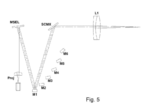
= Figure 5 is a further illustration of the mirror of the sample of Figure
4 with
operating selection of one of the mirrors provided therein;
= Figure 6 is yet a further illustration of the mirror of the sample of
Figure 4
with operating selection of another one of the mirrors provided therein;
= Figure 7 and Figure 8 respectively show an anterior segment and a
retina of an eye obtained according to the invention, respectively with the
distance of
the anterior corneal surface from the upper edge of the image and the distance
of the
retinal surface from the upper edge of the image schematised;
= Figure 9 is a representation analogous to those of Figure 4 and Figure 5
of
a sample arm with curved mirrors to focus the scanning beam at the depths in
accordance with the length of the various paths according to a different
embodiment of
the invention;
= Figure 10 is a representation analogous to those of Figure 4 and Figure 5
of
a sample arm with dispersion compensator devices according to yet a different
embodiment of the invention; and
= Figure 11 shows as in the preceding Figures 9 and 10 yet a further
embodiment combining those of the above-mentioned Figure 9 and 10, that is, by
adopting a sample arm with dispersion compensator devices and curved mirrors
which
focus the scanning beam in accordance with the operating depth of the various
paths.
Detailed description of the invention
With reference to said figures, and based on what already reported in the
introductory part as regards the general architecture of the system, Figure 4,
which is
specifically referred to, shows an example of a sample arm of an FD-OCT
instrument,
such arm being provided with a lens or lens system L1 (of a per se known type)
and a
tilting mirror MSEL angularly positionable in a certain number of positions,
for example
CA 02957355 2017-02-06
WO 2016/034917 PCT/1B2014/064201
8
six. The lens L1 is centred on the sample, in the case of a human eye the axis
of the
lens coinciding with the optical axis, indicated as Z. A plane XY may be
defined, in the
case of the human eye, as the plane tangential to the eye at the incidence
point of the
optical axis Z. The lens L1 rests parallel to such eye, while the tilting
mirror has a
rotation axis orthogonal to the plane ZX, and therefore extending along Y
(axis coming
out of the sheet in the illustration of Figure 4).
The tilting mirror MSEL is in fact hit by a collimated optical beam F coming
from
a projector Pr along the direction X. The sample arm further provides for a
plurality of
mirrors M1 ... Mk ... Mn (n=6 in the specific instance) arranged downstream of
the
tilting mirror MSEL, taking as a reference the path of the optical beam, and
oriented so
as to intercept the above-mentioned optical beam, each when the beam is
reflected in
a respective position of the tilting mirror MSEL.
The deviation of the beam in turn reflected by one of the mirrors Mk towards
the
lens L1, and therefore along the optical axis Z, is provided by a second
tilting scanning
mirror SCM, controlled so as to tilt in coordination with the first mirror
MSEL. In the
example the two mirrors are arranged in a substantial alignment along the
optical axis
Z, while the fixed mirrors M1-M6 are arranged according to an arc shape at
progressively smaller distances from the above-mentioned axis, where M1, the
first
mirror in the sequence, is the closest one to the entering beam segment coming
from
the projector Pr and is the most distant one from the axis. Going from M1 to
M6,
besides decreasing the distance from the axis Z, the angle progressively
varies,
therefore if the first fixed mirror M1 and the optical axis are in a relation
of substantial
mutual parallelism, the following mirrors M2 ... M6 are progressively tilted
to form a
progressively smaller angle between the reflecting face, facing towards the
tilting
mirrors, and the same optical axis.
Clearly, depending on the angular position selected for the first tilting
mirror
MSEL, and correspondingly for the second tilting mirror SCM, optical paths
having
different lengths are determined for the beam in the sample arm. This will
result clearer
by examining Figures 5 and 6, wherein two examples of optical paths
respectively
corresponding to position 1 (longer path, the fixed mirror M1 is hit) and
position 5 (the
CA 02957355 2017-02-06
WO 2016/034917 PCT/1B2014/064201
9
fixed mirror M5 is hit) are in fact illustrated.
With reference to Figure 5, the selector mirror MSEL, tilted to an appropriate
angular position (position 1) selects a path of maximum length containing the
mirror M1
adapted to capture a sample section close to the instrument. In the case of
the eye, the
mirror M1 will be used to capture the anterior eye segment, obtaining an image
as in
Figure 7, which is also connected to that of the previously mentioned Figure
2. The
mirror M6, the one that together with position 6 of the mirror MSEL determines
the
shortest optical path (not shown), will also be selected when a sample section
farther
from the instrument is to be captured, that is, more in depth. In the case of
the eye, the
mirror M6 will be used to capture an image of the retina in particularly
"long" eyes, that
is, having a high axial extension.
The mirrors M2, M3, M4, M5 (in this latter case reference is to be made to
Figure
6) are selected to capture sample sections which are at progressively greater
intermediate depths. For example, the mirror M2 may be used, in the case of an
eye,
for capturing the crystalline lens and the mirrors M3, M4, M5 for capturing
the retina in
increasingly "longer" eyes. An image of the retina captured by selecting
mirror M5 is
shown in Figure 8.
In the depicted embodiment six paths having different length may be obtained,
but such number shall clearly be considered as merely exemplary. In practice,
the
number of implemented paths, by means of a corresponding number of fixed
mirrors
and positions of the tilting mirrors, will depend on a compromise between the
distances
to be measured, the costs, the constructional simplicity, the resolution of
the
spectrometer or the maximum depth that the OCT system can scan.
Optionally the mirror SCM may be replaced by a pair of mirrors SCMx and SCMy
(not shown), tiltable about respective axes orthogonal with each other, so as
to obtain a
concurrent deviation of the beam in two directions. In any case, the beam
finally hits
the lens L1 and is focused by the latter at a predetermined distance where the
sample
to be captured is found. If there are two moving scanning mirrors on axes
orthogonal
with each other, the appropriate combination of the angular positions occupied
in quick
succession by the two mirrors will allow carrying out various scanning
patterns, known
CA 02957355 2017-02-06
WO 2016/034917 PCT/1B2014/064201
to the man skilled in the art, for example the star-shaped scan of multiple
meridians or
the raster scan of multiple parallel sections of the object. If only one
scanning mirror is
provided, it is also possible to envisage a further degree of freedom, that is
a further
tilting about the axis Z so as to select the angle of the section to be
scanned.
Returning to the primary task of the invention, that is to obtain measurements
in
depth of the distances between the eye interfaces, by taking advantage of the
embodiment configuration described above, it is possible to suggest various
strategies
for measuring the distances between the surfaces of a sample.
A first, simple strategy provides for capturing an image of the sample by
selecting
each time a different position of the selection tilting mirror MSEL, and then
a different
mirror Mk, and then another path of different length on the sample arm. If Ml,
then M2,
M3, M4, M5 and M6 are selected, an image of a sample section close to the
instrument
will be captured first via Ml, then another one farther away by selecting M2
and so on
until capturing the deepest section of the sample via M6. Each time that a
mirror Mk is
selected the scanning mirror SCM is tilted correspondingly so as to scan a
sample
section at the selected depth. In order to achieve a fast final measurement,
devices for
selecting the optical path, scanning and capturing the sample having a
correspondingly
fast response must be used that the man skilled in the art may easily find.
The mirror
MSEL may be for example a galvanometric mirror, as well as the scanning mirror
SCM;
the sensor for collecting the power backscattered by the sample towards the
spectrometer may be a high speed line scan camera.
If the sample is an eye, a particularly important measurement in cataract
surgery
is the distance between the anterior corneal surface and the retina. In this
type of
surgery this distance is critical for calculating the power of the artificial
crystalline lens
to be implanted in place of the opacified natural one. By knowing this
distance, an
optical and geometrical model of the anterior segment and the rated optical
and
geometrical data of the artificial lenses, it is possible to assess the power
of the lens to
be implanted into the eye under examination by means of various formulas and
methods well known in the literature.
According to the present invention it is possible to measure all the distances
CA 02957355 2017-02-06
WO 2016/034917 PCT/1B2014/064201
11
between the various intraocular interfaces (anterior and posterior corneal
surfaces,
crystalline lens surfaces, retina). By way of example, it is now supposed that
the axial
eye length is to be measured. It is possible to assume that the image of the
anterior
segment is obtained by using path 1 which includes mirror Ml, and that the
image of
the retina is, on the other hand, obtained using path 5 which includes mirror
M5
(reference is therefore made again to what is schematised in Figures 5 and 6).
From
the image of the anterior segment (illustrated as mentioned in Figure 7) it is
then
possible to determine the distance A of the anterior corneal surface from the
upper
edge of the same image, while from the image of the retina (Figure 8) B is on
the other
hand determined as the distance of the retinal surface from the upper edge of
the
image. Then by knowing the difference in the optical path C between the two
paths of
the sample arm selected respectively for the anterior segment and for the
retina, the
optical axial length OAL may be determined as:
OAL=
Of course, this calculation may be carried out automatically, so that the
operator
directly obtains the OAL value.
As regards the scans that are performed each time that a different path is
selected on the sample arm, a scan may consist for example in 256 A-scans
performed
on adjacent tissue strips moving the scanning mirror (or the two scanning
mirrors, if
provided, about their respective axes), or the scanning mirrors may be kept
still by
repeating many acquisitions of the same tissue strip, or yet a scan on
multiple lines on
a square area may be performed. In this latter case several A-scans may be
captured
on an adequately sized square Cartesian grid, for example 16 rows with 16 A-
scans
each, if the same timing of the line scan is to be maintained.
A reasonable time for scanning both a portion of the anterior segment and a
portion of an inner eye structure during the procedure described above is in
the order
of 10 ms. This time is long enough to collect an amount of radiation on the
sensor that
is appropriate for obtaining a few hundreds of A-scans, but at the same time
it is short
enough to prevent artifacts due to eye movement in the range related to an
entire B-
scan.
CA 02957355 2017-02-06
WO 2016/034917 PCT/1B2014/064201
12
In order to determine which is the right path to obtain an image of the
retina, a
longer time is needed, so that it makes more likely that an eye movement
occurs during
the attempts of selecting the various paths. The strategy described
previously, even
though it may appear satisfactory considering also its marked simplicity, is
subject to
improvements capable of obviating the eye movements of the patient, in
particular
along axis Z, movements that can in fact occur in the passage from one path to
the
other and for which the previous formula does not account. In this way it is
possible to
reduce the incidence of errors which, for example in the measurement of the
axial
length for determining the power of the lens to be implanted in cataract
surgery, may
be critical.
A more complex strategy capable of accounting for eye movements may be
structured as follows. Path 1 is selected which includes mirror M1 and the
anterior
segment is captured. Path 2 is then selected which hits mirror M2 and the
acquisition
goes much deeper. If in the captured image the retina is not detected, path 3
is
selected with mirror M3 to capture the image at an even greater depth. Again,
if the
retina does not appear in the captured image, path 4 is selected with mirror
M4. This
continues until the k-th path selected allows identifying the retina. Then
path 1 is
selected again to re-capture an image of the anterior segment and again back
to the k-
th path to re-capture the retina and so on, alternating acquisitions obtained
by selecting
with mirror MSEL path 1 and the k-th path. The measurement of interest may
then be
obtained by N pairs of images of the anterior segment and of the retina
captured in an
alternating manner thanks to the mirror MSEL, which is rapidly switched
between the
position suitable for shooting the anterior segment and the position suitable
for
shooting the retina. The detail of the calculation is described hereinafter.
If upon the i-th acquisition of the pair of images of the anterior segment and
the
retina A, is used to indicate the distance of the anterior corneal surface
from the upper
edge of the image of the anterior segment (Figure 7), B, to indicate the
distance of the
retinal surface from the upper edge of the image of the retina (Figure 8) and
C, to
indicate the difference in the optical path of the two paths of the sample arm
selected
for the anterior segment and the retina, we find that the optical axial length
OALi which
CA 02957355 2017-02-06
WO 2016/034917 PCT/1B2014/064201
13
may be calculated via the i-th acquisition is:
OAL,= Cf -A
If N acquisitions are considered, an average optical axial length will be
obtained
from the relation:
Ar-
OAL= ¨ 7,`= 0A1,,
=
Even in this case, the calculation will typically be automated by means of
control
software implemented with per se simple techniques.
As is known in the literature, from the optical lengths it is possible to
obtain the
geometrical lengths using the refractive indices of the eye means passed
through. The
measurement of the distances between the various intraocular structures with
equipment as that described above may be carried out in cascade upon
acquisition of
multiple sections of the anterior segment which allow its three-dimensional
measurement or in an ad hoc separate examination uniquely for calculating
distances
between two or more eye interfaces.
In order to improve the transverse resolution of the images captured at the
different eye depths, the mirrors M1, ..., M6 may be made with curved
reflecting
surfaces, paying attention to designing the curves so that the focus of the
scanning
beam coming out of the lens L1 matches the distance at which the scan is to be
performed. Such embodiment solution is illustrated in Figure 9, wherein the
dashed line
shows the radiation beam when mirror M1 is selected and the solid line shows
the
beam when mirror M4 is selected. In the first case a portion of the sample
close to L1 is
to be scanned and the scanning beam focuses this portion; in the second case,
on the
other hand, a farther portion of the sample is to be scanned and the scanning
beam
focuses such farther portion, such focusing being enhanced by the different
curves of
the various mirrors. All of the above is illustrated graphically with even
greater clarity by
the inclusion, in the illustration, of an eye E being examined.
In this type of interferometry a broadband radiation is used which passes
through dispersive components (glass, optical fibres, etc.). The eye also
denotes a
dispersive behaviour. If the radiation going through the sample arm and that
going
CA 02957355 2017-02-06
WO 2016/034917 PCT/1B2014/064201
14
through the reference arm are not balanced in terms of dispersion, that is
they do not
pass through the same lengths in glass and/or tissue, there is a deterioration
of the
instrument's resolution. In view of these considerations, a further
advantageous
embodiment of the invention provides for compensating the dispersion effect by
inserting in the various paths of the same arm elements in glass or an
appropriate
material having different length. These are capable of making the lengths of
the
dispersive tracts present on the reference arm and the sample arm identical or
very
similar to each other, being sized especially considering the lengths of the
tracts
covered by the radiation in the components of the instrument and also in the
eye
tissues in a manner independent of the depth at which the path of the sample
arm is
intended for operation.
Such embodiment solution is schematised in Figure 10 where, close to the
mirrors Ml, M5, there have been placed elements in glass of different
lengths G1,
..., G5. Mirror M6, on the other hand, does not have a corresponding element
in glass.
With this type of configuration also the reference arm will have to be
provided with a
sufficiently long element in glass which has the same dispersion of the eye
means
going from the cornea to the deep area of which the image is captured when the
path
with mirror M6 is activated.
Figure 11 finally shows an embodiment solution wherein the compensation of the
dispersion is combined with the adoption of mirrors having appropriate curves
in order
for the focus of the scanning beam coming out of the lens L1 to match the
distance at
which the scan is to be performed. In practice, the embodiments of Figure 9
and Figure
are here associated to each other.
The present invention therefore provides a fully satisfactory response to the
predetermined task, combining a precise and reliable functional result with a
simple
and an actually feasible and structurally simple configuration at low costs,
also from a
management and maintenance standpoint.
With an N number of different paths on the sample arm, selectable thanks to a
tilting mirror which with a small and quick tilting is driven from one to the
other of N
angular positions useful for acquisition at the desired depth, the acquisition
may go
CA 02957355 2017-02-06
WO 2016/034917 PCT/1B2014/064201
from one depth to the other, and with alternating acquisitions between two
desired
depths, obtained by selecting alternatively the two suitable paths of the
sample arm,
the measurements of the distance between the eye structures of interest
present in
images relating to different depths may be repeated many times in a short time
interval.
In this way, the measurement of the distance between the eye structures is
robust, that
is, safe and reliable, in spite of any movements of the eye being examined.
Such a result is obtained without using multiple reference arms/paths, either
dichroic separation of the beam coming from the sample, or the need of bulky
and
costly sensors with a high number of pixels, or yet longitudinal movements
which are
difficult to fine tune (the movement in bursts of the tilting mirror MSEL in
predetermined
positions ensures the desired precision over time without particular problems
and at
significantly lower management costs).
The preceding solutions only represent illustrative examples and should not be
considered as the only ones adapted to the task. Various combinations of the
conceptual solutions illustrated hereinabove shall be considered as implicitly
understood by the man skilled in the art. The present invention, however, has
been
described thus far with reference to its possible exemplary embodiments. It
must be
understood that there may exist other embodiments, within the scope of overall
optical
configurations different from that disclosed herein and integrated by
additional
components/functionalities, belong to the same inventive scope, all falling
within the
scope of protection of the attached claims.