Note: Descriptions are shown in the official language in which they were submitted.
CA 02957384 2017-02-06
WO 2016/022414
PCT/US2015/043083
1
METHODS AND KITS FOR DIAGNOSING, ASSESSING OR QUANTITATING DRUG
USE, DRUG ABUSE AND NARCOSIS, INTERNUCLEAR OPIITHALMOPLEGIA,
AVIENTION DEFICIT HYPERACTIVITY DISORDER (ADHD), CHRONIC
TRAUMATIC ENCEPHALOPATHY, SCHIZOPHRENIA SPECTRUM DISORDERS
AND ALCOHOL CONSUMPTION
FIELD OF THE INVENTION
100011 The present invention relates to methods and kits for diagnosing,
assessing or quantitating
drug use, drag abuse, and narcosis and For differentiating narcosis from brain
injury such as
structural and non-structural traumatic brain injury and for screening for,
diagnosing, and
assessing the same, for diagnosing and assessing interauclear oplithalmoplegia
(INO) in a subject
by tracking eye movement, for diagnosing and assessing attention deficit
hyperactivity disorder
(ADHD), chronic traumatic encephalopathy-, and schizophrenia spectrum
disorders in a subject
by tracking eye movement, and for detecting, screening for or quantifying
alcohol consumption
and intoxication in a subject by tracking eye movement.
BACKGROUND OF THE INVENTION
Automated Eye Movement Tracking
100021 Automated eye movement tracking has been used for marketing and
advertising research,
the development of assistivc devices for immobile individuals, and for video
games. Spatial
calibration of the device requires the subject to have relatively intact
ocular motility that implies
function of cranial nerves II (optic), III (oculomotor), IV (trochlear) and VI
(abducens) and their
associated nuclei as well as sufficient cerebral function to enable cognition
and volition for
calibration.
100031 Others have successfully demonstrated the clinical applications of eye
movement data
(Lee et al., Brain Research. 2011.; 1399:59-65; Contreras et al.,: Brain
Resoarch 2011; 1398:55-
63; Maruta et al., The Journal of Head Trauma Rehabilitation 2010; 25(4):291-
305). Trojan() et
al., .1- Ne.urol 2012; (published online; ahead of print) recently described
unealibrated eye
movement measurements in a population of minimally conscious and persistently
vegetative
patients. They report data from 11 healthy control subjects evaluating chronic
disorders of
CA 02957384 2017-02-06
WO 2016/022414
PCT/US2015/043083
2
consciousness, not acute changes in intracranial pressure. They sample eye
movements at 60 Hz
rather than 500 Hz, effectively reducing the power of their data 100-fold, and
they report
differences in on-target and off-target fixations between the groups without
spatially calibrated
data. Moreover, they use static stimuli moving in a quasi-periodic way.
[0004] Eye movement tracking for neuropsychiatric and brain injury research
(Heitger, et al.,
Brain, 2009; 132: 2850-2870; Murata, et aL, I Head Trauma Rehabil., 2010; 25:
793-105) has
been performed for nearly 30 years and can evaluate smooth pursuit, saccades,
fixation, pupil
size and other aspects of gaze. Spatial calibration of the eye tracker is
generally performed for
each individual being tracked. With calibration, the eye-tracker measures the
relative position of
pupil and corneal reflection for a period of about 400-800 ms while the
subject looks at a target
or targets of known position to generate meaningful spatial coordinates during
subsequent pupil
movement. The process of spatial calibration implies relatively preserved
neurologic function
because it requires that the subject is able to follow commands and look at
specific points.
[0005] Ti is conceivable that the process of spatial calibration may mask
deficits in ocular
motility. If there is a persistent and replicablc weakness in movement of an
eye, the camera will
interpret the eye's ability to move in the direction of that weakness as the
full potential range of
motion in that direction due to the calibration process. In other words if the
subject is directed to
look at a position but consistently only moves halfway there, the calibration
process will account
for that when tracking subsequent eye movements and interpret movements to the
halfway point
as occurring at the full range of normal motion. If during calibration one eye
only makes it half-
way to the target, but the other eye is fully there, the camera will interpret
both eyes as being
together when one performs half the eye movement as the other. Thus binocular
spatial
calibration may preclude detection of disconjugate gaze unless each eye is
calibrated separately
using a dichoptic apparatus (Schotter, et al., PLoS One, 2012; 7: e35608).
Conjugaey of Eye Movement
[0006] Conjugate gaze is the motion of both eyes in the same direction at the
same time.
Conjugate gaze is believed to be controlled by the following four different
mechanisms: the
CA 02957384 2017-02-06
WO 2016/022414
PCT/US2015/043083
3
saccadic system that allows for voluntary direction of the gaze, the pursuit
system that allows the
'subject to follow a moving object, the optokinetic system that restores gaze
despite movements
of the outside world, and the vestibulo-ocular reflex system (VOR system) that
corrects for the
movements of the head to preserve the stable visual image of the world.
[0007] Disconjugate gaze or strabismus is a failure of the eyes to turn
together in the same
direction. Normal coordinated movements of the eyes produces conjugate gaze,
in which the
eyes are aligned for binocular 3-dimensional vision. Misalignment results in
loss of this vision.
With the visual axis of each eye fixated on a different point, diplopia (or
double vision) usually
results and may be perceived as a blurred image if the two images are very
closely aligned.
However, if the image from the weaker eye is suppressed by higher cortical
centers, there is only
one image with loss of visual acuity (or a blurred image). Pathology usually
resides either in the
oeulomotor muscles or their neuronal pathways including the medial
longitudinal fasiculus, the
paramedian pontine reticular formation, the medullary reticular formation, the
superior
colliculus, or the cranial nerves III, IV, or VI or their nuclei.
100081 Assessment of eye movement coniugaey is commonly performed by primary
care
physicians, neurologists, ophthalmologists, neurosurgeons, emergency medicine
doctors, and
trauma surgeons to rapidly assess global neurologic functioning. In stable
patients,
ophthalmologists and neurologists perform more detailed examination to assess
the alignment of
the eyes such as the cover test and Hirschberg corneal reflex test. Other
tests used to assess
binocular conjugacy include the 'litmus House Fly test, Lang's stereo test,
the Hess screen, red-
filter test, Maddox rod evaluation and Lancaster red-green test. in young
children, who may be
less cooperative with an examiner, binocular gaze conjugacy may only be
assessable with
simpler algorithms, such as following an object moving in a set trajectory
(Cavezian, et at, Res
Dev DisabiL, 2010; 31:. 1102-1108). When such tests are performed in
conjunction with the
remainder of the neuroplithalmie and physical evaluation, one can localize
neurologic lesions
and quantitate ocular motility deficits with great accuracy. Despite this
capability, these tests are
not used routinely in the emergency setting due to the need for a trained
practitioner
to
administer them, the requirement for sophisticated equipment, and the urgent
nature of many
neurologic disorders.
4
100091 Assessment of binocular gaze conjugacy in primates for research
purposes is performed
with the magnetic search coil technique requiring coils implanted into the
bulbar conjunctiva
(Schultz, el ci., J Neurophysiol, 2013; 109: 518-545). This technique was
first described by
Fuchs and Robinson in 1966 (Fuchs, es al., .1 App! Apia, 1966; 21 1068-1070)
and can also
be performed in humans fitted with sclera search coils designed specifically
for tracking eye
movements.
[00101 Experimentally, spatially calibrated eye movement tracking using the
Bouts oculometer
(Bach, et at, .1 iVeurosci Methods, 1983; 9: 9-14), which requires that the
head is rigidly fixed,
shows that healthy seven year old children have increased disconjugacy of eye
movement during
saccades relative to adults while both perform a reading task (Bucci, et al.,
Vision Res., 2006; 46
457466). Research on disconjugacy during reading can he performed using a
dichoptic
apparatus in which the individual eyes are spatially calibrated separately and
presented with
stimuli to assess movements separately tor simultaneous comparison to each
other (Schotter, el
al., PUS One, 2012; 7: e35608). U.S Provisional Application No. 61/881,014,
filed September
23, 2013 teaches
methods for tracking eye movement, and methods and kits for assessing
conjugacy and
disconjugaey of gaze and strabismus.
Brain Injury
100111 One of the problems associated with the study of outcomes after brain
injury, is the
heterogeneous nature of such injury in terms of etiology, anatomic sequelae,
and physiologic and
psychologic impact. The etiology of injury affects the anatomic sequelae and
ranges from global
mechanisms such as acceleration/deceleration and blast, to potentially more
focal mechanisms
such as blunt impact and penetrating trauma. Some injury mechanisms result in
structural
changes to the brain that can be visualized using conventional imaging such as
Iv1R1 and CT
scan, while other injuries appear radiographically normal.
100121 Acceleration/deceleration injury may result in structurally visible
coup/enntrecoup
injuries and less visible diffuse axonal injury (DAI) (Cecil, et al. Journal
of Neurosurgery,
1998; 88 795-801) Acceleration/deceleration is also thought to be one of the
potential
Date Recue/Date Received 2022-03-01
CA 02957384 2017-02-06
WO 2016/022414
PCT/US2015/043083
mechanisms for concussion which is the most common form of civilian
radiographically normal
brain injury (Bayly, et al., Journal of Neurotrauma, 2005; 22 845-856;
Daneshvar, et at.,
Physical Medicine and Rehabilitation Clinics of North America, 2011; 22: 683-
700).
Concussion is brain injury, most often resulting from blunt impact, in the
absence of structural
abnormality by conventional radiographic imaging such as computed tomography
(CT) scan
(McCrory, et at., The Physician and Sports Medicine, 2009; 37: 141-159).
Concussion may
include transient loss or disruption of neurologic function. The 'term
"subconcussion" may be
used to describe the sequelae of brain injury in the absence of transient loss
or disruption of
neurologic function, Both concussion and subconcussion as well as blast injury
may be termed
"non-structural" brain injury,
[0013] Blast injury resembles blunt impact brain injury in that both may be
associated with
radiographically apparent cerebral edema and intracranial hemorrhage, however
with blast
injury the edema onset may be more rapid and severe, and tht.we is greater
likelihood of clinical
vasospasm (Arrnonda, et al., Neurosurgery, 2006; 59: 1215-1225). Blast injury
is very
frequently radiographically normal, yet mild or moderate blast injury is
strongly associated with
post-traumatic stress disorder and other cognitive dysfunctions (Cernak, et
al., The Journal of
Trauma, 2001; 50: 695-706). The actual cause of blast brain injury is
suspected to be
multifactorial and often results in DAT (Leung, et at., Mai Cell Biam.ech,
2008; 5: 155-168). A
shock wave resulting from pressure changes caused by the explosion impacts
both cranial and
non-cranial structures (Courtney, et at., Medical Hypotheses, 2009; 72: 76-83;
Bauman, et at.,
Journal of Neurotrauma, 2009; 26: 841-860). Blast injury affects the brain
through several
mechanisms: primary brain injury caused by blast-wave induced changes in
atmospheric
pressure directly impacting the brain; secondary injury resulting from objects
put in motion by
the blast that impact the head, and tertiary injury resulting from the victim
striking the head
upon falling or being propelled into a solid object (Warden, The .Journal of
Head Trauma
Rehabilitation, 2006; 21: 398-402).
10014] Blunt impact and penetrating trauma can result in both diffuse and
focal injury. One
mechanism by which focal brain injury leads to neurologic damage is cortical
spreading
CA 02957384 2017-02-06
WO 2016/022414
PCT/US2015/043083
6
depression (Ilartings, et al., Journal of Neurotraunia, 2009; 26: 1857-1866),
which is currently
only thought measurable using invasive means.
[0015] Brain injury may be associated with short term sequelae including
headaches and
memory problems, and longer term problems including dementia, Parkinsonism and
motor-
neuron disease (Dareshvar, et A, Physical Medicine and Rehabilitation Clinics
of North
America, 2011, 22 683-700). Both concussion and mild blast injury may be
associated with
post-traumatic stress disorder and cognitive impairment (Taber, et al., The
Journal of
Neuropsychiatry and Clinical Neurosciences, 2006; 18: 141-145). Clinical tests
for concussion
show poor test reliability (Broglio, et al., Journal of Athletic Training,
2007; 42.: 509-514) and
thus concussion remains a diagnosis that is difficult to treat because it is
difficult to detect.
Traumatic brain injury can impact eye movement through a multitude of
mechanisms including
direct compression of cranial nerves, trauma to cranial nerves, injury to
cranial nerve nuclei and
supranuclear impacts.
100161 Many cases of trauma result in elevated intracranial pressure. If
untreated, acute
elevations in intracranial pressure (1CP) due to brain injury can result in
permanent neurologic
impairment or death. Double vision and other ocular disturbances associated
with elevated TCP
were first described by Hippocrates in approximately 400 B.C.
(AronykõVeurosurgery Clinics of
North America, 1993. 4: 599-609). Papilledema, and its association with
elevated TCP was
described by Albrecht von Graefe in 1860 (Pearce, European Neurology, 2009;
61:: 224-249).
In the post-radiographic era, acute and chronic pathology of the optic nerve
and disc, and of
ocular motility are well characterized in people with elevated TCP (Dennis, ei
al., Archives of
Neurology, 1981; 38: 607-615 Zeiner, et al., Child 's Nerv. Syst., 1985; 1:
115-122; Altintas, et
al., Gracie's Archive for Clinical and Experimental Ophthalmology, 2005; 243:
1213-1217).
Clinically apparent disruption of ocular motility may precede computed
tomography (CT)
findings in some subjects with acutely elevated 1CP (Tzekov, et al., Pediatric
Neurosurgery,
1991; 17; 317-320; Chou, etal., Neurosurgery Clinics of North America, 1999;
10: 587-608).
Elevated Intracranial Pressure
CA 02957384 2017-02-06
WO 2016/022414
PCT/US2015/043083
7
[0017] Several potential mechanisms may contribute to cranial nerve
dysfunction due to elevated
intracranial pressure. The IIIrd nerve (oculomotor) may be directly compressed
by the medial
aspect of the temporal lobe with frontal or temporal mess lesions, or diffuse
supratentorial mass
effect. The VIth nerve (abducens) is anatomically vulnerable to infratentorial
mass effect at the
prcpontine cistern and to hydrocephalus from stretch as it traverses the
tentorial edge.
10018] Elevated intracranial pressure slows axoplasmie transport along cranial
nerves
(Balarratnasingam, et al., Brain Research, 2011; 1417: 67-76). The optic nerve
(II) is most
frequently analyzed because it can be visualized directly with ophthalmoscopy,
and indirectly
with ultrasound. Edema of the optic nerve appears earlier than ocular fundus
el-wages, and
resolves after treatment of elevated ICP Gangemi, et cii., Neurochirurgia,
1987; 30: 53-55),
Fluctuating elevated neural pressure leads to impaired axonal transport along
the optic nerve
after as little as 30 minutes in a rabbit model (Balarratnasingam, et al.,
Brain Research, 2011;
1417: 67-76). Axoplasmic flow stasis and intraneuronal isehemia may occur in
the optic nerve
exposed to chronically elevated ICP (Lee, et alõ Current Neurology and
Neuroscience Reports,
2012). Among the nerves impacting ocular motility, the trochleur nerve (IV),
followed by
oeulomotor (III) and then abducens (V1), has the greatest length of exposure
to the subarachnoid
space with the narrowest diameter, and thus may be most vulnerable to a
pressure induced palsy
(TIanson, et al, Neurology, 2004; 62: 33-36; Adler, et al, Journal of
Neurosurgery, 2002: 96:
1103-1113) The optic nerve (H) has approximately the same length of exposure
as the
abducens (M-urali, et at., in Head Injury (ed. Paul Cooper and John Golf-mos)
(McGraw-Hill,
2000)), and thus papilledema, Or swelling of the optic disc apparent on
ophthalmoscopic
examination may be a relatively late indicator of elevated ICP (Killer, et al,
Clinical &
Experimental Ophthalmology, 2009; 37: 444-447; Nazir, et al., .1 /tapas, 2009;
13 62-66).
Papilledema is not always a sensitive marker for hydrocephalus leading to
elevated ICP, and in
one study was present in as few as 14% of patients with a shunt malfunction
(Nazi'', et at.
Aapos, 2009; 13: 62-66) consistent with the relatively short intracranial
course of II compared to
cranial nerves III and IV. Compartmentalization of subarachnoid spaces is
hypothesized to
explain why papilledema may he present in a patient without elevated ICP, and
not occur in
patients with elevated ICP (Killer, et al, Clinical & Experimental
Ophthalmology, 2009; 37:
444-447).
CA 02957384 2017-02-06
WO 2016/022414
PCT/US2015/043083
8
Effect of Drugs and Narcotics on Eye Movement
10019] Methadone has been shown to impact ocular movements during both smooth
pursuit and
saccades, and is thought to impact function of the superior colliculus
(Rothenberg et al.,
Psychophartnaealogy (Berl) 1980; 67:221-227; Rothenberg el al.,
Psychopharmacology (Berl)
1980; 67:229-234, 1980). Narcotic naïve subjects administered methadone had
decreased
smooth pursuit eye movement gain in horizontal pursuit tracking, but showed no
significant
decrease in gain in vertical pursuit tracking. There was a significant
increase in vertical cross
correlation measurements hut none in horizontal cross correlation. No phase
difference between
subjects given methadone and control was present, signifying that the
difference in gain was not
due to failure of eye movement during parts of eye tracking trial or a
difference in frequency of
eye motion compared to target motion. The lack of vertical pursuit gain in
methadone dose
subjects may be due to contamination of vertical data from eyelid motion, as
eyelid motion
occurs with vertical eye motion when movement is greater than 5 degrees from
central position.
Methadone may have induced loss of eyelid control, resulting in contamination
of vertical
pursuit tracking. Methadone did not significantly alter maximum saccade
velocity. However,
initial saccade accuracy is significantly decreased with more pronounced
saccade undershoot
after use of methadone. In addition, the latency to onset of initial saccade
was also significantly
increased.
10020] Similar results may be seen with other pharmacologic agents. Diazepam
is one of the
class of benzodiazepines. Subjects given diazepam showed significant decrease
in smooth
pursuit gain in a dose dependent manner; 5 mg diazepam significantly reduced
gain at 0.4 Hz
and 10 mg diazepam at 0.4, 0.6, 0_8, 1,0, L2, and 1.6 Hz. In contrast to
methadone, diazepam
induced changes in cross-correlation as function of drug as well. Phase of
smooth pursuit did not
show a significant change upon administration of diazepam (Rothenberg et al.,
Psychopharmacology (Berl) 1981; 74:232-236; Rothenberg et al,,
Psychopharmacology (Berl)
1981; 74:237-240). The dose dependent effects of diazepam on different
frequencies of motion
track suggest that smooth pursuit eye tracking after diazepam administration
may be dependent
on stimulus velocity. Saccadic pursuit replaces smooth pursuit upon
administration of diazepam.
Diazepam may induce the above cyc movement changes by its binding to visual
CNS
CA 02957384 2017-02-06
WO 2016/022414
PCT/US2015/043083
9
bartodiazepine binding sites that are important for oculomotor control.
Compared to methadone,
diazepam administration shows a greater reduction in amplitude and replacement
of smooth
pursuit with saccadic pursuit.
[0021] LoraLepam is another of the class of benzodiazepines. When administered
to subjects
undergoing saccade tasks, the gap between successive images were temporally
overlapped with
the original image still on the screen before the next image appeared. In
normal subjects, latency
increases with temporal overlap compared to images separated by 200 nis gap.
With lorazepam
administration, subjects showed significant change during the temporal overlap
but not with 200
ms gap (Masson et al., Behav Brain Res 2000; 108:169-180). Temporal overlap
had no
significant effect on saccadic peak velocity and amplitude in normal subjects.
In lorazepain
administered subjects, saccadic peak velocity and the amplitude of first
saccadic eye movement
significantly decreased. With smooth pursuit eye movement, lorazepam showed
increased
latency and longer reaction time compared to control. In addition, lorazcpam
significantly
decreased eye velocity. Results also indicate that tracking errors in smooth
pursuit induced by
lorazepam are compensated for by saccadic movements of the eyes.
100221 Alcohol consumption also impacts eye movements. Drinking subjects show
decreased
gain during smoothparsuit eye movement in a dose dependent manner. In one
study subjects
were given 0,4 and 0.8 g/kg of alcohol and eye tracking was done on two time
points: 1'1 at 60
min. and 12 aL 180 min, after beverage consumption (Roche et al.,
Psychopharmacology (Berl)
2010; 212:33-44). Ia smooth pursuit eye tracking, high close affected gain at
both time poi-nts
while low dose did not have an effect on gain for the latter time point. For
pro-saccade, latency
was also impaired in a similar, dose dependent manner. Ocular velocity and
accuracy decreased
only after high dose consumption. Anti-saccade showed similar presentation as
pro-saccade with
the exception that high dose improved accuracy at TI and decreased by 12.
[0023] Alcohol significantly affected both pro and anti-saccade accuracy;
however, greater
accuracy for high dose alcohol at Ti may be due to alcohol increasing the
amplitude of anti-
saccade relative to normal conditions and not that alcohol is improving anti-
saccade functioning.
'Ibis suggests that high dose alcohol may he affecting neurocircuitry required
for rapid
CA 02957384 2017-02-06
WO 2016/022414
PCT/US2015/043083
processing of visuospatial information. High dose and low dose alcohol
consumption show
similar impairment in smooth pursuit gain and anti-saccade functions; however,
high dose
patients have less awareness of the impact of this dysfunction, placing them
in higher risk for
inj uric&
Intemuclear ophthahroplegia (INO)
[0024] The medial longitudinal fascieulus is a bilateral axonal bundle that
functionally
coordinates the actions of cranial nerves III, IV and VI, and receives inputs
from the frontal eye
fields, cerebellar flocculus, superior colliculus, accessory oculomotor
nuclei, pontine reticular
formation, fastigial nucleus and cranial nerve VIII (Reulen of al., Brain
1983; 106 (Pt 1):121-
140). It enables saccadic and reflexive eye movements and carries the
descending tectospinal
and medial vestibulospinal tracts into the cervical spinal cord,
[0025] Internuclear ophthalmoplegia (INO) results from impaired function of
thc medial
longtitudinal fasciculus (Zee of al., Baillieres Clin Neural 1992; 1:455-470)
and can be either
unilateral or bilateral.. The most common causes of INO are multiple sclerosis
and
strokeischemic injury. Other causes are trauma, tentorial herniation,
infection, tumor, iatrogenic
injury, hemorrhage, vasculitis, and other (Keane ei al., Arch Neural 2005;
62:714-717).
[0026] INO can be difficult to detect in patients with mild or intermediate
slowing of adduction
and a variety of teclmologies have been proposed to increase the accuracy or
detection, such as
quantitative infrared oculovsraphy (Frohman et al., Neurology 2003; 61:848-
850), saccade testing
and electrooe-ulography (Jozefowicz-Korczynska et al., J Neural 2008; 255:1006-
1011) and
oculo-vestibular evoked myogenic potentials (Roscrigren of al.,
Neurophysiol 2011;
122:1264-1267). MRI sequences in patients with INO may also reveal lesions in
the MLF
(McNulty el. Clin Neuroradiol 2014).
[0027] INO can also be difficult to differentiate from infranuclear palsies of
cranial nerves such
as a partial third nerve palsy, or disorders such as myasthenia gravis or
Guillain Barre or ocular
globe trauma. A method for quantitation of INO would be a useful outcome
measure for
CA 02957384 2017-02-06
WO 2016/022414
PCT/US2015/043083
11
assessing the efficacy of treatments for INTO. Since INO is often seen in
multiple sclerosis such a
method may also be useful for assessing the efficacy of treatments for
multiple sclerosis (MS).
Chronic traumatic encephalonathy
10028] Chronic traumatic eneephalopathy (CTE) is a form of eneepb.alopathy
that is a
progressive degenerative disease, which can currently only be definitively
diagnosed
postmortem, in individuals with a history of multiple concussions and other
forms of head injury.
In March 2014, researchers announced the discovery of an exesome particle
created by the brain
which has been shown to contain trace proteins indicating the presence of the
disease, however, a
test is not yet available. The disease was previously called dementia
pugilistica (DP), as it was
initially found in those with a history of boxing. CTE has been most commonly
found in
professional athletes participating in American football, Ice hockey,
professional wrestling and
other contact sports who have experienced repetitive brain trauma. It has also
been 'Found in
soldiers exposed to a blast or a concussive injury, in both cases resulting in
characteristic
degeneration of brain tissue and the accumulation of tau protein. (Individuals
with CTE may
show symptoms of dementia, such as memory loss, aggression, confusion and
depression, which
generally appear years or many decades after the trauma.
10929] Repeated concussions and injuries less serious than concussions ("sub-
concussions")
incurred during the play of contact sports over a long period have not yet
been found to result in
CTE. In the case of blast injury, a single exposure to a blast and the
subsequent violent
movement of the head in the blast wind can cause the condition.
[00301 CTE is a neurological degenerative disease found in individuals who
have been subjected
to repetitive traumatic brain injuries by way of the acceleration of the head
on impact and the
subsequent damage to axons. (McKee et a).. I. Neuropathol. Exp. Neurol. 68(7):
709-35) While
repetitive brain trauma is thought to be necessary to cause CIE, it is not
sufficient, meaning that
not everyone exposed to repetitive brain trauma will get the disease. Other
risk factors are
possible but have not yet been reported, due to the donated brains in the
brain bank at the Boston
University School of Medicine and elsewhere. which consists mostly of the
brains of athletes
with a history of professional participation in contact sports_ (Saulle et
al., "Chronic 'Traumatic
CA 02957384 2017-02-06
WO 2016/022414
PCT/US2015/043083
12
Encephalopathy: A Review," Rehabilitation Research and Practice 2012: 1)
Professional level
athletes arc the largest demographic to suffer from CTE due to frequent
concussions from play in
contact-sport. These contact-sports include American football, ice hockey,
rugby, boxing, and
wrestling. (Daneshvar et al., MI Sports Med 2011,; 30(1):1-17) Other
individuals that have
been diagnosed with CTE were involved in military service, had a previous
history of chronic
seizures, victims of domestic abuse, and or were involved in activities
resulting in repetitive head
(Daneshvar et al., Phys. Med Rehabil Clin N Am 2011; 22(4): 683-700) Reports
of
CTE have steadily increased in younger athletes, most likely due to increased
awareness of the
issue and perhaps due in part to athletes becoming bigger and stronger
producing greater
magnitudes of force in collision.
100311 The primary physical manifestations of CTE include a. reduction in
brain weight,
associated with atrophy of the frontal and temporal cortices and medial
temporal lobe. The
lateral ventricles and the third ventricle are often enlarged, with rare
instances of dilation of the
fourth ventricle. (Baugh et al., Brain Imaging and Behavior 2012; 6(2): 244-
254) Other
physical manifestations of GTE include anterior cavum septi pellucidi and
posterior
fenestrations, pallor of the substantia nigra and locus ceruleus, and atrophy
of the olfactory
bulbs, thalamus, mammillary bodies, brainstern and cerebellum. (Jancin,
"Chronic traumatic
encephalopathy test sought," .Iniernal Medicine New.. December 2013) As CTE
progresses,
there may be marked atrophy of the hippocampus, entorhinal cortex, and
amygdala. (McKee et
al., J Neuropathol. Exp, Areuro/.2009; 68(7);709.35)
[00321 On a microscopic scale the pathology includes neuronal loss, tau
deposition, TAR DNA-
binding Protein 43 (TDP 43) bcta-amyloid deposition, white matter changes, and
other
abnormalities. The tau deposition occurs as dense neurotibrillary tangles
(1VFT), neurites, and
glial tangles, which are made up of astrocytes and other glial cellsm Beta-
amyloid deposition is a
relatively uncommon feature of GTE. (McKee et al., (2012) "The Spectrum of
Disease in
Chronic. 'fraumatie Encephalopathy" Brain:1-22)
10033] A small group of individuals with CTE have chronic traumatic
encephalomyopathy
(Grigg), characterized by motor neuron disease symptoms and mimics Amyotrophic
Lateral
13
Sclerosis (ALS) (known in the United States as Lou Gehrig's disease).
Progressive muscle
weakness and balance and gait problems seem to be early signs of CTEM.
Effects of Alcohol on Eye Movement
100341 A substantial percentage of trauma subjects arc simultaneously
intoxicated with alcohol.
(Dinh etal., Ernerg Med .12014; 31:390-393; O'KeelTe et al, .1 7'razana 2009;
66:495-498)
Alcohol impedes assessment of patients with acute trauma by decreasing the
Glasgow coma
scale score and obfuscating the clinical examination. (forkovich et at, AMA
1993; 270:51-56;
Rundhaug el al, J Neurosurg 2014; 1:1-8) The impact of alcohol intoxication on
eye
movements has been well-described. Alcohol intoxication (0.6 g/kg for men and
0.55 g/kg for
women) inhibits volitional antisaccades, impairs smooth pursuit and impacts
the latency and
accuracy of pro- and anti-saccades in a dose dependent manner. (Roche et al.,
Psychnpharmacology (Berl) 2010; 212:33-44; Roche et al., Alcohol Olin Exp Res
2014; 38;844-
852; Fogt et al., Aviat Space Environ Med 2001; 72:579-585; Fransson et al.,
Clin lieurophysiol
2010; 121:2134-2142) Alcohol also alters saccadic velocity. (King et ul.. J
S'iud Alcohol 2004;
65:27-36) These findings correlate closely with sclf-rcport of intoxication.
SUMMARY OF THE INVENTION
10036] In a first aspect, the invention provides methods for diagnosing,
assessing or quantitating
drug use, drug abuse or narcosis in a subject by
Date Recue/Date Received 2022-03-01
CA 02957384 2017-02-06
WO 2016/022414
PCT/US2015/043083
14
a) Tracking eye movement of at least one eye of the subject;
b) Analyzing eye movement of at least one eye of the subject;
c) Comparing eye movement of at least one eye of the subject to a normal or
mean eye
movement; and, optionally
d) Calculating a standard deviation or p value for eye movement of at least
one eye of
the subject as compared to the normal or mean eye movement.
[0037] In some instances, eye movement of both eyes of the subject arc tracked
and analyzed. In
some instances, both x and y coordinates of eye position for one or both eyes
of a subject are
collected for at least about 100, 500, 1,000, 5,000, 10,000, 50,000, 100,000,
200,000 or more eye
positions. In some instances, the eye position is effectively the pupil
position. In some instances
the eye movement is tracked for about 30, 60, 90, 100, 120, 150, 180, 200,
220, 240, 270, 300,
360 or more seconds.
100381 The comparing eye movement of at least: one eye of the subject to a
normal or mean eye
movement may feature comparing eye movement of at least one eye of the subject
to the eye
movement of an eye of one or more other subjects or controls. In some
instances, the comparing
eye movement of at least one eye of the subject to a normal or mean eye
movement may feature
comparing the eye movement of both eyes of the subjmi to the eye movement of
one or both
eyes of one or more other subjects or controls.
100391 In some instances, the method may feature collecting raw x and y
cartesian coordinates of
pupil position, normalizing the raw x and y Cartesian coordinates, and sorting
the data by eye.
100401 The method may also feature calculating individual metrics, such as,
for instance,
segment mean, segment median, and segment variance. The method may also
feature calculating
specific metrics such as, 'for example,
CA 02957384 2017-02-06
WO 2016/022414 PCT/US2015/043083
L,wrYtop Var(51,ingrage (.13)
FixarTtoP VAr(g2,nwrage (14)
.L.turXrit Var(ii õFmerage (15)
n.1112rXrit ,77 143 tygt (16)
LitarYbot VaraiLaverage (ii)
A-vorYbot Ilar(k.ovaago (18)
LAvarXiel Var(tlaaverage ,1:5, )
L-mr,a6i Var(2,0veragei t.izs,4) (20)
L..miTant Average. (VarpiAvaliga liaraT3,m-eragc ,k,-is)) (21)
kturratat Averii9e (Vai,(4.24,õtrago k.i,$) Var(I72,avesw, k.us))
(22)
or segment standard deviation and segment skew such as, for instance,
L,SkewTop :4; Skettqfh,overago (27)
RSke-turoP Shu(f12,alltrage t ) (28)
tzT Skew(21,avtInge k-LV) (29)
" SliCW(X2,aVeraga ICV1115,2) (SO)
L.Sfretvirtst Skew(fiLavorage kmi,5,3) (31)
11.Sketellot Skew(P2Averago (32)
L.Sketuriof $ke1l0),tiNtetakt k AA) (33)
ii-Sketac,f 8k eu(i2,average k=1:34) (34)
or segment normalized skew, such as, for instance,
Skete()
SkcktriVorin(Iii,j) (.3.5)
Skew(37- k
jõ
(36) Sket4IN&rin(giA j)
affi,tA
,SketuTopN on% Skoatior-rn(piraverage
R,SkeirTopiV ortn girclorVorm(9.4,01,.<,,mx (3S)
L.Sietwitit/Vorri %WA Sk CU) NOT rn 1 p.trjip (31))
R.SkewRitiVorrie SktiwiYurrn(172mgt (40)
L.SkeINBOtAl!xrrn, 5kewNorrra(171,average (41)
R,SkewBotiVorrn. Skm/VortR02,aymage (42)
L.Ske.toU f Norm = S'ItewN rot (itavergige sA) (43)
17.,91cui Lc f Nurrri =- Skew Norm (tzavcre,g.:, (")
CA 02957384 2017-02-06
WO 2016/022414
PCT/US2015/043083
16
100411 The method may also feature calculating box height, box width, box
area, or box aspect
ratio.
box height
(45)
box width
ElarWidtkA hka 5,k,4 (46)
box aspect ratio
AlptctRafom _______________________________________ (47)
mrzWtdilw,
box area
Boz.4.retAm Boxl eightij, x _naxWitithir1/4 (40
[0042J The method may also feature calculating conjugacy of eye movement or
variance from
perfect conjugacy of eye movement, such as, for example,
CA 02957384 2017-02-06
WO 2016/022414
PCT/US2015/043083
17
0
Conj v(i r X top ----- (57)
Vs -I '
(i3)2 - 0
Coq varXvit (66)
,
E (I3? - 0
card =uttrXbot . r (49)
E
COnj varXief" r (i1)1
ELI I. (00)
E - Cord varY 0top ¨ (61)
Ept
E -
Conj varYrit ;.= (62)
E 8071i VA' - O (61)
Efrs
E Ci4)2 - 0
canj iparYrit b=-= E (64)
E
corki corrxrup = (64)
E 21/2
Conj. Coro-X.1'i*= (60
Conj CortXYbni "42-Lk (67)
Eiic/4
Conj CorrXVief (68)
or variance x ratio top/
variance x ratio left/right (conjugacy), or variance y ratio lefi/right
(eonjugacy).
[0043] In some instances, one or more of the L height, L width, L area, L
varXrit, L varXlef, L
varTotal, R height, R width, R area, R varYtop, R varXrit, R varX1cf, R
varTotal, Conj varX,
Conj varXrit, Conj varXbot, Conj varX1 ef and Conj varYlef may be especially
useful for
demonstrating or detecting that a subject has been using or consuming a drug
or narcotic,
under the influence of a drug or narcotic, is experiencing an impaired mental
state because of a
drug or narcotic, or is in a state of narcosis. In some instances, two, three,
..tbur, five, six, seven,
eight, nine, ten or more metrics may be observed or determined.
CA 02957384 2017-02-06
WO 2016/022414
PCT/US2015/043083
18
100441 In some instances, the method may feature calculating velocity of eye
movement. In such
instances, the distance (z1) traveled by the pupil between times I and 2 may
be determined as the
square root of:
(x2-02 + 0,2-02
where the coordinates of the pupil at time I are xi, yt , and the coordinates
of the pupil at time 2
arc x?,
[0045] The sum of the z's may be obtained to determine the distance traveled
by the eye pupil
over any time period of interest. The sum of the z's over, for instance, I, 2,
3, 5, 7, 10, 15, 20,
25, 30, 60 or 90 or so second intervals may be obtained to determine the
distance traveled during
each segment of a box eye movement trajectory. Pupil velocity may be
determined by dividing
by the time in seconds.
100461 The method may also feature determining whether eye pupil velocity
changes in different
directions of eye movement reflected in an eye movement box trajectory. In
some instances, one
or more of a zioial, ziop, zieft, zright, 4.5011 may be determined with
reference to an eye movement
box trajectory. In some instances, one or more of such a ztual, zicp, zien,
zright, zbottom may be
provided for each eye. Further, in seine instances Wilcoxon statistical
analysis may be employed
to analyze groups of data.
100471 The velocity of eye pupil movement may be slower by 5, 10, 25, 30, 35,
40, 50, 60, 70,
75% or more in a subject using or abusing a drug such as a narcotic or in a
subject in a state of
narcosis compared to a control, a normal, a subject not using or abusing a
drug such as a narcotic
and not in a state of narcosis, or compared to the velocity of eye pupil
movement in the same
subject at a time when that subject is not using or abusing a drug such as a
narcotic or when that
subject is not in a state of narcosis. In some such instances, the velocity of
eye pupil movement
may be only about 25, 30, 40, 50, 60, 70, 75, 80 or 90% as fast In some
instances, when eye
pupil movement is represented in an eye movement trajectory box, and the top
or bottom or left
or right segment of the eye movement trajectory box may demonstrate more or
less slowing of
velocity of eye pupil movement than the other segments. For instance, in many
instances, the
CA 02957384 2017-02-06
WO 2016/022414
PCT/US2015/043083
19
top segment of the eye movement trajectory box may demonstrate more slowing of
velocity of
eye pupil movement than the other three segments of the eye movement
trajectory box.
100481 A standard deviation or p value of 0.05, 0.10, 0.25, 0.3, 0.4, 0.5,
0.75.. 0.8, 0.9, 1.0, 1.5,
2.0, 2,5 or more may reflect that a subject has been using or consuming a drug
or narcotic, is
under the influence of a drug or narcotic, is experiencing an impaired mental
state because of a
drug or narcotic, or is in a state of narcosis. As such, the methods described
herein may be used
to detect drug use, drug abuse, and narcosis and assess or determine the
severity of the same.
100491 In some instances, the drug or narcotic may be a psychoactive compound
with sleep-
inducing properties or a drug that is illegal or prohibited. In some
instances, the drug or narcotic
may be one or more of an opiate, an opioid, morphine, heroin, and their
derivatives, such as
hydrocodone, or cannabis, alcohol, or any other substance classified as a
controlled substance by
the United States Controlled Substances Act. In sonic instances, the drug or
narcotic may be a
prescription medication such as, for instance, a benzodiazepine or
barbiturate.
[0050] In a second aspect, the invention provides methods for differentiating
between drug use,
drug abuse or narcosis and brain injury or a disease characterized by or
featuring brain injury in a
subject by
a) Tracking eye movement of at least one eye of the subject;
b) Analyzing eye movement of at least one eye of the subject;
c) Comparing eye movement of at least one eye of the subject to a normal or
mean eye
movement; and, optionally
d) Calculating a standard deviation or p value for eye movement of at least
one eye of
the subject.
[0051] In sonic instances, the normal or mean eye movement of c) may be
obtained from or
observed in a subject suffering from a brain injury. The brain injury may be,
for instance, a
structural or a non-structural traumatic brain injury such as, for instance, a
concussion, a
subconcussion or a blast injury.
CA 02957384 2017-02-06
WO 2016/022414 PCT/US2015/043083
100521 In some instances, eye niovomcrit 01 both eyes of the subject are
tracked and analyzed. In
some instances, both x and y coordinates of eye position for one or both eyes
of a subject are
collected for at least about 100, 500, 1,000, 5,000, 10,000, 50,000, 100,000,
200,000 or more eye
positions. In some instances, the eye position is effectively the pupil
position. hi some instances
the eye movement is tracked for about 30, 60, 90, 100, 120, 150, 180, 200,
220, 240, 270, 300,
360 or more seconds.
100531 The comparing eye movement of at least one eye of the subject to a
normal or mean eye
movement may feature comparing eye movement of at least one eye of the subject
to the eye
movement of an eye of one or more other subjects or controls. In some
instances, the comparing
eye movement of at least one eye of the subject to a normal or mean eye
movement may feature
comparing the eye movement of both eyes of the subject to the eye movement of
one or both
eyes of one or more other subjects or controls.
100541 In some instances, the method may .feature collecting raw x and y
cartesian coordinates of
pupil position, normalizing the raw x and y Cartesian coordinates, and sorting
the data by eye.
100551 The method may also feature calculating individual metrics, such as,
for instance,
segment mean, segment median, and segment variance. The method may also
feature calculating
specific metrics such as, for example,
7.,..wYtap VtirDiAvorago (13)
itrarYk9) Var(11.2,averw (14)
.4.o.r4rXrit V40-(vgio k.2!A,2) (15)
R,1712tIrit Vor( 2,.aveeage. (16)
L.ttarna Var(Eaverage h...;!A3) (17)
KvarYbo4 V ta.(9.3,4vcragc k) (18)
:LvarXlef Var (flAverEige (19)
L..trarXIej Vax(22,avorage k.1.5,4) (20)
L.varfot4i Aver4gE (17 ar (I/ Averne,1,-,1,5)
+ Vnr(171,aversge. k-1,5)) -(24
R,trarTatal Average (V ar(4tatrt,97p Var(zaverage .e.14))
or segment standard deviation and segment skew such as, for instance,
CA 02957384 2017-02-06
WO 2016/022414 PCT/US2015/043083
21
h.Skeill'op = Siceto()lme,.rago*¨bsa) (27)
art.gketuTop b'kno(0.w..vrage. (26)
1ØS k ete= Sketp(ckaverago (29)
R.6.k mt !tit 31,:t (7:2,a,yerage kõm2) (30)
L.SketvE Ska4-1,avorago At. L:5,3) (31)
fl=Skov-Bot Sket4/(0,301.11-W (32)
L.Sketuief Skov(ih,average (33)
ltSkete)Lef Skow(EflaNnalve it=1:5,4) (34)
or segment normalized skew, such as, for instance,
Skeac(alk,i)
.81tovittorns(A).,u) ¨ = = = , (3;.>)
ctairk.i
..t
Sk/V eworrn( Skew(Vik)
) (38)
atom
L.SkewTopNarm = Shew.Atorm(Viperage (37)
R.SkewrOp.Norm. gketoNtom(92,4yer3,ge 1_1:5.3) (38)
LSkeuillitNorin SkeleN orrraCijAverage 1_2) (39)
R.SketviiitNorni 3kUtLINCIrM(5,2,average
Ske-torfroln(gLavetago (41)
R.SketaBONorrn SketrNoringia,ararago (42)
L.Ske.totefthrot= SketulYorrn(xiroornge (43)
FC.,$kagxf Norm SkewNorni(22,31Trage 1,114 (44)
190561 The method may also feature calculating box height, box width, box
area, or box aspect
ratio,
box height
BorHoightj,4 Dj,h3 (-W)
box width
90;1_,Viathm Rj,k,2 31j.k.4 (48)
box aspect ratio
Boar
tioect not kb-4r = r (41)
13(4W ilahj,k
box area
CA 02957384 2017-02-06
WO 2016/022414
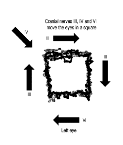
PCT/US2015/043083
22
no/Arefzik = Doxifeightp, x fkalVicithm (48)
[0057] The method may also feature calculating conjugacy of eye movement or
variance from
perfect conjugacy of eye movement, such as, for example,
Conj .v EeirItop =
>.: 1
Con) vorXrit = ¨ __________________ = (58)
Y.:12
E _____________________________________ - 0
Cortj: varXbot C!23, (59)
R
coil) varX/e1
E
E -
COO varYtop (61)
'
Conj. varYrit P (62)
E2
Cztraj varriot Ut (63)
Eio
E 04)2 - 0
coni voryrit (64)
Eilfh
Only CarrX1rtop (a)
ZIA¨ I'
E hOi
CVO CorrXYrit =-7 (6)
Carij CorrXYboi (67)
1,13¨
Eadd
Gorki Corrnfrie,1 (68)
Ei4 ¨ 1
or variance x ratio top/bottom (conjugacy), variance y ratio top/bottom
(conjugacy),
variance x ratio leftfright (conjugacy), or variance y ratio left/right
(conjugacy).
[0058] In some instances, one or more of the L height, L width, L area, L
varXrit, I. varXlef, L
varTotal, R height, R width, R area, R varYtop, R varXrit, R varXlef, R
varTotal, Conj varX,
CA 02957384 2017-02-06
WO 2016/022414
PCT/US2015/043083
23
Conj varXiit, Conj varXbot, Conj varXlef and Conj varYlef may be especially
useful for
demonstrating or detecting or assessing drug use, drug abuse or narcosis. hi
some instances,
two, three, four, live, six, seven, eight, nine, ten or more metrics may be
observed or determined.
[0059] In some instances, the method may feature calculating velocity of eye
movement. In such
instances, the distance (z1) traveled by the pupil between times 1 and 2 may
be determined as the
square root of:
(x2-x1)2+ (y2-y1)2
where the coordinates of the pupil at time 1 arc x j, yj , and the coordinates
of the pupil at time 2
are x2, Y2.
[0060] In some instances, the sum of the z's may be obtained to determine the
distance traveled
by the eye pupil over any time period of interest. The sum of the z's over,
for instance, 1, 2, 3, 5,
7, 10, 15, 20, 25, 30, 60 or 90 or so second intervals may be obtained to
determine at the distance
traveled during each segment of a box eye movement trajectory. Pupil velocity
may be
determined by dividing by the time in seconds.
100611 The method may feature determining whether eye pupil velocity changes
in different
directions of eye movement reflected in an eye movement box trajectory. In
some instances, one
or more of a zioto, zi,p, zjoi, zright, z1,0[(õõ, may he determined with
reference to an eye movement
box trajectory. In sonic instances, one or more of such a zwiat, zt,p, zjeft,
zrighõ 7.1,01,,,,õ may be
provided for each eye. Further, in some instances Wilcoxon statistical
analysis may be employed
to analyze groups of data.
10062I1he velocity of eye pupil movement may be slower by 5, 10, 25, 30, 35,
40, 50, 60, 70,
75% or more in a subject using or abusing a drug such as a narcotic or in a
subject in a state of
narcosis compared to a control, a normal, a subject not using or abusing a
drug such as a narcotic
and not in a state of narcosis, compared to the velocity of eye pupil movement
in the same
subject at a time when that subject is not using or abusing a drug such as a
narcotic or when that
subject is not in a state of narcosiS, or compared to a subject suffetqng from
or experiencing brain
injury, such as, for instance, structural or non-structural traumatic brain
injury. In some such
CA 02957384 2017-02-06
WO 2016/022414
PCT/US2015/043083
24
instances, the velocity of eye pupil movement may be only about 25, 30, 40,
50, 60, 70, 75, 80 or
90% as fast. In some instances, when eye pupil movement is represented in an
eye movement
trajectory box, and the top or bottom or left or right segment of the eye
movement trajectory box
may demonstrate more or less slowing of velocity of eye pupil movement than
the other
segments. For instance, in many instances, the top segment of the eye movement
trajectory box
may demonstrate more slowing of velocity of eye pupil movement than the other
three segments
of the eye movement trajectory box.
100631 A standard deviation or p value of 0.05, 0.10, 0.25, 0.3, 0.4, 0.5,
0.75. 0.8, 0.9, 1.0, 1.5,
2.0, 2.5 or more may reflect that a subject has been using or consuming a drug
or narcotic, is
under the influence of a drug or narcotic, is experiencing an impaired mental
state because of a
drug or narcotic, or is in a state of narcosis. In sonic instances the eye
movement is tracked for
about 30, 60, 90, 100, 120, 150, 180, 200, 220, 240, 270, 300, 360 or more
seconds.
100641 In some instances, the drug or narcotic may be a psychoactive compound
with sleep-
inducing properties or a drug that is illegal or prohibited. In some
instances, the drug or narcotic
may be one or more of an opiate, an opioid, morphine, heroin, and their
derivatives, such as
hydrocodone, or cannabis, alcohol, or any other substance classified as a
controlled substance by
the United States Controlled Substances Act. In some instances, the drug or
narcotic may be a
prescription medication such as, for instance, a benzodiazcpine or
barbiturate.
100651 In a third aspect, the invention provides methods for diagnosing,
assessing or quantitating
drug use, drug abuse or narcosis in a subject by
a) Tracking eye movement of at least one eye of the subject;
b) collecting raw x and y cartcsian coordinates of pupil position;
c) normalizing the raw x and y Cartesian coordinates; and
d) calculating one or more individual metric.
100661 In some instances, eye movement of both eyes of the subject are tracked
and analyzed. in
some instances, both a and y coordinates of eye position !Or one or both eyes
of a subject are
collected for at least about 100, 500, 1,000, 5,000, 10,000, 50,000, 100,000,
200,000 or more eye
CA 02957381 2017-02-06
WO 2016/022414 PCT/US2015/043083
positions. In instances where the eye movement o r both eyes arc tracked, the
meihod may
additionally feature sorting the data by eye.
(00671 The one or more individual metric may be any one of
L.varYtop Vor(Vi,aveTage II5,1) (13)
&WarYtOP = Vat(b,aVerita0 krrt.1¾,i) (14)
L.IscrXrit VartijiVeliVe k33h1.2) (15)
RmarXrit Var(2y,Avera8o kw4tti.2) (10)
L.varYlrot Vor(thaverage k.15,3) (17)
R..varlibot go V dr(#4.average km1:513) (38)
L.varXief Var(,average *.itts.4) (10)
re.varXicl '41 Var(22,1Werag0 00.1:,5.4)
L.varTatta = Average (11 ar(21,avortv k,is) 4- Var(iii,avcrbge k..34)) (21)
Ii.varTotol Average (liar(kAawrage A.1;5) + Var(ikaverge k.vs)) (23)
or segment standard deviation and segment skew such as, for instance,
1,.$kevirep g4Sketv(th,average t-t;sti) (27)
ReketvTop SkevAizaverage k=16,t) (28)
L.SkewRit Skete(21,a,matl h_1.6.2) (20)
itorketvitit akeW(X2.11Veigtv3 k,-15,2)
L.SketoBvt. Skew(gt,tivcivige (31)
R.SItethBot ,40 Sketa(92,avarage 14-144) (32)
L.SketoLef Sketutiutverago (33)
H.SkevoLej SketeM,averogo k.w.4) (34)
or segment normalized skew, such as, for instance,
Sketo(21.kj)
SkiNorat(2j,k,i) (36)
r
Sk0A)(1/ k t)
SketiNerm(9)Aj) (36)
utioAl
L.SketvTopN arra = SketeiVc)rm(frowerage A=1,53) (37)
aSkettfropNorm SktioNorm(haverAge (38)
L.Skew/tititiorm, SketolVorm(ti,averav k.1:5,2) (n)
R.SkownitiVorrn SketelVorm(T2,average *-.1$,2) (40)
.C.Skew.0411form = Skew Norrnakayerap (41)
RSIcetuBotN orm . Skewhrorm(16,avereie )õ..1,5,3) (42)
L.SketriLeftVortu = SkeivNertr(werage h.,415,4) (43)
4.Sktv.),Lef/Vorm = SkelDNOrirKkventge h.1,5õ.4) (44)
CA 02957384 2017-02-06
WO 2016/022414
PCT/US2015/043083
26
100681 The method may also feature calculating box height, box width, box
area, or box aspect
ratio.
box height
Bozlif.ightp,
box width
DoiWitithm 2j,k2 (48)
box aspect ratio
13aalflight k
Avaltilatioi-A -4 = - = == = = (47)
box area
.Boxeirea.i.k Vozif eighto x RoxWidtho (48)
100691 The method may also feature calculating conjugacy of eye movement or
variance from
perfect conjugacy of eye movement, such as, for example, or variance x ratio
top/bottom
(conjugacy), variance y ratio top/bottom (conjugacy), variance x ratio
left/right (conjugacy), or
variance y ratio left/right (conjugacy).
CA 02957384 2017-02-06
WO 2016/022414 PCT/US2015/043083
27
Coro vorKtop ¨ ______________________________________ (57)
f
Conj varxrit = 42)' ¨ 0, (58)
Conj varXbot E (ri$)' 0
0.49
GOV var,Vief E 41)2 ¨11 (00)
E (40' 0
Conj vizrY taxi. vi (61)
E.01,
eOltj varYrit = ¨ 0, (62)
E
Conj varYtot E fi'4)2 (0)
E '
E ¨
cony varYrit - - 0 (64)
E 94
Conj. CiarrXYttip (65)
E=4 ¨
E 4'ha
COTO CorrXYrit =(66)
Coi
CorrXYbet 119 (67)
>214th
Con' acit-rXY40 08)
¨1
[9117(1.1 In some instances, one or more of thc I. height, L width, L area, L
varXrit, L varX1cf, E
varTotal, R height, R width, R area, R varYtop, R varXrit, R varXlef, R
varTotal, Conj. varX,
Conj varXrit, Cod.) varXbot, Conj varXlef and Conj varYlef may be especially
useful for
demonstrating or detecting or assessing drug use, drug abuse or narcosis. In
some instances,
two, three, four, five, six, seven, eight, nine, ten or more metrics may be
observed or determined.
[00711 In some instances, the method may feature calculating velocity of eye
movement In such
instances, the distance (z1) traveled by the pupil between times 1 and 2 may
be determined as the
square root of:
xi)2 (y? Yi)2
CA 02957384 2017-02-06
WO 2016/022414
PCT/US2015/043083
28
where the coordinates of the pupil at time 1 are xi, Yl , and the coordinates
o the pupil at time 2
are X2, Y2.
100721 In some instances, the sum of the z's may be obtained to determine the
distance traveled
by the eye pupil over any time period of interest. The sum of the z's over,
for instance, 1,2, 3, 5,
7, 10, 15, 20, 25, 30, 60 or 90 or so second intervals may he obtained to
determine at the distance
traveled during each segment of a box eye movement trajec,tory. Pupil velocity
may be
determined by dividing by the time in seconds.
[00731 The method may feature determining whether eye pupil velocity changes
in different
directions of eye movement reflected in an eye movement box trajectory. In
sonic instances, one
or more of a zmai, zmr, ziele zrighe may be determined with reference to an
eye movement
box trajectory. In some instances, one or more of such a ztotal, zlep,
ZJeft.zriEN, zbouon may be
provided for each eye. Further, in some instances Wilcoxon statistical
analysis may be employed
to analyze groups of data.
100141 The velocity of eye pupil movement may be slower by 5, 10, 25, 30, 35,
40, 50, 60, 70,
75% or more in a subject using or abusing a drug such as a narcotic or in a
subject in a state of
narcosis compared to a control, a normal, a subject not using or abusing a
drug such as a narcotic
mid not in a state of narcosis, or compared to the velocity of eye pupil
movement in the same
subject at a time when that subject is not using or abusing a drug such as a
narcotic or when that
subject is not in a state of narcosis. In some such instances, the velocity of
eye pupil movement
may be only about 25, 30, 40, 50, 60, 70, 75, 80 or 90% as fast. In some
instances, when eye
pupil movement is represented in an eye movement trajectory box, and the top
or bottom or left
or right segment of the eye movement trajectory box may demonstrate more or
less slowing of
velocity of eye pupil movement than the other segments. For instance, in many
instances, the
top segment of the eye movement trajectory box may demonstrate more slowing of
velocity of
eye pupil movement than the other three segments of the eye movement
trajectory box.
100751 A standard deviation or p value of 0.05, 0.10, 0.25, 0.3, 0.4, 0.5,
0.75. 0.8, 0.9, 1.0, 1.5,
2.0, 2.5 or more may reflect that a subject has been using or consuming a drug
or narcotic, is
under the influence of a drug or narcotic, is experiencing an impaired mental
state because of a
CA 02957384 2017-02-06
WO 2016/022414
PCT/US2015/043083
29
drug or narcotic, or is in a state of narcosis. In some instances the eye
movement is tracked for
about 30, 60, 90, 100, 120, 150, 180, 200, 220, 240, 270, 300, 360 or more
seconds.
[00761 In some instances, the drug or narcotic may be a psychoactive compound
with sleep-
inducing properties or a drug that is illegal or prohibited. In some
instances, the drug or narcotic
may be one or more of an opiate, an opioid, morphine, heroin, and their
derivatives, such as
hydrocodone, or cannabis, alcohol, or any other substance classified as a
controlled substance by
the United States Controlled Substances Act. in some instances, the drug or
narcotic may be a
prescription medication such as, for instance, a benzodiazepine or
barbiturate.
[0077] In a fourth aspect, the invention provides a kit useful for detecting,
screening for or
quantitating drug use, drug abuse or narcosis in a subject, containing a
device for tracking eye
movement, one or more means =foi analyzing eye movement tracking data such as,
for instance,
an algorithin or computer program, and instructions. Processing eye movement
observations,
making measurements of eye movement observations, determining distributions of
values
measured and performing statistical tests may all he accomplished using
suitable computer
software that may be included in such a kit.
[0078] In a fifth aspect, the invention provides methods for diagnosing or
assessing internuelear
ophthalmoplegia (NO) in a subject by
a) Tracking eye movement of both eyes of the subject;
h) Analyzing eye movement of both eyes of the subject;
c) Comparing eye movement of a first eye of the subject to eye movement of a
second
eye of the subject; and
d) Identifying the subject as having eye movement of a first eye that is
significantly
different from eye movement of a second eye.
The method may further feature
e) Tracking eye movement of each eye of the subject separately.
The method may also further feature
I) Identifying the subject as having substantially normal eye movement of
each eye
when eye movement of each eye of the subject is tracked separately
CA 02957384 2017-02-06
WO 2016/022414
PCT/US2015/043083
[00791 A diagnosis of intemuclear ophthairnoplegia (INC)) may be made Or
provided as a
possibility or the status of intemuclear ophthalmoplegia (INO) may be
evaluated or intemuclear
ophthalmoplegia (11'40) may he assessed if the subject is d) identified as
having eye movement of
a first eye that is sigiffleantly different from eye movement of a second eye
when tracking eye
movement of both eyes of the subject and if the subject is also 0 identified
as having
substantially normal eye movement of each eye when eye movement of each eye of
the subject is
tracked separately. 'file method is useful for differentiating intemuclear
ophthalmoplegia (INO)
from infranuclear nerve palsies or other disorders affecting the muscles
associated with ocular
mot il ty.
100801 in a sixth aspect, the invention provides methods for diagnosing or
assessing intemuclear
ophthalmoplegia (INO) in a subject by
a) Tracking eye movement of both eyes of the subject;
b) Analyzing eye movement of both eyes of the subject;
c) Comparing the x or y Cartesian coordinates at any time point for the eye
movement
of a first eye of the subject to the respective x or y Cartesian coordinates
at the time
point for the eye movement of a second eye of the subject;
d) Providing a sum of the differences between all of the x coordinates of the
first eye
compared to the second eye over the time tested or providing a sum of the
differences in y coordinates of the first eye compared to the second eye over
the time
tested or both; and, optionally
e) Providing a total sum of the differences between both x and y
coordinates of the first
eye compared to the second eye over the time tested.
The method may further feature
0 Tracking eye movement of each eye of the subject separately.
The method may also further feature
g) Identifying the subject as baying substantially normal eye movement of each
eye
when eye movement of each eye of the subject is tracked separately.
109811 In sonic instances, the subject suffering from the intemucl ear
ophthalmoplegia (11C0)
may have a total sum of the differences between both x and y coordinates of
the first eye
CA 02957384 2017-02-06
WO 2016/022414
PCT/US2015/043083
31
compared to the second eye over the time tested that is 10%, 20%, 30%, 40%,
50%, 60%, 70%,
80%, 90%, 100%, 150%, 200%, or two, three, four, five, six, eight, ten or more
times greater
than the total sum of the differences between both x and y coordinates of the
first eye compared
to the second eye over the time tested in a healthy control or in a reference
value based upon one
or more healthy controls or based upon the subject at a time before the
internuclear
ophthalmoplegia (INO).
100821A diagnosis of internuclear ophthalmoplegia (INO) may be made or
provided as a
possibility or the status of internuclear ophthalmoplegia (INO) may be
evaluated or internuclear
oplithalmoplegia (IN 0) may be assessed if the subject is d) identified as
having eye movement of
a first eye that is significantly different from eye movement of a second eye
when tracking eye
movement of both eyes of the subject and if the subject is also f) identified
as having
substantially normal eye movement of each eye when eye movement of each eye of
the subject is
tracked separately. The method is usefill for differentiating internuclear
ophthalmoplegia (INO)
from infrunuelear nerve palsies.
[0083] In a seventh aspect, the invention provides methods for assessing or
quantitating or
diagnosing internuclear ophthulrnoplegia (NO) in a subject by
a) Tracking eye movement of both eyes of the subject,
b) collecting raw x and y cartesian coordinates of pupil position;
c) normalizing the raw x and y Cartesian coordinates; and
d) calculating one or more individual metric.
The method may further feature
e) Tracking eye movement of each eye o f the subject separately.
The method may still further feature
I ) calculating one or more second or additional individual metric.
flic method may also further feature
g) Identifying the subject as having substantially normal eye movement of each
eye
when eye movement of each eye of the subject is tracked separately.
CA 02957384 2017-02-06
WO 2016/022414 PCT/US2015/043083
32
[0084] A diagnosis of internuclear ophthalmoplegia (NO) may be made or
provided as a
possibility or the status of internuclear ophthalmoplegia (INO) may be
evaluated or internuclear
ophthahnoplegia (INO) may be assessed lithe subject is identified as having
normal motility or
aspect ratio in the eyes assessed separately and abnormal aspect ratio or
conjugacy when the eye
movement of both eyes is tracked together. The method is useful for
differentiating internuclear
oplithalmoplegia (INO) from infranuclear nerve palsies or other disorders
affecting the muscles
associated with ocular motility.
10085] Eye movement of both eyes of the subject are tracked and analyzed. In
some instances,
both x and y coordinates of cye position for one or both eyes of a subject are
collected for at least
about 100, 500, 1,000, 5,000, 10,000, 50,000, 100,000, 200,000 or more eye
positions. In sonic
instances, the eye position is effectively the pupil position. In some
instances the eye movement
is tracked for about 30, 60, 90, 100, 120, 150, 180, 200, 220, 240, 270, 300,
360 or more
seconds.
[0086] In sotne instances, the method may feature collecting raw r and
yeartesian coordinates of
pupil position, normalizing the raw x and y Cartesian coordinates, and sorting
the data by eye.
[0087] The method may also feature calculating individual metrics, such as,
for instance,
segment mean, segment median, and segment variance. The method may also
feature calculating
specific metrics such as, for example,
or sewn L.ra rY tap V it r(g).Amage
k,õIzzia) (la)
R.varY'top V ar(92,tiverau kr,1,6,t) (14)
iiraf(X/Avano ta,2) (1)
kvarXrit Var(22,4moge (1)
t.i.varYlvt Var(01,avero,g0 (11)
Rm4rYtiot tlat-(k4,,trage yõL.5,3) (16)
LvarXlei lidr(11,.avorage (19
.7xtr X ie f Ar= Vd 02,4Velage 1:5,4 (20)
=LajorT0141 " Average (Var(ii,veragt A..titt) Var(lii,averwi k..,i,$)) (21)
R,varTotni Aver eke (Van) ,Amrage iiilr(gcaverao 4,41:6))
(22)
CA 02957384 2017-02-06
WO 2016/022414 PCT/US2015/043083
33
L.Skeufl'or Skew (Vi,averay. 1,5.,1) (27)
R.SkeurTnp Skew(fizgweivge
L.:Skeittitit SkettEM,aretago (20)
IZ-54twRit Skett02.avetilgo k-173,0 (n)
I-SkewEat Skew(thaverege kmi;5,a)
riskewBo..5414(P2,4verago ic-13.3) (32)
1,-.514e/r/Uf $kew(i-i,p4verago (3)
ii,SkewLef SkewMovaage k.14.4) 04)
or segment normalized skew, such as, for instance,
Skeu.,-(53),t i)
greettiVorm(tjki) ¨ (3-5)
oRiAt
Skew(VjA,1)
Shew.Nortn(i714,4) (30)
L,Skewroptiorrri = SketoWerin1,averago (3?)
R.SketuTepNorm. SkttriNorm(g2A4,..errage (38)
L,SkttanitNorms Sicetorioryn(iievitrege (39)
E,SktwRiiNwrra ActiNurrytOzsiverage k.v3,2) (40)
lakctuDoiNorrn SkewNo ________ ilL(ilLaverago kul:.503) (41)
R.SkewBot.Norm Z SkonNerrtn(f) 2. Average A.1,5,3) (42)
.L.5ketot4Norm SketoNortr1(.14,7Frao (4,1)
R.,5keaaref.Norrn .8ktioNorm(11,4verage k=1:54 (44)
100881 The method may also feature calculating box height, box width, box
area, or box aspect
ratio.
box height
BtaHrighti,k Di,k4 (45)
box width
klozWicithvA (46)
CA 02957384 2017-02-06
WO 2016/022414
PCT/US2015/043083
34
box aspect ratio
Borlfei9Ftt =
AlpectReitili ¨ = (47)
' BaxW
box area
BoxAream Eollighij V4thM (48)
[0089] The method may also feature calculating conjugacy of eye movement or
variance from
perfect conjugaey or eye movement, such as, for example,
Can) va 0rKtop . (V)
(i2)2 -
Conj txwXrit 0 (68)
r
E' -
conj varXbot 0 (59)
2:23
CEnZi VarX kl "" = ) (3, (.60
Y74
E
onc varYtryp
cow vary-y.4 o (2)
E _______________________________ (1V -
Cork' tictrilat (63)
Coq varrrit __________ (64)
EV4
E j4]
Otani Corallop (6)
Conj ClorrXYrit
Y'
Ototj Corr.XY bat (67)
r, 144
Conj Cort,XYlef (68)
L24 -
or variance x ratio top/bolLom (conjugacy), variance y ratio top/bottom
(conjugacy),
CA 02957384 2017-02-06
WO 2016/022414
PCT/US2015/043083
variance x ratio left/right (cottjugaey), or variance y ratio left/right
(conjugacy).
100901 In some instances, one or more of the I. height, L width, L area, L
varXrit, L varXlef, L
varTotal, R height, R width, R area, R varYtop, R varXrit, R varXlef, R
varTotal, Conj varX,
Conj varXrit, Conj varXbot, Conj varXlef and Conj varYlef may be especially
useful for
demonstrating or detecting or assessing internuclear ophtbaimoplegia (NO) or
differentiating, or
distinguishing it from infranuclear nerve palsies. In some instances, two,
three, four, five, six,
seven, eight, nine, ten or more metrics may be observed or determined.
100911 In a eighth aspect, the invention provides a kit useful for detecting,
screening for or
quantitating internuclear ophthalmoplegia (INO) and useful for differentiating
internuclear
opinhalmoplegia (WO) from infranuelear nerve palsies containing a device for
tracking eye
movement, one or more means for analyzing eye movement tracking data such as,
for instance,
an algorithm or computer program, and instructions. Processing eye movement
observations,
making measurements of eye movement observations, determining distributions of
values
measured and performing statistical tests may all be accomplished using
suitable computer
software that may be included in such a kit.
100921 In a ninth aspect, the invention provides methods for diagnosing or
assessing attention
deficit hyperactivity disorder (AMID), chronic traumatic encophalopathy, or a
schizophrenia
spectrum disorder in a subject by
a) Tracking eye movement of both eyes of the subject;
b) Analyzing eye movement of both eyes of the subject;
c) Comparing eye movement of a first eye of the subject to eye movement of a
second
eye of the subject; and
d) Identifying the subject as having eye movement of a first eye that is
significantly
di ffcrem from eye movement of a second eye.
10093] In a tenth aspect, the invention provides methods for diagnosing and
assessing attention
deficit hyperactivity disorder (ADHD), chronic. traumatic oncephalopathy, or a
schizophrenia
spectrum disorder in a subject by
CA 02957384 2017-02-06
WO 2016/022414
PCT/US2015/043083
36
a) Tracking eye movement of both eyes of the subject;
b) Analyzing eye movement of both eyes of the subject;
c) Comparing the x or y Cartesian coordinates at any time point for the eye
movement of a first eye of the subject to the respective x or y Cartesian
coordinates at
the time point for the eye movement of a second eye of the subject;
d) Providing a sum of the differences between all of the x coordinates of
the first eye
compared to the second eye over the time tested or providing a sum of the
differences in
y coordinates of the first eye compared to the second eye over the time tested
or bath;
and, optionally
e) Providing a total sum of the differences between both x and y coordinates
of the first
eye compared to the second eye over the time tested.
100941 In some instances, the subject suffering from the attention deficit
hyperautivity disorder
(ADHD), chronic traumatic encephalopathy, or a schizophrenia spectrum disorder
may have a
total sum of the differences between both x and y coordinates of the first eye
compared to the
second eye over the time tested that is 10%, 20%, 30%, 40%, 50%, 60%, 70%,
80%, 90%,
100%, 150%, 200%, or two, three, four, five, six, eight, ten or more times
greater than the total
sum of the differences between both x and y coordinates of the first eye
compared to the second
eye over the time tested in a healthy control or in a reference value based
upon one or more
healthy controls or based upon the subject at a time before the disease.
100951 In a eleventh aspect, the invention provides methods for diagnosing or
assessing attention
deficit hyperactivity disorder (ADIID), chronic traumatic eneephalopathy, or a
schizophrenia
spectrum disorder by
a) Tracking eye movement of at least one eye of the subject;
b) Analyzing eye movement of at least one eye of the subject;
c) Comparing eye movement of at least one eye of the sub Ca to a normal or
mean eye
movement; and, optionally
d) Calculating a standard deviation or p value for eye movement of at least
one eye of
the subject as compared to the normal or rritri eye movement.
CA 02957384 2017-02-06
WO 2016/022414
PCT/US2015/043083
37
[0096] In sOme instances, eye movement of both eyes of the subject are tracked
and analyzed. In
some instances, both x and y coordinates of eye position for one or both eyes
of a subject are
collected for at least about 100, 500, 1,000, 5,000, 10,000, 50,000, 100,000,
200,000 or more eye
positions. In some instances, the eye position is effectively the pupil
position. In some instances
the eye movement is tracked for about 30, 60, 90, 100, 120, 150, 180, 200,
220, 240, 270, 300,
360 or more seconds.
[0097] The comparing eye movement of at least one eye of the subject to a
normal or mean eye
movement may feature comparing eye movement of at least one eye of the subject
to the eye
movement of the other eye if the subject or may feature comparing eye movement
of at least one
eye of the subject to the eye movement of an eye of one or more other subjects
or controls. In
some instances, the comparing eye movement of at least one eye of the subject
to anomie! or
mean eye movement may feature comparing the eye movement of both eyes of the
subject to the
eye movement of one or both eyes of one or more other subjects or controls.
100981 In some instances, the method may feature collecting raw r and y
cartesian coordinates of
pupil position, normalizing the raw x and y Cartesian coordinates, and sorting
the data by eye.
[0099] The method may also feature calculating individual metrics, such as,
for instance,
segment mean, segment median, and segment variance. The method may also
feature calculating
specific metrics such as, for example,
L.varYtcri.) V ar(fi.,a,veritge k .1 AL) (13)
Ratarnop Var0.72A.,trage (14)
LAterXrii Vc4r(g8 *.L3.2)
R.varXrit 1.41r(Z,2,b040,340)* rdiAl ) (16)
L. trarYboi Var(p),oxeritgo )._143) (17)
R=varYbot 4=1 Vat' (horretage, ,k.) ;5,3 ) (18)
k.i.kirX le! Var (f Averag4.) In. um) (to)
L.akarXt-cf V ar(12,average
(20)
Lvarroial Average
(Ilaq.il,a=verago k.,11,&) + IlarOi,avertige k,.;,1,5).) (21)
R.VarTatal AVe70-9e (iffw(i2,Avaago 4..110+ Vm.(g2Aven-Ago k..A!4))
(22)
or segment standard deviation and segment skew such as, for instance,
CA 02957384 2017-02-06
WO 2016/022414
PCT/US2015/043083
38
L.51ceudTop SkOV(fh,liverage. (27)
R-Sikettlfcip Sketo(0,00-4A (28)
L$ktwRt= 8keto(21,w,tlage k,,14,2) (20)
it.8 key, ni 6 44 8ktw(zizaverno k,s1;:3,2) (30)
L.SketeBa Skev(l !average k..t:N,3) (31)
R.ShefirBot Skew(fI2,aVGV3p h-133) (32)
LgketriUf $ktud,r,Z1,averago k.usdt) (33)
li,SkeioLe f .5%IcElijR2aelvestIgo k.1.4 (34)
or segment normalized skcw, such as, for instance,
Skew( i,u)
SketriNurat(t (3b)
'72s,ifif
Skett,Norn.4-j,k,t) Ske-to(177,1,,o)
(3(1)
L.SkovTopNorrn = Sk,ctinVerm(91,:werage (37)
R.SkeuToptVorm Skew-Aroln(02,0,w,r4g, (38)
L. SkewRithrorm Skem Norrn(rA overate A=1,6,2) (39)
R.5kewRitNorrin SketviVerrnOzaverege (40)
L,SketuBoiNorrn SkMONOrrn(91,13weragy knr.tz,3) (41)
RS arm SketoNorrrk(fejAverw j,t,a) (42)
L.Ske.toLeitiorril = Sktratiortn(iTlAveragc h415,4)
ItSketaeliVOrra = 5keIVP/C/rin(22AV='08C h-1154) (44)
101001 The method may also feature calculating box hoight, box width, box
area, or box aspect
ratio.
box height
Boxile.ightjr,kp,LDida (41)
box width
aryzWidthm ij,k44 (4)
box aspect ratio
Box/ fe,ighij,k
A.81.-.78dR-410j,' (47)
CA 02957384 2017-02-06
WO 2016/022414
PCT/US2015/043083
39
box area
BoxAre4i,k .13ozlitightik BoalYitithirk OS)
101011 The method may also feature calculating conjugacy of eye movement or
variance from
perfect conjugacy of eye movement, such as, for example,
Conj vor,,,Ciap ¨ (1)1 ¨.0, (57)
E&I
E (i3? - 0
coo lko-Xrit = =
E '
corki varXbeg - , (59)
Z.
r4
Cted var.:Clef 0 (60)
Eta
E --- 0
coni- varyti-4. - = , (01)
L'Pa
Canf varYrit = E (f/i?= ¨0
(62)
iir3)2 -
Conj uarY 06oi (63)
E '
E -
Ctnjvarrrit . = (GO
E *)
C Pioxd CorrXYtop (65)
E
r 1,4:02
Coraj CorrX-Yrit = (66)
Eh-1'
E &s93
Cord CorrXYbot (07)
Cord Corrnlef (68)
¨ 1
or variance x rati(
variance x ratio left/right (conjugacy), or variance y ratio left/right
(conjugacy).
[0102] In some instances, one or more of the L height, I, width, L area, L
varXrit, L varXlef, L
varietal, R height, R width, R area, R varYtop, R varXrit, R varXlef, R
varietal, Conj varX,
Conj varXrit, Conj varXbot, Conj varXlef and Conj varYlef may be especially
useful for
CA 02957384 2017-02-06
WO 2016/022414
PCT/US2015/043083
demonstrating or detecting or assessing attention &licit hyperactivity
disorder (ADHD), chronic
traumatic encephalopathy, or a schizophrenia spectrum disorder. In some
instances,
two, ihree,
four, five, six, seven, eight, nine, ten or more metrics may be observed or
determined.
[0103] A standard deviation or p value of 0.25, 0.3, 0.4, 0.5, 0.75. 0.8, 0.9,
1.0, 1.5, 2.0, 2.5 or
more may reflect that a subject has attention deficit hyperactivity disorder
(ADHD) , chronic
traumatic eneephalopathy, or a schizophrenia spectrum disorder. As such, the
methods described
herein may be used to detect attention deficit hyperactivity disorder (ADHD),
chronic traumatic
encephalopathy, or a schizophrenia spectrum disorder and assess or determine
the severity of the
same.
101041 In a twelfth aspect, the invention provides methods for diagnosing,
assessing or
quantitating attention deficit hyperactivity disorder (ADHD), chronic
traumatic eneephalopathy,
or a schizophrenia spectrum disorder in a subject by
a) Tracking eye movement of at least one eye of the subject;
b) collecting raw x and y cartesian coordinates of pupil position;
e) normalizing the raw x and y Cartesian coordinates; and
d) calculating one or more individual metric.
101051 In some instances, eye movement of both eyes of the subject are tracked
and analyzed. In
some instances, both x and y coordinates of eye position for one or both eyes
of a subject arc
collected for at least about 100, 500, 1,000, 5,000, 10,000, 50,000, 100,000,
200,000 or more eye
positions. In instances where the eye movement of both eyes are tracked, the
method may
additionally feature sorting the data by eye.
101061 The one or more individual metric may be any one of
=LvarYtoP Varcgt,average kul*I)
(13)
R.VtirYtOP Val-(9.2,1MMV k.014,1.) (14)
b,ito.rXrit Var(121,averkp kmit0) (15)
gtarXrit Var(xzeverko br.op) (16)
.L,trarYbot VoraiLavorage (17)
R.varYba Var(kviverage (1B)
-Lyn,' X lei Var(ti,Aventgo k=1.41)
Lvar Xlci --=t- 1 / (,1,verage (20)
L.IrorTaal Average (Vaf(21AYeragC fi-.19)) + Var(VI,averhge ,fri!5))
(21)
re-Varrata " AVere,9e (Vas.(iWrIgC .1E=.14) Vor(ii2Avc3tago x.2:5)) (n)
CA 02957384 2017-02-06
WO 2016/022414
PCT/US2015/043083
41
or segment standard deviation and segment skew such as, for instance,
L-Skelii-110Ps Ska41,1Vdrara) (27)
R.,91,3etzTrYp Sktn:(v.mworago (28)
Kw:Skew'. 8kei.01Ataga k.=Llet2) (D)
R=540,41itit 441 Skett#average Avv3,1) (30)
L.Skeu.,Bot Skti/(fri,aveynio kma:3.3) (31)
R.SkewBot Sketp(92,average k,4,4.a) (32)
L.6'ke21.7Lef Sketv(21,wertqc km,s3) (33)
SknoLef ...9ketv(fLarczNo kõ..4,kg) (34)
or segment normalized skew, such as, for instance,
SkeurNorna(da) _________ (35)
etc0fot,1)
Shcmti farna(frjritA) (36)
qt-4.44
L.SkttuTopNorm = SketpNenvitgoLverage 4-1,4,1) (37)
ii.SlictvTa.p.Y Sketi/Norm07,average (38)
L.SiteteRitiVohn SketaiturinCaverage 4.1,4.2) (39)
SkewRif Nrfr,rrn t's, Skew Worrn(012.average (40)
L.SkfuttiotNerrrn Sk.ewNorr,;(171,-averapp., (41)
it.SkettraCtiskrin SkewNc,1-74(172,average (42)
L.SkctoL /If orfa SkewN6Tn1(71.0vertist kb6h4) (43)
LI,Lef Norm = Skew N orrnOtzavvrage (44)
[0107] The method may also feature calculating box height, box width, box
area, or box aspect
ratio.
box height
Ras...Hpri9hts,A ¨ (5)
box width
lioxWidthiA 20,4 (,16)
box aspect ratio
Ear/PION**
AtPeetRali`7),L` Box Width, (47)
CA 02957384 2017-02-06
WO 2016/022414
PCT/US2015/043083
42
box area
BoxArtaiA Bcalleighti,k x BWic1th,08)
[U1081 The method may also feature calculating conjugacy of eye movement or
valiance from
perfect conjugacy of eye movement, such as, for example,
o
corki vorxtop = ,
coni tgarKrit ¨ (58)
El2
E 43)2 0
Conj verXbot = (59)
E )2 ¨
,cord varXkf (ii (Go)
E 0'
= E h:/.$ )2 ¨ 0
Curkf varYtryp ¨ = _ (61)
re.01.
¨
Coni vorn E it 2;-=== , (62)
CORI VarY (63)
E
Conj txtrYrii. = __ = (64)
'
E
con,' =oorrx ytop (65)
E21 ¨1'
E
coni c'cirrXYrit = (66)
E27
Cord CorrXYbot (67)
I
Conj. CorrX1fikr (68)
¨ 1
or variance x ratio top/bottom (conjugaey), variance y ratio top/bottom
(conjugacy),
variance x ratio left/right (conjugacy), or variance y ratio left/right
(conjugaey).
CA 02957384 2017-02-06
WO 2016/022414
PCT/US2015/043083
43
101091 In some instances, one or more of the L height, I width, I. area, I
,=varXrit, I .varXlef, 1.
varTotal, R height, R width, R area, R varYtop, R varXrit, R varXlel, R
varTotal, Conj varX,
Conj =varXrit, Conj varXbot, Conj varXlef and Conj varYlef may be especially
useful for
demonstrating or detecting or assessing attention deficit hyperactivity
disorder (ADHD), chronic
traumatic encephalopathy, and schizophrenia spectrum disorders. In some
instances, two, three,
four, five, six, seven, eight, nine, ten or more metrics may be observed or
determined.
[0110] A standard deviation or p value of 0,25, 0.3, 0.4, 0.5, 0.75. 0.8, 0.9,
1.0, 1.5, 2.0, 2,5 or
more may reflect that a subject has attention deficit hyperactivity disorder
(ADHD), chronic
traumatic encephalopathy, or a schizophrenia spectrum disorder. As such, the
methods described
herein may be used to detect attention deficit hyperactivity disorder (ADHD),
chronic traumatic
encephalopathy, or a schizophrenia spectrum disorders and assess or determine
the severity of
the same.
101111 In a thirteenth aspect, the invention provides a kit useful for
detecting, screening for or
quantitating attention deficit hyperactivity disorder, chronic traumatic
encephalopathy, and
schizophrenia spectrum disorders containing a device for tracking eye
movement, one or more
means for analyzing eye movement tracking data such as, for instance, an
algorithm or computer
program, and instructions. Processing eye movement observations, it:taking
measurements of eye
movement observations, determining distributions of values measured and
performing statistical
tests may all be accomplished using suitable computer software that may be
included in such a
kit.
101121 In a fourteenth aspect, the invention provides methods for diagnosing,
quantitating or
identifying alcohol consumption or intoxication in a subject by
a) Tracking eye movement of both eyes of the subject;
b) Analyzing eye movement of both eyes of the subject;
c) Comparing eye movement of a first eye of the subject to eye movement of a
second eye of the subject; and
d) Identifying the subject as having eye movement of a first eye that is
significantly
different from eye movement of a second eye.
CA 02957384 2017-02-06
WO 2016/022414
PCT/US2015/043083
44
[01131 In a fifteenth aspect, the invention provides methods for diagnosing,
quantitating or
identifying alcohol consumption or intoxication in a subject by
a) Tracking eye movement of both eyes of the subject;
b) Analyzing eye movement of both eyes of the subject;
e) Comparing the x or y Cartesian coordinates at any time point for the eye
movement of a first eye of the subject to the respective x or y Cartesian
coordinates at the time point for the eye movement of a second eye of the
subject;
d) Providing a sum of the differences between all of the x coordinates of the
first eye
compared to the second eye over the time tested or providing a sum of the
differences in y coordinates of the first eye compared to the second eye over
the
time tested or both; and, optionally
e) Providing a total sum of the differences between both x and y coordinates
of the
first eye uompared to the second eye over the time tested.
[0114] In some instances, the subject having consumed alcohol or being
intoxicated may have a
total sum of the differences between both x and y coordinates of the first eye
compared to the
second eye over the time tested that is 10%, 20%, 30%, 40%, 50%, 60%, 70%,
80%, 90%,
100%, 150%, 200%, or two, three, four, five, six, eight, ten or more times
greater than the total
sum of the differences between both x and y coordinates of the first eye
compared to the second
eye over the time tested in a control or in a reference value based upon one
or more controls or
based upon the subject at a time befOre the alcohol consumption or
intoxication.
[0115] In a sixteenth aspect, the invention provides methods for diagnosing,
quantitating or
identifying alcohol consumption or intoxication by
a) Tracking eye movement of at least one eye of the subject;
b) Analyzing eye movement of at least one eye of the subject;
t.;) Comparing eye movement of at least one eye of the subject to a normal or
mean
eye movement; end, optionally
d) Cale alating.a standard deviation or p value for eye movement of at least
one eye
of the subject as compared to the normal or mean eye movement.
CA 02957384 2017-02-06
WO 2016/022414 PC T/US2015/043083
1.01161 In some instances, eye movement of both eyes of the subject are
tricked and analyzed. In
some instances, boa' x and y- coordinates of eye position fir one or both eyes
of a subject are
collected for at least about 100, 500, 1,000, 5,000, 10,000, 50,000, 100,000,
200,000 or more eye
positions. In some instances, the eye position is effectively the pupil
position. In some instances
the eye movement is tracked for about 30, 60, 90, 100, 120, 150, 180, 200,
220, 240, 270, 300,
360 or more seconds.
101171 The comparing eye movement of at least one eye of the subject to a
normal or mean eye
movement may feature comparing eyc movement of at least one eye of the subject
to the eye
movement of the other eye of the subject or may feature comparing eye movement
of at least one
eye of the subject to the eye movement of an eye of one or more other subjects
or controls. In
some instances, the comparing eye movement of at least one eye of the subject
to a normal or
mean eye movement may feature comparing the eye movement of both eyes of the
subject to the
eye movement of one or both eyes of one or more other subjects or controls.
101181 In some ins tanees, the method may feature collecting raw x and y
eartesian coordinates of
pupil position, normalizing the raw x and y Cartesian coordinates, and sorting
the data by eye.
101191 The method may also feature calculating individual metrics, such as,
for instance,
segment mean, segment median, and segment variance. The method may also
feature calculating
specific metrics such as, for example,
.G.varlftup Varigi,raerage (13)
.1, arY top VfwlJ2,Cverag. (14)
.Z turX az; Varckaxeracc (15)
R.i.rar X rit VaTC.214-Misgt (16)
LA/C4fYIN't Var(ftt.avvrago h.oith,,3) (17)
itvarnot Var(ka,avorage Cvl3) (IS)
LvarXljVer(havertige k=1.1,4) (19)
.1'1..tnrX/4 Var (izavtirage (20)
L'rarT 144 Atwage (Var(-1,average ktrus) v..
ar(91,averrige kalq$)) (21)
1?.varT(444 Avtilifje (Vartivirormgc + Var(N,average A.I.:0)
(7.2)
CA 02957381 2017-02-06
WO 2016/022414
PCT/US2015/043083
46
or segment standard deviation and segment skew such as, for instance,
L.5keterop Skett(Vl.averago k=tts,t) .. (27)
liSkeu:Top Sketi02,twerogo k04,0 (28)
L.SketoRtt Skete(2iatwerage icr.or,g) (20)
R=Shwrilt " slicew(114,tiverege ki:1;54) (30)
4.8kewli = acw(fkaverago kati,3) (31)
BASkell'Bot Skeigkavoirego (32)
L.Skeuriesf Skcw423,average k.u.1.4) ($3)
R.SketoLa f +0. Sketv(havexage kw L6,4) (34)
or segment normalized skew, such as, for instance,
kesigS go,l)
SketvIV arnt(x = (30
goo
Skpw(r,.
Sk CUP isl or m 1' (3(3)
ZaketaropNorm = SkeluNortn(ikaverage kwits.1) (37)
R.SkeutropiVorm ze= SIceurNotIn(kaveroge te...mt) (38)
rhaeliathrarka V=A SkettiNIVM(21"Vallet =IAA ) (39)
R...5kewRii4Vorin m, Skew Pforrn(22.average .. (40)
L.SkewBot Norm = SkewNorin(haverap k .1:44) (41)
R,SketaatN oral. SkstoNertneih,avarogt k-irs,$) (43)
L.SketeLefliform = SkeioNerrn(2sAvorruge 4.4:5,4) (4)
ltSkeurLef k orrn = SketliN onrOtaverNse h-Ls,4) (44)
[0120] The method may also feature calculating box hei At, box width, box
area, or box aspect
ratio.
box height
Ecafteightjsk - 4,0 (4)
box width
BozWidthm =hk.2 110 (40)
box aspect ratio
Doxifeightio,
AspeetRatiojA irg Dix:widow (47)
CA 02957384 2017-02-06
WO 2016/022414
PCT/US2015/043083
47
box area
DazAreizi,k ,Duzlicightik (48)
101211 The method may also feature calculating conjugacy of eye movement or
variance from
perfect conjugacy of eye movement, such as, for example,
E - 0
Oonj vorItop (47)
-
Co) varXrit 'Et2 (56)
E _________________________________ 0
coni varXbat (50)
Z43
conj vorXtel 00)
COrd /Az t'Y lop = _____________________________ 01)
E
E - c ar on' vYrit 0 (62)
E
-
Cold var E Ybot: = - = (63)
E
Eiff4)1 -
Coni wo.Yra - (64)
t
Y.7 .1111
COT4f CorrXYtop (65)
E1202
Coni (Jorr.X.-Yrit = (0)
E
171,
CCM,/ (7oreXYbot (67)
El3
r 14N
Conj CorrXYlat s's (68)
or variance x ratio top/bottom (conjugacy), variance y ratio top/bottom
(conjugacy),
variance x ratio left(right (conjugacy), or variance y ratio len/right
(conjugacy).
10122] In some instances, one or more of the left.blinklength.value,
righLaspectRatiornean.value,
right.skewTop.valuz, right .skewTopNorm.value, right.varTotal.value,
right.varXrit.value,
conivarXbot.value, right.distTop.value, rightxlistRit.value,
left.velTop.value, leftvelRitivalue
CA 02957384 2017-02-06
WO 2016/022414 PCT/US2015/043083
48
=
may be especially useful for demonstrating or detecting or assessing alcohol
consumption or
intoxication. In sonic instances, two, three, four, five, six, seven, eight,
nine, ten or more metrics
may be observed or determined.
[01231 A standard deviation or p value of 0.05, 0.1, 0,2, 0.25, 0.3, 0.4, 0.5,
0.75 õ 0.8, 0.9, 1.0,
1.5, 2.0, 2.5 or more may reflect that a subject has consumed alcohol or is
intoxicated. As such,
the methods described herein may be used to detect, diagnose, quantitate or
identify alcohol
consumption or intoxication or determine the severity or extent of the same.
[0124] In a seventeenth aspect, the invention provides methods for diagnosing,
assessing or
quantitating alcohol consumption or intoxication in a subject by
a) 'tracking eye movement of at least one eye of the subject;
b) collecting raw x and y cartcsian coordinates of pupil position;
c) normalizing the raw x and y Cartesian coordinates; and
d) calculating one or more individual metric.
101251 In some instances, eye movement of both eyes of the subject are tracked
and analyzed. In
some instances, both x and y coordinates of eye position for one or both eyes
of a subject are
collected for at least about 100, 500, 1,000, 5,000, 10,000, 50,000, 100,000,
200,000 or more eye
positions. In instances where the eye movement of both eyes are tracked, the
method may
additionally feature sorting the data by eye.
101261 The one or more individual metric may be any one of
L.varYtop litre(yi,average or, i;.5.1) (13)
atarYtop V1r(172,am-4.ge ) (14)
Vor4-1 _FM-TAW ba )51,21 (15)
ii.tarXrit = Var(t-mo,vrAgt ki1d (18)
marl but ¨ V4r(iti.avel age g5,3) (17)
R.vetrYbat Var(lhavoTago 631) (18)
L.tur X le f = Var(11,avertige (19)
L..vor,Ylef Ifur(12,tworage (20)
LaNerTotal Average (V (Tr (2 lAvvro,go k.,,1;6) ;ler(,average *.);$))
(211
ILvarTotai Avffri.ge (V ar(imom Dv) h:.:I;15)+ Var (172,awa ago k=3:5))
(22)
CA 02957381 2017-02-06
WO 2016/022414 PCT/US2015/043083
49
or segment standard deviation and segment skew such as, for instance,
L,SkTop Sktto(ft,avcizgo fr.a:s.r) (27)
R.Skeianp .4 Skcw(amage k.14,1) (28)
1,Skeut1irt Skew(ffluwerago k.bta) (29)
&Skew rti6 skeu.02.aver1ge kr.1:34) (30)
L.Sk4wBot keto(A,tivorao kmi;501) (31)
R.SkettBot wt. Sketp(thiavorago (32)
L -5140014f sitev4(haverav kalisA) (33)
ii.SketuLef rn Skat(*).1.avevage k=1;0 (34)
or segment normalized skew, such as, for instance,
SketiOAk,i)
SketerNynn(Zih,r) (35)
Sketo(fAt)
SkewNorin(fly) ____________________ ¨. (36)
L.AtturopiV orn1 SkeigNormifh,a.verage (31)
RASkewTopiVorm AR SkelinVOr ottVerage I ) (38)
L.SketaRithrortn SkowNorm(Staverage k.,11,2) (39)
R.SkeisRUNorm Ira Sketotlorm(12.averagc .4;es2) (40)
LActuflogNorm ?ft SkeloNorm(iitAve.rage vsa) (41)
R.SkauBallorm SketoPlorin(ih,average k.13) (42)
L.$1einakiNarm SkeloNorv(viverriv 1t5,4)
R.SktuiLef Norm SkewNorrn(22.6yerve (44)
101271 The method may also feature calculating box height, box width, box
area, or box aspect
rati).
box height
Boxileightv, DiA,g (4B)
box width
DozWitithm " j,k ¨ IAA (46)
box aspect ratio
Dori! aighto
ASPeetRati 1.1: ff3 'DaW 'CRAM (47)
CA 02957384 2017-02-06
WO 2016/022414
PCT/US2015/043083
box area
Boxtireal,k BraHeiglap, x BoxWidthi* 08)
10128] The method may also feature calculating conjugaey of eye movement or
variance from
perfect conjugacy of eve movement, such as, for example,
Goni vargto
________________________________ ti
E %)2 -
C,m) verrXrit (58)
Ei:J =
Corj
________________________ E (*3)2 - marXbat (59)
1.43
(k.1)1 ¨
c,Torij vor,Xief Oki)
E
Coq 1.3,4rYtop (61)
1,-11
E - 0
evqj tiat-Yrit ¨ ____________________ - = (62)
Eft2
corli varybot .... ........
E (0)
E (thOt -
Coq varrrit (0)
E 314
E
coni carrxnip (s)
E
E i214
Con) Cor-rXYrit ¨ = (66)
22 ¨
/]1.ien
Ccinj CorrXrbat = (57)
Ei3
E1404
Corti Corr XYlef
or variance x ratio top/bottom (conjugacy), variance y ratio top/bottom
(conjugacy),
variance x ratio left/right (conjugacy), or variance y ratio lellfright
(conjugacy).
191291 In some instances, one or more of the left.blinklength.value,
right.aspeetRatiomean.value,
right.skcwTop.value, right.slcewTopNorm.value, right.varTotal.val tic, ri
glit.varXrit.value,
CA 02957384 2017-02-06
WO 2016/022414
PCT/US2015/043083
51
conj.varXbot.value, right.distTop.value, rightdistRit.value, leftvelTop.value,
lekvelRit.value
may be especially useful for demonstrating or detecting or assessing alcohol
consumption or
intoxication. In some instances, two, three, four, five, six, seven, eight,
nine, ten or more metrics
may be observed or determined.
10130] A standard deviation or p value of 0.05, 0.1, 0.2, 0.25, 0.3, 0.4, 0,5,
0.75. 0.8, 0.9, 1.0,
1.5, 2.0, 2.5 or more may reflect that a subject has consumed alcohol or is
intoxicated. As such,
the methods described herein may be used to detect alcohol consumption or
intoxication or
determine the severity or extent of the same.
[0131] In a eighteenth aspect, the invention provides a kit useful for
detecting, screening for or
quantitating alcohol consumption or intoxication containing a device for
tracking eye movement,
one or more means for analyzing eye movement tracking data such as, for
instance, an algorithm
or computer program, and instructions. Processing eye movement observations,
making
measurements of eye movement observations, determining distributions of values
measured and
performing statistical tests may all be accomplished using suitable computer
software that may
be included in such a kit.
10132] In a nineteenth aspect, the invention provides a computer system. The
computer system
or computing device 1000 can be used to implement a device that includes the
processor 106 and
the display 108, the eye movement/gaze tracker component 104, etc. The
computing system
1000 includes a bus 1005 or other communication component for communicating
information
and a processor 1010 or processing circuit coupled to the bus 1005 for
processing information.
The computing system 1000 can also include one or more processors 1010 or
processing circuits
coupled to the bus for processing information. The computing system 1000 also
includes main
memory 1015, such as a random access memory (RAM) or other dynamic storage
device,
coupled to the bus 1005 for storing information, and instructions to be
executed by the processor
1010. Main memory 1015 can also be used for storing position information,
temporary
variables, or other intermediate information during execution of instructions
by the processor
1010. The computing system 1000 may further include a read only memory (ROM)
1010 or
other static storage device coupled to the bus 1005 for storing static
information and instructions
CA 02957384 2017-02-06
WO 2016/022414
PCT/US2015/043083
52
for the processor 1010. A storage device 1025, such as a solid state device,
magnetic disk or
optical disk, is coupled to the bus 1005 for persistently storing information
and instructions.
[0133] The computing system 1000 may be coupled via the bus 1005 to a display
1035, such as a
liquid crystal display, or active matrix display, for displaying information
to a user. An input
device 1030, such as a keyboard including alphanumeric and other keys, may be
coupled to the
bus 1005 for communicating information and command selections to the processor
1010. In
another implementation, the input device 1030 has a touch screen display 1035.
The input
device 1030 can include a cursor control, such as a mouse, a trackball, or
cursor direction keys,
for communicating direction information and command selections to the
processor 1010 and for
controlling cursor movement on the display 1035.
1.01341 According to various implementations, the processes described heroin
can be
implemented by the computing system 1000 in response to the processor 1010
executing an
arrangement of instructions contained in main memory 1015. Such instructions
can be read into
main memory 1015 from another computer-readable medium, such as the storage
device 1025.
Execution of the arrangement of instructions contained in main memory 1015
causes the
computing system 1000 to perform the illustrative processes described herein.
One or more
processors in a multi-processing arrangement may also he employed to execute
the instructions
contained in main memory 1015. In alternative implementations, hard-wired
circuitry may be
used in place of or in combination with software instructions to effect
illustrative
implementations. Thus, implementations are not limited to any specific
combination of
hardware circuitry and software.
BRIEF DESCRIPTION OF THE FIGURES
10.1351 Figure 1 (A and 13) demonstrates that as the aperture containing a
video moves in a
rectangular pattern, different nerves move the pupils. Figure IA demonstrates
movement of the
left eye, and Figure 1B demonstrates movement of the right eye. Cranial nerve
III moves the
pupil up and down. Cranial nerve VI moves it laterally. 'fhis data was
obtained on a monocular
CA 02957384 2017-02-06
WO 2016/022414
PCT/US2015/043083
53
eye tracker with sequential tracking of each eye. The video goes around five
times with each
tracking recorded in a separate color (red, green, cyan, magenta, blue).
191361 Figure 2 A and B represent the eye-box trajectory of a normal subject
tracked binocularly
(Figure 2A, left eye; Figure 213 right eye). Note that the eyes appear to be
moving relatively the
same, with some differences. Figure 2 C and D are time-course representations
(Figure 2C, left
eye; Figure 2D right eye), in which the x-axis is the Cartesian coordinate of
the eye position and
the y-axis is time.
10137] Figure 3 represents total conjugaey versus age. Normal subjects
demonstrated conjugate
eye movement that was not impacted by age. A linear regression t-test was used
to determine
whether the slope of the relationship between total variance and age yielded a
regression line
statistically significantly different from 0. The test resulted in a t-
statistie of -0.523 and a p-value
of 0.6017 showing that the slope of the regression line was not statistically
significantly different
from 0. Thus in our subject population ranging in age from 7 to 75, there was
no change in
conj ugacy of eye movements with age.
[0138] Figure 4 represents male versus female conjugacy of eye movements.
Normal subjects
demonstrated conjugate eye movement that was not impacted by gender. A Welch
Two Sample
t-test with 68,49 degrees of freedom resulted in a t-statistic of 0.6734 and a
p-value of 0.5029
showing that the difference in the means was not statistically significantly
different from 0.
101391 Figure 5 represents X (horizontal) versus Y (vertical) eonjugacy.
Normal subjects
demonstrated horizontal eye movement that was statistically highly
significantly more conjugate
than vertical eye movement. A paired t-test was used to determine if the mean
of the subject-
paired differences between the total x-variance and total y-variance was
statistically significantly
different from 0. With 124 degrees of freedom, the test resulted in a t-
statistic of -3.0263 and a p-
value of 0.003011 showing that the mean of the subject-paired differences was
statistically
highly significantly different from 0. Specifically, it was shown that for a
particular subject, x-
variance is statistically significantly less than y-variance.
CA 02957384 2017-02-06
WO 2016/022414
PCT/US2015/043083
54
=
101401 Figure 6 demonstrates the test-retest reliability of a stationary to
stationary tracker.
Subjects (n=27) demonstrated high test-retest reliability between two separate
eyetraeking
sessions on the stationary tracker. A paired t-test was used to determine if
the mean of the
subject-paired differences between the total variances for two separate
eyetracking sessions was
statistically significantly different from 0. With 26 degrees of freedom, the
test resulted in a t-
statistic of 1.2778 and a p-value of 0.2126 showing that the mean of the
subject-paired
differences was not statistically significantly different from 0.
101411 Figure 7 demonstrates the test retest reliability of a stationary to
portable tracker.
Subjects (n-24) demonstrated high test-retest reliability between separate
eyetra.cking sessions
on the stationary tracker and the portable tracker. A paired t-test with 23
degrees of freedom (n =
24), resulted in a 1-statistic of 1.3661 and a p-value of 0.1851 showing that
the mean of the
subject-paired differences was not statistically significantly different from
0.
101421 Figure 8 represents findings from a 38 year old right-handed male
recruited from the
emergency room after being hit by a car while riding his bicycle. The patient
was brought in
with a backboard and C-collar, intoxicated with reported loss of consciousness
and normal vitals
but intermittent confusion with retrograde amnesia. On physical examination he
was alert and
oriented x3, had a right eye hematoma and a posterior vertex soft tissue
hematoma. He had
active bleeding over a 5cm vertical laceration overlying the left maxilla. A.
Head CT findings
include bilateral parafalerne posterior vertex subdural hematomas measuring up
to 8min in
thickness. There were multiple punctuate-subcentimeter bilrontal contusions,
right greater than
left. There was a 4 mm left parafalcine subdural hematoma. He had no
significant ophthalmic
history following his last optometric visit 10 years prior. No other major
body injuries.
Quantitative serum alcohol level was 130 mg/dl. Medications administered up to
24 hours prior
to recruitment included acetatnirioplicti 325mg, bacitracin, inoxifloxacin
hydrochloride. B.
Represents eye movement tracking box plots 2 days after triage. The patient
was positive for
12/22 symptoms according to SCAT3 with a severity score of 45/132 and GCS
score of 13/15.
Total SAC score of 17/30. C. Represent eye movement tracking box plots 13 days
after triage.
The patient was positive for 10/22 symptoms according to SCAT3 with a severity
score of
27/132 and GCS score of 15/15. Total SAC score of 24/30. Medications
administered up to 24
CA 02957384 2017-02-06
WO 2016/022414
PCT/US2015/043083
hours prior to eye tracking included ibuprofen.
[0143] Figure 9 represents findings from a 37 year old right-handed female.
The patient fell 2
weeks prior to seeking medical care. She denied loss of consciousness at the
time. After taking
aspirin, she developed word finding difficulty 4 days prior to admission. She
presented to the
emergency room where her examination was otherwise non-focal. A. The head Cl'
showed a
mixed attenuation predominantly hyperdense subdural fluid collection over the
left cerebral
convexity measuring up to 1.7 cm in thickness with associated mass effect upon
the left lateral
ventricle and 7mm left to right midline shift of the septum pellucidum. The
patient underwent
craniotomy and was recruited for the study from NSICU on the third
postoperative day. She
denied word finding difficulty and was neurologically non-focal at the time of
recruitment and
reported no ophthalmic history. Medications administered up to 24 hours prior
to recruitment
included Keppra, Ariccf, Nexium, IIeparin, Acetaminophen, Zoftan. There were
no drugs or
alcohol reported for the past 24 hours. B. Represents eye movement tracking
box plots 3 days
post operatively and 17 days post injury patient. The patient was positive for
6/22 symptoms
according to SCAT3 with a severity score of 17/132 and GCS score of 15/15.
Total SAC score of
18/30. C. Represents eye movement tracking box plots at 35 days post surgery
and 49 days post
injury. The patient was positive for 13/22 symptoms according to SCAT3 with a
severity score
of 32/132 and GCS score of 15/15. Total SAC score of 27;30. No medications,
drugs or alcohol
24 hours prior.
10144] Figure 10 represents findings from a 22 year old right-handed male
recruited from the
emergency. room who was participating in a skateboard competition and
experienced a fall from.
10-15 feet landing on his unhelmeted head. He lost consciousness for
approximately 30 minutes
and then was agitated, confused and amnestic for the event. His trauma bay GCS
was 1.5 and he
had a moderate sized left scalp hematoma on physical examination. A. His head
CT findings
included a comminuted minimally displaced fracture of the left
occipitoparretal bone with
extension to the anterior aspect of the left temporal bone. There was also a
small underlying left
subdural hematoma with pneumocephalus. There was partial pacification of the
left mastoid air
cells, and anon-displaced fracture through the tympanic, roof could not be
completely excluded.
He had no significant ophthalmic history other than eye pressure at the time
of recruitment, and
CA 02957384 2017-02-06
WO 2016/022414
PCT/US2015/043083
56
his last optometric visit was a year prior. His cranial trauma history
included that 1.5 years ago
he fell with loss of consciousness. Medications administered up to 24 hours
prior to recruitment
included levetiracetam 500 mgli 00, 0.82% NaC1 Premix, Ondansetron 4 mg/50 mfõ
Acetaminophen 325 mg. 13. Represents eye movement tracking box plots 1 day
after injury. The
patient was positive for 13/22 SCAT3 symptoms with a severity score of 62/132
and GCS score
of 14/15. The total SAC score was 19/30. C.. Represents eye movement tracking
box plots 12
days after injury. 'Me patient was positive for 19/22 SCAT3 symptoms with a
severity score of
81/132 and GCS score of 15/15. The total SAC score was 17/30, 1). Represents
eye movement
tracking box plots 66 days after injury. The patient was positive for 19/22
SCA1'3 symptoms
with a severity score of 69/132 and GCS score of 15/15. The total SAC score
was 24/30. No
medications, drugs or alcohol were consumed in the 24 hours prior to tracking
on any occasion.
[0145] Figure 11 represents the findings from a 23 year old right-handed male
who fell from
height of 30 feet. The Patient was awake, alert and hypotensive in the field,
GCS 14. He
reported diffuse pain including in head, no vomiting. The neurological
examination was non-
focal, but the patient was intubated for chest and pelvis injuries. He had no
ophthalmic history
other than an optometric visit 6 months prior. He wears corrective lenses for
astigmatism and
reports a learning disability. Medications administered within 24hours prior
to eye tracking
included albuterol, vaneomycin hydrochloride, piperacilin .tazobactam,
aztreonam, pentacel. A.
Represents eye movement tracking box plots 8 days after injury. No SCAT was
performed
initially. B. Represents eye movement tracking box plots 16 days after injury.
The patient was
positive for 16/22 SCAT3 symptoms with a severity score of 18/132 and GCS of
15/15. Total
SAC score of 22/30. C. Represents eye movement tracking box plots 34 days
after injury. The
patient was positive for 10/22 SCAT3 symptoms with a severity score of 27/132
and GCS of
15/15. Total SAC score of 22/30. I/ Represents eye movement tracking box plots
75 days after
injury. The patient was positive for 13/22 SCAT3 symptoms with a severity
score of 39/132 and
GCS of 15/15. Total SAC score of 26/30.
101461 Figure 12 represents the findings from a 47 year old right-handed male
recruited from the
emergency room. The patient was inebriated and crashed his bicycle into a
parked truck. 1-le was
unhelmeted. lie vomited and then became unresponsive. Upon arrival, he was
intuhated, GCS
CA 02957384 2017-02-06
WO 2016/022414
PCT/US2015/043083
57
3T. Radiograph revealed a broken clavicle. Quantitative serum alcohol level
was 284 mg/dl. He
had no ophthalmic history following an optometric visit many years ago. Upon
recruitment 24
hours later the patient was extubated, alert and oriented x3. Medications
administered up to 24
hours prior to recruitment included claritin and hydrocodone-acetaminophen,
lidocaine,
etomidate, and succinyleholine. A. Represents eye movement tracking box plots
a few hours
after triage. The patient was positive for 14/22 SCAT3 symptoms with a
severity score of
72/132 and GCS score of 15115. His total SAC score was 19/30. He reported
feeling severely
worse than baseline. B. Represents eye movement tracking box plots at 92 days
post triage.
The patient was positive for 10/22 SCAT3 symptoms with a severity score of
40/132 and CCS
score of 15/15. His total SAC score was 21/30.
[014711 Figure 13 represents the findings from a 53 year old right-handed
female recruited from
the ER after falling on the street down bus steps, impacting her face. She
denied loss of
conscious or amnesia and presented immobilized with cervical collar. On
examination she had a
lip laceration. She had a medical history significant for migraines and
bitemporal hemianopsia
due to benign pituitary adenoma. Head CT showed moderate multifocal white
matter disease to
right putamen, posteriorly in the right caudate head and left frontal corona
radiate, maybe
ischemic in origin, and bilateral proptosis. Her last optometric visit was one
month prior to
recruitment, and she wears corrective lenses and bifocal contact in right eye.
Medications
administered up to 24hours prior to recruitment included diovan, lidoeaine,
hydrochloide 600mg,
acetaminphen 650mg, vitamins, and tylenol. A. Represents eye movement tracking
box plots a
few hours after triage. The patient was positive for 16/22 SCAT3 symptoms with
a severity
score of 401132 and GCS score of 15/15. The total SAC score was 23/30. B.
Represents aye
movement tracking box plots at 10 days post injury. The patient was positive
for 4/22 SCAT3
symptoms with a severity score of 17/132 and GCS score of 15/15. The total SAC
score was
20/30. C. Represents eye movement tracking box plots 17 days post injury. D.
Represents eye
movement tracking box plots at 113 days post injury. The patient was positive
for 16/22 SCAT3
symptoms with a severity score of 48/132 and GCS score of 15/15. The total SAC
score was
27/30.
CA 02957384 2017-02-06
WO 2016/022414
PCT/US2015/043083
58
101481 Figure 14 represents graphically that while MR[ and CT can detect
structural traumatic
brain injury (TM), eye tracking can detect physiologic disruption of cerebral
function.
101491 Figure 15 is a block diagram of a computer system in accordance with an
illustrative
implementation.
10150] Figure 16 is a schematic diagram showing a configuration of how a
subject's eye
movements are measured, analyzed and displayed by such a computer system as
shown in Figure
8.
11/1511 Figure 17 represents findings from a subject affected by chronic
methadone use. Chronic
methadone use decreases disconjugacy (particularly in the "y" coordinates). A.
Represents eye
movement tracking box plots 1 day postoperative. B. Represents eye movement
tracking box
plots 4 days postoperative. C. Represents eye movement tracking box plots 66
days
postoperative.
101521 Figure 18 represents findings from a 41 year old male chronic methadone
user who tell,
hit his head and sustained an epidural hematoma and skull fracture, He
underwent surgery and
was recruited for eye-tracking on post-op day 1.. Despite use of daily
methadone, which is
associated with decreased disconjugacy, he had a sustained disconjugate eye
movements even at
66 days postoperatively. A. Represents eye movement tracking box plots at
points I -5. B.
Provides a composite of eye movement tracking box plots 1-5. C.. Provides Left
and Right
Aspect determined as described herein. D. Represents AX [L-R} and Conj. varX
determined as
described herein. E. Provides a plot of Left-X, Right-X, Left-Y and Right-Y
determined as
described herein. F. Represents AY and Conj.: varY determined as described
herein.
10153] Figure 19 provides the eye movement tracking trajectories of a 65 year
old male
presented to the emergency room with acute onset of double vision. On
examination he had left
to right (1_, to It) nystagmus, and L impaired adduction on rightward gaze. He
was diagnosed in
the ophthalmology clinic with MO due to a midbrain isehemic stroke. Binocular
afferent eye
tracking was performed and revealed abnormal aspect ratios and conjugacy.
CA 02957384 2017-02-06
WO 2016/022414
PCT/US2015/043083
59
[0154] Figure 20 also provides the eye movement tracking trajectories of a 65
year old male
presented to the emergency room with acute onset of double vision. On
examination he had left
to right (L to R) nystagmus, and L impaired adduction on rightward gaze. He
was diagnosed in
the ophthalmology clinic with INO due to a midbrain ischernie stroke.
Monocular afferent eye
tracking was performed and revealed normal aspect ratios.
19155j Figure 21 also provides the eye movement tracking trajectories of a 65
year old male
presented to the emergency room with acute onset of double vision. On
examination he had left
to right (L to R) nystagmus, and L impaired adduction on rightward gaze. He
was diagnosed in
the ophthalmology clinic with wo due to a midbrain ischemie stroke. Monocular
afferent eye
tracking was performed and revealed normal a.spect ratios.
[0156] Figure 22 provides the eye movement tracking trajectories of a 61 year
old female who
presented with a tumor affecting her left (L) VIth and IIIrd nerves as
confirmed by ophthalmic
examination, Binocular afferent tracking was performed and revealed abnormal
aspect ratios
and conjugaey.
[0157] Figure 23 also provides the eye movement tracking trajectories of a 61
year old female
who presented with a tumor affecting her left (L) VIth and Iftrd nerves as
confirmed by
ophthalmic examination. Monocular afferent eye tracking was performed 'and
revealed abnormal
aspect ratios.
[0158] Figure 24 also provides the eye movement tracking trajectories of a 61
year old female
who presented with a tumor affecting her left (L) Vlth and IIIrd nerves as
confirmed by
ophthalmic examination. Monocular afferent eye tracking was performed and
revealed abnormal
aspect ratios.
101591 Figure 25 represents the eye-box trajectories of a 19 year old male
subject with AMID
tracked binocularly (Figure 25A, left eye; Figure 2513 right eye). The aspect
ratio is provided for
each eye.
CA 02957384 2017-02-06
WO 2016/022414
PCT/US2015/043083
101601 Figure 26 demonstrates the conjugacy of left and right eye movement
represented by Ax
(Figure 26A) and Ay (Figure 26B) for the 19 year old male subject with ADHD
tracked
binocularly demonstrated in Figure 25.
101611 Figure 27 represent the eye-box 'trajectories of a second 19 year old
male subject with
ADHD tracked binocularly (Figure 27A, left eye; Figure 27B right eye). The
aspect ratio is
provided for each eye.
101621 Figure 28 demonstrates the conjugacy of left and right eye movement
represented by Ax
(Figure 28A) and Ay (Figure 28B) for the 19 year old male subject with ADHD
tracked
binocularly demonstrated in Figure 27.
101631 Figure 29 represents the eye-box trajectories and conjugacy of eye
movement of a subject
with possible chronic traumatic encephalopathy (CTE) and .ADHD tracked
binocularly (Figure
29A, left eye; Figure 29B right eye). The aspeet ratio is provided for each
eye (Figures 29C,
29D). The conjugacy of left and right eye movement represented by Ax (Figure
29E) and Ay
(Figure 29F) is represented.
[01641 Figure 30 represents a second testing or repeat of the eye-box
trajectories and conjugacy
of eye movement of the subject with possible chronic traumatic eneephalopathy
(CTE) and
ADHD tracked binocularly as represented in Figure 30, (Figure 30A, left eye;
Figure 30B right
eye) The aspect ratio is provided for each eye (Figures 30C, 30D). The
conjugacy of left and
right eye movement represented by Ax (Figure 30E) and Ay (Figure 30F) is
represented.
[0165] Figure 31 A and B represent the eye-box trajectories of a 31 year old
male subject
tracked binocularly (left eye; right cyc). Figure 31A represents the eye-box
trajectories observed
before drinking alcohol, and Figure 31B represents the eye-box trajectories
observed after
drinking alcohol.
41
101661 Figure 32 A and B represent the eye-box trajectories of a 34 year old
female subjoct
tracked binocularly (left eye; right eye). Figure 32A represents the eye-box
trajectories observed
before drinking alcohol, and Figure 32B represents the eye-box trajectories
observed after
drinking alcohol.
DE'FAILED DESCRIPTION OF THE INVENTION
101671 Before the present methods arc described, it is to be understood that
this invention is not
limited to particular methods and experimental conditions described, as such
methods and
conditions may vary. It is also to be understood that the terminology used
herein is for purposes
of describing particular embodiments only, and is not intended to be limiting,
since the scope of
the present invention will he limited only by the appended claims. As used in
this specification
and the appended claims, the singular forms "a", "an'', and "the" include
plural references unless
the context cleatly dictates otherwise. Thus, for example, references to "the
method" includes
one or more methods, and/or steps of the type described herein and/or which
will become
apparent to those persons skilled in the urt upon reading this disclosure and
so forth in their
entirety.
[0168] Unless defined otherwise, all technical and scientific terms used
herein have the same
meaning as commo3ly understood by one of ordinary skill in the art to which
this invention
belongs. Although any methods and materials similar or equivalent to those
described heroin can
be used in the practice or testing of the invention, the preferred methods and
materials arc now
described.
Definitions.
101691 The terms used herein have the meanings recognized and known to those
of skill in the
art, however, for convenictice and completeness, particular terms and their
meanings are set forth
below.
Date Recue/Date Received 2022-03-01
CA 02957384 2017-02-06
WO 2016/022414
PCT/US2015/043083
62
[0170] "Subject" or "patient" refers to a mammal, preferably a human,
undergoing treatment or
screening for a condition, disorder or diseasc such as, for instance, any
condition characterized
by or featuring disconjugahe gaze or strabismus.
10171] By "assessing or quantitating brain injury" or "assessing or
quantitating structural and
non-structural traumatic brain injury" is meant identifying, diagnosing, or
determining the
severity or physiological effects or sequelae of a traumatic brain injury such
as, for instance,
concussion, subconcussion or blast injury.
[0172] By "localizing a central nervous system lesion" is meant in some
instances determining
information that may predict a likely position of a lesion, for instance,
determining the side of the
body, for instance, left or right, where a lesion may likely be located within
the central nervous
system. In other instances, "localizing a central nervous system lesion" may
mean determining
a particular fossa or compartment, such as, for instance, a fascia compartment
or brain ventricle
in which a lesion is likely located within the central nervous system.
101731 By "control" is meant a subject, individual or patient who has consumed
substantially no
alcohol or absolutely no alcohol in a particular preceding time frame such as,
for instance, the
last 1, 2, 3, 4, 5, 6, 8, 10, 12, 18,24, 48 or 72 hours.
[0174] By "assessing central nervous system integrity" is meant identifying
one or more
symptoms that may indicate a pathology of or affecting the central nervous
system, or
identifying, assessing, quantifying or diagnosing a pathology of the central
nervous system. The
pathology may be, for instance, one or more of increased intracranial
pressure, hydrocephalus,
concussion, dementia, schizophrenia, amyotrophic lateral sclerosis, muscular
sclerosis, autism.
and Fragile X disease.
[0175] By "vergence" or "vergence disorders" refers generally to convergence,
when the eyes
rotate inward as an object moves closer, and to divergence, when the eyes
rotate outward as an
object moves farther away. Both convergence and divergence are tested to some
extent as an
object moves around, effectively assessing sustained vergence. Most vergence
disorders are due
CA 02957384 2017-02-06
WO 2016/022414
PCT/US2015/043083
63
to the pathologies and causes described herein, for instance, trauma. Some
vergence disorders
may be congenital. The methods and algorithms described herein facilitate
screening for such
vergence and vergence disorders.
101761 By "having eye movement of a first eye that is significantly different
from eye movement
of a second eye" is meant displaying eye movement in a first eye over 5, 10,
25, 50, 100, 1,000,
5,000, 10,000 or more observations, tracked with at least x, y coordinate
positions, that is at least
5%, 10%, 15%, 20%, 25%, 30%, 40%, 50%, 75%, or 100% or more valiant compared
to the
corresponding eye movement observations tracked from the second eye. The 5%,
10%, 15%,
20%, 25%, 30%, 40%, 50%, 75%, or 100% or more variant may be calculated or
observed either
numerically or graphically. Alternatively, "having eye movement of a first eye
that is
significantly different from eye movement of a second eye" is meant displaying
eye movement
in a first eye over 5, 10, 25, 50, 100, 1,000, 5,000, 10,000 or more
observations, tracked with at
least x, y coordinate positions, that, when graphically displayed in a
scatterplot as described
herein, is at least 50, 10 , 15 , 20 , 25 , 30 , 40 , 50 , 60 , 750 or 900 or
more variant compared
to the corresponding eye movement observations tracked and graphically
displayed on a
scatterplot as described herein from the second eye.
101771 By "narcosis" is meant unconsciousness, or a state of lacking normal
awareness of the
self or environment induced by one or more narcotic or anesthesia, or any
mental status change
or chronic behavioral affect that may be induced by acute or chronic use of
narcotics.
[0178] By "narcotic" is meant. any psychoactive compound with any sleep-
inducing properties,
or any drug that is prohibited, such as heroin or morphine. It is meant to
include, for instance,
opiates, opioids, morphine, heroin and their derivatives, such as hydrocodone
as well as
cannabis, alcohol, and any other substance classified as a narcotic by the
United States
Controlled Substances Act.
[0179] By "schizophrenia spectrum disorders" is meant disorders featuring one
or more
symptoms of schizophrenia including positive symptoms like hallucinations,
delusions, and
disorganized thoughts and speech; negative symptoms like emotional and
behavioral
CA 02957384 2017-02-06
WO 2016/022414
PCT/US2015/043083
64
disturbances; and cognitive symptoms like difficulty paying attention, and
understanding and
utilizing information. The term encompasses and embraces different
schizophrenia spectrum
disorders, including, for instance, senizotypal personality disorder, schizoid
personality disorder,
delusional disorder, schizoaffeetive disorder, and schizophreniform disorder.
[0180] By "attention deficit hyperactivity disorder" (ADHD) is meant a
psychiatric disorder of
the neurodevelopmental type in which there are significant problems of
attention, hyperactivity,
or acting impulsively that are not appropriate for a person's age. Often,
these symptoms begin by
age six to twelve and persist for more than six months. The term specifically
embraces all such
diagnoses made according to DSM-IV criteria and all diagnoses made via the ICD-
10 criteria. .
By "chronic traumatic encephalopathy (CTE)" is meant symptoms resulting from
trauma, either
as an isolated event or in the form of multiple prior injuries such as
associated with concussion
or subconcussion and associated with the syndrome named "chronic traumatic
encephalopathy."
Eye movement tracking device
101811 According to the methods described, tracking eye movement may be
performed using
any suitable device such as, for example, an Eyelinka 1000 binocular eye
tracker (500 liz
sampling, SR Research). The suitable device, i.e. the eye tracker, may be
stationary or portable.
The eye tracking movement samples may be obtained at any suitable frequency,
such as for
instance, 10 Hz to 10,000 I-1z or more. '[he subject may be positioned an
appropriate distance
from the device, such as, for example, 10, 20, 30, 40, 50, 55, 60, 70, 80, 90
cm or more, or even
a meter or more from the device screen. In some instances, the subject's head
may be stabilited,
such as, for instance by using a chinrest or similar stabilizing mechanism.
The subject may he
seated or reclining. Preferably, the presentation monitor of the device is
adjusted so as to
substantially match the subject's gaze direction. The tracking eye movement
may be performed
for a total of, for example, 30, 60, 90, 120, 150, 180, 200, 220, 240, 270,
300, 330, 360, 400,
450, 500 seconds or more, or for 5, 10, 15, 20, 25, 30, 45, 60, or 90 minutes
or more. As such,
according to the methods provided, 1,000, 5, 000, 10,000, 20,000, 25, 000,
50,000, 75,000,
100,000, 150,000, 200,000, 250,000, 300,000 or more samples of eye position
may be obtained,
Similarly, the tracking eye movement may be performed using a video oculogaphy
device, such
as, for instance, goggles, or using a web-cam based tracking system.
CA 02957384 2017-02-06
WO 2016/022414
PCT/US2015/043083
[0182] According to the methods described, analyzing eye movement may he
performed by any
suitable means. In some instances, a stimulus and an analysis stream are
provided that allows
interpreting raw eye position data. In some instances, an algorithm may be
provided Ibr looking
at pupil position directly thereby yielding information about ocular motility.
Preferably, a device
is adapted into a novel mobile system that may analyze eye movement close in
time or
substantially concurrent to the eye movement itself.
Tracking eye movement in response to a moving or visual stimulus
[0183] According to the methods described, eye movement may be tracked in
response to a
visual stimulus. In some instances, the visual stimulus may be, for instance,
a video such as a
music video that may move, for instance clockwise, along the outer edge, of a
computer monitor.
In some instances, such a video may be provided starting at the upper or
lower, left or right hand
corners, of a screen. The visual stimulus such as a video, e.g. a music video,
may be provided in
a substantially square aperture with an area of approximately 10, 12, 14, 16,
18, 20, 25, or
degrees, for example, approximately 1/10, 1/8, 1/6,115, 1/4, 1/3, 1/2 of the
size of the screen or so.
The visual stimulus, such as, for example a music video, may play
substantially continuously
during the eye movement tracking, and it may in some instances move across the
screen at a
relatively or substantially constant speed. For instance, such a visual
stimulus, for instance, a
music video may cover each edge of a monitor in about 2, 5, 10, 15, 20, 30,45
or 60 seconds or
so. Therefore, in some instances, a full cycle may take, for instance, 10, 20,
30, 40, 50, 60, 75,
100, 120, 150, 180 seconds or so. Multiple cycles of such a visual stimulus,
for instance a music
video may be played, for instance, one, two, three, four, five, six, seven,
eight, nine, ten, twelve,
fifteen, twenty or more full cycles. As such, the visual stimulus may be
provided, the eye
movement may be tracked, in effect, in some instances the video may be played
for a total of, for
example, 30, 60, 90, 120, 150, 180, 200, 220, 240, 270, 300, 330, 360, 400,
450, 500 seconds or
more. In instances where the visual stimulus is in the form of a video, a
countdown video racy
be played in the starting position for, for instance, 5, 10, 15, 20, 25, or 30
seconds or more before
beginning the visual stimulus, e.g. video, to provide subjects sufficient time
to orient to the
visual stimulus. Likewise, the visual stimulus, for instance a video, may be
continued for an
addition 2, 5, 10, 15, 20, 30, 45 or 60 seconds or so after the eye movement
tracking is
CA 02957384 2017-02-06
WO 2016/022414
PCT/US2015/043083
66
performed to reduce or substantially avoid boundary effects. The same result
could be obtained
by having the visual stimulus moving over any distance x relative to any
amount of time t. The
ideal stimulus would move however in the both the x and y Cartesian planes to
optimize the
assessment capability of the method.
Comparing Eye Movement of a First Eve of the Subject to Eve Movement of a
Second Eye of
the Subject or a Control
[0184] According to the methods described, comparing eye movement of a first
eye of the
subject to eye movement of a second eye of the subject, may be pertbrmed by
analyzing data.
Data from the tracking eye movement may provide an indication of whether an
individual
subject's gaze is conjugate (eyes are moving together) versus disconjugate.
Comparing eye
movement of a first eye of the subject to eye movement of a second eye of the
subject may
feature generating seatterplots. Comparing eye movement of a first eye of the
subject to eye
movement of a second eye of the subject, may feature plotting the horizontal
eye position along
one axis and vertical eye position along an orthogonal axis. Such comparing
eye movement of
the subject to a control, or comparing eye movement of a :first eye of the
subject to eye
movement of a second eye of the subject, may feature generating, plotting
pairs of (x,y) values,
for instance, 50,000, 100,000 or more pairs of values (x,y). Such pairs of
values (x,y) may be
plotted representing, for instance, the two components of the instantaneous
angle of pupil
reflection (horizontal, vertical) over a period of time, for instance, 100 or
200 seconds or more.
101851 As such, comparing eye movement of a first eye of the subject to eye
movement of a
second eye of the subject, may feature generating figures substantially
resembling boxes that
reflect the trajectory traveled by the visual stimulation, such as when it
moves across a screen.
In healthy controls, these figures substantially resembling boxes may look
like, for instance,
substantially equilateral rectangles or squares, reflecting the trajectory
traveled by the visual
stimulus across a screen. In instances of neurological damage m increased
intracranial pressure,
such figures may not substantially resemble a box, a rectangle or a square. In
fact, in some
instances, the cranial nerve having reduced or impaired function or conduction
may be id anti lied.
In some instances, the figures generated that reflect the trajectory traveled
by the visual
stimulation may demonstrate abnormal distribution of or absence of normal
plotting pairs in
CA 02957384 2017-02-06
WO 2016/022414
PCT/US2015/043083
67
particular areas. Increased variability along the y-axis may for example
reflect cranial nerve II
dysfunction. Decreased variability along the y-axis, or decreased height to
width ratio may
reflect CN HI dysfunction. Increased height to width ratio may reflect CN IV
or VI dysfunction.
The height of the box may be mathematically determined by assessing the
position of the pupil
as the video traverses the top and bottom of the presented visual stimulus.
This "actual" height
may be different from the perceived height mathematically, since the perceived
height can
represent aberrant pupillary motion due to the patient's ocular motility
dysfunction. The
integrity of the box walls may also be indicative of other types of
dysfunction. Both cranial
nerve palsies and mass effect may cause defects in box trajectory.
Supratentorial mass lesions
and CN HI defects may impact the top and/or bottom of the box. Infratentorial
mass lesions or
CN VI palsies may impact the sides of the box. For instance, in the case of
the left eye, the
upper left quadrant of the =figure may reflect activity, function or
conduction of cranial nerves 111
and VI, the lower left quadrant of the figure may reflect activity, function
or conductirm of
cranial nerves III and IV, while the upper right quadrant and the lower right
quadrants may
reflect activity, function or conduction of cranial nerve III. In the case or
the right eye, the upper
and lower left quadrants of the figure may reflect activity, function or
conduction of cranial
nerve Ill, the lower right quadrant of the figure may reflect activity,
function or conduction of
cranial nerve III, while the upper right quadrant and the lower right quadrant
may reflect activity,
function or conduction of cranial nerves IV and VI.
I9186] Comparing eye movement of a first eye of the subject to eye movement of
a second eye
of the subject, may feature determining the distribution of certain
measurements in the control
population and comparing the subject with these control distributions. In such
instances, visual
stimulus trajectory may be divided into four time components, for instance,
two, three, four,
six or more repetitions of the first few, for instance, 2, 5, 10, 15, 20 or so
seconds of each
rotation cycle. In such instances, comparing eye movement of the subject to a
control may
feature evaluating such variables as the relative variance in each arm, and
thc relative integrity of
each arm.
[0187] Comparing eye movement of the subject to a control, or comparing eye
movement of a
first eye of the subject to eye movement of a second eye of the subject, may
also feature
CA 02957384 2017-02-06
WO 2016/022414
PCT/US2015/043083
68
measuring the integrity of each subject's values. In instances featuring
generating figures
substantially resembling boxes that reflect the trajectory traveled by the
visual stimulation, such
as when it moves across a screen, the sides or arms of the figures (e.g, the
top of the box and the
bottom of the box) may be z-seored using the mean and standard deviation
calculated from the
control population. The resulting score may indicate how different the
subject's values are
compared with the control values, such as, for instance, in units of standard
deviations.
[0188] According to the methods described, identifying the subject as having
eye movement
significantly different from the control, or identifying the subject as having
eye movement of a
first eye that is significantly different from eye movement of a second eye,
may be performed
using a z-score. Because 95% of all values in a normal distribution lie within
two standard
deviations of the mean, a z-score of 2 may be used as a significance
threshold. Subjects with z-
scores above, for instance, 2 in either or both, or 1,2, 3, or 4 sides or arms
of the figures may be
judged to have significant disturbances of ocular motility. Similarly,
identifying the subject as
having eye movement significantly different from the control, or identifying
the subject as
having eye movement of a first eye that is significantly different from eye
movement of a second
eye, may be performed by assessing whether it has or there is a difference
that exceeds a.
predetermined threshold.
[01891 Identifying the subject as having eye movement significantly different
from the control,
or identifying the subject as having eye movement of a first eye that is
significantly differeut
from eye movement of a second eye, may feature determining relative variance.
In some
instances, multiple such as 1,000, 2,000, 3,000, 5,000, 10,000, 20,000 or more
point distributions
may be generated by, for instance, taking multiple samples from a multiple
number of values
randomly chosen with replacement from the multiple control values. For each
subject, the
relative variance in either or both, or 1, 2, 3, or 4 sides or arms of the
figures may be compared
respectively with thc corresponding control distribution, and the percent of
the control
distribution with variance below that of the test value may be determined. A p-
value of .05 a
widely accepted measure of statistical significance corresponds to 95% of
control values falling
below the test value. In such instances, subjects with variance higher than
95% of the values in
the control distributions may be determined to have significant disturbances
of ocular motility.
CA 02957384 2017-02-06
WO 2016/022414
PCT/US2015/043083
69
The video may also move in other trajectories not resembling a rectangle, such
as a triangle,
circle or linear or nonlinear trajectories. As long as the trajectories can be
resolved into vectors
along Cartesian coordinates (horizontal vertical or x,y) the same principles
will apply. In short,
any trajectory (e.g. any shape, or line, or curve, etc.) studied over time may
provide information
about Central Nervous System function or dysfunction.
[01901 Comparing the movement of one eye of a subject to the other eye of a
subject may be
performed by comparing the x,y Cartesian coordinates at any time point I, for
example, by
subtracting the x coordinate of the left eye from the x coordinate of the
right eye or vice versa, or
by subtracting they coordinate of the left eye from the y coordinate of the
right eye or vice versa.
The sums of the differences between all of the x coordinates over the time
tested informs
regarding horizontal movement of the pupil. The sums of the differences in y
coordinates over
time infornls regarding vertical movement of the pupil. The total sum of the
differences between
both x and y coordinates over the time tested may be totaled to obtain a
measure of total
disconjugaey of gan, which may be a surrogate marker for central nervous
system integrity. In
such a way, it is possible to quantitate the extent of central nervous system
(CNS) integrity by
quantitating the extent of disconjugate gaze.
Eye movement tracking without a moving or visual stimulus
[0191] Eye movement may also be tracked without using a moving stimulus. It is
possible to
assess conjugacy without having the stimulus move at all, but by assessing the
x, y coordinates
over times during naturalistic viewing. For example, eye movement may be
tracked during
television watching or live viewing of an environment without a specific
viewing apparatus such
as a monitor or screen.
[0192] According to the methods described, comparing the x or y Cartesian
coordinates at any
time point for the eye movement of a first eye of the subject to the
respective x or y Cartesian
coordinates at any time point for the eye movement of a second eye of the
subject, may be
performed by analyzing data. Data from the tracking eye movement may provide
an indication
of whether an individual subject's gaze is conjugate (eyes are moving
together) versus
disconjugate. Comparing the x or y Cartesian coordinates at any time point for
the eye
CA 02957384 2017-02-06
WO 2016/022414
PCT/US2015/043083
movement of a First eye of the subject to the respective x or y Cartesian
coordinates at any time
point for the eye movement of a second eye of the subject, may feature
generating scatterplots.
Comparing the x or y Cartesian coordinates at any time point for the eye
movement of a [.-i.rst eye
of the subject to the respective x or y Cartesian coordinates at. any time
point for the eye
movement of a second eye of the subject, may feature plotting the difference
between the
horizontal eye positions along one axis and time along an orthogonal axis, as
well as the
difference between the vertical eye positions along one axis and time along an
orthogonal axis.
Such comparing the x or y Cartesian coordinates at any time point for the eye
movement of a
first eye of the subject to the respective x or y Cartesian coordinates at any
time point for the eye
movement of a second eye of the subject, may feature generating, plotting
pairs of (x, y) values,
for instance, 25,000, 50,000, 75,000, 100,000, 150,000 or more pairs of values
(x, y). Such pairs
of values (x, y) may be plotted representing, liar instance, the two
components of the
instantaneous angle of pupil reflection (horizontal, vertical) over a period
of time, for instance,
100 or 200 or 250 or 300 seconds or more.
101931 As such, comparing the x or y Cartesian coordinates at any time point
for the eye
movement of a first eye of the subject to the respective x or y Cartesian
coordinates at the time
point for the eye movement of a second eye of the subject, may allow
generating plots assessing
conjugacy of eye movements over time.
101941 Comparing the x or y Cartesian coordinates at any time point for the
eye movement of a
first eye of the subject to the respective x or y Cartesian coordinates at the
time point for the eye
movement of a second eye of the subject, may feature determining the
distribution of certain
measurements in the control population and comparing the subject with these
control
distributions. In such instances, visual stimulus trajectory may be divided
into four time
components, for instance, two, three, four, five, six or more repetitions of
the first few, for
instance, 2, 5, 10, 15,20 or so seconds of each rotation cycle. In such
instances, comparing the x
or y Cartesian coordinates at any time point for the eye movement of a first
eye of the subject to
the respective x or y Cartesian coordinates at any time point for the eye
movement of a second
eye oldie subject may feature evaluating such variables as the relative
variance in each arm, and
the relative integrity of each arm.
CA 02957384 2017-02-06
WO 2016/022414
PCT/US2015/043083
71
[0195] Comparing the x or y Cartesian coordinates at any time point for the
eye movement of a
first eye of the subject to the respective x or y Cartesian coordinates at the
time point for the eye
movement of a second eye of the subject may be performed by comparing the x, y
Cartesian
coordinates at any time point t, for example, by subtracting the x coordinate
of the left eye from
the x coordinate of the right eye or vice versa, or by subtracting the y
coordinate of the left eye
from they coordinate of the right eye or vice versa. The sums of the
differences between all of
the x coordinates over the time tested informs regarding horizontal movement
of the pupil. The
sums of the differences in y coordinates over time informs regarding vertical
movement of the
pupil. The total sum of the differences between both x and y coordinates over
the time tested
may be totaled to obtain a measure of total disconjugacy of gaze, which may be
a surrogate
marker for central nervous system integrity. In such a way, it is possible to
quantitate the extent
of central nervous system (CNS) integrity by quantitating the extent of
diseonjugate gaze.
[0196] Providing a sum of the differences between all of the x coordinates of
the first eye
compared to the second eye over the time tested or providing a sum of the
differences in y
coordinates of the first eye compared to the second eye over the time tested
or both may be
performed subsequent to comparing the x, y Cartesian coordinates at the time
point t. For
example, by subtracting the x coordinate of the left eye from the x coordinate
of the right eye or
vice versa. Also, by subtracting the y coordinate of the left eye from the y
coordinate of the right
eye or vice versa. The sums of the differences between all of the x
coordinates over the time
tested informs regarding horizontal movement of the pupil. The sums of the
differences in y
coordinates over time informs regarding vertical movement of the pupil. The
total sum of the
differences between both x and y coordinates over the lime tested can be
summed to obtain a
measure of total disconjugaey of gaze, or as an average of five eyebox
trajectory cycles
formulaically represented EIS l OW'S :
X ilk
XAvg,ik = 5 , for all k= 1:2,
where Xi ik refers to the x-coordinate of the pupil, and k refers to the left
or right eye of a subject.
in cases where a subject's data was missing at any given time point in the
five cycles, the
CA 02957384 2017-02-06
WO 2016/022414
PCT/US2015/043083
72
denominator of the equation was the number of cycles where the data was
present. The
difference in the x and y position, for the left and right eye, may then be
computed. This vector
of difference may then be plotted graphically for purposes of assessment and
interpretation. To
have a single metric expressing the level of pupil disconjugation, a variance
of the data may be
computed with respect to an expected mean of zero. This is significant because
the code assumes
that a healthy subject has zero vertical or horizontal pupil position
difference between each eye.
The variance for either horizontal (x) or vertical (substitute y for x)
movement may be computed
as follows:
= Yi(EXjAvg.t-i. ¨ ¨ 0)3.
f=1
1101971 Providing a total sum of the differences between both x and y
coordinates of the first eye
compared to the second eye over the time tested may be performed by
calculating the total
variance in both the horizontal and vertical planes between the first and the
second eyes. The
total variance may be computed as follows:
VarT.õ = Var.!, 4- Vary
101981 In some instances, the Vat., or the 'Vary or both, calculated as
described herein, may be
0.05, 0.07, 0.1, 0.15, 0.20, 0.25, 0.30, 0.40, 0.50, 0,60, 0.75, 0.90, 1.0,
1.10, 1.25, 1.50, 1.75, or
2.0 or more. Similarly, in some instances, the Varibt calculated as described
herein, may be 0.1,
0.15, 0.20,0.25. 0.30, 0.40, 0.50, 0.60, 0.75, 0.90, 1.0, 1.10, 1.25, 1.50,
1.75, 2.0, 2.50, 3.0 or 4.0
or more, in subjects having a neurological disease or condition characterized
by or featuring
disconjugacy of gaze Of strabismus.
101991 Tracking eye movement may feature generating figures substantially
resembling boxes
that reflect the trajectory traveled by the visual stimulation, such as when
it moves across a
screen, often called eye movement box trajectory. In healthy controls, these
figures substantially
resembling boxes may look like, for instance, substantially equilateral
rectangles or squares,
reflecting the trajectory traveled by the visual stimulus across a screen. In
instances of structural
and non-structural traumatic brain injury, neurological damage or increased
intracrani al pressure,
such figures may not substantially resemble a box, a rectangle or a square. In
fact, in some
CA 02957384 2017-02-06
WO 2016/022414
PCT/US2015/043083
73
instances, the cranial nerve having reduced or impaired function or conduction
may he identified.
In seine instances, the figures generated that reflect the trajectory traveled
by the visual
stimulation may demonstrate abnormal distribution of or absence of normal
plotting pairs in
particular areas. Increased variability along the y-axis may for example
reflect cranial nerve 11
dysfunction. Decreased variability along the y-axis, or decreased height to
width ratio may
reflect CN HI dysfunction. Increased height to width ratio may reflect CN IV
or VI dysfunction.
The height of the box may be mathematically determined by assessing the
position of the pupil
as the video traverses the top and bottom of the presented visual stimulus.
This "actual' height
may be different from the perceived height mathematically, since the perceived
height can
represent aberrant pupillary motion due to the patient's ocular motility
dysfunction. The
integrity of the box walls may also be indicative of other types of
dysfunction. Both cranial
nerve palsies and mass effect may cause defects in box trajectory. CN HI
defects may impact the
top and/or bottom of the box. CN VI palsies may impact the sides of the box.
Traumatic Brain Injury
10200] In addition to concussions, sub-concussive head hits also produce
measurable changes in
brain MRI. For instance, there may be persistent changes in white matter
properties in athletes
who did not experience a concussion during a season but had several blows to
the head. A
number of sub-concussive events may be as damaging as a frank concussion. The
MRI changes
reported in this study were causally related to the presence in serum of
players of auto-antibodies
against the brain protein SIO0B. Sub-concussion to leakage of the blood-brain
barrier,
extravasation of brain S1 DOB in blood, activation of an immune response due
to antigen
unmasking and production of auto-antiboides. These auto-antibodies may be
pathogenic as
shown for example in epileptic human brain. (Phan et al., "Extraeranial
sources of S10013 do not
affect serum levels." PI,oS One. 2010; 10 5(9); Carvalho-Tavares et al.,
Neurobiol Dis. 2013;
59:206-19) The link between SlOOB auto-antibodies and CTE needs experimental
confirmation; however, antibodies against SlOOB or other brain protein have
been found in
patients affected by Alzheimer's disease.
102011 Clinical symptoms of chronic traumatic encephalopathy arc only
beginning to be
understood. They are thought to include changes in mood (i.e. depression,
suicidality, apathy,
CA 02957384 2017-02-06
WO 2016/022414
PCT/US2015/043083
74
anxiety), cognition (i.e memory loss, executive dysfunction), behavior (short
fuse, aggression),
and in some cases motor disturbance (i.e. difficulty with balance and gait).
The pathology of
CTE has been broken up into stages, the clinical symptoms and clinical
progression of CTE are
not fully characterized.
102021 The lack of in-vivo techniques to show distinct biomarkers for CIE is
the reason C.fE
cannot be definitively diagnosed during lifetime. The only known diagnosis for
CTE occurs by
studying the brain tissue after death. Concussions are non-structural injuries
and do not result in
brain bleeding, which is why most concussions cannot be seen on routine
neuroimaging tests
such as CT or MM. Acute concussion symptoms (those that occur shortly after an
injury.) should
not be confused with CTE. Differentiating between prolonged post-concussion
syndrome (PCS,
where symptoms begin shortly after a concussion and last for weeks, months,
and sometimes
even years) and CTF, symptoms can be difficult (Poirier, Clinical Pediatric
Emergency
Medicine 2003; 4 (3): 179--85) Research studies are currently examining
whether neuroimaging
can detect subtle changes in axonal integrity and structural lesions that can
occur in C'I'E.
Recently, more progress in in-vivo diagnostic techniques for CTE has been
made, using DTI,
Win MRI, and MRS imaging; however, more research needs to be done before any
such
techniques can he validated.
Drug Use, Drug Abuse and Narcosis
[0203] A patient may have altered mental status for a wide variety of reasons,
which commonly
include impairment due to the use of narcotics. The methods described herein
demonstrate that
an eye tracking algorithm performed while a subject watches television or a
short film clip
continuously playing in a moving aperture can distinguish between normal
subjects and those
administered the narcotic methadone. Eye tracking was performed on 93
methadone patients and
100 controls: These two populations were distinguishable by comparing the
velocity of eye
movements. Eye tracking was then performed on 53 of the methadone patients
before and after
their daily administration of narcotic. The observed eye tracking demonstrated
that pupil
velocity slowed as the eyes moved in one of four trajectories using a box
trajectory. These data
CA 02957384 2017-02-06
WO 2016/022414
PCT/US2015/043083
demonstrate that eye tracking may be useful for detecting narcotic use/abuse
arid to distinguish
between altered mental status from narcotics versus other causes such as brain
injury.
Strad:wally and Non-structurally Rrain Injured Subjects
102041 A purpose of the prospective observational study described herein was
to quantitate
differences in eye tracking of structurally and non-structurally brain injured
subjects relative to
non-brain but bodily injured and healthy non-injured controls to identify the
eye tracking
parameters associated with structural and non-structural injury. Another
purpose was to identify
a correlation between impaired eye tracking and clinical neurologic
functioning. Eye tracking
and clinical concussion assessments were performed on 44 injured subjects, and
eye tracking was
performed only on 31 healthy normal controls. 51 eye tracking parameters were
assessed in each
patient. 10 parameters showed statistically significant differences between
negative controls
(healthy normal people and corporally injured trauma patients) and both
positive controls
(patients with structural brain injury) and patients with non-structural brain
injury. 8 additional
parameters showed statistically significant differences between negative
controls (healthy normal
people and corporally injured trauma patients) and patients with either
structural or non-
structural brain injury. 10 of the eye tracking measures showed statistically
significant
correlation between SCAT or SAC scores, demonstrating that these eye tracking
parameters
correlated with a validated clinical outcome measure.
102051 In order to assess ocular motility including the function of cranial
nerves Ill, IV, and VI
and associated nuclei, a novel technique for automated eye movement tracking
was developed
using temporal rather than spatial calibration. The position of the pupil is
predicted based on
time elapsed since the start of the video rather than spatial calibration,
enabling detection of
impaired ability 'to move the pupil relative to normal controls or the
opposite eye. Temporal
calibration offers the additional advantage of utility to populations that may
not be willing or
able to cooperate with calibration instructions such as young children,
foreign-language
speakers, minimally conscious persons, or aphasics.
[02061 The data presented herein quantitates differences in eye tracking of
structurally and non-
structundly brain injured subjects relative to non-brain but bodily injured
and healthy non-
CA 02957384 2017-02-06
WO 2016/022414
PCT/US2015/043083
76
injured controls to identify the parameters associated with structural and non-
structural injury.
The data presented herein further establish a correlation between impaired eye
tracking and
clinical neurologic functioning.
Tnternuclear Ophthalmoplegia (INO
[0207] The present methods provide an eye tracking algoritlun that detects and
quantitates the
extent of internuclear opbthalnaoplegia (INO) and differentiates it from an
intranuclear palsy.
The algorithm features having an individual observe a short film clip playing
continuously in a
moving aperture for a period of time while being eye tracked. The video is
viewed with both
eyes (binocular afferent), and both eyes are tracked. Pupil positions
(Cartesian coordinates) are
compared over time to determine if the eye movements are coordinated. Aspect
ratio (the ratio
of movements in the horizontal versus vertical plane) and/or horizontal and
vertical conjugacy
(the difference in coordinates between the left and right eyes in the x and y
planes respectively).
are assessed to identify whether the eyes are moving together. If they are not
moving together, a
lesion in the medial longitudinal fasciculus (MLF) resulting in WO can be
distinguished from an
infranucicar palsy by retracking the same individual with first one eye
covered or closed
(monocular afferent, monocular eye tracking) and then the other eye covered or
closed. Thus by
blocking the afferent information to one eye 1NO can be differentiated :from
infranuclear palsy.
102081 An individual with an internuclear ophthalmoplegia (IN()) will have
normal motility
(aspect ratio) in the eyes assessed separately (monocular afferent/monocular
tracking) and
abnormal tracking (abnormal aspect ratio/abnormal conjugacy) in the eyes
assessed together.
10209] The methods described herein feature first tracking the eye movement in
a subject with
both eyes trying to move together and then tracking the eye movement of each
eye separately.
The eye movement of each eye separately is tracked with the other eye covered
or closed or
otherwise maintained as unable to see by any suitable means or manner. The
results obtained
from such eye tracking indicate whether the pathology or problem impacting eye
movement is
with the movement of one eye (in franuclear, i.e. a problem with the nerve
going to that eye, with
muscles innervated by those nerves or with the ocular structures) versus a
problem with eye
movement coordination of both eyes (supranuelear).
CA 02957384 2017-02-06
WO 2016/022414
PCT/US2015/043083
77
[02101 An individual with an infranuclear palsy will demonstrate abnormal
motility in the
affected eye regardless of whether the eyes are tracked separately or together
(with a monocular
or binocular afferent) since the problem is occurring distal to the medial
longitudinal fascieulus
(MT ,F) 'coordination' center.
10211] high resolution automated eye movement ira.cking, oceuning over, for
instance, about
220 seconds, is a powerful tool for detecting subclinically apparent ocular
motility dysfunction,
and thus aid in the rapid diagnosis of multiple neurological disorders or
brain pathologies.
[0212] The data presented herein does not feature a calibration stop in eye
1110VQ1Ilefli tracking.
Thus patients need not reliably follow instructions, and the data does not
filter out the possible
effects of cranial neuropathy. Unlike other studies (Contreras et al., Brain
research 2011;
1398:55-63; Maruta et al., Me Journal of Head Trauma Rehabilitation 2010;
25(4):293-305
Contreras et al.õIournal of Biological Physics 2008; 34(3-4):381-392 and
Trojan et al.. J
Neural 2012;(publishcd online; ahead of print)) the data presented herein does
not use saccade
count or spatial accuracy as the. measure. In addition to results based on the
moving aperture's
periodic envelope presented in this paper, the methodology also affords a very
tine-scale data
showing eye movements in response to the successive frames of the movie
itself.
[021:1] The methods described herein build on pre-existing methods that rely
on intact ocular
motility to address clinical questions. (Lee etal., Brain research. 2011;
1399:59-65; Contreras
et al., Brain research 2011; 1398:55-63; Manta etal., The Journal of Head
Trauma
Rehabilitation 2010; 25(4):293-305). The methods described herein differ in
several ways.
First, the present methods feature diagnosing specific clinical conditions
related to vision and
ocular motility reflecting the function of cranial nerves II, III, IV,Vl and
associated nuclei rather
than measuring cognitive impairment due to primarily cortical mild to moderate
traumatic brain
injury. Second, the present methods use more fine-scale information, using,
for instance, about
100,000 measurements to pull out subtle differences that can be lost through
the somewhat
arbitrary thresholding of velocity measures into saccades. Third, the present
methods do not use
measurements of spatial accuracy, which requires transforming the raw data by
a series of
scaling and rotating processes whose effectiveness depends on the ability of
their subjects to
CA 02957384 2017-02-06
WO 2016/022414
PCT/US2015/043083
78
follow precise cormnands reliably. In such methods previously used, it is
necessary to exclude
the vast majority of neurologically compromised patients. Further, such
methods previously
used lose any information related to the function of cranial nerves II, III,
IV and VI, because the
spatial distortions expected to result from damage to these nerves is reversed
in the process of
spatial calibration.
[0214] Trojano et Neural 2012;(published online; ahead of print) recently
described
uncalibrated eye movement measurements in a population of minimally conscious
and
persistently vegetative patients. The methods described herein differ in
several ways. First,
Trojan() et al. report data from 11 rather than 25 healthy control subjects.
Second, Trojano et al.
evaluate chronic disorders of consciousness rather than acute changes in
intraeranial pressure.
Third, Trojan() et al, sample eye movements at 60 1-17 rather than 500 Hz,
effectively reducing
the power of the data 100-fold. Fourth, Trojan() et al, report differences in
on-target and off-
target fixations between the groups, despite, not having spatially calibrated
the data, making these
values noisy. Finally, Trojano et al, use static stimuli moving in a quasi-
periodic way. The
methods described herein use moving images shown within an aperture that moves
periodically
and allows assessing both coarse and fine eye movement Characteristics in both
controls and
patients.
10215" The methods described herein provide a useful adjunct for diagnosis of
internuclear
ophthalmoplegia (WO) and prospective monitoring of such patients at risk for
developing the
same. The data presented herein demonstrate that patients with grossly intact
extraocular
movements on physical exam, and relatively minimal changes in pathology, may
have profound
disruption on high resolution tracking.
[0216] The data presented herein demonstrates in part that it is possible to
diagnose intemuclear
ophthahnoplegra (NO) by analysis of eye movements during watching of a video.
The methods
described herein are significantly different from other technologies since
imaging studies enable
one to see the brain and invasive techniques enable determination of an
arbitrary pressure or
oxygenation number. The methods described herein actually assess physiologic
functioning.
CA 02957384 2017-02-06
WO 2016/022414
PCT/US2015/043083
79
Attention Deficit Hyperactivity Disorder (APT-ID). Chronic Traumatic
Encephalopathy, and
Schizophrenia Spectrum Disorders
10217] "[he present methods provide an eye tracking algorithm useful for
detecting and
diagnosing attention deficit hyperactivity disorder (ADHD), chronic traumatic
eneephalopathy,
and schizophrenia spectrum disorders. The methods feature an eye tracking
algorithm performed
while someone watches a video playing continuously in an aperture on a viewing
monitor that
assesses how well the eyes move together.
10218] Three individuals with AMID demonstrated profoundly abnormal tracking
particularly
with regards to vertical ocular motility and conjugacy. Two of these people
were diagnosed with
AMID in childhood. The third is a 62 year old male who was diagnosed with ADHD
after a 12
year long career as a professional hockey player in the National Hockey League
ending 21 years
prior, with numerous prior concussions and one hypoxie incident over that time
span. The
former hockey player reports feeling off-balance and disoriented, in addition
to having ADHD,
raising the possibility that he has CTE or chronic traumatic encephalopathy
from his many years
of playing hockey.
[0219] In addition 30 patients were evaluated by a neurologist for elderly
onset mild cognitive
impairment. These patients were evaluated as having possible dementia and
demonstrated eye
tracking with metrics significantly deviated from 40 age matched control
subjects.
102201 High resolution automated eye movement tracking, occurring over, for
instance, about
220 seconds, is a powerful tool for detecting subclinically apparent ocular
motility dysfunction,
and thus aid in the rapid diagnosis of multiple neurological disorders or
brain pathologies.
[0221] The methods described herein provide a useful adjunct for diagnosis of
and assessing
attention deficit hyperactivity disorder (AMID), chronic traumatic
encephalopathy, and
schizophrenia spectrum disorders and prospective monitoring of such patients
at risk for
developing the same. The data presented herein demonstrate that patients with
grossly intact
extraocular movements on physical exam, and relatively minimal changes in
pathology, may
have profound disruption on high resolution tracking.
CA 02957384 2017-02-06
WO 2016/022414
PCT/US2015/043083
[0222] The data presented herein demonstrates in part that it is possible to
diagnose attention
deficit hyperactivity disorder (ADHD), chronic traumatic encephalopathy, and
schizophrenia
spectrum disorders by analysis of eye movements during watching of a video.
The methods
described herein are significantly different from other technologies since
imaging studies enable
one to see the brain and invasive techniques enable determination or an hit
ar___rary pressure or
oxygenation number. The methods described herein actually assess physiologic
fun.ctioning.
Alcohol consumption
[02231 A patient may have altered mental status for a wide variety of reasons,
which commonly
include impairment due to consumption of alcohol. The data presented herein
demonstrates that
an eye tracking algorithm performed while a subject watches television or a
short film clip
continuously playing in a moving aperture can distinguish between normal
subjects and those
who have consumed or arc intoxicated with alcohol. Bye tracking was performed
on 33 normal.
healthy control subjects. These individuals were then enabled to consume
alcohol if they so
wished. Breathalyzer analysis was per thrtned after consumption in all
subjects, of whom 29
were intoxicated. Bye tracking was repeated using a different music video.
Statistical analysis
was performed to compare pre and post alcohol consumption eye tracking
metrics. 11 of 90 eye
tracking metrics were statistically significantly different in pre-versus post
alcohol consumption
eye tracking. This data demonstrates that eye tracking may he useful for
detection of alcohol
consumption and intoxication and to distinguish between altered mental status
from alcohol
versus other causes.
[0224] The present methods provide an eye tracking algorithm useful for
detecting and
diaposing alcohol consumption and intoxication. The methods feature an eye
tracking
algorithm performed while someone watches a video playing continuously in an
aperture on a
viewing monitor that assesses how well the eyes move together,
[0225] Once again, the data presented herein does not feature a calibration
step in eye movement
tracking. Thus patients need not reliably follow instructions, and the data
does not filter out the
possible effects of cranial ncuropathy. Unlike other studies (Contreras el
al., .Brain research
CA 02957384 2017-02-06
WO 2016/022414
PCT/US2015/043083
81
2011; 1398;55-63; Maruta et al., The Journal of Head Trauma Rehabilitation
2010; 25(4):293-
305; Contreras et al.. Journal of Biological ,Phys=ics 2008; 34(3-4):381-392
and Trojan() et 1., J
Neurol 2012;(publishe(l online; ahead of print)) the data presented herein
does not use saccade
count or spatial accuracy as the measure. In addition to results based on the
moving aperture's
periodic envelope presented in this paper, the methodology also affords a very
fine-scale data
showing eye movements in response to the successive ftarnes of the movie
itself.
[02261 Once again, the methods described herein build on pre-existing methods
that rely on
intact ocular motility to address clinical questions. (Lee et al., Brain
research. 2011; 1399:59-
65; Contreras et al., Brain research 2011; 1398:55-63; Maruta et of,, The
Journal of Head
Trauma Rehabilitation 2010; 25(4):293-305). The methods described herein
differ in several
ways. First, the present methods feature identifying alcohol consumption or
intoxication or
diagnosing specific clinical conditions related to vision and ocular motility
reflecting the
function of cranial nerves 11, 111. 1V,VI and associated nuclei. Second, the
present methods use
more fine-scale information, using, for instance, about 100,000 measurements
to pull out subtle
differences that can be lost through the somewhat arbitrary thresholding of
velocity measures
into saccades. Third, the present methods do not use measurements of spatial
accuracy; which
requires transforming the raw data by a series of scaling and rotating
processes whose
effectiveness depends on the ability of their subjects to follow precise
commands reliably. In
such methods previously used, it is necessary to exclude the vast majority of
neurologically
compromised patients. Further, such methods previously used lose any
information related to
the function of cranial nerves 11, 111, IV and VI, because the spatial
distortions expected to result
from damage to these nerves is reversed in the process of spatial calibration.
[0227] The methods described herein provide a useful adjunct for diagnosis of
and assessing
alcohol consumption and intoxication. the data presented herein demonstrate
that patients with
grossly intact extraucular movements on physical exam, and relatively minimal
changes iii
pathology, may have profound disruption on high resolution tracking.
[02281 The data presented herein demonstrates in part that it is possible to
diagnose alcohol
consumption and intoxication by analysis of eye movements during watching of a
video. The
CA 02957384 2017-02-06
WO 2016/022414
PCT/US2015/043083
82
methods described herein are significantly different from other technologies
since imaging
studies enable one to see the brain and invasive techniques enable
determination of an arbitrary
pressure or oxygenation number. The methods described herein actually assess
physiologic
functioning.
Coniuriacy of eve movement
[0229] The present invention utili7,es an eye movement tracking method that is
useful for
quantitating gaze conjugacy, and thus disconjugacy, during naturalistic
viewing. Similarly, the
method assesses vergenee, or the ability of the eyes to focus on a single
point. When the point
moves closer the eyes converge and if it moves further away they diverge.
Watching a moving
stimulus on a monitor thus requires sustained vergence. It may be performed
while a subject
watches television or a video moving inside an aperture with a set trajectory
for about 220
seconds at a fixed distance :from a viewing monitor. It may also be performed
as the subject
views natural stimuli over time, The position of each pupil may be recorded
over time elapsed as
the video travels on its time course, enabling detection of impaired ability
to move the pupils
relative to time and therefore relative to each other. This method has high
test-retest reliability
in control subjects without significant neurologic or ophthalmic impairments
using both a
stationary and portable eye tracking device.
[0230] The present invention utilizes a technique for non-spatially calibrated
tracking peribrmed
while subjects watch a music video moving inside an aperture on a computer
monitor. The
aperture moves around the monitor periphery at a known rate so that the
position of the pupil can
be predicted at any given time based on the time elapsed since the start of
the video. By using
elapsed time, rather than spatial calibration, the method detects impaired
ability to move one
pupil relative to the other fincalibrated tracking not only does not
compensate for impaired
motility, but also in be used M patients who do not follow commands such as
aphasics, tbreign-
language speakers, persistently- vegeiative individuals and small children. It
eau also be used on
animals.
[0231] If the subject's eyes are positioned about 55 cm from the center of the
30 x 35 cm
viewing monitor, the method and associated algorithm elicits pupil movement in
a maximum
CA 02957384 2017-02-06
WO 2016/022414
PCT/US2015/043083
83
range of about 150 in any direction from midpositicn, or approximately 30
total from top to
bottom or side to side. Thus, in some instances, the method and associated
algorithm may not
require or assess the full range of ocular motility, nor the entire visual
field. Use of a larger
monitor, or onc positioned closer to the subject would enable assessment of
these.
102321 Thc observed and measured eon jugacy was significantly higher in the
horizontal plane
than vertical. This may reflect any of multiple factors: (1) the shape of the
monitor was not a
perfect square but rather a 17" diameter rectangle. Each side was traversed in
10 seconds so the
eyes had a greater distance to travel horizontally than vertically. Because
the eyes were moving
faster horizontally they may possibly be more conjugate. (2) Humans have
stronger event
related desynchronization on electroencephalogram with horizontal versus
vertical eye
movements (Kaiser, at al., Clin Neurophysiol, 2009; 120: 1988-1993). Burnans
may have
evolved to have higher conjugacy in the horizontal plane than in the vertical
because more prey
and predators are likely to be at near the same altitude rather than above or
below. Other species
have demonstrated differences in vertical versus horizontal eye movements
(Lisberger, as al., J
Neurophysiol., 1989; 61: 173-185). (3) The control population is predominantly
English
speaking and thus reads from left to right, and reads faster horizontally than
vertically (Sea, et
al., Vision Res., 2002; 42: 1325-1337). Testing of a population that reads
vertically may
potentially yield higher vertical conjugacy.
[0233] The technique described herein differs from uncalibrated tracking using
static stimuli for
on-target and oft-target fixations in a population of minimally conscious and
persistently
vegetative patients that have open eyes (Trojan , et al., .1 1Veurol., 2012
(published online; ahead
of print)). The moving images shown within an aperture that moves periodically
allow assessing
both coarse and fine eye movement characteristics in both controls and
neurologically impaired
subjects. Unlike other studies (Contreras, at al., Brain Res., 2011; 1398: 55-
63; Contreras, at al.,
Biol Phys., 2008; 34: 381-392; Marina, et al., ,T firearl Trauma Rehubil.,
2010; 25; 293-305;
Trojan(); at al., .1" //enrol., 2012 (published online; ahead of print)) the
present methods do not use
saccade count or spatial accuracy which requires transformation of raw data by
a series of
scaling and rotating processes whose effectiveness depends on the ability of
their subjects to
follow precise commands reliably. The present methods also differ from gaze
estimation, which
CA 02957384 2017-02-06
WO 2016/022414
PCT/US2015/043083
84
requires either a fixed head position or multiple light sources and cameras to
localize the pupil
(Ciuestrin, at al., IEEE Trans Blamed Eng.::. 2006; 53: 1124-1133).
102341 Video oculoaraphy is a relatively newer technique that uses infrared
cameras mounted in
goggles to track the center of the pupil's position as the eye moves. It has
been demonstrated to
he useful in screening for neurovestibular and labyrinthine dysfunction and
most recently in
distinguishing these from vertcbrobasilar stroke (Newman-Taker, et at, Stroke,
2013; 44: 1158-
1 161). Video oeulography generally relies on spatial calibration (Hong, at
at, Rehm; Res
Methods, 2005; 37: 133-138; Schreiber, et al., IEEE Trans Bionied Eng., 2004;
51: 676-679).
The use of our non-calibrated stimulus algorithm with video oculography rather
than a sole eye
tracking camera might be an interesting subject for future study.
[0235] The methods described herein provide both sensitivity and specificity.
Because so many
different cortical functions are required for watching a video, any process
impeding global
cranial function or specific cranial nerve function will likely be revealed by
the present methods.
Tracking may be confounded in patients with a history of prior brain insult,
who are intoxicated,
or are under the influence of pharmacologie agents. Patients' cognitive
abilities, attention span
and distractibility will impact the quality of ocular motility data.
[02361 The methods described herein are useful for screening for strabismus.
In a population of
14,006 consecutive patients examined at a pediatric eye clinic in Rome, 2.72%
demonstrated
either A or V-pattern strabismus (Diekmann, et al., Ophthalmic Epiclemiol.,
2012; 19: 302-305).
A-pattern was associated with a greater prevalence of neurological impairment,
hydrocephalus
and meningomyelocele, while those with V-pattern exhibited a greater
prevalence of
craniasynostosis and malformative syndromes (Dielonann, et al., Ophthalmic
Epidennol., 2012;
19: 302-305). Delays in treatment of strabismus onset following binocular
vision maturation
may be associated with permanent disruption of stereopsis and sensory fusion
(Fawcett, (urt.
Op in Ophihahnol., 2005; 16: 298-302).
102371 Given the relatively low prevalence of strabismus, the methods
described herein are
useful for the rapid automated assessment of acquired diseonjugacy and
vergenee disorders.
CA 02957384 2017-02-06
WO 2016/022414
PCT/US2015/043083
Such disconjugacy and vergence disorders may be due to neurologic causes
including trauma,
hydrocephalus, demyelination, inflammation, infection, degenerative disease,
neoplasm/paraneoplastic syndrome, metabolic disease including diabetes, or
vascular disruption
such as stroke, hemorrhage or aneurysm formation. Disconjugacy may also be due
to
ophthalmologic causes such as conjunctivitis, ophthalmoplegia, ocular injury
or other diseases.
As such, the methods described herein are useful for screening for strabismus
or congenital
disconjugate gaze, screening for acquired disconjugate gaze due to neurologic
causes including
trauma, hydrocephalus, demyelination, inflammation, infection, degenerative
disease,
neoplasm/paraneoplastic syndrome, metabolic disease including diabetes, or
vascular disruption
such as stroke, hemorrhage or aneurysm formation. Disc.onjugacy- may also be
due to
ophthalmologic causes such as conjunctivitis, ophthalmoplegia, ocular injury
or other diseases,
and assessing reading/learning disorders.
Binocular eye movement monitoring
[0238] When the human brain is physiologically intact, the eyes move together
with a conjugate
gaze. Only by deliberate conscious effort can an individual overcome this
mechanism (cg when
they deliberately "cross" the eyes.) A failure of the eyes to move in complete
synchrony is
called disconjugate gaze.
102391 Binocular tracking may be used to compare the non-spatially calibrated
trajectory of one
eye to the other. Subtle differences between the trajectories of the two eyes
may be detected.
These differences provide valuable information regarding the physiologic
fimetion or
dysfunction of the movement of one eye relative to the other. In the absence
of known structural
ocular injury, such differences reflect physiologic differences in the
function of the two sides of
the brain. Since brain lesions due to stroke, trauma or concussion, tumors,
clemyelinating
disease, hydrocephalus, degenerative disease, etc. are rarely completely
symmetric, comparing
thc eye movement of one eye to the eye movement of the other eye may be used
to either
confirm the presence of a lesion, to differentiate the existence of a lesion
from other more global
factors that may affect a person's ability to participate in an eye tracking
task, such as fatigue,
CA 02957384 2017-02-06
WO 2016/022414 PCT/US2015/043083
86
.=
intoxication, medications, drug abuse, malingering, or lack of willingness to
participate in an eye
tracking task.
102401 Thus binocular tracking and directly comparing the trajectories
obtained over time, rather
than with spatial calibration, may be used to diagnose pathology and to
distinguish between these
diagnoses and global factors that may impact eye tracking. In addition to or
instead of an eye
tracking camera, a video oculography device such as goggles may be used to
evaluate eye
movements over time rather than with spatial calibration. The eye tracking
device may also be
located remotely and function via the interne or other visualization
mechanism.
Computing System
102411 A computing system according to the invention is described herein.
Implementations of
the observer matter and the functional operations described herein can be
implemented in other
types of digital electronic circuitry, or in computer software, firmware, or
hardware, including
the structures disclosed in this specification and their structural
equivalents, or in combinations
of one or more of them. The computer system or computing device 1000 can be
used to
implement a device that includes the processor 106 and the display 108, the
eye movement/gaze
tracker component 104, etc. The computing system 1000 includes a bus 1005 or
other
communication component for communicating information and a processor 1010 or
processing
circuit coupled to the bus 1005 for processing information, the computing
system 1000 can also
include one or more processors 1010 or processing circuits coupled to the bus
for processing
information. The computing system 1000 also includes main memory 1015, such as
a random
access memory (RAM) or other dynamic storage device, coupled to the bus 1005
tbr storing
information, and instructions to be. executed by the processor 1010. Main
memory 1015 can also
be used for storing position information, temporary variables, or other
intermediate information
during execution of instructions by the processor 1010. The computing system
1000 may further
include a read only memory (ROM) 1010 or other static storage device coupled
to the bus 1005
for storing static information and instructions for the processor 1010. A
storage device 1025,
such as a solid state device, magnetic disk or optical disk, is coupled to the
bus 1005 for
persistently storing information and instructions.
CA 02957384 2017-02-06
WO 2016/022414
PCT/US2015/043083
87
[0242] The computing system 1000 may be coupled via the bus 1005 to a display
1035, such as a
liquid crystal display, or active matrix display, for displaying information
to a user. An input
device 1030, such as a keyboard including alphanumeric and other keys, may be
coupled to the
bus 1005 for cornmunicati ng information aid command selections to the
processor 1010. In
another implementation, the input device 1030 has a touch screen display 1035.
The input
device 1030 can include a cursor control, such as a mouse, a trackball, or
cursor direction keys,
for communicating direction information and command selections to the
processor 1010 and for
controlling cursor movement on the display 1035.
102431 According to various implementations, the processes described herein
can be
implemented by the computing system 1000 in response to the processor 1010
executing an
arrangement of instructions contained in main memory 1015. Such instructions
can be read into
main memory 1015 from another computer-readable medium, such as the storage
device 1025.
Execution of the .arrangement of instructions contained in main memory 1015
causes the
computing system 1000 to perform the illustrative processes described herein.
One or more
processors in a multi-processing arrangement may also be employed to execute
the instructions
contained in main memory 1015. In alternative implementations, hard-wired
circuitry may he
used in place of or in combination with software instructions to effect
illustrative
implementations. Thus, implementations are not limited to any specific
combination of
hardware circuitry and software.
[02441 Implementations of the observer matter and the operations described
herein can be
implemented in digital electronic circuitry, or in computer software,
firmware, or hardware,
including the structures disclosed in this specification and their structural
equivalents, or in
combinations of one or more of them. The observer matter described herein can
be implemented
as one or more computer programs, Le., one or more modules of computer program
instructions,
encoded on one or more computer storage media for execution by, or to control
the operation of,
data processing apparatus. Alternatively or in addition, the program
instructions can be encoded
on an artificially-generated propagated signal, e.g., a machine-generated
electrical, optical, or
electromagnetic signal that is generated to encode information for
transmission to suitable
receiver apparatus for execution by a data processing apparatus. A computer
storage medium
can be, or be included in, a computer-readable storage device, a computer-
readable storage
CA 02957384 2017-02-06
WO 2016/022414
PCT/US2015/043083
88
substrate, a random or serial access memory array or device, or a combination
of one or more of
them. Moreover, while a computer storage medium is not a propagated signal, a
computer
storage medium can be a source or destination of computer program instructions
encoded in an
artificially-generated propagated signal. The computer storage medium can also
be, or be
included in, one or more separate components or media (e.g., multiple CDs,
disks, or other
storage devices). Accordingly, the computer storage medium is both tangible
and non-transitory.
102451 'Ole operations described herein can be performed by a data processing
apparatus on data
stored on one or more computer-readable storage devices or received from other
sources.
[02461 The term "data processing apparatus" or "computing device" encompasses
all kinds of
apparatus, devices, and machines for processing data, including by way of
example a.
programmable processor, a computer, a system on a chip, or multiple ones, or
combinations of
the foregoing. The apparatus can include special purpose logic circuitry,
e.g., an FPGA (neld
programmable gate array) or an ASIC (application-specific integrated circuit).
The apparatus
can also include, in addition to hardware, code that creates an execution
environment for the
computer program in question, e.g., code that constitutes processor firmware,
a protocol stack, a
database management system, an operating system, a eross-platform runtime
environment, a
virtual machine, or a combination of one or more of them. The apparatus and
execution
environment can realize various different computing model infrastructures,
such as web services,
distributed computing and grid computing infrastructures.
102471 A computer program (also known as a program, software, software
application, script, or
code) can be written in any form of programming language, including compiled
or interpreted
languages, declarative or procedural languages, and it can be deployed in any
fomi, including as
a stand-alone program or as a module, component, subroutine, object, or other
unit suitable for
use in a computing environment. A computer program may, but need not,
correspond to a file in
a file system. A program can be stored in a portion of a file that holds other
programs or data
(e.g., one or more scripts stored in a markup language document), in a single
tile dedicated to the
program in question, or in multiple coordinated files (e.g., files that store
one or more modules,
sub-programs, or portions of code). A computer program can be deployed to be
executed on one
CA 02957384 2017-02-06
WO 2016/022414
PCT/US2015/043083
89
computer or on multiple computers that are located at one site or distributed
across multiple sites
and interconnected by a communication network.
[0248] Processors suitable for the execution of a computer program include, by
way of example,
both general and special purpose microprocessors, and any one or more
processors of any kind of
digital computer. Generally, a processor will receive instructions and. data
from a read-only
memory or a random access memory or both. The essential elements of a computer
are a
processor for performing actions in accordance with instructions and one or
more memory
devices for storing instructions and data. Generally, a computer will also
include, or be
operatively coupled to receive data from or transfer data to, or both, one or
more mass storage
devices for storing data, e.g., magnetic, magneto-optical disks, or optical
disks. However, a
computer need not have such devices. Moreover, a computer can be embedded in
another
device, e.g., a mobile telephone, a personal digital assistant (FDA), a mobile
audio or video
player, a game console, a Global Positioning System (GPS) receiver, or a
portable storage device
(e.g., a universal serial bus (USB) flash drive), to name just a few. Devices
suitable for storing
computer program instructions and data include all forms of non-volatile
memory, media and
memory devices, including by way of example semiconductor memory devices,
e.g., EPROM.
EEPROM, and flash memory devices; magnetic disks, e.g., internal hard disks or
removable
disks; magneto-optical disks; and CD-ROM and DVD-ROM disks. The processor and
the
memory can be supplemented by, or incorporated in, special purpose logic
circuitry.
[02491 To provide for interaction with a user, implementations of the observer
matter described
in this specification can be implemented on a computer having a display
device, e.g., a CRT
(cathode ray tube) or I ,Cl) (liquid crystal display) monitor, for displaying
information to the user
and a keyboard and a pointing device, e.g., a mouse or a trackball, by which
the user can provide
input to the computer. Other kinds of devices can be used to provide for
interaction with a user
as well; for example, feedback provided to the user can be any form of sensory
feedback, e.g.,
visual feedback, auditory feedback, or tactile feedback; and input from the
user can be received
in any form, including acoustic, speech, or tactile input.
102501 Described herein are many specific implementation details, these should
not be construed
as limitations on the scope of any inventions or of what may be claimed, but
rather as
CA 02957384 2017-02-06
WO 2016/022414
PCT/US2015/043083
descriptions of features specific to particular implementations of particular
inventions. Certain
features described herein in the context of separate implementations can also
be implemented in
combination in a single implementation. Conversely, various features described
in the context of
a single implemeination can also be implemented in multiple implementations
separately or in
any suitable subcombination. Moreover, although features may be described
above as acting in
certain combinations and even initially claimed as such, one or more features
from a claimed
combination can in some eases be excised from the combination, and the claimed
combination
may be directed to a subcombination or variation of a subcombination.
NM Similarly, while operations are depicted in the drawings in a particular
order, this should
not be understood as requiring that such operations he performed 41 the
particular order shown or
in sequential order, or that all illustrated operations be performed, to
achieve desirable results. In
certain circumstances, multitasking and parallel processing may be
advantageous. Moreover, the
separation of various system components in the implementations described above
should not be
understood as requiring such separation in all implementations, and it should
be understood that
the described program components and systems can generally be integrated in a
single software
product or packaged into multiple software products.
The relationship of aspect ratio and variance as measures of the signal..
102521 When the (x, y) pairs are plotted to show the 'box plots,' they have.
been preprocessed
because the absolute values of the raw data are of limited use since changes
in the signal over
time are most important. There arc many ways to normalize data, including
dividing by the
mean, by the standard deviation, or by the variance. Furthermore, the standard
deviation or
variance can be computed for all the data at once or x can be normalized using
the variance of x
and y can be normalized using the variance of y. Any normalization procedure
for periodic data
likely includes subtracting the mean, so the signal can be plotted as signal
change alternating
around zero. All of these transformations are conventional and widely used in
data analysis by
those of ordinary skill in the art. The details depend on the question being
asked and the type of
modeling or statistical testing being used.
10253] In creating the box plots described herein, the raw data is
preprocessed as follows: for the
x (horizontal) and y (vertical) vectors independently, the mean is subtracted
and divided by the
CA 02957384 2017-02-06
WO 2016/022414
PCT/US2015/043083
91
standard deviation (which is the square root of the variance). This puts all
the data in the same
relative frame (zero-mean, max and min about l and -1). This is the reason the
boxes look
square (even if the stimulus presentation monitor is not square).
102541 This means that 'long' and 'short' sides are reflecting relative
variability. If the
variability is high, the denominator is high and the measure value low. So,
for example, if the
variability of the horizontal (x) data is high relative to the variability of
the vertical (y) data, the
horizontal aspect of the box will be relatively smaller, and the result will
be a tall skinny box
(higher aspect ratio). Conversely, if the variability of the vertical (y) data
is high relative to the
variability of the horizontal (x) data, the vertical range will be reduced and
the result will be a
short fat box (lower aspect ratio).
greater horizontal (x) variance -I- equal variance - - greater vertical (y)
variance -->
= =
'3=1 C:1\ ;" M3..:* =
t
"c&
[02551 Thus, particular implementations of the observer matter have been
described. Other
implementations are within the scope of the following claims. In some cases,
the actions recited
in the claims can be performed in a different order and still achieve
desirable result& In addition,
the processes depicted in the accompanying figures do not necessarily require
the particular
order shown, or sequential order, to achieve desirable results. hi certain
implementations,
multitasking and parallel processing may be advantageous.
Disconiugatc eye tracking assessment
10256] The methods described herein provide means for assessing or quantifying
diseonjugate
gaze or disconjugate eye movement. These means feature receiving an array of
pupil x and y
coordinates that may be generated or obtained according to the methods
described herein. These
coordinates may be averaged across, for instance, five eyebox trajectory
cycles. Formulaically
this can be represented as follows:
CA 02957384 2017-02-06
WO 2016/022414
PCT/US2015/043083
92
,SOfk
XAvg,ik , for all i-1 :N, k=1:2,
s -
where Xijk refers to the x-coordinate of the pupil, and k refers to the left
or right eye of a subject,
The difference in the x and y position, for the left and right eye, may then
be computed. This
vector of difference may then be plotted graphically for purposes of
assessment ancl
interpretation. To have a single metric expressing the level of pupil
disconjugation, a variance
of the data may be computed with respect to an expected mean of zero. This is
significant
because the code assumes that a healthy subject has zero lateral or
longitudinal pupil position
difference between each eye. The variance may he computed as follows:
1
Var = ¨ 0)2.
X iv I.= 1 AVg.1
10257] The total variance may be computed as follows:
Vurrot = V ar, V ary
[0258] The variance in X, Y, mid the total variance may be plotted in order to
assess the amount
of disconjugation (Le, disconjugate gaze) present in a subject.
General Definitions
[0259] Raw x and y cartesian coordinates of pupil position are collected and
stored in a one-
dimensional vector7.
(1)
(2)
[0260] This data is normalized according to the following form::
¨ Mean(x)
(3)
eali(9)
(4)
CA 02957384 2017-02-06
WO 2016/022414
PCT/US2015/043083
93
[0261] Index i corresponds to an individual data point, The size of i depends
on the eye tracking
hardware capture frequency and the time of tracking. 'tbe data is then sorted
by eye (j¨ 1:2, left,
right), cycle (current stimulus method features an apertare that moves around
the computer
--i,
1).,k,t= (6)
screen for five cycles) (k=1:5, first, second, third, fourth, filth) and box
segment (1=1:4, top,
right., bottom, left). Implicit, is that each j, k, 1 has its own data points,
n, Whose size is also
governed by the hardware tracking frequency and time length.
Individual Metrics
segment mean
2frit,f P (7)
i'71),k3= (6)
[0262] Corresponds to the arithmi The
result is one number representing each segment 1.
median
[0263] Corresponds to the statistical median of all data points on each
segment! for all j, k. The
result is one number representing each segment /
(9)
(1%
segment variance
174t-(s,k,f), (14)
Var(goa:t. (4)
[02641 Corresponds to the statistical variance of all data points on each
segment for all/, k. The
result is one number representing each segment 1.
CA 02957384 2017-02-06
WO 2016/022414 PCT/US2015/043083
94
specific metrics
L.vorYtop Var(gi,,,,vrage k.11,53) 03)
R.varYlop Var(p2,averaq (14)
Velet,twortwg (15)
ittifirXrit Var40 kg,Dci)- (16)
L,varnot Vax(litõtwotago
limor1160( Vur(ikavorage kos1;5,3) (IS)
L,var X lei -,-, ar(j,41,Tra,30 k.115,4) (1q)
LoarDef Var(tizaverage (20)
LararTotni = Ationdge (Ilar(itavugo A.1,6) 4- Val-0).1401;0w k.s,$)) (21)
1?...warTptai = Avenge (Yur(havotsgp Vor(172,average k,24) (22)
segment standard deviation
(23)
ez.kr= (24)
[0265] Corresponds to the statistical standard deviation of all data points on
each segment / for
all j, k. The result is one number representing each segment /.
segment. skew
Skew4,,t,1/41) Cab (25)
Acw(i.o.t) (20)
[0266] Corresponds to the statistical skew (how far the mean is from the
median) of all data
points on each segment / for all j, k. The result is one number representing
each segment /.
gecific metrics
l',5kewT $ketKikaverage zs.t) (27)
it.,510e,-tdrop Sketo(pastmlige k.1:6,t)
Skewitit Skew,May.avaip (19)
R,Skeulibt Skewrominge A.1:5,2) (30)
Shwlitsavervi*.i:3,3) (31)
R.51gelliBet Sktte(92,-;avevage Je-1Z,S) (32)
L, Sk eta f Sktv(iAveragt kmit3.4) (33)
JSkiwLsf $k- (34) (.34)
CA 02957384 2017-02-06
WO 2016/022414
PCT/US2015/043083
segment normalized skew
S no(i)
SketoN91-inf,2 pi)) -= (35)
111j,10
, Sketcq
SkewNwin(fii,k,i) (38)
specific metrics
L.SketpropNornt SkewNor-rra(VI,mwAge k¨)ts,1) (37)
Skeurropillorm SkeleiVornt(fhAmage (38)
L.SkeinifitiVom 6kewivorrn(ti,tiverage (39)
R.$ketuRitNowt = SkewNorrn(X2,averago A=1,.coi) (40)
L.SkewBoiNtritn SkewNorrn(Pi,aw4Jgeht:,3) (41)
R.SkewBotNorm.¨ Sionithr onn(filAvorco (42)
L,SkewLeiNorm = Sketv/Vorrn(-ZiAverfige =.1,s,4) (43)
EC,SkopLciiVorrn SktmalimIn(22,avcrege t=1:54) (44)
box height
Boil! D.M0
box width
Boalifidtkok (48)
box aspect ratio
Boall4fsgal
ASPtetfitinoi,k (47)
/3QxWoltlij,k.
box area
BozArf42.0, Boxl ei.ghtik x Vdth. (48)
coniugacy
CA 02957384 2017-02-06
WO 2016/022414 PCT/US2015/043083
96
[0267] The five cycles arc averaged logethr.:r to give one averaged cycle,
rendering:
1,0 I (49)
(50)
[0268] Then the data from the right eye is subtracted from the left eye to
obtain a delta value:
-- 22; (S1)
¨ (2)
[0269] Here x represents the lett normalized raw x pupil position minus the
right normalized raw
x pupil position. /corresponds to the top, right, bottom and left segments of
the box.
variance (conjugaey)
[0270] The variance here does not tellow the traditional form of statistical
variance. In the
traditional form, the average of the data points is subtracted from the sum of
individual data
points. In this ease, the average is forced to zero, thus inferring that the
hypothetical control
patient has perfect conjugaey (left and right eye move precisely together).
Conj twarX = V ert09 -
r-44-1(ir)2
Cfollj vorY Vnr(fj) T (6.4
Ei,z 174
TolcxlVariana Clonj taVar Vcr Var(f)), 06)
CoVdriance Cory CorrXr (56)
E1.12t ¨ I
CA 02957384 2017-02-06
WO 2016/022414
PCT/US2015/043083
97
E (i1)2 - 0
Conj vorgtop (57)
- cord tke X r 0 - (68)
E
- _________________________ 0
Ccfrij varXbot (SO)
Cord varXtel
Coq varYtop (61)
EP1
E{C402 ____________________ G
Conj. vorYrit (62)
E 02
E %)2 -
Ccmj varYbot (63)
COnsi turYrit Y.-0.aq? 70
(64)
'
Gun; corrxyiap
Eli
Cm) CorrXierit E (6)
Et2 v
E
Con) corrXYbot =-- (67)
E3
Conj CorrATIcf E144 (08)
>2; ¨
specitic metrics
variance x ratio top/bottom (conjugacy)
Cott,) varXtopbotRatio Vor(ti) {60)
Var(iiz)
variance y ratio tup/bottoin (colijugacy)
Cold varYtopbutliattc ________________
variance x ratioloft/rinht (conjuRacy)
CA 02957384 2017-02-06
WO 2016/022414
PCT/US2015/043083
98
Vnr(S14)
vaxXleiiittlatio . (71}
ttripp,p;)"
variance y ratio left/right (coniugaey)
Varµl
Con) .varYierritRat io = (72)
Vfnqk)
[02711 The following examples arc set forth to provide those of ordinary skill
in the art with a
description. ()Chow to make and use the methods, kits and compositions of the
invention, and are
not intended to limit the scope thereof. Efforts have been made to insure
accuracy of numbers
used (e.g., amounts, temperature, etc.) but some expetianeutal errors and
deviations should be
accounted for. Unless indicated otherwise, parts arc parts by weight,
molecular weight is average
molecular weight, temperature is in degrees Centigrade, and pressure is at or
near atmospheric.
EXAMPLE I
Background
102721 Eye movements contain clinically important information about
neurological integrity.
Clinical devices may take advantage of the relative ease of automated cyc-
movcment tracking,
for applications such as assessing recovery following clinical intervention. A
technique was
designed that can reliably measure eye movements with precision, without
initial spatial
calibration. Eye movements were tracked without spatial calibration in
neurologically intact
adults and in neurosurgical patients as they watched a short music video move
around the
perimeter of a screen for 220s. Temporal features of the data were measured,
rather than
traditional spatial measures such as accuracy or speed.
[02731 The methods reliably discriminated between the presence and absence of
neurological
impairment using these uncalibrated measurements. The results indicate that
this technique may
be extended to assess neurologic integrity and quantify deficits, simply by
having patients watch
TV.
CA 02957384 2017-02-06
WO 2016/022414
PCT/US2015/043083
99
102741 These methods are useful in a number of contexts, including rapid
assessment of
potentially neurologically injured individuals, monitoring of patients whose
states might
fluctuate between impairment and recovery, and measuring the efficacy of
rehabilitation or
intervention.
[02751 Eye movements have long been known to contain clinically relevant
information about
neurological integrity. Assessment of ocular motility is a standard part of
any neurological exam,
because it is easy and informative. However, there arc seine problems with the
standard clinical
exam including that it is normally administered by an expert, and generally is
only qualitative,
not quantitative.
[0276] The relative ease, portability. and noninvasiveness of automated eye-
movement tracking
devices has made it a promising area of translational research, for
applications such as testing for
concussion on athletic fields and assessing recovery Mowing clinical
intervention. Eye
moVement studies have provided insight into clinical fields from psychiatry to
traumatic brain
injury (TBI) and rehabilitation. (Trojano, et alõ JNeurol,, 2012, 259(9):1888-
95; Ciitchel, at aL,
Arch Neurol., 2012, 69(8):1011-7; Qiu, ei al., PLoS One, 2011, 6(10):e25805;
Plow, at aL,
3(9):825-35; Heitger, et (11., Balla, 2009, 132(Pt 10):2850-70; Pearson, at
aL, Br Sports Mad,
2007, 41(9):610-2; Heitger, et alõ J Neurol Set., 2007, 15;253(1-39 2):34-47;
Suh, et aL,
Neurosci Lett., 2006, 401(1-2):108-13; Suh, at aL, Neurosel Lett., 2006,
410(3):203-7; Heitger,
at al., Brain In]., 2006, 20(8):807-24; Yang, at aL, Image and Vision
Computing, 2002,
20(4):273-87; and T-Teitger, at al., Frog Brain Res., 20(12, 40:433-12 48)
Studies commonly
measure accuracy of spatial fixation, time spent on particular fixation
targets, and saccade count.
(Trojan , et al., J Nowa, 2012, 259(9):1888-95 and Foulsham, at al., Vision
Res., 20] 1,
51(17):1920-31) Despite the promise, it has proven difficult to develop
clinical applications
based on quantitative measurements of eye-movements, (Heitger, ei al., Prog
.Brain Res., 2002,
40;433-12 48 and Foulsham, ei ui, Vision Res., 2011, 51(17):1920-31) possibly
because spatial
calibration can be difficult in clinical settings, and because spatial
calibration precludes the use
of eye tracking for detection of dysfunctional ocular motility.
[0277] The standard use of an eye-tracker requires that the system be
calibrated individually for
every observer at the start clever)' measurement session. Calibration involves
asking the
CA 02957384 2017-02-06
WO 2016/022414
PCT/US2015/043083
100
observer to look at a series or high-contrast dots displayed on a computer
monitor. The
calibration process may be repeated several times until sufficient accuracy
has been achieved.
Only then can eye movements be recorded.
102781 It has been difficult to use eye-tracking in clinical applications with
observers for whom
this calibration process is difficult (e.g., requiring many repetitions) or
impossible. Calibration
requires a willing observer who can follow commands reliably. Many clinical
conditions that
result in a loss of neural integrity, such as stroke or brain injury, also
render the observer
unwilling or unable to .follow instruction.
[02791 Also problematic for using eye-tracking methods to brain injury or
stroke patients, the
calibration process itself may reduce the sensitivity of the eye tracking
test. For example,
consider a patient with impaired vertical ocular motility. Because the
calibration process
assumes that the eyes cover the full range of locations mapped out by the
calibration points, it
assigns the maximum pupil angle up and down incorrectly to the 'top and
'bottom' of the
monitor, respectively. In such instances, all future measurements for that
observer are adjusted
to conform to that incorrect assignment_ Thus, impaired ocular motility may he
undetected in
tests that begin with a spatial calibration of the eye tracker.
102801 Eye movement measurements may reflect severity of damage to the brain,
as well as
recovery following clinical intervention. The methods described herein were
used to test patients
from neurosurgery, emergency department and ophthalmology clinics as well as a
control set of
healthy volunteers. The success of the method involves two features. First,
the methods
described herein do not use spatial measures of accuracy as a variable of
interest. By looking at
eye movement trajectories in the time domain rather than the spatial domain,
it is possible to
quantify measures that do not rely on spatial calibration. Second, the
measures are easily
visualized and evaluated, making them immediately useful to the clinician or
researcher.
Methods
1.02811 Subjects. healthy observers were recruited in New York University
according to MB
approved protocols as determined by the University Committee on Activities
Involving.Human
Subjects (1.1CAII-1S). All participants provided written informed consent, and
the consent forms
were approved by LiCAIHS. Patients with neurological deficit were recruited
from the
CA 02957384 2017-02-06
WO 2016/022414
PCT/US2015/043083
101
neurosurgical practice at Bellevue Hospital. Written informed consent from the
subjects or their
legal proxies were obtained for prospective data collection according to
guidelines established by
the NY1.11RB.
102821 Observers. Because of the potential for uncalibrated eye-tracking to
serve as an initial
screen, the patient population was not restricted to a specific pathology.
Rather, an arbitrary
sample of patients who came through the clinic was recruited. The resulting
sample was
representative of the range of disorders seen in the clinic.
102831 Eye Mtn-en-mit Tracking. Observers' eye movements were recorded using
an Eyelink
1000 binocular eye tracker (500 Hz sampling, SR Research). All observers were
seated
approximately 55 cm from the screen_ Some test patients were tracked on
multiple visits at
different stages al diagnosis, surgery, and recovery.
102841 Visual Stimulus. The visual stimulus provided as a music video that
played continuously
while it moved clockwise along the outer edges of a computer monitor.
Observers were
instructed to watch the video. The stimulus was expected to evoke smooth
pursuit eye
movements as well as possible saccades and microsaccades as the observers
scanned the video.
The video was presented in a square aperture with an area approximately 1/8 of
the size of the
screen (about 16 of visual angle). This square aperture started at the upper
left hand corner of
the screen and moved at a constant speed, taking 10 seconds to traverse each
edge of the
monitor. A full cycle took 40 seconds, and five full cycles were played, for a
total of 200
seconds. A countdown video played in the starting position for 10 seconds
before the music
video began, to give observers time to orient to the stimulus. Only the 200
seconds of the music
video were used for analyses. The eye tracker sampled eye position at 500 I
fz, yielding 100,000
samples of eye position over 200 seconds.
102851 Axis Orientation. The camera and monitor were securely mounted, so that
'horizontal'
for the camera was the same as 'horizontal' for the monitor. Therefore, the
terms 'horizontal'
and 'vertical' are defined with respect to the monitor, not with respect to
head-tilt. However, the
head was typically aligned with the monitor, and a chinrest was used with all
controls and about
half of the patients, to ensure the continued alignment. The eyetracker
converted changes of
CA 02957384 2017-02-06
WO 2016/022414
PCT/US2015/043083
102
pupil angle into two orthogonal components which it labeled x, and y, and
which in turn referred
to horizontal and vertical change, due to the linked orientation of the
monitor and camera.
Therefore, we also refer to horizontal and vertical components as x and y
respectively.
[0286] Data preprocessing,õ There was no spatial calibration so the units of
the raw timecourses
were of limited value, Therefore, for each observer, the timeeourses were
normalized by
subtracting the mean and dividing by the standard deviation. This was done for
each tirnecourse
independently. The different timccourses were treated as distinct data sets
from the same test
patient or neurologically intact control.
[02871 '1'i mecourses. The normalized x- and y- tirnecourscs were plotted
across time (Figure IA
and B). The clockwise movement of the visual stimulus alternated between
horizontal changes
and vertical changes, and the x- and y- timecourses in neurologically intact
observers show the
same alternation.
102881 Visualization: Seatterplots. For -visualization, scatterplots of the
entire time series were
created by plotting the 100,000 (x,y) pairs representing the two orthogonal
components of the
instantaneous angle of pupil reflection over 200 seconds. In neurologically
intact controls, these
figures look like boxes, reflecting the timing of the visual stimulus as it
moved around the
screen.
[0289] Quantitative data analysis and statistics. The x- and y- trajectories
were fit with sirusoidal
functions. The alternations in horizontal and vertical motion of the visual
stimulus were thought
to result in eye movement trajectories that were approximately sinusoidal with
a period of 40 s,
but with different phases for x and y. We further hypothesized that (1) the
phase difference
between x and y should be 45 degrees for neurologically intact controls,
reflecting the 1/4 cycle
alternation of horizontal and vertical eye movements; and (2) the model would
fit data from the
neurologically intact control observers better than it fit data from the
patient group.
102901 Degree of correlation (r) with a. sinusoid was calculated for 1 each
time course. The
sq uarc of this value (r2) is a measure of goodness of fit of the model to the
data. The correlation
values were used because they better suited for statistical analysis.
Throughout the text, 'model
lit' refers to the correlation values (r).
CA 02957384 2017-02-06
WO 2016/022414
PCT/US2015/043083
103
102911 Phase was calculated as phase of the sine function that best fit the
data. The 8 following
complementary procedures were uscd to assess the statistical significance or
any differences in
these two measures (phase difference and model fit) as compared between the
neurologically
intact control observers and the test patient observers.
[0292] ii) Statistical Analysis I: hypothesis testing. For each measure, a
statistical test was
performed to determine whether the data from the test patient population could
have come from
the same underlying distributions as the data from the neurologically intact
control population.
For the phase measure, an unpaired t-test was used. For the sinusoidal fit
measure, the ICruskal-
Wallis analysis of variance (ANOVA) was used which is more appropriate for
data that are not
normally distributed.
[0293] (ii) Statistical Analysis 2: Fisher transformation. The correlation
(r) values for each
timecourse with the best fitting sinusoid were converted to z-scores using the
Fisher
transformation ((1/2)*In((1+r)/(1-4). This normalization enables to complete
the third step of the
analysis.
[0294] (iii) Statistical Analysis 3: classification. The Fisher z-scores
provided an estimate of the
probability of seeing a particular correlation value for a given timecoursc
iithe underlying
population of timecourses had zero mean correlation the null hypothesis). The
null hypothesis
would be expected to be true for tirnecourses that were not fit well by
sinusoids, e.g., timecourses
from impaired observers. Timecourses with z-scores significantly above zero
(c.a., well-matched
to the stimulus trajectory) would be expected to come from unimpaired
observers. A threshold of
77-2 (corresponding to a significance level of alpha=.05) was used to
calculate the specificity and
sensitivity of this test, as reported in the Results following.
Results
[0295] Eye movements were highly reliable and consistent across the group of
neurologically
intact control observers.
Di scussi on
102961 Uncalibrated tracking may provide a quantitative measure of the ability
to fixate, attend,
and follow a stimulus. These date demonstrate that it is possible to collect
reliable high-
CA 02957384 2017-02-06
WO 2016/022414
PCT/US2015/043083
104
frequency eye movement data without first completing a spatial calibration for
each observer.
Many patients are not capable of calibrated eye tracking, The ability to track
eye movements in
these populations provides new insights about a variety of disorders that
disturb the ocular-motor
system, including hut not limited to brain injury, stroke, and psychiatric
disorders. Possible
applications include clinical screening, diagnosis, monitoring the efficacy of
treatment, and
tracking progression of impairment and recovery.
EXAMPLE 2
Materials and Methods
[02971 Subjects. Healthy subjects wore recruited in a university setting in
accordance with ITIFI
approved protocols. All other subjects were recruited directly from our
neurosurgical practice.
Informed consent from the subject or their legal proxy was obtained for
prospective data
collection in all cases in accordance with RR guidelines.
102981 Eye Movement Tracking, The subjects' eye movements were recorded using
an Eyelink
1000 binocular eye tracker (500 Hz sampling, SR Research). Healthy volunteers
were seated 55
cm from the screen with their head stabilized using a ehinrest. Stimulus was
presented on
average 55 cm from patient eyes, with the presentation monitor adjusted to
match gaze direction.
Subjects used a chinrest.
102991 Innovations for tracking patients. Two innovations were provided to
measure ocular
motility in a patient population. The first was a paradigm, consisting of a
stimulus and an
analysis stream that allows interpreting raw eye position data. With few
exceptions, eye
movement studies analyze transformed gaze position, which involves a loss of
information and
excludes many patients from study. A novel algorithm for looking at pupil
position directly,
yielding inthrmation about ocular motility was developed. A device that can be
brought to
patients was provided. With Few exceptions, eye movement data are collected
using a fixed eye
tracker at an unchanging location, which requires subjects to travel to the
tracker and to use the
chair and ehinrest setup that goes with it. The SR Research Eyelink 1000 was
adapted into a
novel mobile system that allows flexibility in location and subject position,
without sacrificing
data quality.
CA 02957384 2017-02-06
WO 2016/022414
PCT/US2015/043083
105
[03001 Visual Stimulus. A music video that moved clockwise along the outer
edge of' a computer
monitor starting at the upper left band corner of the screen was provided.
Spatial calibration was
not performed, and the distance varied between subjects, so that the size of
the stimulus in
degrees may only be approximated.. For a healthy subject seated 55 cm from the
screen with
good spatial calibration, the stimulus was presented in a square aperture with
an area of
approximately 16 degrees (approximately 1/8 of the size of the screen). This
square aperture,
within which a music video played continuously, moved across the screen at a
constant speed,
taking 10 s to cover each edge of the monitor. A full cycle took 40 s, and
live full cycles were
played, for a total of 200 s. A countdown video played in the starting poshion
for 10 s before the
music video began, to provide all subjects time to orient to the stimulus. The
movie continued
for an addition 10 seconds after the 200s trial, to avoid boundary effects
from contaminating the
data. Only the 200s of the music video comprising 5 cycles of 40 s each were
used in all
analyses. At a rate of 500 Hz, this yielded 100,000 samples of eye position
over 200 seconds.
103011 Data anahsis: (1) Visualization. To create a snapshot of the data from
the entire trial that
provided a vivid indication of whether an individual subject's ocular motility
differs from that of
healthy controls, scatterplots of the entire time series were created by
plotting the horizontal eye
position along one axis and vertical eye position along the orthogonal axis.
The 100,000 pairs of
values (x,y) were plotted representing the two components of the instantaneous
angle of pupil
reflection (horizontal, vertical) over 200 seconds. In healthy controls, those
figures look like
boxes, reflecting the trajectory traveled by the aperture as it moved across
the screen. These
visualizations confirmed that the raw eye traces did conform to the square
spatial trajectory of
the stimulus, except in cases of neurological damage.
103021 Data analysis. (2) Time vs. Space. Without spatial calibration, exact
measurements of
error in the spatial domain are impossible. This problem was avoided by
looking at the eye
movement trajectories in the time domain, rather than the spatial domain. By
using a constantly
changing stimulus (a continuously playing movie) with a periodic envelope (the
aperture
trajectory), it was possible to look at relative eye movements over time.
Effectively, each
subject's mean trajectory over the path of the aperture served as its own
calibration_
CA 02957384 2017-02-06
WO 2016/022414
PCT/US2015/043083
106
103031 Data analysis: (3) Statistics. In order to quantitatively assess the
statistical significance of
our results, the distribution of certain measurements in the control
population was determined,
and each subject was compared with these control distributions for each
measure. The stimulus
trajectory was divided into four time components: The first arm consisted of
five repetitions of
the first 10 seconds of each rotation cycle (e_g., seconds 1:10, 41:50, 8190,
121::130, and
161:170). The second, third and fourth anus were defined a.ceordingly. Two
variables were
evaluated: the relative variance in each arm, and the relative integrity of
each arm. Relative
variance was calculated as mean variance across 5 repetitions within an arm
divided by variance
of the whole time course. Integrity was calculated as the percent of missing
values in each arm.
We defined 2 tests based on these measurements, and performed the same tests
in the controls
and the patients. The results of these tests in the control population were
used to determine the
control distributions. The results of these tests for each patient were
compared to the appropriate
control distribution, and confidence intervals were defined as follows.
103941 Integrity. or the integrity measure, each patient's pair of values from
arms 1 (the top of
the box) and 3 (the bottom of the box) was z-scored using the mean and
standard deviation
calculated from the control population. The resulting score indicated how
different the patient
values were compared with the control values, in units of standard deviations.
Because 95% of
all values in a normal distribution lie within two standard deviations of the
mean, a z-score of 2
was used as a significance threshold. Patients with z-scores above 2 in either
or both arms were
thus judged to have significant disturbances of ocular
103051 Relative variance. Because relative variance is a ratio, it cannot be
analyy,ed using z-
scores, since the assumption of a normal distribution does not hold tOr
ratios. Instead, 5,004
point distributions were generated using a bootstrapping method that took
5,000 samples From 25
values randomly chosen with replacement from the 45 control values. For each
subject, the
relative variance in arms 1 and 3 were compared respectively with the
corresponding control
distribution, and the percent of the control distribution with variance below
.that of the test value
was determined. A p-value of .05 (a widely accepted measure of statistical
significance)
corresponds to 95% of control values falling below the test value. Thus,
subjects with variance
CA 02957384 2017-02-06
WO 2016/022414
PCT/US2015/043083
107
higher than 95% of the: values in the control distributions were determined to
have significant
disturbances of ocular motility.
[0306] Units, The units of relative variance are related to size in degree of
visual angle, but are
not exactly identical to degrees of visual angle, because there was no spatial
calibration. These
may be referred to as time-degrees units.
Results
[0307] Successful tracking. Visualization of the eye movement trajectories
across healthy
controls and patients confirmed that the method successfully measured eye
movements without
recourse to traditional calibration techniques.
[0308] Control distributions. As expected, the control distributions for the
integrity
measurements were normally distributed with a mean of 0.2 and an average
standard deviation of
0.05 (5% deviation). The control distributions of relative variance peaked at
0.25 (reflecting
equal variance across the four arms).
[0309] Patient measurements. The integrity measures for the 'top' vs. 'bottom'
arms of the
trajectory for each subject, in units of standard deviation, as compared with
the control
distributions as described above were calculated. Subjects with cranial nerve
palsies or mass
effect showed defects in integrity of eye tracing box trajectory. Subjects
with relatively greater
cranial nerve II palsies due to either compression or papilleticma showed
streaking vertical lines
due to scanning vision.
EXAMPLE 3
Materials and Methods,
103101 Patient Selection. Control subjects were employees, volunteers,
visitors and patients at
the Bellevue Iiospital Center recruited in accordance with Institutional
Review Board policy.
Inclusion criteria for normal control subjects were: age '7 to 100 years,
vision correctable to
within 20/500 bilaterally, intact ocular motility, and ability to provide a
complete
CA 02957384 2017-02-06
WO 2016/022414
PCT/US2015/043083
108
ophthalmologic, medical and neurologic history as well as
medications/drugstalcohol consumed
within the 24 hours prior to tracking. Parents were asked to corroborate
details of the above for
children aged 7-17. Exclusion criteria were history of: strabismus, diplopia,
palsy of cranial
nerves III, IV or VI, papillederna, optic neuritis or other known disorder
affecting cranial nerve
H, macular edema, retinal degeneration, dementia or cognitive impairment,
hydrocephalus,
sarcoidosis, myasthenia gravis, 111 ulti pie sclerosis or other demyelinating
disease, and active or
acute epilepsy, strokelhemorrhage or brain injury sufficiently significant to
result in
hospitalization, Subjects reporting any minor brain injury regardless of loss
of cousciousuess
within the previous week were also excluded.
103111 Additional subjects were recruited from a neurophthalmic practice also
in accordance
with Institutional Review Board policy. These subjects were selected for
participation
specifically because they had known palsies of cranial nerves Ill, IV and VI
respectively, or
other specific ocular pathology,
10312] Visual Stimulus. Each subjects' eye movements were recorded with an
Eyelink 1000 eye
tracker at a fixed distance of 55 era from a computer monitor over a time
period of 220 seconds.
For the stationary tracker the subject was seated in an adjustable height
chair, using an adjustable
height chinrest. Portable tracker subjects were seated in either a height
adjustable or height-fixed
chair, with the monitor height adjusted to the subject. the portable tracker
chinrcst was attached
to the monitor, while the stationary tracker ehini-ost was attached to the
same table as the
computer monitor. The visual stimuli were the music videos Shakira Waka-Waka,
K'naan
Wavin' Flag, or the Under the Sea song froin the Little Mermaid. The video was
played
continuously in a square aperture with an area approximately 1/8 the screen
size while moving
clockwise along the outer edges of the monitor for five complete cycles of 40
seconds each. The
first and last 10 seconds of each data set were discarded to yield 200 seconds
of data, The
afferent stimulus was presented binocularly and eye tracking was performed
binocularly.
Subjects were not spatially calibrated to the tracker to enable independent
analysis of each pupil
position over time.
103131 In a separate example, subjects were assessed for gaze conjugacy using
a naturalistic
CA 02957384 2017-02-06
WO 2016/022414
PCT/US2015/043083
109
viewing stimulus. This consisted of watching television as eye movements were
tracked over
time. Subjects wore not seated at a fixed distance from the monitor but were
able to move their
heads during viewing.
[0314] Data Analysis. The eye tracker sampled pupil position at 500 1.1z,
yielding 100,000
samples over 200 seconds. Seatterplots of the entire time series were created
by plotting the
100,000 (x, y) pairs representing the two orthogonal components of the
instantaneous angle of
pupil reflection over time to create 'box trajectories' that reflected the
temporal nature of the
pupillary movement. These figures look like boxes, reflecting the timing of
the aperture as it
moved around the screen.
[0315] Analysis of Gaze Conjugac_ . Comparing the movement of one eye of a
subject to the
other eye of a subject was performed by comparing the x, y Cartesian
coordinates at any time
point t. For example by subtracting the x coordinate of the left eye from the
x coordinate of the
right eye or vice versa. Also by subtracting the y coordinate of the left eye
from the y coordinate
of the right eye or vice versa. The sums of the differences between all of the
x coordinates over
the time tested informs regarding horizontal movement of the pupil. The sums
of the differences
in y coordinates over time informs regarding vertical movement of the pupil.
The total sum of
the differences between both x and y coordinates over the time tested can be
summed to obtain a
measure of total disconjugacy of gaze, or as an average of five eyebox
trajectory cycles
formulaically represented as follows:
L1=1 Jk
X Avg = 5 for all M.:N., k=1:2õ
where Xijk refers to the x-coordinate of the pupil, and k refers to the left
or right eye of a subject.
In eases where a subjects data was missing at any given time point in the five
cycles, the
denominator of the equation was the number of cycles where the data was
present. The
difference in the x and y position, for the left and right eye, may then he
computed. This vector
of difference may then be plotted graphically for purposes of assessment and
interpretation. To
have a single metric expressing the level of pupil disconjugation, a variance
of the data may be
computed with respect to an expected mean of zero. This is significant because
the code assumes
CA 02957384 2017-02-06
WO 2016/022414
PCT/US2015/043083
110
that a healthy subject has zero vertical or horizontal pupil position
difference between each eye.
The variance for either horizontal (x) or vertical (substitute y for x)
movement may be computed
as follows:
vu = ¨7(7C X7,1 ¨XA,2) ¨
[03161 The total variance in both the horizontal and vertical planes may be
computed as follows:
varrQ, = Var.,
103171 The variance in X, Y, and the total variance may he plotted in order to
assess the amount
of disconjugation present in a subject.
Statistical analyses
103181 Statistics were evaluated using R. Conjugacy of eye movement versus
age: Normal
subjects demonstrated conjugate eye movement that was not impacted by age. A
linear
regression between total variance and age was calculated. A linear regression
t-test was used to
determine whether the slope of the regression line was statistically
significantly different from 0.
10319] Conjugacy of eye movement compared between genders: A Welch Two Sample
t-test
was used to determine if the true difference between the mean of male total
variance and the
mean of female total variance was statistically significantly different from
0.
10320] X (horizontal eye movement) versus Y (vertical eye movement) conjugacy:
A paired t-
test was used to determine if the mean of the subject-paired differences
between the total x-
variance and total y-variance was statistically significantly different from
0.
103211 Test-retest on the stationary tracker and from the stationary to the
portable tracker: A
paired t-test was used to determine if the mean of the subject-paired
differences between the total
variances for two separate eyetraeking sessions was statistically
significantly different from 0.
Results.
10322] 125 unique subjects aged 7 to 75 (mean 34.3 + 15.7 , median 28.2 ;
51.2% female) were
CA 02957384 2017-02-06
WO 2016/022414
PCT/US2015/043083
111
surveyed regarding their past medical history (Table 1), past ophthalmic
history (Table 2) and
any medications, alcohol or drugs of abuse (Table 3) taken within the last 24
hours. The results
indicated that many subjects disclosed prior medical and ophthalmic history
and medication
usage.
CA 02957384 2017-02-06
WO 2016/022414
PCT/US2015/043083
112
TABLE 1
Neurologic/Craniai. Historif
Condi ti ontS ymptoms 'Number of Stbjects
Concussion 9
Migaines 4
Hypertensn'
Hypothryroidisin 3
unspecified Head rt. ijury
Vertigo
Diabetes M.elit-us
Dyslexia
Spiiial injury
7, S Palsy
-*Note: Subjects may exhist in Multiple Cate,gories
SUBSTITUTE SHEET (RULE 26)
CA 02957384 2017-02-06
WO 2016/022414
PCT/US2015/043083
113
TABLE 2
Ophthalmic History
Condition,'Symptoms rumber of Subjects.
Myopia .26
Astigmatism 9
Rweropia 6
Cat arac ts 5
Glaucoma 2
K eratosis
Retinal Detachment 2.
Adie =Syndiome
Chalzion
Corneal Ulcers
Lasik
Orbital Myositis
Pre sbyopia
Sly
1
Trauma from foreign object
Other, unspecified
*Note: Subiects may exhist in Multiple Categories
SUBSTITUTE SHEET (RULE 26)
CA 02957384 2017-02-06
WO 2016/022414
PCT/US2015/043083
114
TABLE 3
Mr*ioniDrtig Usar. in tut 24 tkrl:
=, !Number of Subiem
............................................ µ:=k
Nitiltivrtanani I 'I
. . . .
Sprthriod
'7;6min D: _________________
kspinn.
Advil
Li&'inopri1 ________________________________________ 1
õ iprtor 3,
____________________________________________________ .3
ddell 2.
. ....... ....... ................................... .......
........................................... ....... ....... ......
...............
nydroctiolonlnazide.
i,MUrati
ithn (1.1.111SpeCifitid)
LaWri) .*'!==
'AetOF0:101
az
Norvai.c
SpironoIntone
Albuterot.
Alito7 ______________________
Vitamin 812
.1trtacti
lopidozel
'..:oncertz
........
iwan
SUBSTITUTE SHEET (RULE 26)
CA 02957384 2017-02-06
WO 2016/022414 PCT/US2015/043083
115
Doxycycline 1.
Esomeprazole 1
Ferrous Sulfate 1
Fish Oil. 1.
Flonase 1
Furosemide 1
Gabapentin 1.
Glyburide 1.
Hydroconisone 1,
Kombigyze XR 1.
Lantus 1.
Los art:an 1
Lutera 1
Magnesium. Oxide 1.
Methimazole 1
Motrm 1
Nexium 1
Niquil 1.
Nit D 1.
Novolog 1,
OCP (unspecified) 1,
Omezaprole 1.
Plavix 1
Pranclin. 1.
Prilosec 1.
Singulair
Stribild
Toprot 1.
Trimo-San 1
WelbutTin 1
.Xyzat 1,
Z3sprexa. 1
Zyrtec 1
Admit to Marijuana 1
Admit to Alcohol in past 24
Subjec:ts may exhist in Multiple. Categories
[0323] Normal subjects demonstrated conjugate eye movement that was not
impacted by age
(Figure 3). A linear regression t-test was used to determine whether the slope
of the relationship
between total variance and age yielded a regression line statistically
significantly different from
0. The test resulted in a t-statistic of -0.523 and a p-value of 0.6017
showing that the slope of the
SUBSTITUTE SHEET (RULE 26)
CA 02957384 2017-02-06
WO 2016/022414
PCT/US2015/043083
116
regression line was not statistically significantly different from 0. Thus in
the subject population
ranging in age from 7 to 75, there was no change in conjugacy of eye movements
with age.
[0324] The single greatest outlier (conjugacy of .8214) in the control
population was a 23 your
old male student who wears corrective contact lenses and takes adderal for
attention deficit and
hyperactivity disorder. This subject underwent repeat tracking which remained
disc.onjugate,
(.2600) however less than previously. The second greatest outlier (conjugacy
486) was a 39
year old male hospital employee who denied any ophthalmic or medical history,
as well as the
use of alcohol or drugs in the prior 24 hours. In both of these subjects the X-
conjugacy was not a
statistical outlier and only the y coordinates were disconjugate.
103251 Normal subjects demonstrated conjugate eye movement that was not
impacted by gender
(Figure 4). A Welch Iwo Sample t-test with 68.49 degrees of freedom resulted
in at-statistic of
0.6734 and a p-value of 0.5029 showing that the difference in the means was
not statistically
significantly different from 0.
103261 Normal subjects demonstrated horizontal eye movement that was
statistically highly
significantly more conjugate than vertical eye movement (Figure 5). A paired 1-
test was used to
determine if the mean of the subject-paired differences between the total x-
variance and total y-
variance was statistically significantly different from 0. With 124 degrees of
freedom, the test
resulted in a t-statistic of -3.0263 and a p-value of 0.003011 showing that
the mean of the
subject-paired differences was statistically highly significantly different
from 0. Specifically, it
was shown that for a particular subject, x-variance is statistically
significantly less than y-
variance.
[03271 Subjects (n=27) demonstrated high test-retest reliability between two
separate eyetracking
sessions on the stationary tracker (Figure 6). A paired t-test was used to
determine if the mean of
the. subject-paired differences between the total variances for two separate
eyetracking sessions
was statistically significantly different from 0. With 26 degrees of freedom,
the test resulted in a
t-statistic of 1.2778 and a p-value of 0.2126 showing that the mean of the
subject-paired
differences was not statistically significantly different from 0.
CA 02957384 2017-02-06
WO 2016/022414
PCT/US2015/043083
117
[03281 Subjects (n-24) demonstrated high test-retest reliability between
separate eyetracking
sessions on the stationary tracker and the portable tracker (Figure 7). A
paired t-test with 23
degrees of freedom (n ¨ 24), resulted in a t-s[atistic of 1.3661 and a p-value
of 0.1851 showing
that the mean of the subject-paired differences was not statistically
significantly different from 0.
103291 Figures 1 and 2 represent the eye tracking trajectories o.isubjects
with normal eye
movement.
EXAMPLE 4
Materials and Methods.
[0330] Four groups of subjects were selected as follows:
[0331] (1) subjects who have mild to moderate structural traumatic brain
injury (TBI) as
evidenced by CT scan demonstrating the presence of hemorrhage (subdural,
epidural,
subarachnoid or intraparenchymal), brain contusion, or skull fracture.
[0332] (2) non¨structural TBI subjects (mild TM/concussion), meaning they show
no signs of
structural injury on imaging; however, they complain of usual brain injury
symptoms such as
headache, dizziness, cognitive impairment, etc., A subject with mild traumatic
brain injury is a
person who has had a traumatically induced physiological disruption of brain
function, as
manifested by at least one ofthe following:
a. Any period of loss of consciousness (LO C).
b. Any loss of memory for events immediately before or after the accident.
c. Any alteration in mental state at the time of accident (i.e. feeling
dazed,
disoriented, or confused).
d. Focal neurological deficit(s) that may or may not be transient, but where
the severity of the injury does not exceed the following:
1.) Loss of consciousness of approximately 30 minutes or less
2.) After 30 minutes, an initial Glasgow Coma Scale (GCS) of 13-15
3.) Posttraumatie amnesia (PTA) not greater than 24 hours.
CA 02957384 2017-02-06
WO 2016/022414 PCT/US2015/043083
118
[03331 (3) non-brain injured subjects that have suffered some type of injury
such as to the
extremities or other parts of the body. The subjects have sustained a blunt or
penetrating trauma
such as, to the corpus or extremities (i.e. car accident, falling, violent act
excluding interpersonal
violence).
[03341 (4) Healthy non injured control subjects were employees, volunteers,
visitors and patients
with intact ocular motility, and ability to provide a complete ophthalmologic,
medical and
neurologic history as well as medications/drugs/alcohol consumed within the 24
hours prior to
tracking. Exclusion criteria included any minor brain injury regardless of
loss of consciousness
within the previous month.
103351 Inclusion Criteria. All patients were recruited from the Bellevue
Hospital Emergency
Services (Emergency Room and Trauma Ray), trauma service and neurosurgery
service. They
were between the ages of 18 and 60, consentable and able/willing to
participate and meet criteria
for distribution into one of the three subject populations (structural TBI,
non-structural TBI,
injured/non-TBI) described above.
103361 Exclusion Criteria. Subjects that received minor trauma insufficiently
traumatizing to
result in sufficient sequelae were excluded. Subjects suffering bums, anoxic
injury or
multiple/extensive injuries resulting in any medical, surgical or hemodynamic
instability were
also excluded. Particularly for the purposes of eye tracking all subjects that
were blind (no light
perception), missing eyes, and not opening eyes were excluded from the
research. It is pertinent
that subjects are able to detect light and have both eyes in order for the eye
tracking data to be
effective and significant. Any physical or mental injury or baseline
disability rendering task
completion difficult was excluded, also inability to participate in
longtitudinal care, or obvious
intoxication or blood alcohol level greater than 0.2. Pregnant individuals and
prisoners were also
excluded from the study. Subjects with a history of: strabismus, diplopia,
palsy of cranial nerves
III, IV or VI, papilledema, optic neuritis or other known disorder affecting
cranial nerve II,
macular edema, retinal degeneration, dementia or cognitive impairment,
hydrocephalus,
sarcoidosis, myasthenia gravis, multiple sclerosis or other demyelinating
disease, and active or
CA 02957384 2017-02-06
WO 2016/022414
PCT/US2015/043083
119
acute epilepsy, stroke/hemorrhage or prior brain injury sufficiently
significant to result in
hospitalization were also excluded.
[0337] Subjects underwent eye tracking and SCAT3 validated concussion outcome
assessment
as soon as possible after their injury, and then at regular intervals during
recovery (1 week and 1
month).
Eye tracking
[0338] A portable binocular eye movement tracker was constructed by attaching
an adjustable
WM to a rolling cart. A computer monitor was attached to the proximal portion
of the arm, and at
ehinrest was attached to the distal aspect of the arm such that the chinrest
centered the subject's
eyes 55 cm away from the monitor.
[0339] Visual Stimulus. Subjects eye movements were recorded with an Eyelink
1000 eye
tracker over a time period of 220 seconds. Portable tracker subjects were
seated in either a
height adjustable or height-fixed chair or bed, with the monitor height
adjusted to the subject.
The visual stimuli were the music videos Shakira Waka-Waka, K'naan NAlavin'
Flag, Mission
Kashmir filmmbroo or Michael Jackson Man in the Mirror. '1'he video was played
continuously
in a square aperture with an area approximately 1/9 the screen area while
moving clockwise
along the outer edges of the monitor for five complete cycles of 40 seconds
each. The first and
last 10 seconds of each data set were discarded to yield 200 seconds of data.
The afferent
stimulus was presented binocularly, and eye tracking was performed
binocularly. Subjects were
not spatially calibrated to the tracker to enable independent analysis of each
pupil position over
time.
[0340] Data Analysis. The eye tracker sampled pupil position at 500 Hz,
yielding 100,000
samples over 200 seconds. Scatterplots of the entire time series were created
by plotting the
100,000 (x,y) pairs representing the two orthogonal components of the
instantaneous angle of
pupil reflection over time to create 'box trajectories' that reflected the
temporal nature of the
pupillary movement. These figures look like boxes, reflecting the timing of
the aperture as it
moved around the screen.
CA 02957384 2017-02-06
WO 2016/022414
PCT/US2015/043083
120
[03411 Metrics: 51 cye-tracking parameters were measured per subject, looking
at movement in
each individual eye and conjugate movement between eyes. All data were
analyzed using
XLSTAT version 2012.6.02 (Addinsoft SA RIõ Paris, France) and MedCale version
12.6.1
(MedCale Software, Ostend, Belgium). A p-value of< 0.05 was deemed as
statistically
significant.
[03421 Eye-tracking was performed on 46 patients and 31 controls. The patients
were assigned to
1 of 4 groups (+CT n-13, -CT n=23, corpus injury n=10, and healthy control).
Eye-trucking
parameters were compared among the 4 groups using the Kruskal-Wallis test and
multiple pair-
wise were performed using the Steel-Dwass-Crichlow-Fligner procedure to
compare individual
groups against controls.
[0343] The sports concussion assessment tool (SCAT) was administered, and
standardized
assessment of concussion (SAC) scores were obtained on thirty-seven subjects.
Stepwise
multiple regression analysis was performed to evaluate the impact of each eye-
tracking
parameter on the SCAT and SAC scores. Parameters with p-values > 0.1 were
removed from the
model.
Results
103441 Table 4 provides group means for each of the 51 measured parameters.
......
='
rD
SWUM'? =Alitiji.i.
17r''
....
Variable n 3411414,40 3.3naitnele 3,ksiti EA dint.i tzien
Vona bit n &taint-in 141.airirire Mein Stil do...atm
0
No
1,44innI.Entic l Corpir, ...r3-i 10 031 1045 1..3110 0207
FbAsin..O. 94141 3:?; F., I4y 10 0.940 7013 MR 0.320 0
1...
1....1ages1 Pntia I -CT 22 0.019 1314 0.9.49 0916
0.A-4*n Ratio] -CT 22 0920 0411 12957 Liti C1
7123
Laval Pim I ,-C1; 12 0.944 1.896 L1 0.253
P...As/93.1 R261, )+er 12 03$8 1.106 1.: 87 0.441 1,4
34
I.A.verl Ratio I Voocol 91 5091 1101 LOW 0034 KA:v.4
Ratle l cilit. Vi 31 4,629 1 070 4.954 0.0Q ,1
1-,
L,Hekli I CI:4)6s 00'y 10 3.035 4239 24* 0.43
alicsit I Certinseiti IA 1.100 4 752 2.394 0.748
L.116igiv , .C7' 23 4337 2502 1841 0155 ;R11.41-4 I
-CT 23 0E7 1434 1357 0734
7_1:0ritibt p-C'T 12 a .7 r.lt 7 5a2 3,2 11.511 R.Haight
i 4C7 13 i; 334 2033 1223 047:
1.314h110:0641 3i 1.530 2E8 2382 0.162 ItHeigh I
e.:otiro3 31 3.462 2:547 2342 0.149
LA/kb; Caw. 061y 10 1.814 2.452 2210 0.597 11.Width I
Gaps anly 10 1.045 2459 2302 0103
7....Wid.CT 22 -1.2.22 2460 1E4 0.8411
31,Wic.110 -CT 72 .1293 2.312 I 019 0967
1.Ø16116 t ,-CT LI -1.4i1,1 :3S8 I AA 1321 2.1911&121 -
.Cr 12 1349 2395 1333 1...W3
L.Sidlri 1 COP* 3'. 1194 2462 2.343 0140 12,Widh I
Coati 31 1.'T 245'3 1354 0,1 ; 6
1...AratiCains v illy 10 3.10 10.1N 3.514 1.935
ri.A.-su I Caps* only II 2605 SA40 3.434 1213.
P
LAr031-er - 44.924 4300 32319 4007 3.Arca..i -
a n .3.462 3977 3341 2.5.gs
2
LAtcaltCT 12 4037 3315 4.675 1.090 &Mai =Ci
12 4241 9.652 4.113 2.745 .
tn
,
L.Asc6; [CE01631 31 3243 6.303 5.779 0.321 R.Annt,
C401101 31 1.732 11 7391 3599 143?
0
1.9
...
L.StreriAlaire Csrta...4 ID -112391 0033 _ci_ioti 0.109
R51nixTojaNnta71(443.330193 10 -0247 0_030 -0430 4.13.9
L.01uully04610e! -Cr 23 4.33Z 0203 4.063 0132
11.0k0731l.342i0103!..C7 19 .6.493 0.351 4.097 0213
...3
1
1...7tosvlocNori0 I +CT 11 Ø161 0250 ..3,n.57 0.172
RtikvIerepNonn 1,, a 1.3 Ø305 0.171 4115 0.103
IT'
1...3bonT0pNeira I t'onNtil 3 l. 4437 0.111 4169 0.151
R,SlowTerNertni Cannot 31 .0437 (75 47 4.14 05.99 cl
e,
L,SkorTep i Corpua aril 10 4.149 0310 4,030 0.055
EZ.Skewrop I Ceiput. Drily 10 4.127 0007 4.01 0.033
-1.5beviepl4751" 23 4207 .3.342 4.024 0071
9,...01..e.Tee l -C1- r.3 41136 03.43 4.033 0105
1..501Vr45i I +CI 13 -0.323 0060 4.034. 0.095
[1...S19:=u=Tap-14C1. 13 4294 oat 1 .4.036 0.0&_7,
1.51 35 31 -0.13.7 .7..313 4.37I 111.40
11131eoatteeleolind 31 -0.192 0030 440 0.043
L.SktvPaNcrin P,, Corps ady 10 -0423 0.007 4.167 0.131
011koir1Jt13k7mlC0rp10 only 50 4432 0127 4.097 0.141
1.2586wk137.1crin .;.: -CT 23 4.631 0,113 -a.ns 0193.
ItnewRiNotrril -CT 13 0.615 0233 Air% 0202
L.21tewg.,';97crni i +Cr 19 4349 0263 41351 0,200
R.53ge.44it4onn N-ci 1.3 4545 0.393 4,454 0207
L.SkilYak5iCI ID i COMA 31 4470 01121 .4,07d4 0.175
1107.14a40.50400411C050301 31 41:40 0.164 4035 0406 .0
n
LSkevItit 1 Cotp.is ealy 10 0263 .0 410, _5.,01 0.091
P..k.e..R.II: C010614rly 14 4.142 0.002 -0.4214 0065
Lsrauleit i -vr :3 .0103 0.051 -9022 aossc Tzsk,,kit -
CT 23 4140 0.135 -0.032 0051
la 4427 01106 _n .7,40 0.123 FC.SkouRit
; ;;-C1 13 0389 0.145 4,014 aizi IN)
1-,
u.
CI'
4.
c..)
cc,
c.4)
3.91.1.9188 I Control 51 411.89 0333 -0.913 0.037 8-
21.3...91r reon/nA 31 Ø1.52 00180 4.00010 0943
7.314.0i0.090rm ;Cam/ coi.4. 10 -1203 13.390 ..,:ag7
0.127 1./Ste%B.iN.34911C0t91.003ny 19 -0141 0355 0.000 0.151
1...3k**60Nouti I -Cr 23 0.305 0345 -41,549 4.192 it-
SkeAkot.,l'orail-Cr 20 -0107 2173. -0.041 020L -13. ....,
L.Sitt-oilot.N'crct ,. -CT 12 4232 0:113 0315 (1.146
R,stewllotnAml- cr 12 .ansi 9_195 0370 02 m Q
:Coarol 31 .0330 0.234 4,03 0.121
2.8.1109,4848Nagna I Control 31 -0.391 a.326 4.621 CAM 0
I*
L.SksavaOti CapilL 011y 10 40929 9252 Awl 001%
9.91.7.6.11 Corps only 10 40322 02s2 03.019 008.1 C'
7193-
1-3106.3101:XT 23 .4233 0.123 -4.111.3 9.913 io1verbosi.CT
23 -0.163 a 130 -0007 0073 14
i.,31tavadr i *CT 12 -0.079 9.604 ,,40.4 .0,040
P....0tc=raio I =-=Cfr 1: -6414 0039 0.014 0.039 ,1
1-,
4=n
1.3093.7344!Callrol 31 -0.034 1094 -0.0107 0228
0._20.0448.911 C0 aticl 31 -0234 0.095 -003 0346
1-319.3.1.43.33m ickspils ooly 10 -0208 502 34 4231
0.111 R30owiA000micgc-pas.o0110 10 4.14 6.126 .01:00 ct093
1, .skek.4.,tigdi in i .Cr 2.0 Ø470 3307 .9.033
0.170 9...91: .94.Ø1.1161,..= Z 4370 0359 400 10.910
1.80,ewLaNum14.CT.' 02 4217 3490 OAP 0.176
11,91:494..r370on17CT 12 -0280 0.233 0.02? al 31
L.SkauLnl10urallOgUat 31. 4321 3333 0.006 0.100
1,1.3ktv/e2.100:11000801 31 43.41e 0387 0.1713. 0193
1,319395.er I Corpus ennly 10 -0.01.1 1469 9.011 0.022
921ay/Lel Carpon /1113 1.0 4002 0.075 0.007 6.620
1,3k ec.,-Lt f 1 -CT 23 -UN 1306 -0,201 .0,110 R,Sken,Lxri -
Cr 2b -0355 6.461 40014 ale
L.S.kever,a f i tC"," 12 -0102 01,105 0312 9.022
i1.Skev$1,01! 9-CT' 12 -0.063 (-MO 0.015 0.043
L.911.9%.1.tfiL3n7,01 31 -0037 9.110 10105 9-001
19.3384.144Le0 Contra! 31 -0.909 Ø159 0004 0037
P
1, vaT Y lop i 1131pin only 10 0900 0270 0376 ii 395
.(timYtop i Corm orsly 16 0.301 04E7 0.070 04119
2
LA33Y0c91 -Cr 21 0010 2100 4390 c491 it_vInftirp 1 -
Cr 23 0.912 0930 0233 0292 .
ITI
,
LA.:LEW pi 4,CT 13 0 015 0751 0.141 0225
1.3.3311np14.CT 13 0.013 0341 0.100 (J17
oo
,.
LAN3S-1cp1 CA*0 31 0403 0.196 al:45 +1 . I. ss
303,4770'19141C108801 sr 0.007 0403 0.097 0261 1,4 n,
o
.1..344X07; cNp. 00y 10 0,002 0:749 0,15 0272
g_vArrxril 1 Canzli oelY 10 0.300 0.354 0,115. 0211
il
7.3.311C97 ; -cor 2; OM 3.337 um 0.700 1:3,97)931; -
CI 23 0.002 0.633 0346 (nil
17'
1..itaiXtit i -rcr LI 9005 1.049 939.) 0.000 1/.9.0371:
..er .11 0.003 1 212 13.25 cm.) g
L.V14.74:441 C9.42101 01 0,104 0164 0.014 0.335
R..,...atxti! ],Conlrat 3/ 01133 0.444 ON? C.012
1...944Y4011C914p0.7,94.19- 19 0200 0.774 0333 0270
001,vms1.4b.xt [Culp.. only >0 0.003 0.470 0.084 GM
r_va7Y13oti 4,7T 13 0401 1037 0201 0.350 R.7arY501.4/7
23 0305 2.629 0235 0373
11õ3r17110t1+11r 12 1319)0 9.10? 0.000 0941 73,,n/51.7,117-
C7 12 9.01 l 0.351 0.059 0.061
1_ vafliNstIC.113101 31 0.005 2320 0.042 0.663
R.Ntleeug i<;eatioi 31 0003 1.531 0.694 0231
L..".r I Coipasnnly 10 0.907 0204 0.030 0.360
5Ant3lert 01pkis ody /9 0.909 0.2_34 9338 0.(73
L.,21X1-ar [ -clr 23 0.509 1110! 0218 0.024 liar4744:1;
=CT 23 9903 2.152 0321 0.541
0:73/31-ar I 3.CT 12 0.037 0.665 inns 0.151
Øv.47?12ct)tC7 12 5.500 0259 9.145 025$ .0
n
tvianlaf I Ccairal 31 0,024 0,145 0.022 Ø029
v.v.aitg Como-I 31 0.06; 0.0$3 0.021 45.1.31.
losiToil ; e*Tpus Oily 10 0.065 1023 0473 0373 9...-
.3el Onll , Corms orgy ICI 0.1120 9.393 0.417 10.25.3
LA497.9111 47 23 4.059 5290 0937 1.171 11.3.713Total -
CT 23 0.064 3.316 9.9.37 0.005
1-,
fil
1...3
CO
1....
Loarial4 I -S.7 13 0.052 3242 0,693 0.30 R.Te401i +c7.7
13 0.039 2.091 0120 0331
1..301100111e0019,31 31 002.1 1292 0-IL, i/ 0330
R.A0i.70121 i C0001 31 0.0011 1.565 0312 0.321
Coq 30.,X i Cori>0.3 0.01). 10 0.001
0.020 11315 0.005 ...,
C,7.001-5`4A1.CT 23 0.001 0474
0.00 0,103 qc
,Zurij Vita I TCr 13 0.001 4.417,
0379 0.119 0
0.
Capj vt131101{0001 31 0,001 0,033
0013 0011 C'
-EC:-
(7014 xv,ciop C0051: 0019 10 0.001 ams 0,009 0310
<111100110ft -CT 23 0001 0.195
0027 0041 4.
1-,
4.
Cor0 vacftp : +CT 11 0.032 1411 coe 0.122
COSIJ. VBE'XI(1, ! C63001 31 0.901 0.044 0007 0.009
Co0f06,14.6.1. t Corgasoaly 10 0003 01123 0,005, 0.007
Coic 30r31..Tit 1 -CT 23 0,030 0.132 0023 0037
Co1j310XE111-0CT 13 0.001 0301 03372 0111.
Conj vaazit Omani 31 0.000 0.091 coo 0.021
Cot 11:01r601 I Cotpu0 501y 10 0.000 040 4311 0.021
C0107.0X1,01.1.C.T 2.1 CON 0456 (030 0,100
billi merXWt I +CT 12 0.001 0.9. C109 0205
P
L',0'0.utrY..013t Canna! 31 0.200 0E34 C.004 0,000
2'
Carglat'2120f1Corpsirriy 10 0.300
0.01Z 0,00 0.004 to
IT
,
Coq %naafi .CT at 00130 0200 0.00 0,046
0
=C'c rti walefl-CT 12 0.001 0-015
0073 0.160 C.) n,
o
C06.1364:16f1 Controi .31 0.600
1-01 coat 0.002 1-
...1
I
Cill Mier I Calimo al:4' 10 0.002 0.103 con 0.043
co my I -C't 23- 9.904 0.931
0035 0116 o
0,
CO virr I , Cr 1$ 0.002 CIO oyes 9222
4pni 's=Iuy I Cpiterol 31 0.00I 0209 4036 0.030
Corw =tarincp 1 Corpus ,361y TO 02203 0.796 COO C249
Coq vo..M0pj 4õ-T at (QC 1.129 0.3.00 0.250
coni larropi +CT 13 0.302 04305 0.113 C.212
Orki trellbal Castirvt 31 0.1,k1 0.401 0.040 0.09?
Con; 30f1".01 i CafpoRufk4 10 0302 0.111 062'3 0 043
C,610 01031511; .CT 23 0.001 0350
0.053 0003 ed
n
Cm INTrit 1.-tcr 33 0.001 4246 0350 10271
'Cat wYrit I Cetwol 31 0001 0.460 0.032 0.040
.0tms .t.ne1.11C6=100011i 10 0200
0270 0031 0031 1../
0
1-,
1/1
-cC.3
4.
r...1
0
CC
c..../
_
Co nj .4411,0 .1. -CT 23 0'3 t'.0 62 0.065 =3203
Cnnivarnet14-CT 12 4.001 &4s 0 0.05g. 0.1.1
[In 41 mu Yba 1 Cositt I M. 0300 0-10 av 13 0.0244
...,
(.`,cajnrilm:FICaTplis aaly 10 CAA 0.037 pin z 0.012
3=141
garti rvn4Y1er1 -CT 23 0.001 1.396 44075 0.235 NO
0
Conj %nrilarl ,.CT 12 C.002 0.441 Q065 4.128 1-4
c,
Cc o) UV lit( 1 Cunit;ti. A (.000 0Ø38 0.006 0.00a
4.4
czaj lorvar 1 C:0;1118 only 1) C.003 0438 0.041 0.049
l=J
,4
1-,
Cog) 34EVIr 1-CT 23 0 006 0305 0.1.41 0.2,17 4.=
Coni totVar I -,-CT 13 0.003 We 0,166 0=2
CM icroar 1 Coqtrul It 0803 0275 0,046 4 E6 5
Cant C.(4rXY Caipps c.n!y a -0,107 4,944 COSZ 0226
05e#C.4 eICY 1-CT 19 -0. !,1 I 0.014 4000 0.288
Cot f2orICT 1 var 12 -0232 0.00 Ain; 0.0115
czajcotf-XY; a621181 34 Ø1x4 0.611 4102 4.321
Con] CorrrItepl Calm oily 4 -0.330 0r46 C.22R 0.536
c0414;03T)C140p1 ..Cr 10 4034 0.935 i7j 0 0.041
P
CO Fki C.Trrnap i .CO J. .0228 0.103 Ø 051. 4.166
o
IV
Co rsj Cog),:Yiap I Moral 23 4.303 0.828 C,I) 66
0.345 IT
,
Curd CoarfrIt 1 Carpus rdy 4, .4.1505 0.838 Am=
0
...
4A..
C4 (3J CoarrIt ! ALT 1(3 41334 0.535 (0.48 41.424
Is,
o
i-
Conj Cotr,fril 1 -q:t . 0.1$7 0.350 0247 0..1.28
...1
I
= CV..1112*11. : COCCIVI 23 -49:1'.
c,
Cod 6.(44ISboi 1 ria., (Ay 4 -0.691 0.420. 0015
8431 ..,
Con.j CcIfX11,71 I .Cr 7 43.907 07114 Ø144 0.872
CO CXY1,711.CT 3 .0264 0./21 002,4 0.453
CA(0j Co(rXr07(1Cordi 24 -0940 0957 4.192 0.617
Conj -7,00:1W I alrois anly 4 -0,553 0.129 Ø1/3 8.219
Coq CoirMatf I -CT 9 -6700 0.740 0,p99 0.100
17.080 CvaX'Yler 1 -Cr 3 -8214 0.613 62143 0.411
Cat C044XY1c.11C-Q1k11 el 24. 4.0213 0442 cola 0.620
ed
034i va1Xt31M43100 i f:CrrOsiltYly 10 0 .132. 24031 A
.1383 8.90 n
cot nr.%.rojabotRatia! -CV 23 4,105 20320 5p 3,0 4 718
MN str1,11opballkatio i -.CT 12 0057 0.012 2 .o07 334
Ko
Cot! Nualopbolkatio i COMM 21. 4,272 23.203 3.04 1.0113
(A
-4:1-
4.
cAA
00
4....3
CA 02957384 2017-02-06
WO 2016/022414 PCT/US2015/043083
125
A N = 6 6 "
..e:1 5: r = E
.1. 7 el
n 12;
= ,-,
92 r, r) µt.=
iv tot CR =-= ag g,
ti 'A' 0. kl E, p Ft zr4
o o a a 0 Oaaaaaa
`41 t=t- g Pi =.1
põ,
'n 0
9
4 = 14)
V; 4 4 4 4,gg4
g- 0. I = .
E a"p7 dd,,,36r38,5st,e;ede
103451 Table 5 provides p--values. Ten of the 51 measured parameters
demonstrated statistically
significant differences between negative controls (either normal healthy
people, or corporally
injured but not brain injured controls) and both positive controls
(structurally brain injured) and
non-structurally brain injured people. 8 additional parameters showed
statistically significant
differences between negative controls (healthy normal people and corporally
injured trauma
patients) and patients with either structural or non-structural brain injury.
10 of the eye tracking
measures showed statistically significant correlation between SCAT or SAC
scores, suggesting
that these eye tracking parameters correlated with a validated clinical
outcome measure.
"3
Fl
CT Corpus. CT- CT* SC ,A.71 1AC
CoT104 CT- CT+ SCAT SAC cprpol Cr- CT, $r SAC
ul
....,
4.A.3p4cOtoi 0 1000 fl ',6.,, 0.125 0.0020 R .A,trer1RAli0
0.71 :7!.. 0.600 0.111 ..70r0 MIEX 0,999 0,089 0.011
OP
L.1.141841 0.1:45 Ci.01 3 0,200 R.04v1510 1.000
11024 0.666 Coq 1.%.7X1.0F) 0,244 0374 0.151 No
o
1-.
3...Width 0,073 0,161 0,00.1 R_Wiekli. 0.999
0.00 0.002 Cori VASX61 0090 4421 0111µ c,
L. Arra 0,427 0.001 11,1164 6.02S23 ItAILI 0.694
LEVI! 0.010 Conj 1,14.Xbin 0,310 0:053 1.V002- 14
ls.)
=1=
'1.,5ieva0pcorri1 0Ø0 0.331)I 0..505 0.030
R.3.1.adtTwSu3 0,974 0.30 .0-4313 Co, YaXlet 0.,999 0.033 OM, 0-
4620
4=.=
M
1.õSka/T0p 0,9)3 076 009 ,M,0001 12.34,eve1op 0:398
1.000 1.000 01.406i Conj virY 0.939 0.814 2317
1,.simvitirwacni = . .._ =
0.402 o990 0.900 .. õ.
P.514',4iO7orrn 0112 1 000 0.997 Conj
'010=11,ep .. ... ..
0 010 09101 0364 0.0195.
I-SkroRie 0.356 0.074 0993 vedRii 0,102 0 9725
0.995 Canj %.1;7161 9437 0.151 33.001"
1..5kcoBoNonn 0999 0340 0.494 RSkv*BotNalli -
0.962 0.955 0740 Coll varYb01 0.995 0339 0303
L.3kr54130. 0.99g 0494 0320 5.0007 ItSkv*Ige 0.995
0:906 0:779 Coil vaffitf 0,3339 kkliti 0,0116 11,0011
1.51ratiftierrn 0.8S6 C.''25 0,022 R.Skrid,cfNalin 0355- 0.330
______ 0:90$ Coll 1 e4V01 ...
0.993 0.950 0476
L Skawl,cf 0,712 0.611 0,606 Raavytef 0,599 0:1.99
0,1330- __ cal Nary .
0.1(.79 lisr 1.000 P
2
1..vattytop 0.790 0073 0.359 Rmarltss 0.766 . 0.0211
0,499 Cenj ConICYtop. 0.904 0.966 0904 o
in
,
1,:=,.4)0.0 0.907 4.00 0.405 P.m:kit 001)7 0,011
CL011 '47.0001 Cosi CTr.X.Yrit 0..779 o 2999 0.::177
.c,.
eT
O949 0)00 0,219 R.viirhat 04790 0.102 0,411
Cori C-e,rrXYLxn D.990 10100 0.702
o
1-
...1
I
1.= =Xi et 0,932 0.040 0.405 0.42:55 konal et 0 ..575
0,002 0.017 (,,9(103 corj Corrrfief I 5155 LOU 0.010
17'
L.vitirsgal Q.1.4,5 0.003 9.105 ralleratai 011,6
0.509 4421 Cod 1.2:Xterbakatic 0.979 0140 0.09.
0
Cad i;z75'1er.1-4-11:15c 1.
000 0.413 7,197
Corij ,-ar3.1011Ra01 3.
S73 02906 0,509
ed
n
c,
CA
-C.-5
4.,
C...)
0
CC
Ce4
CA 02957384 2017-02-06
WO 2016/022414
PCT/US2015/043083
127
[0346] These data demonstrate the usefulness of these mathematical algorithms
to detect and
quantitate the extent of structural and non-structural brain injury.
EXAMPLE 5
[0347] The results and data demonstrate the effect of methadone nn eye
movements assessed
during watching a short film clip. 'This methodology was established using the
narcotic
methadone. Methadone is one of the mu opioid receptor binding drugs, which
include morphine,
heroin, fentanyl, and morphine 6-13-g1ucuronide (M6G). This class of narcotics
has a complex
mechanism of action, may not bind to a single receptor and have differences in
their potency,
effectiveness, and tolerability among patients (Pasternak, Pain Med 13 Suppl
2012; 1:54-11).
Methods
103481 The same eye tracking algorithm was performed with a binocular eye
tracker as described
above, including in Example 4. The same eye tracking algorithm may also be
performed with a
monocular eye tracker.
103491 Calculating Velocity. '[he coordinates of the pupil at time I are x, yj
and at time 2 are
X2, Y2
pm] The distance (z1) traveled by the pupil between times I and 2 may be
determined as the
square root of:
'N2 (
_L 1,y2-Y 1)
[0351] The sum of the z's may be obtained to obtain the distance traveled by
the eye pupil over
any time period of interest. The sum of the z's over 10 second intervals may
be obtained to look
at the distance traveled during each segment of the rectangular box
trajectory. Pupil velocity
may be determined by dividing by the amount of time in seconds.
CA 02957384 2017-02-06
WO 2016/022414
PCT/US2015/043083
128
[0352] Whether eye pupil velocities change in different directions eye
movement reflected in a
box trajectory travels around the box. A zrotai, ztov, Zieft.7right, zhottoin
with reference to the box
trajectory may be provided for each eye. Groups may then be compared using the
Wilcoxon
statistical analysis.
Results
[0353] The eye tracking velocity of 93 methadone patients when compared to 100
controls
demonstrates that when the aperture moves clockwise around a rectangular box
trajectory,
movement in the left eye on% box trajectories is slower in methadone patients
than in control
subjects. The top segment of the box trajectory was the most significantly
different between
methadone patients and controls:
[0354] The p-values for velocity comparison in control and methodone groups is
provided
below in Table 6.
Table 6.
S.NO Segment 13-Value
1 Left Eye Top 0.000352
2 Left Eye Right 0.580132
3 Left Eye Bottom 0.027431
4 Left Eye Left 0,046586
Right Eye Top 0.001296
6 Right Eye Right. 0.211476
7 Right Eye Bottom 0.147633
8 Right Eye Left 0.078633
[0355] The eye tracking velocity was measured in 53 chronic methadone
consuming patients
before and after administration of their daily dosage of methadone. The top
trajectory of the box
was most sensitive to an individual dose of methadone.
[0356] The p-values for before and after methadone velocity calculations are
provided below in
Table 7.
CA 02957384 2017-02-06
WO 2016/022414
PCT/US2015/043083
129
'Fable 7.
S.NO Segment P-Value
1 Left Eye Top 0.090069
2 Left Eye Right 0.765767
3 Left Eye Bottom 0.807321
4 Left Eye Left 0.881353
Right Eye Top 0.072061
6 Right Eye Right 0.179416
7 Right Eye Bottom 0.319482
8 Right Eye Left 0.524585
Discussion
103571 The results provided above demonstrate that methadone decreases the
velocity of eye
movements. The results provided above further demonstrate that methadone
decreases the
velocity of eye movements in the top segment of the box trajectory more so
than in the other
segments of the box trajectory. These results demonstrate that eye movement
tracking while
watching a short film clip can detect narcosis.
EXAMPLE 6
[0358] Eye movement tracking was performed in 80 methadone patients and 116
controls
generally in accordance with the procedures set forth herein including in
Examples 4 and 5.
Observations of the metrics measured in methadone patients arc provided in
Table 8. The p-
values are included demonstrating the difference in 51 metrics of eye movement
measured
between 80 methadone patients and 116 controls. Skew and variability were most
affected.
CA 02957384 2017-02-06
WO 2016/022414
PCT/US2015/043083
130
Table 8.
Std.
i
Variable Observations Minimum Maximum Mean deviation
L.Aspeci Ratio1 0 80 0.847 1.654 1.020 0.082 1
L.Aspect Ratio 1 1 116 -3.573 .1.791 0.964 0.465 0.048
L.14eight 1 0 80 1.723 2.547 2.341 0.168 1
I..11eight 1 1 116 1.027 2.761 2.364 0.213 0.374
L. Width 10 80 0.999 2.464 2.312 0.211 1
L.Width 1 1 116 -1.443 2,649 2.315 0.446 0.008
_.
L.Areal 0 80 1.738 6.245 5.442 0.755 1
L.Areal I 116 -1.482 6.675 5.550 0.959 0.199
L.SkewTopNorm1 0 80 -0.418 0.228 -0.091 0.136 1
,
L.SkewTopNorrn 1 116 -0.270 0.392 -0.007 0.127 <
0.0001
L.SkewTop1 0 80 -0.190 0.117 -0.021 0,045 1
L.SkewTop 1 1 116 -0.108 0.289 0.003 0,041
<0.0001
L.SkewRitNorm1 0 80 -0.493 0.221 -0.087 0.163 1
L.SkewRitl\lorm1 1 116 -0.233 0,269 -0.004 0.110 0.001
L.SkewRit1 0 : 80 -0.233 0.033 -0.023 0.049 1
:
L.SkewRill 1 116 -0.119 0,077 -0.002 0.027 0.001
L.SkewBotNorm 0 80 -0.250 0.397 0.023 0.140 1
L.SkewBotNorrn1 1 116 -0.518 0.226 -0.004 0.118 0.277
L.SkewBot1 0 80 -0.080 0.337 0.009 0.052 1
L.SkewBot1 1 116 -0.132 0.147 -0,001 0.025 0.420
L.Ske-wLeiNorm 0 80 -0.272 0.522 0.111 0.166 __ 1
L.SkewLe1Norxxil 1 116 -0.305 0.295 0.015 0,108
<0.0001
L.SkewLefl 0 80 -0.114 0.226 0.023 0.050 1
,
L.SkewLef1 1 116 -0.102 0.210 0.004 0.031
<0.0001
L.varYtop 1 0 80 0.003 0.256 0.050 0.054 1
L.varYtop 1 1 116 0.004 0.876 0.047 0.106 0.005
L.varXril1 0 80 0.004 1.172 0.044 0.134 1
L.varXrit Ii 116 0.005 0.910 0,039 0.093 0.229
L.varYbot1 0 80 0,004 L123 0.048 ___ 0.128 1
L.varYbot 1 1 116 0.001 0.563 0.030 0.068 0.006
L.varX1en 0 80 0.003 0.919 0.045 0.112 1
L. varXlefl 1 116 0.006 0.680 0.046 0.110 0.451
L.varTotall 0 80 0.258 1.223 0.419 0.116 1
L.varTotal 1 1 116 0.330 1.316 0.526 0.104 <
0.0001
CA 02957384 2017-02-06
WO 2016/022414
PCT/US2015/043083
131
Std.
Variable Observations Minimum Maximum Mean
deviation
R.Aspect Ratio 0 80 0,725 1.476 1.013 0.076 1
R.Aspect Ratio 1 116 -3.191 L996 0.970 0.495 _
0.381
R.Height I 0 80 1.617 2.485 ,2.323 0.185 1
R.Height11 116 1.008 2.616 2.355 0.192
0.411
R.Width10 80 1.603 2.467 2.305 0.172 1
R.Width11 116 -1.291 2.524 2.302 0.438
0.024'
R.Areal0 80 2.637 6.061 5.376 0.703 1
R.Areal1 116 -1.423 . 6.190 5.497
0.944 0.156
R.SkewTopNorm10 80 -0.500 0.238 -0.115 0.150 1
R.SkewTopNorm11 116 -0.283 0.424 -0.004 0.147 <0.0001
R.SkewTop10 80 -0.198 0.094 -0.025 0.043 1
R.SkewToP II 116 -0.078 0.255 0.004 0.044 r....1
R.SkewRitNorna I 0 80 -0.529 0.334 -0.072 0.182 .... 1
R.SkewRitNorm1.1 116 -0.286 0.395 0.013 0.115
0.001
R.SkewRit 10 80 -0.249 0.213 -0.017 0.060 1
R.SkewRit11 116 -0.083 0.119 0.006 0.027 1
0.001
R.SkewBotNorrn10 80 -0.263 0.388 -0.004 0.147 1
R.SkewBotNormIl 116 -0.280 0.273 -0.023 0.112
0.665
R.SkewBot 0 80 -0.061 0.340 0.010 0.063 1
R.SkewBot 11 116 -0.051 0.060 -0.002 0.019
0.682
R.SkewLefhorm 0 80 -0.198 0.522 0.109 0.159 1
R.SkewLefNorrri 11 116 -0,251 0.322 0.025 __ 0.111
0.000
R.SkewLef 0 80 -0.019 0.210 0.026 0.047 1
R.SkewLef 1 116 -0.041 0.287 0.011 0.042
0.002
R.varYtop 10 80 0.007 0.397 0.053 0.067 1
R.varYtop 11 116 0.005 0.879 0.043 0.091
0.003
R.varXrit 10 80 0.006 0.577 0.057 0.109 ' 1
R.varXrit 1 116 _0.906 0.889 0.048
0.109 , 0.601
R.varYbot 0 so -0:66'5' 2.031 0.085 0.282 1
R.varYbot 1 116 0.002 0.581 0.031 0.073
0.002 .
r.N,rarXlef10 80 0.004 0.442 0.039 0.066 1
r_varXlef 11 116 0.006 1,193 0.050 0.137
0.447
R.varTota110 80 0.259 1 .189 0.492 0.123 1
R.varTota111 116 0.333 1.352 0.537 0.118 <0.0001 1
CA 02957384 2017-02-06
WO 2016/022414 PCT/US2015/043083
132
,
Std.
Variable Observations Minimum Maximum Mean
deviation
Conj varX I 0 80 0.000 0.328 0.010 0.040 1
,
Conj varX I 1 116 0.000 0.138 0.012 0.021
0.001
Conj varXtop 0 80 0.000 0.186 , 0.007 , 0.021 1
. Conj varXtop 1 116 0.001 0.200 0.014 0.030
0.027
..........
Conj varXritIO 80 0.000 , 1.074 0.020 , 0.121 1
Conj varXritll 116' 0.000 0.162 0.010 0.022 ,
0.071
Conj varXbotIO 80 , 0.000 0.251 0.007
0.029 1
Con] varXbot11 116 0.000 0.232 0.013 0.029
<0.0001
Conj varXrit I 0 80 0.000 0.157 0.006 0.023 1
Conj varXrit I 1 116 0.000 0.110 0.009 0.019
0.000 .
Conj varY I 0 80 0.001 0.148 0.022 0.033 1
Conj varY I 1 116 0.001 0.125 0.012 , 0.016
0.044
Conj varYtop10 80 0.000 0.280 0.017 0.036 1
Conj varYtop11 116 0.000 0.273 0.012 0.028
0.076
Conj varYritIO 80 0.001 0.460 0.032 0.073 1
Conj varYrit11 116 0.001 0.172 0,017 0.025
0.188
Conj varYbot I 0 80 0.000 0.386 0.022 0.061 1
Conj varYbot I 1 116 0.000, 0.118 0.008 0.016
0.514
Coils varYrit I 0 80 0.001 0.156 0.017 0.025 1
Conj varYrit 11 116 0.001 0.112 0.012 0.017
0.161
Conj totVar I 0 80 0.002 0.454 0.032 0.064 1
Conj tolVar 11 116 0.002 0.177 0.024 0.031
0.880
Conj CorrXY I 0 80 -0.052 0.109 0.003 0.016 1
Conj CorrXY I 1 116 -0.016 0.056 0.000 0.006
0.144
Conj varXtopbotRatio I 0 80 0.179 19.432 2.469 3.016 1
Conj varXtopbotRatio I 1 116 0.042 32.506 2.199 3.631
0.071 ,
Conj varYtopbotRatio I 0 80 0.055 304.849 9.665 35.251 1
Conj varYlopboatatio I 1 116 0.016 90.616 4.741 10.397
0.836
Conj varXleffitRatio 0 80 . 0.017 19.894 1.800
2.827 1
Conj varX1cfritRatio 1 116 0.028 39.405 2.597 4.995
0.076.
Conj varYlefiitRatio I 0 80 0.013 22,554 1.671 3.199
1,
Conj varYlefritRatio 1 116 0.040 8.671 1.041 1.066
0,931
BOX SCORE 10 80 0.000 27.000 3.525 5.356
1,
BOX SCORE11 116 0.000 29.000 4.060 5.556
0.062
CA 02957384 2017-02-06
WO 2016/022414
PCT/US2015/043083
133
EXAM PLE 7
Intemuelear Ophthalmoplegia
[0359] A 65 year old male presented to the emergency room with acute onset of
double vision.
On examination he had F. to R nystagmus, and L impaired adduction on rightward
gaze. He was
diagnosed in the ophthalmology clinic with INO due to a midbrain iseheinic
stroke_
Binocular afferent eye tracking was performed and revealed abnormal aspect
ratios and
con jugacy (Figure 19). Monocular afferent eye tracking was performed and
revealed normal
aspect ratios (Figures 20 and 21).
EXAMPLE 8
Infranucicar Nerve Palsies
[0360] A 61 year old female presented with a tumor affecting her L VIth and
Illrd nerves as
confirmed by ophthalmic examination. Binocular afferent tracking was performed
and revealed
abnormal aspect rations and conjugacy (Figure 22). Monocular afferent eye
tracking WaS
performed and revealed abnormal aspect ratios (Figures 23, 24),
EXAMPLE
Materials and Methods
[0361] Mild cognitive impairment subjects were recruited from a private
neurologic practice to
which they were referred for suspicion of dementia. Subjects were evaluated
with standard
clinical dementia assessment measures in addition to eye tracking measures and
compared to an
age matched control group.
[0362] Data analysis was performed using SAS version 9.3i. Data from TO-BIT
and FX]'4,I,INK
trackers were analyzed separately. Wilcoxon rank-sum two sample test was used
to compare the
eye-tracking parameters between the normal control group and the asymptomatic
group. '1'he
Wilcoxon lest is a non-parametric test comparing the distributions cif two
groups and it does not
require the assumption of normality. To get inure accurate results, the exact
Wilcoxon test was
CA 02957384 2017-02-06
WO 2016/022414
PCT/US2015/043083
134
used to obtain the two-sided p-values. Statistical significance was claimed
when the p-value was
less than 0.05. A significant result indicated a difference between the
groups.
Results
[0363] AMID Cases 1 and 2: Both patients are 19 year old males, and both were
diagnosed in
childhood with ADIID which is currently controlled by medication. The eye
movement
tracking of case 1 is demonstrated in Figure 25 (Figure 25A, left eye; Figure
2511 right eye). The
aspect ratio is provided for each eye. Figure 26 demonstrates the conjugacy of
left and right eye
movement of case I represented by Ax (Figure 26A) and Ay (Figure 26B). Figure
27 represents
the eye-box trajectories of ease 2 (Figure 27A, left eye; Figure 27B right
eye). The aspect ratio
is provided for each eye. Figure 28 demonstrates the conjugacy of left and
right eye movement
of case 2 represented by Ax (Figure 28A) and Ay (Figure 28B).
[0364] Case 3 is a 62 year old former professional hockey player (12 years in
NHL) with
occasional sensations of feeling off-balance and disoriented, as well as
having ADHD. Figure 29
represents the eye-box trajectories and conjugacy of eye movement of a subject
with possible
chronic traumatic encepbalopathy (CTE) and ADHD tracked binocularly (Figure
29A, left eye;
Figure 2911 right eye). The aspect ratio is provided for each eye (Figures
29C, 29D). The
conjugacy of left and right eye movement represented by Ax (Figure 29E) and Ay
(Figure 29F) is
represented. Figure 13 represents a second testing or repeat of the eye-box
trajectories and
conjugacy of eye movement of the subject with possible chronic traumatic
encephalopalhy
(CIE) and ADEED tracked binocularly as represented in Figure 29. (Figure 30A,
left eye; Figure
30B right eye) The aspect ratio is provided for each eye (Figures 30C, 30D).
The conjugacy of
left arid right eye movement represented by Ax (Figure 30E) and Ay (Figure
30F) is represented.
[0365] Table 9: statistical comparisons of p-values between normal healthy
controls and subjects
being evaluated in a neurology practice with mild cognitive impairment reveals
that numerous
metrics are markedly different between these groups.
CA 02957384 2017-02-06
WO 2016/022414 PCT/US2015/043083
135
Table 9
Variable Controls (N=40) vs MC1
(N=30)
left area value 0.683838314
eti_aspectRati c_val ue 0.276765067
left height value 0.918940207
left width value 0.024577153
left_skewTop_value 0.730834374
left_skewTopNonn_value 0.790752851
1 eft_skewR it value 0:125967649
1 eft_skewRitKorm_value 0.122620352
left_skewBot value 0.083764878
I eft_skewBotNorm_value 0.109563339
I eft_skewLef value 0.161908877
left_skewLetiCiorm_yalue 0.235422747
left_varTotal_yalue 0.125585311
left_yarXlef value 0.154070763
I ett_varXrit_value 0.922413851
Left varYbot_value 0.012438452
left_varYtop_value 0.220110373
valueright_area._ 0.103235574
right_aspectRatio_value 0.986390881
right_height_value 0.440632018
right_width value 0.101295878
right_skewfop_valuc 0.526856958
right_skewTopNorm_value 0.55292087
right_skewRit value 0.606822362
right_skewRitRorrn_value 0.552910245
ght_skewBot value 0.910105645
right_skewBo&orm_value 0.8992745
right_skewLef value 0.48484475
right_skewLelNorm_yalue 0.476668635
right_varTotal_value 0.30400133
right varXlef_value 0.053170921
rightivarXri t_val ue 0.750642587
right varYbot value 0.014323155
right varYtop value 0.966073028
cortj CorrXY value 0.299001512
conj_totVar_value 0.029506073
contyarX value 0.016645475
co nj_varX17;ot_value 0.026356193
contvarXlef value 0.033797709
conj_varXlefritRatio_value 0.704197445
cottj_varXrit_valuo 0.045756631
conj_varXtop_value 0.020746067
CA 02957384 2017-02-06
WO 2016/022414
PCT/US2015/043083
136
ConLvarXtopbotRatio_valu 0.506630234
conj_varY_ value 0.038604S75
conj_varYbot_value 0.001041257
EXAMPLE 10
103661 "[he present data demonstrates that an eye tracking algorithm performed
while a subject
watches television or a short film clip continuously playing in a moving
aperture can distinguish
between normal subjects and those who have consumed alcohol or are
intoxicated.
Methods:
Patient Selection.
103671Subjects were volunteers recruited in accordance with Institutional
Review Board policy.
Inclusion criteria were: age >21 years, vision correctable to within 20/50
bilaterally, intact ocular
motility, and ability to provide a complete ophthalmologic, medical and
neurologic history as
well as medications/drugs/alcohol consumed within the 24 hours prior to
tracking. Exclusion
criteria WeTe history of: strabismus, diplopia, palsy of cranial nerves III,
IV or VI, papilledema,
optic neuropathy, macular edema, retinal degeneration, dementia or cognitive
impairment,
hydrocephalus, sarcoidosis, inyasthenia gravis, multiple sclerosis or other
demyelinating disease.
Pregnant individuals and prisoners were excluded from the study as were
subjects who were
missing eyes, not opening eyes, or wearing excessive mascara/false eyelashes.
Subjects reporting
any minor brain injury regardless ()floss of consciousness within the previous
week were also
excluded from participating as controls.
Alcohol consumption.
10368] Subjects were permitted to consume alcohol and food ad lib during the
study and were
periodically assessed for Breath ¨ Alcohol Content (BAG) using the AlcoliAWK
pro
Breathalyzer analysis device.
Visual Stimulus.
10369] Subjects' eye movements were recorded with an Eyelink 1000 eye tracker
at a fixed
CA 02957384 2017-02-06
WO 2016/022414
PCT/US2015/043083
137
distance of 55 cm from a computer monitor over a time period of 220 seconds.
Subjects were
seated in either a height adjustable or height-fixed chair or bed, with the
monitor height adjusted
to the subject. The tracker chinrest was attached to the monitor. The visual
stimul i were the
music videos "I Just Can't Wait to be King" from the Lion King and Puss in
Boots (soundtrack).
'Hie video was played continuously in a square aperture with an area
approximately 1/8 the
screen size while moving clockwise along the outer edges of the monitor for
five complete
cycles of 40 seconds each. The first and last 10 seconds of each data set were
discarded to yield
200 seconds of data The afferent stimulus was presented binocularly and eye
tracking was
performed binocularly. Subjects were not spatially calibrated to the tracker
to enable independent
analysis of each pupil position over time.
Data Analysis.
103701 The eye tracker sampled pupil position at 500 yielding 100,000
samples over
200 seconds. Seatterplots of the entire time series were created by plotting
the I 00,000 (x,y)
pairs representing the two orthogonal components of the instantaneous angle of
pupil reflection
over time to create 'box trajectories' that reflected the temporal nature of
the pupillary
movement. These figures look like boxes, reflecting the timing of the aperture
as it moved
around the screen. 200 data points prior to and following each blink were
removed prior to
creating the measures of disconjugaey and aspect ratio to limit noise in the
data from the blink
event
Analysis of Gaze Conjugacy.
[03711 Comparing the movement of one eye of a subject to the other eye of a
subject was
performed by comparing the x,y Cartesian coordinates at any time point t. For
example by
subtracting the x coordinate of the left eye from the x coordinate of the
right eye or vice versa.
Also by subtracting the y coordinate of the left eye from they coordinate of
the right eye or vice
versa. The sums of the differences between all of the x coordinates over the
time tested informs
regarding horizontal movement of the pupil. The sums of the differences in y
coordinates over
time informs regarding vertical movement of the pupil. The total sum of the
differences between
both x and y coordinates over the time tested can be summed to obtain a
measure of total
CA 02957384 2017-02-06
WO 2016/022414
PCT/US2015/043083
138
disconjugacy of gaze, or as an average of live eyebox trajectory cycles
formulaically represented
as follows:
iXii
XAvgok 1=s , for all i-1:N, k-1:2,
where Xik refers to the x-coordinate of the pupil, and k refers to the left or
right eye of a subject.
In cases where a subject's data was missing at any given time point in the
five cycles (including
blinks), the denominator of the equation was the number of cycles where the
data was present.
The difference in ilicx and y position, for the left and right eye, may then
be computed. This
vector of difference may then be ploital graphically for purposes of
assessment and
interpretation. To have a single metric expressing the level of pupil
disconjugation, a variance of
the data may be computed with respect to an expected mean of zero. This is
significant because
the code assumes that a healthy subject has zero vertical or horizontal pupil
position difference
between each eye, The variance for either horizontal (x) or veitieal
(substitute y for x) movement
may be computed as follows:
Varx A.4vst,i2)
103721 The total variance in both the horizontal and vertical planes may be
computed as follows:
Vari,õ = Varr Vas
103731 The variance in X, Y, and the total variance may be plotted in order to
assess the amount
of disconjugaey present in a subject.
1037411 Velocity is calculated in a similar manner. If the coordinates of the
pupil at time 1 are xt,
yt and at time 2 are xi, y2, then the distance (zi) traveled by the pupil
beween times I and 2 is the
square root of:
(x2-02 + (y2-3(1)2
CA 02957384 2017-02-06
WO 2016/022414
PCT/US2015/043083
139
The sum the z's is then obtained to get distance traveled by the pupil over
any time period of
interest. 'Io look at the distance traveled during each segment of the
rectangular trajectory, the
sum of the z's over 10 second intervals is obtained. 'l'o calculate pupil
velocity, one can then
divide by the amount of time in seconds. It is then possible to determine
whether pupil velocities
change in different directions as the pupil travels around the box, obtaining
a zlow, Zi0 LIfl, 4101,
/bottom for each eye.
Statistical analyses
103751 Data analysis was performed using R. version 30.3 and SAS version 9.3.
A p-value of <
0.05 after adjusted for multiple comparisons was deemed statistically
significant.
[0376] The Kruskal-Wallis test was used to compare age, eye-tracking
parameters across the
groups. A significant. result indicated a difference between at least two of
the groups. P-values
adjusted for multiple comparisons were obtained using the bootstrap method, a
resampling-bascd
multiple testing method for correlated variables. (Pollard et al., Journal of
Siaasiical Planning
and Inference 2004; 125:85-100; van der Lean et al., S(a( App! GC77e1 Mol Biol
5:Artiele14,
2006)
[0377] Multiple pai.rwise comparisons for eye-tracking parameters were made
using the
Wilcoxon two sample tests. P-values were first adjusted by the bootstrap
method for correlated
variables and then adjusted by the Bonterroni method ter multiple testing
within a single
variable.
Results:
103781 8 of 90 eye tracking metrics were statistically significantly different
after subjects were
intoxicated versus pre-intoxication metrics in 33 normally healthy controls
versus 29 intoxicated
normal healthy controls. The 29 intoxicated subjects achieved breath alcohol
content levels of
0.08 to 0.29. Three non/minimally drinking subjects had levels <0.02. The
metrics determined
are provided in Table 10.
CA 02957384 2017-02-06
WO 2016/022414
PCT/US2015/043083
140
Table 10. Summary statistics
,
....._,
Variable Observation Minimum Maximum Mean Std.
Deviation '
left.areamean.value I Et0H 29 4.5203 6.0883 5.5777
0.3936
left.areamean.value I Pre Et0H 33 __ 4.4339 13.9577 5.9838
1.4923
leftareamedian.value I Et0FI 29 4.7456 5.9483 5.5955
0.3080
left.areamedian.value I Pre-Et0H . __________ 33 4.5335 14.8343
6.0006 1.6313
left.aspectRatiomean.value I Et0H 29 0.8152 1.2606
0.9940 0.0744
leftaspectRatiomean.value I Pre-
Et0H 33 0.7723 2.4074 1.0102
0.0879
Icft.aspectRatiomedian.value I
Et0H 29 0.8482 1.1811 0.9979
0.0661
Ieft.aspectRatiornedian.value I Pre-
Et0H 33 0.7867 1.4337 1.0080
0.0912
left.heightmean.value I Et0H 29 0.0320 0.2490 0.1083
0.0558
left.heightmean.value I Pre-Et0H 33 0.0140 0.1520
0.0826 0.0304
left.heightmedian.value I Et0H 29 0.0700 2.6400 0.7290
0.5496
left.heightmedian.value I Pre-ELOH 33 0.0300 4.2250
0.6895 0.7735
left.widthmear.vatue I Et0H 29 0.0012 0.0133 0.0026
0.0023 _
leftwidthmears.value I Pre-Et0H 33 0.0008 0.0059 0.0020
0.0009
left.widthmedian.value I Et0H 29 0.0013 0,0088 0.0023
0.0014
leftwidthmeclian.value I Pre-Et0H 33 0.0008 0,0250
0.0028 0.0041
left.skewTop.value [ Et0H 29 0.0014 0.0127 0.0027
0.0022
left.skewTopralue 1 Pre-Et0H 33 0.0008 . 0.0046 0.0021
0.0010
left.skewTopNorm.value I Et0H 29 0.0011 0.0046 0.0023
0.0009
left.skewTopNorm.value I Pre-Et0H 33 0.0011 0.0115
0.0023 0.0018'
left.skewRit.value I EIOH 29 1.9208 2.5503 2.3518
0.1227
left.skewRit.value I Pre-Et0H 33 1.8504 4.4322 2.4529
0.3801
lett.skewRitNorm.vaitle .Et0H 29 2.0514 2.5683 2.3611
0.1066
lect.skewRitNorm.val.ue 1 Pr-Et0H 33 1.8886 4.6118
2.4527 0.4061 !
left.skewBot.value I Et0H 29 14.0000 523.0000 145.7931
109.9296
leftskewBot.value I Pre-Et0H 33 6.0000 845.0000 137.9091
154.7044
left.skewBothiorm.value 1 ELOH 29 -0.0536 00971 0.0062
0.0353
left.skewBotNorm.value I Pre-Et0H 33 -0.1542 0.1242
-0,0071 0.0458
left.skewLef.value 1 Et0-1-1' 29 -1.4730 1.3323 0.0680
0.6243
left.skewLetvalue I Pre-EtaH 33 -1.4095 2.2410 -0.0987
0.7880
left.skewLefNorm.value 1 Et0H 29 -0.0205 0.0289 -0.0003
0.0129
left.skewLefNorm.value 1 Pre-Et0H 33 -0.0199 0.0693
0.0052 0.0197
left.varTotal.value I Et0H 29 -0.6458 0,7596 -0.0442
0.4167
left.varTotalvalue I Pre-Et0H 33 -0.7901 0.8269 0.0635
0.4687
leftvarXlef.value I Et0H 29 -0.1139 0.0595 0.0008
0.0305
left.varXlef.value I Pre-Et011 33 -0.0563 0.0577 0.0011
0.0194-
left.varXrit.value I Et0H 29 -0,8021 1.1655 0.1040 i
0.5126
CA 02957384 2017-02-06
WO 2016/022414 PCT/US2015/043083
141
,
left,varXrit.value I Pre-Et0H I 33 -1.1208 1.1400 -0.0066
0.4536
left.varYbot.value I Et0H 1 29 -0_1498 0.0792 -0.0032
0.0418
left.varYbot.value I Pre-Et0H 33 -0.0616 0.0404 -0.0068
0.0238
left.varYtop.value I Et0H 29 -1,2160 0.9195 0.0091 __ 0.5052
left.varYtop.value I Pre-Et0H 33 -0.8624 0.7247 __________ -0.1074
0.4616
,
left.nblinks.value I Et0H 29 0.4404 0.5811 0.5017 0.0303
left.nblinks.value I Pre-Et0H 33 0.4162 0.7086 0.5093
0.0456
leftblinkrate.value I Et0H 29 0.0009 0.0380 0.0084 0.0091
left.blinkrate.value I Pre-Et0H 33 0.0007 0.0883 0.0122
0.0168
left.blinklength.value [ Et0H 29 0.0029 0.1814 0.0229
0.0334
left.blinklength.value I Pre-Et0H 33 0.0014 0.0820 0.0122
0.0157
right.areamean.value I Et0H 29 0.0039 0.0870 0.0243 0.0238
right.areamean.value I Pre-Et0H 33 0.0014 0,1169 0.0217
0.0276
-
right.areamedian.value I Et0H 29 0.0031 0.2493 0.0369 0.0531
right.areamedian,vallie I Pre-ECOH 33 0.0021 0.1192 0.0236
0.0251
right.aspectRatiomean.value I Et0H 29 0.3233 1.0724 0.6029
0.1716
right.aspectRatiomean.value I Pre-
LION 33 0.1776 0.8423 0.5134
0.1393
right.aspectRatiomedian.value I
Et0H 29 , 0.3414 0.8544 0.5458 0.1.312
right.aspectRatiomedian.value I
Pre-Et0Fi 33 0.1600 0.8756 0.5447 0.1328
right.heightmean.value I Et0H 29 0.3056 0,8362 0.5854 0.1450
right.heightmean.value I Pre-Et0H 33 0.1384 0.8328 0.5163
0.1591
right.heighlmedian.value I Et0H 29 0.2311 0 8736 0.5718
0.1432
right.heightmedian.value I Pre-
Et0H 33 0.2373 0.9088 0.5343 0.1434
right.widthmean.value I Et0H 29 1.9093 2.5100 2.3715 0.1086
right.widthmean.value I Pre-Et0H 33 2.2592 3.1491 2.4196
0.1469
right.widthrnedian.value I Et0H 29 2.0045 2.4862 2,3704
0.0914
right.wicithmedian.value I Pre-Lt0H 33 2.2363 3.2166 2.4236
0.1584
right.skewTop.value I Et0H 29 4.6677 6.2058 5.6003 0.3903
right.skewTop.value I Pre-Et0H 33 5.2227 13.4197 6.0231
1,3533
right,skowTopNorm.value I Et(..Th 29 4.5739 6.3522 5.6109
0.3394
right.skewTopNorm.value I Pre-
EtON 33 5.3237 13.4777 6.0247 1.3573
right.skewRit.value I Et0H 29 0.8075 1.1791 0.9972 0.0163
right.skewRit.value I Pre-Et0H 33 0.9698 1.4652 1.0255
0.0849 I
right.skewRitNorm.value I Et0H 29 0.8042 1.1230 1.0016
0.0685
right.skewRitNorm.value I Pre-
Et0H 33 0.9481 1.3902 1.0223 0.0746
right.skewBot.value I Et0H 29 0.0320 0.2490 0.1083 0.0558
right.skewBot.value I Pre-Ft0H 33 0.0140 0.1520 0.0826
0.0304
right.skewBotNorm.value I Et0H 29 0.0700 I 2.6400 0.7290 1
0.5496
right.skewBotNorm.value I Pre- I-
Et011 33 0.0300 4.2250 0.6895 0.7735
CA 02957384 2017-02-06
WO 2016/022414
PCT/US2015/043083
142
right.skewLef.value 1 EtCH 29 0.0012 0.0088 ' 0.0024
0.0015
right.skewLef.value I Pre-EtCH __ 33 0.0007 0.0059 0.0021
0.0010
right.skowLefNorm.value I EtCH 29 0.0010 0.0382 0.0033
0.0068
right.skewLefNorm.value I Pre-
Et01-1 33 0.0009 0.0050 0.0020
0.0010
right.varTotal.value I EtCH 29 0.0014 0.0056 0.0025
0.0010
right.varTotal.value 1 Pre-Et0H 33 0.0009 0.0053 0.0020
0.0009
right.varXlef.value I EtCH 29 0.0011 0.0104 0.0025 0.0017
right.varXlef.value I Pre-Etal 33 0.0011 0.0047 0.0021 0.0008
right,varXrit.value I Et0H 29 1.9552 2.6794 2.3609 0.1382
right.varXrit.value I Pre-Et0H 33 2.2836 4.4343 2.4808
0.3566
right.varYbot.value 1 Et0H 29 1.9388 2.5701 2.3692
0.1262
right.varYbot.value I Pre-EtCH 33 2.2852 4.3286 2.4766
0.3378
right.varYtop.value I Et0H 29 14.0000 528.0000 145.7931
109.9296
right.varYtop.value I Pre-Et0H 33 6.0000 845.0000 137.9091
154.7044
right.nblinks.value I EtCH 29 -0.1156 0.0705 -0.0014
0.0369
_right.nblinks.value I Pre-Et0H 33 -0.1605 0.0497 -0.0082
0.0388
right.blinkrate.value I EtCH 29 -1.'1502 0.9404 -0.0793
0.6308
right.blinkrate.value I Pre-Et0H 33 -1.7333 1.0785 -0.1312 _
0.6299
right.blinklength.value 1 RON 29 -0.0202 0.0408 0.0040
0.0170_
right.blinklength.value I Pre-Et0-1 33 -0.0253 0.0940 -
0.0079 0.0225
1-
conj.CorrXY.value 1 Et01-1 29 -1.0950 0.9516 0.0503
0.5614
conj.CorrXY.value 1 Pre-Et0H 33 -0.7848 1,1550 0,1369
0.4560
conj_CorrXYbotvalue I Et0H 29 -0.1192 0.0712
0.0077 0.0323 ,
conj.CorrXYbot.value I Pre-Et0H 33 -0.0441 0.0684 0.0039
0.0179
conj.CorrXYlef.value I EtCH 29 -0.8539 1.1384 0.2152
0.5085
conj.CorrXYlef.value I Pre-EtCH 33 -0.5197 1.0710 0.1158
0.3837
conj.CorrXYrit.valuo 1 EtCH 29 -0.1607 0.0339 -0.0096
0.0401
conj.CorrXYrit.value I Pre-Et0H 33 -0.0548 0.0368 -0.0040
0.0209
conj,CorrXYtop.value I EtCH 29 -1.1948 0.7125 -0.0534
0.5140
conj.CorrXYtop.value 1 Pre-a0H 33 -0.7522 0.8306 0.0468
0.4439
conj.totVar.value I Et0H 29 0.4322 0.5636 0.5016 0.0349
conj.totVar.value I Pre Et0H 33 0.4068 0.7315 0.5044
0.0482
conj.varX.value I EtCH 29 0.0031 0.0484 0.0092 0.0096
. conj.varX.value I Pre-Et0H 33 0.0008 0.0604 0.0126
0.0155
-.....õ-õõõ.....-
conj.varXbot.value I Et01-1 29 0.0031 0.2016 0.0296
0.0477
......
conj_varXboLvalue I Pre-E0H 33 0.0011 0.0861 0.0121
0.0157
' conj.varXlet.value 1 EtCH 29 0.0027 0.1103 0.0269 0.0296
conj.varXlef.value I Pre-EtCH 33 0.0023 0.0858 0.0194
0.0209
conj.varXlefritRatio.value 1Et0H 29 0.0015 0.2394 0.0305
0.0481
conj.varXlefritRatio.value I Pre-
Et0H 33 0.0013 0.1074 0.0200 0.0245
contvarXrit.value I Et0H 29 0.1223 1.1381 0.5597
0.2232
conj.varXrit.value I Pre-Et0H 33 0.3108 0.9043 0.5314
0.1344
conj.varXtop.value I Et0H 29 0.1667 08384 0.5108 0.1614
conj.varXtop.value I Pre-EtCH 33 0.3213 0.9875 0.5385
0.1254
CA 02957384 2017-02-06
WO 2016/022414
PCT/US2015/043083
143
conj.varXtopbotRatio.value I Et0H 29 0.0721 _________ 0.9043
0.5726 0.1938
conj.varXtopbotRatio.value I Pre-
Et0H 33 0.3510 0.8511 0.5415
0.1256
corti.varY.value I ELOH 29 0.2076 0.9087 0.5544
0.1644
conj.varY,value I Pre-Et0H 33 0.3161 0.9006 0.5513
0.1234
conj.varYbot.value I Et011 29 2.0645 2.5704 2.3725
0.1012
conj.varYbot.value I Pre-Et0E1 33 2.2017 3.0263 2.4121
0.1284
conj.varYlef.value I Et0H 29 2.1376 2.5419 2.3689
0.0836
conj.varYlef.value I Pre-Et0H 33 2.2539 3.1136 2.4161
0.1409
conj.varYlefritliatio.value I Et0H 29 0.0000 8.0000
0.9655 1.9545
conj_varYlefritRatio.value I Pre-
Et0H 33 0.0000 7.0000 0.4242
1.4149
conj.varYrit.value I Et01-1 29 0.0000 9.0000 1.2069
2.2261
conj.varYrit.value I Pre-Et0H 33 0.0000 10.0000 0.9091
2.3233
COnj.varYtop.value I Et0H 29 0.0000 18.0000 3.2414
4.5954
conj.varYtop.value I Pre-Et0H 33 0.0000 18,0000 2.7273
4.6588
conj.varYtopbotRatio.value I Et0H 29 -0.0112
0.0169 0.0016 0.0055 .
conj.varYtopbotRatio.value I Pre-
Et0E1 33 -0.0250 0,0070 -0.0004 0.0049
conj.boxscore.value I ELOH 29 _____________________________
conj.boxscore.value I Pre-Et0H 33
left.distBot.value I Et0H 29 0.0000 2.0000 0.1724
0.5391 !
left.distBot.value I Pre Et0H 33 0.0000 1.0000 0.0303
0.1741
left.distLef.value I Et0H 29 0.0033 0.1866 0.0345
0.0438
left.distLef.value I Pre-Et0H 33 0.0020 0.4767 0.0310
0.0817
right.distTop.value I Et0H 29 0.0006 0.0575 0.0093
0.0140
right.distTop.value I Pre-Et0E1 33 0.0002 0.0832 0.0067
0.0159
right.distRit.value I Et0H 29 0.0009 0.0289 0.0069
0.0078
right.distRit.value I Pre-Et0H 33 0.0007 0.0093 0.0032
0.0023
right.distBot.value I Et0H 29 0.0005 0.1513 0.0132
0.0296
right.distBot.value I Pre-Et0H 33 0.0004 0.0236 0.0041
0.0047
µ right.distLetvalue I Et011 29 0.0003 0.0381 0.0038
0.0069
' right.distLef.vAe I Pre Et0H 33 0.0003 0.0229 0.0029
0.0041
left.velTop.value I Et0H 29 0.0528 8.8761 1.6318
1.9969
left.velTop.value I Pre-Et0H 33 0.2431 17.0838 2.5358
3.1025
._. .... .
left.velRit.value I Et0H 29 0.0003 0.0770 0.0071
0.0152
left.velRit.value I Pre-Et0H 33 0.0002 0.0076 0.0017
0.0017
I leftvelBot.value I Et0H 29 0.0011 0.0197 0.0059
0.0041
k.......
left.velBot.value I Pre-Et0H 33 0.0007 0.0233 0.0050
0.0047
left.velLetvalue I Et0H 29 0.0318 6.6717 1.5277
1.3925
left.veILef.value I Pre-Et0H 33 0.1686 11.9799 2.1248
2.2258
right.velTop.value I Et011 29 0.0011 0.1813 0.0275
0.0419
right.velTop.value I Pre-Et0H 33 0.0010 0.4732 0.0278
0.0814
right.velRit.value I Et0H 29 0.0020 0.0327 0.0173
0.0210
right.velRit.value I Pre-Et0H 33 0.0007 0.2063 0.0246
0.0492
right.veiBot.vaiue I Et0H 29 0.0014 0.4057 0.0390
0.0815
CA 02957384 2017-02-06
WO 2016/022414
PCT/US2015/043083
144
right.velBot.value I Pre-EtCH 33 0.0008 1.6090 0.0683
0.2783
right.velLel.value I DOH 29 0.1093 7.9867 1.5497 1.6999
right.velLetvalue I Pre-Et0H 33 0.2652 22.0486 2.1207
3.7692
conj.velTop.valde I EtCH 29 0.0013 0.3327 0.0371 0.0653,
conj.velTop.value I Pre-EtCH 33 0.0009 0.0730 0.0153 0.0178
conj.valit.valuc I Et0H 29 0.0005 0.1099 0.0177 , 0.0259
conj.velRit.value I Pre-Et0H 33 0.0003 0.1133 __________ 0.0107
0.0217
conj.velBot.value I Et0H 29 0.1233 9.4359 1.5080 2.0405
conj.velBot.value I Pre-Et0H 33 0.0082 4.7438 1.1951 1.2999.
conj.velLetvalue I Et0H 29 -0.5667 0.7252 0.0431
0.2363
conj.velLef.value I Pre-EtDH 33 -0.5535 0.2262 -0.0179
0,1285
conj.velLef.value I Et0H 29 -0.3613 0.3917 0.0350 0.1490
conj.velLetvalue I Pre-EtDH 33 -0.1613 0.2353 0.0062 0.0779
conj.velRit.value 1 Et0H 29 -0.3083, 0.6047 0.0128
0.1688
conj.velRit.value I Pre-Et0H 33 -0.6585 0.2554 -0.0253
0.1385
conj.velTop.value I Et0H 29 -0.4972 0.3426 0.0174 0.1699
conj.velTop.value I Pre-EtCH 33 -0.2188 0.2594 -0.0170
0.0758