Note: Descriptions are shown in the official language in which they were submitted.
CA 02957396 2017-02-06
WO 2016/020710 PCT/GR2015/000036
1
METHOD OF DETERMINING PIK3CA MUTATIONAL STATUS IN A SAMPLE
TECHNICAL FIELD
The present invention relates to a highly sensitive method for determining
PIK3CA
mutational status in a DNA sample of, for example, circulating tumor cells
(CTCs), cell-
free DNA (cfDNA) in plasma/serum and formalin-fixed paraffin-embedded (FFPE)
tissues.
BACKGROUND OF THE INVENTION
Circulating tumor cells (CTC) detection and enumeration can serve as a "liquid
biopsy"
and an early marker of response to systemic therapy, while their molecular
characterization has a strong potential to be translated to individualized
targeted
treatments and spare cancer patients unnecessary and ineffective therapies.
It has been shown that detection of one or more CTCs in 7.5 mL of blood before
adjuvant
chemotherapy can accurately predict overall survival (OS). Persistent
detection of CTCs
during the first 5 years of follow-up was associated with an increased risk of
late disease
relapse and death and indicates the presence of chemo- and hormone
therapy¨resistant
residual disease. A recent prospective clinical study Confirmed that the
presence of one or
more CTCs predicted for early recurrence and decreased overall survival (OS).
In metastatic breast cancer (MBC), CTCs represent an independent prognostic
factor for
progression-free survival (PFS) and OS, and the CTC enumeration assay
(CellSearchTM
system, Veridex) was cleared by FDA for metastatic breast, prostate, and
colorectal
cancer. Increased numbers of CTCs before the second cycle of therapy was an
early
predictive marker of poor PFS and OS, and could be used to monitor treatment
benefit,
whereas CTCs decrease under treatment was stronger with targeted therapy. The
detection of CTCs in patients with MBC before front-line therapy could define
a subgroup
of patients with dismal clinical outcome.
It is now established that cell-free DNA (cfDNA) is released to the
circulation from cells
undergoing apoptosis or other physiological events induced by micro-
environmental stress
and can be identified in the blood samples of patients with cancer. However,
related to the
= length of the produced cfDNA fragments (DNA integrity), the source of
cfDNA can be
distinguished from the apoptotic or necrotic origin. The term circulating
tumor DNA
(ctDNA), comprises essentially a subtype of total cfDNA that is derived from
the tumor.
Many studies have shown that both ctDNA and CTCs are present in plasma/serum
and
peripheral blood of cancer patients not only in advanced but even at the early
stages.
CA 02957396 2017-02-06
WO 2016/020710 PCT/GR2015/000036
2
Although there are many commercially available cfDNA extraction kits, the
efficiency and
yield are still low due to loss of starting material during extraction, and
its quantification is
variable because of a lack of standardization. Nevertheless, the efficiency of
cfDNA
extraction can directly impact the outcome of mutation detection i.e., assay
sensitivity.
Phosphoinositide 3-kinases (PI3Ks) comprise a family of lipid kinases,
discovered in the
1980s, that are responsible for mediating important biological functions such
as cell
survival, differentiation and proliferation. The phosphatidylinositol 3-kinase
(PI3K)/AKT
signaling pathway is implicated in human diseases including cancer, and
understanding
the intricacies of this pathway may provide new avenues for therapeutic
intervention.
Somatic mutations in the p110a catalytic subunit of PI3K, are very frequent in
many types
of solid cancers such as breast, colorectal, prostate, ovarian, cervical, head
and neck,
esophageal, lung, brain, skin, liver, pancreatic, gastric or thyroid cancer
and play a crucial
role in response to molecular target therapies and often co-occur with HER-2
amplification
in breast cancer. The mutations of PIK3CA have been reported in 18%-40% of
breast
cancer patients, while the vast majority, comprising approximately 90% of
cases, is
clustered at two hot-spot regions in exon 9 and exon 20.
The clinical relevance of detecting PIK3CA hotspot mutations in a DNA sample
of CTCs,
cfDNA or FFPE tissues is very important, as the presence of PIK3CA mutations
is
associated with drug resistance in targeted therapies. The problem is that
mutations are
present in very low amounts in clinical tumor samples and the detection limits
of the
existing methodologies are very low, thus leading to false negative that may
impact clinical
diagnosis and patient management.
Analysis of ctDNA has been shown as a useful tool in order to assess tumor
progression
and to evaluate prognosis, diagnosis and response to treatment. Many studies
have
confirmed the clinical utility of ctDNA and many technologies have been
developed in
order to increase the analytical sensitivity of the methodologies used for
this purpose.
Janku etal., using the beaming method, have shown that the concordance
proportion
between tissues and plasma for PIK3CA mutations in both exons was 91% [Janku
F, et al.
Actionable mutations in plasma cell-free DNA in patients with advanced cancers
referred
for experimental targeted therapies. Oncotarget. 2015;6:12809-21]. In another
study, the
percentage of PIK3CA mutations in ctDNA using a digital PCR (dPCR) assay was
found in
22.7% of the patients with breast cancer [Oshiro C, et al. PIK3CA mutations in
serum DNA
are predictive of recurrence in primary breast cancer patients. Breast Cancer
Res Treat.
2015;150:299-307].
For this reason, a novel method for PIK3CA hotspot mutations has been
developed,
characterized by extreme sensitivity (0.05%) and high specificity (100%)
[Markou A, et al.
PIK3CA mutational status in circulating tumor cells can change during disease
recurrence
CA 02957396 2017-02-06
WO 2016/020710 PCT/GR2015/000036
3
or progression in patients with breast cancer. Clin Cancer Res. 2014 Nov
15;20(22):5823-
34]. This assay offers many advantages: it can detect very low amounts of
mutant alleles
with PIK3CA mutations in presence of an excess of the wild type alleles.
Moreover, by
using the developed method, PIK3CA mutations in DNA isolated from CTCs could
be
detected at a much higher percentage both in early breast cancer patients
(20.3%) and in
patients with clinically confirmed metastasis (35.1%) than reported before.
For this reason, an ultrasensitive and highly specific methodology for the
detection of
PIK3CA hotspot mutations (exons 9 and 20) in CTCs, based on the combination of
allele-
specific priming, competitive blocking probe of wild-type amplification,
asymmetric PCR,
and probe melting analysis [Markou A, et al. PIK3CA mutational status in
circulating tumor
cells can change during disease recurrence or progression in patients with
breast cancer.
Clin Cancer Res. 2014 Nov 15;20(22):5823-34] was developed and validated. Data
also
suggest that PIK3CA mutational status can change during disease recurrence or
progression in patients with breast cancer and that the presence of PIK3CA
mutations in
CTC is associated with worse survival in patients with clinically confirmed
metastasis
[Markou A, et al. PIK3CA mutational status in CTCs can change during disease
recurrence or progression in patients with breast cancer. Clin Cancer Res.
2014 Nov
15;20(22):5823-34].
SUMMARY
It is an object to provide an improved method for determining PIK3CA
mutational status in
a sample of, for example, CTCs.
This object is wholly or partially achieved by a method according to claim 1.
Embodiments
and further details of the invention are set forth in the appended dependent
claims, in the
drawings and in the sequence listing.
Thus, the method relates to determining the presence a PIK3CA allele a sample,
i.e. a
PIK3CA allele containing a mutation (-s) such as a hot spot mutation (-s) in a
sample. The
sample may, for example, be of CTCs but the method may also be used in any
other
types of biological samples, e.g. in cell-free DNA in plasma/serum or FFPE
tissues in solid
tumors. The detection of P1K3CA mutation (-s) may be used to determine many
types of
cancers including cancers of the colon, breast, brain, thyroid, pancreatic,
prostate, head
and neck, ovarian, cervical, liver, stomach, esophageal, skin and lung. The
method is
especially advantageous when determining risks of developing cancer where
PIK3CA
mutations could be present such as e.g. during early diagnosis of breast
cancer.
The inventive method is based on an approach that was first described for BRAF
mutations by L. Zhou et al. [Zhou L, et al. Rare allele enrichment and
detection by allele-
CA 02957396 2017-02-06
WO 2016/020710 PCT/GR2015/000036
4
specific PCR, competitive probe blocking, and melting analysis. BioTechniques
2011;50:311-81 Using the basic approach of this method, de novo primers and
probes
were designed and all-the experimental conditions were checked in order to
detect
PIK3CA hotspot mutations in a sample of CTCs. Aspects of melting analysis and
unlabeled probes were licensed from the University of Utah to IDAHO
technology.
The method according to the invention can enhance rare allele detection in a
homogeneous system. The method comprises an asymmetric and allele specific
Polymerase Chain Reaction (PCR) using competitive blocking probe, and a
melting
analysis. The method requires a mutant allele specific primer complementary to
the 3'
.. (three prime) end of the first strand of the mutant allele DNA target to be
amplified and an
unlabeled blocking probe (competitive probe), which is an oligonucleotide
complementary
to the wild type sequence of the corresponding first strand of wild type DNA
and exactly at
the position in which the mutation to be detected is present. Furthermore, the
method
includes a common primer that is complementary to the 3' end of the second
strand of the
.. DNA target to be amplified by the PCR.
The method according to the invention for determining the presence of a PIK3CA
mutant
allele in a sample of, for example, CTCs then comprises the steps of:
= performing an asymmetric and allele specific Polymerase Chain Reaction
(PCR), and
= performing a melting analysis of the DNA produced in the PCR,
The PCR is carried out by the use of:
= a mutant allele specific primer that is complementary to the 3' (three
prime) end of a first
strand of the mutant allele DNA target to be amplified,
= an unlabeled blocking probe that is an oligonucleotide complementary to
the wild type
sequence of the first strand of wild type DNA corresponding to the first
strand of the
mutant allele and at the corresponding position in which the mutation to be
detected is
present, and which probe is blocked from acting as a primer for DNA synthesis
in the PCR
reaction; and
= a common primer that is complementary to the 3' end of the second strand
of the DNA
target to be amplified by the PCR,
The melting analysis is carried out by the use of
= a melting probe being a non-labeled probe that is an oligonucleotide that
comprises a
sequence that is complementary to a wild type allele sequence and overlaps
with a
sequence of the mutant allele; and
= a detectable component for measuring the melting temperature of double-
stranded DNA
.. components at least including the double-stranded component of the melting
probe bound
CA 02957396 2017-02-06
WO 2016/020710 PCT/GR2015/000036
to an amplified mutant allele strand or wild allele strand, wherein the
melting temperature
differs between the double-stranded component of the melting probe bound to
the
amplified mutant allele strand and the melting probe bound to the amplified
wild allele
strand.
5 Allele specific PCR is then used to enrich rare alleles. The allele
specific PCR requires a
first primer being the mutant allele specific primer (reverse or forward) that
is designed to
be completely specific for the desired mutated allele and its 3'-end is
designed to be
exactly at the mutation site that should be detected. A second primer being
the common
primer (forward or reverse) is also used, which primer binds to the
complementary strand
that the first primer binds to and can be used for amplifying both the mutant
and the wild
type strand allele. In this way, the other alleles present in a sample, e.g.
the wild type, are
mismatched and non-specific amplification is limited. However by only using a
mutant
allele specific primer, this inhibition is not 100% complete in all cases
described so far,
since the wild type is usually present at an excess concentration.
Therefore, the method comprises the use of competitive probe blocking, wherein
an
unlabeled blocking probe is used. The unlabeled blocking probe (competitive
probe) is an
oligonucleotide complementary to the wild type sequence exactly in the
position that the
mutation to be detected is present. This unlabeled blocking probe is blocked
at its 3'-end
for use as a primer in the PCR, e.g. blocked by having an additional phosphate
group at
its 3'-end as compared to normal primers for PCR. This unlabeled blocking
probe is used
for competitive blocking of the wild type allele and is added at a higher
concentration than
the mutant allele specific primer, e.g. 5 to 20 times or 10 times higher
concentration of the
allele specific primer. There is an overlap in the sequences of mutant allele
specific primer
and the unlabeled blocking probe, and when both the mutant allele specific
primer and
this unlabeled blocking primer are present (wild type and mutant); the
unlabeled blocking
probe hybridizes to the wild type, and the mutant allele specific primer to
the mutant allele.
Thus, the unlabeled blocking probe competes with the allele specific primer
for increased
sensitivity, since it is designed to be matched with wild-type and thus binds
exactly on the
wild type allele. The unlabeled blocking probe may be designed so that
corresponding
hotspot mutations are placed as close to the center of the unlabeled blocking
probe as
possible. In this way, non-specific amplification of the wild type may be
reduced to a
minimum extent. Rare allele enrichment is optimal with an excess of blocking
probe and
reverse primer as compared with the allele specific primer.
The asymmetric PCR includes the allele specific PCR, wherein the mutant allele
specific
primer is added at a lower concentration (e.g. 10 times lower) in respect to a
common
primer. In this way this mutant allele specific primer is fully used in the
PCR only by the
mutant allele that is present at very low concentrations. In the presence of a
mutant allele
CA 02957396 2017-02-06
WO 2016/020710 PCT/GR2015/000036
6
and after some PCR cycles the mutant allele specific primer is fully used, and
the strand
that includes the mutation information is then produced in an excess, since it
is used as a
constant template for the other primer that is common for both alleles and the
amplification of the wild type allele is limited by the use of the mutant
allele specific primer
and the unlabeled blocking probe. The produced single-stranded PCR products
contain
the mutation information. After PCR, these are in excess and are recognized by
the probe
that is in excess, not completely complementary, so the melting curve is at a
lower
temperature.
The melting analysis follows the PCR reaction and includes a step of
increasing the
temperature from a temperature that is lower than the melting temperature of
interest to a
temperature above the melting temperature of interest and detecting the
melting
temperature of double-stranded DNA.
The melting analysis includes the use of a melting probe being a non-labeled
probe that is
an oligonucleotide that comprises a sequence that is complementary to a wild
type allele
sequence and overlaps with a sequence of the mutant allele, preferably around
the
position of the mutation. The melting probe (unlabeled blocking probe) may
then comprise
a sequence that overlaps with the sequence of mutant allele specific primer.
The melting
probe provides a different melting temperature for its binding to the mutant
allele as
compared to its binding to the wild type allele. The melting probe may, as
also exemplified
herein, be the unlabeled blocking probe. The melting temperature of the
unlabeled
blocking probe to the mutant allele is lower than the melting temperature of
the unlabeled
blocking probe to the wild type allele. The unlabeled blocking probe is added
at a very
high concentration, and this is mainly used in the reaction to block the wild
type sequence,
wherein the mutant allele specific primer will not be able to bind non-
specifically to the wild
type and give non-specific PCR products. Moreover, this same unlabeled
blocking probe
is recognizing the single strands that contain the mutation information as
well as the wild
type single strands as described above. As a result, the resulting melting
curves are like
signatures specific for the allele under the probe.
The measurement of the melting temperature between the melting probe and the
complementary first strand of the asymmetric PCR product may be performed by
the use
of a fluorescence detection technique, wherein a fluorescent dye is used. In
one
embodiment, the fluorescent dye is a dye that emits fluorescence only in the
presence of
double stranded DNA in the measured sample. The dye may be LC-Green Plus. By
measuring emission of fluorescence of double stranded DNA and the fluorescent
dye
LC-Green Plus, the melting curves are derived, that are characteristics for
the mutant
allele, since the melting temperature of the mutant DNA sequence is lower than
that of the
wild type sequence. The method may include increasing the temperature after
the end of
WO 2016/020710 PCT/GR2015/000036
7
PCR reaction; when all the products are double stranded and emit fluorescence
at 100%.
Then the temperature is gradually increased, and fluorescence starts to
decrease when
the temperature reaches the one that is characteristic of the DNA sequence,
that is the
Tm. Tm is the temperature at which 50% of the DNA is double stranded and 50%
is single
.. stranded.
The melting analysis using the dye may, for example, comprise the steps of 55
to 60
degrees C annealing for 10 s and 95 degrees C for 1 min, wherein the
temperature
gradually is increased by 0.2 degrees C/s increments (ramp rate) beginning at
the
temperature of 55 to 60 degrees C and measuring the melting temperature by
detecting
the dye (data collection step).
The mutant allele DNA target to be amplified in the PCR reaction may comprise
or consist
of exon 9 (SEQ NO ID: 1) and/or exon 20 (SEQ ID NO: 2) of PIK3CA and the
mutant
allele specific primer sequence is complementary to a DNA strand of the exon 9
(SEQ NO
ID: 1) or exon 20 (SEQ NO ID: 2).
The melting probe may be the unlabeled blocking probe. The unlabeled blocking
probe
may have a 3'- end that is modified by an added phosphate group as compared to
a PCR
primer for amplification. This will block the use of the unlabeled blocking
probe as a PCR
primer for synthesis of a DNA strand. Optionally, the unlabeled blocking probe
may be
modified with one or more non-fluorescent moieties, such as but not limited to
non-
fluorescent minor-groove binders, biotin, spacers, linkers, phosphates, base
analogs, non-
natural bases, and the like.
As discussed above, the detectable component may comprise a fluorescent
component
and wherein the melting analysis then may include detecting the fluorescent
component.
The fluorescent component may be a fluorescent dye, such as a fluorescent dye
of the
TM
group that consists of LC-Green Plus or SYBR Green I that is emitting
fluorescence only
in the presence of double stranded DNA in the sample.
The unlabeled blocking probe is added at higher concentration than the mutant
allele
specific primer in the reaction in order to block the amplification of the
wild type allele
sequence. The unlabeled probe preferentially binds to the wild type DNA and
competes
with primer binding. At the same time, the lower concentration of the mutant
allele specific
primer leads to extend the mutant allele sequence only. Thus, the
concentration of
blocking probe should be higher than the mutant primer as to be bound to the
excess of
the wild type alleles. Moreover, the different concentrations between the
mutant allele
specific primer and 'the common primer lead to produce the strand that
includes the
mutation information in an excess in order to increase the sensitivity of the
method.
Date Recue/Date Received 2020-06-05
CA 02957396 2017-02-06
WO 2016/020710 PCT/GR2015/000036
8
The mutation may be present in Exon 9 (SEQ NO ID: 1) of PIK3CA, wherein the
mutant
allele specific primer comprises or consists of the sequence 5'- TTTCTCCTGATT-
3' (SEQ
ID NO: 3), wherein T indicates the mutation site and A indicates an additional
mismatch
which inhibits the amplification of the wild type allele sequence, in order to
increase the
amplification of the mutant rare alleles only and lead to enhance the
specificity of method.
The sequence of the mutant allele specific primer may preferably comprise or
be
5'-ACTCCATAGAAAATCTTTCTCCTGATT-3' (SEQ ID NO: 4).
The unlabeled blocking probe may comprise or consist of the sequence 5'-
CTGATCAGTGA-3' (SEQ ID NO: 5), wherein C indicates the exact position where
the
sequence is complementary to wild type site, and a PCR blocking component,
which
blocks the unlabeled blocking probe from acting as a primer for DNA synthesis
in the PCR
reaction. The sequence of the unlabeled blocking probe may preferably comprise
or be
5'-CTTTCTCCTGATCAGTGATTTCAGAG -P-3' (SEQ ID NO: 6), wherein P is phosphate
acting as the PCR blocking component.
The common primer may have 75 % to 100 % identity to the sequence 5'-
GCTCAAAGCAATTTCTACACGAGA-3' (SEQ ID NO: 7). This means that a common
primer may have at least 75%, or at least 80%, or at least 85% or at least 90%
identity,
such as 75-100%, 76-100%, 77-100%, 78-100%, 79-100%, 80-100%, 81-100%, 82-
100%,
83-100%, 84-100%, 85-100%, 86-100%, 87-100%, 89-100%, 90-100%, 91-100%, 92-
100%, 93-100%, 94-100%, 95-100%, 96-100%, 97-100%, 98-100%, 99-100% or about
100% identity, to the nucleic acid sequence 5'-GCTCAAAGCAATTTCTACACGAGA-3'
(SEQ ID NO: 7).
The mutation may also be present in Exon 20 (SEQ ID NO: 2) of PIK3CA, wherein
the
mutant allele specific primer comprises or consists of the sequence 5'-
AATGATGCACG-
3' (SEQ ID NO: 8), wherein G indicates the mutation site. The sequence of the
mutant
allele specific primer may preferably comprise or be
5'- ATGAAACAAATGAATGATGCACG-3' (SEQ ID NO: 9).
The unlabeled blocking probe may comprise or consist of a sequence 5'-
TGCACATCATG-3' (SEQ ID NO: 10), wherein A indicates the exact position where
the
sequence is complementary to wild type site, and a PCR blocking component,
which
blocks the unlabeled blocking probe from acting as a primer for DNA synthesis
in the PCR
reaction. The sequence of the unlabelled blocking probe may preferably
comprise or be
5'- GAATGATGCACATCATGGTGG-P-3' (SEQ ID NO: 11), wherein P is phosphate acting
as the PCR blocking component.
The common primer may then have 75 % to 100 % identity to the sequence
5'- TCTCAGTTATCTTTTCAGTTCAATGC-3' (SEQ ID NO: 12). This means that a
common primer may have at least 75%, or at least 80%, or at least 85% or at
least 90%
CA 02957396 2017-02-06
WO 2016/020710 PCT/GR2015/000036
9
identity, such as 75-100%, 76-100%, 77-100%, 78-100%, 79-100%, 80-100%, 81-
100%,
82-100%, 83-100%, 84-100%, 85-100%, 86-100%, 87-100%, 89-100%, 90-100%, 91-
100%, 92-100%, 93-100%, 94-100%, 95-100%, 96-100%, 97-100%, 98-100%, 99-100%
or about 100% identity, to the nucleic acid sequence
5'- TCTCAGTTATCTTTTCAGTTCAATGC-3' (SEQ ID NO: 12).
The present document also relates to a method for
i) diagnosing malignant neoplastic disease in a subject, and/or
ii) predicting efficacy of treatment of malignant neoplastic disease in a
subject, and/or
iii) assessing outcome of treatment of malignant neoplastic disease in a
subject, and/or
iv) assessing recurrence of malignant neoplastic disease in a subject
wherein the subject is a mammal having, or is suspected of having, a malignant
neoplastic
disease,
wherein said method comprises analyzing presence of a PIK3CA mutant allele DNA
in a
sample according to the steps as described herein.
The method as described herein may advantageously be used for detecting a
presence of
a PIK3CA mutant allele DNA in a sample and
i) diagnosing malignant neoplastic disease in a subject, and/or
ii) predicting efficacy of treatment of malignant neoplastic disease in a
subject, and/or
iii) assessing outcome of treatment of malignant neoplastic disease in a
subject, and/or
iv) assessing recurrence of malignant neoplastic disease in a subject
wherein the subject is a mammal having, or is suspected of having, a malignant
neoplastic
disease.
The sample analyzed may be a biological sample, and said biological sample may
be
obtained from a subject. Advantageously the subject is a human.
The malignant neoplastic disease may be selected from the group consisting of
breast,
colon, endometrial, esophageal, gastric, head and neck, liver, ovarian,
thyroid, skin,
pancreatic, prostate and stomach cancer.
Advantageously, the malignant neoplastic disease is breast cancer.
When diagnosing and/or prognosing malignant neoplastic disease in a subject,
the
method comprises the steps of
a) obtaining a biological sample from a given subject
CA 02957396 2017-02-06
WO 2016/020710
PCT/GR2015/000036
b) performing the method for analyzing presence of a PIK3CA mutant allele DNA
in a
DNA sample obtained from said biological sample as described herein
c) detecting a presence of PIK3CA mutant allele DNA in said DNA sample; and
d) comparing the amount PIK3CA mutant allele DNA detected in said DNA sample
to a
5 positive and/or negative control, thereby diagnosing and/or prognosing
the malignant
neoplastic disease in the subject.
Further embodiments are wherein the positive control comprises cells from a
cell line
carrying the mutation. Even further embodiments are wherein the negative
control
comprises cells from healthy subjects who are not suffering from malignant
neoplastic
10 disease.
When predicting outcome of treatment in a subject suffering from malignant
neoplastic
disease or predicting response to treatment, the method comprises the steps of
a) obtaining a biological sample from a given subject
b) performing the method for analyzing the presence of a PIK3CA mutant allele
DNA in a
DNA sample obtained from said biological sample as described herein; and
c) detecting a presence of PIK3CA mutant allele DNA in said DNA sample; and
d) comparing the amount of PIK3CA mutant allele DNA detected in said DNA
sample to a
positive and/or negative control, thereby predicting the outcome of treatment
of the
malignant neoplastic disease in said subject based on the detected presence of
PIK3CA
mutant allele DNA in said DNA sample.
When assessing efficacy of treatment of malignant neoplastic disease in a
subject who is
being treated for malignant neoplastic disease, the method comprises the steps
of
a) obtaining a biological sample from a subject who is undergoing treatment
for malignant
neoplastic disease
b) performing the method for analyzing the presence of a PIK3CA mutant allele
DNA in a
DNA sample obtained from said biological sample as described herein;
C) detecting a presence of PIK3CA mutant allele DNA in said DNA sample; and
d) repeating steps a) to c) at one or more time points during treatment of
said subject for
malignant neoplastic disease, and wherein a change in relative presence of
PIK3CA
mutant allele DNA in said DNA sample over time indicates the efficacy of
treatment.
Thus, an indication of effective treatment is a relative change in decreasing
presence of
PIK3CA mutant allele DNA in said DNA sample relative a previous sample
analyzed in the
steps of repeating the method.
SUBSTITUTE SHEET (RULE 26)
CA 02957396 2017-02-06
WO 2016/020710 PCT/GR2015/000036
11
Optionally, a scoring may be done of the detected PIK3CA mutant allele DNA in
said DNA
sample according to a standard scoring system known in the art or described
herein.
The sample may be any sample possibly comprising malignant neoplastic disease,
preferably a biological sample from a subject having malignant neoplastic
disease, and
that subject will be, is in-between or is currently under treatment.
When assessing recurrence of malignant neoplastic disease, the method
comprises the
steps of
a) obtaining a biological sample from a subject having previously had
malignant neoplastic
disease,
b) detecting the presence of PIK3CA mutant allele DNA in a DNA sample obtained
from
said biological sample,
c) repeating steps a) and b) at one or more time points post treatment of said
subject for
malignant neoplastic disease, and wherein a change in relative presence of
PIK3CA
mutant allele DNA in said DNA sample over time may indicate recurrence of
malignant
neoplastic disease.
Thus, an indication of recurrence is a relative change in increasing amounts
of PIK3CA
mutant allele DNA in said DNA sample that identify malignant neoplastic
disease, i.e. an
over-time increase in presence of PIK3CA mutant allele DNA in said DNA sample
relative
a previous sample analyzed in the steps of repeating the method.
The invention also relates to a polynucleotide for detecting presence of a
mutation in exon
9 of PIK3CA in a sample, comprising or consisting of at least the sequence
5'-TTTCTCCTGATT-3' (SEQ ID NO: 3), preferably a sequence comprising or
consisting
of 5'-ACTCCATAGAAAATCTTICTCCTGATT-3' (SEQ ID NO: 4), wherein T indicates a
mutation site and A indicates an additional mismatch The polynucleotide may
advantageously be used as the mutant allele specific primer for detecting the
presence of
a mutation in exon 9 of PIK3CA in the method as described above. The
polynucleotide
may be used as a prognostic marker for breast cancer.
The invention also relates to a polynucleotide for detecting presence of a
mutation in exon
20 of PIK3CA in a sample, comprising or consisting of at least the sequence
5'-AATGATGCACG -3' (SEQ ID NO: 8), preferably a sequence comprising or
consisting
of 5'-ATGAAACAAATGAATGATGCACG-3' (SEQ ID NO: 9), wherein G indicates the
mutation site. The polynucleotide may be used as the mutant allele specific
primer for
detecting the presence of a mutation in exon 20 of PIK3CA in the method as
described
herein. The polynucleotide may be used as a prognostic marker for breast
cancer.
CA 02957396 2017-02-06
WO 2016/020710
PCT/GR2015/000036
12
The invention also relates to a kit for detecting a PIK3CA mutant allele DNA
in a sample,
like CTCs, ctDNA or FFPEs. The Kit for detecting a mutation in PIK3CA in a
sample may
also be used for
i) detecting a PIK3CA mutant allele DNA in a biological sample, and/or
ii) detecting malignant neoplastic disease in a subject; or
iii) diagnosing or prognosing malignant neoplastic disease in a subject; or
iv) predicting outcome of treatment of malignant neoplastic disease in a
subject; or
v) assessing efficacy of treatment of malignant neoplastic disease in a
subject; or
vi) assessing recurrence of malignant neoplastic disease in a subject;
The kit may comprise
- a first polynucleotide for detecting a mutation in exon 9 of PIK3CA in a
sample, said first
polynucleotide comprising or consisting of at least the sequence 5'-
TTTCTCCTGATT-3'
(SEQ ID NO: 3), preferably said first polynucleotide comprising or consisting
of
5'-ACTCCATAGAAAATCTTTCTCCTGATT-3' (SEQ ID NO: 4), and/or
- a second polynucleotide for detecting a mutation in exon 20 of PIK3CA in a
sample, said
second polynucleotide comprising or consisting of at least the sequence
5'-AATGATGCACG -3' (SEQ ID NO: 8), preferably said second polynucleotide
comprising
or consisting of 5'-ATGAAACAAATGAATGATGCACG-3' (SEQ ID NO: 9). The first
and/or
second polynucleotides may be used as mutant allele specific primers in the
method as
described above.
The kit may further comprise a third polynucleotide comprising or consisting
of at least the
sequence 5'-CTGATCAGTGA-3' (SEQ ID NO: 5) and a PCR blocking component,
preferably said third polynucleotide comprises or is
5'-CTTTCTCCTGATCAGTGATTTCAGAG-P-3' (SEQ ID NO: 6), wherein P is phosphate
and A an additional mismatch, and/or a fourth polynucleotide comprising or
consisting of
at least the sequence 5'-TGCACATCATG-3' (SEQ ID NO: 10) and a PCR blocking
component, preferably wherein said fourth polynucleotide comprises or is
5'-GAATGATGCACATCATGGTGG-P-3' (SEQ ID NO: 11), wherein P is phosphate. The
third and/or fourth polynucleotides may be used as unlabeled blocking probes
in the
method described above.
The kit may further comprise a fifth polynucleotide having a sequence having
75% to
100% identity to the sequence 5'-GCTCAAAGCAATTTCTACACGAGA-3' (SEQ ID NO: 7),
and/or a sixth polynucleotide having 75% to 100% identity to the sequence
5'-TCTCAGTTATCTTITCAGTTCAATGC-3' (SEQ ID NO: 12). The fifth and/or sixth
polynucleotides may be used as common primers in the method described above.
SUBSTITUTE SHEET (RULE 26)
CA 02957396 2017-02-06
WO 2016/020710 PCT/GR2015/000036
13
BRIEF DESCRIPTION OF FIGURES
Fig.1 illustrates the experimental flowchart of the current study.
Fig.2A-D depicts the specificity of the developed PIK3CA mutation assay for
exon 9 1633
G>A (A) and for exon 20 3140 A>G (B) and the sensitivity of the developed
PIK3CA
mutation assay for exon 9 1633 G>A (C) and for exon 20 3140 A>G (D).
Fig.3A-D shows the detection of PIK3CA mutations in CTC in patients with
operable
breast cancer for exon 9 1633 G>A (A) and for exon 20 3140 A>G (B). The
detection of
PIK3CA mutations in CTC in patients with clinically confirmed metastasis for
exon 9 1633
G>A (C) and for exon 20 3140 A>G (D).
Fig. 4 depicts the Kaplan¨Meier curve, which estimates of OS in months for
patients with
breast cancer with clinically confirmed metastasis, with respect to P1K3CA
mutational
status in CTCs.
Fig. 5 shows the detection of PIK3CA mutations in cell free DNA in patients
with clinically
confirmed metastatic breast cancer for exon 9 1633 G>A (A,B) and for exon 20
3140 A>G
(C).
Fig. 6 presents the principle of the method
Fig. 7 presents the nucleotide sequences for exons 9 and 20
DEFINITIONS
The terms used in this invention are, in general, expected to adhere to
standard
definitions generally accepted by those having ordinary skill in the art of
molecular biology.
A few exceptions, as listed below, have been further defined within the scope
of the
present invention.
"At least one" as used herein means one or more, i.e. 1, 2, 3, 4, 5, 6, 7, 8,
9, 10 etc.
"Detection", "detect", "detecting" as used herein includes qualitative and/or
quantitative
detection (measuring levels) with or without reference to a control, and
further refers to the
identification of the presence, absence, or quantity of a given PIK3CA mutant
allele DNA
molecule.
As used herein, the term "nucleic acid sequence", "nucleic acid molecule",
"nucleic acid"
and the like refers to a polynucleotide molecule (DNA ¨ deoxyribonucleic acid,
or RNA ¨
ribonucleic acid) comprising a string of nucleic acid bases. These nucleic
acid bases are
"A" (adenine), "T" (thymidine)/"U" (uracil), "C" (cytidine) and "G"
(guanidine). In RNA, "T" is
replaced with "U". DNA or RNA may be single-stranded or double-stranded. By an
RNA
CA 02957396 2017-02-06
WO 2016/020710 PCT/GR2015/000036
14
sequence "corresponding to" a nucleic acid sequence expressed herein as a DNA
sequence, the same nucleic acid sequence but wherein "T" is replaced by "U" to
get the
corresponding RNA sequence is intended. The term, "nucleic acid" may comprise
both
DNA and/or RNA sequences unless one or the other is specifically referred to.
As used herein in connection with nucleic acid molecules (DNA and RNA
molecules), the
term "isolated" means that the molecule has been removed from its original
environment.
This means that a nucleic acid molecule when present in a living organism is
not
"isolated". Breaking of chemical bonds and/or by other means separating the
sequence
from its natural environment means that the nucleic acid molecule is
"isolated".
As used herein, the term "primer" refers to an oligonucleotide which, produced
synthetically, is capable of acting as a point of initiation of nucleic acid
synthesis when
placed under conditions in which synthesis of a primer extension product which
is
complementary to a nucleic acid strand is induced, i.e., in the presence of
nucleotides and
an agent for polymerization such as DNA polymerase, reverse transcriptase or
the like,
and at a suitable temperature and pH. The primer is preferably single stranded
for
maximum efficiency, but may alternatively be double stranded. If double
stranded, the
primer is first treated to separate its strands before being used to prepare
extension
products. The primer must be sufficiently long to prime the synthesis of
extension
products in the presence of the agents for polymerization. The exact lengths
of the
primers will depend on many factors, including temperature and the source of
primer. For
example, depending on the complexity of the target sequence, a primer
typically contains
15 to 25 or more nucleotides, although it can contain fewer nucleotides. Short
primer
molecules generally require cooler temperatures to form sufficiently stable
hybrid
complexes with a template.
The term "mutant allele-specific primer" refers to a primer that hybridizes to
mutant allele
sequence and is capable of discriminating between the variants of the target
sequence in
that only with the mutations, the primer is efficiently extended by the
nucleic acid
polymerase under suitable conditions. With other variants of the target
sequence, the
extension is less efficient or inefficient.
The term "forward primer" refers to a primer that forms an extension product
by binding in
the 5' to 3' direction to the 3' end of a strand of a denatured DNA analyte.
The term "reverse primer" refers to a primer that forms an extension product
by binding in
the 3' to 5' direction to the 5' end of a strand of a denatured DNA analyte.
The term "amplicon" refers to the amplification product of a nucleic acid
extension assay,
.. such as PCR.
CA 02957396 2017-02-06
WO 2016/020710 PCT/GR2015/000036
The term "unlabeled probe" refers to an oligonucleotide that is not covalently
linked to a
dye and that is configured to hybridize perfectly or partially to a target
sequence. The dye
that is present in the mixture is free to bind to or disassociate from the
unlabeled probe,
particularly as the probe hybridizes to and melts from the target sequence.
5 The term blocking probe or competitive probe refers to an oligonucleotide
that is
complementary to a wild type allele sequence and competes with the mutant
allele
specific primer in order to avoid the amplification of non-specific wild type
products.
As used herein, the term "melting temperature" (Tm) in relation to an
oligonucleotide is
defined as the temperature at which 50% of the DNA forms a stable double-helix
and the
10 other 50% has been separated into single stranded molecules. As known to
those of skill
in the art, PCR annealing temperature is typically a few degrees less than the
Tm, the
latter of which is calculated based on oligo and salt concentrations in the
reaction.
The terms "complementary" or "complementarity" are used in reference to
antiparallel
strands of polynucleotides related by the Watson-Crick base-pairing rules. The
terms
15 "perfectly complementary" or "100% complementary" refer to complementary
sequences
that have Watson-Crick pairing of all the bases between the antiparallel
strands, i.e. there
are no mismatches between any two bases in the polynucleotide duplex. However,
duplexes are formed between antiparallel strands even in the absence of
perfect
complementarity. The terms "partially complementary" or "incompletely
complementary"
refer to any alignment of bases between antiparallel polynucleotide strands
that is less
than 100% perfect (e.g., there exists at least one mismatch or unmatched base
in the
polynucleotide duplex). The duplexes between partially complementary strands
are
generally less stable than the duplexes between perfectly complementary
strands.
The terms "polynucleotide" and "oligonucleotide" are used interchangeably.
"Oligonucleotide" is a term sometimes used to describe a shorter
polynucleotide.
The terms "hybridized" and "hybridization" refer to the base-pairing
interactions between
two nucleic acids that result in formation of a duplex. It is not a
requirement that two
nucleic acids have 100% complementarity over their full length to achieve
hybridization.
By "variant thereof' or "variants thereof' and the like , as used in the
present document, a
nucleic acid sequence(s) is intended, having an identity to a specified
nucleic acid
sequence of at least 85% or at least 90%, such as 85-100%, 86-100%, 87-100%,
89-
100%, 90-100%, 91-100%, 92-100%, 93-100%, 94-100%, 95-100%, 96-100%, 97-100%,
98-100%, 99-100% or about 100%.
PCR (polymerase chain reaction) is a method for amplification of nucleic acid
molecules.
The PCR reaction is well-known to the person skilled in the art and involves
contacting a
sample with a pair of so called oligonucleotide primers (one forward and one
reverse
CA 02957396 2017-02-06
WO 2016/020710 PCT/GR2015/000036
16
primer) under conditions allowing the hybridization between the primers and a
target
(template) sequence having complementarity to the primers and which target
sequence
possibly is present in the sample in order to amplify the target sequence.
"Diagnosis" as used herein encompasses the identification of the nature of a
disease.
"Prognosis" as used herein encompasses a forecast as to the probable outcome
of a
disease, the prospects as to recovery from a disease as indicated by the
nature and
symptoms of a disease.
"True positives" refers to the presence of PIK3CA specific mutations in a
localized or a
metastasized malignant neoplasm.
"False negatives" refers to the presence of PIK3CA specific mutations either
in a localized
or a metastasized malignant neoplasm and are not categorized as such by a
diagnostic
assay.
"True negatives" refers to those subjects who do not have a localized or a
metastasized
malignant neoplasm and who are categorized as such by a diagnostic assay.
"False positives" refers to those subjects who do not have a localized or a
metastasized
malignant neoplasm but are categorized by a conventional diagnostic assay as
having a
localized or metastasized malignant neoplasm.
Depending on context, the term "false positives" may also refer to those
subjects who do
not have malignant neoplasm but are categorized by a diagnostic assay as
having
malignant neoplasm or a non-malignant disease.
"Sensitivity", as used herein in the context of its application to diagnostic
assays, refers to
the proportion of all subjects with localized or metastasized malignant
neoplasm that are
correctly identified as such (that is, the number of true positives divided by
the sum of the
number of true positives and false negatives).
"Specificity" of a diagnostic assay, as used herein in the context of its
application to
diagnostic assays, refers to the proportion of all subjects with neither
localized or
metastasized malignant neoplasm that are correctly identified as such (that
is, the number
of true negatives divided by the sum of the number of true negatives and false
positives).
The terms "neoplasm" or "tumor" may be used interchangeably and refer to an
abnormal
mass of tissue wherein the growth of the mass surpasses and is not coordinated
with the
growth of normal tissue. A neoplasm or tumor may be defined as "benign" or
"malignant"
depending on the following characteristics: degree of cellular differentiation
including
morphology and functionality, rate of growth, local invasion and metastasis. A
"benign"
neoplasm is generally well differentiated, has characteristically slower
growth than a
CA 02957396 2017-02-06
WO 2016/020710 PCT/GR2015/000036
17
malignant neoplasm and remains localized to the site of origin. In addition a
benign
neoplasm does not have the capacity to infiltrate, invade or metastasize to
distant sites.
A "malignant" neoplasm is generally poorly differentiated (anaplasia), has
characteristically rapid growth accompanied by progressive infiltration,
invasion, and
destruction of the surrounding tissue. Furthermore, a malignant neoplasm has
the
capacity to metastasize to distant sites. The term "metastasis" refers to the
spread or
migration of cancerous cells from a primary (original) tumor to another organ
or tissue,
and is typically identifiable by the presence of a "secondary tumor" or
"secondary cell
mass" of the tissue type of the primary (original) tumor and not of that of
the organ or
tissue in which the secondary (metastatic) tumor is located. For example a
carcinoma of
the lung that has migrated to bone is said to be metastasized lung cancer, and
consists of
cancer cells originating from epithelial lung cells growing in bone tissue.
"Healthy" refers to a subject possessing good health. Such a subject
demonstrates an
absence of any malignant or non-malignant disease. In the context of this
application, a
"healthy individual" is only healthy in that they have an absence of any
malignant or non
malignant disease; a "healthy individual" may have other diseases or
conditions that
would normally not be considered "healthy".
"Subject" as used herein includes humans, nonhuman primates such as
chimpanzees and
other apes and monkey species, farm animals such as cattle, sheep, pigs, goats
and
horses, domestic mammals such as dogs and cats, laboratory animals including
rodents
such as mice, rats and guinea pigs, and the like. The term does not denote a
particular
age or sex. Thus, adult and newborn subjects, as well as fetuses, whether male
or female,
are intended to be covered. In preferred embodiments, the subject is a mammal,
including
humans and non-human mammals. In the most preferred embodiment, the subject is
a
human.
"Blood plasma" or "plasma" is the straw-colored/pale-yellow liquid component
of blood that
normally holds the blood cells in whole blood in suspension. It makes up about
55% of
total blood by volume. It is the intravascular fluid part of extracellular
fluid (all body fluid
outside of cells). It is mostly water (93% by volume), and contains dissolved
proteins
including albumins, immunoglobulins, and fibrinogen, glucose, clotting
factors, electrolytes
(Na, Ca2+, Me2+, HCO3-, C1 etc.), hormones and carbon dioxide.
As used herein a "biological sample" encompasses a variety of sample types
obtained
from any subject having or not having malignant neoplasm. A typical subject is
a human.
For example, biological samples include samples obtained from a tissue or
blood fluids
collected from an individual suspected of having a malignant neoplasm.
CA 02957396 2017-02-06
WO 2016/020710 PCT/GR2015/000036
18
The term "treatment" as used herein is defined as the management of a patient
through
medical or surgical means. The treatment improves or alleviates at least one
symptom of
a medical condition or disease and is required to provide a cure. The term
"treatment
outcome" or "outcome of treatment" as used herein is the physical effect upon
the patient
of the treatment.
As used herein the term circulating tumor cells (CTC) are cells that have shed
into the
vasculature from a primary tumor and circulate in the bloodstream. CTCs thus
constitute
seeds for subsequent growth of additional tumors (metastasis) in vital distant
organs,
triggering a mechanism that is responsible for the vast majority of cancer-
related deaths
The term, cell-free DNA (cfDNA) refers to DNA that is released to the
circulation from cells
undergoing apoptosis or other physiological events induced by micro-
environmental
stress. The cfDNA can be detected in the blood of patients with cancer.
The term circulating tumor DNA (ctDNA) refers to DNA that is released to the
circulation
from cells of the primary tumor and provide information about the status of
the primary
tumor or metastatic tumor.
As used herein, the term PIK3CA refers to the official name of the gene called
"phosphatidylinosito1-4,5-bisphosphate 3-kinase, catalytic subunit alpha." .
The
phosphatidylinositol 3-kinase (PI3K)/AKT signaling pathway is implicated in
human
diseases including cancer, and understanding the intricacies of this pathway
may provide
new avenues for therapeutic intervention. Somatic mutations in the p110a
catalytic
subunit of PI3K, are very frequent and play a crucial role in response to
molecular target
therapies and often co-occur with HER-2 amplification in breast cancer. The
mutations of
PIK3CA have been reported in 18%-40% of breast cancer patients, while the vast
majority, comprising approximately 90% of cases, are clustered at two hot-spot
regions in
exon 9 and exon 20, which encode the helical and kinase domains, respectively.
Aberrant
activation of the PI3K pathway correlates with a diminished response to HER2-
directed
therapies, as the outcome of HER2-positive patients treated with trastuzumab
is
significantly worse in patients with P/K3CA-mutated compared with wild-type
tumors.
The term mutant allele DNA refers to the DNA sequence that includes at least
one
mutation between two exons in PIK3CA gene according to the reference sequence.
The
mutant allele DNA could have either the 1633G>A hotspot mutation in exon 9 or
the
3140A>G hotspot mutation in exon 20.
The mutation is a permanent alteration in the DNA sequence that makes up a
gene, such
that the sequence differs from what is found in reference sequence. Mutations
range in
size; they can affect anywhere from a single DNA building block (base pair) to
a large
segment of a chromosome that includes multiple genes.
CA 02957396 2017-02-06
WO 2016/020710 PCT/GR2015/000036
19
As used herein, the term "reference genome" or "reference sequence" refers to
any
particular known genome sequence, whether partial or complete, of any organism
or virus
which-may be used to reference identified sequences from a subject. For
example, a
reference genome used for human subjects as well as many other organisms is
found at
the National Center for Biotechnology Information at www.ncbi.nlm.nih.gov. A
"genome"
refers to the complete genetic information of an organism or virus, expressed
in nucleic
acid sequences.
The term "wild allele strand' refers to the DNA sequence that encodes the
phenotype
most common in a particular natural population. Originally, the wild type was
conceptualized as a product of the standard "normal" allele at a locus, in
contrast to that
produced by a non-standard, "mutant" allele.
The term "hotspot mutation" refers to mutations occurring at a chromosomal
region, which
is more susceptible to genetic damage/change than average sequences.
DETAILED DECRIPTION
The present invention provides an ultra-sensitive and highly specific
molecular method for
high throughput mutation detection of PIK3CA hotspots mutations in a sample
of, for
example, circulating tumor cells or circulating tumor DNA. The importance of
PIK3CA
mutations is associated with the response to molecular targeted therapies in
breast
cancer.
MATERIALS AND METHODS
Patients
As a training group, a total of 78 samples were analyzed: i) 63 peripheral
blood samples;
37 from patients with clinically confirmed metastasis and 26 from healthy
female
volunteers, used to define the specificity of the assay, and ii) 15 primary
breast tumor
tissues (FFPEs). As an independent group, a total of 175 peripheral blood
samples were
obtained from 118 patients with operable breast cancer and 57 patients with
clinically
confirmed metastasis; in addition, for 76 of these breast cancer patients (32
with
metastasis and 44 with operable breast cancer) FFPEs from the primary tumor
were also
analyzed. For 157 of these samples, information on the expression of CK-19 in
the
EpCAM positive CTC fraction was also available through previous studies. In
the
independent group of the 118 patients with operable breast cancer 9 patients
relapsed
and died due to disease progression (median follow up: 42 months). In
addition, patients
CA 02957396 2017-02-06
WO 2016/020710 PCT/GR2015/000036
with HER2+ tumors received trastuzumab for 12 months whereas patients with HR+
tumors received endocrine treatment (either LH/RH analogues plus tamoxifen or
aromatase inhibitors). Adjuvant radiotherapy was also administered according
to the
guidelines. All study participants signed an informed consent form to
participate in the
5 study, which was approved by the ethics and scientific committees of the
institutions.
Positive lmmunomagnetic Selection of CTC
CTC were isolated from 20 mL peripheral blood as previously described [Strati
A, et al.
Gene expression profile of circulating tumor cells in breast cancer by RT-
qPCR. BMC
10 Cancer 2011;11:422]. More specifically, after dilution of peripheral
blood with 20 mL PBS
(pH=7.3), peripheral blood mononuclear cells (PBMC) were obtained by gradient
density
centrifugation using Ficoll-Paque TM PLUS (GE Healthcare, Bio-Science AB) at
670g for
min at room temperature. The interface cells were removed, washed twice with
40 mL
of sterile PBS (pH=7.3, 4 C) at 530g for 10 min, and resuspended in 10 mL of
PBS. Cells
15 were dyed with trypan blue and counted in a hemocytometer.
lmmunomagnetic Ber-EP4
[anti-epithelial cell adhesion molecule (EpCAM)]-coated capture beads
(Dynabeads
Epithelial Enrich, Invitrogen) were used to enrich for epithelial cells.
DNA Extraction from CTC
20 Genome DNA (gDNA) was extracted from CTC as previously described
[Chimonidou M,
et al. DNA methylation of tumor suppressor and metastasis suppressor genes in
circulating tumor cells. Clin Chem 2011;57:1169-77]. After removal of the
aqueous phase
of Trizol, DNA was precipitated (from the interphase) by adding 150 pL of 100%
ethanol.
Samples were mixed by inversion and kept at room temperature for 2-3 min, and
then
25 DNA was sedimented by centrifugation (2000g, 5 min, 4 C) and washed
twice in a
solution containing 0.1 mol/L sodium citrate in 10% ethanol (500 pL). After
each wash, the
DNA pellet was stored in the washing solution for 30 min at room temperature
with
periodic mixing and centrifuged (2000g, 5 min, 4 C). Following these 2 washes,
the DNA
pellet was suspended in 1 mL of 75% ethanol, kept for 10-20 min at room
temperature
30 with periodic mixing and centrifuged (2000g, 5 min, 4 C). Isolated gDNA
was then air
dried for 15 min and dissolved in 50 pL of 8 mmol/L NaOH. The DNA
concentration was
determined in the Nanodrop ND-1000 spectrophotometer. Peripheral blood from 26
female healthy volunteers that was collected for specificity studies was
processed by
using exactly the same procedure as used for patients' samples.
CA 02957396 2017-02-06
WO 2016/020710 PCT/GR2015/000036
21
DNA Extraction from plasma
Cell-free DNA was isolated from plasma samples using the QIAannp Circulating
Nucleic
Acid kit (QIAGEN) according to the manufacturer's instructions. Firstly,
peripheral blood
samples in EDTA were within 1 hour used for isolation in plasma, and plasma
samples
were stored at -70 C till cfDNA isolation. Just before cfDNA isolation, plasma
samples
were thawed at room temperature and centrifuged at 13,400 g for 10 min at 4 C
to remove
residual precipitated cellular components. In all, 2.00 mL of plasma was mixed
with 1.6
mL Buffer ACL (containing 1.0 pg carrier RNA) of working solution and 200 pl
proteinase
K (18 mg m1-1) and incubated for 30 min at 60 C. DNA isolation was then
processed as
described in the manufacturer's protocol.
Primer and Probe Designs
All oligonucleotides were de novo in-silico designed for each of P1K3CA exon 9
(SEQ ID
NO: 1) and exon 20 (SEQ ID NO: 2), by using the PrimerPremier 5 software
(Premier
Biosoft International), and synthesized by IDT (Intergraded DNA Technologies).
The
sequences for exon 9 (SEQ ID NO: 1) and exon 20 (SEQ ID NO: 2), see Fig. 7.
For each
exon, one allele-specific primer (matched to 1633 G>A mutation for exon 9 and
to 3140
A>G mutation for exon 20), one unlabeled competitive blocking probe, and one
primer for
asymmetric amplification were designed according to the study of Zhou and
colleagues
[Zhou L, et al. Rare allele enrichment and detection by allele-specific PCR,
competitive
probe blocking, and melting analysis. BioTechniques 2011;50:311-8]. For exon
9, primer
set Si was designed to amplify the region (70 bp) that includes the hotspot
mutation of
exon 9. Reverse primer (allele-specific primer) was designed to amplify the
mutant allele
by matching the 30 end to the derived allele. Unlabeled probe and forward
primers were
designed to be matched with wild-type. Blocking probe competes with the allele-
specific
primer for increased sensitivity. For exon 20, primer set S2 was designed to
amplify the
region (104 bp) that includes the hotspot mutation of exon 20. Forward primer
(allele-
specific primer) was designed to amplify the mutant allele by matching the 30
end to the
derived allele. Unlabeled probe and reverse primers were designed to be
matched with
wild-type. Hotspot mutations were placed as close to the center of the
unlabeled probe as
possible. All primers and probes were designed with attention to avoiding
amplification of
a pseudogene on chromosome 22 that has >95% homology to exon 9 of P1K3CA. All
primers and probes sequences are given in detail in Table 1.
CA 02957396 2017-02-06
WO 2016/020710
PCT/GR2015/000036
22
Table 1: Sequences of primers and probes designed and used in this study
Exon 9 (1633 G>A mutation)
Reverse primer 5'- ACTCCATAGAAAATCT1TCTCCTGATT-3'
(mutant allele
(SEQ ID NO: 4), Si
specific primer)
Forward primer 5'- GCTCAAAGCAATTTCTACACGAGA-3'
(common primer)
(SEQ ID NO: 7), Si
Unlabeled blocking 5'- CTTTCTCCTGATCAGTGATTTCAGAG-P-3'
probe
(SEQ ID NO: 6), Si
Exon 20 (3140 A>G mutation)
Forward primer 5'- ATGAAACAAATGAATGATGCACG-3'
(mutant allele
(SEQ ID NO: 9), S2
specific primer)
Reverse primer 5'- TCTCAGTTATL iiiiCAGTTCAATGC-3'
(common primer)
(SEQ ID NO: 12), S2
Unlabeled probe 5'- GAATGATGCACATCATGGTGG-P-3'
(SEQ ID NO: 11), S2
PCR and melting analysis
Real-time PCR and melting curves were obtained using the LightScanner 32
(Idaho
Technology, USA) using glass capillary tubes (Roche Applied Science, Germany).
However, the same results can be also obtained by using the LightCycler 2.0
(IVD)
instrument and LightCycler 480 (Roche Diagnostics). The LC-Green Plus (Idaho
Technology, USA) was used for fluorescence measurements. Two gDNA samples
isolated from MCF-7 (c.1633G>A: E545K; heterozygous), and 147D (c.3140A>G:
lo H1047R; heterozygous) breast cancer cell lines were used as PIK3CA
mutant controls.
PCR conditions and melting analysis protocols for each exon are described in
detail in
Table 2. The PCR reaction mix for each exon is described in detail in Table 3.
SUBSTITUTE SHEET (RULE 26)
CA 02957396 2017-02-06
WO 2016/020710 PCT/GR2015/000036
23
Table 2: PCR conditions and melting analysis protocols for each PIK3CA exon
Exon 9 (1633 G>A) Exon 20 (3140 A>G)
95 C/2 min 95 C/2 min
PCR conditions 95 C/0 s 95 C/5 s
80 cycles 61 C/4 s 63 C/5 s
72 C/3 s
60 C/10 s 55 C/10 s
Melting curve 95 C/1 min 95 C/1 min
Conditions 405 C/1 min 40 C/1 min
Table 3: PCR reaction mix for the detection of PIK3CA hotspot mutations in
exon 9 and exon
20
Exon 9 (1633 G>A Exon 20 (3140 A>G
mutation) mutation)
Initial Final Initial Final
Reagents V (A) V (pL)
conc conc conc conc
Forward primer 10 pM 0.5 0.5 pM 1 pM 1.0 0.1 pM
Reverse primer 1 pM 0.5 0.05 pM 10 pM 1.0 1.0 pM
Unlabeled probe 10 pM 0.5 0.5 pM 10 pM 1.0 1.0 pM
dNTP's 10 mM 0.2 0.2 mM 10 mM 0.2 0.2 mM
MgCl2 25 mM 1.0 2.5 mM 25 mM 0.8 2.0 mM
BSA 10 pg/pL 0.5 -0.5 pg/pL 10 pg/pL 0.5
0.5 pg/pL
PCR buffer 5X 2.0 1X 5X 2.0 1X
Taq polymerase 5U/pL 0.1 -0.05U/pL 5U/pL 0.1 0.05U/pL
LC Green I 10X 1.0 1X 10X 1.0 1X
H20 - 1.7 - - 1.4 -
gDNA 25 ng/pL 2.0 5.0 ng/pL 50 ng/pL 1.0
5.0 ng/pL
CA 02957396 2017-02-06
WO 2016/020710 PCT/GR2015/000036
24
Statistical analysis
Correlations between PIK3CA mutational status in CTCs and primary tumors were
assessed by using the Chi-square test. Cohen's kappa coefficient, a
statistical measure of
inter-rater agreement or inter-annotator agreement for qualitative items, was
used for the
evaluation of agreement between PIK3CA mutations in CTCs and primary tumors,
as well
as between PIK3CA mutations in CTCs and CK-19 mRNA expression since it is
generally
thought to be a more robust measure than simple percent agreement calculation
since k
takes into account the agreement occurring by chance. Disease Free Interval
(DFI) and
Overall Survival (OS) curves were calculated by using the Kaplan¨Meier method
and
comparisons were performed using the log rank test. P values <0.05 were
considered
statistically significant. Statistical analysis was performed using the SPSS
Windows
version 19.0 (SPSS, Chicago, IL).
EXAMPLE 1
Development and validation of an ultrasensitive and highly specific method for
PIK3CA hotspot mutations
The experiment flowchart of the study is outlined in Figure 1.
Initially, an ultrasensitive and highly specific methodology for PIK3CA
hotspot mutations
(exon 9 and 20) in CTC was developed and validated. This assay is performed in
a closed
tube format and is based on the combination of allele-specific priming,
competitive probe
blocking of wild-type amplification, asymmetric PCR, and probe melting
analysis [Markou
A, et al. PIK3CA mutational status in circulating tumor cells can change
during disease
recurrence or progression in patients with breast cancer. Clin Cancer Res.
2014 Nov
15;20(22):5823-34]. In this assay design, allele-specific PCR sensitivity and
specificity
were enhanced with an unlabeled competitive wild-type specific blocking probe
by
asymmetric amplification and probe melting analysis. The melting analysis peak
of this
unlabeled competitive probe at 60 C was able to indicate the presence of
PIK3CA
mutations in both exons. The peak of the derivative melting curve of the
unlabeled
blocking probe and the DNA template of the WT PIK3CA exon 9, as amplified with
the WT
allele-specific primer, and the peak of the melting curve of the unlabeled
blocking probe
and the DNA template of the mutant PIK3CA exon 9, as amplified with the mutant
allele-
specific primer differ around 4 C in both cases (results not shown). In all
experiments,
targeting the mutant allele, the mutation is detected by the derivative
melting of this
unlabeled blocking probe and mutant PIK3CA sequence, as amplified with the
mutant
allele-specific primer. So, a mutation is only detected if this peak at this
lower temperature
60 C is present. The other peak that is due to the PCR product and can be
detected at
CA 02957396 2017-02-06
WO 2016/020710
PCT/GR2015/000036
higher temperatures can be seen for both the mutant and WT, in case that there
is a non-
specific amplification of the WT, by using the mutant allele-specific primer.
All primers and
probes were de-novo in-silico designed for each of PIK3CA exons 9 and 20,with
attention
to avoiding amplification of a pseudogene on chromosome 22 that has >95%
homology to
5 exon 9 of PIK3CA.
Protocol optimization. The PIK3CA mutation assay was extensively optimized for
both
exons in a number of experiments, using as positive and negative controls gDNA
samples
from cancer cell lines (MCF-7 and T47D) and wild-type (WT) gDNA isolated from
healthy
donors, with respect to: PCR annealing temperature, Mg+2 concentration, primer
and
10 unlabeled probe concentration, the number of PCR cycles, duration of
each asymmetric
PCR step, primer ratio for asymmetric PCR and amount of target DNA, and
melting
analysis conditions (data not shown).
To develop a highly specific method with a low detection limit for the
detection of PIK3CA
mutations in CTCs, the protocol and conditions were initially optimized
according to the
15 best results. Allele specific PCR amplification and detection were
enhanced by using
asymmetric PCR, a wild type blocking probe, and probe melting analysis. Rare
allele
enrichment was optimal with an excess of blocking probe and common primer
compared
with the allele specific primer. It was observed that as the concentration of
the mutant
allele specific primer decreased, the specificity increased while the PCR
efficiency
20 decreased. Increasing specificity was reflected by the ACq between wild
type and PIK3CA
DNA. Decreasing PCR efficiency was evidenced by an increase in the Cq of
PIK3CA
DNA. Although, PCR efficiency was affected by decreasing the concentrations of
either
primer, only lower concentrations of the mutant allele specific primer
increased the
specificity. In order to compensate for the lower PCR efficiency, 80 cycles
were typically
25 performed.
Allele specific enrichment with a blocking probe is affected by the
annealing/extension
temperature. Specificity is optimal when the annealing/extension temperature
is between
the melting temperatures (Tm) of the wild type blocking probe for the matched
wild type
allele and the mismatched mutation allele. If the annealing temperature is
lower than or
equal to the Tm of the mutant allele, the probe suppresses amplification of
both wild type
and mutant alleles, limiting the sensitivity. If the annealing temperature is
higher than or
equal to the Tm of the wild type allele, preferential blockage and PCR
efficiency decrease
limiting the sensitivity.
EXAMPLE 2
Specificity study. The assay specificity of the developed method was first
evaluated by
analyzing gDNA isolated from 26 healthy female volunteers, in exactly the same
way that
SUBSTITUTE SHEET (RULE 26)
CA 02957396 2017-02-06
WO 2016/020710 PCT/GR2015/000036
26
was followed for patients with breast cancer. The developed method is highly
specific,
since there was no case of healthy female donors with any mutation in both
PIK3CA
exons in any of these samples. Figures 2A and B depict the specificity of the
developed
PIK3CA mutation assay and characteristic derivative melting curves obtained
after PCR in
the presence of unlabeled blocking probes which detect exon 9 1633G>A, hotspot
mutation (A) and exon 20 3140A>G, hotspot mutation (B) are shown. Baseline is
PCR
negative control. PIK3CA mutations are detected by the derivative melting of
the
unlabeled blocking probe and mutant PIK3CA sequence, as amplified with the
mutant
allele¨specific primer. Mutations are detected only if this peak at 60 C is
present. The
other peak at higher temperatures is due to the PCR product and can be seen
for both the
mutant and WT, in case that there is a nonspecific amplification of the WT, by
using the
mutant allele¨specific primer.
As can be seen in Fig.2a, in exon 9, one of the healthy donor's gDNA (N18) was
amplified
by the amplification-refractory mutation system (ARMS)-PCR-specific primer and
gave a
.. peak at 77.5 C, but not at 60.0*C. This could be explained by the fact that
even by using
the PIK3CA hotspot mutation¨specific primers, a very low amount of the wild-
type
sequence that is present at very high concentrations could be nonspecifically
amplified.
To avoid this, the unlabeled probe that plays a key role as a blocker was
used, as it is
wild-type specific and binds at the same sequence as the mutant-specific
primer. In the
case of N18, this WT sequence was nonspecifically amplified and this is why
the melting
curve peak at 77.5 C was detected. However, it isn't detected any peak at the
melting
curve for the unlabeled probe at 60.0 C that is specifically indicating the
presence of the
specific mutation that we are looking for. These results confirm the 100%
specificity of the
method.
EXAMPLE 3
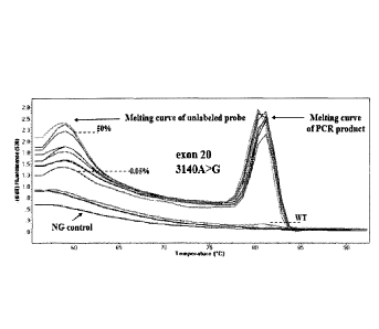
Sensitivity study. The sensitivity of the developed method was further
evaluated by
mixing mutated gDNA from cell lines, with WT gDNA at ratios of 50%, 25%,
12.5%, 2.5%,
1.25%, 0.5%, 0.25%, 0.125%, and 0.05% (see Fig. 2C for exon 9 1633G>A, hotspot
.. mutation and Fig. 2D for exon 20 3140A>G, hotspot mutation). The WT gDNA
samples
that were used for dilutions were selected to match mutated gDNA quantity,
quality, and
quantification cycle (Cq), to minimize PCR bias. Melting curves were generated
and the
ability to discriminate melting transitions of the cell line dilutions from
that of WT sample
was assessed. For exon 9, it was possible to clearly discriminate a dilution
corresponding
to 0.05% of MCF-7 cell line (Fig. 2C), while for exon 20, it could also
discriminate a ratio of
0.05% of T47D cell line dilution (Fig. 2D). Melting curves were highly
reproducible.
CA 02957396 2017-02-06
WO 2016/020710 PCT/GR2015/000036
27
For both exons, PIK3CA mutations are detected by the derivative melting of the
unlabeled
blocking probe and mutant PIK3CA sequence, as amplified with the mutant
allele¨specific
primer. Mutations are detected only if this peak at 60 C is present. The other
peak at
higher temperatures is due to the PCR product and can be seen for both the
mutant and
WT, in case that there is a nonspecific amplification of the WT, by using the
mutant allele¨
specific primer.
Especially, to get reliable information for the molecular characterization of
CTCs,
sensitivity, specificity, and robustness of the mutation detection systems
used is extremely
important. A highly sensitive method for PIK3CA mutations has been developed,
based on
HRMA [Vorkas PA, et al. PIK3CA hotspot mutation scanning by a novel and highly
sensitive high-resolution small amplicon melting analysis method. J Mol Diagn
2010;12:697-704]. Despite the fact that this method is much more sensitive
(1%) than the
traditional Sanger sequencing, when this assay was applied in CTCs samples,
the
detection of mutations in CTCs failed.
EXAMPLE 4
Detection of PIK3CA mutations in EpCAM positive CTCs of breast cancer patients
with clinically confirmed metastasis and primary tissues (FFPEs)
As a training group, 37 peripheral blood samples from patients with clinically
confirmed
metastasis and 15 primary breast tumor tissues (FFPEs) for PIK3CA mutations
were
analyzed. P1K3CA mutations were detected in 6/37 patients (16.2%) for exon 9
and 4/37
patients (10.8%) for exon 20, in the EpCAM-positive CTC fractions. In total,
PIK3CA
mutations were detected in 10/37 (27.0%) patients with metastatic breast
cancer. In the
primary breast tumor tissues, PIK3CA mutations were detected in 9/15 (60.0%)
for exon 9
and 7/15 (46.7%) for exon 20. There were six cases where both hotspot
mutations were
detected in the same FFPE sample; in total, PIK3CA mutations were detected in
10/15
(66.7%) FFPE samples.
EXAMPLE 5
Independent group. Subsequently, the assay was evaluated in an independent
group of
118 patients with operable breast cancer and 57 patients with clinically
confirmed
metastasis. For 76 of these patients with breast cancer, FFPEs from the
primary tumor
were also available.
CA 02957396 2017-02-06
WO 2016/020710 PCT/GR2015/000036
28
Detection of PIK3CA mutations in CTCs of operable breast cancer patients.
In the independent group, the EpCAM-positive CTC fractions from 118 patients
with
operable breast cancer after the primary cancer had been removed and before
adjuvant
chemotherapy had been initiated were analyzed. PIK3CA mutations were detected
in
3/118 (2.5%) for exon 9 1633G>A (A), hotspot mutation (Fig. 3A) as well as in
21/118
(17.8%) for exon 20 3140A>G, hotspot mutation (Fig. 3b). There was one case
where
both hotspot mutations were detected in the CTC sample. In total, PIK3CA
mutations
were detected in 24/118 (20.3%) of operable breast cancer patients. Baseline
is PCR
negative control.
Detection of PIK3CA mutations in CTCs of breast cancer patients with
clinically
confirmed metastasis.
In the independent group, the EpCAM-positive CTC fractions from 57 patients
with
metastatic breast cancer were analyzed. From these 57 patients, 24 had bone
metastasis,
3 in the liver, 2 in the brain, 9 in the lung, 3 both bone and liver, and 6
both in the lung and
bone and 2 in more than two different sites. PIK3CA mutations were detected in
8/57
(14.0%) for exon 9 1633G>A, hotspot mutation (Fig. 3C) and in 12/57 (21.1%)
for exon 20
3140A>G, hotspot mutation, in the EpCAM-positive CTC fractions (Fig. 3D). In
total,
PIK3CA mutations were detected in 20/57 (35.1%) in this group of patients. The
peaks at
60t in the melting curves in Fig. 3C and 3D indicate the presence of exon 9
and exon 20
mutations, respectively. In this context, if no peak is detected at 60 C, the
sample is
considered as wild-type for the mutation examined. Baseline is PCR negative
control.
Detection of PIK3CA mutations in corresponding primary tumors.
In the independent group, the PIK3CA mutational status in CTCs and
corresponding
primary tumors was compared in 76 patients with breast cancer (32 with
clinically
confirmed metastasis and 44 with operable breast cancer) as for these patients
corresponding FFPEs were also available (Table 4).
CA 02957396 2017-02-06
WO 2016/020710 PCT/GR2015/000036
29
Table 4. Independent group: PIK3CA mutational status in CTC and corresponding
primary
tumors (FFPEs)
PIK3CA mutations in P1K3CA mutations in PIK3CA mutations in
primary tumors (FFPEs) CTC exon 9, 1633G>A CTC exon 20, 3140A>G
Early breast cancer, (n=44)
CTC pos CTC neg CTC pos CTC neg
pos 0 21 2 3
FFPEs neg 1 22 8 31
Concordance oh 22/44=50% 33/44=75%
P=0.523 P=0.317
Cohen's kappa coefficient k=0.045 k=0.136
Clinically confirmed metastasis, (n=32)
CTC pos CTC neg CTC pos CTC neg
pos 4 13 2 6
FFPEs neg 1 14 2 22
Concordance % 18/32=56.2% 24/32=75%
P=0.208 P=0.254
Cohen's kappa coefficient k=0.161 k=0.200
All patients, (n=76)
CTC pos CTC neg CTC pos CTC neg
pos 4 34 4 9
FFPEs neg 2 36 10 53
Concordance % 40/76=52.6% 57/76=75%
P=0.337 P=0.188
Cohen's kappa coefficient k=0.053 k=0.145
Exon 9, 1633G>A. Concerning all patients, the exon 1633G>A hotspot mutation
was
observed in 38/76 (50%) of the primary tumor samples and in 6 of 76 (7.9%)
corresponding EpCAM-positive CTC fraction samples. For 4 patients that were
carrying
this PIK3CA hotspot mutation in their primary tumor, the identical mutation
was also
detected in the CTCs. In 34 patients, this mutation was identified in the
primary tumor, but
CA 02957396 2017-02-06
WO 2016/020710 PCT/GR2015/000036
not in the EpCAM-positive CTC fraction, whereas 36 patients were found
negative for this
mutation both in the primary tumor and in the CTCs. However, in two cases,
this hotspot
mutation was identified in the EpCAM-positive CTC fraction, but not in the
corresponding
primary tumor. In patients with operable breast cancer, 1633G>A was observed
in 21/44
5 (47.7%) of FFPEs and in 1/44 (2.3%) corresponding CTC samples; none of
the patients
carrying this PIK3CA hotspot mutation in her primary tumor had the identical
mutation in
CTC. In 21 patients, this mutation was identified in the primary tumor, but
not in CTCs,
whereas 22 patients were found negative for this mutation both in the primary
tumor and
in CTCs. However, in one case, this hotspot mutation was identified in the CTC
fraction,
10 but not in the corresponding primary tumor. In patients with metastasis,
1633G>A was
observed in 17/32(53.1%) of FFPEs and in 5/32(15.6%) corresponding CTCs. For 4
patients that were carrying this PIK3CA hotspot mutation in their primary
tumor, the
identical mutation was also detected in the CTCs. In 13 patients, this
mutation was
identified in the primary tumor, but not in CTCs, while 14 patients were found
negative for
15 this mutation both in the primary tumor and in the CTCs. However, in one
case, this
hotspot mutation was identified in the CTC fraction, but not in the
corresponding primary
tumor.
Exon 20, 3140 A>G. Concerning all patients, exon 20 3140 A>G hotspot mutation
was
observed in 13/76 (17.1%) of FFPEs and in 14/76 (18.4%) corresponding CTC
samples.
20 For 4 /13 patients that were carrying this PIK3CA hotspot mutation in
their primary tumor,
the identical mutation was also detected in the EpCAM-positive CTC fraction.
In 9
patients, this mutation was identified in the primary tumor, but not in CTCs,
whereas 53
patients were found negative for this mutation both in the primary tumor and
in CTCs. It is
remarkable that in 10 patients this hotspot mutation was detected only in the
EpCAM-
25 positive CTC fraction, but not in the corresponding primary tumor. In
the group of patients
with operable breast cancer, 3140 A>G was observed in 5/44 (11.4%) of FFPEs
and in
10/44 (22.7%) corresponding CTCs. For 2/5 patients that were carrying this
PIK3CA
hotspot mutation in their primary tumor, the identical mutation was also
detected in CTCs.
In 3 patients, this mutation was identified in the primary tumor but not in
CTCs, whereas
30 31 patients were found negative for this mutation both in the primary
tumor and in CTCs.
However, in 8 patients, this hotspot mutation was detected only in CTCs, but
not in
corresponding FFPEs. In the group of patients with metastasis, 3140 A>G was
observed
in 8/32 (25.0%) of the primary tumor samples and in 4/32 (12.5%) corresponding
CTCs.
For 2/8 patients that were carrying this PIK3CA hotspot mutation in their
primary tumor,
the identical mutation was also detected in CTCs. In 6 patients, this mutation
was
identified in the primary tumor, but not in CTCs, whereas 22 patients were
found negative
for this mutation both in the primary tumor and in CTCs. However, in 2
patients, this
hotspot mutation was detected only in CTC, but not in the corresponding
primary tumor.
CA 02957396 2017-02-06
WO 2016/020710
PCT/GR2015/000036
31
There was no concordance between the presence of both these hotspot PIK3CA
mutations in primary tumors and corresponding CTC, as this was statistically
evaluated
separately for operable and metastasis verified breast cancer patients, or for
all patients
together (see Table 4).
The findings suggest that PIK3CA mutational status in CTCs can change during
disease
recurrence or progression in patients with breast cancer. When the PIK3CA
mutational
status in CTCs and the corresponding primary tumors in a subgroup of 76
patients were
compared, it was observed that the same mutation was present both in the
primary tumor
and in CTCs in a minority of samples. In most patients, the mutation was
identified in the
primary tumor but not in CTCs, whereas many patients were negative for this
mutation
both in primary tumor and in CTCs. However, there were 12 cases where the
hotspot
mutations were identified in CTCs, but not in the corresponding primary tumor;
in two
patients, 1633 G>A was observed in CTCs, but not in corresponding FFPEs,
whereas in
10 patients 3140 A>G was observed in CTCs, but not in corresponding primary
tumor.
Similar findings, reflecting the heterogeneity of CTCs have been reported.
EXAMPLE 6
PIK3CA mutation status in CTCs in respect to CK-19 mRNA expression.
In the independent group, it was further evaluated whether PIK3CA mutational
status in
CTCs is correlated with CK-19 mRNA expression, for 157 of these patients (57
with
clinical metastasis and 100 with early breast cancer; Table 5). The detection
of CK-19
mRNA-positive cells in the peripheral blood is the most sensitive biomarker
for occult
tumor cells in operable and metastatic breast cancer,
SUBSTITUTE SHEET (RULE 26)
CA 02957396 2017-02-06
WO 2016/020710 PCT/GR2015/000036
32
Table 5. Independent group: comparison between PIK3CA mutations in CTC and CK-
19
expression
Patients PIK3CA PIK3CA PIK3CA mutations in
mutations mutations CTC At least one
in CTC in CTC mutation
exon 9, exon 20,
1633G>A 3140A>G
Early breast cancer, (n=100)
pos neg pos neg pos neg
pos 0 36 3 33 3 33
CK-19 neg 2 62 15 49 17 47
Concordance (0/0) 62/100 (62%) 52/100 (52%) 50/100 (50%)
P P=0.407 P=0.049 P=0.023
Cohen's kappa coefficient k=0.59 k=0.49 k=0.46
Clinically confirmed metastasis Verified, (n=57)
pos neg pos neg pos neg
pos 6 19 5 20 11 14
CK-19 neg 2 30 7 25 9 23
Concordance (0/0) 36/57 (63.2%) 30/57 (52.6%) 44/57 (77.2%)
P P=0.073 P=0.564 P=0.167
Cohen's kappa coefficient k=0.61 k=0.48 k=0.52
All patients, (n=157)
pos neg pos neg pos neg
pos 6 55 8 53 14 47
CK-19 neg 4 92 22 74 26 70
Concordance % 98/157 (62.4%) 82/157 84/157 (53.5%)
(52.2%)
P ' P=0.140 P=0.093 P=0.350
Cohen's kappa coefficient k=0.61 k=0.48 k=0.48
CA 02957396 2017-02-06
WO 2016/020710 PCT/GR2015/000036
33
In operable breast cancer, only 3/100 samples were positive for both PIK3CA
mutations
and CK-19 mRNA expression, all in exon 20. There were 33/100 samples positive
for CK-
19, not carrying mutations in PIK3CA, while it is highly remarkable that
PIK3CA hotspot
mutations were identified in CTC of 17 patients who were negative for CK-19
mRNA
expression. In patients with clinically confirmed metastasis, 11/57(19.3%)
samples were
positive both for PIK3CA mutations and CK-19 expression, 6 in exon 9 and 5 in
exon 20.
There were 14/57 (24.6%) samples positive for CK-19, not carrying these
hotspot
mutations in PIK3CA. It is highly remarkable that PIK3CA hotspot mutations
were
identified in CTCs of 9 patients who were negative for CK-19 mRNA expression.
These 26
samples (17 from patients with operable breast cancer and 9 from patients with
clinically
confirmed metastasis) that were found positive for P1K3CA mutations in CTC,
but were
=negative for CK-19 mRNA expression, would have been characterized as CTC-
negative if
PIK3CA mutations were not detected. There was no concordance concerning the
presence of both these hotspot PIK3CA mutations and CK-19 mRNA expression in
CTCs
(see Table 2).
An important observation of this study was that a significant number of
patients (17/118 in
the operable breast cancer group and 9/57 in the metastasis verified group)
that were
negative for CK-19 mRNA expression were carrying PIK3CA hotspot mutations in
CTCs.
These patients would have been characterized as CTC-negative if PIK3CA
mutations
were not detected. Molecular characterization of CTCs has demonstrated that
CTC are
highly heterogeneous. This could be, at least part, attributed to
epithelial¨mesenchymal
transition and mesenchymal¨epithelial transition. In this context, it is not
expected that all
CK-19¨positive CTCs would be PIK3CA mutation¨positive, or that all our samples
that are
CK49¨negative would not carry PIK3CA mutations in CTCs. The CK-19 real-time
PCR
assay is very specific and sensitive, and it has already been demonstrated in
previous
studies its clinical significance [Stathopoulou A, Vlachonikolis I, Mavroudis
D, Perraki M,
Kouroussis C, Apostolaki S, et al. Molecular detection of cytokeratin-19-
positive cells in
the peripheral blood of patients with operable breast cancer: evaluation of
their prognostic
significance. J Clin Oncol 2002;20:3404-121. However, there is always a number
of
patients that do not relapse even if they are CK-/9¨positive, or do relapse
even if they are
CK-/9¨negative, and the same has been shown even when using the FDA-cleared
CellSearchTM system, a clear indication that one marker is not a panacea and
not
enough to verify the presence of a malignant CTC population in our samples.
CA 02957396 2017-02-06
WO 2016/020710 PCT/GR2015/000036
34
EXAMPLE 7
Clinical significance of PIK3CA mutational status in CTCs in patients with
verified
metastasis.
The correlation between PIK3CA mutational status in CTCs and the clinical
outcome of
this relatively small group of patients with clinically confirmed metastasis
were further
evaluated. Kaplan¨Meier survival analysis, performed by using patients'
postoperative
survival, demonstrated that patients who carried PIK3CA hotspot mutations on
CTC
(n=20) had a significant shorter OS than those without (n=37) (P=0.047, log-
rank test; see
Fig. 4).
A striking finding is also that the presence of PIK3CA mutations in CTCs is
associated
with worse survival in metastatic patients. This is the first time that the
presence of gene
mutations in CTCs is correlated with patient survival, in any type of cancer.
Matching
patients who have cancers with activating mutations in the PI3K signaling
pathway to
phase I protocols testing PI3K inhibitors have improved response rates and
survival
[Tsimberidou AM, et al. Personalized medicine in a phase I clinical trials
program: the MD
Anderson Cancer Center initiative. Clin Cancer Res 2012;18: 6373-83]. In
respect to this,
our findings that PIK3CA mutations can be exclusively present in CTCs, while
absent in
the primary tumor, can be beneficial for patients, as was recently shown in a
pilot
prospective study where administration of herceptin was based on the presence
of CK-19
mRNA¨positive CTCs [Georgoulias V, et al. Trastuzumab decreases the incidence
of
clinical relapses in patients with early breast cancer presenting chemotherapy-
resistant
CK-19 mRNA-positive circulating tumor cells: results of a randomized phase II
study. Ann
Oncol 2012;23:1744-50]. Mutations in the PI3K/AKT signaling pathway, which is
frequently deregulated in tumor cells, have been recently identified in breast
cancer stem
cells that are thought to have a central role in the initiation, progression,
and clinical
response of breast cancer [Donovan CA, et al. Correlation of breast cancer
axillary lymph
node metastases with stem cell mutations. JAMA Surg 2013;148:873-8].
EXAMPLE 8
Detection of PIK3CA mutations in ctDNA of breast cancer patients with verified
metastasis.
The developed method was used to detect hot spot PIK3CA mutations in ctDNA
isolated
from plasma of breast cancer patients with verified metastasis. First of all,
all ctDNA
samples extracted from plasma were examined for their DNA quality; to verify
DNA
quality, primers specific for the wild type in exactly the same PIK3CA gene
region for exon
9 that were used to assess for hotspots mutations. The mutation status of
PIK3CA gene in
WO 2016/020710 PCT/GR2015/000036
ctDNA samples was detected by the developed methodology exactly as previously
described (Fig. 5). PIK3CA mutations were detected in 4/24 (16%) for exon 9
(Fig. 5A, 5B)
and in 6/24 (25%) for exon 20 (Fig 5C), in ctDNA samples. The peaks at 60 C in
the
melting curves in Fig. 5A, 5B and 5C indicate the presence of exon 9 and exon
20
5 mutations, respectively. In this context, if no peak is detected at 60 C,
the sample is
considered as wild-type for the mutation examined.
In this study demonstrated that this assay can detect PIK3CA mutations in
ctDNA isolated
from plasma of cancer patients. The assay sensitivity is extremely low (0.05%)
and the
specificity is very high; this assay can detect very rare PIK3CA mutant
alleles in the
10 presence of a high amount of wild type alleles. Overall, 4 out of 24
samples were positive
for 1633 G>A mutation in exon 9 and 6 out of 24 were positive for 3140 A>G
mutation in
ctDNA isolated from plasma samples. Circulating tumor DNA appears to be an
extremely
effective and advantageous source of biomarkers for determining in real time
the mutational
status of tumors; however, in this case highly sensitive, robust and specific
methodologies
15 are needed. Here evidence provide that this assay offers a useful tool
for the detection of
PIK3CA mutations in ctDNA, isolated from plasma of cancer patients.
25
Date Recue/Date Received 2020-06-05