Note: Descriptions are shown in the official language in which they were submitted.
LIPOSOME ENCAPSULATED AFFINITY DRUG
BACKGROUND
Cancer is a very difficult disease to treat due to diversity of cancer type,
mechanisms
involved in disease progression and patient variability associated with
underlying patient
genetic make up. Early efforts to treat cancer have involved the use of
cytotoxic agents
including antifolates. Antifolates refers to a class of molecules that
antagonize (i.e., block)
the actions of folic acid (vitamin B9). Folic acid's primary function in the
body is to serve as
a cofactor to various methyltransferases involved in serine, methionine,
thymidine and purine
biosynthesis. Consequently antifolates inhibit cell division, DNA/RNA
synthesis and repair
and protein synthesis.
The rationale for introducing antifolates as anti cancer agents was based on
folates
being important for survival of all dividing cells because folates are
essential ingredients for
DNA (nucleic acid) synthesis during cell replications. Folate absorption by
any cell, normal
or cancerous, is primarily mediated by reduced-folate carriers (RFCs), which
is an abundant
cross-membrane transporter with low affinity for folates.
Because cancer cells are fast-growing cells and thus have a high demand for
DNA
precursors in the form of folates, they are susceptible to the effects of
antifolates. Fast
growing noimal cells, such as cells that line the gastrointestinal tract and
cells of the bone
marrow, divide rapidly as well using folates supplied primarily via RFCs.
Normal cells are
therefore also susceptible to antifolates because the RFC mediated transport
mechanisms
which antifolates employ to infiltrate and kill cancer cells also have the
potential to result in a
collateral effect of killing fast-growing normal cells, thereby causing
unwanted antifolate-
related toxicities.
Antifolates work by interfering with the action of folates, depriving cancer
cells of the
DNA precursors they need to proliferate, or grow. Antifolates as a class are
used for their
1
Date Recue/Date Received 2020-08-10
CA 02957775 2017-02-09
WO 2016/025882 PCT/US2015/045353
antiproliferative effect in the treatment of cancer to inhibit cell growth and
division, which
causes cancer cells to die. The fast replicating cancer cells requiring
increased amount of folates
compared to most normal cells led to the clinical development of antifolates
as anticancer agents
almost 70 years ago. However, though antifolate-based therapy was shown to be
effective for
cancer treatment, their clinical development has often been derailed due to a
compelling clinical
dilemma. This dilemma stems from two competing clinical dynamics. On one hand,
antifolates
are designed to be folate mimic molecules with most of them intended to reach
cancer cells by
using RFCs as the preferred cross-membrane transport mechanism. On the other
hand, fast
renewing normal tissues in the body such as, for example, the bone marrow or
intestinal track
tissue cells are, like cancer cells, also highly folate-dependent and use also
RFCs as the primary
cross-membrane folate cell supply mechanism. The net result of these two
clinical dynamics is
that bone marrow and gastrointestinal (GI) tract cells, for example, have
typically been a very
prevalent site of patients' life-threatening antifolate-related toxicities.
Some of these toxicities
have included mucositis, diarrhea, anemia, neutropenia, and low white blood
counts. The
consequence of these antifolate-related intractable side effects in patients
has been that
antifolates exhibiting highly effective cytotoxic or anti-cancer properties
have typically failed
during their development or have, to date, limited use in clinical practice
because these
antifolates also tend to have debilitating side effects in the form of
unacceptable toxicities in
normal cells.
Antifolates as a class remain a promising treatment modality for cancer
despite the
associated risk of severe and even life-threatening toxicities for patients.
The challenge has been
to figure out a way to effectively deliver antifolates in a manner that
reduces and/or avoids
damage to normal cells. Recently, because of the availability of newer
alternative therapies for
cancer, antifolates have lost favor in comparison to such therapies in spite
of the exceptional
effectiveness of antifolates in killing cancer cells.
2
CA 02957775 2017-02-09
WO 2016/025882 PCT/US2015/045353
BRIEF SUMMARY
A neutral or anionic immunoliposome with affinity and specificity to folate
receptor or
receptors containing an aqueous bioactive agent such as anti-cancer
(antineoplastic) agent is
surprisingly effective against cells presenting folate receptors on their cell
surface.
In one example embodiment, a liposomal antifolate composition is provided. The
liposomal antifolate composition comprises: a liposome including an interior
space; a bioactive
antifolate agent disposed within said interior space; a PEG attached to an
exterior of the
liposome; and a targeting moiety comprising a protein with specific affinity
for at least one folate
receptor, said targeting moiety attached to at least one of the PEG and the
exterior of the
liposome. For the liposomal antifolate composition of claim 1, the PEG may
have a number
average molecular weight (Mn) of 200 to 5000 daltons.
An example liposomal antifolate composition is also provided. The example
liposomal
antifolate composition comprises a medium comprising a liposome including an
interior space;
an aqueous bioactive antifolate agent disposed within said interior space; a
targeting moiety
comprising a protein with specific affinity for at least one folate receptor,
said targeting moiety
disposed at an the exterior of the liposome. The medium in this composition
may be an aqueous
solution. The aqueous solution may comprise at least one cryoprotectants
selected from the
group consisting of mannitol; trehalose; sorbitol; and sucrose. The liposomal
antifolate
composition may further comprise a steric stabilizer attached to the exterior
of the liposome,
wherein the targeting moiety is attached to at least one of the steric
stabilizer and the exterior of
the liposome. The steric stabilizer is at least one selected from the group
consisting of
polyethylene glycol (PEG); poly-L-lysine (PLL); monosialoganglioside (GM1);
poly(vinyl
pyrrolidone) (PVP); poly(acrylamide) (PAA); poly(2-methyl-2-oxazoline); poly(2-
ethy1-2-
oxazoline); phosphatidyl polyglycerol; poly[N-(2-hydroxypropyl) methacryl
amide]; amphiphilic
poly-N-vinylpyrrolidones; L-amino-acid-based polymer; and polyvinyl alcohol.
The PEG may
have a number average molecular weight (Mn) of 200 to 5000 daltons.
In any of the example compositions, liposomes, products, kits and methods, the
additional features of the following paragraphs may be incorporated:
The liposomal antifolate composition can further comprise at least one of an
immunostimulatory agent and a detectable marker disposed on at least one of
the PEG and an
3
CA 02957775 2017-02-09
WO 2016/025882 PCT/US2015/045353
exterior of the liposome. The liposomal antifolate composition may have a
feature wherein the at
least one of an immunostimulatory agent and a detectable marker is covalently
bonded to at least
one of the PEG and the exterior of the liposome. The immunostimulating agent
may be at least
one selected from the group consisting of protein immunostimulating agent;
nucleic acid
immunostimulating agent; chemical immunostimulating agent; hapten; and
adjuvant. For
example, the immunostimulating agent may be fluorescein isothiocyanate (FITC).
As another
example, the immunostimulating agent is at least one selected from the group
consisting of:
fluorescein; DNP; beta glucan; beta-1,3-glucan; and beta-1,6-glucan. The
detectable marker may
be at least one selected from the group consisting of fluorescein and
fluorescein isothiocyanate
(FITC). As an example, the immunostimulatory agent and the detectable marker
is the same - for
example, it may be fluorescein isothiocyanate (FITC).
The liposomal antifolate composition may have a diameter in the range of 30-
150 nm,
such as, for example, in the range of 40-70 nm. As another feature, the
liposome can be an
anionic liposome or a neutral liposome. For example, the zeta potential of the
liposome can be
less than or equal to zero such as in the range of 0 to -150 mV or in the
range of -30 to -50 mV.
The liposomal antifolate composition comprises liposomes. The liposomes may be
formed of any liposomal components. For example, the liposomal component may
comprise at
least one of an anionic lipid and a neutral lipid. As another example, the
liposomal component is
at least one selected from the group consisting of: DSPE; DSPE-PEG-maleimide;
HSPC; HSPC-
PEG; cholesterol; cholesterol-PEG; and cholesterol-maleimide. As another
example, the
liposomal components comprise at least one selected from the group consisting
of: DSPE;
DSPE-PEG-FITC; DSPE-PEG-maleimide; cholesterol; and HSPC.
As discussed, the liposome may enclose an aqueous solution. For example, the
liposome
can enclose a bioactive antifolate agent and an aqueous pharmaceutically
acceptable carrier. The
pharmaceutically acceptable carrier may comprise trehalose such as, for
example, 5% to 20%
weight percent of trehalose. The pharmaceutically acceptable carrier, for
example, may compris
citrate buffer at a concentration of between 5 to 200 mM and a pH of between
2.8 to 6.
Independently of other ingredients, the pharmaceutically acceptable carrier
may comprise a total
concentration of sodium acetate and calcium acetate of between 50 mM to 500
mM.
The bioactive antifolate agent may be water soluble. As an example, the
liposomal
antifolate composition may have a liposome and some of the liposome may
comprise less than
4
CA 02957775 2017-02-09
WO 2016/025882 PCT/US2015/045353
200,000 molecules of the bioactive antifolate agent. For example, the liposome
may comprise
between 10,000 to 100,000 molecules of the bioactive antifolate agent.
The bioactive antifolate agent may comprise pemetrexed. In another example
embodiment, the bioactive antifolate agent may comprise lometrexol. In another
example
embodiment, the bioactive antifolate agent is at least one selected from the
group consisting of
methotrexate; ralitrexed; aminopterin; pralatrexate; lometrexol; thiophene
analog of lometrexol;
furan analog of lometrexol; trimetrexed; LY309887; and GW 1843U89.
Alternatively, or in
addition, the bioactive antifolate agent is at least one selected from at
least one from the group
consisting of proguanil; pyrimethamine; trimethoprim and 6-Substituted Pyrrolo
and
Thieon[2,3-d]pyrrolopyrimidine class of GARFT inhibitors. Lometrexol analogs
are described,
for example, in Habeck et al., Cancer Research, v. 54, page 1021-1026, Feb 15,
1994.
The bioactive antifolate agent or any bioactive agent may be at a pH of 5-8 in
the
liposomal antifolate composition. Alternatively, the liposomal antifolate
composition may
comprise bioactive antifolate agent at a pH of 2-6.
Any of the moieties, such as the targeting moiety, the detectable label, the
immunostimulatory agent, the steric stabilizer, and any optional moieties and
agents may be
bound to the liposome or liposomal component directly or indirectly. Indirect
binding may
include binding through a steric stabilizer (e.g., PEG), a functional group
such as maleimide, an
ionic bond (avidin, streptavidin, biotin and the like), or a binding pair (NTA-
nickel and the like).
Combinations of these indirect binding mechanism are also envisioned such as,
for example,
PEG- maleimide.
In the liposomal antifolate composition or other composition, the targeting
moiety may
be bound via a maleimide functional group to at least one selected from the
group consisting of a
liposomal component and a PEG molecule. The targeting moiety may have specific
affinity for
at least one selected from the group consisting of: folate receptor alpha;
folate receptor beta; and
folate receptor delta. For example, the targeting moiety has specific affinity
for at least two
selected from the group consisting of: folate receptor alpha; folate receptor
beta; and folate
receptor delta. As a further example, the targeting moiety may have has
specific affinity for all
three of folate receptor alpha; folate receptor beta; and folate receptor
delta.
In an example embodiment, the targeting moiety has specific affinity for an
epitope on a
tumor cell surface antigen that is present on a tumor cell but absent or
inaccessible on a non-
CA 02957775 2017-02-09
WO 2016/025882 PCT/US2015/045353
tumor cell. The tumor cell may be, for example, a malignant cell. The tumor
cell surface antigen
can be at least one selected from the group consisting of: folate receptor
alpha; folate receptor
beta; and folate receptor delta. In one sample measurement of affinity, the
targeting moiety may
bind folate receptor with an affinity that is at least 2 folds, 5 folds, 10
folds, 25 folds, 100 folds,
500 folds or 5000 folds stronger than a binding affinity to a reduced folate
carrier.
In the example embodiments which involve a targeting moiety, the targeting
moiety may
be a protein comprising an antigen binding sequence of an antibody. The
antigen binding
sequence of an antibody comprises one or more complementary determining
regions of antibody
origin. The protein may comprise an antibody. In an example embodiment, the
targeting moiety
is at least one selected from the group consisting of an antibody; a humanized
antibody; an
antigen binding fragment of an antibody; a single chain antibody; a single-
domain antibody: a bi-
specific antibody; a synthetic antibody; a pegylated antibody; and a
multimeric antibody.
The liposomes of the liposomal antifolate composition or liposomal composition
may
comprise up to 200 or up to 250 targeting moieties per liposome. As an
example, the liposome
may comprise 30 to 200 targeting moieties.
One aspect is also directed to a method of delivering a bioactive antifolate
agent to a
tumor expressing folate receptor on its surface, the method comprising:
administering any of the
compositions such as the liposomal antifolate composition in an amount to
deliver a
therapeutically effective dose of the bioactive antifolate agent to the tumor.
Administering may
be selected from the group consisting of: infusion; injection; parenteral
administration; and
topical administration. The subject may be any animal or any mammal. Examples
of suitable
animals are listed in this disclosure. For example, the subject can be a
human.
The compositions may be prepared using any suitable method. One example method
of
preparing a liposomal antifolate composition or liposomal composition
comprises the steps of:
forming a mixture comprising: (1) liposomal components; (2) the bioactive
antifolate agent in
aqueous solution; (3) the targeting moiety which optionally may be already
attached or bonded to
a liposomal component. The next steps involves homogenizing the mixture to
form liposomes in
said aqueous solution; and extruding the mixture through a membrane to form
liposomes
enclosing the bioactive antifolate agent in an aqueous solution. The method
may comprise an
optional step of removing excess bioactive antifolate agent in aqueous
solution outside of the
liposomes after said extruding step. The method may further comprise an
optional step of
6
CA 02957775 2017-02-09
WO 2016/025882 PCT/US2015/045353
lyophilizing said composition after said removing step to form a lyophilized
composition. The
method may include another optional step. The step is reconstituting said
lyophilizing
composition by dissolving said lyophilizing composition in a solvent after
said lyophilizing step.
The mixture may comprise at least one selected from the group consisting of
mannitol;
trehalose; sorbitol; and sucrose. The one or more liposomal components further
comprises a
steric stabilizer. The steric stabilizer may be at least one selected from the
group consisting of
polyethylene glycol (PEG); poly-L-lysine (PLL); monosialoganglioside (GM1);
poly(vinyl
pyrrolidone) (PVP); poly(acrylamide) (PAA); poly(2-methyl-2-oxazoline); poly(2-
ethy1-2-
oxazoline); phosphatidyl polyglycerol; poly[N-(2-hydroxypropyl)
methacrylamide]; amphiphilic
poly-N-vinylpyrrolidones; L-amino-acid-based polymer; and polyvinyl alcohol.
The PEG may
have a number average molecular weight (Mn) of 200 to 5000 daltons. In the
method of making
a composition the solvent may be an aqueous solvent.
A targeted liposomal composition that selectively targets folate receptors is
provided.
The example targeted liposomal composition comprises a liposome including an
interior space; a
bioactive agent disposed within said interior space; a steric stabilizer
molecule attached to an
exterior of the liposome; and a targeting moiety comprising a protein with
specific affinity for at
least one folate receptor, said targeting moiety attached to at least one of
the steric stabilizer and
the exterior of the liposome. The steric stabilizer may be at least one
selected from the group
consisting of polyethylene glycol (PEG); poly-L-lysine (PLL);
monosialoganglioside (GM1);
poly(vinyl pyrrolidone) (PVP); poly(acrylamide) (PAA); poly(2-methyl-2-
oxazoline); poly(2-
ethy1-2-oxazoline); phosphatidyl polyglycerol; poly[N-(2-hydroxypropyl)
methacrylamide];
amphiphilic poly-N-vinylpyrrolidones; L-amino-acid-based polymer; and
polyvinyl alcohol. For
example, the PEG may have a number average molecular weight (Mn) of 200 to
5000 daltons.
In this targeted liposomal composition, the bioactive agent comprises at least
one of the group
consisting of ellipticine; paclitaxel; pemetrexed; methotrexate; ralitrexed;
aminopterin;
pralatrexate; lometrexol; thiophene analog of lometrexol; furan analog of
lometrexol;
trimetrexed; LY309887; GW 1843U89; proguanil; pyrimethamine; trimethoprim and
6-
Substituted Pyrrolo and Thieon[2,3-d]pyrrolopyrimidine class of GARFT
inhibitors.
The composition may be made, for example, by forming a mixture comprising: (1)
liposomal components; (2) the bioactive agent in aqueous solution; (3) the
targeting moiety. The
next steps involve homogenizing the mixture to form liposomes in said aqueous
solution; and
7
CA 02957775 2017-02-09
WO 2016/025882 PCT/US2015/045353
extruding the mixture through a membrane to form liposomes enclosing the
bioactive antifolate
agent in an aqueous solution. An optional step may involve removing excess
bioactive antifolate
agent in aqueous solution outside of the liposomes after said extruding step.
Another optional
step involves lyophilizing said composition after said removing step to form a
lyophilized
composition. Another optional step involves reconstituting said lyophilizing
composition by
dissolving said lyophilizing composition in a solvent after said lyophilizing
step. The other
components and steps may be shared from the other method of making as
discussed herein.
A kit for providing any liposomal composition, including liposomal antifolate
composition is also provided. The kit can comprise the liposomal components,
an instruction for
using the composition to encapsulate a bioactive agent, and optionally, in a
separate container,
the bioactive agent.
8
CA 02957775 2017-02-09
WO 2016/025882 PCT/US2015/045353
BRIEF DESCRIPTION OF THE DRAWINGS
Figure 1A is a schematic illustrating normal tissue.
Figure 1B is a schematic illustrating cancerous tissue.
Figure 2 is a schematic illustrating and example embodiment and its
binding
mechanism.
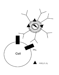
Figure 3 is a schematic illustrating a fluorochrome conjugated antibody
binding to a
folate receptor on a cell surface.
Figure 4 is a schematic showing an example liposome binding to and
internalizing into
a cell expressing folate receptor alpha.
Figure 5 is a schematic illustrating the effect of internalization of an
example liposomal
composition on cell proliferation using p38 protein kinase pathways.
Figure 6 depicts data from flow cytometry analysis of KB cells using
flurochrome.
Figure 7 depicts data from flow cytometry analysis of OVCAR-3 (ovarian)
cells using
flurochrome.
Figure 8 depicts data from flow cytometry analysis of NCIH2452
(mesothelioma) cells
using flurochrome.
Figure 9 depicts data from flow cytometry analysis of CCD841 (normal
colon) cells
using flurochrome.
Figure 10 depicts data from flow cytometry analysis of 5L0003 (lung) cells
using
flurochrome.
Figure 11 depicts data from flow cytometry analysis of CCD841 (normal
colon) cells
using flurochrome.
Figure 12 is a bar chart depicting surface levels of an example liposomal
composition in
normal or cancer cells.
Figure 13 depicts data from flow cytometry analysis of ovarian cancer cells
using
RhodoRed.
Figure 14 depicts data from flow cytometry analysis of KB folate receptor
alpha high
cells using RhodoRed.
Figure 15 depicts data from flow cytometry analysis of normal breast cells
using
RhodoRed.
9
CA 02957775 2017-02-09
WO 2016/025882 PCT/US2015/045353
Figure 16 depicts data from flow cytometry analysis of normal colon cells
using
RhodoRed.
Figure 17 depicts a bar chart showing liposome concentration dependent
targeting vs.
untargeting liposome concentration dependent detection.
Figure 18A depicts data from flow cytometry analysis of untreated cells.
Figure 18B depicts data from flow cytometry analysis of cells treated with
an example
liposomal composition according to an example embodiment.
Figure 19 depicts lung cancer cells exposed to various reagents as listed.
Figure 20 is a line graph illustrating correlation between growth
inhibition and folate
receptor alpha expression.
Figure 21 is a bar graph summarizing results and demonstrating that an
example
liposomal composition of an example embodiment inhibits cancer cell growth.
Figure 22A is a schematic depicting the cell cycle of a normal cell.
Figure 22B is a chart showing Propidium Iodide quantification of cells in
various stages of
cell cycle.
Figure 23A depicts data from Propidium Iodide quantification of cells that
are untreated.
Figure 23B depicts data from Propidium Iodide quantification of cells that
are treated with
pemetrexed.
Figure 24 is a bar chart showing cell cycle stasis by an example embodiment
of a
liposomal composition.
Figure 25 depicts analysis of cells for Mac-1 to determine maturing
neutrophils.
Figure 26A depicts flow cytometry data from normal cells.
Figure 26B depicts flow cytometry data from pemetrexed treated cells.
Figure 27 is a bar chart depicting the number of differentiated neutrophils
in pemetrexed
treated and example embodiment treated samples.
CA 02957775 2017-02-09
WO 2016/025882 PCT/US2015/045353
DETAILED DESCRIPTION
Antifolate drugs, as discussed above, were designed as folate mimetic
molecules that
work by interfering with the action of folates once inside a cell, depriving
cells of the DNA
precursors they need to replicate and proliferate. Because cancer cells are
fast growing cells with
a high demand for DNA precursors in the form of folates, they take up
antifolate drugs in the
same manner as folates and are as a result susceptible to the effects of
antifolates. However, fast
growing normal cells, such as cells that line the gastrointestinal (GI) tract
and cells of the bone
marrow such as, for example, neutrophils, divide rapidly as well using folates
supplied primarily
via RFCs. Normal cells are therefore also susceptible to the toxic effects of
antifolates because
the RFCs mediated transport mechanism which most antifolates are designed to
use to infiltrate
and kill cancer cells is the same mechanism that normal cells use to supply
themselves with
folates. As a result, treatment of cancers using very promising and effective
antifolates has been
a difficult challenge in the clinical care of patients because of the high
likelihood of the treatment
causing collateral damages to fast-growing normal cells, thereby causing
antifolate- related
severe and potentially life-threatening toxicities.
As discussed above, antifolates as a class are used for their
antiproliferative effect in the
treatment of cancer to inhibit cell growth and division, which causes cancer
cells to die. The fast
replicating cancer cells require increased amount of folates when compared to
most normal cells.
This led to the clinical development of antifolates as anticancer agents
almost 70 years ago.
However, though antifolate-based therapies were shown to be effective for
cancer treatment, the
clinical development of antifolates has been problematic and often derailed in
view of a
compelling clinical dilemma. This dilemma stems from two competing clinical
dynamics. On
one hand, antifolates are designed to be folate mimic molecules with most of
them intended to
reach cancer cells using RFCs as the preferred cross-membrane transport
mechanism. On the
other hand, fast renewing tissues in the body such as the bone marrow or
intestinal track tissue
cells are, like cancer cells, also highly folate-dependent and use also RFCs
as the primary cross-
membrane folate cell supply mechanism. The net result of these two clinical
dynamics is that
bone marrow and gastrointestinal (GI) tract cells have been the most prevalent
sites of patients'
life-threatening antifolate-related toxicities. Some of these toxicities have
included mucositis,
diarrhea, anemia, neutropenia, and low white blood counts. Such toxicities,
alone or in
combination, were in a number of instances blamed for patient death from
antifolate-based
11
CA 02957775 2017-02-09
WO 2016/025882 PCT/US2015/045353
treatment. The consequence is that to date many effective promising
antifolates continue to fail
during their development, not because of a lack of effectiveness against
cancer cells, but instead
because of patient safety concerns. The few that have managed to reach the
stage of becoming
medicines have limited use in clinical practice again due to safety concerns.
Antifolates as a class remain a promising treatment modality for cancer
despite the
associated risk of severe and even life-threatening toxicities for patients.
The challenge is to
figure out a way to deliver these highly effective antifolates in a manner
that avoids damage to
normal cells.
Prior efforts have generally focused on using RFCs to deliver an anticancer
agent.
However, the present inventors exploit another pathway that is especially
prevalent in cancer
cells involving folate receptors, including, but not limited to, for example,
folate receptor alpha,
folate receptor beta and/or folate receptor delta. It has been observed in
cancer biology that
cancer cells preferentially express folate receptor alpha in contrast to
normal cells in order to
efficiently uptake folates for the sustainment of their fast replication and
proliferation needs.
Cancer cells are very efficient at supplying themselves with folates contained
in the blood stream
as compared to normal cells. One way that cancer cells do this is by their
overexpression of
folate receptors, such as, for example, folate receptor alpha. As cancer
progresses, tumor cell
surface folate receptor alpha levels tend to increase, most likely due to
increasing needs for folate
supply.
Because of its high affinity to folate receptor alpha, folic acid was
conventionally
investigated as a targeting moiety for delivering anti-cancer or cytotoxic
molecules to cancer
cells with the intent to preferentially deliver a cytotoxic drug to cancer
cells, either conjugated to
a liposome containing the cytotoxic drug or conjugated to the cytotoxic drug
itself. This
approach has not led to improved patient safety in large part because, as
recognized by the
inventors, this approach fails to appreciate a key biological difference in
exploiting folate
pathways as an approach to deliver a cytotoxic to cancer cells while reducing
and/or minimizing
exposure of normal cells to the cytotoxic drug; with folic acid as the
targeting ligand, normal
cells were not being spared from toxicity since such a targeted drug was still
being taken up by
normal cells via RFCs. In other words, a targeted drug using folic acid as the
targeting moiety is
biologically no different than a regular untargeted antifolate because a drug
of such construct
binds to both folate receptor alpha and RFCs just like any other folate mimic
molecule that is
12
CA 02957775 2017-02-09
WO 2016/025882 PCT/US2015/045353
indiscriminately taken up by both cancer and normal cells. Therefore, using
folic acid as the
targeting moiety does not provide the selective delivery of cytotoxic agents
to cancer cells while
avoiding normal cells. Thus, with folic acid as the targeting moiety, drug
related toxicity
remained a concern in patient care. As a result, leading experts suggested
that trying to exploit
folate receptors as a means for selective targeting of cancer cell may be
ineffective, guiding the
efforts of those skilled in the art away from attempting to exploit folate
receptors.
Targeting an antifolate to a folate receptor with a targeting moiety has not
been attempted
to date. Because antifolates mimic folates, one would not consider exploiting
the folate pathways
to deliver an antifolate in a targeted way. It would be considered redundant
since the reduced
folate carrier already transport folate into the cells. From this
understanding, it was inherently
logical to conclude that because an antifolate mimics a folate, an antifolate
drug will be taken up
effectively by a folate receptor by a cell and further assistance using, for
example, an antibody
would not be necessary. A counter-intuitive approach was taken by the current
inventors.
Because it was important to shield antifolates from being taken up by normal
cells via RFCs in
order to reduce or prevent antifolate-related toxicity, the inventors found
that this goal could be
achieved by, among other things, exploiting a cancer specific morphology which
has been
unappreciated as useful to the field of antifolate research: the loss of
polarity by tumor tissue
cells.
Disruption of cell polarity and tissue disorganization is a hallmark of
advanced epithelial
tumors. As illustrated in Figure 1A, normal simple epithelium generally
comprises a monolayer
of individual cells that display a distinct apical- basal polarity. Cells are
tightly packed and
connected to each other by the apical junctional complexes (Figure 1A-101),
which separate
apical and basolateral membrane domains. In normal tissue where polarity is
preserved, folate
receptor alpha is attached at the apical surface of cells situated away from,
and out of direct
contact with folates in the blood circulation (Figure 1A-102). Figure 1B
illustrates how cells in
high-grade epithelial tumors display loss of apical-basal polarity and overall
tissue
disorganization, putting folate receptor alpha in direct contact with folates
in the blood
circulation (1B-103). This feature of tumor tissue cells, was believed by the
inventors to have
greater significance for antifolate based therapies than conventional thinking
had appreciated.
The inventors discovered that this held a significant potential to
rehabilitate antifolates as
13
CA 02957775 2017-02-09
WO 2016/025882 PCT/US2015/045353
anticancer therapies while reducing and/or even minimizing associated severe
and sometime life-
threatening toxicities associated with antifolates.
In this regard, the inventors designed a chemical entity to deliver an
antifolate agent in a
manner that selectively targets folate receptors that are highly expressed in
cancer cells, such as,
for example, folate receptor alpha, beta and delta while avoiding RFCs (the
folate pathway used
by normal cells), to selectively expose the antifolate to tumor tissue cells
while reducing or
avoiding exposure of antifolates to normal cells. This is made possible by
recognizing that
following loss of polarity, tumor tissue cells not only overexpress and expose
folate receptors,
such as folate receptor alpha but also that folate receptors in cancer cells
are in direct contact
with blood circulation, both of which are not the case for the normal tissues.
This approach may
also extend to other cell surface fol ate receptors (e.g. folate receptor
beta, folate receptor delta,
etc.) because of their structural and functional similarities to folate
receptor alpha.
The disclosure relates in general to liposome compositions useful for
delivering a variety
of bioactive agents, such as, for example, antifolates, methods of making the
liposomal
compositions and methods for treating patients using the liposomal
compositions. There is
special utility in providing an antifolate encapsulating liposome that is
targeted to folate
receptors but which is not specifically targeted to reduced folate carriers.
More specifically, the disclosure is based on the discovery that a neutral or
anionic
liposome (i.e., a non-cationic liposome) with affinity and specificity to a
folate receptor or more
than one folate receptor containing one or more bioactive agent such as, for
example, an anti-
cancer (antineoplastic) agent is surprisingly effective against cells
presenting and expressing
folate receptors on their cell surface.
In an example embodiment, a liposomal antifolate composition is provided. The
liposomal antifolate composition may comprise a liposome including an interior
space; a
bioactive antifolate agent disposed within the interior space; a PEG molecule
attached to an
exterior of the liposome; and a targeting moiety comprising a protein with
specific affinity for at
least one folate receptor, the targeting moiety attached to at least one of
the PEG and the exterior
of the liposome.
The term attach or attached refers, for example, to any type of bonding such
as covalent
bonding, ionic bonding (e.g., avidin-biotin) bonding by hydrophobic
interactions, and bonding
via functional groups such as maleimide, or linkers such as PEG. For example,
a detectable
14
CA 02957775 2017-02-09
WO 2016/025882 PCT/US2015/045353
marker, a steric stabilizer, a liposome, a liposomal component, an
immunostimulating agent may
be attached to each other directly, by a maleimide functional group, or by a
PEG-malemide
group.
The liposomes in some example embodiments include a steric stabilizer that may
increase
their longevity in circulation. The basic concept is that one or more steric
stabilizers such as a
hydrophilic polymer (Polyethylene glycol (PEG)), a glycolipid
(monosialoganglioside (GM1)) or
others occupies the space immediately adjacent to the liposome surface and
exclude other
macromolecules from this space. Consequently, access and binding of blood
plasma opsonins to
the liposome surface are hindered, and thus interactions of macrophages with
such liposomes, or
any other clearing mechanism, are inhibited and longevity of the liposome in
circulation is
enhanced. In example embodiments, the steric stabilizer or the population of
steric stabilizers
may be a PEG or a combination comprising PEG. In an example embodiment, the
steric
stabilizer may be a PEG with a number average molecular weight (Mn) of 200 to
5000 daltons.
These PEGs can be of any structure such as linear, branched, star or comb
structure and are
commercially available.
The liposomes contained in the liposome composition of various example
embodiments
can be any liposome known or later discovered in the art. In general, the
liposomes of the
example embodiments may have any liposome structure, structures having an
inner space
sequestered from the outer medium by one or more lipid bilayers, or any
microcapsule that has a
semi-permeable membrane with a lipophilic central part where the membrane
sequesters an
interior. A lipid bilayer can be any arrangement of amphiphilic molecules
characterized by a
hydrophilic part (hydrophilic moiety) and a hydrophobic part (hydrophobic
moiety). Usually
amphiphilic molecules in a bilayer are arranged into two dimensional sheets in
which
hydrophobic moieties are oriented inward the sheet while hydrophilic moieties
are oriented
outward. Amphiphilic molecules forming the liposomes of the example
embodiments can be any
known or later discovered amphiphilic molecules, e.g., lipids of synthetic or
natural origin or
biocompatible lipids. Liposomes of the example embodiments may also be formed
by
amphiphilic polymers and surfactants, e.g., polymerosomes and niosomes. For
the purpose of
this disclosure, without limitation, these liposome-forming materials also are
referred to as
"lipids".
CA 02957775 2017-02-09
WO 2016/025882 PCT/US2015/045353
The liposome composition may be a liquid or it may be dry, such as, for
example, in the
form of a dry powder or a dry cake. The dry powder or dry cake may have
undergone primary
drying under, for example, lyophilization conditions or optionally, it may
have undergone both
primary drying only or both primary drying and secondary drying. In the dry
form, the powder or
cake may, for example, have between 1% to 6% moisture, for example, such as
between 2% to
5% moisture or between 2% to 4% moisture. One example method of drying is
lyophilization
(also called freeze-drying, or cyrodessication). Any of the compositions and
methods of the
disclosure may involve the liposomes, lyophilized liposomes or liposomes
reconstituted from
lyophilized liposomes. In lyophilization, lyoprotectants or cryoprotectants,
molecules protect
freeze-dried material may be used. These molecules are typically polyhydroxy
compounds such
as sugars (mono-, di-, and polysaccharides), polyalcohols, and their
derivatives, glycerol, or
polyethyleneglycol, trehalose, maltose, sucrose, glucose, lactose, dextran,
glycerol, and
aminoglycosides. The lyoprotectants or cryoprotectants may, for example,
comprise up to 10%
or up to 20% of a solution outside the liposome or inside the liposome or both
outside and inside
the liposome.
The liposomes of the example embodiments may, for example, have a diameter of
in the
range of 30-150 nm (nanometer). In other example embodiments, the liposome
may, for
example, have a diameter in the range of 40-70 nm.
The liposomes of the example embodiments may, for example, preferably be
anionic or
neutral. That is, the liposome should not be cationic. The determination of
the charge (i.e.,
anionic, neutral or cationic) may be made by measuring the zeta potential of
the liposome. In an
example embodiment, the zeta potential of the liposome is less than or equal
to zero. In another
example embodiment, the zeta potential of the liposome is in a range of 0 to -
150 mV. In another
example embodiment, the zeta potential should be in the range of -30 to -50
mV.
The properties of liposomes are influenced by the nature of lipids used to
make the
liposomes. A wide variety of lipids have been used to make liposomes. These
include cationic,
anionic and neutral lipids. Cationic lipids are used to make cationic
liposomes which are
commonly used as gene transfection agents. The positive charge on cationic
liposomes enables
interaction with the negative charge on cell surfaces. Following binding of
the cationic liposomes
to the cell, the liposome is transported inside the cell through endocytosis.
However, cationic
liposomes will bind to both normal cells and tumor cells. Because the example
embodiments are
16
CA 02957775 2017-02-09
WO 2016/025882 PCT/US2015/045353
intended to specifically and selectively target tumor cells while
substantially sparing normal
cells, the use of cationic lipids is not preferred. Using a mixture of, for
example, neutral lipids
such as HSPC and anionic lipids such as PEG-DSPE results in the formation of
anionic
liposomes which are less likely to non-specifically bind to normal cells.
Specific binding to
tumor cells can be achieved by using a tumor targeting antibody such as, for
example, a folate
receptor antibody, including, for example, folate receptor alpha antibody,
folate receptor beta
antibody and/or folate receptor delta antibody.
As an example, at least one (or some) of the lipids is/are amphipathic lipids,
defined as
having a hydrophilic and a hydrophobic portions (typically a hydrophilic head
and a hydrophobic
tail). The hydrophobic portion typically orients into a hydrophobic phase
(e.g., within the
bilayer), while the hydrophilic portion typically orients toward the aqueous
phase (e.g., outside
the bilayer). The hydrophilic portion may comprise polar or charged groups
such as
carbohydrates, phosphate, carboxylic, sulfato, amino, sulfhydryl, nitro,
hydroxy and other like
groups. The hydrophobic portion may comprise apolar groups that include
without limitation
long chain saturated and unsaturated aliphatic hydrocarbon groups and groups
substituted by one
or more aromatic, cyclo-aliphatic or heterocyclic group(s). Examples of
amphipathic compounds
include, but are not limited to, phospholipids, aminolipids and sphingolipids.
Typically, for example, the lipids are phospholipids. Phospholipids include
without
limitation phosphatidylcholine, phosphatidylethanolamine,
phosphatidylglycerol,
phosphatidylinositol, phosphatidylserine, and the like. It is to be understood
that other lipid
membrane components, such as cholesterol, sphingomyelin, cardiolipin, etc. may
be used.
In an example embodiment, the lipids may be anionic and neutral (including
zwitterionic
and polar) lipids including anionic and neutral phospholipids. Neutral lipids
exist in an
uncharged or neutral zwitterionic form at a selected pH. At physiological pH,
such lipids include,
for example, di oleoylphosphatidylglycerol (DOPE]), diacylphosphatidylcholine,
diacylphosphatidylethanolamine, ceramide, sphingomyelin, cephalin,
cholesterol, cerebro sides
and diacylglycerols. Examples of zwitterionic lipids include without
limitation
dioleoylphosphatidylcholine (DOPC), dimyristoylphosphatidylcholine (DMPC), and
dioleoylphosphatidylserine (DOPS). An anionic lipid is a lipid that is
negatively charged at
physiological pH. These lipids include without limitation
phosphatidylglycerol, cardiolipin,
diacylphosphatidylserine, diacylphosphatidic acid, N-dode- canoyl
phosphatidylethanolamines,
17
CA 02957775 2017-02-09
WO 2016/025882 PCT/US2015/045353
N-succinyl phosphatidylethanolamines, N-glutarylphosphatidylethanolamines,
lysylphosphatidylglycerols, palmitoyloleyolphosphatidylglycerol (POPG), and
other anionic
modifying groups joined to neutral lipids.
Collectively, anionic and neutral lipids are referred to herein as non-
cationic lipids. Such
lipids may contain phosphorus but they are not so limited. Examples of non-
cationic lipids
include lecithin, lysolecithin, phosphatidylethanolamine,
lysophosphatidylethanolamine,
dioleoylphosphati- dylethanolamine (DOPE), dipalmitoyl phosphatidyl
ethanolamine (DPPE),
dimyristoylphosphoethanolamine (DMPE), distearoyl-phosphatidy 1-ethanolamine
(DSPE),
palmitoyloleoyl-phosphatidylethanolamine (POPE)
palmitoyloleoylphosphatidylcholine (POPC),
egg phosphatidylcholine (EPC), di stearoylphosphatidylcholine (DSPC),
di oleoylphosphatidylcholine (DOPC), dipalmitoylphosphatidylcholine (DPPC),
dioleoylphosphatidylglycerol (DOPG), dipalmitoylphosphatidylglycerol (DPPG),
palmitoyloleyolphosphatidylglycerol (POPG), 16-0-monomethyl PE, 16-0- dimethyl
PE, 18-1-
trans PE, palmitoyloleoyl-phosphatidylethanolamine (POPE), 1-stearoy1-2-
oleoylphosphatidyethanolamine (SOPE), phosphatidylserine,
phosphatidylinositol.
sphingomyelin, cephalin, cardiolipin, phosphatidic acid, cerebrosides,
dicetylphosphate, and cho-
lesterol.
Liposomes of example embodiments may be assembled using any liposomal assembly
method using liposomal components (also referred to as liposome components).
Liposomal
components include, for example, lipids such as DSPE, HSPC, cholesterol and
derivatives of
these components. Other suitable lipids are commercially available for
example, by Avanti Polar
Lipids, Inc. (Alabaster, Alabama, U.S.A.). A partial listing of available
negatively or neutrally
charged lipids suitable for making anionic liposomes, may be, for example, at
least one of the
following: DLPC, DMPC, DPPC, DSPC, DOPC, DMPE, DPPE, DOPE, DMPA=Na, DPPA=Na,
DOPA=Na, DMPG=Na, DPPG=Na, DOPG=Na, DMPS=Na, DPPS=Na, DOPS=Na. DOPE-
Glutaryl.(Na)2, Tetramyristoyl Cardiolipin=(Na)2, DSPE-mPEG-2000=Na, DSPE-mPEG-
5000=Na, and DSPE-Maleimide PEG-2000=Na.
Derivatives of these lipids may, for example, include, at least, the bonding
(preferably
covalent bonding) of one or more steric stabilizers and/or functional groups
to the liposomal
component after which the steric stabilizers and/or functional groups should
be considered part
of the liposomal components. Functional groups comprises groups that can be
used to attach a
18
CA 02957775 2017-02-09
WO 2016/025882 PCT/US2015/045353
liposomal component to another moiety such as a protein. Such functional
groups include, at
least, maleimide. These steric stabilizers include at least one from the group
consisting of
polyethylene glycol (PEG); poly-L-lysine (PLL); monosialoganglioside (GM1);
poly(vinyl
pyrrolidone) (PVP); poly(acrylamide) (PAA); poly(2-methyl-2-oxazoline); poly(2-
ethy1-2-
oxazoline); phosphatidyl polyglycerol; poly[N-(2-hydroxypropyl)
methacrylamide]; amphiphilic
poly-N-vinylpyrrolidones; L-amino-acid-based polymer; and polyvinyl alcohol.
Because a liposomal components may include any molecule(s) (i.e.,
chemical/reagent/protein) that is bound to it, the liposomal components may,
for example,
include, at least, DSPE, DSPE-PEG, DSPE-maleimide, HSPC; HSPC-PEG: HSPC-
maleimide;
cholesterol; cholesterol-PEG; and cholesterol-maleimide. In a preferred
embodiment, the
liposomal components that make up the liposome comprises DSPE; DSPE-FITC; DSPE-
maleimide; cholesterol; and HSPC.
In an example embodiment, at least one component of the lipid bilayer is
functionalized
(or reactive). As used herein, a functionalized component is a component that
comprises a
reactive group that can be used to crosslink reagents and moieties to the
lipid. If the lipid is
functionalized, any liposome that it forms is also functionalized.
In example embodiments, the reactive group is one that will react with a
crosslinker (or
other moiety) to form crosslinks. The reactive group may be located anywhere
on the lipid that
allows it to contact a crosslinker and be crosslinked to another moiety (i.e.,
steric stabilizer,
targeting moiety, etc.). In some embodiments. it is in the head group of the
lipid, including for
example a phospholipid. An example of a reactive group is a maleimide group.
Maleimide
groups may be crosslinked to each other in the presence of dithiol
crosslinkers such as but not
limited to dithiolthrietol (DTT).
It is to be understood that the example embodiments contemplate the use of
other
functionalized lipids, other reactive groups, and other crosslinkers. In
addition to the maleimide
groups, other examples of reactive groups include but are not limited to other
thiol reactive
groups, amino groups such as primary and secondary amines, carboxyl groups,
hydroxyl groups,
aldehyde groups, alkyne groups, azide groups, carbonyls, halo acetyl (e.g.,
iodoacetyl) groups,
imidoester groups, N-hydroxysuccinimide esters, sulfhydryl groups, pyridyl
disulfide groups,
and the like.
19
CA 02957775 2017-02-09
WO 2016/025882 PCT/US2015/045353
Functionalized and non-functionalized lipids are available from a number of
commercial
sources including Avanti Polar 5 Lipids (Alabaster, Ala.).
The liposomes of example embodiments may further comprise an immunostimulatory
agent, a detectable marker, or both disposed on its exterior. For example,
immunostimulatory
agent or detectable marker may be ionically bonded or covalently bonded to an
exterior of the
liposome, including, for example, optionally to the steric stabilizer.
Immunostimulatory agents, also known as immunostimulants, immunostimulators,
haptens and adjuvants, are substances that stimulate the immune system by
inducing activation
or increasing activity of any of its components.
These immunostimulatory agents can include one or more of a hapten, an
adjuvant, a
protein immunostimulating agent, a nucleic acid immunostimulating agent, and a
chemical
immunostimulating agent. Many adjuvants contain a substance designed to
stimulate immune
responses, such as lipid A, Bortadella pertussis or Mycobacterium tuberculosis
derived proteins.
Certain adjuvants are commercially available as, for example, Freund's
Incomplete Adjuvant and
Complete Adjuvant (Difco Laboratories, Detroit, Mich.); Merck Adjuvant 65
(Merck and
Company, Inc., Rahway, N.J.); AS-2 (SmithKline Beecham, Philadelphia. Pa.);
aluminum salts
such as aluminum hydroxide gel (alum) or aluminum phosphate; salts of calcium,
iron or zinc; an
insoluble suspension of acylated tyrosine; acylated sugars; cationically or
anionically derivatized
polysaccharides; polyphosphazenes; biodegradable microspheres; monophosphoryl
lipid A and
quil A. Cytokines, such as GM-CSF, interleukin-2, -7, -12, and other like
growth factors, may
also be used as adjuvants. In a preferred embodiment, the immunostimulant may
be at least one
selected from the group consisting of fluorescein, DNP, beta glucan, beta-1,3-
glucan, beta-1,6-
glucan.
A detectable marker may, for example, include, at least, a radioisotope, a
fluorescent
compound, a bioluminescent compound, chemiluminescent compound, a metal
chelator, an
enzyme, a dye, an ink, a magnetic compound, a biocatalyst or a pigment that is
detectable by any
suitable means known in the art. e.g., magnetic resonance imaging (MRI),
optical imaging,
fluorescent/luminescent imaging, or nuclear imaging techniques.
The immunostimulatory agent and/or detectable marker may be attached to the
exterior
by co-incubating it with the liposome. For example, the immunostimulatory
agent and/or
detectable marker may be associated with the liposomal membrane by hydrophobic
interactions
CA 02957775 2017-02-09
WO 2016/025882 PCT/US2015/045353
or by an ionic bond such as an avidin/biotin bond or a metal chelation bond
(e.g.. Ni-NTA).
Alternatively, the immunostimulatory agent or detectable marker may be
covalently bonded to
the exterior of the liposome such as, for example, by being covalently bonded
to a liposomal
component or to the steric stabilizer which is the PEG.
One example reagent is fluorescein isothiocyanate (FITC) which, based on our
experiments, may surprisingly serve as both an immunostimulant and a
detectable marker.
Example embodiments also provide for a liposome that encloses an interior
space. In an
example embodiment, the interior space may comprise, but is not limited to, an
aqueous solution.
The interior space may comprise a bioactive agent, such as, for example, an
antifolate agent and
an aqueous pharmaceutically acceptable carrier. The pharmaceutically
acceptable carrier may
comprise, for example, trehalose. In an example embodiment, the trehalose may,
for example, be
present at about 5% to 20% weight percent of trehalose or any combination of
one or more
lyoprotectants or cryoprotectants at a total concentration of 5% to 20%. The
interior space may,
for example, comprise a citrate buffer at a concentration of between 5 to 200
mM. The citrate
buffer may buffer the interior space at a pH of between 2.8 to 6. Independent
of the trehalose or
citrate concentration, the pharmaceutically acceptable carrier may comprise a
total concentration
of sodium acetate and calcium acetate of between 50 mM to 500 mM.
In an example embodiment, the bioactive antifolate agent may, for example, be
a water
soluble bioactive agent. That is, the bioactive agent may form an aqueous
solution. According to
example embodiments, each liposome may comprise an interior space that
contains less than
200,000 molecules of the bioactive agent. For example, in an example
embodiment, the liposome
may comprise between 10,000 to 100,000 of a bioactive antifolate agent.
In an example embodiment, the bioactive agent can be at least one from the
group
consisting of pemetrexed, lometrexol, methotrexate, ralitrexed, aminopterin,
pralatrexate,
lometrexol analogs thereof, thiophene analog of lometrexol, furan analog of
lometrexol,
trimetrexed, LY309887; and GW 1843U89. In another embodiment, the bioactive
agent can be at
least one from the group consisting of proguanil, pyrimethamine, trimethoprim
and 6-Substituted
Pyrrolo and Thieon[2,3-d]pyrrolopyrimidine class of GARFT inhibitors. In one
preferred
embodiment, the bioactive antifolate agent is pemetrexed. In another example
embodiment, the
bioactive antifolate agent is lometrexol.
21
CA 02957775 2017-02-09
WO 2016/025882 PCT/US2015/045353
The pH of a solution comprising the bioactive agent may, for example, be set,
for
example, to from 5 to 8 or from 2 to 6.
According to the example embodiments, the liposomes contained in the liposome
composition of the examples can also be targeting liposomes, e.g., liposomes
including one or
more targeting moieties or biodistribution modifiers on the surface of the
liposomes. Example
embodiments of targeting liposomes may, for example, be called
immunoliposomes. A targeting
moiety can be any agent that is capable of specifically binding or interacting
with a desired
target. In an example embodiment, a targeting moiety may be a moiety that
binds with specificity
and affinity to a folate receptor, such as, for example, folate receptor
alpha, folate receptor beta
and/or folate receptor delta. Folate receptors are distinct and different from
reduced folate
carriers and exploit different pathways to the interior of the cells. The
targeting moiety,
according to example embodiments, specifically and preferentially binds to
and/or internalizes
into, a target cell in which the liposome-entrapped entity exerts its desired
effect. A target cell
may, for example, be a cancer cell, a tumor cell and/or a metastatic cell. In
an example
embodiment, the liposome carrying a targeting moiety is internalized by a
target cell.
In any of the example embodiments of this disclosure, the targeting moiety may
be a
protein which an antigen binding sequence of an antibody. In an example
embodiment, the
protein may, for example, have a three-dimensional structure of, at least, the
antigen binding site
of an antibody. One example of such a protein as a targeting moiety is an
antibody. However a
complete antibody is not necessary. For example. a protein which is a
targeting moiety of any of
the example embodiments may comprise one or more complementary determining
regions
(CDRs) of antibody origin. Examples of suitable proteins that can serve as
targeting moieties
include at least one selected from the group consisting of an antibody, a
humanized antibody, an
antigen binding fragment of an antibody, a single chain antibody, a single-
domain antibody, a bi-
specific antibody, a synthetic antibody, a pegylated antibody and a multimeric
antibody. An
antibody may have a combination of these characteristics. For example, a
humanized antibody
may be an antigen binding fragment and may be pegylated and multimerized as
well. Antibodies
to folate receptor alpha are commercially available.
An example antibody that may be employed is a mufine antibody against folate
receptor
alpha. The sequence is described in U.S. patent US5646253. For example, based
on the
sequences disclosed, the gene was synthesized and placed into a transient
expression vector and
22
CA 02957775 2017-02-09
WO 2016/025882 PCT/US2015/045353
the antibody was produced in HEK-293 transient expression system. The antibody
can be a
complete antibody, a Fab, or any of the various antibody variations discussed.
Each of the liposomes may comprise, for example from 30 to 250 targeting
moieties,
such as, for example. from 30-200 targeting moieties. Alternatively, each of
the liposomes may
comprise less than 220 targeting moieties such as, for example, less than 200
moieties. The
targeting moieties can be attached, such as, for example, by being covalently
bonded to the
outside of the liposome. The molecules that are on the outside of the liposome
may, for example,
comprise, at least, a lipid, a steric stabilizer, a maleimide, a cholesterol
and the like. In an
example embodiment, the targeting moiety may be covalently bound via a
maleimide functional
group to at least one selected from the group consisting of a liposomal
component and a steric
stabilizer such as a PEG molecule. It is possible that all the targeting
moieties are bound to one
component such as PEG. It is also possible that the targeting moieties are
bound to different
components. For example, some targeting moieties may be bound to the lipid
components or
cholesterol, some targeting moieties may be bound to the steric stabilizer
(e.g., PEG) and still
other targeting moieties may be bound to a detectable marker or to another
targeting moiety.
In an example embodiment, the targeting moiety has affinity and specificity
for at least
one or more antigen where the antigen is selected from the group consisting of
folate receptor
alpha, folate receptor beta, and folate receptor delta. In an example
embodiment, the targeting
moiety has specific affinity (i.e., affinity and specificity) for at least two
antigens selected from
the group consisting of folate receptor alpha, folate receptor beta, and
folate receptor delta. In
another example embodiment, the targeting moiety has specific affinity for
three antigens which
are, for example, folate receptor alpha; folate receptor beta; and folate
receptor delta. The
targeting moiety may have affinity and specificity to an epitope of the
antigen because
sometimes a targeting moiety does not bind the complete antigen but just an
epitope of many
epitopes in an antigen. In an example embodiment, the targeting moiety has
specific affinity for
an epitope on a tumor cell surface antigen that is present on a tumor cell but
absent or
inaccessible on a non-tumor cell. For example, in some situations, the tumor
antigen may be on
the surface of both normal cells and malignant cancer cells but the tumor
epitope may only be
exposed in a cancer cell. As a further example, a tumor antigen may experience
a confirmation
change in cancer causing cancer cell specific epitopes to be present. A
targeting moiety with
specific affinity to epitopes described above are useful and envisioned in the
example
23
CA 02957775 2017-02-09
WO 2016/025882 PCT/US2015/045353
embodiments. In these embodiments, the tumor cell with cancer cell specific
epitopes may be a
cancer cell. Examples of such tumor cell surface antigens include, at least,
folate receptor alpha,
folate receptor beta and folate receptor delta.
Example embodiments relate to a liposomal antifolate composition comprising: a
medium comprising a liposome including an interior space; an aqueous bioactive
antifolate agent
disposed within said interior space; a targeting moiety comprising a protein
with specific affinity
for at least one folate receptor, said targeting moiety disposed at an the
exterior of the liposome.
In the example embodiments, the medium is an aqueous solution. In an example
embodiment,
the interior space, the exterior space (i.e., the medium), or both the
interior space and the
medium contains one or more lyoprotectants or cryoprotectants which are listed
above. In an
example embodiment, the cryoprotectants mannitol, trehalose, sorbitol, and
sucrose are
preferred.
As discussed above, the liposomes of example embodiments may comprise a steric
stabilizer that can increase their longevity in circulation. The basic concept
is that one or more
steric stabilizers such as a hydrophilic polymer (Polyethylene glycol (PEG)),
a glycolipid
(monosialoganglioside (GM1)) or others occupies the space immediately adjacent
to the
liposome surface and exclude other macromolecules from this space.
Consequently, access and
binding of blood plasma opsonins to the liposome surface are hindered, and
thus interactions of
macrophages with such liposomes, or any other clearing mechanism, are
inhibited and longevity
of the liposome in circulation is enhanced.
For any of the example embodiments which incorporate a steric stabilizer, the
steric
stabilizer may be at least one from the group consisting of polyethylene
glycol (PEG), poly-L-
lysine (PLL), monosialoganglioside (GM1), poly(vinyl pyrrolidone) (PVP),
poly(acrylamide)
(PAA), poly(2-methyl-2-oxazoline), poly(2-ethyl-2-oxazoline), phosphatidyl
polyglycerol,
poly[N-(2-hydroxypropyl) methacrylamide], amphiphilic poly-N-
vinylpyffolidones, L-amino-
acid-based polymer, and polyvinyl alcohol. In example embodiments, the steric
stabilizer or the
population of steric stabilizer is PEG. In an example embodiment, the steric
stabilizer is a PEG
with a number average molecular weight (Mn) of 200 to 5000 daltons. These PEGs
can be of any
structure such as linear, branched, star or comb structure and are
commercially available.
According to example embodiments, the liposome composition may be provided as
a
pharmaceutical composition containing the example liposome composition of the
example
24
CA 02957775 2017-02-09
WO 2016/025882 PCT/US2015/045353
embodiments and a carrier, e.g., pharmaceutically acceptable carrier. Examples
of
pharmaceutically acceptable carries are normal saline, isotonic dextrose,
isotonic sucrose.
Ringer's solution, and Hanks' solution. A buffer substance can be added to
provide pH optimal
for storage stability. For example, pH between about 6.0 and about 7.5, more
preferably pH
about 6.5, is optimal for the stability of liposome membrane lipids, and
provides for excellent
retention of the entrapped entities. Histidine, hydroxyethylpiperazine-
ethylsulfonate (HEPES),
morpholipoethylsulfonate (MES), succinate, tartrate, and citrate, typically at
2-20 mM
concentration, are exemplary buffer substances. Other suitable carriers
include, e.g., water,
buffered aqueous solution, 0.4% NaCl, 0.3% glycine, and the like. Protein,
carbohydrate, or
polymeric stabilizers and tonicity adjusters can be added, e.g., gelatin,
albumin, dextran, or
polyvinylpyrrolidone. The tonicity of the composition can be adjusted to the
physiological level
of 0.25-0.35 mol/kg with glucose or a more inert compound such as lactose,
sucrose, mannitol,
or dextrin. These compositions may be sterilized by conventional, well known
sterilization
techniques, e.g., by filtration. The resulting aqueous solutions may be
packaged for use or
filtered under aseptic conditions and lyophilized, the lyophilized preparation
being combined
with a sterile aqueous medium prior to administration.
The pharmaceutical liposome compositions can also contain other
pharmaceutically
acceptable auxiliary substances as required to approximate physiological
conditions, such as pH
adjusting and buffering agents, tonicity adjusting agents and the like, for
example, sodium
acetate, sodium lactate, sodium chloride, potassium chloride, calcium
chloride, etc. Additionally,
the liposome suspension may include lipid-protective agents which protect
lipids against free-
radical and lipid-peroxidative damages on storage. Lipophilic free-radical
quenchers, such as
alpha-tocopherol and water-soluble iron-specific chelators, such as
ferrioxamine, are suitable.
The concentration of the liposomes of example embodiments in the fluid
pharmaceutical
formulations can vary widely, i.e., from less than about 0.05% usually or at
least about 2-10% to
as much as 30 to 50% by weight and will be selected primarily by fluid
volumes, viscosities, etc.,
in accordance with the particular mode of administration selected. For
example, the
concentration may be increased to lower the fluid load associated with
treatment. This may be
particularly desirable in patients having atherosclerosis-associated
congestive heart failure or
severe hypertension. Alternatively, liposome pharmaceutical compositions
composed of irritating
lipids may be diluted to low concentrations to lessen inflammation at the site
of administration.
CA 02957775 2017-02-09
WO 2016/025882 PCT/US2015/045353
Example embodiments relate to a method of delivering a bioactive agent, such
as, for
example, an antifolate, to a tumor expressing folate receptor on its surface.
An example method
comprises the step of administering at least one of any of the compositions
comprising a
liposome in this disclosure in an amount to deliver a therapeutically
effective dose of the
bioactive antifolate agent to the tumor.
The amount of liposome pharmaceutical composition administered will depend
upon the
particular therapeutic entity entrapped inside the liposomes, the disease
state being treated, the
type of liposomes being used, and the judgment of the clinician. Generally the
amount of
liposome pharmaceutical composition administered will be sufficient to deliver
a therapeutically
effective dose of the particular therapeutic entity.
The quantity of liposome pharmaceutical composition necessary to deliver a
therapeutically effective dose can be determined by routine in vitro and in
vivo methods,
common in the art of drug testing. See, for example, D. B. Budman, A. H.
Calvert, E. K.
Rowinsky (editors). Handbook of Anticancer Drug Development, LWVV, 2003.
Therapeutically
effective dosages for various therapeutic entities are well known to those of
skill in the art; and
according to the example embodiments a therapeutic entity delivered via the
pharmaceutical
liposome composition and provides at least the same or higher activity than
the activity obtained
by administering the same amount of the therapeutic entity in its routine non-
liposome
formulation. Typically the dosages for the liposome pharmaceutical composition
of the example
embodiments may, for example, range between about 0.005 and about 500 m2 of
the therapeutic
entity per kilogram of body weight, most often, between about 0.1 and about
100 mg therapeutic
entity/kg of body weight.
An effective amount is a dosage of the agent sufficient to provide a medically
desirable
result. The effective amount will vary with the desired outcome, the
particular condition being
treated or prevented, the age and physical condition of the subject being
treated, the severity of
the condition, the duration of the treatment, the nature of the concurrent or
combination therapy
(if any), the specific route of administration and like factors within the
knowledge and expertise
of the health practitioner. It is preferred generally that a maximum dose be
used, that is, the
highest safe dose according to sound medical judgment.
For example, if the subject has a tumor, an effective amount may be that
amount that
reduces the tumor volume or load (as for example determined by imaging the
tumor). Effective
26
CA 02957775 2017-02-09
WO 2016/025882 PCT/US2015/045353
amounts may also be assessed by the presence and/or frequency of cancer cells
in the blood or
other body fluid or tissue (e.g., a biopsy). If the tumor is impacting the
normal functioning of a
tissue or organ, then the effective amount may be assessed by measuring the
normal functioning
of the tissue or organ. In some instances the effective amount is the amount
required to lessen or
eliminate one or more, and preferably all, symptoms.
The example embodiments provide pharmaceutical compositions. Pharmaceutical
compositions are sterile compositions that comprise a sample liposome and
preferably antifolate
agent(s), preferably in a pharmaceutically-acceptable carrier.
The term "pharmaceutically-acceptable carrier" may, for example, refer to one
or more
compatible solid or liquid filler, diluents or encapsulating substances which
are suitable for
administration to a human or other subject contemplated by the example
embodiments.
The term "carrier" denotes an organic or inorganic ingredient, natural or
synthetic, with
which liposome compositions are combined to facilitate administration. The
components of the
pharmaceutical compositions are comingled in a manner that precludes
interaction that would
substantially impair their desired pharmaceutical efficiency. Suitable
buffering agents include
acetic acid and a salt (1-2% WN); citric acid and a salt (1-3% W/V); boric
acid and a salt (0.5-
2.5% W/V); and phosphoric acid and a salt (0.8-2% W/V). Suitable preservatives
include
benzalkonium chloride (0.003-0.03% W/V); chlorobutanol (0.3-0.9% W/V); and
parabens (0.01-
0.25% W/V).
Unless otherwise stated herein, a variety of administration routes are
available. The
particular mode selected will depend, of course, upon the particular active
agent selected, the
particular condition being treated and the dosage required for therapeutic
efficacy. The methods
provided, generally speaking, may be practiced using any mode of
administration that is
medically acceptable, meaning any mode that produces effective levels of a
desired response
without causing clinically unacceptable adverse effects. Possible
administration routes include
injections, by parenteral routes such as intramuscular, subcutaneous,
intravenous, intraarterial,
intraperitoneal, intraarticular, intraepidural, intrathecal, intravenous,
intramuscular, intra sternal
injection or infusion or others, as well as oral, nasal, mucosal, sublingual,
intratracheal,
ophthalmic, rectal, vaginal, ocular, topical, transdermal, pulmonary,
inhalation.
In an example embodiment, the liposome pharmaceutical composition may, for
example,
be prepared as an infusion composition, an injection composition, a parenteral
composition, or a
27
CA 02957775 2017-02-09
WO 2016/025882 PCT/US2015/045353
topical composition, either as a liquid solution or suspension. However, solid
forms suitable for
solution in, or suspension in, liquid vehicles prior to injection may also be
prepared. The
composition may, for example, also be formulated into an enteric-coated tablet
or gel capsule
according to known methods in the art.
For the delivery of liposomal drugs formulated according to example
embodiments, to
tumors of the central nervous system, a slow, sustained intracranial infusion
of the liposomes
directly into the tumor (a convection-enhanced delivery. or CED) may be of
particular
advantage. See Saito, et al., Cancer Research, vol. 64, p. 2572-2579, 2004;
Mamot, et al., J.
Neuro-Oncology, vol. 68, p. 1-9, 2004. The compositions may, for example, also
be directly
applied to tissue surfaces. Sustained release, pH dependent release, or other
specific chemical or
environmental condition mediated release administration is also specifically
included in the
example embodiments, e.g., by such means as depot injections, or erodible
implants. A few
specific examples are listed below for illustration.
For oral administration, the compounds may, for example, be formulated readily
by
combining the liposomal compositions with pharmaceutically acceptable carriers
well known in
the art. Such carriers enable formulation as tablets, pills, dragees,
capsules, liquids, gels, syrups,
slurries, films, suspensions and the like, for oral ingestion by a subject to
be treated. Suitable
excipients may, for example, include, fillers such as sugars, including
lactose, sucrose, mannitol,
or sorbitol; cellulose preparations such as, for example, maize starch, wheat
starch, rice starch,
potato starch, gelatin, gum tragacanth, methyl cellulose,
hydroxypropylmethylcellulose, sodium
carboxymethylcellulose, and/or polyvinylpyrrolidone (PVP). Optionally the oral
formulations
may also be formulated in saline or buffers for neutralizing internal acid
conditions or may be
administered without any carriers.
Pharmaceutical preparations which can be used orally include push-fit capsules
made of
gelatin, as well as soft, sealed capsules made of gelatin and a plasticizer,
such as glycerol or
sorbitol. The push-fit capsules can contain the liposomal composition
suspended in suitable
liquids, such as aqueous solutions, buffered solutions, fatty oils, liquid
paraffin, or liquid
polyethylene glycols. In addition, stabilizers may be added. All formulations
for oral
administration should be in dosages suitable for such administration.
For buccal administration, the compositions may take the form of tablets or
lozenges
formulated in conventional manner.
28
CA 02957775 2017-02-09
WO 2016/025882 PCT/US2015/045353
For administration by inhalation, the compositions may be conveniently
delivered in the
form of an aerosol spray presentation from pressurized packs or a nebulizer,
with the use of a
suitable propellant, e.g., dichlorodifluoromethane, trichlorofluoromethane,
ichlorotetrafluoroethane, carbon dioxide or other suitable gas. In the case of
a pressurized aerosol
the dosage unit may be determined by providing a valve to deliver a metered
amount.
When it is desirable to deliver the compositions systemically, they may be
formulated for
parenteral administration by injection, e.g., by bolus injection or continuous
infusion.
Formulations for injection may be presented in unit dosage form, e.g., in
ampoules or in multi-
dose containers. Pharmaceutical parenteral formulations include aqueous
solutions of the
ingredients. Aqueous injection suspensions may contain substances which
increase the viscosity
of the suspension, such as sodium carboxymethyl cellulose, sorbitol, or
dextran. Alternatively,
suspensions of liposomes may be prepared as oil-based suspensions. Suitable
lipophilic solvents
or vehicles include fatty oils such as sesame oil, or synthetic fatty acid
esters, such as ethyl oleate
or triglycerides.
Alternatively, the liposomal compositions may be in powder form or lyophilized
form for
constitution with a suitable vehicle, e.g., sterile pyrogen-free water, before
use.
The compositions may also be formulated in rectal or vaginal compositions such
as
suppositories or retention enemas, e.g., containing conventional suppository
bases such as cocoa
butter or other glycerides.
The example embodiments contemplate administration of agents to subjects
having or at
risk of developing a cancer including for example a solid tumor cancer, using
the compositions
and liposomes of example embodiments. In an example embodiment, the cancer
may, for
example, be distinguished by the expression of folate receptors on its cell
surface. The folate
receptor may, for example, include folate receptor alpha, folate receptor beta
or folate receptor
delta. The example embodiments contemplate that the compositions are able to
deliver higher
quantities of the bioactive agents, alone or in combination, to these subjects
without excessive
delivery to normal cells (i.e., cells not expressing folate receptors).
Any cancers that express folate receptors may be treated. It should be noted
that some
cancers may express folate receptors in an early stage while the majority of
cancers may express
folate receptors at late stages. The cancer may be carcinoma, sarcoma or
melanoma. Carcinomas
include without limitation to basal cell carcinoma, biliary tract cancer,
bladder cancer, breast
29
CA 02957775 2017-02-09
WO 2016/025882 PCT/US2015/045353
cancer, cervical cancer, choriocarcinoma, CNS cancer, colon and rectum cancer,
kidney or renal
cell cancer, larynx cancer, liver cancer, small cell lung cancer, non-small
cell lung cancer
(NSCLC, including adenocarcinoma, giant (or oat) cell carcinoma, and squamous
cell
carcinoma), oral cavity cancer, ovarian cancer, pancreatic cancer, prostate
cancer, skin cancer
(including basal cell cancer and squamous cell cancer), stomach cancer,
testicular cancer, thyroid
cancer, uterine cancer, rectal cancer, cancer of the respiratory system, and
cancer of the urinary
system.
Sarcomas are mesenchymal neoplasms that arise in bone (osteosarcomas) and soft
tissues
(fibrosarcomas). Sarcomas include without limitation liposarcomas (including
myxoid
liposarcomas and pleiomorphic liposarcomas), leiomyosarcomas,
rhabdomyosarcomas,
malignant peripheral nerve sheath tumors (also called malignant schwannomas,
neurofibrosarcomas, or neurogenic sarcomas), Ewing's tumors (including Ewing's
sarcoma of
bone, extraskeletal (i.e., not bone) Ewing's sarcoma, and primitive
neuroectodermal tumor),
synovial sarcoma, angiosarcomas, hemangiosarcomas. lymphangiosarcomas,
Kaposi's sarcoma,
hemangioendothelioma, desmoid tumor (also called aggressive fibromatosis ),
dermatofibrosarcoma protuberans (DFSP), malignant fibrous histiocytoma (MFH),
hemangiopericytoma, malignant mesenchymoma, alveolar soft-part sarcoma,
epithelioid
sarcoma, clear cell sarcoma, desmoplastic small cell tumor, gastrointestinal
stromal tumor
(GIST), and chondrosarcoma.
Melanomas are tumors arising from the melanocytic system of the skin and other
organs.
Examples of melanoma include without limitation lentigomaligna melanoma,
superficial
spreading melanoma, nodular melanoma, and acral lentiginous melanoma.
The cancer may be a solid tumor lymphoma. Examples include Hodgkin's lymphoma,
Non-Hodgkin's lymphoma, and B cell lymphoma.
The cancer may be without limitation bone cancer, brain cancer, breast cancer,
colorectal
cancer, connective tissue cancer, cancer of the digestive system, endometrial
cancer, esophageal
cancer, eye cancer, cancer of the head and neck, gastric cancer, intra-
epithelial neoplasm,
melanoma neuroblastoma, Non-Hodgkin's lymphoma, non-small cell lung cancer,
prostate
cancer, retinoblastoma, or rhabdomyosarcoma.
The example embodiments may be practiced in any subject that is likely to
benefit from
delivery of agents as contemplated herein. Human subjects are preferred
subjects in example
CA 02957775 2017-02-09
WO 2016/025882 PCT/US2015/045353
embodiments but subjects may also include animals such as household pets
(e.g., dogs, cats,
rabbits, ferrets, etc.), livestock or farm animals (e.g., cows, pigs, sheep,
chickens and other
poultry), horses such as thoroughbred horses, laboratory animals (e.g., mice,
rats, rabbits, etc.),
mammal and the like. Subjects also include fish and other aquatic species.
The subjects to whom the agents are delivered may be normal subjects.
Alternatively they
may have or may be at risk of developing a condition that can be diagnosed or
that can benefit
from delivery of one or more particular agents. In an example embodiment, such
conditions
include cancer (e.g., solid tumor cancers or non-solid cancer such as
leukemias). In a more
preferred embodiment, these conditions include cancers involving cells that
express folate
receptors on their cell surface.
Tests for diagnosing the conditions embraced by the example embodiments are
known in
the art and will be familiar to the ordinary medical practitioner. The
determination of whether a
cell type expresses folate receptors can be made using commercially available
antibodies. These
laboratory tests include without limitation microscopic analyses, cultivation
dependent tests
(such as cultures), and nucleic acid detection tests. These include wet
mounts, stain-enhanced
microscopy, immune microscopy (e.g., FISH), hybridization microscopy, particle
agglutination,
enzyme-linked immunosorbent assays, urine screening tests, DNA probe
hybridization, serologic
tests, etc. The medical practitioner will generally also take a full history
and conduct a complete
physical examination in addition to running the laboratory tests listed above.
A subject having a cancer may, for example, be a subject that has detectable
cancer cells.
A subject at risk of developing a cancer may, for example, be a subject that
has a higher than
normal probability of developing cancer. These subjects include, for instance,
subjects having a
genetic abnormality that has been demonstrated to be associated with a higher
likelihood of
developing a cancer, subjects having a familial disposition to cancer,
subjects exposed to cancer
causing agents (i.e., carcinogens) such as tobacco, asbestos, or other
chemical toxins, and
subjects previously treated for cancer and in apparent remission.
In an example embodiment, the methods may selectively deliver a liposomal
antifolate
composition to the tumor at a rate which is higher, e.g. at least two-fold
greater, than a cell not
expressing folate receptor.
Example embodiments relate to a method of making any of the compositions of
this
disclosure. In an example embodiment, the method involves forming a mixture
comprising: (1)
31
CA 02957775 2017-02-09
WO 2016/025882 PCT/US2015/045353
liposomal components; (2) the bioactive antifolate agent in aqueous solution;
and (3) the
targeting moiety. The mixture may then be homogenized to form liposomes in
said aqueous
solution. Further, the mixture may be extruded through a membrane to form
liposomes enclosing
the bioactive antifolate agent in an aqueous solution. It is understood the
liposomal components
comprise any lipid (including cholesterol) of this disclosure including
functionalized lipids and
lipids attached to targeting moieties, detectable labels, and steric
stabilizers, or any subset of all
of these. It is further noted that the bioactive antifolate in aqueous
solution may comprise any
reagents and chemicals discussed for the interior or exterior of the liposome
including, for
example, buffers, salts, cryoprotectants and the like.
The method may further comprise the optional step of lyophilizing the
composition after
said removing step to form a lyophilized composition. As stated above, the
bioactive antifolate
agent in aqueous solution may comprise cryoprotectants which may be any
cryoprotectants are
listed in this disclosure. If the composition is to be lyophilized, a
cryoprotectant may be
preferred.
Further, after the lyophilizing step, the method can comprise the optional
step of
reconstituting said lyophilized composition by dissolving the lyophilized
composition in a
solvent after said lyophilizing step. Methods of reconstitution are well
known. One preferred
solvent is water. Other preferred solvents include saline solutions and
buffered solutions.
While certain example embodiments are discussed, it should be understood that
liposomes can be made by any method that is known or will become known in the
art. See, for
example, G. Gregoriadis (editor), Liposome Technology, vol. 1-3, 1st edition,
1983; 2nd edition,
1993, CRC Press, 45 Boca Raton, Fla. Examples of methods suitable for making
liposome
composition include extrusion, reverse phase evaporation, sonication, solvent
(e.g., ethanol)
injection, microfluidization, detergent dialysis, ether injection, and
dehydration/rehydration. The
size of liposomes can be controlled by controlling the pore size of membranes
used for low
pressure extrusions or the pressure and number of passes utilized in
microfluidisation or any
other suitable methods.
In general, the bioactive antifolate agent is contained inside, that is, in
the inner (interior)
space of the liposomes. In an example embodiment, the substituted ammonium is
partially or
substantially completely removed from the outer medium surrounding the
liposomes. Such
removal can be accomplished by any suitable means known to one skilled in the
art, e.g.,
32
CA 02957775 2017-02-09
WO 2016/025882 PCT/US2015/045353
dilution, ion exchange chromatography, size exclusion chromatography,
dialysis, ultrafiltration,
precipitation, etc. Therefore, one optional step may comprise a step of:
removing bioactive
antifolate agent in aqueous solution outside of the liposomes after said
extruding step.
Another example embodiment relates to a targeted liposomal composition that
selectively
targets folate receptors comprising: a liposome including an interior space, a
bioactive agent
disposed within said interior space, a steric stabilizer molecule attached to
an exterior of the
liposome, and a targeting moiety comprising a protein with specific affinity
for at least one folate
receptor, said targeting moiety attached to at least one of the steric
stabilizer and the exterior of
the liposome.
The components of this example embodiment may be the same as described for
other
embodiments of this disclosure. For example, the bioactive agent, the steric
stabilizer which may
be PEG, are as described in other parts of this disclosure.
In example embodiment, the bioactive agent of the example embodiment may be
ellipticine; paclitaxel or any other bioactive agents listed in this
disclosure. Agents related to
ellipticine or paclitaxel are also envisioned. These include, at least,
taxanes such as docetaxel and
cabazitaxel.
The example embodiments further contemplate in vitro applications of the
compositions
and methods. In vitro use may be, for example, in the use such as cell
culturing and tissue
engineering where selective treatment of a subpopulation of cells are desired.
For example,
during the culture of stem cells from a normal patient or a patient suffering
from cancer, the cells
can be treated with a sample composition or sample liposome as discussed to
address cancerous
subpopulations of cells. The cancerous subpopulation may arise because the
donor originally has
cancer or because the cells spontaneously transform during in vitro
procedures.
According to example embodiments, the liposomes and liposome compositions can
be
provided in a kit comprising a container with the liposomes, and optionally, a
container with the
entity and an instruction, e.g., procedures or information related to using
the liposome
composition in one or more applications. Such instruction can be provided via
any medium, e.g.,
hard paper copy, electronic medium, or access to a database or website
containing the
instruction.
33
CA 02957775 2017-02-09
WO 2016/025882 PCT/US2015/045353
EXAMPLES
The following examples are intended to illustrate but not to limit the
invention in any
manner, shape, or form, either explicitly or implicitly. While they are
typical of those that might
be used, other procedures, methodologies, or techniques known to those skilled
in the art may
alternatively be used.
Using the procedures of this disclosure including the procedures in the
Example section,
example compositions and example liposomes such as the liposomal antifolate
composition are
constructed. The example compositions comprise example liposomes. Both example
composition and example liposome are used in the experiments described in the
examples
section and throughout this disclosure are specific embodiments of the
disclosure and are not
meant to define the full scope of the disclosure.
Example 1: Production of folate receptor alpha targeted liposomes containing
Pemetrexed and a
Hapten
Production of Pemetrexed Liposomes
Pemetrexed disodium heptahydrate (ALIIVITA) is highly water soluble with a
solubility
of 100 mg/ml at neutral pH. Pemetrexed is encapsulated in liposomes by the
following
procedure. First, the lipid components of the liposome membrane are weighed
out and combined
as a concentrated solution in ethanol at a temperature of around 65 C. In this
example, the lipids
used are hydrogenated soy phosphitidyl choline, cholesterol, DSPE-PEG-2000
(1,2-distearoyl-
sn-glycero-3-phosphoethanolamine-N-[methoxy(polyethylene glycol)-2000]), PEG-
DSPE-
malemide and PEG-DSPE-FITC. The molar ratio of HSPC: Cholesterol: PEG-DSPE is
approximately 55:40:5. Next, Pemetrexed is dissolved in an aqueous buffer at a
concentration of
100 mg/ml. The drug solution is heated to 65 C. The ethanolic lipid solution
is injected into the
Pemetrexed solution using a small bore needle. During this step the drug
solution is well stirred
using a magnetic stirrer. The mixing is performed at an elevated temperature
(63 C -72 C) to
ensure that the lipids are in the liquid crystalline state (as opposed to the
gel state that they attain
at temperatures below the lipid transition temperature Tm = 51 C -54 C). As a
result, the lipids
are hydrated and form multiple bilayer (multilamellar) vesicles (MLV)
containing pemetrexed in
the aqueous core.
34
CA 02957775 2017-02-09
WO 2016/025882 PCT/US2015/045353
Downsizing of MLV's Using Filter Extrusion
The MLVs are fragmented into unilamellar (single bilayer) vesicles of the
desired size by
high-pressure extrusion using three passes through stacked (track-etched
polycarbonate)
membranes. The membranes used during the first pass have a pore size of 200nm.
The
membranes used during the second pass have a pore size of 100nm followed by 80
nm pore size
membranes as the final pass. During extrusion the temperature is maintained
above the Tm to
ensure plasticity of the lipid membranes. As a result of the extrusion, large
and heterogeneous in
size and lamellarity MLVs turn into small, homogenous (80-100 nm) unilamellar
vesicles (ULV)
that sequester the drug in their interior. A Malvern Zetasizer Nano ZS
instrument
(Southborough, MA) with back scattering detector (90 ) was used for measuring
the
hydrodynamic size (diameter) at 25 C in a quartz micro cuvette. The samples
were diluted 50-
fold in formulation matrix before analysis.
Our results show that liposomes down sized using filter extrusion had an
average particle
size of 85 nM with a PDI of 0.007 and a zeta potential of -43.7. As an
alternative to filter
extrusion, high pressure microfluidization can also be used to down size
liposomes. We have
been able to produce liposomes having a size from 40 nm and up, such as
between 30-150 nm
(data not shown) or even smaller than 30 nm, and particularly between 40 nm
and 120 nm using
methods such as high pressure filter extrusion or microfluidization alone or
in combination.
Tangential Flow Filtration (TFF) and Drug formulation
After the ULV' s containing Pemetrexed have been produced, the extra-liposomal
Pemetrexed is removed using dialysis or tangential flow diafiltration against
a suitable buffer.
Although any buffer solution can be used, in this example the buffer used was
5 mM Sodium
Citrate, 60mM Sodium Chloride. pH 6.1. Upon completion of Dialysis, filter
sterilize using 0.22
micron filter.
Thiolation of Anti-Folate receptor alpha antibody
In order to conjugate the antibody to the PEG-DSPE-malemide moieties on the
liposome,
the antibody needs to be thiolated. In this example, thiolation of the
antibody is achieved using
Traut's reagent ( Themo Fisher Scientific). The antibody is added to freshly
prepared 14 mM
Trauts reagent and 5 mM EDTA in phosphate buffered saline at a pH of 8.1.
After incubation
CA 02957775 2017-02-09
WO 2016/025882 PCT/US2015/045353
with gentle stirring for 60 minutes the thiolated antibody is separated from
excess Trauts reagent
by dialysis against 200 volumes of 25 mM HEPES pH 7.0, 60 mM NaCl for a
minimum of 4
hours.
Conjugation of Thiolated Antibody to the Pemetrexed Liposomes
The amount of thiolated antibody to be used is calculated based on the desired
number of
antibodies per liposomes. A 2 fold excess of of each preparation of thiolated
antibody is added to
diafiltered sterile liposomes. The reaction vessel is overlaid with Nitrogen
gas and incubated
overnight with slow stirring at room temperature of 4 C. The conjugation
reaction is stopped by
blocking unreacted maleimide groups by adding a stock aqueous 100mM L-Cysteine-
HC1
solution to a final concentration of 15 mM in the reaction mixture. Free
thiolated antibody is then
separated from the antibody conjugated liposomes by using size exclusion
chromatography.
Example 2: Cell lines used for Experiments
Cells lines used in the experiments are commercially available from sources
such as the
ATCC (American Type Culture Collection of Manassas, Virginia, U.S.A). The cell
lines, their
ATCC accession numbers and growth conditions are listed below.
Calu-3 (ATCC HTB-55); EMEM (Cat. # 30-2003); 10% HI FBS; 1% Pen/Strep; 1% L-
Glutamine.
KB; EMEM (Cat. # 30-2003); 10% HI FBS; 1% Pen/Strep; 1% L-Glutamine.
CCD841 (ATCC CRL-1790); EMEM (Cat. #30-2003); 10% HI FBS; 1% Pen/Strep; 1%
L-Glutamine.
Hs578Bst (ATCC HTB-125); Hybri-Care Medium pH 7.0 (Cat.# 46-X); 30 ng/ml mouse
EGF; 10% HI FBS; 1% Pen/Strep; 1% L-Glutamine.
NCI-H2087 (ATCC CRL-5922); RPMI-1640 (Cat. #30-2001); 5% HI FBS; 1%
Pen/Strep; 1% L-Glutamine.
NCI-H2452 (ATCC CRL-5946); RPMI-1640 (Cat. # 30-2001); 10% HI FBS; 1%
Pen/Strep; 1% L-Glutamine.
OVCAR-3 (ATCC HTB-161); RPMI-1640 (Cat. # 30-2001); 20% HI FBS; 1%
Pen/Strep; 1% L-Glutamine.
SKBR3; McCoy 5A Medium; 10% HI FBS; 1% Pen/Strep; 1% L-Glutamine.
36
CA 02957775 2017-02-09
WO 2016/025882 PCT/US2015/045353
SL0003 (ATCC PTA-6231); F-12K Medium; 10% HI FBS; 1% Pen/Strep; 1% L-
Glutamine.
A549 (ATCC CCL-185); F-12K Medium; 10% HI FBS; 1% Pen/Strep; 1% L-Glutamine.
Example 3 Determining binding specificity of one sample construct
The level of folate receptor alpha on the cell surface was measured by flow
cytometry
with a monoclonal antibody conjugated with a fluorochrome. A shift to the
right after binding of
an antibody (see, for example, Figure 6, line 606) compared to the line before
antibody treatment
(see, for example, Figure 6, line 602 and 604) indicates the detection of
receptor by flow
cytometry. The more the histogram (e.g., Figure 6, line 606) shifts to the
right relative to the
untreated cells (see, for example, Figure 6, line 602 and 604) the higher the
levels of receptors
are on the cell surface. The plots demonstrate high levels of folate receptor
alpha on cancer cells,
but almost undetectable levels on normal cells.
The example liposome which is part of the example liposomal composition
constructed,
binds to the cell surface to cells that are folate receptor alpha positive,
but not cells which are
folate receptor alpha negative. The example liposome contains antibody to
folate receptor alpha.
When the example liposome is incubated for a short period (30 minutes) with
folate receptor
alpha+ cells, you can detect the example liposome on the cell surface by
measuring the level of
FITC integrated in the liposome by flow cytometry. A shift of the peak of the
histogram
indicates that the example liposome is detected on the cell surface. The more
the peak shifts to
the right, the more example liposome is detected on the cells.
In this experiment we determined the binding of example liposome to cells to
access their
affinity and specificity. Briefly the example liposome which comprises a
detectable label, were
coincubated with cells and the cells were analyzed by flow cytometry. The
following data shows
that the example liposome binds to folate receptor alpha positive cancer
cells, but not folate
receptor alpha negative, normal cells.
Figure 3 is a schematic depicting the measurement of folate receptor alpha on
the cell
surface.
Figure 6 is a representative histograms of KB cancer cell lines expressing
high surface
levels of folate receptor alpha as measured by flow cytometry. In Figure 6,
label 602 = no
antibody; label 604 = isotype control; label 606 = anti-folate receptor alpha
APC.
37
CA 02957775 2017-02-09
WO 2016/025882 PCT/US2015/045353
Figure 7 is a representative histograms of OVCAR-3 cancer cell line expressing
high
surface levels of folate receptor alpha as measured by flow cytometry. In
Figure 7, label 702 =
no antibody; label 704 = anti-folate receptor alpha APC.
Figure 8 is a representative histograms of NCIH2452 cancer cell line
expressing high
surface levels of folate receptor alpha as measured by flow cytometry. In
Figure 8, label 802 =
no antibody; label 804 = isotype control; label 806 = anti-folate receptor
alpha APC.
Figure 9 is a representative histogram of normal cell line derived from colon
epithelia
expressing low surface levels of folate receptor alpha. In Figure 9, label 902
= no antibody; label
904 = isotype control; 906 = anti-folate receptor alpha APC.
Figure 2 is a schematic depicting an example liposome binding to the cell
surface.
Figure 10 is a representative histograms of SL0003 (lung) cell line. The lung
cancer cell
line binds high levels of the example liposome. Label 1002 -= untreated cells.
Label 1004 -= the
example liposome treated cells.
Figure 11 is a representative histograms of CCD841 (normal colon) cell line.
folate
receptor alpha-negative cell line bind little example liposome. Label 1102 =
untreated cells.
Label 1104 = example liposome treated cells.
Figure 12 shows composite data derived from lung (SL0003) and ovarian (OVCAR-
3)
cells demonstrating high levels of example liposome binding on the cell
surface compared to
normal cells derived from colon (CCD841) and breast (Hs578), P<0.05. Shown are
surface levels
of example liposome detected at 30 minutes or 4 hours of incubation at 37 C.
In these experiments, the assays were performed as follows:
Cell were collected and washed in 0.2% Bovine serum albumin in PBS (FACS
buffer.) Cell were
resuspended in 100 pl volume in FACS buffer. 5 [1.1 of anti-folate receptor
alpha monoclonal
conjugated with APC was added (cat# FAB5646A; R&D Systems). The cells were
incubated for
30 min in the dark at 4 C. 100 pl of FACS buffer was added to wash the cells
and then the cells
were evaluated by flow cytometry (FL4). For measuring the example lyposome on
cell surface.
Cell were collected, counted, and washed in 0.2% Bovine serum albumin in PBS
(FACS buffer.).
20,000 cells were resuspended100 pl volume in FACS buffer. 2 1 of the example
liposome was
added to the cells. The cells were incubated at 37 C for 30 minutes, washed
with FACS buffer,
and evaluated by flow cytometry.
38
CA 02957775 2017-02-09
WO 2016/025882 PCT/US2015/045353
Example 4 RhodoRed Experiment on Sample Compositions and Sample Liposomes
Figure 4 Schematic depicting sample liposome in the cell. Sample liposome was
labeled
with pH-RhodoRed, which fluoresces in the presence of reduced pH, such as in
the endo-
lysosome of the cell. Internalization is seen as a shift to the right of the
peak relative to untreated
cells.
Figure 13 shows that RhodoRed-labeled cells of the example liposome is
internalized by
ovarian cancer cells. This is evident because peak 1504 (cells treated with
the example
composition/example liposome) is shifted to the right of the untreated peak
1502. Similarily, in
Figure 14, we see that relative to the untreated peak 1502, the treated peak
with increasing
amounts of example liposome, begin to shift right as seen in peaks 1504, 1506,
1508, 1510 and
1512 referring to 10 jil, 20 1, 30 1, 40 trl, and 50 1 respectively (see
Figure 17). The same data
is plotted in a bar chart in Figure 17. In Figure 17 a control pH-RhodoRed-
labeled liposome
lacking anti-folate receptor a (FOLR1) was assessed for comparison. Liposomes
lacking anti-
FOLR1were not internalized by KB cells. In contrast, pH-RhodoRed labeled
sample liposome
was internalized by KB cells. This data in Figure 17, as shown, is the result
of 18 hour
incubation at 37 C, dose response, also quantified in Figure 14. Folate
Receptor alpha negative
normal cell lines (breast cell; left panel and colon; right panel) did not
internalize pH-RhodoRed
labeled sample liposome. Figures 15 shows that internalization is minimal in
normal breast cells.
Peak 1704 is only slightly shifted relative to peak 1702. Figure 16 shows that
internalization is
minimal in normal colon cells as peak 1802 and 1804 were not substantially
shifted.
To further evaluate the internalization. SL0003 lung cancer cells were
assessed for MAP
kinase activation levels by PhosFlow. Schematic depicting sample liposome
inside the cell
activating kinases is shown in Figure 5. Figure 18A and Figure 18B shows the
effect of
pemetrexed on the reduction of basal levels of phosphorylation of p38 at 30
minutes post-
treatment. Figure 18A shows untreated cells with 52.5% phosphyrylated p38.
When the cells
were treated with the example composition comprising the example liposome in
Figure 18B, this
percentage is reduced to 8.95%. Figure 19 shows the quantification of
phosphorylated levels of
p38 in cancer cells and normal cells at 30 minutes post-treatment. The sample
composition and
sample liposome, labeled as "Targeted Liposome," affects p38 activation in
SL0003 lung cancer
cells.
39
CA 02957775 2017-02-09
WO 2016/025882 PCT/US2015/045353
We interpret the data as follows: Sample liposome enters cells that are FOLR1
positive.
sample liposomes were labeled with a dye (RhodoRed) that can only be detected
by flow
cyometry if it enters the cell. Various cells were incubated with differing
amounts of RhodoRed-
labeled sample liposome and the level of fluorescence was measured by flow
cytometry (FL2.)
RhodoRed labeled sample liposomes enters cancer cells that express FOLR1, but
not normal
cells that are FOLR1 negative. Shown are ovarian cancer cells with the peak
shifting to right
indicating the drug has entered the cell (Figure 13). The same experiment was
done with FOLR1
high KB cells with titrated amounts of RhodoRed-labeled sample liposome
(Figure 14). These
data are quantified in Figure 17. Sample liposomes entered KB cells when
treated with high
concentrations but the control liposomes that lacked anti-FOLR1 antibody were
not able to enter
the cell.
A second measure of sample liposome entering the cell is intracellular
activation
pathways. Cells respond to ligands binding to their receptors by activating
kinases, in this case
p38. The activated kinases can be measured by flow cytometry. The cells are
incubated with
sample liposome for 30 minutes and then lysed with a mild detergent. The
activated p38 kinase
is detected with an antibody by flow cytometry. A shift under the red line
gate indicates a higher
level of phosphorylated p38 (See Figure 18A and 18BG). Cancer cells may have a
higher basal
level of activated kinases. In this case, pemetrexed reduces p38 activation
similarly to sample
liposome demonstrating that the pemetrexed inside sample liposome is active
(Figure 19).
Experimental conditions are as follows:
For Figures 4, 13, 14, 15, 16 and 17: Measuring uptake of RhodoRed ¨labeled
sample
liposome. Cells (OvCAR-3, KB, normal colon and normal breast) were plated cell
at 7,000
cell/well the night before the experiment. The next day, cells were treated
with the following 1)
No drug 2) RhodoRed-labeled anti-FOLR1 monoclonal antibdy (MABFRAH H/L) Ab -
(lul); 3)
sample liposome (Liposome FOLR1 Ab conjugated - (5u1); 4) Non targeted pH
Rhodored
liposome - (5u1)/ Liposomes with no anti-FOLR1. The cells were incubated at 37
C for 18 hours
and 24 hours. 100 ul of ice cold FACS buffer was added and the cells were
evaluated by flow
cytometry (FL2).
In addition, for Figure 17, measuring p38 phosphorylation (PhosFlow) SL0003,
normal
colon cells, and normal breast cells were seeded at 10,000 cells/well. Cells
were treated with:
Fresh Pemetrexed (50 uM, 10 uM). LEAF-001 (Liposome 070715 MPF; 5traut/50 Mab,
CA 02957775 2017-02-09
WO 2016/025882 PCT/US2015/045353
15traut/50Mab, 45traut/50Mab) (13.33X dilution) , anti-folate receptor alpha
(MABFRA H/L
1.01 mg/ml) (13.33X dilution) to determine the effect of the antibody in
sample liposome, or no
treatment. The cells were gently mixed and quickly placed in incubator. At
each time point (30
minutes- 4 hours), the plates were removed from the incubator and immediately
fixed with
formaldehyde for a final concentration of 2%. The plates were incubated for 5
minutes at room
temperature. 25 Ill of media was removed. 25 [1.1 of FACS buffer was added.
100111 of cell lysis
buffer (FACS buffer, 0.2% triton X-100, 0.3% formaldehyde) were added. Cells
were collected
into 1.5 ml centrifuge tubes and vortexed for 3 minutes to lyse the cells. The
PE-conjugated anti-
P38 (BD Pharmingen) antibody or PE-conjugated isotype control was added and
the cells were
incubated for 30 minutes in the dark at 4oC. 200 il FACS buffer was added to
wash. The tubes
were spun and the supernatant was carefully removed. The cells were read on
the flow cytometer
(in FL2).
Example 5 MTS Assay
The MTS (3-(4,5-dimethylthiazol-2-y1)-5- (3-c arboxymethoxypheny1)-2-(4-
sulfopheny1)-
2H-tetrazolium) assay is a well-known colorimetric assay for assessing cell
metabolic activity.
The cell lines were used for the assays and their growth conditions are as
follows:
(a) Calu-3: EMEM, 10% HI FBS, 1% Pen/Strep, 1X L-glutamine;
(b) KB: EMEM, 10% HI FBS, 1% Pen/Strep, 1X L-glutamine;
(c) NCI-H2087: RPMI, 5% HI FBS, 1% Pen/Strep, 1X L-glutamine;
(d) NCI-H2452: RPMI, 10% HI FBS, 1% Pen/Strep, IX L-glutamine;
(e) SKBR3: McCoy's, 10% HI FBS, 1% Pen/Strep, 1X L-glutamine;
(f) CHO: FreeStyle CHO, 5% HI FBS, 1%Pen/Strep, lx L-glutamine;
(g) A549: F-12K, 10% HI FBS, 1%Pen/Strep, 1X L-glutamine: and
(h) SL0003: F-12K, 10% HI FBS, 1%Pen/Strep, 1X L-glutamine.
The night before, the cells are seeded according to the amount of cells
required for each
cell line in 96 well tissue culture plate. Final volume in each well is 100
[IL (see Table of cell
lines for reference; all cell lines obtained from ATCC.). Cell line used and
assay conditions are
as follows:
(1) Calu-3: 10000 cells per well. Stock is 3.1x10⁵/ml: diluted 3.55mL of cell
to 7.45mL
of completed media. Transfer 100 [(Ito each well (ill refers to microliter).
41
CA 02957775 2017-02-09
WO 2016/025882 PCT/US2015/045353
(2) KB: 3000 cells per well. Stock is 2.0x10⁵/ml: diluted 1.65mL of cell to
9.35mL of
completed media. Transfer 100 pi to each well.
(3) NCI-H2087: 3000 cells per well. Stock is 3.7x10⁵/m1: diluted 892 pl of
cell to
10.1mL of completed media. Transfer 100 pi to each well.
(4) NCI-H2452: 5000 cells per well. Stock is 5.0x10⁴/m1: no need for dilution
(5) SKBR3: 4000 cells per well. Stock is 5.5x10⁵/ml: diluted 800 1 cell to
10.2mL of
completed media. Transfer 100 pi to each well.
(6) CHO: 3000 cells per well. Stock is 3.6x10⁵/ml: diluted ImL cell to 11 mL
of
completed media. Transfer 100 pl to each well.
(7) A549: 3000 cells per well. Stock is 2.3x10⁵/ml: diluted 1.43mL cell to
9.57mL of
completed media. Transfer 100 pl to each well.
(8) SL0003: 3000 cells per well. Stock is 2.3x10⁵/ml: diluted 1.43mL cell to
9.57mL of
completed media. Transfer 100 I to each well.
The seeded cells are incubated at 37 C and 5% CO2 overnight. The next day, the
drugs
were prepared in the cell-specific cell culture media and titrated 2-fold
diluted concentrations
added to the cells. The preparations are as follows:
Pemetrexed heptahydrate (5 mM stock). Top dilution 10uM: Add 2 pi of stock to
998 pl
of completed media.
Example liposome/Liposome FOLR-1 Ab (0.4mg/m1= 666.67 piuM). Top dilution 10
M: Add 9 pl of stock to 591 1 of completed media.
Liposome Lot 0707F (stock is 2mM). Top dilution 10 M: Add 3 pi of stock to
597 1 of
completed media.
On day 4, the effect on cellular proliferation was measured with MTS assay. 10
1 of
reagent (Celltiter 96 Aqueous One Solution) were added to each well. This is
a colormetric
assay that turns a deep purple when there is extensive cellular proliferation.
The plates were
incubated for 2 hours at 37oC and the absorbance was measured at 490nm.
Percent inhibition of
cell growth was calculated using the untreated cell absorbance values set at
100% for each cell
line.
Example 6 Sample Liposome Effect on Cellular Proliferation
42
CA 02957775 2017-02-09
WO 2016/025882 PCT/US2015/045353
Figure 20 shows folate receptor alpha surface levels on cancer cells correlate
with
susceptibility to sample liposome growth inhibition. Shown are the levels of
inhibition in A:
p=0.05. To further assess the effect of sample liposome on cell cycle, 5L0003
(lung cancer) cells
were treated with pemetrexed or sample liposome for 4 days.
Figure 21 is a chart showing cell lines derived from patient with lung or
breast cancer
were treated with titrated concentrations of pemetrexed or the example
liposome. Cells were
incubated for 90 hours at 37 C and cellular viability and number were assessed
by MTS. Shown
are results from 10 mM pemetrexed compared to sample liposome with estimated
10 mM
pemetrexed. Sample liposome demonstrates a similar efficacy as pemetrexed.
Cells were lysed and the DNA labeled with propidium idodine to quantify cell
cycle
(Figure 23A) Pemetrexed treatment induces cells to accumulate in S phase
(Figure 23B)
Composite data demonstrating that sample liposome induces the same effect on
cell cycle as
pemetrexed with an accumulation of cells in S phase is shown in Figure 24.
This data shows that pemetrexed is an effective chemotherapy by stopping
cancer cells
from dividing. We tested whether the pemetrexed contained within sample
liposome was
effective in inhibiting cells from dividing. Several cancer cell lines were
treated with either 10
mM pemetrexed or sample liposome with an estimated matched concentration of
pemetrexed for
4 days. The cells were then assessed for numbers of cells that divided. The
data show that sample
liposome and pemetrexed have similar effects on each of the cell lines.
The FOLR1 expression on the cell surface (see Figure 20) correlates with
susceptibility
to sample liposome. We used a second measure of the effects of pemetrexed on
the ability of
cells to divide. Cells treated with pemetrexed cannot produce new DNA and
become trapped in
the S phase of the cell cycle. Sample liposome has the same effect as
pemetrexed.
Example 7 Evaluation of the effect of sample liposome on cell cycle
SL0003 (lung cancer) cells were prepared as described in the Example
describing MTS
assays (Example 5).
For this assay, the cells were cultured for 4 or 5 days (shown is day 5.).
More
specifically, SC0003 cells (lung adenocarcinoma) were seeded in 96 well plates
and treated the
next day with titrated concentrations of LEAF-001 or pemetrexed. At day 5,
cells were lysed
43
CA 02957775 2017-02-09
WO 2016/025882
PCT/US2015/045353
with FACS buffer. 0.2% triton X-100, 0.3% formaldehyde. The DNA was stained
with
Propidium Idodine for 30 minutes was labeled with Propidium Iodide to evaluate
cell cycle
The cells were washed and evaluated by flow cytometry (FL2). Figure 22A
depicts a
schematic showing the cell cycle. The experimental results are shown in Figure
22B. By
inhibiting the formation of precursor purine and pyrimidine nucleotides,
pemetrexed prevents the
formation of DNA and RNA, which are required for the growth and survival of
both normal cells
and cancer cells.
Example 8 Sample
liposome reduces the toxicity of pemetrexed on bone marrow-derived
neutrophils .
CD34+ cells were induced to differentiate into neutrophils with IL-3, stem
cell factor,
and G-CSF. By day 2, there is a dramatic increase in mature neutrophils
depicted in the oval
(Figure 26A). In the presence of pemetrexed (2-50 mM), neutrophil
differentiation is inhibited
Figure 26B; n=4 donors).
Mac-1 expression on neutrophils in drawn circles in Figure 25A and Figure 25B
is shown
in Figure 25. As can be seen in Figure 27, sample liposome (at 10 mM
pemetrexed) reduces the
toxicity of pemetrexed on neutrophil differentiation (n=3 donors.) Cells were
treated with a
calculated estimation of sample liposome at 10 mM pemetrexed for two days.
Numbers of
differentiated neutrophils were assessed by flow cytometry as shown in Figure
26A and 26B.
The circle denotes maturing neutrophils expressing Mac-1 and CD15.
One of the side effects from pemetrexed treatment is the reduction of
neutrophils in the
bloodstream. This is the result of CD34+ stem cells not differentiating, or
developing, into
mature neutrophils in the bone marrow. We measured the effect of sample
liposome on
neutrophil differentiation compared to the same dose of pemetrexed (10mM.)
Stem cells from 4
donors were purchased and treated with a panel of growth factors to induce
neutrophil
differentiation. CD34+ cells that were also treated with pemetrexed failed to
develop into mature
neutrophils.
The level of a molecule called Mac-1 is elevated on more mature neutrophils.
This
molecule is elevated on cells in the circles drawn on the plots. A shift to
the red indicates
increased levels on the cells.
44
CA 02957775 2017-02-09
WO 2016/025882 PCT/US2015/045353
In contrast, sample liposome was able to reduce this toxicity by allowing more
cells to
develop into neutrophils. See, e.g., Figure 27.
Experiments were performed as follows: CD34+ stem cells were obtained from
ATCC.
CD34+ cells were thawed at 37 C for 1 minute. While on ice, the cells were
transferred to cold
stem cell medium ("StemSpan SFEM" - Stem Cell Tech. cat.# 9650), 10% heat
activated fetal
bovine serum (HI FBS.) Each vial contained approximated 5x10⁵ cell/ml. The
cells were placed
in 96 well tissue culture plates ¨ 35,000 cell/well.
The neutrophils GROWTH media contained 100 ng/ml of stem cell factor human
(SCF-
Sigma H8416, lot# MKBT8036V), 20 ng/ml of granulocyte colony- stimulation
factor,
human (G-CSF- Sigma H5541, lot # SLBC9602V), 10 ng/ml of IL3 recombinant human
(Sigma
5RP3090. lot #1008AFC13) in StemSpam media as above.
The cells were also treated as follows 1) StemSpam media alone with no growth
cytokines; 2) StemSpam media + growth cytokines, 3) 50, 10 or 2 p M
pemetrexed, 4) sample
liposome (equivalent to 10iuM pemetrexed), or 5) anti-folate alpha Ab (1.01
mg/ml) - 5ug/m1
Cells were incubated for 1-5 days and assayed at each time point for mature
neutrophils
by flow cytometry with antibodies to CD15, Mac-1. and CD34. The cells shown in
the circle on
the plots are maturing neutrophils expressing Mac-1 and CD34.
Example 9 Results and Discussion
Folate receptor alpha expression is restricted to specific organs beyond the
fetal/embryonic stage in humans in noncancerous states. As shown in Figure 1A,
in the setting of
normal polarity, normal simple epithelium comprises a monolayer of individual
cells that display
a distinct apical- basal polarity. Cells are tightly packed and connected to
each other by the
apical junctional complexes, which separate apical and basolateral membrane
domains (Figure
IA label 101). In normal tissue where polarity is preserved, folate receptor
alpha is attached at
the apical surface of cells situated away from, and out of direct contact with
folates in the blood
circulation (Figure lA label 102). By contrast, disruption of cell polarity
and tissue
disorganization is a hallmark of advanced epithelial tumors. Figure 1B shows
how cells in high-
grade epithelial tumors display loss of apical-basal polarity and overall
tissue disorganization,
putting folate receptor alpha in direct contact with folates in the blood
circulation (Figure 1B,
label 103). In addition, tumor tissue cells in general express higher levels
of folate receptor alpha
CA 02957775 2017-02-09
WO 2016/025882 PCT/US2015/045353
than normal cells that happen to express this receptor. This differentiating
feature of tumor tissue
cells from normal epithelial cells is at the core of the design of the new
chemical entity designed
to rehabilitate antifolates as anticancer therapies while minimizing
associated severe and
sometime life-threatening toxicities. Such chemical entity delivers an
antifolate agent in a
manner that selectively targets specifically folate receptor alpha, not with
folic acid but with a
folate receptor alpha specific targeting moiety to bypass RFCs. This approach
limits the exposure
of the antifolate to tumor tissue cells only due to loss of cell polarity,
because these tumor tissue
cells overexpress folate receptor alpha during the time this receptor is in
direct contact with
blood circulation. This is not the case for limited normal tissues where
folate receptor alpha is
expressed, because the receptor is not in direct contact with circulating
blood.
From this point on, folate receptor alpha can also be used interchangeably
with folate
receptor alpha that describes the gene encoding the folate receptor alpha
protein. Both terms are
used interchangeably to describe the folate receptor alpha protein. In
addition, the new chemical
entity will be referred, for purpose of illustration, the example liposome
(also referred to as the
targeted liposome). Methods for making the example compositions and example
liposomes are
disclosed throughout the specification and at least in Example 1. The
discussion below refers to
some experiments performed on a few example compositions and a few example
liposomes. It is
not meant to define all possible example compositions and all possible example
liposomes.
Figure 2 illustrates the example liposome and how it binds to a cell that
expresses folate
receptor alpha. In addition to being a hapten, FTIC serves as an imaging agent
that allows
visualization of binding of the example liposome to the folate receptor alpha
on the surface of a
folate receptor alpha-expressing cell while Figure 3 illustrates the construct
designed on one
hand to document binding to the folate receptor alpha and, on the other hand,
to quantify the
number of folate alpha receptors exposed on the cell surface.
Figure 4 illustrates internalization of the example liposome into a folate
receptor alpha
expressing cell using RhodoRed. The exercise is to demonstrate that the
example liposome is
internalized independent of bioactive agent payload. Figure 5 illustrate
further the effect of
internalization of the example liposome on the cell proliferation using p38
protein kinase
pathways as a read out of the cellular response to stress.
The next series of illustrations (Figures 6-11) describe the experiments and
results
showing first that the folate receptor alpha targeting antibody used binds
preferentially folate
46
CA 02957775 2017-02-09
WO 2016/025882 PCT/US2015/045353
receptor alpha. In these experiments, the level of folate receptor alpha on
the cell surface was
measured by flow cytometry with a monoclonal antibody conjugated with a
fluorochrome. A
shift to the right indicates the detection of receptor by flow cytometry. The
more the histogram
shifts to the right, the higher the levels of receptors are on the cell
surface. The plots demonstrate
high levels of folate receptor alpha on cancer cells (Figures 6-8), but almost
undetectable levels
on normal cells (Figure 9).
The example liposome can have an antibody targeting preferentially folate
receptor alpha.
When the example liposome is incubated for a short period (30 minutes) with
folate receptor
alpha positive cells, you can detect example liposome on the cell surface by
measuring the level
of FITC integrated in the example liposome by flow cytometry. A shift of the
histogram line to
the right indicates that the example liposome is detected on the cell surface.
The more the
histogram shifts to the right, the more the example liposome drug is detected
on the cells. The
experiments show that the example liposome binds to folate receptor alpha
expressing lung
cancer cells (Figure 10) but not to normal colon epithelial cells (Figure 11).
The example liposome binding experiments described above were repeated using
multiple cancer cell lines overexpressing folate receptor alpha (lung-SL0003
and ovarian-
OVCAR-3) and normal cells derived from colon (CCD841) and breast (Hs578)
tissues. Figure
12 shows that the composite data derived from lung (SL0003) and ovarian (OVCAR-
3) cancer
cells, which have high levels of cell surface folate receptor alpha,
demonstrate significantly
higher levels of the example liposome binding on the cell surface compared to
normal cells
derived from colon (CCD841) and breast (Hs578) (with a p-value <0.05). Data
shown in Figure
12 comprise surface levels of the example liposome detected at 30 minutes and
at 4 hours of
incubation at 37 degrees Celsius.
Another series of experiments was conducted to show that upon binding to
folate receptor
alpha expressing cells, the example liposome is further taken into the cells
(internalized). This
was assessed in two ways:
First, the example liposome liposomes were labeled with a dye (RhodoRed) that
can only
be detected by flow cytometry if it enters the cell (Figure 4). Various cells
were incubated with
differing amounts of RhodoRed-labeled example liposome and the level of
fluorescence was
measured by flow cytometry (FL2). Shown in Figure 13 are ovarian cancer cells
with a shift to
right indicating the example liposome has entered the cell. RhodoRed labeled
example liposome
47
CA 02957775 2017-02-09
WO 2016/025882 PCT/US2015/045353
enters cancer cells that express folate receptor alpha (Figure 13-14), but not
normal cells that are
folate receptor alpha negative (Figure 15-16).
The same experiment was specifically conducted in high folate receptor alpha
expressing
KB cells this time with titrated amounts of RhodoRed-labeled the example
liposome. As shown
in Figure 17, the example liposome entered KB cells when treated with high
concentrations but
the control liposomes that lacked anti-folate receptor alpha antibody were not
able to enter the
cell.
Taken together, these results from the RhodoRed experiments provide evidence
that the
technology used in the design construct of the example liposome is such that
the example
liposome as a delivery system, armed with a folate receptor targeting moiety
other than folic acid
or its analogues, enters cancer cells expression folate receptor alpha
regardless of its liposome
bioactive agent payload. Furthermore, the same experiments demonstrate
preferential targeting
of folate receptor alpha expressing cancer cells by the example liposome while
limiting exposure
of normal cells to the bioactive agent payload.
A second measure of the example liposome entering the cell was based on
assessing
intracellular activation pathways. Cells respond to stress from ligands
binding to their receptors
or internalization by activating p38 protein kinase pathways (Figure 20). The
activated kinases
can be measured by flow cytometry. The cells were incubated with the example
liposome for 30
minutes and then lysed with a mild detergent. The activated p38 kinase was
detected with an
antibody by flow cytometry. Cancer cells may have a higher basal level of
activated kinases
(Figure 18A). A shift under the control line gate indicates a higher level of
phosphorylated p38.
In this case, pemetrexed reduces p38 activation similarly to at two different
concentration (10
uM and 50 uM). The example liposome reduces phosphorylated p38 more
substantially
demonstrating that the pemetrexed inside the example liposome is active
(Figure 19).
Another series of experiments was conducted to show that the example liposome
inhibits
cellular proliferation in similar degree to free pemetrexed at matched
concentrations as
pemetrexed is an effective chemotherapy in stopping cancer cells from
dividing. By inhibiting
the formation of precursor purine and pyrimidine nucleotides, pemetrexed
prevents the formation
of DNA and RNA, which are required for the growth and survival of both normal
cells and
cancer cells. This was done in two ways:
48
CA 02957775 2017-02-09
WO 2016/025882 PCT/US2015/045353
First, we tested whether the pemetrexed contained within the example liposome
was
effective in inhibiting cells from dividing, also referred to as cell
proliferation. Several cancer
cell lines were treated with either 10 mM pemetrexed or the example liposome
with an estimated
matched concentration of pemetrexed for four days. The cells were then
assessed for numbers of
cells that divided. The results demonstrated that there is a correlation
between cell growth
inhibition and folate receptor alpha expression on the cancer cell surface
(Figure 20). The results
further showed that not only was there a susceptibility of folate receptor
alpha expressing cancer
cells to the example liposome but also that the example liposome and
pemetrexed have similar
effects on each of the cell lines (Figure 21).
To further assess the effect of the example liposome on cell cycle, a second
approach was
used to measure the effects of pemetrexed on the ability of cells to divide.
The rationale was that
cells treated with pemetrexed cannot produce new DNA and become trapped in the
S phase of
the cell cycle (Figures 22a and 22b). Cell lines derived from patient with
lung or breast cancer
were treated with titrated concentrations of pemetrexed or the example
liposome. Cells were
incubated for 90 hours at 37 degrees Celsius and cellular viability and number
were assessed by
MTS. Cells were lysed and the DNA labeled with prop idium iodine to quantify
cell cycle (Figure
23a). The data showed that pemetrexed treatment induces cells to accumulate in
S phase (Figure
23b). Furthermore, SC0003 cells (lung adenocarcinoma) were seeded in 96 well
plates and
treated the next day with titrated concentrations of example liposome or
pemetrexed. On day 5,
the cells were fixed and lysed and the DNA was labeled with Propidium Iodide
to evaluate cell
cycle. The results demonstrated that the example liposome induces the same
effect on cell cycle
as pemetrexed on each of the cell lines as measured by accumulation of cells
in S phase (Figure
24).
Another experiment was conducted to assess the impact of the example liposome
on bone
marrow cells. The rationale is that one of the side effects from an antifolate
treatment, such as
pemetrexed containing treatment, is the reduction of neutrophils in the
bloodstream, leading to
severe and sometime life threatening infections. This is due to CD34+ stem
cells not
differentiating, or developing, into mature neutrophils in the bone marrow. We
measured the
effect of the example liposome on neutrophil differentiation compared to the
same dose of
pemetrexed (10 mM) Stem cells from four human donors were purchased and
treated with a
panel of growth factors to induce neutrophil differentiation. More
specifically, CD34+ stem cells
49
were induced to differentiate into neutrophils with IL-3, stem cell factor,
and G-CSF.
Cells were treated with 10 mM pemetrexed for two days or with a calculated
estimation of
the example liposome at 10 mM pemetrexed for two days. Numbers of
differentiated
neutrophils were assessed by flow cytometry.
The results showed that in absence of pemetrexed, there is a dramatic increase
in
mature neutrophils by day 2, as depicted in the oval area of Figure 25 and
Figure 26A. In the
presence of pemetrexed (2-50 mM), however, neutrophil differentiation is
inhibited (Figure
26B; n=4 donors) demonstrating that CD34+ stem cells treated with pemetrexed
failed to
develop into mature neutrophils. In contrast to free premetrexed, the example
liposome was
able to reduce this toxicity by allowing more CD34+ stem cells to develop and
mature into
differentiated neutrophils when compared to pemetrexed at similar pemetrexed
concentration
(Figure 27).
Taken together, these results from the experiments conducted provide evidence
that
the technology used in the design construct of the example liposome is such
that the example
liposome as a delivery system of a bioactive agent/payload, armed with a
folate receptor
targeting moiety other than folic acid or its analogues, enters tumor cells
expressing folate
receptor alpha the cells regardless of its liposome bioactive agent payload,
preserves efficacy
of the bioactive agent in folate receptor alpha expressing cancer cells and
minimizes exposure
of normal cells to the toxic effects of an antifolate agent payload such as a
pemetrexed
payload, thereby offering the opportunity to reintroduce in the clinical
setting otherwise very
efficacious but toxic agents, such as antifolates as a class, without
typically associated severe
and sometime life-threatening toxicities.
Although the invention has been described with reference to the presently
preferred
embodiment, it should be understood that various modifications can be made
without
departing from the spirit of the invention. Accordingly, the scope of the
invention should be
determined with reference to the appended claims, along with the full scope of
equivalents to
which such claims are entitled.
Date Recue/Date Received 2020-08-10