Note: Descriptions are shown in the official language in which they were submitted.
CA 02957930 2017-02-10
WO 2016/024870 PCT/NZ2015/050108
1
CANCER DIAGNOSIS AND THERAPY
TECHNICAL FIELD
The present invention provides a novel approach to cancer diagnosis and cancer
therapy. In particular, the identification and specific targeting of cancer
stem cell
populations present in a tumour to eradicate or slow or prevent tumour growth
and spread,
including the potential for tumour metastasis, is contemplated within the
scope of the
present invention. The present invention is particularly useful in the
identification and
treatment of tumours.
BACKGROUND OF THE INVENTION
Next to cardiovascular disease, cancer is one of the most significant health
conditions worldwide that accounts for approximately one in four deaths. In
the United
States alone, health costs are estimated to run into the hundreds of billions
of dollars per
annum, with around a hundred billion dollars in direct expenditures currently.
This
expenditure is estimated to be up to US$207 billion by 2020. The incidence of
cancer is
widely expected to increase as the population ages worldwide, further
augmenting the
impact of this spectrum of diseases.
The current treatment regimens for cancer,
established in the 1970s and 1980s, have not changed dramatically. These
treatments,
which include surgery, radiotherapy and chemotherapy, and other modalities
including
newer targeted therapies, have shown limited overall survival benefit when
utilised in more
advanced stage cancers since, among other things, these therapies primarily
target the
tumour bulk rather than cancer stem cells, which are thought to drive
tumourigenesis.
Conventional cancer diagnosis and therapies to date have attempted to
selectively
detect and eradicate neoplastic cells that are largely fast-growing (i.e.,
cells that form the
tumour bulk). Standard cancer treatment regimens have often been largely
designed to the
deliver the highest dose of radiation and/or administer chemotherapeutic agent
without
undue toxicity, i.e., often referred to as the "maximum tolerated dose" (MTD)
or "no
observed adverse effect level" (NOAEL). Chemotherapy is often added to
radiotherapy to
improve cancer control, at the expense of increased toxicities. Many
conventional cancer
chemotherapies (e.g., alkylating agents such as cyclophosphannide;
antimetabolites such as
5-Fluorouracil; plant alkaloids such as vincristine) and conventional
radiation therapies exert
their toxic effects on cancer cells largely by interfering with cellular
mechanisms involved in
cell growth and DNA replication. Chemotherapy protocols also often involve
administration
of a combination of chemotherapeutic agents in an attempt to increase the
efficacy of the
treatment. Despite the availability of a large variety of chemotherapeutic
agents, these
therapies have many limitations. For example, chemotherapeutic agents are
notoriously
CA 02957930 2017-02-10
WO 2016/024870 PCT/NZ2015/050108
2
toxic due to non-specific effects on fast-growing cells whether normal or
malignant. For
example, chemotherapeutic agents cause significant, and often serious
toxicities, including
bone marrow depression, immunosuppression, gastrointestinal distress, etc.
Other types of traditional cancer therapies include surgery, hormonal therapy,
immunotherapy, epigenetic therapy, anti-angiogenesis therapy, targeted therapy
(e.g.,
therapy directed to a cancer target with agents such as Gleevec and other
tyrosine kinase
inhibitors, Velcade , Sutent0 etc.), and radiation therapy to eradicate
neoplastic cells in a
patient. All of these approaches, often in combination, can pose significant
drawbacks for
the patient including a lack of efficacy, toxicity and loss of quality of
life. Accordingly, new
and more effective therapies and/or regimens for improving the long-term
prospect
including survival and reduced side effects of treatment of cancer patients
are needed.
Cancer stem cells comprise a unique subpopulation (typically r-0.1-10%) of a
tumour
that, relative to the remaining 90% or so of the tumour (i.e., the tumour
bulk), are more
tumourigenic, relatively more slow-growing or quiescent, and often more
chemotherapy
and/or radiotherapy resistant than the tumour cells. Given that conventional
therapies and
regimens have, in large part, been designed to attack rapidly proliferating
cells (i.e., those
cancer cells that comprise the tumour bulk), cancer stem cells which are often
slow-growing
are relatively more resistant than faster growing tumour cells to conventional
therapies and
regimens. Furthermore, cancer stem cells may possess other features that endow
them with
chemo-resistance such as multi-drug resistance, and develop and/or enhance
anti-apoptotic
pathways. These features would constitute a key reason for the failure of
standard cancer
treatments to ensure long-term benefit in most patients especially those with
more
advanced-stage cancers (i.e., the failure to adequately target and eradicate
cancer stem
cells). In some instances, a cancer stem cell(s) is the founding cell of a
tumour (i.e., it is a
progenitor giving rise to the cancer cells that comprise the tumour bulk).
Two models of cancer stem cell proliferation have been proposed. The
stochastic
model postulates that oncogenic mutations occur randomly in normal cells and
that every
cell within a tumour has a low but equal likelihood of re-initiating a tumour.
In contrast, the
cancer stem cell model posits that tumours arise from a small, phenotypically
distinct
subset of cancer cells that give rise to the heterogeneous cell lineages
observed in a
tumour.
Cancer stem cells have several properties that distinguish them from the
remainder
of the cancer cell population. Most importantly, they undergo asymmetrical
cell division, a
unique type of cell division in which one offspring cell remain identical to
the parent cell,
while the other differentiates. In normal adult tissues, self-renewal is
displayed exclusively
by adult stem cells. Like embryonic stem cells, cancer stem cells sit on top
of the tumour
cell hierarchy and can respond to stimuli to generate cells further along the
differentiation
CA 02957930 2017-02-10
WO 2016/024870 PCT/NZ2015/050108
3
spectrum, albeit in an aberrant manner.
Cancer stem cells are also resistant to
chemotherapy and radiotherapy, which could explain why conventional treatments
are
ineffective in curing cancer and relapse occurs in the generally more
aggressive forms.
Moreover, some cancer stem cells are relatively quiescent shielding them from
drugs that
target highly proliferating cells. Finally, cancer stem cells can result in
metastasis in
cancers.
Cancer stem cells have been identified in a large variety of cancer types. For
example, leukaemia cells bearing the specific phenotype CD34+CD38- (comprising
<1% of a
given leukaemia), unlike the remaining 99+% of the leukaemia bulk, were able
to
recapitulate the leukaemia from when it is derived when transferred into
immunodeficient
mice (Bonnet et at. (1997) Nat Med 3:730-737). That is, these cancer stem
cells are found
as <1 in 10,000 leukaemia cells, yet this low frequency population is able to
initiate and
serially transfer a human leukaemia with the same histologic phenotype as in
the original
tumour into severe combined immunodeficiency/non-obese diabetic (NOD/SCID)
mice.
Similar studies involving cancer stem cells isolated from, for example, human
breast
cancer (CD44+CD241' lin; Al-Hajj et al. (2003) Proc Nat. Acad. Sci USA
100:3983-3988),
human acute lymphoblastic leukaemia (CD34+CD10-, CC34+CD19-; Cox et at. (2004)
Blood
104(19):2919-2925), and multiple myeloma (CD138-; Matsui et al. (2004) Blood
103(6):2332) have all been shown to have increased tumourigenic potential in
recapitulation studies in mice.
Since conventional cancer therapies target rapidly proliferating cells (i.e.,
cells that
form the tumour bulk) these treatments are believed to be relatively
ineffective at targeting
and impairing cancer stem cells. In fact, cancer stem cells, including
leukaemia stem cells,
have been shown to be relatively resistant to conventional chemotherapeutic
agents (e.g.,
Ara-C, Daunorubicin) as well as newer targeted therapies (e.g., Gleevec ,
Velcade()). For
example, leukaemic stem cells are relatively slow-growing or quiescent,
express multi-drug
resistance genes, and utilise other anti-apoptotic mechanisms, features which
contribute to
their chemo-resistance. Further, by virtue of their chemo-resistance, cancer
stem cells may
contribute to treatment failure, and may also persist following treatment or
recur at a later
date following apparent initial clinical remission.
Targeting cancer stem cells is expected to provide for improved long-term
outcomes
for cancer patients. Accordingly, a need exists to provide new therapeutic
agents and/or
treatments designed to target cancer stem cells to achieve more successful
therapeutic
outcomes. The present invention seeks to address this problem.
CA 02957930 2017-02-10
WO 2016/024870 PCT/NZ2015/050108
4
SUMMARY OF THE INVENTION
The inventions described and claimed herein have many attributes and
embodiments
including, but not limited to, those set forth or described or referenced in
this Summary of
the Invention. It is not intended to be all-inclusive and the inventions
described and
claimed herein are not limited to or by the features or embodiments identified
in this
Summary of the Invention, which is included for purposes of illustration only
and not
restriction.
Applicants have identified discrete populations of cancer stem cells that have
been
shown to be associated with an extensive range of different tumour types,
affecting the
major organ systems examined. Accordingly, identification of these cancer stem
cells and
the cancer stem cell populations provides a novel approach to the management
of cancer,
as well as in prognostic, diagnostic and follow-up applications. In addition,
the Applicants
have surprisingly demonstrated that these cancer stem cells express markers
associated
with key regulatory systems including, for example, the Renin-Angiotensin
System (RAS)
including the Pro/Renin Receptor System (PRRS) and the associated bypass
pathways. This
novel insight provides a novel target and unique therapeutic opportunity in
the management
of cancer by employing established and/or novel drugs that specifically target
these
regulatory pathways in an attempt to eradicate, or arrest growth,
proliferation and/or
differentiation of cancer stem cell populations. This has the potential to
reduce both the
tumourigenic and metastatic potential of nascent and established tumours.
Accordingly, in one aspect of the present invention there is provided a method
for
preventing, treating, or managing cancer in a patient in need thereof, the
method
comprising administering a therapeutic agent to the patient in an amount
sufficient to
selectively eradicate, or inhibit the growth, proliferation and/or
differentiation of cancer
stem cells in a tumour within the cancer, wherein the cancer stem cells are
characterised by
(i) the expression of one or more embryonic stem cell biomarkers, and (ii) the
expression of
one or more biomarkers associated with the Renin-Angiotensin System.
In another aspect of the present invention there is provided a method for
preventing,
treating, or managing cancer in a patient in need thereof, the method
comprising
administering a therapeutic agent to the patient in an amount sufficient to
selectively
eradicate, or inhibit the growth, proliferation and/or differentiation of
cancer stem cells
within the cancer, wherein the cancer stem cells are characterised by (i) the
expression of
one or more embryonic stem cell biomarkers, and (ii) the expression of one or
more
biomarkers associated with the Renin-Angiotensin System, and wherein the
cancer is a solid
cancer or blood cancer.
In yet another aspect of the present invention there is provided a method for
preventing, treating, or managing cancer in a patient in need thereof, the
method
CA 02957930 2017-02-10
WO 2016/024870 PCT/NZ2015/050108
comprising administering a therapeutic agent to the patient in an amount
sufficient to
selectively eradicate, or inhibit the growth, proliferation and/or
differentiation of cancer
stem cells within the cancer, wherein the cancer stem cells are characterised
by (i) the
expression of one or more embryonic stem cell biomarkers, and (ii) the
expression of one or
5 more biomarkers associated with the Renin-Angiotensin System, and wherein
the tumour is
selected from the group consisting of squamous cell carcinoma of the oral
cavity, squamous
cell carcinoma of the skin, melanoma, lung cancer, breast cancer, kidney
cancer, brain
cancer, bowel cancer, thyroid cancer, prostate cancer, lymphoma, leukaemia and
sarcomas.
In yet a further aspect of the present invention there is provided a method
for
preventing, treating, or managing cancer in a patient in need thereof, the
method
comprising administering a therapeutic agent to the patient in an amount
sufficient to
selectively eradicate, or inhibit the growth, proliferation and/or
differentiation of cancer
stem cells within the cancer, wherein the cancer stem cells are characterised
by (i) the
expression of one or more embryonic stem cell biomarkers, and (ii) the
expression of one or
more biomarkers associated with the Renin-Angiotensin System, and wherein the
tumour is
a squamous cell carcinoma.
In another aspect of the present invention there is provided a method for
preventing,
treating, or managing cancer in a patient in need thereof, the method
comprising
administering a therapeutic agent to the patient in an amount sufficient to
selectively
eradicate, or inhibit the growth, proliferation and/or differentiation of
cancer stem cells
within the cancer, wherein the cancer stem cells are characterised by (i) the
expression of
one or more stem cell biomarker selected from the group consisting of Cripto,
ABCG2,
Alkaline Phosphatase/ALPL, CD9, FGF-4, GDF-3, Integrin alpha 6/CD49f, Integrin
beta
1/CD29, NANOG, OCT-3/4, Podocalyxin, SOX2, SSEA-3, SSEA-4, STAT3, SSEA-1,
FoxD3,
DPPA5/ESG1, Rex-1/ZFP42, DPPA4, LIN-28A, UTF1, Lefty-A, Lefty-1, TBX3, ESGP,
TRA-1-
60(R), TRA-1-81, 5T4, TBX2, ZIC3, CD30/TNFRSF8, KLF5, c-Myc, GCNF/NR6A1,
SUZ12,
Smad2, CDX2, TROP-2, CD117/c-kit, LIN-41, Integrin alpha 6 beta 4, THAP11,
Smad2/3,
TBX5, TEX19, Oct-4A, TEX19.1, DPPA2, Activin RIB/ALK-4, Activin RIB, FGF-5,
GBX2,
Stella/Dppa3, DNMT3B, F-box protein 15/FBX015, LIN-28B, Integrin alpha 6 beta
1, KLF4,
ERR beta/NR3B2, EpCAM/TROP1, TERT, CHD1, Cbx2, c-Maf, L1TD1, and (ii) the
expression
of one or more biomarkers associated with the Renin-Angiotensin System.
In yet another aspect of the present invention there is provided a method for
preventing, treating, or managing cancer in a patient in need thereof, the
method
comprising administering a therapeutic agent to the patient in an amount
sufficient to
selectively eradicate, or inhibit the growth, proliferation and/or
differentiation of cancer
stem cells within the cancer, wherein the cancer stem cells are characterised
by (i) the
expression of one or more embryonic stem cell biomarker selected from the
group
CA 02957930 2017-02-10
WO 2016/024870 PCT/NZ2015/050108
6
consisting of 0C14, SOX2, NANOG and PSTAT3, and (ii) the expression of one or
more
bionnarkers associated with the Renin-Angiotensin System selected from the
group
consisting of Renin Receptor (RR), Angiotensin II Receptor 2 and a secreted
form of the
Renin Receptor (sRR).
In yet a further aspect of the present invention there is provided a method
for
preventing, treating, or managing cancer in a patient in need thereof, the
method
comprising administering a therapeutic agent(s) to the patient in an amount
sufficient to
selectively eradicate or, inhibit the growth, proliferation and/or
differentiation of cancer
stem cells within the cancer, wherein the cancer stem cells are characterised
by (i) the
expression of one or more stem cell biomarker selected from the group
consisting of Oct-4,
SOX2, NANOG and PSTAT3, and (ii) the expression of one or more biomarkers
associated
with the Renin-Angiotensin System selected from the group consisting of Renin
Receptor,
Angiotensin II Receptor 2 and a secreted form of the Renin Receptor, and
wherein the
therapeutic agent is selected from the group consisting of Direct Renin
Inhibitors (DRIs),
Angiotensin-Converting Enzyme Inhibitors (ACEIs), Angiotensin Receptor
Blockers (ARBs),
Beta-Blockers, Cyclo-oxygenase 2 Inhibitors, Chymase Inhibitors, Inhibitors of
Cathepsin B,
Cathepsin D and Cathepsin G, Calcium, Vitamin D, and Calcium Channel Blockers.
In yet another aspect of the present invention there is provided a method for
determining presence or absence of cancer in a subject, the method comprising:
(i) detecting and/or measuring the levels of cancer stem cells present in a
biological sample obtained from the subject using biomarker expression
analysis;
(ii) comparing the levels of the cancer stem cells obtained from the
biological
sample against the cancer stem cell level from a control population;
wherein, an increased level in the cancer stem cells obtained from the
biological
sample relative to the control population is diagnostic that the subject has,
or is predisposed
to developing, cancer.
In another aspect of the present invention there is provided a method for
determining presence or absence of cancer in a subject, the method comprising:
detecting and/or measuring the level of cancer stem cells in a biological
sample obtained from the subject using biomarker expression analysis;
(ii) comparing the level of the cancer stem cells obtained from the
biological
sample against the cancer stem cell level from a control population,
wherein, an increased level in the cancer stem cells obtained from the
biological
sample relative to the control population is diagnostic that the subject has,
or is predisposed
to developing, cancer, and
(iii) administering a prophylactic or therapeutic regime(s) to the subject
who has,
or is predisposed to developing, cancer.
CA 02957930 2017-02-10
WO 2016/024870 PCT/NZ2015/050108
7
In another aspect of the present invention there is provided a pharmaceutical
composition for use in a method for treatment of cancer, wherein the
pharmaceutical
composition comprises a therapeutic agent sufficient to selectively eradicate
or, inhibit the
growth, proliferation and/or differentiation of cancer stem cells within a
cancer, and wherein
the method comprises administering the therapeutic agent to a patient with
cancer.
In another aspect of the present invention there is provided a kit or article
of
manufacture for use in the treatment of cancer, the kit comprising a
therapeutic agent
sufficient to selectively eradicate, or inhibit the growth, proliferation
and/or differentiation of
cancer stem cells within a cancer, together with instructions for how to
administer a
therapeutic dose to the subject.
BRIEF DESCRIPTION OF THE FIGURES
Figure 1 shows the main pathways associated with the RAS. ACE: Angiotensin
Converting Enzyme; ACEIs: Angiotensin Converting Enzyme inhibitors; Cox2i:
Cox2
inhibitors; p-blockers: Beta-Blockers; ATIIR2: Angiotensin II Receptor 2;
ATIIR1:
Angiotensin II Receptor 1; (Pro)-RR: Pro(Renin) Receptors [also called Renin
Receptor
(RR)]; Vit D: Vitamin D; XX: major blockades; ++: major promoting steps.
Figure 2 shows the combined pathways associated with the RAS. ACE: Angiotensin
Converting Enzyme; ACEI: Angiotensin Converting Enzyme Inhibitors; Cox2i: Cox2
inhibitors; p-blockers: Beta-Blockers; ATIIR2: Angiotensin II Receptor 2;
ATIIR1:
Angiotensin II Receptor 1; (Pro)-RR: Pro(Renin) Receptors [also called Renin
Receptor
(RR)]; Vit D: Vitamin D; XX: major blockades; x: minor blockades; ++: major
promoting
steps; +: minor blocking steps.
Figure 3 shows the expression of OCT4, SOX2, ATIIR2 and RR by the cancer stem
cell population associated with oral tongue squamous cell carcinoma (OTSCC) as
evidenced
by individual immunohistochemical staining profiles.
Figure 4 shows Western blot analysis of OTSCC cancer stem cells using
antibodies
specific for the RR, namely anti-ATP6IP2 primary antibody (ab40790) and Goat
anti-rabbit
HRP secondary antibody (A16110). The predicted 39 kDa renin receptor protein
band was
present in both OTSCC samples analysed. No staining was observed for cells
associated
with human liver tissue or secondary antibody alone (negative controls).
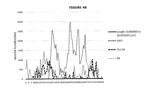
Figures 5A and 5B shows the co-localisation of OCT4 and RR by the cancer stem
cell population associated with OTSCC. Figure 5A shows immunohistochemical co-
staining
using antibodies specific to OCT4 and RR. Figure 5B shows quantification of
the relative
fluorescence signal for OCT4 (dots) and RR (long dashed line).
CA 02957930 2017-02-10
WO 2016/024870 PCT/NZ2015/050108
8
Figure 6 shows the expression of OCT4, SOX2, ATIIR2 and RR by the cancer stem
cell population associated with melanoma as evidenced by the
immunohistochemical
staining profiles.
Figures 7A and 78 shows co-localisation of OCT4 and RR by the cancer stem cell
population associated with melanoma. Figure 7A shows immunohistochemical
staining
using antibodies specific to OCT4 and RR. Figure 7B shows quantification of
the relative
fluorescence signal for OCT4 (dots) and RR (long dashed line).
Figure 8 shows the expression of OCT4, SOX2, ATIIR2 and RR by the cancer stem
cell population associated with sarcoma (leiomyosarconna) as evidenced by the
immunohistochemical staining profiles.
Figures 9A and 98 shows the co-localisation of OCT4 and RR by cancer stem cell
population associated with sarcoma. Figure 9A shows immunohistochemical
staining using
antibodies specific to OCT4 and RR.
Figure 9B shows quantification of the relative
fluorescence signal for OCT4 (dots) and RR (long dashed line).
Figure 10 shows expression of OCT4, SOX2, ATIIR2 and RR by the cancer stem
cell
population associated with bowel cancer as evidenced by the
immunohistochemical staining
profiles.
Figures 11A and 118 shows the co-localisation of OCT4 and RR in the cancer
stem
cell population associated with bowel cancer. Figure 11A shows
immunohistochemical
staining using antibodies specific to 0C14 and RR. Figure 11B shows
quantification of the
relative fluorescence signal for OCT4 (dots) and RR (long dashed line).
Figure 12 shows the expression of OCT4, 50X2, ATIIR2 and RR by the cancer stem
cell population associated with brain cancer (glioblastoma multiforme) as
evidenced by the
immunohistochemical staining profiles.
Figures 13A and 138 shows the co-localisation of OCT4 and RR in the cancer
stem
cell population associated with brain cancer (glioblastoma multiforme). Figure
13A shows
immunohistochemical staining using antibodies specific to 0C14 and RR. Figure
13B shows
quantification of the relative fluorescence signal for OCT4 (dots) and RR
(long dashed line).
Figure 14 shows the expression of OCT4, SOX2, ATIIR2 and RR in the cancer stem
cell population associated with breast cancer as evidenced by the
immunohistochemical
staining profiles.
Figures 15A and 158 shows the co-localisation of OCT4 and RR in the cancer
stem
cell population associated with breast cancer. Figure 15A shows
immunohistochemical
staining using antibodies specific to OCT4 and RR. Figure 15B shows
quantification of the
relative fluorescence signal for OCT4 (dots) and RR (long dashed line).
CA 02957930 2017-02-10
WO 2016/024870 PCT/NZ2015/050108
9
Figure 16 shows the expression of OCT4, SOX2, ATIIR2 and RR by the cancer stem
cell population associated with lung cancer (metastatic lung adenocarcinoma)
as evidenced
by the immunohistochemical staining profiles.
Figures 17A and 176 shows the co-localisation of OCT4 and RR in cancer stem
cell
population associated with lung cancer (metastatic lung adenocarcinoma).
Figure 17A
shows immunohistochemical staining using antibodies specific to OCT4 and RR.
Figure 17B
shows quantification of the relative fluorescence signal for 0C14 (dots) and
RR (long dashed
line).
Figure 18 shows the expression of OCT4, SOX2, ATIIR2 and RR by the cancer stem
cell population associated with B cell lymphoma as evidenced by the
immunohistochemical
staining profiles.
Figures 19A and 196 shows the co-localisation of OCT4 and RR by the cancer
stem
cell population associated with B cell lymphoma. Figure 19A shows
immunohistochemical
staining using antibodies specific to 0C14 and RR. Figure 19B shows
quantification of the
relative fluorescence signal for OCT4 (dots) and RR (long dashed line).
Figure 20 shows the expression of OCT4, SOX2, ATIIR2 and RR by the cancer stem
cell population associated with kidney cancer (metastatic renal cell cancer)
as evidenced by
the immunohistochemical staining profiles.
Figures 21A and 216 shows the co-localisation of OCT4 and RR by the cancer
stem
cell population associated with kidney cancer (metastatic renal cell cancer).
Figure 21A
shows immunohistochemical staining using antibodies specific to OCT4 and RR.
Figure 21B
shows quantification of the relative fluorescence signal for OCT4 (dots) and
RR (long dashed
line).
Figure 22 shows the expression of OCT4, SOX2, ATIIR2 and RR by the cancer stem
cell population associated with thyroid cancer as evidenced by the
immunohistochemical
staining profiles.
Figures 23A and 23B shows the co-localisation of OCT4 and RR by the cancer
stem
cell population associated with thyroid cancer. Figure 23A shows
immunohistochemical
staining using antibodies specific to OCT4 and RR. Figure 23B shows
quantification of the
relative fluorescence signal for OCT4 (dots) and RR (long dashed line).
Figure 24 shows the expression of OCT4, SOX2, ATIIR2 and RR by the cancer stem
cell population associated with chronic lynnphocytic leukaemia as evidenced by
the
immunohistochemical staining profiles.
Figures 25A and 256 shows the co-localisation of OCT4 and RR by the cancer
stem
cell population associated with chronic lymphocytic leukaemia.
Figure 25A shows
immunohistochemical staining using antibodies specific to OCT4 and RR. Figure
25B shows
quantification of the relative fluorescence signal for OCT4 (dots) and RR
(long dashed line).
CA 02957930 2017-02-10
WO 2016/024870 PCT/NZ2015/050108
Figure 26 shows the expression of OCT4, SOX2, ATIIR2 and RR by the cancer stem
cell population associated with skin squamous cell carcinoma as evidenced by
the
immunohistochemical staining profiles.
Figures 27A and 278 shows the co-localisation of OCT4 and RR by the cancer
stem
5 cell population associated with skin squamous cell carcinoma. Figure 27A
shows
immunohistochemical staining using antibodies specific to OCT4 and RR. Figure
27B shows
quantification of the relative fluorescence signal for OCT4 (dots) and RR
(long dashed line).
Figure 28 shows the expression of OCT4, SOX2, ATIIR2 and RR by the cancer stem
cell population associated with prostate cancer as evidenced by the
immunohistochemical
10 staining profiles.
Figures 29A and 298 shows the co-localisation of OCT4 and RR by the cancer
stem
cell population associated with prostate cancer. Figure 29A shows
immunohistochemical
staining using antibodies specific to OCT4 and RR. Figure 29B shows
quantification of the
relative fluorescence signal for OCT4 (dots) and RR (long dashed line).
Figure 30 shows the expression of OCT4 and SOX2 in a human seminoma tissue
sample, ATIIR2 in human kidney and RR in human placental tissues as respective
positive
controls. The negative control shows absence of staining without the primary
antibody in a
brain cancer (glioblastonna multiforme) tissue section.
SELECTED DEFINITIONS
Unless defined otherwise, all technical and scientific terms used herein have
the
same meaning as commonly understood to one of ordinary skill in the art to
which the
inventions belong. Although any assays, methods, devices and materials similar
or
equivalent to those described herein can be used in the practice or testing of
the invention,
various assays, methods, devices and materials are now described.
It is intended that reference to a range of numbers disclosed herein (for
example 1
to 10) also incorporates reference to all related numbers within that range
(for example, 1,
1.1, 2, 3, 3.9, 4, 5, 6, 6.5, 7, 8, 9 and 10) and also any range of rational
numbers within
that range (for example 2 to 8, 1.5 to 5.5 and 3.1 to 4.7) and, therefore, all
sub-ranges of
all ranges expressly disclosed herein are expressly disclosed. These are only
examples of
what is specifically intended and all possible combinations of numerical
values between the
lowest value and the highest value enumerated are to be considered to be
expressly stated
in this application in a similar manner.
As used in this specification, the words "comprises", "comprising", and
similar words,
are not to be interpreted in an exclusive or exhaustive sense. In other words,
they are
intended to mean "including, but not limited to".
CA 02957930 2017-02-10
WO 2016/024870 PCT/NZ2015/050108
11
As used herein, the term "antibodies" refer to molecules that contain an
antigen
binding site, e.g., immunoglobulins. Immunoglobulin molecules can be of any
type (e.g.,
IgG, IgE, IgM, IgD, IgA and IgY), class (e.g., IgGI, IgG2, IgG3, IgG4, IgAl
and IgA2) or
subclass. Antibodies include, but are not limited to, monoclonal antibodies,
polyclonal
antibodies, multispecific antibodies, human antibodies, humanised antibodies,
murine
antibodies, camelised antibodies, chimeric antibodies, single domain
antibodies, single chain
Fvs (scFv), single chain antibodies, Fab fragments, F(ab') fragments,
disulfide-linked Fvs
(sdFv), and anti- idiotopic (anti-Id) antibodies (including, e.g., anti-Id
antibodies to
antibodies of the invention), and epitope-binding fragments of any of the
above.
As used herein, the term "cancer" refers to a neoplasm or tumour resulting
from
abnormal uncontrolled growth of cells. The term "cancer" encompasses a disease
involving
both pre-malignant and malignant cancer cells. In some examples, cancer refers
to a
localised overgrowth of cells that has not spread to other parts of a subject,
i.e., a benign
tumour. In other examples, cancer refers to a malignant tumour, which has
invaded and
destroyed neighboring body structures and/or spread to distant sites.
As used herein, the term "cancer cells" refer to cells that acquire a
characteristic set
of functional capabilities during their development, including the ability to
evade apoptosis,
are self-sufficienct in growth signals and are insensitivite to anti-growth
signals, tissue
invasion/metastasis, significant growth potential, and/or sustained
angiogenesis. The term
"cancer cell" is meant to encompass both pre-malignant and malignant cancer
cells.
As used herein, the term "cancer stem cell(s)" refers to a cell that can be a
progenitor of a highly proliferative cancer cell. A cancer stem cell has the
ability for
assymmetrical division and to re-grow a tumour as demonstrated by its ability
to form
tumours in immunocompromised mice, and typically to form tumours upon
subsequent
serial transplantation in immunocompromised mice. Cancer stem cells are also
typically
slow-growing relative to the bulk of a tumour; that is, cancer stem cells are
generally
quiescent.
In certain examples, but not all, the cancer stem cell may represent
approximately 0.1 to 10% of a tumour.
As used herein, the term "cancer stem cell population" is intended to mean one
or
more cancer stem cells, in other words a single cancer stem cell or multiple
cancer stem
cells, the single cancer stem cell or multiple cancer stem cells being capable
of driving
tumourigenesis of a given cancer.
As used herein, the term "squamous cell carcinomas" refers to the epithelial
tumours
found in many different organs, including the skin, upper aerodigestive tract
(including oral
cavity) and paranasal sinuses, oesophagus, lungs, and cervix, and other organs
which show
squamous cell differentiation. Included are head and neck squamous cell
carcinomas, lung
squamous cell carcinomas, skin squamous cell carcinomas, otic squamous cell
carcinomas,
CA 02957930 2017-02-10
WO 2016/024870 PCT/NZ2015/050108
12
vulval squamous cell carcinomas, cervical squamous cell carcinomas,
oesophageal
squamous cell carcinomas, upper aerogigestive tract and paranasal sinus
squamous cell
carcinomas and the like.
As used herein, the term "Renin-Angiotensin System (RAS)" or "Renin-
Angiotensin-
Aldosterone System (RAAS)" is a hormone system that regulates blood pressure
and fluid
balance. The wider pathway associated with RAS also includes the Pro/Renin
Receptor
System (PRRS) and the associated bypass pathways. By way of example, refer to
Figures 1
and 2. There are a number of known drugs which target the RAS including PRRS,
as
described in more detail below.
As used herein, the term "effective amount" refers to the amount of a therapy
that is
sufficient to result in the prevention of the development, recurrence, or
onset of cancer and
one or more symptoms thereof, to enhance or improve the prophylactic effect(s)
of another
therapy, reduce the severity, the duration of cancer, ameliorate one or more
symptoms of
cancer, prevent the advancement of cancer, cause regression of cancer, and/or
enhance or
improve the therapeutic effect(s) of another therapy. In an example of the
invention, the
amount of a therapy is effective to achieve one, two or three or more results
following the
administration of one, two, three or more therapies: (1) a stabilisation,
reduction or
eradication of the cancer stem cell population; (2) a stabilisation, reduction
or eradication in
the cancer cell population; (3) a stabilisation or reduction in the growth of
a tumour or
neoplasm; (4) an impairment in the formation of a tumour; (5) eradication,
removal, or
control of primary, regional and/or metastatic cancer; (6) a reduction in
mortality; (7) an
increase in disease-free, relapse-free, progression-free, and/or overall
survival, duration, or
rate; (8) an increase in the response rate, the durability of response, or
number of patients
who respond or are in remission; (9) a decrease in hospitalisation rate, (10)
a decrease in
hospitalisation lengths, (11) the size of the tumour is maintained and does
not increase or
increases by less than 10%, preferably less than 5%, preferably less than 4%,
preferably
less than 2%, (12) an increase in the number of patients in remission, (13) an
increase in
the length or duration of remission, (14) a decrease in the recurrence rate of
cancer, (15)
an increase in the time to recurrence of cancer, and (16) an amelioration of
cancer-related
symptoms and/or quality of life.
As used herein, the terms "manage", "managing", and "management" in the
context
of the administration of a therapy to a subject refer to the beneficial
effects that a subject
derives from a therapy (e.g., a prophylactic or therapeutic agent) or a
combination of
therapies, while not resulting in a cure of cancer. In certain examples, a
subject is
administered one or more therapies (e.g., one or more prophylactic or
therapeutic agents)
to "manage" cancer so as to prevent the progression or worsening of the
condition.
CA 02957930 2017-02-10
WO 2016/024870 PCT/NZ2015/050108
13
As used herein, the terms "prevent", "preventing" and "prevention" in the
context of
the administration of a therapy to a subject refers to the prevention or
inhibition of the
recurrence, onset, and/or development of a cancer or a symptom thereof in a
subject
resulting from the administration of a therapy (e.g., a prophylactic or
therapeutic agent), or
a combination of therapies (e.g., a combination of prophylactic or therapeutic
agents). In
some examples, such terms refer to one, two, three or more results following
the
administration of one or more therapies: (1) a stabilisation, reduction or
eradication of the
cancer stem cell population, (2) a stabilisation, reduction or eradication of
the cancer cell
population, (3) an increase in the response rate, (4) an increase in the
duration of
remission, (5) a decrease in the recurrence rate of cancer, (6) an increase in
the time to
recurrence of cancer, (7) an increase in the disease-free, relapse-free,
progression-free,
and/or overall survival of the patient, and (8) an amelioration of cancer-
related symptoms
and/or quality of life. In specific examples, such terms refer to a
stabilisation, reduction or
eradication of the cancer stem cell population.
As used herein, the term "marker" or "biomarker" in the context of a tissue
(e.g. a
normal cell or tumour cell) means any antigen, molecule or other chemical or
biological
entity that is specifically found in or on a tissue that it is desired to be
identified or identified
in or on a particular tissue affected by a disease or disorder, for example
cancer. The term
"tumourigenic biomarker" is also relevant to this definition in the context of
cancer. In
specific examples, the marker is a cell surface antigen that is differentially
or preferentially
expressed by specific cell types. In specific examples, the marker is a
nuclear antigen that
is differentially or preferrentially expressed by specific cell types. In
specific examples the
marker is an intracellular antigen that is differentially or preferrentially
expressed by specific
cell types.
As used herein, the term "prophylactic agent" refers to any molecule,
compound,
and/or substance that is used for the purpose of preventing cancer.
Examples of
prophylactic agents include, but are not limited to, proteins, immunoglobulins
(e.g., multi-
specific Igs, single chain Igs, Ig fragments, polyclonal antibodies and their
fragments,
monoclonal antibodies and their fragments), antibody conjugates or antibody
fragment
conjugates, peptides (e.g., peptide receptors, selectins), binding proteins,
chennospecific
agents, chemotoxic agents (e.g., anti-cancer agents), proliferation based
therapy, and small
molecule drugs.
As used herein, the term "therapeutic agent" refers to any molecule, compound,
and/or substance that is used for the purpose of treating and/or managing a
disease or
disorder. Examples of therapeutic agents include, but are not limited to,
proteins,
immunoglobulins (e.g., multi-specific Igs, single chain Igs, Ig fragments,
polyclonal
antibodies and their fragments, monoclonal antibodies and their fragments),
peptides (e.g.,
CA 02957930 2017-02-10
WO 2016/024870 PCT/NZ2015/050108
14
peptide receptors, selectins), binding proteins, biologies, chemospecific
agents, chemotoxic
agents (e.g., anti-cancer agents), proliferation-based therapy agents,
hormonal agents,
radioinnmunotherapies, targeted agents, epigenetic therapies, differentiation
therapies,
biological agents, radiation agents, chemotherapy, anti-angiogenic agents, and
small
molecule drugs.
As used herein, the terms "therapies" and "therapy" can refer to any
method(s),
composition(s), and/or agent(s) that can be used in the prevention, treatment
and/or
management of a cancer or one or more symptoms thereof. In certain examples,
the terms
"therapy" and "therapies" refer to chemotherapy, radiation therapy, surgery,
hormonal
therapy, anti-angiogenic therapy, biological therapy, proliferation based
therapy, prodrug-
activating enzyme therapy, small molecule therapy, toxin therapy, antibody
therapy,
imnnunotherapy, radioimmunotherapy, targeted therapy, epigenetic therapy,
demethylation
therapy, histone deactylase inhibitor therapy, differentiation therapy and/or
other therapies
useful in the prevention, management and/or treatment of a cancer or one or
more
symptoms thereof.
As used herein, the terms "treat", "treatment" and "treating" in the context
of the
administration of a therapy to a subject refer to the reduction or inhibition
of the
progression and/or duration of cancer, the reduction or amelioration of the
severity of
cancer, and/or the amelioration of one or more symptoms thereof resulting from
the
administration of one or more therapies. In specific examples, such terms
refer to one, two
or three or more results following the administration of one, two, three or
more therapies:
(1) a stabilization, reduction or eradication of the cancer stem cell
population; (2) a
stabilisation, reduction or elimination in the cancer cell population; (3) a
stabilisation or
reduction in the growth of a tumour or neoplasm; (4) an impairment in the
formation of a
tumour; (5) eradication, removal, or control of primary, regional and/or
metastatic cancer;
(6) a reduction in mortality; (7) an increase in disease-free, relapse-free,
progression-free,
and/or overall survival, duration, or rate; (8) an increase in the response
rate, the durability
of response, or number of patients who respond or are in remission; (9) a
decrease in
hospitalisation rate, (10) a decrease in hospitalisation lengths, (11) the
size of the tumour is
maintained and does not increase or increases by less than 10%, preferably
less than 5%,
preferably less than 4%, preferably less than 2%, and (12) an increase in the
number of
patients in remission. In certain examples, such terms refer to a
stabilisation or reduction
in the cancer stem cell population. In some examples, such terms refer to a
stabilisation or
reduction in the growth of cancer cells. In some examples, such terms refer to
a
stabilisation or reduction in the cancer stem cell population and a reduction
in the cancer
cell population. In some examples, such terms refer to a stabilisation or
reduction in the
growth and/or formation of a tumour. In some examples, such terms refer to the
CA 02957930 2017-02-10
WO 2016/024870 PCT/NZ2015/050108
eradication, removal, or control of primary, regional, or metastatic cancer
(e.g., the
minimisation or delay of the spread of cancer). In some examples, such terms
refer to a
reduction in mortality and/or an increase in survival rate of a patient
population. In further
examples, such terms refer to an increase in the response rate, the durability
of response,
5 or number of patients who respond or are in remission. In some examples,
such terms
refer to a decrease in hospitalisation rate of a patient population and/or a
decrease in
hospitalisation length for a patient population.
The term "sample" or "biological sample" as used herein means any sample taken
or
derived from a subject. Such a sample may be obtained from a subject, or may
be
10 obtained from biological materials intended to be provided to the
subject. For example, a
sample may be obtained from blood being assessed, for example, to investigate
the cancer
status of a subject. Included are samples taken or derived from any subjects
such as from
normal healthy subjects and/or healthy subjects for whom it is useful to
understand their
cancer status. Preferred samples are biological fluid samples. The term
"biological fluid
15 sample" as used herein refers to a sample of bodily fluid obtained for
the purpose of, for
example, diagnosis, prognosis, classification or evaluation of a subject of
interest, such as a
patient. In certain embodiments, such a sample may be obtained for the purpose
of
determining the cancer status of a patient. The sample may be any sample known
in the
art in which cancer stem cells may be detected. Included are any body fluids
such as a
whole blood sample, plasma, serum, ovarian follicular fluid sample, seminal
fluid sample,
cerebrospinal fluid, saliva, sputum, urine, pleural effusions, interstitial
fluid, synovial fluid,
lymph, tears, for example, although whole blood sample, plasma and serum are
particularly
suited for use in this invention. In addition, one of skill in the art would
realise that certain
body fluid samples would be more readily analysed following a fractionation or
purification
procedure, for example, separation of whole blood into serum or plasma
components.
The term "purified" as used herein does not require absolute purity. Purified
refers
in one embodiment to at least 90%, or 95%, or 98%, or 99% homogeneity of, to
provide
an example, of a polypeptide or antibody in a sample.
The term "subject" as used herein is preferably a mammal and includes human,
and
non-human mammals such as cats, dogs, horses, cows, sheep, deer, mice, rats,
primates
(including gorillas, rhesus monkeys and chimpanzees), possums and other
domestic farm or
zoo animals. Thus, the assays, methods and kits described herein have
application to both
human and non-human animals, in particular, and without limitation, humans,
primates,
farm animals including cattle, sheep, goats, pigs, deer, alpacas, llamas,
buffalo, companion
and/or pure bred animals including cats, dogs and horses. Preferred subjects
are humans,
and most preferably "patients" who as used herein refer to living humans who
may receive
or are receiving medical care or assessment for a disease or condition.
Further, while a
CA 02957930 2017-02-10
WO 2016/024870 PCT/NZ2015/050108
16
subject is preferably a living organism, the invention described herein may be
used in post-
mortem analysis as well.
The term "ELISA" as used herein means an enzyme linked immunosorbent assay, a
type of competitive binding assay comprising antibodies and a detectable label
used to
quantitate the amount of an analyte in a sample.
The term "capture antibody" as used herein means an antibody which is
typically
immobilized on a solid support such as a plate, bead or tube, and which
antibody binds to
and captures analyte(s) of interest, for example membrane bound markers
associated with
a cancer stem cell population.
The term "detection antibody" as used herein means an antibody comprising a
detectable label that binds to analyte(s) of interest. The label may be
detected using
routine detection means for a quantitative, semi-quantitative or qualitative
measure of the
analyte(s) of interest, for example membrane bound markers associated with a
cancer stem
cell population.
As used herein, the term "relating to the presence or amount" of an analyte
reflects
that assay signals are typically related to the presence or amount of an
analyte through the
use of a standard curve calculated using known concentrations of the analyte
of interest. As
the term is used herein, an assay is "configured to detect" an analyte if an
assay can
generate a detectable signal indicative of the presence or amount of a
physiologically
relevant concentration of the analyte. Typically, an analyte is measured in a
sample.
A level "higher" or "lower" than a control, or a "change" or "deviation" from
a control
(level) in one embodiment is statistically significant. A higher level, lower
level, deviation
from, or change from a control level or mean or historical control level can
be considered to
exist if the level differs from the control level by about 5% or more, by
about 10% or more,
by about 20% or more, or by about 50% or more compared to the control level.
Statistically
significant may alternatively be calculated as P.Ø05. Higher levels, lower
levels, deviation,
and changes can also be determined by recourse to assay reference limits or
reference
intervals. These can be calculated from intuitive assessment or non-parametric
methods.
Overall, these methods may calculate the 0.025, and 0.975 fractiles as 0.025*
(n+1) and
0.975 (n+1). Such methods are well known in the art. Presence of a marker
absent in a
control may be seen as a higher level, deviation or change. Absence of a
marker present in
a control may be seen as a lower level, deviation or change.
DETAILED DESCRIPTION
There is an emerging concept that cancer stem cells drive the persistence or
recurrence of a tumour. Conventional cancer therapies, while successful in
eradicating the
bulk of tumours, are typically less effective on insidious cancer stem cells.
Further,
CA 02957930 2017-02-10
WO 2016/024870 PCT/NZ2015/050108
17
selective drug resistance exhibited by these cells contributes to significant
morbidity and
mortality in cancer sufferers. Accordingly, there is a need for therapeutic
regimes that
specifically and selectively target cancer stem cells.
The present invention is predicated on the surprising and unexpected discovery
that
discrete cancer stem cell populations are associated with certain tumours
including (e.g.,
squamous cell carcinoma of the oral cavity, squamous cell carcinoma of the
skin,
melanoma, lung cancer, breast cancer, kidney cancer, brain cancer, bowel
cancer, thyroid
cancer, prostate cancer, lymphoma, leukaemia and sarcomas. The cancer stem
cell
populations associated with these tumours are characterised by unique
biomarker
expression profiles that allows for the specific identification and diagnosis
of certain cancers.
Importantly, it has also been revealed by the Applicants that these cancer
stem cell
populations express key components of the the Renin-Agiotensin System (RAS),
including
the Renin Receptor (RR) Angiotensin II Receptor 2 (ATIIR2), as well as a
secreted form of
the Renin Receptor (sRR). In reference to Figures 3, 6, 8, 10, 12, 14, 16, 18,
20, 22, 24,
26 and 28, the Applicants demonstrate co-expression of RR and ATIIR2 by the
cancer stem
cell populations associated with various tumours. These cancer stem cell
populations are
characterised by, for example, the expression of OCT4, SOX2, PSTAT3 and NANOG.
Accordingly, the expression of the components of RAS by these cancer stem cell
populations
provides a novel and unique therapeutic approach by targeting the cancer stem
cells
associated with various tumours from the extensive array of drugs that target
RAS such as,
Angiotensin-Converting Enzyme Inhibitors (ACEis), Angiotensin Receptor
Blockers (ARBs),
Direct Renin Inhibitors (DRIs), Beta-Blockers, Cyclo-oxygenase 2 Inhibitors,
Chymase
Inhibitors, Inhibitors of Cathepsin B, Cathepsin D and Cathepsin G, Calcium
Supplements,
Vitamin D and Calcium Channel Blockers.
In addition, the present invention also contemplates indirect inhibitors of
the RAS
(e.g., Calcium Channel Blockers).
Accordingly, in one aspect of the present invention there is provided a method
for
preventing, treating, or managing cancer in a patient in need thereof, the
method
comprising administering a therapeutic agent to the patient in an amount
sufficient to
selectively eliminate or inhibit the growth, proliferation and/or
differentiation of cancer stem
cells in a tumour associated with the cancer, wherein the cancer stem cells
are
characterised by (i) the expression of one or more embryonic stem cell
biomarkers, and (ii)
the expression of one or more biomarkers associated with the Renin-Angiotensin
System.
In another aspect of the present invention there is provided a method for
preventing,
treating, or managing cancer in a patient in need thereof, the method
comprising
administering a therapeutic agent to the patient in an amount sufficient to
selectively inhibit
the growth, proliferation and/or differentiation of cancer stem cells within
the cancer,
CA 02957930 2017-02-10
WO 2016/024870 PCT/NZ2015/050108
18
wherein the cancer stem cells are characterised by (i) the expression of one
or more
embryonic stem cell biomarkers, and (ii) the expression of one or more
biomarkers
associated with the Renin-Angiotensin System, and wherein the cancer is a
solid tumour or
blood cancer.
In one example, the one or more embryonic stem cell markers is selected from
the
group consisting of Cripto, ABCG2, Alkaline Phosphatase/ALPL, CD9, FGF-4, GDF-
3, Integrin
alpha 6/CD49f, Integrin beta 1/CD29, Nanog, Oct-3/4, Podocalyxin, 50X2, SSEA-
3, SSEA-4,
STAT3, SSEA-1, FoxD3, DPPA5/ESG1, Rex-1/ZFP42, DPPA4, LIN-28A, UTF1, Lefty-A,
Lefty-
1, TBX3, ESGP, TRA-1-60(R), TRA-1-81, 514, TBX2, ZIC3, CD30/INFRSF8, KLF5, c-
Myc,
GCNF/NR6A1, SUZ12, Smad2, CDX2, TROP-2, CD117/c-kit, LIN-41, Integrin alpha 6
beta 4,
THAP11, Smad2/3, TBX5, TEX19, Oct-4A, TEX19.1, DPPA2, Activin RIB/ALK-4,
Activin RIIB,
FGF-5, GBX2, Stella/Dppa3, DNMT3B, F-box protein 15/FBX015, LIN-28B, Integrin
alpha 6
beta 1, KLF4, ERR beta/NR3B2, EpCAM/TROP1, TERT, CHD1, Cbx2, c-Maf and L1TD1.
In
another example, the one or more embryonic stem cell biomarkers consists in
OCT4, SOX2,
NANOG and PSTAT3. In yet another example, the one or more biomarkers
associated with
the RAS is selected from the group consisting of RR, ATIIR2, and sRR.
In yet another aspect of the present invention there is provided a method for
preventing, treating, or managing cancer in a patient in need thereof, the
method
comprising administering a therapeutic agent to the patient in an amount
sufficient to
selectively eradicate, or inhibit the growth, proliferation and/or
differentiation of cancer
stem cells within the cancer, wherein the cancer stem cells are characterised
by (i) the
expression of one or more embryonic stem cell biomarkers, and (ii) the
expression of one or
more biomarkers associated with the Renin-Angiotensin System, and wherein the
tumour is
selected from the group consisting of squamous cell carcinoma of the oral
cavity, squamous
cell carcinoma of the skin, melanoma, lung cancer, breast cancer, kidney
cancer, brain
cancer, bowel cancer, thyroid cancer, prostate cancer, lymphoma, leukaemia and
sarcomas.
In yet a further aspect of the present invention there is provided a method
for
preventing, treating, or managing cancer in a patient in need thereof, the
method
comprising administering a therapeutic agent to the patient in an amount
sufficient to
selectively eradicate, or inhibit the growth, proliferation and/or
differentiation of cancer
stem cells in a tumour associated with the cancer, wherein the cancer stem
cells are
characterised by (i) the expression of one or more embryonic stem cell
biomarkers, and (ii)
the expression of one or more biomarkers associated with the Renin-Angiotensin
System,
and wherein the tumour is a squamous cell carcinoma.
In another aspect of the present invention there is provided a method for
preventing,
treating, or managing cancer in a patient in need thereof, the method
comprising
administering a therapeutic agent to the patient in an amount sufficient to
selectively
CA 02957930 2017-02-10
WO 2016/024870 PCT/NZ2015/050108
19
eliminate, or inhibit the growth, proliferation and/or differentiation of
cancer stem cells in a
tumour associated with the cancer, wherein the cancer stem cells are
characterised by (i)
the expression of one or more stem cell biomarker selected from the group
consisting of
Cripto, ABCG2, Alkaline Phosphatase/ALPL, CD9, FGF-4, GDF-3, Integrin alpha
6/CD49f,
Integrin beta 1/CD29, NANOG, 0C13/4, Podocalyxin, SOX2, SSEA-3, SSEA-4, STAT3,
SSEA-
1, FoxD3, DPPA5/ESG1, Rex-1/ZFP42, DPPA4, LIN-28A, UTF1, Lefty-A, Lefty-1,
TBX3,
ESGP, TRA-1-60(R), TRA-1-81, 5T4, TBX2, ZIC3, CD30/TNFRSF8, KLF5, c-Myc,
GCNF/NR6A1, SUZ12, Smad2, CDX2, TROP-2, CD117/c-kit, LIN-41, Integrin alpha 6
beta 4,
THAP11, Srnad2/3, TBX5, TEX19, Oct-4A, TEX19.1, DPPA2, Activin RIB/ALK-4,
Activin RIB,
FGF-5, GBX2, Stella/Dppa3, DNMT3B, F-box protein 15/FBX015, LIN-28B, Integrin
alpha 6
beta 1, KLF4, ERR beta/NR3B2, EpCAM/TROP1, TERT, CHD1, Cbx2, c-Maf and L1TD1,
and
(ii) the expression of one or more biomarkers associated with the Renin-
Angiotensin
System.
In yet another aspect of the present invention there is provided a method for
preventing, treating, or managing cancer in a patient in need thereof, the
method
comprising administering a therapeutic agent to the patient in an amount
sufficient to
selectively eliminate, or inhibit the growth, proliferation and/or
differentiation of cancer
stem cells in a tumour associated with the cancer, wherein the cancer stem
cells are
characterised by (i) the expression of one or more stem cell marker selected
from the group
consisting of OCT4, 50X2, NANOG and PSTAT3, and (ii) the expression of one or
more
biomarkers associated with the Renin-Angiotensin System selected from the
group
consisting of Renin Receptor, Angiotensin II Receptor 2, and a secreted for of
the Renin
Receptor.
In yet a further aspect of the present invention there is provided a method
for
preventing, treating, or managing cancer in a patient in need thereof, the
method
comprising administering a therapeutic agent to the patient in an amount
sufficient to
selectively eradicate, or inhibit the growth, proliferation and/or
differentiation of cancer
stem cells in a tumour associated with the cancer, wherein the cancer stem
cells are
characterised by (i) the expression of one or more stem cell biomarker
selected from the
group consisting of OCT4, SOX2, NANOG and PSTAT3, and (ii) the expression of
Renin
Receptor, Angiotensin II Receptor 2 and/or a secreted form of Renin Receptor,
and wherein
=the therapeutic agent is selected from the group consisting of Direct Renin
Inhibitors
(DRIs), ACEis, ARBs, Beta-Blockers, Cyclo-oxygenase 2 Inhibitors, Chymase
Inhibitors,
Inhibitors of Cathepsin B, Cathepsin D and Cathepsin G, Calcium, Vitamin D,
and Calcium
Channel Blockers .
CA 02957930 2017-02-10
WO 2016/024870 PCT/NZ2015/050108
In one example, the cancer stem cells are characterised by the expression of
SOX2,
OCT4, PSTAT3 and NANOG. These cells are said to have a marker expression
profile:
SOX2+Oct-4+PSTAT3+NANOG+.
In a related example, the cancer stem cells are cancer stem cells of squamous
cell
5 carcinoma of oral tongue and are characterised by the marker expression
profile
CD44+SOX2+OCT4+NANOG+. In a further example, the cancer stem cells of squamous
cell
carcinoma of oral tongue are characterised by the marker expression profile
CD44 SOX2+OCT4+NANOG+CD34-. In yet a further example, the cancer stem cells
are
cancer stem cells of squamous cell carcinoma of oral tongue and are
characterised by the
10 marker expression profile CD44 SOX2+0CT4+NANOG CD341363-EMA-.
The cancer stem cells may co-express with embryonic stem cell markers,
lymphatic
cell markers, epithelial cancer cell markers or any combination thereof.
Accordingly, in one
example, the cancer stem cells co-express with embryonic stem cell markers and
epithelial
cancer cell markers. In another example, the cancer stem cells co-express with
lymphatic
15 cell markers and epithelial cancer cell markers. In a further example,
the cancer stem cells
co-express with embryonic stem cell markers, lymphatic cell markers, and
epithelial cancer
cell markers.
While the present invention is particularly relevant to solid tumours, it also
extends
to blood cancers. For example, the Applicants demonstrate that Chronic
Lymphocytic
20 Leukaemia comprise cancer stem cells as characterised by the expression
of OCT4, SOX2,
ATIIR2 and RR (Figure 24), which cancer stem cells also co-express the RR and
ATIIR2
(Figures 25A and 25B). Accordingly, the therapeutic methods and compositions
according to
the present invention extend to blood cancers as well.
Accordingly, the present invention provides compositions and methods related
to
identifying and targeting the growth and proliferation of cancer stem cells as
the cause of
tumour growth, spread and metastasis. In particular, the compositions and
methods are
directed to cancer stem cells which display unique tumouriogenic biomarker
expression
profiles. By specifically targeting cancer stem cells, it is assumed that the
tunnourigenic and
metastatic potential of the (nascent or established) tumour is significantly
diminished,
thereby leading to enhanced therapeutic outcomes.
The cancer stem cells may be associated with a variety of cancers, including
but not
limited to, squamous cell carcinoma of the upper aerodigestive tract
(including oral cavity),
squamous cell carcinoma of the skin, melanoma, lung cancer, breast cancer,
kidney cancer,
brain cancer, bowel cancer, thyroid cancer, prostate cancer, lymphoma,
leukemia and
sarcomas.
Squamous cell carcinomas include head and neck squamous cell carcinomas
(including squamous cell carcinomas of the upper aerodigestive tract
[including oral cavity]
CA 02957930 2017-02-10
WO 2016/024870 PCT/NZ2015/050108
21
and paranasal sinuses and elsewhere), oesophageal squamous cell carcinomas,
skin
squamous cell carcinomas, lung squamous cell carcinomas, vulval squamous cell
carcinomas
and cervical squamous cell carcinomas. In one example the upper aerodigestive
tract
squamous cell carcinoma is squamous cell carcinoma of oral tongue.
The present invention provides methods for preventing, treating, and/or
managing
cancer, the method comprising administering to a subject in need thereof a
course of
therapy that stabilises, reduces, or eradicate the cancer stem cell
population. In certain
examples, the stabilisation, reduction, or elimination of the cancer stem cell
population is
achieved by administering a therapy that targets the growth and proliferation
of the cancer
stem cells.
Surprisingly, Applicants demonstrate that the cancer stem cell populations
identified
in the methods according to the present invention co-express components of
RAS, and in
cancer stem cell populations associated with multiple different tumour types.
By way of
illustration only, the Applicants demonstrate co-expression of the RR and
ATIIR2 in OTSCC
(Figures 4A, 4B), melanoma (Figures 7A, 7B), sarcoma (Figures 9A, 9B), bowel
cancer
(Figures 11A, 11B), brain cancer (Figures 13A, 13B), breast cancer (Figures
15A, 15B), lung
cancer (Figures 17A, 17B), B cell lymphoma (Figures 19A, 19B), and kidney
(Figures 21A,
21B), thyroid cancer (23A, 23B), chronic lymphocytic cancer (25A, 25B), skin
squamous cell
carcinoma (27A, 27B), prostate cancer (29A, 29B).
Accordingly, therapy that targets the growth and proliferation of cancer stem
cell
populations comprises administering a therapeutic agent that selectively
targets
components of the RAS and/or Pro/Renin Receptor Systems (PRRS) expressed by
the
cancer stem cells. Figures 1 and 2 show the types of inhibitors/drugs that
target these
systems, useful in accordance with the compositions and methods according to
the present
invention.
Examples of known therapeutics that target the Renin-Angiotensin System
include,
but are not limited to, ACEIs, ARBs, DRIs, Beta-Blockers, Cyclo-oxygenase 2
Inhibitors,
Inhibitors of Cathepsin B, Cathepsin D and Cathepsin G, Calcium Channel
Blockers, Calcium
Supplements and Vitamin D.
Examples of ACEIs include, but are not limited to, Benazepril (Lotesin),
Captopril
(Capoten), Cilazipril, Enalapril (Vasotec, Renitec), Fosinopril (Monopril),
Lisinopril (Lisodur,
Lopril, Novatec, Prinivil, Zestril), Moexipril, Perindopril (Coversay, Aceon),
Quinapril
(Accupril), Ramipril (Altace, Tritace, Ramace, Ramiwin), Trandolapril,
Delapril, Zofenopril
and Imidapril.
Examples of ARBs include, but are not limited to, Losartan, Irbesartan,
Candesartan,
Eprosartan, Olnnesartan, Telmisartan, PD123319 and Valsartan.
CA 02957930 2017-02-10
WO 2016/024870 PCT/NZ2015/050108
22
Examples of Beta-Blockers include, but are not limited to, Acebutolol
(Sectral),
Atenolol (Tenormin), Betaxolol (Betoptic), Bisoprolol (Cardicor, Emcor,
Zebeta), Carteolol
(Teoptic), Carvedilol (Coreg, Eucardic), Celiprolol (Celectol), Labetalol
(Trandate),
Levobunolol (Betagan), Metipranolol (Metipranolol Minims), Metoprolol
(Betaloc, Lopresor,
Lopressor, Toprol XL), Nadolol (Corgard), Nebivolol (Bystolic, Nebilet),
Oxprenolol
(Trasicor), Pindolol (Visken), Propranolol (Inderal LA), Sotalol (Beta-
Cardone, Sotacor), and
Timolol (Betim, Nyogel, Timoptol).
Examples of Cyclo-oxygenase 2 Inhibitors include, but are not limited to,
Celecoxib,
Nepafenac, Ibuprofen (Dolgesic), Indonnethacin, Sulindac, Xanthohumol,
Meclofenamate
Sodium, Meloxicam, Rofecoxib, Bromfenac Sodium, Ibuprofen Lysine, Ketorolac
(Ketorolac
tromethamine), Diclofenac Sodium, Etodolac, Ketoprofen, Naproxen Sodium,
Piroxicam,
Acemetacin, Phenacetin, Tolfenamic Acid, Ninnesulide, Flunixin Meglumin,
Aspirin,
Bufexamac, Niflumic acid, Licofelone, Oxaprozin, Lornoxicam, Lumiracoxib,
Zaltoprofen,
Ampiroxicam, Valdecoxib, Nabumetone, Mefenamic Acid, Carprofen, Amfenac Sodium
monohydrate, Asaraldehyde and Suprofen.
Examples of Chynnase Inhibitors include, but are not limited to, TY-51469 (2-
[4-(5-
fluoro-3-methylbenzo[b]thiophen-2-yl)sulfonamido-3-methanesulfonyl-
phenyl]thiazole-4-
carboxylic acid), Eglin C, CI, SUN13834, Chymostatin, TJK002 a benzimidazole
inhibitor,
ONO-WH-236, Amblyomma americanum tick serine protease inhibitor 6 (AamS6), N-
tosyl-
L-phenylalanyl chloromethyl ketone (TPCK), Alpha-aminoalkylphosphonate diaryl
esters,
Serine protease inhibitor A3 (serpinA3), Squamous cell carcinoma antigen (SCCA-
2),
Bortezomib (Velcade), R05066852 and 17beta-estradiol.
Examples of Cathepsin B Inhibitors include, but are not limited to, Cystatin
B,
Cystatin C, Cysteine peptidase inhibitor E64, [Pt(dmba)(aza-N1)(dmso)] complex
1 (a
potential anti-tumoral drug with lower IC50 than cisplatin in several tumoral
cell lines),
2,3,7,8-Tetrachlorodibenzo-p-dioxin (TCDD), CA-074Me, Lipidated CtsB inhibitor
incorporated into the envelope of a liposomal nanocarrier (LNC-NS-629),
Proanthocyanidin
(PA) and ahpatinin Ac (1) and ahpatinin Pr (2).
Examples of Cathepsin D Inhibitors include, but are not limited to, non-
peptidic
acylguanidine inhibitors of Cathepsin D, Pepstatin A, Bm-Aspin, SIPI, Via,
RNAi-Rab27A and
Solanum lycopersicum aspartic protease inhibitor (SLAPI).
Examples of Cathepsin G Inhibitors include, but are not limited to, WFDC12,
Phenylmethylsulfonyl fluoride (PMSF), Ecotin, SerpinB1, SerpinA3, CeEI, or
Caesalpinia
echinata elastase inhibitor, SLPI (secretory leukocyte protease inhibitor),
Alpha1-Antitrypsin
(AAT), Bauhinia bauhinoides cruzipain inhibitor, Alpha-Aminoalkylphosphonate
diaryl esters,
Greglin, [2-[3-[[(1-benzoy1-4-piperidinyl)methylamino]carbony1]-2-
naphthaleny1]-1-(1-
naphthaleny1)-2-oxoethyll-phosphonic acid (KPA), Lynnpho-Epithelial Kazal-Type-
related
CA 02957930 2017-02-10
WO 2016/024870 PCT/NZ2015/050108
23
Inhibitor (LEKTI), Trappin-2 A62L, SV-66, SCGI, Bortezomib, Human
monocyte/neutrophil
elastase inhibitor (MNEI), a 42-kDa serpin protein and Anti-leukoproteinase
(ALP).
Examples of Calcium Channel Blockers include, but are not limited to,
Dihydropyridine Calcium Channel Blockers, Phenylalkylamine Calcium Channel
Blockers,
Benzothiazepine Calcium Channel Blockers, Non-Selective Calcium Channel
Blockers, as well
as "Other" Calcium Channel blockers.
Examples of Dihydropyridine Calcium Channel Blockers include, but are not
limted
to, Amlodipine (Norvasc), Aranidipine (Sapresta), Azelnidipine (Calblock),
Bamidipine
(HypoCa), Benidipine (Coniel), Cilnidipine (Atelec, Cinalong, Siscard) Not
available in US,
Clevidipine (Cleviprex), Isradipine (DynaCirc, Prescal), Efonidipine (Landel),
Felodipine
(Plendil), Lacidipine (Motens, Lacipil), Lercanidipine (Zanidip), Manidipine
(Ca!slot,
Madipine), Nicardipine (Cardene, Carden SR), Nifedipine (Procardia, Adalat),
Nilvadipine
(Nivadil), Nimodipine (Nimotop), Nisoldipine (Baymycard, Sular, Syscor),
Nitrendipine
(Cardif, Nitrepin, Baylotensin), Pranidipine (Acalas).
Examples of Phenylalkylamine Calcium Channel Blockers include, but are not
limited
to, Verapannil (Calan, Isoptin), Gallopamil and Fendiline.
Examples of Benzothiazepine Calcium Channel Blockers include, but are not
limited
to, Diltiazem (Cardizem) and Fendiline.
Examples of Non-Selective Calcium Channel Blockers include, but are not
limited to,
Mibefradil, Bepridil, Flunarizine, Fluspirilene and Fendiline.
Examples of other Calcium Channel Blockers include, but are not limited to,
Gabapentin, Pregabalin and Ziconotide.
An example of DRIs includes, but is not limited to, Aliskiren.
In certain examples, the cancer stem cells may be partially differentiated and
committed toward a specific cell lineage associated with a particular tumour.
In one example, the partially differentiated cancer stem cells are
characterised by
expression of one or more tumourigenic biomarkers, or co-express with one of
more
tumourigenic biomarkers, selected from the group consisting of epithelial
cancer cell
markers, lymphatic cell markers, blood vascular markers, myeloid cell markers,
as well as
combinations thereof.
Examples of epithelial cancer cell markers include, but are not limited to,
p63,
epithelial membrane antigen (EMA) and cytokeratins including CYK 5, CYK6, CYK
8 and
CYK18.
Examples of cancer stem cell markers include, but are not limited to, CD44,
CD133,
CD24, and ALDH1.
Examples of lymphatic cell markers include, but are not limited to, LYVE-1 and
VEGFR-3.
CA 02957930 2017-02-10
WO 2016/024870 PCT/NZ2015/050108
24
Examples of blood vascular markers include, but are not limited to, CD34 and
ACE.
Examples of haemogenic endothelial markers include, but are not limited to,
TAL-1.
Examples of myeloid markers include, but are not limited to, tryptase and
CD163.
Examples of epithelial to mesenchymal transition (EMT) markers include, but
are not
limited to, Twist, Slug, SNAIL, Bmi1 and MMP-9.
Examples of proliferation markers include, but are not limited to, Ki67.
For example, any given tumour may possess different populations of cells,
including
cancer stem cells, partially differentiated cancer stem cells and mature
tumour cells etc.
By way of illustration only, the partially differentiated stem cells
associated with a
squamous cell carcinoma may express certain markers such as EMA and p63.
Similarly,
partially differentiated cancer stem cells associated with leukemia may
express markers
such as TAL-1 and GATA-2.
As such, the present invention also contemplates not only identification of
cancer
stem cells (expressing e.g., embryonic stem cells markers), but also to the
identification of
partially differentiated cancer stem cells that are committed to a certain
cell lineage
associated with a particular tumour/cancer or mature tumour cells.
Accordingly, the
present invention may be used to provide an initial prognosis or diagnosis as
to the
existence of progenitor cancer stem cells (and therefore likelihood of
possessing, or being
predisposed to, cancer), which may be followed up with subsequent
interrogations to
determine the type of cancer by investigating e.g., the profile of partially
differentiated
cancer stem cells or mature tumour cells. The latter investigations may be
conducted using
biomarker expression profiles on cells obtained from a biological sample
obtained from, or
known to be associated with, a tumour e.g., tissue biopsy or a blood/serum
sample through
tumour shedding. The specific cell marker signatures associated with partially
differentiated
cancer stem cells and/or mature tumour cells for any given tumour type would
be well
known to a person skilled in the art.
Gene expression profiles and signatures associated with cancer stem cells, for
example solid tumour stem cells, as well as novel stem cell cancer markers are
useful for
the diagnosis, characterization, prognosis and treatment of tumours involving
cancer stem
cells. Biomarker expression analysis can be used to determine the population
(or sub-
populations) of cancer stem cells responsible for tumourigenesis. These
sub/populations of
cancer stem cells can then be targeted using effective treatment regimes in
order to
prevent the growth and spread of a tumour, as well as its potential
metastasis.
As such, the present invention is also useful in the prognosis and diagnosis
of cancer,
in particular by profiling a biological fluid sample for the presence of a
cancer stem cell or
cancer stem cell population.
CA 02957930 2017-02-10
WO 2016/024870 PCT/NZ2015/050108
Accordingly, in another aspect of the present invention there is provided a
method
for determining presence or absence of cancer in a subject, the method
comprising:
(i)
detecting and/or measuring the level of a cancer stem cell population in a
biological sample obtained from the subject using biomarker expression
analysis;
5 (ii)
comparing the level of the cancer stem cell population obtained from the
sample against the cancer stem cell level from a control population,
wherein, an increased level in the cancer stem cell population obtained from
the
biological sample relative to the control population is diagnostic that the
subject has, or is
predisposed to developing, cancer. In another aspect of the present invention
there is
10
provided a method for determining presence or absence of cancer in a subject,
the method
comprising:
(i) detecting and/or measuring the level of a cancer stem cell population
in a
biological sample obtained from the subject using biomarker expression
analysis;
(ii) comparing the level of the cancer stem cell population obtained from
the
15 sample against the cancer stem cell level from a control population,
wherein, an increased level in the cancer stem cell population obtained from
the
biological sample relative to the control population is diagnostic that the
subject has, or is
predisposed to developing, cancer, and
(iii) administering a prophylactic or therapeutic regime to the subject who
has, or
20 is predisposed to developing, cancer.
In certain examples according to the prognostic and diagnostic methods of the
present invention, the cancer stem cells are characterised by expression of
one or more
embryonic stem cell biomarkers as well as expression of one or more biomarkers
associated
with the Renin-Angiotensin System. In one example, the one or more embryonic
stem cell
25
markers is selected from the group consisting of OCT4, SOX2, NANOG, CD44, SAL-
4,
SSEA4, PSTAT3, and HIFI. In another example, the one or more embryonic stem
cell
biomarkers consists in OCT4, SOX2, NANOG and PSTAT3. In yet another example,
the one
or more biomarkers associated with the Renin-Angiotensin System is selected
from the
group consisting of RR, ATIIR2 and sRR.
In other examples according to the prognostic and diagnostic methods of the
present
invention, the therapeutic agent is selected from the group consisting of
ACEIs, ARBs, Direct
DRIs, Beta-Blockers, Cyclo-oxygenase 2 Inhibitors, Chymase Inhibitors,
Inhibitors of
Cathepsin B, Cathepsin D and Cathepsin G, Calcium Channel Blockers, Calcium
Supplements
and Vitamin D.
In another aspect of the present invention there is provided a method for
determining presence or absence of cancer in a subject, the method comprising:
CA 02957930 2017-02-10
WO 2016/024870 PCT/NZ2015/050108
26
(i) detecting and/or measuring the level of a partially differentiated
cancer stem
cell population in a biological sample obtained from the subject using
biomarker expression
analysis;
(ii) comparing the level of the partially differentiated cancer stem cell
population
obtained from the sample against the partially differentiated cancer stem cell
level from a
control population,
wherein, an increased level in the partially differentiated cancer stem cell
population
obtained from the biological sample relative to the control population is
diagnostic that the
subject has, or is predisposed to developing, cancer.
In another aspect of the present invention there is provided a method for
determining presence or absence of cancer in a subject, the method comprising:
(I) detecting and/or measuring the level of a partially
differentiated cancer stem
cell population in a biological sample obtained from the subject using
biomarker expression
analysis;
(ii) comparing the level of the partially differentiated cancer stem cell
population
obtained from the sample against the partially differentiated cancer stem cell
level from a
control population,
wherein, an increased level in the partially differentiated cancer stem cell
population
obtained from the biological sample relative to the control population is
diagnostic that the
subject has, or is predisposed to developing, cancer, and
(iii) administering a prophylactic or therapeutic regime to the subject who
has, or
is predisposed to developing, cancer.
In an example according to the prognostic and diagnostic methods of the
present
invention, the biomarker analysis is performed using gene expression and/or
protein
analysis techniques.
The therapeutic agent(s) comprises any known or as yet unidentified
therapeutic
which selectively targets the cancer stem cell population. Examples of known
therapeutics
are listed herein. In an example, the therapeutic agent results in an
approximately 25%
reduction, an approximately 40% reduction, an approximately 50% reduction or
an
approximately 75% reduction in cancer cells. This includes both cancer stem
cells and
mature/bulk tumour cells.
In a related example, reduction in cancer cells is determined by comparing the
amount of cells with a cancer cell marker phenotype present in a tissue sample
from the
human subject to the amount of cells with the same cancer cell marker
phenotype present
in a tissue sample from the same human subject at an earlier time point.
The growth, proliferation and/or differentiation of cancer stem cells may be
selectively eradicated or inhibited using a therapeutic agent. Accordingly, in
one example,
CA 02957930 2017-02-10
WO 2016/024870 PCT/NZ2015/050108
27
growth, proliferation and/or differentiation of the cancer stem cells is
selectively inhibited
using a therapeutic agent that binds to a cell surface antigen and arrests the
growth and
proliferation of the cancer stem cells.
In another example, growth, proliferation and/or differentiation of the cancer
stem
cells is selectively inhibited using a therapeutic agent that comprises a
differentiation factor
capable of differentiating a cancer stem cell to a non-malignant cell lineage
or away from a
malignant cell lineage potential. The differentiated cells no longer retain
the ability to be
tumourigenic.
By effectively arresting the growth, proliferation and/or differentiation of
the cancer
stem cells, their ability to differentiate into tumour/bulk cells is also
inhibited, resulting in
reduced spread and growth of the tumour.
The inventors of the present application have also surprisingly identified
that the
cancer stem cells co-express with lymphatic cell lineages (e.g. lymphatic
cells which
express, for example LYVE-1 and/or VEGFR-3). Accordingly, this would suggest
that the
spread and metastasis of cancer may be via the lymphatic system rather than
the more
accepted blood vasculature (i.e., angiogenesis) and provides alternative
targets as a means
for preventing the spread of cancer to secondary sites (i.e., metastasis).
In certain examples, the therapeutic agent results in the stabilisation of a
cancer
stem cell population after a period of time (e.g., after 2, 5, 10, 20, 30 or
more doses of a
therapy, or after 2 weeks, 1 month, 2 months, 1 year, 2 years, 3 years, 4
years or more).
In other examples, the therapeutic agent achieves a 5%-40%, 10%-60%, 20% to
99% or
higher reduction in the cancer stem cell population. In other examples, a
reduction in a
cancer stem cell population is achieved after 2 weeks, 1 month, 2 months, 3
months, 4
months, 6 month, 9 months, 1 year, 2 years, 3 years, or 4 years of
administration of one or
more therapies. In further examples, in accordance with the therapy, the
reduction in a
cancer stem cell population is monitored periodically (e.g., after 2, 5, 10,
20, 30 or more
doses of one or more therapies, or after 2 weeks, 1 month, 2 months, 1 year, 2
years, 3
years, 4 years or more after receiving one or more therapies). The
stabilization, reduction
or elimination of a cancer stem cell population stabilises, reduces or
eliminates the cancer
cell population produced by the cancer stem cell population, and thus,
stabilises, reduces or
eliminates the growth of a tumour, the bulk size of a tumour, the formation of
a tumour
and/or the formation of metastases. In other words, the stabilization,
reduction or
elimination of the cancer stem cell population prevents the formation,
reformation or growth
of a tumour and/or metastases by cancer cells.
Therapeutic agent or agents according to the present invention can inhibit
cancer
stem cell growth in vitro and in vivo. Examples of different therapeutic
agents are listed in
further detail below.
CA 02957930 2017-02-10
WO 2016/024870 PCT/NZ2015/050108
28
Conventional cancer therapies, including chemotherapy, target mature tumour
cells
in an attempt to control or treat the cancer. For example, conventional
chemotherapy
agents seek to interrupt normal cell-cycle. As a consequence, standard
chemotherapy only
effectively targets those cells that are rapid-cycling. Whereas mature tumour
cells are
rapid-cycling, cancer stem cells are slow-cycling. As such, conventional
chemotherapy
involves the targeting and killing of mature tumour cells, as well as other
non-tumour rapid-
cycling cells present, but having an enriching effect on cancer stem cells, as
they are not
targeted and so continue to proliferate.
This then gives the cancer stem cells an opportunity to either repopulate the
tumour
bulk cells, resulting in tumour recurrence, or metastasise and form a
secondary tumour
elsewhere in the body.
Cancer stem cell biology is still poorly understood, with many studies
focusing on
identifying single markers that can be used as independent prognostic
indicators and
identifiers of these cells.
The present inventors have investigated the overall protein expression profile
of
cancer stem cells and ultimately the molecular systems that control
tumourigenesis. For
example, it is likely that the RAS may play a role in this (supported by the
expression of
ACE by these cells; refer to Example 2/Table 5), although other important
molecular
triggers and signalling pathways are also likely to be involved.
By understanding the molecular triggers and signalling pathways involved in
tumourigenesis, with an emphasis on the role of cancer stem cell populations,
the inventors
have identified novel treatment targets that will allow selective targeting of
the cancer stem
cells, thereby eliminating the driving force behind cancer.
In addition, by inducing
differentiation of these cells to a mature cell type such as adipose tissue,
this would
eradicate the malignant potential of these cells.
In addition to the methods described herein, the present invention also
provides
pharmaceutical compositions useful in the treatment or prevention of cancer.
The
pharmaceutical compositions of the present invention comprise therapeutic
agents sufficient
to selectively eradicate, or inhibit the growth, proliferation and/or
differentiation of cancer
stem cells in a tumour associated with a cancer.
Accordingly in another aspect of the present invention there is provided a
pharmaceutical composition for use in a method for treatment of cancer,
wherein the
pharmaceutical composition comprises a therapeutic agent(s) sufficient to
selectively inhibit
eradicate, or the growth, proliferation and/or differentiation of cancer stem
cells in a tumour
associated with a cancer, and wherein the method comprises administering the
therapeutic
agent to a patient with cancer.
CA 02957930 2017-02-10
WO 2016/024870 PCT/NZ2015/050108
29
In yet another aspect of the present invention there is provided a kit or
article of
manufacture for use in the treatment of cancer, the kit comprising a
therapeutic agent(s)
sufficient to selectively eradicate, or inhibit the growth, proliferation
and/or differentiation of
cancer stem cells in a tumour associated with a cancer, together with
instructions for how to
administer a therapeutic dose to the subject.
Since the cancer stem cells according to the present invention have been shown
to
express key regulatory components of the (e.g.) Renin-Angiotensin System
and/or the
Pro/Renin System, the cancer stem cell populations associated with certain
tumours may be
effectively targeted by administering a therapeutic amount of a drug which is
known to also
target these systems. Examples include, but are not limited to, Angiotensin-
Converting
Enzyme Inhibitors (ACEIs), Angiotensin Receptor Blockers (ARBs), Direct Renin
Inhibitors
(DRIs), Beta-Blockers, Cyclo-oxygenase 2 Inhibitors, Chymase Inhibitors,
Cathepsin
Inhibitors including Cathepsin B Inhibitors, Cathepsin D Inhibitors and
Cathepsin G
Inhibitors, Calcium Channel Blockers, Calcium Supplements and Vitamin D, as
described
above.
In certain examples the method for treatment of cancer comprises determining
in
vitro that the therapeutic agent can decrease the amount of cancer stem cells
in a sample
comprising cancer stem cells, prior to administration of the therapeutic
agent.
Further detail with respect to the present invention is presented under the
following
sub-headings.
Renin-Angiotensin System
The renin-angiotensin system (RAS) is traditionally known to preserve fluid
volume
during periods of restricted dietary salt and also prevents ischaemia during
acute volume
loss. The main
effector peptide of the RAS is angiotensin II (ATII). It induces
vasoconstriction and sympathetic activation, raises aldosterone levels, and
promotes renal
salt and water retention via the angiotensin II receptor 1 (ATIIR1). Over the
last few
decades, the RAS has been a drug target of particular interest because of its
involvement in
cardiovascular disease (CVD) and renovascular disease. The CVD and
renovascular disease
can be understood as a continuum of risk factors, target organ damage, events,
and
mortality. Risk factors (such as hypertension, dyslipidemia, diabetes, and
smoking) led to
the development of target organ damage including atherosclerosis, left
ventricular
hypertrophy (LVH), and renal impairment. Target organ damage progressively
worsens,
leading ultimately to myocardial infarction (MI), heart failure (HF), end-
stage renal disease
(ESRD), stroke, or death.
ATII the main effector peptide of the RAS, plays an active role during all
stages of
this continuum. The first step in the RAS cascade is the formation of
angiotensin I (ATI)
CA 02957930 2017-02-10
WO 2016/024870 PCT/NZ2015/050108
from the precursor angiotensinogen under the action of renin; early evidence
for the
importance of RAS in CVD came from the consistent finding that renin activity
is predictive
of the risk of cardiovascular (CV) events. ATI is then converted to ATII, the
principal
effector peptide of the RAS, by angiotensin-converting enzyme (ACE). In
addition, ATII can
5 be produced in tissues by enzymes such as chymase. This locally produced
ATII is believed
to mediate paracrine and autocrine functions. ATII acts via ATIIR1 and ATIIR2.
Activation
of ATIIR1 results in vasoconstriction, aldosterone and vasopressin secretion,
sodium
retention, and decreased renal perfusion. Hence, these receptors mediate the
deleterious
effects of ATII, including elevated blood pressure (BP) and cardiac and
vascular remodelling.
10 The effects of the ATII receptors have been less clearly defined because
of the limited
expression of these receptors in adults, because of their unconventional
signaling pathways,
and because many ATII-mediated actions are masked by opposing ATI-mediated
effects.
However, it is now recognised that ATIIR2 generally oppose the actions of
ATIIR1,
mediating various antiproliferative and anti-inflammatory effects and
promoting tissue
15 differentiation and regeneration and apoptosis.
Additional components of the RAS have been identified in the last decade,
including
bioactive angiotensin peptides, such as angiotensin III, angiotensin IV, and
angiotensin-(1-
7), the effects of which have not yet been fully elucidated for the CV and
renal system.
The discovery of the renin receptor has shed further light on the biology of
the RAS.
20 Renin, simply considered until recently as the rate-limiting enzyme of
RAS activation, has
also turned out to be the ligand for a protein known as the renin/prorenin
receptor that
binds renin and prorenin about equally, regardless of their biologic
activities. Prorenin,
which represents 70% to 90% of total circulating renin, when bound to the
receptor induces
an increase in the catalytic efficiency of angiotensinogen conversion to ATI,
which
25 contributes to local production of ATII and its systemic levels, as well
as binding of
renin/prorenin to the renin/prorenin receptor, exerting physiologic effects
that are
independent of ATII, including activation of intracellular signal pathways,
enhanced
synthesis of DNA, and stimulation of the release of plasminogen activator
inhibitor 1,
collagen 1, flbronectin, and transforming growth factor 13-1.6
30 There are a number of known drugs which target the RAS. The two major
classes of
drugs that target the RAS are the angiotensin-converting enzyme (ACE)
inhibitors and the
selective ATI receptor blockers (ARBs). Although both of these drug classes
target ATII, the
differences in their mechanisms of action have implications for their effects
on other
pathways and receptors that may have therapeutic implications. Both ACEIs and
ARBs are
effective antihypertensive agents that have been shown to reduce the risk of
cardiovascular
and renal events.
CA 02957930 2017-02-10
WO 2016/024870 PCT/NZ2015/050108
31
Direct inhibition of renin, the most proximal aspect of the RAS, became
clinically
feasible from 2007 with the introduction of Aliskiren. This latter drug has
been shown to be
efficacious for the management of hypertension. Combined therapy of direct
renin-
inhibitors with ACEIs or ARBs has been tested in some clinical situations as
congestive HF
and proteinuria with diverse results.
RAS drugs include, but are not limited to, Angiotensin-Converting Enzyme
Inhibitors
(ACEIs), Angiotensin Receptor Blockers (ARBs), Direct Renin Inhibitors (DRIs),
Beta-
Blockers, Cyclo-oxygenase 2 Inhibitors, Chymase Inhibitors, Cathepsin
Inhibitors including
Cathepsin B Inhibitors, Cathepsin D Inhibitors and Cathepsin G Inhibitors,
Calcium Channel
Blockers, Calcium Supplements and Vitamin D, as described above.
Squamous Cell Carcinomas
Squamous cells are flat cells that form the surface layers of an epithelium.
They can
be identified histologically by the fact that they look flattened and thin
under a microscope.
Epithelia lined by squamous cells can be classified as either simple squamous
epithelium or
stratified squamous epithelium.
Squamous cell carcinomas refers to the epithelial tumours found in many
different
organs, including the skin, upper aerodigestive tract including the coral
cavity, nasal cavity,
oesophagus, lungs, cervix and gastrointestinal tract, which show squamous cell
differentiation. Included are head and neck squamous cell carcinomas, lung
squamous cell
carcinomas, skin squamous cell carcinomas, otic squamous cell carcinomas,
vulval
squamous cell carcinomas, cervical squamous cell carcinomas, oesophageal
squamous cell
carcinomas, and the like. It is a malignant tumour of the epithelium that
shows squamous
cell differentiation. Squamous cell carcinoma is usually developed in the
epithelial layer of
the skin and sometimes in various mucous membranes of the body. This type of
cancer can
be seen on the skin, lips, inside the mouth, larynx or oesophagus.
The most common non-cutaneous tumour of the head and neck is squamous cell
carcinoma of the of the upper aerodigestive tract (including larynx,
oropharynx, oral cavity
[including oral tongue and floor of the mouth]). Somewhat less common are
tumours of the
salivary gland, jaw, nose and paranasal sinuses, and ear.
Most head and neck cancers first manifest as an asymptomatic lump, ulceration,
or
visible mucosal lesion (e.g., leukoplakia, erythroplakia). Subsequent symptoms
depend on
location and extent of the tumour and include pain, paresthesia, nerve
palsies, trismus, and
halitosis. Head and neck cancers may remain localised for months to years.
Local tissue
invasion is eventually followed by metastasis to regional lymph nodes. Distant
lymphatic
metastases tend to occur late. Haematogenous metastases are usually associated
with
CA 02957930 2017-02-10
WO 2016/024870 PCT/NZ2015/050108
32
large or persistent tumours and occur more commonly in immunocompromised
patients.
Common sites of distant metastases are the lungs, liver, bone, and brain.
Oral cavity and oropharyngeal squamous cell carcinoma affects about 30,000
Americans each year. Oral squamous cell carcinoma is the most common oral
cavity and
oropharyngeal cancer. The chief risk factors for oral cavity squamous cell
carcinoma are
smoking and/or alcohol use. Squamous cell carcinoma of the oral tongue may
also result,
from Plummer-Vinson syndrome, syphilis, or chronic trauma. About 40% of oral
cavity
squamous cell carcinomas affect the oral tongue and 20% affect the floor of
the mouth,
with the remainder affecting the lip buccal mucosal, retromolar trigone,
alveolus and hard
palate. Oropharyngeal squamous cell carcinoma affects the base of the tongue
and soft
palate and tonsillar area and have been associated human papilloma virus
infection with
alcohol and smoking playing a lesser role.
Oral lesions are asymptomatic initially. They may appear in areas of
erythroplakia or
leukoplakia and may be exophytic or ulcerated. Both variants are indurated and
firm with a
rolled border. Oraopharyngeal cancer usually presents as an asymmetric
swelling and sore
throat; pain often radiates to the ipsilateral ear. A metastatic mass in the
neck may be the
first symptom.
If squamous cell carcinoma of the tongue is localised (no lymph node
involvement),
5-yr survival is about 50%. For localised squamous cell carcinoma of the floor
of the mouth,
5-yr survival is 65% but with lymph node metastasis, the 5-yr survival is 20%.
For lower
lip lesions, 5-yr survival is 90%, and metastases are rare. Squamous cell
carcinoma of the
upper lip tends to be more aggressive and metastatic. For oropharyngeal
squamous cell
carcinoma, 5-yr survival is 68% if patients are treated before lymph node
involvement but
only 17% after involvement. Metastases reach the regional lymph nodes first
and later the
lungs. Surgery and radiation therapy are the treatments of choice for oral
cavity cancer.
Radiotherapy and often chemotherapy are the treatment of choice for
oropharyngeal cabcer
Characterization of Squamous Carcinoma Stem Cells
In squamous cell carcinomas, characterisation of cancer stem cells allows for
the
development of new treatments that are specifically targeted against this
critical population
of cells, particularly their ability to self-renew, resulting in more
effective therapies.
In human squamous cell carcinomas it is shown herein that there is a
subpopulation
of tumourigenic cancer cells with both self-renewal and differentiation
capacity. These
tumourigenic cells are responsible for tumour maintenance, and also give rise
to large
numbers of abnormally differentiating progeny that are not tumourigenic, thus
meeting the
criteria of cancer stem cells.
CA 02957930 2017-02-10
WO 2016/024870 PCT/NZ2015/050108
33
Cancer stem cells of squamous carcinoma are identified by their phenotype with
respect to particular markers, and/or by their functional phenotype. In some
examples, the
these cancer stem cells are identified and/or isolated by binding to the cell
with reagents
specific for the markers of interest. The cells to be analysed may be viable
cells, or may be
fixed or embedded cells.
In some examples, the reagents specific for the markers of interest are
antibodies,
which may be directly or indirectly labeled. Such antibodies will usually
include antibodies
specific for CD44 and antibodies specific for a lineage panel. The lineage
panel will usually
include reagents specific for markers of normal leukocytes, fibroblasts,
endothelial,
mesothelial cells, etc. Such markers may include reagents specific for one or
more, two or
more, three or more of the following markers: CD44, SOX2, OCT4, NANOG, and a
lack of
expression of p63, CD34 and EMA.
Cancer Therapy ¨ General
Any therapy (e.g., therapeutic or prophylactic agent) which is useful, has
been used,
or is currently being used for the prevention, treatment, and/or management of
cancer can
be used in compositions and methods of the present invention. Therapies (e.g.,
therapeutic
or prophylactic agents) include, but are not limited to, peptides,
polypeptides, antibodies,
conjugates, nucleic acid molecules, small molecules, mimetic agents, synthetic
drugs,
inorganic molecules, and organic molecules. Non-limiting examples of cancer
therapies
include chemotherapies, radiation therapies, radioimmunotherapies, hormonal
therapies,
targeted therapies, epigenetic therapies, differentiation therapies, anti-
angiogenic therapies
small molecule therapies, epigenetic therapies, toxin therapies,
differentiation therapies,
prodrug activating enzyme therapies, antibody therapies, protein therapies,
and/or
biological therapies including immunotherapies, and surgery. In certain
examples, a
prophylactically and/or therapeutically effective regimen of the invention
comprises the
administration of a combination of therapies.
Examples of cancer therapies include, but are not limited to: acivicin;
aclarubicin;
acodazole hydrochloride; acronine; adozelesin; aldesleukin; altretamine;
ambomycin;
ametantrone acetate; aminoglutethimide; amsacrine; anastrozole; anthracyclin;
anthramycin; asparaginase; asperlin; azacitidine (Vidaza); azetepa;
azotomycin;
batimastat; benzodepa; bicalutamide; bisantrene hydrochloride; bisnafide
dimesylate;
bisphosphonates (e.g., pamidronate (Aredria), sodium clondronate (Bonefos),
zoledronic
acid (Zometa), alendronate (Fosamax), etidronate, ibandornate, cimadronate,
risedromate,
and tiludromate); bizelesin; bleomycin sulfate; brequinar sodium; bropirimine;
busulfan;
cactinonnycin; calusterone; caracemide; carbetimer; carboplatin; carrnustine;
carubicin
hydrochloride; carzelesin; cedefingol; chlorambucil; cirolemycin; cisplatin;
cladribine;
CA 02957930 2017-02-10
WO 2016/024870 PCT/NZ2015/050108
34
crisnatol mesylate; cyclophosphamide; cytarabine (Ara-C); dacarbazine;
dactinomycin;
daunorubicin hydrochloride; decitabine (Dacogen); demethylation agents;
dexornnaplatin;
dezaguanine; dezaguanine mesylate; diaziquone; docetaxel; doxorubicin;
doxorubicin
hydrochloride; droloxifene; droloxifene citrate; dronnostanolone propionate;
duazomycin;
edatrexate; eflornithine hydrochloride; EphA2 inhibitors; elsamitrucin;
enloplatin;
enpromate; epipropidine; epirubicin hydrochloride; erbulozole; esorubicin
hydrochloride;
estramustine; estramustine phosphate sodium; etanidazole; etoposide; etoposide
phosphate; etoprine; fadrozole hydrochloride; fazarabine; fenretinide;
floxuridine;
fludarabine phosphate; fluorouracil; flurocitabine; fosquidone; fostriecin
sodium;
gemcitabine; gemcitabine hydrochloride; histone deacetylase inhibitors (HDAC-
Is);
hydroxyurea; idarubicin hydrochloride; ifosfamide; ilmofosine; imatinib
mesylate (Gleevec,
Glivec); interleukin II (including recombinant interleukin II, or rIL2),
interferon alpha-2a;
interferon alpha-2b; interferon alpha-nl ; interferon alpha-n3; interferon
beta-I a; interferon
gamma-I b; iproplatin; irinotecan hydrochloride; lanreotide acetate;
lenalidomide
(Revlimid); letrozole; leuprolide acetate; liarozole hydrochloride; lometrexol
sodium;
lomustine; losoxantrone hydrochloride; masoprocol; maytansine; mechlorethamine
hydrochloride; anti-CD2 antibodies (e.g., siplizumab (MedImmune Inc.;
International
Publication No. WO 02/098370, which is incorporated herein by reference in its
entirety));
megestrol acetate; melengestrol acetate; nnelphalan; menogaril;
mercaptopurine;
methotrexate; methotrexate sodium; metoprine; meturedepa; mitindomide;
mitocarcin;
mitocromin; mitogillin; mitomalcin; mitomycin; mitosper; mitotane;
mitoxantrone
hydrochloride; mycophenolic acid; nocodazole; nogalamycin; ormaplatin;
oxaliplatin;
oxisuran; paclitaxel; pegaspargase; peliomycin; pentamustine; peplomycin
sulfate;
perfosfamide; pipobronnan; piposulfan; piroxantrone hydrochloride; plicamycin;
plomestane; porfimer sodium; porfiromycin; predninnustine; procarbazine
hydrochloride;
puromycin; puromycin hydrochloride; pyrazofurin; riboprine; rogletimide;
safingol; safIngol
hydrochloride; semustine; sinntrazene; sparfosate sodium; sparsomycin;
spirogermanium
hydrochloride; spironnustine; spiroplatin; streptonigrin; streptozocin;
sulofenur; talisomycin;
tecogalan sodium; tegafur; teloxantrone hydrochloride; temoporfin; teniposide;
teroxirone;
testolactone; thiamiprine; thioguanine; thiotepa; tiazofurin; tirapazannine;
toremifene
citrate; trestolone acetate; triciribine phosphate; trimetrexate; trimetrexate
glucuronate;
triptorelin; tubulozole hydrochloride; uracil mustard; uredepa; vapreotide;
verteporfin;
vinblastine sulfate; vincristine sulfate; vindesine; vindesine sulfate;
vinepidine sulfate;
vinglycinate sulfate; vinleurosine sulfate; vinorelbine tartrate; vinrosidine
sulfate;
vinzolidine sulfate; vorozole; zeniplatin; zinostatin; zorubicin
hydrochloride.
Other examples of cancer therapies include, but are not limited to: 20-epi-
I,25
dihydroxyvitamin D3; 5-ethynyluracil; abiraterone; aclarubicin; acylfulvene;
adecypenol;
CA 02957930 2017-02-10
WO 2016/024870 PCT/NZ2015/050108
adozelesin; aldesleukin; ALL-TK antagonists; altretamine; annbamustine;
amidox;
amifostine; aminolevulinic acid; amrubicin; amsacrine; anagrelide;
anastrozole;
andrographolide; angiogenesis inhibitors;/-/; antagonist D; antagonist G;
antarelix; anti-
dorsalizing morphogenetic protein- 1 ; antiandrogen, prostatic carcinoma;
antiestrogen;
5 antineoplaston; antisense oligonucleotides; aphidicolin glycinate; apoptosis
gene
modulators; apoptosis regulators; apurinic acid; ara-CDP-D L-PTBA; arginine
deanninase;
asulacrine; atamestane; atrimustine; axinastatin 1; axinastatin 2; axinastatin
3; azasetron;
azatoxin; azatyrosine; baccatin III derivatives; balanol; batimastat; BCR/ ABL
antagonists;
benzochlorins; benzoylstaurosporine; beta lactam derivatives; beta-alethine;
betaclamycin
10 B; betulinic acid; bFGF inhibitor; bicalutamide; bisantrene;
bisaziridinylspermine; bisnafide;
bistratene A; bizelesin; breflate; bropirimine; budotitane; buthionine
sulfoximine;
calcipotriol; calphostin C; camptothecin derivatives; canarypox IL-2;
capecitabine;
carboxamide-amino-triazole; carboxyamidotriazole; CaRest M3; CARN 700;
cartilage
derived inhibitor; carzelesin; casein kinase inhibitors (ICOS);
castanospermine; cecropin B;
15 cetrorelix; chlorins; chloroquinoxaline sulfonamide; cicaprost; cis-
porphyrin; cladribine;
clomifene analogues; clotrimazole; collismycin A; collismycin B;
connbretastatin A4;
combretastatin analogue; conagenin; crambescidin 816; crisnatol; cryptophycin
8;
cryptophycin A derivatives; curacin A; cyclopentanthraquinones; cycloplatam;
cypemycin;
cytarabine ocfosfate; cytolytic factor; cytostatin; dacliximab; decitabine;
dehydrodidemnin
20 B; deslorelin; dexamethasone; dexifosfamide; dexrazoxane; dexverapamil;
diaziquone;
didemnin B; didox; diethylnorspermine; dihydro-5-azacytidine; dihydrotaxol,
dioxamycin;
diphenyl spiromustine; docetaxel; docosanol; dolasetron; doxifluridine;
droloxifene;
dronabinol; duocarmycin SA; ebselen; econnustine; edelfosine; edrecolomab;
eflornithine;
elemene; ennitefur; epirubicin; epristeride; estramustine analogue; estrogen
agonists;
25 estrogen antagonists; etanidazole; etoposide phosphate; exemestane;
fadrozole;
fazarabine; fenretinide; filgrastim; finasteride; flavopiridol; flezelastine;
fluasterone;
fludarabine; fluorodaunorunicin hydrochloride; forfenimex; formestane;
fostriecin;
fotemustine; gadolinium texaphyrin; gallium nitrate; galocitabine; ganirelix;
gelatinase
inhibitors; gemcitabine; glutathione inhibitors; HMG CoA reductase inhibitors
(e.g.,
30 atorvastatin, cerivastatin, fluvastatin, lescol, lupitor, lovastatin,
rosuvastatin, and
simvastatin); hepsulfann; heregulin; hexamethylene bisacetamide; hypericin;
ibandronic
acid; idarubicin; idoxifene; idramantone; ilmofosine; ilomastat;
imidazoacridones;
imiquimod; immunostimulant peptides; insulin-like growth factor-1 receptor
inhibitor;
interferon agonists; interferons; interleukins; iobenguane; iododoxorubicin;
ipomeanol, 4-
35 iroplact; irsogladine; isobengazole; isohomohalicondrin B; itasetron;
jasplakinolide;
kahalalide F; lamellarin-N triacetate; lanreotide; leinamycin; lenograstim;
lentinan sulfate;
leptolstatin; letrozole; leukemia inhibiting factor; leukocyte alpha
interferon;
CA 02957930 2017-02-10
WO 2016/024870 PCT/NZ2015/050108
36
leuprolide+estrogen+progesterone; leuprorelin; levamisole; LFA-3TIP (Biogen,
Cambridge,
MA; International Publication No. WO 93/0686 and U.S. Patent No. 6,162,432);
liarozole;
linear polyamine analogue; lipophilic disaccharide peptide; lipophilic
platinum compounds;
lissoclinamide 7; lobaplatin; lombricine; lonnetrexol; lonidamine;
losoxantrone; lovastatin;
loxoribine; lurtotecan; lutetium texaphyrin; lysofylline; lytic peptides;
maitansine;
mannostatin A; marimastat; masoprocol; maspin; matrilysin inhibitors; matrix
metal
loproteinase inhibitors; menogaril; nnerbarone; meterelin; methioninase;
metocloprannide;
MIF inhibitor; mifepristone; miltefosine; mirimostim; mismatched double
stranded RNA;
mitoguazone; mitolactol; mitomycin analogues; mitonafide; mitotoxin fibroblast
growth
factor-saporin; mitoxantrone; mofarotene; molgramostim; monoclonal antibody,
human
chorionic gonadotrophin; nnonophosphoryl lipid A+myobacterium cell wall sk;
mopidamol;
multiple drug resistance gene inhibitor; multiple tumour suppressor 1 -based
therapy;
mustard anticancer agent; mycaperoxide B; mycobacterial cell wall extract;
myriaporone;
N-acetyldinaline; N-substituted benzamides; nafarelin; nagrestip;
naloxone+pentazocine;
napavin; naphterpin; nartograstim; nedaplatin; nemorubicin; neridronic acid;
neutral
endopeptidase; nilutamide; nisamycin; nitric oxide modulators; nitroxide
antioxidant;
nitrullyn; 06-benzylguanine; octreotide; okicenone; oligonucleotides;
onapristone; oracin;
oral cytokine inducer; ormaplatin; osaterone; oxaliplatin; oxaunomycin;
paclitaxel;
paclitaxel analogues; paclItaxel derivatives; palauamine; palmitoylrhizoxin;
pannidronic acid;
panaxytriol; panomifene; parabactin; pazelliptine; pegaspargase; peldesine;
pentosan
polysulfate sodium; pentostatin; pentrozole; perflubron; perfosfamide;
perillyl alcohol;
phenazinomycin; phenylacetate; phosphatase inhibitors; picibanil; piloca(pine
hydrochloride;
pirarubicin; piritrexim; placetin A; placetin B; plasminogen activator
inhibitor; platinum
complex; platinum compounds; platinum-triamine complex; porfimer sodium;
porfiromycin;
prednisone; propyl bis-acridone; prostaglandin 32; proteasome inhibitors;
protein A-based
immune modulator; protein kinase C inhibitor; protein kinase C inhibitors,
microalgal;
protein tyrosine phosphatase inhibitors; purine nucleoside phosphorylase
inhibitors;
purpurins; pyrazoloacridine; pyridoxylated hemoglobin polyoxyethylene
conjugate; raf
antagonists; raltitrexed; ramosetron; ras farnesyl protein transferase
inhibitors; ras
inhibitors; ras-GAP inhibitor; retelliptine demethylated; rhenium Re 186
etidronate;
rhizoxin; ribozymes; Rh I retinamide; rogletimide; rohitukine; romurtide;
roquinimex;
rubiginone BI; ruboxyl; safingol; saintopin; SarCNU; sarcophytol A;
sargramostim; Sdi 1
mimetics; semustine; senescence derived inhibitor 1 ; sense oligonucleotides;
signal
transduction inhibitors; signal transduction modulators; gamma secretase
inhibitors, single
chain antigen binding protein; sizofiran; sobuzoxane; sodium borocaptate;
sodium
phenylacetate; solverol; somatomedin binding protein; sonermin; sparfosic
acid; spicamycin
D; spiromustine; splenopentin; spongistatin 1 ; squalamine; stem cell
inhibitor; stem-cell
CA 02957930 2017-02-10
WO 2016/024870 PCT/NZ2015/050108
37
division inhibitors; stipiamide; stromelysin inhibitors; sulfinosine;
superactive vasoactive
intestinal peptide antagonist; suradista; suramin;
swainsonine; synthetic
glycosaminoglycans; tallimustine; 5-fluorouracil; leucovorin; tamoxifen
methiodide;
tauromustine; tazarotene; tecogalan sodium; tegafur; tellurapyrylium;
telomerase
inhibitors; temoporfin; temozolomide; teniposide; tetrachlorodecaoxide;
tetrazomine;
thaliblastine; thiocoraline; thrombopoietin; thrombopoietin mimetic;
thymalfasin;
thymopoietin receptor agonist; thymotrinan; thyroid stimulating hormone; tin
ethyl
etiopurpurin; tirapazamine; titanocene bichloride; topsentin; toremifene;
totipotent stem
cell factor; translation inhibitors; tretinoin; triacetyluridine; triciribine;
trimetrexate;
triptorelin; tropisetron; turosteride; tyrosine kinase inhibitors;
tyrphostins; UBC inhibitors;
ubenimex; urogenital sinus-derived growth inhibitory factor; urokinase
receptor
antagonists; vapreotide; variolin B; vector system, erythrocyte gene therapy;
thalidomide;
velaresol; veramine; verdins; verteporfin; vinorelbine; vinxaltine; anti-
integrin antibodies
(e.g., anti-integrin avp3 antibodies); vorozole; zanoterone; zeniplatin;
zilascorb; and
zinostatin stimalamer. A non-limiting list of compounds that could be used to
target cancer
stem cells includes: inhibitors of interleukin-3 receptor (IL-3R) and CD123
(including
peptides, peptide-conjugates, antibodies, antibody-conjugates, antibody
fragments, and
antibody fragment-conjugates that target IL-3R or CD! 23); cantharidin;
norcantharidin and
analogs and derivatives thereof; Notch pathway inhibitors including gamma
secretase
inhibitors; sonic hedgehog/smoothened pathway inhibitors including cyclopamine
and
analogs thereof; antibodies to CD96; certain NF- kB/proteasonne inhibitors
including
parthenolide and analogs thereof; certain triterpenes including celastrol;
certain mTOR
inhibitors; compounds and antibodies that target the urokinase receptor;
sinefungin; certain
inosine monophosphate dehydrogenase (IMPDH) inhibitors; PPAR-alpha and PPAR-
gamma
agonists and antagonists (including pioglitazone, tesaslitazar, muraglitazar,
peliglitazar,
lobeglitazone, balaglitazone, ragaglitazar, rosiglitazone, farglitazar,
sodelglitazar, reglitazar,
naveglitazar, oxeglitazar, metaglidasen, netoglitazone, darglitazone,
englitazone,
thiazolidinediones, aleglitazar, edaglitazone, rivoglitazone, troglitazone,
imiglitazar, and
sipoglitazar); telomerase inhibitors; antibodies to EpCAM (ESA); GSK-3 beta
agonists and
antagonists (including Lithium, 6- bromoinirubin-3'-oxime (BIO), TDZD8); Wnt
pathway
inhibitors including antibodies to frizzled or small molecules that inhibit
disheveled/frizzled
or beta catenin; anti- CD20 antibodies and conjugates (e.g. Rituxan, Bexxar.
Zevalin) for
novel use in multiple myeloma or melanoma; anti-CD133 antibody; anti-CD44
antibody;
antibodies to IL-4; certain differentiation agents such as versnarinone;
compounds that
target CD33 such as an antibody or betulinic acid; compounds that target
lactadherin such
as an antibody; small molecules or antibodies that target CXCR4 or SDF-I ;
small molecules
or antibodies that target multi-drug resistance pumps; inhibitors of survivin;
inhibitors of
CA 02957930 2017-02-10
WO 2016/024870 PCT/NZ2015/050108
38
XIAP; small molecules that target BcI- 2; antibodies to CLL-I; and furin
inhibitors (such as
cucurbitacins). An additional non-limiting list of compounds that could also
be used to target
cancer stem cells includes i) antibodies, antibody fragments, and proteins
that are either
naked or conjugated to a therapeutic moiety that target certain cell surface
targets on
cancer stem cells, or ii) small molecules known in the art including ones that
can be further
optimized ( e.g. via chemistry) or identified via a cancer stem cell-based
screen (e.g. such
as one that would determine whether a compound impairs proliferation or
viability of a
cancer stem cell through standard methods, the cell surface and intracellular
targets
including (not meant to be exhaustive) are: Rexl (Zfp42), CTGF, Activin A,
Wnt, FGF-2, HIF-
I, AP- 2gamma, Bmi-1, nucleostemin, hiwi, Moz-TIF2, Nanog, beta-arrestin-2,
OCT4, SOX2,
stella, GDF3, RUNX3, EBAF, TDGF-I, nodal, ZFPY, PTNE, EvM5 Pax3, McI-I, c-kit,
Lex-1, Zfx,
lactadherin, aldehyde dehydrogenase, BCRP, telonnerase, CD133, BcI-2, CD26,
Gremlin, and
FoxC2.
In some examples, the therapy(ies) used is an immunomodulatory agent. Non-
limiting examples of immunomodulatory agents include proteinaceous agents such
as
cytokines, peptide mimetics, and antibodies (e.g., human, humanised, chimeric,
monoclonal, polyclonal, Fvs, ScFvs, Fab or F(ab)2 fragments or epitope binding
fragments),
nucleic acid molecules (e.g., antisense nucleic acid molecules and triple
helices), small
molecules, organic compounds, and inorganic compounds. In particular,
immunomodulatory
agents include, but are not limited to, methotrexate, leflunomide,
cyclophosphamide,
Cytoxan, Imnnuran. cyclosporine A, minocycline, azathioprine, antibiotics
(e.g., FK506
(tacrolimus)), methylprednisolone (MP), corticosteroids, steroids,
mycophenolate mofetil,
rapamycin (sirolimus), mizoribine, deoxyspergualin, brequinar,
malononitriloamides (e.g.,
lefiunamide), T cell receptor modulators, cytokine receptor modulators, and
mast cell
modulators. Other examples of immunomodulatory agents can be found, e.g., in
U.S.
Publication No. 2005/0002934 Al at paragraphs 259-275 which is incorporated
herein by
reference in its entirety. In one example, the immunomodulatory agent is a
chemotherapeutic agent. In an alternative example, the immunomodulatory agent
is an
immunomodulatory agent other than a chemotherapeutic agent. In some examples,
the
therapy(ies) used in accordance with the invention is not an immunomodulatory
agent.
In some examples, the therapy(ies) used is an anti-angiogenic agent. Non-
limiting
examples of anti-angiogenic agents include proteins, polypeptides, peptides,
fusion
proteins, antibodies (e.g., human, humanised, chimeric, monoclonal,
polyclonal, Fvs, ScFvs,
Fab fragments, F(ab)2 fragments, and antigen-binding fragments thereof) such
as
antibodies that specifically bind to TNF-a, nucleic acid molecules (e.g.,
antisense molecules
or triple helices), organic molecules, inorganic molecules, and small
molecules that reduce
or inhibit angiogenesis. Other examples of anti-angiogenic agents can be
found, e.g., in
CA 02957930 2017-02-10
WO 2016/024870 PCT/NZ2015/050108
39
U.S. Publication No. 2005/0002934 Al at paragraphs 277-282, which is
incorporated by
reference in its entirety. In other examples, the therapy(ies) used in
accordance with the
invention is not an anti-angiogenic agent.
In certain examples, the therapy used is an alkylating agent, a nitrosourea,
an
antimetabolite, and anthracyclin, a topoisonnerase II inhibitor, or a mitotic
inhibitor.
Alkylating agents include, but are not limited to, busulfan, cisplatin,
carboplatin,
cholorambucil, cyclophosphamide, ifosfamide, decarbazine, mechlorethamine,
mephalen,
and themozolomide. Nitrosoureas include, but are not limited to carmustine
(BCNU) and
lomustine (CCNU). Antimetabolites include but are not limited to 5-
fluorouracil,
capecitabine, methotrexate, gemcitabine, cytarabine, and fludarabine.
Anthracyclins include
but are not limited to daunorubicin, doxorubicin, epirubicin, idarubicin, and
mitoxantrone.
Topoisomerase II inhibitors include, but are not limited to, topotecan,
irinotecan, etopiside
(VP- 16), and teniposide. Mitotic inhibitors include, but are not limited to
taxanes
(paclitaxel, docetaxel), and the vinca alkaloids (vinblastine, vincristine,
and vinorelbine).
The invention includes the use of agents that target cancer stem cells. In
certain
examples, the agent is a small molecule, biologic, or an agent including a
peptide or
antibody or antibody fragment that is naked or is attached directly or
indirectly to a
therapeutic moiety via chemical or recombinant technology. Non-limiting
examples of
therapeutic moieties include, but are not limited to, therapeutic enzymes,
chemotherapeutic
agents, cytokines, bacterial toxins, diphtheria toxin, Pseudomonas exotoxin,
radionuclides,
RNase, and antimetabolites. In some examples, the agent used is an agent that
binds to a
marker, e.g., an antigen on a cancer stem cell. In a specific example, the
agent binds to an
antigen that is expressed at a greater level on cancer stem cells than on
normal stem cells.
In another specific example the agent binds to an antigen that is expressed at
the same
level on cancer stem cells as on normal stem cells.
In a specific example, the agent binds specifically to a cancer stem cell
antigen that
is not, or is, on a normal stem cell. In other examples, the therapy(ies) used
in accordance
with the invention is an agent that binds to a marker on cancer stem cells, In
one example,
the agent that binds to a marker on cancer stem cells is an antibody or
antibody fragment -
either of which may be naked or conjugated to a therapeutic moiety such as
therapeutic
enzymes, chemotherapeutic agents, cytokines, bacterial toxins, diphtheria
toxin,
Pseudomonas exotoxin, radionuclides, RNase, and antimetabolites.
For example, in a specific example, the agent binds specifically to the IL-3
Receptor
(IL-3R) or the a-subunit thereof (i.e., the CD123 antigen). In some examples,
the agent
that binds to the IL-3R is an antibody that is specific for IL-3R or the a-
subunit thereof. The
antibody may be conjugated to a therapeutic moiety (e.g., a chemotherapeutic
agent, a
plant-, fungus- or bacteria-derived toxin, or a radionuclide, RNase) using a
linking agent,
CA 02957930 2017-02-10
WO 2016/024870 PCT/NZ2015/050108
either chemically or recombinantly, to effect a cell killing response. In
certain examples, the
antibody or antibody-conjugate binds to the a-subunit of IL-3R (i.e., the CDI
23 antigen). In
other words, the antibody or antibody-conjugate binds to the IL-3R a- subunit
but not the
IL-3R 13-subunit. In other examples, the antibody or antibody-conjugate
immuospecifically
5 binds to the IL-3R, containing both the a and 13 subunits. Methods for
preparing antibodies
to IL-3R and mimetics of antibodies to IL-3R are described, e.g., in United
States Patent No.
6,733,743 B2, which is incorporated herein by reference in its entirety.
In other examples, the agent that binds to a marker on cancer stem cells is a
ligand.
In some examples, the ligand is a cytokine that binds to a cytokine receptor
on cancer stem
10 cells. In a particular example, the ligand is interleukin-3 (IL-3) which
can be conjugated to a
therapeutic moiety including a toxin. The IL-3-toxin conjugate can be in the
form of a fusion
protein in examples where the toxin is a protein, such as diphtheria toxin.
Methods for
preparing and isolating an IL-3-diphtheria toxin fusion protein ("IL3DT") are
described in
Frankel et al., "Diphtheria toxin fused to human interleukin-3 is toxic to
blasts from patients
15 with myeloid leukemias," Leukemia 14:576 (2000) and Urieto et al.,
"Expression and
purification of the recombinant diphtheria fusion toxin DT388IL3 for phase I
clinical trials,"
Protein Expression and Purification 33: 123-133 (2004),'the disclosures of
which are
incorporated by reference in their entireties. In other examples, the therapy
is not IL3DT.
In certain examples, antibodies that bind to a marker on cancer stem cells are
20 substantially non-immunogenic in the treated subject. Methods for obtaining
non-
immunogenic antibodies include, but are not limited to, chimerizing the
antibody,
humanising the antibody, generating antibody fragments, and generating
antibodies from
the same species as the subject receiving the therapy. See, for example,
paragraphs 539-
573 of U.S. Publication No. 2005/0002934 Al, which is incorporated by
reference in its
25 entirety. Antibodies that bind to markers in cancer stem cells can be
produced using
techniques known in the art.
In some examples, the therapy used comprises the use of x-rays, gamma rays and
other sources of radiation to destroy cancer stem cells and/or cancer cells.
In specific
examples, the radiation therapy is administered as external beam radiation or
teletherapy,
30 wherein the radiation is directed from a remote source. In other
examples, the radiation
therapy is administered as internal therapy or brachytherapy wherein a
radioactive source is
placed inside the body close to cancer stem cells, cancer cells and/or a
tumour mass.
In some examples, the therapy used is a proliferation based therapy. Non-
limiting
examples of such therapies include a chemotherapy and radiation therapy as
described
35 supra.
Currently available therapies and their dosages, routes of administration and
recommended usage are known in the art and have been described in such
literature as the
CA 02957930 2017-02-10
WO 2016/024870 PCT/NZ2015/050108
41
Physician's Desk Reference (60th ed., 2006). Routes of administration known in
the art
include, without limitation, oral, topical, parenteral, sublingual, rectal,
vaginal, ocular,
intradermal, intratumoural, intracerebral, intrathecal, and intranasal. In
some examples, the
therapies are administered as part of a composition comprising a
pharmaceutically
acceptable carrier or excipient.
IN VITRO ASSAYS
The therapies described herein can be tested in vitro and/or in vivo for their
ability to
reduce the amount of cancer cells and/or cancer cells, or inhibit their
proliferation. The
ability of a therapy to stabilize or reduce the amount of cancer stem cells,
cancer cells
and/or immune cells (e.g., lymphocytes) or inhibit their proliferation can be
assessed by:
detecting the expression of antigens on cancer stem cells, cancer cells, and
immune cells;
detecting the proliferation cancer stem cells, cancer cells and immune cells;
detecting the
cancer stem cells and cancer cells using functional assays. Techniques known
to those of
skilled in the art can be used for measuring these activities. For example,
cellular
proliferation can be assayed by 3H- thymidine incorporation assays and trypan
blue cell
counts. Antigen expression can be assayed, for example, by immunoassays
including, but
are not limited to, competitive and non-competitive assay systems using
techniques such as
Western blots, immunohistochemistry radioimmunoassays, ELISA (enzyme linked
innmunosorbent assay), "sandwich" immunoassays, immunoprecipitation assays,
precipitin
reactions, gel diffusion precipitin reactions, innnnunodiffusion assays,
agglutination assays,
complement-fixation assays, immunoradiometric assays, fluorescent
immunoassays, protein
A immunoassays, immunofluorescence, flow cytometry, and FACS analysis.
A compound, pharmaceutical composition, therapeutic or prophylactic agent of
the
present invention may be tested in vitro and then in vivo for the desired
therapeutic or
prophylactic activity prior to use in humans. For example, assays which can be
used to
determine whether administration of a specific compound is effective include
cell and tissue
culture assays in which a patient tissue sample (e.g., a cancer cell or cancer
stem cell) is
grown in culture and exposed to, or otherwise contacted with, a compound, and
the effect
of such compound upon the tissue sample is observed. The tissue sample can be
obtained
by biopsy from the patient. This test allows the identification of the
therapeutically most
effective therapy (e.g., prophylactic or therapeutic agent) for each
individual patient.
A therapy is preferably tested in vitro and then in vivo for the desired
therapeutic or
prophylactic activity prior to use in humans. For example, assays which can be
used to
determine whether administration of a specific compound is effective include
cell and tissue
culture assays in which a patient tissue sample (e.g., a cancer cell or cancer
cell) is grown
in culture and exposed to, or otherwise contacted with, a prophylactic or
therapeutic
compound, and the effect of such compound upon the tissue sample is observed.
The
CA 02957930 2017-02-10
WO 2016/024870 PCT/NZ2015/050108
42
tissue sample can be obtained by biopsy from the patient. This test allows the
identification
of the therapeutically most effective therapy (e.g., prophylactic or
therapeutic agent) for
each individual patient. In certain examples, the effect of a therapy is
assessed in a cell
viability assay using standard assays known in the art such as, for example,
the XTT assay.
TOXICITY ASSAYS
The toxicity and/or efficacy of the therapies described herein can be
determined by
standard pharmaceutical procedures in cell or tissue cultures or experimental
animals, e.g.,
for determining the LD50 (the dose lethal to 50% of the population) and the
ED50 (the dose
therapeutically effective in 50% of the population). The dose ratio between
toxic and
therapeutic effects is the therapeutic index and it can be expressed as the
ratio LD50/ED50-
Regimens that exhibit large therapeutic indices are preferred. While regimens
that exhibit
toxic side effects may be used, care should be taken to design a delivery
system that
targets such agents to the site of affected tissue in order to minimise
potential damage to
uninfected cells and, thereby, reduce side effects.
The data obtained from the cell culture assays and animal studies can be used
in
formulating a range of dosage of the therapies for use in humans. The dosage
of such
agents lies preferably within a range of circulating concentrations that
include the ED50 with
little or no toxicity to normal tissues. The dosage may vary within this range
depending
upon the dosage form employed and the route of administration utilised. For
any therapy
used in the method of the invention, the prophylactically and/or
therapeutically effective
dose can be estimated initially from cell culture assays. A dose may be
formulated in
animal models to achieve a circulating plasma concentration range that
includes the IC50
(i.e., the concentration of the test compound that achieves a half-maximal
inhibition of
symptoms) as determined in cell culture. Such information can be used to more
accurately
determine useful doses in humans. Levels of compounds in plasma may be
measured, for
example, by high performance liquid chromatography.
CA 02957930 2017-02-10
WO 2016/024870 PCT/NZ2015/050108
43
ARTICLES OF MANUFACTURE
The present invention also encompasses a finished packaged and labelled
pharmaceutical product(s). This article of manufacture includes the
appropriate unit dosage
form in an appropriate vessel or container such as a glass vial or other
container that is
hermetically sealed. The pharmaceutical product may contain, for example, a
prophylactic
or therapeutic agent in a unit dosage form in a first container, and in a
second container,
sterile water for injection. Alternatively, the unit dosage form may be a
solid suitable for
oral, transdermal, intranasal, or topical delivery.
In a specific example, the unit dosage form is suitable for intravenous,
intramuscular, intranasal, oral, topical or subcutaneous delivery. Thus, the
invention
encompasses solutions, preferably sterile, suitable for each delivery route.
In some examples, the pharmaceutical product is a prophylactic and/or
therapeutic
agent disclosed herein. In some examples, the pharmaceutical product is a
composition
comprising a prophylactic and/or therapeutic agent and a pharmaceutically
acceptable
carrier or excipient. In a specific example, the pharmaceutical composition is
in a form for
an appropriate route of administration. Such routes include, without
limitation, oral, topical,
parenteral, sublingual, rectal, vaginal, ocular, intradermal, intratumoural,
intracerebral,
intrathecal, and intranasal routes.
As with any pharmaceutical product, the packaging material and container are
designed to protect the stability of the product during storage and shipment.
Further, the
products of the invention include instructions for use or other informational
material that
advise the physician, technician or patient on how to appropriately prevent or
treat the
disease or disorder in question. In other words, the article of manufacture
includes
instruction means indicating or suggesting a dosing regimen including, but not
limited to,
actual doses, the frequency of administration, the duration of administration
monitoring
procedures for cancer cell counts, cancer stem cell counts, lymphocyte counts,
neutrophil
counts, and other monitoring information.
Specifically, the invention provides an article of manufacture comprising
packaging
material, such as a box, bottle, tube, vial, container, sprayer, insufflator,
intravenous (i.v.)
bag, envelope and the like; and at least one unit dosage form of a
pharmaceutical agent
contained within said packaging material, wherein said pharmaceutical agent
comprises a
prophylactic or therapeutic agent, and wherein said packaging material
includes instruction
means which indicate that said agent can be used to prevent, manage, treat,
and/or
ameliorate one or more symptoms associated with cancer, or one or more
symptoms
thereof by administering specific doses and using specific dosing regimens as
described
herein.
CA 02957930 2017-02-10
WO 2016/024870 PCT/NZ2015/050108
44
In certain examples, the article of manufacture include labeled antibodies
that
selectively or specifically bind to cancer stem cells, and that selectively or
specifically bind
to cancer cells. As such, the article contains a method to adjust the dosages
used in the
treatment regimens, and to monitor the efficacy of the regimens.
The present invention provides that the adverse effects that may be reduced or
avoided by the methods of the invention are indicated in informational
material enclosed in
an article of manufacture for use in preventing, treating and/or managing
cancer. Adverse
effects that may be reduced or avoided by the methods of the invention
include, but are not
limited to, vital sign abnormalities (fever, tachycardia, bardycardia,
hypertension,
hypotension), haematological events (anemia, lymphopenia, leukopenia,
thrombocytopenia), headache, chilis, dizziness, nausea, asthenia, back pain,
chest pain
(chest pressure), diarrohea, myalgia, pain, pruritus, psoriasis, rhinitis,
sweating, injection
site reaction, and vasodilation.
Further, the information material enclosed in an article of manufacture for
use in
preventing, treating and/or managing cancer can indicate that foreign proteins
may also
result in allergic reactions, including anaphylaxis, or cytosine release
syndrome. The
information material should indicate that allergic reactions may exhibit only
as mild pruritic
rashes or they may be severe such as erythroderma, Stevens-Johnson syndrome,
vasculitis,
or anaphylaxis. The information material should also indicate that
anaphylactic reactions
(anaphylaxis) are serious and occasionally fatal hypersensitivity reactions.
Allergic reactions
including anaphylaxis may occur when any foreign protein is injected into the
body. They
may range from mild manifestations such as urticaria or rash to lethal
systemic reactions.
Anaphylactic reactions occur soon after exposure, usually within 10 minutes.
Patients may
experience paresthesia, hypotension, laryngeal oedema, mental status changes,
facial or
pharyngeal angioedema, airway obstruction, bronchospasm, urticaria and
pruritus, serum
sickness, arthritis, allergic nephritis, glomerulonephritis, temporal
arthritis, or eosinophilia.
KITS
The present invention also provides a pharmaceutical pack or kit comprising
one or
more containers filled with reagents for detecting, monitoring and/or
measuring cancer
stem cells.
In one example, the pharmaceutical pack or kit optionally comprises
instructions for the use of the reagents provided for detecting and/or
measuring cancer
stem cells. In another example, the pharmaceutical pack or kit optionally
comprises a
notice in the form prescribed by a governmental agency regulating the
manufacture, use or
sale of pharmaceuticals or biological products, which notice reflects approval
by the agency
of manufacture, for use or sale for human administration.
CA 02957930 2017-02-10
WO 2016/024870 PCT/NZ2015/050108
In an example, the pharmaceutical pack or kit comprises in one or more
containers a
cancer stem cell surface marker-binding agent. In certain examples, the agent
is an
antibody that selectively or specifically binds to a cancer stem cell surface
marker. The
agent may be an antibody (including, e.g., human, humanised, chimeric,
monoclonal,
5 polyclonal, Fvs, ScFvs, Fab or F(ab)2 fragments or epitope binding
fragments), which cross-
reacts with any cancer stem cell surface marker. In another example, the
antibody reacts
with any one of the cancer stem cell surface markers listed in Table 1 of U.S.
Patent No.
6,004,528 or Tables 1, 2, or 3 of U.S. Patent Application No. 09/468,286, and
U.S. Patent
Application Publication Nos. 2006/0083682, 2007/0036800, 2007/0036801,
2007/0036802,
10 2007/0041984, 2007/0036803, and 2007/0036804, each of which is
incorporated by
reference herein. In accordance with this example, the pharmaceutical pack or
kit
comprises one or more antibodies which bind to cancer stem cell surface
markers, wherein
each antibody binds to a different epitope of the cancer stem cell surface
marker and/or
binds to the cancer stem cell surface marker with a different affinity.
15 For antibody based kits, the kit can comprise, for example: (1) a first
antibody
(which may or may not be attached to a solid support) which binds to a cancer
stem cell
surface marker protein; and, optionally, (2) a second, different antibody
which binds to
either the cancer stem cell surface marker protein bound by the first
antibody, or the first
antibody and is conjugated to a detectable label (e.g., a fluorescent label,
radioactive
20 isotope or enzyme). The antibody-based kits may also comprise beads for
conducting an
imnnunoprecipitation. Each component of the antibody-based kits is generally
in its own
suitable container. Thus, these kits generally comprise distinct containers
suitable for each
antibody. Further, the antibody-based kits may comprise instructions for
performing the
assay and methods for interpreting and analysing the data resulting from the
performance
25 of the assay. As an example, a kit may include an anti-CD34 antibody for
positive selection,
an anti-CD38 antibody for negative selection, and an anti-CD123 antibody for
positive
selection to isolate and/or quantify and/or assist in the determination of the
amount of
leukemia cancer stem cells (which are CD34+/CD38-/CD123+).
For nucleic acid micoarray kits, the kits generally comprise (but are not
limited to)
30 probes specific for certain genes attached to a solid support surface.
In other examples, the
probes are soluble. In one such example, probes can be either oligonucleotides
or longer
length probes including probes ranging from 150 nucleotides in length to 800
nucleotides in
length. The probes may be labeled with a detectable label. The microarray kits
may
comprise instructions for performing the assay and methods for interpreting
and analysing
35 the data resulting from the performance of the assay. The kits may also
comprise
hybridization reagents and/or reagents necessary for detecting a signal
produced when a
probe hybridizes to a cancer stem cell surface marker nucleic acid sequence.
Generally, the
CA 02957930 2017-02-10
WO 2016/024870 PCT/NZ2015/050108
46
materials and reagents for the microarray kits are in one or more containers.
Each
component of the kit is generally in its own suitable container.
For Quantitative PCR, the kits generally comprise pre-selected primers
specific for
certain cancer stem cell surface marker nucleic acid sequences. The
Quantitative PCR kits
may also comprise enzymes suitable for amplifying nucleic acids (e.g.,
polymerases such as
Taq), and deoxyribonucleotides and buffers needed for the reaction mixture for
amplification. The Quantitative PCR kits may also comprise probes specific for
the nucleic
acid sequences associated with or indicative of a condition. The probes may or
may not be
labeled with a flourophore. The probes may or may not be labeled with a
quencher
molecule. In some examples, the Quantitative PCR kits also comprise components
suitable
for reverse- transcribing RNA including enzymes (e.g., reverse transcriptases
such as AMV,
MMLV and the like) and primers for reverse transcription along with
deoxynucleotides and
buffers needed for the reverse transcription reaction. Each component of the
quantitative
PCR kit is generally in its own suitable container. Thus, these kits generally
comprise
distinct containers suitable for each individual reagent, enzyme, primer and
probe. Further,
the quantitative PCR kits may comprise instructions for performing the assay
and methods
for interpreting and analysing the data resulting from the performance of the
assay.
A kit can optionally further comprise a predetermined amount of an isolated
cancer
stem cell surface marker polypeptide or a nucleic acid encoding a cancer stem
cell surface
marker, e.g., for use as a standard or control. The diagnostic methods of the
present
invention can assist in conducting or monitoring a clinical study. In
accordance with the
present invention, suitable test samples, e.g., of serum or tissue, obtained
from a subject
can be used for diagnosis.
Based on the results obtained by use of the pharmaceutical pack or kit (i.e.
whether
the cancer stem cell amount has stabilised or decreased), the medical
practitioner
administering the cancer therapy or regimen may choose to continue the therapy
or
regimen. Alternatively, based on the result that the cancer stem cell amount
has increased,
the medical practitioner may choose to continue, alter or halt the therapy or
regimen.
***
Any reference to prior art documents in this specification is not to be
considered an
admission that such prior art is widely known or forms part of the common
general
knowledge in the field.
CA 02957930 2017-02-10
WO 2016/024870 PCT/NZ2015/050108
47
As used in this specification, the words "comprises", "comprising", and
similar words,
are not to be interpreted in an exclusive or exhaustive sense. In other words,
they are
intended to mean "including, but not limited to".
The invention is further described with reference to the following examples.
It will be
appreciated that the invention as claimed is not intended to be limited in any
way by these
examples.
CA 02957930 2017-02-10
WO 2016/024870 PCT/NZ2015/050108
48
EXAMPLES
EXAMPLE 1: MATERIALS AND METHODS
MATERIALS
Antibodies
The antibodies used for immunohistochemistry, their optimised dilutions and
sources are
listed in alphabetical order in Table 1.
Table 1. Antibodies, Dilution and Sources
Antibody Species Source Optimised Optimised Optimised Control
Dilution ¨ Dilution ¨ Antibody tissue
DAB Fluorescent Dilution ¨
Autostainer IHC I/1/B
IHC
Santa Cruz Rabbit Newcastle 1:300 1:200 Seminoma
Biotechnology upon Tyne,
Nanog UK
Abcam Rabbit Billerica, 1:1000 1:1000 Placenta
Ma, USA
Renin Receptor
Cell Marque Mouse Santa Cruz, 1:300
Senninoma
Oct-4 Monoclonal Ca, USA
Thermo Fisher Rabbit Santa Cruz, 1:2000 1:500 Colon
Scientific polyclona I California, Cancer
Sox2 USA
Life Mouse Rockford, 1:5000
Ubiquitously
Technologies monoclonal II, USA
expressed
13-actin
Abcam Goat anti- Carlsbad, 1:500
Secondary Rabbit Ca, USA
antibody
CA 02957930 2017-02-10
WO 2016/024870
PCT/NZ2015/050108
49
Abcam Sheep anti- Carlsbad, 1:500
Secondary Mouse Ca, USA
antibody
Abcam Goat anti- Carlsbad, 1:2000
Secondary Mouse Ca, USA
HRP
conjugated
antibody
Abcam Sheep anti- Carlsbad,
Secondary goat Ca, USA
HRP
conjugated
antibody
Abcam Goat anti- Carlsbad, 1:2000
Secondary Rabbit Ca, USA
HRP
conjugated
antibody
Life Goat anti- Cambridge, 1:500
Technologies Mouse England
Alexafluor
Fluorophore
488
Life Donkey Cambridge, 1:500
Technologies, anti-sheep England
Alexafluor
Fluorophore
488
Life Goat Anti- Carlsbad, 1:2000
Technologies Mouse Ca, USA
Alexafluor 555
Life Goat anti- Carlsbad, 1:500
Technologies Rabbit Ca, USA
Alexafluor 594
CA 02957930 2017-02-10
WO 2016/024870 PCT/NZ2015/050108
Life Chicken Carlsbad, 1:500
Technologies anti-Goat Ca, USA
Fluorophore
647
Life Goat Anti- Carlsbad, 1:2000
Technologies, Rabbit Ca, USA
Alexafluor 647
Abcam Goat Anti- Carlsbad, 1:2000
Fluorophore Mouse Ca, USA
Abcann Goat Anti- Carlsbad, 1:2000
Fluorophore Rabbit Ca, USA
Bio-Rad - Cambridge, - -
Prestained England
SDS- PAGE
Standards,
Broad Range
Bio-Rad - Cambridge, - -
Precision Plus England
ProteinTM Dual
Xtra Standards
CA 02957930 2017-02-10
WO 2016/024870
PCT/NZ2015/050108
51
Chemicals and Reagents
The Chemicals and Reagents used in this study and their sources are listed in
Table 2.
Table 2. Chemicals and Reagents
Emsure Sodium Chloride Darmstadt, Germany5
Emsure Potassium Chloride Darmstadt, Germany
Sigma-Aldrich Sodium Citrate, pH6 St Louis, Mo, USA
2.491g Citrate, 1000 mL ddH20, pH 6
Sigma-Aldrich Tween 20TM St Louis, Mo, USA
Sigma-Aldrich Sodium Borohydride St Louis, Mo, USA
Sigma-Aldrich Bovine Serum Albumin (BSA) St Louis, Mo, USA
Merck 1,4 Dithiothrietol (DTT) Darmstadt, Germany
GE Healthcare 2-D Quant Kit Piscataway, NJ, USA
BSA Standard
Copper Solution
Colour Reagent A
Colour Reagent B
Precipitant
Co-Precipitant
Leica Micromount, Invitrogen Gold Antifade containing Nussloch, Germany
DAPI
Leica SurgipathR MicromountR Mounting Medium Nussloch, Germany
Buffers, Solutions and Stains
The buffers, solutions and stains used in this study are listed in Table 3.
Table 3. Buffers, solutions and stains
Sigma Aldrich SAFCR Tris Hydrochloride St Louis, Mo, USA
Sigma Aldrich Tween20-rm St Louis, Mo, USA
Tris-Buffered Saline (TBS), pH 7.6
10X: 80g (Sigma Aldrich) NaCI, 2g (Sigma Aldrich) KCI, 30g
Tris Base, 1000mL ddH20
TBS/Tween (TBST, TBS + 1% Tween20)
Phosphate-Buffered Saline (PBS), pH 7.4
10X: 80g (Sigma Aldrich) NaCI, 2g (Sigma Aldrich) KCI, 14.4g
Na2HPO4. 2.4g KH2PO4, 1000mL ddH20
PBS/Tween (PBST, PBS + 1% Tween20)
CA 02957930 2017-02-10
WO 2016/024870 PCT/NZ2015/050108
52
Bio-Rad ReadyPrepTM Bio Protein Extraction kit Hercules, Ca, USA
(Reducing):
5.6mL ReadyPrep Proteomic grade water
One vial containing 5.6g Buffer Powder
10uL tributyl phosphine (TBP, reducing agent)
Transfer contents of 1mL ampoule to screw cap storage vial
provided
Store remaining at -80C
Sigma Aldrich Life Science RIPA buffer St Louis, Mo, USA
Thermo Fisher scientific HaItTM Protease & Phosphatase Rockford, II, USA
Inhibitor Cocktail
Bio-Rad 2x Laemmli Sample Buffer Hercules, Ca, USA
Bio-Rad 2-mercaptoethanol Hercules, Ca, USA
Bio-Rad 10X Tris/Glycine/SDS Buffer Hercules, Ca, USA
SDS-PAGE Running Buffer
Life TechnologiesTm, Molecular ProbesR QubitR Protein assay Carlsbad, Ca, USA
Kit:
QubitR Protein Buffer
QubitR Protein reagent
Life Technologies, Novex, iBindTM Solution Kit Carlsbad,
California,
iBindTM 5x Buffer USA
iBindTM 100x Additive
Life Technologies, Novex, iBindTM FD Solution Kit Carlsbad,
California,
iBindTM 5x Buffer USA
iBindTM 100x Additive
iBindTM 10% SDS
Life TechnologiesTm QubitR Protein Standards Eugene, Or, USA
Bio-Rad ClarityTM Western ECL Substrate Kit: Hercules, Ca, USA
Luminol/enhancer solution
Peroxide solution
Leica BondTM Dewax Solution. Nussloch, Germany
The kits used for the autostainer immunohistochemistry and their sources are
listed in Table
4.
CA 02957930 2017-02-10
WO 2016/024870 PCT/NZ2015/050108
53
Table 4. Immunohistochemistry Autostainer Kits
Autostainer Kit Cat# Source
Cat#AR9222 Nussloch, Germany
Leica BondTM Primary Antibody Diluent Cat#AR9352 Nussloch, Germany
Leica BondTM Enzyme Pretreatment Kit Cat#AR9551 Nussloch, Germany
Leica BondTM Epitope Retrieval Solution 1 Cat#AR9961 Nussloch, Germany
Leica BondTM Epitope Retrieval Solution 2 Cat#AR9640 Nussloch, Germany
Leica BondTM Intense R Detection Cat#D59263 Nussloch, Germany
Leica Polymer BondTM Intense Refine Red Cat#D59390 Nussloch, Germany
Detection
Leica Polymer BondTM Intense Refine Detection Cat#D59800 Nussloch,
Germany
CA 02957930 2017-02-10
WO 2016/024870 PCT/NZ2015/050108
54
METHODS
Formalin-fixed paraffin-embedded section preparation
4um sections of formalin fixed and paraffin-embedded SCCOT sections were cut
using a
microtome, stretched in a water-bath at 40 C , transferred onto glass slides
and dried at
room temperature overnight.
H&E staining
H&E staining was performed on one fornnalin fixed and paraffin embedded slide
per
patient.
H&E Staining Analysis
I-I&E stained slides were used for initial grading of lesions into well-
differentiated,
moderately-differentiated and poorly-differentiated groups by a consultant
pathologist. They
were subsequently used as a reference for tissue morphology and orientation of
IHC
staining patterns within the specimens.
Automated fluorescent immunohistochemistry procedure
Automated fluorescent immunostaining with either double stains, triple stains,
or
quadruple stains was performed according to protocols provided by the
manufacturer, using
a Leica Bond RX Innmunohistochemistry Autostainer, Nussloch, Germany.
Automated DAB immunohistochemistry staining procedure
Automated DAB immunostaining with single stains was performed according to
protocols provided by the manufacturer, using a Leica Bond RX
Immunohistochemistry
Autostainer, Nussloch, Germany.
Visualisation, photography, and image analysis
DAB stained slides were viewed with an Olympus BX53 microscope, and an Olympus
SC100 microscope camera.
DAB immunostained slides were viewed and captured using an Olympus BX53
Microscope, and Olympus SC100 Microscope Camera.. Qualitative analyses of the
images were
described as either non-staining, weak-positive staining, strong positive
staining.
Observations about the location of positive staining within the tissue,
morphology of positive
cells, and apparent location of staining within positive staining cells were
also noted.
Fluorescent immunostained slides were viewed and captured using an Olympus
FV1200 biological confocal laser-scanning microscope from Tokyo, Japan.
CA 02957930 2017-02-10
WO 2016/024870 PCT/NZ2015/050108
Qualitative analyses of the images were described as either non-staining,
staining or
co-staining.
Localisation of marker expression within cells was analysed using FV-10
software,
generating the relative intensity of expression of multiple markers across the
diameter of
5 single cells. Expression was separated into nuclear, cytoplasmic, and
membranous regions.
The average staining intensity within each of these portions was calculated in
9 individual
cells per patient. This allowed generalisations to be made regarding the
precise localisation
of staining within the cells of that patient. For example 70% nuclear, 30%
cytoplasmic, 0%
membranous.
Western Blotting
Sample Handling and Preparation
Surgical specimens were transferred directly from the operating theatre to the
GMRI
laboratory where they were placed into Cryovials and snap frozen with liquid
nitrogen and
stored at -80 C.
Snap-frozen tissues were taken from the freezer and dissected to an
appropriate size
(40-80mg). The mass of the sample to be used was recorded, and any remaining
tissue was
immediately returned to the original Cryovial and stored at -80 C for future
research.
Samples were kept on ice as much as possible.
Samples were added to individual Eppendorf tubes containing RIPA buffer and a
protease inhibitor, 10mg/500uL/5uL, and then transferred to a glass mortar and
ground on
ice with a pestle, until homogenized. The tissue suspension was then returned
to the
eppendorf tube, incubated on ice for 5 minutes, then centrifuged for at
13300rpm 4 C for
20 minutes. The supernatant was then transferred to a new Eppendorf tube, and
stored on
ice. The pellet was discarded.
Protein Assay
QubitTM Working Solution was prepared by diluting 1u1 QubitTM Reagent with
199u1
QubitTM Buffer to make 200uL for each of the standards and samples. Three
protein
standards were then prepared using 10u1 (protein standards) added to 190-199u1
QubitTM
Working Solution. Standards were then vortexed for 2-3 seconds, and incubated
at room
temperature for 2 minutes. Each of the standards was then placed into the
QubitTM 2.0
fluorometer sequentially to derive a standard curve.
Experimental protein samples were prepared using 10uL of lysate added to 190uL
of
QubitTM Working Solution to make up 200u1. Samples were then read using the
QubitTM 2.0
CA 02957930 2017-02-10
WO 2016/024870 PCT/NZ2015/050108
56
Fluorometer, and the protein concentration in the original stock sample was
calculated using
the dilution calculator feature.
If the sample protein concentration could not be read n because there was
excess
protein in the sample, the lysate was diluted and the process repeated until a
satisfactory
reading was obtained.
Tissue lysate samples were then aliquoted into eppendorf tubes to reduce
contamination risk and damage caused by repeated freeze/thaw cycles and then
stored at -
80 C for later use.
Lysate samples were retrieved from the freezer, and kept on ice. The volume of
each
sample was calculated based on the protein concentrations (to allow loading of
equal
amounts of protein per well on the gel) and then Extraction buffer was added
to these so
that each sample was of equal volume (10uL).
Working solution (9.5uL of Laennmli buffer and 0.5uL 8-mercaptoethanol) was
prepared to make 10uL per sample. 10uL sample and 10uL working solution was
then boiled
on a heating block for 10 minutes.
Gel Electrophoresis
Laemmli SDS-PAGE running buffer was prepared by adding 100mL 10x stock to
900mL ddH20.
The comb and tape were removed from a mini-SDS PAGE gel and the Mini-Protean
tetra cell was assembled and the inner and outer chambers were filled with lx
running
buffer.
2.5uL of the protein standard (molecular weight markers) was loaded into gel
well 1
and 20uL of the protein samples were loaded into the remaining wells.
The gel was then run at 110V for 5 minutes until the samples entered the gel
and
the voltage was then increased to 200V to for 30 minutes. The gel was then
removed, and
the wells and bottom of the gel (containing loading dye) were cut off with a
scalpel.
Semi-Dry Transfer of Gel to Membrane
The transfer sandwich was assembled in the Bio-Rad Trans-Blot Turbom Transfer
system, in the correct order: filter paper, PVDF membrane, gel, filter paper.
A small amount
of 1 x running buffer was applied to wet the membrane and the roller was used
to remove
any trapped air bubbles. The chamber was then closed and the transfer initated
with the
correct settings (TURBO, 1 MINI GEL (1,3A, 25V, 7 min) A: Run) and run for 7
minutes. The
blot was then removed from the sandwich and placed briefly in working
solution.
CA 02957930 2017-02-10
WO 2016/024870 PCT/NZ2015/050108
57
Immunodetection
lx iBind TM solution was prepared with 23.7mL ddH20, and 300uL Additive and
6mL
'Bind TM buffer. The blotted membrane was immersed for 10 minutes in 10nnL of
the iBindTM
solution on a rocking platform. An iBindTM Card was placed on the stage of the
iBindTM
Western Device, and 5mL of lx iBindTM solution was pipetted evenly across the
flow region,
with an additional 1mL of iBindTM solution pooled in the centre of the card.
The blotted
membrane was then placed on the iBindTM Card with the protein side down and
the low
molecular weight region closest to the stack. The blotting roller was then
used to remove
any air bubbles, and the lid was closed. Antibodies and lx iBindTM solution
were then added
to the wells in the following sequence. 1: Primary antibody (5x concentration)
in 2mL of
working solution, 2: 2mL iBindTM solution, 3: HRP secondary antibody (5x
concentration) in
2mL of working solution, 4: 6nnL iBindTM solution, and incubated for 2.5 hours
or overnight
at room temperature.
Imaging and data analysis
The blotted membrane was rinsed in tap water for 5 minutes on a rocking
platform.
The chemiluminescent substrate kit (ClarityTM Western ECL Substrate Kit) was
prepared in a
1:1 ratio: 7mL Luminol enhancer solution and 7mL peroxide solution and
incubated for 15
minutes on a rocking platform. The chemiluminescent signals were then captured
using a
Bio-Rad ChemiDocTM MP Imaging System, and Bio-Rad Image LabTM Software Version
5Ø
Re-probing
Blotted membranes were stored in TBST at 4 degreesC. When required, blotted
membranes were re-probed for an additional marker. The blotted membrane was
washed in
tap water for 10 minutes on a rocking platform, and then incubated in 10mL of
iBindTM
solution for 10 minutes on a rocking platform. 'Bine Card, iBindTM working
solution and
antibodies were prepared as previously described, with the exception of using
fluorescent
secondary antibodies rather than HRP secondary antibodies. The membrane was
then
incubated for 3.5 hours.
Protein quantification
Protein extracted from an initial sample of poorly differentiated tongue SCC
sample.
Protein quantification was performed using the 2-D Quant kit. Protein
standards were
prepared by placing OuL, 10uL, 20uL, 30uL, 40uL, 50uL of BSA solution into
Eppendorf
tubes. OTSCC and control tissue (placenta) samples were prepared covering a
range of
protein lysates (2uL, 5uL, 15uL, 30uL) in Eppendorf tubes. 500uL of
precipitant solution was
added to each, and each tube was vortexed for 5 seconds. 500uL of co-
precipitant was then
CA 02957930 2017-02-10
WO 2016/024870 PCT/NZ2015/050108
58
added to each tube and again vortexed for 5 seconds. All protein standards and
solutions
were then centrifuged at 13,300rpm for 15 minutes. The supernatant was poured
off and
discarded and the tubes were again centrifuged briefly to move all remaining
solution from
the bottom of the tubes. This solution was then pipetted off, taking care not
to disturb the
pellet.
500uL copper solution was prepared for each of the protein standards and
samples,
1:5 copper solution:ddH20. 500uL copper solution was then added to each of the
tubes,
and vortexed for 5 seconds, to re-dissolve the precipitated protein.
1000uL of colour reagent solution was prepared for each of the protein
standards
and samples, 1:200 colour reagent A:colour reagent B. 1000uL of colour reagent
solution
was added to each tube and left to develop for 5 minutes.
200uL each of ddH20, standard solutions and samples were loaded sequentially
to a well
plate.
The well plate was placed in a plate-reader. A standard curve was generated
from
the standard solutions, and the absorbance of samples were read at 480nm.
The standard curve was then generated and the samples read against it, to give
a total
protein concentration in mg/mL in the stock sample protein lysate.
Example 2: Expression Analysis in Squamous Cell Carcinoma of Oral Cavity
The following markers were analysed in tissue samples obtained from patients
having squamous cell carcinoma of oral tongue (SCCOT).
1. Embryonic stem cell markers
OCT4: Embryonic stem cell marker, associated with maintenance of pluripotency
and
self-renewal, not expressed in normal adult tissues. Transcription factor with
expression
normally confined to the nucleus although some cytoplasmic expression in
cancer cells has
been noted in the literature (also noted in our results).
NANOG: Embryonic stem cell marker, associated with maintenance of pluripotency
and self-renewal, expression controlled by SOX2 and OCT4. Transcription factor
with
expression normally confined to nucleus, but again some cytoplasmic expression
in cancer
cells has been noted in the literature (also noted in our results).
SOX2: Embryonic stem cell marker, associated with maintenance of pluripotency
and
self-renewal. Transcription factor with expression mainly in the nucleus but
again some
cytoplasmic expression in cancer cells has been noted in the literature (also
noted in our
results).
pSTAT3: Activated Signal Transducer and Activator of Transcription. Known stem
cell
marker associated with maintenance of pluripotency, self-renewal. Constitutive
activation of
CA 02957930 2017-02-10
WO 2016/024870 PCT/NZ2015/050108
59
pSTAT3 is recognised in cancer, playing a key role in control of cell-cycle
progression and
anti-apoptosis and angiogenesis.
The results of the marker expression and marker co-localisation analyses
performed
on squamous cell carcinoma of oral tongue (SCCOT) samples are summarised in
Table 5,
below.
Table 5. Summary of marker expression within moderately differentiated
sections of SCC of
the oral tongue (to date). Investigation of co-localisations.
Antibody Antibody Co- Interpretation
Autostainer against against expression?
protein protein
marker 1 marker 2
Autostainer CD44 (Ms)* NANOG (Rb)* Yes Primitive cancer stem
cell
population
Autostainer CD44 (Ms)* SOX2(Rb)* Yes Primitive cancer stem
cell
population
Autostainer EMA (Ms)* SOX2(Rb)* No EMA cells are more
differentiated
(downstream) of SOX2
positive cells
*
(Rb) primary antibody raised in rabbit
(Ms) primary antibody raised in mouse
8. Positive controls
Positive control tissues (target protein specific) were identified, tested and
then
stained simultaneously with experimental tissues on the autostainer.
9. Results
The results of these data show:
1. Definitive co-localisation of cancer stem cell markers within tongue
cancer. The
proteins that have been co-localised within this cancer stem cell population
to date
are: NANOG, OCT4, SOX2, CD44, LYVE-1, VEGFR-3 and ACE.
CA 02957930 2017-02-10
WO 2016/024870 PCT/NZ2015/050108
2. Cancer stem cells do not co-express the epithelial cancer markers EMA, p63
or
cytokeratins 5,6,8,18. This suggests that these cells are more primitive
(upstream)
than the bulk cancer cells that do express these markers.
3. For the first time, lymphatic markers (LYVE-1 and VEGFR-3) and cancer stem
cell
5
markers (CD44) and embryonic stem cell markers (NANOG, OCT4, SOX2) have been
shown to be co-expressed. This has interesting implications for understanding
the
biology of this cell population, and suggests that these cells may be actively
involved
in driving metastasis via lymphatic channels as distinct from the blood
vessels, and
also supports our hypothesis that these cancer stem cells are truly
pluripotent and
10
capable of de novo lymphangiogenesis (in addition to self-renewal and
formation of
the bulk tumour cells).
4. Expression of ACE by the cancer stem cell population is also novel, and
supports the
hypothesis that the renin-angiotensin system (RAS) is involved in regulation
of
cancer stem cells in some capacity.
Example 3: Expression Analysis Other Cancers
To perform co-expression studies for the expression of the embryonic stem cell
marker, OCT4, and Renin Receptor, paraffin sections were stained sequentially
for the
presence of the two proteins. The images were captured using the Olympus FV-
1200
Confocal Microscope and analysed using the Olympus Fluoview FV1000 software. A
representative cancer stem cell from each of the cancer systems examined were
selected
and a cross-sectional line drawn through the middle of it, with the relative
expression levels
of OCT4, the Renin Receptor relative to the cell nucleus, shown by the
expression of 4',6-
diamidino-2- phenylindole (DAPI). This evident from the data in the
accompanying figures
by cancer type, namely: OTSCC (Figures 4A, 4B), melanoma (Figures 7A, 7B),
sarcoma
(Figures 9A, 9B), bowel cancer (Figures 11A, 11B), brain cancer (Figures 13A,
13B), breast
cancer (Figures 15A, 15B), lung cancer (Figures 17A, 17B), B cell lymphoma
(Figures 19A,
19B), and kidney (Figures 21A, 21B), thyroid cancer (23A, 23B), chronic
lymphocytic cancer
(25A, 25B), skin squamous cell carcinoma (27A, 27B), prostate cancer (29A,
29B).
The Pro/Renin receptor is a cell surface and cytoplasmic cellular protein
detected at
about 39 KDa, this protein is then able to be cleaved intracellularly by a
number of proteins,
such as but not limited to Furin and Cathepsin B (e.g. Wang et al. (1991) J.
Biol. Chem.
266(19): 12633-12638). The cleaved product is a truncated form of the original
protein,
which is about 25 KDa in size. Both the full length and the cleaved Pro/Renin
forms are
shown in Fig. 5. The latter cleaved form is actively secreted into the
extracellular fluid,
eventually making its way into bodily fluids such as blood, lymph.
Accordingly, this marker
CA 02957930 2017-02-10
WO 2016/024870 PCT/NZ2015/050108
61
may be used as a biomarker of cancer, and in particular cancer with an
associated cancer
stem cell population(s).
***
Although the invention has been described by way of example, it should be
appreciated that variations and modifications may be made without departing
from the
scope of the invention as defined in the claims. Furthermore, where known
equivalents
exist to specific features, such equivalents are incorporated as if
specifically referred in this
specification.