Note: Descriptions are shown in the official language in which they were submitted.
CA 02958290 2017-02-16
WO 2015/042570
PCT/US2014/056942
DESCRIPTION
METHODS AND COMPOSITIONS RELATING TO CANCER THERAPY WITH
DNA DAMAGING AGENTS
CROSS-REFERENCE TO RELATED APPLICATIONS
[0001] This application claims the benefit of priority of U.S. Provisional
Patent
Application No. 61/881,331, filed September 23, 2013, which is hereby
incorporated by
reference in its entirety.
BACKGROUND OF THE INVENTION
I. Field of the Invention
[0002] The present invention relates generally to the fields of biology,
chemistry and
medicine. More particularly, it concerns methods and compositions relating to
oncology and
cancer treatment.
II. Description of Related Art
[0003] Homologous recombination (HR) and non-homologous end-joining
(NHEJ)
are competing pathways that repair double-stranded DNA breaks (DSBs) generated
by
radiation and some chemotherapeutic drugs. HR also serves additional functions
such as
promoting cellular tolerance to DNA-damaging drugs that disrupt replication
forks
(Thompson, et al., 2001). Both HR and NHEJ facilitate DNA repair following the
recruitment of upstream sensor/effector proteins. The HR pathway catalyzes DSB
repair by
identifying of a stretch of homologous DNA and by replicating from this
homologous DNA
template, while NHEJ repairs DSBs by processing and re-ligating the DSB ends
(Thompson,
et al., 2001; Lieber, et al., 2004). Like HR, the canonical version of NHEJ is
thought to
repair DNA with high fidelity (Arlt, et al., 2012; Guirouilh-Barbat, et al.,
2004). However,
some DSBs can undergo extensive degradation prior to re-ligation using
processes termed
microhomology-mediated end joining and single-strand annealing, both of which
create
mutagenic deletions (Guirouilh-Barbat, et al., 2004; Bennardo, et al., 2008).
Similarly,
mutations can arise if replication-disrupting lesions are not properly
repaired prior to DNA
replication, in which case these lesions may prompt homology-mediated
polymerase template
switching (Malkova, et al., 2012).
- 1 -
CA 02958290 2017-02-16
WO 2015/042570
PCT/US2014/056942
[0004] The efficiencies of these repair processes have important
implications for
carcinogenesis and malignant tumor progression. Like HR, the canonical version
of NHEJ is
thought to repair DNA with high fidelity (Arlt, et al., 2012; Guirouilh-
Barbet, et al., 2004).
However, some DSBs can undergo extensive degradation prior to re-ligation
using processes
termed microhomology-mediated end joining and single-strand annealing, both of
which
create mutagenic deletions (Guirouilh-Barbat, et al., 2004; Bennardo, et al.,
2008). Similarly,
mutations can arise if replication-disrupting lesions are not properly
repaired prior to DNA
replication, in which case these lesions may prompt homology-mediated
polymerase template
switching (Malkova, et al., 2012).
[0005] The cellular efficiencies of these repair processes can directly
impact tumor
responsiveness during the treatment of cancer patients. The most striking
examples are the
hypersensitivities of HR-deficient tumors to PARP inhibitors (Bryant, et al.,
2004; Farmer, et
al., 2004; 0' Shaughnessy, et al., 2011) or platinum-based chemotherapies
(Edwards, et al.,
2008; Sakai, et al., 2008). At present, however, available methods to measure
HR
proficiency from human tumor biopsy tissues are limited (Willers, et al.,
2009; Birkelbach, et
al., 2013). Methods for measuring NHEJ from clinical specimens are also
limited. Some
studies have measured the rate of DSB rejoining in tumors (e.g. H2AX
phosphorylation
kinetics), and rapid DSB rejoining may predict resistance of human tumors to
radiotherapy
and some chemotherapy drugs (reviewed in Redon, et al., 2012). However, a
single method
that could successfully predict the relative efficiencies of both HR and NHEJ
is needed.
SUMMARY OF THE INVENTION
[0006] In some embodiments, there are provided compositions and
methods
concerning methods for predicting efficacy of a DNA damaging agent for
treating cancer,
methods for evaluating treatment with a DNA damaging agent in a cancer
patient, methods
for treating a human cancer patient with a DNA damaging agent, methods for
prognosing a
cancer patient, and/or methods for using an algorithm to treat a cancer
patient with a DNA
damaging agent.
[0007] Such methods and compositions may involve methods comprising
measuring
the level of expression of 1, 2, 3, 4, 5, 6, 7, 8, 9, 10, 11, 12, 13, 14, 15,
16, 17, 18, 19, 20, 21,
22, 23, 24, 25, 26, 27, 28, 29, 30, 31, or 32, genes (or any range derivable
therein) from a
biological sample from the patient: RPA, ATRIP, ATR, Mrell/Rad5O/NBS1, ATM,
MDC1,
- 2 -
CA 02958290 2017-02-16
WO 2015/042570
PCT/US2014/056942
BRCA1, 53BP1, CtIP, RIF1, Ku70, Ku80, artemis, DNA-pk, XRCC4/Ligase IV, RAD51,
Palb2, BRCA2, RAD52, XRCC3/RAD51C, XRCC2/RAD51B/RAD51D, RAD51AP1, BLM,
PAR, RAD54L, RAD54B, Fbhl, WRN, PARI, HELQ, MYC, or STAT3. In certain
embodiments, the genes to be measured may include, include at least, or at
most 1, 2, 3, 4, 5,
6, 7, 8, 9, 10, 11, or 12 genes (or any range derivable therein) chosen from
among PARI,
RAD51, BLM, RIF1, Ku80, BRCA1, RAD51AP1, AD54B, Plkl, BRCA2, RAD51C, and
PALB2. In other embodiments, the genes to be measured may exclude secondary
regulators
of damage response such as TP53, PTEN, and cell cycle checkpoint genes or one
or more
genes of CtIP, RAD51B, DNA-PKcs, PIAS4, XRCC3, XRCC2, RAD52, XRCC4, Artemis,
RAD51D, WRN, RAD54L, HELQ, MDC1, LIG, PIAS1, Fbhl, RNF8, RNF4, or TP53BP1.
In certain embodiments, the expression level may be measured by measuring the
transcription
factor that up-regulates the DNA repair genes, such as MYC or STAT3. In
particular
embodiments, expression levels of RIF1, PARI, RAD51, and Ku80 are measured. In
certain
embodiments, those four genes of RIF1, PARI, RAD51, and Ku80 are the only
genes whose
expression is measured.
[0008] In further embodiments, the genes to be measured may be genes
directly
relevant to replication stress and/or the DSB repair pathway, particularly
genes involved in
the cellular preference toward homologous recombination (HR) versus non-
homologous end
joining (NHEJ). The genes may include one or more NHEJ genes, including NHEJ
genes
involved in binding of DNA ends, such as Ku70 or Ku80, or ligation of DNA
ends, such as
Artemis, DNA-pk, or XRCC4/Ligase IV. The genes may include one or more HR
gens, such
as genes that encode a mediator of RAD51 assembly, like Palb2, BRCA2, RAD52,
XRCC3/RAD51C, XRCC2/RAD51B/RAD51C/RAD51D, or RAD51AP1, or a gene that
encodes a helicase or translocase, like PARI, RAD54L, RAD54B, Fbhl, WRN, or
HELQ. In
further embodiments, the genes may include or exclude one or more genes
involved in
damage sensing, such as one or more genes of RPA, ATRIP, ATR, Mrell/Rad5O/NBS1
or
ATM.
[0009] Expression may be measured of gene transcripts or of
polypeptides.
Expression of gene transcripts can be evaluated using any number of assays,
including but
not limited to assays involving hybridization and/or amplification, such as a
reverse-
transcriptase polymerase chain reaction (RT-PCR), real-time PCR or qPCR,
microarray
hybridization, RNA sequencing, etc. Protein-based expression assays are also
possible, such
- 3 -
CA 02958290 2017-02-16
WO 2015/042570
PCT/US2014/056942
as with one or more antibodies specific to the polypeptide. Methods that may
be employed
include, but are not limited to, those discussed in US Patent Publication
20100216131,
20100210522, 20100167939, 20100159445, and 20100143247, all of which are
hereby
incorporated by reference.
[0010] In certain embodiments, the expression of any of these genes is
overexpressed
compared to a reference or control sample. In certain embodiments the
reference or control
reflects the level of one or more non-responders or poor responders or the
level of a group of
patients that may be either non-responders or poor responders or random
responders. It is
contemplated that in some embodiments, the highest levels of expression
correspond to the
greatest chance of efficacy. In some embodiments, the patient has a level of
expression that
places him/her in the top quarter or top half of responders as far as success
of response. In
other embodiments, the patient has a level of expression that places him/her
in the top 10, 20,
30, 40, 50 percentile as compared to a control or reference level. In certain
embodiments, the
control or reference sample is the level for responders for that cancer
therapy. In some
embodiments, a level of expression of a particular gene may be compared to be
both
responders and non-responders.
[0011] In certain embodiments, methods comprise determining a
response score that
predicts the patient's resistance to a DNA-damaging chemotherapy and/or the
patient's
sensitivity to a DNA-damaging radiation therapy. The response score may be
based on
expression levels of the genes measured compared to control expression levels.
The response
score may be calculated based on the sum of the expression level of the genes
selected from
any genes mentioned herein. The response score may be a log transformation of
the
expression level and may also times -1 to generate a score less than 0. The
response score
may be a recombination proficiency score (RPS). The response score may be
determined by
a computer using an algorithm.
[0012] The DNA-damaging chemotherapy or DNA damaging agent may be a
platinum-based compound, DNA cross-linker, a topoisomerase inhibitor, or a
PARP
inhibitor. In certain embodiments, the platinum-based compound may be
cisplatin or
carboplatin. In further embodiments, the topoisomerase inhibitor may be
irinotecan or
topotecan. In still further embodiments, a PARP inhibitor may be used as
selected from the
group consisting of a tetracycline compound, 4-hydroxyquinazoline and a
derivative thereof,
and a carboxamino-benzimidazole and a derivative thereof
- 4 -
CA 02958290 2017-02-16
WO 2015/042570
PCT/US2014/056942
[0013] In some embodiments, methods are performed in vitro on a
biological sample
from the patient. The sample comprises cancer cells in some embodiments.
[0014] In further embodiments, the patient may be treated with the
DNA damaging
agent within or after 1,2, 3,4, 5, 6, 7, 8, 9, 10, 11, 12, 13, 14, 15, 16, 17,
18, 19, 20, 21, 22,
23, 24, 25, 26, 27, 28, 29, 30 days, 1,2, 3,4, 5, 6, 7, 8, 9, 10, 11, 12, 13,
14, 15, 16, 17, 18,
19, 20 weeks, 1, 2, 3, 4, 5, 6, 7, 8, 9, 10, 11, 12, 13, 14, 15, 16, 17, 18,
19, 20 months (or any
range or value derivable therein) after the patient has been determined to be
a patient with
predicted sensitivity to treatment with the DNA damaging agent. The patient
may have
previously undergone surgery as cancer treatment. The patient may be
determined to be a
patient with predicted sensitivity to treatment with the DNA damaging agent
using an
algorithm or may be predicted to be more likely to respond to a DNA-damaging
chemotherapy than not. The response to a therapy may be defined as a reduction
in tumor
size by at least 1, 2, 3, 4, 5, 6, 7, 8, 9, 10, 11, 12, 13, 14, 15, 16, 17,
18, 19, 20, 21, 22, 23, 24,
25, 26, 27, 28, 29, 30, 50, 60, 70, 80, 99 or 100 % (or any range or value
derivable therein)
after a first or full course of treatment
[0015] The cancer involved may be basal cell carcinoma, biliary tract
cancer; bladder
cancer; bone cancer; brain and CNS cancer; breast cancer; cervical cancer;
choriocarcinoma;
colon and rectum cancer; connective tissue cancer; cancer of the digestive
system;
endometrial cancer; esophageal cancer; eye cancer; cancer of the head and
neck; gastric
cancer; intra-epithelial neoplasm; kidney cancer; larynx cancer; leukemia;
liver cancer; lung
cancer, small cell lung cancer, non-small cell lung cancer; lymphoma including
Hodgkin's
and non-Hodgkin's lymphoma; melanoma; myeloma; neuroblastoma; oral cavity
cancer, lip
cancer, tongue cancer, mouth cancer, pharynx cancer; ovarian cancer;
pancreatic cancer;
prostate cancer; retinoblastoma; rhabdomyosarcoma; rectal cancer; renal
cancer; cancer of the
respiratory system; sarcoma; skin cancer; stomach cancer; testicular cancer;
thyroid cancer;
uterine cancer; cancer of the urinary system, sarcoma, or metastatic cancer.
[0016] In further embodiments, methods may be provided for treating a
human
patient with cancer comprising treating the patient with a DNA damaging agent
after the
patient has been determined to be a patient with predicted sensitivity to
treatment with the
DNA damaging agent, wherein the determination is made based on measuring the
level of
expression of two or more of the following human genes, from a biological
sample from the
patient, RPA, ATRIP, ATR, Mrel 1/Rad5O/NBS1, ATM, MDC1, BRCA1, 53BP1, CtIP,
- 5 -
CA 02958290 2017-02-16
WO 2015/042570
PCT/US2014/056942
RIF1, Ku70, Ku80, artemis, DNA-pk, XRCC4/Ligase IV, RAD51, Palb2, BRCA2,
RAD52,
XRCC3/RAD51C, XRCC2/RAD51B/RAD51D, RAD51AP1, BLM, PAR, RAD54L,
RAD54B, Fbhl, WRN, PARI, HELQ, MYC, or STAT3.
[0017] In still further embodiments, methods may be provided for
using an algorithm
__ to predict therapeutic efficacy of a DNA damaging agent on a cancer patient
comprising
measuring the level of expression of at least two of the following genes from
a biological
sample from the patient: PARI, BLM, RAD51, RIF1, BRCA1, Ku80, RAD51AP1,
RAD54B,
Plkl, BRCA2, RAD51C, PALB2, MYC, or STAT3, and calculating a response score
that
predicts the therapeutic efficacy of a DNA damaging agent on the cancer
patient based on the
__ level of expression.
[0018] In certain embodiments, methods may be provided for evaluating
cancer
treatment with a DNA damaging agent on a human cancer patient comprising
measuring the
level of expression of at least one human gene involved in the repair of
double-stranded DNA
breaks from a biological sample from the patient; comparing the level of
expression to a
__ reference or control level of expression of that gene; and, determining
whether the patient is
likely to have a positive response to the DNA damaging agent.
[0019] In further embodiments, methods may be provided for treating a
human
patient with cancer comprising measuring, in a tumor cell or tissue from the
patient, the level
of expression of two or more of genes selected from the group of RPA, ATRIP,
ATR,
__ Mrell/Rad5O/NBS1, ATM, MDC1, BRCA1, 53BP1, CtIP, RIF1, Ku70, Ku80, artemis,
DNA-pk, XRCC4/Ligase IV, RAD51, Palb2, BRCA2, RAD52, XRCC3/RAD51C,
XRCC2/RAD51B/RAD51D, RAD51AP1, BLM, PAR, RAD54L, RAD54B, Fbhl, WRN,
PARI, HELQ, MYC, and STAT3. Methods may further comprise treating the patient
with a
DNA damaging agent after the patient has been determined to be a patient with
predicted
__ sensitivity to treatment with the DNA damaging agent. In other embodiments,
there may be
provided a method of treating a cancer patient comprising measuring, in a
tumor cell or tissue
from the patient, the level of expression of RIF1, PARI, RAD51 and Ku80.
[0020] The methods may further provide calculating a recombination
proficiency
(RPS) score from the measured level of expression. The methods may also
comprise
__ comparing the calculated RPS score with a reference RPS score. In further
embodiments, the
methods may comprise treating the patient with a DNA damaging agent if the
calculated RPS
score is lower than the reference RPS score.
- 6 -
CA 02958290 2017-02-16
WO 2015/042570
PCT/US2014/056942
[0021] In further embodiments, there may be provided a method of
predict
therapeutic efficacy of a treatment regimen comprising radiation, platinum-
based compound,
DNA cross-linker, a topoisomerase inhibitor, and/or a PARP inhibitor, said
method
comprising: measuring, in a tumor cell or tissue from a cancer patient, the
expression level of
two or more genes chosen from the group of PARI, BLM, RAD51, Rifl, BRCA1,
Ku80,
RAD51AP1, RAD54B, Plkl, BRCA2, RAD51C, PALB2, MYC, and STAT3; calculating a
recombination proficiency (RPS) score using the measured gene expression
levels; and
comparing the calculated RPS score to a reference RPS score, wherein a
calculated RPS
score lower than the reference RPS score would indicate an increased
likelihood of response
by the patient to said treatment regimen.
[0022] The measuring step may comprise qPCR, RNA sequencing,
microarray
analysis, or any methods known in the art. In certain embodiments, the DNA
damaging agent
is radiation, platinum-based compound, DNA cross-linker, a topoisomerase
inhibitor, or a
PARP inhibitor. In further embodiments, the predicted response to radiation is
the opposite
to the predicted response to a DNA damaging chemotherapy.
[0023] In further embodiments, there may be provided kits comprising
oligonucleotides, such as primers or probes, that bind or are capable of
hybridizing,
respectively, to 1, 2, 3, 4, 5, 6, 7, 8, 9, 10, 11, 12, 13, 14, 15, 16, 17,
18, 19, 20, 21, 22, 23, 24,
25, 26, 27, 28, 29, 30, 31, 32, 33, 34, or 35 (or any range derivable therein)
genes or
transcripts thereof selected from the group consisting of: RPA, ATRIP, ATR,
Mrell/Rad5O/NBS1, ATM, MDC1, BRCA1, 53BP1, CtIP, RIF1, Ku70, Ku80, artemis,
DNA-pk, XRCC4/Ligase IV, RAD51, Palb2, BRCA2, RAD52, XRCC3/RAD51C,
XRCC2/RAD51B/RAD51D, RAD51AP1, BLM, PAR, RAD54L, RAD54B, Fbhl, WRN,
PARI, HELQ, MYC, or STAT3. In certain embodiments, the genes may be 1, 2, 3,
4, 5, 6,
7, 8, 9, 10, 11, or 12 genes of PARI, RAD51, BLM, RIF1, Ku80, BRCA1, RAD51AP1,
AD54B, Plkl, BRCA2, RAD51C, and PALB2.
[0024] In other embodiments, the genes may exclude secondary
regulators of damage
response such as TP53, PTEN, and cell cycle checkpoint genes or one or more
genes of CtIP,
RAD51B, DNA-PKcs, PIAS4, XRCC3, XRCC2, RAD52, XRCC4, Artemis, RAD51D,
WRN, RAD54L, HELQ, MDC1, LIG, PIAS1, Fbhl, RNF8, RNF4, or TP53BP1. In
particular embodiments, the kit comprise oligonucleotides that bind or are
capable of
hybridizing, respectively, to two, three, four, five or six genes chosen from
the group of
- 7 -
CA 02958290 2017-02-16
WO 2015/042570
PCT/US2014/056942
RIF1, PARI, RAD51, Ku80, MYC and STAT3, or to all four genes of RIF1, PARI,
RAD51,
and Ku80. In particular embodiments, the kit includes one or more
oligonucleotides capable
of hybridizing to a RIF1 gene sequence, one or more oligonucleotides capable
of hybridizing
to a PARI gene sequence, one or more oligonucleotides capable of hybridizing
to a RAD51
gene sequence, and one or more oligonucleotides capable of hybridizing to a
Ku80 gene
sequence.
[0025] Particularly, the oligonucleotides are 20 to 500 nucleotides
long; in some
embodiments they are 20 to 200 nucleotides in length. Each oligonucleotide may
be a probe
or primer that is labeled or unlabeled, and can hybridize under stringent
hybridization
conditions to an mRNA or cDNA encoded by one of the genes.
[0026] In further embodiments, the kids comprise labelled
oligonucleotides, such as
primers or probes. These labelled oligonucleotides are not naturally occuring
and are
markedly different from naturally occuring nucleotides in structure at least
because they have
non-nucleotide portions linked to nucleotides in a non-natural way.
[0027] The kits may be used in detecting gene expression in cells or tissue
from a
patient, particularly tumor tissue from a cancer patient. For example, in real-
time TaqMan
PCR, oligonucleotides may be used as PCR primers, and/or TaqMan probe. In
sequencing,
the oligonucleotides may be PCR primers and/or sequencing primers. In next-
generation
sequencing, the oligonucleotides may be used as capturing probes to capature
targeted genes
or mRNAs or cDNAs. In microarray-based gene expression profiling, the
oligonucleotides
may be probes attached to a solid support forming a hybridization chip.
[0028] Thus, the kits may additional include one or more reagents
useful for PCR
reactions, sequencing reactions, and/or hybridization reactions, such as Taq
polymerase,
reaction buffers, dNTPs, etc.
[0029] Other embodiments are set forth in the claims and in the disclosure.
[0030] Throughout this application, the term "about" is used to
indicate that a value
includes the inherent variation of error for the measurement or quantitation
method.
[0031] The use of the word "a" or "an" when used in conjunction with
the term
"comprising" may mean "one," but it is also consistent with the meaning of
"one or more,"
"at least one," and "one or more than one."
- 8 -
CA 02958290 2017-02-16
WO 2015/042570
PCT/US2014/056942
[0032] The words "comprising" (and any form of comprising, such as
"comprise" and
"comprises"), "having" (and any form of having, such as "have" and "has"),
"including" (and
any form of including, such as "includes" and "include") or "containing" (and
any form of
containing, such as "contains" and "contain") are inclusive or open-ended and
do not exclude
additional, unrecited elements or method steps.
[0033] The compositions and methods for their use can "comprise,"
"consist
essentially of," or "consist of' any of the ingredients or steps disclosed
throughout the
specification. Compositions and methods "consisting essentially of' any of the
ingredients or
steps disclosed limits the scope of the claim to the specified materials or
steps which do not
materially affect the basic and novel characteristic of the claimed invention.
[0034] It is contemplated that any embodiment discussed in this
specification can be
implemented with respect to any method or composition of the invention, and
vice versa.
Furthermore, compositions of the invention can be used to achieve methods of
the invention.
[0035] Other objects, features and advantages of the present
invention will become
apparent from the following detailed description. It should be understood,
however, that the
detailed description and the specific examples, while indicating specific
embodiments of the
invention, are given by way of illustration only, since various changes and
modifications
within the spirit and scope of the invention will become apparent to those
skilled in the art
from this detailed description. Note that simply because a particular compound
is ascribed to
one particular generic formula doesn't mean that it cannot also belong to
another generic
formula.
BRIEF DESCRIPTION OF THE DRAWINGS
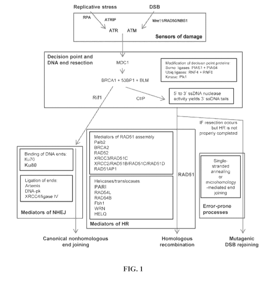
[0036] FIG. 1 Pathways and genes involved in repair of double-strand
DNA
breaks (DSBs) and the tolerance of replication stress. Shown is a simplified
overview of
the mechanistic steps and genes involved in DNA repair, with an emphasis on
those that
facilitate homologous recombination and non-homologous end joining. All of the
displayed
genes were considered candidates for the Recombination Proficiency Score (RPS)
system,
except those within the blue box. The four genes whose expression levels were
ultimately
chosen to comprise the RPS are displayed in red.
- 9 -
CA 02958290 2017-02-16
WO 2015/042570
PCT/US2014/056942
[0037] FIG. 2 Cell lines with low RPS overexpress a wide array of HR-
related
genes. Mean mRNA levels are shown for the CCLE cell lines with low RPS scores.
These
mRNA levels were mined from the CCLE database, and displayed values represent
log2
transformed mRNA measurements of each gene normalized to the median mRNA among
the
starting 634 carcinoma cell lines. Therefore an expression level of zero
indicates a median
expression level, and any positive value indicates overexpression. For
example, a value of
+0.25 indicates a 19% increase in expression above the median. Error bars
denote standard
error.
[0038] FIGS. 3A-3B RPS correlates with sensitivity to different
classes of
treatment and HR deficiency in cell lines. A) CCLE carcinoma cell lines were
binned into
quartiles, based on RPS. Sensitivity data were mined from the CCLE database
and plotted
for different oncologic therapies, and differences between the highest and
lowest quartiles
were determined by Student's T test. B) HR repair efficiency correlates with
RPS. Six
representative cell lines were co-transfected with an HR reporter-containing
plasmid (pDR-
GFP) plus an I-Sce I expressing plasmid (pCI3ASce) or an empty vector control
plasmid
(pCAG), and were subjected to FACS analysis 48 hours later. Reported HR
efficiency
represents the percent GFP+ cells with pDR-GFP + pCI3ASce, normalized to
background
(pDR-GFP + pCAG).
[0039] FIGS. 4A-4B CCLE carcinoma cell lines with low RPS have
elevated
genomic instability. SNP array-based DNA copy number variations (CNVs) were
mined
from the CCLE database. DNA deletions (left) and amplifications (right) were
binned by
size, wherein bins represent 10-fold increments in mutation size. High and low
RPS groups
were defined as the top and bottom quartiles, respectively. Size-based
distributions of CNVs
are shown for A) TP53 WT cells and B) TP53 mutant cells. Error bars denote
standard error.
Asterisks denote significant differences, based on Student's T test.
[0040] FIGS. 5A-5D Low RPS is associated with genomic instability in
human
tumors. SNP array-based DNA copy number variations (CNVs) were mined from the
Cancer Genome Atlas. DNA deletions (left) and amplifications (right) were
binned by size,
wherein bins represent 10-fold increments in size. High and low RPS groups
were defined as
the top and bottom quartiles, respectively. Size-based distributions of CNVs
are shown for
A) TP53 WT NSCLC tumors, B) TP53 mutant NSCLC tumors, C) TP53 WT breast
tumors,
- 10 -
CA 02958290 2017-02-16
WO 2015/042570
PCT/US2014/056942
D) TP53 mutant breast tumors. Error bars denote standard error. Asterisks
denote significant
differences, based on Student's T test.
[0041]
FIGS. 6A-6B RPS is prognostic and correlates with treatment sensitivity
in clinical tumors. A) Kaplan Meier survival curves are shown for NSCLC
patients treated
on the JBR.10 trial with either surgery alone (S) or surgery followed by
chemotherapy (S+C).
Low and high RPS groups were defined as the bottom 25th percentile and the
remaining upper
75th percentile, respectively. B) Four clinical datasets of non-small cell
lung cancer were
analyzed for prognostic impact of RPS on survival, using multivariate analyses
that
controlled for overall stage. Points in the Forest plot represent treatment-
specific hazard
ratios of RPS (as a continuous variable). Boxes denote hazard ratio and
diamonds denote
modeled hazard ratio values that summarize the combined impact of all four
datasets. Error
bars denote 95% confidence intervals.
Black= surgery alone, green= surgery +
chemotherapy.
[0042]
FIG. 7 Most of the drug sensitivity values mined from the CCLE database
were generated in the same cell lines. A Venn diagram displays the number of
CCLE
carcinoma cell lines for which treatment sensitivity data (topotecan,
irinotecan, or paclitaxel)
was available.
[0043]
FIG. 8 A representative panel of cancer cell lines exhibits expected levels
of drug resistance. Cells were plated into 96-well tissue culture plates. The
indicated for
drugs were added for three days thereafter, and average survival from six
replicates was
measured using CellGlo reagent (Promega). Error bars represent the standard
error.
[0044]
FIG. 9 Measurements of mRNA by real time qRT-PCR for six
representative cell lines generated RPS values that were comparable to RPS
values
calculated from array-based mRNA levels reported in the CCLE database. Cells
were
grown to 70% confluence and mRNA was isolated with TRIzol (Life Technologies)
using the
manufacturer's instructions. The resulting mRNA was quantified using the Qubit
RNA BR
assay (LifeTechnologies). An equal amount of RNA (1.5 iug) from each cell line
was treated
with DNAse-I (ThermoScientific), and cDNA was synthesized using Applied
Biosystems
High Capacity cDNA Reverse Transcription Kit (LifeTechnologies). PCR was
performed
with an Applied Biosystems 7900HT Sequence Detection System, using Applied
Biosystems
2X Taqman Universal Master Mix II. These qRT-PCR derived mRNA levels were
normalized to the levels in PC3 cells, log2 transformed, and summed to
generate an RPS
- 11 -
CA 02958290 2017-02-16
WO 2015/042570
PCT/US2014/056942
value for each cell line. The following Taqman Assay components were used in
the PCR
reaction: Ku80 (also known as XRCC5): Hs00897854 ml; RIF1: Hs00871714 ml; PARI
(also known as PARPBP): Hs01550690 ml; RAD51: Hs00153418 ml.
DETAILED DESCRIPTION OF THE INVENTION
[0045] Human tumors exhibit a wide range of malignant features and
responsiveness
to treatments that damage DNA. The inventors hypothesized that a component of
this
variability can be explained by differential efficiencies of DNA repair
pathways. To study
this further the inventors developed an analytic tool to indirectly quantify
the efficiency of
HR in individual cancers. This scoring system may be based on the expression
of four DNA
repair genes in a tumor cell or tissue: RIF1, PARI, RAD51, and Ku80. In
certain examples, it
was shown here that the Recombination Proficiency Score (RPS) correlates with
sensitivity to
specific classes of chemotherapy, associates with degree of genomic
instability within tumor
cells, and provides valuable information that is not available using existing
diagnostic
methods.
I. Response Score
[0046] In certain embodiments, a response score may be determined
based on the
expression level of one or more genes involved in DNA repair pathways, such as
replicative
stress and the DSB repair pathways, or more particularly, the genes involved
in cellular
preference toward HR versus NHEJ. The Score may be expressed as a
Recombination
Proficiency Score (RPS).
[0047] In particular embodiments, the RPS score may be based on the
expression of
four DNA repair genes: RIF1, PARI, RAD51, and Ku80. It is shown herein that
the RPS
correctly predicts sensitivity to various classes of DNA-damaging treatment,
correlates with
degree of genomic instability within tumor cells, and provides valuable
prognostic
information that is not available using existing diagnostic methods. The RPS
is a novel
scoring system that quantifies the expression of four genes to predict DSB
repair pathway
preference. In particular, mRNA levels for relevant DNA repair genes in
carcinoma cell lines
were compared to topotecan sensitivity. This identified a gene expression
scoring system
termed the Recombinant Proficiency Score (RPS) that is in inverse relationship
with the
- 12 -
CA 02958290 2017-02-16
WO 2015/042570
PCT/US2014/056942
expression level of repair genes. Low RPS can identify tumors that harbor HR
suppression
and hypersensitivity to specific chemotherapeutic classes.
[0048] When faced with a DSB, the cell's decision of whether to
utilize HR vs. NHEJ
is influenced by the cell cycle stage. NHEJ is the dominant pathway for
repairing DSBs
during GO/G1 stages of the cell cycle, while HR occurs generally during S and
G2. This
regulation of repair is governed primarily by BRCA1 and 53BP1 proteins, which
compete for
occupancy at the DSB site (Chapman, et al., 2013). Stabilization of 53BP1 in
cooperation
with RIF1 leads to the exclusion of BRCA1 protein from the repair complex, and
the DSB
subsequently progresses to repair by NHEJ (Zimmermann, et al., 2013; Chapman,
et al.,
2013). If 53BP1 is excluded from the repair complex, then the DSB progresses
to repair by
HR. In this case, the DSB ends are processed into HR substrates, which
involves 5' to 3'
nuclease activity that generates 3' single-stranded DNA tails. This end
processing is
promoted by several proteins including CtIP, BRCA1, and the MRN
(Mrell/RAD50/NBS1)
complex. The nuclease activity is also specifically triggered by interactions
between Mre 11
and cyclin dependent kinase 2, thereby promoting the phosphorylation of CtIP
preferentially
in S/G2 cells (Buis, et al., 2012).
[0049] Given the wide biological diversity known to exist between
different classes
of human malignancies, the analysis was limited to cell lines derived only
from carcinomas.
Cellular resistance to the topoisomerase-I inhibiting drug topotecan was
selected as a
surrogate marker for HR proficiency. Topotecan is a derivative of
camptothecin, and this
class of drugs was selected because it disrupts replication forks and exerts
toxicity
preferentially in cells that harbor HR defects (Nitiss, et al., 1988;
Arnaudeau, et al., 2001).
Topotecan IC50 data were available for 279 of the 634 carcinoma cell lines.
[0050] To focus the inventors analysis on the primary cellular
features that mediate
specific phenotypes, the analysis was restricted to genes with direct
relevance to replication
stress and the DSB repair pathways. The analysis was further limited to 31
central proteins
that participate in cellular preference toward HR vs. NHEJ, following the
ataxia
telangiectasia mutated (ATM) and/or ataxia telangiectasia and Rad3-related
protein (ATR)
activation steps of DNA damage response. Secondary regulators of damage
response (like
TP53, PTEN, and cell cycle checkpoint genes) were not considered as gene
candidates for the
scoring system, since they exert cellular influences that extend beyond the
scope of
replication stress and DSB repair. Pearson's correlation analyses demonstrated
that 12 of the
- 13 -
CA 02958290 2017-02-16
WO 2015/042570
PCT/US2014/056942
final list of 31 candidate genes had expression levels that significantly
correlated (defined as
p<0.05) with cellular sensitivity to topotecan. In all 12 cases, increasing
gene expression
levels directly correlated with increasing topotecan sensitivity.
[0051] In certain embodiments, the response score, such as a RPS
(Recombinant
Proficiency Score), can be calculated using techniques for measuring gene
expression,
including, but not limited to, NanoString and RT-PCR. In certain embodiments,
the RPS
score may be calculated from microarray. In further embodiments, when using
microarray
data (from which all of our current data have come), there are methods that
may be used for
normalizing the raw gene expression values. For example, the data may be
normalized as
such: Raw Affymetrix CEL files were converted to a single value for each probe
set using
Robust Multi-array Average (RMA) and normalized using quantile normalization.
Either the
original Affymetrix U133+2 CDF file or a redefined custom CDF file (ENTREZG -
v15) was
used for the summarization. Any methods known in the art may be used for
microarray data
normalization and pre-processing and calculation of RPS values may be feasible
across
various methods of normalization.
[0052] There may also be methods for calculating RPS with modalities
other than
microarray. Although most studies using RT-PCR generally normalize to some
sort of
housekeeper gene (like actin or a ribosomal RNA), the inventors actually found
the raw data
of gene expression of the RPS genes worked the best (i.e. agreed best with the
microarray
data, again Sean may want to elaborate). In a particular embodiment, a
NanoString-based
method may be used to measure the degraded forms of mRNA that are generally
found in
typical patient tumor biopsy material (formalin fixed paraffin-embedded
tissue). For this,
the NanoString company may provide probes and housekeeping genes to serve as
normalization controls.
[0053] In certain embodiments, the information provided by RPS may be a
continuous variable or not a continuous variable. This depends somewhat on the
source of
the material (i.e. tumor type). When plotting RPS-based data, the highest and
lowest
quartiles may be focused, because that makes the result more visually obvious
and more
statistically significant. However the effect may be continuous over the full
range of RPS
values. In fact, the Forest plot uses RPS as a continuous variable to
calculate hazard ratios.
- 14 -
CA 02958290 2017-02-16
WO 2015/042570
PCT/US2014/056942
II. Use of the Response Score
[0054] In some aspects, embodiments comprise treating a subject with
a specific
therapeutic agent or evaluating efficacy of treatment. Examples of therapeutic
agents (anti-
cancer agents) include, but are limited to, e.g., chemotherapeutic agents,
growth inhibitory
agents, cytotoxic agents, agents used in radiation therapy, anti-angiogenesis
agents, apoptotic
agents, anti-tubulin agents, and other-agents to treat cancer, such as anti-
HER-2 antibodies,
anti-CD20 antibodies, an epidermal growth factor receptor (EGFR) antagonist
(e.g., a
tyrosine kinase inhibitor), HER1/EGFR inhibitor (e.g., erlotinib (TarcevaTm),
platelet derived
growth factor inhibitors (e.g., GleevecTM (Imatinib Mesylate)), a COX-2
inhibitor (e.g.,
celecoxib), interferons, cytokines, antagonists (e.g., neutralizing
antibodies).
[0055] In certain embodiments, the patient's sensitivity to a
chemotherapeutic agent
positively correlates with the expression level of the genes described herein.
The
chemotherapeutic agent may be a platinum-based compound, such as cisplatin,
carboplatin,
oxaliplatin, satraplatin, picoplatin, Nedaplatin, Triplatin, Lipoplatin, or a
liposomal version of
cisplatin.
[0056] In certain embodiments, the chemotherapeutic agent may be a
DNA cross-
linker. Alkylating agents such as 1, 3-bis(2-chloroethyl)-1-nitrosourea (BCNU,
carmustine))
and nitrogen mustard which are used in chemotherapy can cross link with DNA at
N7
position of guanine on the opposite strands forming interstrand crosslink.
Cisplatin (cis-
diamminedichloroplatinum(II)) and its derivatives forms DNA cross links as
monoadduct,
interstrand crosslink, intrastrand crosslink or DNA protein crosslink. Mostly
it acts on the
adjacent N-7 guanine forming 1, 2 intrastrand crosslink.
[0057] In further embodiments, the chemotherapeutic agent may be a
topoisomerase
inhibitor. Topoisomerase inhibitors are drugs that affect the activity of two
enzymes:
topoisomerase I and topoisomerase II. When the DNA double-strand helix is
unwound,
during DNA replication or transcription, for example, the adjacent unopened
DNA winds
tighter (supercoils), like opening the middle of a twisted rope. The stress
caused by this effect
is in part aided by the topoisomerase enzymes. They produce single- or double-
strand breaks
into DNA, reducing the tension in the DNA strand. This allows the normal
unwinding of
DNA to occur during replication or transcription. Inhibition of topoisomerase
I or II interferes
with both of these processes. Two topoisomerase I inhibitors, irinotecan and
topotecan, are
semi-synthetically derived from camptothecin, which is obtained from the
Chinese
- 15 -
CA 02958290 2017-02-16
WO 2015/042570
PCT/US2014/056942
ornamental tree Camptotheca acuminate. Drugs that target topoisomerase II can
be divided
into two groups. The topoisomerase II poisons cause increased levels enzymes
bound to
DNA. This prevents DNA replication and transcription, causes DNA strand
breaks, and leads
to programmed cell death (apoptosis). These agents include etoposide,
doxorubicin,
mitoxantrone and teniposide. The second group, catalytic inhibitors, are drugs
that block the
activity of topoisomerase II, and therefore prevent DNA synthesis and
translation because the
DNA cannot unwind properly. This group includes novobiocin, merbarone, and
aclarubicin,
which also have other significant mechanisms of action
[0058] In still further embodiments, the chemotherapeutic agent may
be a PARP
inhibitor. As used herein, "PARP inhibitor" (i.e., an inhibitor of poly ADP
ribose
polymerase) shall mean an agent that inhibits PARP more than it inhibits any
other
polymerase. In one embodiment, the PARP inhibitor inhibits PARP at least two-
fold more
than it inhibits any other polymerase. In another embodiment, the PARP
inhibitor inhibits
PARP at least 10-fold more than it inhibits any other polymerase. In a third
embodiment, the
PARP inhibitor inhibits PARP more than it inhibits any other enzyme. In one
particular
embodiment, the PARP inhibitor is olaparib, rucaparib, veliparib, CEP 9722, MK
4827,
BMN-673, 3-aminobenzamide, a tetracycline compound, 4-hydroxyquinazoline and a
derivative thereof, and a carboxamino-benzimidazole and a derivative thereof,
[0059] In some embodiments, the chemotherapeutic agent is any of (and
in some
embodiments selected from the group consisting of) alkylating agents such as
thiotepa and
CYTOXAN cyclosphosphamide; alkyl sulfonates such as busulfan, improsulfan and
piposulfan; aziridines such as benzodopa, carboquone, meturedopa, and uredopa;
ethylenimines and methylamelamines including altretamine, triethylenemelamine,
trietylenephosphoramide, triethiylenethiophosphoramide and
trimethylolomelamine;
acetogenins (especially bullatacin and bullatacinone); delta-9-
tetrahydrocannabinol
(dronabinol, MARINOL8); beta-lapachone; lapachol; colchicines; betulinic acid;
a
camptothecin (including the synthetic analogue topotecan (HYCAMTIN ), CPT-11
(irinotecan, CAMPTOSAR ), acetylcamptothecin, scopolectin, and 9-
aminocamptothecin);
bryostatin; callystatin; CC-1065 (including its adozelesin, carzelesin and
bizelesin synthetic
analogues); podophyllotoxin; podophyllinic acid; teniposide; cryptophycins
(particularly
cryptophycin 1 and cryptophycin 8); dolastatin; duocarmycin (including the
synthetic
analogues, KW-2189 and CB1-TM1); eleutherobin; pancratistatin; a sarcodictyin;
- 16 -
CA 02958290 2017-02-16
WO 2015/042570
PCT/US2014/056942
spongistatin; nitrogen mustards such as chlorambucil, chlornaphazine,
cholophosphamide,
estramustine, ifosfamide, mechlorethamine, mechlorethamine oxide
hydrochloride,
melphalan, novembichin, phenesterine, prednimustine, trofosfamide, uracil
mustard;
nitrosureas such as carmustine, chlorozotocin, fotemustine, lomustine,
nimustine, and
ranimnustine; antibiotics such as the enediyne antibiotics (e.g.,
calicheamicin, especially
calicheamicin gammall and calicheamicin omegaI 1 (see, e.g., Agnew, Chem.
Intl. Ed. Engl.,
33: 183-186 (1994)); dynemicin, including dynemicin A; an esperamicin; as well
as
neocarzinostatin chromophore and related chromoprotein enediyne antiobiotic
chromophores), aclacinomysins, actinomycin, authramycin, azaserine,
bleomycins,
cactinomycin, carabicin, caminomycin, carzinophilin, chromomycinis,
dactinomycin,
daunorubicin, detorubicin, 6-diazo-5-oxo-L-norleucine, ADRIAMYCN doxorubicin
(including morpholino-doxorubicin, cyanomorpholino-doxorubicin, 2-pyrrolino-
doxorubicin
and deoxydoxorubicin), epirubicin, esorubicin, idarubicin, marcellomycin,
mitomycins such
as mitomycin C, mycophenolic acid, nogalamycin, olivomycins, peplomycin,
potfiromycin,
puromycin, quelamycin, rodorubicin, streptonigrin, streptozocin, tubercidin,
ubenimex,
zinostatin, zorubicin; anti-metabolites such as methotrexate and 5-
fluorouracil (5-FU); folic
acid analogues such as denopterin, methotrexate, pteropterin, trimetrexate;
purine analogs
such as fludarabine, 6-mercaptopurine, thiamiprine, thioguanine; pyrimidine
analogs such as
ancitabine, azacitidine, 6-azauridine, carmofur, cytarabine, dideoxyuridine,
doxifluridine,
enocitabine, floxuridine; androgens such as calusterone, dromostanolone
propionate,
epitiostanol, mepitiostane, testolactone; anti-adrenals such as
aminoglutethimide, mitotane,
trilostane; folic acid replenisher such as frolinic acid; aceglatone;
aldophosphamide
glycoside; aminolevulinic acid; eniluracil; amsacrine; bestrabucil;
bisantrene; edatraxate;
defofamine; demecolcine; diaziquone; elformithine; elliptinium acetate; an
epothilone;
etoglucid; gallium nitrate; hydroxyurea; lentinan; lonidainine; maytansinoids
such as
maytansine and ansamitocins; mitoguazone; mitoxantrone; mopidanmol;
nitraerine;
pentostatin; phenamet; pirarubicin; losoxantrone; 2-ethylhydrazide;
procarbazine; PSK
polysaccharide complex (JHS Natural Products, Eugene, Oreg.); razoxane;
rhizoxin;
sizofiran; spirogermanium; tenuazonic acid; triaziquone; 2,2',2"-
trichlorotriethylamine;
trichothecenes (especially T-2 toxin, verracurin A, roridin A and anguidine);
urethan;
vindesine (ELDISNE , FILDESIN ); dacarbazine; mannomustine; mitobronitol;
mitolactol;
pipobroman; gacytosine; arabinoside ("Ara-C"); thiotepa; taxoids, e.g., TAXOL
paclitaxel
(Bristol-Myers Squibb Oncology, Princeton, N.J.), ABRAXANETM Cremophor-free,
- 17 -
CA 02958290 2017-02-16
WO 2015/042570
PCT/US2014/056942
albumin-engineered nanoparticle formulation of paclitaxel (American
Pharmaceutical
Partners, Schaumberg, Ill.), and TAXOTERE doxetaxel (Rhone-Poulenc Rorer,
Antony,
France); chloranbucil; gemcitabine (GEMZAR ); 6-thioguanine; mercaptopurine;
methotrexate; platinum analogs such as cisplatin and carboplatin; vinblastine
(VELBAN );
platinum; etoposide (VP-16); ifosfamide; mitoxantrone; vincristine (ONCOVIN );
oxaliplatin; leucovovin; vinorelbine (NAVELBINE ); novantrone; edatrexate;
daunomycin;
aminopterin; ibandronate; topoisomerase inhibitor RFS 2000;
difluoromethylornithine
(DMF0); retinoids such as retinoic acid; capecitabine (XELODA );
pharmaceutically
acceptable salts, acids or derivatives of any of the above; as well as
combinations of two or
more of the above such as CHOP, an abbreviation for a combined therapy of
cyclophosphamide, doxorubicin, vincristine, and prednisolone, and FOLFOX, an
abbreviation for a treatment regimen with oxaliplatin (ELOXATINTm) combined
with 5-FU
and leucovovin. Additional chemotherapeutic agents include the cytotoxic
agents useful as
antibody drug conjugates, such as maytansinoids (DM1, for example) and the
auristatins
MMAE and MMAF, for example.
[0060] The chemotherapeutic agents may be administered serially
(within minutes,
hours, or days of each other) or in parallel; they also may be administered to
the patient in a
pre-mixed single composition. It is contemplated that a therapy as disclosed
herein may be
used in vitro or in vivo. These processes may involve administering several
agents at the
same time or within a period of time wherein separate administration of the
substances
produces a desired therapeutic benefit. This may be achieved by contacting the
cell, tissue, or
organism with a composition, such as a pharmaceutically acceptable
composition, that
includes two or more agents, or by contacting the cell with two or more
distinct
compositions, wherein one composition includes one agent and the other
includes another.
[0061] "Prognosis" refers to as a prediction of how a patient will
progress, and
whether there is a chance of recovery. "Cancer prognosis" generally refers to
a forecast or
prediction of the probable course or outcome of the cancer, with or without a
treatment. As
used herein, cancer prognosis includes the forecast or prediction of any one
or more of the
following: duration of survival of a patient susceptible to or diagnosed with
a cancer, duration
of recurrence-free survival, duration of progression free survival of a
patient susceptible to or
diagnosed with a cancer, response rate in a group of patients susceptible to
or diagnosed with
a cancer, duration of response in a patient or a group of patients susceptible
to or diagnosed
- 18 -
CA 02958290 2017-02-16
WO 2015/042570
PCT/US2014/056942
with a cancer, and/or likelihood of metastasis in a patient susceptible to or
diagnosed with a
cancer. Prognosis also includes prediction of favorable responses to cancer
treatments, such
as a conventional cancer therapy. A response may be either a therapeutic
response (sensitivity
or recurrence-free survival during or after a treatment) or a lack of
therapeutic response
(residual disease, which may indicate resistance or recurrence during or after
a treatment).
[0062] By "subject" or "patient" is meant any single subject for
which therapy is
desired, including humans, cattle, dogs, guinea pigs, rabbits, chickens, and
so on. Also
intended to be included as a subject are any subjects involved in clinical
research trials not
showing any clinical sign of disease, or subjects involved in epidemiological
studies, or
subjects used as controls.
[0063] As used herein, "increased expression" or "overexpression" or
"decreased
expression" refers to an expression level of a gene in the subject's sample as
compared to a
reference level representing the same gene or a different gene. In certain
aspects, the
reference level may be a reference level of expression from a non-cancerous
tissue from the
same subject. Alternatively, the reference level may be a reference level of
expression from a
different subject or group of subjects. For example, the reference level of
expression may be
an expression level obtained from a sample (e.g., a tissue, fluid or cell
sample) of a subject or
group of subjects without cancer, or an expression level obtained from a non-
cancerous tissue
of a subject or group of subjects with cancer. The reference level may be a
single value or
may be a range of values. The reference level of expression can be determined
using any
method known to those of ordinary skill in the art. In some embodiments, the
reference level
is an average level of expression determined from a cohort of subjects with
cancer or without
cancer or include both. The reference level may also be depicted graphically
as an area on a
graph. In certain embodiments, a reference level is a normalized level, a
median, an average
value, while in other embodiments, it may be a level that is not stable with
respect to the
tissue or biological sample being tested.
[0064] "About" and "approximately" shall generally mean an acceptable
degree of
error for the quantity measured given the nature or precision of the
measurements. Typically,
exemplary degrees of error are within 20 percent (%), preferably within 10%,
and more
preferably within 5% of a given value or range of values. Alternatively, and
particularly in
biological systems, the terms "about" and "approximately" may mean values that
are within
an order of magnitude, preferably within 5-fold and more preferably within 2-
fold of a given
- 19 -
CA 02958290 2017-02-16
WO 2015/042570
PCT/US2014/056942
value. Numerical quantities given herein are approximate unless stated
otherwise, meaning
that the term "about" or "approximately" can be inferred when not expressly
stated.
[0065] III. Nucleic Acid AssaysAspects of the methods include
assaying nucleic
acids to determine expression levels. Arrays can be used to detect differences
between two
samples. An array comprises a solid support with nucleic acid probes attached
to the support.
Arrays typically comprise a plurality of different nucleic acid probes that
are coupled to a
surface of a substrate in different, known locations. These arrays, also
described as
"microarrays" or colloquially "chips" have been generally described in the
art, for example,
U.S. Pat. Nos. 5,143,854, 5,445,934, 5,744,305, 5,677,195, 6,040,193,
5,424,186 and Fodor
et al., 1991), each of which is incorporated by reference in its entirety for
all purposes.
Techniques for the synthesis of these arrays using mechanical synthesis
methods are
described in, e.g., U.S. Pat. No. 5,384,261, incorporated herein by reference
in its entirety for
all purposes. Although a planar array surface is used in certain aspects, the
array may be
fabricated on a surface of virtually any shape or even a multiplicity of
surfaces. Arrays may
be nucleic acids on beads, gels, polymeric surfaces, fibers such as fiber
optics, glass or any
other appropriate substrate, see U.S. Pat. Nos. 5,770,358, 5,789,162,
5,708,153, 6,040,193
and 5,800,992, which are hereby incorporated in their entirety for all
purposes.
[0066] In addition to the use of arrays and microarrays, it is
contemplated that a
number of difference assays could be employed to analyze genes, their
expression and
activities, and their effects. Such assays include, but are not limited to,
nucleic acid
amplification, polymerase chain reaction, quantitative PCR, RT-PCR, RNA
sequencing (e.g.,
by next-generation sequencing techniques), in situ hybridization, Northern
hybridization,
hybridization protection assay (HPA) (GenProbe), branched DNA (bDNA) assay
(Chiron),
rolling circle amplification (RCA), single molecule hybridization detection
(US Genomics),
Invader assay (ThirdWave Technologies), and/or Bridge Litigation Assay
(Genaco).
[0067] The term "primer," as used herein, is meant to encompass any
nucleic acid that
is capable of priming the synthesis of a nascent nucleic acid in a template-
dependent process.
In particular embodiments, primers are oligonucleotides from ten to twenty
and/or thirty base
pairs in length, but longer sequences can be employed. Primers may be provided
in double-
stranded and/or single-stranded form, although the single-stranded form is
preferred.
- 20 -
CA 02958290 2017-02-16
WO 2015/042570
PCT/US2014/056942
IV. Kits
[0068] In various aspects, a kit is envisioned containing one or more
oligonucleotides
or other reagents described herein. The kit may contain one or more sealed
containers, such
as a vial, containing any of the reagents described herein and/or reagents for
preparing any of
the reagents described herein. In some embodiments, the kit may also contain a
suitable
container means, which is a container that will not react with components of
the kit, such as
an Eppendorf tube, an assay plate, a syringe, a bottle, or a tube. The
container may be made
from sterilizable materials such as plastic or glass.
[0069] The kit may further include instructions that outline the
procedural steps for
carrying out the diagnostic, treatment, or prevention of disease, and will
follow substantially
the same procedures as described herein or are known to those of ordinary
skill. The
instruction information may be in a computer readable media containing machine-
readable
instructions that, when executed using a computer, cause the display of a real
or virtual
procedure of using the kit described herein.
[0070] The kit can further comprise reagents for labeling mRNA of genes to
be
measured in the sample. The kit may also include labeling reagents, including
at least one of
amine-modified nucleotide, poly(A) polymerase, and poly(A) polymerase buffer.
Labeling
reagents can include an amine-reactive dye.
[0071] In further embodiments, a kit is provided comprising
oligonucleotides that
bind or are capable of hybridizing, respectively, to two, three, four, five or
six genes chosen
from the group of PARI, BLM, RAD51, RIF1, BRCA1, Ku80, RAD51AP1, RAD54B, Plkl,
BRCA2, RAD51C, PALB2, MYC, and STAT3. In some embodiments, the kit includes
oligonucleotides that bind or are capable of hybridizing, respectively, to
two, three, four, five,
six, seven or eight genes chosen from the group consisting of PARI, RIF1,
Ku80, RAD51,
BLM, BRCA1, RAD51AP1 and RAD54B. In particular embodiments, the kit includes
oligonucleotides capable of hybridizing, respectively, to two, three, four,
five, six, seven or
eight genes chosen from the group consisting of RIF1, PARI, RAD51, Ku80, MYC
and
STAT3. In particular embodiments, the kit includes one or more
oligonucleotides capable of
hybridizing to a RIF1 gene sequence, one or more oligonucleotides capable of
hybridizing to
a PARI gene sequence, one or more oligonucleotides capable of hybridizing to a
RAD51
gene sequence, and one or more oligonucleotides capable of hybridizing to a
Ku80 gene
sequence.
-21 -
CA 02958290 2017-02-16
WO 2015/042570
PCT/US2014/056942
[0072] In certain embodiments, the oligonucleotides may be, be at
least, or be at most
3, 4, 5, 6, 7, 8,9, 10, 11, 12, 13, 14, 15, 16, 17, 18, 19, 20, 21, 22, 23,
24, 25, 26, 27, 28, 29,
30, 31, 32, 33, 34, 35, 36, 37, 38, 39, 40, 41, 42, 43, 44, 45, 46, 47, 48,
49, 50, 51, 52, 53, 54,
55, 56, 57, 58, 59, 60, 61, 62, 63, 64, 65, 66, 67, 68, 69, 70, 71, 72, 73,
74, 75, 76, 77, 78, 79,
80, 81, 82, 83, 84, 85, 86, 87, 88, 89, 90, 91, 92, 93, 94, 95, 96, 97, 98,
99, 100, 101, 102,
103, 104, 105, 106, 107, 108, 109, 110, 120, 130, 140, 150, 160, 170, 180,
190, 200, 210,
220, 230, 240, 250, 260, 270, 280, 290, or 300 nucleotides, or any range
derivable therein, in
length. Each oligonucleotide may be a probe or primer that is labeled or un-
labeled, and can
hybridize under stringent hybridization conditions to an mRNA or cDNA encoded
by one of
the genes, or may be part of or full-length cDNA encoded by one of the genes,
or may be part
of or full-length cDNA encoded by one of the genes.
[0073] The kits may be used in detecting gene expression in cells or
tissue from a
patient, particularly tumor tissues from a cancer patient. For example, in
real-time TaqMan
PCR, oligonucleotides may be used as PCR primers, and/or TaqMan probe. In
sequencing,
the oligonucleotides may PCR primers and/or sequencing primers. In next-
generation
sequencing, the oligonucleotides may be used as capturing probes to capature
targeted genes
or mRNAs or cDNAs. In microarray-based gene expression profiling, the
oligonucleotides
may be probes attached to a solid support forming a hybridization chip.
[0074] Thus, the kits may additional include one or more reagents
useful for PCR
reactions, sequencing reactions, and/or hybridization reactions, such as Taq
polymerase,
reaction buffers, dNTPs, etc.
EXAMPLES
[0075] The following examples are included to demonstrate preferred
embodiments
of the invention. It should be appreciated by those of skill in the art that
the techniques
disclosed in the examples which follow represent techniques discovered by the
inventor to
function well in the practice of the invention, and thus can be considered to
constitute
preferred modes for its practice. However, those of skill in the art should,
in light of the
present disclosure, appreciate that many changes can be made in the specific
embodiments
which are disclosed and still obtain a like or similar result without
departing from the spirit
and scope of the invention.
- 22 -
CA 02958290 2017-02-16
WO 2015/042570
PCT/US2014/056942
[0076] Unless defined otherwise, all technical and scientific terms
used herein have
the same meanings as commonly understood by one of skill in the art to which
the disclosed
invention belongs. Publications cited herein and the materials for which they
are cited are
specifically incorporated by reference.
[0077] Those skilled in the art will recognize, or be able to ascertain
using no more
than routine experimentation, many equivalents to the specific embodiments of
the invention
described herein. Such equivalents are intended to be encompassed by the
claims.
EXAMPLE 1. The RPS system was developed using data from carcinoma cell lines.
[0078] The inventors sought to create a method that predicts the
efficiency of HR
repair within any given cancer. To accomplish this, the inventors developed a
scoring system
that correlates gene expression patterns with HR proficiency in human cancer
cell lines.
Gene expression levels and corresponding drug sensitivity data were collected
from the
Broad-Novartis Cancer Cell Line Encyclopedia (CCLE) (Barretina, et al., 2012).
Given the
wide biological diversity known to exist between different classes of human
malignancies,
the inventors limited this analysis to cell lines derived only from
carcinomas. Cellular
resistance to the topoisomerase-I inhibiting drug topotecan was selected as a
surrogate marker
for HR proficiency. Topotecan is a derivative of camptothecin, and this class
of drugs was
selected because it disrupts replication forks and exerts toxicity
preferentially in cells that
harbor HR defects (Nitiss, et al., 1988; Arnaudeau, et al., 2001). Topotecan
sensitivity data
were available for 279 of the 634 carcinoma cell lines.
[0079] To focus the inventors analysis on the primary cellular
features that mediate
specific phenotypes, the inventors restricted the analysis to genes with
direct relevance to
replication stress and the DSB repair pathways (Figure 1). The inventors
further limited the
analysis to 33 central proteins that participate in cellular preference toward
HR vs. NHEJ,
following the ataxia telangiectasia mutated (ATM) and/or ataxia telangiectasia
and Rad3-
related protein (ATR) activation steps of DNA damage response. Levels of mRNA
were
available for all of these genes except Ku70. Secondary regulators of damage
response (like
TP53, PTEN, and cell cycle checkpoint genes) were not considered as gene
candidates for the
scoring system, since they exert cellular influences that extend beyond the
scope of
replication stress and DSB repair. Pearson's correlation analyses demonstrated
that 12 of the
final list of 32 candidate genes had expression levels that significantly
correlated (defined as
- 23 -
CA 02958290 2017-02-16
WO 2015/042570
PCT/US2014/056942
p<0.05) with cellular sensitivity to topotecan (Table 1). In all 12 cases,
increasing gene
expression levels directly correlated with increasing topotecan sensitivity.
Table 1 - DNA repair genes that significantly associate with topotecan
sensitivity.
A) Significant Genes
Genes known to HR-related
antagonize HR genes Correlation Coeff. p-value
PARI -0.23 <0.005
RAD51 -0.18 <0.005
BLM -0.18 <0.005
RIF1 -0.17 0.005
Ku80 -0.16 0.006
BRCA1 -0.16 0.006
RAD51AP1 -0.16 0.007
RAD54B -0.15 0.02
Plk1 -0.14 0.02
BRCA2 -0.14 0.02
RAD51C -0.13 0.03
PALB2 -0.12 0.04
B) Non-significant Genes
Genes Correlation Coeff. p-value
CtIP -0.11 0.06
RAD51B -0.11 0.06
DNA-PKcs -0.10 0.09
PIAS4 -0.09 0.15
XRCC3 -0.08 0.16
XRCC2 -0.08 0.18
RAD52 -0.07 0.25
XRCC4 -0.07 0.27
Artemis -0.06 0.29
RAD51D -0.06 0.30
WRN -0.06 0.30
RAD54L -0.06 0.33
HELQ -0.04 0.47
MDC1 -0.02 0.70
LIG4 -0.02 0.70
PIAS1 -0.02 0.72
Fbh1 -0.01 0.90
RNF8 0.01 0.89
RNF4 0.01 0.83
TP53BP1 0.05 0.45
EXAMPLE 2 - HR-related genes are highly expressed in cancer cells that harbor
low
HR efficiency.
[0080] RIF1, Ku80, and PARI were among the genes whose expression
most strongly
associated with topotecan sensitivity. RIF1 and Ku80 are known to promote NHEJ
and
antagonize HR (Zimmermann, et al., 2013; Chapman, et al., 2013; Bennardo, et
al., 2008).
- 24 -
CA 02958290 2017-02-16
WO 2015/042570
PCT/US2014/056942
PARI is a helicase capable of disrupting RAD51 nucleofilaments, and it has
been reported to
antagonize HR repair (Moldovan, et al., 2012).
[0081] Topotecan sensitivity also correlated with the overexpression
of a family of
HR-related genes, including RAD51, BLM, BRCA1, RAD51-AP1, RAD54B, PLK1,
BRCA2, RAD51C, and PALB2. This observation appears counterintuitive on the
surface,
since RAD51 and many of these RAD51-associated proteins are generally
considered to
promote HR. However, RAD51 overexpression has been previously shown to occur
in the
setting of HR defects caused by BRCA mutations (Martin, et al., 2007; Honrado,
et al.,
2005). To investigate a possible connection between BRCA mutation phenotypes
and the
inventors observed expression patterns, the inventors analyzed gene expression
levels in
CCLE cell lines that harbor BRCA1 (HCC1937 and MDA-MB436) or BRCA2 (CAPAN1)
mutations. Consistent with published observations in BRCA1-mutant human tumors
(Martin,
et al., 2007), these three cell lines significantly overexpressed RAD51 and
RAD51AP1. In
addition, the inventors found that BRCA-defective cells significantly
overexpress additional
genes known to promote various mechanisms required for HR, including CtIP
which
promotes the 5' to 3' ssDNA end resection (Sartori, et al., 2007), Plkl which
promotes the
phosphorylation of 53BP1 and RAD51 (Yata, et al., 2012; van Vugt, et al.,
2010), and several
genes (XRCC2, XRCC3, PALB2) that promote RAD51 filament assembly (Thompson, et
al.,
2012). These data suggest that BRCA-defective cells respond to their HR
defects by
increasing the expression of a fairly broad array of HR-related genes. The
overexpression of
HR genes as a compensatory mechanism has been proposed previously,
particularly since
RAD51 overexpression is known to partially suppress the HR defects that occur
when key
HR genes are mutated (Martin, et al., 2007; Takata, et al., 2001).
[0082] These findings were used to refine the list of genes to be
used in the
Recombination Proficiency Score (RPS). The inventors hypothesized that when HR-
deficiency occurs in wild type BRCA backgrounds, cells respond via
compensatory
overexpression of HR-related genes that mirrors the phenotypes observed in
BRCA mutant
cells. As such, the inventors reasoned that many of the HR-related genes were
reporting
redundant predictive information in response to low HR proficiency. Gene
expression levels
were combined to generate a single model that predicts topotecan sensitivity,
starting with
genes that have known HR-antagonizing activities (RIF1, Ku80, and PARI) in
order of their
independent predictive power (Table 2). The family of HR-related genes was
then
- 25 -
CA 02958290 2017-02-16
WO 2015/042570
PCT/US2014/056942
subsequently added incrementally into this model. The addition of RAD51
improved the
model's correlation with topotecan sensitivity (relative to the initial 3
genes), however the
inclusion of additional HR-related genes did not further improve the
correlation. Therefore,
the final four genes selected to derive the RPS were Rifl , PARI, Ku80, and
RAD51.
Table 2 ¨ Correlation coefficients Significant DNA repair genes from Table lA
were
combined to determine the optimal number of genes, wherein sum of their
expression levels
correlated most strongly with topotecan sensitivity.
Combinations of genes Correlation Coeff.
PARI -0.23
PARI + RIF1 -0.25
PARI + RIF1 + Ku80 -0.26
PARI + RIF1 + Ku80 + RAD51 -0.28
PARI + RIF1 + Ku80 + RAD51 + BLM -0.27
PARI + RIF1 + Ku80 + RAD51 + BLM + BRCA1 -0.27
PARI + RIF1 + Ku80 + RAD51 + BLM + BRCA1 + RAD51AP1 -0.26
PARI + RIF1 + Ku80 + RAD51 + BLM + BRCA1 + RAD51AP1 + RAD54B -0.26
[0083] Elevated mRNA levels for any of these genes correlated with greater
sensitivity to topotecan. The RPS was defined as the sum of these four
expression levels
multiplied times -1, using the log2 transformed mRNA values of each gene
normalized to the
median mRNA within the starting 634 carcinoma cell lines. The median RPS score
within
the carcinoma cell lines was approximately zero, the bottom 25th percentile of
RPS scores
were less than -1.08, and the top 25th percentile of RPS scores were greater
than 1.2.
[0084] Interestingly, CCLE cell lines with low RPS scores did indeed
overexpress a
broad array of HR-related genes (Figure 2). These data support the existence
of a
compensatory mechanism that responds to low HR efficiency. Furthermore, these
results
suggest that this compensatory mechanism is not limited to only the most
extreme HR
defects, like those resulting from BRCA mutations. MEN1 protein was considered
as a
possible mediator of this proposed compensatory process, since MEN1 has been
shown to
stimulate the transcription of several HR genes, including BRCA1, RAD51, and
RAD51AP1
(Fang, et al., 2013). However this explanation was deemed unlikely, since MEN1
mRNA
levels did not significantly correlate with RPS in CCLE cell lines.
EXAMPLE 3 - RPS predicts HR proficiency in individual cancer cell lines.
[0085] The predictive value of the RPS was further tested based on
sensitivity to
different types of chemotherapeutic agents. Similar to results with topotecan,
low RPS scores
- 26 -
CA 02958290 2017-02-16
WO 2015/042570
PCT/US2014/056942
correlated to sensitivity to irinotecan, another topoisomerase-I inhibiting
drug (Figure 3A).
This is expected, since topoisomerase-I inhibitors generate replication fork
disruptions, which
require HR for repair (Nitiss, et al., 1988; Arnaudeau, et al., 2001). As a
control, this analysis
was repeated using the non-DNA damaging drug paclitaxel, and RPS did not show
a
correlation with sensitivity to this agent. These results support the
specificity of RPS to
DNA-related damage and repair. It should be noted that complete drug
sensitivity data was
not available for all three chemotherapy agents in all cell lines evaluated
(see FIG 7 for
breakdown). However, comparable results were observed when the analyses were
repeated
on the subset of 137 cell lines that were tested with all three agents.
[0086] The ability of RPS to predict repair pathway preference was further
tested by
measuring HR repair efficiency in representative cell lines with low RPS (RKO,
DU 145,
COLO 205) or with mid/high RPS (PC3, HCC44, NCI-H650). These cell lines
exhibited
expected levels of sensitivity to topotecan and paclitaxel when independently
re-tested in the
inventors laboratory (FIG 8), which were comparable to the sensitivities mined
from the
CCLE database. These six cell lines were tested using a modified version of
the previously
described DR-GFP reporter method (Pierce, et al., 1999). This method utilizes
a reporter
DNA construct that carries two non-functional copies of green fluorescence
protein (GFP),
one of which is interrupted by an I-SceI endonuclease site. Induction of a DSB
at the I-SceI
site can lead to repair by homologous gene conversion that generates a
functional copy of
GFP. As demonstrated in Figure 3B, RPS correlated with HR efficiency on linear
regression
analysis (R2=0.833, two-sided p=0.003). For consistency with the other
results, RPS values
for these cells were calculated using array-based mRNA levels from the CCLE
database. The
inventors verified the identity of the inventors six cell lines by short
tandem repeat profiling
(Genetic Resources Core Facility at Johns Hopkins School of Medicine), and
independent
quantitation by real time qRT-PCR generated mRNA measurements that were
comparable to
the mRNA levels reported in the CCLE database (FIG 9).
EXAMPLE 4 - Cell lines with high RPS have elevated genomic instability.
[0087] HR plays a central role in maintaining genomic stability in
cells. The
inventors hypothesized that cells with low RPS would exhibit more genome
instability than
cells with high RPS. To test this hypothesis, SNP array-based DNA copy number
variations
(CNVs) were analyzed using CCLE carcinoma cell lines (Figure 4). Low RPS
scores were
associated with more frequent DNA amplifications. This finding is consistent
with published
-27 -
CA 02958290 2017-02-16
WO 2015/042570
PCT/US2014/056942
analyses of HR-defective cell lines, showing that mutations in RAD51D or XRCC3
promote
DNA amplifications (Hinz, et al., 2006). These amplifications are proposed to
result from
stress-induced replication fork disruption and subsequent homology-mediated
polymerase
template switching (Arlt, et al., 2012; Malkova, et al., 2012). A study in
RAD51 defective S.
cerevisiae demonstrated that cells with deregulated HR frequently channel DSBs
into repair
by non-allelic break-induced replication, thereby stimulating the formation of
segmental
duplications (Payen, et al., 2008). Additionally, the inventors found that
cells with low RPS
harbored relatively frequent DNA deletions. Deletions are characteristic of
error prone repair
processes like microhomology-mediated end joining and single-strand annealing
(Guirouilh-
Barbat, et al., 2004; Bennardo, et al., 2008). Of note, the distributions of
CNV sizes were not
strongly influenced by RPS. Taken together, these results suggest that low RPS
cells have
reduced HR proficiency and rely more on error-prone processes to rejoin DSBs
and/or to
tolerate replication stress.
[0088] Mutations in TP53 are also known to exert major influences on
cellular
resistance to DNA damaging therapies and genomic instability. Additionally,
TP53 mutation
status has been shown to influence HR efficiency (Linke, et al., 2003; Sirbu,
et al., 2011).
Therefore, the inventors re-examined RPS -associated CNVs in the context of
TP53 mutation
status. The average RPS was not significantly different between the 238 TP53
WT cell lines
and the 386 TP53 mutant cell lines (0.25 vs. 0.41, p=0.41). Also, the
association between
increased CNVs and low RPS was observed in both TP53 WT and mutant cell line
groups.
The magnitude of RPS dependence was less pronounced in TP53 mutant cells, due
to a high
background of deletions in TP53 mutant cells. A high deletion frequency is not
surprising in
TP53 mutant cells, since deletions are known to occur 40-300 times more
following TP53
inactivation (Gebow, et al., 2000). These data suggest, therefore, that TP53
mutation status
and RPS offer independent predictive information regarding genomic
instability.
[0089] A possible relationship between RPS and TP53 status was
further studied by
examining resistance to DNA damage. The ability of RPS to associate with
topotecan
sensitivity on logistic regression was similar in both TP53 WT and mutant cell
line subgroups
(p< 0.003 for both). This association supports the role of RPS as a predictor
of HR
proficiency, which is distinct from TP53-dependent activities like apoptotic
threshold
modulation and cell cycle regulation.
- 28 -
CA 02958290 2017-02-16
WO 2015/042570
PCT/US2014/056942
EXAMPLE 5 - Human tumors with low RPS exhibit unfavorable clinical
characteristics
and elevated genomic instability.
[0090] The RPS system was clinically validated using tumor datasets
from the Cancer
Genome Atlas (CGA). Breast and non-small cell lung cancer (NSCLC) tumor types
were
selected for this analysis, because these datasets contained large sample
sizes, annotations of
clinical features, SNP array-based DNA CNV data, and adequate details on
patient outcomes.
Although some differences existed between different cancer types, tumors with
lower RPS
generally exhibited adverse clinical characteristics (Table 3). Low RPS tumors
tended to be
more locally/regionally advanced and to harbor more frequent TP53 mutations.
For example,
the lower quartile RPS tumors were significantly more likely to have lymph
node invasion in
non-small cell lung cancers (p=0.008). Similarly, breast cancers with low RPS
commonly
exhibited estrogen receptor loss (p=0.0001) and HER2 amplification (p=0.007).
Table 3 Low RPS is associated with adverse clinical features in human tumors
RPS Quartile
Proqnostic Factor 0-25th 25-50th 50-75th 75-100th p-value
Non-small cell lung cancer
13/4 tumor 16% 11% 14% 8% 0.75
Lymph node invasion 51% 33% 33% 14% 0.0085
1P53 mutation 89% 75% 86% 44% <0.0001
Breast cancer
13/4 tumor 11% 16% 10% 16% 0.66
Lymph node invasion 48% 59% 58% 46% 0.29
1P53 mutation 60% 40% 39% 19% <0.0001
Estrogen receptor loss 46% 23% 17% 6% <0.0001
HER2 amplification 14% 19% 19% 0% 0.0072
p-values denote differences in frequencies among groups based on a likelihood
ratio test
[0091] These adverse features associated with low RPS may be the
result of low-
fidelity repair processes, which in turn promote genomic instability and
malignant
progression. To explore this hypothesis the inventors analyzed CNV as a
function of RPS
using these same two CGA tumor datasets. Both carcinoma types exhibited at
least one class
of elevated CNV in the setting of low RPS (Figure 5). This RPS -associated
genome
instability was observed in both TP53 WT and mutant tumors. These results
suggest that
mutagenic DNA repair processes dominate in low RPS tumors, thereby promoting
the
evolution of malignant clinical features.
- 29 -
CA 02958290 2017-02-16
WO 2015/042570
PCT/US2014/056942
EXAMPLE 6 - RPS is prognostic and predictive of treatment sensitivity in
clinical
tumors.
[0092] Next the inventors evaluated whether RPS can predict clinical
outcomes in
human tumors. NSCLC was considered an appealing tumor type for this analysis,
since
NSCLC-directed chemotherapy regimens are generally platinum-based and since
lung cancer
is a leading cause of cancer mortality. The inventors also sought to
distinguish the prognostic
and predictive utilities of RPS. Specifically, the inventors hypothesized that
low RPS would
confer a poor prognosis, because of elevated mutagenesis and associated
adverse tumor
features. However, the inventors also hypothesized that sensitivity to
platinum-based
chemotherapeutic agents is expected to simultaneously render low RPS tumors
treatment-
sensitive, given that HR-defective cells are hypersensitive to DNA cross-
linkers. These
opposing effects were predicted to counteract one another in low RPS tumors
treated with
chemotherapy.
[0093] The power of RPS to predict outcome in NSCLC patients was
investigated
using data from the JBR.10 clinical trial, which had previously demonstrated a
benefit to
adjuvant chemotherapy in early-stage NSCLC (Zhu, et al., 2010). Specifically,
JBR.10 had
randomly assigned patients to receive cisplatin + vinorelbine chemotherapies
vs. no further
treatment, following the resection of stage I-II NSCLCs. This dataset was
ideal for the
inventors analysis because of its prospective randomized trial design,
combined with uniform
treatment details. As such, it does not suffer from the biases intrinsic to
retrospectively
collected datasets. In patients whose treatment consisted of surgery only, low
RPS predicted
for inferior 5-year overall survival relative to higher RPS (15% vs. 60%, p =
0.004, log rank
test). This clinically validates the prognostic power of RPS (Figure 6A).
Chemotherapy
significantly improved 5-year overall survival in low RPS tumors (15% vs. 77%,
p = 0.01)
but did not in high RPS tumors (60% vs. 72%, p = 0.55). This clinically
validates the ability
of RPS to predict sensitivity to platinum-based chemotherapy.
[0094] These data suggest that the poor prognoses associated with low
RPS might be
negated by chemotherapy, since low RPS tumors are especially sensitivity to
platinum-based
chemotherapy. In the JBR.10 trial, for example, patients treated with
chemotherapy had
similar 5-year overall survival rates regardless of low vs. higher RPS (77%
vs. 72%, p =
0.70). To study this further, the inventors selected three additional datasets
containing
retrospectively collected data on NSCLC patients (Okayama, et al., 2012; Tang,
et al., 2013;
- 30 -
CA 02958290 2017-02-16
WO 2015/042570
PCT/US2014/056942
Shedden, et al., 2008). After controlling for stage on multivariate analysis,
low RPS was
again associated with poor survival in patients treated with surgery alone
(Figure 6B).
Specifically, the inventors combined data from all four datasets using
previously described
methodology (Neyeloff, et al., 2012) and found that low RPS confers a
continuous hazard
ratio of 1.24 (95% CI = 1.12- 1.36). When this analysis was repeated on
patients treated with
surgery plus adjuvant chemotherapy, the poor prognosis associated with low RPS
was
diminished (hazard ratio = 0.94, 95% CI = 0.69-1.21). Taken together, these
findings support
the hypothesis that patients with low RPS tumors have adverse underlying
prognoses, but that
HR suppression and associated sensitivity to platinum-based drugs counteracts
these adverse
prognostic features. Therefore, RPS may help oncologists select which
therapies will be
effective for individual patients, thereby enabling more personalized care.
EXAMPLE 7- Materials And Methods
[0095] Study Design: The inventors sought to create a method that
predicts the
efficiency of HR repair using publically available data on human cancer cell
lines.
Specifically, the inventors developed a scoring system that correlates gene
expression
patterns with HR proficiency. Data for mRNA expression, copy number variation,
and drug
sensitivity for human carcinoma cell lines (n=634) were collected from the
Broad-Novartis
Cancer Cell Line Encyclopedia (CCLE). Robust Multi-array Average (RMA)-
normalized
mRNA expression values were normalized to the median value across all
carcinoma samples
and subsequently log2 transformed. SNP array-based DNA copy number values were
filtered
to eliminate individual SNPs. For CNV analysis, minimum deletion size was
defined as copy
number segment mean < -0.6, while minimum insertion size was defined as copy
number
segment mean > +1.4 (log2 [copy number/2]). Deletions and insertions were
binned by size,
whereby bins represent 10-fold increments in size. Drug sensitivities for
topotecan (n=279),
irinotecan (n=180), and paclitaxel (n=257) were determined by IC50 values.
IC50 values? 8
[iM were outliers and, therefore, censored from the analysis. TP53 mutation
status was
determined by hybrid capture sequencing data, which was available for all
carcinoma cell
lines. In the six cell lines used for HR reporter experiments (RKO, DU 145,
COLO 205, PC3,
HCC44, NCI-H650), sensitivities to topotecan and paclitaxel were confirmed in
the inventors
lab using an acute continuous 3-day exposure of cells to drugs; this method is
identical to the
method that was used to generate the CCLE drug sensitivity data.
-31 -
CA 02958290 2017-02-16
WO 2015/042570
PCT/US2014/056942
[0096] Quantification of HR efficiency in cells: An HR reporter-
containing plasmid
(pDR-GFP), an I-See I expressing plasmid (pCI3ASce), and an empty vector
control plasmid
(pCAG) were provided by Maria Jasin. Cells transiently co-transfected with
combinations of
either pDR-GFP + pCI3ASce or pDR-GFP + pCAG. To accomplish this, 0.5 x 10⁶
cells at
80% confluence were electroporated with 15 g of each plasmid in 4mm cuvettes,
using the
following settings: 325-375 V, 975 F. Electroporation voltages were optimized
in order to
minimize differences in transfection efficiencies between the six cell lines.
Cells were
transferred into the appropriate complete growth medium and allowed to grow
for 48 hours,
following which they were analyzed with a Becton-Dickinson FACScan. Live cells
were
collected based on size/complexity and 7-aminoactinomycin D (7-AAD) exclusion.
The
fraction of live cells exhibiting GFP positivity was quantified. To account
for any remaining
differences that persisted in transfection efficiencies between cell lines,
the GFP positivity
resulting from pDR-GFP + pCI3ASce transfection was normalized to GFP
positivity resulting
from pDR-GFP + pCAG transfection. Experiments were performed in triplicate,
and the
displayed error bars denote standard error.
[0097] Evaluation of RPS in human tumor datasets and association with
clinical
characteristics: Breast and non-small cell lung cancer (NSCLC) tumor datasets
were
collected from the Cancer Genome Atlas (CGA). Stage IV patients with
metastatic disease or
those patients without a specified stage were excluded from analysis. TP53
mutation status
was determined by SNP array-based DNA copy number data. Normalized mRNA
expression, copy number variation, and TP53 mutation status were available for
295 breast
cancers and 153 NSCLCs. CNV analysis was performed as described for the CCLE
carcinoma cell lines. Clinical characteristics and prognostic factors were
available for 280
breast cancers and 145 NSCLCs with available mRNA expression data.
[0098] Validation of the RPS system using clinical databases: Four publicly
available NSCLC datasets were collected from Gene Expression Omnibus (GEO;
accession
numbers G5E14814 [JBR.10 trial], G5E31210 [Japanese National Cancer Center
Research
Institute], and G5E42127 [MD Anderson Cancer Center]) and from the National
Cancer
Institute caArray website at https://array.nci.nih.gov/caarray/project/jacob-
00182 [Director's
Challenge Consortium]. mRNA expression values were normalized to the median
value
across all patient samples within each respective dataset and subsequently
log2 transformed.
Patient samples were grouped based on type of treatment. In total, 581
patients underwent
- 32 -
CA 02958290 2017-02-16
WO 2015/042570
PCT/US2014/056942
surgery alone and 164 patients received surgery + chemotherapy. Cox
proportional hazard
analysis for overall survival was used to determine the hazard ratio for the
RPS as a
continuous variable. All NSCLC dataset analyses were limited to stage I and II
patients.
[0099] Statistical analysis: All analyses were performed using JMP
9.0 (SAS
Institute Inc.; Cary, NC). A p-value < 0.05 was considered statistically
significant.
************************
[00100] All of the methods and apparatuses disclosed and claimed
herein can be made
and executed without undue experimentation in light of the present disclosure.
While the
compositions and methods of this invention have been described in terms of
preferred
embodiments, it will be apparent to those of skill in the art that variations
may be applied to
the methods and apparatuses and in the steps or in the sequence of steps of
the methods
described herein without departing from the concept, spirit and scope of the
invention. More
specifically, it will be apparent that certain agents which are both
chemically and
physiologically related may be substituted for the agents described herein
while the same or
similar results would be achieved. All such similar substitutes and
modifications apparent to
those skilled in the art are deemed to be within the spirit, scope and concept
of the invention
as defined by the appended claims.
- 33 -
CA 02958290 2017-02-16
WO 2015/042570
PCT/US2014/056942
REFERENCES
The following references to the extent that they provide exemplary procedural
or
other details supplementary to those set forth herein, are specifically
incorporated herein by
reference.
Arlt, et al., PLoS Genet 8, e1002981 (Sep, 2012).
Arnaudeau, et al., J Mol Riot 307, 1235 (Apr 13, 2001).
Barretina et at., Nature 483, 603 (Mar 29, 2012).
Bennardo, et al., PLoS Genet 4, el000110 (Jun, 2008).
Biedermann, et al., Proc Natl Acad Sci USA 88, 1394 (Feb 15, 1991).
Bindra, et al., Oncogene 26, 2048 (Mar 29, 2007).
Birkelbach et al., J Thorac Oncol 8, 279 (Mar, 2013).
Bryant et al., Nature 434, 913 (Apr 14, 2005).
Buis, et al., Nat Struct Mol Biol 19, 246 (Feb, 2012).
Carter, et al., Nat Genet 38, 1043 (Sep, 2006).
Chapman et al., Mot Cell 49, 858 (Mar 7,2013).
Chapman, et al., J Cell Sci 125, 3529 (Aug 1, 2013).
Edwards et al., Nature 451, 1111 (Feb 28, 2008).
Fang et al., Mot Cell Riot, (May 6, 2013).
Farmer et al., Nature 434, 917 (Apr 14, 2005).
Flygare et al., Exp Cell Res 268, 61 (Aug 1,2001).
Fulop, et al., Nature 347, 479 (Oct 4, 1990).
Galanty et al., Nature 462, 935 (Dec 17, 2009).
Galanty, et al., Genes Dev 26, 1179 (Jun 1, 2012).
Gebow, et al., Mot Cell Riot 20, 4028 (Jun, 2000).
Guirouilh-Barbat et al., Mot Cell 14, 611 (Jun 4, 2004).
Hinz et al., Nucleic Acids Res 34, 1358 (2006).
Honrado et al., J Clin Oncol 23, 7503 (Oct 20, 2005).
Kim, et al., Nucleic Acids Res 29, 4352 (Nov 1,2001).
Liauw, et al., Sci Trans' Med 5, 173sr2 (Feb 20, 2013).
Lieber, et al., DNA Repair (Amst) 3, 817 (Aug-Sep, 2004).
Linke et al., Cancer Res 63, 2596 (May 15, 2003).
Malkova, et al., Annual review of genetics 46, 455 (2012).
Martin et al., Cancer Res 67, 9658 (Oct 15, 2007).
Moldovan et al., Mot Cell 45, 75 (Jan 13, 2012).
Mortensen, et al., Proc Natl Acad Sci USA 93, 10729 (Oct 1, 1996).
Neyeloff, et al., BMC Res Notes 5, 52 (2012).
Nitiss, et al., Proc Natl Acad Sci USA 85, 7501 (Oct, 1988).
Okayama et al., Cancer Res 72, 100 (Jan 1,2012).
O'Shaughnessy et al., N Engl J Med 364, 205 (Jan 20, 2011).
Payen, et al., PLoS Genet 4, e1000175 (2008).
Pierce, et al., Genes Dev 13, 2633 (1999).
- 34 -
CA 02958290 2017-02-16
WO 2015/042570
PCT/US2014/056942
Raderschall et al., Cancer Res 62, 219 (Jan 1,2002).
Redon et al., Biochim Biophys Acta 1819, 743 (Jul, 2012).
Richardson, et al., Oncogene 23, 546 (Jan 15, 2004).
Sakai et al., Nature 451, 1116 (Feb 28, 2008).
Sarasin, et al., Mutat Res 659, 49 (Jul-Aug, 2008).
Sartori et al., Nature 450, 509 (Nov 22, 2007).
Shah et al., Mol Cell 39, 862 (Sep 24, 2010).
Shedden et al., Nat Med 14, 822 (Aug, 2008).
Shrivastav, et al., Cell Res, (Dec 24, 2008).
Sirbu et al., PLoS One 6, e23053 (2011).
Steensma, N Engl J Med, (May 1, 2012).
Takata et al., Mol Cell Biol 21, 2858 (2001).
Tang et al., Clin Cancer Res 19, 1577 (Mar 15, 2013).
Thompson, et al., Mutat Res 477, 131 (2001).
Thompson, Mutat Res 751, 158 (Oct-Dec, 2012).
van Vugt et al., PLoS Biol 8, e1000287 (Jan, 2010).
Wahba, et al., Elife 2, e00505 (2013).
Willers et al., Mol Cancer Res 7, 1304 (Aug, 2009).
Winnepenninckx et al., J Natl Cancer Inst 98, 472 (Apr 5, 2006).
Yata et al., Mol Cell 45, 371 (Feb 10, 2012).
Zhu et al., J Clin Oncol 28, 4417 (Oct 10, 2010).
Zimmermann, et al., Science 339, 700 (Feb 8, 2013).
- 35 -