Note: Descriptions are shown in the official language in which they were submitted.
CA 02958868 2017-02-21
WO 2016/029199
PCT/US2015/046453
COMPOSITIONS AND METHODS TO TREAT VISION DISORDERS
CROSS REFERENCE TO RELATED APPLICATIONS
[0001] This application claims the benefit of priority of U.S. Provisional
Application Number
62/040,721, filed August 22, 2014 and U.S. Provisional Application Number
62/194,120, filed
July 17, 2015, the content of which are hereby incorporated herein by
reference in their entirety.
FIELD OF THE INVENTION
[0002] The disclosure generally relates to sterols and uses thereof to treat
vision disorders that
affect the normal function of the lens in the eye in a subject having or at
risk of developing
such vision disorders.
SUMMARY OF THE INVENTION
l00031 The invention provides a method of treating or preventing vision
disorders, the method
comprising administering to an individual in need thereof an effective amount
of a lanosterol:
and a prodrug or pharmaceutically acceptable salt thereof.
[0004] The invention also provides an ophthalmic pharmaceutical composition
comprising a
pharmaceutically acceptable ophthalmic carrier and lanosterol with a structure
of formula I:
\\\
2
HO `N
/ \ H
and a prodrug or pharmaceutically acceptable salt thereof.
[0005] In various aspects of the method, the vision disorder is a disorder of
the eye that affects
function, clarity and/or structure of the lens of the eye. Such eye diseases
include, but are not
limited to, cataracts of the eye, presbyopia of the eye, and nuclear sclerosis
of the eye lens. In
addition, vision disorders refer to retinal degeneration, such as as Refsum
disease, Smith-
Lemli-Opitz syndrome (SLOS) and Schnyder crystalline corneal dystrophy (SCCD),
abetalipoproteinemia and familial hypobetalipoproteinemia.
[0006] In one embodiment, the present invention provides a method of
ameliorating at least
one symptom associated with a vision disorder by administering to a subject a
therapeutically
1
CA 02958868 2017-02-21
WO 2016/029199
PCT/US2015/046453
or prophylactically effective amount of a sterol of formula 1. In various
aspects of the method,
the composition is administered topically, subconjunctivally, retrobulbarly,
periocularly,
subretinally, suprachoroidally, or intraocularly. Subjects that receive the
invention sterol can
include, but are not limited to mammals, avians, amphibians, reptiles and
other vertebrates. In
one embodiment, the subjects are horses, pigs, dogs, cats, rodents and/or
other companion pets.
In another embodiment, the subjects are humans.
[0007] In one embodiment, the present invention relate to an ophthalmic
pharmaceutical
composition comprising the invention sterol in an ophthalmic pharmaceutically
acceptable
carrier. In some embodiments, the pharmaceutical composition comprises
lanosterol, or
derivatives thereof in an ophthalmic pharmaceutically acceptable carrier. In
certain
embodiments of the invention, the pharmaceutically acceptable carrier is
water, a buffer or a
solution of sodium chloride. In some embodiments, the pharmaceutically
acceptable carrier is
sterile. In other embodiments, the pharmaceutically carrier is an ointment. In
still other
embodiments, the pharmaceutically acceptable carrier is a gel. Gels can be
formulated using
gel formulating materials that are well known in the art, including but not
limited to, high
viscosity carboxymethylcellulose, hydroxypropylmethylcellulose, polyethylene
oxide and
carbomer. In some aspects of the composition, the pharmaceutically acceptable
ophthalmic
carrier is a cyclodextrin. In one embodiment, the cyclodextrin is (2-
hydroxypropy1)-3-
cyclodextrin.
[0008] Certain embodiments of the invention also contemplate kits that
comprise components
useful for treating and/or preventing a symptom associated with a vision
disorder. Such kits
comprise a container comprising invention sterol in a pharmaceutically
acceptable carrier and
instructions for administering the invention sterol such that at least one
symptom associated
with the vision disorder is ameliorated or prevented. Such vision disorder
includes, but is not
limited to, cataracts, presbyopia, and nuclear sclerosis of the eye lens. In
addition, vision
disorders refer to retinal degeneration, such as Refsum disease, Smith-Lemli-
Opitz syndrome
(SLOS) and Schnyder crystalline corneal dystrophy (SCCD), abetalipoproteinemia
and
familial hypobetalipoproteinemia. The containers included in some of the kits
contemplated
herein are droppers for the administration of eye drops. In other embodiments,
the container is
a tube for dispensing ointment or gel. In still other embodiments, the
container is any
appropriate container for drug delivery including, but not limited to, a
syringe, or other
container appropriate for delivery of a drug ophthalmically or topical
application.
[0009] In other aspects, the invention provides a method for inhibiting or
preventing protein
aggregation. In various aspects of the method, the protein is an amyloid-
forming protein or a
2
CA 02958868 2017-02-21
WO 2016/029199
PCT/US2015/046453
protein underlying a loss-of-function disease. In some aspects, the amyloid-
forming protein is
selected from the group consisting of Hsp27, aA-crystallin, aB-crystallin,
3l32-crystallin, PB1-
crystallin, 7D-crystallin, Hsp22, Hsp20, tau, Alpha-synuclein, IAPP, beta-
amyloid, PrP,
Huntingtin, Calcitonin, Atrial natriuretic factor, Apolipoprotein Al, Serum
amyloid A, Medin,
Prolactin, Transthyretin, Lysozyme, Beta 2 microglobulin, Gelsolin,
Keratoepithelin, Cystatin,
Immunoglobulin light chain AL, and S-IBM. In other aspects, the protein
underlying a loss-of-
function disease is selected from the group consisting of mutant P-
glucosidase, cystic fibrosis
transmembrane receptor, hexosaminidase A, hexosaminidase B, 3-galactosidase,
and alpha-
glucosidase.
[0010] Other features and advantages of the present disclosure will become
apparent from the
following detailed description. It should be understood, however, that the
detailed description
and the specific examples, while indicating specific embodiments of the
disclosure, are given
by way of illustration only, because various changes and modifications within
the spirit and
scope of the disclosure will become apparent to those skilled in the art from
this detailed
description. The entire document is intended to be related as a unified
disclosure, and it should
be understood that all combinations of features described herein are
contemplated, even if the
combination of features are not found together in the same sentence, or
paragraph, or section
of this document. In addition to the foregoing, the invention includes, as an
additional aspect,
all embodiments of the invention narrower in scope in any way than the
variations specifically
mentioned above. For example, if aspects of the invention are described as
"comprising" a
feature, embodiments also are contemplated "consisting of' or "consisting
essentially of' the
feature.
[0011] In one embodiment, this invention discloses the use of a composition
for the preparation
of medicament to treat and/or prevent vision disorders in a subject, said
composition comprises
a pharmaceutically acceptable ophthalmic carrier and a pharmaceutically
effective amount of
lanosterol. Said subject is having or at risk of developing a vision disorder
that affects the
normal structure of the lens in the eye. Said subject may be selected from the
group consisting
of amphibians, reptiles, avians, and mammals; wherein said mammal may be
selected from the
group consisting of rodents, cats, dogs, pigs, horses and humans In another
embodiment, said
vision disorder is selected from the group consisting of cataract, congenital
cataracts, cortical
opacities, posterior subcapsular cataract, presbyopia nuclear sclerosis,
retinal degenerative
disorder, Refsum disease, Smith-Lemli-Opitz syndrome, Schnyder crystalline
corneal
dystrophy, drusen, age-related macular degeneration, and diabetic retinopathy,
and lanosterol
inhibits crystallin protein aggregation.
3
CA 02958868 2017-02-21
WO 2016/029199
PCT/US2015/046453
[0012] In yet another embodiment, this invention discloses the use of a
composition for the
preparation of medicament to treat cataract or blindness/impaired vision in a
subject, said
composition comprises a pharmaceutically acceptable ophthalmic carrier and a
pharmaceutically effective amount of lanosterol, wherein said lanosterol
dissolves lens
crystallin protein aggregate(s) in the eye of said subject; wherein the lens
crystallin protein is
any of a¨crystallin, 13¨crystallin or 7¨crystallin. The above mentioned
composition may be
formulated as an ophthalmic solution, an ophthalmic ointment, an ophthalmic
wash, an
intraocular infusion solution, a wash for anterior chamber, an internal
medicine, an injection,
or preservative for extracted cornea.
[0013] In yet another embodiment, this invention discloses a method for
dissolving amyloid-
like fibrils of crystallin proteins, comprising the step of contacting the
amyloid-like fibrils with
lanosterol in a sufficient amount and duration. so as to dissolve the amyloid-
like fibrils of
crystalline proteins, wherein the method may be performed in situ, in vitro or
in vivo. The
method may be performed on a subject selected from the group consisting of
amphibians,
reptiles, avians, and mammals; wherein said mammal may be selected from the
group
consisting of rodents, cats, dogs, pigs, horses and humans.
[0014] In another embodiment, this invention discloses a kit for treating
and/or preventing
vision disorders that affect the normal structure of the eye in a subject,
comprising a
fonriulation of a pharmaceutically effective amount of lanosterol, a
pharmaceutically
acceptable carrier and instructions for administering said formulation such
that said
administration treats and/or prevents said vision disorder. In yet another
embodiment, this
invention discloses an ophthalmic pharmaceutical composition for treating
and/or preventing
vision disorders in a subject, said composition comprises a pharmaceutically
acceptable
ophthalmic carrier and a pharmaceutically effective amount of lanosterol;
wherein said
composition may be formulated as an ophthalmic solution, an ophthalmic
ointment, an
ophthalmic wash, an intraocular infusion solution, a wash for anterior
chamber, an internal
medicine, an injection, or preservative for extracted cornea.
[0015] In another embodiment, this invention discloses a method for
identifying and/or treating
a subject at risk of developing cataract or blindness/impaired vision
associated with formation
of lens crystallin protein aggregate(s) in an eye, comprising: a) assaying for
amount of
lanosterol synthase activity in the subject; b) determining whether the amount
of lanosterol
synthase activity is less than that of a control population without cataract
or blindness/impaired
vision, wherein an amount of lanosterol synthase activity less than that of a
control population
4
CA 02958868 2017-02-21
WO 2016/029199
PCT/US2015/046453
is indicative of a higher risk of developing cataract or blindness /impaired
vision associated
with the formation of lens crystallin protein aggregate(s); and c) treating
the subject with
lanosterol in an effective amount and duration so as to prevent or reverse
formation of lens
crystallin protein aggregate(s) in an eye of the subject, thereby identifying
and treating the
subject at risk of developing cataract or blindness/impaired vision associated
with formation of
lens crystallin protein aggregatc(s) in the eye of the subject.
[0016] In another embodiment, this invention discloses a method of identifying
and/or treating
a subject at risk of developing cataract or blindness/impaired vision
associated with formation
of lens crystallin protein aggregate(s) in an eye of the subject, comprising:
a) determining
whether both alleles of the lanosterol synthase gene are affected with a
mutation which
decreases lanosterol synthase expression or activity, wherein presence of a
mutation in both
alleles of the lanosterol synthease increases the risk of developing cataract
or
blindness/impaired vision associated with formation of lens crystallin protein
aggregate(s) in
an eye of a subject; and b) treating the subject with lanosterol in an
effective amount and
duration so as to prevent or reverse formation of lens crystallin protein
aggregate(s) in an eye
of the subject, thereby identifying and treating the subject at risk of
developing cataract or
blindness/impaired vision associated with formation of lens crystallin protein
aggregate(s) in
the eye or the subject. In one embodiment, the mutation in lanosterol synthase
gene is at codon
581 changing tryptophan (W) to arginine (R) or codon 588 changing glycine (G)
to serine (S).
BRIEF DESCRIPTION OF THE DRAWINGS
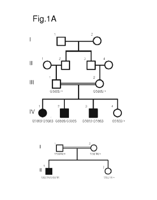
[0017] Figure 1 shows identification of mutations in LSS causing congenital
cataracts. Fig.
1A, Pedigrees of affected families and cataract phenotype. Squares and circles
indicate males
and females respectively. 1, wild-type allele; W581R and G5885 are the two
mutations. Fig
1B, Upper panel, DNA sequencing data of an unaffected individual and an
affected child (II-
1) with a homozygous W581R mutation; lower panel, DNA sequencing data of an
unaffected
individual and an affected child (IV-1) with a homozygous G5885 mutation. The
underlined
sequence indicates the nucleic acid change. Fig 1C, Left, colour photograph of
patient l's right
eye in the first pedigree (IV-1) with a total cataract; right, colour
photograph of patient 2's right
eye in the same pedigree (IV-3) with a cataract.
[0018] Figure 2 shows LSS mutations abolished the cyclase enzymatic function.
Fig 2A,
Conservation of W581R and G588 in LSS across several species: Homo sapiens,
Pan
troglodytes, Bos taurus, Mus musculus, Rattus norvegicus, Gallus gallus and
Danio rerio. Fig
CA 02958868 2017-02-21
WO 2016/029199
PCT/US2015/046453
2B, Computer modelling of LSS structure and impact of the LSS W581R and G588S
mutations. A computer modelling analysis identifies a loop originating from
C584 and ending
at E578 with the key side chain of W581 at the tip of the loop stabilizing the
sterol. The loop
is fixed by an S¨S bridge and the E578¨R639 salt bridge. Amide nitrogen N of
G588 interacts
with the C584 from the previous helical turn and the Ca hydrogen of G588 is in
close proximity
to the critical E578, which then forms a strong salt bridge with R639 of the
same supporting
helix. The mutation G588S causes the side chain of the serine to clash into
the E578 residue of
the loop and is incompatible with the structure. Arrow indicates the location
of the mutant side
chain. Fig. 2C, Effect of engineered expression of the wild-type protein (WT
LSS) and LSS
mutants on sterol content. Wild-type LSS markedly increased lanosterol
production, whereas
neither W581R nor the G588S mutant exhibited any cyclase activity. n=3 in each
group;
***P<0.001.
[0019] Figure 3 shows lanosterol reduced intracellular aggregation of various
crystallin
mutant proteins. Fig 3A, Confocal images of crystallin protein aggregates in
human lens
progenitor cells. The cataract-causing Y118D mutant of aA-crystallin formed
p62-positive
intracellular inclusion bodies or aggresomes. Green, eGFP¨crystallin proteins;
red, p62; blue,
nuclei. Cells transfected with peGFP- Ni were used as a control. Fig 3B
Confocal images of
inhibitory effect of LSS on crystalline aggregates. Fig 3C, Inhibition of
crystallin mutant
aggregation by wild-type LSS (WT LSS) and lanosterol, but not mutant LSS or
cholesterol.
Fig. 3D, Increase in soluble aA-crystallin (Y118D) mutant protein by co-
expression of wild-
type LSS but not LSS mutants (Y1 18D co-expressed with pcDNA3.1¨N- Flag was
used as a
control). Quantitative analysis was performed using densitometry of crystallin
proteins by
western blot analysis of the supernatant or insoluble fraction of cell
lysates. n=3 in each group;
representative western blot analysis is shown in - Fig. 9c; *P<0.05, **P<0.01.
Fig 3E, Confocal
images of the re-dissolution of pre-formed crystalline aggregates by
lanosterol. Fig 3F,
Lanosterol significantly reduced the intracellular aggregation by various
cataract-causing
mutant crystallin proteins in a concentration-dependent manner (n=3, P<lx10-
4). Cholesterol
did not reduce intracellular aggregation (n=3, P>0.1). Fig. 3G Lanosterol
increased the soluble
fractions of various crystallin mutants in human lens progenitor cells. n=3;
P<0.001. Fig. 3H,
Effects of DMSO, cholesterol or lanosterol on aA-crystallin Y118D aggregates
in human lens
progenitor cells by serial live cell imaging. Fig. 31, Effect of lanosterol on
dissolution of
intracellular crystallin aggregates over time (n=22 from 3 biological
replicates). The mean
6
CA 02958868 2017-02-21
WO 2016/029199
PCT/US2015/046453
SD values are shown as black symbols. The data are best fitted by the single
exponential decay
process (red line).
[0020] Figure 4 shows lanosterol re-dissolved pre-formed amyloid-like fibrils
of crystallin
proteins. Fig 4A, Negatively stained TEM photographs of aggregates of aA-
crystallin mutant
proteins treated by a liposome vehicle, cholesterol or lanosterol in
liposomes. Images in the
right column of the lanosterol group show a 5X magnification of the image on
their right.
Fig.4B, Effect of lanosterol on the re-dissolution of crystallin aggregates by
ThT fluorescence
(n=3). Fig.4B (i), b/gamma-crystallin mutants; Fig.4B (ii), a-crystallin
mutants. Each bar
results from three independent samples.
[0021] Figure 5 shows lanosterol reduced cataract severity and increased
clarity. Fig 5A,
Photographs of a cataractous rabbit lens treated with lanosterol showing
increased lens clarity.
Fig 5A(i), left, before treatment; Fig 5A(ii), right, after treatment. Fig 5B,
Boxplot of the
quantification of the treatment effect of lanosterol (n=13). Fig 5C,
Photographs of a cataractous
dog lens treated with lanosterol showing increased lens clarity. Fig 5C(i),
left, before treatment;
Fig 5C(ii), right, after treatment. d, Boxplot of the quantification of the
treatment effect of
lanosterol (n=7). Range, median (horizontal line) and mean (circle) are
presented. Crosses
indicate the maximum and minimum cataract grades measured. Whiskers indicate
the standard
deviation and the box encompasses a 40% confidence interval.
[0022] Figure 6A shows homozygosity mapper plots the genome-wide homozygosity
as bar
charts. To emphasize regions of interest, any score higher than 80% of the
maximum score
reached in this project is coloured in red. Figure 6B shows the homozygosity
scores were
plotted against the physical position on chromosome 21, which contains the LSS
gene. Red bars
indicate regions with highest scores. The right side of the chromosome
contains a long
continuous homozygous region, where the LSS gene is located.
[0023] Figure 7 shows representative confocal images of cells co-transfected
with Flag¨LSS
and eGFP. Human lens progenitor cells were co-transfected with either the wild-
type or the
mutated LSS gene and the eGFP gene for 4 h and cultured for 16 h in fresh
culture medium.
The cellular distribution of LSS was then visualized using an anti-Flag
antibody (purple). The
distribution of eGFP (green) was used as a control. The nuclei were stained
and visualized by
Hoechst 33342 (blue).
[0024] Figure 8 shows representative confocal images of cells co-transfected
with LSS and
various cataract-causing crystallin mutants. Fig 8A, R116C mutant of aA-
crystallin. Fig 8B,
R120G mutant of aB-crystallin. Fig 8C, V187E mutant of 13B2-crystallin. Fig
8D, G129C
7
CA 02958868 2017-02-21
WO 2016/029199
PCT/US2015/046453
mutant of 7C-crystallin. Fig 8AE, W43R mutant of 7D-crystallin. Human lens
progenitor cells
were co-transfected with either the wild-type or the mutated Flag-LSS gene and
the mutant
GFP-crystallin gene for 4 h and cultured for 16 h in fresh culture medium. All
crystallin
mutants formed p62-positive aggregates as indicated by the co-localization of
the mutant
crystallins and p62. Cells co-transfected with GFP-crystallin and pcDNA3.1-N-
Flag were used
as controls. The formation of intracellular aggregates of various crystallin
proteins was
visualized by fluorescence of GFP (green). Wild-type or mutated LSS was
detected with an
anti-Flag antibody (red), p62 was stained using an anti-p62 antibody, while
the nuclei were
stained and visualized by Hoechst 33342 staining (blue). Quantitative analysis
of cells with
aggregates is summarized in Fig 3c.
[0025] Figure 9 shows inhibition of crystallin mutant aggregation by wild-type
LSS and
lanosterol in HLEB-3 cells (Fig 9A) or HeLa cells (Fig 9B). Cells co-
transfected with LSS and
crystallin mutant constructs were cultured for 24 h before assaying for
aggregates. The rescue
experiments were performed by addition of 40Ã4M sterols (lanosterol or
cholesterol) to the
cell culture medium for 2 h, the sterol medium was then replaced with fresh
culture medium
and the cells were cultured for a further 12 h. The percentage of cells with
crystallin aggregates
were calculated from ten randomly selected viewing fields. The values of the
wild-type LSS
group, mutant group, or mutant plus lanosterol group were calculated.
Aggregates were
significantly lower in the wild-type LSS and lanosterol groups compared to the
control group
(P < 1 x 10⁻⁴), while aggregates in mutant LSS or cholesterol groups showed no
difference to
the control group (P> 0.1). Fig. 9C, Human lens progenitor cells were co-
transfected with
wild-type or mutant LSS plus aA-crystallin (Y118D). aA-crystallin (Y118D) co-
expressed
with pcDNA3.1-N-Flag was used as a control. After transfection for 4 h and
incubation in fresh
culture medium for another 24 h, the cells were lysed and centrifuged to
separate supernatant
and insoluble fractions. LSS and crystallin fusion proteins were detected by
antibodies against
Flag and GFP, respectively. Red arrows indicate higher crystalline content in
the soluble
fraction versus in the insoluble fraction in cells containing the WT-LSS. Data
were quantified
from three independent experiments and summarized in Fig 3D.
[0026] Figure 10 shows lanosterol significantly reduced the intracellular
aggregation caused
by various cataract-causing mutant crystallin proteins in a concentration-
dependent manner
when assayed in HLEB-3 or HeLa cells. Fig 10A, Representative confocal images
of HLEB-3
cells transfected with various cataract-causing crystallin mutants. Fig 10B,
Representative
confocal images of HeLa cells transfected with various cataract-causing
crystallin mutants.
8
CA 02958868 2017-02-21
WO 2016/029199
PCT/US2015/046453
Cells were transfected with various crystallin constructs for 4 h and cultured
for an additional
24 h in fresh culture medium. Then the cells were treated with 10, 20 and 40 M
lanosterol in
1% (HLEB-3 cells) or 2% DMSO (HeLa cells) for 2 h and cultured for another 12
h. Cells
treated with 1% (HLEB-3 cells) or 2% DMSO (HeLa cells) were used as the
controls.
Formation of intracellular aggregates of various crystallin proteins was
visualized by
fluorescence of GFP (green) and the nuclei were stained with Hoechst 33342
(blue). Typical
intracellular aggregates are indicated by arrows. Fig 10C, Concentration
dependence of the
aggregation-dissolving effects of lanosterol when assayed in HLEB-3 cells. Fig
10D,
Concentration dependence of the aggregation-dissolving effects of lanosterol
when assayed in
HeLa cells.
[0027] Figure 11 shows treatment by lanosterol, but not cholesterol, increased
cataract-
causing mutant crystallins in soluble fractions when compared to a control
group or a mutant
LSS group. Fig 11A, Human lens progenitor cells were transfected with mutant
crystallin genes
for 4 h, and then incubated in fresh culture medium for another 24 h. The
cells were harvested
and lysed. Supernatant and insoluble fractions were separated by
centrifugation and analyzed
by western blot analysis. LSS and crystallin fusion proteins were identified
by antibodies
against Flag and GFP tags, respectively. The lanosterol-treated group is
highlighted by red
boxes. Cells treated with 1% DMSO were used as a control. 13-Actin was used as
an internal
protein loading control of total cell lysates (TCL). S, supernatant; P,
insoluble fraction. Fig
11B, Effect of DMSO (n = 4) and cholesterol (n = 7) on the size changes of aA-
crystallin
(Y118D) aggregates in human lens progenitor cells evaluated by single-particle
tracking in
live-cell imaging. Fig 11C, Evaluation of the effect of lanosterol on the
dissolution of crystallin
aggregates by turbidity. Crystallin aggregates were formed by incubating 5 mg
m1-1 protein
solution at 60 C for 2 h (a-crystallins) or 37 C for 48 h (13- and 7-
crystallins) in the presence
of 1 M guanidine chloride. The preformed aggregates were re-suspended in PBS
at a final
protein concentration of 0.2 mg m1-1 and were treated with 500 it.M sterols in
500 it.M DPPC
liposome and incubated at 37 C for 24 h. Aggregates treated with 500 it.M
DPPC liposome
only were used as the controls. Fig 11D, Concentration-dependent effect of
lanosterol on the
re-dissolution of amyloid-like fibrils by aA-crystallin mutants evaluated by
ThT fluorescence.
Aggregates treated with 500 1...1M DPPC liposome only were used as the
controls.
[0028] Figure 12 shows grading system of cataractous lenses. Fig 12A, Lenses
were placed
above a grid and photographed. The degree of transparency was scored as 0, a
clear lens and
absence of opacification (gridlines clearly visible, a'); 1, a blurry lens and
a slight degree of
9
CA 02958868 2017-02-21
WO 2016/029199
PCT/US2015/046453
opacification (minimal clouding of gridlines, with gridlines still visible,
b'); 2, a cloudy lens
and presence of diffuse opacification involving almost the entire lens
(moderate clouding of
gridlines, with main gridlines visible, c'); or 3, an opaque lens and presence
of extensive thick
opacification involving the entire lens (total clouding of gridlines, with
gridlines not seen at
all, d'). Fig 12 B, Lanosterol reduced cataract severity and increased clarity
in isolated
cataractous rabbit lenses. Rabbit lenses (n = 13) were dissected and incubated
with lanosterol
for 6 days and subsequently assessed for lens clarity and transparency. Pairs
of photographs of
each cataractous rabbit lens showing before and after treatment with scores
underneath are
shown. Fig 12C, Lanosterol reduced cataract severity and increased lens
clarity in dogs. Dog
eyes with cataracts (n = 7) were treated with lanosterol for 6 weeks and
assessed for lens clarity
and transparency. A pair of photographs of each study eye before and after
treatment is shown
with scores underneath. Three control eyes treated with vehicles alone are
also presented.
DETAILED DESCRIPTION
[0029] Reference will now be made in detail to specific embodiments of the
invention
including the best modes contemplated by the inventors for carrying out the
invention.
Examples of these specific embodiments are illustrated in the accompanying
drawings. While
the invention is described in conjunction with these specific embodiments, it
will be understood
that it is not intended to limit the invention to the described embodiments.
On the contrary, it
is intended to cover alternatives, modifications, and equivalents as may be
included within the
spirit and scope of the invention as defined by the appended claims. In the
following
description, specific details are set forth in order to provide a thorough
understanding of the
present invention. The present invention may be practiced without some or all
of these specific
details. In addition, well-known features may not have been described in
detail to avoid
unnecessarily obscuring the invention.
[0030] The present invention relates to a method of and compositions for
treating or preventing
vision disorders that affect the normal structure of the eye in a subject
having or at risk of
developing such vision disorders, comprising administering to such subject a
composition
comprising a pharmaceutically acceptable carrier and a pharmaceutically
effective amount of
a sterol having the formula I. For example, an exemplary compound of the
invention comprises
administering to a patient an opthalmological pharmaceutically effective
amount of lanosterol
(3 13-Hydroxy -8 ,24-1 ano stadiene ; 8,24-Lanostadien-3 [3 - ol).
[0031] In other embodiments, the present disclosure describes sterols and
methods of using
sterols. For example, the sterols of formula I are formulated in ophthalmic
pharmaceutical
CA 02958868 2017-02-21
WO 2016/029199
PCT/US2015/046453
compositions comprising a pharmaceutically acceptable ophthalmic carrier to
inhibit crystallin
protein aggregation. In certain other embodiments, the present disclosure
describes methods of
using sterols of formula 1 to inhibit crystallin protein aggregation. In yet
other embodiments,
compounds of the invention are able to reverse aggregation of crystallin
protein and inhibit
further aggregation of crystallin protein.
Methods of Treating or Preventing Vision Disorders
[0032] The present invention provides ophthalmic pharmaceutical compositions
and methods
of using the present invention in preventing and/or treating vision disorders
that affect the
normal structure of the lens in the eye in a subject having or at risk of
developing such vision
disorders. As described herein, a vision disorder that affects the normal
structure of the lens in
the eye (referred herein as the phrase "vision disorder") refers to conditions
that affect the
structure of the lens as to cause vision dysfunction, such as changes to the
clarity or rigidity of
the lens of the eye. Such conditions include cataracts, presbyopia and nuclear
sclerosis. In
addition, vision disorders refer to retinal degeneration, such as as Refsum
disease, Smith-
Lemli-Opitz syndrome (SLOS) and Schnyder crystalline corneal dystrophy (SCCD),
abetalipoproteinemia and familial hypobetalipoproteinemia. In certain
embodiments, the
present invention provides compositions and methods of use thereof to
alleviate or reverse
crystalline protein aggregation. In alternative embodiments, there are
provided compositions
and methods for inhibiting, preventing and/or treating the disruption of intra-
or inter-protein
interactions that form the macro-structure essential for lens transparency and
refractive index.
[0033] The term "cataract" as referred to in the present invention means a
disease or condition
that exhibits symptoms of causing cloudiness or opacity on the surface and/or
the inside of the
lens or inducing the swelling of the lens, and it includes both congenital
cataract and acquired
cataract (cf. PDR Staff, "PDR of Ophthalmic Medicines 2013", PDR Network,
2012). In some
embodiments, the cataract is an age-related cataract, a diabetic cataract, a
cataract associated
with surgery, a cataract resulting from exposure to radiation, a cataract
resulting from a genetic
illness, a cataract resulting from an infection, or a cataract resulting from
medication. In some
embodiments, the individual has a hereditary form of cataract with early
onset. Concrete
examples of such are congenital cataract such as congenital pseudo-cataract,
congenital
membrane cataract, congenital coronary cataract, congenital lamellar cataract,
congenital
punctuate cataract, and congenital filamentary cataract; and acquired cataract
such as geriatric
cataract, secondary cataract, browning cataract, complicated cataract,
diabetic cataract,
traumatic cataract, and others inducible by electric shock, radiation,
ultrasonic, drugs, systemic
11
CA 02958868 2017-02-21
WO 2016/029199
PCT/US2015/046453
diseases, and nutritional disorders. Acquired cataract further includes
postoperative cataract
with symptoms of causing cloudiness in the posterior encapsulating a lens
inserted to treat
cataract.
[0034] Nuclear sclerosis refers to a condition, generally in older animals,
that results similarly
in opacity of the lens. It is an age-related change in the density of the
crystalline lens nucleus
that is caused by compression of older lens fibers in the nucleus by new fiber
formation.
[0035] Presbyopia refers to a vision condition in which the crystalline lens
of the eye loses its
flexibility, which makes it difficult to focus on close objects.
[0036] In some embodiments, the invention provides a method of treating or
preventing a
vision disorder, the method comprising administering to an individual in need
thereof an
effective amount of a composition comprising a compound having a structural
formula I. In
some embodiments, the compound is a sterol having a structural formula I.
[0037] An individual "in need of' treatment according to the invention is an
individual that is
suffering from a vision disorder that affects the normal function of the lens
in the eye. For
example, the individual may have or is at risk for developing an age-related
cataract or a
cataract. Individuals at risk of developing a cataract include, but are not
limited to, individuals
with a family history of developing cataracts, individuals with a mutation
linked to a cataract,
individuals exposed to radiation, diabetics, and the like. For example, in one
aspect, the
individual has been diagnosed with cataract in one eye, and the compound is
administered to
prevent or slow cataract formation in the contralateral eye. Similarly, an
individual "in need
of' treatment according to the invention is an individual that may have or is
at risk for
developing presbyopia. Similarly, an individual "in need of' treatment
according to the
invention is an individual that has or is at risk for developing nuclear
sclerosis. Preferably the
individual is human, however, animals that suffer from or who are at risk for
an eye disease
(animals in need of treatment) can also be identified by one skilled in the
art. Mammals in need
of treatment, such as cats, dogs, pigs, horses, cows and rodents can be
identified. Additionally,
animals such as avians, reptiles, amphibians, and fish that are in need of
treatment can be
identified.
[0038] "Treating" a vision disorder does not require a 100% abolition or
reversal of a vision
disorder. In some embodiments, "treating" vision disorders according to
inventive method
alleviates, inhibits, prevents and/or reverses dysfunction of the lens, e.g.,
opacity or inflexibility
of the lens by, e.g., at least about 5%, at least about 10% or at least about
20% compared to
levels observed in the absence of the inventive composition or method (e.g.,
in a biologically-
matched control subject or specimen that is not exposed to the invention
composition or
12
CA 02958868 2017-02-21
WO 2016/029199
PCT/US2015/046453
compound of the inventive method). In some embodiments, dysfunction (such as
cataract
formation, opacity or crystalline aggregation on or in the lens) is treated by
at least about 30%,
at least about 40%, at least about 50%, or at least about 60%, at least about
70%, at least about
80%, at least about 90%, or more (about 100%) compared to lens dysfunction in
the absence
of the compound of the inventive method. Lens dysfunction, such as opacity or
cloudiness or
cataracts, generally are detected using any of a number of optic tests
including, but not limited
to, visual acuity testing, ophthalmoscopy, slit-lamp examination, keratometry,
tonometry,
contrast testing, glare sensitivity, wavefront mapping.
[0039] Similarly, "prevention" does not require 100% inhibition or deterrence
of a vision
disorder. For example, any reduction in cloudiness or opacity, or deceleration
of cataract
progression constitutes a beneficial biological effect in a subject. Also
exemplary, any decrease
in crystalline aggregation in the lens of an eye constitutes a beneficial
biological effect. In this
regard, the invention reduces the vision disorder, e.g., at least about 5%, at
least about 10% or
at least about 20% compared to levels observed in the absence of the inventive
method (e.g.,
in a biologically-matched control subject or specimen that is not exposed to
the compound of
the inventive method). In some embodiments, the vision disorder is reduced by
at least about
30%, at least about 40%, at least about 50%, or at least about 60%, at least
about 70%, at least
about 80%, at least about 90%, or more (about 100%).
[0040] Inhibiting, preventing or reversal of dysfunction does not require a
100% inhibition,
prevention, abolition or reversal. For example, any inhibition of aggregation
constitutes a
beneficial biological effect in a subject. In this regard, the invention
inhibits a vision disorder
that affects the normal function of the lens of the eye in a subject, e.g., at
least about 5%, at
least about 10% or at least about 20% compared to levels observed in the
absence of the
inventive method (e.g., in a biologically-matched control subject or specimen
that is not
exposed to the compound of the inventive method). In some embodiments, the
vision disorder
is inhibited, prevented and/or reversed by at least about 30%, at least about
40%, at least about
50%, or at least about 60%. In some embodiments, the inventive method inhibits
amyloid
formation by at least about 70%, at least about 80%, at least about 90%, or
more (about 100%)
compared to amyloid formation in the absence of the compound of the inventive
method.
[0041] An "effective amount" of an ophthalmic pharmaceutical composition
comprising a
compound of formula 1 is an amount that inhibits, prevents or reverses
dysfunction of the lens
in an individual. An ophthalmic pharmaceutical composition of the present
invention is being
administered to a subject in need thereof at an effective amount to treat the
vision disorder. As
used herein, "therapeutically effective amount" means a dose that alleviates
at least one of the
13
CA 02958868 2017-02-21
WO 2016/029199
PCT/US2015/046453
signs, symptoms, or causes of a vision disorder, or any other desired
alteration of a biological
system. In preventative applications, the term "prophylactically effective
amount" means a
dose administered to a patient susceptible to or otherwise at risk of a
particular disease, which
may be the same or different dose as a therapeutically effective amount. The
effective amount
of the composition for a particular individual can depend on the individual,
the severity of the
condition of the individual, the type of formulation being applied, the
frequency of
administration, and the duration of the treatment. In accordance with the
present invention,
administration of an ophthalmic pharmaceutical formulation of the present
invention such as,
e.g., lanosterol, even at relatively low concentrations in liquid drops, e.g.,
at least 10-9 M, at
least 0.5 to 1x10' M, at least 0.5 to 1x10' M, at least 0.5 to 1x10⁻⁶ M, at
least 0.5 to 1x10⁻⁵
M, at least 0.5 to 1x10⁻⁴ M, or at least 0.5 to lx10-3 M, or any concentration
falling in a range
between these values (e.g., 10-9 M to 10-3 M), may reverse such vision
disorders with only
one, two, three or multiple, daily applications and does so rapidly.
Route of Administration
[0042] As will be understood by those skilled in the art, the most appropriate
method of
administering a compound to a subject is dependent on a number of factors. In
various
embodiments, the compound according to the invention is administered locally
to the eye, e.g.,
topically, subconjunctivally, retrobulbarly, periocularly, subretinally,
suprachoroidally, or
intraocularly.
[0043] Pharmaceutical compositions that are particularly useful for
administration directly to
the eye include aqueous solutions and/or suspensions formulated as eye drops
and thickened
solutions and/or suspensions formulated as ophthalmic gels (including gel-
forming solutions)
or ointments, which is an ophthalmic solution, ophthalmic ointment, ophthalmic
wash,
intraocular infusion solution, wash for anterior chamber, internal medicine,
injection, or
preservative for extracted cornea. Other dosage forms for ophthalmic drug
deliver include
ocular inserts, intravitreal injections and implants. Injectable solutions can
be directly injected
into the cornea, crystalline lens and vitreous or their adjacent tissues using
a fine needle. The
composition also can be administered as an intraocular perfusate.
[0044] Additional contemplated routes of administration include, but are not
limited to, one or
more of: oral (e.g., as a tablet, capsule, or as an ingestible solution),
mucosal (e.g., as a nasal
spray or aerosol for inhalation), nasal, parenteral (e.g., by an injectable
form), gastrointestinal,
intraspinal, intraperitoneal, intramuscular, intravenous, intrauterine,
intradermal, intracranial,
14
CA 02958868 2017-02-21
WO 2016/029199
PCT/US2015/046453
intratracheal, intravaginal, intracerebroventricular, intracerebral,
subcutaneous, transdermal,
rectal, buccal, epidural and sublingual.
[0045] In some embodiments, the mode for delivery of a composition of the
invention to the
eye is via a contact lens. The lens may be provided pre-treated with the
desired compound.
Alternatively, the lens is provided in a kit with components for preparing a
coated lens, which
are provided as lyophilized powders for reconstitution or as concentrated or
ready-to-use
solutions. The compositions can be provided as kits for single or multi-use.
[0046] In some embodiments, the mode for delivery of a composition of the
invention to the
eye is via an ophthalmic rod (Gwon et al., Ophthalmology. 1986 September; 93(9
Suppl):82-
5). In some embodiments, the mode for delivery of a composition of the
invention to the eye is
via an intraocular lens-hydrogel assembly (Garty et al., Invest Ophthalmol Vis
Sci, 2011 Aug.
3; 52(9):6109-16).
Dose
[0047] The composition comprising the compound is provided in a
therapeutically effective
amount that achieves a desired biological effect at a medically-acceptable
level of toxicity. The
dosage of the compositions may vary depending on the route of administration
and the severity
of the disease. The dosage may also be adjusted depending on the body weight,
age, sex, and/or
degree of symptoms of each patient to be treated. The precise dose and route
of administration
will ultimately be at the discretion of the attendant physician or
veterinarian. It will be
appreciated that it may be necessary to make routine variations to the dosage
depending on the
age and weight of the patient as well as the severity of the condition to be
treated. The frequency
of administration depends on the formulation and the aforementioned
parameters. For example,
it may be desirable to apply eye drops at least once per day, including 2, 3,
4, or 5 times per
day.
[0048] Persons of ordinary skill can easily determine optimum dosages, dosing
methodologies
and repetition rates. Optimum dosages may vary depending on the relative
potency of the
particular pharmaceutical composition and the method of administration.
Acceptable dosages
can generally be estimated based on EC50 (effective concentration for 50% of
the test group)
found to be effective in in vitro and in vivo animal models. In general,
dosage is from 0.01 ug
to 100 g per kg of body weight, and may be given once or more daily, weekly,
monthly or
yearly, or even once every 2 to 20 years. Persons of ordinary skill in the art
can easily estimate
repetition rates for dosing based on measured residence times and
concentrations of the drug
in bodily fluids or tissues. Following successful treatment, it may be
desirable to have the
CA 02958868 2017-02-21
WO 2016/029199
PCT/US2015/046453
patient undergo maintenance therapy to prevent the recurrence of the disease
state, wherein the
therapeutic compositions described herein are administered in maintenance
doses, ranging
from 0.01 ng to 100 g per kg of body weight, once or more daily, to once every
20 years.
Exemplary doses of the compounds for administration to a human (of
approximately 70 kg
body weight) via systemic route are 0.1 mg to 5 g, e.g., 1 mg to 2.5 g of the
compound per unit
dose.
[0049] Preferred concentrations of the compound of formula I range from about
1 ng/ml to 500
ng/ml, for example, about 1 ng/ml, about 2 ng/ml, about 3 ng/ml, about 4
ng/ml, about 5 ng/ml,
about 10 ng/ml, about 20 ng/ml, about 30 ng/ml, about 40 ng/ml, about 50
ng/ml, about 60
ng/ml, about 70 ng/ml, about 80 ng/ml, about 90 ng/ml, about 100 ng/ml, about
120 ng/ml,
about 140 ng/ml, about 160 ng/ml, about 180 ng/ml, about 200 ng/ml, about 250
ng/ml, about
300 ng/ml, about 350 ng/ml, about 400 ng/ml, about 450 ng/ml, or about 500
ng/ml. The
inhibitor may be provided in combination with other pharmaceutically active
agents.
[0050] The pharmaceutical compositions described herein can be administered as
a single dose
or in multiple doses; administered either as individual therapeutic agents or
in combination
with other therapeutic agents; and combined with conventional therapies, which
may be
administered sequentially or simultaneously. In one embodiment of the
invention, daily
dosages in human and/or animal therapy of the present ophthalmic formulations
are about 1
drop per eye, about 2 drops per eye, about 3 drops per eye, about 4 drops per
eye, about 5 drops
per eye, about 6 drops per eye, about 7 drops per eye, about 8 drops per eye,
about 9 drops per
eye, about 10 drops per eye, about 11 drops per eye, about 12 drops per eye or
more than about
12 drops per eye. In another embodiment of the invention, daily administration
schedule for
the present ophthalmic formulations in human and/or animal therapy is about 1
time per day,
about 2 times per day, about 3 times per day, about 4 times per day, about 5
times per day,
about 6 times per day, about 7 times per day, about 8 times per day, about 9
times per day,
about 10 times per day, about 11 times per day, about 12 times per day or more
than about 12
times per day. Dosages can be standardized for instance by means of a standard
pharmacopeial
medicinal dropper of 3 mm in external diameter, which when held vertically
delivers 20 drops
of water of total weight of 0.9 to 1.1 grams at 25 C.
[0051] When administered according to the dosage schedule described above, the
treatment
regimen in humans and/or animals can continue indefinitely or until no further
improvement is
observed. Alternately, the treatment regimen can last for 1 day, 2 days, 3
days, 4 days, 5 days,
6 days, 7 days, 8 days, 9 days, 10 days, 11 days, 12 days, 13 days, 14 days,
15 days, 16 days,
17 days, 18 days, 19 days, 20 days, 21 days, 22 days, 23 days, 24 days, 25
days, 26 days, 27
16
CA 02958868 2017-02-21
WO 2016/029199
PCT/US2015/046453
days, 28 days, 29 days, 30 days, 31 days, 32 days, 33 days, 34 days, 35 days,
36 days, 37 days,
38 days, 39 days, 40 days, 41 days, 42 days, 43 days, 44 days, 45 days, 46
days, 47 days, 48
days, 49 days, 50 days, 60 days, 70 days, 80 days, 90 days, 100 days, 150
days, 200 days, 250
days, 300 days, 400 days, 500 days, 750 days, 1000 days or more than 1000
days.
Compounds Effective in Treating or Preventing Cataract
[0052] In various embodiments, the compound of the inventive method or
composition is
lanosterol having a compound of formula I:
\\
H
HO-
/ \ H
, or a prodrug or pharmaceutically
acceptable salt thereof.
[0053] For example, the compound of the inventive method or composition is
lanosterol, a
prodrug or pharmaceutically acceptable salt thereof. In one embodiment, the
compound is
lanosterol. In another embodiment, any prodrug or pharmaceutically acceptable
salt of the
above compounds are contemplated to be within the scope of the invention.
Pharmaceutical Compositions
[0054] In some embodiments of the invention, pharmaceutical compositions of
one or more
therapeutic compounds can be prepared by formulating one or more of these
therapeutic
compounds in a pharmaceutically acceptable carrier. As used herein,
"pharmaceutically or
therapeutically acceptable carrier" refers to a carrier medium which does not
interfere with the
effectiveness of the biological activity of the active ingredients and which
is not toxic to the
host or patient. The type of carrier which is used in the pharmaceutical
preparation will depend
on the method by which the therapeutic compounds are to be administered. Many
methods of
preparing pharmaceutical compositions for various routes of administration are
well known in
the art.
[0055] As used herein, "pharmaceutically acceptable ophthalmic carrier" refers
to a
pharmaceutically acceptable excipient, carrier, binder, and/or diluent for
delivery of the
compound of the structural formula 1 directly or indirectly to, on or near the
eye. Accordingly,
17
CA 02958868 2017-02-21
WO 2016/029199
PCT/US2015/046453
the invention further comprises a composition comprising the compound of the
structural
formula I and a pharmaceutically acceptable ophthalmic carrier.
[0056] Optionally, the composition includes a free acid, free base, salt
(e.g., an acid or base
addition salt), hydrate or prodrug of the compound of structural formula I.
The phrase
"pharmaceutically acceptable salt" or "pharmaceutically acceptable acid," as
used herein,
refers to pharmaceutically acceptable organic or inorganic salts or acids,
respectively, of a
compound of Formula I. The counter ion may be any organic or inorganic moiety
that stabilizes
the charge on the parent compound. Furthermore, a pharmaceutically acceptable
salt (or acid)
may have more than one charged atom in its structure. Instances where multiple
charged atoms
are part of the pharmaceutically acceptable salt (or acid) can have multiple
counter ions. Hence,
a pharmaceutically acceptable salt (acid) can have one or more charged atoms
and/or one or
more counter ion.
[0057] Exemplary salts include, but are not limited, to sulfate, citrate,
acetate, oxalate, chloride,
bromide, iodide, nitrate, bisulfate, phosphate, acid phosphate, isonicotinate,
lactate, salicylate,
acid citrate, tartrate, oleate, tannate, pantothenate, bitartrate, ascorbate,
succinate, maleate,
gentisinate, fumarate, gluconate, glucuronate, saccharate, formate, benzoate,
glutamate,
methanesulfonate, ethanesulfonate, benzenesulfonate, p-toluenesulfonate, and
pamoate (i.e.,
1,1'-methylene-bis-(2-hydroxy-3-naphthoate)) salts. A pharmaceutically
acceptable salt may
involve the inclusion of another molecule such as an acetate ion, a succinate
ion or other
counter ion.
[0058] The prodrug is a material that includes the compound of structural
formula I covalently
bound to a carrier moiety. The carrier moiety can be released from the
compound of structural
formula 1, in vitro or in vivo to yield compound of structural formula I.
Prodrug forms are well
known in the art as exemplified in Sloan, K. B., Prodrugs, M. Dekker, New
York, 1992; and
Testa, B. and Mayer, J. M., Hydrolysis in drug and prodrug metabolism:
chemistry,
biochemistry, and enzymology, Wiley-VCH, Zurich, 2003.
[0059] In some embodiments of the invention, pharmaceutical compositions are
prepared by
dissolving the invention composition in an appropriate solvent. Appropriate
solvents include,
but are not limited to, water, saline solution (for example, NaC1), buffered
solutions, ointments,
gels or other solvents. In certain embodiments, the solvents are sterile.
[0060] Aqueous solutions and diluents for suspensions that are used in
preparation of eye drops
can include distilled water, physiological saline, and the like. These
pharmaceutical
compositions can be formulated by admixing, diluting or dissolving the
compound, optionally,
with appropriate pharmaceutical additives such as excipients, disintegrators,
binders,
18
CA 02958868 2017-02-21
WO 2016/029199
PCT/US2015/046453
lubricants, diluents, buffers, antiseptics, moistening agents, emulsifiers,
dispersing agents,
stabilizing agents and dissolving aids in accordance with conventional methods
and
formulating in a conventional manner depending upon the dosage form. Non-
aqueous solutions
and diluents for suspensions can include edible (eg vegetable) oil, liquid
paraffin, mineral oil,
propylene glycol, p-octyldodecanol, polysorbate, macrogols, aluminum
monostearate as well
as similar solvents.
[0061] Various additives may be contained in eye drops, ophthalmic gels and/or
ophthalmic
ointments as needed. These can include additional ingredients, additives or
carrier suitable for
use in contact on or around the eye without undue toxicity, incompatibility,
instability,
irritation, allergic response, and the like. Additives such as solvents,
bases, solution adjuvants,
suspending agents, thickening agents, emulsifying agents, stabilizing agents,
buffering agents,
isotonicity adjusting agents, pH-adjusting agents, chelating agents, soothing
agents,
preservatives, corrigents, flavoring agents, coloring agents, excipients,
binding agents,
lubricants, surfactants, absorption-promoting agents, dispersing agents,
preservatives,
solubilizing agents, and the like, can be added to a formulation where
appropriate.
[0062] For example, eye drops can be formulated by dissolving the compound in
sterilized
water in which a surface active agent is dissolved and optionally adding
appropriate
pharmaceutical additives such as a preservative, a stabilizing agent, a
buffer, an antioxidant
and a viscosity improver.
[0063] For example, buffering agents are added to keep the pH constant and can
include
pharmaceutically acceptable buffering agents such as borate buffer, citrate
buffer, tartrate
buffer, phosphate buffer, acetate buffer or a Tris-HC1 buffer (comprising
tris(hydroxymethyl)
aminomethane and HC1). For example, a Tris-HC1 buffer having pH of 7.4
comprises 3 g/1 of
tris-(hydroxymethyl)-aminomethane and 0.76 g/1 of HC1. In yet another aspect,
the buffer is
10x phosphate buffer saline ("PBS") or 5xPBS solution. Buffering agents are
included in an
amount that provides sufficient buffer capacity for the expected physiological
conditions.
[0064] Other buffers include, but are not limited to, buffers based on HEPES
(N-12-
hydroxyethyl lpeperazine-N'- 12-ethanesulfonic acid}) having pKa of 7.5 at 25
C. and pH in
the range of about 6.8-8.2; BES (N,N-bis12-hydroxyethy112-aminoethanesulfonic
acid) having
pKa of 7.1 at 25 C. and pH in the range of about 6.4-7.8; MOPS (3-1N-
morpholino lpropanesulfonic acid) having pKa of 7.2 at 25 C. and pH in the
range of about
6.5-7.9; TES (N-tris{hydroxymethyl}-methyl-2-aminoethanesulfonic acid) having
pKa of 7.4
at 25 C. and pH in the range of about 6.8-8.2; MOBS (4-1N-morpholino 1
butanesulfonic acid)
having pKa of 7.6 at 25 C. and pH in the range of about 6.9-8.3; DIPSO (3-
(N,N-bis12-
19
CA 02958868 2017-02-21
WO 2016/029199
PCT/US2015/046453
hydroxyethyl 1 amino)-2-hydroxypropane)) having pKa of 7.52 at 25 C. and pH
in the range of
about 7-8.2; TAPS ( { (2-hydroxy-3 { tris(hydroxymethyl)methylamino}-l-
propanesulfonic
acid)) having pKa of 7.61 at 25 C. and pH in the range of about 7-8.2; TAPS
({ (2-hydroxy-
1,1-bis(hydroxymethyl)ethyl)aminol-l-propanesulfonic acid)) having pKa of 8.4
at 25 C. and
pH in the range of about 7.7-9.1; TABS (N-tris(hydroxymethyl)methy1-4-
aminobutanesulfonic
acid) having pKa of 8.9 at 25 C. and pH in the range of about 8.2-9.6; AMPSO
(N-(1,1-
dimethy1-2-hydroxyethyl)-3-amino-2-hydroxypropanesulfonic acid)) having pKa of
9.0 at 25
C. and pH in the range of about 8.3-9.7; CHES (2-
cyclohexylamino)ethanesulfonic acid)
having pKa of 9.5 at 25 C. and pH in the range of about 8.6-10.0; CAPSO (3-
(cyclohexylamino)-2-hydroxy- 1-propanesulfonic acid) having pKa of 9.6 at 25
C. and pH in
the range of about 8.9-10.3; and CAPS (3-(cyclohexylamino)-1-propane sulfonic
acid) having
pKa of 10.4 at 25 C. and pH in the range of about 9.7-11.1.
[0065] In addition to a buffer, isotonizers can be added to eye drops to make
the preparation
isotonic with the tear. Isotonizers include, but are not limited to, sugars
such as dextrose,
glucose, sucrose and fructose; sugar alcohols such as mannitol and sorbitol;
polyhydric
alcohols such as glycerol, polyethylene glycol and propylene glycol; and salts
such as sodium
chloride, sodium citrate, benzalkonium chloride, phedrine chloride, potassium
chloride,
procaine chloride, chloram phenicol, and sodium succinate. Isotonizers are
added in an amount
that makes the osmotic pressure of the eye drop equal to that of the tear.
[0066] Preservatives can be added to maintain the integrity of the eye drop
and/or ophthalmic
ointment. Examples of preservatives include, but are not limited to, sorbic
acid, benzalkonium
chloride, benzododecinium bromide, parabens, chlorobutanol, benzylic alcohol,
phenylethyl
alcohol, edentate disodium, sorbic acid, polyquatemium-1, or other agents
known to those
skilled in the art.
[0067] In some embodiments, thickeners are used to increase the viscosity of
ophthalmic
preparations such as eye drops, ophthalmic gels and/or ophthalmic ointments.
Thickeners that
can be used include, but are not limited to, glycerol, polyethylene glycol,
carboxymethyl
cellulose and carboxyvinyl polymers.
[0068] In addition to the above, in some embodiments, it is desirable to use
additional agents
which include, but are not limited to, stabilizers such as sodium sulfite,
sodium carbonate, and
propylene glycol; antioxidants such as ascorbic acid, sodium ascorbate,
butylated hydroxy
toluene (BHT), butylated hydroxyanisole (BHA), tocopherol, sodium thiosulfate;
and/or
chelating agents such as ethylene-diamine-tetra-acetic acid (EDTA), ethylene
glycol-bis-(2-
aminoethyl)-N,N,N,N-tetraacetic acid (EGTA) and sodium citrate.
CA 02958868 2017-02-21
WO 2016/029199
PCT/US2015/046453
[0069] Eye drops, ophthalmic gels and/or ophthalmic ointments can be prepared
by aseptic
manipulation or alternatively sterilization is performed at a suitable stage
of preparation. For
example, a sterile pharmaceutical composition can be prepared by mixing
sterile ingredients
aseptically. Alternatively, the sterile pharmaceutical composition can be
prepared by first
mixing the ingredients then sterilizing the final preparation. Sterilization
methods can include,
but are not limited to, heat sterilization, irradiation and filtration.
[0070] Ophthalmic ointments (eye ointments) can be aseptically prepared by
mixing the active
ingredient into a base that is used for preparation of eye ointments followed
by formulation
into pharmaceutical preparations with any method known in the art. Typical
bases for eye
ointments are exemplified by vaseline, jelene 50, plastibase and macrogol. In
addition,
surfactants may be added to increase hydrophilia.
[0071] A number of effective methods for controlled release of an active agent
are available.
See, for example, Wagh V. D., Inamdar B., Samanta M. K., Polymers used in
ocular dosage
form and drug delivery systems. Asian J Pharm 2, 2008, 12-17 and the
literature references
cited therein, the contents of which are incorporated herein by reference. The
use of polymers
(e.g., cellulose derivatives such as hydroxypropylmethylcellulose (HPMC) and
hydroxypropylcellulose (HPC), poly (acrylic acid) (PAA), polyacrylates,
cyclodextrins and
natural gums, polyorthoesters (POEs) and mucoadhesive polymers); semisolids
such as gels,
films and other inserts; resins such as ion exchange resins; iontophoretic
delivery; and colloidal
particles such as microspheres and nanoparticles, are specifically
contemplated.
[0072] The compounds of the invention may also be provided in combination with
other
therapeutic agents. In some embodiments, the compounds of the invention may be
co-
formulated with other active agents, including, but not limiting to, anti-
infective agents,
antibiotics, antiviral agents, anti-fungal, anti-protozoal agent, anti-
inflammatory drugs, anti-
allergic agents including anti-histamines, artificial tears vasoconstrictors,
vasodilators, local
anesthetics, analgesics, intraocular pressure-lowering agents,
immunoregulators, anti-oxidants,
vitamins and minerals, an enzyme inhibitor or alternatively, proteases and
peptidases, a
cytokine inhibitor, and the like.
[0073] In various embodiments, the compounds of the invention may also be
provided in
combination with an ocular therapeutic selected from the group consisting of
Acular (ketorolac
tromethamine ophthalmic solution) 0.5%, Acuvail (ketorolac tromethamine), AK-
Con-A
(naphazoline ophthalmic), Akten (lidocaine hydrochloride), Alamast, Alphagan
(brimonidine),
Alrex, Astepro (azelastine hydrochloride nasal spray), AzaSite (azithromycin),
Bepreve
(bepotastine besilate ophthalmic solution), Besivance (besifloxacin ophthalmic
suspension),
21
CA 02958868 2017-02-21
WO 2016/029199
PCT/US2015/046453
Betaxon, BSS Sterile Irrigating Solution, Cosopt, Durezol (difluprednate),
Eylea (aflibercept),
Lotemax, Lucentis (ranibizumab), Lumigan (bimatoprost ophthalmic solution),
Macugen
(pegaptanib), Ocuflox (ofloxacin opthalmic solution) 0.3%, OcuHist, Ozurdex
(dexamethasone), Quixin (levofloxacin), Rescula (unoprostone isopropyl
ophthalmic solution)
0.15%, Restasis (cyclosporine ophthalmic emulsion), Salagen Tablets, Travatan
(travoprost
ophthalmic solution), Valcyte (valganciclovir HC1), Viroptic, Vis tide
(cidofovir), Visudyne
(verteporfin for injection), Vitrasert Implant, Vitravene Injection, ZADITOR,
Zioptan
(tafluprost ophthalmic solution), Zirgan (ganciclovir ophthalmic gel), Zymaxid
(gatifloxacin
ophthalmic solution), Atropine, Flurbiprofen, Physostimine, Azopt, Gentamicin,
Pilocarpine,
Bacitracin, Goniosol, Polymyxin B, Betadine, Gramicidin, Prednisolone,
Betaxolol, Humorsol,
Proparacaine, Betoptic, Hylartin, Propine, Brinzolamide, Hypertonic NaC1,
Puralube, BSS,
Indocycanine Green, Rose Bengal, Carbachol, Itraconazole, Sodium Hyaluronate,
Cefazolin,
Latanoprost, Suprofen, Celluvisc, Mannitol, Terramycin, Chloramphenicol,
Methazolamide,
Timolol, Ciloxan, Miconazole, Tobramycin, Ciprofloxacin, Miostat,
Triamcinolone, Cosopt,
Muro 128, Trifluridine, Demecarium, Neomycin, Tropicamide, Dexamethasone,
Neptazane,
Trusopt, Dipivefrin, Ocuflox, Vidarabine, Dorzolamide, Ofloxacin, Vira-A,
Epinephrine,
Oxytetracycline, Viroptic, Fluorescein, Phenylephrine, and Xalatan.
Kits
[0074] Some embodiments of the invention relate to kits for preventing and/or
ameliorating
one or more symptoms associated with an eye disease. The kits can comprise one
or more
containers that contain one or more of the therapeutic compounds described
herein. The
compounds can be present in the container as a prepared pharmaceutical
composition, or
alternatively, the compounds can be unformulated. In such embodiments, the kit
can include
the unformulated compounds in a container that is separate from the
pharmaceutically
acceptable carrier. Prior to use, the compound in diluted or otherwise mixed
with the
pharmaceutically acceptable carrier.
[0075] Some embodiments of the kits provided herein also comprise instructions
which
describe the method for administering the pharmaceutical composition in such a
way that one
or more symptoms associated with an eye disease which includes, but is not
limited to, retinal
degeneration, presbyopia, cataracts and/or nuclear sclerosis of the eye lens.
In some
embodiments, the instructions also describe the procedure for mixing the
therapeutic
compounds contained in the kit with ophthalmic pharmaceutically acceptable
carriers.
22
CA 02958868 2017-02-21
WO 2016/029199
PCT/US2015/046453
[0076] In some embodiments of the invention, the container that comprises the
therapeutic
compounds described herein is a container which is used for ophthalmic
administration. In
certain embodiments, the container is a dropper for administering eye drops.
In other
embodiments, the container is a tube for administering an ophthalmic gel or an
ophthalmic
ointment.
[0077] Some embodiments of this invention are further illustrated by the
following examples
that should not be construed as limiting. It will be appreciated by those of
skill in the art that
the techniques disclosed in the examples which follow represent techniques
discovered by the
inventor to function well in the practice of the embodiments of the invention
described herein,
and thus can be considered to constitute preferred modes for the practice of
these embodiments.
Those of skill in the art will, however, in light of the present disclosure,
appreciate that many
changes can be made in the specific embodiments which are disclosed herein and
still obtain a
like or similar result without departing from the spirit and scope of the
invention.
Devices
[0078] Some embodiments of the invention relate to devices for administering
the invention
sterol to a subject. In some embodiments, the devices include an interior
portion, cavity or
reservoir that contains the invention sterol formulated in a pharmaceutically
acceptable carrier.
In such embodiments, the pharmaceutically carriers include, but are not
limited to, solutions,
gels, and ointments. In certain embodiments, the interior portion, cavity or
reservoir contains
one or more of the invention sterol-containing pharmaceutical preparations
described herein.
[0079] In some embodiments, the devices contemplated herein also comprise an
applicator that
is coupled to the interior portion, cavity or reservoir of the device. The
applicator can be
cylindrical, conical or any other shape that permits the invention sterol-
containing
pharmaceutical preparation to be delivered from the interior portion, cavity
or reservoir to the
eye. In a preferred embodiment, the applicator is a tapered cylinder wherein
the wide end is
coupled to the interior portion, cavity or reservoir and the tapered end forms
the exit opening
for passage of the invention sterol-containing pharmaceutical preparation to
the eye.
[0080] Unless defined otherwise, all technical and scientific terms used
herein have the same
meaning as commonly understood to one of ordinary skill in the art to which
this invention
belongs. Although any methods, devices and materials similar or equivalent to
those described
herein can be used in the practice or testing of the invention, the preferred
methods, devices
and materials are now described.
23
CA 02958868 2017-02-21
WO 2016/029199
PCT/US2015/046453
[0081] All publications mentioned herein are incorporated herein by reference
in full for the
purpose of describing and disclosing the methodologies that are described in
the publications
which might be used in connection with the presently described invention. The
publications
discussed above and throughout the text are provided solely for their
disclosure prior to the
filing date of the present application. Nothing herein is to be construed as
an admission that the
inventors are not entitled to antedate such disclosure by virtue of prior
invention.
[0082] The following examples are intended to illustrate but not to limit the
invention in any
manner, shape, or form, either explicitly or implicitly. While they are
typical of those mat might
he used, other procedures, methodologies, or techniques known to those skilled
in the art may
alternatively be used.
EXAMPLE 1
[0083] The human lens is comprised largely of crystallin proteins assembled
into a highly
ordered, interactive macro-structure essential for lens transparency and
refractive index. Any
disruption of intra- or inter-protein interactions will alter this delicate
structure, exposing
hydrophobic surfaces, with consequent protein aggregation and cataract
formation. Cataracts
are the most common cause of blindness worldwide, affecting tens of millions
of people 1, and
currently the only treatment is surgical removal of cataractous lenses. The
precise mechanisms
by which lens proteins both prevent aggregation and maintain lens transparency
are largely
unknown. Lanosterol is an amphipathic molecule enriched in the lens. It is
synthesized by
lanosterol synthase (LSS) in a key cyclization reaction of a cholesterol
synthesis pathway. Here
we identify two distinct homozygous LSS missense mutations (W581R and G588S)
in two
families with extensive congenital cataracts. Both of these mutations affect
highly conserved
amino acid residues and impair key catalytic functions of LSS. Engineered
expression of wild-
type, but not mutant, LSS prevents intracellular protein aggregation of
various cataract-causing
mutant crystallins. Treatment by lanosterol, but not cholesterol,
significantly decreased
preformed protein aggregates both in vitro and in cell-transfection
experiments. We further
show that lanosterol treatment could reduce cataract severity and increase
transparency in
dissected rabbit cataractous lenses in vitro and cataract severity in vivo in
dogs. Our study
identifies lanosterol as a key molecule in the prevention of lens protein
aggregation and points
to a novel strategy for cataract prevention and treatment.
[0084] Cataracts account for over half of all cases of blindness worldwide,
with the only
established treatment involving surgical removal of the opacified lens. In
developed nations,
cataract surgeries amount to a significant portion of healthcare costs owing
to the sheer
24
CA 02958868 2017-02-21
WO 2016/029199
PCT/US2015/046453
prevalence of the disease among ageing populations. In addition, there is
major morbidity
associated with cataracts in developing countries, where there is limited
access to surgical care.
[0085] High concentrations of crystallin proteins in lens fibres contribute to
lens transparency
and refractive properties2. The crystallin superfamily is composed of a-, b-
and c-crystallins,
which are some of the most highly concentrated intracellular proteins in the
human body.
Protein aggregation is the single most important factor in cataract
formation'. Factors that lead
to protein aggregation include mutations in crystallin proteins, which are
known to cause
congenital cataracts, or oxidative stress, which in turn contributes to age-
related cataracts.
However, the precise mechanisms by which lens proteins maintain transparency
or cause
opacification are not completely understood.
[0086] Lanosterol synthase (2,3-oxidosqualene-lanosterol cyclase, LSS; EC
5.4.99.7) is
encoded by the LSS gene. The LSS protein catalyses the conversion of (S)-2,3-
oxidosqualene
to lanosterol, which is a key early rate-limiting step in the biosynthesis of
cholesterol, steroid
hormones, and vitamin D (ref. 4). LSS was found to be expressed in the lens5.
It was previously
reported that the specific combination of hypomorphic mutations on LSS and
FDFT1 (farnesyl
diphosphate farnesyl transferase 1) could decrease cholesterol levels in the
lens and result in
cataracts in Shumiya cataract rats (SCR)6. Here we identify novel homozygous
mutations in
the LSS gene in two consanguineous families and investigate the ability of
lanosterol to
alleviate protein aggregation and cataract formation.
[0087] Three children with severe congenital cataract from a consanguineous
family of
Caucasian descent (Fig. la) were identified. Whole-exome sequencing was
performed to an
average of no less than 55-fold depth coverage on the target region (Table la)
in order to
identify the causal mutation. On average, 60,800-80,800 SNPs were detected in
each exome
(Table lb). Using a consanguineous recessive model and filtering against
common variants
(minor allele frequency Ø5%) in public databases including dbSNP and the
1000 Genomes
Project, as well as mutation function predictions (predicted by SIFT7,
Polyphen28, Phylop9 and
Mutationtaster10), we narrowed down potential candidate gene variants and
identified a variant
(G588S) in LSS on chromosome 21 as the most likely candidate (Table lc). Three
affected
children were homozygous for the GRA transition (G588S) in LSS, (GRch37/hg19:
chr21:47615645; NM_001001438.2:c.1762G.A, NM_001001438.1: p.G588S), while the
unaffected father, mother and remaining child were heterozygous for the change
(Fig. la, b).
Whole-genome SNP genotyping identified three long continuous homozygous
regions in this
family by HomozygosityMapper11 (chr2:q22.1¨q24.1, chr2:q31.1¨q32.1 and
chr21:q22.3; -
Fig. 6a and Table 1d). The LSS gene was located in one of the homozygous
regions on
CA 02958868 2017-02-21
WO 2016/029199
PCT/US2015/046453
chromosome 21 (Fig. 6B). Furthermore, we screened for mutations in the LSS
gene in 154
families with congenital cataracts and identified another homozygous mutation,
W581R
(GRch37/hg19: chr21:47615666;
NM_001001438.2:c.1741T.C, NM 001001438.1:
p.W581R), in a second consanguineous family (Fig. 1A, B, C). These two
mutations were
absent in 11,000 control chromosomes.
[0088] The amino acid residues W581 and G588 in LSS are highly conserved (Fig.
2A). We
performed computational modelling analysis to investigate the effects of the
W581R and
G588S mutations on the 3D structure and function of LSS. The amino acid
tryptophan at
position 581 has been reported to contribute to the catalytic site of the
cyclase activity'. The
G588S mutant was modelled by in-place replacement followed by side-chain
refinement. The
S588 side-chain refinement could not resolve the van der Waals clash between
the serine side
chain and the backbone carbonyl of E578, which forms a key salt bridge with
R639. The
orientation of the E579:C584 loop needed to be distorted to accommodate the
mutation. The
side chain of the mutant S588 clashed into an adjacent loop, indicating that
the mutation was
incompatible with the normal enzymatic structure and function of LSS (Fig.
2B). Supporting
the in silico results, expression of wild-type LSS in a cell transfection
experiment exhibited
cyclase activity and dramatically increased the amount of lanosterol
production in the lipid
fraction in HeLa cells, while neither the W581R nor the G5885 mutant protein
demonstrated
any cyclase activity (Fig. 2C). In contrast, the cholesterol level was
unaffected by the
expression of wild-type or mutant LSS, suggesting that there may be an
alternative pathway
for cholesterol homeostasis. The W581R and G5885 mutations did not alter
subcellular
localization or cause aggregates of LSS protein when compared to that of wild-
type LSS,
suggesting that the cataract phenotype was not due to the formation of light-
scattering particles
by mutant LSS proteins themselves (Fig. 7). The aggregation of crystallins,
the major structural
proteins in the lens, is a predominant cause of various types of cataracts3.
To mimic protein
aggregation in the cataractous lens, six known cataract-causing mutant
crystallin proteins were
expressed in human lens progenitor cells, human lens epithelial line B-3 (HLEB-
3), or HeLa
cells. These mutant crystallins formed p62-positive inclusion
bodies/aggresomes in all three
transfected cell lines, suggesting that aggregation is an intrinsic property
of mutant crystallins
(Fig. 3A and Figs 8 and 9)13. Co-expression of wild-type LSS and a cataract-
causing mutant
crystallin protein significantly reduced both the number and size of
intracellular crystallin
aggregates, whereas LSS mutants failed to do so alone (Fig. 3B, C and Figs 8
and 9). Western
blot analysis indicated that the Y118Dmutant of aA-crystallin was released
from intracellular
aggregates and became more soluble with wild-type LSS (Fig. 3D and Fig. 9C).
Furthermore,
26
CA 02958868 2017-02-21
WO 2016/029199
PCT/US2015/046453
addition of lanosterol, but not cholesterol, in the culture medium of cells co-
expressing an LSS
mutant and a mutant crystallin successfully reduced crystallin aggregation
(Fig. 3C and Figs 8
and 9). This result indicated that lanosterol, but not cholesterol, could be
an effective agent to
release mutant crystallin proteins from aggregation.
[0089] Supporting this hypothesis, lanosterol significantly inhibited
aggresome formation of
both wild-type and mutated crystallin proteins in a concentration-dependent
manner, while
cholesterol had no effect (Fig. 3E, F and Fig. 10). Lanosterol, but not
cholesterol, increased
the amounts of mutant crystallins in the soluble fractions of cell lysates
(Fig. 3G and Fig. 11A).
Using serial live-cell imaging of cells expressing a GFP-fused Y118D mutant of
aA-crystallin,
we showed that addition of lanosterol could effectively diminish crystallin
aggregates with a
half-life of 22268 minutes (Fig. 3H), whereas addition of DMSO or cholesterol
did not reduce
aggresome formation (Fig. 11B). Single-particle tracking in live cells clearly
showed that
lanosterol has an important role in the dissociation of pre-formed
intracellular protein
aggregates.
[0090] To investigate whether lanosterol has a direct effect on dissolution of
the aggregated
proteins, the aggregates of five wild-type and nine mutant crystallins were
obtained by heating
wild-type and mutated crystallins in the presence of 1M guanidine chloride.
Under this
condition, all crystallin proteins formed amyloid-like fibrils as revealed by
the enhancement of
thioflavin T (ThT) fluorescence, the fibrillary structures under negatively
stained transmission
electron microscopy (TEM), and the low turbidity value (Fig. 4 and Fig. 11C).
The morphology
of the amyloid-like fibrils obtained here was similar to those crystallin
proteins reported
previously14. PBS containing liposomes formed by dipalmitoyl
phosphatidylcholine (DPPC)
was used to increase the solubility of sterol compounds and mimic the
condition of sterols in
cell membranes. Lanosterol, but not cholesterol, successfully re-dissolved the
aggregated
crystallin proteins from the amyloid-like fibrils in a concentration-dependent
manner as
indicated by the disappearance of fibrillar structures in the negatively
stained TEM
photographs and the decrease in ThT fluorescence intensity (Fig. 4 and Fig.
11D). As an
example, the re-dissolved aA-crystallins could be identified in negatively
stained TEM pictures
and were around 15 nm in size (Fig. 4A)15.
[0091] To assess the effect of lanosterol on cataract reduction in lens
tissues, naturally
occurring cataractous lenses from rabbits were isolated and incubated these in
a 25mM
lanosterol solution for 6 days and compared lens clarity before and after
treatment of lanosterol.
A strong trend of reduction in cataract severity, as demonstrated by an
increase in lens clarity
(P<0.003, Wilcoxon Test, Fig. 5A, B, Table 2A and Fig. 12A, B) was observed.
We further
27
CA 02958868 2017-02-21
WO 2016/029199
PCT/US2015/046453
investigated the effect of lanosterol in reversing cataracts in dogs in vivo.
Lanosterol treatment
significantly reduced cataract severity and increased lens clarity (P<0.009,
Wilcoxon Test, Fig.
5C, D; Table 2B and Fig. 12C).
[0092] Homozygous mutations affecting the catalytic function of LSS cause
extensive
congenital cataracts with severe vision loss. The critical role of lanosterol
in cataract prevention
is supported by the observation that a rat strain harbouring compound LSS
mutations
recapitulates the human cataract disease phenotype6. Consistent with this
notion, inhibition of
LSS by U18666A, an LSS inhibitor (also known as an oxidosqualene cyclase
inhibitor), was
found to cause cataracts16. Furthermore, lanosterol treatment markedly
decreased protein
aggregation caused by mutant crystallin proteins in cell culture, while
reducing preformed
cataract severity increasing lens clarity in animal models. It is conceivable
that the amphipathic
nature of lanosterol allows it to intercalate into and coat hydrophobic core
areas of large protein
aggregates, effectively allowing these aggregations to gradually become water
soluble again.
[0093] In summary, lanosterol plays a key role in inhibiting lens protein
aggregation and
reducing cataract formation, suggesting a novel strategy for the prevention
and treatment of
cataracts. Cataracts are the leading cause of blindness and millions of
patients every year
undergo cataract surgery to remove the opacified lenses. The surgery, although
very successful,
is nonetheless associated with complications and morbidities. Therefore,
pharmacological
treatment to reverse cataracts could have large health and economic impacts.
In addition, our
results may have broader implications for the treatment of protein aggregation
diseases,
including neurodegenerative diseases and diabetes, which collectively are a
significant cause
of morbidity and mortality in the elderly population, by encouraging the
investigation of mall-
molecule approaches, such as the one demonstrated here.
Methods
[0094] Study participants. All participants had standard complete ophthalmic
examinations
and imaging studies. Demographic data, risk factors, and a blood sample were
collected at the
initial visit. We recruited a consanguineous family consisting of two adults
and four children.
The parents were first cousins, and three of their four children were
diagnosed with retinal
degeneration and cataract (Fig. 1A). We screened for LSS mutations in an
additional 154
congenital cataract pedigrees and identified another family with a homozygous
W581R
mutation.
[0095] Exome capture and sequencing. Exome capture was carried out using
Agilent
SureSelect Human All Exon Kit (in solution) according to the manufacturer's
protocols.
28
CA 02958868 2017-02-21
WO 2016/029199
PCT/US2015/046453
Briefly, genomic DNA samples were randomly fragmented by Covaris with a base-
pair peak
of 150-200 bp for the resulting fragments, and adapters were ligated to both
ends of the
fragments. The adapter-ligated templates were purified using Agencourt AMPure
SPRI beads,
and fragments with insert size ¨250 bp were excised. Extracted DNA was
amplified by
ligation-mediated PCR, purified, and hybridized to the SureSelect Biotinylated
RNA Library
(BAITS) for enrichment. Hybridized fragments bound to the strepavidin beads,
whereas non-
hybridized fragments were washed out after 24 h. Captured ligation-mediated
PCR products
were subjected to the Agilent 2100 Bioanalyzer to estimate the magnitude of
enrichment. Each
captured library was then loaded onto the Illumina Genome Analyzer II
platform, and paired-
end sequencing was performed with read lengths of 90 bp, which provided at
least 50x average
coverage depth for each sample. Raw image files were processed by Illumina
base-calling
software with default parameters.
[0096] Read mapping and variant detection. Sequence reads in each individual
were aligned to
the human reference genome (NCBI build 37, hg19) using BWA17 (version
0.5.9¨r16). BAM
files created by BWA were then processed using the GATK18 best practice
pipeline using
Genome Analysis ToolKit (version GATK 2.8) for re-alignment and variation (SNV
and indel)
detection. Variations that passed VQSR filtering criteria were extracted for
the subsequent
analyses.
[0097] The consensus genotypes in the target regions were called by SOAPsnp
(v1.03) and
BWA (version 0.5.9¨r16) with the recommended parameters. A consensus genotype
with
Phred-like quality of at least 20 and at least 4x coverage depth was
considered to be a high-
confidence genotype. The genotypes that were different from the reference were
extracted as
candidate SNPs, and the SNP results were filtered as follows: Phred-like SNP
quality >20,
overall depth of 4x to 200x, copy number estimate <2, and distance between two
adjacent
SNPs no less than 5 bp.
[0098] Functional annotation of genetic variants. Variants were functionally
annotated using
ANNO VAR and categorized into missense, nonsense, read-through, and splice-
site mutations,
which are likely to be deleterious compared with synonymous and noncoding
mutations. Based
on these annotations, variants were filtered first for the nonsynonymous,
splice acceptor-site
and donor-site and then filtered against available public databases (dbSNP129
and 1000
Genome variants databases). The variants that were found to be homozygous
mutations in the
three affected subjects and heterozygous mutations in the carriers (parents),
but were absent in
the public databases, were considered to be candidate causal variants.
29
CA 02958868 2017-02-21
WO 2016/029199
PCT/US2015/046453
[0099] Mutation screening of LSS and gene. Sanger DNA sequencing was performed
to
validate the G588S mutation in LSS. The 22 exons of the LSS gene were
amplified by PCR
and sequenced on the Genetic Analyzer 3130 (Applied Biosystems). The primers
used to
amplify the exons in LSS are presented in Table 3A. We screened for mutations
in the LSS
gene in 154 families with congenital cataracts and identified another
homozygous mutation,
W581R, in a second consanguineous family. These two mutations were absent in
11,000
control chromosomes, including 2,000 chromosomes from an unaffected control
population in
San Diego and the 1000 Genomes Project, and 8,000 chromosomes from an exome
sequencing
database at the University of Washington. Due to a previous report that a
FDFT1 mutation
modifies cataract phenotypes, we screened variants in the FDFT1 gene,
identifying only one
common non- synonymous variant rs4731 (GRch37/hg19: chr8:11666337;
NM_001287742.1:
c.134A . G, NM_001274671.1:p.K45R). The variant was excluded as the causal
mutation since
an unaffected daughter harboured the same homozygous change, and a relatively
high
frequency of general population possess this variant (minor allele frequency
.4% in 1000
Genome Project data) (Table 1E).
[0100] 3D modelling of the G5885 mutation. The model of the G5885 mutant was
built from
two structures as determined by Ruf et al.' and deposited in the Protein Data
Bank as entries
1W6K and 1W6J12. The X-ray coordinates were used to build a full- atom model
of the
enzyme, and it was refined using the Internal Coordinate Mechanics program
(ICM) and its
PDB conversion protoco1.21 To analyse the effect of the G5885-mutation-induced
clash on
lanosterol binding, we analysed all side chains involved in the pocket of the
enzyme interacting
with lanosterol using the 1W6K structure. The areas of contact were calculated
as the
differences between the solvent-accessible area of each residue with and
without lanosterol and
were sorted by size using the ICM program.22
[0101] Plasmid constructs and site-directed mutagenesis. The clone containing
LSS cDNA was
purchased from Thermo Scientific Inc. The coding sequence of wild- type LSS
was cloned and
inserted into the pcDNA3.1-N-Flag plasmid (Invitrogen). The mutants were
constructed via
site-directed mutagenesis by overlap extension using PCR. The common PCR
primers were:
NdeI forward, 59-CATATGACGG AGGGCACGTGTCT-39 and XhoI reverse, 59-
CTCGAGTCAGGGGTGGCCA GCAAG-39. The primers for constructing the W581R and
G5885 mutants were: W581R forward, 59-TGGGAAGGCTCCCGGGGAGTTTGCT-39;
reverse, 59- GTGAAGCAAACTCCCCGGGAGCCTTC-39 ; G5885 forward, 59-
GCTTCACCTACAGCACCTGGTTTG-39; G5885 reverse, 59-
CCAAACC
CA 02958868 2017-02-21
WO 2016/029199
PCT/US2015/046453
AGGTGCTGTAGGTGAAG-39. The recombinant pcDNA3.1-N-Flag plasmids containing the
wild-type or mutated LSS genes were transformed into E. coli DH5a cells. The
cDNA of aA-,
aB-, bB2-, cC- and cD-crystallin were cloned from the total cDNA of human lens
as described
previous ly.23-26 The mutants were constructed by site-directed mutagenesis
using the primers
listed in Table 3B. The amplified fragments were digested by XhoI and BamHI,
and then
inserted into the eukaryotic expression vector peGFP-N1 or the prokaryotic
expression vector
pET28a. The plasmids were obtained using the Plasmid Maxiprep kit (Vigorous)
and verified
by DNA sequencing. Crystallin gene constructs were made as a C-terminus eGFP
fusion
protein, while LSS was made as an N-terminal Flag-tagged protein.
[0102] Cell culture and transfection. HeLa cells and human lens epithelial B-3
cells (HLEB-3)
were obtained from ATCC. Human lens progenitor cells were isolated from a
fetal human
eye.27 The HeLa cells were cultured in DMEM medium contain- ing 10% FBS
(Gibco). The
HLEB-3 cells were cultured in F12 medium with 20% FBS, while human lens
progenitor cells
were cultured in MEM medium containing 20% FBS and 10 mg m1-1 FGF (Gibco). All
cells
were cultured at 37 uC in 5% CO2 incubator. Cells routinely tested negative
for mycoplasma
contamination.
[0103] To assess the effect of LSS expression on sterol content, HeLa cells
were trans- fected
with wild-type LSS or LSS mutants fused with a Flag tag at the N-terminus of
the coding
region. The cells were harvested after 24 h transfection and the lipid
fraction was extracted for
LC¨MS analysis. Cells transfected with the vector pcDNA3.1-N-Flag plasmids
were used as a
control. The expression levels of the wild-type and mutant LSS were normalized
by western
blot analysis using mouse anti-Flag (F1804; Sigma-Aldrich) and mouse anti-
actin antibodies
(B S 6007 M ; Bioworld Technology).
[0104] To assess the effect of lanosterol on crystallin aggregation, human
lens progenitor cells
were co-transfected with LSS and various crystallin constructs for 4 h. Cells
co-transfected
with crystallin mutants and pcDNA3.1-N-Flag were used as a control. Human lens
progenitor
cells co-transfected with LSS and crystallin mutant constructs were cultured
for 12 h before
assaying for aggregates. The rescue experiments were performed after 16 h by
addition of 40
mM sterols (lanosterol or cholesterol, Sigma-Aldrich) to the cell culture
medium for 2 h, which
was then replaced with fresh culture medium and cells cultured for 24 h. The
percentage of
cells with crystallin aggregates was calculated from ten randomly selected
viewing fields. The
values of the wild-type LSS group, mutant group, and mutant plus lanosterol
group were
calculated. Cells treated with 1% DMSO were used as the controls.
31
CA 02958868 2017-02-21
WO 2016/029199
PCT/US2015/046453
1101051 The impact of LSS and lanosterol on intracellular crystallin
aggregation were evaluated
in single-blinded observer studies. Experiments have been repeated at least
three times. P
values were calculated using Student's t-tests. Fluorescence microscopy. Equal
amounts of the
human lens progenitor cells, HLEB-3 cells or HeLa cells were seeded on glass
coverslips
pretreated with TC (Solarbio). After culturing for 24 h to reach 90%
confluency, the cells were
transfected with plasmids containing various LSS or crystallin genes or co-
transfected with
plasmids containing a certain crystallin gene and those containing the wild-
type or mutated
LSS gene. The controls were cells transfected with the plasmids containing
peGFP-N1 and/or
peDNA3.1-N-Flag. Both transfection and co-transfection were performed using
Lipofectamine
3000 (Invitrogen) according to the instructions from the manufacturer.
[0106] The effect of wild-type or mutated LSS on the intracellular aggregation
of various
cataract-causing crystallin mutants was evaluated by co-expression of Flag¨LSS
and
crystalline¨GFP in the human lens progenitor cells, HLEB-3 cells or HeLa
cells. The
intracellular distributions of the proteins were visualized using GFP or
antibody against Flag.
After co-transfection for 4 h, the cells were cultured in fresh media for 24
h, and then analysed
by microscopy.
[0107] The effect of lanosterol or cholesterol on the aggresome formation of
various crystallins
was studied by transfecting the cells with plasmids containing various
crystallin genes. The
cells were incubated for 24h to enable efficient protein expression and
aggresome formation.
The cells were then treated with 0-40 mM sterols in 1% (for human lens
progenitor cells) or
2% DMSO (for HeLa cells). Cells treated with 1% or 2% DMSO were used as the
control.
After treatment for 2 h, the media was replaced with fresh media. After 12 h,
the cells were
used for microscopy analysis.
[0108] The microscopy samples were prepared by washing the slips with
phosphate buffered
saline (PBS) three times. The cells were fixed with 4% paraformaldehyde for 40
mm followed
by another three washes with PBS. The cells were permeabilized with 0.1%
Triton X-100
(Sigma) in PBS for 10 mm and blocked with 5% normal goat serum in PBS for 1 h
at 37 C.
Immunostaining was carried out by adding mouse anti-Flag antibody (1:500) or
mouse anti-
p62 antibody (1:200, ab56416; Abcam) in PBS buffer containing 5% normal goat
serum and
incubated for 1 h at 37 C. Then the slips were washed three times with PBS,
and further
incubated with Alexa 649-conjugated goat anti-mouse IgG (1:250) for lh at
ambient
temperature. The nuclei were counterstained with Hoechst 33342 (Invitrogen).
The mounted
cells were analysed using a Carl Zeiss LSM 710 con- focal microscope.
32
CA 02958868 2017-02-21
WO 2016/029199
PCT/US2015/046453
[0109] Live-cell imaging. Human lens progenitor cells were transfected with
plasmids
containing aA-crystallin (Y118D) mutant. After a 24 h transfection period, the
cells with stable
expression of aA-crystallin(Y118D) mutant were screen by incubation in culture
medium
containing 0.8 mg m1-1 G418 for 7 days. Then the obtained cells were seeded
onto glass bottom
cell culture dishes (In Vitro Scientific) and treated with 1% DMSO, 40mM
cholesterol in 1%
DMSO or 40 mM lanosterol in 1% DMSO for 4 h. Fresh culture medium was added,
and the
cells were analysed by serial live-cell imaging. Live-cell images were viewed
with an Olympus
IX81 microscope and captured with CellSens Dimension soft- ware (Olympus).
Quantitative
analysis of the size of aggregates was performed by measuring the fluorescence
intensity of
p62-positive aggregates using single-par- tide tracking in live-cell imaging.
The live-cell
imaging was conducted using three biological replicates with 1-8 repetitions
each.
[0110] Lipid extraction of the cells. Extraction of lipids was performed using
the Bligh and
Dyer method.' In brief, ¨1 x 10⁶-107 HeLa cells were washed 3-5 times with PBS
and then
scraped in 400-ml ice-cold methanol and transferred to a 1.5 ml Eppendorf tube
with the
addition of 200 ml chloroform. The samples were vortex- agitated for 1 mM and
then mixed
with 300 ml of 1 M KC1. The organic and aqueous phases were separated by
microcentrifugation at 20,817 x g for 5 mM at 4 C. After separation, the
lower organic phase
was collected. Then the residual aqueous phase was re-extracted twice using
300m1
chloroform. The collected organic phases were dried using a SpeedVac sample
concentrator
under vacuum. The dried samples were stored at 280 C for further LC¨MS
analysis.
[0111] LC¨MS analysis. The dried lipid extracts were re-suspended in 100 ml
methanol. The
samples were vortex-agitated for 10 mM, treated by 80 W ultrasonic sonica-
tion for 30 mM,
microcentrifuged at 20,817g for 10 mM, and then the supernatant was
transferred to a new
Eppendorf tube. The microcentrifugation treatment was repeated three times.
The derived
samples were analyzed by an Agilent 1290/6460 triple quadrupole LC/MS using an
alternative
Atmospheric Pressure Chemical Ionisation (APCI) source. The lipids were
separated using an
Agilent SB -C18 column. Selective ion monitoring was performed using the
electron ionization
mode. The highly pure lanosterol and cholesterol were used as controls. The MS
determination
was performed using a gas temperature of 350 C, a gas flow rate of 4 1 min-1,
a nebulizer of
60 p.s.i., a vaporizer of 350 C, a capillary of 3,500 V and a corona current
of 4 mA. To
optimize the sensitivity and specificity, two qualifier ions were selected for
the MS analysis of
each compound (369.3/161.1 and 369.3/147 for cholesterol, and 409.2/191.3 and
409.2/109 for
lanosterol). Western blotting. The cell lysates were prepared in RIPA buffer
containing 50 mM
Tris (pH 8.0), 150 mM NaC1, 1% Triton X-100, 1 mM EDTA, 0.5% sodium
deoxycholate and
33
CA 02958868 2017-02-21
WO 2016/029199
PCT/US2015/046453
0.1% SDS. The supernatant and precipitation fractions were separated by
centrifugation. The
proteins were separated by a 12.5% SDS¨ PAGE and transferred to a PVDF
membrane (GE
Healthcare). The mouse anti- bodies against Flag (F1804; Sigma-Aldrich) or GFP
(MB2005;
Bioworld Technology) were used to identify the overexpressed LSS and
crystallin proteins,
respectively. Quantification of the western blot bands was achieved using the
software
GELPRO (Media Cybernetics). The presented quantitative data were calculated
from three
independent experiments.
[0112] Protein expression and purification. The recombinant His-tagged wild-
type and
mutated b- and c-crystallin proteins were overexpressed in Escherichia coli
and purified using
an Ni-NTA affinity column followed by gel filtration chromatography using the
same protocol
as described elsewhere23,24,26,29. The over- expression and purification of
the non-tagged aA-
and aB-crystallins were per- formed as described previously30. The purity of
the proteins was
estimated to be above 95% as evaluated by one homogeneous band on 12.5%
SDS¨PAGE,
10% native-PAGE and a single peak in the size-exclusion chromatography
profile. The protein
concentration was determined according to the Bradford method by using BSA as
the
standard31. All protein samples were prepared in 20 mM PBS buffer containing
150 mM NaC1,
1 mM EDTA and 1 mM DTT.
[0113] Protein aggregation and aggregate dissociation. The aggregates of the
wild-type and
mutated aA- and aB-crystallin proteins were obtained by heating the protein
solutions
containing 1 M guanidine chloride (ultrapure, Sigma-Aldrich) at a con-
centration of 5 mg/ml
at 60 C for 2 h. The aggregates of the wild-type and mutated b- and c-
crystallins were prepared
by heating the protein solutions containing 1 M guanidine chloride at 37 C for
48 h. The
formation of aggregates was confirmed by ThT fluorescence, turbidity
(absorbance at 400 nm)
and transmission electron microscopy (TEM) observations. The preformed
aggregates were re-
suspended in 20 mM PBS with a final concentration of 0.2 mg/ml (approxmately
10 mM). The
re-suspended aggregates were treated with 500 mM lanosterol or cholesterol in
liposomes
formed by 500 mM DPPC (Sigma-Aldrich) at 37 C. Aggregates treated by 500 mM
DPPC
liposome were used as a negative control. After 24 h of treatment, the protein
solutions were
used for ThT fluorescence, turbidity and negatively stained TEM observations.
The TEM
samples were prepared by depositing the protein solutions onto a freshly glow-
discharged
carbon-coated copper grid. Negative-staining samples were obtained by staining
the grid with
1.25% uranyl acetate for 30 s. The negatively stained TEM pictures were
obtained on a Hitachi
34
CA 02958868 2017-02-21
WO 2016/029199
PCT/US2015/046453
H-7650B transmission electron microscope with a voltage of 120 kV and a
magnification of
48,000.
[0114] Treatment of cataractous rabbit lenses. Rabbits were euthanized by CO2
inhalation and
lenses were immediately dissected and treated with vehicle or lanosterol
dissolved in vehicle
to make 25 mM solutions. Lens tissues were incubated in these solutions for 6
days in the dark
at room temperature. Cataracts were examined under a microscope and
photographed. Degree
of cataract was assessed by a blinded examiner using a previously described
opacification
grading system, shown below32-33. Improvements in lens clarity and
transparency were
quantified by visual inspection and grading. Lens clarity was scored by
transmission of light,
clarity of a grid image underneath the lens (Fig. 12), and improvement in
overall clarity of a
lens or improvement in clarity of localized areas of cortical cataract.
Wilcoxon test was used
to evaluate the treatment effect.
[0115] Cataract grading system. Grade 0: absence of opacification (gridlines
clearly
visible); N Grade 1: a slight degree of opacification (minimal clouding of
gridlines, with
gridlines still visible); N Grade 2: presence of diffuse opacification
involving almost the entire
lens (moderate clouding of gridlines, with main gridlines visible); N Grade 3:
presence of
extensive, thick opacification involving the entire lens (total clouding of
gridlines, with
gridlines not seen at all)
[0116] Preparation of drug-loaded nanoparticles. Lanosterol was loaded into a
lipid- polymer
hybrid nanoparticle through an adapted nanoprecipitation method'. In brief,
the desired
concentration of lanosterol was mixed with polycaprolactone (PCL) polymer
dissolved in
acetonitrile. Lecithin and 1
,2-dis tearoyl-s n-glyc ero-3 - phosphoethanolamine-N-
carboxy(polyethylene glycol) 2000 (DSPE-PEG-COOH) were dissolved in a 4%
ethanol
aqueous solution at 20% of the PCL polymer weight and heated above 60uC. The
lanosterol/PCL solution was then added into the preheated lipid solution under
gentle stirring
followed by rigorous vortexing for 3 mm. The mixture solution was then stirred
for 2 h to allow
the nanoparticles to form and the acetonitrile to evaporate. Next, the
nanoparticle solution was
washed three times using an Amicon Ultra-4 centrifugal filter (Millipore) with
a molecular
weight cut-off of 10 kDa to remove the remaining organic solvent and free
molecules. The
resulting nanoparticles were then re-suspended in PBS buffer for sub- sequent
use. The size,
size distribution, and surface zeta potential of the drug-loaded nanoparticles
were characterized
by dynamic light scattering. The loading yield of lanosterol was quantified by
high-
performance liquid chromatography.
CA 02958868 2017-02-21
WO 2016/029199
PCT/US2015/046453
[0117] Treatment of cataractous lenses in dogs. To assess the effect of
lanosterol treatment on
cataracts in live animals, dogs were pre-medicated with intramuscular
injections of
acepromaxine and butorphanol. After 20 mm, induction of anaesthesia was
performed by
application of intravenous propofol. Dogs were then immediately intubated and
maintained on
oxygen and 2% isoflurane at 21 min-1. Lanosterol (100mg)-loaded nanoparticles
were initially
injected into the vitreous cavity in the test eye using a 28-gauge needle, and
then were given
every 3 days for the duration of the experiment. Treatment eyes or sham eyes
were randomized.
The control eye was given an injection with empty nanoparticle carriers as a
negative control.
The treatment eyes were treated with lanosterol in topical eye drops (see
below for eye drop
formulation). One 50-ml drop of lanosterol was administered three times daily
to the test eye
over 6 weeks. Degree of cataract severity was examined by slit lamp and
photographed at the
beginning and the end of the 6-week treatment period. Prior to examinations,
pupils were
dilated with 1% tropicamide and 10% phenylephrine. Degree of cataract severity
was assessed
by a blinded examiner and scored based on canine cataract stage, shown
below35.
Improvements in lens clarity and transparency were quantified. Wilcoxon test
was used to
evaluate the treatment effect.
[0118] Grading system of canine cataracts. Grade 0: absence of opacification
(no cataract); N
Grade 1: a slight degree of opacification (incipient stage); N Grade 2:
presence of diffuse
opacification involving almost the entire lens (immature stage); N Grade 3:
presence of
extensive, thick opacification involving the entire lens (mature stage)
Topical vehicle solution.
Double distilled H20 was added to 1.1 g (EDTA)2Na combined with 0.055g
alkyldimethylbenzylammonium chloride until a final volume of 1.1 1 (pH 5.66)
was
achieved. 25mM lanosterol in the topical vehicle solution. Double distilled
H20 was added to
a mixture of 12.5 g lanosterol, 1.1 g (EDTA)2Na, 0.055 g alkyldimethyl-
benzylammonium
chloride and 200 ml Et0H to a final volume of 1.1 1.
[0119] In one embodiment, a formulation of lanosterol eye drop solution is:
Recipe
A vehicle only solution:
Hydroxypropyl- P-Cyclodextrin 165g
Polys orb ate 80 lg
EDTA2Na 1.1g
Alkyldimethylbenzylammonium chloride 0.055g
Et0H 200m1
36
CA 02958868 2017-02-21
WO 2016/029199
PCT/US2015/046453
Then add ddH20 till the final volume is 1.1L (PH 5.66)
5mM Lanosterol in a vehicle solution:
Lanosterol 2.5g
Hydroxypropyl-P-Cyclodextrin 165g
Polysorbate 80 lg
EDTA2Na 1.1g
Alkyldimethylbenzylammonium chloride 0.055g
Et0H 200m1
Then add ddH20 till the final volume is 1.1L (PH 5.66)
37
CA 02958868 2017-02-21
WO 2016/029199 PCT/US2015/046453
Table 1 Exome sequencing and variants
Table la. Summary of exome sequencing data production
Average Fraction of
Fraction of
Total effective Mismatch Coverage of
Sample sequencing target covered
target covered
rate target region
yield(Mb)
depth >= 4x >=
10x
Iv-1 3,409.20 60.16 0.20% 99.60% 99.10%
97.60%
IV-2 3,314.58 58.62 0.20% 99.60% 99.20%
97.80%
Iv-3 3,327.63 57.24 0.20% 99.80% 99.20%
97.40%
111-2 3,029.40 51.89 0.21% 99.80% 99.30%
97.70%
111-1 6,877.08 54.24 0.29% 96.30% 89.40%
81.80%
Iv-4 6,331.78 44.12 0.29% 96.50% 88.80%
79.80%
Table 1 b. Summary of detected variants
Total Heterzygot Homozygot
missense nonsense readthroug synonymou
Sample
splicing intergenic
intronic
tion
IV-1 61,189 35,571 25,618 6,105 69 39 7,296
32 5,371 36,598
IV-2 60,829 34,698 26,131 6,074 62 41 7,211
38 5,178 36,572
IV-3 61,078 35,238 25,840 6,221 78 43 7,265
38 5,099 36,544
111-2 62,753 39,001 23,752 6,393 64 38 7,588
34 5,764 36,924
III-1 80,067 49,694 30,373 7,247 93 49 8,166
47 15,063 41,391
IV-4 80,893 48,211 32,682 7,252 85 50 8,184
50 14,547 42,414
Table lc. Variant prioritization pipeline after exome sequencing
III-2 I7-1 I7-4
III-1 IV-2 I7-3
Filters (carrier (affected (unaffected
Combine
(carrier father) (affected son) (affected son)
mother) daughter) daughter)
Total variations 80,067 62,753 61,189 60,829 61,078 80,893
Missense, Nonsense, Splicing 7,389 6,495 6,213 6,177 6,342
7,387
Affected: 1/1; carrier: 0/1;
5,792 4,661 3,127 3,123 3,085 5,638
9
unaffected: 0/1 or 0/0 '
Not in dbSNP 3,724 2,969 1,954 1,929 1,928 3,589
5
Not in 1000 Genomes Project 1,032 767 227 264 245
1,059 1
Predicted damaging 267 269 31 45 41 264 1
*Homozygous in affected child, heterozygous in carrier, no homozygous mutants
in unaffected child
Table id. Summary of whole-gnome genotyping data
Sample Total loci Captured SNP
I7-1 4,641,218 4,440,318 559,832
I7-2 4,641,218 4,446,992 605,499
I7-3 4,641,218 4,445,267 526,794
III-2 4,641,218 4,448,054 537,925
III-1 4,641,218 4,446,581 574,880
I7-4 4,641,218 4,450,657 584,347
38
CA 02958868 2017-02-21
WO 2016/029199 PCT/US2015/046453
Table le. Coding variants detected on gene FDFT1
DI-1 DI-2 IV-1 IV-2 IV-3
IV-4
Position
ref SNP REF ALT Function (carrier (carrier
(affected (affected (affected (unaffected
(GRch37/419)
father) mother) daughter) son)
son) daughter)
chr8:11666337 rs4731 A G nonsynonymous A/G A/G GIG
A/G GIG GIG
chr8:11683653 rs904011 T C synonymous C/C C/C C/C
C/C C/C C/C
Table 2 Treatment effect of lanosterol in rabbit cataract lenses and dog
cataract
Table 2 a. Treatment effect of lanosterol in rabbit cataract lenses
Sample number Before treatment After treatment
1 3 1
2 2 0
3 2 1
4 2 0
3 1
6 2 1
7 2 1
8 2 0
9 1 1
1 0
11 2 1
12 1 1
13 2 1
Table 2 b. Treatment effect of lanosterol in dog cataract
Treatment group Control group
Study eye _________
Before After Before After
1 2 1 1 1
2 1 0 2 2
3 2 1 1 1
4 3 1
5 1 0
6 2 0
7 2 1
39
CA 02958868 2017-02-21
WO 2016/029199
PCT/US2015/046453
Table 3 Primers used for sequencing of each exon in the human LSS gene and
construction of crystallin mutants
Table 3 a. Primers used for PCR-amplification and sequencing of each exon in
the
human LSS gene
Amplicon Sequence (5 -3 )
LSS-Exon1-F
GCCTGAGCGCCTGCCGAGGCCT
LSS-Exon1-R
GACACCTGAGGACCACCGGCCAT
LSS-Exon2-F
GTGGTCCTAGGTGCTGAGGAGA
LSS-Exon2-R
CGTGCTCCTCACGGCTCACCCCT
LSS-Exon3-F
CTTGGGCTGTATGTGAAGAGGGT
LSS-Exon3-R
CCTAGACCAGGCTGGGCCAGGAT
LSS-Exon4-F
GTTGGAGTGAGGTGCTCAGGAGGA
LSS-Exon4-R
GCAGCTGCCTGGAAACCCAAGCAT
LSS-Exon5-F
GCATTCTTAGTTTTCTGAGGAAACTC
LSS-Exon5-R CCACTGTTTCAG
CTG CAAGTG CAT
LSS-Exon6-F
CAGAGGGTGAAGCTTCCCAGCT
LSS-Exon6-R
GCTGTCACAGCCTGCACCTGAC
LSS-Exon7-F
GAAAGGGCCCAAGGTATGGATGCT
LSS-Exon7-R
GTGAGTGGACAGGTGTGGTTAGAT
LSS-Exon8-F
GAGCCAGGCCTACCAGGTGCT
LSS-Exon8-R
GCAGGGGATGAGTGCGTGAAT
LSS-Exon9-F
GCAGTGCATGGAGCTCCAGGCT
LSS-Exon9-R
CCAGGAAACCCCACTCCCAGCT
LSS-Exon10-F
GTGGATCTGGACGAGACCTTGT
LSS-Exon1O-R
CACTGGGATGCAGCTGGGGCT
LSS-Exon11-F
GTGCAGGGTCTGGGTAGCAGCT
LSS-Exon11-R GACATGATTG
CAAAGGAAG CAT
LSS-Exon12-F
CTGGAGGCAGTGGCTGGGAGT
LSS-Exon12-R
GCAAGTGTGTGGCCAGCAGTGCT
LSS-Exon13-F
GGCAGGATGTGGCCAGGACCAT
LSS-Exon13-R GCACTTCTGCCTGCAGGAGCT
LSS-Exon14-F CCAGTCTGTCTCAGCGATGT
LSS-Exon14-R
CCAAAAACGCCAAGGGAGGAGT
LSS-Exon15-F
CTGGCTGCACCCACACCTTTGGT
LSS-Exon15-R
GCTCATCTGCAGGACACGAGGT
LSS-Exon16-F
GTTGTCAGCCCTAGTGTTGCCT
LSS-Exon16-R
CAGGTTTGTGTACCACAGTGCT
LSS-Exon17-F
GAGCTGCAGAGCCTGGGCAGCCA
LSS-Exon17-R CCGTGTCACAGAATGATGCGT
LSS-Exon18-F
GAATTGGGATAGGTAAACTGCT
LSS-Exon18-R
CGCAGTGTGTGAGAGCAGAAACCT
LSS-Exon19-F
CTTAATGCCTGAGGCACTGGAGT
LSS-Exon19-R
CACTCATGACAGAGCATTGGGTT
LSS-Exon20-F
CAAGGCAGCCTGCTGGGGTGA
LSS-Exon 20-R
CACCGGCTCACAGCTGAGTGT
LSS-Exon21-F
CTCACTGCAGCATTCCAGGGTT
LSS-Exon21-R
GTGGAAACAGCCATGCACGCT
LSS-Exon22-F
GCCAACAGCCAGGGCTCCAGTT
LSS-Exon 22-R
GGTTGGAGCCCAAGACAGGGT
Table 3 b. Primers used in construction of crystallin mutants
Gene Primer (5 -3 )
aA-R1160-For
TTCCCGTGAGTTCCACTGCCGCTACCGCCTGCCGTCGCTGC
aA-R1160-Rev CGGCAGGCGGTAGCGGCAGTGGAACTCACGGG
aA-R116H-For
TTCCCGTGAGTTCCACCACCGCTACCGCCTGCCGTCGCCAC
aA-R116H-Rev CGGCAGGCGGTAGCGGTGGTGGAACTCACGGG
aA-Y1180-For
GAGTTCCACCGCCGCGACCGCCTGCCGTCCAACTTACGAC
aA-Y1180-Rev CGTTGGACGGCAGGCGGTCGCGGCGGTGGAACT
CA 02958868 2017-02-21
WO 2016/029199
PCT/US2015/046453
aB-R120G-For CAGGGAGTTCCACGGGAAATACCGGATAGGGGG
aB-R120G-Rev GGATCCGGTATTTCCCGTGGAACTCCCT
fiB2-V187E-For AGGTGCAGTCCGAGCGCCGTATGTGGAG
fiB2-V187E-Rev ATACGGCGCTCGGACTGCACCT
/3B2-V187M-F or AGGTGCAGTCCATGCGCCGTATGTGATG
fiB2-V187M-Rev ATACGGCGCTCGGACTGCACCT
fiB2-R188H-For TGCAGTCCGTGCACCGTATCCCGCCAC
fiB2-R188H-Rev GGATACGGTGCACGGACTGCA
yC-G129C-For CACGTGCTGGAGTGCTGCTGGGCTGC
yC-G129C-Rev CAGCAGCACTCCAGCACGTG
yD-W43R-For GTGGACAGCGGCTGCCGGATGCTCTATGAGCTGGCGG
yD-W43R-Rev GCTCATAGAGCATCCGGCAGCCGCTGTCCAC
41
CA 02958868 2017-02-21
WO 2016/029199
PCT/US2015/046453
References
1 Pascolini, D. & Mariotti, S. P. Global estimates of visual impairment: 2010.
Br J Ophthalmol
96, 614-618, doi:10.1136/bj ophthalmo1-2011-300539 (2012).
2 Bloemendal, H. et al. Ageing and vision: structure, stability and function
of lens crystallins.
Prog. Biophys. Mol. Biol. 86, 407-485 (2004).3 Moreau, K. L. & King, J. A.
Protein
misfolding and aggregation in cataract disease and prospects for prevention.
Trends Mol
Med 18, 273-282, doi:S1471-4914(12)00039-1 [pill 10.1016/j.molmed.2012.03.005
(2012).
4 Huff, M. W. & Telford, D. E. Lord of the rings--the mechanism for
oxidosqualene:lanosterol
cyclase becomes crystal clear. Trends Pharmacol Sci 26, 335-340, doi:S0165-
6147(05)00127-6 [pill 10.1016/j.tips.2005.05.004 (2005).
Diehn, J. J., Diehn, M., Marmor, M. F. & Brown, P. 0. Differential gene
expression in
anatomical compartments of the human eye. Genome Biol 6, R74, doi:gb-2005-6-9-
r74 [pill
10.1186/gb-2005-6-9-r74 (2005).
6. Mori,M. et al. Lanosterolsynthasemutationscausecholesteroldeficiency-
associated cataracts
in the Shumiya cataract rat. J. Clin. Invest. 116, 395 - 404 (2006).
7 Ng, P. C. & Henikoff, S. Predicting deleterious amino acid substitutions.
Genome Res 11,
863-874, doi:10.1101/gr.176601 (2001).
8. Adzhubei, I. A. et al. A method and server for predicting damaging missense
mutations.
Nature Methods 7, 248 - 249 (2010).
9. Pollard, K. S., Hubisz, M. J., Rosenbloom, K. R. & Siepel, A. Detection of
nonneutral
substitution rates on mammalian phylogenies. Genome Res. 20, 110 - 121 (2010).
10. Schwarz, J. M., Cooper, D. N., Schuelke, M. & Seelow, D. MutationTaster2:
mutation
prediction for the deep-sequencing age. Nature Methods 11, 361 - 362 (2014).
11. Seelow, D., Schuelke, M., Hildebrandt, F. & Nurnberg, P.
HomozygosityMapper - an
interactive approach to homozygosity mapping. Nucleic Acids Res. 37, W593 -
W599
(2009)
12 Thoma, R. et al. Insight into steroid scaffold formation from the structure
of human
oxidosqualene cyclase. Nature 432, 118-122, doi:10.1038/nature02993 (2004).
13. Dobson, C. M. Protein folding and misfolding. Nature 426, 884-890 (2003).
14. Ecroyd, H. & Carver, J. A. Crystallin proteins and amyloid fibrils. Cell.
Mol. Life Sci. 66,
62 - 81 (2009).
42
CA 02958868 2017-02-21
WO 2016/029199
PCT/US2015/046453
15. Braun, N. et al. Multiple molecular architectures of the eye lens
chaperone aBcrystallin
elucidated by a triple hybrid approach. Proc. Natl Acad. Sci. USA 108, 20491 -
20496
(2011)
16 Cenedella, R. J. et al. Direct perturbation of lens membrane structure may
contribute to
cataracts caused by U18666A, an oxidosqualene cyclase inhibitor. J Lipid Res
45, 1232-
1241, doi:10.1194/j1r.M300469-JLR200 M300469-JLR200 [ph] (2004).
17. Li,H. &Durbin,R.Fastandaccuratelong-readalignmentwithBurrows-Wheeler
transform.
Bioinformatics 26, 589-595 (2010).
18. DePristo, M. A. et al. A framework for variation discovery and
genotyping using next-
generation DNA sequencing data. Nature Genet. 43, 491-498 (2011).
19. Ruf,A.etal.Themonotopicmembraneproteinhumanoxidosqualenecyclaseis active
as
monomer. Biochem. Biophys. Res. Commun. 315, 247-254 (2004).
20. Cardozo,T. ,Totrov,M. &Abagyan,R.HomologymodelingbytheICMmethod.
Proteins
23, 403-414 (1995).
21. Abagyan,R. &Argo s ,P. Optimalprotoc olandtraj ec toryvisualizationfor
conformational
searches of peptides and proteins. J. Mol. Biol. 225, 519-532 (1992).
22. Xu, J. et al. The congenital cataract-linked A2V mutation impairs
tetramer formation
and promotes aggregation of bB2-crystallin. PLoS ONE 7, e51200 (2012).
23. Wang ,B .etal.AnovelCRYGDmutation(p.Trp43Arg)causingautosomaldominant
congenital cataract in a Chinese family. Hum. Mutat. 32, E1939-E1947 (2011).
24. Gu,F.etal.AnovelmutationinAlphaA-crystallin(CRYAA)causedautosomal dominant
congenital cataract in a large Chinese family. Hum. Mutat. 29, 769 (2008).
25. Li, X.-Q. et al. A novel mutation impairing the tertiary structure and
stability of cC-
crystallin (CRYGC) leads to cataract formation in humans and zebrafish lens.
Hum. Mutat.
33, 391-401 (2012).
26. Nagineni,C.N.&Bhat,S.P.Humanfetallensepithelialcellsinculture:aninvitro
model for
the study of crystallin expression and lens differentiation. Cum Eye Res. 8,
285-291 (1989).
27. B ligh,E. G. &D yer,W.J. Arapidmethodoftotallipidextractionandpurific
ation. Can. J.
Biochem. Physiol. 37, 911-917 (1959).
28. Wang, S., Leng, X.-Y. & Yan, Y.-B. The benefits of being b-crystallin
heteromers:
bB 1 -crystallin protects bA3 -crystallin against aggregation during co-
refolding.
Biochemistry 50, 10451-10461 (2011).
29. Sun,T. -
X. ,D as ,B . K. &Li ang,J.J. N. Conformationalandfunctionaldifferences
between
43
CA 02958868 2017-02-21
WO 2016/029199
PCT/US2015/046453
recombinant human lens aA- and aB-crystallin. J. Biol. Chem. 272, 6220-6225
(1997).
30. Bradford, M. M. A rapid and sensitive method for the quantitation of
microgram
quantities of protein utilizing the principle of protein-dye binding. Anal.
Biochem. 72, 248-
254 (1976).
31. Geraldine,P.etal.Preventionofselenite-inducedcataractogenesisbyacetyl-L-
carnitine:
an experimental study. Exp. Eye Res. 83, 1340-1349 (2006).
32. Makri3O. E. ,Ferlemi,A.V. ,Lamari,F. N. &Georg akopoulo s , C. D. S
affron administration
prevents selenite-induced cataractogenesis. Mol. Vis. 19, 1188-1197 (2013).
33. Zhang, L. et al. Self-assembled lipid¨polymer hybrid nanoparticles: a
robust drug
delivery platform. ACS Nano 2, 1696-1702 (2008). La Croix, N. Cataracts: When
to refer.
Top. Companion Anim. Med. 23, 46-50 (2008).
34. La Croix, N. Cataracts: When to refer. Top. Companion Anim. Med. 23, 46-
50 (2008).
44