Note: Descriptions are shown in the official language in which they were submitted.
CA 02958871 2017-02-21
WO 2016/029228
PCT/US2015/046610
Advanced Electromagnetic Motion and Tracking Peripherally Inserted Central
Venous
Catheter System with Extended Endovascular Applications
Related Applications
This application claims the benefit of priority U.S. Provisional Patent
Application Ser.
No. 62041077, filed 22 August 2014.
Background- The Changing Modern Medical Landscape
There has been an ongoing significant increase in costs of healthcare services
in the modern era
and globally. For example, in the United States, this is generally the net
result of (1) increased supply
through advances in modern medical care which have extended the capabilities
of medicine, and, (2)
increased demand through population base increases resulting from the
synergistic effects of (a)
increasing birth rates and (b) increasing life expectancy. The effects have
led to exponential increases in
US expenditures toward healthcare per capita and in net total. The trend is
global. Thus, in modern
medical practice situations, there is an urgent need to use technology toward
a more efficient delivery of
care in order to keep the costs of healthcare from ballooning. So-called "POC"
(Point of Care) medical
delivery technologies seek to advance the independence of individual
practitioners and practitioner
systems in delivering state of the art healthcare with maximum efficiency.
As an example of POC technologies, there are devices currently in use that
utilize state-of-the-art
microsensor, microcomputer and microfluidic technology to automate entire
laboratory chemical testing
processes that are routinely performed of the blood, urine and serum in
clinical medical practice within
very portable and sometimes handheld systems. These sorts of systems greatly
improve healthcare
efficiencies as many extra steps, which may include additional labor, extra
specially trained personnel and
extra resources, are removed. A recent patent application of Holmes et al.
"Point-of-care fluidic systems
and uses thereof' US 8283155 details such a laboratory device. This sort of
device replicates the services
of a "bricks and mortar" medical laboratory with a single operator using
single drops of blood obtained
via capillary hypodermic sampling. The net effect of this technology is much
more efficient medical care
via elimination of many of the costly labor steps and supplies related to the
standard method of body fluid
laboratory testing, which generally includes patient registration, sample
collection, sample transport,
storage and processing, results generation, and, results reporting. All of
these processes can effectively be
completed through the use of the described point of care fluidic system and by
a single operator. To
enable such a point of care engineered system for placement of a PICC
(Peripherally Inserted Central
Catheter) implant, is the immediate goal of the current invention. The
invention calls upon and applies a
1
CA 02958871 2017-02-21
WO 2016/029228 PCT/US2015/046610
modern motion control system of novel design which allows for
telecommunication integration with
medical data networks. The device would enable significant increases in
efficiency of PICC delivery,
and, importantly, the overall safety of the procedure would be improved.
Introduction
Central venous access fluid access tubes, including central venous lines (CVL)
and peripherally
inserted central venous lines (PICC lines), are a fundamental element of many
acute and nonacute
medical scenarios [1]. Although methods to achieve durable and safe central
venous access have reached
a significant level of sophistication, the risk for implant contamination has
generally increased in PICC
line placement, in particular due to environmental droplet or contact
contamination in the uncontrolled
and often hostile bedside delivery environment [2]. With technologically
driven expansion of medical
services and healthcare development, placement of PICC has shifted from an
operative surgical or
angiographic suite procedure, to a bedside "point of care" procedure.
Unfortunately, the tools for PICC
placement have not changed to accommodate the point of care delivery.
Currently, PICC catheters and
implant procedure tools (namely a guidewire) need to be handled in the open
environment at the bedside
under maximum sterile barrier technique by a single operator during the point
of care implant. The
elongate and cumbersome catheters and guidewires are stored in and exposed to
the open ambient
environment of the bedside hospital ward for the time of placement by the
single operator, which is
generally about 45 minutes [3]. The number of central line and PICC infections
is rising with increasing
numbers of procedures. In one large multi hospital study, incidence has been
measured at 10 % of
hospital acquired Staphylococcus aureus bacteremia cases [4], and, a specific
PICC related proportion of
all cause hospital acquired infection of 2% was observed in another large
study [5]. Concurrently with
increasing incidence of PICC related infection, resistant pathogen strains are
becoming an increasing
concern [6]. Therefore, there has been an ongoing focus of the World Health
Initiatives to standardize
approaches to aseptic techniques and maximum sterile barrier utilization [7,
8, 9] for central line
placement.
This invention is directed toward the goal of fundamentally changing the point
of care PICC
implant procedure thereby bringing it to a new level of efficiency and maximum
sterile barrier technique.
The inventive system employs technology aimed specifically at eliminating
environmental contamination
through contact and airborne droplet spread to the implant. This invention
utilizes one or more of the
following concepts: (1) full linear and rotary actuation of a linear (or
otherwise) constrained, sterile
enclosed catheter and guidewire magnetically through the sterile enclosure,
(2) customizable catheter
terminal coupling creation via use of an integrated peel away sheath and its
separable component of a
2
CA 02958871 2017-02-21
WO 2016/029228 PCT/US2015/046610
compression ring construct that allows the catheter to be cut and fused to a
hub at any length, and, (3)
seamless network connectivity of the endovascular tool to the electronic
medical record (EMR) via a
networked microcomputer device such as a smartphone. Through the use of this
invention, the catheter
implant is never exposed to the open environment and is directly placed from a
sterile fluid filled
hydrostatic and hemostatically actuated enclosure directly into the patient's
bloodstream without exposure
to the ambient environment. The magnetic actuation is performed by a
lightweight single operator, single
hand operated robotic motion control system that brings added efficiencies of
electronic medical record
(EMR) integration to the operator through the networked platform. The motion
control actuator mounts to
the sterile enclosure (containing the implant) which is supported by a single
operator hand or can be
supported by other means (gantries, supported on the patient's bed, etc.).
This leaves the other operator
hand to control the local insertion site or attend to other needs. The
system's immediate functionality
substitutes 3 "operator hands", effectively replicating the assistance of a
dedicated angiography
technologist assistant in catheter manipulation, and, recreating or improving
upon the traditional sterile
operative environment of an angiography procedure room. Seamless integration
of the EMR for the
.. operator leads to further decrease in resource requirements of PICC
delivery. Furthermore, the
electromagnetic motion and tracking (EMMT) PICC technology can be adapted for
placement of larger
bore CVC' s, and, it can also be adapted to placement of nested
sheath/catheter/catheter based tool
systems used for more complex endovascular procedures, such as cerebrovascular
clot retrieval or
coronary artery angioplasty and stenting. Flexibility of the system is
achieved through use of a novel
method for terminal fitting creation on the catheter tube, that allows for
placement of a standard luer lock
fitting, or, a hemostatic endovascular tool introduction port with a possible
integrated fluid delivery port.
This allows a vascular introducer sheath (instead of a PICC) to be left in
place with a distally located tip
in a body and a proximally located hemostatic access hub. A second system in a
second configuration
can be employed to work through a previously placed hemostatic access port to
provide further
functionality, and, thereby enable a repertoire of endovascular procedures,
such as cerebrovascular clot
retrieval, that could be delivered by a single device (in multiple
configurations) and a single user with
extreme efficiency and ease.
EMR integration is also a natural EMMT PICC system advantage, thus, there are
further
increases in procedural efficiency that can be obtained via the system's
network connectivity at the point
of care. For example, documentation of the "time out" among other
documentation events can be
streamlined through the EMMT PICC operator interface. Additional benefits of
network connectivity
include more futuristic functionality such as local first operator + remote
second operator system
manipulation during a procedure, which may be advantageous in battlefield
situations, or, during more
3
CA 02958871 2017-02-21
WO 2016/029228 PCT/US2015/046610
technically challenging procedures for which the device may be modified to
accommodate, such as
cerebrovascular clot removal.
The asepsis level of the procedures performed without using the EMMT PICC
invention are
limited by the extent and ability of current "maximum sterile barrier
technique" described by CMS and
JACII0, among other national and international regulatory agencies. The EMMT
PICC solution achieves
a new level of maximum sterile barrier technique which can only be realized
through use of the motion
control system that is the subject of this patent application. It is the
sincere hope that the solution
proposed herein will eventually significantly reduce infection rates, thereby
improving clinical outcomes
while simultaneously significantly improving efficiency of the healthcare
delivery.
The Advanced Telerobotic EMMT PICC system is designed to be a cornerstone
medical
technology for future routine practice. It enables a new paradigm of
endovascular procedural efficiency
through a synergistic application of many technologies for a seamless electro -
mechanical and network
augmented single operator practice. The goals include improved patient
outcomes, extreme cost
efficiency and complete operator satisfaction. The tool, in its most basic
form, enables a method for
delivering a typical peripherally inserted central venous access catheter
implant that will be the obvious
"best practice" alternative.
To extend point of care technologies toward a simple and ubiquitous
endovascular surgical
procedure is the immediate goal of this invention. To establish a basic
electromechanical method for
similar and more complicated procedures is an additional goal. The inventive
design improves the
experience of the endovascular operator in the medical system, giving him/her
the equivalent of
additional -hands" for manipulating lengthy catheters, catheter based tools
and guidewires with extreme
precision and ease, and, with a new level of maximum sterile barrier
technique. The design of the system
allows the operator to manipulate, typically, two (e.g., a catheter and
guidevvire) endovascular tools
through a typical Seldinger technique [101 based vascular access port without
moving positions to grasp
these tools (as is the usual method), and, with a single hand. The goal is to
achieve the most efficient
medical resource utilization during endovascular procedures that will be
required in the future medical
era.
Additional significant advantages of the proposed procedural aiding system are
derived from
service line delivery integration into a healthcare enterprise computer
database and network. For example.
as disposable supplies are used by the EMMT PICC system, automated restocking
orders can be placed.
The tool could alert the operator of additions to the worklist or changes in
patient triage at a facility. The
tool could be used to document and report events surrounding the procedure
such as "time out" and
consent. The tool could generate and issue procedure reports. There are a
number of ways the system
4
CA 02958871 2017-02-21
WO 2016/029228 PCT/US2015/046610
could be used to augment "point of care- PICC delivery through computer
network integration.
Summary of the Invention
In a first aspect, the invention provides a method of placing a catheter
inside a body (preferably a
living human body), comprising: providing an enclosed tube comprising a
guidewire and one or more
external magnets that are external to the enclosed tube and coupled to one or
more ferromagnetic
components within the enclosed tube; the enclosed tube open at one end to
provide an entry to the body;
moving at least one of the external magnets to provide a motive force to move
at least a portion of the
guidewire from inside the tube to inside the body; and moving a catheter over
the guidewire into place
within the body. The interior of the enclosed tube should be sterile.
In this method, preferably at least one other external magnet is coupled to
one or more
ferromagnetic component within the tube that is, in turn, coupled to the
catheter that is also within the
tube; and the at least one other external magnet is moved to provide a motive
force to move at least a
portion of the catheter from inside the tube to inside the body. Preferably,
the one or more external
magnets comprise a first external magnet that is coupled to a first
ferromagnetic actuator that moves the
guidewire and a second external magnet that is coupled to a second
ferromagnetic actuator that moves the
catheter. Preferably, the one or more external magnets provide a magnetic
field that translates down the
length of the sterile tube in the proximal direction toward the body, and is
are rotatable around the
circumference of the tube to provide rotation about the central axis of the
guidewire and/or catheter within
the tube. It is desirable for the catheter to be closely fitted within the
enclosed tube to provide radial
constrainment (this improves mechanical deliverability of the device).
Preferably, the enclosed tube has
an inner diameter that is 50% or less, preferably 30% or less, more preferably
10% or less than the outside
diameter of the catheter. Preferably, a sterile saline solution is added
through the distal end of the tube.
Typically, at the end of the procedure, a portion of the catheter is
(optionally) cut and a hub is
attached to the distal end of the catheter; wherein the hub has a larger
diameter than the catheter.
Desirably the hub provides for infusion and withdrawal by standard medical
fittings. The hub is typically
a bulky apparatus that will be fitted to the catheter after insertion. In some
preferred embodiments, the
exposed end of the catheter has a fitting (for example threads or an external
or internal fitting for a snap-
on or snap-in) for attachment to an injection port. An externally applied
adhesive tape (with or without
additional heating) may optionally be used to further strengthen the seal. The
catheter may have several
fittings along its length so that, after placement, the catheter can be cut to
a desired length and still easily
be attached to an injection port.
Although the inventive methods are primarily concerned with care for humans,
the invention can
5
WO 2016/029228 PCT/US2015/046610
also be applied to non-human animals, as well as cadavers for medical research
and training.
In a second aspect, the invention provides catheter placement apparatus,
comprising: a sterile
enclosed tube comprising: a guidewire, a first ferromagnetic component coupled
to the guidewire, and a
catheter; one or more external magnets that are external to the enclosed tube
and coupled to one or more
ferromagnetic components within the sterile tube; and wherein the largest
dimension of the enclosed tube
is the length direction and wherein the one or more external magnets
comprises: a first external magnet
that is coupled to the first ferromagnetic component that is coupled to the
guidewire and wherein the first
external magnet is translatable in the direction of the length of the enclosed
tube. Preferably,
the enclosed tube is open or openable at at least one end to provide access to
a body. Preferably, the
catheter placement apparatus comprises at least two external magnets, a first
external magnet and a
second external magnet; wherein the first external magnet is coupled to a
first ferromagnetic actuator that
is coupled to the guidewire and wherein the second external magnet is coupled
to a second ferromagnetic
actuator that is coupled to the catheter. The ferromagnetic components can be
integral with the guidewire
and/or catheter, or can be separate components that are joined with or in
proximity to the guidewire
and/or catheter such that movement of each component will move the guidewire
and/or catheter with
which it is coupled. In some preferred embodiments, one or more of the
external magnets comprise a
IIalbach array of magnets. Preferably, the first external magnet and the
second external magnet are
translatable in the direction of the length of the enclosed tube. Also,
preferably, the first external magnet
or the second external magnet has a magnetic field that is rotatable in the
direction around the
circumference of the enclosed tube (that is, rotates in the direction that is
perpendicular to tube length.
The enclosed tube need not be entirely enclosed and should be open or openable
at at least one
end to provide access to a body. Preferably, the mounting is designed to limit
hydrostatic pressure as the
tube may be filled with sterile saline that is placed in direct contiguity
with the blood pool. The
ferromagnetic components can be integral with the guidewire and/or catheter,
or can be separate
components that are joined with or in proximity to the guidewire and/or
catheter such that movement of
each component will move the guidewire and/or catheter with which it is
coupled.
The catheter can be single or multilumen. Typical lengths for the catheter are
between 10 and 150
cm; more typically between 50 and 100 cm. Typical lengths for the guidewire
are between 10 and 300
cm; more typically 50 and 200 cm.
In a related aspect, the invention provides a sheath for vascular introduction
having a proximal
separable portion which serves as an annular constrainment ring to allow for
hub fusion to an encircled
catheter that is introduced through the vascular introduction sheath.
6
Date recue / Date received 2021-12-21
WO 2016/029228 PCT/US2015/046610
In another aspect, the invention provides a method for mounting a plastic or
metal hub to a bare
tubular conduit to a flexible catheter using a constrainment ring incorporated
into a vascular introduction
sheath or to a rigid tubular catheter using an internally passing obturator, a
proximal sleeve and an
adhesive bonding agent.
The invention includes each of the concepts described here and also includes
any combination of
the concepts described herein. Exemplary (but non-limiting) structures for
accomplishing the goals of the
invention are shown in the drawings. The descriptions are not to be understood
as limited only to the
specifically described embodiments, but are to be understood as describing
features that may be part of
the invention as separate features or in combination with other features of
the invention.
System Advantages
Working with cumbersome catheters and guidewires in bedside procedures is
difficult, especially
in busy hospital environments. Sterility is a basic challenge when working
with lengthy catheters and
guidewires in any situation, but, keeping sterility of tools in bedside
procedures is often particularly
challenging mainly due to physical space limitations. The supply for PICC and
other central venous
catheters is often bottlenecked by basic space and time required for sterile
delivery procedure technique.
Additional systems based impediments compound the effect in bedside procedures
(checking medicines,
labs and indications, having witnessed documentation of "timeout",
certification and time stamp of the
procedure, documentation of post procedure proper or improper function,
ordering chest x ray,
notification of certified placement or need for further
manipulations/reattempt, reordering supplies).
The advantages of the invention in various embodiments include one or
(typically) more of the
following:
= Minimization of sterile prep area and prep and drape supplies;
= Optimization of sterility and maneuverability even in close quarters;
= Minimization of bedside environmental exposure to body fluids and blood
versus
unconstrained used catheters and guidewires;
= Compactness and space requirements for operation of catheter and
guidewire;
= Lack of exposure of the catheter and guidewire to the ambient infectious
environment;
= Optimization of operator comfort and workflow even in close quarters;
= Enablement of very controlled, tactile responsive and intuitive portable
bedside procedures with
one handed catheter + guidewire operation;
7
Date recue / Date received 2021-12-21
CA 02958871 2017-02-21
WO 2016/029228 PCT/US2015/046610
= Maximization of connectivity to the EMR through the entire procedure
process;
= Maximization of efficient ordering cueing and reporting through EMR;
= Minimization per patient cost;
= Minimization of staffing requirements for healthcare provider systems;
= Minimization of shipping and handling costs of single use expendable units;
= Minimization of the carbon footprint of the medical service line through
cradle to grave
engineering and design of the whole product;
= Improvement in patient experience of care;
= Improvement in physician satisfaction and outcomes, and;
= Improvement in effectiveness of IR/PICC lab utilization for PICC delivery
through a very high level
of EMR and health system IT network communication integration of very specific
procedural level
data (eg, how far did the guidewire enter the basilic vein before encountering
obstruction).
Through the implementation of this advanced telerobotics EMMT PICC system, the
most cost effective
manner to place a PICC. It will be achieved through integration of many small
and large multi system
advantages that all combine to achieve a breakthrough gain in effectiveness.
As a result, the per unit cost
for catheter delivery will drop significantly in this important medical
service line through use of the
technology.
Brief Description of the Figures:
Figure 1: Schematic view of advanced telerobotic network.
Figure 2: Advanced telerobotic operator experience- independence, consistency
and empowerment
through a multi-tiered network integrated and elegantly designed mechanical
"smart" instrument.
Figure 3: Catheter placement apparatus.
Figure 4: Component details of apparatus.
Figure 5: Nested catheter and guidewire electromagnetic actuation sub assembly
detail.
Figure 6: View of sterile enclosed components.
Figure 7: Linear constrainment guides- moveable vs fixed, perforated vs
through-bore.
Figure 8: View of catheter and Hub fusion, and, sheath and Hub fusion.
Figure 9: System integrated vascular introducer sheath.
8
CA 02958871 2017-02-21
WO 2016/029228 PCT/US2015/046610
Detailed Description of an EMMT PICC system embodiment
Figure 1 represents a schematic depiction of operational features of the
telerobotic platform
within the medical system. The figure details the fact that the EMMT PICC
device (2,3,4,5,6,7,8) is
largely excluded from the sterile field (1) which includes an in place
vascular introducer sheath that may
or may not be of the type described in the description of the current
invention. It may be entirely excluded
from the sterile field beyond the hub attachment to the vascular introducer
sheath in the sterile field (1).
The lengthy catheter and guidewire required for the implant procedure and
eventual implant are housed
within a sterile enclosure (4) that can be supported by a sterile or
nonsterile hand via a proximally, or
otherwise, located hand grasp (2) that includes controllers for the individual
catheter and guidewire
motion control elements, "rotor translators" (5,7) which provide rotational
and linear translational motion
(relative to the hand grasp and via mechanical linkage of the hand grasp) (2)
to the catheter and guidewire
via magnetic coupling to these end drive elements through the sterile
enclosure. The hand grasp is
mechanically coupled to the motion control system through the shaft of the
sterile enclosure
and possibly through an additional external support that may be needed (3).
Controllers for various fluid
reservoirs or advanced sensors and or actuators (6) coupled via tubing or
other means to the device
including fluid or fluid medication administration via hydrostatic pressure
infusion or suction, via
electrical stimulation or detection, via optical stimulation or detection or
via fluid pressure detection may
also be integrated, but, these are generally not needed for PICC placement.
There is a capability for a
second operator (12) to assist or replace the first operator (9) through a
computer network (11) to the
motion control elements (7, 5) via a smartphone (10) or similar networked
computer device. This is
generally not needed in PICC, but, this would certainly be useful in certain
endovascular workflow
scenarios. In the case that the operator at the bedside (9) is completely
replaced, the structural support (3)
may require mounting to the patient's bed or a gantry that is linked to the
patient, and, importantly,
additional support at the sheath entry site at the patient's skin. However,
the device (2,3,4,5,6,7,8) in its
most basic form is designed to be hand supported by the bedside operator (9)
who is responsible for
supporting and guiding its position relative to the patient's skin and
vascular introducer sheath.
Additional gantry supports of the device for the single operator may be
useful.
Figure 2 presents benefits to the operator of the proposed system. There are
nested levels of
awareness that are presented to the operator. In the immediate procedure field
(16) the operator has
control of the patient access site and vascular introduction sheath with one
hand in the sterile field (1).
The second hand holds the hand grasp (2) and operator interface (8). The
hangrasp + operator control
interface may be operated with a variety of hand grasp positions, single or
dual handed. The smartphone
(10) is in the immediate procedural field, although it generally is non
sterile and visible, possibly linked to
9
CA 02958871 2017-02-21
WO 2016/029228 PCT/US2015/046610
operator control through voice activated commands (e.g., "save that image,-
"infuse lOcc saline over 1
second", etc). The operator is simultaneously able to observe and interact
with the patient in the usual
and customary fashion, which is very important in -point of care" endovascular
procedures. Additionally,
the unobtrusive addition of the smartphone, which may be laying on the
patient's body, may brine an
additional layer of information to the operator in the local environment (17).
This may include
information about the catheter tip derived from a number of catheter tip based
sensor types (13), such as
blood flow direction, blood pressure, and possible electromagnetic tracking.
Electrocardiographic data
(14) transduced from the patient could be presented through the smartphone.
Imaging data (17) from local
ultrasound, fluoroscopy, digital radiography or other local imaging data could
be presented on the
smartphone screen directly to the operator during the procedure. Furthermore,
additional extended
networks (18) would be available, including EMR data, PACS imaging data,
supply chain data and
triage/scheduling data as they are specifically relevant to PICC placement.
The system comprises a "smart
instrument" toward a new level of best medical practice in PICC delivery at
the "point of care" through
extreme technological empowerment of the operator.
Overview of Procedure and Device - Operator Advantages
Highly effective patient care is enhanced through the invention. The
operator's hand(s) is(are)
ready to attend to the compact sterile field (1). A 12 inch (-30 cm) square
would be more than sufficient
as compared to a 3 ft x 6 ft (-100 x 200 cm) flat, sterile workspace that is
required to unfurl typical PICC
systems in order to place the guidewire through the catheter prior to
insertion and then lay out the catheter
prior to insertion. Within or external to the 12 inch square operative sterile
field and in a sterile gloved
operator hand, the operator interface controller and hand grasp assembly (39)
is placed possibly encircling
or otherwise fixedly mounted to the sterile enclosed catheter assembly housing
(29) which itself is
physically mounted on and connected to an external electromagnetic motion
control unit
(19,20,21,22,23,24,25,26,27,28) that may include a power source 20 in a
subunit 22. The rotor translator
elements of the electromagnetic motion control unit have mounted magnetically
to the catheter (23
mounts to 34, 35a and 35b) and guidewire (27 mounts to 34,36,38) which are
inside the sterile enclosure
assembly housing (29). If placed in the sterile field, a portion of or the
entire operator control interface
and hand grasp assembly could be enclosed in a sterile enclosure such as bag
or sheath, or, it could be
sterilized for each use. This hand grasp (39) gives the operator physical
control of sterile contained end
drive elements: a catheter (35b), and, a guidewire (38). It could also give
the operator control over one or
many fluid reservoirs or other actuators (6) that could be coupled to the
sterile enclosed catheter assembly
to deliver or suction fluid, or perform other tasks. The sterile end drive
elements, the catheter (35b) and
CA 02958871 2017-02-21
WO 2016/029228 PCT/US2015/046610
guidewire (38) are contained within the sterile enclosed catheter assembly
(29). The sterile enclosed
catheter assembly and operator control interface and hand grasp are mounted to
the external
electromagnetic motion control unit (19) before the operator dons gloves to
enter the sterile field to
perform the procedure, in this case of PICC placement. Via the operator
interface controller/hand grasp
(39) the external electromagnetic motion control unit (19) and sterile
enclosed catheter assembly (29) are
manipulated as a unit. The operator control interface and hand grasp
assembly(39) may be electronically
linked (wirelessly or wired) with with a smart phone (10), and, then
(wirelessly or wired) electronically
linked to the external electromagnetic motion control unit (19). Any
combination of linkages is possible.
Wired connections could also be utilized imposing a little added
inconvenience. There are a variety of
mechanical and hybrid haptic feedback that are available to be displayed. In
the operator's sterile field and
at his or her fingertips, there is (preferably) an optically clear enclosure
(30) that allows the spring state of
the guidewire to be observed visually and thereby sensed through transmitted
motion. The smartphone
(10) may provide a visual data display including immediate procedural field
(16) data as well as
scheduling, EMR and triage data or other extended network data (17). The
smartphone (10) could be used
to give voice commands to the motion control system (19), fluid reservoir
system (6), local area
networked tools (13,14,15) or extended network resources (18).
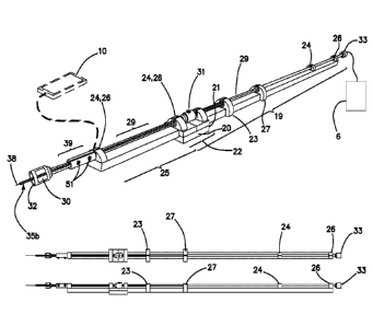
The basic elements of the EMMT PICC apparatus combine and allow access to
nested networks,
giving the operator a seamlessly networked experience of procedural
efficiency. The first layer of
networking involves the basic elements of the apparatus: the external
electromagnetic motion control unit
(19), the sterile enclosed catheter assembly (29), the operator control
interface and hand grasp assembly
(39), and, an optional mechanical structural linking member (25) that may
bolster the linkage between the
operator control interface/hand grasp to (39, 51) to the motion control unit
(19). This linking member (25)
would have properties of light weight and, optionally, flexibility. In some
embodiments, the linking
member (25) would not be necessary as the tubular shaft of the sterile
enclosed catheter assembly (29)
would be of suitable structural characteristics to provide the necessary
support alone. The first layer of the
system can allow for the user to observe the actual spring state of the
catheter and guidewire through an
observation window (30) and/or possibly for electromechanical haptic feedback
of the motion control
system to the cellphone display or to the operator control interface (51).
Additional digital equipment can
be networked locally through the second layer local network (17) (e.g.,
bluetooth) allowing for hybrid
.. data to be generated/displayed (eg tracking, stored energy display,
telemetry monitoring). The third
conceptual network layer is the extended network (18) which can be integrated
through GPS/GSM/3G/4G
WiFi/internet or other connectivity to systems including EMR, nursing triage,
physician ordering, and
radiology PACS interfaces, essentially any data that is available via the
smartphone hub. The system
11
CA 02958871 2017-02-21
WO 2016/029228 PCT/US2015/046610
documents and logs the "time out- among other procedural checks as an example
of EMR connectivity
supporting the procedural efficiency. The operator's experience is a wealth of
very useful and
empowering information access toward delivery of the catheter implant. The
continuous access to the
EMR and extended hospital/operations networks brings additional advantages.
In one preferred embodiment, the invention combines: an electronic, light
weight battery (or other
electrical source) powered mechanical motion control system (19) for
manipulation of principally
magnetic nonsterile actuators (23, 27) which link to other
manipulators/actuators that control the motion
of the end drive elements (35b,38) which are within a sterile enclosure
(enclosed tube 29). The magnetic
array of the nonsterile, ideally cylindrically shaped (e.g., barrel), magnetic
actuators (23, 27) preferably
comprises a unipolar configured circular Halbach array in order to generate
the maximum and most
uniform magnetic field strength within the space, although other magnetic
configurations would be
acceptable.
The sterile enclosed end drive elements could contain diametrically aligned
cylindrical centrally
fenestrated magnetic elements (34), possibly diametrically aligned Neodymium
fixed magnets, or,
ferromagnetic rods which are coupled to sterile enclosed single use catheter
(35b), for eventual implant,
and single use/recyclable guidewire (38). The catheter and guidewire will have
these magnetic actuators
(34) incorporated into the sterile enclosed catheter assembly (29), thereby a
magnetic or electromagnetic
coupling system, eventually connecting end drive elements to the operators
supporting hand: sterile
catheter assembly (34, 35a, 35b) to external and nonsterile catheter rotor
translator (23) and sterile
guidewire assembly (34, 36, 38) to external and nonsterile guidewire rotor
translator (27). These end drive
elements are then linked to an operator control interface and integrated hand
grasp (39,51) through a
physical structural linkage to the external electromagnetic motion control
unit (19) and sterile enclosed
catheter assembly (29) and then a magnetic linkage of rotor translators
(23,27) through the sterile
enclosure (29). Haptic feedback of the rotor translator motors to the operator
interface/hand grasp (39, 51)
is possible. The operator control interface additionally allows control of
optional fluid reservoir/advanced
actuator/sensor (6) controllers electronically (with or without physical
wires). The advanced
actuator/sensor can incorporate sensors such as blood pressure or EKG sensors.
The fluid
reservoir/advanced actuator/sensor array (6) is also capable of providing
haptic feedback to the operator
interface/hand grasp. A second visual and auditory operator interface, a
smartphone (10), provides access
to a broad band telecommunications link (bluetooth and wifi integration) to
achieve a display which may
be programmed to allow visual or audible haptic feedback (e.2., physical
observation of the catheter
location, guidewire stored energy, motor enemy, blood flow direction, EKG data
or combinations of
such), and, the smartphone also may link to local (17) and extended (18) data
networks and display this
12
CA 02958871 2017-02-21
WO 2016/029228 PCT/US2015/046610
data. Thus, the optional smartphone may be used for immediate procedural
information regarding the
insertion of the endovascular instruments (catheter/guidewire/fluids/advanced
actuators/sensors) and it
also may provide other data from the extended networks that are generally not
immediately relevant to the
physical act of instrument operation.
Integration of ultrasound imaging, digital radiographic imaging and/or
electromagnetic tracking
to improve efficiency and effectiveness of the system is possible for its
intended purpose of PICC
delivery. The guidewire and/or catheter (38, 35b) may be designed to provide
supplemental information
through the sterile enclosed catheter assembly via transducers that can be
incorporated through advanced
assembly techniques and eventually to the smartphone interface (10). The
supplemental information may
be directly transduced from the patient's bloodstream from the remote location
of the catheter or
guidewire tip. The supplemental information could include internal and
external patient electrical
potential readings (electrocardiographic data). Other telemetry could be
displayed such as temperature at
the guidewire tip. Sensors such as these would help to guide a central venous
catheter to its intended
location. The use of a supplemental sub system, which is labeled in figures as
"fluid reservoir" (6) could
be used to extend the capabilities of the system as described. The sub system
can include a fluid reservoir
and advanced sensor and actuator array. In addition to hydrostatic contact to
the guidewire/catheter tip,
electrical or optical contact could be made. This sort of contact would enable
advanced sensors, which
could also be used to deliver treatment, thereby comprising actuators. The
actuator could represent a fluid
pump system connected to the sterile enclosed catheter assembly. This pump
system could administer
fluid from the catheter and at its distal open tip (35b) via the fluid
reservoir/advanced actuator/sensor
array (6). Treatment could also be electrically or optically mediated. This
may require that electrical
signal and/or fiber optic leads, and, possibly, grounding leads be
incorporated into the sterile enclosed
catheter assembly (29), which would require special engineering that is within
the scope of prior art.
Additional electrical or optical connections would need to be designed and
integrated into the sterile
enclosed catheter (and guidewire) assembly to connect the actuators
attached/incorporated into the
catheter and/or guidewire (35b,38) to the control system (10) via electrical
slip rings and/or fiber optic
rotary joints. The smartphone computer system could additionally be utilized
to control further
subsystems in order to deliver treatments from the catheter and guidewire
tips.
The inventive PICC design philosophy is to provide the best "up front"
operator experience of
any PICC system while minimizing per unit cost. Use of the system naturally
translates to improved
throughput and system wide gains. The image of a well-trimmed fly fishing rod
is an example of the ideal
heft and physical form of the device. It is a trusty tool, both sensitive and
strong, that is controlled by a
skilled operator. This is the design vision that the operator will appreciate.
It, the EMMT PICC system, is
13
CA 02958871 2017-02-21
WO 2016/029228 PCT/US2015/046610
indeed its own tool.
The sterile set up is easy and compact. First, attention is turned to the
smartphone (10) and
ordering information is reviewed and the procedural time out checklist is run
and documented. Next.
attention is turned to the sterile access site which is prepped out and
draped, usually a 12" (30 cm) square
sterile drape surrounding a 4" (10 cm) sterile prepped skin surface will be
easily sufficient. Next, the
sterile enclosed catheter assembly (29) containing the eventual implanted
catheter (35b) is snapped into
position and mounted to the motion control unit (19). This is accomplished via
mounts that are fixed
(26), linearly translating (24) or with linear translating and coaxial
rotational movement (23, 27) relative
to the sterile enclosed catheter assembly (29) longitudinal axis. The sterile
enclosed catheter assembly
(29) may require "through bore mounting" in some of these mounts, or, some
mounts may provide a
temporary fenestration for slotted fitting of the sterile enclosed catheter
assembly shaft. The operator
interface and handle assembly (51, 39) is mounted to the sterile enclosed
catheter assembly (29) directly
or possibly with an additional mechanical support (25). The operator interface
handle assembly (51, 29)
may be draped with a sterile bag and placed in the sterile field. Next the
operator dons gloves and gown
and -enters" the sterile field. Using ultrasound, standard Seldineer access
1101 is performed and a venous
access sheath assembly of special design is placed (43) with an occluding and
removable dilator (50). The
handheld system comprised of (19) (29) (39) and (25) is then grasped,
controlled and fitted to the venous
access sheath at a coupling assembly which includes a compression ring (44)
when the sheath dilator (50)
is removed. In alternative workflows, the sheath assembly (43) and dilator
(50) had been placed
previously by a first operator, and, that sheath may have been utilized for
venous access. Using the
operator control interface/hand grasp (39, 51), the catheter (35b) and
guidevvire (38) are guided into a
position that is felt to be satisfactory based on depth and dead reckoning (no
guidance is typically the
manner of placement). A radiograph can be obtained at this point in any manner
that would be
convenient. If the catheter is in good position, the euidewire is retracted
and the catheter pinned to the
skin and removed from the sterile enclosed catheter assembly (29) through a
releasable mechanism (see,
for example, Embolic coil delivery system with mechanical release mechanism US
7,901,444), and/or it is
cut with a blade. A hub is then mounted as to the cut catheter which is now
ready for use. The hub
mounting procedure is further detailed later in this description.
The operator is able to manipulate the catheter and guide wire independently
using one hand
through the use of integrated handgrip (39) and possibly by
joysticks/actuators of the system's operator
interface (51). The system may be supported in part by a gantry or hanging
from a tripod mounted boom.
Voice control of certain aspects of the system may be available through the
cellphone interface (10). The
hand guiding the system is also able to sense the physical linkage to the
actuation system through directly
14
CA 02958871 2017-02-21
WO 2016/029228 PCT/US2015/046610
transmitted vibration from the tip of the guidewire (38), as it is a well-
balanced device. The entire system
is designed to be light and easily managed in one hand if necessary, and, to
provide a tactile sensitivity
through its structural form. Operating the system with one hand allows the
operator's other hand to be
free to control the vascular access site and to operate in other manners that
may be desired and
.. advantageous, even possibly operating the first or a second hand held unit
simultaneously in a different
(not PICC) application of this technology (dual system operation).
Eyes are placed toward the proximal end of the system, at the patient skin
access site sheath
assembly to system front vent fitting, where a window (30) is positioned that
allows the operator to see
the guidewire and catheter and visibly obtain a cue about the potential energy
stored in the spring of the
guidewire/catheter based on how much the catheter is buckling from the
centerline. Alternatively, or in
addition to a window, the system may include an electronic sensor that
measures resistive force and/or
deformation of the guidewire and alerts the user. The window (30) may be built
into an area where the
guide wire (38) and catheter (35b) may be less constrained by the sterile
enclosure, and, this window in
turn is built into and incorporates a front vent connection fitting (32) such
as a luer lock fitting. The
smartphone (10) and its display is generally placed near to the window so that
the operator's body
position and center of focus do not need to change much during the procedure.
The smartphone display
demonstrates other information and allows integrated use of the EMR throughout
the entire procedure
(e.g., physician ordering information, triage information, timeout, telemetry,
reporting. chest x-ray
ordering and other features).
In some embodiments, the motion control system and sterile enclosed end drive
elements may be
fitted to the introducer sheath and then secured to the patient as well as a
gantry mounted to the patient in
some manner, and, then the drive mechanism of the system may operate via its
own onboard algorithm. It
may also be teleoperated fully or in part by a remote operator (12).
The system may deliver best performance through use of an integrated vascular
introducer sheath
assembly. To understand this, one must understand the geometrical and
structural concept of "linear
constrainment" of guidewires (and, sometimes, catheters). A key point to
understand regarding "linear
constrainment" is that, in general, the specific tool of an endovascular
guidewire achieves its best physical
performance characteristics when it is allowed to extend fully in a straight
line. In any position aside from
the linear state, the guidewire or catheter has some mechanical stored spring
energy and increased friction
and physical binding force that results in the net effect of poor handling
that is often characterized by
endovascular medical specialists as "poor pushability and poor trackability.-
To be free of any such
superimposed system aberrations, the guidewire and catheter should be held as
close as possible to a
physical straight line configuration. The system derives its elemental form
through embodiment of a
CA 02958871 2017-02-21
WO 2016/029228 PCT/US2015/046610
linear constrained system that may remain flexible, for example a straight 1
meter length of medical grade
plastic tube as a constrainment guide. A spiral tube constrainment guide could
have advantages in
portability of form, however, there would be costs in ultimate performance of
the system.
Linear constrainment is further divided into two basic forms: (1) the
constrainment force and tip
pushability and pullability that is provided to a guidewire or catheter by
tight cylindrical constrainment
alone; and (2) the directional and constrainment force provided to a tubular
structure (catheter) by a
centrally passing guidewire (the "rail"). Typically for a PICC, the guidewire
and catheter are between 50
cm and 2 m, preferably between 75 cm and 150 cm; in some embodiments the
guidewire is at least 30%
longer than the catheter, in some embodiments about twice as long as the
catheter.
Linear constrainment of a guidewire is useful because when a well constrained
guidewire
encounters a feature at its advancing tip that generates resistance at the
tip, that resistance can be
transmitted through the rigid guidewire and thereby sensed at the back end of
the wire. This sensitivity,
which is a feature of the rigid metal in its linear state, can be damped or
completely lost into stored spring
energy within a buckled guidewire or catheter/guidewire system. Minimizing
buckling maximizes ability
to sense the guidewire tip. Indeed, constrainment gives a catheter its ability
to be moved precisely at the
tip. Thus, elimination of the external body guidewire buckling is
advantageous. For this reason,
constrainment of a bare catheter without a hub is the best solution to
maximize performance of a catheter
delivery system. It can be seen that a hub such as Luer lock would increase
the diameter requirement of
the constrainment guide, and, thereby, would increase the buckling potential
of the guidewire or catheter
within the constrainment system, impeding its function. And, for this reason a
hub must be mounted to
the catheter delivered by the system in its most functional design, after the
catheter has been removed
from the system's constrainment. Thus, the system sheath should have a design
to aid in catheter + hub
fusion. This will be discussed further in the following sub section titled
"System integrated vascular
introducer sheath detail."
Sterile enclosed catheter/guidewire actuation subcomponents
The electromechanical actuation system subcomponents are important for
effective functionality
of the EMMT PICC system. There are four major design features regarding the
EMMT PICC
electromagnetic actuation subsystem to consider closely, first, magnetic force
generation and its
optimization, second, hydrostatic force management, third, end drive element
releasability, and, fourth,
linear constrainment/support.
For example, in the PICC designed system with a single catheter that may
rotate and translate
(although rotation is not required in every instance) and a single guidewire
that may rotate and translate
16
CA 02958871 2017-02-21
WO 2016/029228 PCT/US2015/046610
(although rotation of the wire is not required in every instance), it will be
important to maximize the
magnetic force of alignment between the external catheter rotor translator
(23) and its internal sterile
enclosed end drive catheter electromagnetic actuator sub assembly (34, 35a,
35b), and, similarly, the
external guidewire rotor translator (27) and its internal, sterile enclosed
end drive guidewire
electromagnetic actuator sub assembly (34,36,38). Stated another way, the end
drive electromagnetic
actuation elements are magnetically coupled for maneuverability through the
constraining internally
sterile enclosure assembly (29) to the external rotor translators which are
mechanically operated by an
external motion control system mounted to the external surface of the sterile
enclosed catheter assembly
(29) via a mounting system. Fixed magnet(s) or electromagnets) of the external
rotor translators (23, 27)
are coupled to fixed magnet(s) or ferromagnetic aligning and ideally barrel
shaped structure(s) (34) within
the enclosure which are braced against the inner surface of the sterile
enclosure and simultaneously
coupled to the end drive element (a catheter (35b) or guidewire(38) through a
low hydrostatic profile
linkage (35a or 36)). Use of single magnet systems would be simplest. However,
it is important to point
out that a simple single dipole magnet coupling at a single point on the
circumference of the rotational
path of the tubular enclosure may result in a relative increase in contact pad
force at the coupling
alignment point and relative increase in frictional losses with motion
compared with other arrangements
of magnets. An encircling or near encircling diametrically aligned ring magnet
or encircling dipole
magnetic array would have significant advantages to a single magnetic coupling
point. Furthermore,
encircling magnetic arrays can be fashioned in order to focus the magnetic
flux closely into the central
portion of the ring, as in a circular IIalbach magnetic array. Fixed magnetic
barrel shaped circular
Halbach arrays with unipolar diametrically aligned magnetic fields would be
ideal. Either of these types
of arrangements for the external rotor translator (23 or 27) magnet would have
significant advantages in
end drive element force of coupling: (1) the wall contact forces of friction
would essentially cancel out
leaving a net alignment force and minimized friction force, and, (2) the
magnetic field flux outside of a
Halbach rotor translator and thus interference with the external environment
would be absolutely
minimized. Fixed magnets, in a IIalbach arrangement, would be the simplest
alternative for the outer (23,
27) or inner (34) magnetic elements , however, in the case of the external
rotor translator (23 or 27)
magnetic element, electromagnets, even superconducting electromagnets could be
used to increase the
amount of force applied to the sterile enclosed end drive actuator elements
(34, 35a, 35b, 36, 38). The
.. rotor translators (23, 27) would themselves be actuated externally through
physical attachment to an
electronic mechanical supporting structure with possible linearly fixed (26)
and linearly translating (24)
constrainment rings comprising the external electromagnetic motion control
system. Indeed, so called
-frameless" electromagnetic arrays could be utilized as rotor translators (23,
27) so as to have no moving
17
CA 02958871 2017-02-21
WO 2016/029228
PCT/US2015/046610
mechanical parts involved in the rotational actuation of the end drive
elements within the constraining
sterile enclosed catheter assembly, but, a mechanical means of moving the
external rotor translator
magnets through their useful orbits would likely use much less power and
require much less electrical
apparatus and complexity. Creating a C shaped circular Halbach array for the
external rotor translator
.. magnet element (23 or 27) would have advantages in that the sterile
enclosed catheter assembly could be
mounted through the perforation/gap in the ring (through the gap in the "C")
quickly and easily at any
point along the length of the sterile enclosed catheter assembly. This would
allow a quick release manner
that a solid ring magnet array would not allow. IIowever, a solid ring through
mounting over the distal
end cap and then shaft of the sterile enclosed catheter assembly (29) is
certainly reasonable as an
approach. One slight inconvenience with this mounting is that it would require
temporary removal of any
fluid reservoir and advanced sensor and actuator (6) associated tubing and
leads that may be connected
prior to mounting. In the case of fluid connection, this is not likely to be a
problem as flushing air from
the system via coupling to a fluid reservoir (6) could occur just as easily
after mounting the sterile
enclosed catheter assembly (29) to the external electromagnetic motion control
system (19). In addition,
a fluid access portal (31) is incorporated into the sterile enclosure. The
sterile enclosed catheter assembly
(29) could simply be placed coaxially through the bore of the two (or one or
greater number) of partially
retracted external rotor translator assemblies (23, 27) and any number and
types of supporting mounts
(24,26) prior to attachment to any fluid reservoirs/advanced
actuators/advanced sensors (6) that may be
used.
As the rotor translators provide the linear translation component of their
force to the end drive
elements of the catheter (35b) and guidewire (38) there is a potential to
exert a coupled hydrostatic
pressure on the fluid column suspended within the sterile enclosed catheter
assembly, similar to a
hypodermic syringe. This could result in a net outward pressure force or
inward suction force at the front
vent hub opening (32) of the sterile enclosed catheter assembly if the
internal catheter (35b) or guidewire
(38) was being pushed forward or retracted. The motion of rotation would
probably have less of an effect
on the hydrostatic pressure generation, unless the end drive magnetic actuator
was purposefully designed
to generate pressure differential through rotation, such as a propeller or
turbine impeller form, which may
have utility in some instances. The hydrostatic force related to pushing or
pulling the catheter and
guidewire through the sterile enclosure with regard to PICC applications would
have to be controlled to
maintain the purest balance and functionality of the system in its coupling to
a fluid filled artery or vein,
for example. Continuous positive pressure flushing from a connected fluid
reservoir (6) is a very good
option. Attention to minimize the cross sectional profile of the end sterile
enclosed magnetic drive
element/electromagnetic actuator (34) is important in minimizing the net force
that these elements place
18
CA 02958871 2017-02-21
WO 2016/029228 PCT/US2015/046610
on the fluid column contained in the sterile enclosed catheter assembly.
Integrated design of perforations
of the inner sterile enclosed magnetic actuation element is important to
consider. This could involve
placement of an internal feiTomagnetic rod, a diametrically aligned
cylindrical fixed magnet, or, internally
placed circular Halbach array, any of which could be engineered to support
large central or otherwise
aligned perforations or tubular windows with little loss in capability for
magnetic force generation. To
some degree, the internal electromagnetic actuator element should mimic the
form of the external
catheter/guidewire rotor translator, but, with a smaller outer diameter, ease
of sterilization, and, a low cost
so as to be disposable after a single use. A diametrically aligned cylindrical
fixed magnet may be optimal
as it has a large central fenestration and can be closely approximated to the
external magnetic rotor
translators. This could be embedded into plastic in order to develop a
supporting structure for the end
drive elements. In the case of coupling to the catheter end drive element, a
cylindrical sieve plate mount
(35a) from the enclosed magnetic actuator element (34) to the end drive
catheter (35b) could also
be employed in order to help maintain neutral hydrostatic pressure between the
inner lumen and outer
space of the catheter in front of the magnetic actuator element (34) which
would be braced against the
inner wall of the sterile enclosed catheter assembly (29). In some
embodiments, the catheter is sealed with
an a-ring at its distal exit, at the front vent (32) which could lead to
hydrostatic pressure generation along
the length of the catheter, outside of the catheter, within the sterile
enclosed catheter assembly (29).
The cylindrical sieve plate mount (35a) would solve this potential fluid
management problem. The sterile
enclosed magnetic drive element/electromagnetic actuator (34) can be braced
within the outer tubular
constrainment guide via close tolerance fitting, thus it preferably has a
cylindrical form to fill the inner
lumen of the tube. It would be possible to use hydrophilic coatings in order
to further minimize the
friction between the inner wall of the sterile enclosed catheter assembly
tubular shaft and the magnetic
drive element/electromagnetic actuator (34). If significant hydrostatic force
generation was still present
following engineering optimization, additional attention could be placed to
the attached fluid reservoir (6)
and its controllers in optimization of the system with regard to hydrostatic
pressure generation. For
example, (6) could be programmed to open the chamber with advancement and
close the fluid chamber
with retraction. This would have the net effect of introduction of fluid on
advancement and static fluid
column with increased drag on the motion in retraction. This would be an
acceptable arrangement as the
sterile enclosed catheter assembly would remain "bloodless" and there would be
little loss in
functionality. A continuous positive pressure at (6) would also work and would
be simpler to achieve.
Furthermore, there could be many arrangements and methods to deliver fluid via
(6) including liquid
medication administration and even CO2 gas for related angiography methods.
having perforated or low
hydrostatic profile magnetic drive elements/electromagnetic actuators (34)
within the sterile enclosed
19
CA 02958871 2017-02-21
WO 2016/029228 PCT/US2015/046610
catheter assembly (29) is important to the final functionality of the system.
Preferably, components are
porous and/or block less than 90%, more preferably less than 50% of the cross-
section (perpendicular to
flow) of the enclosed tube.
Mounting the magnetic drive element/electromagnetic actuator (34) to a
guidewire (38) is a
fundamentally different geometric arrangement relative to mounting of a
catheter (35b) with respect to
hydrostatic force generation. Again, a perforated structure will be desired to
minimize hydrostatic force
generation. The larger the net cross section of the fenestration void area,
the less the hydrostatic force
generated by translational motion within the enclosing tube of the sterile
enclosed catheter assembly. A
cross braced annular sieve mount (36) is a desirable form of the coupling
spanning a cylindrical sterile
enclosed Halbach array actuator, for example, to the central or off center
placed guidewire end drive
element (38). Alternatively, a simple single or dual post mount radially and
centrally projecting from a
cylindrical magnetic drive element/electromagnetic actuator could be used
rather than a cross braced
array.
Alone with hydrostatic force generation, releasability of the magnetic drive
elements/electromagnetic actuators from their respective end drive elements of
the catheter and guidewire
should be considered. There is always the magnetic releasability which is
inherent in the system. The
catheter and guidewire are held in position by magnetic force that may be
overcome by pulling or pushing
against the support tube of the sterile enclosed catheter assembly. Additional
mechanical and/or electric
and/or electromagnetic release actuation may be utilized further. As the most
simplistic method, a
designed "breakaway" release is possible, which would require an engineered
fracture point in the linkage
holding the catheter (35b) to the catheter electromagnetic actuator (34) and
this fracture point could
involve any portion of the sieve mount (35a) attachment or some specially
designed portion of the
catheter (35b) itself. The catheter could also just be cut by a scissors at
any point along its length once it
was pushed out of the sterile enclosed catheter assembly (29). Still a second
point of breakaway
releasability would likely have utility as a fail safe. In the case of the
guidewire (38), it could be fractured
at a structural score location proximal to the guidewire electromagnetic
actuator and annular sieve (34,
36) or, it could also be cut by a scissors or shear from the actuator assembly
(34, 36). More complex
actuated release of the elements is also possible. There are several methods
for embolization coil and
guidewire release that are known to those versed in the art, and, any of these
release methods could be
used in a modification in order to serve the purpose at hand. (Embolic coil
delivery system with
mechanical release mechanism US 7901444, for an example of rapid exchange over
the wire catheter with
breakaway feature see WO 1993011822). Additional electrical, mechanical or
magnetic actuators would
have to be modified for use with the system.
CA 02958871 2017-02-21
WO 2016/029228 PCT/US2015/046610
Releasability of the catheter (35b, 42) would eventually be required in order
to create a free
catheter implant to be left in place within a patient, as is the goal of the
PICC procedure. The guidewire
(38) would not need to be separated for catheter implant creation, and, in
some cases it would be better to
keep the guidewire within the sterile enclosure for ease of disposal and
environmental protection from
.. blood contamination. To free the catheter and guidewire completely from the
motion control system and
the sterile enclosure assembly would allow the system to derive a level of
flexibility toward the next steps
that are typical and essential to a host of endovascular procedures. For
example, diagnostic aortogram and
superior mesenteric access sheath placement could be performed with a modified
system, and, the
released catheter tube, a superior mesenteric access sheath possibly with a
hemostatic valve and sideport
.. access fused, could be used in the further steps of a procedure that relies
on work performed through a
celiac access sheath, such as internally bleeding vessel embolization. These
additional procedural steps
can be thought of as building onto a PICC placement procedure, which is
similar to selective vascular
introducer sheath placement. Artefiography would require injection of
intravenous contrast for digital
subtraction fluoroscopic x ray angiography. Thus, the invention includes use
of the inventive devices and
.. methods in these applications. The contrast administration through a
catheter that is not separated from
the delivery system would rely on fluid administration via a fluid reservoir
actuation mechanism (6) that
is connected via tubing to the lumen of the sterile enclosure and eventually
to the tip of the catheter (35b).
There are prior art addressing such mechanisms (Medical fluid injection system
US 20140039310).
Modifications of these techniques could be applied to the current system with
a key integration into the
operator control interface and eventually the catheter tip. Additional design
features related to (1)
hydrostatic seal points and (2) fluid continuity with the catheter lumen would
be required. These
hydrostatic seal points could be utilized to isolate lumen(s) of the contained
catheter(s) element relative to
the external fluid reservoir, and, most straightforwardly, the entire housing
could have an o ring seal at its
front vent / luer lock (32) fitting closely around the outer diameter of the
catheter (35b). This would
.. provide fluid continuity of the catheter lumen at the catheter tip to the
fluid reservoir (6). Additionally, in
particular with noted placement of the previously described sieve mount
holding the catheter to its
electromagnetic actuator, the front vent (32) associated o ring seal could be
loosened which would have
the effect of allowing fluid to escape around the catheter from the sterile
enclosure into the vascular
introducer sheath, which would have great utility in certain circumstances
allowing fluid administration
through the delivery sheath as well as the catheter tip. The o ring seal could
be adjustable so as to be
switched between delivering fluid from the catheter tip or the catheter tip
and introducer sheath tip.
External Electromagnetic Motion Control System
21
CA 02958871 2017-02-21
WO 2016/029228 PCT/US2015/046610
The entire system is designed to be a handheld device or hand supported and
guided device. Its
weight could be supported in part by a stand/mount, or, on the patient's bed
as is typical of other catheter
and guidewire systems that are used in angiographic procedures. End drive
elements (34, 35a, 35b, 36,
38) can be manipulated via an operator interface that is incorporated into a
handle (39) for additional
support/guiding of the entire device. The motion control system (19) can be
coupled to the operator
interface handle by means of the actual sterile enclosure tubular shaft
(proximal portion of 29) and
possibly with an additional supporting member (40) that bridges the operator
interface handle (39) to the
motion control system (19). The motion control system (19) is comprised of a
power supply (20) that is
ideally entirely portable and onboard, a battery. Wired power from an external
source could be used as
well. The motion control system also incorporates a structural support for a
gantry approximating the
length of the sterile enclosed catheter and guidewire elements. Ideally, the
length would accommodate
possible extension of a bare guidewire through a catheter to be deployed to a
target length for the catheter
and then advancement of the catheter over the extended guidewire to the
target. This could result in a
tubular enclosure that is twice the length of the eventual implant, assuming a
first guidewire deployment
to target followed by guidewire deployment over that catheter. For example,
for a 60cm PICC, a
guidewire of length greater than 120cm would allow bare advancement of the
guidewire alone to a target
60 cm or less from the system front vent (32). Over this bare wire extending
60 cm from the front vent,
the catheter (60cm) could be advanced, covering the external guidewire and
exposing the sterile enclosed
portion of the guidewire. The guidewire could then be retracted and the
catheter released or cut for the
implant. However, it is usually the case that a catheter is advanced
simultaneously with an internally
passing guidewire, such that the bare guidewire is exposed only 5 to 10 cm
beyond the catheter tip. In this
case, the entire motion control system could be considerably shorter, for
example 70cm rather than
120cm. Indeed, the length of the entire system could be equal to that of the
eventual implant, for example
60cm, if the guidewire was simply an internal supportive structure to aid
delivery of the catheter implant,
a stylette. For purposes of catheter and guidewire manipulation techniques, it
is better to have some bare
exposed guidewire in front of a catheter to allow more maneuverability.
The system support gantry would hold one or more rotor translator magnetic
elements (23, 27).
These elements can undergo linear movements along the length of the sterile
enclosure and shaft of the
motion control system 19, and, they can undergo rotational movements around
the central axis of the
elongated tubular sterile enclosure. This motion would likely be accomplished
through a system of gears,
worm screws and possibly belts and pulleys linked to electric motors that are
involved in the mechanical
linkage of the rotor translator magnetic elements (23, 27) to the supporting
structure of the external
electromagnetic motion control system (19). It would have an onboard
microcomputer and
22
CA 02958871 2017-02-21
WO 2016/029228
PCT/US2015/046610
communication system (21). There are a variety of motor and linkage
configurations which could be
engineered to maximize functionality and minimize cost and complexity.
The electromagnetic motion control system (19) would be designed to mount
fixedly to the sterile
enclosed catheter/guidewire assembly (29). The mounting process would involve
disposal of the tubular
shaft of the sterile enclosure coaxially through the centers of the external
rotor translator elements (23,
27) and possible other constrainment guides (24 which moves linearly, 26 which
is fixed in place). As
stated before, this could be achieved by passing the distal end of the sterile
enclosed catheter/guidewire
assembly through the rotor translator bore, or, by creating a window for
passage of the sterile enclosure
through the annulus of the rotor translator. Either manner is acceptable.
Additional nonmagnetic coaxial
guides for the sterile enclosure could provide additional support as needed
(24, 26).
The sterile enclosed catheter assembly (29) locks to the electromagnetic
motion control system
(19) gantry to provide rigid attachment without rotation of the elements
relative to one another. An
optional support member (25) linking the proximal sterile enclosed
catheter/guidewire assembly to the
operator interface handle is possible and may have benefits.
The entire sterile enclosed system can be coupled via tubing to a fluid
reservoir system (6) for
control of fluid flow at the proximal end of a catheter. There may be a vent
attachment point of the sterile
enclosed catheter system (29) to the external fluid reservoir at the distal
end of (29) where a rear vent and
coupling fitting (33) can be located. Additional venting(s) can be placed in
the middle or anywhere along
the length of the enclosure (31). A front vent and attachment fitting (30) is
on the proximal end. Simple
control of fluid pressure at the end of the catheter would be useful in
extended applications of the system.
Operator Interface / Hand Grasp support
The operator control interface and hand grasp support sub assembly (51, 39) is
designed to give
the user, ideally, one handed control of the catheter and guidewire. This hand
provides physical support
and control over the orientation of the sterile enclosed catheter assembly
(29), the electromagnetic motion
control system (19) attached fixedly to it, and, the operator interface hand
grasp (39) and possible support
shaft (25) that comprise the major elements of the system. Fluid pressure
control is allowed through the
operator interface which is connected to the fluid reservoir (6) controller.
The operator interface and hand
grasp are placed at the proximal side of the system in order to place the
operator at the traditional
endovascular operating position, with one hand able to control the vascular
access site and attend to the
requirements of hemostasis and general endovascular procedural work required
and typically performed
at this location. The operator interface itself may be controlled by one or
more finger joysticks (51) or
possibly by a combination of switches, voice commands, joysticks and other
manners enabled through
23
CA 02958871 2017-02-21
WO 2016/029228 PCT/US2015/046610
wireless or wired linkage to a smartphone (10). The operator interface could
alternatively be controlled
remotely through a local or extended area network (11,12) by a second operator
allowing the first
operator to attend to other procedural issues.
.. Catheter to hub coupling components and discussion of advanced applications
In order to deliver a useful PICC implant, the delivered catheter requires a
functional hub
termination at its distal end to be used for medical treatment by infusion or
sampling. An ability to create
a hub at any position along a catheter "on the fly" would have particular
advantages in the application
application to PICC, because each patient anatomy and access site location has
a particular optimal
.. catheter length. The PICC with an "on the fly" hub creation could be
customized on a per unit basis with
a luer lock fitting at the skin level for use. In the case of alternative
catheter applications as conduits for
interventional radiology/endovascular procedures, a hub with two elements (1)
a hydrostatic pressure
infusion side port, and, (2) a hemostatic introduction port for a guidewire or
other tool. The most basic
hub termination is a luer lock fitting, which can be capped with an
appropriate additional fitting, or,
attached to medical tubing or syringes as the luer fitting is essentially an
industry standard fitting.
More complex hub terminators to be fitted to catheter tubes are generally not
used in PICC but
instead for interventional radiology procedures. A very useful hub includes an
often T-shaped or Y-
shaped ensemble of (1) a hydrostatic pressure infusion side port, and, (2) a
hemostatic introduction port
for a guidewire or other tool. This type of arrangement allows for external
hydrostatic pressure infusion or
.. sampling while the main lumen is partially occupied by a tool such as a
guidewire entering the catheter
lumen at a hemostatic (atmospheric sealing "dripless" valve) with tip
displaced into the patient.
An ideal pre fabricated and non customizable length catheter + hub for use in
the proposed
invention would be very "low profile," essentially having an outer diameter
very similar or slightly larger
than the diameter of the implanted catheter. A hollow metal screw tipped
hypotube bonded to the silastic
.. catheter would be an example. A typical bulky luer lock hub could then be
attached easily by threaded
fitting following catheter deployment. PICC silastic catheter material is
usually of outer diameter 5F,
which is less than 2mm (1.67mm). The diameter of a male luer fitting, which is
the universal medical
tubing adaptor, is about 45mm. Therefore, it would be useful to fuse a luer
lock type hub to a catheter
following its removal from the sterile enclosed catheter assembly of the
proposed invention, in order to
maximize the pushability of the catheter while it was disposed within the
sterile enclosed catheter
assembly. There would be less buckling and shortening of the catheter and
guidewire if they were
constrained closely. Methods to achieve catheter to hub fusion in (1) flexible
type catheters (100) (typical
PICC), and, in contrast, (2) rigid plastic PMMA catheters (101) (access
sheaths, diagnostic an2iographic
24
CA 02958871 2017-02-21
WO 2016/029228 PCT/US2015/046610
catheters) are briefly described. Furthermore, a hub including a hemostatic
valve and a side port fluid
access is proposed for extended applications beyond PICC.
In the first case, where the deployed catheter is a flexible, compliant (e.g.,
silastic) catheter, the
catheter would have a property of increasing its diameter by stretching in
order to accommodate an
internally passing, rigid tubular structure / hypotube (102) with or without
external etching, external
diameter variability (flared or beveled shape) or scoring barbs. The catheter
could additionally be
interposed between a rigid internal hypotube and an external compression ring
(103), in order to create a
fitting of the catheter tube to a large diameter bulky hub with a high
pressure seal capability (900 psi).
This sort of fitting would also be very durable. In order to be most useful to
the PICC application and to
.. the other extended applications, the compression ring (103) could be
mounted to the initial access sheath
(104) gaining first introduction to the vessel. This can be a removable and
very low profile "peel away
sheath." This would allow a very easy transition from bare catheter to
catheter + hub construct as the
encircling compression ring (103) would already be in position encircling the
catheter/implant at its initial
introduction, and, the catheter and compression ring would be ready and in
close position to accept the
internal fitting rigid hypotube (102) and bulky external hub (105) component.
In this case of flexible
tubing (100), a bare or released catheter would be cut a few cm from the
insertion point access sheath, a
hypotube (102) + bulky hub (105) construct would be coaxially pushed into the
catheter lumen, and, this
construct could be pushed into the access sheath hub associated breakaway
compression ring (105). Force
could be exerted to the separated compression ring (105) alone to make the
fitting, or, the compression
ring (105) could be pressed into position while attached to the vascular
introducer sheath (104), which
may provide additional grasping support structure. In this way, the hub could
be created with few physical
steps and minimal risk for inadvertent catheter dislodgement and free
embolization into the patient's
bloodstream. Additionally, bulky hub (105) modifications could include side
port access tubes (106) and
hemostatic valves (107) in order to create an introducer sheath arrangement
with ability to accept a
coaxial instrument (catheter and/or guidewire) while still having a fluid
"side port" for administration of
fluid or fluid suction. This could allow for creation of customizable length
sheaths (a sheath having a
sideport and hemostatic valve with or without luer adaptors where a catheter
would only have a luer lock
port) as well as customizable length catheters (typically only having a luer
adaptor fitting at the distal
end).
In the case of typical vascular introducer sheaths currently in use for
endovascular procedures,
these are generally fabricated from more rigid PMMA material or braided
composite material rather than
compliant silastic material typical of PICC. Thus, the sheath tubing materials
currently in wide use would
generally not be capable of stretching over a hypotube of the same diameter to
form a seal as previously
CA 02958871 2017-02-21
WO 2016/029228 PCT/US2015/046610
described. In this case, hub fusion would be more challenging. One method to
overcome this challenge
would involve a hub fusion method that is disclosed as follows and in the
accompanying drawings. First,
a hemostatic valve with or without a sideport or luer fitting (105) assembly
would be held on an internally
passing obturator (108) of outer diameter matching the inner diameter of the
tubular introducer sheath
conduit to which the hub or hemostatic valve is to be fused. The hub would
have a proximal end sleeve
(109) that could accommodate the sheath tube internally coaxially, of slightly
larger diameter than the
tubular sheath (101) conduit outer diameter. The hub (105) would eventually
decrease abruptly in
diameter to a diameter smaller than the sheath tubing, which would stop the
internal progression of the
catheter bracing it against the hub (105) and providing a preliminary seal.
With the tubular sheath (101)
braced in this manner against the hub and the lumen of both elements
internally occluded and bridged by
the obturator, a bonding agent could be applied to fill the void between the
sleeve and the internally
placed sheath creating a hemostatic high pressure bond. The bonding agent
could be applied in a variety
of manners, but, engineering its delivery into the hub components through
microfluidic channels is one
method.
Another method would be use of the obturator (108) to place the bonding agent
via the inner
lumen of the composite structure. The obturator would protect the lumen from
embolization or occlusion
by the bonding agent. Ideally, the bonding agent would he unable to bond to
the obturator, or, fittings
could be designed to protect the obturator from the bonding agent. The bonding
agent would have to be of
suitable viscosity to form a gapless bond through flowing and cohesion without
air gaps/filling voids. A
number of bonding agents are currently in use that would meet these
specifications.
In another embodiment of a system that would not require catheter/sheath to
hub fusion, the
catheter and a larger diameter bulky luer fitting on the distal end of the
catheter could be pre-fused and
mounted within a suitably large diameter tubular housing of a sterile enclosed
catheter assembly. An array
of internal spacer disks or a metallic spring supports (not pictured) could be
placed within the sterile
enclosure and alongside the length of the catheter in order to provide
internal constrainment of the
catheter tube against the inner wall of the sterile enclosure proximal to the
bulky hub, thereby
constraining the catheter where there would be potential for buckling.
Fashioning the internal spacer disk
array or metallic spring(s) to accommodate the catheter closely fitting within
a "notch" along the outer
peripheral surface of the spacer disk would allow the disks to be easily
released from the catheter when
they were freed from the sterile enclosure through a port in the distal end of
the sterile enclosure. This sort
of internal disk/spring spacer system would provide a way to maintain
pushability of a narrow catheter +
larger diameter hub for use in the electromagnetic actuation of the sterile
enclosed system. The disks or
metal spring, being non-encircling to the catheter implant, could be easily
removed from the implant
26
CA 02958871 2017-02-21
WO 2016/029228 PCT/US2015/046610
when it was removed entirely from its tubular enclosure. The front vent and
catheter seal could be left in
position around the catheter with little increase in hub bulk and no
functional detriment.
Single and multi lumen catheters
Conceptually, we have generally considered the catheter (35b) to be single
lumen to this point.
Next, we will consider the system functioning in the case of carrying a dual
lumen catheter (42). A
detailed diagram demonstrates how a catheter of this configuration may appear
in cross section. The
guidewire would pass only through a single lumen of the catheter. Otherwise,
to this point, the system
would perform nearly identically. In the next section, we consider hub fusion
to a dual lumen catheter
(42), which presents important subtle modifications of design to the hub
fusion that should be considered
to provide a dual lumen PICC implant.
System integrated vascular introducer sheath detail
The EMMT PICC system, comprised of the external electromagnetic motion control
system (19),
the sterile enclosed catheter assembly (29), operator control interface and
hand grasp (39), a possible
supplementary mechanical linkage of (39) to (19) and possibly (29) and the
smartphone device (10), is
designed to deliver an implant which is part of the catheter assembly
(34,35a,35b) stored in the sterile
enclosed catheter assembly (29) into the body of the patient to serve as a
central venous catheter.
This generally requires passage through a previously placed vascular
introducer sheath bridging the
external environment to the endovascular endoluminal environment. The sheath
is typically placed via the
Seldinger method 1101 and modifications of this technique. The Seldineer
access method begins with the
percutaneous introduction of a needle into a tubular vessel or hollow viscus
organ preferably via a "single
wall" puncture of the tubular structure via the needle with the tip placed
inside the lumen of the hollow
viscus organ. The percutaneous needle access can be aided by imaging such as
ultrasound or fluorscopy.
The needle placement is then followed by advancement through this needle of a
supporting guidewire
through the needle extending some distance of typically 5-30 cm into the
tubular lumen. The guidewire is
typically 0.038, 0.035 or 0.018 in diameter. The needle is then removed
completely from the vessel and
removed from the back of the guidewire leaving the guidewire bridging the
external environment, passing
through the skin and subcutaneous tissues and entering the lumen of the
tubular organ.
Over this supporting guidewire, a tubular "access sheath" for secondary tool
coaxial placement is
eventually advanced, possibly following skin incision and serial dilation of
the arterial or venous puncture
zone tract along the guidewire using sequentially larger and larger diameter
dilators in order to minimize
the trauma to the arterial or vascular wall. The vascular introduction sheath
(43) is eventually passed over
27
CA 02958871 2017-02-21
WO 2016/029228 PCT/US2015/046610
this guidewire with a mated and tapered space occupying internal dilator (50).
The dilator (50) is
eventually removed and the hollow, tubular sheath (46) that is left in place
provides a conduit for entry
and reentry into the tubular viscus lumen. The sheath is usually made of
medical plastic and may often
have a "break away" or "peel away" capability that allows the sheath to be
removed while leaving in
place a centrally passing second catheter or sheath passing through the first
"peel away sheath." A
hemostatic valve (107) is often integrated into a typical vascular introducer
sheath. This is the usual
method for so-called "access sheath placement- for interventional
radiology/endovascular surgical
procedures. It is important to note that the system, in the case of dual lumen
PICC, delivers a portion of
flexible medical endovascular catheter tubing (35b) which is releasable from
the electromagnetic actuator
and the magnetic elements of the sterile enclosed catheter assembly (29). The
catheter (35b) may be
trimmed with the guidewire retracted completely back into the sterile enclosed
catheter assembly (29), for
example, by an 11 blade scalpel or scissors. The catheter (35b) must
eventually be fitted to a luer lock
retention hub carrying and mounting one or more lumens (47). The two lumen
example is further detailed
in following descriptions as an example, although it is technically more
challenging than would be for a
single lumen catheter.
The sheath designed for integration with the current invention (43) through
which the bare
catheter (35b, 42) is deployed is a design that has advantages specifically
when utilized with the EMMT
PICC system as described, particularly in that it allows mounting of the free
catheter (35b,42) to an
eventual hub construct through the addition of a separate piece carrying, in
the case of a dual lumen
catheter, 2 additional luer lock lumens with luer lock adaptors as well (47).
The mounting of (47) to (42
or 35b similarly) occurs through two separate and internally isolated rigid
hypotubes (49) likely to be
constructed of stainless steel or plastic that may be externally fused. These
hypo tubes may be flared
proximally or distally so that pressing (42) over (49) provides some elastic
constrainment of the bare dual
lumen catheter (42) to the hub (47) . Directional friction etching may also be
used. However, this level of
constrainment is likely to be insufficient. The dreaded complication of
embolization of the free catheter
into the body must be avoided completely, requiring a durable and completely
permanent fitting.
Therefore, the sheath (43) conduit tube has key features to be noted.
First, a portion of the sheath may be separable so as to "peel away" (46) over
the implanted
catheter (35b,42) separated from the remaining magnetically charged and
perforated portions of the
catheter assembly (34, 35a) that remain attached to the sterile enclosed
catheter assembly unit (29) with
minimal mechanical disruption. This "peel away" method and functionality is
well known to practitioners
of the art of interventional radiology. It is typical that most vascular
introducer sheaths for PICC and other
implanted central lines "peel away" completely and are completely removed,
being temporary access
28
CA 02958871 2017-02-21
WO 2016/029228 PCT/US2015/046610
conduits.
Second, a portion, at least, of the sheath will not peel away (44, 45) from
around the eventual
implant catheter, for example a dual lumen catheter (42) or a single lumen
catheter (35b). These left in
place encircling member(s) (44,45) may serve additional functions when
combined with the catheter
(35b,42) and a hub element (47) which is intended to be eventually fixedly
fused to the catheter externally
to serve as an external connector. These functions of the left in place
portions of the introducer sheath (44,
45) principally involve securing the fusion of the catheter (35b, 42) to the
hub element (47) and then
securing the hub and catheter assembly to the skin. In this case of a dual
lumen hub, coupling of a variety
of medical tubes for fluid sampling and administration is accomplished usually
through a system of
female and male luer lock fittings. An important and useful feature is the
fusion aiding ring (44). An
additional mechanical finger grasp/securement device (45) can be integrated
for the operator to facilitate
press fitting the fusion aiding ring (44) into position. The hub/hypotube
element (47) could incorporate
another mated finger grasp that would be natural and easy for the operator to
manage, possibly by a single
hand.
Another useful feature of the non-peel-away sheath components (44, 45) may
include facilitating
retention of the implant to the patient's skin surface. This could be
accomplished through incorporation of
adhesive external retention device into a fusion ring assembly (44) or finger
grasp/retention assembly
(45). Another method could incorporate that the mechanical press fitting of a
fusion ring (44) to the rigid
hypotubes (49,102) for fusing single lumen (35b) or dual lumen catheters (42)
activates deployment of a
mechanical retention device that could include semi permanent tissue barbs or
sutures. In another
conception, the removable dilator (50) could be utilized to deploy a suture
mediated retention of some
portion of the fusion ring (44) or finger grasp/retention device (45) to the
patient's skin.
In the end the fusion ring (44) is integrated into the external portion of the
implant along with the
finger grasp/retention device (45) and hub assembly (47). An additional
element, a hemostatic coupling
.. may be integrated into the finger grasp portion (45) and or other sheath
elements (44, 46) accepting fitting
of the sterile enclosed catheter assembly (29). Features such as mechanical
cushioning derived through
flexibility or possibly telescopic entry would be useful for the the mounting
point of the sterile enclosed
catheter assembly (29) and its front vent (32) to the sheath (43). This
mounting typically will include a
quick release hemostatic valve that could free the sterile enclosed catheter
assembly (29) from the sheath
assembly (43) quickly and with minimal motion of the sheath within the
patient. The fitting of (32) to
(43) should integrate well into the mechanical feedback observation window
(30). Luer lock is proposed
initially but any other type of easily handled and non cumbersome fitting
would work. A "slip-tip" type
coaxial fitting design may be preferable at this location. The hemostatic
coupling portion of the finger
29
CA 02958871 2017-02-21
WO 2016/029228
PCT/US2015/046610
grasp (45) may be incorporated into the mechanical fusion ring (44) or
incorporated into the peel away
sheath/finger grasp (46) or may be slotted and/or otherwise peeled away
independently from the sheath
(46).
LITERATURE CITATIONS [tt]
1. Rivera AM, Strauss KW, van Zundert A, Mortier E. The history of peripheral
intravenous catheters: how little plastic tubes revolutionized medicine. Acta
Anaesthesiol
Belg. 2005;56(3):27182. Review.
2. Fernstrom A, Goldblatt M. Aerobiology and its role in the transmission of
infectious
diseases. J Pathog. 2013;2013:493960. doi: 10.1155/2013/493960. Epub 2013 Jan
13.
3. Neuman ML, Murphy BD, Rosen MP. Bedside placement of peripherally inserted
central catheters: a cost effectiveness analysis. Radiology. 1998
Feb;206(2):4238.
4. Stuart RL, Cameron DR, Scott C, Kotsanas D, Grayson ML, Korman TM,
Gillespie EE,
Johnson PD. Peripheral intravenous catheter associated Staphylococcus aureus
bacteraemia:
more than 5 years of prospective data from two tertiary health services. Med J
Aust. 2013
Jun 3;198(10):5513.
5. Moran J, Colbert CY, Song J, Mathews J, Arroliga AC, Varghees S, Hull J,
Reddy S.
Screening for novel risk factors related to peripherally inserted central
catheter
associated complications. J Hosp Med. 2014 Aug;9(8):4819. doi:
10.1002/jhm.2207.
Epub 2014 Jun 9.
6. CDC Newsroom Press Release: CDC and Partners Celebrate World Health Day
2011 to
Draw Attention to the Issue. April 7, 2011
http://www.cdc.gov/media/releases/2011/p0407_antimicrobialresistance.html
7. CDC Guidelines for the Prevention of Intravascular Catheter Related
Infections, 2011.
8. A Multifactorial Intervention for Reducing Catheter Related Bacteremias in
Intensive
Care Medicine Departments. Pilot Study Report. Madrid: Ministry of Health and
Consumer Affairs, 2009.
9. The Joint Commission. Preventing Central Line¨Associated Bloodstream
Infections: A
Global Challenge, a Global Perspective. Oak Brook, IL: Joint Commission
Resources,
May 2012. http://w,Nw.PreventingCLABSIs.pdf.
.. 10. Seldinger, Sven Ivar (1953) 'Catheter Replacement of the Needle in
Percutaneous
Arteriography: A new technique', Acta Radiologica [Old Series], 39:5, 368 -
376