Note: Descriptions are shown in the official language in which they were submitted.
CA 02959141 2017-02-23
WO 2016/033331
PCT/US2015/047197
CONDITIONALLY ACTIVE CHIMERIC ANTIGEN RECEPTORS FOR MODIFIED
T-CELLS
FIELD OF THE DISCLOSURE
[0001] This disclosure relates to the field of protein evolution.
Specifically, this
disclosure relates to a method of generating a conditionally active chimeric
antigen receptor
from a wild type protein. The conditionally active chimeric antigen receptor
is reversibly or
irreversibly inactivated at a wild type normal physiological condition, but is
active at an
aberrant condition.
BACKGROUND OF THE DISCLOSURE
[0002] There is a considerable body of literature describing the potential
for evolving
proteins for a variety of characteristics, especially enzymes. For example,
enzymes may be
evolved to be stabilized for operation at different conditions such as at an
elevated
temperature. In situations where there is an activity improvement at the
elevated temperature,
a substantial portion of the improvement can be attributed to the higher
kinetic activity
commonly described by the Q10 rule where it is estimated that in the case of
an enzyme the
turnover doubles for every increase of 10 degrees Celsius.
[0003] In addition, there exist examples of natural mutations that
destabilize proteins at
their normal operating conditions. Certain mutants can be active at a lower
temperature, but
at a reduced level compared to the wild type proteins. This is also typically
described by a
reduction in activity as guided by the Q10 or similar rules.
[0004] It is desirable to generate useful molecules that are conditionally
activated. For
example, it is desirable to generate molecules that are virtually inactive at
wild-type operating
conditions but are active at other than wild-type operating conditions at a
level that is equal to
or better than at wild-type operating conditions, or that are activated or
inactivated in certain
microenvironments, or that are activated or inactivated over time. Besides
temperature, other
conditions for which the proteins can be evolved or optimized include pH,
osmotic pressure,
osmolality, oxidative stress and electrolyte concentration. Other desirable
properties that can
be optimized during evolution include chemical resistance, and proteolytic
resistance.
[0005] Many strategies for evolving or engineering molecules have been
published.
However, engineering or evolving a protein to be inactive or virtually
inactive (less than 10%
activity and preferably less than 1% activity) at a wild type operating
condition, while
maintaining activity equivalent or better than its corresponding wild type
protein at a
1
CA 02959141 2017-02-23
WO 2016/033331
PCT/US2015/047197
condition other than a wild-type operating condition, requires that
destabilizing mutation(s)
co-exist with activity increasing mutations that do not counter the
destabilizing effect. It is
expected that destabilization would reduce the protein's activity greater than
the effects
predicted by standard rules such as Q10. Therefore, the ability to evolve
proteins that work
efficiently at lower temperature, for example, while being inactivated under
the normal
operating condition for the corresponding wild-type protein, creates an
unexpected new class
of proteins.
[0006] Chimeric antigen receptors have been used in treating cancers. US
2013/0280220 discloses methods and compositions providing improved cells
encoding a
chimeric antigen receptor that is specific for two or more antigens, including
tumor antigens.
Cells expressing the chimeric antigen receptor may be used in cell therapy.
Such cell therapy
may be suitable for any medical condition, although in specific embodiments
the cell therapy
is for cancer, including cancer involving solid tumors.
[0007] The present invention provides engineered conditionally active
chimeric antigen
receptors that are inactive or less active at a normal physiological condition
but active at an
aberrant physiological condition.
[0008] Throughout this application, various publications are referenced by
author and
date. The disclosures of these publications are hereby incorporated by
reference in their
entireties into this application in order to more fully describe the state of
the art as known to
those skilled therein as of the date of the disclosure described and claimed
herein.
BRIEF DESCRIPTION OF THE DRAWINGS
[0009] Figure 1 depicts a schematic representation of a chimeric antigen
receptor in
accordance with one embodiment of the present invention. ASTR is an antigen-
specific
targeting region, L is a linker, ESD is an extracellular spacer domain, TM is
a transmembrane
domain, CSD is a co-stimulatory domain, and ISD is an intracellular signaling
domain.
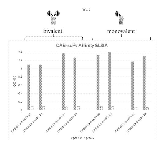
[00010] Figures 2 and 3 show that expressing the conditionally active
antibodies of
Example 6 as bivalent or monovalent antibodies does not significantly alter
that selectivity of
these antibodies under pH 6.0 and over pH 7.4.
[00011] Figure 4 is a profile of a size exclusive chromatograph indicating
that the
conditionally active antibodies of Example 7 do not aggregate.
[00012] Figure 5 shows on and off rates for the conditionally active
antibodies of
Example 7 as measured by a surface plasmon resonance (SPR) assay.
2
CA 02959141 2017-02-23
WO 2016/033331
PCT/US2015/047197
[00013] Figures 6A-6B show the selectivity of the conditionally active
antibodies as
measured by the SPR assay of Example 7.
SUMMARY OF THE DISCLOSURE
[00014] In one aspect, the present invention provides a chimeric antigen
receptor (CAR)
for binding with a target antigen, comprising at least one antigen specific
targeting region
evolved from a wild-type protein or a domain thereof and having at least one
of: (a) a
decrease in activity in the assay at the normal physiological condition
compared to the
antigen specific targeting region of the wild-type protein or a domain
thereof, and (b) an
increase in activity in the assay under the aberrant condition compared to the
antigen specific
targeting region of the wild-type protein or a domain thereof; a transmembrane
domain; and
an intracellular signaling domain. In some embodiments, the chimeric antigen
receptor
further comprises an extracellular spacer domain or at least one co-
stimulatory domain.
[00015] The chimeric antigen receptor may include an antigen specific
targeting region
that has a decrease in a binding affinity to the target antigen at a normal
physiological
condition compared to the antigen specific targeting region of the wild-type
protein or the
domain thereof.
[00016] The chimeric antigen receptor may include an antigen specific
targeting region
that has an increase in activity in the assay under the aberrant condition
compared to the
antigen specific targeting region of the wild-type protein or a domain thereof
and a decrease
in a binding affinity to the target antigen at a normal physiological
condition compared to the
antigen specific targeting region of the wild-type protein or the domain
thereof.
[00017] In any of the foregoing chimeric antigen receptors the antigen
specific targeting
region may also have an increase in selectivity in the assay under the
aberrant condition
compared to the antigen specific targeting region of the wild-type protein or
a domain
thereof.
[00018] Any of the foregoing chimeric antigen receptors may be configured
such that a
protein containing the antigen receptor has an increase in expression level
compared to the
wild-type protein or a domain thereof.
[00019] In an alternative embodiment, the present invention provides a
chimeric antigen
receptor (CAR) for binding with a target antigen, comprising at least one
antigen specific
targeting region evolved from a wild-type protein or a domain thereof and
having an increase
in selectivity in the assay under the aberrant condition compared to the
antigen specific
3
CA 02959141 2017-02-23
WO 2016/033331
PCT/US2015/047197
targeting region of the wild-type protein or a domain thereof; a transmembrane
domain; and
an intracellular signaling domain. In some embodiments, the chimeric antigen
receptor
further comprises an extracellular spacer domain or at least one co-
stimulatory domain.
[00020] In another aspect, the present invention provides an expression
vector,
comprising a polynucleotide sequence encoding the chimeric antigen receptor of
the
invention. The expression vector is selected from lentivirus vectors, gamma
retrovirus
vectors, foamy virus vectors, adeno associated virus vectors, adenovirus
vectors, pox virus
vectors, herpes virus vectors, engineered hybrid viruses, and transposon
mediated vectors.
[00021] In yet another aspect, the present invention provides a genetically
engineered
cytotoxic cell that comprises a polynucleotide sequence encoding the chimeric
antigen
receptor of the invention. The cytotoxic cell may be a T cell and may be
selected from a
naive T cell, a central memory T cell, and an effector memory T cell.
[00022] In yet another aspect, the present invention provides a
pharmaceutical
composition, comprising the chimeric antigen receptor, the expression vector,
and/or the
genetically engineered cytotoxic cell of the invention, and a pharmaceutically
acceptable
excipient.
[00023] In yet another aspect, the present invention provides a method for
producing a
chimeric antigen receptor comprising at least one antigen specific targeting
region, a
transmembrane domain and an intracellular signaling domain. The method
includes steps of
generating the at least one antigen specific targeting region from a wild-type
protein or a
domain thereof that binds specifically with a target antigen by evolving the
DNA which
encodes the wild-type protein or a domain thereof using one or more
evolutionary techniques
to create mutant DNAs; expressing the mutant DNAs to obtain mutant
polypeptides;
subjecting the mutant polypeptides and the wild-type protein or a domain
thereof to an assay
under a normal physiological condition and to an assay under an aberrant
condition; and
selecting a conditionally active antigen specific targeting region from the
mutant
polypeptides which exhibits at least one of: (a) a decrease in activity in the
assay at the
normal physiological condition compared to the antigen specific targeting
region of the wild-
type protein or a domain thereof, and (b) an increase in activity in the assay
under the
aberrant condition compared to the antigen specific targeting region of the
wild-type protein
or a domain thereof.
[00024] The chimeric antigen receptor produced by the method may include an
antigen
specific targeting region that has a decrease in a binding affinity to the
target antigen at a
4
CA 02959141 2017-02-23
WO 2016/033331
PCT/US2015/047197
normal physiological condition compared to the antigen specific targeting
region of the wild-
type protein or the domain thereof.
[00025] The chimeric antigen receptor produced by the method may include an
antigen
specific targeting region that has an increase in activity in the assay under
the aberrant
condition compared to the antigen specific targeting region of the wild-type
protein or a
domain thereof and a decrease in a binding affinity to the target antigen at a
normal
physiological condition compared to the antigen specific targeting region of
the wild-type
protein or the domain thereof.
[00026] In any of the foregoing chimeric antigen receptors produced by the
method the
antigen specific targeting region may also have an increase in selectivity in
the assay under
the aberrant condition compared to the antigen specific targeting region of
the wild-type
protein or a domain thereof.
[00027] Any of the foregoing chimeric antigen receptors producted by the
method may
be configured such that a protein containing the antigen receptor has an
increase in
expression level compared to the wild-type protein or a domain thereof.
[00028] In an alternative embodiment of the method, the chimeric antigen
receptor
(CAR) produced by the method for binding with a target antigen, comprises at
least one
antigen specific targeting region evolved from a wild-type protein or a domain
thereof and
having an increase in selectivity in the assay under the aberrant condition
compared to the
antigen specific targeting region of the wild-type protein or a domain
thereof; a
transmembrane domain; and an intracellular signaling domain. In some
embodiments, the
chimeric antigen receptor further comprises an extracellular spacer domain or
at least one co-
stimulatory domain.
[00029] In yet another aspect, the present invention provides a method for
treating a
cancer in a subject, comprising the step of introducing an expression vector
comprising a
polynucleotide sequence encoding the chimeric antigen receptor of the
invention into a
cytotoxic cell obtained from the subject to produce a genetically engineered
cytotoxic cell;
and administering the genetically engineered cytotoxic cell to the subject.
DEFINITIONS
[00030] In order to facilitate understanding of the examples provided
herein, certain
frequently occurring methods and/or terms will be defined herein.
CA 02959141 2017-02-23
WO 2016/033331
PCT/US2015/047197
[00031] As used herein in connection with a measured quantity, the term
"about" refers
to the normal variation in that measured quantity that would be expected by
the skilled artisan
making the measurement and exercising a level of care commensurate with the
objective of
the measurement and the precision of the measuring equipment used. Unless
otherwise
indicated, "about" refers to a variation of +/- 10% of the value provided.
[00032] The term "agent" is used herein to denote a chemical compound, a
mixture of
chemical compounds, an array of spatially localized compounds (e.g., a VLSIPS
peptide
array, polynucleotide array, and/or combinatorial small molecule array),
biological
macromolecule, a bacteriophage peptide display library, a bacteriophage
antibody (e.g., scFv)
display library, a polysome peptide display library, or an extract made from
biological
materials such as bacteria, plants, fungi, or animal (particular mammalian)
cells or tissues.
Agents are evaluated for potential enzyme activity by inclusion in screening
assays described
herein below. Agents are evaluated for potential activity as conditionally
active biologic
therapeutic enzymes by inclusion in screening assays described herein below.
[00033] The term "amino acid" as used herein refers to any organic compound
that
contains an amino group (--NH2) and a carboxyl group (--COOH); preferably
either as free
groups or alternatively after condensation as part of peptide bonds. The
"twenty naturally
encoded polypeptide-forming alpha-amino acids" are understood in the art and
refer to:
alanine (ala or A), arginine (arg or R), asparagine (asn or N), aspartic acid
(asp or D),
cysteine (cys or C), gluatamic acid (glu or E), glutamine (gin or Q), glycine
(gly or G),
histidine (his or H), isoleucine (ile or I), leucine (leu or L), lysine (lys
or K), methionine (met
or M), phenylalanine (phe or F), proline (pro or P), serine (ser or S),
threonine (thr or T),
tryptophan (tip or W), tyrosine (tyr or Y), and valine (val or V).
[00034] The term "amplification" as used herein means that the number of
copies of a
polynucleotide is increased.
[00035] The term "antibody" as used herein refers to intact immunoglobulin
molecules,
as well as fragments of immunoglobulin molecules, such as Fab, Fab', (Fab')2,
Fv, and SCA
fragments, that are capable of binding to an epitope of an antigen. These
antibody fragments,
which retain some ability to selectively bind to an antigen (e.g., a
polypeptide antigen) of the
antibody from which they are derived, can be made using well known methods in
the art (see,
e.g., Harlow and Lane, supra), and are described further, as follows.
Antibodies can be used
to isolate preparative quantities of the antigen by immunoaffinity
chromatography. Various
other uses of such antibodies are to diagnose and/or stage disease (e.g.,
neoplasia) and for
therapeutic application to treat disease, such as for example: neoplasia,
autoimmune disease,
6
CA 02959141 2017-02-23
WO 2016/033331
PCT/US2015/047197
AIDS, cardiovascular disease, infections, and the like. Chimeric, human-like,
humanized or
fully human antibodies are particularly useful for administration to human
patients.
[00036] An Fab fragment consists of a monovalent antigen-binding fragment
of an
antibody molecule, and can be produced by digestion of a whole antibody
molecule with the
enzyme papain, to yield a fragment consisting of an intact light chain and a
portion of a heavy
chain.
[00037] An Fab fragment of an antibody molecule can be obtained by treating
a whole
antibody molecule with pepsin, followed by reduction, to yield a molecule
consisting of an
intact light chain and a portion of a heavy chain. Two Fab' fragments are
obtained per
antibody molecule treated in this manner.
[00038] An (Fab)2 fragment of an antibody can be obtained by treating a
whole
antibody molecule with the enzyme pepsin, without subsequent reduction. A
(Fab)2 fragment
is a dimer of two Fab' fragments, held together by two disulfide bonds.
[00039] An Fv fragment is defined as a genetically engineered fragment
containing the
variable region of a light chain and the variable region of a heavy chain
expressed as two
chains.
[00040] The term "antigen" or "Ag" as used herein is defined as a molecule
that
provokes an immune response. This immune response may involve either antibody
production, or the activation of specific immunologically-competent cells, or
both. A person
skilled in the art will understand that any macromolecule, including virtually
all proteins or
peptides, can serve as an antigen. Furthermore, antigens can be derived from
recombinant or
genomic DNA. A person skilled in the art will understand that any DNA, which
comprises a
nucleotide sequence or a partial nucleotide sequence encoding a protein that
elicits an
immune response therefore encodes an "antigen" as that term is used herein.
Furthermore,
one skilled in the art will understand that an antigen need not be encoded
solely by a full
length nucleotide sequence of a gene. It is readily apparent that the present
invention
includes, but is not limited to, the use of partial nucleotide sequences of
more than one gene
and that these nucleotide sequences are arranged in various combinations to
elicit the desired
immune response. Moreover, a skilled person will understand that an antigen
need not be
encoded by a "gene" at all. It is readily apparent that an antigen can be
generated, synthesized
or can be derived from a biological sample. Such a biological sample can
include, but is not
limited to a tissue sample, a tumor sample, a cell or a biological fluid.
7
CA 02959141 2017-02-23
WO 2016/033331
PCT/US2015/047197
[00041] "Antigen loss escape variants" as used herein refer to cells which
exhibit
reduced or loss of expression of the target antigen, which antigens are
targeted by the CARs
of the invention.
[00042] The term "autoimmune disease" as used herein is defined as a
disorder that
results from an autoimmune response. An autoimmune disease is the result of an
inappropriate and excessive response to a self-antigen. Examples of autoimmune
diseases
include but are not limited to, Addision's disease, alopecia greata,
ankylosing spondylitis,
autoimmune hepatitis, autoimmune parotitis, Crohn's disease, diabetes (Type
1), dystrophic
epidermolysis bullosa, epididymitis, glomerulonephritis, Graves' disease,
Guillain-Barr
syndrome, Hashimoto's disease, hemolytic anemia, systemic lupus erythematosus,
multiple
sclerosis, myasthenia gravis, pemphigus vulgaris, psoriasis, rheumatic fever,
rheumatoid
arthritis, sarcoidosis, scleroderma, Sjogren's syndrome,
spondyloarthropathies, thyroiditis,
vasculitis, vitiligo, myxedema, pernicious anemia, ulcerative colitis, among
others.
[00043] The term "autologous," as used herein refers to any material
derived from the
same individual to which it is later to be reintroduced. For example, T cells
from a patient
may be isolated, genetically engineered to express a CAR and then reintroduced
into the
patient.
[00044] The term "B-cell associated diseases" as used herein include B-cell
immunodeficiencies, autoimmune diseases and/or excessive/uncontrolled cell
proliferation
associated with B- cells (including lymphomas and/or leukemias). Examples of
such diseases,
wherein bispecific CARs of the invention may be used for therapeutic
approaches include but
are not limited to systemic lupus erythematosus (SLE), diabetes, rheumatoid
arthritis (RA),
reactive arthritis, multiple sclerosis (MS), pemphigus vulgaris, celiac
disease, Crohn's
disease, inflammatory bowel disease, ulcerative colitis, autoimmune thyroid
disease, X-
linked agammaglobulinaemis, pre-B acute lymphoblastic leukemia, systemic lupus
erythematosus, common variable immunodeficiency, chronic lymphocytic leukemia,
diseases
associated with selective IgA deficiency and/or IgG subclass deficiency, B
lineage
lymphomas (Hodgkin's lymphoma and/or non-Hodgkin's lymphoma), immunodeficiency
with thymoma, transient hypogammaglobulinemia and/or hyper IgM syndrome, as
well as
virally-mediated B-cell diseases such as EBV mediated lymphoproliferative
disease, and
chronic infections in which B-cells participate in the pathophysiology.
[00045] The term "blood-brain barrier" or "BBB" refers to the physiological
barrier
between the peripheral circulation and the brain and spinal cord which is
formed by tight
junctions within the brain capillary endothelial plasma membranes, creating a
tight barrier
8
CA 02959141 2017-02-23
WO 2016/033331
PCT/US2015/047197
that restricts the transport of molecules into the brain, even very small
molecules such as urea
(60 Daltons). The blood-brain barrier within the brain, the blood-spinal cord
barrier within
the spinal cord, and the blood-retinal barrier within the retina are
contiguous capillary barriers
within the central nerve system (CNS), and are herein collectively referred to
as the "blood-
brain barrier" or "BBB." The BBB also encompasses the blood-cerebral spinal
fluid barrier
(choroid plexus) where the barrier is comprised of ependymal cells rather than
capillary
endothelial cells.
[00046] The terms "cancer" and "cancerous" as used herein refer to or
describe the
physiological condition in mammals that is typically characterized by
unregulated cell
growth. Examples of cancer include, but are not limited to B-cell lymphomas
(Hodgkin's
lymphomas and/or non-Hodgkins lymphomas), brain tumor, breast cancer, colon
cancer, lung
cancer, hepatocellular cancer, gastric cancer, pancreatic cancer, cervical
cancer, ovarian
cancer, liver cancer, bladder cancer, cancer of the urinary tract, thyroid
cancer, renal cancer,
carcinoma, melanoma, head and neck cancer, brain cancer, and prostate cancer,
including but
not limited to androgen-dependent prostate cancer and androgen-independent
prostate cancer.
[00047] The term "chimeric antigen receptor" or "CAR" or "CARs" as used
herein refers
to engineered receptors, which graft an antigen specificity onto cytotoxic
cell, for example T
cells, NK cells and macrophages. The CARs of the invention may comprise at
least one
antigen specific targeting region (ASTR), an extracellular spacer domain
(ESD), a
transmembrane domain (TM), one or more co-stimulatory domains (CSD), and an
intracellular signaling domain (ISD). In an embodiment, the ESD and/or CSD are
optional. In
another embodiment, the CAR is a bispecific CAR, which is specific to two
different antigens
or epitopes. After the ASTR binds specifically to a target antigen, the ISD
activates
intracellular signaling. For example, the ISD can redirect T cell specificity
and reactivity
toward a selected target in a non-MHC-restricted manner, exploiting the
antigen-binding
properties of antibodies. The non-MHC-restricted antigen recognition gives T
cells
expressing the CAR the ability to recognize an antigen independent of antigen
processing,
thus bypassing a major mechanism of tumor escape. Moreover, when expressed in
T cells,
CARs advantageously do not dimerize with endogenous T cell receptor (TCR)
alpha and beta
chains.
[00048] The term "co-express" as used herein refers to simultaneous
expression of two
or more genes. Genes may be nucleic acids encoding, for example, a single
protein or a
chimeric protein as a single polypeptide chain. For example, the CARs of the
invention may
be co- expressed with a therapeutic control (for example truncated epidermal
growth factor
9
CA 02959141 2017-02-23
WO 2016/033331
PCT/US2015/047197
(EGFRt)), wherein the CAR is encoded by a first polynucleotide chain and the
therapeutic
control is encoded by a second polynucleotide chain. In an embodiment, the
first and second
polynucleotide chains are linked by a nucleic acid sequence that encodes a
cleavable linker.
Alternately, the CAR and the therapeutic control are encoded by two different
polynucleotides that are not linked via a linker but are instead encoded by,
for example, two
different vectors.
[00049] The term "cognate" as used herein refers to a gene sequence that is
evolutionarily and functionally related between species. For example, but
without limitation,
in the human genome the human CD4 gene is the cognate gene to the mouse 3d4
gene, since
the sequences and structures of these two genes indicate that they are highly
homologous and
both genes encode a protein which functions in signaling T cell activation
through MHC class
II-restricted antigen recognition.
[00050] The term "conditionally active biologic protein" refers to a
variant, or mutant, of
a wild-type protein which is more or less active than the parent wild-type
protein under one
or more normal physiological conditions. This conditionally active protein
also exhibits
activity in selected regions of the body and/or exhibits increased or
decreased activity under
aberrant, or permissive, physiological conditions. Normal physiological
conditions are those
of temperature, pH, osmotic pressure, osmolality, oxidative stress and
electrolyte
concentration which would be considered within a normal range at the site of
administration,
or at the tissue or organ at the site of action, to a subject. An aberrant
condition is that which
deviates from the normally acceptable range for that condition. In one aspect,
the
conditionally active biologic protein is virtually inactive at wild-type
conditions but is active
at other than wild-type conditions at a level that is equal or better than at
wild-type
conditions. For example, in one aspect, an evolved conditionally active
biologic protein is
virtually inactive at body temperature, but is active at lower temperatures.
In another aspect,
the conditionally active biologic protein is reversibly or irreversibly
inactivated at the wild
type conditions. In a further aspect, the wild-type protein is a therapeutic
protein. In another
aspect, the conditionally active biologic protein is used as a drug, or
therapeutic agent. In yet
another aspect, the protein is more or less active in highly oxygenated blood,
such as, for
example, after passage through the lung or in the lower pH environments found
in the kidney.
[00051] "Conservative amino acid substitutions" refer to the
interchangeability of
residues having similar side chains. For example, a group of amino acids
having aliphatic
side chains is glycine, alanine, valine, leucine, and isoleucine; a group of
amino acids having
aliphatic-hydroxyl side chains is serine and threonine; a group of amino acids
having amide-
CA 02959141 2017-02-23
WO 2016/033331
PCT/US2015/047197
containing side chains is asparagine and glutamine; a group of amino acids
having aromatic
side chains is phenylalanine, tyrosine, and tryptophan; a group of amino acids
having basic
side chains is lysine, arginine, and histidine; and a group of amino acids
having sulfur-
containing side chains is cysteine and methionine. Preferred conservative
amino acids
substitution groups are: valine-leucine-isoleucine, phenylalanine-tyrosine,
lysine-arginine,
alanine-valine, and asparagine-glutamine.
[00052] The term "corresponds to" is used herein to mean that a
polynucleotide sequence
is homologous (i.e., is identical, not strictly evolutionarily related) to all
or a portion of a
reference polynucleotide sequence, or that a polypeptide sequence is identical
to a reference
polypeptide sequence. In contradistinction, the term "complementary to" is
used herein to
mean that the complementary sequence is homologous to all or a portion of a
reference
polynucleotide sequence. For illustration, the nucleotide sequence "TATAC"
corresponds to a
reference "TATAC" and is complementary to a reference sequence "GTATA."
[00053] The term "co-stimulatory ligand" as used herein includes a molecule
on an
antigen presenting cell (e.g., dendritic cell, B cell, and the like) that
specifically binds a
cognate co-stimulatory molecule on a T cell, thereby providing a signal which,
in addition to
the primary signal provided by, for instance, by the binding of a TCR/CD3
complex with an
MHC molecule loaded with peptide, mediates a T cell response, including, but
not limited to,
proliferation, activation, differentiation, and the like. A co-stimulatory
ligand can include, but
is not limited to, CD7, B7-1 (CD80), B7-2 (CD86), PD-L1, PD-L2, 4-1BBL, OX4OL,
an
inducible costimulatory ligand (ICOS-L), an intercellular adhesion molecule
(ICAM),
CD3OL, CD40, CD70, CD83, HLA-G, MICA, MICB, HVEM, a lymphotoxin beta receptor,
3/TR6, ILT3, ILT4, HVEM, an agonist or an antibody that binds to a Toll ligand
receptor and
a ligand that specifically binds with B7-H3. A co-stimulatory ligand also
encompasses, inter
alia, an antibody that specifically binds with a co-stimulatory molecule
present on a T cell,
such as, but not limited to, CD27, CD28, 4-1BB, 0X40, CD30, CD40, PD-1, ICOS,
a
lymphocyte function-associated antigen-1 (LFA-1), CD2, CD7, LIGHT, NKG2C, B7-
H3,
and a ligand that specifically binds with CD83.
[00054] The term "co-stimulatory molecule" as used herein refers to the
cognate binding
partner on a T cell that specifically binds with a co-stimulatory ligand,
thereby mediating a
co-stimulatory response by the T cell, such as, but not limited to,
proliferation. Co-
stimulatory molecules include, but are not limited to an MHC class 1 molecule,
BTLA and a
Toll ligand receptor.
11
CA 02959141 2017-02-23
WO 2016/033331
PCT/US2015/047197
[00055] The term "co-stimulatory signal" as used herein refers to a signal,
which in
combination with a primary signal, such as TCR/CD3 ligation, leads to T cell
proliferation
and/or upregulation or down regulation of key molecules.
[00056] The term "cytotoxic cell" as used herein means a cell which can
injure or
destroy invading microorganisms, tumor cells or other diseased tissue cells.
This term is
meant to include natural killer (NK) cells, activated NK cells, neutrophils, T
cells,
eosinophils, basophils, B- cells, macrophages and lymphokine-activated killer
(LAK) cells
among other cell types. The cytotoxic cell, through an antibody, receptor,
ligand or
fragments/derivatives thereof, is bound to a target cell to form a stable
complex, and
stimulates the cytotoxic cell to destroy the target cell.
[00057] Cytotoxic cells may also include other immune cells with tumor
lytic
capabilities including but not limited to natural killer T cells (Heczey et
al., "Invariant NKT
cells with chimeric antigen receptor provide a novel platform for safe and
effective cancer
immunotherapy," Blood, vol. 124, pp.2824-2833, 2014) and granulocytes. Further
,cytotoxic
cells may include immune cells with phagocytic capability including but not
limited to
macrophages and granulocytes, cells with stem cell and/or progenitor cell
properties
including, but not limited to, hematopoietic stem/progenitor cells (Zhen et
al., "HIV-specific
Immunity Derived From Chimeric Antigen Receptor-engineered Stem Cells," Mol
Ther., vol.
23, pp.1358-1367, 2015), embryonic stem cells (ESCs), cord blood stem cells,
and induced
pluripotent stem cells (iPSCs) (Themeli et al., "New cell sources for T cell
engineering and
adoptive immunotherapy," Cell Stem Cell., vol. 16, pp.357-366, 2015).
Additionally,
cytotoxic cells include "synthetic cells" such as iPSC-derived T cells
(TiPSCs) (Themeli et
al., "Generation of tumor-targeted human T lymphocytes from induced
pluripotent stem cells
for cancer therapy," Nat Biotechnol., vol.31, pp.928-933, 2013) or iPSC-
derived NK cells.
[00058] The term "degrading effective" amount refers to the amount of
enzyme which is
required to process at least 50% of the substrate, as compared to substrate
not contacted with
the enzyme.
[00059] The term "directional ligation" refers to a ligation in which a 5'
end and a 3' end
of a polynucleotide are different enough to specify a preferred ligation
orientation. For
example, an otherwise untreated and undigested PCR product that has two blunt
ends will
typically not have a preferred ligation orientation when ligated into a
cloning vector digested
to produce blunt ends in its multiple cloning site; thus, directional ligation
will typically not
be displayed under these circumstances. In contrast, directional ligation will
typically be
displayed when a digested PCR product having a 5' EcoR I-treated end and a 3'
BamH I is
12
CA 02959141 2017-02-23
WO 2016/033331
PCT/US2015/047197
ligated into a cloning vector that has a multiple cloning site digested with
EcoR I and BamH
I.
[00060] The term "disease targeted by genetically modified cytotoxic cells"
as used
herein encompasses the targeting of any cell involved in any manner in any
disease by the
genetically modified cells of the invention, irrespective of whether the
genetically modified
cells target diseased cells or healthy cells to effectuate a therapeutically
beneficial result. The
genetically modified cells include but are not limited to genetically modified
T cells, NK
cells, and macrophages. The genetically modified cells express the CARs of the
invention,
which CARs may target any of the antigens expressed on the surface of target
cells.
Examples of antigens which may be targeted include but are not limited to
antigens expressed
on B-cells; antigens expressed on carcinomas, sarcomas, lymphomas, leukemia,
germ cell
tumors, and blastomas; antigens expressed on various immune cells; and
antigens expressed
on cells associated with various hematologic diseases, autoimmune diseases,
and/or
inflammatory diseases. Other antigens that may be targeted will be apparent to
those of skill
in the art and may be targeted by the CARs of the invention in connection with
alternate
embodiments thereof.
[00061] The terms "genetically modified cells", "redirected cells",
"genetically
engineered cells" or "modified cells" as used herein refer to cells that
express the CARs of the
invention.
[00062] The term "DNA shuffling" is used herein to indicate recombination
between
substantially homologous but non-identical sequences, in some embodiments DNA
shuffling
may involve crossover via non-homologous recombination, such as via cer/lox
and/or flp/frt
systems and the like. DNA shuffling can be random or non-random.
[00063] The term "drug" or "drug molecule" refers to a therapeutic agent
including a
substance having a beneficial effect on a human or animal body when it is
administered to the
human or animal body. Preferably, the therapeutic agent includes a substance
that can treat,
cure or relieve one or more symptoms, illnesses, or abnormal conditions in a
human or
animal body or enhance the wellness of a human or animal body.
[00064] An "effective amount" is an amount of a conditionally active
biologic protein or
fragment which is effective to treat or prevent a condition in a living
organism to whom it is
administered over some period of time, e.g., provides a therapeutic effect
during a desired
dosing interval.
[00065] The term "electrolyte" as used herein defines a mineral in the
blood or other
body fluids that carries a charge. For example, in one aspect, the normal
physiological
13
CA 02959141 2017-02-23
WO 2016/033331
PCT/US2015/047197
condition and aberrant condition can be conditions of "electrolyte
concentration". In one
aspect, the electrolyte concentration to be tested is selected from one or
more of ionized
calcium, sodium, potassium, magnesium, chloride, bicarbonate, and phosphate
concentration.
For example, in one aspect, normal range of serum calcium is 8.5 to 10.2
mg/dL. In this
aspect, aberrant serum calcium concentration may be selected from either above
or below the
normal range, m another example, in one aspect, normal range of serum chloride
is 96-106
milliequivalents per liter (mEq/L). In this aspect, aberrant serum chloride
concentration may
be selected from either above or below the normal range, in another example,
in one aspect, a
normal range of serum magnesium is from 1.7-2.2 mg/dL. In this aspect, an
aberrant serum
magnesium concentration may be selected from either above or below the normal
range, in
another example, in one aspect, a normal range of serum phosphorus is from 2.4
to 4.1
mg/dL. In this aspect, aberrant serum phosphorus concentration may be selected
from either
above or below the normal range. In another example, in one aspect, a normal
range of
serum, or blood, sodium is from 135 to 145 mEq/L. In this aspect, aberrant
serum, or blood,
sodium concentration may be selected from either above or below the normal
range. In
another example, in one aspect, a normal range of serum, or blood, potassium
is from 3.7 to
5.2 mEq/L. In this aspect, aberrant serum, or blood, potassium concentration
maybe selected
from either above or below the normal range. In a further aspect, a normal
range of serum
bicarbonate is from 20 to 29 mEq/L. In this aspect, aberrant serum, or blood,
bicarbonate
concentration may be selected from either above or below the normal range. In
a different
aspect, bicarbonate levels can be used to indicate normal levels of acidity
(pH), in the blood.
The term "electrolyte concentration" may also be used to define the condition
of a particular
electrolyte in a tissue or body fluid other than blood or plasma. In this
case, the normal
physiological condition is considered to be the clinically normal range for
that tissue or fluid.
In this aspect, aberrant tissue or fluid electrolyte concentration may be
selected from either
above or below the normal range.
[00066] The term "epitope" as used herein refers to an antigenic
determinant on an
antigen, such as an enzyme polypeptide, to which the paratope of an antibody,
such as an
enzyme-specific antibody, binds. Antigenic determinants usually consist of
chemically active
surface groupings of molecules, such as amino acids or sugar side chains, and
can have
specific three-dimensional structural characteristics, as well as specific
charge characteristics.
As used herein "epitope" refers to that portion of an antigen or other
macromolecule capable
of forming a binding interaction that interacts with the variable region
binding body of an
14
CA 02959141 2017-02-23
WO 2016/033331
PCT/US2015/047197
antibody. Typically, such binding interaction is manifested as an
intermolecular contact with
one or more amino acid residues of a CDR.
[00067] As used herein, the term "evolution", or "evolving", refers to
using one or more
methods of mutagenesis to generate a novel polynucleotide encoding a novel
polypeptide,
which novel polypeptide is itself an improved biological molecule &/or
contributes to the
generation of another improved biological molecule. In a particular non-
limiting aspect, the
present disclosure relates to evolution of conditionally active biologic
proteins from a parent
wild type protein. In one aspect, for example, evolution relates to a method
of performing
both non-stochastic polynucleotide chimerization and non-stochastic site-
directed point
mutagenesis disclosed in U.S. patent application publication 2009/0130718,
which is
incorporated herein by reference. More particularly, the present disclosure
provides methods
for evolution of conditionally active biologic enzymes which exhibit reduced
activity at
normal physiological conditions compared to a wild-type enzyme parent
molecule, but
enhanced activity under one or more aberrant conditions compared to the
antigen specific
targeting region of the wild-type enzyme.
[00068] The terms "fragment", "derivative" and "analog" when referring to a
reference
polypeptide comprise a polypeptide which retains at least one biological
function or activity
that is at least essentially same as that of the reference polypeptide.
Furthermore, the terms
"fragment", "derivative" or "analog" are exemplified by a "pro-form" molecule,
such as a low
activity proprotein that can be modified by cleavage to produce a mature
enzyme with
significantly higher activity.
[00069] The term "gene" as used herein means the segment of DNA involved in
producing a polypeptide chain; it includes regions preceding and following the
coding region
(leader and trailer) as well as intervening sequences (nitrons) between
individual coding
segments (exons).
[00070] The term "heterologous" as used herein means that one single-
stranded nucleic
acid sequence is unable to hybridize to another single-stranded nucleic acid
sequence or its
complement. Thus areas of heterology means that areas of polynucleotides or
polynucleotides
have areas or regions within their sequence which are unable to hybridize to
another nucleic
acid or polynucleotide. Such regions or areas are for example areas of
mutations.
[00071] The term "homologous" or "homeologous" as used herein means that
one single-
stranded nucleic acid sequence may hybridize to a complementary single-
stranded nucleic
acid sequence. The degree of hybridization may depend on a number of factors
including the
amount of identity between the sequences and the hybridization conditions such
as
CA 02959141 2017-02-23
WO 2016/033331
PCT/US2015/047197
temperature and salt concentrations as discussed later. Preferably the region
of identity is
greater than about 5 bp, more preferably the region of identity is greater
than 10 bp.
[00072] The benefits of this disclosure extend to "industrial applications"
(or industrial
processes), which term is used to include applications in commercial industry
proper (or
simply industry) as well as non-commercial industrial applications (e.g.
biomedical research
at a non-profit institution). Relevant applications include those in areas of
diagnosis,
medicine, agriculture, manufacturing, and academia.
[00073] The term "immune cell" as used herein refers to cells of the
mammalian immune
system including but not limited to antigen presenting cells, B-cells,
basophils, cytotoxic T
cells, dendritic cells, eosinophils, granulocytes, helper T cells, leukocytes,
lymphocytes,
macrophages, mast cells, memory cells, monocytes, natural killer cells,
neutrophils,
phagocytes, plasma cells and T cells.
[00074] The term "immune response" as used herein refers to immunities
including but
not limited to innate immunity, humoral immunity, cellular immunity, immunity,
inflammatory response, acquired (adaptive) immunity, autoimmunity and/or
overactive
immunity
[00075] The term "isolated" as used herein means that the material is
removed from its
original environment (e.g., the natural environment if it is naturally
occurring). For example,
a naturally-occurring polynucleotide or enzyme present in a living animal is
not isolated, but
the same polynucleotide or enzyme, separated from some or all of the
coexisting materials in
the natural system, is isolated. Such polynucleotides could be part of a
vector and/or such
polynucleotides or enzymes could be part of a composition, and still be
isolated in that such
vector or composition is not part of its natural environment.
[00076] The term "isolated nucleic acid" as used herein to define a nucleic
acid, e.g., a
DNA or RNA molecule, that is not immediately contiguous with the 5' and 3'
flanking
sequences with which it normally is immediately contiguous when present in the
naturally
occurring genome of the organism from which it is derived. The term thus
describes, for
example, a nucleic acid that is incorporated into a vector, such as a plasmid
or viral vector; a
nucleic acid that is incorporated into the genome of a heterologous cell (or
the genome of a
homologous cell, but at a site different from that at which it naturally
occurs); and a nucleic
acid that exists as a separate molecule, e.g., a DNA fragment produced by PCR
amplification
or restriction enzyme digestion, or an RNA molecule produced by in vitro
transcription. The
term also describes a recombinant nucleic acid that forms part of a hybrid
gene encoding
16
CA 02959141 2017-02-23
WO 2016/033331
PCT/US2015/047197
additional polypeptide sequences that can be used, for example, in the
production of a fusion
protein.
[00077] The term "lentivirus" as used herein refers to a genus of the
Retroviridae family.
Lentiviruses are unique among the retroviruses in being able to infect non-
dividing cells; they
can deliver a significant amount of genetic information into the DNA of the
host cell, so they
are one of the most efficient ways to deliver a gene delivery vector. HIV,
SIV, and FIV are
all examples of lentiviruses. Vectors derived from lentiviruses offer the
means to achieve
significant levels of gene transfer in vivo.
[00078] The term "ligand" as used herein refers to a molecule, such as a
random peptide
or variable segment sequence that is recognized by a particular receptor. As a
person skilled
in the art will recognize, a molecule (or macromolecular complex) can be both
a receptor and
a ligand. In general, the binding partner having a smaller molecular weight is
referred to as
the ligand and the binding partner having a greater molecular weight is
referred to as a
receptor.
[00079] The term "ligation" as used herein refers to the process of forming
phosphodiester bonds between two double stranded nucleic acid fragments
(Sambrook et al.,
(1982). Molecular Cloning: A Laboratory Manual. Cold Spring Harbour
Laboratory, Cold
Spring Harbor, NY., p. 146; Sambrook et al., Molecular Cloning: a laboratory
manual, 2nd
Ed., Cold Spring Harbor Laboratory Press, 1989). Unless otherwise provided,
ligation may be
accomplished using known buffers and conditions with 10 units of T4 DNA ligase
("ligase")
per 0.5 micrograms of approximately equimolar amounts of the DNA fragments to
be ligated.
[00080] The terms "linker" or "spacer" as used herein refer to a molecule
or group of
molecules that connects two molecules, such as a DNA binding protein and a
random
peptide, and serves to place the two molecules in a preferred configuration,
e.g., so that the
random peptide can bind to a receptor with minimal steric hindrance from the
DNA binding
protein. "Linker" (L) or "linker domain" or "linker region" as used herein
refers to an oligo-
or polypeptide region of from about 1 to 100 amino acids in length, which
links together any
of the domains/regions of the CARs of the invention. Linkers may be composed
of flexible
residues like glycine and serine so that the adjacent protein domains are free
to move relative
to one another. Longer linkers may be used when it is desirable to ensure that
two adjacent
domains do not sterically interfere with one another. Linkers may be cleavable
or non-
cleavable. Examples of cleavable linkers include 2A linkers (for example T2A),
2A-like
linkers or functional equivalents thereof and combinations thereof. In some
embodiments, the
linkers include the picornaviral 2A-like linker, CHYSEL sequences of porcine
teschovirus
17
CA 02959141 2017-02-23
WO 2016/033331
PCT/US2015/047197
(P2A), Thosea asigna virus (T2A) or combinations, variants and functional
equivalents
thereof. Other linkers will be apparent to those skilled in the art and may be
used in
connection with alternate embodiments of the invention.
[00081] The term "mammalian cell surface display" as used herein refers to
a technique
whereby a protein or antibody, or a portion of an antibody, is expressed and
displayed on a
mammalian host cell surface for screening purposes; for example, by screening
for specific
antigen binding by a combination of magnetic beads and fluorescence-activated
cell sorting.
In one aspect, mammalian expression vectors are used for simultaneous
expression of
immunoglobulins as both a secreted and cell surface bound form as in DuBridge
et al., US
2009/0136950, which is incorporated herein by reference. In another aspect,
the techniques
are employed for screening a viral vector encoding for a library of antibodies
or antibody
fragments that are displayed on the cell membranes when expressed in a cell as
in Gao et al.,
US 2007/0111260, incorporated herein by reference. Whole IgG surface display
on
mammalian cells is known. For example, Akamatsuu et al. developed a mammalian
cell
surface display vector, suitable for directly isolating IgG molecules based on
their antigen-
binding affinity and biological activity. Using an Epstein-Ban virus-derived
episomal vector,
antibody libraries were displayed as whole IgG molecules on the cell surface
and screened for
specific antigen binding by a combination of magnetic beads and fluorescence-
activated cell
sorting. Plasmids encoding antibodies with desired binding characteristics
were recovered
from sorted cells and converted to a form suitable for production of soluble
IgG. See
Akamatsuu et al. J. Immunol. Methods, vol. 327, pages 40-52, 2007,
incorporated herein by
reference. Ho et al. used human embryonic kidney 293T cells that are widely
used for
transient protein expression for cell surface display of single-chain Fv
antibodies for affinity
maturation. Cells expressing a rare mutant antibody with higher affinity were
enriched 240-
fold by a single-pass cell sorting from a large excess of cells expressing WT
antibody with a
slightly lower affinity. Furthermore, a highly enriched mutant was obtained
with increased
binding affinity for CD22 after a single selection of a combinatory library
randomizing an
intrinsic antibody hotspot. See Ho et al., "Isolation of anti-CD22 Fv with
high affinity by Fv
display on human cells," Proc Nail Acad Sci USA, vol. 103, pages 9637-9642,
2006,
incorporated herein by reference.
[00082] B cells specific for an antigen may also be used. Such B cells may
be directly
isolated from peripheral blood mononuclear cells (PBMC) of human donors.
Recombinant,
antigen-specific single-chain Fv (scFv) libraries are generated from this pool
of B cells and
screened by mammalian cell surface display by using a Sindbis virus expression
system. The
18
CA 02959141 2017-02-23
WO 2016/033331
PCT/US2015/047197
variable regions (VRs) of the heavy chains (HCs) and light chains (LCs) can be
isolated from
positive clones and recombinant fully human antibodies produced as whole IgG
or Fab
fragments. In this manner, several hypermutated high-affinity antibodies
binding the QP virus
like particle (VLP), a model viral antigen, as well as antibodies specific for
nicotine can be
isolated. See Beerli et al., "Isolation of human monoclonal antibodies by
mammalian cell
display," Proc Natl Acad Sci USA, vol. 105, pages 14336-14341, 2008,
incorporated herein
by reference.
[00083] Yeast cell surface display may also be used in the present
invention, for
example, see Kondo and Ueda, "Yeast cell-surface display-applications of
molecular
display," Appl. Microbiol. Biotechnol., vol. 64, pages 28-40, 2004, which
describes for
example, a cell-surface engineering system using the yeast Saccharomyces
cerevisiae.
Several representative display systems for the expression in yeast S.
cerevisiae are described
in Lee et al, "Microbial cell-surface display," TRENDS in Bitechnol., vol. 21,
pages 45-52,
2003. Also Boder and Wittrup, "Yeast surface display for screening
combinatorial
polypeptide libraries," Nature Biotechnol., vol. 15, pages 553, 1997.
[00084] The term "manufacturing" as used herein refers to production of a
protein in a
sufficient quantity to permit at least Phase I clinical testing of a
therapeutic protein, or
sufficient quantity for regulatory approval of a diagnostic protein.
[00085] As used herein, the term "microenvironment" means any portion or
region of a
tissue or body that has a constant or temporal, physical or chemical
difference from other
regions of the tissue or other regions of the body.
[00086] As used herein, the term "molecular property to be evolved"
includes reference
to molecules comprised of a polynucleotide sequence, molecules comprised of a
polypeptide
sequence, and molecules comprised in part of a polynucleotide sequence and in
part of a
polypeptide sequence. Particularly relevant¨ but by no means limiting-
examples of
molecular properties to be evolved include protein activities at specified
conditions, such as
related to temperature; salinity; osmotic pressure; pH; oxidative stress, and
concentration of
glycerol, DMSO, detergent, and/or any other molecular species with which
contact is made in
a reaction environment. Additional particularly relevant¨but by no means
limiting¨
examples of molecular properties to be evolved include stabilities¨ e.g. the
amount of a
residual molecular property that is present after a specified exposure time to
a specified
environment, such as may be encountered during storage.
[00087] The term "mutations" as used herein means changes in the sequence
of a wild-
type nucleic acid sequence or changes in the sequence of a peptide. Such
mutations may be
19
CA 02959141 2017-02-23
WO 2016/033331
PCT/US2015/047197
point mutations such as transitions or transversions. The mutations may be
deletions,
insertions or duplications.
[00088] The term "multispecific antibody" as used herein is an antibody
having binding
affinities for at least two different epitopes. Multispecific antibodies can
be prepared as full-
length antibodies or antibody fragments (e.g. F(ab)2 bispecific antibodies).
Engineered
antibodies may bind to two, three or more (e.g. four) antigens (see, e.g., US
2002/0004587
Al). One conditionally active antibody may be engineered to be multispecific,
or two
antibodies may be engineered to comprise a hetero-dimer that binds to two
antigens.
Multispecific antibodies can also be multifunctional.
[00089] As used herein, the degenerate "N,N,G/T" nucleotide sequence
represents 32
possible triplets, where "N" can be A, C, G or T.
[00090] The term "naturally-occurring" as used herein as applied to the
object refers to
the fact that an object can be found in nature. For example, a polypeptide or
polynucleotide
sequence that is present in an organism (including viruses) that can be
isolated from a source
in nature and which has not been intentionally modified by man in the
laboratory is naturally
occurring. Generally, the term naturally occurring refers to an object as
present in a non-
pathological (un-diseased) individual, such as would be typical for the
species.
[00091] As used herein, "normal physiological conditions", or "wild type
operating
conditions", are those conditions of temperature, pH, osmotic pressure,
osmolality, oxidative
stress and electrolyte concentration which would be considered within a normal
range at the
site of administration, or the site of action, in a subject.
[00092] As used herein, the term "nucleic acid molecule" is comprised of at
least one
base or one base pair, depending on whether it is single-stranded or double-
stranded,
respectively. Furthermore, a nucleic acid molecule may belong exclusively or
chimerically to
any group of nucleotide-containing molecules, as exemplified by, but not
limited to, the
following groups of nucleic acid molecules: RNA, DNA, genomic nucleic acids,
non-
genomic nucleic acids, naturally occurring and not naturally occurring nucleic
acids, and
synthetic nucleic acids. This includes, by way of non-limiting example,
nucleic acids
associated with any organelle, such as the mitochondria, ribosomal RNA, and
nucleic acid
molecules comprised chimerically of one or more components that are not
naturally occurring
along with naturally occurring components.
[00093] Additionally, a "nucleic acid molecule" may contain in part one or
more non-
nucleotide-based components as exemplified by, but not limited to, amino acids
and sugars.
CA 02959141 2017-02-23
WO 2016/033331
PCT/US2015/047197
Thus, by way of example, but not limitation, a ribozyme that is in part
nucleotide-based and
in part protein-based is considered a "nucleic acid molecule".
[00094] The terms "nucleic acid sequence coding for" or a "DNA coding
sequence of or
a "nucleotide sequence encoding" as used herein refer to a DNA sequence which
is
transcribed and translated into an enzyme when placed under the control of
appropriate
regulatory sequences such as promoters. A "promotor" is a DNA regulatory
region capable of
binding RNA polymerase in a cell and initiating transcription of a downstream
(3' direction)
coding sequence. The promoter is part of the DNA sequence. This sequence
region has a start
codon at its 3' terminus. The promoter sequence does include the minimum
number of bases
where elements necessary to initiate transcription at levels detectable above
background.
However, after the RNA polymerase binds the sequence and transcription is
initiated at the
start codon (3' terminus with a promoter), transcription proceeds downstream
in the 3'
direction. Within the promotor sequence will be found a transcription
initiation site
(conveniently defined by mapping with nuclease Si) as well as protein binding
domains
(consensus sequences) responsible for the binding of RNA polymerase.
[00095] The term "oligonucleotide" (or synonymously an "oligo") refers to
either a
single stranded polydeoxynucleotide or two complementary polydeoxynucleotide
strands
which may be chemically synthesized. Such synthetic oligonucleotides may or
may not have
a 5' phosphate. Those that do not will not ligate to another oligonucleotide
without adding a
phosphate with an ATP in the presence of a kinase. A synthetic oligonucleotide
will ligate to
a fragment that has not been dephosphorylated.
[00096] As used herein, the term "operably linked" refers to a linkage of
polynucleotide
elements in a functional relationship. A nucleic acid is "operably linked"
when it is placed
into a functional relationship with another nucleic acid sequence. For
instance, a promoter or
enhancer is operably linked to a coding sequence if it affects the
transcription of the coding
sequence. Operably linked means that the DNA sequences being linked are
typically
contiguous and, where necessary to join two protein coding regions, contiguous
and in
reading frame.
[00097] A coding sequence is "operably linked to" another coding sequence
when RNA
polymerase will transcribe the two coding sequences into a single mRNA, which
is then
translated into a single polypeptide having amino acids derived from both
coding sequences.
The coding sequences need not be contiguous to one another so long as the
expressed
sequences are ultimately processed to produce the desired protein.
21
CA 02959141 2017-02-23
WO 2016/033331
PCT/US2015/047197
[00098] As used herein the term "parental polynucleotide set" is a set
comprised of one
or more distinct polynucleotide species. Usually this term is used in
reference to a progeny
polynucleotide set which is preferably obtained by mutagenization of the
parental set, in
which case the terms "parental", "starting" and "template" are used
interchangeably.
[00099] The term "patient", or "subject", refers to an animal, for example
a mammal,
such as a human, who is the object of treatment. The subject, or patient, may
be either male
or female.
[000100] As used herein the term "physiological conditions" refers to
temperature, pH,
osmotic pressure, ionic strength, viscosity, and like biochemical parameters
which are
compatible with a viable organism, and/or which typically exist
intracellularly in a viable
cultured yeast cell or mammalian cell. For example, the intracellular
conditions in a yeast cell
grown under typical laboratory culture conditions are physiological
conditions. Suitable in
vitro reaction conditions for in vitro transcription cocktails are generally
physiological
conditions. In general, in vitro physiological conditions comprise 50-200 mM
NaC1 or KC1,
pH 6.5-8.5, 20-45 degrees C and 0.001-10 mM divalent cation (e.g., Mg, Ca);
preferably
about 150 mM NaC1 or KC1, pH 7.2-7.6, 5 mM divalent cation, and often include
0.01-1.0
percent nonspecific protein (e.g., bovine serum albumin (BSA)). A non-ionic
detergent
(Tween, NP-40, Triton X-100) can often be present, usually at about 0.001 to
2%, typically
0.05-0.2% (v/v). Particular aqueous conditions may be selected by the
practitioner according
to conventional methods. For general guidance, the following buffered aqueous
conditions
may be applicable: 10-250 mM NaC1, 5-50 mM Tris HC1, pH 5-8, with optional
addition of
divalent cation(s) and/or metal chelators and/or non- ionic detergents and/or
membrane
fractions and/or anti-foam agents and/or scintillants. Normal physiological
conditions refer to
conditions of temperature, pH, osmotic pressure, osmolality, oxidative stress
and electrolyte
concentration in vivo in a patient or subject at the site of administration,
or the site of action,
which would be considered within the normal range in a patient.
[000101] Standard convention (5' to 3') is used herein to describe the
sequence of double
stranded polynucleotides.
[000102] The term "population" as used herein means a collection of
components such as
polynucleotides, portions or polynucleotides or proteins. A "mixed population"
means a
collection of components which belong to the same family of nucleic acids or
proteins (i.e.,
are related) but which differ in their sequence (i.e., are not identical) and
hence in their
biological activity.
22
CA 02959141 2017-02-23
WO 2016/033331
PCT/US2015/047197
[000103] A molecule having a "pro-form" refers to a molecule that undergoes
any
combination of one or more covalent and noncovalent chemical modifications
(e.g.
glycosylation, proteolytic cleavage, dimerization or oligomerization,
temperature- induced or
pH-induced conformational change, association with a co-factor, etc.) en route
to attain a
more mature molecular form having a property difference (e.g. an increase in
activity) in
comparison with the reference pro-form molecule. When two or more chemical
modifications
(e.g. two proteolytic cleavages, or a proteolytic cleavage and a
deglycosylation) can be
distinguished en route to the production of a mature molecule, the reference
precursor
molecule may be termed a"pre-pro-form" molecule.
[000104] As used herein, the term "receptor" refers to a molecule that has
an affinity for a
given ligand. Receptors can be naturally occurring or synthetic molecules.
Receptors can be
employed in an unaltered state or as aggregates with other species. Receptors
can be attached,
covalently or non-covalently, to a binding member, either directly or via a
specific binding
substance. Examples of receptors include, but are not limited to, antibodies,
including
monoclonal antibodies and antisera reactive with specific antigenic
determinants (such as on
viruses, cells, or other materials), cell membrane receptors, complex
carbohydrates and
glycoproteins, enzymes, and hormone receptors.
[000105] The term "reductive reassortment", as used herein, refers to the
increase in
molecular diversity that is accrued through deletion (and/or insertion) events
that are
mediated by repeated sequences.
[000106] The term "restriction site" as used herein refers to a recognition
sequence that is
necessary for the manifestation of the action of a restriction enzyme, and
includes a site of
catalytic cleavage. It is appreciated that a site of cleavage may or may not
be contained
within a portion of a restriction site that comprises a low ambiguity sequence
(i.e. a sequence
containing the principal determinant of the frequency of occurrence of the
restriction site).
When an enzyme (e.g. a restriction enzyme) is said to "cleave" a
polynucleotide, it is
understood to mean that the restriction enzyme catalyzes or facilitates a
cleavage of a
polynucleotide.
[000107] As used herein, the term "single-chain antibody" refers to a
polypeptide
comprising a VH domain and a VL domain in polypeptide linkage, generally liked
via a
spacer peptide, and which may comprise additional amino acid sequences at the
amino-
and/or carboxy- termini. For example, a single-chain antibody may comprise a
tether
segment for linking to the encoding polynucleotide. As an example a scFv is a
single-chain
antibody. Single-chain antibodies are generally proteins consisting of one or
more
23
CA 02959141 2017-02-23
WO 2016/033331
PCT/US2015/047197
polypeptide segments of at least 10 contiguous amino substantially encoded by
genes of the
immunoglobulin superfamily (e.g, see The Immunoglobulin Gene Superfamily, A.
F.
Williams and A. N. Barclay, in Immunoglobulin Genes, T. Honjo, F. W. Alt, and
THE.
Rabbits, eds., (1989) Academic press: San Diego, Calif., pp. 361-368, which is
incorporated
herein by reference), most frequently encoded by a rodent, non-human primate,
avian,
porcine bovine, ovine, goat, or human heavy chain or light chain gene
sequence. A
functional single-chain antibody generally contains a sufficient portion of an
immunoglobulin
superfamily gene product so as to retain the property of binding to a specific
target molecule,
typically a receptor or antigen (epitope).
[000108] The members of a pair of molecules (e.g., an antibody-antigen pair
and ligand-
receptor pair) are said to "specifically bind" to each other if they bind to
each other with
greater affinity than to other, non-specific molecules. For example, an
antibody raised against
an antigen to which it binds more efficiently than to a non-specific protein
can be described
as specifically binding to the antigen.
[000109] The term "stimulation" as used herein means a primary response
induced by
binding of a stimulatory molecule (e.g., a TCR/CD3 complex) with its cognate
ligand thereby
mediating a signal transduction event, such as, but not limited to, signal
transduction via the
TCR/CD3 complex. Stimulation can mediate altered expression of certain
molecules, such as
downregulation of TGF-P, and/or reorganization of cytoskeletal structures, and
the like.
[000110] The term "stimulatory molecule" as used herein means a molecule on
a T cell
that specifically binds with a cognate stimulatory ligand present on an
antigen presenting cell.
[000111] The term "stimulatory ligand" as used herein means a ligand that
when present
on an antigen presenting cell (e.g, a dendritic cell, a B-cell, and the like)
can specifically bind
with a cognate binding partner (referred to herein as a "stimulatory
molecule") on a T cell,
thereby mediating a primary response by the T cell, including, but not limited
to, activation,
initiation of an immune response, proliferation, and the like. Stimulatory
ligands are well-
known in the art and encompass, inter alia, an MHC Class I molecule loaded
with a peptide,
an anti-CD3 antibody, a superagonist anti-CD28 antibody, and a superagonist
anti-CD2
antibody.
[000112] The term "target cell" as used herein refers to cells which are
involved in a
disease and can be targeted by the genetically modified cytotoxic cells of the
invention
(including but not limited to genetically modified T cells, NK cells, and
macrophages). Other
target cells will be apparent to those skilled in the art and may be used in
connection with
alternate embodiments of the invention.
24
CA 02959141 2017-02-23
WO 2016/033331
PCT/US2015/047197
[000113] The terms "T cell" and "T-lymphocyte" are interchangeable and used
synonymously herein. Examples include, but are not limited to, naive T cells,
central memory
T cells, effector memory T cells and combinations thereof.
[000114] The term "transduction" as used herein refers to the introduction
of a foreign
nucleic acid into a cell using a viral vector. "Transfection" as used herein
refers to the
introduction of a foreign nucleic acid into a cell using recombinant DNA
technology. The
term "transformation" means the introduction of a "foreign" (i.e. extrinsic or
extracellular)
gene, DNA or RNA sequence to a host cell, so that the host cell will express
the introduced
gene or sequence to produce a desired substance, such as a protein or enzyme
coded by the
introduced gene or sequence. The introduced gene or sequence may also be
called a "cloned"
or "foreign" gene or sequence, may include regulatory or control sequences,
such as start,
stop, promoter, signal, secretion, or other sequences used by a cell's genetic
machinery. The
gene or sequence may include nonfunctional sequences or sequences with no
known
function. A host cell that receives and expresses introduced DNA or RNA has
been
"transformed" and is a "transformant" or a "clone." The DNA or RNA introduced
to a host
cell can come from any source, including cells of the same genus or species as
the host cell,
or cells of a different genus or species
[000115] The term "treating" includes: (1) preventing or delaying the
appearance of
clinical symptoms of the state, disorder or condition developing in an animal
that may be
afflicted with or predisposed to the state, disorder or condition but does not
yet experience or
display clinical or subclinical symptoms of the state, disorder or condition;
(2) inhibiting the
state, disorder or condition (i.e., arresting, reducing or delaying the
development of the
disease, or a relapse thereof in case of maintenance treatment, of at least
one clinical or
subclinical symptom thereof); and/or (3) relieving the condition (i.e.,
causing regression of
the state, disorder or condition or at least one of its clinical or
subclinical symptoms). The
benefit to a patient to be treated is either statistically significant or at
least perceptible to the
patient or to the physician.
[000116] "Tumor," as used herein refers to all neoplastic cell growth and
proliferation,
whether malignant or benign, and all pre-cancerous and cancerous cells and
tissues
[000117] As used herein, the term "tumor microenvironment" refers to any
and all
elements of the tumor milieu including elements that create a structural and
or functional
environment for the malignant process to survive and/or expand and/or spread.
[000118] As used herein, the term "variable segment" refers to a portion of
a nascent
peptide which comprises a random, pseudorandom, or defined kernal sequence. A
"variable
CA 02959141 2017-02-23
WO 2016/033331
PCT/US2015/047197
segment" refers to a portion of a nascent peptide which comprises a random
pseudorandom,
or defined kernal sequence. A variable segment can comprise both variant and
invariant
residue positions, and the degree of residue variation at a variant residue
position may be
limited: both options are selected at the discretion of the practitioner.
Typically, variable
segments are about 5 to 20 amino acid residues in length (e.g., 8 to 10),
although variable
segments may be longer and may comprise antibody portions or receptor
proteins, such as an
antibody fragment, a nucleic acid binding protein, a receptor protein, and the
like.
[000119] "Vector", "cloning vector" and "expression vector" as used herein
refer to the
vehicle by which a polynucleotide sequence (e.g. a foreign gene) can be
introduced into a
host cell, so as to transform the host and promote expression (e.g.
transcription and
translation) of the introduced sequence. Vectors include plasmids, phages,
viruses, etc.
[000120] As used herein, the term "wild-type" means that the polynucleotide
does not
comprise any mutations. A "wild type protein", "wild-type protein", "wild-type
biologic
protein", or "wild type biologic protein", refers to a protein which can be
isolated from nature
that will be active at a level of activity found in nature and will comprise
the amino acid
sequence found in nature. The terms "parent molecule" and "target protein"
also refer to the
wild-type protein. The "wild-type protein" preferably has some desired
properties, such as
higher binding affinity, or enzymatic activity, which may be obtained by
screening of a
library of proteins for a desired properties, including better stability in
different temperature
or pH environments, or improved selectivity and/or solubility.
[000121] The term "working", as in "working sample", for example, is simply
a sample
with which one is working. Likewise, a "working molecule", for example is a
molecule with
which one is working.
DETAILED DESCRIPTION
[000122] For illustrative purposes, the principles of the present invention
are described
by referencing various exemplary embodiments. Although certain embodiments of
the
invention are specifically described herein, one of ordinary skill in the art
will readily
recognize that the same principles are equally applicable to, and can be
employed in other
systems and methods. Before explaining the disclosed embodiments of the
present invention
in detail, it is to be understood that the invention is not limited in its
application to the details
of any particular embodiment shown. Additionally, the terminology used herein
is for the
purpose of description and not of limitation. Furthermore, although certain
methods are
described with reference to steps that are presented herein in a certain
order, in many
26
CA 02959141 2017-02-23
WO 2016/033331
PCT/US2015/047197
instances, these steps may be performed in any order as may be appreciated by
one skilled in
the art; the novel method is therefore not limited to the particular
arrangement of steps
disclosed herein.
[000123] It must be noted that as used herein and in the appended claims,
the singular
forms "a", "an", and "the" include plural references unless the context
clearly dictates
otherwise. Furthermore, the terms "a" (or "an"), "one or more" and "at least
one" can be
used interchangeably herein. The terms "comprising", "including", "having" and
"constructed from" can also be used interchangeably.
[000124] The present disclosure is directed to a chimeric antigen receptor
(CAR) for
binding with a target antigen, comprising at least one antigen specific
targeting region
evolved from a wild-type protein or a domain thereof and having at least one
of: (a) a
decrease in activity in the assay at the normal physiological condition
compared to the
antigen specific targeting region of the wild-type protein or a domain
thereof, and (b) an
increase in activity in the assay under the aberrant condition compared to the
antigen specific
targeting region of the wild-type protein or a domain thereof; a transmembrane
domain; and
an intracellular signaling domain. In some embodiments, the chimeric antigen
receptor
further comprises an extracellular spacer domain or at least one co-
stimulatory domain.
[000125] The CARs of the present invention have at least one of (1) their
affinity to the
target antigen reversibly or irreversibly reduced at the normal physiological
condition, and
(2) an increased affinity, in comparison with the same CAR without the
conditionally active
antigen specific targeting region. These CARs can direct cytotoxic cells to a
disease site
where an aberrant condition is present, such as a tumor microenvironment or
synovial fluid.
As a result of these properties, the CARs can preferentially direct the
cytotoxic cells to a
disease site while because of their low affinity for normal tissue. Such CARs
can
dramatically reduce side-effects and allow higher doses of therapeutics to be
used to increase
therapeutic efficacy. The CARs are particularly valuable for development of
novel
therapeutics that are required for short or limited periods of time within a
subject. Examples
of beneficial applications include systemic treatments at high dosages, as
well as localized
treatments at high concentrations.
[000126] The chimeric antigen receptor may include an antigen specific
targeting region
that has a decrease in a binding affinity to the target antigen at a normal
physiological
condition compared to the antigen specific targeting region of the wild-type
protein or the
domain thereof.
27
CA 02959141 2017-02-23
WO 2016/033331
PCT/US2015/047197
[000127] The chimeric antigen receptor many include an antigen specific
targeting region
that has an increase in activity in the assay under the aberrant condition
compared to the
antigen specific targeting region of the wild-type protein or a domain thereof
and a decrease
in a binding affinity to the target antigen at a normal physiological
condition compared to the
antigen specific targeting region of the wild-type protein or the domain
thereof.
[000128] In any of the foregoing chimeric antigen receptors the antigen
specific targeting
region may also have an increase in selectivity in the assay under the
aberrant condition
compared to the antigen specific targeting region of the wild-type protein or
a domain
thereof.
[000129] Any of the foregoing chimeric antigen receptors may be configured
such that a
protein containing the antigen receptor has an increase in expression level
compared to the
wild-type protein or a domain thereof.
[000130] In an alternative embodiment, the present invention provides a
chimeric antigen
receptor (CAR) for binding with a target antigen, comprising at least one
antigen specific
targeting region evolved from a wild-type protein or a domain thereof and
having an increase
in selectivity in the assay under the aberrant condition compared to the
antigen specific
targeting region of the wild-type protein or a domain thereof; a transmembrane
domain; and
an intracellular signaling domain. In some embodiments, the chimeric antigen
receptor
further comprises an extracellular spacer domain or at least one co-
stimulatory domain.
[000131] The present disclosure is also directed to methods of evolving a
wild-type
protein or a domain thereof to generate a conditionally active protein that
has at least one of:
(a) a decrease in activity in the assay at the normal physiological condition
compared to the
antigen specific targeting region of the wild-type protein or a domain
thereof, and (b) an
increase in activity in the assay under the aberrant condition compared to the
antigen specific
targeting region of the wild-type protein or a domain thereof. The
conditionally active
protein may be engineered into a CAR.
[000132] The chimeric antigen receptor produced by the method may include
an antigen
specific targeting region that has a decrease in a binding affinity to the
target antigen at a
normal physiological condition compared to the antigen specific targeting
region of the wild-
type protein or the domain thereof.
[000133] The chimeric antigen receptor produced by the method may include
an antigen
specific targeting region that has an increase in activity in the assay under
the aberrant
condition compared to the antigen specific targeting region of the wild-type
protein or a
domain thereof and a decrease in a binding affinity to the target antigen at a
normal
28
CA 02959141 2017-02-23
WO 2016/033331
PCT/US2015/047197
physiological condition compared to the antigen specific targeting region of
the wild-type
protein or the domain thereof.
[000134] In any of the foregoing chimeric antigen receptors produced by the
method the
antigen specific targeting region may also have an increase in selectivity in
the assay under
the aberrant condition compared to the antigen specific targeting region of
the wild-type
protein or a domain thereof.
[000135] Any of the foregoing chimeric antigen receptors producted by the
method may
be configured such that a protein containing the antigen receptor has an
increase in
expression level compared to the wild-type protein or a domain thereof.
[000136] In an alternative embodiment of the method, the chimeric antigen
receptor
(CAR) produced by the method for binding with a target antigen, comprises at
least one
antigen specific targeting region evolved from a wild-type protein or a domain
thereof and
having an increase in selectivity in the assay under the aberrant condition
compared to the
antigen specific targeting region of the wild-type protein or a domain
thereof; a
transmembrane domain; and an intracellular signaling domain. In some
embodiments, the
chimeric antigen receptor further comprises an extracellular spacer domain or
at least one co-
stimulatory domain.
CHIMERIC ANITGEN RECEPTORS
[000137] The immune system of mammals, especially humans, has cytotoxic
cells for
targeting and destroying diseased tissue and/or pathogens. Using these
cytotoxic cells to
remove unwanted tissue (i.e. target tissue) such as tumors is a promising
therapeutic
approach. Other tissues that may be targeted for removal include glandular
(e.g. prostate)
hyperplasia, warts, and unwanted fatty tissue. However, this relatively new
therapeutic
approach has achieved only limited success so far. For example, using T cells
to target and
destroy tumors has relatively low long term benefits because the cancer cells
may adapted to
the new therapy by reducing expression of surface antigens to reduce the
effectiveness of this
therapy. Cancer cells can even dedifferentiate to evade detection in response
to tumor-
specific T cells. See Maher, "Immunotherapy of Malignant Disease Using
Chimeric Antigen
Receptor Engrafted T Cells," ISRN Oncology, vol. 2012, article ID 278093,
2012.
[000138] Cytotoxic cells expressing chimeric antigen receptors can
significantly improve
the specificity and sensitivity of these cytotoxic cells. For example, T cells
expressing a CAR
(CAR-T cells) are capable of using the CAR to direct the T cells to target
tumor cells
expressing a cell surface antigen that specifically binds to the CAR. Such CAR-
T cells can
29
CA 02959141 2017-02-23
WO 2016/033331
PCT/US2015/047197
deliver the cytotoxic agent more selectively to the tumor cells. CAR-T cells
can directly
recognize a target molecule and thus are typically not restricted by
polymorphic presenting
elements such as human leukocyte antigens (HLAs). Advantages of this CAR
targeting
strategy are threefold. First, since the CAR-T cell function is not dependent
upon HLA status,
the same CAR-based approach can in principle be used in all patients with
tumors that
express the same target surface antigen. Second, corruption of antigen
processing and
presenting machinery is a common attribute of tumor cells and may facilitate
immune escape.
However, this affords no protection against CAR-T cells. Third, a range of
macromolecules
can be targeted using this system, including proteins, carbohydrates, and
glycolipids.
[000139] A chimeric antigen receptor of the present invention is a chimeric
artificial
protein comprising at least one antigen specific targeting region (ASTR), a
transmembrane
domain (TM), and an intracellular signaling domain (ISD). In some embodiments,
the CAR
may further comprise an extracellular spacer domain (ESD) and/or a co-
stimulatory domain
(CSD). See Figure 1.
[000140] The ASTR is an extracellular region of the CAR for binding to a
specific target
antigen including proteins, carbohydrates, and glycolipids. In some
embodiments, the ASTR
comprises an antibody, especially a single-chain antibody, or a fragment
thereof. The ASTR
may comprise a full length heavy chain, an Fab fragment, a single chain Fv
(scFv) fragment,
a divalent single chain antibody or a diabody, each of which are specific to
the target antigen.
[000141] The ASTR may also comprise another protein functional domain to
recognize
and bind to the target antigen. Because the target antigen may have other
biological functions,
such as acting as a receptor or a ligand, the ASTR may alternatively comprise
a functional
domain for specifically binding with the antigen. Some examples of proteins
with functional
domains include linked cytokines (which leads to recognition of cells bearing
the cytokine
receptor), affibodies, ligand binding domains from naturally occurring
receptors, soluble
protein/peptide ligands for a receptor, for example on a tumor cell. In fact,
almost any
molecule that is capable of binding to a given antigen with high affinity can
be used in the
ASTR, as will be appreciated by those skilled in the art.
[000142] In one embodiment, the CAR of the invention comprises at least two
ASTRs
which target at least two different antigens or two epitopes on the same
antigen. In an
embodiment, the CAR comprises three or more ASTRs which target at least three
or more
different antigens or epitopes. When a plurality of ASTRs is present in the
CAR, the ASTRs
may be arranged in tandem and may be separated by linker peptides (Figure 1).
CA 02959141 2017-02-23
WO 2016/033331
PCT/US2015/047197
[000143] In one embodiment, the ASTR comprises a full-length IgG heavy
chain that is
specific for the target antigen and having the VH, CHE hinge, and the CH2 and
CH3 (Fc) Ig
domains, if the VH domain alone is sufficient to confer antigen-specificity
("single-domain
antibodies"). If both, the VH and the VL domains are necessary to generate a
fully active
ASTR, the VH-containing CAR and the full-length lambda light chain (IgL) are
both
introduced into the same cytotoxic cell to generate an active ASTR. In another
embodiment,
each ASTR of the CAR comprises at least two single chain antibody variable
fragments
(scFv), each specific for a different target antigen. scFvs, in which the C-
terminus of one
variable domain (VH or VL) is tethered to the N-terminus of the other variable
domain (VL or
VH, respectively) via a polypeptide linker, have been developed without
significantly
disrupting antigen binding or specificity of the binding (Chaudhary et al., "A
recombinant
single-chain immunotoxin composed of anti-Tac variable regions and a truncated
diphtheria
toxin," Proc. Natl. Acad. Sci., vol. 87, page 9491, 1990; Bedzyk et al.,"
"Immunological and
structural characterization of a high affinity anti-fluorescein single-chain
antibody, " J. Biol.
Chem., vol. 265, page 18615, 1990). These scFvs lack the constant regions (Fc)
present in the
heavy and light chains of a native antibody. The scFvs, specific for at least
two different
antigens, are arranged in tandem. In an embodiment, an extracelluar spacer
domain may be
linked between the ASTR and the transmembrane domain.
[000144] In another embodiment, an scFv fragment may be fused to all or a
portion of the
constant domains of the heavy chain. In a further embodiment, an ASTR of the
CAR
comprises a divalent (or bivalent) single-chain variable fragment (di-scFvs,
bi-scFvs). In
CARs comprising di-scFVs, two scFvs each specific for an antigen are linked
together to
form a single peptide chain with two VH and two VL regions (Xiong et al.,
"Development of
tumor targeting anti-MUC-1 multimer: effects of di-scFv unpaired cysteine
location on
PEGylation and tumor binding," Protein Engineering Design and Selection, vol.
19, pages
359-367, 2006; Kufer et al., "A revival of bispecific antibodies," Trends in
Biotechnology,
vol. 22, pages 238-244, 2004).
[000145] In yet another embodiment, an ASTR comprises a diabody. In a
diabody, the
scFvs are created with linker peptides that are too short for the two variable
regions to fold
together, driving the scFvs to dimerize. Still shorter linkers (one or two
amino acids) lead to
the formation of trimers, the so-called triabodies or tribodies. Tetrabodies
may also be used in
the ASTR.
31
CA 02959141 2017-02-23
WO 2016/033331
PCT/US2015/047197
[000146] When two or more ASTRs are present in a CAR, the ASTRs are connected
to
each other covalently on a single polypeptide chain, through an oligo-or
polypeptide linker,
an Fc hinge or a membrane hinge region.
[000147] The antigens targeted by the CAR are present on the surface or
inside of cells in
a tissue that targeted for removal, such as tumors, glandular (e.g. prostate)
hyperplasia, warts,
and unwanted fatty tissue. While the surface antigens are more efficiently
recognized and
bound by the ASTR of CARs, intracellular antigens may also be targeted by the
CARs. In
some embodiments, the target antigens are preferably specific for cancer,
inflammatory
disease, neuronal-disorders, diabetes, cardiovascular disease, or infectious
diseases.
Examples of target antigens include antigens expressed by various immune
cells, carcinomas,
sarcomas, lymphomas, leukemia, germ cell tumors, blastomas, and cells
associated with
various hematologic diseases, autoimmune diseases, and/or inflammatory
diseases.
[000148] Antigens specific for cancer which may be targeted by the ASTR
include one or
more of 4-IBB, 5T4, adenocarcinoma antigen, alpha-fetoprotein, BAFF, B-
lymphoma cell,
C242 antigen, CA- 125, carbonic anhydrase 9 (CA-IX), C-MET, CCR4, CD152, CD19,
CD20, CD200, CD22, CD221, CD23 (IgE receptor), CD28, CD30 (TNFRSF8), CD33,
CD4,
CD40, CD44 v6, CD51, CD52, CD56, CD74, CD80, CEA, CNT0888, CTLA-4, DR5,
EGFR, EpCAM, CD3, FAP, fibronectin extra domain-B, folate receptor 1, GD2, GD3
ganglioside, glycoprotein 75, GPNMB, HER2/neu, HGF, human scatter factor
receptor
kinase, IGF-1 receptor, IGF-I, IgGl, LI -CAM, IL-13, IL-6, insulin- like
growth factor I
receptor, integrin a5131, integrin av133, MORAb-009, MS4A1, MUC1, mucin CanAg,
N-
glycolylneuraminic acid, NPC-1C, PDGF-R a, PDL192, phosphatidylserine,
prostatic
carcinoma cells, RANKL, RON, ROR1, SCH 900105, SDC1, SLAMF7, TAG-72, tenascin
C, TGF beta 2, TGF-P, TRAIL-R1, TRAIL-R2, tumor antigen CTAA16.88, VEGF-A,
VEGFR-1, VEGFR2 or vimentin.
[000149] Antigens specific for inflammatory diseases which may be targeted
by the
ASTR include one or more of A0C3 (YAP-1), CAM-3001, CCL11 (eotaxin-1), CD125,
CD147 (basigin), CD154 (CD40L), CD2, CD20, CD23 (IgE receptor), CD25 (a chain
of IL-2
receptor), CD3, CD4, CD5, IFN-a, IFN-7, IgE, IgE Fc region, IL-1, IL-12, IL-
23, IL-13, IL-
17, IL-17A, IL-22, IL-4, IL-5, IL-5, IL-6, IL-6 receptor, integrin a4,
integrin a437, Lama
glama, LFA-1 (CD1 la), MEDI-528, myostatin, OX-40, rhuMAb 37, scleroscin,
SOST, TGF
beta 1, TNF-a or VEGF-A.
[000150] Antigens specific for neuronal disorders which may be targeted by
the ASTR of
the invention include one or more of beta amyloid or MABT5102A. Antigens
specific for
32
CA 02959141 2017-02-23
WO 2016/033331
PCT/US2015/047197
diabetes which may be targeted by the ASTR of the invention include one or
more of L-I13 or
CD3. Antigens specific for cardiovascular diseases which may be targeted by
the ASTR of
the invention include one or more of C5, cardiac myosin, CD41 (integrin alpha-
lib), fibrin II,
beta chain, ITGB2 (CD 18) and sphingosine-1 -phosphate.
[000151] Antigens specific for infectious diseases which may be targeted by
the ASTR of
the invention include one or more of anthrax toxin, CCR5, CD4, clumping factor
A,
cytomegalovirus, cytomegalovirus glycoprotein B, endotoxin, Escherichia coli,
hepatitis B
surface antigen, hepatitis B virus, HIV-1 , Hsp90, Influenza A hemagglutinin,
lipoteichoic
acid, Pseudomonas aeruginosa, rabies virus glycoprotein, respiratory syncytial
virus and
TNF-a.
[000152] Further examples of target antigens include surface proteins found
on cancer
cells in a specific or amplified fashion, e.g. the IL-14 receptor, CD19, CD20
and CD40 for B-
cell lymphoma, the Lewis Y and CEA antigens for a variety of carcinomas, the
Tag72
antigen for breast and colorectal cancer, EGF-R for lung cancer, folate
binding protein and
the HER-2 protein which is often amplified in human breast and ovarian
carcinomas, or viral
proteins, e.g. gp120 and gp41 envelope proteins of HIV, envelope proteins from
the Hepatitis
B and C viruses, glycoprotein B and other envelope glycoproteins of human
cytomegalovirus, and the envelope proteins from oncoviruses such as Kaposi's
sarcoma-
associated Herpes virus. Other potential target antigens include CD4, where
the ligand is the
HIV gp120 envelope glycoprotein, and other viral receptors, for example ICAM,
which is the
receptor for the human rhinovirus, and the related receptor molecule for
poliovirus.
[000153] In another embodiment, the CAR may target antigens that engage
cancer-
treating cells, such as NK cells and other cells mentioned herein, to activate
the cancert-
treating cells by acting as immune effector cells. One example of this is a
CAR that targets
the CD16A antigen to engage NK cells to fight CD30-expressing malignancies.
The
bispecific, tetravalent AFM13 antibody is an example of an antibody that can
deliver this
effect. Further details of this type of embodiment can be found, for example,
in Rothe, A., et
al., "A phase 1 study of the bispecific anti-CD30/CD16A antibody construct
AFM13 in
patients with relapsed or refractory Hodgkin lymphoma," Blood, 25 June 2015,
Vl. 125, no.
26, pp. 4024-4031.
[000154] The extracellular spacer domain of the CAR is a hydrophilic region
which is
located between the ASTR and the transmembrane domain. In some embodiments,
this
domain facilitates proper protein folding for the CAR. The extracellular
spacer domain is an
optional component for the CAR. The extracellular spacer domain may comprise a
domain
33
CA 02959141 2017-02-23
WO 2016/033331
PCT/US2015/047197
selected from Fc fragments of antibodies, hinge regions of antibodies, CH2
regions of
antibodies, CH3 regions of antibodies, artificial spacer sequences or
combinations thereof.
Examples of extracellular spacer domains include CD8a hinge, artificial
spacers made of
polypeptides which may be as small as, three glycines (Gly), as well as CH1
and CH3
domains of IgGs (such as human IgG4).
[000155] The transmembrane domain of the CAR is a region that is capable of
spanning
the plasma membrane of the cytotoxic cells. The transmembrane domain is
selected from a
transmembrane region of a transmembrane protein such as, for example, Type I
transmembrane proteins, an artificial hydrophobic sequence or a combination
thereof.
Examples of the transmembrane domain include the transmembrane regions of the
alpha, beta
or zeta chain of the T cell receptor, CD28, CD3 epsilon, CD45, CD4, CD5, CD8,
CD9,
CD16, CD22, CD33, CD37, CD64, CD80, CD86, CD134, CD137, CD154. Synthetic
transmembrane domains may comprise a triplet of phenylalanine, tryptophan and
valine.
Optionally, a short oligo- or polypeptide linker, preferably between 2 and 10
amino acids in
length, may form the linkage between the transmembrane domain and the
intracellular
signaling domain of the CAR. A glycine-serine doublet provides a particularly
suitable linker
between the transmembrane domain and the intracellular signaling domain.
[000156] The CAR of the invention also comprises an intracellular signaling
domain. The
intracellular signaling domain transduces the effector function signal and
directs the cytotoxic
cell to perform its specialized function, i.e., harming and/or destroying the
target cells.
Examples of the intracellular signaling domain include the chain of the T cell
receptor
complex or any of its homologs, e.g., n chain, FcsRly and 13 chains, MB 1
(Iga) chain, B29
(Ig ) chain, etc., human CD3 zeta chain, CD3 polypeptides (A, 6 and e), syk
family tyrosine
kinases (Syk, ZAP 70, etc.), src family tyrosine kinases (Lck, Fyn, Lyn, etc.)
and other
molecules involved in T cell transduction, such as CD2, CD5 and CD28.
Specifically, the
intracellular signaling domain may be human CD3 zeta chain, FcyRIII, FcsRI,
cytoplasmic
tails of Fc receptors, an immunoreceptor tyrosine-based activation motif
(ITAM) bearing
cytoplasmic receptors and combinations thereof.
[000157] The intracellular signaling domains used in the CAR may include
intracellular
signaling domains of several types of various other immune signaling
receptors, including,
but not limited to, first, second, and third generation T cell signaling
proteins including CD3,
B7 family costimulatory, and Tumor Necrosis Factor Receptor (TNFR) superfamily
receptors
(Park et al., "Are all chimeric antigen receptors created equal?" J Clin
Oncol., vol. 33,
pp.651-653, 2015). Additionally intracellular signaling domains include
signaling domains
34
CA 02959141 2017-02-23
WO 2016/033331
PCT/US2015/047197
used by NK and NKT cells (Hermanson, et al., "Utilizing chimeric antigen
receptors to direct
natural killer cell activity," Front Immunol., vol. 6, p.195, 2015) such as
signaling domains of
NKp30 (B7-H6) (Zhang et al., "An NKp30-based chimeric antigen receptor
promotes T cell
effector functions and antitumor efficacy in vivo," J Immunol., vol. 189,
pp.2290-2299,
2012),and DAP12 (Topfer et al., "DAP12-based activating chimeric antigen
receptor for NK
cell tumor immunotherapy," J Immunol., vol. 194, pp.3201-3212, 2015), NKG2D,
NKp44,
NKp46, DAP10, and CD3z. Additionally intracellular signaling domains also
includes
signaling domains of human Immunoglobulin receptors that contain
immunoreceptor
tyrosine based activation motif (ITAM) such as FcgammaRI, FcgammaRIIA,
FcgammaRIIC,
FcgammaRIIIA, FcRL5 (Gillis et al., "Contribution of Human FcyRs to Disease
with
Evidence from Human Polymorphisms and Transgenic Animal Studies," Front
Immunol.,
vol. 5, p.254, 2014).
[000158] In some embodiments, the intracellular signaling domain comprises
a
cytoplasmic signaling domain of TCR zeta, FcR gamma, FcR beta, CD3 gamma, CD3
delta,
CD3 epsilon, CD5, CD22, CD79a, CD79b, or CD66d. It is particularly preferred
that the
intracellular signaling domain in the CAR comprises a cytoplasmic signaling
domain of
human CD3 zeta.
[000159] The CAR of the present invention may comprise a co-stimulatory
domain,
which has the function of enhancing cell proliferation, cell survival and
development of
memory cells for the cytotoxic cells that express the CAR. The CAR of the
invention may
comprise one or more co-stimulatory domains selected from co-stimulatory
domains of
proteins in the TNFR superfamily, CD28, CD137 (4-1BB), CD134 (0X40), Dap10,
CD27,
CD2, CD7, CD5, ICAM-1, LFA-1(CD1 la/CD18), Lck, TNFR-I, PD-1, TNFR-II, Fas,
CD30,
CD40, ICOS LIGHT, NKG2C, B7-H3, or combinations thereof. If the CAR comprises
more
than one co-stimulatory domain, these domains may be arranged in tandem,
optionally
separated by a linker. The co-stimulatory domain is an intracellular domain
that may locate
between the transmembrane domain and the intracellular signaling domain in the
CAR.
[000160] In some embodiments, two or more components of the CAR of the
invention are
separated by one or more linkers. For example, in a CAR comprising at least
two ASTRs, the
two ASTRs may be separated by a linker. Linkers are oligo- or polypeptide
regions of from
about 1 to 100 amino acids in length. In some embodiments, the linkers may be,
for example,
5-12 amino acids in length, 5-15 amino acids in length or 5 to 20 amino acids
in length.
Linkers may be composed of flexible residues like glycine and serine so that
the adjacent
protein domains are free to move relative to one another. Longer linkers, for
example those
CA 02959141 2017-02-23
WO 2016/033331
PCT/US2015/047197
longer than 100 amino acids, may be used in connection with alternate
embodiments of the
invention, and may be selected to, for example, ensure that two adjacent
domains do not
sterically interfere with one another. Examples of linkers which may be used
in the instant
invention include but are not limited to 2A linkers (for example T2A), 2A-like
linkers or
functional equivalents thereof.
CONDITIONALLY ACTIVE ANTIGEN SPECIFIC TARGETING REGION
[000161] The CARs are chimeric proteins that are generated by fusing all
the different
domains discussed above together to form a fusion protein. The CAR is
typically generated
by an expression vector comprising polynucleotide sequences that encode the
different
domains of the CAR. The ASTR of the present invention, which functions to
recognize and
bind with an antigen on target cells, is conditionally active. Specifically,
the ASTR is less
active or inactive at a normal physiological condition and active at an
aberrant condition for
binding with the target antigen, in comparison with an ASTR of the
corresponding wild-type
protein. The present invention provides a method to generate the conditionally
active ASTR
from a wild-type protein or its binding domain (wild-type ASTR).
[000162] The wild-type protein that suitable to be used in whole or in part
for at least its
binding domain for the target antigen, as an ASTR in the present invention may
be
discovered by generating a protein library and screening the library for a
protein with a
desired binding affinity to the target antigen. The wild-type protein may be
discovered by
screening a cDNA library. A cDNA library is a combination of cloned cDNA
(complementary DNA) fragments inserted into a collection of host cells, which
together
constitute some portion of the transcriptome of the organism. cDNA is produced
from fully
transcribed mRNA and therefore contains the coding sequence for expressed
proteins of an
organism. The information in cDNA libraries is a powerful and useful tool for
discovery of
proteins with desired properties by screening the libraries for proteins with
the desired
binding affinity to the target antigen.
[000163] In some embodiments where the wild-type proteins are antibodies,
the wild-type
antibodies can be discovered by generating and screening antibody libraries.
The antibody
libraries can be either polyclonal antibody libraries or monoclonal antibody
libraries. A
polyclonal antibody library against a target antigen can be generated by
direct injection of the
antigen into an animal or by administering the antigen to a non-human animal.
The antibodies
so obtained represent a library of polyclonal antibodies that bind to the
antigen. For
preparation of monoclonal antibody libraries, any technique which provides
antibodies
36
CA 02959141 2017-02-23
WO 2016/033331
PCT/US2015/047197
produced by continuous cell line cultures can be used. Examples include the
hybridoma
technique, the trioma technique, the human B-cell hybridoma technique, and the
EBV-
hybridoma technique (see, e.g., Cole (1985) in Monoclonal Antibodies and
Cancer Therapy,
Alan R. Liss, Inc., pp. 77-96). Techniques described for the generating single
chain
antibodies (see, e.g., U.S. Patent No. 4,946,778) can be adapted to produce
single chain
antibody library.
[000164] There are other methods for generation and screening of antibody
libraries for
discovery of the wild-type antibody. For example, fully human antibody display
libraries can
be utilized. Such a library is a population of antibodies displayed on the
surface of host
cell(s). Preferably, the antibody library is representative of the human
repertoire of antibodies
in that they have the capability of binding to a wide range of antigens.
Because the antibodies
are displayed on the surface of cells, the effective affinity (due to avidity)
of each antibody in
the library is increased. Unlike other popular library types, such as phage
display libraries,
where avidity of the antibodies for screening and identification purposes is
less desirable, the
super avidity provided by cell surface display in the present invention, is
desirable. Cell
surface display libraries enable the identification of low, medium and high
binding affinity
antibodies, as well as the identification of non-immunogenic and weak epitopes
in the
screening or selection step.
GENERATION OF EVOLVED MOLECULES FROM PARENT MOLECULE
[000165] The wild-type protein, or its binding domain (wild-type ASTR)
undergoes a
process of mutagenesis to produce a population of mutant polypeptides, which
can then be
screened to identify a mutant ASTR with an enhanced binding affinity to the
target antigen at
an aberrant condition, and optionally, substantially the same or a reduction
in binding affinity
to the target antigen at a normal physiological condition, in comparison with
the wild-type
ASTR.
[000166] Any chemical synthetic or recombinant mutagenic method may be used
to
generate the population of mutant polypeptides. The practice of the present
invention may
employ, unless otherwise indicated, conventional techniques of cell biology,
cell culture,
molecular biology, transgenic biology, microbiology, recombinant DNA, and
immunology,
which are within the skill of the art. Such techniques are explained fully in
the literature. See,
for example, Molecular Cloning A Laboratory Manual, 2nd Ed., ed. by Sambrook,
Fritsch
and Maniatis (Cold Spring Harbor Laboratory Press: 1989); DNA Cloning, Volumes
I and II
(D. N. Glover ed., 1985); Oligonucleotide Synthesis (M. J. Gait ed., 1984);
Mullis et al. U.S.
37
CA 02959141 2017-02-23
WO 2016/033331
PCT/US2015/047197
Patent No: 4,683, 195; Nucleic Acid Hybridization (B. D. Hames & S. J. Higgins
eds. 1984);
Transcription And Translation (B. D. Hames & S. J. Higgins eds. 1984); Culture
Of Animal
Cells (R. I. Freshney, Alan R. Liss, Inc., 1987); Immobilized Cells And
Enzymes (IRL Press,
1986); B. Perbal, A Practical Guide To Molecular Cloning (1984); the treatise,
Methods In
Enzymology (Academic Press, Inc., N.Y.); Gene Transfer Vectors For Mammalian
Cells (J.
H. Miller and M. P. Cabs eds., 1987, Cold Spring Harbor Laboratory); Methods
In
Enzymnology, Vols. 154 and 155 (Wu et al. eds.), Immunochemical Methods In
Cell And
Molecular Biology (Mayer and Walker, eds., Academic Press, London, 1987);
Handbook Of
Experimental Immunology, Volumes l-IV (D. M. Weir and C. C. Blackwell, eds.,
1986);
Manipulating the Mouse Embryo, (Cold Spring Harbor Laboratory Press, Cold
Spring
Harbor, N.Y., 1986).
[000167] The disclosure provides for a method for generating a nucleic acid
mutant
encoding a mutant polypeptide being conditionally active, the method
comprising modifying
the nucleic acid by (i) substituting one or more nucleotides for a different
nucleotide, wherein
the nucleotide comprises a natural or non-natural nucleotide; (ii) deleting
one or more
nucleotides, (iii) adding one or more nucleotides, or (iv) any combination
thereof. In one
aspect, the non-natural nucleotide comprises inosine. In another aspect, the
method further
comprises assaying the polypeptides encoded by the modified nucleic acids for
altered
enzyme activity, thereby identifying the modified nucleic acid(s) encoding a
polypeptide
having altered enzyme activity. In one aspect, the modifications of step (a)
are made by PCR,
error-prone PCR, shuffling, oligonucleotide- directed mutagenesis, assembly
PCR, sexual
PCR mutagenesis, in vivo mutagenesis, cassette mutagenesis, recursive ensemble
mutagenesis, exponential ensemble mutagenesis, site-specific mutagenesis, gene
reassembly,
gene site saturated mutagenesis, ligase chain reaction, in vitro mutagenesis,
ligase chain
reaction, oligonuclteotide synthesis, any DNA-generating technique and any
combination
thereof. In another aspect, the method further comprises at least one
repetition of the
modifying step.
[000168] The disclosure further provides a method for making a
polynucleotide from two
or more nucleic acids, the method comprising: (a) identifying regions of
identity and regions
of diversity between two or more nucleic acids, wherein at least one of the
nucleic acids
comprises a nucleic acid of the disclosure; (b) providing a set of
oligonucleotides which
correspond in sequence to at least two of the two or more nucleic acids; and,
(c) extending the
oligonucleotides with a polymerase, thereby making the polynucleotide.
38
CA 02959141 2017-02-23
WO 2016/033331
PCT/US2015/047197
[000169] Any technique of mutagenesis can be employed in various
embodiments of the
disclosure. Stochastic or random mutagenesis is exemplified by a situation in
which a parent
molecule is mutated (modified or changed) to yield a set of progeny molecules
having
mutation(s) that are not predetermined. Thus, in an in vitro stochastic
mutagenesis reaction,
for example, there is not a particular predetermined product whose production
is intended;
rather there is an uncertainty¨ hence randomness¨regarding the exact nature of
the
mutations achieved, and thus also regarding the products generated. Stochastic
mutagenesis is
manifested in processes such as error-prone PCR and stochastic shuffling,
where the
mutation(s) achieved are random or not predetermined. The variant forms can be
generated
by error-prone transcription, such as an error-prone PCR or use of a
polymerase which lacks
proof-reading activity (see, Liao (1990) Gene 88: 107-111), of the first
variant form, or, by
replication of the first form in a mutator strain (mutator host cells are
discussed in further
detail below, and are generally well known). A mutator strain can include any
mutants in any
organism impaired in the functions of mismatch repair. These include mutant
gene products
of mutS, mutT, mutH, mutL, ovrD, dcm, vsr, umuC, umuD, sbcB, recJ, etc. The
impairment
is achieved by genetic mutation, allelic replacement, selective inhibition by
an added reagent
such as a small compound or an expressed antisense RNA, or other techniques.
Impairment
can be of the genes noted, or of homologous genes in any organism.
[000170] Other mutagenesis methods include oligonucleotide-directed
mutagenesis
technologies, error-prone polymerase chain reactions (error-prone PCR) and
cassette
mutagenesis, in which a specific region of the parental polynucleotide is
replaced with a
synthetically mutagenized oligonucleotide. In these cases, a number of mutant
sites are
generated around certain sites in the parental sequence.
[000171] In oligonucleotide-directed mutagenesis, a short sequence is
replaced with a
synthetically mutagenized oligonucleotide. In oligonucleotide-directed
mutagenesis, a short
sequence of the polynucleotide is removed from the polynucleotide using
restriction enzyme
digestion and is replaced with a synthetic polynucleotide in which various
bases have been
altered from the original sequence. The polynucleotide sequence can also be
altered by
chemical mutagenesis. Chemical mutagens include, for example, sodium
bisulfite, nitrous
acid, hydroxylamine, hydrazine or formic acid. Other agents which are
analogues of
nucleotide precursors include nitrosoguanidine, 5-bromouracil, 2- aminopurine,
or acridine.
Generally, these agents are added to the PCR reaction in place of the
nucleotide precursor
thereby mutating the sequence. Intercalating agents such as proflavine,
acriflavine, quinacrine
and the like can also be used. Random mutagenesis of the polynucleotide
sequence can also
39
CA 02959141 2017-02-23
WO 2016/033331
PCT/US2015/047197
be achieved by irradiation with X-rays or ultraviolet light. Generally,
plasmid
polynucleotides so mutagenized are introduced into E. coli and propagated as a
pool or
library of hybrid plasmids.
[000172] Error-prone PCR uses low-fidelity polymerization conditions to
introduce a low
level of point mutations randomly over a long sequence. In a mixture of
fragments of
unknown sequence, error-prone PCR can be used to mutagenize the mixture.
[000173] In cassette mutagenesis, a sequence block of a single template is
typically
replaced by a (partially) randomized sequence. Reidhaar-Olson J F and Sauer R
T:
Combinatorial cassette mutagenesis as a probe of the informational content of
protein
sequences. Science 241(4861):53-57, 1988.
[000174] Alternatively, any technique of non-stochastic or non-random
mutagenesis can
be employed in various embodiments of the disclosure. Non-stochastic
mutagenesis is
exemplified by a situation in which a parent molecule is mutated (modified or
changed) to
yield a progeny molecule having one or more predetermined mutations. It is
appreciated that
the presence of background products in some quantity is a reality in many
reactions where
molecular processing occurs, and the presence of these background products
does not detract
from the non-stochastic nature of a mutagenesis process having a predetermined
product.
Site-saturation mutagenesis and synthetic ligation reassembly, are examples of
mutagenesis
techniques where the exact chemical structure(s) of the intended product(s)
are
predetermined.
[000175] One method of site-saturation mutagenesis is disclosed in U.S.
patent
application publication 2009/0130718, which is incorporated herein by
reference. This
method provides a set of degenerate primers corresponding to codons of a
template
polynucleotide, and performs polymerase elongation to produce progeny
polynucleotides,
which contain sequences corresponding to the degenerate primers. The progeny
polynucleotides can be expressed and screened for directed evolution.
Specifically, this is a
method for producing a set of progeny polynucleotides, comprising the steps of
(a) providing
copies of a template polynucleotide, each comprising a plurality of codons
that encode a
template polypeptide sequence; and (b) for each codon of the template
polynucleotide,
performing the steps of (1) providing a set of degenerate primers, where each
primer
comprises a degenerate codon corresponding to the codon of the template
polynucleotide and
at least one adjacent sequence that is homologous to a sequence adjacent to
the codon of the
template polynucleotide; (2) providing conditions allowing the primers to
anneal to the copies
of the template polynucleotides; and (3) performing a polymerase elongation
reaction from
CA 02959141 2017-02-23
WO 2016/033331
PCT/US2015/047197
the primers along the template; thereby producing progeny polynucleotides,
each of which
contains a sequence corresponding to the degenerate codon of the annealed
primer; thereby
producing a set of progeny polynucleotides. Site-saturation mutagenesis
relates to the
directed evolution of nucleic acids and screening of clones containing the
evolved nucleic
acids for resultant binding activity of interest.
[000176] Site saturation mutagenesis relates generally to a method of: 1)
preparing a
progeny generation of molecule(s) (including a molecule that is comprised of a
polynucleotide sequence, a molecule that is comprised of a polypeptide
sequence, and a
molecule that is comprised in part of a polynucleotide sequence and in part of
a polypeptide
sequence), that is mutagenized to achieve at least one point mutation,
addition, deletion,
and/or chimerization, from one or more ancestral or parental generation
template(s); 2)
screening the progeny generation molecule(s)¨ preferably using a high
throughput
method¨for desired binding affinity to the target antigen; 3) optionally
obtaining &/or
cataloguing structural &/or and functional information regarding the parental
&/or progeny
generation molecules; and 4) optionally repeating any of steps 1) to 3).
[000177] In site saturation mutagenesis, there is generated (e.g. from a
parent
polynucleotide template)-in what is termed "codon site-saturation mutagenesis
"-a progeny
generation of polynucleotides, each having at least one set of up to three
contiguous point
mutations (i.e. different bases comprising a new codon), such that every codon
(or every
family of degenerate codons encoding the same amino acid) is represented at
each codon
position. Corresponding to--and encoded by--this progeny generation of
polynucleotides,
there is also generated a set of progeny polypeptides, each having at least
one single amino
acid point mutation. In a preferred aspect, there is generated¨ in what is
termed "amino acid
site-saturation mutagenesis"-one such mutant polypeptide for each of the 19
naturally
encoded polypeptide-forming alpha-amino acid substitutions at each and every
amino acid
position along the polypeptide. This yields--for each and every amino acid
position along the
parental polypeptide-- a total of 20 distinct progeny polypeptides including
the original amino
acid, or potentially more than 21 distinct progeny polypeptides if additional
amino acids are
used either instead of or in addition to the 20 naturally encoded amino acids.
[000178] Other mutagenesis techniques can also be employed which involve
recombination and more specifically a method for preparing polynucleotides
encoding a
polypeptide by a method of in vivo re-assortment of polynucleotide sequences
containing
regions of partial homology, assembling the polynucleotides to form at least
one
41
CA 02959141 2017-02-23
WO 2016/033331
PCT/US2015/047197
polynucleotide and screening the polynucleotides for the production of
polypeptide(s) having
a useful property.
[000179] In another aspect, mutagenesis techniques exploit the natural
property of cells to
recombine molecules and/or to mediate reductive processes that reduce the
complexity of
sequences and extent of repeated or consecutive sequences possessing regions
of homology.
[000180] Various mutagenesis techniques can be used alone or in combination
to provide
a method for generating hybrid polynucleotides encoding biologically active
hybrid
polypeptides. In accomplishing these and other objects, there has been
provided, in
accordance with one aspect of the disclosure, a method for introducing
polynucleotides into a
suitable host cell and growing the host cell under conditions that produce
hybrid
polypeptides.
[000181] Chimeric genes have been made by joining 2 polynucleotide
fragments using
compatible sticky ends generated by restriction enzyme(s), where each fragment
is derived
from a separate progenitor (or parental) molecule. Another example is the
mutagenesis of a
single codon position (i.e. to achieve a codon substitution, addition, or
deletion) in a parental
polynucleotide to generate a single progeny polynucleotide encoding for a
single site-
mutagenized polypeptide.
[000182] Further, in vivo site specific recombination systems have been
utilized to
generate hybrids of genes, as well as random methods of in vivo recombination,
and
recombination between homologous but truncated genes on a plasmid. Mutagenesis
has also
been reported by overlapping extension and PCR.
[000183] Non-random methods have been used to achieve larger numbers of
point
mutations and/or chimerizations, for example comprehensive or exhaustive
approaches have
been used to generate all the molecular species within a particular grouping
of mutations, for
attributing functionality to specific structural groups in a template molecule
(e.g. a specific
single amino acid position or a sequence comprised of two or more amino acids
positions),
and for categorizing and comparing specific grouping of mutations.
[000184] Any of these or other methods of evolving can be employed in the
present
disclosure to generate a new population of mutant polypeptides (library) from
the wild-type
protein.
EXPRESSION OF EVOLVED MOLECULES
[000185] The mutant polynucleotides generated from the evolving step may,
or may not
be size fractionated on an agarose gel according to published protocols,
inserted into an
42
CA 02959141 2017-02-23
WO 2016/033331
PCT/US2015/047197
expression vector, and transfected into an appropriate host cell to produce
the mutant
polypeptides (expression). The expression may use routine molecular biology
techniques.
Thus, the expression step can use various known methods.
[000186] For example, briefly, mutant polynucleotides generated from the
evolving step
are then digested and ligated into an expression vector, such as plasmid DNA
using standard
molecular biology techniques. The vector is then transformed into bacteria or
other cells
using standard protocols. This can be done in an individual well of a multi-
well tray, such as
a 96-well tray for high throughput expression and screening. The process is
repeated for each
mutant polynucleotide.
[000187] Polynucleotides selected and isolated as described are introduced
into a suitable
host cell. A suitable host cell is any cell which is capable of promoting
recombination and/or
reductive reassortment. The selected polynucleotides are preferably already in
a vector which
includes appropriate control sequences. The host cell can be a higher
eukaryotic cell, such as
a mammalian cell, or a lower eukaryotic cell, such as a yeast cell, or
preferably, the host cell
can be a prokaryotic cell, such as a bacterial cell. Introduction of the
construct into the host
cell can be effected by calcium phosphate transfection, DEAE-Dextran mediated
transfection,
or electroporation (e.g. Ecker and Davis, 1986, Inhibition of gene expression
in plant cells by
expression of antisense RNA, Proc Natl Acad Sci USA, 83:5372-5376).
[000188] As representative examples of expression vectors which may be
used, there may
be mentioned viral particles, baculovirus, phage, plasmids, phagemids,
cosmids, fosmids,
bacterial artificial chromosomes, viral DNA (e.g., vaccinia, adenovirus, foul
pox virus,
pseudorabies and derivatives of SV40), P1 -based artificial chromosomes, yeast
plasmids,
yeast artificial chromosomes, and any other vectors specific for specific
hosts of interest
(such as bacillus, aspergillus and yeast). Thus, for example, the DNA may be
included in any
one of a variety of expression vectors for expressing a polypeptide. Such
vectors include
chromosomal, nonchromosomal and synthetic DNA sequences. Large numbers of
suitable
vectors are known to those of skill in the art, and are commercially
available. The following
vectors are provided by way of example; Bacterial: pQE vectors (Qiagen),
pBluescript
plasmids, pNH vectors, (lambda-ZAP vectors (Stratagene); ptrc99a, pKI(223-3,
pDR540,
pRIT2T (Pharmacia); Eukaryotic: pXT1, pSG5 (Stratagene), pSVK3, pBPV, pMSG,
pSVLSV40 (Pharmacia). However, any other plasmid or other vector may be used
so long as
they are replicable and viable in the host. Low copy number or high copy
number vectors
may be employed with the present disclosure.
43
CA 02959141 2017-02-23
WO 2016/033331
PCT/US2015/047197
[000189] The mutant polynucleotide sequence in the expression vector is
operatively
linked to an appropriate expression control sequence(s) (promoter) to direct
RNA synthesis.
Particular named bacterial promoters include lad, lacZ, T3, T7, gpt, lambda
PR, PL and trp.
Eukaryotic promoters include CMV immediate early, HSV thymidine kinase, early
and late
SV40, LTRs from retrovirus, and mouse metallothionein-1. Selection of the
appropriate
vector and promoter is well within the level of ordinary skill in the art. The
expression vector
also contains a ribosome binding site for translation initiation and a
transcription terminator.
The vector may also include appropriate sequences for amplifying expression.
Promoter
regions can be selected from any desired gene using chloramphenicol
transferase (CAT)
vectors or other vectors with selectable markers. In addition, the expression
vectors
preferably contain one or more selectable marker genes to provide a phenotypic
trait for
selection of transformed host cells such as dihydrofolate reductase or
neomycin resistance for
eukaryotic cell culture, or such as tetracycline or ampicillin resistance in
E. coli.
[000190] Eukaryotic DNA transcription can be increased by inserting an
enhancer
sequence into the expression vector. Enhancers are cis-acting sequences of
between 10 to
300 bp that increase transcription by a promoter. Enhancers can effectively
increase
transcription when either 5 or 3' to the transcription unit. They are also
effective if located
within an intron or within the coding sequence itself. Typically, viral
enhancers are used,
including 5V40 enhancers, cytomegalovirus enhancers, polyoma enhancers, and
adenovirus
enhancers. Enhancer sequences from mammalian systems are also commonly used,
such as
the mouse immunoglobulin heavy chain enhancer.
[000191] Mammalian expression vector systems also typically include a
selectable marker
gene. Examples of suitable markers include, the dihydrofolate reductase gene
(DHFR), the
thymidine kinase gene (TK), or prokaryotic genes conferring drug resistance.
The first two
marker genes prefer the use of mutant cell lines that lack the ability to grow
without the
addition of thymidine to the growth medium. Transformed cells can then be
identified by
their ability to grow on non-supplemented media. Examples of prokaryotic drug
resistance
genes useful as markers include genes conferring resistance to G418,
mycophenolic acid and
hygromycin.
[000192] The expression vectors containing the DNA segments of interest can
be
transferred into host cells by well-known methods, depending on the type of
cell production
hosts. For example, calcium chloride transfection is commonly utilized for
prokaryotic host
cells, whereas calcium phosphate treatment, lipofection, or electroporation
may be used for
eukaryotic host cells. Other methods used to transform mammalian cell
production hosts
44
CA 02959141 2017-02-23
WO 2016/033331
PCT/US2015/047197
include the use of polybrene, protoplast fusion, liposomes, electroporation,
and
microinjection (see, generally, Sambrook et al., supra).
[000193] Once the expression vector has been introduced into an appropriate
host, the
host is maintained under conditions suitable for high level expression of the
introduced
mutant polynucleotide sequences to produce the mutant polypeptides. The
expression vector
is typically replicable in the host organisms either as episomes or as an
integral part of the
host chromosomal DNA. Commonly, expression vectors will contain selection
markers, e.g.,
tetracycline or neomycin, to permit detection of those cells transformed with
the desired
DNA sequences (see, e.g., U.S. Patent No. 4,704,362, which is incorporated
herein by
reference).
[000194] Therefore, in another aspect of the disclosure, mutant
polynucleotides can be
generated by the process of reductive reassortment. The method involves the
generation of
constructs containing consecutive sequences (original encoding sequences),
their insertion
into an appropriate vector, and their subsequent introduction into an
appropriate host cell.
The reassortment of the individual molecular identities occurs by
combinatorial processes
between the consecutive sequences in the construct possessing regions of
homology, or
between quasi-repeated units. The reassortment process recombines and/or
reduces the
complexity and extent of the repeated sequences, and results in the production
of novel
molecular species. Various treatments may be applied to enhance the rate of
reassortment.
These could include treatment with ultra-violet light, or DNA damaging
chemicals, and/or the
use of host cell lines displaying enhanced levels of "genetic instability".
Thus the
reassortment process may involve homologous recombination or the natural
property of
quasi-repeated sequences to direct their own evolution.
[000195] The cells are then propagated and "reductive reassortment" is
effected. The rate
of the reductive reassortment process may be stimulated by the introduction of
DNA damage
if desired, in vivo reassortment is focused on "inter-molecular" processes
collectively referred
to as "recombination" which in bacteria, is generally viewed as a "RecA-
dependent"
phenomenon. The disclosure can rely on recombination processes of a host cell
to recombine
and re-assort sequences, or the cells' ability to mediate reductive processes
to decrease the
complexity of quasi-repeated sequences in the cell by deletion. This process
of "reductive
reassortment" occurs by an "intra-molecular", RecA- independent process. The
end result is a
reassortment of the molecules into all possible combinations.
[000196] In one aspect, the host organism or cell comprises a gram negative
bacterium, a
gram positive bacterium or a eukaryotic organism. In another aspect of the
disclosure, the
CA 02959141 2017-02-23
WO 2016/033331
PCT/US2015/047197
gram negative bacterium comprises Escherichia coli, or Pseudomonas
fluorescens. In another
aspect of the disclosure, the gram positive bacterium comprise Streptomyces
diversa,
Lactobacillus gasseri, Lactococcus lactis, Lactococcus cremoris, or Bacillus
subtilis. In
another aspect of the disclosure, the eukaryotic organism comprises
Saccharomyces
cerevisiae, Schizosaccharomyces pombe, Pichia pastoris, Kluyveromyces lactis,
Hansenula
plymorpha, or Aspergillus niger. As representative examples of appropriate
hosts, there may
be mentioned: bacterial cells, such as E. coli, Streptomyces, Salmonella
typhimurium; fungal
cells, such as yeast; insect cells such as Drosophila S2 and Spodoptera Sf9;
animal cells such
as CHO, COS or Bowes melanoma; adenoviruses; and plant cells. The selection of
an
appropriate host is deemed to be within the scope of those skilled in the art
from the
teachings herein.
[000197] In addition to eukaryotic microorganisms such as yeast, mammalian
tissue cell
culture may also be used to express the mutant polypeptides of the present
invention (see,
Winnacker, "From Genes to Clones," VCH Publishers, N.Y., N.Y. (1987), which is
incorporated herein by reference). Eukaryotic cells are preferred, because a
number of
suitable host cell lines capable of secreting intact immunoglobulins have been
developed in
the art, and include the CHO cell lines, various COS cell lines, HeLa cells,
myeloma cell
lines, B-cells or hybridomas. Expression vectors for these cells can include
expression
control sequences, such as an origin of replication, a promoter, an enhancer
(Queen et al.,
Immunol. Rev., vol. 89, page 49, 1986), and necessary processing information
sites, such as
ribosome binding sites, RNA splice sites, polyadenylation sites, and
transcriptional
terminator sequences. Preferred expression control sequences are promoters
derived from
immunoglobulin genes, cytomegalovirus, 5V40, Adenovirus, Bovine Papilloma
Virus, and
the like.
[000198] In one embodiment, the eukaryotic host cells are selected from
CHO, HEK293,
IM9, DS-1, THP-1, Hep G2, COS, NIH 3T3, C33a, A549, A375, SK-MEL-28, DU 145,
PC-
3, HCT 116, Mia PACA-2, ACHN, Jurkat, MM1, Ovcar 3, HT 1080, Panc-1, U266,
769P,
BT-474, Caco-2, HCC 1954, MDA-MB-468, LnCAP, NRK-49F, and 5P2/0 cell lines;
and
mouse splenocytes and rabbit PBMC. In one aspect, the mammalian hoist cell is
selected
from a CHO or HEK293 cell line. In one specific aspect, the mammalian host
cell is a CHO-
S cell line. In another specific aspect, the mammalian system is a HEK293 cell
line. In
another embodiment, the eukaryotic host is a yeast cell system. In one aspect,
the eukaryotic
host is selected from S. cerevisiae yeast cells or picchia yeast cells.
46
CA 02959141 2017-02-23
WO 2016/033331
PCT/US2015/047197
[000199] In another embodiment, mammalian host cells may be created
commercially by
a contract research or custom manufacturing organization. For example, for
recombinant
antibodies or other proteins, Lonza (Lonza Group Ltd, Basel, Switzerland) can
create vectors
to express these products using the GS Gene Expression SystemTM technology
with either
CHOK1SV or NSO cell production hosts.Host cells containing the polynucleotides
of interest
can be cultured in conventional nutrient media modified as appropriate for
activating
promoters, selecting transformants or amplifying genes. The culture
conditions, such as
temperature, pH and the like, are those previously used with the host cell
selected for
expression, and will be apparent to the ordinarily skilled artisan.
[000200] As discussed above, expression optimization for the conditionally
active ASTR
can be achieved by optimization of vectors used (vector components, such as
promoters,
splice sites, 5' and 3' termini and flanking sequences), gene modification of
host cells to
reduce gene deletions and rearrangements, evolution of host cell gene
activities by in vivo or
in vitro methods of evolving relevant genes, optimization of host
glycosylating enzymes by
evolution of relevant genes, and/or by chromosome wide host cell mutagenesis
and selection
strategies to select for cells with enhanced expression capabilities.
[000201] Protein expression can be induced by a variety of known methods,
and many
genetic systems have been published for induction of protein expression. For
example, with
appropriate systems, the addition of an inducing agent will induce protein
expression. Cells
are then pelleted by centrifugation and the supernatant removed. Periplasmic
protein can be
enriched by incubating the cells with DNAse, RNAse, and lysozyme. After
centrifugation,
the supernatant, containing the new protein, is transferred to a new multi-
well tray and stored
prior to assay.
[000202] Cells are typically harvested by centrifugation, disrupted by
physical or
chemical means, and the resulting crude extract is retained for further
purification. Microbial
cells employed for expression of proteins can be disrupted by any convenient
method,
including freeze-thaw cycling, sonication, mechanical disruption, or use of
cell lysing agents.
Such methods are well known to those skilled in the art. The expressed
polypeptide or
fragment thereof can be recovered and purified from recombinant cell cultures
by methods
including ammonium sulfate or ethanol precipitation, acid extraction, anion or
cation
exchange chromatography, phosphocellulose chromatography, hydrophobic
interaction
chromatography, affinity chromatography, hydroxylapatite chromatography and
lectin
chromatography. Protein refolding steps can be used, as necessary, in
completing
configuration of the polypeptide. If desired, high performance liquid
chromatography
47
CA 02959141 2017-02-23
WO 2016/033331
PCT/US2015/047197
(HPLC) can be employed for final purification steps. The screening of a
conditionally active
ASTR can be aided by the availability of a convenient high throughput
screening or selection
process. Cell surface display expression and screening technology (for
example, as defined
above) can be employed to screen mutant proteins for conditionally active
ASTR.
SCREENING OF MUTANTS TO IDENTIFY REVERSIBLE OR NONREVERSIBLE
MUTANTS
[000203] Identifying desirable molecules is most directly accomplished by
measuring
protein activity at the permissive condition and the wild type condition. The
mutants with the
largest ratio of activity (permissive/wild type) can then be selected and
permutations of the
point mutations are generated by combining the individual mutations using
standard methods.
The combined permutation protein library is then screened for those proteins
displaying the
largest differential activity between the permissive and wild type condition.
[000204] Activity of supernatants can be screened using a variety of
methods, for example
using high throughput activity assays, such as fluorescence assays, to
identify protein mutants
that are sensitive at whatever characteristic one desires (temperature, pH,
etc). For example,
to screen for temporally sensitive mutants, the enzymatic or antibody activity
of each
individual mutant is determined at lower temperatures (such as 25 degrees
Celsius), and at
temperatures which the original protein functions (such as 37 degrees
Celsius), using
commercially available substrates. Screening can be carried out in a variety
of media such as
serum and BSA, among others. Reactions can initially be performed in a multi
well assay
format, such as a 96-well assay, and confirmed using a different format, such
as a 14 ml tube
format.
[000205] In one aspect, the method further comprises modifying at least one
of the
nucleic acids or polypeptides prior to testing the candidates for conditional
biologic activity,
in another aspect, the testing of step (c) further comprises testing for
improved expression of
the polypeptide in a host cell or host organism, in a further aspect, the
testing of step (c)
further comprises testing for enzyme activity within a pH range from about pH
3 to about pH
12. In a further aspect, the testing of step (c) further comprises testing for
enzyme activity
within a pH range from about pH 5 to about pH 10. In a further aspect, the
testing of step (c)
further comprises testing for enzyme activity within a pH range from about pH
6 to about pH
8. In a further aspect, the testing of step (c) further comprises testing for
enzyme activity at
pH 6.7 and pH 7.5. In another aspect, the testing of step (c) further
comprises testing for
enzyme activity within a temperature range from about 4 degrees C to about 55
degrees C. In
48
CA 02959141 2017-02-23
WO 2016/033331
PCT/US2015/047197
another aspect, the testing of step (c) further comprises testing for enzyme
activity within a
temperature range from about 15 degrees C to about 47 degrees C. In another
aspect, the
testing of step (c) further comprises testing for enzyme activity within a
temperature range
from about 20 degrees C to about 40 degrees C. In another aspect, the testing
of step (c)
further comprises testing for enzyme activity at the temperatures of 25
degrees C and 37
degrees C. In another aspect, the testing of step (c) further comprises
testing for enzyme
activity under normal osmotic pressure, and aberrant (positive or negative)
osmotic pressure,
In another aspect, the testing of step (c) further comprises testing for
enzyme activity under
normal electrolyte concentration, and aberrant (positive or negative)
electrolyte
concentration. The electrolyte concentration to be tested is selected from one
of calcium,
sodium, potassium, magnesium, chloride, bicarbonate and phosphate
concentration, in
another aspect, the testing of step (c) further comprises testing for enzyme
activity which
results in a stabilized reaction product.
[000206] In another aspect, the disclosure provides for a purified antibody
that
specifically binds to the polypeptide of the disclosure or a fragment thereof,
having enzyme
activity. In one aspect, the disclosure provides for a fragment of the
antibody that
specifically binds to a polypeptide having enzyme activity.
Antibodies and Antibody-based Screening Methods
[000207] The disclosure provides isolated or recombinant antibodies that
specifically bind
to an enzyme of the disclosure. These antibodies can be used to isolate,
identify or quantify
the enzymes of the disclosure or related polypeptides. These antibodies can be
used to isolate
other polypeptides within the scope the disclosure or other related enzymes.
The antibodies
can be designed to bind to an active site of an enzyme. Thus, the disclosure
provides methods
of inhibiting enzymes using the antibodies of the disclosure.
[000208] The antibodies can be used in immunoprecipitation, staining,
immunoaffinity
columns, and the like. If desired, nucleic acid sequences encoding for
specific antigens can be
generated by immunization followed by isolation of polypeptide or nucleic
acid,
amplification or cloning and immobilization of polypeptide onto an array of
the disclosure.
Alternatively, the methods of the disclosure can be used to modify the
structure of an
antibody produced by a cell to be modified, e.g., an antibody's affinity can
be increased or
decreased. Furthermore, the ability to make or modify antibodies can be a
phenotype
engineered into a cell by the methods of the disclosure.
[000209] Methods of immunization, producing and isolating antibodies
(polyclonal and
monoclonal) are known to those of skill in the art and described in the
scientific and patent
49
CA 02959141 2017-02-23
WO 2016/033331
PCT/US2015/047197
literature, see, e.g., Coligan, CURRENT PROTOCOLS IN IMMUNOLOGY, Wiley/Greene,
NY (1991); Stites (eds.) BASIC AND CLINICAL IMMUNOLOGY (7th ed.) Lange Medical
Publications, Los Altos, Calif. ("Stites"); Goding, MONOCLONAL ANTIBODIES:
PRINCIPLES AND PRACTICE (2d ed.) Academic Press, New York, N. Y. (1986);
Kohler
(1975) "Continuous cultures of fused cells secreting antibody of predefined
specificity",
Nature 256:495; Harlow (1988) ANTIBODIES, A LABORATORY MANUAL, Cold Spring
Harbor Publications, New York. Antibodies also can be generated in vitro,
e.g., using
recombinant antibody binding site expressing phage display libraries, in
addition to the
traditional in vivo methods using animals. See, e.g., Hoogenboom (1997)
"Designing and
optimizing library selection strategies for generating high-affinity
antibodies", Trends
Biotechnol. 15:62-70; and Katz (1997) "Structural and mechanistic determinants
of affinity
and specificity of ligands discovered or engineered by phage display", Annu.
Rev. Biophys.
Biomol. Struct. 26:27-45.
[000210] Polypeptides or peptides can be used to generate antibodies which
bind
specifically to the polypeptides, e.g., the enzymes, of the disclosure. The
resulting antibodies
may be used in immunoaffinity chromatography procedures to isolate or purify
the
polypeptide or to determine whether the polypeptide is present in a biological
sample. In such
procedures, a protein preparation, such as an extract, or a biological sample
is contacted with
an antibody capable of specifically binding to one of the polypeptides of the
disclosure.
[000211] In immunoaffinity procedures, the antibody is attached to a solid
support, such
as a bead or other column matrix. The protein preparation is placed in contact
with the
antibody under conditions in which the antibody specifically binds to one of
the polypeptides
of the disclosure. After a wash to remove non-specifically bound proteins, the
specifically
bound polypeptides are eluted.
[000212] The ability of proteins in a biological sample to bind to the
antibody may be
determined using any of a variety of procedures familiar to those skilled in
the art. For
example, binding may be determined by labeling the antibody with a detectable
label such as
a fluorescent agent, an enzymatic label, or a radioisotope. Alternatively,
binding of the
antibody to the sample may be detected using a secondary antibody having such
a detectable
label thereon. Particular assays include ELISA assays, sandwich assays,
radioimmunoassays,
and Western Blots.
[000213] Polyclonal antibodies generated against the polypeptides of the
disclosure can
be obtained by direct injection of the polypeptides into an animal or by
administering the
polypeptides to a non-human animal. The antibody so obtained will then bind
the polypeptide
CA 02959141 2017-02-23
WO 2016/033331
PCT/US2015/047197
itself. In this manner, even a sequence encoding only a fragment of the
polypeptide can be
used to generate antibodies which may bind to the whole native polypeptide.
Such antibodies
can then be used to isolate the polypeptide from cells expressing that
polypeptide.
[000214] For preparation of monoclonal antibodies, any technique which
provides
antibodies produced by continuous cell line cultures can be used. Examples
include the
hybridoma technique, the trioma technique, the human B-cell hybridoma
technique, and the
EBV-hybridoma technique (see, e.g., Cole (1985) in Monoclonal Antibodies and
Cancer
Therapy, Alan R. Liss, Inc., pp. 77-96).
[000215] Techniques described for the production of single chain antibodies
(see, e.g.,
U.S. Pat. No. 4,946,778) can be adapted to produce single chain antibodies to
the
polypeptides of the disclosure. Alternatively, transgenic mice may be used to
express
humanized antibodies to these polypeptides or fragments thereof. Antibodies
generated
against the polypeptides of the disclosure may be used in screening for
similar polypeptides
(e.g., enzymes) from other organisms and samples. In such techniques,
polypeptides from the
organism are contacted with the antibody and those polypeptides which
specifically bind the
antibody are detected. Any of the procedures described above may be used to
detect antibody
binding.
SCREENING METHODOLOGIES AND "ON-LINE" MONITORING DEVICES
[000216] In practicing the methods of the disclosure, a variety of
apparatus and
methodologies can be used to in conjunction with the polypeptides and nucleic
acids of the
disclosure, e.g., to screen polypeptides for enzyme activity, to screen
compounds as potential
modulators, e.g., activators or inhibitors, of an enzyme activity, for
antibodies that bind to a
polypeptide of the disclosure, for nucleic acids that hybridize to a nucleic
acid of the
disclosure, to screen for cells expressing a polypeptide of the disclosure and
the like.
Arrays, or "Biochips"
[000217] Nucleic acids or polypeptides of the disclosure can be immobilized
to or applied
to an array. Arrays can be used to screen for or monitor libraries of
compositions (e.g., small
molecules, antibodies, nucleic acids, etc.) for their ability to bind to or
modulate the activity
of a nucleic acid or a polypeptide of the disclosure. For example, in one
aspect of the
disclosure, a monitored parameter is transcript expression of an enzyme gene.
One or more,
or, all the transcripts of a cell can be measured by hybridization of a sample
comprising
transcripts of the cell, or, nucleic acids representative of or complementary
to transcripts of a
cell, by hybridization to immobilized nucleic acids on an array, or "biochip."
By using an
51
CA 02959141 2017-02-23
WO 2016/033331
PCT/US2015/047197
"array" of nucleic acids on a microchip, some or all of the transcripts of a
cell can be
simultaneously quantified. Alternatively, arrays comprising genomic nucleic
acid can also be
used to determine the genotype of a newly engineered strain made by the
methods of the
disclosure. Polypeptide arrays" can also be used to simultaneously quantify a
plurality of
proteins. The present disclosure can be practiced with any known "array," also
referred to as a
"microarray" or "nucleic acid array" or "polypeptide array" or "antibody
array" or "biochip,"
or variation thereof. Arrays are generically a plurality of "spots" or "target
elements," each
target element comprising a defined amount of one or more biological
molecules, e.g.,
oligonucleotides, immobilized onto a defined area of a substrate surface for
specific binding
to a sample molecule, e.g., mRNA transcripts.
[000218] In practicing the methods of the disclosure, any known array
and/or method of
making and using arrays can be incorporated in whole or in part, or variations
thereof, as
described, for example, in U.S. Pat. Nos. 6,277,628; 6,277,489; 6,261,776;
6,258,606;
6,054,270; 6,048,695; 6,045,996; 6,022,963; 6,013,440; 5,965,452; 5,959,098;
5,856,174;
5,830,645; 5,770,456; 5,632,957; 5,556,752; 5,143,854; 5,807,522; 5,800,992;
5,744,305;
5,700,637; 5,556,752; 5,434,049; see also, e.g., WO 99/51773; WO 99/09217; WO
97/46313;
WO 96/17958; see also, e.g., Johnston (1998) "Gene chips: Array of hope for
understanding
gene regulation", Curr. Biol. 8:R171-R174; Schummer (1997) "Inexpensive
Handheld
Device for the Construction of High-Density Nucleic Acid Arrays",
Biotechniques 23:1087-
1092; Kern (1997) "Direct hybridization of large-insert genomic clones on high-
density
gridded cDNA filter arrays", Biotechniques 23:120-124; Solinas-Toldo (1997)
"Matrix-Based
Comparative Genomic Hybridization: Biochips to Screen for Genomic Imbalances",
Genes,
Chromosomes & Cancer 20:399-407; Bowtell (1999) "Options Available-From Start
to
Finish¨for Obtaining Expression Data by Microarray", Nature Genetics Supp.
21:25-32. See
also published U.S. patent applications Nos. 20010018642; 20010019827;
20010016322;
20010014449; 20010014448; 20010012537; 20010008765.
CAPILLARY ARRAYS
[000219] Capillary arrays, such as the GIGAMATRIXTm Diversa Corporation,
San Diego,
Calif., can be used in the methods of the disclosure. Nucleic acids or
polypeptides of the
disclosure can be immobilized to or applied to an array, including capillary
arrays. Arrays can
be used to screen for or monitor libraries of compositions (e.g., small
molecules, antibodies,
nucleic acids, etc.) for their ability to bind to or modulate the activity of
a nucleic acid or a
polypeptide of the disclosure. Capillary arrays provide another system for
holding and
52
CA 02959141 2017-02-23
WO 2016/033331
PCT/US2015/047197
screening samples. For example, a sample screening apparatus can include a
plurality of
capillaries formed into an array of adjacent capillaries, wherein each
capillary comprises at
least one wall defining a lumen for retaining a sample. The apparatus can
further include
interstitial material disposed between adjacent capillaries in the array, and
one or more
reference indicia formed within of the interstitial material. A capillary for
screening a sample,
wherein the capillary is adapted for being bound in an array of capillaries,
can include a first
wall defining a lumen for retaining the sample, and a second wall formed of a
filtering
material, for filtering excitation energy provided to the lumen to excite the
sample. A
polypeptide or nucleic acid, e.g., a ligand, can be introduced into a first
component into at
least a portion of a capillary of a capillary array. Each capillary of the
capillary array can
comprise at least one wall defining a lumen for retaining the first component.
An air bubble
can be introduced into the capillary behind the first component. A second
component can be
introduced into the capillary, wherein the second component is separated from
the first
component by the air bubble. A sample of interest can be introduced as a first
liquid labeled
with a detectable particle into a capillary of a capillary array, wherein each
capillary of the
capillary array comprises at least one wall defining a lumen for retaining the
first liquid and
the detectable particle, and wherein the at least one wall is coated with a
binding material for
binding the detectable particle to the at least one wall. The method can
further include
removing the first liquid from the capillary tube, wherein the bound
detectable particle is
maintained within the capillary, and introducing a second liquid into the
capillary tube. The
capillary array can include a plurality of individual capillaries comprising
at least one outer
wall defining a lumen. The outer wall of the capillary can be one or more
walls fused
together. Similarly, the wall can define a lumen that is cylindrical, square,
hexagonal or any
other geometric shape so long as the walls form a lumen for retention of a
liquid or sample.
The capillaries of the capillary array can be held together in close proximity
to form a planar
structure. The capillaries can be bound together, by being fused (e.g., where
the capillaries
are made of glass), glued, bonded, or clamped side-by-side. The capillary
array can be
formed of any number of individual capillaries, for example, a range from 100
to 4,000,000
capillaries. A capillary array can form a micro titer plate having about
100,000 or more
individual capillaries bound together.
ENGINEERING CONDITIONALLY ACTIVE ANTIBODIES
[000220] Conditionally active antibodies may be engineered to generate
multispecific
conditionally active antibodies. The multispecific antibody may be an antibody
with
53
CA 02959141 2017-02-23
WO 2016/033331
PCT/US2015/047197
polyepitopic specificity, as described in WO 2013/170168, incorporated herein
by reference
in its entirety. Multispecific antibodies include, but are not limited to, an
antibody comprising
a heavy chain variable domain (VH) and a light chain variable domain (VL),
where the VHVL
unit has polyepitopic specificity, antibodies having two or more VL and VH
domains where
each VHVL unit binds to a different epitope, antibodies having two or more
single variable
domains with each single variable domain binding to a different epitope, and
antibodies
comprising one or more antibody fragments as well as antibodies comprising
antibody
fragments that have been linked covalently or non-covalently.
[000221] To construct multispecific antibodies, including bispecific
antibodies,
antibody fragments having at least one free sulfhydryl group are obtained. The
antibody
fragments may be obtained from full-length conditionally active antibodies.
The
conditionally active antibodies may be digested enzymatically to produce
antibody
fragments. Exemplary enzymatic digestion methods include, but are not limited
to, pepsin,
papain and Lys-C. Exemplary antibody fragments include, but are not limited
to, Fab, Fab',
F(ab')2, Fv, diabodies (Db); tandem diabodies (taDb), linear antibodies (see
U.S. Patent No.
5,641,870, Example 2; Zapata et al., Protein Eng., vol. 8, pages 1057-1062
(1995)); one-
armed antibodies, single variable domain antibodies, minibodies (Olafsen et al
(2004) Protein
Eng. Design & Se., vol. 17, pages 315-323), single-chain antibody molecules,
fragments
produced by a Fab expression library, anti-idiotypic (anti-Id) antibodies,
complementary
determining regions (CDRs), and epitope-binding fragments. Antibody fragments
may also
be produced using DNA recombinant technology. The DNA encoding the antibody
fragments
may be cloned into plasmid expression vectors or phagemid vectors and
expressed directly in
E. Coli. Antibody enzymatic digestion methods, DNA cloning and recombinant
protein
expression methods are well known to those skilled in the art.
[000222] Antibody fragments may be purified using conventional techniques
and may
be subjected to reduction to generate a free thiol group. Antibody fragments
having a free
thiol group may be reacted with a cross-linker, for example, bis-maleimide.
Such crosslinked
antibody fragments are purified and then reacted with a second antibody
fragment having a
free thiol group. The final product in which two antibody fragments are
crosslinked is
purified. In certain embodiments, each antibody fragment is a Fab and the
final product, in
which the two Fabs are linked through bis-maleimide, is referred to herein as
bismaleimido-
(thio-Fab)2, or bis-Fab. Such multispecific antibodies and antibody analogs,
including bis-
Fabs, can be exploited to quickly synthesize a large number of antibody
fragment
54
CA 02959141 2017-02-23
WO 2016/033331
PCT/US2015/047197
combinations, or structural variants of native antibodies or particular
antibody/fragment
combinations.
[000223] Multispecific antibodies can be synthesized with modified cross-
linkers such
that additional functional moieties may be attached to the multispecific
antibodies. Modified
cross-linkers allow for attachment of any sulfhydryl-reactive moiety. In one
embodiment, N-
succinimidyl-S-acetylthioacetate (SATA) is attached to bis-maleimide to form
bis-
maleimido-acetylthioacetate (BMata). After deprotection of the masked thiol
group, any
functional group having a sulfhydryl-reactive (or thiol-reactive) moiety may
be attached to
the multispecific antibodies.
[000224] Exemplary thiol-reactive reagents include a multifunctional linker
reagent, a
capture, i.e. an affinity, label reagent (e.g. a biotin-linker reagent), a
detection label (e.g. a
fluorophore reagent), a solid phase immobilization reagent (e.g. SEPHAROSETM,
polystyrene, or glass), or a drug-linker intermediate. One example of a thiol-
reactive reagent
is N-ethyl maleimide (NEM). Such multispecific antibodies or antibody analogs
having
modified cross-linkers may be further reacted with a drug moiety reagent or
other label.
Reaction of a multispecific antibody or antibody analog with a drug-linker
intermediate
provides a multispecific antibody-drug conjugate or antibody analog-drug
conjugate,
respectively.
[000225] Other techniques for making multispecific antibodies may also be
used in the
present invention. References (incorporated herein by reference) describing
these techniques
include: (1) Milstein and Cuello, Nature, vol. 305, page 537 (1983)), WO
93/08829, and
Traunecker et al., EMBO J., vol. 10, page 3655 (1991) on recombinant co-
expression of two
immunoglobulin heavy chain-light chain pairs having different specificities;
(2) U.S. Pat. No.
5,731,168 on "knob-in-hole" engineering; (3) WO 2009/089004A1 on engineering
electrostatic steering effects for making antibody Fc-heterodimeric molecules;
(4) U.S. Pat.
No. 4,676,980, and Brennan et al., Science, vol. 229, page 81(1985) on cross-
linking two or
more antibodies or fragments; (5) Kostelny et al., J. Immunol., vol. 148,
pages 1547-1553
(1992) on using leucine zippers to produce bi-specific antibodies; (6)
Hollinger et al., Proc.
Natl. Acad. Sci. USA, vol. 90, pages 6444-6448 (1993) on using "diabody"
technology for
making bispecific antibody fragments; (7) Gruber et al., J. Immunol., vol.
152, page 5368
(1994) on using single-chain Fv (sFv) dimers; (8) Tutt et al. J. Immunol. 147:
60 (1991) on
preparing trispecific antibodies; and (9) US 2006/0025576A1 and Wu et al.
Nature
Biotechnology, vol. 25, pages 1290-1297 (2007) on engineered antibodies with
three or more
CA 02959141 2017-02-23
WO 2016/033331
PCT/US2015/047197
functional antigen binding sites, including "Octopus antibodies" or "dual-
variable domain
immunoglobulins" (DVDs).
[000226] Multispecific antibodies of the present invention may also be
generated as
described in WO/2011/109726, incorporated herein by reference in its entirety.
[000227] In one embodiment, a conditionally active antibody for crossing
the blood-
brain barrier (BBB) is engineered to make a multispecific antibody (e.g. a
bispecific
antibody). This multispecific antibody comprises a first antigen binding site
which binds a
BBB-R and a second antigen binding site which binds a brain antigen. At least
the first
antigen binding site for BBB-R is conditionally active. A brain antigen is an
antigen
expressed in the brain, which can be targeted with an antibody or small
molecule. Examples
of such antigens include, without limitation: beta-secretase 1 (BACE1),
amyloid beta (Abeta),
epidermal growth factor receptor (EGFR), human epidermal growth factor
receptor 2
(HER2), Tau, apolipoprotein E4 (ApoE4), alpha-synuclein, CD20, huntingtin,
prion protein
(PrP), leucine rich repeat kinase 2 (LRRI(2), parkin, presenilin 1, presenilin
2, gamma
secretase, death receptor 6 (DR6), amyloid precursor protein (APP), p75
neurotrophin
receptor (p75NTR), and caspase 6. In one embodiment, the antigen is BACE1.
[000228] The BBB has endogenous transport systems that are mediated by a
BBB
receptor (BBB-R), which is a specific receptor that allows transport of
macromolecules
across the BBB. For example, an antibody that can bind to a BBB-R may be
transported
across BBB using the endogenous transport systems. Such an antibody may serve
as a vehicle
for transport of drugs or other agents across BBB by using the endogenous BBB
receptor
mediated transport system that traverses the BBB. Such antibodies need not
have high
affinity to a BBB-R. Antibodies that are not conditionally active antibodies
with low
affinities for BBB-R have been described as crossing the BBB more efficiently
than a high
affinity antibody, as described in US 2012/0171120 (incorporated herein by
reference).
[000229] Another method for engineering antibodies to enter the brain is to
engineer
antibodies to be delivered to the brain via the central nervous system
lymphatic vessels.
Thus, the antibodies can be engineered to bind to or mimic immune cells such
as T-cells, or
synovial or cerebrospinal fluids that travel to the central nervous system via
lymphatic
vessels. Details of the lymphatic vessels of the central nervous system are
described in, for
example, Louveau, A., et al., "Structural and functional features of central
nervous system
lymphatic vessels," Nature 523, pp. 337-341, 16 July 2015 and the articles
citing this article
that are publicly available as of the date of filing of this application, all
of which are hereby
56
CA 02959141 2017-02-23
WO 2016/033331
PCT/US2015/047197
incorporated by reference in their entirety herein for the purpose of
providing details of the
central nervous system lymphatic system and the fluids and cells that travel
in this system.
[000230] Unlike traditional antibodies, conditionally active antibodies are
not required
to have low affinity for BBB-R to cross the BBB and remain inside the brain.
Conditionally
active antibodies can have high affinity for the BBB-R on the blood side of
the BBB, and
little or no affinity on the brain side of the BBB. Drugs, such as drug
conjugates, may be
coupled to a conditionally active antibody to be transported with the antibody
across the BBB
into the brain.
[000231] A BBB-R is a transmembrane receptor protein expressed on brain
endothelial
cells which is capable of transporting molecules across the blood-brain
barrier. Examples of
BBB-R include transferrin receptor (TfR), insulin receptor, insulin-like
growth factor
receptor (IGF-R), low density lipoprotein receptors including without
limitation low density
lipoprotein receptor-related protein 1 (LRP1) and low density lipoprotein
receptor-related
protein 8 (LRP8), and heparin-binding epidermal growth factor-like growth
factor (HB-EGF).
An exemplary BBB-R herein is a transferrin receptor (TfR). The TfR is a
transmembrane
glycoprotein (with a molecular weight of about 180,000) composed of two
disulphide-bonded
sub-units (each of apparent molecular weight of about 90,000) involved in iron
uptake in
vertebrates.
[000232] In some embodiments, the present invention provides a
conditionally active
antibody generated from a parent or wild¨type antibody against a BBB-R. The
conditionally
active antibody binds the BBB-R on the blood side of the BBB, and has a lower
affinity to
the BBB-R than the parent or wild-type antibody on the brain side of the BBB.
In some other
embodiments, the conditionally active antibody has affinity to the BBB-R than
the wild type
or parent antibody on the blood side of the BBB, and has no affinity to the
BBB-R on the
brain side of the BBB.
[000233] Blood plasma is a body fluid that is very different from brain
extracellular
fluid (ECF). As discussed by Somjen ("Ions in the Brain: Normal Function,
Seizures, and
Stroke," Oxford University Press, 2004, pages 16 and 33) and Redzic
("Molecular biology of
the blood-brain and the blood-cerebrospinal fluid barriers: similarities and
differences,"
Fluids and Barriers of the CNS, vol. 8:3, 2011), the brain extracellular fluid
has significantly
less 1( , more Mg2+ and 1-1+ than blood plasma. The differences in ion
concentrations between
blood plasma and brain ECF lead to significant differences in osmotic pressure
and
osmolality between the two fluids. Table 1 shows the concentrations of common
ions in
millimoles for both blood plasma and brain ECF.
57
CA 02959141 2017-02-23
WO 2016/033331
PCT/US2015/047197
Table 1. Common ions in plasma (arterial plasma) and brain extracellular fluid
(CSF)
ARTERIAL PLASMA CF
tiUMAN
AT IftMAN.RAT
N 150 148 1.47 152
4,6 5:3 2,9 3,4
Ca. total 2,4 3..1 1.14 1,1
Ca2+, frce i,=4 1,5 1,0 1.0
pea
Mg, twat 0,86 0,8 1.15 1,3
Mg.. free 0,47 0,44 0.7
FP: 0,000039 0.000032 0,000047 0,00005
pH 7.41 7,5 7,3 7.3
Ci 99 1.19
HCO-3- 26.8 31 23.:3 28
=
[000234] Brain ECF also contains significantly more lactate than blood
plasma and
significantly less glucose than blood plasma (Abi-Saab et al., "Striking
Differences in
Glucose and Lactate Levels Between Brain Extracellular Fluid and Plasma in
Conscious
Human Subjects: Effects of Hyperglycemia and Hypoglycemia," Journal of
Cerebral Blood
Flow & Metabolism, vol. 22, pages 271-279, 2002).
[000235] Thus, there are several physiological conditions that are
different between the
two sides of the BBB, such as pH, concentrations of various substances (such
as lactose,
glucose, K+, Mg2+), osmotic pressure and osmolality. For the physiological
condition of pH,
human blood plasma has a higher pH than human brain ECF. For the physiological
condition
of K+ concentration, brain ECF has a lower K+ concentration than human blood
plasma. For
the physiological condition of Mg2+ concentration, the human brain ECF has
significantly
more Mg2+ than human blood plasma. For the physiological condition of osmotic
pressure,
the human brain ECF has an osmotic pressure that is different from that of
human blood
plasma. In some embodiments, the physiological conditions of brain ECF may be
the
composition, pH, osmotic pressure and osmolality of brain ECF of patients with
a particular
neurological disorder, which may be different from the physiological condition
of the brain
ECF of the general population.
[000236] The present invention thus provides a method for evolving a DNA
that
encodes a template antibody against a BBB-R to create a mutant DNA library.
The mutant
DNA library is then expressed to obtain mutant antibodies. The mutant
antibodies are
58
CA 02959141 2017-02-23
WO 2016/033331
PCT/US2015/047197
screened for a conditionally active antibody that has binds to the BBB-R under
at least one
blood plasma physiological condition and has a low or no affinity to the BBB-R
under at least
one brain physiological condition in the brain ECF compared to the template
antibody. Thus,
the selected mutant antibody has a low or high affinity to the BBB-R at the
blood plasma side
and a low or no affinity to the BBB-R at the brain ECF side. This selected
mutant antibody is
useful as a conditionally active antibody for transport across the BBB.
[000237] Such a conditionally active antibody is advantageous for crossing
the BBB
and remaining in the brain ECF. The low affinity to the BBB-R at the brain
side lowers the
rate (or removes) the conditionally active antibody is transported back across
the BBB out of
the brain and back into the blood relative to the template antibody.
[000238] In some other embodiments, the present invention provides a method
for
evolving a DNA that encodes a template antibody against a BBB-R to create a
mutant DNA
library. The mutant DNA library is then expressed to obtain mutant antibodies.
The mutant
antibodies are screened for a conditionally active antibody that binds to the
BBB-R under at
least one blood plasma physiological condition and little or no affinity to
the BBB-R under at
least one brain physiological condition. Thus, the selected mutant antibody
has affinity to the
BBB-R at the plasma side and little or no affinity to the BBB-R at the brain
ECF side. This
selected mutant antibody is a conditionally active antibody.
[000239] Such a conditionally active antibody is advantageous in crossing
the BBB and
remaining in the brain ECF. After binding to the BBB-R at the blood plasma
side, the
conditionally active antibody is transported across the BBB, and the little to
no affinity to the
BBB-R at the brain ECF side means that the conditionally active antibody is
unlikely to be
transported out of the brain.
[000240] The affinity of the conditionally active antibody to a BBB-R may
be measured
by its half maximal inhibitory concentration (IC50), which is a measure of how
much of the
antibody is needed to inhibit the binding of a known BBB-R ligand to the BBB-R
by 50%. A
common approach is to perform a competitive binding assay, such as competitive
ELISA
assy. An exemplary competitive ELISA assay to measure IC50 on TfR (a BBB-R) is
one in
which increasing concentrations of anti-TfR antibody compete against
biotinylated TfRA for
binding to TfR. The anti-TfR antibody competitive ELISA may be performed in
Maxisorp
plates (Neptune, N.J.) coated with 2.5 ug/m1 of purified murine TfR
extracellular domain in
PBS at 4 C overnight. Plates are washed with PBS/0.05% Tween 20 and blocked
using
Superblock blocking buffer in PBS (Thermo Scientific, Hudson, N.H.). A
titration of each
individual anti-TfR antibody (1:3 serial dilution) is combined with
biotinylated anti-TfR" (0.5
59
CA 02959141 2017-02-23
WO 2016/033331
PCT/US2015/047197
nM final concentration) and added to the plate for 1 hour at room temperature.
Plates are
washed with PBS/0.05% Tween 20, and HRP-streptavidin (Southern Biotech,
Birmingham)
is added to the plate and incubated for 1 hour at room temperature. Plates are
washed with
PBS/0.05% Tween 20, and biotinylated anti-TfRA bound to the plate is detected
using TMB
substrate (BioFX Laboratories, Owings Mills).
[000241] A high IC50 indicates that more of the conditionally active
antibody is
required to inhibit binding of the known ligand of a BBB-R, and thus that the
antibody's
affinity for that BBB-R is relatively low. Conversely, a low IC50 indicates
that less of the
conditionally active antibody is required to inhibit binding of the known
ligand, and thus that
the antibody's affinity for that BBB-R is relatively high.
[000242] In some embodiments, the IC50 of the conditionally active
antibodies from a
BBB-R in the blood plasma may be from about 1 nM to about 100 M, or from
about 5 nM
to about 100 M, or from about 50 nM to about 100 M, or from about 100 nM to
about 100
M, or from about 5 nM to about 10 M, or from about 30 nM to about 1 M, or
from about
50 nM to about 1 M.
CONDITIONALLY ACTIVE BIOLOGICAL PROTEINS FOR SYNO VIAL FLUID
[000243] Joint diseases are a major cause of disability and early
retirement in the
industrialized countries. Joint diseases often lead to damage at a joint which
is difficult to
repair. Synovial fluid is a body fluid that is found in the synovial cavity of
the joints (e.g.,
knee, hip, shoulder) of a human or animal body between the cartilage and
synovium of facing
articulating surfaces. Synovial fluid provides nourishment to the cartilage
and also serves as a
lubricant for the joints. The cells of the cartilage and synovium secrete
fluid that serve as a
lubricant between the articulating surfaces. Human synovial fluid comprises
approximately
85% water. It is derived from the dialysate of blood plasma, which itself is
made up of water,
dissolved proteins, glucose, clotting factors, mineral ions, hormones, etc.
Proteins such as
albumin and globulins are present in synovial fluid and are believed to play
an important role
in the lubricating the joint area. Some other proteins are also found in human
synovial fluid,
including the glycoproteins such as alpha-l-acid glycoprotein (AGP), alpha-l-
antitrypsin
(AlAT) and lubricin.
[000244] Synovial fluid has a composition that is very different from other
parts of the
body. Thus, synovial fluid has physiological conditions that are different
from other parts of
the body, such as the blood plasma. For example, synovial fluid has less than
about 10 mg/dL
of glucose whereas the mean normal glucose level in human blood plasma is
about
CA 02959141 2017-02-23
WO 2016/033331
PCT/US2015/047197
100 mg/dL, fluctuating within a range between 70 and 100 mg/dL throughout the
day. In
addition, the total protein level in the synovial fluid is about one third of
the blood plasma
protein level since large molecules such as proteins do not easily pass
through the synovial
membrane into the synovial fluid. It has also been found that the pH of human
synovial fluid
is higher than the pH in human plasma (Jebens et al., "On the viscosity and pH
of synovial
fluid and the pH of blood," The Journal of Bone and Joint Surgery, vol. 41 B,
pages 388-400,
1959; Fan- et al., "Significance of the hydrogen ion concentration in synovial
fluid in
Rheumatoid Arthritis," Clinical and Experimental Rheumatology, vol. 3, pages
99-104,
1985).
[000245] Thus, the synovial fluid has several physiological conditions that
are different
from those of the other parts of body, such as the physiological conditions in
the blood
plasma. The synovial fluid has a pH that is higher than other parts of the
body, especially the
blood plasma. The synovial fluid has a lower concentration of glucose than
other parts of the
body, such as blood plasma. The synovial fluid also has a lower concentration
of protein than
other parts of the body, such as blood plasma.
[000246] Several antibodies have been used to treat joint disease by
introducing the
antibodies into the synovial fluid. For example, the synovial fluid in an
injured joint is known
to contain many factors which have an influence on the progression of
osteoarthritis (see, for
example, Fernandes, et al., "The Role of Cytokines in Osteoarthritis
Pathophysiology",
Biorheology, vol. 39, pages 237-246, 2002). Cytokines, such as Interleukin-1
(IL-I) and
Tumor Necrosis Factor-a (TNF-a), which are produced by activated synoviocytes,
are known
to upregulate matrix metalloproteinase (MMP) gene expression. Upregulation of
MMP leads
to degredation of the matrix and non-matrix proteins in the joints. Antibodies
that neutralize
cytokines may stop the progression of osteoarthritis.
[000247] Using antibodies as drug is a promising strategy for the treatment
of joint
diseases. For example, antibodies (such as antibody against aggrecan or
aggrecanase) have
been developed to treat osteoarthritis, which has by far the greatest
prevalence among joint
diseases (W01993/022429A1). An antibody against acetylated high-mobility group
box 1
(HMGB1) has been developed for diagnosis or treatment of joint diseases that
are
inflammatory, autoimmune, neurodegenerative or malignant diseases/disorders,
such as
arthritis. This antibody may be used to detect the acetylated form of HMGB1 in
synovial
fluid (WO 2011/157905A1). Another antibody (CD20 antibody) has also been
developed to
treat damage to connective tissue and cartilage of the joints.
61
CA 02959141 2017-02-23
WO 2016/033331
PCT/US2015/047197
[000248] However, the antigens of these antibodies are often expressed in
other parts of
the body carrying important physiological functions. Antibodies against these
antigens,
though efficacious in treating joint diseases, may also significantly
interfere with the normal
physiological functions of these antigens in other parts of the body.
Therefore, severe side
effects may be experienced by patients. It is thus desirable to develop
therapeutics, such as
antibodies against cytokines or other antigens that can preferentially bind to
their antigens
(proteins or other macromolecules) at higher affinity in the synovial fluid,
while not binding
or only weakly binding to the same antigens in other parts of the body in
order to reduce side
effects.
[000249] Such conditionally active biological proteins may be conditionally
active
antibodies. In some embodiments, the present invention also provides
conditionally active
biological proteins that are proteins other than antibodies. For example, a
conditionally active
immune regulator may be developed by the present invention for preferentially
regulating the
immune response in the synovial fluid, which may less or no effect on the
immune response
at other parts of the body.
[000250] The conditionally active biological proteins may be conditionally
active
suppressors of cytokine signaling (SOCS). Many of these SOCS are involved in
inhibiting the
JAK-STAT signaling pathway. The conditionally active suppressors of cytokine
signaling
can preferentially suppress the cytokine signaling in the synovial fluid,
while not or to a
lesser extent suppressing the cytokine signaling in other parts of the body.
[000251] In some embodiments, the present invention provides a
conditionally active
biological protein derived from a wild-type biological protein. The
conditionally active
biological protein has a lower activity under at least one physiological
condition in certain
parts of the body such as in blood plasma than the wild-type biological
protein, and has a
higher activity than the wild-type biological protein under at least one
physiological condition
in the synovial fluid. Such conditionally active biological proteins can
preferentially function
in the synovial fluid, but not or to a lesser extent act upon other parts of
the body.
Consequently, such conditionally active biological proteins may have reduced
side effects.
[000252] In some embodiments, the conditionally active biological proteins
are
antibodies against an antigen in or exposed to synovial fluid. Such antigens
may be any
proteins involved in immune response/inflammation in a joint disease, though
the antigen is
often a cytokine. The conditionally active antibody has a lower affinity to
the antigen than the
wild-type antibody for the same antigen under at least one physiological
condition in other
parts of the body (such as blood plasma), while has higher affinity for the
antigen than the
62
CA 02959141 2017-02-23
WO 2016/033331
PCT/US2015/047197
wild-type antibody under at least one physiological condition of synovial
fluid. Such
conditionally active antibodies can bind weakly or not at all to the antigen
in other parts of
the body, but bind, for example bind strongly and tightly or bind stronger to
the antigen in
synovial fluid.
CONDITIONALLY ACTIVE BIOLOGICAL PROTEINS FOR TUMORS
[000253] Cancer cells in a solid tumor are able to form a tumor
microenvironment in
their surroundings to support the growth and metastasis of the cancer cells. A
tumor
microenvironment is the cellular environment in which the tumor exists,
including
surrounding blood vessels, immune cells, fibroblasts, other cells, soluble
factors, signaling
molecules, an extracellular matrix, and mechanical cues that can promote
neoplastic
transformation, support tumor growth and invasion, protect the tumor from host
immunity,
foster therapeutic resistance, and provide niches for dormant metastases to
thrive. The tumor
and its surrounding microenvironment are closely related and interact
constantly. Tumors can
influence their microenvironment by releasing extracellular signals, promoting
tumor
angiogenesis and inducing peripheral immune tolerance, while the immune cells
in the
microenvironment can affect the growth and evolution of cancerous cells. See
Swarts et al.
"Tumor Microenvironment Complexity: Emerging Roles in Cancer Therapy," Cancer
Res,
vol., 72, pages 2473-2480, 2012.
[000254] The tumor microenvironment is often hypoxic. As the tumor mass
increases,
the interior of the tumor grows farther away from existing blood supply, which
leads to
difficulties in fully supplying oxygen to the tumor microenvironment. The
partial oxygen
pressure in the tumor environment is below 5 mm Hg in more than 50% of locally
advanced
solid tumors, in comparison with a partial oxygen pressure at about 40 mm Hg
in blood
plasma. In contrast, other parts of the body are not hypoxic. The hypoxic
environment leads
to genetic instability, which is associated with cancer progression, via
downregulating
nucleotide excision repair and mismatch repair pathways. Hypoxia also causes
the
upregulation of hypoxia-inducible factor 1 alpha (HIF1-a), which induces
angiogenesis, and
is associated with poorer prognosis and the activation of genes associated
with metastasis.
See Weber et al., "The tumor microenvironment," Surgical Oncology, vol. 21,
pages 172-
177, 2012 and Blagosklonny, "Antiangiogenic therapy and tumor progression,"
Cancer Cell,
vol. 5, pages 13-17, 2004.
[000255] In addition, tumor cells tend to rely on energy generated from
lactic acid
fermentation, which does not require oxygen. So tumor cells are less likely to
use normal
63
CA 02959141 2017-02-23
WO 2016/033331
PCT/US2015/047197
aerobic respiration that does require oxygen. A consequence of using lactic
acid fermentation
is that the tumor microenvironment is acidic (pH 6.5-6.9), in contrast to
other parts of the
body which are typically either neutral or slightly basic. For example, human
blood plasma
has a pH of about 7.4. See Estrella et al., "Acidity Generated by the Tumor
Microenvironment Drives Local Invasion," Cancer Research, vol. 73, pages 1524-
1535,
2013. The nutrient availability in the tumor microenvironment is also low due
to the
relatively high nutrient demand of the proliferating cancer cells, in
comparison with cells
located in other parts of the body.
[000256] Further, the tumor microenvironment also contains many distinct
cell types
not commonly found in other parts of the body. These cell types include
endothelial cells and
their precursors, pericytes, smooth muscle cells, Wbroblasts, carcinoma-
associated
Wbroblasts, myoWbroblasts, neutrophils, eosinophils, basophils, mast cells, T
and B
lymphocytes, natural killer cells and antigen presenting cells (APC) such as
macrophages and
dendritic cells (Lorusso et al., "The tumor microenvironment and its
contribution to tumor
evolution toward metastasis," Histochem Cell Biol, vol. 130, pages 1091-1103,
2008).
[000257] Accordingly, the tumor microenvironment has at least several
physiological
conditions that are different from those of other parts of body, such as the
physiological
conditions in blood plasma. The tumor microenvironment has a pH (acidic) that
is lower than
other parts of the body, especially the blood plasma (pH 7.4). The tumor
microenvironment
has a lower concentration of oxygen than other parts of the body, such as
blood plasma. Also,
the tumor microenvironment has a lower nutrient availability than other parts
of the body,
especially the blood plasma. The tumor microenvironment also has some distinct
cell types
that are not commonly found in other parts of the body, especially the blood
plasma.
[000258] Some cancer drugs include antibodies that can penetrate into the
tumor
microenvironment and act upon the cancer cells therein. Antibody-based therapy
for cancer is
well established and has become one of the most successful and important
strategies for
treating patients with haematological malignancies and solid tumors. There is
a broad array of
cell surface antigens that are expressed by human cancer cells that are
overexpressed,
mutated or selectively expressed in cancer cells compared with normal tissues.
These cell
surface antigens are excellent targets for antibody cancer therapy.
[000259] Cancer cell surface antigens that may be targeted by antibodies
fall into
several different categories. Haematopoietic differentiation antigens are
glycoproteins that are
usually associated with clusters of differentiation (CD) groupings and include
CD20, CD30,
CD33 and CD52. Cell surface differentiation antigens are a diverse group of
glycoproteins
64
CA 02959141 2017-02-23
WO 2016/033331
PCT/US2015/047197
and carbohydrates that are found on the surface of both normal and tumor
cells. Antigens that
are involved in growth and differentiation signaling are often growth factors
and growth
factor receptors. Growth factors that are targets for antibodies in cancer
patients include
CEA2, epidermal growth factor receptor (EGFR; also known as ERBB1)12, ERBB2
(also
known as HER2)13, ERBB3 (REF. 18), MET (also known as HGFR)19, insulin-like
growth
factor 1 receptor (IGF1R)20, ephrin receptor A3 (EPHA3)21, tumor necrosis
factor (TNF)-
related apoptosis-inducing ligand receptor 1 (TRAILR1; also known as
TNFRSF10A),
TRAILR2 (also known as TNFRSF10B) and receptor activator of nuclear factor-KB
ligand
(RANKL; also known as TNFSF11)22. Antigens involved in angiogenesis are
usually
proteins or growth factors that support the formation of new microvasculature,
including
vascular endothelial growth factor (VEGF), VEGF receptor (VEGFR), integrin
aV[33 and
integrin a5131 (REF. 10). Tumor stroma and the extracellular matrix are
indispensable support
structures for a tumor. Stromal and extracellular matrix antigens that are
therapeutic targets
include fibroblast activation protein (FAP) and tenascin. See Scott et al.,
"Antibody therapy
of cancer," Nature Reviews Cancer, vol. 12, pages 278-287, 2012.
[000260] In addition to antibodies, other biological proteins have also
shown promise in
treating cancers. Examples include tumor suppressors such as Retinoblastoma
protein (pRb),
p53, pVHL, APC, CD95, 5T5, YPEL3, 5T7, and 5T14. Some proteins that induce
apoptosis
in cancer cells may also be introduced into tumors for shrinking the size of
tumors. There are
at least two mechanisms that can induce apoptosis in tumors: the tumor
necrosis factor-
induced mechanism and the Fas-Fas ligand-mediated mechanism. At least some of
the
proteins involved in either of the two apoptotic mechanisms may be introduced
to tumors for
treatment.
[000261] Cancer stem cells are cancer cells that have the ability to give
rise to all cell
types found in a particular cancer sample, and are therefore tumor-forming.
They may
generate tumors through the stem cell processes of self-renewal and
differentiation into
multiple cell types. It is believed that cancer stem cells persist in tumors
as a distinct
population and cause relapse and metastasis by giving rise to new tumors.
Development of
specific therapies targeted at cancer stem cells may improve the survival and
quality of life of
cancer patients, especially for sufferers of metastatic disease.
[000262] These drugs for treating tumors often interfere with normal
physiological
functions in other parts of the body besides tumors. For example, proteins
inducing apoptosis
in tumors may also induce apoptosis in some other parts of the body thus
causing side effects.
In embodiments where an antibody is used to treat tumors, the antigen of the
antibody may
CA 02959141 2017-02-23
WO 2016/033331
PCT/US2015/047197
also be expressed in other parts of the body where they perform normal
physiological
functions. For example, monoclonal antibody bevacizumab (targeting vascular
endothelial
growth factor) to stop tumor blood vessel growth. This antibody can also
prevent blood
vessel growth or repair in other parts of the body, thus causing bleeding,
poor wound healing,
blood clots, and kidney damage. Development of a conditionally active
biological protein that
concentrates on targeting mainly or solely tumors is highly desirable for more
effective tumor
therapies.
[000263] In some embodiments, the present invention provides a
conditionally active
biological protein generated from a wild-type biological protein that may be a
candidate for
tumor treatment. The conditionally active biological protein has lower
activity under at least
one physiological condition in parts of the body other than the tumor
microenvironment such
as blood plasma than the wild-type biological protein, while it has higher
activity under at
least one physiological condition in the tumor microenvironment than the wild-
type
biological protein. Such conditionally active biological proteins can
preferentially act upon
cancer cells in the tumor microenvironment for treating tumors, and thus will
be less likely to
cause side effects. In the embodiment where the biological protein is an
antibody against an
antigen on the surface of the tumor cells where the antigen is exposed to the
tumor
microenvironment, the conditionally active antibody has lower affinity to the
antigen than the
wild-type antibody in other parts of the body, e.g. a non-tumor
microenvironment, while it
has higher affinity to the antigen than the wild-type antibody in the tumor
microenvironment.
Such conditionally active antibodies can bind weakly or not at all to the
antigen in other parts
of the body, but have greater binding, or bind strongly and tightly, to the
antigen in the tumor
microenvironment.
[000264] In some embodiments, the conditionally active antibody is an
antibody against
an immune checkpoint protein, resulting in inhibition of the immune
checkpoints. Such
conditionally active antibodies have at least one of (1) an increased binding
affinity to the
immune checkpoint protein in a tumor microenvironment in comparison to the
wild-type
antibody from which the conditionally active antibody is derived, and, (2) a
decreased
binding affinity to the immune checkpoint protein in a non-tumor
microenvironment in
comparison to the wild-type antibody from which the conditionally active
antibody is
derived.
[000265] The immune checkpoints function as endogenous inhibitory pathways
for the
immune system to maintain self-tolerance and modulate the duration and extent
of immune
response to antigenic stimulation, i.e., foreign molecules, cells and tissues
See Pardo11,
66
CA 02959141 2017-02-23
WO 2016/033331
PCT/US2015/047197
Nature Reviews Cancer, vol. 12, pages 252-264, 2012. Inhibition of immune
checkpoints by
suppressing one or more checkpoint proteins can cause super-activation of the
immune
system, especially T-cells, thus inducing the immune system to attack tumors.
Checkpoint
proteins suitable for the present invention include CTLA4 and its ligands CD80
and CD86,
PD1 and its ligands PDL1 and PDL2, T cell immunoglobulin and mucin protein-3
(TIM3)
and its ligand GAL9, B and T lymphocyte attenuator (BTLA) and its ligand HVEM
(herpesvirus entry mediator), receptors such as killer cell immunoglobulin-
like receptor
(KIR), lymphocyte activation gene-3 (LAG3) and adenosine A2a receptor (A2aR),
as well as
ligands B7-H3 and B7-H4. Additional suitable immune checkpoint proteins are
described in
Pardo11, Nature Reviews Cancer, vol. 12, pages 252-264, 2012 and Nirschl &
Drake, Clin
Cancer Res, vol. 19, pages 4917-4924, 2013, the disclosures of which are
hereby
incorporated herein by reference.
[000266] CTLA-4 and PD1 are two of the best known immune checkpoint
proteins.
CTLA-4 can down-regulate pathways of T-cell activation (Fong et al., Cancer
Res.
69(2):609- 615, 2009; and Weber, Cancer Immunol. Immunother, 58:823-830,
2009).
Blockading CTLA-4 has been shown to augment T-cell activation and
proliferation.
Inhibitors of CTLA-4 include anti-CTLA-4 antibodies. Anti-CTLA-4 antibodies
bind to
CTLA-4 and block the interaction of CTLA-4 with its ligands CD80 or CD86
thereby
blocking the down-regulation of the immune responses elicited by the
interaction of CTLA-4
with its ligand.
[000267] The checkpoint protein PD1 is known to suppress the activity of T
cells in
peripheral tissues at the time of an inflammatory response to infection and to
limit
autoimmunity. An in vitro PD1 blockade can enhance T-cell proliferation and
cytokine
production in response to stimulation by specific antigen targets or by
allogeneic cells in
mixed lymphocyte reactions. A strong correlation between PD1 expression and
reduced
immune response was shown to be caused by the inhibitory function of PD1,
i.e., by inducing
immune checkpoints (Pardo11, Nature Reviews Cancer, 12: 252-264, 2012). A PD1
blockade
can be accomplished by a variety of mechanisms including antibodies that bind
PD1 or its
ligands, PDL1 or PDL2.
[000268] Past research has discovered antibodies against several checkpoint
proteins
(CTLA4, PD1, PD-L1). These antibodies are effective in treating tumors by
inhibiting the
immune checkpoints thereby super-activating the immune system, especially the
T-cells, for
attacking tumors (Pardo11, Nature Reviews Cancer, vol. 12, pages 252-264,
2012). However,
the super-activated T-cells may also attack host cells and/or tissues,
resulting in collateral
67
CA 02959141 2017-02-23
WO 2016/033331
PCT/US2015/047197
damage to a patient's body. Thus, therapy based on use of these known
antibodies for
inhibition of immune checkpoints is difficult to manage and the risk to the
patient is a serious
concern. For example, an FDA approved antibody against CTLA-4 carries a black
box
warning due to its high toxicity.
[000269] The present invention addresses the problem of collateral damage
by super-
activated T-cells by providing conditionally active antibodies against immune
checkpoint
proteins. These conditionally active antibodies preferentially activate the
immune
checkpoints in a tumor-microenvironment. At the same time, the immune
checkpoints in the
non-tumor-microenvironment(s), e.g. normal body tissue, are not inhibited or
are less
inhibited by the conditionally active antibodies such that in the non-tumor
microenvironment
the potential for collateral damage to the body is reduced. This goal is
achieved by
engineering the conditionally active antibod to be more active in the tumor
microenvironment
than in the non-tumor microenvironment.
[000270] In some embodiments, the conditionally active antibody against an
immune
checkpoint protein may have a ratio of binding activity to an immune
checkpoint protein in
the tumor-microenvironment to the binding activity to the same immune
checkpoing protein
in a non-tumor microenvironment of at least about 1.1, or at least about 1.2,
or at least about
1.4, or at least about 1.6, or at least about 1.8, or at least about 2, or at
least about 2.5, or at
least about 3, or at least about 5, or at least about 7, or at least about 8,
or at least about 9, or
at least about 10, or at least about 15, or at least about 20. A typical assay
for measuring the
binding activity of an antibody is an ELISA assay.
[000271] Highly immunogenic tumors, such as malignant melanoma, are most
vulnerable to a super-activated immune system achieved by immune system
manipulation.
Thus the conditionally active antibodies against immune checkpoint proteins
may be
especially effective for treating such highly immunogenic tumors. However,
other types of
tumors are also vulnerable to a super-activated immune system.
[000272] In some embodiments, the conditionally active antibodies against
the immune
checkpoint proteins may be used in combination therapy. For example,
combination therapy
may include a conditionally active antibody against a tumor cell surface
molecule (tumor
specific antigen) and a conditionally active antibody against an immune
checkpoint protein.
In one embodiment, both the binding activity of the conditionally active
antibody to the
tumor cell surface molecule and the binding activity of the conditionally
active antibody to
the immune checkpoint protein may reside in a single protein, i.e., a
bispecific conditionally
active antibody as disclosed herein. In some further embodiments, combination
therapy may
68
CA 02959141 2017-02-23
WO 2016/033331
PCT/US2015/047197
include a conditionally active antibody against a tumor cell surface molecule
(tumor specific
antigen) and two or more conditionally active antibodies against two or more
edifferent
immune checkpoint proteins. In one embodiment, all of these binding activities
may reside in
a single protein, i.e., a multispecific antibody as disclosed herein.
[000273] Since the conditionally active antibodies are more active in a
tumor
microenvironment in comparison with the activity of the wild-type antibody
against the same
tumor cell surface molecule or checkpoint protein from which the conditionally
active
antibody is derived, these combination therapies can provide both an enhanced
efficacy and a
significant reduction in toxicity. The redcued toxicity of these conditionally
active antibodies,
especially the antibodies against the immune checkpoint proteins, can allow
safe use of
potent antibodies, such as ADC antibodies as described herein, as well as a
higher dose of the
antibodies.
[000274] In some embodiments, the conditionally active antibodies against
the
checkpoint proteins may be in a prodrug form. For example, the conditionally
active
antibodies may be prodrugs that have no desired drug activity before being
cleaved and
turned into a drug form. The prodrugs may be cleaved preferentially in a tumor-
microenvironment, either because the enzyme that catalyzes such cleavage
exists
preferentially in the tumor-microenvironment or because the conditionally
active antibodies
make the cleavage site more accessible in a tumor microevironment, in
comparison with the
accessibility of the cleavage site in a non-tumor microenvironment.
Conditionally active biological proteins for stem cell niches, including tumor
stem cells
[000275] Stem cells exist in an environment called stem cell niche in the
body, which
constitutes a basic unit of tissue physiology, integrating signals that
mediate the response of
stem cells to the needs of organisms. Yet the niche may also induce
pathologies by imposing
aberrant functions on stem cells or other targets. The interplay between stem
cells and their
niches creates the dynamic system necessary for sustaining tissues, and for
the ultimate
design of stem-cell therapeutics (Scadden, "The stem-cell niche as an entity
of action,"
Nature, vol. 441, pages 1075-1079, 2006). Common stem cell niches in
vertebrates include
the germline stem cell niche, the hematopoietic stem cell niche, the hair
follicle stem cell
niche, the intestinal stem cell niche, and the cardiovascular stem cell niche.
[000276] The stem cell niche is a specialized environment that is different
from other
parts of the body (e.g. blood plasma) (Drummond-Barbosa, "Stem Cells, Their
Niches and
the Systemic Environment: An Aging Network," Genetics, vol. 180, pages 1787-
1797, 2008;
Fuchs, "Socializing with the Neighbors: Stem Cells and Their Niche," Cell,
vol. 116, pages
69
CA 02959141 2017-02-23
WO 2016/033331
PCT/US2015/047197
769-778, 2004). The stem cell niche is hypoxic where oxidative DNA damage is
reduced.
Direct measurements of oxygen levels have revealed that bone marrow is, in
general, quite
hypoxic (-1%-2% 02), in comparison to blood plasma (Keith et al., "Hypoxia-
Inducible
Factors, Stem Cells, and Cancer," Cell, vol. 129, pages 465-472, 2007;
Mohyeldin et al.,
"Oxygen in Stem Cell Biology: A Critical Component of the Stem Cell Niche,"
Cell Stem
Cell, vol. 7, pages 150-161, 2010). In addition, the stem cell niches need to
have several
other factors to regulate stem cell characteristics within the niches:
extracellular matrix
components, growth factors, cytokines, and factors of the physiochemical
nature of the
environment including the pH, ionic strength (e.g. Ca2+ concentration) and
metabolites.
[000277] Accordingly, the stem cell niche has at least several
physiological conditions
that are different from those of the other parts of body, such as the
physiological conditions in
the blood plasma. The stem cell niche has a lower oxygen concentration (1-2%)
than other
parts of the body, especially the blood plasma. Other physiological conditions
for the stem
cell niche including pH and ionic strength, may also be different from other
parts of the body.
[000278] Stem cell therapy is an interventional strategy that introduces
new adult stem
cells into damaged tissue in order to treat disease or injury. This strategy
depends on the
ability of stem cells to self-renew and give rise to subsequent offspring with
variable degrees
of differentiation capacities. Stem cell therapy offers significant potential
for regeneration of
tissues that can potentially replace diseased and damaged areas in the body,
with minimal risk
of rejection and side effects. Therefore, delivering a drug (biological
protein (e.g. antibody)
or chemical compound) to the stem cell niche for influencing the renewal and
differentiation
of stem cells is an important part of stem cell therapy.
[000279] There are several examples on how the stem cell niches influence
the renewal
and/or differentiation of the stem cells in mammals. The first is in the skin,
where the [3-1
integrin is known to be differentially expressed on primitive cells and to
participate in
constrained localization of a stem-cell population through interaction with
matrix
glycoprotein ligands. Second, in the nervous system, the absence of tenascin C
alters neural
stem-cell number and function in the subventricular zone. Tenascin C seems to
modulate
stem-cell sensitivity to fibroblast growth factor 2 (FGF2) and bone
morphogenetic protein 4
(BMP4), resulting in increased stem-cell propensity. Third, another matrix
protein, the Arg¨
Gly¨Asp-containing sialoprotein, osteopontin (OPN), has now been demonstrated
to
contribute to haematopoietic stem cell regulation. OPN interacts with several
receptors
known to be on haematopoietic stem cells, CD44, and a4 and a5131 integrins.
OPN
CA 02959141 2017-02-23
WO 2016/033331
PCT/US2015/047197
production can vary markedly, particularly with osteoblast activation. Animals
deficient in
OPN have an increased HS-cell number, because a lack of OPN leads to
superphysiologic
stem-cell expansion under stimulatory conditions. Therefore, OPN seems to
serve as a
constraint on haematopoietic stem cell numbers, limiting the number of stem
cells under
homeostatic conditions or with stimulation. See Scadden, "The stem-cell niche
as an entity of
action," Nature, vol. 441, pages 1075-1079, 2006.
[000280] Xie et al. "Autocrine signaling based selection of combinatorial
antibodies that
transdifferentiate human stem cells," Proc Nail Acad Sci USA, vol. 110, pages
8099-8104,
2013) discloses a method of using antibodies to influence stem cell
differentiation. The
antibodies are agonists for a granulocyte colony stimulating factor receptor.
Unlike the
natural granulocyte-colony stimulating factor that activates cells to
differentiate along a
predetermined pathway, the isolated agonist antibodies transdifferentiated
human myeloid
lineage CD34+ bone marrow cells into neural progenitors. Melidoni et al.
("Selecting
antagonistic antibodies that control differentiation through inducible
expression in embryonic
stem cells," Proc Nail Acad Sci US A, vol. 110, pages 17802-17807, 2013) also
discloses a
method of using an antibody to interfere the interaction between FGF4 and its
receptor
FGFR13, therefore block the autocrine FGF4-mediated embryonic stem cell
differentiation.
[000281] Knowledge of the functions of ligands/receptors in stem cell
differentiation
has enabled the strategy of applying biological proteins to interfere with
these
ligands/receptors for the purpose of regulating or even directing stem cell
differentiation. The
ability to control differentiation of genetically unmodified human stem cells
through the
administration of antibodies into the stem cell niche can provide new ex vivo
or in vivo
approaches to stem cell-based therapeutics. In some embodiments, the present
invention
provides a conditionally active biological protein generated from a wild-type
biological
protein that is capable of entering the stem cell niches, including cancer
stem cells, to
regulate stem cell or tumor development. The conditionally active biological
protein has
lower activity than the wild-type biological protein under at least one
physiological condition
in other parts of the body, while it has higher activity than the wild-type
biological protein
under at least one physiological condition in the stem cell niche, for example
the cancer stem
cell environment. Such conditionally active biological proteins will be less
likely to cause
side effects and preferentially act in the stem cell niche to regulate renewal
and differentiation
of stem cells. In some embodiments, the conditionally active biological
proteins are
antibodies. Such conditionally active antibodies can bind weakly or not at all
to their antigens
in other parts of the body, but bind strongly and tightly to the antigens in
the stem cell niche.
71
CA 02959141 2017-02-23
WO 2016/033331
PCT/US2015/047197
[000282] The conditionally active proteins for the synovial fluid, tumor
microenvironment and stem cell niches of the present invention are generated
by a method
for evolving a DNA that encodes a wild-type biological protein to create a
mutant DNA
library. The mutant DNA library is then expressed to obtain mutant proteins.
The mutant
proteins are screened for a conditionally active biological protein that has a
higher activity
than the wild-type biological protein under at least one physiological
condition of a first part
of the body selected from the group consisting of synovial fluid, tumor
microenvironment,
and stem cell niches, and has lower activity than the wild-type biological
protein under at
least one physiological condition at a second part of the body that is
different from the first
part of the body. The second part of the body may be the blood plasma. Such
selected mutant
biological proteins are conditionally active biological proteins that have
high activity in the
first part of the body but low activity in the second parts of the body.
[000283] Such conditionally active biological proteins are advantageous in
lowering
side effects of the wild-type protein, since the conditionally active
biological protein has
lower activity in the other parts of the body where the conditionally active
biological protein
is not intended to act. For instance, if the conditionally active biological
protein is intended to
be introduced into the tumor microenvironment, the fact that the conditionally
active
biological protein has low activity in parts of the body other than the tumor
microenvironment means such conditionally active biological protein will be
less likely to
interfere with normal physiological functions in parts of the body other than
the tumor
microenvironment. At the same time, the conditionally active biological
protein has high
activity in the tumor microenvironment, which gives the conditionally active
biological
protein a higher efficacy in treating tumors.
[000284] Because of the reduced side effects, the conditionally active
biological protein
will allow a significantly higher dose of the protein to be safely used, in
comparison with the
wild-type biological protein. This is especially beneficial for an antibody
against a cytokine
or a growth factor, because antibodies against the cytokine or growth factor
may interfere
with normal physiological functions of the cytokine or growth factor in other
parts of the
body. By using a conditionally active biological protein, with reduced side
effects, higher
doses may be used to achieve higher efficacy.
[000285] The conditionally active biological proteins for acting in one of
a synovial
fluid, tumor microenvironment, or stem cell niche can also enable new drug
targets to be
used. Using traditional biological proteins as therapeutics may cause
unacceptable side
effects. For example, inhibition of an epidermal growth factor receptor (EGFR)
can very
72
CA 02959141 2017-02-23
WO 2016/033331
PCT/US2015/047197
effectively suppress tumor growth. However, a drug inhibiting EGFR will also
suppress
growth at the skin and gastrointestinal (GI) tract. The side effects render
EGFR unsuitable as
a tumor drug target. Using a conditionally active antibody that binds to EGFR
at high affinity
in only the tumor microenvironment, but not or at very low affinity at any
other parts of the
body, will significantly reduce the side effects and at the same time suppress
tumor growth.
In this case, EGFR may become an effective new tumor drug target by using
conditionally
active antibodies.
[000286] In another example, suppressing cytokines is often beneficial in
repairing joint
damage. However, suppressing cytokines in other parts of the body also may
suppress the
immune response of the body, causing an immune deficiency. Thus, cytokines in
synovial
fluid are not ideal targets for developing traditional antibody drugs for
treatment of joint
damage. However, by using conditionally active antibodies that preferentially
bind to
cytokines in the synovial fluid, while not or only weakly to the same
cytokines in other parts
of the body, the side effect of immune deficiency can be dramatically reduced.
Therefore,
cytokines in synovial fluid may become suitable targets for repairing joint
damage by using
conditionally active antibodies.
CONDITIONALLY ACTIVE VIRAL PARTICLES
[000287] Viral particles have long been used as delivery vehicles for
transporting
proteins, nucleic acid molecules, chemical compounds or radioactive isotopes
to a target cell
or tissue. Viral particles that are commonly used as delivery vehicles include
retoviruses,
adenovinises, lentivinis, herpes virus, and adeno-associated viruses. The
viral particles
recognize their target cells through a surface protein that serves as a
recognition protein for
specific binding to a cellular protein that serves as target protein of the
target cells, often in a
ligand-receptor binding system (Lentz, "The reognition event between virus and
host cell
receptor: a target for antiviral agents," J. of Gen. Virol., vol.. 71, pages
751-765, 1990,
incorporated herein by reference). For example, the viral recognition, protein
may be a ligand
for a receptor on the target cells. The specificity between a ligand and a
receptor allows the
viral particles to specifically recognize and deliver their content to a
target cell.
[000288] Techniques for developing artificial viral particles from wild-
type viruses are
well known to a person skilled in the art. Known artificial viral particles as
delivery vehicles
include these based on retroviruses (see, e.g., WO 90/07936; WO 94/03622; WO
93/25698;
WO 93/25234; U.S. Pat. No. 5,219,740; WO 93/11230; WO 93/10218; U.S. Pat. No.
4,777,127; GB Patent No. 2,200,651; EP 0 345 242; and WO 91/02805), al.ph
avirus (e.g.,
73
CA 02959141 2017-02-23
WO 2016/033331
PCT/US2015/047197
Sindbis virus vectors, Semliki forest virus (A'FCC VR-67; ATCC VR-1247), Ross
River
virus (ATCC VR-373; ATCC VR-1246), Venezuelan equine encephalitis virus (ATCC
VR-
923; ATCC VR-1250; ATCC VR 1249; ATCC VR-532)), and adeno-associated viruses
(see,
e.g., WO 94/12649, WO 93/03769; WO 93/19191; WO 94/28938; WO 95/11984 and WO
95/00655).
[0002891 Generally, the artificial viral particles are constructed by
inserting a foreign
recognition protein into a virus particle, often replacing the native
recognition protein by
recombinant technology. The foreign recognition protein may be, for example,
an. antibody, a
receptor, a lieand or a collagen binding domain. The present invention
provides a
conditionally active recognition protein that is inactive or less active for
binding to a cell at a
normal physiological condition, and that is active or more active for binding
to a cell at an
aberrant condition. The conditionally active recognition protein can thereby
preferentially
bind to target cells of diseased tissue and/or at a disease site based on the
presence of an
abnormal condition at that site and avoid or only minimally bind to the cells
of normal tissue
where a normal physiological condition exists. The conditionally active
recognition protein
may be expressed and displayed on the surface of a viral particle.
[000290] In some embodiments, the present invention provides a method of
evolving a
wild-type recognition protein and screening for a conditionally active
recognition protein.
The conditionally active recognition protein is less active in binding to a
cell than the wild-
type recognition protein under a normal physiological condition, and more
active in binding
to a cell than the wild-type recognition protein under an aberrant condition.
Such a
conditionally active recognition protein may be inserted into a viral particle
by well-known
recombinant technology to generate a conditionally active viral particle.
[000291] In another embodiment, the present invention provides a
conditionally active
viral particle comprising a conditionally active recognition protein, which
allows the
conditionally active viral particle to recognize and bind with the target
cells of diseased tissue
or at a disease site, but not the cells of normal tissue. Such a conditionally
active viral particle
can preferentially deliver therapeutics within the viral particle to the
disease tissue or disease
site, while the conditionally active viral particle delivers less or does not
deliver the
therapeutics to the cells of normal tissue.
[000292] In some embodiments, the target cells at a disease site are inside
a zone or
microenvironment with an abnormal p1-1 (e.g., p1-1 6.5) or an abnormal
temperature, in
comparison with the pI-i or temperature in other parts of the body that are
healthy or not
suffering from the particular disease or disease state. In this embodiment,
the conditionally
74
CA 02959141 2017-02-23
WO 2016/033331
PCT/US2015/047197
active recognition protein is less active than a wild-type recognition protein
in binding with a
target protein of a target cell at a normal physiological pH or temperature,
and more active
than a wild-type recognition protein in binding with the target protein of a
target cell at an
abnormal pH or temperature. In this manner, the recognition protein will
preferentially bind
at a site where an abnormal pH or temperature is encountered thereby
delivering a treatment
to the site of a disease.
[000293] In one embodiment, the viral particle may comprise a conditionally
active
antibody of the present invention, and especially the variable region of an
antibody (e.g., Fab,
Fab', Fv). Such a conditionally active antibody can bind to the target protein
(as antigen) of a
target cell with lower affinity than a wild-type antibody under a normal
physiological
condition which may be encountered at a location with normal tissue, and a
higher affinity
than the wild-type antibody under aberrant condition which may be encountered
at a disease
site or diseased tissue. The conditionally active antibody may be derived from
the wild-type
antibody according to the method of the present invention.
[000294] In an embodiment, the target protein on the target cell includes
tyrosine kinase
growth factor receptors which are overexpressed on the cell surfaces in, for
example, many
tumors. Exemplary tyrosine kinase growth factors are VEGF receptors, FGF
receptors, PDGF
receptors, IGF receptors, EGF receptors, TGF-alpha receptors, TGF-beta
receptors, F1B-EGF
receptors, ErbB2 receptors, ErbB3 receptors, and ErbB4 receptors.
Conditionally active DNA/RNA modifying proteins
[000295] DNA/RNA modifying proteins have been discovered as a form of new
genome-engineering tools, particularly one called CRISPR, which can allow
researchers to
perform microsurgery on genes, precisely and easily changing a DNA sequence at
exact
locations on a chromosome (genome editing, Mali et al., "Cas9 as a versatile
tool for
engineering biology," Nature Methods, vol. 10, pages 957-963, 2013). For
example, sickle-
cell anemia is caused by a single base mutation, which can potentially be
corrected using
DNA/RNA modifying proteins. The technology may precisely delete or edit bits
of a
chromosome, even by changing a single base pair (Makarova et al., "Evolution
and
classification of the CRISPR-Cas systems," Nature Reviews Microbiology, vol.
9, pages 467-
477, 2011).
[000296] Genome editing with CRISPR has the ability to quickly and
simultaneously
make multiple genetic changes to a cell. Many human illnesses, including heart
disease,
diabetes, and neurological diseases, are affected by mutations in multiple
genes. This
CRISPR-based technology has the potential to reverse the disease causing
mutations and cure
CA 02959141 2017-02-23
WO 2016/033331
PCT/US2015/047197
these diseases or at least reduce the severity of these diseases. Genome
editing relies on
CRISPR associated (Cas) proteins (a family of enzymes) for cutting the genomic
DNA.
Typically, the Cas protein is guided by a small guide RNA to a targeted region
in the
genome, where the guide RNA matches the target region. Because the Cas protein
has little
or no sequence specificity, the guide RNA serves as a pointer for the Cas
protein to achieve
precise genome editing. In one embodiment, one Cas protein may be used with
multiple
guide RNAs to simultaneously correct multiple gene mutations.
[000297] There are many Cas proteins. Examples include Casl, Cas2, Cas3',
Cas3",
Cas4, Cas5, Cas6, Cas6e, Cas6f, Cas7, Cas8al, Cas8a2, Cas8b, Cas8c, Cas9,
Cas10, CaslOd,
Csyl, Csy2, Csy3, Cse 1, Cse2, Cscl, Csc2, Csa5, Csn2, Csm2, Csm3, Csm4, Csm5,
Csm6,
Cmri , Cmr3, Cmr4, C.mr5, Cmr6, Csbl, Csb2, Csb3, Csx17, Csx14, Csx10, Csx16,
CsaX,
Csx3, Csxl , Csx15, Csfl, Csf2, Csf3, and Csf4 ((Makarova et al., "Evolution
and
classification of the CRISPR-Cas systems," Nature Reviews Microbiology, vol.
9, pages 467-
477, 2011).
[000298] To conduct genome editing, the Cas protein has to enter the target
cell. Cells
in a subject may have a different intracellular pH inside of the cells. Some
cells in diseased
tissue have an abnormal intracellular plI. For example, some tumor cells tend
to have an
alkaline intracellular pH of about 7.12-7.65, while cells in normal tissue
have a neutral
intracellular pH ranging from 6.99-7.20. See Cardone et al., "The role of
disturbed pH
dynamics and the Na(+)/H(+) exchanger in metatasis," Nat. Rev. Cancer, vol. 5,
pages 786-
795, 2005. In chronic hypoxia, the cells in diseased tissue have an
intracellular pH of about
7.2-7.5, also higher than the intracellular pH of normal tissue (Rios et al.,
"Chronic hypoxia
elevates intracellular pH and activates Na+/H+ exchange in pulmonary arterial
smooth
muscle cells," American Journal of Physiology - Lung Cellular and Molecular
Physiology,
vol. 289, pages L867-L874, 2(X)5). Further, in ischemia cells, the
intracellular pIl is typical'
in a range of 6.55-6.65, which is lower than the intracellular pH of normal
tissue (Haqberg,
"Intracellular pH during ischemia in skeletal muscle: relationship to membrane
potential,
extracellular pH, tissue lactic acid and ATP," Pflugers Arch., vol. 404, pages
342-347, 1985).
More examples of abnormal intracellular pII in diseased tissue are discussed
in Han et al.,
"Fluorescent Indicators for Intracellular pH," Chem Rev., vol. 110, pages 2709-
2728, 2010.
[000299] The present invention provides a method for producing a
conditionally active
Cas protein from a wild-type Cas protein, where the conditionally active Cas
protein has at
least one of (1) a decreased enzymatic activity relative to the activity of
the wild-type Cas
protein under a normal physiological condition inside a normal cell, and (2)
an increased
76
CA 02959141 2017-02-23
WO 2016/033331
PCT/US2015/047197
enzymatic activity relative to the activity of the wild-type Cas protein under
an aberrant
condition inside a target cell such as one of the diseased cells discussed
above. In some
embodiments, the normal physiological condition is an intracellular pH about
neutral, and the
aberrant condition is a different intracellular pH that is above or below
neutral. In an
embodiment, the aberrant condition is an intracellular pH of from 7.2 to 7.65
or an
intracellurlar pH of from 6.5-6.8.
[000300] In some embodiments, the conditionally active Cas protein may be
delivered
to a target cell using the conditionally active viral particle of the present
invention. The
conditionally active viral particle comprises the conditionally active Cas
protein and at least
one guide RNA for directing the Cas protein to the location at which Cas
protein will edit the
genomic DNA,
[000301] Multispecific antibodies have high selectivity at preferentially
targeting tissues
containing all or most of the targets (antigens) that a multispecific antibody
can bind to. For
example, a bispecific antibody provides selectivity for target cells by
displaying greater
preference to target cells that express both of the antigens recognized by the
bispecific
antibody, in comparison with non-target cells that may express only one of the
antigens.
Therefore, due to the dynamism of the system, there are more bispecific
antibodies being
bound to the target cells than non-target cells at equilibrium.
[000302] The multispecific antibodies engineered herein, or their antigen-
recognition
fragments, may be used as ASTR in the chimeric antigen receptor of the present
invention.
ENGINEERING CYTOTOXIC CELLS
[000303] Once a conditionally active ASTR is identified by the screening
step, the
chimeric antigen receptor may be assembled by ligating the polynucleotide
sequences
encoding the individual domains to form a single polynucleotide sequence (the
CAR gene,
which encodes the conditionally active CAR). The individual domains include a
conditionally
active ASTR, a TM, and an ISD. In some embodiments, other domains may also be
introduced in the CARs, including an ab ESD and a CSD (Figure 1). If the
conditionally
active CAR is a bispecific CAR, the CAR gene may be, for example, in the
following
configuration in the N-terminal to C-terminal direction: N-terminal signal
sequence - ASTR 1
- linker - ASTR 2 - extracellular spacer domain - transmembrane domain - co-
stimulatory
domain - intracellular signaling domain. In one embodiment, such a CAR gene
may comprise
two or more co-stimulatory domains.
77
CA 02959141 2017-02-23
WO 2016/033331
PCT/US2015/047197
[000304] Alternatively, the polynucleotide sequence encoding the
conditionally active
CAR may be in the following configuration in the N-terminal to C-terminal
direction: N-
terminal signal sequence - ASTR 1 - linker - ASTR 2 - transmembrane domain -
co-
stimulatory domain - intracellular signaling domain. In an embodiment, such a
CAR may
comprise two or more co-stimulatory domains. If a CAR comprises more than two
ASTRs,
the polynucleotide sequence encoding the CAR may be in the following
configuration in the
N-terminal to C-terminal direction: N-terminal signal sequence - ASTR 1 -
linker - ASTR 2 -
linker - (antigen-specific targeting region)õ - transmembrane domain - co-
stimulatory domain
- intracellular signaling domain. Such a CAR may further comprise an
extracellular spacer
domain. Each ASTR may be separated by a linker. In an embodiment, such a CAR
may
comprise two or more co-stimulatory domains.
[000305] The conditionally active CAR is introduced into the cytotoxic
cells by an
expression vector. Expression vectors comprising a polynucleotide sequence
encoding a
conditionally active CAR of the invention are also provided herein. Suitable
expression
vectors include lentivirus vectors, gamma retrovirus vectors, foamy virus
vectors, adeno
associated virus (AAV) vectors, adenovirus vectors, engineered hybrid viruses,
naked DNA,
including but not limited to transposon mediated vectors, such as Sleeping
Beauty, Piggybak,
and Integrases such as Phi31. Some other suitable expression vectors include
Herpes simplex
virus (HSV) and retrovirus expression vectors.
[000306] Adenovirus expression vectors are based on adenoviruses, which
have a low
capacity for integration into genomic DNA but a high efficiency for
transfecting host cells.
Adenovirus expression vectors contain adenovirus sequences sufficient to: (a)
support
packaging of the expression vector and (b) to ultimately express the CAR gene
in the host
cell. The adenovirus genome is a 36 kb, linear, double stranded DNA, where a
foreign DNA
sequence (such as CAR genes) may be inserted to substitute large pieces of
adenoviral DNA
in order to make the expression vector of the present invention (Grunhaus and
Horwitz,
"Adenoviruses as cloning vectors," Seminars Virol., vol. 3, pages 237-252,
1992).
[000307] Another expression vector is based on an adeno associated virus,
which takes
advantage of the adenovirus coupled systems. This AAV expression vector has a
high
frequency of integration into the host genome. It can even infect nondividing
cells, thus
making it useful for delivery of genes into mammalian cells, for example, in
tissue cultures or
in vivo. The AAV vector has a broad host range for infectivity. Details
concerning the
generation and use of AAV vectors are described in U.S. Patent Nos. 5,139,941
and
4,797,368, each incorporated herein by reference.
78
CA 02959141 2017-02-23
WO 2016/033331
PCT/US2015/047197
[000308] Retrovirus expression vectors are capable of integrating into the
host genome,
delivering a large amount of foreign genetic material, infecting a broad
spectrum of species
and cell types and being packaged in special cell lines. The retrovirus vector
is constructed by
inserting a nucleic acid (e.g., one encoding the CAR) into the viral genome at
certain
locations to produce a virus that is replication defective. Though the
retrovirus vectors are
able to infect a broad variety of cell types, integration and stable
expression of the CAR gene
requires the division of host cells.
[000309] Lentivirus vectors are derived from lentiviruses, which are
complex retroviruses
that, in addition to the common retroviral genes gag, pol, and env, contain
other genes with
regulatory or structural function (U.S. Patent Nos. 6,013,516 and 5,994,136).
Some examples
of lentiviruses include the Human Immunodeficiency Viruses (HIV-1, HIV-2) and
the Simian
Immunodeficiency Virus (SIV). Lentivirus vectors have been generated by
multiply
attenuating the HIV virulence genes, for example, the genes env, vif, vpr, vpu
and nef are
deleted making the vector biologically safe. Lentivirus vectors are capable of
infecting non-
dividing cells and can be used for both in vivo and ex vivo gene transfer and
expression of the
CAR gene (U.S. Patent No. 5,994,136, incorporated herein by reference).
[000310] Expression vectors comprising the conditionally active CAR gene
can be
introduced into a host cell by any means known to person skilled in the art.
The expression
vectors may include viral sequences for transfection, if desired.
Alternatively, the expression
vectors may be introduced by fusion, electroporation, biolistics,
transfection, lipofection, or
the like. The host cell may be grown and expanded in culture before
introduction of the
expression vectors, followed by the appropriate treatment for introduction and
integration of
the vectors. The host cells are then expanded and screened by virtue of a
marker present in
the vectors. Various markers that may be used include hprt, neomycin
resistance, thymidine
kinase, hygromycin resistance, etc. As used herein, the terms "cell," "cell
line," and "cell
culture" may be used interchangeably. In some embodiments, the host cell is a
T cell, NK cell
and NKT cell.
[000311] In another aspect, the present invention also provides genetically
engineered
cytotoxic cells which comprise and stably express the conditionally active CAR
of the
invention. In one embodiment, the genetically engineered cells include T-
lymphocytes (T
cells), naive T cells (TN), memory T cells (for example, central memory T
cells (TCm),
effector memory cells (TEA)), natural killer cells, and macrophages capable of
giving rise to
therapeutically relevant progeny. In another embodiment, the genetically
engineered cells are
autologous cells. Examples of suitable T cells include CD4E/CD8-, CD47CD8+,
CD47CD8-
79
CA 02959141 2017-02-23
WO 2016/033331
PCT/US2015/047197
or CD4+/CD8+ T cells. The T cells may be a mixed population of CD4+/CD8- and
CD47CD8+
cells or a population of a single clone. CD4+ T cells of the invention may
also produce IL-2,
IFN-gamma, TNF-alpha and other T cell effector cytokines when co-cultured in
vitro with
cells expressing the target antigens (for example CD20+ and/or CD 19+ tumor
cells). CD8+ T
cells of the invention may lyse cells expressing the target antigen. In some
embodiments, T
cells may be any one or more of CD45RA+ CD62L+ naive cells, CD45R0 CD62I7
central
memory cells, CD62L" effector memory cells or a combination thereof (Berger et
al.,
"Adoptive transfer of virus-specific and tumor-specific T cell immunity,"
Curr. Opin.
Immunol., vol. 21, pages 224-232, 2009).
[000312] Genetically engineered cytotoxic cells may be produced by stably
transfecting
cells with an expression vector comprising the CAR gene of the invention.
Additional
methods to genetically engineer cells using the expression vector include
chemical
transformation methods (e.g., using calcium phosphate, dendrimers, liposomes
and/or
cationic polymers), non-chemical transformation methods (e.g.,
electroporation, optical
transformation, gene electrotransfer and/or hydrodynamic delivery) and/or
particle-based
methods (e.g., impalefection, using a gene gun and/or magnetofection).
Transfected cells
demonstrating the presence of a single integrated un-rearranged vector and
expressing the
conditionally active CAR may be expanded ex vivo.
[000313] Physical methods for introducing an expression vector into a host
cell include
calcium phosphate precipitation, lipofection, particle bombardment,
microinjection,
electroporation, and the like. Methods for producing cells comprising vectors
and/or
exogenous nucleic acids are well-known in the art. See, for example, Sambrook
et al. (2001,
Molecular Cloning: A Laboratory Manual, Cold Spring Harbor Laboratory, New
York).
Chemical methods for introducing an expression vector into a host cell include
colloidal
dispersion systems, such as macromolecule complexes, nanocapsules,
microspheres, beads,
and lipid-based systems including oil-in-water emulsions, micelles, mixed
micelles, and
liposomes.
[000314] Whether prior to or after genetic modification of the cytotoxic
cells to express a
desirable conditionally active CAR, the cells can be activated and expanded in
number using
methods as described, for example, in U.S. Patent Nos. 6,352,694; 6,534,055;
6,905,680;
6,692,964; 5,858,358; 6,887,466; 6,905,681; 7,144,575; 7,067,318; 7,172,869;
7,232,566;
7,175,843; 5,883,223; 6,905,874; 6,797,514; 6,867,041; and US 20060121005. For
example,
the T cells of the invention may be expanded by contact with a surface having
attached
thereto an agent that stimulates a CD3/TCR complex associated signal and a
ligand that
CA 02959141 2017-02-23
WO 2016/033331
PCT/US2015/047197
stimulates a co-stimulatory molecule on the surface of the T cells. In
particular, T cell
populations may be stimulated by contact with an anti-CD3 antibody, or antigen-
binding
fragment thereof, or au anti-CD2 antibody immobilized on a surface, or by
contact with a
protein kinase C activator (e.g., bryostatin) in conjunction with a calcium
ionophore. For co-
stimulation of an accessory molecule on the surface of the T cells, a ligand
that binds the
accessory molecule is used. For example, T cells can be contacted with an anti-
CD3 antibody
and an anti-CD28 antibody, under conditions appropriate for stimulating
proliferation of the
T cells. To stimulate proliferation of either CD4+ T cells or CD8+ T cells, an
anti-CD3
antibody and an anti-CD28 antibody. Examples of an anti-CD28 antibody include
9.3, B-T3,
XR-CD28 (Diaclone, Besancon, France) and these can be used in the invention,
as can other
methods commonly known in the art (Berg et al., Transplant Proc. 30(8):3975-
3977, 1998;
Haanen et al., J. Exp. Med. 190(9):13191328, 1999; Garland et al., J. Immunol.
Meth. 227(1-
2):53-63, 1999).
[000315] In various embodiments, the present invention provides
pharmaceutical
compositions comprising a pharmaceutically acceptable excipient and a
therapeutically
effective amount of the conditionally active CAR of the invention. The
conditionally active
CAR in the composition may be any one or more of a polynucleotide encoding the
CAR, a
protein comprising the CAR or genetically modified cells expressing the CAR
protein. The
CAR protein may be in the form of a pharmaceutically acceptable salt.
Pharmaceutically
acceptable salts refers to salts which can be used as salts of a therapeutic
protein in the
pharmaceutical industry, including for example, salts of sodium, potassium,
calcium and the
like, and amine salts of procaine, dibenzylamine, ethylenediamine,
ethanolamine,
methylglucamine, taurine, and the like, as well as acid addition salts such as
hydrochlorides,
and basic amino acids and the like.
[000316] The pharmaceutically acceptable excipient may include any
excipient that is
useful in preparing a pharmaceutical composition that is generally safe, non-
toxic, and
desirable, and includes excipients that are acceptable for veterinary use as
well as for human
pharmaceutical use. Such excipients may be solid, liquid, semisolid, or, in
the case of an
aerosol composition, gaseous. One type of excipient includes pharmaceutically
acceptable
carriers, which may be added to enhance or stabilize the composition, or to
facilitate
preparation of the composition. Liquid carriers include syrup, peanut oil,
olive oil, glycerin,
saline, alcohols and water. Solid carriers include starch, lactose, calcium
sulfate, dihydrate,
terra alba, magnesium stearate or stearic acid, talc, pectin, acacia, agar and
gelatin. The
81
CA 02959141 2017-02-23
WO 2016/033331
PCT/US2015/047197
carrier may also include a sustained release material such as glyceryl
monostearate or
glyceryl distearate, alone or with a wax.
[000317] The pharmaceutically acceptable carriers are determined in part by
the particular
composition being administered, as well as by the particular method used to
administer the
composition. Accordingly, there are a wide variety of suitable formulations of
pharmaceutical
compositions of the present invention. A variety of aqueous carriers can be
used, e.g.,
buffered saline and the like. These solutions are sterile and generally free
of undesirable
matter. These compositions may be sterilized by conventional, well known
sterilization
techniques. The compositions may contain pharmaceutically acceptable auxiliary
substances
as required to approximate physiological conditions such as pH adjustment
agents and
buffering agents, toxicity adjusting agents and the like, for example, sodium
acetate, sodium
chloride, potassium chloride, calcium chloride, sodium lactate and the like.
The concentration
of CAR in these formulations can vary widely, and will be selected primarily
based on fluid
volumes, viscosities, and body weight in accordance with the particular mode
of
administration selected and the patient's needs.
[000318] In various embodiments, the pharmaceutical compositions according
to the
invention may be formulated for delivery via any suitable route of
administration. "Route of
administration" may refer to any administration pathway known in the art,
including but not
limited to aerosol, nasal, oral, intravenous, intramuscular, intraperitoneal,
inhalation,
transmucosal, transdermal, parenteral, implantable pump, continuous infusion,
topical
application, capsules and/or injections.
[000319] The pharmaceutical compositions according to the invention can be
encapsulated, tableted or prepared in an emulsion or syrup for oral
administration. The
pharmaceutical compositions are made following the conventional techniques of
pharmacy
involving milling, mixing, granulation, and compression, when necessary, for
tablet forms; or
milling, mixing and filling for hard gelatin capsule forms. When a liquid
carrier is used, the
preparation may be in the form of syrup, elixir, emulsion or an aqueous or non-
aqueous
suspension. Such a liquid formulation may be administered directly p.o. or
filled into a soft
gelatin capsule.
[000320] The pharmaceutical compositions may be formulated as: (a) liquid
solutions,
such as an effective amount of the packaged nucleic acid suspended in
diluents, such as
water, saline or PEG 400; (b) capsules, sachets or tablets, each containing a
predetermined
amount of the active ingredient, as liquids, solids, granules or gelatin; (c)
suspensions in an
appropriate liquid; and (d) suitable emulsions. Particularly, suitable dosage
forms include, but
82
CA 02959141 2017-02-23
WO 2016/033331
PCT/US2015/047197
are not limited to, tablets, pills, powder, dragees, capsules, liquids,
lozenges, gels, syrups,
slurries, suspensions, etc.
[000321] The solid formulations comprise suitable solid excipients such as
carbohydrates
or protein fillers including, e.g., sugars such as lactose, sucrose, mannitol,
or sorbitol; starch
from corn, wheat, rice, potato, or other plants; cellulose such as methyl
cellulose,
hydroxypropyhnethyl cellulose, and sodium carboxy-methylcellulose; and gums
including
arabic and tragacanth; and proteins, e.g., gelatin and collagen.
Disintegrating or solubilizing
agents may be added, such as cross-linked polyvinyl pyrrolidone, agar, alginic
acid, or a salt
thereof, such as sodium alginate. Tablet forms can include one or more of
lactose, sucrose,
mannitol, sorbitol, calcium phosphates, corn starch, potato starch,
tragacanth,
microcrystalline cellulose, acacia, gelatin, colloidal silicon dioxide,
croscannellose sodium,
talc, magnesium stearate, stearic acid, and other excipients, colorants,
fillers, binders,
diluents, buffering agents, moistening agents, preservatives, flavoring
agents, dyes,
disintegrating agents, and pharmaceutically acceptable carriers.
[000322] The liquid suspensions comprise a conditionally active CAR, in
admixture with
excipients suitable for the manufacture of aqueous suspensions. Such
excipients include a
suspending agent, such as sodium carboxymethylcellulose, methylcellulose,
hydroxypropylmethylcellulose, sodium alginate, polyvinylpyrrolidone, gum
tragacanth and
gum acacia, and dispersing or wetting agents such as a naturally occurring
phosphatide (e.g.,
lecithin), a condensation product of an alkylene oxide with a fatty acid
(e.g., polyoxyethylene
stearate), a condensation product of ethylene oxide with a long chain
aliphatic alcohol (e.g.,
heptadecaethylene oxycetanol), a condensation product of ethylene oxide with a
partial ester
derived from a fatty acid and a hexitol (e.g., polyoxyethylene sorbitol mono-
oleate), or a
condensation product of ethylene oxide with a partial ester derived from fatty
acid and a
hexitol anhydride (e.g., polyoxyethylene sorbitan mono-oleate). The liquid
suspension can
also contain one or more preservatives such as ethyl or n-propyl p-
hydroxybenzoate, one or
more coloring agents, one or more flavoring agents and one or more sweetening
agents, such
as sucrose, aspartame or saccharin. Formulations can be adjusted for
osmolality.
[000323] The lozenge forms can comprise the active ingredient in a flavor,
usually
sucrose and acacia or tragacanth, as well as pastilles comprising the active
ingredient in an
inert base, such as gelatin and glycerin or sucrose and acacia emulsions,
gels, and the like
containing, in addition to the active ingredient, carriers known in the art.
It is recognized that
the conditionally active CAR, when administered orally, must be protected from
digestion.
This is typically accomplished either by complexing the conditionally active
CAR with a
83
CA 02959141 2017-02-23
WO 2016/033331
PCT/US2015/047197
composition to render it resistant to acidic and enzymatic hydrolysis or by
packaging the
conditionally active CAR in an appropriately resistant carrier such as a
liposome. Means of
protecting proteins from digestion are well known in the art. The
pharmaceutical
compositions can be encapsulated, e.g., in liposomes, or in a formulation that
provides for
slow release of the active ingredient.
[000324] The pharmaceutical composition may be formulated as aerosol
formulations
(e.g., they can be "nebulized") to be administered via inhalation. Aerosol
formulations can be
placed into pressurized acceptable propellants, such as
dichlorodifluoromethane, propane,
nitrogen, and the like. Suitable formulations for rectal administration
include, for example,
suppositories, which consist of the packaged nucleic acid with a suppository
base. Suitable
suppository bases include natural or synthetic triglycerides or paraffin
hydrocarbons, in
addition, it is also possible to use gelatin rectal capsules which consist of
a combination of
the packaged nucleic acid with a base, including, for example, liquid
triglycerides,
polyethylene glycols, and paraffin hydrocarbons.
[000325] The pharmaceutical composition may be formulated for parenteral
administration, such as, for example, by intra-articular (in the joints),
intravenous,
intramuscular, intradermal, intraperitoneal, and subcutaneous routes, include
aqueous and
non-aqueous, isotonic sterile injection solutions, which can contain
antioxidants, buffers,
bacteriostats, and solutes that render the formulation isotonic with the blood
of the intended
recipient, and aqueous and nonaqueous sterile suspensions that can include
suspending
agents, solubilizers, thickening agents, stabilizers, and preservatives, in
the practice of this
invention, compositions can be administered, for example, by intravenous
infusion, orally,
topically, intraperitoneally, intravesically or intrathecally. In one aspect,
parenteral modes of
administration are preferred methods of administration for compositions
comprising the CAR
protein or genetically engineered cytotoxic cells. The compositions may
conveniently be
administered in unit dosage form and may be prepared by any of the methods
well-known in
the pharmaceutical art, for example as described in Remington's Pharmaceutical
Sciences,
Mack Publishing Co. Easton Pa., 18th Ed., 1990. Formulations for intravenous
administration
may contain a pharmaceutically acceptable carrier such as sterile water or
saline,
polyalkylene glycols such as polyethylene glycol, oils of vegetable origin,
hydrogenated
naphthalenes and the like.
[000326] The pharmaceutical composition may be administered by at least one
mode
selected from parenteral, subcutaneous, intramuscular, intravenous,
intrarticular,
intrabronchial, intraabdominal, intracapsular, intracartilaginous,
intracavitary, intracelial,
84
CA 02959141 2017-02-23
WO 2016/033331
PCT/US2015/047197
intracelebellar, intracerebroventricular, intracolic, intracervical,
intragastric, intrahepatic,
intramyocardial, intraosteal, intrapelvic, intrapericardiac, intraperitoneal,
intrapleural,
intraprostatic, intrapulmonary, intrarectal, intrarenal, intraretinal,
intraspinal, intrasynovial,
intrathoracic, intrauterine, intravesical, bolus, vaginal, rectal, buccal,
sublingual, intranasal, or
transdermal. The method can optionally further comprise administering, prior
to,
concurrently, or after the conditionally active CAR at least one composition
comprising an
effective amount of at least one compound or protein selected from at least
one of a
detectable label or reporter, a TNF antagonist, an antirheumatic, a muscle
relaxant, a narcotic,
a non-steroid anti-inflammatory drug (NSAK)), an analgesic, an anesthetic, a
sedative, a local
anesthetic, a neuromuscular blocker, an antimicrobial, an antipsoriatic, a
corticosteriod, an
anabolic steroid, an erythropoietin, an immunization, an immunoglobulin, an
immunosuppressive, a growth hormone, a hormone replacement drug, a
radiopharmaceutical,
an antidepressant, an antipsychotic, a stimulant, an asthma medication, a beta
agonist, an
inhaled steroid, an epinephrine or analog thereof, a cytotoxic or other anti-
cancer agent, an
anti-metabolite such as methotrexate, or an antiproliferative agent.
[000327] The types of cancers to be treated with the genetically engineered
cytotoxic cells
or pharmaceutical compositions of the invention include, carcinoma, blastoma,
and sarcoma,
and certain leukemia or lymphoid malignancies, benign and malignant tumors,
and
malignancies e.g., sarcomas, carcinomas, and melanomas. The cancers may be non-
solid
tumors (such as hematological tumors) or solid tumors. Adult tumors/cancers
and pediatric
tumors/cancers are also included.
[000328] Hematologic cancers are cancers of the blood or bone marrow.
Examples of
hematological (or hematogenous) cancers include leukemias, including acute
leukemias (such
as acute lymphocytic leukemia, acute myelocytic leukemia, acute myelogenous
leukemia and
myeloblastic, promyelocytic, myelomonocytic, monocytic and erythroleukemia),
chronic
leukemias (such as chronic myelocytic (granulocytic) leukemia, chronic
myelogenous
leukemia, and chronic lymphocytic leukemia), polycythemia vera, lymphoma,
Hodgkin's
disease, non-Hodgkin's lymphoma (indolent and high grade forms), multiple
myeloma,
Waldenstrom's macroglobulinemia, heavy chain disease, myelodysplastic
syndrome, hairy
cell leukemia and myelodysplasia.
[000329] Solid tumors are abnormal masses of tissue that usually do not
contain cysts or
liquid areas. Solid tumors can be benign or malignant. Different types of
solid tumors are
named for the type of cells that form them (such as sarcomas, carcinomas, and
lymphomas).
Examples of solid tumors, such as sarcomas and carcinomas, include
fibrosarcoma,
CA 02959141 2017-02-23
WO 2016/033331
PCT/US2015/047197
myxosarcoma, liposarcoma, chondrosarcoma, osteosarcoma, and other sarcomas,
synovioma,
mesothelioma, Ewing's tumor, leiomyosarcoma, rhabdomyosarcoma, colon
carcinoma,
lymphoid malignancy, pancreatic cancer, breast cancer, lung cancers, ovarian
cancer, prostate
cancer, hepatocellular carcinoma, squamous cell carcinoma, basal cell
carcinoma,
adenocarcinoma, sweat gland carcinoma, medullary thyroid carcinoma, papillary
thyroid
carcinoma, pheochromocytomas sebaceous gland carcinoma, papillary carcinoma,
papillary
adenocarcinomas, medullary carcinoma, bronchogenic carcinoma, renal cell
carcinoma,
hepatoma, bile duct carcinoma, choriocarcinoma, Wilms' tumor, cervical cancer,
testicular
tumor, seminoma, bladder carcinoma, melanoma, and CNS tumors (such as a glioma
(such as
brainstem glioma and mixed gliomas), glioblastoma (also known as glioblastoma
multiforme)
astrocytoma, CNS lymphoma, germinoma, medulloblastoma, Schwannoma
craniopharyogioma, ependymoma, pinealoma, hemangioblastoma, acoustic neuroma,
oligodendroglioma, menangioma, neuroblastoma, retinoblastoma and brain
metastases).
[000330] The present invention also provides a medical device, comprising
at least one
CAR protein, a polynucleotide sequence encoding a CAR, or a host cell
expressing a CAR,
wherein the device is suitable for administering the at least one
conditionally active CAR by
at least one mode selected from parenteral, subcutaneous, intramuscular,
intravenous,
intrarticular, intrabronchial, intraabdominal, intracapsular,
intracartilaginous, intracavitary,
intracelial, intracelebellar, intracerebroventricular, intracolic,
intracervical, intragastric,
intrahepatic, intramyocardial, intraosteal, intrapelvic, intrapericardiac,
intraperitoneal,
intrapleural, intraprostatic, intrapulmonary, intrarectal, intrarenal,
intraretinal, intraspinal,
intrasynovial, intrathoracic, intrauterine, intravesical, bolus, vaginal,
rectal, buccal,
sublingual, intranasal, or transdermal.
[000331] In a further aspect, the invention provides a kit comprising at
least one CAR
protein, a polynucleotide sequence encoding a CAR, or a host cell expressing a
CAR, in
lyophilized form in a first container, and an optional second container
comprising sterile
water, sterile buffered water, or at least one preservative selected from the
group consisting of
phenol, m-cresol, p-cresol, o-cresol, chlorocresol, benzyl alcohol,
phenylmercuric nitrite,
phenoxyethanol, formaldehyde, chlorobutanol, magnesium chloride, alkylparaben,
benzalkonium chloride, benzethonium chloride, sodium dehydroacetate and
thimerosal, or
mixtures thereof in an aqueous diluent. In one aspect, in the kit, the
concentration of
conditionally active CAR or a specified portion or variant in the first
container is
reconstituted to a concentration of about 0.1 mg/ml to about 500 mg/ml with
the contents of
the second container, in another aspect, the second container further
comprises an isotonic
86
CA 02959141 2017-02-23
WO 2016/033331
PCT/US2015/047197
agent. In another aspect, the second container further comprises a
physiologically acceptable
buffer. In one aspect, the disclosure provides a method of treating at least
one wild-type
protein mediated condition, comprising administering to a patient in need
thereof a
formulation provided in a kit and reconstituted prior to administration.
[000332] Also provided is an article of manufacture for human
pharmaceutical or
diagnostic use comprising a packaging material and a container comprising a
solution or a
lyophilized form of at least one CAR protein, polynucleotide sequence encoding
a CAR, or a
host cell expression a CAR. The article of manufacture can optionally comprise
having the
container as a component of a parenteral, subcutaneous, intramuscular,
intravenous,
intrarticular, intrabronchial, intraabdominal, intracapsular,
intracartilaginous, intracavitary,
intracelial, intracelebellar, intracerebroventricular, intracolic,
intracervical, intragastric,
intrahepatic, intramyocardial, intraosteal, intrapelvic, intrapericardiac,
intraperitoneal,
intrapleural, intraprostatic, intrapulmonary, intrarectal, intrarenal,
intraretinal, intraspinal,
intrasynovial, intrathoracic, intrauterine, intravesical, bolus, vaginal,
rectal, buccal,
sublingual, intranasal, or transdermal delivery device or system.
[000333] In some embodiments, the present invention provides a method
comprising
retrieving cytotoxic cells from a subject, genetically engineering the
cytotoxic cells by
introducing a CAR gene of the present invention into the cytotoxic cells, and
administering
the genetically engineered cytotoxic cells to the subject. In some
embodiments, the cytotoxic
cells are selected from T cells, naive T cells, memory T cells, effector T
cells, natural killer
cells, and macrophages. In one embodiment, the cytotoxic cells are T cells.
[000334] In one embodiment, the T cells are obtained from a subject. T
cells can be
obtained from a number of sources, including peripheral blood mononuclear
cells, bone
marrow, lymph node tissue, cord blood, thymus tissue, tissue from a site of
infection, ascites,
pleural effusion, spleen tissue, and tumors. In certain embodiments of the
present invention,
any number of T cell lines available in the art, may be used. In certain
embodiments of the
present invention, T cells can be obtained from blood collected from a subject
using any
number of techniques known to the skilled artisan, such as FicollTM
separation.
[000335] In one preferred embodiment, cells from the circulating blood of
an individual
are obtained by apheresis. The apheresis product typically contains
lymphocytes, including T
cells, monocytes, granulocytes, B cells, other nucleated white blood cells,
red blood cells, and
platelets. In one embodiment, the cells collected by apheresis may be washed
to remove the
plasma fraction and to place the cells in an appropriate buffer or media for
subsequent
processing steps. In one embodiment of the invention, the cells are washed
with phosphate
87
CA 02959141 2017-02-23
WO 2016/033331
PCT/US2015/047197
buffered saline (PBS). In an alternative embodiment, the wash solution lacks
calcium and
may lack magnesium or may lack many if not all divalent cations. Again,
surprisingly, initial
activation steps in the absence of calcium lead to magnified activation. As
those of ordinary
skill in the art would readily appreciate a washing step may be accomplished
by methods
known to those in the art, such as by using a semi-automated "flow-through"
centrifuge (for
example, the Cobe 2991 cell processor, the Baxter CytoMate, or the Haemonetics
Cell Saver
5) according to the manufacturer's instructions. After washing, the cells may
be resuspended
in a variety of biocompatible buffers, such as, for example, Ca2 -free, Mg2 -
free PBS,
PlasmaLyte A, or another saline solution with or without buffer.
Alternatively, the
undesirable components of the apheresis sample may be removed and the cells
directly
resuspended in culture media.
[000336] In another embodiment, T cells are isolated from peripheral blood
by lysing the
red blood cells and depleting the monocytes, for example, by centrifugation
through a
PERCOLLTM gradient or by counterflow centrifugal elutriation. A specific
subpopulation of
T cells, such as CD3+, CD28 , CD4+, CD8+, CD45RA , and CD45R0+ T cells, can be
further
isolated by positive or negative selection techniques. For example, enrichment
of a T cell
population by negative selection can be accomplished with a combination of
antibodies
directed to surface markers unique to the negatively selected cells. One
method is cell sorting
and/or selection via negative magnetic immunoadherence or flow cytometry that
uses a
cocktail of monoclonal antibodies directed to cell surface markers present on
the cells
negatively selected. To enrich CD4+ cells by negative selection, a monoclonal
antibody
cocktail typically includes antibodies to CD 14, CD20, CD11b, CD 16, HLA-DR,
and CD8.
In certain embodiments, it may be desirable to enrich for or positively select
for regulatory T
cells which typically express CD4+, CD25 , CD62Lhi, GITR , and FoxP3 .
[000337] For example, in one embodiment, T cells are isolated by incubation
with anti-
CD3/anti-CD28 (i.e., 3x28)-conjugated beads, such as DYNABEADS M-450 CD3/CD28
T, for a time period sufficient for positive selection of the desired T cells.
In one
embodiment, the time period is about 30 minutes. In a further embodiment, the
time period
ranges from 30 minutes to 36 hours or longer and all integer values there
between. In a
further embodiment, the time period is at least 1, 2, 3, 4, 5, or 6 hours. In
yet another
preferred embodiment, the time period is 10 to 24 hours. In one preferred
embodiment, the
incubation time period is 24 hours. For isolation of T cells from patients
with leukemia, use
of longer incubation times, such as 24 hours, can increase cell yield. Longer
incubation times
may be used to isolate T cells in any situation where there are few T cells as
compared to
88
CA 02959141 2017-02-23
WO 2016/033331
PCT/US2015/047197
other cell types, such in isolating tumor infiltrating lymphocytes (TIL) from
tumor tissue or
from immune-compromised individuals. Further, use of longer incubation times
can increase
the efficiency of capture of CD8+ T cells. Thus, by simply shortening or
lengthening the time
T cells are allowed to bind to the CD3/CD28 beads and/or by increasing or
decreasing the
ratio of beads to T cells (as described further herein), subpopulations of T
cells can be
preferentially selected for or against at culture initiation or at other time
points during the
process. Additionally, by increasing or decreasing the ratio of anti-CD3
and/or anti-CD28
antibodies on the beads or other surface, subpopulations of T cells can be
preferentially
selected for or against at culture initiation or at other desired time points.
The skilled person
would recognize that multiple rounds of selection can also be used in the
context of this
invention. In certain embodiments, it may be desirable to perform the
selection procedure and
use the "unselected" cells in the activation and expansion process.
"Unselected" cells can also
be subjected to further rounds of selection.
[000338] The obtained cytotoxic cells are then genetically engineered as
described herein.
A polynucleotide encoding the CAR, typically located in an expression vector,
is introduced
into the cytotoxic cells such that the cytotoxic cells will express,
preferably stably, the CAR.
The polynucleotide encoding the CAR is typically integrated into the cytotoxic
cell host
genome. In some embodiments, the polynucleotide introduction need not result
in integration
but rather only transient maintenance of the polynucleotide introduced may be
sufficient. In
this way, one could have a short term effect, where cytotoxic cells could be
introduced into
the host and then turned on after a predetermined time, for example, after the
cells have been
able to migrate to a particular site for treatment.
[000339] Depending upon the nature of the cytotoxic cells and the diseases
to be treated,
the genetically engineered cytotoxic cells may be introduced into the subject,
e.g. a mammal,
in a wide variety of ways. The genetically engineered cytotoxic cells may be
introduced at the
site of the tumor. In one embodiment, the genetically engineered cytotoxic
cells navigate to
the cancer or are modified to navigate to the cancer. The number of
genetically engineered
cytotoxic cells that are employed will depend upon a number of factors such as
the
circumstances, the purpose for the introduction, the lifetime of the cells,
the protocol to be
used. For example, the number of administrations, the ability of the cells to
multiply, and the
stability of the recombinant construct. The genetically engineered cytotoxic
cells may be
applied as a dispersion, generally being injected at or near the site of
interest. The cells may
be in a physiologically-acceptable medium.
89
CA 02959141 2017-02-23
WO 2016/033331
PCT/US2015/047197
[000340] It should be appreciated that the treatment method is subject to
many variables,
such as the cellular response to the CAR, the efficiency of expression of the
CAR by the
cytotoxic cells and, as appropriate, the level of secretion, the activity of
the expressed CAR,
the particular need of the subject, which may vary with time and
circumstances, the rate of
loss of the cellular activity as a result of loss of genetically engineered
cytotoxic cells or the
expression activity of individual cells, and the like. Therefore, it is
expected that for each
individual patient, even if there were universal cells which could be
administered to the
population at large, each patient would be monitored for the proper dosage for
the individual,
and such practices of monitoring a patient are routine in the art.
[000341] The following examples are illustrative, but not limiting, of the
methods of the
present disclosure. Other suitable modifications and adaptations of the
variety of conditions
and parameters normally encountered in the field, and which are obvious to
those skilled in
the art, are within the scope of this disclosure.
EXAMPLES
Example 1: General Description of a Multiwall Assay (for example, 96- well
assay) for
Temperature Mutants:
[000342] Fluorescent substrate is added to each well of a multiwall plate,
at both wild-
type and new, lower reaction temperatures (for example, either 37 C or 25 C as
mentioned
above) for an appropriate time period. Fluorescence is detected by measuring
fluorescence in
a fluorescent plate reader at appropriate excitation and emission spectra (for
example, 320 nm
exitation/405 nm emission). Relative fluorescence units (RFU) are determined.
Supernatant
from wild type molecule and plasmid/vector transformed cells are used as
positive and
negative controls. Duplicate reactions are performed for each sample, reaction
temperature,
and positive and negative control.
[000343] Mutants that are active at the lower temperature (for example, the
mutants active
at 25 C) and that have a decrease in activity at the wild type temperature
(for example, a
10%, 20%, 30%, 40% or more decrease in activity at 37 C), thus having a ratio
of activities
greater than or equal to about 1.1 or more (e.g., the ratio of the activities
at 25 C or 37 C
(25 C/37 C) is greater than or equal to 1.1 or more), can be deemed to be
putative primary
temperature sensitive hits. These putative primary temperature sensitive hits
can then be
rescreened, using the same assay, to confirm any primary hits.
CA 02959141 2017-02-23
WO 2016/033331
PCT/US2015/047197
Example 2: General Description of a Different Assay Format for Confirmation of
Activity (for example, a 14-mL assay) for Temperature Mutants:
[000344] Mutants that are identified as temperature sensitive primary hits
are expressed in
14 ml culture tubes and their enzymatic activity is measured at wild type (for
example, 37 C)
and the lower temperature (for example, 25 C). Protein is expressed and
purified as described
above for the multiwall format, with the exception that the expression is
performed in
different format (14 ml tubes) rather than the multiwall (96-well plate)
format.
[000345] Each mutant supernatant is transferred to a multiwall plate, for
example a 96-
well microplate. Fluorescent substrate is added to each tube at the indicated
reaction
temperatures (wild-type, lower temperature) for a required period of time.
Wild-type
molecules are used as a positive control and supernatant from cells
transformed with only
vector is used as a negative control. Fluorescence is detected by measuring
fluorescence in a
fluorescent plate reader at the appropriate emission spectra (for example, 320
nm
exitation/405 ran emission). Relative fluorescence units (RFU) are determined.
Duplicate
reactions can are performed for each sample, reaction temperature, and
positive and negative
control.
[000346] Mutants that are active at the lower temperatures (for example, 25
C) but that
demonstrate at least a 30% or more decreased activity at wild type (for
example, 37 C), thus
have a ratio of activity at lower temperature (for example, 25 C) to wild type
temperature (for
example, 37 C) equal to or greater than 1.5, are identified as temperature
sensitive hits.
[000347] The activities of mutants at the lower temperature (for example 25
C) are
compared to the activity of the wild-type molecule at the wild-type
temperature (for example
37 C). If molecules are more active than the wild-type molecules at the lower
temperature
(for example 25 C), as indicated by a residual activity >1, preferably 2 or
greater than 2, and
if the mutants demonstrate an overall decrease in activity when compared to
the wild-type
molecule at the wild-type temperature (37 C), the phenotype of the mutants as
temperature
sensitive mutants can be confirmed.
Example 3: General Description of Further Evolution of Hits Discovered:
[000348] If desired, a new, combinatorial variant library is generated from
all or selected
mutant hits previously identified. The new library can be designed to contain
every possible
combination of amino acid variants for each of the selected mutants, and
rescreened as
described for new hits.
91
CA 02959141 2017-02-23
WO 2016/033331
PCT/US2015/047197
Example 4: General Description of Reversibility of Enzymatic Activity
Following
Decrease in Temperature:
[000349] Temperature sensitive, evolved mutants can be further assayed to
determine
whether enzymatic activity at lower temperatures (for example, 25 C) is
reversible or
irreversible by exposing the mutants to elevated temperatures followed by a
return to the
lower temperature (for example, 25 C). The temperature sensitive mutants are
expressed in
any desired format, for example in 14 ml culture tubes, as briefly described.
The mutants are
tested for their activities under several conditions, including the wild-type
temperature (for
example, 37 C) as well as other temperatures, and subsequently re- exposure to
the requisite
lower temperature of (25 C for example). Mutants that are active at lower
temperatures, show
decreased activity when raised to higher or wild-type temperatures (i.e., the
ratio of the
activities at lower to higher temperatures is equal to or greater than 1, 1.5,
or 2 or higher), and
exhibit a baseline activity when lowered again to the lower temperature are
scored as
"Reversible Hits". Mutants that are active at the lower temperature, show
decreased activity
when raised to higher or wild-type temperatures (i.e., the ratio of the
activities at the lower to
higher temperatures is equal to or greater than 1, 1.5 or 2 or higher), and
exhibit at least the
same amount of decreased activity when lowered again to the lower temperature
are scored as
"Irreversible Hits".
Example 5: Materials and methods to screen for conditionally active
angiostatin
variants. Materials and methods to screen for conditionally active angiostatin
variants can be
adapted from Chi and Pizzo, "Angiosatin is directly cytotoxic to tumor cells
at low
extracellular pH: a mechanism dependent on cell surface-associated ATP
synthase", Cancer
Res. 2006; 66(2):875-882, which is incorporated herein by reference.
[000350] Materials
[000351] Wild-type angiostatin kringles 1 to 3, derived from human
plasminogen, can be
obtained from Calbiochem (Darmstadt, Germany) and reconstituted in sterile
PBS.
Polyclonal antibodies directed against the catalytic beta-subunit of ATP
synthase can be
generated and bovine Fl ATP synthase subunit can be purified as previously
described
(Moser et al., "Angiostatin binds ATP synthase on the surface of human
endothelial cells",
Proc Natl Acad Sci USA 1999;96:2811-6; Moser et al. "Endothelial cell surface
Fl-FO ATP
synthase is active in ATP synthesis and is inhibited by angiostatin", Proc
Natl Acad Sci US
A;2001;98:6656-61). Cariporide can be solubilized in sterile water and sterile
filtered.
92
CA 02959141 2017-02-23
WO 2016/033331
PCT/US2015/047197
[000352] Cell culture
[000353] A549 (human epithelial cell line derived from a lung carcinoma
tissue), or an
alternative cancer cell line (DU145, LNCaP, or PC-3 cells) can be obtained
from, for
example, the ATCC. Human umbilical vein endothelial cells (HUVEC) can be
isolated from
human umbilical veins as described. (Grant et al., "Matrigel induces thymosin
h 4 gene in
differentiating endothelial cells", J Cell Sci 1995;108:3685-94.). HUVEC cells
can be used as
a positive control as a cell line that express ATP synthase on the cell
surface. Cells can be
cultured in DMEM (Life Technologies, Carlsbad, CA) with 1% penicillin
streptomycin and
10% serum replacement medium 3 (Sigma, St. Louis, MO) to minimize the presence
of
plasminogen. Low-pH (6.7) medium can be prepared by reducing bicarbonate to 10
mmol/L
at 5% CO2 and supplementing with 34 mmol/L NaC1 to maintain osmolality or
incubation of
22 mmol/L bicarbonate medium under 17% CO2 conditions. The method of lowering
pH
used can be varied by experimental constraints and assay.
[000354] Flow cytometry
[000355] To assure ATP synthase is functional on the cell surface of the
tumor cell line,
flow cytometry experiments can be performed. For example, A549 Cell lines can
be cultured
in varying pH medium (10, 22, and 44 mmol/L bicarbonate DMEM), under hypoxia
(0.5%
02, 5% CO2, N2 balanced) versus normoxia (21% 02, 5% CO2) for 0, 12, 24, 48,
and 72
hours. Live cells can be blocked, incubated with anti- [3-subunit antibody,
washed, blocked,
incubated with a secondary goat anti-rabbit antibody-FITC (Southern Biotech,
Birmingham,
AL), and again washed, with all steps performed at 4 degrees C. Propidium
iodide (BD
Biosciences, San Jose, CA) can be included with all samples to discriminate
cells with
compromised membranes. The mean fluorescent intensity of FITC in 10,000 cells
can be
quantified by FACSCalibur flow cytometer (Becton Dickinson, Franklin Lakes,
NJ) and cells
with propidium iodide uptake can be excluded to eliminate detection of
mitochondrial ATP
synthase on CELLQuest software (BD Biosciences).
[000356] Cell surface ATP generation assay
[000357] A549 or 1-LN cells (60,000 per well) in 96-well plates can be
refreshed with
medium and treated with angiostatin, angiostatin variant, anti-beta-subunit
antibody, rabbit
IgG raised to bovine serum albumin (Organon Teknika, West Chester, PA),
piceatannol (a
known inhibitor of ATP synthase Fl used as a positive control, Sigma), or
medium alone for
30 minutes at 37 degrees C, 5% CO2. Cells can be then incubated with 0.05
mmol/L ADP for
20 seconds. Supernatants can be removed and assayed for ATP production by
CellTiterGlo
luminescence assay (Promega, Madison, WI) as described (23). Cell lysates can
be similarly
93
CA 02959141 2017-02-23
WO 2016/033331
PCT/US2015/047197
analyzed to confirm that intracellular pools of ATP did not vary under any
conditions.
Recordings can be made on the Luminoskan Ascent (Thermo Labsystems, Helsinki,
Finland).
Data are expressed in moles of ATP per cell based on standards determined for
each
independent experiment.
[000358] Cell proliferation assay
[000359] The effect of angiostatin on cancer cell lines can be assessed
with a 344,5-
dimethylthiazol-2-y0-5-(3-carboxymethoxyphenyl)- 2-(4- sulfopheny0-2H-
tetrazolium, inner
salt (MTS) proliferation assay in serum- free medium. Relative cell numbers in
each well of a
96-well microplate after incubation for 20 hours, 37 degrees C, and 5% CO2 in
the presence
or absence of angiostatin can be determined using the AQueous One Cell
Proliferation Assay
(Promega) per protocol of the manufacturer. Medium pH can be regulated at 5%
CO2 through
bicarbonate concentration.
[000360] Assessment of cellular cytotoxicity
[000361] To quantify cell death and cell lysis, the activity of lactate
dehydrogenase
(LDH) released from the cytosol into supernatant can be measured with the
Cytotoxicity
Detection kit (Roche, Indianapolis, IN). Cancer cells (e.g. A549 cells) (5,000
per well)
treated with angiostatin, angiostatin variant, anti-beta- subunit antibody,
rabbit IgG,
cariporide, and Triton X (a detergent used to permeabilize cells as a positive
control) can be
incubated at 37 degrees C and 5% CO2 or 17% CO2 for 15 hours at neutral and
low pH
conditions, respectively. An index of cytotoxicity can be calculated by
dividing the average
absorbance from treated samples in quadruplicate by the average absorbance
from untreated
samples in quadruplicate corresponding to the same pH medium. Assessment of
cellular
necrosis and apoptosis. To determine the mode of angiostatin induced cell
death a histone-
DNA ELISA can be performed. The effects of angiostatin, angiostatin variants,
anti-beta-
subunit antibody, rabbit IgG, and cariporide on A549 cells (5,000 per well)
can be
determined using an ELISA apoptosis and necrosis assay (Roche) that is
dependent on
detection of extranuclear histone-DNA fragments. Apoptosis or necrosis can be
determined
from, respectively, the cell lysates or supernatants of quadruplicate samples
after 15 hours of
incubation at 37 degrees C, in the presence or absence of agents. The
apoptotic or necrotic
indices can be calculated by dividing the average absorbance from treated
samples in
quadruplicate by the average absorbance from untreated samples in
quadruplicate
corresponding to the same pH medium. Medium pH can be regulated by incubation
at 5%
CO2 or 17% CO2.
94
CA 02959141 2017-02-23
WO 2016/033331
PCT/US2015/047197
[000362] Intracellular pH (pHi) measurement
[000363] pHi can be measured by fluorescence in cells plated on 35-mm
microwell dishes
with glass coverslips (MatTek, Ashland, MA). Cells can be plated on growth
factor-reduced,
phenol-red free Matrigel (BD Biosciences). After overnight growth, medium can
be changed
and cells can be loaded with the pH-sensitive fluorescent dye cSNARF
(Molecular Probes,
Eugene, OR) for 15 minutes followed by 20 minutes recovery in fresh medium.
Cells can
then be mounted on a microscope stage at 37 degrees C, 5% CO2 for 1 hour-long
collection
of emission spectra from which pHi can be calculated as described from fields
containing
between 7 and 15 cells each (Wahl ML, Grant DS. "Effects of microenvironmental
extracellular pH and extracellular matrix proteins on angiostatin's activity
and on intracellular
pH", Gen Pharmacol 2002;35:277-85). At the start of spectra collection, medium
can be
removed from the dish and cells can be challenged with 1 mL of fresh medium in
the
presence or absence of pH inhibitors angiostatin, anti-beta-subunit, rabbit
IgG, or cariporide,
a sodium-proton exchange inhibitor. Medium pH can be regulated by bicarbonate
concentration, as described above, with fixed % CO2.
Example 6: Generation of scFv conditionally active antibodies
[000364] Two conditionally active antibodies (CAB-scFv-63.9-4 and CAB-scFv-
63.9-6)
for a drug target X were expressed as homodimers with wild type human IgG1 Fc
(resulting
in bivalent antibodies CAB-scFv-63.9-4-01 and CAB-scFv-63.9-6-01 in FIGS. 2-
3), as well
as heterodimers in the knob-in-hole system resulting in a monovalent scFv
(resulting in
monovalent antibodies scFv CAB-scFv-63.9-4-02 and CAB-scFv-63.9-6-02 in FIGS.
2-3).
[000365] The binding affinities of these antibodies to the drug target X at
pH 6.0 and pH
7.4 were measured by the ELISA assay. As show in FIG. 2, the scFv antibodies
showed
affinities to drug target X at both pH 6.0 and pH 7.4, which were comparable
to the full
bivalent antibodies. Further, the selectivity of these scFv antibodies at pH
6.0 over pH 7.4 as
shown in FIG. 3 was also comparable to the full bivalent antibodies. This
example
demonstrated that the conditionally active antibodies of the present invention
have
comparable affinity and selectivity either as scFv antibodies or full bivalent
antibodies. Thus,
the conditionally active antibodies of the present invention may be inserted
as a single DNA
chain in a DNA molecule that encodes CAR in the CAR-T platform of the present
invention.
CA 02959141 2017-02-23
WO 2016/033331
PCT/US2015/047197
Example 7: CAR-T cells
[000366] Conditionally active antibodies for the drug target X were
generated by
simultaneously screening for selectivity and affinity, as well as expression
level at both pH
6.0 and pH 7.4, in accordance with one embodiment of the present invention.
The screening
was done in serum using a FLAG tag because there were human antibodies in the
serum
which might cause false positives for the screening. The screening buffer was
a carbonate
buffer (krebs buffer with ringer ¨ standard buffer but different from PBS).
The generated
conditionally active antibodies were found to have a higher affinity to the
drug target X at pH
6.0 but lower affinity to the same drug target X at pH 7.4, both in comparison
with the wild-
type antibody. Further, these conditionally active antibodies all have high
expression levels as
shown in Table 2 below, with column "Clone" showing the antibodies and the
expression
level "mg/ml" being shown in the second column.
[000367] The clones of these antibodies were sent to a service provider
with a requested
expression level ("amount ordered", expected expression levels). However, the
actual
expression levels of these antibodies ("amount delivered") were very high and
exceeded the
expected expression levels.
Table 2. Conditionally active antibodies with high expression levels
amount amount
Clone mg/m1
ordered delivered
BAP063.6-huml0F10-FLAG 7 150 294
BAP063.6-HC-H100Y-FLAG 6.6 150 238
BAP063.8-1,C046HC04-FLAG 7 200 332.5
BAP063.8-1,C062HCO2-FLAG 5.8 200 220.4
BAP063.9-13-1-FLAG 5.3 50 123
BAP063.9-29-2-FLAG 4.9 50 102
BAP063.9-45-2-FLAG 5.4 50 129
BAP063.9-13-3-FLAG 5.9 50 130
BAP063.9-21-3-FLAG 5.3 50 117
BAP063.9-21-4-FLAG 7 50 176
BAP063.9-29-4-FLAG 8.2 50 196
BAP063.9-48-3-FL1G 7 50 125
BAP063.9-49-4-FLAG 5.3 50 126
BAP063.9-61-1-FLAG 5.1 50 97
BAP063.9-61-2-FLAG 5 50 92
[000368] The conditionally active antibodies did not show aggregation in a
buffer as
demonstrated in FIG. 4, using BAP063.9-13-1 antibody as an example. The
BAP063.9-13-1
96
CA 02959141 2017-02-23
WO 2016/033331
PCT/US2015/047197
antibody was analyzed by size exclusion chromatography. In FIG. 4, only one
peak was
detected, demonstrating little or no aggregation of the antibody.
[000369] The conditionally active antibodies were also assayed using
surface plasmon
resonance (SPR) to measure their on and off rates to the drug target X. The
SPR assay has
been known to measure on and off rates for the conditionally active
antibodies. The SPR
assay was performed in the presence of bicarbonate. The in vivo on and off
rate (in animals
and humans) of the conditionally active antibodies is a very important feature
for the
conditionally active antibodies.
[000370] It was observed that the conditionally active antibodies have
quick on-rates at
pH 6.0 and slower on-rates at pH 7.4, in comparison with the negative control
(BAP063
10F10 which has similar on-rates at both pH 6.0 and pH 7.4) (FIG. 5). In
addition, raising the
temperature from room temperature to 60 C does not significantly alter the
SPR assay
results (FIG. 5). The SPR assay also showed that these conditionally active
antibodies were
highly selective at pH 6.0 as compared to pH 7.4 (FIGS. 6A-6B show one
antibody as an
example).
[000371] The conditionally active biological antibodies are summarized in
Table 3. Two
of the antibodies were expressed as scFv (BAP063.9-13.3 and BAP063.9-48.3),
which were
ready to be inserted into a CAR in the CAR-T platform. Incubating the
antibodies at 60 C
for one hour did not change the affinities of most of the antibodies
("Thermostability"). In the
two columns reporting data using SPR to measure binding activity at pH 6.0 and
pH 7.4 (the
last two columns of Table 3), a comparison was made to "BAP063.6-huml OF10-
FLAG" (a
negative control, second row in Table 3). The selectivity of these antibodies
may be
determined by the differences between the data in the two last columns. The
two scFv
antibodies had very high selectivity (75% and 50% at pH 6 over 0% at pH 7.4).
97
c o
'2 ,g.
P cs
o
'F'D t,.)
,-, ,1
Table 3 - Summary of the conditionally active
antibodies k...)
O L.)
c.,
I= I 1 1 1 I I
Co.)
FD Cro ne CAB scfv mg/m1 Aggregation Thermostability increased
binding at p1-17.4
Ka [1V1-s]
Kd[541 KORA] pH6.0 SPR activity SPR activity at
(....)
(PBS,pH7.4) (1 h 60 C) after heat
treatment pH 6.0 pH 7.4
.E,
BAP063.6 -hun10F10 -FLAG 7 No 100% No
5.14E+06 8.38E-04 1.63 E-10 IGO% 100%
= Fo.P 063.6-HC-H100Y - FLAG
6.6 N.D. 2.41E+06 5,12 E-03 212E-09 80% 40%
'-C
,-,-
C cs Fy.m.P063.9 -13 -1 -FLAG 3. :: : .c.::
: : : ..., ..4.-:
' ' ::N" :: :11.0''"
:: :: ]:Y:sW ]:] 198E+06 233E03 146E09 IN% 75%
'73
BAP 063.9-29-2- FLAG ' , 9 ,]] ]]-1.40] ,], 100%
,],] Yes 1.19E+06 2.14 E-03 1.79 E-09 90% 50%
0 c.n
-4. BAP 063.9-45-2-FLAG 5 . 4. :]:1149-.] :]:4 ceduced
!:: i:: Yss ::!:i I .53E+06 2.31 E-03 1.51 E-09 75%
a' 19A.F063. 9-13-3-FLAG Tel,* 5 . 9 ::::::::islq::::::
100% I:I: :: :: I I I "?..O.0 I I I I I I I i: 1.42E+06
1.82E-03 1.28E-09 75%.
CD
t--' PTV, 063 . 9-21 -3 -FT.D.c.
', :] :]]tiliti] :]: ]
"100% iii ii :: :: :lio:: ::: 1.53E+06 4.13E03 2.69E09
50% 25%
a' 19A.F063. 9-21-4-FLAG 7 :: Aid: ::: :
i 00% :: :: :1:1:1:1k1:1:1:1:1:1:1:1:11:1:1:1:1:1:1: 1.03E+06 3.26E-03
3.16E09 50%.
- = sm. BAP063. 9-29-4-FLAG %-r: . 2 ___________
]-14Ø:: ],], 100% (yes) 140E+06 2.21E03 1.58E09 75%
:: ::":M ]] .
rn
n BAP 063.9-48-3- FLAG Yet 7 : :]:k55)&:: ::: :
:reduced:: :: :: lio: 8.92E+05 2.33E-03 2.61E09 50% :
:::151C : ,4
',5-
( ,-P, :
,.0
.
cp r
0 ,-` =
a.
r
CD
Iv
I=
o
,-S
r
0 a,
....1
.t
CD
I
(tDt
rl.,'J
'.
CD n
'-C 0
'28
CD ,C?t
,--S
CD It?_.',
cn CIO
P-0 Sa,
CD zr,
n
n
co n
AD (tD,
,-s
CP
CD 2
Fr
,...)
=
, =
E =
1-
col
1-
-:
--.1
CA 02959141 2017-02-23
WO 2016/033331
PCT/US2015/047197
[000373] It is to be understood, however, that even though numerous
characteristics and
advantages of the present invention have been set forth in the foregoing
description, together
with details of the structure and function of the invention, the disclosure is
illustrative only,
and changes may be made in detail, especially in matters of shape, size and
arrangement of
parts within the principles of the invention to the full extent indicated by
the broad general
meanings of the terms in which the appended claims are expressed.
99