Note: Descriptions are shown in the official language in which they were submitted.
CA 02959376 2017-02-24
WO 2016/033444
PCT/US2015/047405
Compositions, methods and kits for treating complement related disorders
Related application
This application claims the benefit of provisional application having serial
number
62/043,084 filed August 28, 2014 entitled, "Compositions, methods and kits for
treating
complement related disorders" with inventors Rajendra Kumar-Singh, Derek
Leaderer, and
Siobhan Cashman and which is hereby incorporated herein by reference in its
entirety.
Technical field
The present invention generally relates to compositions and methods to treat
complement related disorders.
Government support
This invention was made with government support under grants EY021805 and
EY013837 awarded by The National Institute of Health/NEI. The government has
certain
rights in the invention.
Background
Complement system is a humoral component of innate immune system, which is
responsible for inactivating invading pathogens and maintaining tissue
homeostasis.
(Thurman, J.M., et al. 2011 Lab Invest 91: 4-11) The complement system is
potent and hence
is tightly regulated by a variety of soluble and membrane bound inhibitors of
complement(Thurman, J.M., et al. 2011 Lab Invest 91: 4-11, Zipfel, P.F., et
al. 2009 Nat Rev
Immunol 9: 729-740). Inappropriate activation of complement has been
associated with a
wide variety of inherited and acquired diseases, including autoimmune,
inflammatory,
hematological, neurodegenerative, cancer, ischemia/ reperfusion injuries,
organ
transplantation and sepsis (Zipfel, P.F., et al. 2009 Nat Rev Immunol 9: 729-
740, Makrides,
S.C. 1998 Pharmacol Rev 50: 59-87, Holers, V.M. 2008 Immunol Rev 223: 300-
316).
Foreign surfaces present in biomaterials such as medical implants,
hemodialysis filters and
gene delivery systems also trigger activation of complement(Makrides, S.C.
1998 Pharmacol
Rev 50: 59-87).
1
CA 02959376 2017-02-24
WO 2016/033444
PCT/US2015/047405
Acute activation of complement occurs in diseases such as sepsis or transplant
rejection. However, majority of disorders associated with activation of
complement are
chronic, e.g. age related macular degeneration (AMD), paroxysmal nocturnal
hemoglobinuria
or rheumatoid arthritis (Zipfel, P.F., et al. 2009 Nat Rev Immunol 9: 729-
740). A portion of
the chronic diseases involving complement are caused by deficiencies in
regulators of
complement (Zipfel, P.F., et al. 2009 Nat Rev Immunol 9: 729-740). The
deficiencies in
complement regulator are primarily of the alternative pathway and can involve
the classical
pathway, such as in hereditary angioedema or systemic lupus erythematosus
(SLE)
(Mayilyan, K.R. 2012 Protein Cell 3: 487-496).
Activation of complement leads to formation of membrane attack complex (MAC),
a
pore that disrupts the cell membrane and subsequently lyses the cell (Walport,
M.J. 2001 N
Engl J Med 344: 1058-1066). Elevated levels of MAC coupled with polymorphisms
or
mutations in complement regulators are found in patients with chronic diseases
such as
AMD, indicating that failure at a variety of check points in complement
activation are
associated with disease pathogenesis (Mullins, R.F., et al. 2011 Exp Eye Res
93: 565-567).
In cases individuals with AMD, the individuals with a reduced ability to form
MAC are
partially protected from disease pathogenesis without significant
complications, supporting
the premise that long-term attenuation of complement activation for chronic
disorders may be
a viable approach for the treatment of AMD and other complement-associated
disorders such
as rheumatoid arthritis (Nishiguchi, K.M., et al. 2012 Invest Ophthalmol Vis
Sci 53: 508-512,
Piccoli, A.K., et al. 2011 Rev Bras Reumatol 51: 503-510).
At the time the present application is filed, there are only few FDA-approved
inhibitors of complement available to patients (Ricklin, D., and Lambris, J.D.
2013 J
Immunol 190: 3839-3847). In the context of chronic disorders such as AMD, some
of these
therapeutic agents would require repeated injections of the complement
inhibitor into the eye,
a mode of delivery associated with significant side effects (Wu, L., et al.
2008 Graefes Arch
Clin Exp Ophthalmol 246: 81-87, Shima, C., et al. 2008 Acta Ophthalmol 86: 372-
376).
There is a need for inhibitors of complement to treat complement diseases such
as
AMD and liver disorders.
Summary
An aspect of the invention provides a pharmaceutical composition for treating
a
complement-related condition in a subject including a recombinant chimeric
protein having
amino acid sequences from at least two of a CD46 protein, a CD55 protein, and
a CD59
2
CA 02959376 2017-02-24
WO 2016/033444
PCT/US2015/047405
protein, or a nucleotide sequence encoding the recombinant chimeric protein,
such that the
recombinant chimeric protein negatively modulates classical and alternative
complement
pathways. In an embodiment of the composition, the recombinant chimeric
protein is a
soluble active complement terminator. In an embodiment of the composition, the
nucleotide
sequence encoding the amino acid sequence of the CD59 protein includes at
least one
mutation conferring loss of function of a glycosyl phosphatidyl inositol (GPI)
anchoring
domain, such that the mutation is at least one of a substitution, a deletion,
and an addition. In
an embodiment of the composition, the nucleotide sequence encoding the amino
acid
sequence of the CD55 protein includes at least one mutation conferring loss of
function of a
glycosyl phosphatidyl inositol (GPI) anchoring domain, such that the mutation
is at least one
of a substitution, a deletion, and an addition. In an embodiment of the
composition, the
nucleotide sequence encoding the amino acid sequence of the CD46 protein
includes at least
one mutation conferring loss of function of membrane spanning domain, such
that the
mutation comprises at least one of a substitution, a deletion, and an
addition. In some
embodiments, the composition is formulated in a dose effective to treat the
subject for the
complement-related condition. In some embodiments, the amino acid sequence of
the CD59
protein comprises a secretory signal peptide.
In some embodiments of the composition, the protein further includes a linker
connecting at least one of: amino acid sequences of the CD59 protein and the
CD46 protein;
amino acid sequences of the CD46 protein and the CD55 protein; and amino acid
sequences
of the CD55 protein and the CD59 protein. In another embodiment of the
composition, the
nucleotide sequence further encodes a linker including at least one amino acid
for example a
glycine, a serine, or an alanine. In an embodiment of the composition, the
amino acid
sequences of the CD46, CD55 and CD59 proteins are encoded by nucleic acid
encoding a
protein fusion in the same reading frame as a transcription fusion in which
expression of the
proteins is operably linked and expression.
In an embodiment of the composition, the CD46 protein amino acid sequence
includes at least one of: a short consensus repeat domain and a
serine/threonine/proline rich
domain, or such that nucleotide sequence encoding the CD46 protein amino acid
sequence
includes at least one mutation, for example a substitution, a deletion or an
addition resulting
in loss of membrane spanning domain, or such that nucleotide sequence encoding
CD55
protein amino acid sequence includes at least one mutation resulting in loss
of function of a
glycosyl phosphatidyl inositol (GPI) anchoring domain of the CD55 protein, the
mutation
including a substitution, a deletion, or an addition, or such that the CD55
protein amino acid
3
CA 02959376 2017-02-24
WO 2016/033444
PCT/US2015/047405
sequence includes at least one of: a short consensus repeat domain and a
serine/threonine/proline rich domain.
In an embodiment of the composition, the nucleotide sequence encoding the
recombinant chimeric protein comprises a plasmid. In some embodiments, the
nucleotide
sequence includes a viral vector. In an embodiment of the composition, the
vector is at least
one selected from the group of: an adenovirus, an adeno-associated virus, a
herpesvirus, a
poxvirus, and a lentivirus. In some embodiments, the nucleotide sequence
includes a
promoter from a gene selected from the group consisting of: a beta actin for
example a
chicken beta actin, a peripherin/RDS, cGMP phosphodiesterase, and a rhodopsin.
Some
embodiments of the composition further includes a delivery vehicle engineered
to target a
cell or a tissue, the delivery vehicle selected from the group of: a liposome,
a lipid, a
polycation, a peptide, a nanoparticle, a gold particle, and a polymer. An
embodiment of the
composition further includes at least one of: a pharmaceutically acceptable
salt or emollient.
An embodiment of the composition further includes an agent selected from the
group
consisting of: anti-tumor, anti-coagulant, anti-viral, antibacterial, anti-
mycobacterial, anti-
fungal, anti-proliferative and anti-apoptotic.
An aspect of the invention provides a method of treating a complement-related
condition in a subject including: contacting a cell of the subject with a
composition including
a CD46 protein, a CD55 protein, and a CD59 protein or a recombinant chimeric
protein
operably linked to a promoter sequence causing expression of the recombinant
chimeric
protein in a cell, such that the nucleotide sequence encodes amino acid
sequences of each of
the CD59 protein, the CD46 protein, and the CD55 protein, or the composition
includes a
vector carrying a nucleotide sequence encoding the CD46 protein, the CD55
protein, the
CD59 protein or the recombinant chimeric protein; measuring symptoms of the
complement-
related condition in the subject; comparing symptoms of the subject to
symptoms prior to
contacting; and measuring a decrease in symptoms of the complement-related
condition in
the subject, thereby treating the complement-related condition. In an
embodiment of the
method, the recombinant chimeric protein is a soluble active complement
terminator.
In an embodiment of the method, measuring includes measuring at least one of:
an
amount of a protein of a complement pathway, and an amount of Membrane attack
complex.
In an embodiment, measuring Membrane attack complex includes analyzing an
amount of
membrane attack complex in a cell such that the cell is selected from:
muscular, epithelial,
endothelial, and vascular, or such that the cell is selected from a tissue in
at least one of: eye,
heart, kidney, thyroid, brain, stomach, lung, liver, pancreas, and vascular
system. In
4
CA 02959376 2017-02-24
WO 2016/033444
PCT/US2015/047405
embodiments of the methods, the condition is selected from the group of:
macular
degeneration, age-related macular degeneration, inflammatory bowel disease,
thyroiditis,
cryoglobulinaemia, fetal loss, organ graft rejection, sepsis, viral infection,
fungal infection,
bacterial infection, toxic shock syndrome (TSS), membranoproliferative
glomerulonephritis,
dense deposit disease, peroximal nocturnal hemoglobinurea, lupus nephritis,
membranous
nephritis, immunoglobulin A nephropathy, goodpasture syndrome, post-
streptococcal
glomerulonephritis, systemic lupus erythematosus, atypical hemolytic uremic
syndrome,
systemic lupus erythromatosis, lupus arthritis, rheumatoid arthritis,
Sjogren's syndrome,
Behcet's syndrome, systemic sclerosis, Alzheimer's disease, multiple
sclerosis, myasthenia
gravis, Guillain-Barre syndrome, cerebral lupus, stroke, adult respiratory
distress syndrome,
chronic obstructive pulmonary disease, cystic fibrosis, haemolytic anaemia,
paroxysmal cold
haemoglobinuria, paroxysmal nocturnal haemoglobinuria, vasculitis, pemphigus,
bullous
pemphigoid, phototoxic reactions, psoriasis, anaphylactic shock, allergy,
asthma, myocardial
infarction, diabetic retinopathy, microvasculopathy, dermatomyositis, B-cell
lymphoproliferative disorders, demyelinating disease, acute kidney injury,
COPD, Rh
disease, immune hemolytic anemia, immune thrombocytopenic purpura, Complement
associated glomerulopathies, and atherosclerosis.
In some embodiments of the method, the cell is contacted in vitro or ex vivo
or in vivo
or in situ. In some embodiments, prior to contacting the cell, the method
further includes
engineering the vector carrying the nucleotide encoding the recombinant
chimeric protein. In
some embodiments of the method, engineering includes mutating nucleic acid
encoding the
CD55 protein amino acid sequence such that at least one mutation results in
loss of function
of glycosyl phosphatidyl inositol (GPI) anchoring domain, or such that
engineering includes
mutating nucleic acid encoding the CD46 protein amino acid sequence such that
at least one
mutation results in removal of a membrane spanning domain, or such that
engineering
includes mutating nucleic acid sequence encoding CD59 protein amino acid
sequence such
that at least one mutation results in loss of function of glycosyl
phosphatidyl inositol (GPI)
anchoring domain, or such that engineering includes recombinantly joining
nucleic acid
encoding the CD46 protein C-terminus with nucleic acid encoding amino acids of
CD55
protein N-terminus, and recombinantly joining nucleic acid sequence encoding
the CD55
protein C-terminus with nucleic acid encoding the CD59 protein N-terminus. In
some
embodiments, the mutation includes at least one of: a substitution, a
deletion, and an addition.
In some embodiments of the method, contacting the cell includes administering
the
composition by at least one route selected from the group consisting of:
intravenous,
5
CA 02959376 2017-02-24
WO 2016/033444
PCT/US2015/047405
intramuscular, intraperitoneal, intradermal, mucosal, subcutaneous,
sublingual, intranasal,
oral, intra-ocular, intravitreal, topical, transdennal, vaginal, and infusion.
An aspect of the invention provides a kit for regulating or of treating a
complement-
related condition in a subject, the method including: a composition comprising
a recombinant
chimeric protein including amino acid sequences from each of a CD46 protein, a
CD55
protein, and a CD59 protein, or a nucleotide sequence encoding the recombinant
chimeric
protein, such that the composition negatively modulates classical and
alternative complement
pathways and is formulated in a dose effective to treat the subject for the
complement-related
condition; instructions for treating the subject; and, a container.
An aspect of the invention provides a pharmaceutical composition for treating
a
complement-related condition in a subject including a recombinant chimeric
protein having
amino acid sequences from a CD55 protein, and a CD59 protein, or a nucleotide
sequence
expressing the recombinant chimeric protein, such that the recombinant
chimeric protein
negatively modulates classical and alternative complement pathways.
An aspect of the invention provides a pharmaceutical composition for treating
a
complement-related condition in a subject including amino acid sequences from
at least two
of a CD46 protein, a CD55 protein, and a CD59 protein, or a first recombinant
chimeric
protein including amino acid sequences from each of a CD46 protein, a CD55
protein, and a
CD59 protein, or a second recombinant chimeric protein having amino acid
sequences from a
CD55 protein, and a CD59 protein, or a nucleotide sequence expressing the
first recombinant
chimeric protein, or a nucleotide sequence expressing the second recombinant
chimeric
protein, such that the first or the second protein negatively modulates
classical and alternative
complement pathways.
An aspect of the invention provides a pharmaceutical composition for treating
a
complement-related condition in a subject including a first recombinant
chimeric protein
comprising amino acid sequences from each of a CD46 protein, a CD55 protein,
and a CD59
protein, and a second recombinant chimeric protein having amino acid sequences
from a
CD55 protein, and a CD59 protein, or a nucleotide sequence expressing the
first recombinant
chimeric protein and a nucleotide sequence expressing the second recombinant
chimeric
protein, such that the first and the second recombinant chimeric proteins
negatively modulate
classical and alternative complement pathways.
An aspect of the invention provides a method of treating a complement-related
condition in a subject including: contacting a cell of the subject with a
composition including
a CD55 protein, and a CD59 protein or a recombinant chimeric protein operably
linked to a
6
CA 02959376 2017-02-24
WO 2016/033444
PCT/US2015/047405
promoter sequence causing expression of the recombinant chimeric protein in a
cell, such that
the nucleotide sequence encodes amino acid sequences of each of the CD59
protein, and the
CD55 protein, or the composition includes a vector carrying a nucleotide
sequence encoding
the CD55 protein, the CD59 protein or the recombinant chimeric protein; and,
observing
symptoms of the complement-related condition in the subject; comparing
symptoms of the
subject to symptoms prior to contacting; and observing a decrease in symptoms
of the
complement-related condition in the subject, thereby treating the complement-
related
condition. In an embodiment of the method, recombinant chimeric protein is a
dual
terminator of Active Complement.
In an embodiment of the method, measuring includes measuring at least one of
an
amount of a protein of a complement pathway, and Membrane attack complex. In
an
embodiment of the method, measuring Membrane attack complex includes analyzing
an
amount of membrane attack complex in a cell; such that the cell is selected
from: muscular,
epithelial, endothelial, and vascular, or such that the cell is selected from
a tissue in at least
one of: eye, heart, kidney, thyroid, brain, stomach, lung, liver, pancreas,
and vascular system.
In embodiments of the methods, the condition is selected from the group of:
macular
degeneration, age-related macular degeneration, inflammatory bowel disease,
thyroiditis,
cryoglobulinaemia, fetal loss, organ graft rejection, sepsis, viral infection,
fungal infection,
bacterial infection, toxic shock syndrome (TS S), membranoproliferative
glomerulonephritis,
dense deposit disease, peroximal nocturnal hemoglobinurea, lupus nephritis,
membranous
nephritis, immunoglobulin A nephropathy, goodpasture syndrome, post-
streptococcal
glomerulonephritis, systemic lupus erythematosus, atypical hemolytic uremic
syndrome,
systemic lupus erythromatosis, lupus arthritis, rheumatoid arthritis,
Sjogren's syndrome,
Behcet's syndrome, systemic sclerosis, Alzheimer's disease, multiple
sclerosis, myasthenia
gravis, Guillain-Barre syndrome, cerebral lupus, stroke, adult respiratory
distress syndrome,
chronic obstructive pulmonary disease, cystic fibrosis, haemolytic anaemia,
paroxysmal cold
haemoglobinuria, paroxysmal nocturnal haemoglobinuria, vasculitis, pemphigus,
bullous
pemphigoid, phototoxic reactions, psoriasis, anaphylactic shock, allergy,
asthma, myocardial
infarction, diabetic retinopathy, microvasculopathy, dermatomyositis, B-cell
lymphoproliferative disorders, demyelinating disease, acute kidney injury,
COPD, Rh
disease, immune hemolytic anemia, immune thrombocytopenic purpura, Complement
associated glomerulopathies, and atherosclerosis.
In alternative embodiments of the method, the cell is contacted in vitro, ex
vivo, or in
vivo, and if in vivo, possibly also in situ. In some embodiments, prior to
contacting the cell,
7
CA 02959376 2017-02-24
WO 2016/033444
PCT/US2015/047405
the method further includes engineering the vector carrying the nucleotide
encoding the
recombinant chimeric protein. In some embodiments of the method, engineering
includes
mutating nucleic acid encoding the CD55 protein amino acid sequence such that
at least one
mutation results in loss of function of glycosyl phosphatidyl inositol (GPI)
anchoring domain,
or such that engineering includes mutating nucleic acid encoding the CD46
protein amino
acid sequence such that at least one mutation results in removal of a membrane
spanning
domain, or such that engineering includes mutating nucleic acid sequence
encoding CD59
protein amino acid sequence such that at least one mutation results in loss of
function of
glycosyl phosphatidyl inositol (GPI) anchoring domain, or such that
engineering includes
recombinantly joining nucleic acid encoding the CD46 protein C-terminus with
nucleic acid
encoding amino acids of CD55 protein N-terminus, and recombinantly joining
nucleic acid
sequence encoding the CD55 protein C-terminus with nucleic acid encoding the
CD59
protein N-terminus. In some embodiments, the mutation includes at least one
of: a
substitution, a deletion, and an addition. In some embodiments of the method,
contacting the
cell includes administering the composition by at least one route selected
from the group
consisting of: intravenous, intramuscular, intraperitoneal, intradermal,
mucosal,
subcutaneous, sublingual, intranasal, oral, intra-ocular, intravitreal,
topical, transdermal,
vaginal, and infusion.
Brief description of figures
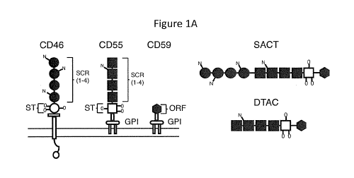
Figure IA and Figure 1B are a schematic drawing and a photograph showing
structure
and expression of SACT and DTAC.
Figure lA is a schematic drawing of the structure of the human membrane-
associated
complement regulators CD46, CD55 and CD59 and the soluble recombinant proteins
SACT
and DTAC. Both CD55 and CD46 each contain four short consensus repeat (SCR)
domains
and a serine/threonine (SIT) rich region. The SCR and S/T domains are sites of
N- and 0-
linked glycosylation, respectively. CD46 inserts in the membrane via a
hydrophobic domain,
and CD55 and CD59 each attach to the membrane via a glycophosphatidylinositol
(GPI)
anchor. CD59 contains a short functional unit of 76 amino acids. Both SACT and
DTAC
contain the four SCR domains and the S/T-rich region of human CD55 separated
by a poly
glycine linker from the functional domain of human CD59. SACT additionally
contains the
four SCR domains of human CD46 at the N-terminus separated by a polyglycine
linker from
the SCRs of CD55. Both SACT and DTAC contain a secretory signal derived from
human
8
CA 02959376 2017-02-24
WO 2016/033444
PCT/US2015/047405
CD59. In engineering the recombinant proteins the membrane-spanning domain of
CD46
and the signals for attachment of a GPI-anchor to each of CD55 and CD59 were
not included.
Figure 1B is a photograph of a western blot showing media from cells
transfected
with pDTAC, pGFP or pSACT probed with antibodies against CD46, CD55 and CD59.
Figure 2A, Figure 2B, Figure 2C and Figure 2D are schematic drawings, a
photograph
and a bar graph showing that SACT acts as a co-factor for Factor I mediated
cleavage of C3b.
Figure 2A is a schematic drawing of Factor I cleavage of C3b. C3b consists of
two
polypeptide chains (a' and 13), joined by a disulfide linkage. Factor!
mediates cleavage of the
104kDa a' chain into inactive fragments, iC3bH and iC3bL.
Figure 2B is a schematic drawing showing CD46 binding of C3b deposited on the
cell
membrane to act as a co-factor for Factor 1-mediated cleavage to inactive
iC3b.
Figure 2C is a photograph of a western blot of purified C3b incubated in media
from
cells transfected with either pSACT, pDTAC or pGFP in the presence or absence
of Factor
and probed with an antibody specific for C3. The western blot shows increased
cleavage of
the a' chain in the presence of media from cells transfected with pSACT
compared to
cleavage occured in the presence of either pGFP or pDTAC.
Figure 2D is a bar graph of quantification of western blot data showing a 51.8
10.5%
(p=0.007) and 46.2 4.8% (p=0.0007) reduction in the amount of the a' chain of
C3b in media
from pSACT-transfected cells containing C3b and Factor! compared to media from
pGFP-
transfected cells and pDTAC-transfected cells containing C3b and Factor I,
respectively. The
signal intensities for the a' chain were normalized to the signal intensity of
the 13 chain.
Figure 3A, Figure 3B and Figure 3C are a schematic drawing and bar graphs
showing
that SACT and DTAC accelerate degradation of C3 convertase.
Figure 3A is a schematic drawing of dissociation of CD55 and factor B binding
to
C3b to accelerate degradation of the C3 convertase.
Figure 3B is a bar graph of quantification of immunostaining of Factor B
binding to
agarose-bound C3b using an antibody for Factor B. The graph shows that media
from
pDTAC- or pSACT-transfected cells resulted in a 16.1 6.4% (p=0.0214, n=11) and
16.8 6.1% (p=0.0127, n=11) reduction in C3b-bound Factor B, respectively,
compared to
media from pGFP-transfected cells (n=10). The Factor B binding is presented as
% staining
relative to the average staining intensity of Factor B bound to C3b in the
presence of media
from pGFP-transfected cells.
9
CA 02959376 2017-02-24
WO 2016/033444
PCT/US2015/047405
Figure 3C is a bar graph of quantification of human complement-mediated lysis
of
sheep erythrocytes that were incubated with media from cells transfected with
either pDTAC
or pSACT in the presence or absence of CD55 blocking antibody. A significant
reduction in
protection against cell lysis was observed for both DTAC and SACT in the
presence of
antibody.
Figure 4A and Figure 4B are a schematic drawing and a bar graph showing that
SACT and DTAC inhibit incorporation of C9 into the membrane attack complex.
Figure 4A is a schematic drawing of CD59 function. CD59 binds to the membrane-
associated C5b-8 protein complex, preventing incorporation of C9 and formation
of the
membrane attack complex (MAC). MAC forms a pore on the cell surface, reducing
integrity
of the membrane.
Figure 4B is a bar graph of quantification of lysis of sheep erythrocytes by
C9-
depleted human serum incubated with C9 in the presence of media transfected
with pGFP,
pDTAC or pSACT. Media from pDTAC- and pSACT-transfected cells reduced human
complement-mediated lysis of erythrocytes by 34.8 3.6% (p<0.0001) and 29.9
4.6%
(p<0.0001), respectively, compared to erythrocytes incubated in the presence
of media from
pGFP-transfected cells.
Figure 5A and Figure 5B are bar graphs showing that SACT and DTAC protect both
sheep erythrocytes and murine hepatocytes from human complement-mediated lysis
in vitro.
Figure 5A is a bar graph of quantification of lysis of sheep erythrocytes
(hemolysis)
by human serum in the presence of media from cells transfected with pGFP,
pDTAC or
pSACT shows a 47 2.9% (p<0.0001) and 21.5 2.8% (p<0.0001) reduction in lysis
by DTAC
and SACT, respectively, compared to media from the GFP-transfected cells.
Figure 5B is a graph of quantification of propidium iodide (PI) uptake by
murine
hepatocytes incubated with normal human serum (NHS) in the presence of media
from cells
transfected with pGFP, pDTAC or pSACT. Control sample of hepatocytes incubated
with
heat-inactivated NHS (hiNHS) in the presence of media from pGFP-transfected
cells is also
shown. Hepatocytes incubated with media from pDTAC- or pSACT-transfected cells
were
observed to have 28.73% 10.21% (p=0.014, n=8) or 20.8 9.0% (p=0.037, n=8)
reduction in
PI uptake, respectively compared to hepatocytes incubated with media from pGFP-
transfected cells (n=7).
CA 02959376 2017-02-24
WO 2016/033444
PCT/US2015/047405
Figure 6A and Figure 6B are micrographs and a bar graph showing that DTAC and
SACT reduce deposition of the Membrane Attack Complex in vitro.
Figure 6A is a set of fluorescent micrographs of murine hepatocytes incubated
with
NHS in the presence of media from pGFP-, pDTAC- or pSACT-transfected cells.
Cells were
stained with an antibody for MAC or for DAPI.
Figure 6B graphs the quantification of MAC staining intensity/area, and shows
a
53.8 10.4% (p=0.0004, n=6) or 67.8 9.2% (p<0.0001, n=6) reduction in MAC
deposition on
murine hepatocytes incubated with media from cells transfected with pDTAC or
pSACT,
respectively, compared to hepatocytes incubated with media from pGFP-
transfected cells
(n=6). Control sample of hepatocytes incubated with hiNHS in the presence of
media from
pGFP-transfected cells is also shown. DAPI, 4',6-diamidino-2-phenylindole;
MAC,
membrane attack complex; hiNHS, heat-inactivated normal human serum.
Figure 7A, Figure 7B, Figure 7C and Figure 7D are micrographs and bar graphs
showing that DTAC protects murine liver vasculature from human MAC deposition
in vivo.
Figure 7A is a set of fluorescent micrographs of cryosections showing
AAV2/8GFP
transduction of murine liver. Efficient transduction was observed throughout
the tissue.
Higher magnification of boxed region is also shown.
Figure 7B is a set of fluorescent micrographs of liver cryosections stained
with anti-
MAC antibody harvested from mice injected in the intraperitoneal space with
AAV2/8pA or
AAV2/8DTAC and perfused with mPECAM1 antibody and NHS. Higher magnification of
boxed regions is also shown.
Figure 7C is a set of bar graphs of quantification of MAC staining intensity
(IU) of
liver sections, showing a 56.7 16.4% (p=0.0061) reduction in human MAC
deposition on the
liver vasculature of AAV2/8DTAC-injected relative to AAV2/8polyA-injected
mice.
Figure 7D is a bar graph of quantification of MAC staining intensity per area
of blood
vessels which shows a 55.6 11.3% (p=0.0006) reduction in human MAC deposition
in livers
of AAV2/8DTAC-injected mice compared to AAV2/8polyA-injected. Staining
intensity was
averaged from 8 sections per mouse. n=6 for AAV2/8polyA- and n=6 for
AAV2/8DTAC-
injected mice. (DIC: differential interference contrast; IU: intensity unit)
Figure 8A, Figure 8B and Figure 8C are micrographs and bar graphs showing that
SACT protects murine liver vasculature from human MAC deposition in vivo.
11
CA 02959376 2017-02-24
WO 2016/033444
PCT/US2015/047405
Figure 8A is a set of fluorescent micrographs of liver cryosections stained
with anti-
MAC antibody harvested from mice injected in the intraperitoneal space with
AAV2/8pA or
AAV2/8SACT and perfused with mPECAMI antibody and NHS. Higher magnification of
boxed regions is also shown.
Figure 8B is a bar graph of quantification of MAC staining intensity (IU) of
liver
sections which shows a 63.2% 20.5% (p=0.0075) reduction in human MAC
deposition on
the liver vasculature of AAV2/8SACT-injected compared to AAV2/8polyA-injected
mice.
Figure 8C is a bar graph of quantification of MAC staining intensity per area
of blood
vessels which shows a 61.1 18.9% (p=0.0056) reduction in human MAC deposition
on the
blood vessels of AAV2/8SACT-injected relative to AAV2/8polyA-injected mice.
Staining
intensity was averaged from 8 sections per mouse. n=8 for AAV2/8polyA- and n=9
for
AAV2/8SACT-injected mice.
Figure 9 is a schematic drawing of structures of soluble terminator of
activated
complement (STAC) showing the structure of the human membrane-associated
complement
regulators, CD46, CD55 and CD59 and the soluble recombinant protein STAC. STAC
contains a secretory signal derived from human CD59. The four SCR domains and
SIT ¨rich
region of human CD46 were attached via a poly glycine linker to CD59. The four
SCR
domains and the SIT-rich region of human CD55 were engineered to be separated
by a poly
glycine linker from the functional domain of human CD46.
Figure 10A and Figure 10B are a photograph and a bar graph showing that STAC
acts
as a co-factor for Factor I mediated cleavage of C3b.
Figure 10A is a photograph of a western blot of purified C3b incubated in
media
obtained from cells transfected with either pAdCAGGFP or with pAdCAGSTAC in
the
presence or absence of Factor I and probed with an antibody for C3. The data
shows
increased cleavage of a' chain in the presence of media from cells transfected
with
pAdCAGSTAC compared to cleavage that occurred in the presence of pAdCAGGFP.
Figure 10B is a graph of quantification of western blot data which shows a
34.3% 3.9%(n=4; p=0.0001) reduction in the amount of the C3b a' chain in media
from
pAdCAGSTAC-transfected cells containing C3b and Factor I compared to media
from
pAdCAGGFP-transfected cells containing C3b and Factor I (n=4). The signal
intensities for
the a' chain were normalized to the signal intensity of the 13 chain.
12
CA 02959376 2017-02-24
WO 2016/033444
PCT/US2015/047405
Figure 11 is a bar graph showing that the CD55 portion of STAC retains
functionality.
The graph illustrates quantification of human complement-mediated lysis of
sensitized sheep
erythrocytes which were incubated with media from cells transfected with
either
pAdCAGGFP or pAdCAGSTAC in the presence or absence of CD55 blocking antibody.
STAC treated samples in the absence of mAb blocking were observed to have a
32.1% 10.4% reduction in cell lysis (n=6, p=0.0115) compared to GFP treated
samples (n=6)
with no antibody blocking. Sheep erythrocytes suspended in media from
pAdCAGSTAC
transfected cells containing CD55 blocking antibody showed a non-statistically
significant
reduction of 13.4% 9.4% in cell lysis (n=6; p=0.1831). Cell lysis that occured
in GFP media
without blocking antibody was set to 100% cell lysis.
Figure 12 is a bar graph showing that STAC was unable to prevent C9
incorporation
into membrane attack complex. The graph is a quantification of lysis of sheep
erythrocytes by
C9-depleted human serum incubated with or without C9 in the presence of media
transfected
with pAdCAGGFP, pAdCAGSTAC or pAdCAGsCD59 (positive control). Erythrocytes
treated with sCD59+C9 were observed to have a 21% 9.1% (n=8; p=0.033)
reduction in cell
lysis compared to GFP+C9 treated cells (n=14). Samples treated with STAC+C9
showed no
decrease in cell lysis (n=14; p=0.428).
Detailed Description
A variety of disorders are associated with the activation of complement. CD46,
CD55
and CD59 are the major membrane associated regulators of complement on human
cells.
Independent expression of CD55, CD46 or CD59 through gene transfer protects
murine
tissues against human complement mediated attack. The example of the present
application
describe the potential of combining the complement regulatory properties of
CD46, CD55
and CD59 into single gene products expressed from an adeno associated virus
(AAV) vector
in a soluble non-membrane anchored form.
Dysregulation of the complement system is one of the major factors
contributing
towards the etiology of AMD, one of the leading causes of blindness in the
elderly (Gehrs et
al. 2010 Arch Ophthalmol 128: 349-358). The most devastating form of the
disease affects
approximately 10% of patients (Klein 2008 Ophthalmology 115: 1026-1031), and
involves
the growth of attenuated blood vessels from the choroidal vasculature through
Bruch's
membrane and into the retina. The plasma released by these "ill-formed"
vessels damages
13
CA 02959376 2017-02-24
WO 2016/033444
PCT/US2015/047405
photoreceptors and other retinal cells, eventually leading to a severe loss of
vision. The vast
majority of AMD patients, however, present with extracellular deposits which
occur between
the retinal pigment epithelium (RPE) and Bruch's membrane called drusen and
which
eventually lead to atrophy of the RPE (geographic atrophy).
A potential role for complement in AMD was considered because complement
proteins were identified in drusen of AMD eyes (Johnson et al. 2001 Exp Eye
Res 73: 887-
896; Johnson et al. 2000 Exp Eye Res 70: 441-449; Mullins et al. Eye (Lond)
15: 390-395;
and Mullins et al. 2000 FASEB J 14: 835-846) . Polymorphisms have been
identified in a
number of complement genes and were observed to be either strongly predictive
of or
protective against AMD. A single amino acid change, Y402H, in factor H
accounts for as
much as 40-50% of AMD in aging eyes (Edwards et al. 2005 Science 308: 421-424;
Hageman et al. 2005 Proc Natl Acad Sci USA 102: 7227-7232; and Haines et al.
2005
Science 308: 419-421).
Haplotype variants in both Factor B and complement component 2 (C2) result in
a
significantly reduced risk of developing AMD (Gold et al. 2006 Nat Genet 38:
458-462), and
an R8OG substitution in complement component 3 (C3) increased the risk of
having AMD to
as much as 22% (Yates et al. 2007 N Engl J Med 357: 553-561). The factor B
(32Q) variant
has been shown to have a 4-fold lower binding affinity for C3b, with a reduced
ability to
form the convertase (Montes et al. 2009 Proc Nat! Acad Sci USA 106: 4366-
4371). In
addition, polymorphisms in C2, C3, and factor B have been shown to be
significantly linked
with progression to both types of advanced AMD disease, choroidal
neovascularization and
geographic atrophy (Klein 2008 Ophthalmology 115: 1026-1031; Mailer et al.
2007 Nat
Genet 39: 1200-1201; and Reynolds etal. 2009 Invest Ophthalmol Vis Sci 50:
5818-5827).
Deposition of complement proteins has been observed in the choriocapillaris of
patients with diabetic retinopathy (Gerl et al. 2002 Invest Ophthalmol Vis Sci
43: 1104-
1108), and in retinal vessels of diabetic subjects (Zhang et al. 2002 Diabetes
51: 3499-3504).
The retinal vessels exhibited a significant reduction in expression of
complement regulatory
proteins CD55 and CD59. Complement components have also been observed in the
epiretinal
membranes of patients suffering from proliferative vitreoretinopathy (PVR),
and upregulation
of the classical pathway initiator protein, Clq, and altered expression of
other proteins of the
cascade have been observed in glaucomatous eyes (Baudouin et al. 1990 Am J
Ophthalmol
110: 593-598; Stasi etal. 2006 Invest Ophthalmol Vis Sci 47: 1024-1029; and
Tezel et al.
2010 Invest Ophthalmol Vis Sci 51(11): 5697-707).
14
CA 02959376 2017-02-24
WO 2016/033444
PCT/US2015/047405
There are few animal models that are useful to directly investigate the role
of
complement in retinal function and pathology. Most of these models have been
used to
analyze the impact of different complement proteins in the development of
laser-induced
choroidal neovascularization (CNV) in the mouse retina. Other studies have
demonstrated the
dependence of retinal pathology on the alternative pathway, rather than
classical or lectin
pathway, and on the formation of the membrane attack complex (Bora et al. 2007
J Immunol
178: 1783-1790; Bora et al. 2006 J Immunol 177: 1872-1878; and Bora et al.
2005 J Immunol
174: 491-497). Previous studies have demonstrated a significant role played by
the
anaphylatoxins, C3a and C5a, in the development of CNV (Nozaki et al. 2006
Proc Natl
Acad Sci USA 103: 2328-2333). Aged mice with deficiency of factor H exhibited
altered
architecture in Bruch's membrane, RPE and photoreceptors, and reduced ERGs
(Coffey et al.
2007 Proc Natl Acad Sci USA 104: 16651-16656), and manifested a loss of
integrity of
retinal vessels (Lundh von Leithner et al. 2009 Am J Pathol 175: 412-421). The
alternative
complement pathway has been implicated also as a major factor in light-induced
retinal
degeneration which has been shown to be significantly reduced in a mouse
deficient in Factor
D (Rohrer et al. 2007 Invest Ophthalmol Vis Sci 48: 5282-5289). Ganglion cells
of C3-
deleted mice exhibited transient, but significant, protection from
degeneration due to retinal
ischemia reperfusion (Kuehn et al. 2008 Exp Eye Res 87:89-95).
One of three distinct complement pathways (classical, lectin or alternative)
initiates
the complement cascade (Markiewski et al. 2007 Am 1 Pathol 171: 715-727) and
these
pathways converge at the point in the pathway of the breakdown of C3 into C3a
and C3b.
The breakdown of C3 initiates the final part of the pathway that culminates in
the formation
of the membrane attack complex (MAC), a pore-like structure that inserts in
the membranes
of self- or non-self cells causing their lysis. In addition to the potential
for cell lysis by the
production of the opsonin C3b, activation of C3 generates the anaphylatoxins,
C3a and C5a,
both of which are powerful and pleiotropic effectors of inflammation. Unlike
the classical or
lectin pathways, the alternative pathway is constitutively active with small
amounts of C3
hydrolysis and conversion to the convertase occurring in the serum.
An approach for delivery of inhibitors of complement for chronic diseases such
as
AMD or rheumatoid arthritis is the use of somatic gene therapy. Gene therapy
is efficacious
in humans for treatment of single gene disorders and patients with complex
disorders such as
rheumatoid arthritis have also been successfully treated using a gene therapy
approach (Ginn,
S.L., et al. 2013 J Gene Med 15: 65-77). Use of gene therapy has been found to
be uniquely
efficacious in mobilizing soluble versions of otherwise membrane-associated
inhibitors of
CA 02959376 2017-02-24
WO 2016/033444
PCT/US2015/047405
complement. For example, CD59 (protectin) is a naturally occurring inhibitor
of MAC found
tethered to the membranes of cells via a glycosylphosphatidylinositol (GPI)
anchor.
Membrane-associated CD59 is a potent inhibitor of MAC and soluble membrane-
independent
CD59 has been reported to be efficacious in vivo only when delivered via a
gene therapy
vector such as adeno-associated virus (AAV) (Cashman, S.M., etal. 2011 PLoS
One 6:
el9078).
CD55 (decay accelerating factor) is a GPI anchored protein that regulates
complement
activity by accelerating the decay of the classical as well as the alternative
C3 convertase
(Walport, M.J. 2001 N Engl J Med 344: 1058-1066). CD46 (membrane cofactor
protein), is
a ubiquitously expressed type I transmembrane glycoprotein which acts as a
cofactor for
factor I mediated cleavage of C3b and C4b and prevents formation of the
classical and
alternative C3 convertase (Riley-Vargas, R.C., et al. 2004 Immunol 25: 496-
503). CD46
regulates amplification loop of the alternative pathway of activation of
complement. CD55
and CD46 have different properties and each contain a series of 60 amino acid
repeat motifs
called short consensus repeats (SCR) that act as complement regulatory modules
(Coyne,
K.E., etal. 1992 J Immunol 149: 2906-2913). Species specificity between human
and mouse
complement proteins limits the testing of human complement inhibitors in
murine tissues in
vivo (Kim, D.D., et al. 2006 Clin Immunol 118: 127-136). The methods and
compositions
provided herein relate to engineering a novel non-membrane associated
recombinant protein
for optimal inhibition of complement activation. This is achieved by
simultaneous targeting
of both the classical and alternative pathways of complement and at different
points of the
complement cascade. The classical and alternative pathways of complement are
targeted by
combining the properties of CD55, CD46 and CD59. The examples herein describe
the
synthesis of the novel engineered recombinant protein and measure merits of
its ability to
inhibit human complement in cell culture and human complement in murine
tissues in vivo by
gene therapy. The examples herein also describe a recombinant protein that
combines the
complement inhibitory properties of CD55 and CD59.
A non-membrane associated recombinant molecule, SACT is provided herein that
exhibits the combinatorial properties of CD46, CD55 and CD59. SACT is a
secreted protein
that can act as a co-factor for Factor I mediated cleavage of C3b, accelerate
the degradation
of C3 convertase, attenuate recruitment of C9 into the MAC, protect cells from
human
complement mediated lysis and inhibit deposition of human MAC on mouse cells
in vitro and
in vivo. Also provided herein is a composition DTAC which was observed to have
the
16
CA 02959376 2017-02-24
WO 2016/033444
PCT/US2015/047405
combined properties of CD55 and CD59, and which does not act as a co-factor
for Factor I
mediated cleavage of C3b.
Complement is a critical first line of immune defense in vertebrates,
affording
protection against both foreign organisms and the threat of damaged self-cells
(Walport, M.J.
2001 N Engl J Med 344: 1058-1066).Over-expression of complement regulators for
the
treatment of complement-mediated pathologies poses risks as well as benefits.
Therefore, the
complement pathway at the combined points of C3 convertase formation and decay
and the
formation of MAC was inhibited, which allows Clq to interact with modified
self and non-
self surfaces, permitting C3b-mediated phagocytosis of offending cells and
organisms
(Trouw, et al. 2008 Mol Immunol 45: 1199-1207). The functions of CD55, CD46
and CD59
were combined into a single engineered protein, SACT. Further, because CD46
functions at
the level of C3b degradation and CD55 prevents formation or accelerates the
decay of
convertases without altering C3b, it is here envisioned that CD46 interferes
with C3b-
mediated elimination of target cells. This may be especially important for the
treatment of
lupus-like diseases where reduced clearance of apoptotic cells can result in
an autoimmune
response to the dying cells (Trouw, et al. 2008 Mol Immunol 45: 1199-1207).
Studies of the
diversity of genetic factors involved in SLE, provide a comprehensive
illustration of the
importance of maintaining the potential for activation of upstream components
of the cascade
and blocking downstream events (Karp, D.R. 2005 Curr Opin Rheumatol 17: 538-
542).
Therefore a second recombinant molecule that includes only CD55 and CD59
functions,
DTAC was generated and tested.
A gene therapy approach was used to deliver SACT or DTAC to cells in culture
or to
murine livers in vivo. Significant progress in the field of gene therapy
indicates that this is a
viable approach for the treatment of inherited or acquired diseases.
Activation of
complement plays a significant role in many disorders including rheumatoid
arthritis, a
chronic disease of the complement system. Elevated levels of C3 and MAC and
reduced
levels of CD59 have been documented in the synovial tissue of rheumatoid
arthritis patients
(Kemp, P.A., et al. 1992 J Clin Lab Immunol 37: 147-162, Konttinen, Y.T., et
al. 1996 Ann
Rheum Dis 55: 888-894). These studies are further supported by the observation
that
injection of rat knee joints with monoclonal antibody against CD59 results in
spontaneous
and acute arthritis and an increase in joint pathology in CD59 -I- mice, a
phenotype that can
be corrected by use of a membrane-targeted recombinant CD59 (Kemp, P.A., et
al. 1992 J
Clin Lab Immunol 37: 147-162). Attenuation of complement activation by
targeting C5 was
found to be effective in a murine model of rheumatoid arthritis, indicating
that there are
17
CA 02959376 2017-02-24
WO 2016/033444
PCT/US2015/047405
multiple points of the complement cascade that may serve as targets for
complement based
therapeutics (Kemp, P.A., etal. 1992 J Clin Lab Immunol 37: 147-162).
Therefore, SACT
and DTAC each are particularly effective inhibitors of complement activation
because they
concomitantly target and attenuate various points of the complement cascade.
Even though
repeat injections of inhibitors of complement activation into patients with
rheumatoid arthritis
is feasible, a long-lasting single injection via gene therapy is potentially
preferred for
efficiency and for convenience of the patient. Adeno-associated virus (AAV)
has been
shown to persist in humans for years and for over a decade in large animals
(Colella, P., et al.
2009 Trends Mol Med 15: 23-31, Jiang, H., etal. 2006 Mol Ther 14: 452-455).
Furthermore,
AAV is not associated with any known human disease.
A strong case for delivery of inhibitors of complement via a gene therapy
approach
may be made for diseases such as AMD. Approximately 50% of patients that
suffer from
AMD have polymorphisms in the complement regulator Factor H (Klein, R.J., et
al. 2005
Science 308: 385-389). Individuals that are homozygous for a Y402H
polymorphism in
Factor H have approximately 70% more MAC in their choroidal blood vessels and
retinal
pigment epithelium (RPE) (Mullins, R.F., et al. 2011 Exp Eye Res 93: 565-567).
Individuals
with an advanced form of AMD known as geographic atrophy have reduced levels
of
complement inhibitors on their RPE (Ebrahimi, K.B., et al. 2013 J Pathol 229:
729-742). A
commonly occurring polymorphism in C9 in the Japanese population that prevents
those
individuals from efficiently assembling MAC is protective against the
progression of AMD,
suggesting that inactivation of complement via a gene therapy approach may be
a viable
avenue for treatment of this disease (Nishiguchi, K.M., et al. 2012 Invest
Ophthalmol Vis Sci
53: 508-512).
However, all of the inhibitors of complement activation currently in clinical
trials are
small molecules, aptamers or antibodies that would need to be re-injected on a
frequent basis
into the eye of AMD patients (Keane, P.A., et al. 2012 J Ophthalmol 2012:
483034). These
inhibitors have significant side effects such as increased intraocular
pressure, endophthalmitis
and retinal detachment (Wu, L., etal. 2008 Graefes Arch Clin Exp Ophthalmol
246: 81-87,
Shima, C., et al. 2008 Acta Ophthalmol 86: 372-376). A single injection that
may produce a
therapeutic protein locally for an extended time such as an AAV vector, which
mediates
expression of SACT or DTAC, may be particularly attractive for treatment of
diseases such
as AMD. Species restriction between complement proteins limits the testing of
human CD55
and human CD46 with respect to murine complement and thus in murine models of
AMD
(Kim, D.D., et at. 2006 Clin Immunol 118: 127-136). Inventors of the present
application
18
CA 02959376 2017-02-24
WO 2016/033444
PCT/US2015/047405
have shown that human CD55 or human CD46 efficiently inhibited human
complement
deposited on murine retinal tissues in ex vivo murine models of MAC deposition
(Sweigard,
J.H., et al. 2011 Gene Ther 18: 613-621).
At present, there are more than 25 small molecules, antibodies or proteins
under
clinical and preclinical development for attenuation of activation of
complement. These
molecules are aimed at a wide variety of indications including acute kidney
injury, COPD,
paroxysmal nocturnal hemoglobinuria, rheumatoid arthritis, sepsis, AMD and
transplantation.
The majority of these therapeutics target complement at the level of C3 or C5
(Ricklin, D., et
at. 2013 J Immunol 190: 3839-3847). Mutations in CD46 have been shown to
predispose
individuals to familial hemolytic uremic syndrome (Kavanagh, D., et al. 2008
Annu Rev Med
59: 293-309). Deficiency of CD55 has been associated with primary autoimmune
hemolytic
anemia, SLE and in paroxysmal nocturnal hemoglobinuria (Richaud-Patin, Y., et
al. 2003
Immunol Lett 88: 95-99, Iwamoto, N., et al. 1995 Blood 85: 2228-2232). CD55
has also been
shown to attenuate ischemic reperfusion organ damage (Weeks, C., et al. 2007
Clin Immunol
124: 311-327). Mutations or deficiencies in CD59 have been shown to result in
chronic
hemolysis and relapsing peripheral demyelinating disease in infancy,
paroxysmal nocturnal
hemoglobinuria, autoimmune hemolytic anemia or SLE (Iwamoto, N., et al. 1995
Blood 85:
2228-2232, Nevo, Y., et al. 2013 Blood 121: 129-135). It is here envisioned
that complement
inhibitors such as SACT and DTAC will be useful for the treatment of these
disorders.
Fodor et at. have described a membrane-associated recombinant molecule
containing
the combinatorial properties of CD55 and CD59 (Fodor, W.L., et al. 1995 J
Immunol 155:
4135-4138). Similarly, Kroshus et al. have described a soluble molecule
combining the
properties of CD46 and CD55 and demonstrated that this molecule could reduce
acute
cardiac tissue injury in a pig-to-human transplant model (Kroshus, T.J., et
al. 2000
Transplantation 69: 2282-2289). A recombinant protein comprised of select
domains from
CR2 and factor H demonstrated increased survival, reduced autoantibody
production and
improved kidney function in a murine model of lupus (Sekine, H., et al. 2011
Arthritis
Rheum 63: 1076-1085). However, none of these studies delivered the recombinant
protein via
a gene therapy approach.
For in vivo examples herein an AAV2 pseudotyped with AAV8 capsid (AAV2/8) was
used. This vector has been shown to have a very high efficiency of
transduction of the liver of
mice (Paneda, A., et at. 2009 Hum Gene Ther 20: 908-917). The in vivo examples
were
performed in the liver in part because large amounts of human MAC can readily
form on
murine liver (Gandhi, J., et al. 2011 PLoS One 6: e21621). Further, the liver
was selected in
19
CA 02959376 2017-02-24
WO 2016/033444
PCT/US2015/047405
part because hepatocytes are responsible for the biosynthesis of 80-90% of the
plasma
components of complement (Qin, X., et al. 2006 Cell Mol Immunol 3: 333-340).
Finally, the
liver receives 25% of total blood flow, allowing for a wide distribution of
DTAC and SACT
throughout the circulatory system, which would be relevant for the treatment
of systemic
disorders involving activation of complement (Myers, J.D., et al. 1948 J Clin
Invest 27: 620-
627). Expression of DTAC and SACT in vivo indicated that both are potent
inhibitors of
human complement in an in vivo setting and the data shown in examples herein
lends support
to the therapeutic value of these molecules if secreted from the liver.
To investigate whether the order of the complement regulatory regions affects
functional capability the proteins provided herein were assayed in comparison
to soluble
terminator of activated complement (STAC), U.S. patent number 8, 877,896
issued
November 4, 2014. STAC contains the following: N-terminus of STAC contains the
human
CD59 start codon, secretory signal peptide and SCR domain; a polyglycine
linker attaches the
four SCR domains and S/T ¨rich region of human CD46 to the C-terminus of CD59;
and four
SCR domains and S/T ¨rich region of human CD55 are linked to the C-terminus of
CD46 via
a polyglycine sequence (Figure 9). The c-DNA for STAC was prepared the protein
was
expressed between a CMV enhancer/chicken 13-actin promoter (CAG) and a rabbit
globin
polyadenylation (pA) termination sequence. (Ibid.)
The functionality of each of the CD46, CD55 and CD59 components was
individually
measured in STAC as described in examples herein. It was observed that the
CD59 portion in
STAC was non-functional. CD59 is a potent inhibitor of the terminal pathway of
the
complement system (Rollins SA, et al., J Immunol. 1990; 144 (9):3478-83). CD59
functions
by blocking C9 incorporation into the membrane attack complex (MAC), thereby
blocking
pore formation in cellular membranes (Rollins SA, et al., J Immunol. 1990; 144
(9):3478-83).
The CD59 portion of STAC was observed to be unable to prevent C9
incorporation.
Therefore, the CD59 portion of STAC is non-functional and hence this portion
of STAC does
not contribute function as an inhibitor of complement.
The invention herein provides a functional non-membrane associated recombinant
protein, SACT, which exhibits the combinatorial properties of CD46, CD55 and
CD59, and
also provides a recombinant protein DTAC, which has the combinatorial
properties of CD55
and CD59. Each of these proteins were observed to surprisingly exhibit
properties and
functions of their modular components and each of these proteins is a potent
inhibitor of
activation of complement in vitro and in vivo. Each of the components of the
SACT protein
and the DTAC protein were observed to exhibit their biological function in
contrast to STAC.
CA 02959376 2017-02-24
WO 2016/033444
PCT/US2015/047405
SACT protein
A membrane bound CD59 was previously observed to protect cells from complement-
mediated disease, however the site of expression of the regulator, yielded
only a "patch" of
protection in the ocular tissue such as the RPE. Thus, a secreted regulator of
CD59 (sCD59 or
rmiCD59) is here engineered, which was capable of diffusing through the retina
and offer
protection to the entire affected region (Kumar-Singh et al., PCT application
serial number
PCT/US09/00947 filed February 13, 2009 which is hereby incorporated by herein
in its
entirety).
Soluble sCD59 was previously considered an inefficient regulator of complement
in
vivo unless it was fused with a membrane targeting moiety (Mizuno etal.
200IArthritis
Rheum 44: 2425-2434; Bora 2010 J Biol Chem 285: 33826-33833; Song et al. 2003
J Clin
Invest 111: 1875-1885; and Zhang et al. 1999 J Clin Invest 103: 55-61). A
membrane-
independent sCD59 expressed in vivo in murine ocular tissue via an adenovirus
or AAV
vector significantly reduced MAC deposition and laser-induced choroidal
neovascularization
in a mouse model of neovascular AMD (Cashman et al. 2011 PLoS ONE 6(4):
e19078,
which is hereby incorporated by reference in its entirety). Adenovirus-
delivered sCD59 was
observed to inhibit human MAC deposition even on murine liver vasculature.
Without being limited by any particular theory or mechanism of action, it is
here
envisioned that a recombinant fusion protein containing at least two of CD59
protein, CD46
protein and CD55 protein is a potent regulator of a number of complement
pathways and
proteins. Examples herein provide methods for engineering a novel Soluble
Active
Complement Terminator (SACT) having small functional units of each of CD46
protein,
CD55 protein, and CD59 protein that are effective for treating complement-
related conditions
by modulating the complement cascade, and provide the composition. The
resulting SACT
protein composition includes functional units of CD46 protein, CD55 protein,
and CD59
protein that in certain embodiments are operably linked. For example, the
functional units are
connected by a linker, which is a sequence of amino acids that does not affect
the function of
the components or the structural stability of the protein. Furthermore, the
protein in certain
embodiments is mutated to remove or delete a sequence encoding a protein
membrane
anchor. In a related embodiment, an exemplary SACT protein includes a
secretory signal at
the N-terminus. The SACT protein is approximately 130 KDa and was obtained
retaining
only the units/domains of each component protein that are involved in
complement
regulation. Other soluble complement regulators such as factor H (150kDa) and
sCR I
21
CA 02959376 2017-02-24
WO 2016/033444
PCT/US2015/047405
(200kDa) are larger, and these have been used to regulate complement (Ripoche
et al. 1984
Biochem .1 221: 89-96; and Yoon et al. 1985 .1 Immunol 134: 3332-3338).
The recombinant SACT protein engineered herein is differs from naturally
occurring
regulators because it includes multiple complement regulatory domains from
different
combinations of CD59, CD46, and CD55 proteins and is membrane independent.
Hence
SACT protein is capable of diffusing and blanketing a large group of affected
cells or tissue
for treatment after a single administration at one time. For example the SACT
protein
includes an amino acid sequence from at least two of a CD46 protein, a CD55
protein, and a
CD59 protein. For example, the SACT protein includes at least one of: the CD46
protein and
the CD59 protein, the CD46 protein and the CD55 protein, and the CD55 protein
and the
CD59 protein. Alternatively, the SACT protein includes each of CD59 protein,
CD46 protein
and CD55 protein, operably linked and expressed for example in a soluble form.
In various
embodiments, the CD46 protein, the CD55 protein, and the CD59 protein are
derived from
mammalian proteins (e.g., human, mouse, and rabbit). For example the SACT
protein
comprises a CD46 protein and a CD59 protein that are human proteins and a CD55
protein
that is a murine protein, or comprises each of CD46, CD55, and CD59 that are
human
proteins. Thus in various embodiments the SACT protein comprises proteins that
are from the
same mammal type, or from different types of mammals.
Without being limited by any particular theory or mechanism of action, it is
here
envisioned that the SACT protein synergistically blocks complement activation
at multiple
steps in the complement pathway, including each of the complement pathways
regulated by
each of CD59 protein, CD46 protein, and CD55 protein. The SACT protein was
observed in
Examples herein to have inhibited MAC deposition in vivo when delivered by an
adenovirus
vector, and is therefore potentially effective as an anti-complement therapy
for treating or
even preventing complement-associated diseases or conditions.
In various embodiments, the SACT protein or composition includes a CD46
protein
encoded by a full length nucleic acid of CD46 which was modified to remove the
amino acid
sequences for signal sequence and hydrophobic transmembrane spanning domains.
Alternatively the nucleic acid sequence of CD46 protein is modified by point
mutations,
substitutions or deletions to obtain a nucleic acid sequence that encodes a
modified amino
acid sequence with the modification located in the hydrophobic transmembrane
spanning
domain, such that the resulting protein fails to attach to cell membranes.
The term "membrane independent CD46" as used herein refers to a CD46 amino
acid
sequence that lacks a hydrophobic transmembrane spanning domain or has a
modified
22
CA 02959376 2017-02-24
WO 2016/033444
PCT/US2015/047405
hydrophobic transmembrane spanning domain that lacks functional ability to
bind to a cell
membrane or a cell-membrane-associated structure such as a membrane-bound
protein. The
scope of the CD46 protein herein is envisioned to include conservative
sequence
modifications including deletions, substitutions, and additions as has been
described herein.
As used herein, the term "conservative sequence modifications" refers to amino
acid
modifications that do not significantly affect or alter the characteristics of
the CD46 protein
containing the amino acid sequence, i.e., amino acid sequences of CD46 protein
that present
the side chains at the same relative positions in the amino acid sequence will
function in a
manner similar to human CD46 protein. Such conservative modifications include
amino acid
substitutions, additions and deletions. Modification of the amino acid
sequence of CD46
protein is achieved using any known technique in the art e.g., site-directed
mutagenesis or
PCR based mutagenisis. Such techniques are described in Sambrook et al.,
Molecular
Cloning: A Laboratory Manual, Cold Spring Harbor Press, Plainview, NY, 1989
and Ausubel
et al., Current Protocols in Molecular Biology, John Wiley & Sons, New York,
NY, 1989.
Conservative amino acid substitutions are ones in which the amino acid residue
is
replaced with an amino acid residue having a functionally similar side chain.
Families of
amino acid residues having similar side chains have been defined in the art.
These families
include amino acids with basic side chains (e.g., lysine, arginine,
histidine), acidic side chains
(e.g., aspartic acid, glutamic acid), uncharged polar side chains (e.g.,
glycine, asparagine,
glutamine, serine, threonine, tyrosine, cysteine, tryptophan), nonpolar side
chains (e.g.,
alanine, valine, leucine, isoleucine, proline, phenylalanine, methionine),
beta-branched side
chains (e.g., threonine, valine, isoleucine) and aromatic side chains (e.g.,
tyrosine,
phenylalanine, tryptophan, histidine).
In certain embodiments, the CD46 amino acid sequence is an amino acid sequence
that is substantially identical to that of the wild type sequence. The term
"substantially
identical" is used herein to refer to a first amino acid sequence that
contains a sufficient or
minimum number of amino acid residues that are identical to aligned amino acid
residues in a
second amino acid sequence such that the first and second amino acid sequences
can have a
common structural domain and/or common functional activity. For example, amino
acid
sequences that contain a common structural domain having at least about 60%
identity, or at
least 75%, 85%, 95%, 96%, 98%, or 99% identity are substantially identical.
Calculations of sequence identity between sequences are performed as follows.
To
determine the percent identity of two amino acid sequences, the sequences are
aligned for
optimal comparison purposes (e.g., gaps can be introduced in one or both of a
first and a
23
CA 02959376 2017-02-24
WO 2016/033444
PCT/US2015/047405
second amino acid sequence for optimal alignment). The amino acid residues at
corresponding amino acid positions or nucleotide positions are then compared.
When a
position in the first sequence is occupied by the same amino acid residue or
nucleotide as the
corresponding position in the second sequence, then the proteins are identical
at that position.
The percent identity between the two sequences is a function of the number of
identical
positions shared by the sequences, taking into account the number of gaps, and
the length of
each gap, which need to be introduced for optimal alignment of the two
sequences.
The comparison of sequences and determination of percent identity between two
sequences are accomplished using a mathematical algorithm. Percent identity
between two
amino acid sequences is determined using an alignment software program using
the default
parameters. Suitable programs include, for example, CLUSTAL W by Thompson et
al., Nuc.
Acids Research 22:4673, 1994 (www.ebi.ac.uk/clustalw), BL2SEQ by Tatusova and
Madden, FEMS Microbiol. Lett. 174:247, 1999
(www.ncbi.nlm.nih.gov/blast/b12seq/b12.html), SAGA by Notredame and Higgins,
Nuc.
Acids Research 24:1515, 1996 (igs-server.cnrs-mrs.fri¨cnotred), and DIALIGN by
Morgenstern et al., Bioinformatics 14:290, 1998 (bibiserv.techfak.uni-
bielefeld.de/dialign).
In various embodiments, the SACT protein or composition includes a CD55
protein
and/or a CD59 protein. In various embodiments, the CD55 protein includes a
full length
nucleic acid of CD55. Alternatively, the CD55 protein is a portion or
homologue of full
length nucleic acid sequence or amino acid sequence as described herein. In
certain
embodiments, the CD55 protein includes conservative sequence modifications of
the CD59
protein.
Mature human CD59 protein is composed of 77 amino acids and has a molecular
weight of about 18kD to about 21 kD. Precursor human CD59 protein includes an
amino
terminal signal peptide of 25 amino acids and a carboxyl-terminal peptide of
26 amino acids
which allows for attachment of a membrane anchor. Amino acid sequences of
precursor
human CD59 protein, a mature human CD59 protein, and CD59 protein of other
mammals,
e.g., baboon, African green monkey, owl monkey, marmoset, HVS-15, pig, rabbit,
rat, and
mouse, are shown in Sims et al. U.S. patent number 7,166,568 issued January
23, 2007
which is incorporated herein by reference in its entirety.
The protein structure of CD59 is characterized as a single cysteine-rich
domain,
having a hydrophobic core with three loops and a small fourth helical loop (Yu
et al., Journal
of Experimental Medicine, 185(4):745-753, 1997). Disulfide-bonded cysteine
pairs connect
each of these loops (Yu et al., 1997).
24
CA 02959376 2017-02-24
WO 2016/033444
PCT/US2015/047405
The structure of the gene encoding CD59 has been characterized (Fodor et al.
U.S.
patent number 5,624,837, issued April 29, 1997). The gene is located on the
short arm of
chromosome 11 in humans, specifically chromosome 11p13 and 11p14 (Online
Mendelian
Inheritance in Man accession number and107271), and consists of four exons
spanning 20 kb
(Petranka et al. Proc. Nat. Acad. Sci. 89:7876-7879, 1992). An untranslated
first exon is
preceded by a G and C-rich promoter region that lacks a consensus TATA or CAAT
motif.
The second exon encodes the hydrophobic leader sequence of the protein, and
the third exon
encodes the N-terminal portion of the mature protein. The fourth exon encodes
the remainder
of the mature protein, including the hydrophobic sequence for
glycophosphoinosital anchor
attachment to a cell membrane.
CD59 is a glycosylphosphatidylinositol-anchored glycoprotein that is expressed
on
human peripheral blood leukocytes, erythrocytes, and many cell lines. The
protein is
expressed on both hematopoietic and non-hematopoietic cells, for example on
endothelial
cells, peripheral nerve fibers, neurons, microglia, oligodendrocytes,
astrocytes, ependymal
cells, epithelial cells, acinar cells of the salivary glands, bronchial
epithelium, renal tubules
and squamous epithelium. See Nose, M. et al. 1990 Immunology 70(2): 145-149;
Vedeler, C.
et al. 1994 Immunology 82(4): 542-547; and Hidestima, T. et al. 1990
Immunology 69(3):
396:401. A cDNA encoding CD59 was reported by Sawada, R. etal. 1989 Nucleic
Acids Res
17(16) 6728. CDNA encoding CD59 has also been cloned from human T-cell
leukemia (YT)
and human erythroleukemia (K562) cell lines, and CD59 has been transiently
expressed in
COS cells (Walsh, L.A. etal. 1990 Eur J. Immol 21(3): 847-850). Human CD59
which is
encoded by a nucleic acid sequence includes 26 amino acids located at the C
terminus, which
contains a signal sequence for attachment of a GPI anchor at amino acid
asparagine at
position 77. The amino acid sequence of full length cDNA of CD59 is shown in
Fodor et al.,
U.S. patent number 5,624,837 issued April 29, 1997.
Analysis of the physical association of CD59 with components of MAC shows that
separate binding sites for CD59 are contained within the a-chains of each of
human C8 and
human C9 (See Kimberley et al. 2007 Mol Immunol 44: 73-81). The binding site
for
interactions of human CD59 with human C9 has been identified as amino acid
residues 42 to
58 in the sequence of mature human CD59, that bind to the region of human C9
corresponding to human amino acid residues 334 to 418 of that protein, more
particularly
human C9 amino acid residues 359 to 384, immediately C-terminal to the
predicted
membrane-inserting domain of C9 (Sims et al. PCT/US96/17940 filed November 8,
1996,
which is incorporated herein by reference in its entirety).
CA 02959376 2017-02-24
WO 2016/033444
PCT/US2015/047405
The active surface exposed amino acid residue side chains that are available
to bind
C8/C9, identified from solution structure of mature human CD59 from published
NMR data
and the knowledge of the active portion of the CD59 molecule, are histidine at
position 44,
asparagine at position 48, aspartic acid at position 49, threonine at
positions 51 and 52,
arginine at position 55, and glutamic acid at position 58. NMR structures for
CD59 are
described in deposits by Kieffer et al., Human Complement Regulatory Protein
CD59
(Extracellular Region, Residues 1 70; NMR, 10 Structures), MMDB Id: 891, PDB
Id: I ERH;
Kieffer et al., Human Complement Regulatory Protein CD59 (Extracellular
Region, Residues
1 70; NMR, Restrained), MMDB Id: 890, PDB Id: lERG; Fletcher et al., CD59
Complexed
With Glenac-Beta-1,4-(Fuc-Alpha-1,6)-Glenac-Beta-1 (NMR, 10 Structures), MMDB
Id:
498, PDB Id: I CDS; Fletcher et al., CD59 Complexed With GIcnac-Beta-1,4-
Glcnac-Beta-1
(NMR, 10 Structures), MMDB Id: 497, PDB Id: I CDR. The 1CDS and I CDR deposits
by
Fletcher et al. Amino acid sequences of CD59 that present these side chains at
the same
relative positions function in a manner similar to human CD59 (Sims et al.),
and such
variants are within the scope of the methods, kits and pharmaceutical
compositions herein.
A CD59 protein in certain embodiments used in construction of the SACT protein
herein lacks the primary amino acid sequence for a functional GPI anchor. An
embodiment
which is a functional equivalent protein includes a modified GPI anchor domain
amino acid
sequence that is functionally defective and lacks the ability to target a
membrane. Additional
methods of obtaining a SACT protein having a membrane-independent CD59 protein
include
non-recombinant methods such as providing an inhibitor of membrane
association, for
example, synthesizing CD59 in vivo or in vitro such that the GPI anchor is
lacking. Methods
of obtaining the membrane-independent CD59 are shown in examples herein.
Additional
recombinant techniques for altering the nucleic acid sequence and amino acid
sequence of a
molecule are well known in the art of genetics and molecular biology.
In various embodiments, the composition includes an amino acid sequence of a
CD59
protein having a full length nucleic acid of CD59 protein that was modified to
remove the
signal sequence for attachment of the GPI anchor at the nucleotides encoding
amino acid
asparagine at position 77. Alternatively the nucleic acid sequence of CD59 is
modified by
one or more point mutations, substitutions or deletions to obtain a nucleic
acid sequence that
encodes an amino acid sequence that has a modified amino acid sequence at the
GPI anchor
location, such that the protein is unable to attach to a membrane of a cell.
The term
"membrane independent CD59" as used herein refers to a CD59 amino acid
sequence that
lacks a GPI anchor or has a modified GPI anchor that lacks function, viz.,
that lacks ability to
26
CA 02959376 2017-02-24
WO 2016/033444
PCT/US2015/047405
bind to a cell membrane or a cell-membrane-associated structure such as a
membrane-bound
protein.
GPI anchoring involves a multi-step pathway in the endoplasmic reticulum
including
the interaction of numerous gene products. Many proteins including CD59
require GPI to be
expressed at the cell surface and to function effectively. The mechanism by
which structure
in a protein signal encodes for attachment of GPI anchors is reviewed by
Orlean, P. et al.
2007 JLR 48:993-1011. GPI attachment generally involves an amino acid sequence
that
contains: a hydrophobic N-terminal secretion signal that targets the protein
to the ER, and a
C-terminal GPI signal anchor sequence. The amino acid to which the GPI becomes
linked is
referred to as the omega (c)) residue, with amino acids N-terminal to the
omega residue
referred to as omega-minus (co-) and with amino acids C-terminal to the omega
residue
referred to as omega-plus (co+). The GPI anchor sequence includes a stretch of
about ten
polar amino acids (i.e., co-10 to co-1), for example arginine, lysine,
aspartate, glutamate,
asparagine, or glutamate, that form a flexible linker region. The co residue
has been observed
to be one of: glycine, alanine, serine, asparagine, aspartic acid, or
cysteine. Mutation
including substitution and deletion of nucleic acids encoding amino acids at
omega positions
are used to reduce or eliminate the attachment of the GPI anchor or reduce or
eliminate the
effective functionality of the GPI anchor. For example, such a variation
includes substituting
the nucleic acids encoding hydrophobic leucine (e.g., nucleic acids CTG) and
alanine (e.g.,
nucleic acids GCA) with nucleic acids encoding glycine (e.g., nucleic acids
CAG) and
glutamate (e.g., nucleic acids GAA), which are less hydrophobic (i.e., more
hydrophilic)
amino acids. Alternatively, a variation includes substituting the co residue
with another amino
acid, for example substituting a glycine for a tyrosine.
In other portions of the protein not involved in GPI anchoring, the STAC
protein
herein includes amino acid sequences from a CD59 protein having conservative
sequence
modifications. As used herein, the term "conservative sequence modifications"
refers to
amino acid modifications that do not significantly affect or alter the
characteristics of the
CD59 protein or membrane-independent CD59 containing the amino acid sequence,
i.e.,
amino acid sequences of CD59 that present these side chains at the same
relative positions
will function in a manner similar to human CD59. Such conservative
modifications include
amino acid substitutions, additions and deletions. Modification of the amino
acid sequence
of CD59 is achieved using any known technique in the art e.g., site-directed
mutagenesis or
PCR based mutagenesis. Such techniques are described in Sambrook et al.,
Molecular
Cloning: A Laboratory Manual, Cold Spring Harbor Press, Plainview, NY, 1989
and Ausubel
27
CA 02959376 2017-02-24
WO 2016/033444
PCT/US2015/047405
et al., Current Protocols in Molecular Biology, John Wiley & Sons, New York,
NY, 1989.
Conservative amino acid substitutions are ones in which the amino acid residue
is replaced
with an amino acid residue having a similar side chain such as replacing a
small amino acid
with a different small amino acid, a hydrophilic amino acid with a different
hydrophilic
amino acid, etc..
Examples herein include methods, compositions and kits having a chimeric
soluble
terminator of activated complement (SACT) protein with amino acid sequences
from each of
a CD46 protein, a CD55 protein, and a CD59 protein, and a nucleic acid
engineered for
encoding and expressing the recombinant SACT protein, such that the SACT
negatively
modulates classical and alternative complement pathways and treats complement-
related
conditions. Alternatively, the SACT protein includes amino acid sequences from
at least two
of a CD46 protein, a CD55 protein, and a CD59 protein. The phrase "complement-
related" as
used herein and in the claims includes without limitation "complement-
associated", and
refers to any cell or tissue that is affected by a complement pathway.
Examples of
complement-related disorders or conditions include macular degeneration, lupus
nephritis,
Sjogren's syndrome, organ graft rejection, asthma, and chronic obstructive
pulmonary
disease.
Vectors
Methods herein for treating or preventing a complement-related disorder
include
contacting cells with a pharmaceutical composition including a C3 protein, a
CD46 protein, a
CD55 protein, a SACT protein, or a DTAC protein in which the protein is
recombinantly
produced. The term "recombinant" refers to proteins produced by manipulation
of
genetically modified organisms, for example micro-organisms.
An exemplary source of the protein includes a polynucleotide sequence that
encodes
the protein, for example, a nucleotide sequence encoding the protein, or
functional
equivalent, is inserted into an appropriate expression vector, i.e., a vector
that contains the
necessary nucleic acid encoding elements that regulate transcription and
translation of the
inserted coding sequence, operably linked to the nucleotide sequence encoding
the amino
acid sequence of the recombinant protein.
Methods that are well known to those skilled in the art are used to construct
expression vectors containing a sequence encoding a protein operably linked to
appropriate
transcriptional and translational control elements. These methods include in
vitro
recombinant DNA techniques, synthetic techniques and in vivo recombination or
genetic
28
CA 02959376 2017-02-24
WO 2016/033444
PCT/US2015/047405
recombination. Such techniques are described in Sambrook et al., Molecular
Cloning: A
Laboratory Manual, Cold Spring Harbor Press, Plainview, NY, 1989.
A variety of commercially available expression vector/host systems are useful
to carry
and express a protein encoding sequence. These include but are not limited to
microorganisms such as bacteria transformed with recombinant bacteriophage,
plasmid or
cosmid DNA expression vectors; yeast transformed with yeast expression
vectors; insect cell
systems contacted with virus expression vectors (e.g., baculovirus); plant
cell systems
transfected with virus expression vectors (e.g., cauliflower mosaic virus,
CaMV; tobacco
mosaic virus, TMV) or transformed with bacterial expression vectors (e.g., Ti,
pBR322, or
pET25b plasmid); or animal cell systems. See Ausubel et al., Current Protocols
in Molecular
Biology, John Wiley & Sons, New York, NY, 1989.
Virus vectors include, but are not limited to, adenovirus vectors, lentivirus
vectors,
adeno-associated virus (AAV) vectors, and helper-dependent adenovirus vectors.
For
example, virus vectors deliver a nucleic acid sequence that encodes a SACT
protein or a
DTAC protein that as shown herein treat complement-related conditions.
Adenovirus
packaging vectors are commercially available from American Type Tissue Culture
Collection
(Manassas, VA). Methods of constructing adenovirus vectors and using
adenovirus vectors
are shown in Klein et al., Ophthalmology, 114:253-262, 2007 and van Leeuwen et
al., Eur. J.
Epidemiol., 18:845-854, 2003.
Adenovirus vectors have been used in eukaryotic gene expression (Levrero et
al.,
Gene, 101:195-202, 1991) and vaccine development (Graham et al., Methods in
Molecular
Biology: Gene Transfer and Expression Protocols 7, (Murray, Ed.), Humana
Press, Clifton,
NJ, 109-128, 1991). Further, recombinant adenovirus vectors are used for gene
therapy (Wu
et al., U.S. patent number 7,235,391).
Recombinant adenovirus vectors are generated, for example, from homologous
recombination between a shuttle vector and a provirus vector (Wu et al., U.S.
patent number
7,235,391). The adenovirus vectors herein are replication defective, for
example, are
conditionally defective, lacking an adenovirus region, and a polynucleotide
encoding a
peptide or protein is introduced at the position from which the coding
sequences have been
removed. The polynucleotide encoding the gene of interest alternatively is
inserted in
another region.
Helper cell lines may be derived from human cells, such as 293 human embryonic
kidney cells, muscle cells, hematopoietic cells or other human embryonic
mesenchymal or
epithelial cells. Alternatively, the helper cells may be derived from the
cells of other
29
CA 02959376 2017-02-24
WO 2016/033444
PCT/US2015/047405
mammalian species that are permissive for human adenovirus, e.g., Vero cells
or other
monkey embryonic mesenchymal or epithelial cells. Generation and propagation
of these
replication defective adenovirus vectors using a helper cell line is described
in Graham et al,
J. Gen. Virol., 36:59-72, 1977.
Lentiviral vector packaging vectors are commercially available from Invitrogen
Corporation (Carlsbad CA). An HIV-based packaging system for the production of
lentiviral
vectors is prepared using constructs in Naldini et al., Science 272: 263-267,
1996; Zufferey
et al., Nature Biotechnol., 15: 871-875, 1997; and Dull etal., J. Virol. 72:
8463-8471, 1998.
A number of vector constructs are available to be packaged using a system
based on
third-generation lentiviral SIN vector backbone (Dull et al., J. Virol. 72:
8463-8471, 1998).
For example the vector construct pRRLsinCMVGFPpre contains a 5' LTR in which
the HIV
promoter sequence has been replaced with that of Rous sarcoma virus (RSV), a
self-
inactivating 3' LTR containing a deletion in the U3 promoter region, the HIV
packaging
signal, RRE sequences linked to a marker gene cassette consisting of the
Aequora jellyfish
GFP driven by the CMV promoter, and the woodchuck hepatitis virus PRE element,
which
appears to enhance nuclear export. The GFP marker gene allows quantitation of
transfection
or transduction efficiency by direct observation of UV fluorescence microscopy
or flow
cytometry (Kafri et al., Nature Genet., 17: 314-317, 1997 and Sakoda etal., J.
Mol. Cell.
Cardiol., 31: 2037-2047, 1999).
Manipulation of retroviral nucleic acids to construct a retroviral vector
containing the
gene that encodes for a peptide or protein and packaging cells is accomplished
using
techniques known in the art. See Ausubel, et al., 1992, Volume 1, Section III
(units 9.10.1-
9.14.3); Sambrook, etal., 1989. Molecular Cloning: A Laboratory Manual. Second
Edition.
Cold Spring Harbor Laboratory Press, Cold Spring Harbor, N.Y.; Miller, et al.,
Biotechniques. 7:981-990, 1989; Eglitis, et al., Biotechniques. 6:608-614,
1988; U.S. patent
numbers 4,650,764, 4,861,719, 4,980,289, 5,122,767, and 5,124,263; and PCT
patent
publications numbers WO 85/05629, WO 89/07150, WO 90/02797, WO 90/02806, WO
90/13641, WO 92/05266, WO 92/07943, WO 92/14829, and WO 93/14188.
A retroviral vector is constructed and packaged into non-infectious
transducing viral
particles (virions) using an amphotropic packaging system. Examples of such
packaging
systems are found in, for example, Miller, et al., Mol. Cell Biol. 6:2895-
2902, 1986;
Markowitz, et al., J. Virol. 62:1120-1124, 1988; Cosset, et al., J. Virol.
64:1070-1078, 1990;
U.S. patent numbers 4,650,764, 4,861,719, 4,980,289, 5,122,767, and 5,124,263,
and PCT
CA 02959376 2017-02-24
WO 2016/033444
PCT/US2015/047405
patent publications numbers WO 85/05629, WO 89/07150, WO 90/02797, WO
90/02806,
WO 90/13641, WO 92/05266, WO 92/07943, WO 92/14829, and WO 93/14188.
Generation of "producer cells" is accomplished by introducing retroviral
vectors into
the packaging cells, a process of contacting referred to herein as
"transducing",
"transfecting", or "infecting". Examples of such retroviral vectors are found
in, for example,
Korman, et at., Proc. Natl. Acad. Sci. USA. 84:2150-2154, 1987; Morgenstern,
et al., Nucleic
Acids Res. 18:3587-3596, 1990; U.S. patent numbers 4,405,712, 4,980,289, and
5,112,767;
and PCT patent publications numbers WO 85/05629, WO 90/02797, and WO 92/07943.
Herpesvirus packaging vectors are commercially available from Invitrogen
Corporation, (Carlsbad, CA). Exemplary herpesviruses are an a-herpesvirus,
such as
Varicella-Zoster virus or pseudorabies virus; a herpes simplex virus such as
HSV-1 or HSV-
2; and a herpesvirus such as Epstein-Barr virus. A method for preparing empty
herpesvirus
particles that can be packaged with a desired nucleotide segment, for example
a nucleotide or
polynucleotide sequence, in the absence of a helper virus that is capable to
most
herpesviruses is shown in Fraefel et al. (U.S. patent number 5,998,208, issued
December 7,
1999).
The herpesvirus DNA vector can be constructed using techniques familiar to the
skilled artisan. For example, DNA segments encoding the entire genome of a
herpesvirus is
divided among a number of vectors capable of carrying large DNA segments,
e.g., cosmids
(Evans, et al., Gene 79, 9-20, 1989), yeast artificial chromosomes (YACS)
(Sambrook, J. et
al., MOLECULAR CLONING: A LABORATORY MANUAL, 2nd Edition, Cold Spring
Harbor Press, Cold Spring Harbor, N.Y., 1989) or E. coil F element plasmids
(O'Conner, et
al., Science 244:1307-1313, 1989).
For example, sets of cosmids have been isolated which contain overlapping
clones
that represent the entire genomes of a variety of herpesviruses including
Epstein-Barr virus,
Varicella-Zoster virus, pseudorabies virus and HSV-1. See M. van ZijI et al.,
J. Virol. 62,
2191, 1988; Cohen, et al., Proc. Nat'l Acad. Sci. U.S.A. 90, 7376, 1993;
Tomkinson, et al., J.
Virol. 67, 7298, 1993; and Cunningham et al., Virology 197, 116, 1993.
AAV is a dependent parvovirus in that it depends on co-infection with another
virus
(either adenovirus or a member of the herpes virus family) to undergo a
productive infection
in cultured cells (Muzyczka, Curr Top Microbiol Immunol, 158:97 129, 1992).
For example,
recombinant AAV (rAAV) virus is made by co-transfecting a plasmid containing
the gene of
interest, for example, the a gene of interest, flanked by the two AAV terminal
repeats
(McLaughlin et al., J. Virol., 62(6):1963 1973, 1988; Samulski et al., J.
Virol, 63:3822 3828,
31
CA 02959376 2017-02-24
WO 2016/033444
PCT/US2015/047405
1989) and an expression plasmid containing the wild-type AAV coding sequences
without
the terminal repeats. Cells are also contacted or transfected with adenovirus
or plasmids
carrying the adenovirus genes required for AAV helper function. Recombinant
AAV virus
stocks made in such fashion include with adenovirus which must be physically
separated
from the recombinant AAV particles (for example, by cesium chloride density
centrifugation).
Adeno-associated virus (AAV) packaging vectors are commercially available from
GeneDetect (Auckland, New Zealand). AAV has been shown to have a high
frequency of
integration and infects non-dividing cells, thus making it useful for delivery
of genes into
mammalian cells in tissue culture (Muzyczka, Curr Top Microbiol Immunol,
158:97 129,
1992). AAV has a broad host range for infectivity (Tratschin et al., Mol.
Cell. Biol., 4:2072
2081, 1984; Laughlin et al., J. Virol., 60(2):515 524, 1986; Lebkowski et al.,
Mol. Cell. Biol.,
8(10):3988 3996, 1988; McLaughlin etal., J. Virol., 62(6):1963 1973, 1988).
Methods of constructing AAV vectors and using AAV vectors are described, for
example in U.S. patent numbers 5,139,941 and 4,797,368. Use of AAV in gene
delivery is
further described in LaFace etal., Virology, 162(2):483 486, 1988; Zhou et
al., Exp.
Hematol, 21:928 933, 1993; Flotte et al., Am. J. Respir. Cell Mol. Biol.,
7(3):349 356, 1992;
and Walsh et al., J. Clin. Invest, 94:1440 1448, 1994.
Recombinant AAV vectors have been used successfully for in vitro and in vivo
transduction of marker genes (Kaplitt et al., Nat Genet., 8(2):148 54, 1994;
Lebkowski etal.,
Mol. Cell. Biol., 8(10):3988 3996, 1988; Samulski et al., EMBO J., 10:3941
3950,1991;
Shelling and Smith, Gene Therapy, 1: 165 169, 1994; Yoder et al., Blood, 82
(Supp.):
1:347A, 1994; Zhou etal., Exp. Hematol, 21:928 933, 1993; Tratschin etal.,
Mol. Cell. Biol.,
5:3258 3260, 1985; McLaughlin etal., J. Virol., 62(6):1963 1973, 1988) and
transduction of
genes involved in human diseases (Flotte et al., Am. J. Respir. Cell Mol.
Biol., 7(3):349 356,
1992; Ohi etal., Gene, 89(2):279 282, 1990; Walsh etal., J. Clin. Invest,
94:1440 1448,
1994; and Wei etal., Gene Therapy, 1:261 268, 1994).
In certain embodiments, the vectors herein are non-viral vectors for example
synthetic
gene delivery vehicles or vectors that are not related to a virus particle and
that specifically
deliver the gene material to the target cells or tissue. Examples of non-viral
vectors include
liposomes, peptides, nanoparticles, emulsions, or encapsulated two or more
phase systems or
other suitable preparation. Thus, in certain embodiments a method, kit, or
composition
involves a non-viral vector with nucleic acid that is loaded and contacted to
a tissue or cell.
For example a liposome containing naked DNA encoding a protein is encapsulated
in the
32
CA 02959376 2017-02-24
WO 2016/033444
PCT/US2015/047405
liposome and the liposome is contacted to the tissue or cell such that the
nucleic acid is
effectively delivered to the tissue or cell for treatment of a complement-
related disease.
Pharmaceutical compositions
An aspect of the present invention provides pharmaceutical compositions that
include
at least one of CD46 protein, a CD55 protein, a DTAC protein, and a SACT
protein or a
nucleic acid encoding and expressing the protein, for treating a complement-
related disorder
by negatively modulating complement proteins or pathways. In certain
embodiments, the
pharmaceutical composition is compounded for systemic delivery to a subject,
for example
the composition is formulated as an injection. The composition in another
embodiment is
formulated as an ophthalmologic formulation for administration to the eye and
may be
compounded for delivery to the fundus, or for release locally at the retina or
otherwise
formulated to provide effective treatment of the vessels and/or tissue
involved in complement
disorders negatively affecting the ocular tissues. In related embodiments, the
pharmaceutical
composition is formulated sufficiently pure for administration to a human
subject, e.g., to the
vascular system or endothelial system of a human subject. In certain
embodiments, these
compositions optionally further include one or more additional therapeutic
agents. In certain
embodiments, the additional therapeutic agent or agents are selected from the
group
consisting of growth factors, anti-inflammatory agents, vasopressor agents
including but not
limited to nitric oxide and calcium channel blockers, collagenase inhibitors,
topical steroids,
matrix metalloproteinase inhibitors, ascorbates, angiotensin II, angiotensin
Ill, calreticulin,
tetracyclines, fibronectin, collagen, thrombospondin, transforming growth
factors (TGF),
keratinocyte growth factor (KGF), fibroblast growth factor (FGF), insulin-like
growth factors
(IGFs), IGF binding proteins (IGFBPs), epidermal growth factor (EGF), platelet
derived
growth factor (PDGF), neu differentiation factor (NDF), hepatocyte growth
factor (HGF),
vascular endothelial growth factor (VEGF), heparin-binding EGF (HBEGF),
thrombospondins, von Willebrand Factor-C, heparin and heparin sulfates, and
hyaluronic
acid.
In certain embodiments, a plurality of therapeutic agents are included in the
pharmaceutical composition to treat the same, a concurrent or a related
symptom, condition
or disease. In some embodiments, the therapeutic agent is a drug that may
include without
limitation anti-coagulant, anti-tumor, anti-viral, anti-bacterial, anti-
mycobacterial, anti-
fungal, anti-proliferative or anti-apoptotic agents. Drugs that are included
in the compositions
of the invention are well known in the art. See for example, Goodman &
Gilman's The
33
CA 02959376 2017-02-24
WO 2016/033444
PCT/US2015/047405
Pharmacological Basis of Therapeutics, 9th Ed., Hardman, etal., eds., McGraw-
Hill, 1996,
the contents of which are herein incorporated by reference herein in their
entireties.
As used herein, the term "pharmaceutically acceptable carrier" includes any
and all
solvents, diluents, or other liquid vehicle, dispersion or suspension aids,
surface active agents,
isotonic agents, thickening or emulsifying agents, preservatives, solid
binders, lubricants and
the like, as suited to the particular dosage form desired. Remington's
Pharmaceutical
Sciences Ed. by Gennaro, Mack Publishing, Easton, PA, 1995 provides various
carriers used
in formulating pharmaceutical compositions and known techniques for the
preparation
thereof. Some examples of materials which can serve as pharmaceutically
acceptable carriers
include, but are not limited to, sugars such as glucose and sucrose;
excipients such as cocoa
butter and suppository waxes; oils such as peanut oil, cottonseed oil,
safflower oil, sesame
oil, olive oil, corn oil, and soybean oil; glycols such a propylene glycol;
esters such as ethyl
oleate and ethyl laurate; agar; buffering agents such as magnesium hydroxide
and aluminum
hydroxide; alginic acid; pyrogen-free water; isotonic saline; Ringer's
solution; ethyl alcohol;
and phosphate buffer solutions, as well as other non-toxic compatible
lubricants such as
sodium lauryl sulfate and magnesium stearate, as well as coloring agents,
releasing agents,
coating agents, preservatives and antioxidants can also be present in the
composition, the
choice of agents and non-irritating concentrations to be determined according
to the judgment
of the formulator.
Therapeutically effective dose
Methods provided herein involve contacting cells or tissues with a
pharmaceutical
composition, for example, administering a therapeutically effective amount of
a
pharmaceutical composition having as an active agent at least one of CD46
protein, CD55
protein, a DTAC protein, and a SACT protein, a nucleic acid encoding a protein
or a source
of expression of the protein, to a subject in need thereof, in such amounts
and for such time as
is necessary to achieve the desired result including reduction or preventing
of indicia of the
complement-related condition.
The compositions, according to the method of the present invention, may be
administered using any amount and any route of administration effective for
treating the
complement-related disorder. Thus, the expression "amount effective for
treating a
complement-related disease or condition", as used herein, refers to a
sufficient amount of
composition to beneficially prevent or ameliorate the symptoms of the disease
or condition.
The exact dosage is chosen by the individual physician in view of the patient
to be
treated. Dosage and administration are adjusted to provide sufficient levels
of the active
34
CA 02959376 2017-02-24
WO 2016/033444
PCT/US2015/047405
agent(s) or to maintain the desired effect. Additional factors which may be
taken into
account include the severity of the disease state, e.g., intermediate or
advanced stage of
macular degeneration; age, weight and gender of the patient; diet, time and
frequency of
administration; route of administration; drug combinations; reaction
sensitivities; and
tolerance/response to therapy. Long acting pharmaceutical compositions might
be
administered hourly, twice hourly, every three to four hours, daily, twice
daily, every three to
four days, every week, or once every two weeks depending on half-life and
clearance rate of
the particular composition.
The active agents of the invention are preferably formulated in dosage unit
form for
ease of administration and uniformity of dosage. The expression "dosage unit
form" as used
herein refers to a physically discrete unit of active agent appropriate for
the patient to be
treated. It will be understood, however, that the total daily usage of the
compositions of the
present invention will be decided by the attending physician within the scope
of sound
medical judgment. For any active agent, the therapeutically effective dose can
be estimated
initially either in cell culture assays or in animal models, as provided
herein, usually mice,
but also potentially from rats, rabbits, dogs, or pigs. The animal cell model
and in vivo
model provided herein are also used to achieve a desirable concentration and
total dosing
range and route of administration. Such information can then be used to
determine useful
doses and routes for administration in humans.
A therapeutically effective dose refers to that amount of active agent that
ameliorates
the symptoms or condition or prevents progression of the disease or condition.
Therapeutic
efficacy and toxicity of active agents can be determined by standard
pharmaceutical
procedures in cell cultures or experimental animals, e.g., ED50 (the dose is
therapeutically
effective in 50% of the population) and LD50 (the dose is lethal to 50% of the
population).
The dose ratio of toxic to therapeutic effects is the therapeutic index, and
it can be expressed
as the ratio, LD50/ED50. Pharmaceutical compositions which exhibit large
therapeutic
indices are preferred. The data obtained from cell culture assays and animal
studies are used
in formulating a range of dosage for human use.
The daily dosage of the products may be varied over a wide range, such as from
0.001
to 1000 mg (I gram) per adult human per day. For ocular administration, the
compositions
are provided for example in the form of a solution containing 0.001, 0.01,
0.05, 0.1, 0.5, 1.0,
2.5, 5.0, 10.0, 15.0, 25.0, 50.0, 100.0, 250.0, or 500.0 micrograms of the
active ingredient for
the symptomatic adjustment of the dosage to the patient to be treated.
CA 02959376 2017-02-24
WO 2016/033444
PCT/US2015/047405
A unit dose typically contains from about 0.001 micrograms to about 500
micrograms
of the active ingredient, preferably from about 0.1 micrograms to about 100
micrograms of
active ingredient, more preferably from about 1.0 micrograms to about 10
micrograms of
active ingredient. An effective amount of the drug is ordinarily supplied at a
dosage level of
from about 0.0001 mg/kg to about 25 mg/kg of body weight per day. For example,
the range
is from about 0.001 to 10 mg/kg of body weight per day, or from about 0.001
mg/kg to 1
mg/kg of body weight per day. The compositions may be administered on a
regimen of, for
example, one to four or more times per day. A unit dose may be divided for
example,
administered in two or more divided doses.
Administration of a source of expression of a protein is administration of a
dose of a
viral vector or a nucleic acid vector, for example the dose contains at least
about 50, 100,
500, 1000, or at least about 5000 particles per cell to be treated.
Alternatively, the dose of a
viral vector or a nucleic acid vector is at least about 104 to about 105;
about 105 to about 106;
106 to about 107; 107 to about 108; about 108 to about 109; about 109 to about
101 ; or at
least about 1010 to about 1011. The dose effective for treating a cell number
can be calculated
from the area in need of treatment by methods known to one of skill in the
art.
Administration of pharmaceutical compositions
As formulated with an appropriate pharmaceutically acceptable carrier in a
desired
dosage, the pharmaceutical composition provided herein is administered to
humans and other
mammals for example topically (as by powders, ointments, or drops), orally,
rectally,
mucosally, sublingually, parenterally, intracisternally, intravaginally,
intraperitoneally,
bucally, sublingually, ocularly, or intranasally, depending on preventive or
therapeutic
objectives and the severity and nature of a complement-related disorder or
condition.
Injections include intravenous injection or intra-ocular injection into the
aqueous or
the vitreous humor, or injection into the external layers of the eye, such by
subconjunctival
injection or subtenon injection.
Liquid dosage forms for example for intravenous, ocular, mucosal, or other
administration include, but are not limited to, pharmaceutically acceptable
emulsions,
microemulsions, solutions, suspensions, syrups and elixirs. In addition to the
active agent(s),
the liquid dosage forms may contain inert diluents commonly used in the art
such as, for
example, water or other solvents, solubilizing agents and emulsifiers such as
ethyl alcohol,
isopropyl alcohol, ethyl carbonate, ethyl acetate, benzyl alcohol, benzyl
benzoate, propylene
glycol, 1,3-butylene glycol, dimethylformamide, oils (in particular,
cottonseed, groundnut,
corn, germ, olive, castor, and sesame oils), glycerol, tetrahydrofurfuryl
alcohol, polyethylene
36
CA 02959376 2017-02-24
WO 2016/033444
PCT/US2015/047405
glycols and fatty acid esters of sorbitan, and mixtures thereof. Besides inert
diluents, the
ocular, oral, or other systemically-delivered compositions can also include
adjuvants such as
wetting agents, and emulsifying and suspending agents.
Dosage forms for topical or transdermal administration of an inventive
pharmaceutical composition include ointments, pastes, creams, lotions, gels,
powders,
solutions, sprays, inhalants, or patches. The active agent is admixed under
sterile conditions
with a pharmaceutically acceptable carrier and any needed preservatives or
buffers as may be
required. For example, ocular or cutaneous routes of administration are
achieved with
aqueous drops, a mist, an emulsion, or a cream. Administration may be
therapeutic or it may
be prophylactic. The invention includes ophthalmological devices, surgical
devices,
audiological devices or products which contain disclosed compositions (e.g.,
gauze bandages
or strips), and methods of making or using such devices or products. These
devices may be
coated with, impregnated with, bonded to or otherwise treated with a
composition as
described herein.
Transdermal patches have the added advantage of providing controlled delivery
of the
active ingredients to the eye and body. Such dosage forms can be made by
dissolving or
dispensing the compound in the proper medium. Absorption enhancers can also be
used to
increase the flux of the compound across the skin. The rate can be controlled
by either
providing a rate controlling membrane or by dispersing the compound in a
polymer matrix or
gel.
Injectable preparations, for example, sterile injectable aqueous or oleaginous
suspensions may be formulated according to the known art using suitable
dispersing or
wetting agents and suspending agents. The sterile injectable preparation may
also be a sterile
injectable solution, suspension or emulsion in a nontoxic parenterally
acceptable diluent or
solvent, for example, as a solution in 1,3-butanediol. Among the acceptable
vehicles and
solvents that may be employed are water, Ringer's solution, U.S.P. and
isotonic sodium
chloride solution. In addition, sterile, fixed oils are conventionally
employed as a solvent or
suspending medium. For this purpose any bland fixed oil can be employed
including
synthetic mono- or diglycerides. In addition, fatty acids such as oleic acid
are used in the
preparation of injectables. The injectable formulations can be sterilized, for
example, by
filtration through a bacterial-retaining filter, or by incorporating
sterilizing agents in the form
of sterile solid compositions which can be dissolved or dispersed in sterile
water or other
sterile injectable medium prior to use. In order to prolong the effect of an
active agent, it is
often desirable to slow the absorption of the agent from subcutaneous or
intramuscular
37
CA 02959376 2017-02-24
WO 2016/033444
PCT/US2015/047405
injection. Delayed absorption of a parenterally administered active agent may
be
accomplished by dissolving or suspending the agent in an oil vehicle.
Injectable depot forms
are made by forming microencapsule matrices of the agent in biodegradable
polymers such as
polylactide-polyglycolide. Depending upon the ratio of active agent to polymer
and the
nature of the particular polymer employed, the rate of active agent release
can be controlled.
Examples of other biodegradable polymers include poly(orthoesters) and
poly(anhydrides).
Depot injectable formulations are also prepared by entrapping the agent in I
iposomes or
microemulsions which are compatible with body tissues.
Compositions for rectal or vaginal administration are preferably suppositories
which
can be prepared by mixing the active agent(s) of this invention with suitable
non-irritating
excipients or carriers such as cocoa butter, polyethylene glycol or a
suppository wax which
are solid at ambient temperature but liquid at body temperature and therefore
melt in the
rectum or vaginal cavity and release the active agent(s).
Solid dosage forms for oral administration include capsules, tablets, pills,
powders,
and granules. In such solid dosage forms, the active agent is mixed with at
least one inert,
pharmaceutically acceptable excipient or carrier such as sodium citrate or
dicalcium
phosphate and/or a) fillers or extenders such as starches, sucrose, glucose,
mannitol, and
silicic acid, b) binders such as, for example, carboxymethylcellulose,
alginates, gelatin,
polyvinylpyrrolidinone, sucrose, and acacia, c) humectants such as glycerol,
d) disintegrating
agents such as agar-agar, calcium carbonate, potato or tapioca starch, alginic
acid, certain
silicates, and sodium carbonate, e) solution retarding agents such as
paraffin, f) absorption
accelerators such as quaternary ammonium compounds, g) wetting agents such as,
for
example, cetyl alcohol and glycerol monostearate, h) absorbents such as kaolin
and bentonite
clay, and i) lubricants such as talc, calcium stearate, magnesium stearate,
solid polyethylene
glycols, sodium lauryl sulfate, and mixtures thereof.
Solid compositions of a similar type may also be employed as fillers in soft
and hard-
filled gelatin capsules using such excipients as milk sugar as well as high
molecular weight
polyethylene glycols and the like. The solid dosage forms of tablets, dragees,
capsules, pills,
and granules can be prepared with coatings and shells such as enteric
coatings, release
controlling coatings and other coatings well known in the pharmaceutical
formulating art. In
such solid dosage forms the active agent(s) may be admixed with at least one
inert diluent
such as sucrose or starch. Such dosage forms may also comprise, as is normal
practice,
additional substances other than inert diluents, e.g., tableting lubricants
and other tableting
aids such a magnesium stearate and microcrystalline cellulose. In the case of
capsules,
38
CA 02959376 2017-02-24
WO 2016/033444
PCT/US2015/047405
tablets and pills, the dosage forms may also comprise buffering agents. They
may optionally
contain opacifying agents and can also be of a composition that they release
the active
agent(s) only, or preferentially, in a certain part of the intestinal tract,
optionally, in a delayed
manner. Examples of embedding compositions which can be used include polymeric
substances and waxes.
The following examples and claims are illustrative only and not intended to be
further
limiting. Those skilled in the art will recognize or be able to ascertain
using no more than
routine experimentation, numerous equivalents to the specific procedures
described herein.
Such equivalents are within the scope of the present invention and claims. The
contents of all
references including issued patents and published patent applications cited in
this application
are hereby incorporated by reference.
A portion of the embodiments herein was published in J Gene Med. 2015 Jun;17(6-
7):101-15 as "Adeno-associated virus mediated delivery of an engineered
protein that
combines the complement inhibitory properties of CD46, CD55 and CD59 with co-
authors
Rajendra Kumar-Singh, Derek Leaderer and Siobhan M. Cashman which is hereby
incorporated in its entirety herein in its entirety.
The invention now having been fully described, it is further exemplified by
the
following examples and claims.
Examples
Example 1: Cell Lines
Hepalcl c7 and HEK293 cell lines were obtained from ATCC and maintained in
aMEM and DMEM, respectively, supplemented with 10% FBS. The human embryonic
retinoblast, 911, cell line was maintained in DMEM supplemented with 10% FBS
(Fallaux,
F.J., 1996 Hum Gene Ther 7: 215-222). Cell culture reagents were purchased
from Invitrogen
Life Technologies and cells were maintained in a humidified incubator at 37 C
with 5%
CO2.
Example 2: Structure and synthesis of SACT and DTAC
A cDNA was synthesized by GenScript (Piscataway,NJ) to encode the Soluble
Active
Complement Terminator (SACT) which contains the sequence encoding the human
CD59
(ATCC cat. 10658204) secretory peptide followed by the coding sequence for
amino acids
34-296 of human CD46 (ATCC cat. 7491463) encoding the four SCR domains of CD46
39
CA 02959376 2017-02-24
WO 2016/033444
PCT/US2015/047405
(Lublin, D.M., etal. 1988 J Exp Med 168: 181-194). The human CD46 sequence is
attached
via a sequence encoding a five glycine linker to a sequence encoding amino
acids 33-356 of
human CD55 (ATCC cat. 5830488) which comprise the SCR domains and STP region
of
CD55 (Coyne, K.E., et al. 1992 J Immunol 149: 2906-2913). An additional
sequence
encoding a five glycine linker attaches the STP region of CD55 to a sequence
encoding the
76 amino acid functional domain of human CD59. A cDNA encoding the Dual
Terminator of
Active Complement (DTAC) was also synthesized by GenScript to contain the
sequence
encoding the human CD59 secretory peptide followed by the coding sequence for
amino
acids 33-356 of human CD55 (as described above). The sequence encoding the STP
region of
CD55 sequence was attached via a sequence encoding a five glycine linker to a
sequence
encoding the 76 amino acid functional domain of human CD59. The cDNAs encoding
SACT
and DTAC were cloned into the Xhof and EcoRV sites of pAAVCAG, a modified
version of
pAAV-MCS (Stratagene) containing a chicken 13-actin promoter/CMVenhancer (CAG)
and a
rabbit globin polyadenylation signal (generously provided by C. Cepko and
Matsuda), to
generate pAAV2CAGSACT and pAAV2CAGDTAC, respectively.
The nucleotide sequence of SACT (SEQ ID NO: 1) is shown below:
ATGGGAATCCAAGGAGGGTCTGTCCTGTTCGGGCTGCTGCTCGTCCTGGCT
GTCTTCTGCCATTCAGGTCATAGCGGATGTGAGGAGCCACCAACATTTGA
AGCTATGGAGCTCATTGGTAAACCAAAACCCTACTATGAGATTGGTGAAC
GAGTAGATTATAAGTGTAAAAAAGGATACTTCTATATACCTCCTCTTGCCA
CCCATACTATTTGTGATCGGAATCATACATGGCTACCTGTCTCAGATGACG
CCTGTTATAGAGAAACATGTCCATATATACGGGATCCTTTAAATGGCCAA
GCAGTCCCTGCAAATGGGACTTACGAGTTTGGTTATCAGATGCACTTTATT
TGTAATGAGGGTTATTACTTAATTGGTGAAGAAATTCTATATTGTGAACTT
AAAGGATCAGTAGCAATTTGGAGCGGTAAGCCCCCAATATGTGAAAAGGT
TTTGTGTACACCACCTCCAAAAATAAAAAATGGAAAACACACCTTTAGTG
AAGTAGAAGTATTTGAGTATCTTGATGCAGTAACTTATAGTTGTGATCCTG
CACCTGGACCAGATCCATTTTCACTTATTGGAGAGAGCACGATTTATTGTG
GTGACAATTCAGTGTGGAGTCGTGCTGCTCCAGAGTGTAAAGTGGTCAAA
TGTCGATTTCCAGTAGTCGAAAATGGAAAACAGATATCAGGATTTGGAAA
AAAATTTTACTACAAAGCAACAGTTATGTTTGAATGCGATAAGGGTTTTTA
CCTCGATGGCAGCGACACAATTGTCTGTGACAGTAACAGTACTTGGGATC
CCCCAGTTCCAAAGTGTCTTAAAGTGGGAGGCGGAGGTGGAGGTGACTGT
GGCCTTCCCCCAGATGTACCTAATGCCCAGCCAGCTTTGGAAGGCCGTAC
CA 02959376 2017-02-24
WO 2016/033444
PCT/US2015/047405
AAGTTTTCCCGAGGATACTGTAATAACGTACAAATGTGAAGAAAGCTTTG
TGAAAATTCCTGGCGAGAAGGACTCAGTGATCTGCCTTAAGGGCAGTCAA
TGGTCAGATATTGAAGAGTTCTGCAATCGTAGCTGCGAGGTGCCAACAAG
GCTAAATTCTGCATCCCTCAAACAGCCTTATATCACTCAGAATTATTTTCC
AGTCGGTACTGTTGTGGAATATGAGTGCCGTCCAGGTTACAGAAGAGAAC
CTTCTCTATCACCAAAACTAACTTGCCTTCAGAATTTAAAATGGTCCACAG
CAGTCGAATTTTGTAAAAAGAAATCATGCCCTAATCCGGGAGAAATACGA
AATGGTCAGATTGATGTACCAGGTGGCATATTATTTGGTGCAACCATCTCC
TTCTCATGTAACACAGGGTACAAATTATTTGGCTCGACTTCTAGTTTTTGT
CTTATTTCAGGCAGCTCTGTCCAGTGGAGTGACCCGTTGCCAGAGTGCAG
AGAAATTTATTGTCCAGCACCACCACAAATTGACAATGGAATAATTCAAG
GGGAACGTGACCATTATGGATATAGACAGTCTGTAACGTATGCATGTAAT
AAAGGATTCACCATGATTGGAGAGCACTCTATTTATTGTACTGTGAATAAT
GATGAAGGAGAGTGGAGTGGCCCACCACCTGAATGCAGAGGAAAATCTC
TAACTTCCAAGGTCCCACCAACAGTTCAGAAACCTACCACAGTAAATGTT
CCAACTACAGAAGTCTCACCAACTTCTCAGAAAACCACCACAAAAACCAC
CACACCAAATGCTCAAGCAACACGGAGTACACCTGTTTCCAGGACAACCA
AGCATTTTCATGAAACAACCCCAAATAAAGGAAGTGGAACCACTTCAGGT
ACTACCGGCGGAGGTGGAGGTCTGCAGTGCTACAACTGTCCTAACCCAAC
TGCTGACTGCAAAACAGCCGTCAATTGTTCATCTGATTTTGATGCGTGTCT
CATTACCAAAGCTGGGTTACAAGTGTATAACAAGTGTTGGAAGTTTGAGC
ATTGCAATTTCAACGACGTCACAACCCGCTTGAGGGAAAATGAGCTAACG
TACTACTGCTGCAAGAAGGACCTGTGTAACTTTAACGAACAGCTTGAATG
ATGA
The amino acid sequence of SACT (SEQ ID NO: 2) is shown below:
MGIQGGSVLEGLLLVLAVECHSGHSGCEEPPTFEAMELIGKPKPYYEIGERVD
YKCKKGYFYIPPLATHTIC DRNHTWLPVSDDACYRETCPYIRDPLNGQAVPA
NGTYEFGYQMHFICNEGYYLIG EEILYCELKG S VAIWSGKPP IC EKVLCTPPPK
IKNGKHTFSEVEVFEYLDAVTYSCDPAPGPDPFSLIGESTIYCGDNSVWSRAAP
ECKVVKCREPVVENGKQISGEGKKFYYKATVMFECDKGFYLDGSDTIVCDSN
STWDPPVPKCLKVGGGGGGDCGLPPDVPNAQPALEGRTSFPEDTVITYKCEE
SFVKIPGEKDSVICLKGSQWSDIEEFCNRSC EVPTRLN SA SLKQPY ITQNYFPV
GTVVEYECRPGYRREPSLSPKLTCLQNLKWSTAVEFCKKKSCPNPGEIRNGQI
DVPGGILFGATISFSCNTGYKLEGSTSSFCLISGSSVQWSDPLPECREIYCPAPP
41
CA 02959376 2017-02-24
WO 2016/033444
PCT/US2015/047405
QIDNGIIQGERDHYGYRQSVTYACNKGFTMIGEHSIYCTVNNDEGEWSGPPPE
CRGKSLTSKVPPTVQKPTTVNVPTTEVSPTSQKTTTKTTTPNAQATRSTPVSR
TTKHFHETTPNKGSGTTSGTTGGGGGLQCYNCPNPTADCKTAVNC SSDFDAC
LITKAGLQVYNKCWKFEHCN FNDVTTRLRENELTYYCCKKDLCN FNEQLE
The nucleotide sequence of DTAC (SEQ ID NO: 3) is shown below:
ATGGGAATCCAAGGAGGGTCTGTCCTGTTCGGGCTGCTGCTCGTCCTGGCT
GTCTTCTGCCATTCAGGTCATAGCGGAGGTGACTGTGGCCTTCCCCCAGAT
GTACCTAATGCCCAGCCAGCTTTGGAAGGCCGTACAAGTTTTCCCGAGGA
TACTGTAATAACGTACAAATGTGAAGAAAGCTTTGTGAAAATTCCTGGCG
AGAAGGACTCAGTGATCTGCCTTAAGGGCAGTCAATGGTCAGATATTGAA
GAGTTCTGCAATCGTAGCTGCGAGGTGCCAACAAGGCTAAATTCTGCATC
CCTCAAACAGCCTTATATCACTCAGAATTATTTTCCAGTCGGTACTGTTGT
GGAATATGAGTGCCGTCCAGGTTACAGAAGAGAACCTTCTCTATCACCAA
AACTAACTTGCCTTCAGAATTTAAAATGGTCCACAGCAGTCGAATTTTGTA
AAAAGAAATCATGCCCTAATCCGGGAGAAATACGAAATGGTCAGATTGAT
GTACCAGGTGGCATATTATTTGGTGCAACCATCTCCTTCTCATGTAACACA
GGGTACAAATTATTTGGCTCGACTTCTAGTTTTTGTCTTATTTCAGGCAGCT
CTGTCCAGTGGAGTGACCCGTTGCCAGAGTGCAGAGAAATTTATTGTCCA
GCACCACCACAAATTGACAATGGAATAATTCAAGGGGAACGTGACCATTA
TGGATATAGACAGTCTGTAACGTATGCATGTAATAAAGGATTCACCATGA
TTGGAGAGCACTCTATTTATTGTACTGTGAATAATGATGAAGGAGAGTGG
AGTGGCCCACCACCTGAATGCAGAGGAAAATCTCTAACTTCCAAGGTCCC
ACCAACAGTTCAGAAACCTACCACAGTAAATGTTCCAACTACAGAAGTCT
CACCAACTTCTCAGAAAACCACCACAAAAACCACCACACCAAATGCTCAA
GCAACACGGAGTACACCTGTTTCCAGGACAACCAAGCATTTTCATGAAAC
AACCCCAAATAAAGGAAGTGGAACCACTTCAGGTACTACCGGCGGAGGT
GGAGGTCTGCAGTGCTACAACTGTCCTAACCCAACTGCTGACTGCAAAAC
AGCCGTCAATTGTTCATCTGATTTTGATGCGTGTCTCATTACCAAAGCTGG
GTTACAAGTGTATAACAAGTGTTGGAAGTTTGAGCATTGCAATTTCAACG
ACGTCACAACCCGCTTGAGGGAAAATGAGCTAACGTACTACTGCTGCAAG
AAGGACCTGTGTAACTTTAACGAACAGCTTGAATGATGA
The amino acid sequence of DTAC (SEQ ID NO: 4) is shown below:
MGIQGG SVLFGLLLVLAVFCHSG HSGGDCGLPPDVPNAQPALEGRTSFPEDT
VITYKCEESFVKIPGEKDSVICLKGSQWSDIEEFCNRSCEVPTRLNSASLKQPYI
42
CA 02959376 2017-02-24
WO 2016/033444
PCT/US2015/047405
TQNYFPVGTVVEYECRPGYRREPSLSPKLTCLQNLKWSTAVEFCKKKSCPNP
GEIRNGQIDVPGGILFGATISFSCNTGYKLFGSTSSFCLISGSSVQWSDPLPECR
EIYCPAPPQIDNGIIQGERDHYGYRQSVTYACNKGFTMIGEHSIYCTVNNDEG
EWSGPPPECRGKSLTSKVPPTVQKPTTVNVPTTEVSPTSQKTTTKTTTPNAQA
TRSTPVSRTTKHFHETTPNKGSGTTSGTTGGGGGLQCYNCPNPTADCKTAVN
CSSDFDACLITKAGLQVYNKCWKFEHCNFNDVTTRLRENELTYYCCKKDLC
NFNEQLE
Example 3: Construction of Adeno-associated Virus (AAV) Constructs
Recombinant AAV was generated via triple transfection of 293 cells with each
of
pAAV2CAGSACT, pAAV2CAGDTAC and pAAV2CAGGFP, pHelper (Stratagene) and
pAAV2/8Rep/Cap (Cashman, S.M., et al. 2011 PLoS One 6: e19078). The resulting
AAV
vectors, AAV2/8SACT, AAV2/8DTAC and AAV2/8GFP were purified by iodixanol
gradient and dialyzed in Ringer's lactate buffer (Zolotukhin, S. 2005 Hum Gene
Ther 16:
551-557). Viral genomes were titered by real-time quantitative PCR using
primers targeting
AAV2 inverted terminal repeats (ITRs) as described in (Fagone, P., et al. 2012
Hum Gene
Ther Methods 23: 1-7).
Example 4: Western Blot Analyses
The human embryonic retinoblast, 911, cell line was transfected with
pAAV2CAGDTAC, pAAV2CAGSACT or pAAV2CAGGFP using Lipofectamine 2000 as
per manufacture's protocol (Invitrogen). 72 hours post-transfection, media was
collected,
centrifuged and electrophoresed on a 10% Tris-HC1 gel and proteins were
subsequently
transferred to a nitrocellulose membrane, probed with a mouse anti-human CD46
antibody
(MEM258, Serotec) at a dilution of 1:10,000; a goat anti-human CD55 antibody
(AF2009,
R&D systems, Minneapolis MN) at a dilution of 1:20,000 or a rabbit anti-human
CD59
antibody (ab124396, Abcam) at a dilution of 1:5,000. An IRDye linked secondary
antibody
was used followed by detection with the Odyssey Li-Cor System (Li-Cor
Biosciences,
Lincoln NB).
Example 5: Complement Assays with Hepal cic7 cells
For FACS analyses, hepalc1c7 cells were plated in aMEM/2% FBS without phenol
red at 50% confluency. After three days, the hepalc1c7 cells were collected by
trypsinization
(0.25% EDTA) and resuspended in 1X phosphate-buffered saline (PBS) containing
0.5%
43
CA 02959376 2017-02-24
WO 2016/033444
PCT/US2015/047405
PBS. 5x10⁵ cells were centrifuged at 1200RPM/4 C and resuspended in 5001.1 of
media from
911 cells transfected with pDTAC, pSACT or pGFP. Normal human serum (NHS;
Complement Technology, Tyler, TX) or heat-inactivated (hi; 56 C for one hour)
NHS was
added to each sample to a final concentration of 1% and samples were incubated
with
constant rotary motion at 37 C for one hour. Cell lysis was determined using
the propidium
iodide (PI) exclusion method in which Jul of PI (2mg/m1) was added to each
sample and
25,000 cells were counted by FACS (FACS Calibur) for PI uptake (CellQuest Pro
software,
Becton Dickinson).
For in vitro MAC deposition, 35,000 hepal cl c7 cells were seeded per well in
an eight
well chamber slide (Becton Dickinson) in aMEM/2% FBS. 24 hours later, media
was
removed and the cells were washed three times with 1XPBS and the cells were
incubated
with 10% NHS or hiNHS resuspended in media from 911 cells transfected with
pDTAC,
pSACT or pGFP for 10 minutes at 37 C. Cells were then washed twice with cold
1XPBS and
fixed for 15 minutes with 3.7% formaldehyde. Cells were stained for MAC
deposition as
described in examples herein.
Example 6: Hemolytic Assays
Sensitized sheep erythrocytes (Complement Technology) were washed twice with
Gelatin Veronal Buffer (GVB2+) and suspended to a concentration of 5 x 10⁸
cells/ml then
25111 of erythrocyte suspension was used per reaction. 125 1 of media from
either pGFP-,
pDTAC- or pSACT-transfected 911 cells containing NHS to a final concentration
of 0.3%
was added to the erythrocyte suspension. The erythrocytes were incubated for
one hour at
37 C, centrifuged at 500xg for four minutes at 4 C and absorbance of the
resultant
supernatant was read at 405nm (Filter Max F5 multi-mode microplate reader,
Molecular
Devices; Sunnyvale, CA).
For CD55 blocking assays, media from pGFP-, pDTAC- or pSACT-transfected 911
cells that had been incubated with or without anti-CD55 antibody (ab33111,
Abcam; Boston,
MA) at room temperature for 30 min was added to the erythrocytes along with
0.3% NHS.
After a one hour incubation at 37 C, supernatant was collected and absorbance
was read as
described in examples herein.
For C9-incorporation assays, suspended erythrocytes were incubated with 0.1%
C9-
depleted serum (Complement Technology) for one hour at 37 C to permit
formation of the
C5b-8 complex. After washing twice with GVB2+, media from pGFP-, pDTAC- or
pSACT-
transfected 911 cells that had been pre-incubated on ice for 30 minutes in the
presence or
44
CA 02959376 2017-02-24
WO 2016/033444
PCT/US2015/047405
absence of 0.04 g/m1 C9 (Complement Technology) was added. Following a 30-
minute
incubation at 37 C, samples were centrifuged and absorbance was determined, as
described
in 49). Data from hemolytic assays were normalized to the amount of lysis of
erythrocytes in
media from pGFP-transfected cells (set at 100% lysis).
Example 7: Factor I Cofactor Activity
In vitro cofactor activity was assayed as described in (Johnson, J.B., et al.
2009 J
Virol 83: 7602-7611). Media from pGFP-, pDTAC- or pSACT-transfected 911 cells
was
incubated with 3ug of C3b, plus 10Ong of factor I in a total volume of 20u1 at
37 C for four
hours. Reactions were terminated by adding 5111 of SDS-PAGE sample buffer
containing 13-
mercaptoethanol and boiling. C3b reaction products were analyzed by western
blot using a
10% SDS-PAGE gel. The gel was transferred to a membrane and the membrane was
probed
with polyclonal goat anti-human C3 (A213, Complement Technology) at a dilution
of
1:1,000. Data were normalized using the signal intensity of the uncleaved p
chain of C3b.
Example 8: Degradation of Alternative Pathway C3 Convertase
Microtiter plates were coated with 0.1% agarose in water and dried for 36
hours at
37 C, then the wells were blocked with 1% bovine serum albumin in PBS for two
hours at
room temperature as described in (Happonen, K.E., et al. 2012 J Biol Chem 287:
8092-8100).
NHS diluted in Me EGTA was added to the agarose-coated plate and incubated at
37 C for
one hour. Following washing, media from pGFP-, pDTAC- or pSACT-transfected 911
cells
were added to the plate and the plates were incubated at 37 C for one hour.
Factor B
remaining bound to the plate was detected using a factor B (A235, Complement
Technologies) specific antibody followed by HRP-conjugated secondary antibody.
Example 9: In Vivo Liver MAC Deposition Assay
The method was a protocol described in Gandhi, J., et al. 2011 PLoS One 6:
e21621.
Subjects were 6-10 week old C57BL/6J mice and were injected intraperitoneally
with 3.3 x
10" genome copies of AAV2/8DTAC, AAV2/8 SACT, or AAV2/8polyA. After three
weeks,
the mice were injected intracardially with 200 tg of anti-mouse PECAM antibody
(clone
2H8, 1.4 mg/mL, prepared as described in examples herein). After 4-6 hour
incubation, the
mice underwent a cardiac perfusion of 1 ml of gelatin veronal buffer (GVB2+)
followed by
1.5ml of 90% NHS (Complement Technology Inc., Tyler TX) in GVB2+. Following a
15
minute incubation at 37 C, the median lobe of the liver was harvested and
fixed overnight in
CA 02959376 2017-02-24
WO 2016/033444
PCT/US2015/047405
4% paraformaldehyde at 4 C. Cryosections of 8ptin were obtained and stained
for MAC as
described in examples herein. Imaging was performed using an Olympus IX51
microscope
equipped with a Retiga 2000r camera. Intensity of MAC staining over the entire
section and
around the vessels was quantified using ImageJ software (National Institutes
of Health;
Bethesda, MD, USA). Large blood vessels were defined as those with a diameter
larger than
two cell widths and include arteries, arterioles, veins and venules and
exclude capillaries and
sinusoids. The outer and inner boundaries of the large blood vessels were
traced using the
free-hand selection tool and total intensity and total area was calculated for
each using the
measure function. To calculate the average vessel intensity, the following
equation was
utilized: X=Pouter¨"KA
nner, ,outer Ainned=
Example 10: Immunohistochemistry
To detect MAC deposition on hepal cic7 cells, cells were incubated for 2.5
hours at
room temperature with mouse anti-human C5b-9 (1:100) (ab66768, Abcam,
Cambridge MA)
in 0.05% triton containing 6% normal goat serum (NGS). Cy3 conjugated goat
anti mouse
(1:200) in 0.05% triton containing 3% NGS for one hour at room temperature was
used for
secondary detection. For detection of MAC deposition on liver vasculature,
liver sections
were incubated for 2.5 hours at room temperature with rabbit anti-human C5b-9
(Complement Technology, Tyler TX) (1:400) in 0.5% triton, following a one hour
blocking
with 6% normal goat serum (NGS) and 0.5% triton. Cy3-conjugated goat anti-
rabbit (1:200)
was used for one hour at room temperature for secondary detection.
Images were captured using an Olympus IX51 microscope and hnageJ software was
used to quantify fluorescence. Raw fluorescence units were measure and
background for each
image was subtracted. All statistical analyses were performed using Prism
Software 5.0a
(GraphPad Software Inc., La Jolla, CA, USA).
Example I 1: Design and Synthesis of SACT and DTAC
A plasmid containing an expression cassette for Soluble Active Complement
Terminator (SACT) was generated. SACT includes an engineered DNA sequence
designed to
express a protein composed of the four short consensus repeat (SCR) domains of
human
CD46 separated by a polyglycine linker from the four SCR domains and
serine/threonine
(S/T)-rich region of human CD55. An additional polyglycine linker separates
the S/T-rich
region of CD55 from the functional domain (amino acids 1-76) of human CD59
(Figure 1A).
The N terminus of SACT contains a secretory signal derived from the native
human CD59.
46
CA 02959376 2017-02-24
WO 2016/033444
PCT/US2015/047405
The membrane-spanning domain of CD46 and the signals for attachment of a GPI-
anchor to
each of CD55 and CD59 are not included in the recombinant protein therefore,
SACT does
not anchor to the plasma membrane (Figure IA).
A smaller recombinant protein Dual Terminator of Active Complement (DTAC) was
also generated which is a protein engineered to contain the four SCR domains
and S/T-rich
region of human CD55 separated by a poly glycine linker from the functional
domain (as
described in examples herein) of human CD59. DTAC contains the secretory
peptide of
human CD59 (Figure 1A). DTAC is rendered membrane-independent by engineering
the
protein to omit the CD55 and CD59 signal peptides for attachment of a GPI-
anchor.
To express SACT in vivo using a gene therapy approach, a cDNA encoding SACT
was inserted into the plasmid, pAAVCAG, containing adeno-associated virus
serotype 2
(AAV2) inverted terminal repeats to generate pSACT (Cashman, S.M., et al. 2011
PLoS One
6: e19078). As a negative control, the same construct devoid of the SACT cDNA
was used
and referred to as pAAVCAG. A cDNA encoding DTAC was inserted into pAAVCAG for
expression from an AAV2 virus, generating pDTAC.
Example 12: SACT and DTAC are Secreted Proteins
The predicted molecular weight of SACT and DTAC proteins are 76 kDa and 47 kDa
respectively (Serial Cloner 2.6.1; Serial Basic Software). Both CD46 and CD55
contain N-
and 0-linked glycosylation sites (Coyne, K.E., et al. 1992 J Immunol 149: 2906-
2913,
Ballard, L.L., et al. 1988 J Immunol 141: 3923-3929). These modifications
increase the
molecular weight of CD46 and CD55 by ¨8kDa (Ballard, L.L., et al. 1988 J
Immunol 141:
3923-3929) and ¨29kDa respectively (Coyne, K.E., et al. 1992 J Immunol 149:
2906-2913).
Given the number of glycosylation sites retained by SACT and DTAC, the
expected
molecular weight of SACT and DTAC was determined to be approximately 105-
113kDa and
76kDa, respectively. For western blot analyses of media from pSACT-transfected
human
embryonic 911 retinoblasts (HER) were probed with antibodies for CD46, CD55 or
CD59
and a protein band of approximately 110 kDa, which is consistent with the
predicted
molecular weight of glycosylated SACT was observed. This 110 kDa band was
absent in
media from pGFP or pDTAC transfected cells (Figure 1B) (Fallaux, F.J., 1996
Hum Gene
Ther 7: 215-222). For western blot analyses of pDTAC-transfected cells were
probed with the
above antibodies and data indicated the presence of a ¨76 kDa band, consistent
with the
predicted molecular weight of glycosylated DTAC. This band was absent in media
from cells
transfected with pGFP or pSACT (Figure 1B). This 76kDa protein was observed
only for
47
CA 02959376 2017-02-24
WO 2016/033444
PCT/US2015/047405
membrane probed with antibodies against CD55 and CD59 and showed no reactivity
to
antibody for CD46 (Figure 1B). Therefore, SACT and DTAC are secreted, and
these protein
each contain the expected combination of complement regulatory domains and
glycosylation
sites.
Example 13: SACT acts as a co-factor for Factor I mediated cleavage of C3b
Spontaneous `tickover' of the alternative pathway of the complement system
results
in the formation of C3b on cell surfaces (Walport, M.J. 2001 N Engl J Med 344:
1058-1066).
The amount of C3b present is regulated by proteolytic cleavage of C3b by the
serine protease
Factor I. Factor I cleaves the 104 kDa a' chain of C3b into inactivated 67 kDa
iC3bH and 42
kDa iC3bL chains respectively (Figure 2A) (Riley-Vargas, R.C., et al. 2004
Immunol 25:
496-503). CD46 functions as a co-factor for Factor I, and accelerates the
formation of iC3bH
and iC3bL (Figure 2B).
To examine whether SACT exhibits cofactor properties similar to CD46, 3 pg of
C3b
were incubated in media prepared from 911 cells transfected with pSACT, pDTAC
or pGFP.
Incubations were performed in the presence or absence of 10Ong Factor I
(Cashman, S.M., et
al. 2011 PLoS One 6: e19078). The relative amount of 104 kDa a' chain of C3b
remaining
after four hours of incubation was measured by quantitative western blot using
a polyclonal
anti-C3 antibody (Figure 2C). The uncleaved p chain of C3b was used to
normalize the data.
Media from pSACT-transfected cells containing C3b and Factor I was observed to
have had a 51.8 10.5% (p=0.007) reduction in the amount of the 104 kDa a'
chain of C3b
relative to media from pGFP-transfected cells containing C3b and Factor I, and
a 46.2 4.8%
(p=0.0007) reduction relative to media from pDTAC-transfected cells containing
C3b and
Factor I (Figure 2D). There was no significant difference (p=0.34) between the
amount of
104 kDa a' chain of C3b exposed to the media of pGFP and pDTAC transfected
cells
containing C3b and Factor I (Figure 2D). Therefore, SACT can act as a cofactor
for Factor I
mediated cleavage of C3b.
Example 14: SACT and DTAC Accelerate Degradation of the C3-convertase
Binding of Factor B to membrane associated C3b results in the formation of the
C3
convertase (Walport, M.J. 2001 N Engl J Med 344: 1058-1066). CD55 can prevent
binding
of Factor B to C3b, and cause dissociation of Factor B from C3b, thereby
reducing the
amount of C3 convertase available for further activation of complement (Figure
3A)(Walport,
M.J. 2001 N Engl J Med 344: 1058-1066). To determine whether SACT or DTAC
could
48
CA 02959376 2017-02-24
WO 2016/033444
PCT/US2015/047405
dissociate Factor B from C3b and reduceC3 convertase activity, the amount of
Factor B
remaining in association with C3b immobilized on agarose on incubation in the
presence of
SACT or DTAC was quantified. Agarose-coated microtiter plates were incubated
with
normal human serum (NHS) in the presence of Mg2+EGTA to allow formation of the
alternative pathway C3 convertase. The wells were subsequently incubated with
media from
pSACT-, pDTAC- or pGFP- transfected 911 cells. The amount of Factor B
associated with
the agarose-bound C3b after one hour at 37 C was determined by antibody
staining for Factor
B following numerous washes to remove unbound Factor B. Quantification of
Factor B
staining indicated that relative to media from pGFP- transfected 911 cells,
media from
pDTAC- or pSACT-transfected cells resulted in a 16.1 6.4% (p=0.0214) and 16.8
6.1%
(p=0.0127) reduction in C3b-bound Factor B, respectively (Figure 3B).
Therefore, DTAC
and SACT accelerate the decay of the C3 convertase.
Example 15: A CD55 Blocking Antibody Reduces the Ability of SACT and DTAC to
Protect
Against Complement-mediated Cell Lysis
To analyze the function of the CD55-derived SCRs in SACT and DTAC, human
complement-mediated lysis of sensitized sheep erythrocytes in the presence of
media from
either pSACT- or pDTAC-transfected 911 cells in the presence of CD55 blocking
antibody
was quantified. At an antibody concentration of 1 mg/ml, the ability of media
from pDTAC-
and pSACT-transfected cells to protect sheep erythrocytes from human
complement was
observed to be reduced by 40.4% 1.84% (p<0.0001) and 14.2% 2.88% (p=0.0006)
respectively relative to media from transfected 911 cells without blocking
antibody (Figure
3C). At lower concentrations (250ng/m1) of blocking antibody, the ability of
media from
pDTAC- and pSACT-transfected cells to protect sheep erythrocytes from lysis
was observed
to be reduced by 7.33% 2.66%(p=0.02) and 11.2% 3.09% (p=0.0046) respectively
relative
to media from transfected cells without blocking antibody (Figure 3C).
Therefore, the CD55-
derived SCRs in SACT and DTAC are functionally active.
Example 16: SACT and DTAC Attenuate Recruitment of C9 into the Membrane Attack
Complex
Formation of the membrane attack complex (MAC) begins with the assembly of the
C5b-8 complex on the cell membrane, followed by the recruitment and
polymerization of
multiple units of C9 to form the lytic pore known as MAC (Walport, M.J. 2001 N
Engl J Med
344: 1058-1066). CD59 acts as an inhibitor of MAC formation by preventing the
recruitment
49
CA 02959376 2017-02-24
WO 2016/033444
PCT/US2015/047405
and polymerization of C9 (Figure 4A) (Zipfel, P.F., etal. 2009 Nat Rev Immunol
9: 729-
740).
To determine whether the CD59 module of SACT or DTAC can attenuate recruitment
of C9 into MAC, antibody-sensitized sheep erythrocytes were incubated with C9-
depleted
NHS to permit assembly of the C5b-8 complex on the cell surface. Purified C9
protein was
subsequently added with media from pDTAC- or pSACT-transfected 911 cells and
formation
of the MAC was measured by quantification of hemoglobin released due to lysis
of the sheep
erythrocytes. It was observed that media from pDTAC- and pSACT-transfected 911
cells
reduced the release of hemoglobin from sheep erythrocytes by 34.8 3.6%
(p<0.0001) and
29.9 4.6% (p<0.0001) respectively relative to erythrocytes incubated with NHS
in the
presence of media from pGFP-transfected cells (Figure 4B). Therefore, DTAC and
SACT
attenuate the recruitment of C9 into the MAC, a property which is consistent
with the
presence of functional CD59.
Example 17: SACT and DTAC Attenuate Human Complement-mediated Lysis of
Hepatocytes in vitro
To determine whether DTAC or SACT protects sheep erythrocytes from NHS
mediated cell lysis, IgG-sensitized sheep erythrocytes were incubated in NHS
pre-
conditioned with media from pDTAC, pSACT or pGFP transfected 911 cells. pDTAC
and
pSACT transfected media were observed to have reduced NHS mediated lysis of
sheep
erythrocytes by 47 2.9% (p<0.0001) and 21.5 2.8% (p<0.0001) respectively
(Figure 5A),
compared to pGFP transfected media. These data indicate that DTAC and SACT
attenuates
NHS mediated cell lysis.
Complement mediated attack of transplanted organs, such as liver and kidney,
is
considered a primary cause of transplant rejection (Satoh, S., 1997
Transplantation 64: 1117-
1123). To determine whether SACT and DTAC can protect hepatocytes from human
complement attack, murine hepa-1 cle7 cells were incubated in media from pDTAC-
,
pSACT- or pGFP-transfected 911 cells containing NHS or heat inactivated NHS
(hiNHS) to
a final concentration of 1%. Cell lysis was quantified by FACS analysis of
propidium iodide
uptake. A total of 73.5 3.79% of cells were observed to be lysed in hepa-lc1c7
cells
incubated with NHS pre-conditioned with media from pGFP-transfected cells
(Figure 5B).
On the contrary, 49.3 5.7% and 55.5 4.8% of cells were observed to be lysed in
the NHS
that was pre-conditioned with media from DTAC or SACT respectively, resulting
in a
28.7% 10.2% (p=0.014) or 20.8 9.0% (p=0.037) reduction in NHS mediated cell
lysis
CA 02959376 2017-02-24
WO 2016/033444
PCT/US2015/047405
attributable to DTAC and SACT, respectively (Figure 5B). This result indicates
that DTAC
and SACT protects murine hepatocytes from human complement mediated attack.
Example 18: DTAC and SACT Reduce Formation of the Membrane Attack Complex In
Vitro
To determine whether DTAC or SACT-mediated reduction in lysis of hepatocytes
was consistent with a reduction in formation of the MAC on the cell membrane,
murine
hepalc1c7 cells were incubated in media from pGFP-, pDTAC- or pSACT-
transfected 911
cells containing 10% NHS. Cells were subsequently fixed and stained with
antibody against
human C5b-9 and staining intensity was quantified using Image J (Figure 6A).
The media
from pDTAC- and pSACT-transfected cells were observed to have a 53.8 10.37%
(p=0.0004) and 67.8 9.15% (p<0.0001) reduction in MAC deposition on murine
hepatocytes,
respectively compared to media from pGFP-transfected cells (Figure 6B).
Therefore, DTAC
and SACT-mediated reduction of cell lysis causes a reduced formation of the
MAC on the
surface of cells.
Example 19: SACT and DTAC protect Murine Liver from Human MAC Deposition In
Vivo
A number of complement-mediated pathologies, including organ transplant
rejection,
have been shown to involve endothelial cells (Zipfel, P.F., et al. 2009 Nat
Rev Immunol 9:
729-740, Satoh, S., 1997 Transplantation 64: 1117-1123, Anderson, D.H., et al.
2010 Prog
Retin Eye Res 29: 95-112). To overcome the limitation of testing the ability
of human
complement regulators to protect murine endothelium from complement attack in
vivo, an in
vivo model was developed for human MAC deposition on murine liver vascular
endothelium
(Gandhi, J., et al. 2011 PLoS One 6: e21621). In this model, murine vascular
endothelium is
primed for complement attack by intracardial injection of an antibody against
murine
platelet/cell adhesion molecule (mPECAM-1). The injection is followed by
perfusion with
PBS to replace the blood and a subsequent perfusion with 90% NHS. Using this
model, it has
been shown that adenovirus mediated expression of human soluble CD59 can
inhibit
deposition of human MAC on murine liver (Gandhi, J., et al. 2011 PLoS One 6:
e21621).
To examine whether SACT or DTAC can protect murine liver vasculature from
human MAC deposition, each of the pSACT, pDTAC, and pGFP plasmids were used to
generate a recombinant adeno-associated virus (AAV) serotype 2 pseudotyped
with AAV
serotype 8 capsid for each recombinant protein. An AAV2/8 construct devoid of
a transgene
51
CA 02959376 2017-02-24
WO 2016/033444
PCT/US2015/047405
was described in Cashman, S.M., et al. 2011 PLoS One 6: e19078. These vectors
are referred
to as AAV2/8SACT, AAV2/8DTAC, AAV2/8GFP and AAV2/8polyA, respectively.
To examine the tropism of AAV2/8 in murine liver, 3.3 x 10¹¹ genome copies of
AAV2/8GFP were injected into the peritoneum of 6 to 8 week old C57BL/6J mice.
After 3
weeks, mice were sacrificed, livers harvested and cryosections examined for
GFP expression.
GFP expression from AAV2/8GFP was observed to be throughout the liver,
including cells
proximal to blood vessels and sinusoids (Fig 7A). These results contrast with
previous studies
utilizing an adenovirus vector expressing GFP from the same CAG promoter, in
which it was
observed that GFP expression was almost exclusively in the capsule of the
liver following
intraperitoneal delivery of the vector (Gandhi, J., etal. 2011 PLoS One 6:
e21621).
Having observed efficient transduction of murine liver with AAV2/8GFP, mice
were
injected intraperitoneally with a similar titer of AAV2/8DTAC, AAV2/8SACT or
AAV2/8polyA. Three weeks post-injection with AAV, the mice were administered
an
intracardial injection of mPECAM-1 and followed by perfusion with 90% NHS
after about 4-
6 hours. After about 15 minutes, the livers were harvested for cryosectioning
and stained for
human MAC using antibody against C5b-9 (Fig 7B; 8A). MAC staining was observed
in the
blood vessels and sinusoids of the livers of mice injected with AAV2/8polyA
(Figures 7B;
8A). Mice injected with either AAV2/8DTAC or AAV2/8SACT had significantly less
MAC
deposition on both liver blood vessels and sinusoids relative to control
(AAV2/8polyA)-
injected mice (Figures 7C; 8B). Quantification of MAC staining intensity using
ImageJ
indicated a 56.7 16.4% (p=0.0061) reduction in human MAC deposition on the
liver
vasculature of AAV2/8DTAC-injected relative to AAV2/8polyA-injected mice
(Figure 7C).
Similarly, AAV2/8SACT-injected animals showed a 63.2% 20.5% (p=0.0075)
reduction in
human MAC deposition in their liver vasculature relative to AAV2/8polyA-
injected mice
(Figure 8B). Endothelial cells of both the sinusoids and blood vessels
indicated deposition of
MAC. Quantification of larger (non-capillary) blood vessels of the liver
indicated a
significant reduction of 56.0 11.3% (p=0.0006) and 61.1 18.9% (p=0.0056) in
human MAC
deposition for AAV2/8DTAC- and AAV2/8SACT-injected animals, respectively,
relative to
AAV2/8polyA-injected animals (Figures 7D; 8C). Therefore, DTAC and SACT
provide
significant protection to murine liver vasculature from activated human
complement in vivo.
Example 20: Synthesis of STAC
To compare the functionality of recombinant molecules having different order
of the
complement regulatory regions, soluble terminator of activated complement
(STAC),
52
CA 02959376 2017-02-24
WO 2016/033444
PCT/US2015/047405
described in U.S. patent number 8,877,896 was utilized. The N-terminus of STAC
contains
the human CD59 start codon, secretory signal peptide and SCR domain. A
polyglycine linker
attaches the four SCR domains and SIT ¨rich region of human CD46 to the C-
terminus of
CD59. The four SCR domains and SIT¨rich region of human CD55 are then linked
to the C-
terminus of CD46 via a polyglycine sequence (Figure 9). The c-DNA for STAC was
inserted
into pShuttle between a CMV enhancer/chicken 13-actin promoter (CAG) and a
rabbit globin
polyadenylation (pA) termination sequence.
The amino acid sequence of STAC(SEQ ID NO: 5) is shown below:
MGIQGGSVLFGLLLVLAVFCHSGHSLQCYNCPNPTADCKTAVNCSSDFDACL
ITKAGLQVYNKCWKFEHCNFNDVTTRLRENELTYYCCKKDLCNFNEQLEGG
GGGCEEPPTFEAMELIGKPKPYYEIGERVDYKCKKGYFYIPPLATHTICDRNH
TWLPVSDDACYRETCPYIRDPLNGQAVPANGTYEFGYQMHFICNEGYYLIGE
EILYCELKGSVAIWSGKPPICEKVLCTPPPKIKNGKHTFSEVEVFEYLDAVTYS
CDPAPGPDPFSLIGESTIYCGDNSVWSRAAPECKVVKCRFPVVENGKQISGFG
KKFYYKATVMFECDKGFYLDGSDTIVCDSNSTWDPPVPKCLKVGGGGGGDC
GLPPDVPNAQPALEGRTSFPEDTVITYKCEESFVKIPGEKDSVICLKGSQWSDI
EEFCNRSCEVPTRLNSASLKQPYITQNYFPVGTVVEYECRPGYRREPSLSPKLT
CLQNLKWSTAVEFCKKKSCPNPGEIRNGQIDVPGGILFGATISFSCNTGYKLFG
STSSFCLISGSSVQWSDPLPECREIYCPAPPQIDNGIIQGERDHYGYRQSVTYAC
NKGFTMIGEHSIYCTVNNDEGEWSGPPPECRGKSLTSKVPPTVQKPTTVNVPT
TEVSPTSQKTTTKTTTPNAQATRSTPVSRTTKHFHETTPNKGSGTTSGTT
Example 21: STAC functions as a co-factor for Factor I mediated degradation of
C3b
CD46 functions as a co-factor for Factor I mediated proteolytic cleavage of
C3b and
C4b (Seya T, et al., J Exp Med. 1986; 163(4):837-55). C3b is a component of C3-
convertase
and thereby promotes its own formation. By enhancing the cleavage of C3b to
its inactive
form, CD46 acts as a negative regulator of C3-convertase formation and the
complement
system (Seya T, et al., J Exp Med. 1986; 163(4):837-55). To test whether STAC
retains
CD46 functionality, 3}.tg of C3b was incubated in pAdCAGGFP or pAdCAGSTAC
transfected media in the presence or absence of 10Ong of Factor I. Following a
4-hour
incubation, samples were examined by quantitative western blot using a
polyclonal anti-C3
antibody to assess the amount of C3b a' chain present relative to C3b 13 chain
(Figure 10A).
Samples incubated in media containing STAC were observed to have a 34.3% 3.9%
reduction in C3b a' chain signal intensity (p=0.0001) in the presence of
Factor I relative to
53
CA 02959376 2017-02-24
WO 2016/033444
PCT/US2015/047405
media from pGFP transfected cells containing C3b and Factor I (Figure 10B).
This
enhancement of Factor I degradation of C3b indicates that STAC exhibits CD46
functionality.
Example 22: STAC displays CD55 functionality
To assess functionality of CD55, a hemolytic assay was performed using GFP and
STAC transfected media that had been pre-incubated in the presence or absence
of an
antibody against CD55's functional site. Erythrocytes suspended in STAC
transfected media
were observed to have a 32.1% 10.4% (n=6; p=0.0115) reduction in cell lysis
compared to
the GFP media control (Figure 11). Pre-incubation with 1000ng/m1 of antibody
resulted in a
non-statistically significant reduction in cell lysis of l3.4% 9.4% compared
to control (n=6;
p=0.183) (Figure 11). The addition of antibody to GFP transfected media
resulted in no
statistically significant change in cell lysis, indicating that the antibody
has no inherent toxic
effect. Taken together, these data indicate that the CD55 portion of STAC is
functionally
active.
Example 23: STAC does not inhibit the incorporation of C9 into membrane attack
complex
CD59 is a potent inhibitor of the terminal pathway of the complement system
(Rollins
SA, et al., J Immunol. 1990; 144 (9):3478-83). CD59 functions by blocking C9
incorporation
into the membrane attack complex (MAC), thereby blocking pore formation in
cellular
membranes (Rollins SA, et al., J Immunol. 1990; 144 (9):3478-83). To test
whether STAC
retained CD59 function, sensitized sheep erythrocytes were incubated in 0.2%
C9-depleted
normal human serum to allow formation of C5b-8 complex. The cells were then
treated with
pAdCAGGFP, pAdCAGSTAC or pAdCAGsCD59 media that had been pre-incubated with
or without C9. The positive control of sCD59 was observed to have a 21% 9.2%
(n=8;
p=0.033) reduction in cell lysis compared to GFP media containing C9 (Figure
12). STAC
media containing C9, was observed to have no reduction in cell lysis (n=14;
p=0.428),
indicating that the CD59 portion of STAC was unable to prevent C9
incorporation and
therefore is non-functional (Figure 12).
The comparison of functionalities of STAC with SACT and DTAC the recombinant
proteins provided herein indicate that the order of protein components CD59,
0746 and
CD55 in this order as in SACT permits the individual functions of these three
components.
54