Note: Descriptions are shown in the official language in which they were submitted.
CA 02959805 2017-03-01
WO 2016/022995 PCT/US2015/044357
M1CROPATTERNED INTRAOCULAR IMPLANT
This International Patent Cooperation Treaty Patent Application claims the
benefit of
United States Provisional Patent Application No. 62/034,401, filed August 7,
2014, hereby
incorporated by reference herein.
I. TECHNICAL FIELD
Generally, an intraocular implant having on the external surface a plurality
of pattern
surface elements disposed in spaced apart relation defining a tortuous pathway
adapted to
control a flow of fluid, or a flow of particles suspended in a fluid, or
inhibits the adhesion,
growth or migration of cells. In particular, an intraocular implant which
implanted between an
intraocular lens and the surface of the posterior capsule of the eye inhibits
growth or migration
of residual lens epithelial cells after cataract surgery by providing
structural barriers to reduce
posterior capsule pacification of the eye.
IL BACKGROUND
Visually impairing cataract is the leading cause of preventable blindness in
the world.
Presently, the only known treatment for cataract is the surgical removal of
the pacified lens of
the affected eye and replacement with an artificial intraocular lens,
typically including an
intraocular lens optic and haptics ("IOL"). Technological advances in cataract
surgery with IOL
implantation have made cataract surgery among the most effective surgical
procedures.
Now referring primarily to Figures 1 and 2, which show a top view and a cross
section
view of a phakic eye (1). The most common technique of cataract surgery may be
extracapsular
cataract extraction ("ECCE") which involves the creation of an incision (2)
near the outer edge
of a cornea (3) and a circular opening (4)(shown in Figures 3 and 4) in an
anterior lens capsule
(5)(also referred to as the "anterior capsule") through which the pacified
natural lens (6) can be
removed from the lens capsule (7)(also referred to as the "capsular bag"). Now
referring
primarily to Figures 3 and 4 which show a top view and a cross section view of
a pseudophalcic
eye (8), the lens capsule (7) anchored to the ciliary body (9) through the
zonular fibers (10) can
be left substantially intact. An IOL (11) can then be placed within the lens
capsule (7) through
the circular opening (4) in the anterior capsule (5). The IOL (11) can be
acted on by zonular
forces exerted on the outer circumference (12) of the lens capsule (7) which
establishes the
location of the IOL (II) within the lens capsule (7). The intact posterior
capsule (13) acts as a
barrier to the vitreous humor (14) within the posterior segment of the phakic
or pseudophakic
eye (1 )(8) .
SUBSTITUTE SHEET (RULE 26)
CA 02959805 2017-03-01
WO 2016/022995
PCT/US2015/044357
The most frequent complication to ECCE and other methods of cataract surgery
can be
pacification of the posterior capsule (13). Posterior capsule (13)
pacification ("PCO") results
from the migration of residual lens epithelial cells ("LEC")(16) between the
IOL (11) and the
surface of the posterior capsule (13) subsequent to cataract surgery. The
residual LECs (16)
once located between the JUL (11) and the surface of the posterior capsule
(13) can proliferate
leading to clouding of the normally clear posterior capsule (13). Clouding of
the posterior
capsule (13) can decrease visual acuity, if the pacification occurs within
the visual axis (15) of
the pseudophakic eye (8).
Visually significant PCO requires an additional surgery to clear the visual
axis (15) of
the pseudophakic eye (8). Presently, the most widely utilized procedure to
clear the visual axis
(15) of PCO may be Neodymium: Yttrium-Aluminum-Garnet ("Nd:YAG") laser
capsulotomy.
However, there may be substantial problems with this procedure such as JUL
(11) damage,
postoperative intraocular pressure spikes, vitreous floaters, cystoid macular
edema, retinal
detachment, and JUL (11) subluxation, or the like. Additionally, pediatric
patients can be
difficult to treat and a delay in treatment can lead to irreversible
amblyopia. Many
underdeveloped countries do not have access to a Nd:YAG laser and the cost can
be prohibitive.
Prevention or inhibition of PCO fall into two broad categories: mechanical and
pharmacological. Mechanical mechanisms to inhibit PCO have primarily focused
on
configuration of the JUL (11). Configuring the JUL (11) to include a sharp
posterior edge may
provide a structural barrier to the migration of residual LECs (16) between
the IOL (11) and the
surface of the posterior capsule (13). Cleary et al., Effect of Square-edged
Intraocular Lenses on
Neodymium: YAG Laser Capsulotomy Rates in the United States. J. Cataract &
Refractive
Surgery, Vol. 33, p. 1899-1906 (November 2007). However, while introduction of
square edged
IOLs (11) appears to have reduced incidence of PCO, a review of Medicare
claims data from
1993 to 2003 evidences that the number of laser capsulotomies performed in the
United States to
treat PCO in recipients of square edged JUL (11) remains substantial.
Pharmacological mechanisms have been proposed as a way to inhibit or prevent
PCO.
The effect of topical treatment with nonsteroidal anti-inflammatory drugs
("NSAIDs") such as
diclofenac and indomethacin after phacoemulsification do not appear to inhibit
PCO. Man et al.,
Effect of Diclofenac on Prevention of Posterior Capsule pacification in Human
Eyes, Can J
Ophthalmol, 41; 624-629 (2006). Additionally, the majority of pharmacological
agents tested in-
vitro for inhibition of migration and proliferation of LECs (16) are
antimetabolites and
antimitotics which have not been used clinically because of their toxic side
effects. Man UU,
Ozturk F, Kaynak S, et al. Prevention of Posterior Capsule pacification by
Intraoperative
Single-dose Pharmacologic Agents, J Cataract Refract Surg, 27:1079-87(2001);
Man UU,
2
CA 02959805 2017-03-01
WO 2016/022995
PCT/US2015/044357
Ozturk F, Kaynak S. Ilker SS, Ozer E, GUler, Prevention of Posterior Capsule
Opacification by
Retinoic Acid and Mitomycin, Graefes Arch Clin Exp Ophthalmol 239: 693-
7(2001); Cortina P,
Gomez-Lechon MJ, Navea A, Menezo JL, Terencio MC, Diaz-Llopis, M Diclofenac
Sodium and
Cyclosporine A Inhibit Human Lens Epithelial Cell Proliferation in Culture,
Graefes Arch Clin
Exp Ophthalmol 235: 180-5(1997); Ismail MM, Alio JL, Ruiz Moreno JM,
Prevention of
Secondary Cataract by Antimitotic Drugs: Experimental Study, Ophthalmic Res,
28:64-9
(1996); Emery J, Capsular Opacification After Cataract Surgery, Curr Opin
Ophthalmol, 10:73-
80 (1999); Hartmann C, Wiedemann P, Gothe K, Weller M Heimann K, Prevention of
Secondary Cataract by Intracapsular Administration of the Antibiotic
Daunomycin,
Ophthalmologie, 4:102-6 (1990).
Also, available is a sealed capsule irrigation device which functions to allow
selective
irrigation of the lens capsule (7) with LEC (16) inhibiting pharmacologic
agents. Maloof
Neilson G, Milverton EJ, Pandy SK, Selective and specific targeting of lens
epithelial cells
during cataract surgery using sealed-capsule irrigation, J Cataract Refract
Surg, 29:1566-68
(2003). It is not clear, however, that use of the device can be reduced to
routine practice.
Problems relating to incomplete seal of the lens capsule (7) resulting in
leakage of potentially
toxic chemicals into the anterior chamber (17) of the pseudophakic eye (8),
rupture of the lens
capsule (7) during manipulation of the irrigation device, difficulty in
assessing kill of LECs (16)
within the lens capsule (7) and an increase in the duration of routine
cataract surgery limit the
usefulness of the irrigation device.
Another prominent problem with routine cataract surgery and other surgical
procedures
such as retinal surgery, cornea transplant surgery, glaucoma surgery, or the
like, can be
postoperative administration of antibiotics to prevent endophthalmitis.
Topical antibiotic and
anti-inflammatory eye drops represent the mainstay of drug delivery for
intraocular surgery.
However, there has yet to be a prospective randomized study showing that
topical antibiotics
prevent endophthalmitis. Also, because the human cornea acts as a natural
barrier to biologic
and chemical insults, intraocular bioavailability usually requires frequent
dosing regimens for
each medication. Topical drops can be difficult for young and elderly patients
and the drop
schedule can be cumbersome and confusing particularly when following surgery
each eye (1)(8)
is on a different drop schedule. These difficulties can result in non-
compliance with serious
consequences such as endophthalmitis, glaucoma, and cystoid macular edema.
Recent
prospective studies supporting the use of intracameral antibiotic injections
for prophylaxis of
endophthalmitis have stirred debate regarding the risks associated with this
method of antibiotic
prophylaxis including the short duration of protective effect (possibly less
than 24 hours), the
introduction of potentially contaminated substances in the anterior chamber
(17), endothelial
3
CA 02959805 2017-03-01
WO 2016/022995
PCT/US2015/044357
cell toxicity, toxic anterior segment syndrome, dilutional and osmolarity
errors during mixing,
and the like. Also, the systemic administration of drugs for treatment of
localized ocular
conditions may not be preferred because of the inefficiency associated with
indirect delivery of
the drugs to a target organ.
Recognizing these disadvantages of conventional delivery of antibiotics and
other drugs
to the eye (1)(8), external ocular inserts were developed utilizing
biologically inert materials to
act as a reservoir for slow release of the drug. These external ocular inserts
may be placed
within the upper and lower conjunctival fomix of the eye (1)(8) to achieve a
unifoitn sustained
rate of release of drug in therapeutically effective amounts. However,
patients can be intolerant
of these devices due to difficulty in insertion and removal and mild to
moderate conjunctival
irritation during use which may explain why external ocular inserts have not
been widely
accepted in clinical practice.
III. DISCLOSURE OF INVENTION
Accordingly, a broad object of the invention can be to provide a biocompatible
intraocular implant configured for implantation in a localized region of the
eye having an
external surface including a plurality of pattern surface elements (also
referred to as "surface
elements") disposed in spaced apart relation defining a tortuous pathway which
traverses the
plurality surface elements adapted to control the flow of fluid, the flow of
particles suspended in
a fluid flow, or inhibits the growth or migration of cells.
Another broad object of the invention can be to provide a biocompatible
intraocular
implant having a plurality of patterned surface elements which intraocularly
implanted between
an IOL and the surface of the posterior capsule of the eye provides a
mechanical barrier which
inhibits migration of residual LECs after cataract surgery for treatment of
PCO.
Another broad object of the invention can be to provide a biocompatible
intraocular
implant and methods of treatment of an ocular condition by implantation of a
biocompatible
intraocular implant inside the eye with embodiments which can be intraocularly
implanted in the
posterior capsule of the eye to provide mechanical or pharmaceutical barriers
to interrupt
progression of the ocular condition, in the ciliary sulcus between the iris
and the lens, or in the
anterior chamber overlaying the iris.
Another broad object of the invention can be to provide a biocompatible
intraocular
implant locatable between the surface of the posterior capsule of the eye and
an implanted IOL
to provide a mechanical barrier which inhibits growth or migration of residual
lens epithelial
cells after cataract surgery by providing structural barriers to reduce
posterior capsule
pacification of the eye.
4
CA 02959805 2017-03-01
WO 2016/022995
PCT/US2015/044357
Another broad object of the invention can be to provide a biocompatible
biodegradable
intraocular implant locatable between the surface of the posterior capsule of
the eye and an
implanted IOL to provide a biodegradable mechanical barrier for treatment of
an ocular
condition.
Another broad object of the invention can be to provide a biocompatible
biodegradable
intraocular implant locatable between the surface of the posterior capsule of
the eye and an
implanted IOL which includes a biocompatible biodegradable material which
continually, or
substantially continually, releases a therapeutically effective amount of an
active agent to treat
an ocular condition.
Another broad object of the invention can be to provide a biocompatible
biodegradable
intraocular implant locatable between the surface of the posterior capsule of
the eye and an
implanted IOL during cataract surgery which by mechanical or pharmaceutical
barriers inhibits
migration of residual lens epithelial cells on the surface of the posterior
capsule.
Another broad object of the invention can be to provide a biocompatible
biodegradable
intraocular implant locatable between the surface of the posterior capsule of
the eye and an
implanted IOL during cataract surgery which by mechanical or pharmaceutical
barriers inhibits
proliferation of residual lens epithelial cells to the surface of the
posterior capsule as a
prophylaxis of PCO.
Another broad object of the invention can be to provide a biocompatible or
biocompatible biodegradable intraocular implant locatable anterior to the
natural crystalline lens
or an implanted IOL within the ciliary sulcus for administration of one or
more active agents.
Another broad object of the invention can be to provide a biocompatible or
biocompatible biodegradable intraocular implant locatable in the anterior
chamber overlaying
the iris.
Naturally, further objects of the invention are disclosed throughout other
areas of the
specification, drawings, photographs, and claims.
IV. BRIEF DESCRIPTION OF THE DRAWINGS
Figure 1 is a top view of the phakic eye with the natural lens intact.
Figure 2 is a cross section 2-2 of the phakic eye with the natural lens
intact.
Figure 3 is a top view of the pseudophakic eye having the natural lens
replaced with an
IOL.
Figure 4 is a cross section 4-4 of the pseudophakic eye having the natural
lens replaced
with an IOL.
5
CA 02959805 2017-03-01
WO 2016/022995
PCT/US2015/044357
Figure 5 is a perspective view of a particular embodiment of the inventive
intraocular
implant of generally circular configuration.
Figure 6 is a front view of a particular embodiment of the inventive
intraocular implant
of generally circular configuration.
Figure 7 is a side view of a particular embodiment of the inventive
intraocular implant of
generally circular configuration which teffninates radially in an annular
member.
Figure 8 is a cross-section 8-8 of the particular embodiment of the inventive
intraocular
implant shown in Figure 5.
Figure 9 is a front view or a back view of a particular embodiment of the
inventive
intraocular implant further providing patterned surface elements.
Figure 10 is enlarged partial back view of the particular embodiment of the
inventive
intraocular implant shown in Figure 9 providing patterned surface elements.
Figure 11 is a front view of a particular embodiment of the inventive
intraocular implant
which further provides radial slit elements originating at the outer boundary.
Figure 12 is a front view of a particular embodiment of the inventive
intraocular implant
which further provides radial slit elements originating at the aperture
element.
Figure 13 is a front view of a particular embodiment of the inventive
intraocular implant
which further provides perforation elements.
Figure 14 is a front view of a particular embodiment of the inventive
intraocular implant
which further provides two more flexible membrane zones.
Figure 15 is a front view of a particular embodiment of the inventive
intraocular implant
which further provides one or more boundary recess elements.
Figure 16 is a front view of a particular embodiment of the inventive
intraocular implant
which includes both radial slit elements originating from the aperture element
and boundary
recess elements which periodically interrupt the outer boundary.
Figure 17 is a perspective view of a particular embodiment of the inventive
intraocular
implant including a plurality of layer stacked front to back.
Figure 18 is a perspective view of an embodiment of the inventive intraocular
implant
which includes radial capillary elements.
Figure 19 is a perspective view of an embodiment of the inventive intraocular
implant
which includes corrugate elements.
Figure 20 is a front view of an embodiment of the intraocular implant affixed
to a
packaging substrate in the foliff of a sterile card prior to implantation.
Figure 21 is a side view of an embodiment of the intraocular implant affixed
to a
packaging substrate in the form of a sterile card prior to implantation.
6
CA 02959805 2017-03-01
WO 2016/022995
PCT/US2015/044357
Figure 22 is a front view of a particular embodiment of the inventive
intraocular implant
of generally circular configuration which terminates radially in an annular
member.
Figure 23 is a back view of a particular embodiment of the inventive
intraocular implant
of generally circular configuration which terminates radially in an annular
member.
Figure 24 is a side view of a particular embodiment of the inventive
intraocular implant
of generally circular configuration which tettninates radially in an annular
member.
Figure 25 is cross-section 25-25 shown in Figure 22 of the particular
embodiment of the
inventive intraocular implant of generally circular configuration which
terminates radially in an
annular member.
Figure 26 is a front perspective view of the particular embodiment of the
inventive
intraocular implant of generally circular configuration which terminates
radially in an annular
member having the inner annular surface of the annular member engaged with the
haptics of an
IOL engaged with the front surface of the intraocular implant.
Figure 27 is front side view of the particular embodiment of the inventive
intraocular
implant of generally circular configuration which terminates radially in an
annular member
having the inner annular surface of the annular member engaged with the
haptics of an IOL
engaged with the front surface of the intraocular implant.
Figure 28 is cross-section 28-28 shown in Figure 27 of the particular
embodiment of the
inventive intraocular implant of generally circular configuration which
terminates radially in an
annular member having the inner annular surface of the annular member engaged
with the
haptics of an IOL engaged with the front surface of the intraocular implant.
Figure 29 is a back side view of a the particular embodiment of the of the
inventive
intraocular implant shown in Figures 26 through 28 inclusive of patterned
surface elements on
the back side.
Figure 30 is an enlarged view of a portion of Figure 29 showing a particular
embodiment
of patterned surface elements on the back side of the inventive intraocular
implant.
Figure 31 is a front perspective view of a particular embodiment of the
inventive
intraocular implant including a flexible membrane joined about the
circumference of an optical
lens and extending radially outwardly to terminate in an annular member and
having a plurality
of radial struts extending between the circumference of the optical lens and
the inner annular
surface of the annular member.
Figure 32 is a cross-section view 32-32 of the particular embodiment of the
inventive
intraocular implant shown in Figure 31.
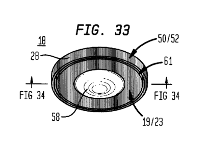
Figure 33 is a back perspective view of a particular embodiment of the
inventive
intraocular implant a flexible membrane joined about the circumference of an
optical lens and
7
CA 02959805 2017-03-01
WO 2016/022995
PCT/US2015/044357
extending radially outwardly to terminate in an annular member and having an
annular channel
disposed in the back surface of the intraocular implant.
Figure 34 is a cross section 34-34 of the particular embodiment of the
inventive
intraocular implant shown in Figure 33.
Figure 35 is a plan view of an embodiment of the inventive intraocular implant
having
haptics coupled to an optical lens having patterned surface elements disposed
on the haptics and
about the circumference of the optical lens.
Figure 36 is a cross-section 34-34 of the embodiment of the inventive
intraocular implant
shown in Figure 33.
Figure 37 is a side view of the embodiment of the inventive intraocular
implant shown in
Figure 33.
Figure 38 is an enlarged view of a circumferential portion of the inventive
intraocular
implant shown in Figure 33.
Figure 39 is an enlarged view of a particular embodiment of a plurality
patterned surface
elements in the form of a plurality of raised surface elements coupled to the
external surface of
embodiments of the inventive intraocular implant.
Figure 40 is a cross section 40-40 of the plurality of patterned surface
elements shown in
Figure 39.
Figure 41 is an enlarged view of a particular embodiment of a plurality of
patterned
surface elements in the Rhiii of a plurality of recessed elements which can be
coupled to the
external surface of embodiments of the inventive intraocular implant.
Figure 42 is a cross section 42-42 of the plurality of patterned surface
elements shown in
Figure 41.
Figure 43 is an enlarged view of a particular embodiment of a plurality of
patterned
surface elements in the form of a plurality of raised surface elements on the
back surface and a
plurality of recessed elements on the front surface which can be coupled to
the external surface
of embodiments of the inventive intraocular implant.
Figure 44 is a cross section 44-44 of the plurality of patterned surface
elements shown in
Figure 43.
Figure 45 is an enlarged view of a particular embodiment of a plurality
patterned surface
elements in the form of a plurality of raised surface elements coupled to the
external surface of
embodiments of the inventive intraocular implant.
Figure 46 is a cross section 46-46 of the plurality of patterned surface
elements shown in
Figure 45.
8
CA 02959805 2017-03-01
WO 2016/022995
PCT/US2015/044357
Figure 47 is an enlarged view of a particular embodiment of a plurality of
patterned
surface elements in the form of a plurality of raised surface elements coupled
to the external
surface of embodiments of the inventive intraocular implant.
Figure 48 is a cross section 48-48 of the plurality of patterned surface
elements shown in
Figure 47.
Figure 49 is an enlarged view of a particular embodiment of a plurality of
patterned
surface elements in the form of a plurality of raised surface elements coupled
to the external
surface of embodiments of the inventive intraocular implant.
Figure 50 is a cross section 50-50 of the plurality of patterned surface
elements shown in
Figure 49.
Figure 51 is an enlarged view of a particular embodiment of a plurality of
patterned
surface elements in the form of a plurality of raised surface elements coupled
to the external
surface of embodiments of the inventive intraocular implant.
Figure 52 is a cross section 52-52 of the plurality of patterned surface
elements shown in
Figure 51.
Figure 53 is a cross section view 53-53 shown in Figure 39 of a group of
surface
elements having a pattern in which the height of the plurality of surface
elements increases
approaching the middle of the pattern.
Figure 54 is a cross section view 54-54 shown in Figure 39 of a group of
surface
elements having a pattern in which the height of the plurality of surface
elements decreases
approaching the ends of the pattern.
Figure 55 is an enlarged plan view of a plurality of groups of surface
elements with each
group of surface elements having a pattern and each group of surface elements
having an angle
of rotation on the external surface of an intraocular implant different from
the angle of rotation
of adjacent groups of surface elements on the intraocular implant.
Figure 56 is an enlarged plan view of a plurality of sections bounded by an
interconnected periphery defining a plurality of polygons which bounds a
plurality of patterned
surface areas each including a plurality of groups of surface elements having
a pattern where the
patterns in adjacent sections have different angles of rotation with respect
to each other.
Figure 57 is an illustration of a plurality of patterned surface elements
having regular
geometries with adjacent sections of patterned surface elements having
different angles of
rotation.
Figure 58 is an illustration of a plurality of patterned surface elements
having irregular
geometries with adjacent sections of patterned surface elements having
different angles of
rotation.
9
CA 02959805 2017-03-01
WO 2016/022995
PCT/US2015/044357
Figure 59 is an illustration of a plurality of patterned surface elements
having a
combination of regular and irregular geometries with adjacent sections of
patterned surface
elements having different angles of rotation.
Figure 60 is an enlarged plan view of a plurality of sections bounded by an
interconnected periphery which depicts how the axis of a plurality of sections
can be rotated
with respect to the fluid flow, the flow of a suspension of particles in a
fluid flow, or the
adhesion, growth or migration of cells.
Figure 61 is an enlarged plan view of a plurality of patterned surface
elements having
adjacent patterned surface elements conjoined.
Figure 62 is an enlarged view of an embodiment of an inventive intraocular
implant
including a plurality of patterned surface elements in the form of a plurality
of raised concentric
bands of increasing diameter radially spaced apart about a central point and
periodically
interrupted circumferentially by a plurality of gaps defining a tortuous
pathway on said
intraocular implant which traverses the plurality of patterned surface
elements.
Figure 63 is a cross-section view 63-63 of the embodiment of the inventive
intraocular
implant shown in Figure 62.
Figure 64 is an enlarged view of an embodiment of an inventive intraocular
implant
including a plurality of patterned surface elements in the form of a plurality
of raised concentric
bands of increasing diameter radially spaced apart about a central point and
periodically
interrupted circumferentially by a plurality of gaps defining a tortuous
pathway on the
intraocular implant which traverses the plurality of patterned surface
elements which increase in
density approaching the central point.
Figure 65 is a cross-section view 65-65 of the embodiment of the inventive
intraocular
implant shown in Figure 64.
Figure 66 is an enlarged view of an embodiment of an inventive intraocular
implant
having a plurality of groups of patterned surface elements repeated over the
external surface of
the intraocular implant with increasing density approaching a central point.
Figure 67 is a cross-section view 67-67 of the embodiment of the inventive
intraocular
implant shown in Figure 66.
Figure 68 is an illustration of embodiments of the inventive intraocular
implant having a
plurality of sectors bound by an interconnected periphery defining a plurality
of patterned
surface areas each including a plurality of groups of surface elements having
a pattern where the
pattern in adjacent sections has different angles of rotation with respect to
each other.
CA 02959805 2017-03-01
WO 2016/022995
PCT/US2015/044357
Figure 69 is an illustration of a particular embodiment of the inventive
intraocular
implant having a plurality of groups of patterned surface elements having an
angle of rotation in
each of a plurality of sectors to direct fluid flow radially from a central
point.
Figure 70 is an illustration of a particular embodiment of the inventive
intraocular
implant having a plurality of groups of patterned surface elements having an
angle of rotation in
each of a plurality of sectors to direct fluid flow circumferentially from a
central point.
Figure 71 is an illustration of a particular embodiment of the inventive
intraocular
implant having a plurality of groups of patterned surface elements having an
angle of rotation in
each of a plurality of sectors to direct fluid flow in particular sectors
radially from a central point
and to direct fluid flow in particular sectors circumferentially from a
central point.
Figure 72 shows an embodiment of the intraocular implant held by forceps for
implantation into an eye having the natural lens removed.
Figure 73 is top view of the pseudophakic eye having the natural lens removed
allowing
an embodiment of the intraocular implant to be positioned on the surface the
posterior capsule
through an opening made in the anterior capsule.
Figure 74 is a cross section view of the pseudophakic eye having the natural
lens
removed allowing an embodiment of the intraocular implant to be positioned on
the surface the
posterior capsule through an incision made in the anterior capsule.
Figure 75 is a cross section view of the pseudophakic eye having the
intraocular implant
of Figures 5 through 8 positioned between the surface the posterior capsule
and the implanted
IOL.
Figure 76 is a cross section view of the pseudophakic eye having the
intraocular implant
of Figures 22 through 25 or Figures 26 through 30 positioned between the
surface of the
posterior capsule and the implanted IOL.
Figure 77 is a cross section view of the pseudophakic eye having the
intraocular implant
of Figures 31 and 32 or 33 and 34 positioned on the surface of the posterior
capsule.
Figure 78 is a cross section view of the phakic eye having the intraocular
implant of
Figure 5 through 8 positioned between the iris and the natural crystalline
lens of the eye.
Figure 79A is a representative image of a cell migration assay showing
migration of cells
on a smooth surface ("SM") of a flexible membrane as shown in the examples of
Figures 5
through 8.
Figure 79B is a representative image of a cell migration assay showing
migration of cells
on a flexible membrane having patterned surface elements (+1SK10x5) as shown
in the example
of Figure 39.
11
CA 02959805 2017-03-01
WO 2016/022995
PCT/US2015/044357
Figure 79C is a representative image of a cell migration assay showing
migration of cells
on a flexible membrane having patterned surface elements (+10SK50x50) as shown
in the
example of Figure 39.
Figure 80 is a bar graph which compares cell migration on a flexible membrane
having
a smooth surface ("SM") to cell migration on flexible membrane having
patterned surface
elements ("+1SK10x5") or ("10SK50x50) as shown in the example of Figure 39.
Figure 81A is a representative image of scratch wound assay showing migration
of cells
on a smooth surface ("SM") of a flexible membrane as shown in the examples of
Figures 5
through 8.
Figure 81B is a representative image of scratch wound assay showing migration
of cells
on a flexible membrane having patterned surface elements ("-3SK2x2") as shown
in the
examples of Figures 41 and 42.
Figure 82C is a representative image of scratch wound assay showing migration
of cells
on a flexible membrane having patterned surface elements ("+3SK2x2") as shown
in the
examples of Figures 39 and 40.
Figure 81D is a representative image of scratch wound assay showing migration
of cells
on a flexible membrane having patterned surface elements ("+7SK10x5") as shown
in the
examples of Figures 39 and 40.
Figure 82 is a bar graph which compares cell migration on a flexible membrane
having
a smooth surface ("SM") to cell migration on flexible membrane having
patterned surface
elements ("-3SK2x2"), ("+3SK2x2") or ("+7SK10x5") as shown in the examples of
Figures 39
and 40 or Figures 41 and 42.
Figure 83A is a representative image of a cell migration assay showing
migration of
LECs between a collagen membrane and an IOL as shown in the illustrative
example of Figures
3 and 4.
Figure 83B is a representative image of a cell migration assay showing
migration of
LECs between a collagen membrane and a smooth backside of the inventive
intraocular implant
of Figures 22 through 25 having an IOL engaged with the front side as
illustrated in the example
of Figures 26 and 27.
Figure 83C is a representative image of a cell migration assay showing
migration of
LECs between a collagen membrane and the backside of inventive intraocular
implant having
patterned surface elements and an IOL engaged with the front side as
illustrated in the example
of Figures 26 through 30.
12
CA 02959805 2017-03-01
WO 2016/022995
PCT/US2015/044357
Figure 84 is a bar graph comparing each of the IOL, the inventive intraocular
implant
having a smooth surface, and the inventive intraocular implant having
patterned surface
elements to normalized area covered by LEC migration.
Figure 85A is an image of the eye of a rabbit having an IOL inserted in the
capsular bag
with the eye stained to show migration of LECs.
Figure 85B is an image of the eye of rabbit having the inventive intraocular
implant of
Figures 23 through 25 with the IOL engaged with the front surface of the
intraocular implant
inserted in the capsular bag to engage the back surface of the intraocular
implant without
patterned surface elements with the posterior capsule of the eye with the eye
stained to show
migration of LECs.
Figure 85C is an image of the eye of rabbit having the inventive intraocular
implant of
Figures 26 through 30 with the IOL engaged with the front surface of the
intraocular implant
inserted in the capsular bag to engage the back surface of the intraocular
implant having
patterned surface elements with the posterior capsule of the eye with the eye
stained to show
migration of LECs.
Figure 86 is a bar graph comparing LEC migration of each of the IOL only and
the
inventive intraocular implant with or without patterned surface elements to a
PCO score.
Figure 87 is a bar graph comparing LEC migration for each of the IOL only and
the
inventive intraocular implant of Figures 26 through 30 with the IOL engaged
with the front
surface of the intraocular implant inserted in the capsular bag to either
engage the back surface
of the intraocular implant with patterned surface elements with the posterior
capsule of the eye
and the annular member engaged to the anterior capsule of the eye ("IOL with
annular member
up-) or engage the back surface of the intraocular implant having patterned
surface elements
with the anterior capsule of the eye and the annular member engaged to the
posterior capsule
"IOL with annular member down").
V. MODE(S) FOR CARRYING OUT THE INVENTION
Generally, an intraocular implant having on the external surface a plurality
of pattern
surface elements disposed in spaced apart relation defining a tortuous pathway
which controls a
flow of fluid, a flow of particles suspended in a flow of fluid, or inhibits
the growth or migration
of cells. In particular, an intraocular implant which implanted between an IOL
and the surface of
the posterior capsule of the eye inhibits growth or migration of residual lens
epithelial cells after
cataract surgery by providing structural barriers to reduce PCO of the eye. In
particular, an
intraocular implant which implanted between an IOL and the surface of the
posterior capsule of
13
CA 02959805 2017-03-01
WO 2016/022995
PCT/US2015/044357
the eye inhibits migration of residual lens epithelial cells after cataract
surgery by providing
structural barriers to reduce PCO of the eye.
DEFINITIONS
"A" or "an" entity refers to one or more of that entity; for example, "a
polymer" refers to
one or more of those compositions or at least one composition. As such, the
terms "a" or "an",
"one or more- and "at least one" can be used interchangeably herein.
Furthermore, the language
"selected from the group consisting of' refers to one or more of the elements
in the list that
follows, including combinations of two or more of the elements.
-About" for the purposes of the present invention means that values or ranges
of values
may be expressed as from "about" one particular value to "about" another
particular value. In the
context of such a value or range of values -about- means plus or minus 10% of
the value or
range of values recited or claimed. When such a range of values is expressed,
an embodiment
includes from about one particular value to about the other particular value.
Also, when such a
range of values is expressed, another embodiment includes from one particular
value to the other
particular value and it will be understood that each particular value forms
another embodiment.
"Active agent" for the purposes of this invention means any substance used to
treat an
ocular condition.
"Biocompatible" for the purposes of this invention means the ability of any
material to
perform the intended function of an embodiment of the invention without
eliciting any
undesirable local or systemic effects on the recipient and can include non-
biodegradable
materials such as: polyurethanes, polyisobutylene, polydimethylsiloxane
elastomer, ethylene-
alpha-olefin copolymers, acrylic polymers and copolymers, vinyl halide
polymers and
copolymers, polyvinyl esters, polyvinylidene chloride, polyacrylonitrile,
polyvinyl ketones,
polyvinyl aromatics such as polystyrene, copolymers of vinyl monomers and
olefins such as
ethylene-methyl methacrylate copolymers, acrylonitrile-styrene copolymers,
acrylonitrile
butadiene styrene resins, ethylene-vinyl acetate copolymers, polyamides such
as Nylon 66 and
polycaprolactone, alkyd resins, polycarbonates, polyoxyethylenes, polyimides,
polyesters, epoxy
resins, rayon-triacetate. cellophane, silicon rubber, silicon hydrogel, or
biodegradable materials,
as defined herein or combinations thereof.
"Biodegradable" for the purposes of this invention means the ability of any
biocompatible material to breakdown within the physiological environment of
the eye by one or
more physical, chemical, or cellular processes at a rate consistent with
providing structural or
phatmaceutical barriers (or both) at a therapeutic level controllable by
selection of a polymer or
mixture of polymers (also referred to as polymeric materials), including, but
not limited to:
14
CA 02959805 2017-03-01
WO 2016/022995
PCT/US2015/044357
polylactide polymers (PLA), copolymers of lactic and glycolic acids (PLGA),
polylactic acid-
polyethylene oxide copolymers, poly(g-caprolactone-co-L-lactic acid (PCL-LA),
glycine/PLA
copolymers, PLA copolymers involving polyethylene oxides (PEO), acetylated
polyvinyl
alcohol (PVA)/polycaprolactone copolymers, hydroxybutyrate-hydroxyvalerate
copolymers,
polyesters such as, but not limited to, aspartic acid and different aliphatic
diols, poly(alkylene
tartrates) and their copolymers with polyurethanes, polyglutamates with
various ester contents
and with chemically or enzymatically degradable bonds, other biodegradable
nonpeptidic
polyamides, amino acid polymers, polyanhydride drug carriers such as, but not
limited to,
poly(sebacic acid) (PSA), aliphatic-aromatic homopolymers, and poly(anhydride-
co-imides),
poly(phosphoesters) by matrix or pendant delivery systems, poly(phosphazenes),
poly(iminocarbonate), crosslinked poly(ortho ester), hydroxylated polyester-
urethanes, or the
like. Hydrogels such as methylcellulose which act to release drug through
polymer swelling are
specifically excluded from the term.
Intraocular" for the purposes of this invention means inside the eyeball (also
referred to
as an "eye-) and without limitation to the forgoing the anterior chamber, the
ciliary sulcus, and
posterior capsule of the eye; however, specifically excluding the external
surface of the eye or
intracorneal or intrasclera regions of the eye.
"Localized Region" for the purposes of this invention means substantially
within a
localized tissue region of the eye therapeutically affected (whether
structurally or
pharmaceutically) by implantation of embodiments of an intraocular implant.
"Ocular condition" for the purposes of this invention means a disease, ailment
or
condition which affects or involves the eye or any one of the parts or regions
of the eye, such as
PCO. The eye includes the eyeball and the tissues and fluids which constitute
the eyeball, the
periocular muscles (such as the oblique and rectus muscles) and the portion of
the optic nerve
which is within or adjacent to the eyeball.
"Posterior ocular condition" for the purposes of this invention means a
disease, ailment
or condition which affects or involves a posterior ocular region or site such
as the choroid or
sclera (in a position posterior to a plane through the posterior wall of the
lens capsule), vitreous,
vitreous chamber, retina, optic nerve (i.e. the optic disc), and blood vessels
and nerve which
vascularize or innervate a posterior ocular region or site.
"Substantially" for the purposes of this invention means largely, but not
wholly, the
same form, manner or degree and the particular element will have a range of
configurations as a
person of ordinary skill in the art would consider as having the same function
or result. When a
particular element is expressed as an approximation by use of the antecedent
"substantially," it
will be understood that the particular element forms another embodiment.
CA 02959805 2017-03-01
WO 2016/022995
PCT/US2015/044357
-Suitable for implantation" for the purposes of this invention means with
regard to
embodiments of the intraocular implant dimensions which allow insertion or
implantation
without causing excessive tissue damage.
"Therapeutic level" for the purposes of this invention means an amount or a
concentration of an active agent that has been locally delivered to an ocular
region that is
appropriate to reduce, inhibit, or prevent a symptom of an ocular condition.
Now generally referring to Figures 5-72, particular embodiments of the
inventive
intraocular implant (18) can provide a biocompatible flexible membrane or a
biocompatible
biodegradable flexible membrane (also generally referred to as a "flexible
membrane" (19))
having an outer boundary (13) configured to allow the intraocular implant (18)
to locate in the
concavity of the posterior capsule (13) of the pseudophakic eye (4), or other
localized region
inside the eye (1)(8) such as the ciliary sulcus (139) or anterior chamber (5)
depending on the
application. As an illustrative example, the intraocular implant (18) can be
located in the
posterior capsule (13) for the purpose of isolating the surface of the
posterior capsule (13) from
migration of residual LECs after cataract surgery, or reducing or preventing
the migration of
residual LECs between the surface of an IOL (11) implanted in the lens capsule
(7) and the
surface of the posterior capsule (13).
Now referring generally to Figures 5 through 34, embodiments of the inventive
intraocular implant (18) can provide a flexible membrane (19) having an outer
boundary (20)
which can define a substantially circular, ovoid, or other outer boundary
configuration suitable
for implantation into the concavity of the posterior capsule (13) of the
pseudophakic eye (8), or
other localized region inside the eye (1)(8). As to particular embodiments,
the outer boundary
(20) of the flexible membrane (19) can define a circular area (21) having a
diameter in a range
of about 8 millimeters ("mm") to about 15 mm, depending on the recipient and
the application.
The diameter of the flexible membrane (19) can be selected from the group
including or
consisting of: about 8.0 mm to about 9.0 mm, about 8.5 mm to about 9.5 mm,
about 9.0 mm to
about 10.0 mm, about 9.5 mm to about 10.5 mm, about 10.0 mm to about 11.0 mm,
about 10.5
mm to about 11.5 mm, about 11.0 mm to about 12.0 mm, about 11.5 mm to about
12.5 mm.
about 12.0 mm to about 13.0 mm, about 12.5 mm to about 13.5 mm, about 13.0 mm
to about
14.0 mm, about 13.5 mm to about 14.5 mm, about 14 mm to about 15.0 mm. As to
particular
embodiments, the diameter of the intraocular implant (18) can be pre-selected
to allow the outer
boundary (20) to engage the outer circumference (12) of the localized region
of the eye (1)(8) to
fix the position of the intraocular implant (18) in the localized region of
the eye (1)(8) excluding
any other attachment elements on or in the circular area (21) of the flexible
membrane (19) of
the intraocular implant (18).
16
CA 02959805 2017-03-01
WO 2016/022995
PCT/US2015/044357
The flexible membrane (19) can, but need not necessarily, be a thin pliable
sheet of
biocompatible or biodegradable material solid or continuous between a front
surface (22) and a
back surface (23)(also referred to as "a first side" and "a second side"
respectively or "opposed
sides"). As to particular embodiments of the intraocular implant (18), the
front surface (22) and
the back surface (23) can, need not necessarily, be disposed in substantially
parallel opposed
relation having a thickness (24) therebetween in a range of about 5 microns
("gm") to about 400
gm. As to particular embodiments, the thickness can be selected from the group
including or
consisting of: about 5 gm to about 100 gm, about 50 gm to about 150 gm, about
100 gm to
about 200 i.tm, about 150 gm to about 250 gm, about 200 gm to about 300 gm,
about 250 Irm to
about 300 gm, about 300 gm to about 400 gm, and about 350 gm to about 400 mn.
Depending
upon the thickness (24) of the intraocular implant (18), the optical power of
the JUL (11) can be
adjusted, if necessary.
As to particular embodiments, the flexible membrane (19) can, but need not
necessarily,
have a uniform thickness (24) disposed between substantially flat or flat
front and back surfaces
(22)(23)(as shown in the examples of Figures 5 through 8). However,
embodiments of the
intraocular implant (18) can provide a flexible membrane (19) thinner
proximate the center and
thicker proximate the outer boundary (20) or can provide a flexible membrane
(19) thicker
proximate the center and thinner proximate the outer boundary (20), depending
upon the
application. As to other embodiments, the thickness (24) of the flexible
membrane (19) can be
thinner in the center of the circular area (21) to align with the visual axis
(15) of the eye (1)(8) to
increase visual acuity or promote directional biodegradation of the
intraocular implant (18) from
the center toward the outer boundary (20).
As to particular embodiments, the outer boundary (20) of the flexible membrane
(19) can
have an edge (25) which intersects the front surface (22) or the back surface
(23) at substantially
a right angle (as shown in the examples of Figures 5-9).
Again referring generally to Figures 5 through 34, particular embodiments of
the
inventive intraocular implant (18) can, but need not necessarily, include an
aperture element
(26) defining a passage opening (27) sufficiently large to align with the
visual axis (15) of the
eye (1)(8) to provide a line of sight which passes through the intraocular
implant (18).
Embodiments of the inventive intraocular implant (18) can, but need not
necessarily, include an
aperture element (26) having a configuration selected from the group including
or consisting of:
a circle, an oval, a square, a triangle, or other configuration of passage
opening (27) alignable
with the visual axis (15) and having a passage opening (27) sufficient to
provide a line of sight
which passes through the intraocular implant (18). As to those embodiments of
the intraocular
implant (18) utilized in combination with an IOL (11), the passage opening
(27) can be
17
CA 02959805 2017-03-01
WO 2016/022995
PCT/US2015/044357
dimensioned in relation to the IOL (11) to avoid reduction in the field of
vision provided by the
IOL (11) or a reduction in clarity of vision within the visual field of the
IOL (11). As to those
embodiments of the intraocular implant (18) in which the passage opening (27)
has insufficient
dimension to avoid overlaying all or part of the visual field afforded by the
IOL (11), the
intraocular implant (18) can be configured to provide sufficient clarity of
vision within the
visual field afforded by the IOL (11).
As to particular embodiments of the intraocular implant (18) having an
aperture element
(26) of substantially circular configuration, the aperture element (26) can
have a diameter in the
range of about 1.5 mm and about 9.0 mm, depending upon the application and the
recipient. As
to particular embodiments, the aperture element (26) can have diameter
selected from the group
including or consisting of: about 1.5 mm to about 2.5 mm, about 2.0 mm to
about 3.0, about 2.5
mm to about 3.5 mm. about 3.0 mm to about 4.0 mm, about 3.5 mm to about 4.5
mm, about 4.0
mm to about 5.0 mm, about 4.5 mm to about 5.5 mm, about 5.0 mm to about 6.0
mm, about 5.5
mm to about 6.5 mm, about 6.0 mm to about 7.0 mm, about 6.5 mm to about 7.5
mm, about 7.0
mm to about 8.0 mm, about 7.5 mm to about 8.5 mm, and about 8.0 mm to about
9.0 mm.
Now referring primarily to Figures 9 and 10, particular embodiments can, but
need not
necessarily, have a plurality of patterned surface elements (28) coupled to
the external surface
(67) of the intraocular implant (18), such as the front surface (22), whether
in whole or in part,
or the back surface (23) of the intraocular implant (18). As to particular
embodiments, the
patterned surface elements (28) can be adapted to engage the surface of the
posterior capsule
(13) to reduce travel of the intraocular implant (18) or maintain the
alignment of the center of
the intraocular implant (18) with the visual axis (15) of the eye (1)(8). The
plurality of patterned
surface elements (28) can, but need not necessarily, provide an irregular or
uniform pattern,
texture. or roughness sufficient to fix or reduce travel of the intraocular
implant (18) in the
posterior capsule (13). As to certain embodiments of the intraocular implant
(18) the plurality of
patterned surface elements (28) can, but need not necessarily, provide pockets
(29) which
function to provide a localized space to deliver or sequester an amount of an
active agent (30).
The intraocular implant (18) and the plurality of pattern surface elements
(28) can be one piece
or the plurality of patterned surface elements (28) can be applied to the
intraocular implant (18)
as a patterned surface element layer (31).
Now referring primarily to Figures 11 through 12, and 16 through 18,
particular
embodiments of the flexible membrane (19) can, but need not necessarily
include, one or more
radial slit elements (32) cut through the thickness (24) of the flexible
membrane (19). As to
particular embodiments, the radial slit elements (32) originate at the outer
boundary (20) cut a
distance radially toward the center of the flexible membrane (19) (as shown in
the examples of
18
CA 02959805 2017-03-01
WO 2016/022995
PCT/US2015/044357
Figure 11). The one or more radial slit elements (32) can have sufficient slit
length (34) and slit
width (33) to allow the flexible membrane (19) to confolin to a greater extent
with the localize
region of the eye or the concavity of the posterior capsule (13) of the eye
(1)(8) or other
localized region inside the eye (1)(8). The radial slit elements (14) can have
a greater slit width
(33) at the outer boundary (20) of the flexible membrane (19) than proximate
the center of the
flexible membrane (19). The flexible membrane (19) when received by the
concavity of the
posterior capsule (13) can defoini to reduce the slit width (33) at the outer
boundary (20) of the
flexible membrane (19). In addition, the radial slit elements (32) can provide
one or more
interruptions in the outer boundary (20) which can be of lesser or greater
slit width (33) or slit
length (34) to control the rate at which the flexible membrane (19)
biodegrades within a
localized region of the eye (1)(8) such as the posterior capsule (13) of the
eye (1)(8).
Now referring specifically to Figures 12 and 16 through 18, the aperture
element (26)
can, but need not necessarily, include one or more radial slit elements (32)
each originating at
the aperture element (26) and terminating at a distance from the outer
boundary (20) of the
flexible membrane (19). The one or more radial slit elements (32) can have
sufficient slit length
(34) and slit width (33) to allow the flexible membrane (19) to confoun to a
greater extent to the
localized region of the eye (1)(8) such as the concavity of the posterior
capsule (13) and with
respect to embodiments of the intraocular implant (18) which are biodegradable
can function to
promote directional biodegradation of the intraocular implant (18) proximate
the aperture
element (26) toward the outer boundary (20). Again, the radial slit elements
(32) can provide
one or more interruptions in the aperture element (26) which can be of lesser
or greater slit
width (33) or slit length (34) to control the rate at which the flexible
membrane (19) biodegrades
within the localized region of the eye (1)(8) such as the posterior capsule
(13) of the eye (1)(8).
Now referring primarily to Figure 13, embodiments of the flexible membrane
(19) can,
but need not necessarily, include one or more perforation elements (35) which
provide a
corresponding one or more perforation openings (36) which communicate between
the front
surface (22) and the back surface (23) of the flexible membrane (19) for the
purpose of
increasing rate of biodegradation of the flexible membrane (19) or control
release rate of an
active agent (30). The active agent (30) shown in the example of Figures 12,
13 and 16 as a
stipple is not intended to be limited to these particular embodiments of the
intraocular implant
(18) or limit the active agent (30) to any particular composition, particle
size, or amount.
Now referring primarily to Figure 14, embodiments, can but need not
necessarily,
include two or more flexible membrane zones (37). As to certain embodiments,
the two or more
flexible membrane zones (37) can be established as a first annular zone (38)
surrounded by a
second annular zone (39). The first annular zone (38) can be of a different
biocompatible or
19
CA 02959805 2017-03-01
WO 2016/022995
PCT/US2015/044357
biocompatible biodegradable material then the second annular zone (39). For
example, the first
annular zone (38) can provide a biocompatible biodegradable material selected
for a greater rate
of biodegradation or release of active agent (30) relative to the second
annular zone (39) which
can provide a biocompatible biodegradable material selected for a lesser rate
of biodegradation
or release of active agent (30) release. As to these embodiments, the
prominent function of the
first annular zone (38) can be to provide a pharmaceutical barrier or
treatment of an ocular
disorder, while the prominent function of the second annular zone (39) can be
to provide a
mechanical barrier or treatment of an ocular disorder. In particular
embodiments of the inventive
intraocular implant (18) for the inhibition of PCO, the first annular zone
(38) can be made of the
biocompatible biodegradable material poly(lactide-co-glycolide) having an
active agent (30),
such as an alkylphosphocholine, dispersed substantially uniformly throughout
which can
provide a pharmaceutical barrier to the proliferation of LECs (16) on the
surface of the posterior
capsule (13) to inhibit or prevent PCO by release of a therapeutic level of
alkylphosphocholine
of about 1.0 millimolar ("mM") for a period of about five days. The first
annular zone (38) can
substantially biodegrade in the entirety in a period of about five days to
about ten days. The
second annular zone (39) can be made of the same or different biocompatible
biodegradable
material having the same or a different active agent (30) dispersed
substantially uniformly
throughout to provide both a mechanical barrier to inhibit migration of LECs
(16) toward to the
surface of the posterior capsule (13) and can provide a pharmaceutical barrier
by release of the
same or different active agent (30), such as alkylphosphocholine, at a
therapeutic level or
provide a localized concentration of about 1.0 mM for a period of at least
twenty days to inhibit
or prevent PCO.
Now referring primarily to Figures 15 through 18, particular embodiments of
the flexible
membrane (19) can, but need not necessarily, include one or more boundary
recess elements
(40) located along the outer boundary (20). The outer boundary (20) of the
flexible membrane
(19) can be interrupted once or periodically to provide one or more boundary
recess elements
(40) which can be configured, for example, as semicircular extensions (as
shown in the example
of Figure 15) or semicircular notches (as shown in the example of Figure 16),
triangular notches,
indents, or the like which can function to allow added flexure or to more
readily locate the
flexible membrane (19) in a localized region of the eye (1)(8) such as the
posterior capsule (13)
of the eye (1)(8), as above described, or can function to reduce sequestration
of fluids within eye
(1)(8) or reduce sequestration of peripheral cortical material during the
final irrigation and
aspiration steps in cataract surgery.
Now referring primarily to Figure 17, certain embodiments of the flexible
membrane
(19) can, but need not necessarily, include two or more flexible membrane
layers (41). The two
CA 02959805 2017-03-01
WO 2016/022995
PCT/US2015/044357
or more membrane layers (41) can take the form of a first flexible membrane
layer (42) and a
second flexible membrane layer (43) or additional flexible membrane layers
(44) extruded as a
single piece, coupled together as one unit, or stacked front to back (whether
single piece,
coupled or stacked the term "coupled" may be used to refer to the association
of a plurality of
flexible membrane layers (41)). Each of the first flexible membrane layer (42)
and the second
flexible membrane layer (43) or additional flexible layers (44) can be
generated from the same
or different biocompatible or biocompatible biodegradable materials. As a non-
limiting
example, in embodiments of the invention for the treatment of PCO, the first
flexible membrane
layer (42) can be made of a biocompatible or biocompatible biodegradable
material which can
have the back surface (23) disposed facing the surface of the posterior
capsule (13) to provide
both a mechanical barrier to the migration of LECs (16) over the surface of
the posterior capsule
(13) but to further function as a pharmaceutical barrier which inhibits
proliferation or kills LECs
(16) by the substantially continuous release of an active agent (30), such as
alkylphosphocholine, at a rate which provides a therapeutic level of active
agent (30), such as a
localized concentration of about 1.0 mM for a period of at about five days to
inhibit or prevent
PCO. The front surface (22) of the first flexible membrane layer (42) can be
coupled adjacent
the back surface (23) of the second flexible membrane layer (43) (for example
by melt co-
extrusion) produced from the same or different biocompatible biodegradable
material and the
front surface (22) of the second flexible membrane layer (43) can be disposed
facing an IOL
(11) implanted into the lens capsule (7) to provide a mechanical barrier to
migration of LECs
(16) toward or over the surface of the posterior capsule (13) and can further
function as a
pharmaceutical barrier which inhibits proliferation or kills LECs (16) by the
substantially
continuous release of the same active agent (30) (such as an
alkylphosphocholine) or a different
active agent (30) (such as mitomycin-C) at a therapeutic level, such as a
localized concentration
of about 0.04 milligrams per milliliter ("mg/mL"), for a period of about five
days to inhibit or
prevent PCO. Thus, by configuring the layers in different combinations the
rate of release of
various active agents (30) can be adjusted depending on the application.
Now referring primarily to Figure 18, particular embodiments of the
intraocular implant
(18) can, but need not necessarily, include radial capillaries (45) which
communicate between
the outer boundary (20) and the aperture element (26) of the flexible membrane
(19) configured
to allow or facilitate circulation of fluid within a localized region of the
eye (1)(8), for example,
between the flexible membrane (19) and the posterior capsule (13) of the eye
(1)(8).
Now referring primarily to Figure 19, particular embodiments of the
intraocular implant
(18) can further provide one or more corrugate elements (46) which can, but
need not
necessarily, be disposed in substantially linear parallel relation to generate
undulations in the
21
CA 02959805 2017-03-01
WO 2016/022995
PCT/US2015/044357
flexible membrane (19) sufficient when the flexible membrane (19) locates
against a surface of
a localized region of the eye (1)(8), such as the posterior capsule (13), to
provide corrugate
channels (47) in which fluids of the eye (1)(8) can circulate.
Again referring in general to Figures 5-39, as to those embodiments of the
intraocular
implant (18) which include an active agent (30), the active agent (30) can,
but need not
necessarily, be mixed with or dispersed in the biodegradable polymer of the
flexible membrane
(19). The composition of the biodegradable polymers of the flexible membrane
(19) of the
intraocular implant (18) can be varied to provide a continuous or
substantially continuous
release of a therapeutic level of a particular active agent (30) or a
particular mixture of active
agents (30) effective to treat or alleviate symptoms of an ocular condition.
One or more active
agents (30) can be selected from the group including or consisting of: ace-
inhibitors,
endogenous cytokines, agents that influence the basement membrane, agents that
influence the
growth of endothelial or epithelial cells, adrenergic agonists or blockers,
cholinergic agonists or
blockers, aldose reductase inhibitors, analgesics, anesthetics, antiallergics,
anti-inflammatory
agents, antihypertensives, pressors, antibacterials, antivirals, antifungals,
antiprotozoals, anti-
infectives, antitumor agents, antimetabolites such as daunomycin,
antiangiogenic agents,
tyrosine kinase inhibitors, antibiotics such as aminoglycosides such as
gentamicin, kanamycin,
neomycin, and vancomycin; amphenicols such as chloramphenicol; cephalosporins,
such as
cefazolin HC1; penicillins such as ampicillin, penicillin, carbenicillin,
oxycillin, methicillin;
lincosamides such as lincomycin; polypeptide antibiotics such as polymixin and
bacitracin;
tetracyclines such as tetracycline, minocycline, and doxycycline; quinolones
such as
ciprofloxacin, moxifloxacin, gatifloxacin, and levofloxacin; sulfonamides such
as chloramine T:
sulfones such as sulfanilic acid; anti-viral drugs such as acyclovir,
gancyclovir, vidarabine,
azidothymidine, dideoxyinosine, dideoxycytosine; epinephrine; isoflurphate;
adriamycin;
bleomycin; mitomycin; ara-C; actinomycin D; scopolamine; and the like,
analgesics, such as
codeine, morphine, ketorolac, naproxen, an anesthetic, lidocaine; beta.-
adrenergic blocker or
beta.-adrenergic agonist such as ephedrine, and epinephrine; aldose reductase
inhibitor such as
epalrestat, ponalrestat, sorbinil, tolrestat; antiallergic such as cromolyn,
beclomethasone,
dexamethasone, and flunisolide; colchicine, anihelminthic agents such as
ivermectin and
suramin sodium; antiamebic agents such as chloroquine and chlortetracycline;
and antifungal
agents such as amphotericin; anti-angiogenesis compounds such as anecortave
acetate; retinoids
such as Tazarotene, anti-glaucoma agents such as brimonidine (Alphagan and
Alphagan P),
acetozolamide, bimatoprost (Lumigan), timolol, mebefunolol; memantine; alpha-2
adrenergic
receptor agonists; 2-methoxyestradiol; anti-neoplastics such as vinblastine,
vincristine,
interferons; alpha, beta and gamma., antimetabolites such as folic acid
analogs, purine analogs,
22
CA 02959805 2017-03-01
WO 2016/022995
PCT/US2015/044357
and pyrimidine analogs; immunosuppressants such as azathyprine, cyclosporine
and mizoribine;
miotic agents, such as carbachol, mydriatic agents such as atropine, etc.,
protease inhibitors such
as aprotinin, camostat, gabexate, vasodilators such as bradykinin, epidermal
growth factor, basic
fibroblast growth factor, nerve growth factors, steroidal anti-inflammatory
agents such as 21-
acetoxypregnenolone, alclometasone, algestone, amcinonide, beclomethasone,
betamethasone,
budesonide, chloroprednisone, clobetasol, clobetasone, clocortolone,
cloprednol, corticosterone,
cortisone, cortivazol, deflazacort, desonide, desoximetasone, dexamethasone,
diflorasone,
diflucortolone, difluprednate, enoxolone, fluazacort, flucloronide,
flumethasone, flunisolide,
fluocinolone acetonide, fluocinonide, fluocortin butyl, fluocortolone,
fluorometholone,
fluperolone acetate, fluprednidene acetate, fluprednisolone, flurandrenolide,
fluticasone
propionate, formocortal, halcinonide, halobetasol propionate, halometasone,
halopredone
acetate, hydrocortamate, hydrocortisone, loteprednol etabonate, mazipredone,
medrysone,
meprednisone, methylprednisolone, mometasone furoate, paramethasone,
prednicarbate,
prednisolone, prednisolone 25-diethylamino-acetate, prednisolone sodium
phosphate,
prednisone, prednival, prednylidene, rimexolone, tixocortol, triamcinolone,
triamcinolone
acetonide, triamcinolone benetonide, triamcinolone hexacetonide; vascular
endothelial growth
factor inhibitors such as bevacizumab, ranibisumab, pegatanib; transforming
growth factor
inhibitors; fibroblast growth factor inhibitors, and any of their derivatives,
or in combinations
thereof.
As to particular embodiments of the inventive intraocular implant (18), the
active agent
(30) can be dispersed throughout the biocompatible biodegradable polymer of
the flexible
membrane (18) by mixing the active agent (30) into the melted biodegradable
polymer and then
solidifying the resulting biodegradable polymer by cooling, having the active
agent (30)
substantially uniformly dispersed throughout. The biodegradable polymer or
mixture of
biodegradable polymers can be selected to have a melting point that is below
the temperature at
which the active agent (30) becomes reactive or degrades. Alternatively, the
active agent (30)
can be dispersed throughout the biodegradable polymer by solvent casting, in
which the
biodegradable polymer and the active agent (30) are dissolved in a solvent.
The solvent can then
be evaporated, leaving the active agent (30) in the polymeric matrix of the
biodegradable
material. Alternatively, the biodegradable intraocular implant (18) can be
placed in a solvent
having a concentration of the active agent (30) dissolved therein and in which
the biodegradable
intraocular implant (18) swells. Swelling of the biodegradable intraocular
implant (18) draws an
amount of the active agent (30) into the biocompatible or biocompatible
biodegradable material.
The solvent can then be evaporated leaving the active agent (30) within the
intraocular implant
(18). As to each method of dispersing the active agent (30) throughout the
biodegradable
23
CA 02959805 2017-03-01
WO 2016/022995
PCT/US2015/044357
polymer of the intraocular implant (18), therapeutic levels of active agent
(30) can be achieved
in biocompatible biodegradable polymer to treat a particular ocular condition.
The
biodegradable polymer as a weight percent ("wt. %") of the resulting
intraocular implant (18)
can be selected from the group including or consisting of: at least about 10
wt. %, about 10 wt.
% to about 20 wt. %, about 15 wt. % to about 25 wt. %, about 20 wt. % to about
30 wt. %, about
25 wt. % to about 35 wt. %, about 30 wt. % to about 40 wt. %, about 35 wt. %
to about 45 wt.
%, about 40 wt. % to about 50 wt. %, about 45 wt. % to about 55 wt. %, about
50 wt. % to about
60 wt. %, about 55 wt. % to about 65 wt. %, about 60 wt. % to about 70 wt. %,
about 75 wt. %
to about 85 wt. %, about 80 wt. % to about 90 wt. %, or combination thereof,
with the balance
of the weight being the active agent (30) or other non-active agents (48)
dispersed in the
biocompatible biodegradable polymer (as shown in the examples of Figures 12
and 13).
Other non-active agents (48) can, but need not necessarily, be included in the
biocompatible biodegradable polymer formulation for a variety of purposes. For
example, as
preservative agents, buffering agents, or electrolyte agents. Preservative
agents can be selected
from the group including or consisting of: sodium bisulfite, sodium bisulfate,
sodium
thiosulfate, benzalkonium chloride, chlorobutanol, thimerosal, phenylmercuric
acetate,
phenylmercuric nitrate, methylparaben, polyvinyl alcohol and phenylethyl
alcohol, or the like,
or combinations thereof
Buffering agents can be selected from the group including or consisting of:
sodium
carbonate, sodium borate, sodium phosphate, sodium acetate, sodium
bicarbonate, or the like, or
combination thereof.
Electrolyte agents can be selected from the group including or consisting of:
sodium
chloride, potassium chloride, or the like, or combinations thereof.
An illustrative example of producing biodegradable embodiments of the
inventive
intraocular implant (18) for treating or alleviating symptoms of an ocular
condition, such as
PCO can be made by combining an amount active agent (30) and biodegradable
polymer, as
above described, to form an active agent polymer material. The active agent
polymer material
can be extruded or molded to form embodiments of the biocompatible
biodegradable intraocular
implant (18) having active agent release characteristics at a therapeutic
level. As one example,
the intraocular implant (18) can substantially continuously release active
agent (30) to provide a
localized concentration of alkylphosphocholine at therapeutic levels of about
0.5 mM to 1.5 mM
for about 5 days or release mitomycin-C to provide a localized concentration
of 0.04 mg/mL for
a period of about five days to inhibit or prevent PCO.
Embodiments of the flexible membrane (19) can be made by a variety of methods,
and
while not particularly limited, examples of molding methods which can be used
to form a film
24
CA 02959805 2017-03-01
WO 2016/022995
PCT/US2015/044357
or sheet includes T-die molding, inflation molding, calendar molding, heat
press molding, spin
cast molding, injection molding, cast molding, or the like.
Biodegradable embodiments of the flexible membrane (19) can be molded in
thinner
thickness (24) in order to increase biodegradability, but its thickness (24)
can be freely adjusted
to satisfy strength, flexibility and release of active agents (30) to achieve
therapeutically
effective levels of active agent (30) in the localized region of the eye
(1)(8) into which the
intraocular implant (18) is implanted. Thickness of the flexible membrane (19)
as above
described can an elastic modulus of about 1,200 MPa or less, more preferably
600 MPa or less.
Tensile strength can fall in the range of about 10 megapascal ("MPa-) to 100
MPa, more
preferably in a range of 15 MPa to 70 MPa, further more preferably in a range
of 20 MPa to 50
MPa.
Now referring primarily to Figures, 20 and 21, embodiments of the intraocular
implant
(11) may also be removably fixed to the surface of a packaging substrate (49),
such as a sterile
card, from which it can be lifted as further described below.
Now referring primarily to Figures 22 through 34, embodiments of the
intraocular
implant (18) can, but need not necessarily, further include an annular member
(50) having
substantially concentric inner and outer annular surfaces (51)(52) joined
about or to the front
surface (22) of the intraocular implant (18). As to these embodiments, the
outer annular surface
(52) defines the outer boundary (20) of the intraocular implant (18).
Embodiments of the annular
member (50), while typically having a substantially circular outer annular
surface (52), can have
an outer annular surface (52) of elliptical or other foim suitable for
implantation in a localized
region of the eye (1)(8).
Embodiments of the inner and outer annular surfaces (51)(52) can, but need not
be,
substantially circular with the outer annular surface (52) having an outer
annular surface
diameter (53) of between about 7 mm and about 15 mm. The outer annular surface
diameter (53)
can be selected from the group including or consisting of: about 7.0 to about
8.0mm, about 7.5
mm to about 8.5 mm, about 8.0 mm to about 9.0 mm, about 8.5 mm to about 9.5
mm, about 9.0
mm to about 10.0 mm, about 9.5 mm to about 10.5 mm, about 10.0 mm to about
11.0 mm, about
10.5 mm to about 11.5 mm, about 11.0 mm to about 12.0 mm, about 11.5 mm to
about 12.5
mm, about 12.0 mm to about 13.0 mm, about 12.5 mm to about 13.5 mm, about 13.0
mm to
about 14.0 mm, about 13.5 mm to about 14.5 mm, about 14 mm to about 15.0 mm.
As to particular embodiments, the outer annular surface diameter (53) of the
annular
member (50) can be pre-selected to allow the outer boundary (20) to engage the
outer
circumference (12) of the localized region of the eye (1)(8) to position of
the intraocular implant
CA 02959805 2017-03-01
WO 2016/022995
PCT/US2015/044357
(18) in the localized region of the eye (1)(8) excluding any other attachment
elements on or in
the circular area (21) of the flexible membrane (19) of the intraocular
implant (18).
The annular member (50) can have an annular member width (54) between the
inner and
outer annular surfaces (51)(52) of between about 500 wn and about 1500 pm. As
an illustrative
example, the annular member width (54) of the embodiment shown in Figures 22
through 30
can be about 900 pm to about 1100 pm with particular embodiments having a
annular member
width (54) of about 1000 pm.
Now referring primarily to Figures 25, 28, and 34, the outer annular surface
height (55)
of the outer annular surface (52) can be greater or substantially greater than
the thickness (24) of
the flexible membrane (19). The outer annular surface height (55) can be
between about 10 pm
to about 1500 pm, depending upon the application. As an illustrative example,
the thickness (24)
of the flexible membrane (19) can be between of about 5 pm and about 400 [tm
while the outer
annular surface height (55) can be between about 300 pm to about 1500 gm.
In regard to the particular example shown in Figures 26 through 30, the
thickness (24) of
the flexible membrane (19) can be about 100 pm and the outer annular surface
height (55) can
be about 1300 wn and the inner annular surface height (56) can be about 1200
gm. The inner
annular surface height (56) of the inner annular surface (51) can, but need
not necessarily, be
sufficiently greater than the thickness (24) of the flexible membrane (19) to
provide an inner
annular surface (51) having sufficient inner annular surface height (56) to
engage the haptics
(57) of an IOL (11) overlaying or engaged with the front surface (22) of the
flexible membrane
(19) to align the optical lens (58) of the IOL (11) with the aperture element
(26) of the flexible
membrane (19) to provide a line of sight which passes through the aperture
opening (27) and the
optical lens (58) of the IOL (11).
Now referring primarily to Figures 31 and 32, embodiments of the above
described
intraocular implants (18) can, but need not necessarily, include a plurality
of radial struts (59)
coupled to the front surface (22) of the flexible membrane (19) between the
circumference of the
optical lens (58) of the implanted IOL (11), and depending upon the
embodiment, the outer
boundary (20) of the flexible membrane (10) or the inner annular surface (56)
of the annular
member (50). The plurality of radial struts (59) can have dimensional
relations which maintain
the front surface (22) and the back surface (23) of the flexible membrane (19)
and the annular
member (50) in proper relation to the pseudophakic eye (8) upon implantation
as shown in the
example of Figures 73 through75.
As to particular embodiments, the outer annular surface (52) can intersect the
back
surface (23) of the flexible membrane (19) at an angle (60) which upon contact
with the surface
of the posterior capsule (13) provides a mechanical barrier which impedes
migration of LECs
26
CA 02959805 2017-03-01
WO 2016/022995
PCT/US2015/044357
(16) toward the center of the intraocular implant (18). While the angle (60)
of the intersection of
the outer annular surface (52) with the back surface (23) of the flexible
membrane (19) can be
substantially orthogonal, the angle (60) of intersection can be between about
90 degrees to about
120 degrees. The angle (60) at which the outer annular surface (52) joins the
back surface (23)
can provide a sharp edge that is not blunt or rounded.
Now referring primarily to Figures 33 and 34, embodiments of the intraocular
implant
(18) can, but need not necessarily, include an annular channel (61) disposed
in the back surface
(23) of the intraocular implant (18). The annular channel (61) including outer
and inner channel
walls (62)(63) joined by a channel base (64). The outer and inner channel
walls (62)(63) can be
disposed in the back surface (23) of the intraocular implant (18) to define an
annular channel
width (65) of between about 5 lam and about 500 pm. The annular channel depth
(66) can be
between about 2.5 i_tm to about 750m.
As to particular embodiments, the annular channel (61) can comprise
substantially
concentric or concentric inner and outer channel walls (62)(63). As to
particular embodiments,
the annular channel (61) can, but need necessarily, be disposed in the back
surface (23) of the
intraocular implant (18) between the outer annular surface (52) and the inner
annular surface
(51) of the annular member (50). As to these particular embodiments the
annular channel depth
(66) can, but need not necessarily, exceed the thickness of the flexible
membrane (19) (as shown
in the example of Figure 34). As to particular embodiments, the annular
channel (61) can
comprise a plurality of concentric annular channels (61).
Figures 5 through 34, provide exemplary embodiments of the inventive
intraocular
implant (18) which can be used in certain applications without any IOL (11),
or as to certain
applications in combination with an IOL (11). Now referring in general to
Figures 5 through 30
and primarily to Figures 31 through 34, the intraocular implant (18) can, but
need not
necessarily, include an optical lens (58) joined to the flexible membrane
(19). The optical lens
(58) can be aligned with the aperture element (26) of the intraocular implant
(18) to provide a
line of sight which passes through the optical lens (58). The intraocular
implant (18) including
the optical lens (58) provides in one piece one or more of the above described
features of the
intraocular implant (18) along with an optical lens (58) which can obviate the
use of a discrete
IOL (11) in combination with discrete embodiments of the intraocular implant
(18).
The particular embodiment shown in Figures 31 through 34, shows the optical
lens (58),
the flexible membrane (19) and the annular member (50) formed as one piece
(any haptics (57)
being omitted from the embodiment). The flexible membrane (19) can be joined
about the
circumference of the optical lens (58). The flexible membrane (19) can extend
radially outward
to terminate in an outer annular surface (52) of the annular member (50). The
dimensional
27
CA 02959805 2017-03-01
WO 2016/022995
PCT/US2015/044357
relations of the flexible member (19) and the annular member (50) can be as
above-described. A
plurality of radial struts (59) can be radially coupled to the front surface
(22) of the flexible
member (19) between the circumference of the optical lens (58) and the inner
annular surface
(51) of the annular member (50). The plurality of radial struts (59) having
dimensional relations
sufficient to maintain the front surface (22) and the back surface (23) of the
flexible membrane
(19) and the annular member (50) in proper relation to the pseudophakic eye
(8) upon
implantation as shown in the non-limiting example of Figures 73 through 75.
Accordingly, the
surgical method described below can include the steps of implanting the one
piece intraocular
implant (18) including the optical lens (58) into the lens capsule (7).
Now referring primarily to Figures 39 through 72, embodiments of the
intraocular
implant (18) in the form of the examples shown in Figures 5 through 34, or
IOLs (11), can but
need not necessarily, include inventive patterned surface elements (28) which
provide an
irregular or uniform pattern, texture, roughness, or dimensional relations on
the external surface
(67) of intraocular implants or IOLs (11) which can control the flow of
fluids, or the flow of
particles or cells suspended in fluids or the adhesion, growth or migration of
cells whether
driven by cytoplasmic displacement or extension of membrane blebs or by
alteration of
cytoskeletal structures and adhesions, as in the movement of fibroblasts and
epithelial cells with
translocation occurring as individual cells or in groups, including chains of
cells and sheet-like
layers, and particularly the adhesion, growth, and migration of residual LECs
(16). The
patterned surface elements (28) can be configured to provide a mechanical
barrier to the flow of
fluids, the flow of suspended cells, or the adhesion, growth or migration of
residual LECs (16)
to eliminate, substantially eliminate or reduce posterior capsule (13)
opacification of the
pseudophakic eye (8) after cataract surgery.
Now referring primarily to Figures 35 through 38, an IOL (11) can include
patterned
surface elements (28) coupled to the optical lens (58) or haptics (57) while
maintaining a line of
sight through the optical lens (58). The patterned surface elements (28) can
be adapted to inhibit
migration of LECs (16) between the IOL (11) and the surface of the posterior
capsule (13) of the
eye (8).
Now referring primarily to 39 through 72, patterned surface elements (28)
(also referred
to as "surface elements") can be coupled to the external surface (67) of the
intraocular implant
(18) in spaced apart relation defining a tortuous pathway (68) which traverses
the plurality
surface elements (28). The plurality of surface elements (28) can include a
plurality of raised
surface elements (69) or a plurality of recessed surface elements (70) which
project outwardly or
recess inwardly respectively in relation to the external surface (67) of an
intraocular implant
(18) or IOL (11). The plurality of raised surface elements (69) or recessed
elements (70) can be
28
CA 02959805 2017-03-01
WO 2016/022995
PCT/US2015/044357
bounded by a corresponding plurality of channels (71) or spacer elements (85)
respectively to
form a pattern (72) over the entirety or over a portion of the external
surface (67) of the
intraocular implant (18) or IOL (11). The plurality of raised elements (69) or
recessed elements
(70) can be disposed in spaced apart relation on the external surface (67) of
the intraocular
implant (18) or IOL (11) to dispose the plurality of channels (71) in a non-
linear or tortuous
pathway (68). Referring to Figures 39 through 44, as to particular embodiments
the plurality of
surface elements (28) include a pattern (72) in the form of a sinusoid or a
sinusoidal curve (73)
and certain patterns (72) of the plurality of surface elements (28) can, but
need not necessarily,
include a pattern (72) in which the sinusoidal curve (73) has periodicity in
mutually
perpendicular directions (as shown in the examples of Figures 39 through 44).
The plurality of raised or recessed surface elements (69)(70) can be produced
from one
or more of the biocompatible or biodegradable materials, as above described,
which as to certain
embodiments can be a material different than used to form the biocompatible or
biocompatible
biodegradable flexible membrane (19). The top surface (74) of each of the
plurality of raised
surface elements (69) can be generally flat or planar having a surface area
sufficiently small to
reduce or prevent adhesion or migration of residual LECs (16) across the
plurality of raised
surface elements (69) and each of the plurality of channels (71) can be
sufficiently small to
reduce or prevent migration or adhesion between the plurality of raised
surface elements (69).
Embodiments of the top surface (74) of each of the plurality of raised surface
elements
can have a lesser dimension between two sidewalls (75)(as shown in the example
Figure 40) in
the range of about 500 nanometers to about 4 micrometers. Depending upon the
application, the
lesser dimension can be selected from the group including: about 400
nanometers to about 1
micrometer, about 500 nanometers to about 1.5 micrometers, 1 micrometer to
about 2.0
micrometers, 1.5 micrometers to about 2.5 micrometers, 2.0 micrometers to
about 3.0
micrometers, 2.5 micrometers to about 3.5 micrometers, 3.0 micrometers to
about 4.0
micrometers, and 3.5 micrometers to about 4.0 micrometers, or combinations
thereof.
Understandably, the top surface (74) as between two or more of the plurality
of raised
surface elements (69) can be configured in substantially similar configuration
and similar in
dimensional relations or as between two or more of the plurality of raised
surface elements (69)
can be substantially different in configuration or irregular in dimensional
relations. The lesser
dimension can as to particular embodiments relate to the width of one of the
plurality of raised
elements (69) and the greater dimension as to particular embodiments can
relate to a length of
one of the plurality of raised elements (69). However, numerous and varied
embodiments can be
produced in which the top surface (74) has an irregular surface area, or may
be substantially
circular or can be a regular polygon, or the like, which do not afford a
distinction between width
29
CA 02959805 2017-03-01
WO 2016/022995
PCT/US2015/044357
and length. Accordingly, the above dimensions afford guidance sufficient for
the person of
ordinary skill in the art to provide a plurality of raised surface elements
(69) in spaced apart
relation having a wide variety of configurations useful in inhibiting
adhesion, growth or
migration of cells toward the center of the intraocular implant (18) or IOL
(11).
The sidewalls (75) of each of the plurality of raised surface elements (69)
can be
generally vertical to the external surface (67) of the intraocular implant
(18) when the flexible
membrane (19) is disposed in a generally flat condition. The sidewalls (75)
can have a sidewall
height (76) in the range of about 400 nanometers to about 6 micrometers.
Depending upon the
application, the sidewall height (51) can be selected from the group including
or consisting of:
about 400 nanometers to about 1 micrometer, about 500 nanometers to about 1.5
micrometers, 1
micrometer to about 2.0 micrometers, 1.5 micrometers to about 2.5 micrometers,
2.0
micrometers to about 3.0 micrometers, 2.5 micrometers to about 3.5
micrometers, 3.0
micrometers to about 4.0 micrometers, 3.5 micrometers to about 4.5
micrometers, 4.0
micrometers to about 5.0 micrometers, about 4.5 micrometers to about 5.5
micrometers, and
about 5.0 micrometers to about 6.0 micrometers, or combinations thereof.
Each of the plurality of channels (71) defined by opposed sidewalls (75) can
have a
channel width (77) in the range of about 100 nanometers and about 2.5
micrometers. Depending
upon the application, a suitable channel width (77)(as shown in the example
Figure 40) can be
selected from the group including: 100 nanometers to about 300 nanometers,
about 200
nanometers to about 400 nanometers, about 300 nanometers to about 500
nanometers, about
400 nanometers to about 600 nanometers, about 500 nanometers to about 700
nanometers, about
600 nanometers to about 800 nanometers, about 700 nanometers to about 900
nanometers, about
800 nanometers to about 1 micrometer, about 900 nanometers to about 1.1
micrometer, 1
micrometer to about 1.2 micrometer, 1.1 micrometer to about 1.3 micrometer,
1.2 micrometer to
about 1.4 micrometer, 1.3 micrometer to about 1.5 micrometers, 1.4 micrometer
to about 1.6
micrometer, 1.5 micrometer to about 1.7 micrometer, 1.6 micrometer to about
1.8 micrometer,
1.7 micrometer to about 1.9 micrometer, and about 1.8 micrometer to about 2
micrometer, or
combinations thereof.
Now referring primarily to Figures 39 through 54, which provide examples of
raised
surface elements (69) or recessed surface elements (70) which can be useful in
controlling the
flow of fluids, the flow of particles suspended in fluids, or inhibiting the
adhesion, growth or
migration of cells (and particularly LECs (16)) between the back surface (23)
of a flexible
membrane (19) and the surface of the posterior capsule (13) of the a eye
(1)(8) or can be useful
in inhibiting the migration of cells between the front surface (22) of the
intraocular implant (18)
and an engaged IOL (11).
CA 02959805 2017-03-01
WO 2016/022995
PCT/US2015/044357
As to the example of Figures 39 through 44, the patterned surface elements
(28) can
have the topography (or reverse topography) of a shark's skin as described in
United States
Patent No. 7,650,848, hereby incorporated by reference herein to the extent
the description does
not conflict with the express description of embodiments of the patterned
surface elements (28)
described herein. The topography of the patterned surface elements (28) can be
scaled to inhibit
adhesion and migration of residual LECs (16) between the back surface (23) of
the intraocular
implant (18) and the surface of the posterior capsule (13) of the pseudophakic
eye (8). As one
illustrative example, the topography of the patterned surface elements (28)
can be characterized
as a plurality of patterned surface elements (28) organized in a repeating
pattern (72) each
pattern (72) including a group surface elements (78). Each group of surface
elements having
seven bar elements (79) in spaced apart parallel relation with the plurality
of bars decreasing in
length approaching the ends of the pattern (72) to form a diamond pattern
(80). The diamond
pattern (80) can have an overall diamond length (81) in the range of about 15
micrometers and
about 25 micrometers. Each of the seven bar elements (79) can have a bar width
(82) in the
range of about 1 micrometer and about 2.5 micrometers and a plurality of
channels (71) each
having channel width (77) of about 400 nanometers and about 2 micrometer. The
seven bar
elements (79) can have a bar length (83) in the range of about 4 micrometers
and about 20
micrometers. The side wall height (76) for each of the seven bar elements (79)
can be in the
range of about 1 micrometer and about 5 micrometers. The group of surface
elements (78) in the
diamond pattern (80) can be disposed on the external surface (67) of an
intraocular device (18) a
shown in the example of Figures 39 and 44 to provide, as above described, a
tortuous pathway
(68) which defines a sinusoidal curve (73) have periodicity in mutually
perpendicular directions.
Now referring primarily to Figures 41 and 42, particular non-limiting examples
of the
patterned surface elements (20) can take the foim of the topography of a
shark's skin as above
described; however, the plurality of raised surface elements (69) and
plurality of channels (71)
can be replaced by a corresponding plurality of recessed surface elements (70)
having
corresponding plurality of bottom surfaces (84) and a plurality of spacer
elements (85) having a
corresponding spacer width (86) to form substantially the same diamond pattern
(80) having
substantially the same dimensional relations as above described. Any of the
patterns (72) formed
from a plurality of patterned surface elements (28) described herein as a
plurality of raised
elements (69) and a corresponding plurality of channel elements (71) can take
the constructional
form of a plurality of recessed surface elements (70) and a plurality of
spacer elements (85)
having substantially the same or similar pattern or dimensional relations in
the ranges above
described.
31
CA 02959805 2017-03-01
WO 2016/022995
PCT/US2015/044357
Now referring primarily to Figures 43 and 44, particular embodiments of the
patterned
surface elements (28) can take the form of a plurality of raised surface
elements (69) on one side
of the flexible membrane (19) and a plurality of recessed elements (70) on the
opposed side of
the flexible membrane (19). While Figures 43 and 44 show a plurality of raised
surface elements
(69) on the back side (23) of the flexible membrane (19) and a plurality of
recessed surface
elements (70) on the front side (22) of the flexible membrane (19); it is not
intended to preclude
other embodiments in which the plurality of recessed surface elements (70) can
occur on the
back side (23) while the plurality of raised surface elements (69) can occur
on the front side (22)
of the flexible membrane (19).
Now referring primarily to Figures 45 and 46, particular embodiments of the
patterned
surface elements (28) can take the form of a plurality of raised surface
elements (69) each in the
form of a cylindrical element (87) in spaced apart relation of columns and
rows. Each of the
plurality of cylindrical elements (87) having substantially circular top
surface (88) having a
diameter in the range of about 400 nanometers and about 600 nanometers and
side wall height
(76) of about 400 nanometers to about 600 nanometers. The plurality of
cylindrical elements
(87) can be established on centers in the range of about 600 nanometers and
about 1 micrometer
affording a distance between the sidewalls (77) of between about 200
nanometers and about 400
nanometers.
Now referring primarily to Figures 47 and 48, particular embodiments of the
plurality of
patterned surface elements (28) can take the form of group of surface elements
(78) in the foini
of a repeating bar pattern (89) each characterized by four bar elements (90)
of substantially
equal length in parallel spaced apart relation having corresponding aligned
bar first ends (91)
and aligned bar second ends (92) with a cross bar (93) disposed in generally
perpendicular
relation a distance from the aligned first ends (91) or aligned second ends
(92) of the four bar
elements (90). Each of the four bar elements (90) can have a bar width (82) in
the range of about
2 micrometers and about 5 micrometers and having a corresponding one of a
plurality of
channels (71) each having channel width (77) of about 400 nanometers and about
1 micrometer.
The four bar elements (90) can each have a bar length (83) in the range of
about 4 micrometers
and about 20 micrometers. The side wall height (76) of each of the four bar
elements (90) can be
in the range of about 1 micrometer and about 3 micrometers. The cross bar (93)
can be disposed
a distance from the aligned bar first ends (91) or aligned bar second ends
(92) of the four bar
elements (90) (or may alternate between the aligned bar first ends (91) and
aligned bar second
ends (92) as the pattern repeats) in the range of about 400 nanometers and
about 1 micrometer.
The length of the cross bar (93) can be sufficient to perpendicularly span the
distance of the
32
CA 02959805 2017-03-01
WO 2016/022995
PCT/US2015/044357
spaced apart relation of the four bar elements (90). The cross bar (93) having
dimensional
relations otherwise similar to the four bar elements (90).
Now referring primarily to Figures 49 and 50, embodiments of the patterned
surface
elements (28) can take the form of a plurality of raised surface elements (69)
each having a
hexagonal top surface (94) of generally hexagonal configuration in regular
spaced apart
tessellation. Each of the hexagonal top surfaces (94) can have a face width
(95) in the range of
about 400 nanometers and about 600 nanometers and side wall height (76) of
about 400
nanometers and about 600 nanometers. The corresponding plurality of channels
(71) can have a
channel width (77) of about 100 nanometers and about 200 nanometers between
each of the
plurality of raised elements (69).
Now referring primarily to Figures 51 and 52, embodiments of the patterned
surface
elements (28) can include a plurality of raised elements (69) in the form of a
plurality of bar
elements (96) in a herringbone pattern (97). Each of the plurality of bar
elements (96) can be of
substantially equal length in the range of about 4 micrometers and about 20
micrometers and
having a bar width (82) in the range of about 2 micrometers and about 5
micrometers. The side
wall height (76) of each of the plurality of bar elements (96) can be in the
range of about 1
micrometer and about 3 micrometers. The corresponding plurality of channels
(71) between the
plurality of bar elements (96) can have a channel width (77) of about 400
nanometers and about
1 micrometer.
Now referring primarily to Figures 53 and 54, embodiments of the plurality of
surface
elements (28) coupled to said external surface (67) of an intraocular implant
(18) can, but need
not necessarily, vary in side wall height (76) between adjacent surface
elements (28). As to
particular embodiments in which a plurality of surface elements (28) form a
pattern (72) the side
wall height (76) can. but need not necessarily, vary within the pattern (72)
either increasing in
side wall height (76) approaching the center of the pattern (72) (as shown in
the example of
Figure 53) or decreasing in sidewall height (76) approaching the middle of the
pattern (as shown
in the example of Figure 54).
Now referring primarily to Figure 55, embodiments of the plurality of surface
elements
(28) on an intraocular implant (18) can, but need not necessarily, include a
plurality of groups of
surface elements (78). Each of the plurality of groups of surface elements
(78) can have a
pattern (72) of surface elements (28). The plurality of groups of surface
elements (78) can have
the same pattern (72) or different patterns (72). The plurality of groups of
surface elements (78)
can, but need not be, repeated continuously or discontinuously over a part or
the entirety of the
external surface (67) of the intraocular implant (18) to define a tortuous
pathway (68) which
traverses the plurality surface elements (28). A first group of surface
elements (99) and a second
33
CA 02959805 2017-03-01
WO 2016/022995
PCT/US2015/044357
group of surface elements (100) can be disposed at different angles of
rotation (98) on the
external surface (67) of the intraocular device (18) effective to produce a
tortuous pathway (68)
between the groups of surface elements (78) as well as between a plurality of
surface elements
(28) within each one of the groups of surface elements (78).
The example of Figure 55 illustrates a first group of surface elements (99)
and a second
group of surface elements (100). In an embodiment, at least one of the first
or second group of
surface elements (99)(100) can include a pattern (72) characterized by an axis
A'-A' (101)
passing through a center of mass (102) of the first or second group of surface
elements (99)(100)
substantially orthogonal to an axis B'-B' (103) of at least one surface
element (28) within the
pattern (72). In another embodiment, the axis A'-A' (101) passing through the
center of mass
(102) of the pattern can be substantially orthogonal to the axis B'-B' (103)
of at least two
surface elements (28) of the pattern (72). In another embodiment, the axis A.-
A' (101) passing
through the center of mass (102) of the pattern (72) can be substantially
orthogonal to the axis
of at least three surface elements (28) of the pattern (72). In another
embodiment, the axis
A'-A' (101) passing through the center of mass (102) of the pattern (72) can
be substantially
orthogonal to the axis B'-B' of at least four of the surface elements (28) of
the pattern (72). In
yet another embodiment, the axis A.-A' (101) passing through the center of
mass (102) of the
pattern (72) can be substantially orthogonal to the axis B'-B' of all surface
elements (28) of the
pattern (72).
The first group of surface elements (99) or the second group of surface
elements (100)
can have the axis A'-A. (101) disposed at the same or different angles of
rotation (98) (01 and
02) with respect to an axis A-A (104) disposed orthogonal to a direction of a
fluid flow (105), a
flow of particles suspended in a fluid flow (106), or an adhesion, a growth or
a migration of
cells (107) over the external surface (67) of an intraocular implant (18). The
angle of rotation
(98) (01 and 02) of the axis A'-A' of the first or second groups of surface
elements (99)(100)
from orthogonal or noimal to axis A-A (104) can vary from about 0 degrees to
about 50 degrees
to the axis A-A (104) disposed orthogonal to the direction of fluid flow
(105), flow of particles
suspended in that fluid flow (106), or the growth or migration of cells (107).
The angle of
rotation (98) (01 and 02) of the first or second group of surface elements
(99)(100)(or any of a
plurality of groups of surface elements (78)) in relation to the axis A-A
(101) can be selected
from the group including or consisting of: about 5 degrees, about 5 degrees to
about 15 degrees,
about 10 degrees to about 20 degrees about 15 degrees to about 25 degrees,
about 20 degrees to
about 30 degrees, about 25 degrees to about 35 degrees, about 30 degrees to
about 40 degrees,
about 35 degrees to about 45 degrees, about 40 degrees to about 50 degrees, or
combinations
thereof.
34
CA 02959805 2017-03-01
WO 2016/022995
PCT/US2015/044357
Now referring primarily to Figures 56 through 60, embodiments can, but need
not
necessarily, include a plurality of sections (108) bounded by an
interconnected periphery (109)
extending over an entirety or a part of the external surface (67) of an
intraocular implant (18)
defining a plurality of patterned surface areas (110) each including a
plurality of surface
elements (28) or a plurality of groups of surface elements (78) defining a
tortuous pathway (68).
As to particular embodiments, the interconnected periphery (109) can define a
plurality of
polygons (111) each bounding one of the plurality of patterned surface areas
(110). While the
examples of Figures 56-60, include an interconnected periphery (109) which
defines a plurality
of polygons (111) in the foun of squares or rectangles; this is not intended
to preclude an
interconnected periphery (109) which defines a plurality of other polygonal
forms, such as:
triangles, pentagons, hexagons, heptagons, octagons, nonagons, decagons,
parallelograms,
diamonds, circles, ellipses, ovals, stars, crosses, or the like, or
combinations thereof each
bounding one of the plurality of patterned surface areas (110).
Now referring primarily to Figure 56, an embodiment can, but need not
necessarily,
include a plurality of sections (108)(as an illustrative example 108A, 108B,
108C, 108D)
bounded by an interconnected periphery (109) defining a plurality of squares
(112) each having
a section width (113) and a section length (114) of between about 20
nanometers and about
1000 j.im with the section width (113) or the section length (114), with
particular embodiments
increasing incrementally throughout the range in increments of about 20
nanometers. In the
examples of Figures 26 through 29, the back surface (23) of the intraocular
implant (18)
includes a plurality of sections (108) bounded by an interconnected periphery
(109) defining a
plurality of squares (112) each having a section width (113) and a section
length (114) of about
500 1,tm defining a plurality of patterned surface areas (110) each including
a plurality of groups
of surface elements (78) in the form shown in the examples of Figures 39 and
40.
The plurality of surface elements (28) or the plurality of groups of surface
elements (78)
within each of the plurality of sections (108) can have an angle of rotation
(98), as above
described. The angle of rotation (98) of the plurality of groups of surface
elements (78) in
adjacent sections (108) can be the same or different in relation to the axis A-
A (104) disposed
orthogonal to the direction fluid flow (105), the flow of particles suspended
in the fluid flow
(106), or the growth or migration of cells (107) over the external surface
(67) of the intraocular
implant (18).
As illustrated by the example of Figure 56, a plurality of groups of surface
elements (78)
in a section (108) can be parallel to the direction of flow fluid flow (105),
the flow of particles
suspended in the fluid flow (106), or the growth or migration of cells (107)
when the axis A'-A'
(101) passing through the center of mass (102) of a pattern (72) being
substantially orthogonal
CA 02959805 2017-03-01
WO 2016/022995
PCT/US2015/044357
to the axis B'-B' (103) of at least one surface elements (28) in the pattern
(72) is parallel to the
axis A-A (104) as shown in the example of sections (108B and 108D).
A plurality of groups of surface elements (78) in a section (108) can be
orthogonal to the
direction of flow fluid flow (105), the flow of particles suspended in the
fluid flow (106), or the
growth or migration of cells (107) when the axis A'-A' (101) passing through
the center of mass
(102) of a pattern (72) being substantially orthogonal to the axis B'-B' of at
least one surface
elements (28) in the pattern (72) is orthogonal to the axis A-A (104) as shown
in the example of
sections (108A and 108C).
The plurality of groups of surface elements (78) in a section (108) can be
oblique to the
direction of flow fluid flow (105), the flow of particles suspended in the
fluid flow (106), or the
growth or migration of cells (107) when the axis A'-A' (101) passing through
the center of mass
(102) of a pattern (72) being substantially orthogonal to the axis B'-13" of
at least one surface
elements (28) in the pattern (72) is at an angle to the axis A-A (104) as
shown in the example
Figure 60.
For the plurality of sections shown in the Figure 56, the axis A'-A' as to
some sections
(108B and 108D) can be parallel to the direction of flow fluid flow (105), the
flow of particles
suspended in the fluid flow (106), or the growth or migration of cells (107)
while the axis A'-A'
of other sections (108A and 108C) can concurrently be orthogonal to the
direction of flow fluid
flow (105), the flow of particles suspended in the fluid flow (106), or the
growth or migration of
cells (107).
Now referring primarily to Figures 57 through 59, embodiments can include a
plurality
of sections (108) in which the plurality of groups of surface element (78) are
repeated over a
pattern surface area (110) to control fluid flow (105), a flow of particles
suspended in the fluid
flow (106), or growth or migration of cells (107). Figure 57 illustrates that
a plurality of groups
of surface elements (78) can include a plurality of surface elements (28) of
regular geometries
with adjacent sections (108) having the plurality groups of surface elements
(78) disposed a
different angles of rotation (98). Figure 58 illustrates that a plurality of
groups of surface
elements (8) can include a plurality surface elements (28) of irregular
geometries with adjacent
sections (108) having the plurality groups of surface elements (78) disposed a
different angles of
rotation (98). Figure 59 illustrates that a plurality of groups of surface
elements (78) can include
a plurality of surface elements (78) of both regular and irregular geometries
with adjacent
sections (108) having the plurality groups of surface elements (78) disposed a
different angles of
rotation (98). Embodiments can, but need not necessarily, include at least
some of the plurality
of surface elements (28) of a pattern (72) having aspect ratios greater than
1.
36
CA 02959805 2017-03-01
WO 2016/022995
PCT/US2015/044357
Now referring primarily to Figure 60, the axis C-C (115) of a plurality of
sections (108)
can, but need not necessarily, be rotated with respect to the direction of
fluid flow (105), a flow
of particles suspended in the fluid flow (106), or growth or migration of
cells (107). As to
particular embodiments, rotation of a plurality of sections (108) can dispose
the axis A'-A'
(101) of a plurality of groups of surface elements (78) within one or more of
the plurality of
patterned surface areas (110) at a section angle (116) to the direction of
fluid flow (105), a flow
of particles suspended in the fluid flow (106), or growth or migration of
cells (107) of between
about 5 degrees and about 175 degrees, preferably about 15 degrees to 150
degrees, more
preferably about 50 degrees to about 135 degrees and more preferably about 75
degrees to about
125 degrees to the flow direction.
Now referring primarily to Figure 61, as to particular embodiments, one or
more of a
plurality of surface elements (28) included in adjacent groups of surface
elements (78) can, but
need not necessarily, be conjoined to increase the length of the tortuous
pathway (68) traversing
the plurality of surface elements (28). As illustrated in the example of
Figure 61 patterned
surface elements (117, 118 and 119) are joined with a one or more patterned
surface element
(28) included in adjacent groups of surface elements (78) to produce one or
more an elongated
surface elements (120).
Now referring primarily to Figures 62 and 63, a plurality of surface elements
(28) can,
but need not necessarily, be coupled to the external surface (67) of an
intraocular implant (18) in
the form of a plurality of concentric bands (121)(whether raised or recessed
(69)(70)) of
increasing diameter disposed about a central point (122). The plurality of
concentric bands (121)
can be radially spaced apart and periodically interrupted circumferentially by
a plurality of gaps
(123) to define a tortuous pathway (68) between an outer diameter (124) of the
plurality of
concentric bands (121) and the central point (122). The plurality of gaps
(123) in a first
concentric band (125) can each be aligned with a patterned surface element
(28) in an adjacent
second concentric band (126) to radially extend the length of the tortuous
path (68).
Now referring primarily to Figures 64 and 65, the plurality of surface
elements (28) can,
but need not necessarily, be coupled to the external surface (67) of an
intraocular implant (18) in
the form of a plurality of radial bands (127) extending from a central point
(122). The plurality
of radial bands (127) can be circumferentially spaced apart and a periodically
interrupted
radially by a plurality of gaps (123) to define the tortuous pathway (68)
between an outer
diameter (124) defined by the plurality of radial bands (127) and the central
point (122). The
plurality of gaps (123) in a first radial band (128) can each be aligned with
a patterned surface
element (28) of an adjacent second radial band (129) into circumferentially
extend the length of
the tortuous path (68).
37
CA 02959805 2017-03-01
WO 2016/022995
PCT/US2015/044357
Now referring primarily to Figures 66 and 67, the plurality of surface
elements (28) can,
but need not necessarily, have a surface element density (130) which varies
over the external
surface (67) of an intraocular implant (18). With respect to a plurality of
surface elements (28)
as shown in the examples of Figures 62 or 64, the surface element density
(130) can increase or
decrease approaching the central point (122).
Now referring primarily to Figure 68, the interconnected periphery (109) can,
but need
not necessarily, define a plurality of sectors (131) each defined by two radii
(132)(133) of a
circle or ellipse (134) connected by an arc (135). Each sector (131) defines a
patterned surface
area (110). A plurality of groups of surface elements (78) in each of the
plurality of sectors
(131) can have an angle of rotation (98), as above described for a plurality
of sections (108),
which is the same or different in relation to adjacent sectors (131) or in
relation to flow of fluid
(105), the flow of particles suspended in a fluid flow (106), or the growth or
migration of cells
(107) over the external surface (67) of an intraocular implant (18).
Now referring primarily to Figure 69, as to particular embodiments, the
plurality of
channels (71) between patterned surface elements (28) of a pattern (72) within
each sector (131)
can be aligned to direct a fluid flow (105), a suspension of particles within
a fluid flow (106) or
the adhesion, growth or migration of cells (107) radially in relation to
central point (122). As to
particular embodiments the axis A'-A' (101) of a pattern (72) can be disposed
substantially
orthogonally to one radii (132)(133) of the circle or ellipse (134).
Now referring primarily to Figure 70, as to particular embodiments, the
plurality of
channels (71) between patterned surface elements (28) of a pattern (72) within
each sector (131)
can be aligned to direct a fluid flow (105), a suspension of particles within
a fluid flow (106) or
the adhesion, growth or migration of cells (107) circumferentially in relation
to central point
(122). As to particular embodiments, the axis A'-A' (101) of a pattern (72)
can be disposed
substantially parallel to one radii (132)(133) of the circle or ellipse (1340.
Now referring primarily to Figure 71, as to particular embodiments, the
plurality of
channels (71) between patterned surface elements (28) of a pattern (72) within
some sectors
(131) can be or aligned to direct a fluid flow (105), a suspension of
particles within a fluid flow
(106) or the adhesion, growth or migration of cells (107) circumferentially in
relation to central
point (122) and the plurality of channels (71) between patterned surface
elements (28) of a
pattern (72) within other sectors (131) can be or aligned to direct a fluid
flow (105), a
suspension of particles within a fluid flow (106) or the adhesion, growth or
migration of cells
(107) radially in relation to central point (122). As to particular
embodiments, the axis A'-A'
(101) of a pattern (72) in some sectors (131) can be disposed substantially
parallel to one radii
(132)(133) of the circle or ellipse (134) and the axis A'-A' (101) of a
pattern (72) in other
38
CA 02959805 2017-03-01
WO 2016/022995
PCT/US2015/044357
sectors (131) can be disposed substantially orthogonal to one radii (132)(133)
of the circle or
ellipse (134).
Again referring primarily to Figures 1-4, as above described the most common
surgical
technique of cataract surgery may be ECCE (although use of embodiments of the
inventive
intraocular implant (18) is not limited to cataract surgery or to any
particular technique of
cataract surgery) which involves the creation of a circular opening (4) in the
anterior lens
capsule (5) through which the opacified lens (6) can be removed. The remaining
portion of the
lens capsule (7), anchored to the ciliary body (9) through the zonular fibers
(10) can be left
intact. The IOL (11) can then be placed within the lens capsule (5). The IOL
(11) can be acted
on by zonular forces exerted on the outer circumference (12) of the lens
capsule (5) to establish
the location of the IOL (11) within the lens capsule (5). The intact posterior
capsule (13) acts as
a barrier to the vitreous humor (14).
Now referring primarily to Figures 72 through 74, following cataract
extraction and
cortex removal by ECCE or other surgical procedures to treat other ocular
conditions,
embodiments of the biocompatible or biocompatible biodegradable intraocular
implant (18) can
be held in forceps (136) as shown for example in Figure 72. Embodiments of the
intraocular
implant (18) may also be removably fixed to the surface of an intraocular
implant packaging
substrate (49) from which it can be lifted with the forceps (136) prior to
insertion into the eye
(1)(8) as shown for example in Figures 73 and 74. The intraocular implant (18)
can be folded
upon itself to reduce the apparent dimension for passage through the corneal
or scleral incision
(2) as well as circular opening (4) in the anterior lens capsule (5)
surrounded by the pupil (137)
of the iris (138), as shown in Figures 73 and 74.
Now referring primarily to Figure 74, the intraocular implant (18) can be
positioned
within the lens capsule (7) having a back surface (23)(which can further
provide patterned
surface elements (28) in any of the embodiments above described) proximate the
surface or
engaging the surface of the posterior capsule (13). The passage opening (27),
of embodiments of
the intraocular implant (18) which provide an aperture element (26), can be
aligned with the
visual axis (15) of the eye (1)(8) to provide a line of sight which passes
through the passage
opening (27) of the intraocular implant (18). The IOL (11) can then be located
inside the lens
capsule (7) by conventional methods to overlay the intraocular implant (18)
placed in the cavity
of the posterior capsule (13).
Figure 75 illustrates the IOL (11) overlying the intraocular implant (18) with
the passage
opening (27) of the aperture element (26) centered underneath the IOL (11). If
centration of the
intraocular implant (18) is not adequate, it can be readily manipulated into
position with a
Sinskey Hook or similar instrument. As to particular embodiments, the outer
boundary (20) of
39
CA 02959805 2017-03-01
WO 2016/022995
PCT/US2015/044357
the intraocular implant (18) engages the outer circumference (12) of the lens
capsule (7) as
shown in Figure 75 to maintain centration of the intraocular implant (18)
without the use of
attachment elements such as hooks extending from or passages in the circular
area (21) for
attaching to or through which tissue can be drawn. Once implanted into the
eye, particular
embodiments of the biocompatible biodegradable intraocular implant (18) can
biodegrade as
above described with normal turnover of the fluid of the eye.
Now referring primarily to Figure 76, embodiments of the intraocular implant
(18)
having an annular member (50) can be placed in the cavity of the posterior
capsule (13) with the
outer annular surface (52) located proximate the outer circumference (12) of
the lens capsule (7)
with the back surface (23) either proximate or engaging the surface of the
posterior capsule (13)
or proximate or engaging the surface of the anterior capsule (5). The IOL (11)
can be positioned
to overlay the front surface (22) of intraocular implant (11) with the haptics
(57) engaged with
the inner annular surface (51) of the annular member (50) and the optical lens
(58) of the IOL
(11) substantially aligned with the passage opening (27) of the aperture
element (26) to provide
a visual axis (15) for the pseudophakic eye (8).
Now referring primarily to Figure 77, a one-piece intraocular implant (18) as
shown in
the examples of Figures 31 and 34 can be placed in the cavity of the posterior
capsule (13) with
the outer annular surface (52) of the annular member (50) located proximate
the outer
circumference of the lens capsule (7). The one piece IOL (11) can be located
in the lens capsule
(7) by conventional methods to align the optical lens (58) of the one piece
IOL (11) with the
visual axis (15) of the eye (8).
Now referring primarily to Figure 78, in those surgical procedures in which
the natural
crystalline lens (3) is not removed such as retinal surgery, cornea transplant
surgery, glaucoma
surgery, or the like, or in cataract surgery in which the intraocular implant
(18) is not located
posterior the IOL (11) (for example, due to posterior capsule tear), the
intraocular implant (18)
can be placed anterior to the natural lens (6) or the IOL (18) within the
ciliary sulcus (139).
EXAMPLE 1. Now referring primarily to Figures 39 through 42 smooth and
patterned
flexible membranes (19) were fabricated by casting biomedical grade
polydimethylsiloxane
elastomer ("PDMSe", SILASTIC MDX4-4210; Dow Corning, Midland, MI) against
negative
silicon wafer molds. The flexible membranes (19) produced by this method
included a plurality
of patterned surface elements (28) and non-linear channel elements (71)
arranged in a pattern
(72) as shown in the examples of Figures 39 through 42 that either protruded
from the surface of
the PDMSe flexible membrane (19) as shown in the example of Figures 39 and 40
or were
recessed into the PDMSe flexible membrane (19) as shown in the examples of
Figures 41 and
42. A pattern (72) with patterned surface elements (28) protruding 3 pm from
the surface of the
CA 02959805 2017-03-01
WO 2016/022995
PCT/US2015/044357
flexible membrane (19) that were 2 p.m wide and spaced by 2 pm would be called
+3SK2x2.
The patterns (72) replicated for testing included smooth unpattemed "SM", -
3SK2x2, +3SK2x2,
and +7SK10x5.
EXAMPLE 2: Now referring primarily to Figures 79A through 79C and 80, circular
flexible membranes (19)(diameter = about 20 mm) including "SM", "+1SK10x5",
and
+10SK50x50 samples were adhered to a 12-well plate with the axis A'-A' (101)
of the plurality
of groups of surface elements (78) of the "+1SK10x5", and "+10SK50x50" samples
aligned
orthogonal to the direction of cell adhesion, growth or migration (107) and
treated with 15
g/mL fibronectin (BD Biosciences, San Jose, CA) in phosphate buffered saline
(Life
Technologies, Carlsbad, CA) overnight to facilitate cell attachment. A
modified scratch-wound
assay was created by blocking cell attachment to the samples using SM PDMSe
rectangles (3
mm x 320 mm) placed along the center of the flexible member (19) to simulate
wound areas
(140). LECs (16)(ATCC CRL-11421; ATCC, Manassas, VA) were seeded over the
entire
assembly at about 1x10⁴ cells/cm2 and maintained in growth media (Eagle's
Minimum essential
media; ATCC), 20% fetal bovine serum (Life Technologies), 50 U/mL
penicillin/streptomyocin
(Life Technologies), and 1 1g/mL Fungizoneantimycotic (Life Technologies).
When LECs (16)
reached approximately 70% confluence, the SM PDMSe rectangles were removed to
allow LEC
(16) migration across the empty patterned (72) "+1SK10x5" and "+10SK50x50" or
SM
unpatterned area of the simulated wound area (140). Migration was monitored
via light
microscopy until Day 7 when samples were stained with CellTracker Orange CMTMR
(Life
Technologies) according to the manufacturer's instructions and fixed with 4%
paraformaldehyde (Electron Microscopy Sciences, Hatfield, PA) for 15 minutes
at room
temperature. Fluorescent microscopy images were taken of the simulated wounded
area (140)
and the average area covered by LECs (16) within the simulated wound area
(140) was
calculated using ImageJ software (National Institutes of Health, Bethesda,
MD). Experiments
were performed in triplicate with n = 3 replicates. As evidenced by Figures
79A through 79 and
presented in the bar graph of Figure 80, each of the "+1SK10x5" and
"+10SK50x50" samples
significantly increased LEC (16) adhesion, growth or migration (107) compared
to the SM
sample. Sample -+10SK50x50" increased LEC (16) coverage by 64%, p=0.024
(ANOVA)
compared to the SM sample. Sample "+1SK10x5 increased coverage by 462% as
compared to
the SM sample.
EXAMPLE 3. Now referring primarily to Figures 81A through 81D and 82, circular
flexible membranes (19)(diameter = about 20 mm) including "SM", -3SK2x2,
+3SK2x2, and
+7SK10x5 samples were adhered to a 12-well plate with the axis A'-A' (101) of
the plurality of
groups of surface elements (78) of the -3SK2x2, +3SK2x2, and +7SK10x5 samples
aligned
41
CA 02959805 2017-03-01
WO 2016/022995
PCT/US2015/044357
orthogonal to the direction of cell adhesion, growth or migration (107) and
treated with 15
ug/mL fibronectin (BD Biosciences, San Jose, CA) in phosphate buffered saline
(Life
Technologies, Carlsbad,CA) overnight to facilitate cell attachment. A modified
scratch-wound
assay was created by blocking cell attachment to the samples using SM PDMSe
rectangles (3
mm x 320 mm) placed along the center of the flexible member (19) to simulate
wound areas
(140). LECs (16)(ATCC CRL-11421; ATCC, Manassas, VA) were seeded over the
entire
assembly at 1x10⁴ cells/cm2 and maintained in growth media (Eagle's Minimum
essential
media; ATCC), 20% fetal bovine serum (Life Technologies), 50 U/mL
penicillin/streptomyocin
(Life Technologies), and 1 1g/mL Fungizoneantimycotic (Life Technologies).
When LECs (16)
reached approximately 70% confluence, the SM PDMSe rectangles were removed to
allow LEC
(16) migration across the empty patterned (72) or SM unpatterned area of the
simulated wound
area (140). Migration was monitored via light microscopy until Day 7 when
samples were
stained with CellTracker Orange CMTMR (Life Technologies) according to the
manufacturer's instructions and fixed with 4% paraformaldehyde (Electron
Microscopy
Sciences, Hatfield, PA) for 15 minutes at room temperature. Fluorescent
microscopy images
were taken of the simulated wounded area (140) and the average area covered by
LECs (16)
within the simulated wound area (140) was calculated using ImageJ software
(National
Institutes of Health, Bethesda, MD). Experiments were perfoinied in triplicate
with n = 3
replicates. As evidenced by Figures 81A through 81C and presented in the bar
graph of Figure
82, each of the -3SK2x2, +3SK2x2, and +7SK10x5 samples significantly reduced
LEC (16)
adhesion, growth or migration (107) compared to the SM sample. Sample +7SK10x5
reduced
LEC (16) coverage by 80%, p=0.0001 (ANOVA) compared to the SM sample. Samples -
3SK2x2 and +3SK2x2 reduced coverage by 32% and 61% respectively compared to
the SM
sample.
EXAMPLE 4. Now referring to Figures 22 through 30, steel casting molds were
designed and machined by 103 MicroStructures (Wheeling, IL) for prototype
intraocular
implant (18) production. Intraocular implants (18) (as shown by the examples
of Figures 22
through 30) were replicated in PDMSe and sterilized by immersion in 70%
ethanol in water
(vol/vol) prior to use. Intraocular implants (18) were designed with a
generally circular annular
member (50) (outer annular surface (52) diameter of about 9.5 mm). A thin
flexible membrane
(19) (thickness (24) of about 0.1 mm) spanned the area between the annular
member (50) and an
aperture element (26) providing a visual axis (15) for the optical lens (58)
(diameter of 5.5 mm)
of an IOL (11). The inner annular surface (51) of the annular member (50) had
an annular inner
surface height (56) of about 1.2 mm to which the haptics (57) of the IOL (11)
were engaged to
retain the IOL (11) within the intraocular implant (18). The flexible membrane
(19) was
42
CA 02959805 2017-03-01
WO 2016/022995
PCT/US2015/044357
designed to rest against the posterior capsule (13) to inhibit LEC (16)
migration from the outer
circumference (12) of the lens capsule (7) as shown in the example of Figure
77. The back
surface (23) of the flexible membrane (19) was either unpatterned SM (negative
control) or
patterned +7SK10x5. An interconnected boundary (109) defined a plurality of
sections (108)
each having a square patterned surface area (11) with alternating orthogonal
orientation of the
pattern (72) axis A'-A' (101) in each 500 ttrn square section (108) to produce
a surface to block
adhesion, growth or migration (107) of LECs (16) from all directions.
EXAMPLE 5. Now referring primarily to Figures 83A through 83C and 84, an IOL
(11)
with or without a flexible membrane (19) produced in accordance with the
method of Example 4
was placed into a 6-well plate containing a collagen-coated transwell insert
(Corning, Corning,
NY). Each assay valuated IOLs (11) (ACRYLSOF IQ ; TORIC ; Alcon, Minitab,
Inc., Fort
Worth, TX) without a flexible membrane (19), IOLs (11) combined with SM
unpatterned
flexible membranes (19) and IOLs (11) combined with flexible membranes (19)
having
patterned surface elements (28). A silicone annular member (50) was placed
around the outside
of the well to either engage the haptics (57) of the IOL (11) or to establish
the same surface area
available for cell attachment around all IOLs (11) and IOL (11)/flexible
membrane (19)
combinations. The entire assembly was weighted down (-5 g) to ensure that IOLs
(11)
maintained contact with the collagen flexible membranes (19). LECs (16) were
seeded into each
well at 1 3 104 cells/cm2 and maintained in growth media (Eagle's minimum
essential media,
20% fetal bovine serum, 50 U/mL penicillin/streptomycin, and 1 g/mL Fungizone
antimycotic). After 7 days, LECs (16) were stained with CellTracker Orange
CMTMR (Life
Technologies) according to the manufacturer's instructions and fixed with 4%
paraformaldehyde (Electron Microscopy Sciences, Hatfield, PA) for 15 minutes
at room
temperature. Fluorescent microscopy was used to focus on cells attached to the
collagen
membrane both outside and behind each sample, images were taken of each sample
type, and
the average surface area coverage behind the IOL (11) was calculated using
ImageJ software for
n=3 replicates in three experiments. Intraocular implants (18) having a
circular annular member
(50) with an outer annular surface (52) orthogonally intersecting a thin
membrane modified with
the +7SK10x5 pattern (as shown in the example of Figure 83C) reduced LEC (16)
migration
between the collagen membrane and the IOL (11) by 50% (P = 0.0005; ANOVA,
Tukey
Test)(as presented by the bar graph in Figure 84) compared with the IOL (11)
only condition (as
shown in the example of Figure 83A and Figure 84). Intraocular implants (18)
with circular
annular members (50) with an outer annular surface (52) orthogonally
intersecting a thin smooth
membrane SM (as shown in the example of Figure 83B) reduced LEC (16) migration
compared
with the IOL (11) only condition (as shown in the example of Figure 84A).
43
CA 02959805 2017-03-01
WO 2016/022995
PCT/US2015/044357
EXAMPLE 6. Now referring primarily to Figures 22 through 25 and 26 through 30
and
85A through 85C and 86, an animal study evaluated the stability and
opacification of the
capsular bag (7)(opacification of the anterior lens capsule (5) and
opacification of the posterior
capsule (13)) associated with intraocular implant (18) as shown in the
examples of Figures 22
through 25 and 26 through 30 as a secondary implant around an JUL (11)
(SA6OAT; single-
piece hydrophobic acrylic; ACRYSOF JUL manufactured by Alcon; all in +20.0
D). The
efficacy of an intraocular implant (18) having flexible membrane (19)
including patterned
surface elements (28) (as shown in the examples of Figures 26 through 30) to
prevent capsular
bag (7) opacification compared to the intraocular implant (18) having an
flexible membrane (19)
without patterned surface elements (28) (as shown in the examples of Figures
22 through 25)
and the commercially available JUL (11) control was assessed in a rabbit
model. Four weeks
after implantation of the JUL (11) control or the intraocular implants (18)
with or without
patterned surface elements (28), PCO was scored on a scale of 1 to 5. Results
of this study
evidence that the presence of a intraocular implant (18) as shown in Figures
22 through 25 or 26
through 30 eliminated clinically significant PCO. Clinical examination via
slit lamp (as shown
in the examples of Figures 85 A through 85C) demonstrated a significant
reduction in PCO in
all eyes with the intraocular implant (18) as compared to eyes (8) with an JUL
(11) only (0.64
vs. 2.33, p=0.0004, Table 1 and Figure 86). Images were also evaluated for
clinically significant
PCO, as to whether a follow up Nd:YAG laser capsulotomy would be required;
conservative
estimates were that 5 of the 5 JUL only eyes and 0 of 6 eyes in each
intraocular implant (18)
group (whether patterned and un-patterned) would require follow up Nd:YAG
laser
capsulotomy. Gross examination results confiuned clinical findings: the
average central PCO
score for eyes (8) with JUL (11) alone (no intraocular implant (18)) was 2.08
1.28, while eyes
implanted with both IOL (11) and intraocular implant (18) scored 0.28 0.32
(p < 0.00001;
Student's T-test).
Table 1
Implant Type Total PCO Score YAG laser treatment
recommended
due to clinically significant PCO
IOL alone 2.33 1.03 5/5 eyes
I0L+Membrane 0.64+ 0.69 0/12 eyes
EXAMPLE 7. Now referring primarily to Figures 33 through 34 and Figure 87,
additional experiments were conducted with intraocular implants (18)
fabricated in accordance
with the procedure of Example 4 and the experimental method described in
Example 6 with
implantation of the intraocular implant (18) retaining the JUL (11) with back
surface (23) of the
flexible membrane (19) engaged with the anterior capsule (5) and the top
annular surface (74) of
44
CA 02959805 2017-03-01
WO 2016/022995
PCT/US2015/044357
the annular member (50) engaging the posterior capsule (13). Results of this
study evidence that
the presence of an intraocular implant (18) as shown in Figures 22 through 25
or 26 through 30
implanted as above described substantially reduced PCO. The average central
PCO score for
eyes (8) with IOL (11) alone (no intraocular implant (18)) was 2.08 1.28,
while eyes
implanted with both IOL (11) and intraocular implant (18) in the instant
orientation scored 0.40
0.32 (p <0.00001; Student's T-test). Reduction of PCO may be due to a "dual
square edge"
that occurs due to the two 90 edges of the top surface (74) of the annular
member (50) being
engaged with the surface of the posterior capsule (13). An intraocular implant
(18) as shown in
the example of Figure 33 having an annular channel (61) can be utilized to
provide a similar
-dual square edge" and can be combined with a plurality of patterned surface
elements (28) for a
combined effect in reducing PCO.
As can be easily understood from the foregoing, the basic concepts of the
present
invention may be embodied in a variety of ways. The invention involves
numerous and varied
embodiments of an intraocular implant (18) which as to particular embodiments
can be used to
control the flow of fluids (105), the flow of particles suspended in a flow of
fluids (106), or the
adhesion, growth, or migration (107) of LECs (16) between the intraocular
implant (18) and a
surface of a localized region of the eye (1)(8) such as the surface of the
posterior capsule (13) of
the eye (1)(8) to reduce opacification of the posterior capsule (13).
As such, the particular embodiments or elements of the invention disclosed by
the
description or shown in the figures or tables accompanying this application
including the best
mode are not intended to be limiting, but rather exemplary of the numerous and
varied
embodiments generically encompassed by the invention or equivalents
encompassed with
respect to any particular element thereof. In addition, the specific
description of a single
embodiment or element of the invention may not explicitly describe all
embodiments or
elements possible; many alternatives are implicitly disclosed by the
description and figures.
It should be understood that each element of an apparatus or each step of a
method may
be described by an apparatus term or method term. Such terms can be
substituted where desired
to make explicit the implicitly broad coverage to which this invention is
entitled. As but one
example, it should be understood that all steps of a method may be disclosed
as an action, a
means for taking that action, or as an element which causes that action.
Similarly, each element
of an apparatus may be disclosed as the physical element or the action which
that physical
element facilitates. As but one example, the disclosure of "an implant" should
be understood to
encompass disclosure of the act of "implanting" -- whether explicitly
discussed or not -- and,
conversely, were there effectively disclosure of the act of "implanting", such
a disclosure should
be understood to encompass disclosure of "an implant" and even a "means for
implanting."
CA 02959805 2017-03-01
WO 2016/022995
PCT/US2015/044357
Such alternative terms for each element or step are to be understood to be
explicitly included in
the description.
In addition, as to each term used it should be understood that unless its
utilization in this
application is inconsistent with such interpretation, common dictionary
definitions should be
understood to be included in the description for each term as contained in the
Random House
Webster's Unabridged Dictionary, second edition, each definition hereby
incorporated by
reference.
Thus, the applicant(s) should be understood to claim at least: i) each of the
intraocular
implants herein disclosed and described, ii) the related methods disclosed and
described, iii)
similar, equivalent, and even implicit variations of each of these devices and
methods, iv) those
alternative embodiments which accomplish each of the functions shown,
disclosed, or described,
v) those alternative designs and methods which accomplish each of the
functions shown as are
implicit to accomplish that which is disclosed and described, vi) each
feature, component, and
step shown as separate and independent inventions, vii) the applications
enhanced by the various
systems or components disclosed, viii) the resulting products produced by such
systems or
components, ix) methods and apparatuses substantially as described
hereinbefore and with
reference to any of the accompanying examples, x) the various combinations and
permutations
of each of the previous elements disclosed.
The background section of this patent application provides a statement of the
field of
endeavor to which the invention pertains. This section may also incorporate or
contain
paraphrasing of certain United States patents, patent applications,
publications, or subject matter
of the claimed invention useful in relating information, problems, or concerns
about the state of
technology to which the invention is drawn toward. It is not intended that any
United States
patent, patent application, publication, statement or other information cited
or incorporated
herein be interpreted, construed or deemed to be admitted as prior art with
respect to the
invention.
The claims set forth in this specification, if any, are hereby incorporated by
reference as
part of this description of the invention, and the applicant expressly
reserves the right to use all
of or a portion of such incorporated content of such claims as additional
description to support
any of or all of the claims or any element or component thereof, and the
applicant further
expressly reserves the right to move any portion of or all of the incorporated
content of such
claims or any element or component thereof from the description into the
claims or vice-versa as
necessary to define the matter for which protection is sought by this
application or by any
subsequent application or continuation, division, or continuation-in-part
application thereof, or
to obtain any benefit of, reduction in fees pursuant to, or to comply with the
patent laws, rules,
46
CA 02959805 2017-03-01
WO 2016/022995
PCT/US2015/044357
or regulations of any country or treaty, and such content incorporated by
reference shall survive
during the entire pendency of this application including any subsequent
continuation, division,
or continuation-in-part application thereof or any reissue or extension
thereon.
The claims set forth in this specification, if any, are further intended to
describe the
metes and bounds of a limited number of the preferred embodiments of the
invention and are not
to be construed as the broadest embodiment of the invention or a complete
listing of
embodiments of the invention that may be claimed. The applicant does not waive
any right to
develop further claims based upon the description set forth above as a part of
any continuation,
division, or continuation-in-part, or similar application.
47