Note: Descriptions are shown in the official language in which they were submitted.
CA 2960267 2017-03-08
WO 2010/100419
PCT/GB2010/000379
1
ARTIFICIAL AIRWAY DEVICE
The present invention relates to an artificial airway device, and in
particular to
such a device which seeks to provide protection against gastric reflux and
access to the gastrointestinal tract using flexible fiberoptic gastroscopes of
any diameter.
For at least seventy years, endotracheal tubes comprising a long slender tube
with an inflatable balloon disposed near the tube's distal end have been used
for establishing airways in unconscious patients. In operation, the
endotracheal tube's distal end is inserted through the mouth of the patient,
into
the patient's trachea. Once positioned, the balloon is inflated so as to form
a
seal with the interior lining of the trachea. After this seal is established,
positive pressure may be applied to the tube's proximal end to ventilate the
patient's lungs. Also, the seal between the balloon and the inner lining of
the
trachea protects the lungs from aspiration (e.g., the seal prevents material
regurgitated from the stomach from being aspirated into the patient's lungs).
Although they have been successful, endotracheal tubes suffer from several
major disadvantages. The principal disadvantage of the endotracheal tube
relates to the difficulty of properly inserting the tube. Inserting an
endotracheal tube into a patient is a procedure that requires a high degree of
skill. Also, even for skilled practitioners, insertion of an endotracheal tube
is
CA 2960267 2017-03-08
WO 2010/100419
PCT/GB2010/000379
2
sometimes difficult or not possible. In many instances, the difficulty of
inserting endotracheal tubes has tragically led to the death of a patient
because
it was not possible to establish an airway in the patient with sufficient
rapidity. Also, inserting an endotracheal tube normally requires manipulation
of the patient's head and neck and further requires the patient's jaw to be
forcibly opened widely. These necessary manipulations make it difficult, or
undesirable, to insert an endotracheal tube into a patient who may be
suffering
from a neck injury.
The laryngeal mask airway device is a well known device that is useful for
establishing airways in unconscious patients, and which seeks to address the
above-described drawbacks associated with endotracheal tubes.
In contrast to the endotracheal tube, it is relatively easy to insert a
laryngeal
mask airway device into a patient and thereby establish an airway. Also, the
laryngeal mask airway device is a "forgiving" device in that even if it is
inserted improperly, it still tends to establish an airway. Accordingly, the
laryngeal mask airway device is often thought of as a "life saving" device.
Also, the laryngeal mask airway device may be inserted with only relatively
minor manipulation of the patient's head, neck and jaw. Further, the laryngeal
mask airway device provides ventilation of the patient's lungs without
requiring contact with the sensitive inner lining of the trachea and the
internal
diameter of the airway tube is typically significantly larger than that of the
CA 2960267 2017-03-08
WO 2010/100419
PCT/GB2010/000379
3
endotracheal tube. Also, the laryngeal mask airway device does not interfere
with coughing to the same extent as endotracheal tubes. Largely due to these
advantages, the laryngeal mask airway device has enjoyed increasing
popularity in recent years.
U.S. Patent No. 4,509,514 describes a laryngeal mask airway device which
consists of the basic parts which make up most if not all laryngeal mask
airway devices, namely an airway tube opening at one end into the interior of
a hollow mask portion shaped to fit readily behind the larynx of a patient.
The
periphery of the mask is formed by a cuff which in use forms a seal around
the opening of the larynx. This enables the airway to be established
effectively.
Laryngeal mask airway devices with specific provision for gastric-discharge
drainage have been developed, as exemplified by U.S. Pat. No. 4,995,388
(Figs. 7 to 10); U.S. Pat. No. 5,241,956; and U.S. Pat. No. 5,355,879. These
devices generally incorporate a small-diameter drainage tube having an end
located at the distal end of the mask, so as to lie against the upper end of
the
upper oesophageal sphincter when the mask is in place, the tube being of
sufficient length to extend out of the mouth of the patient to enable active
or
passive removal of gastric discharge from the upper oesophageal sphincter.
According to alternative proposals, the drainage tube may extend beyond the
distal end of the mask, into the oesophagus itself (U.S. Pat. No. 4,995,388,
CA 2960267 2017-03-08
WO 2010/100419
PCT/GB2010/000379
4
Figs. 7 and 11).
Such devices are generally useful in providing for extraction of regurgitated
matter, but are still not always fully effective in preventing aspiration of
gastric contents into the patient's lungs. In particular, where the gastric
discharge is as a result of the patient vomiting, rather than merely from
regurgitation of the gastric matter, the substantial pressure of the vomited
matter may in certain cases be enough to dislodge the mask altogether, for
example as illustrated by Fig. 62, even where a drainage tube is provided,
potentially affecting the integrity of the artificial airway and/or resulting
in the
vomited matter being aspirated into the lungs of the patient.
As will be appreciated, the potential for the mask to become dislodged under
vomiting is also inherent in masks such as that disclosed by U.S. Patent No.
4,509,514, which do not feature a drainage tube, as illustrated by Figs. 60
and
61.
Particularly where a mask does not provide for gastric drainage, and even
where a gastric drainage tube is provided, there is even a risk of a
potentially
fatal build up of pressure in the oesophagus if vomited matter cannot be
effectively vented from the oesophagus, which might for example occur if the
mask becomes jammed in the pherynx.
CA 2960267 2017-03-08
WO 2010/100419
PCT/GB2010/000379
It can be demonstrated that the human anatomy in the region of the upper
oesophagus and lower pharynx provides a channel which has the characteristics
of a venturi tube with respect to fluids rising forcibly from the stomach.
Fluids
flowing though a tube gain velocity where there is a constriction in the tube.
The
flowing liquid has a pressure and since the total energy of the moving liquid
must remain the same, a gain in velocity must produce an equivalent loss in
pressure, as illustrated by Fig. 1.
If the constriction in the tube, (represented in the human anatomy by a
muscular
sphincter known as the upper oesophageal sphincter), is succeeded by a second
widening of the tube beyond it, (represented in the human anatomy by the lower
pharyngeal region), then the fluid on arriving at this dilated region will
experience a reduction in velocity and thus a gain in pressure. This
phenomenon
can be demonstrated by an experiment in which a light-weight ball is sucked
upwards into an inverted funnel when air is blown downwards through the
funnel (see Fig. 2). The same principle applies for any fluid.
Thus, an object placed in such a dilated portion of a tube through which a
liquid
is flowing may be drawn from the area of high pressure towards the low
pressure area, in other words towards the constricted part of the tube.
However, such an object cannot itself exert a pressure circumferentially
against the constricted neck of the tube because were it to do so it would
tend to
cut off the flow of fluid by obstructing the outlet. Accordingly, in the ball
CA 2960267 2017-03-08
WO 2010/100419
PCT/GB2010/000379
6
experiment, the ball rises upwards until it reaches an equilibrium position,
in
which the gravitational force exerted on the ball is balanced by the pressure
difference of the air above and below it. In order for an object placed in the
fluid
stream at the point of dilation of a tube to be drawn against the walls of the
constricted area, it is necessary for such an object to have a similar form to
the
tube at the point where it dilates (see Fig. 3).
Previous LMA prototypes designed for example according to my patents U.S.
Pat. No. 4,995,388 (Figs. 7 to 10); U.S. Pat. No. 5,241,956; and U.S. Pat. No.
5,355,879 provided channels to accept regurgitant fluids arising from the
oesophagus in which the diameter of the channels is approximately constant and
equivalent to the diameter of the constricted area of the anatomy known as the
upper oesophageal sphincter, as shown diagrammatically in Fig. 4.
Such devices, once pressed against the sphinctral region (indicated by the
outer
cone shape "C" of Fig. 4) provide conditions in which liquids arising from the
oesophagus maintain approximately the same velocity as they pass through the
tube of the device. Such devices, when correctly positioned, mimic the anatomy
of the sphincter, but not that of the oesophagus, in which conditions of lower
flow and therefore of higher pressure prevail during reflux of fluids. These
devices therefore, are unable to act according to Bemouilli's principle
unless,
like the ball in Fig. 2, they remain just out of sealing contact with the
walls of
the cone-shaped area of the pharynx, as shown in Fig. 5 below.
CA 2960267 2017-03-08
WO 2010/100419
PCT/GB2010/000379
7
Such a position of the device is very undesirable however, because the
principal object of such devices having a drainage tube communicating with the
oesophageal opening is to avoid leakage of any gastric fluids arising from the
oesophagus from leaking around the sides of the device, because such leakage
risks contamination of the larynx by these fluids with consequent grave risk
to
the patient.
Existing devices provided with gastric drainage tubes do not have tubes with a
diameter as great as that of the oesophageal sphincter and therefore can only
offer an increase in velocity of fluids entering the drainage tube, which as
seen
above results in a reduced pressure in the narrower tube, which will tend to
cause fluids from the higher pressure region to force the distal end of the
device away from the sphincter.
The present invention seeks to ameliorate the problems associated with the
prior-art described above.
The inventor has appreciated that the above-described Bernoulli Principle may
potentially be favourably applied to an artificial airway device, and
accordingly
according to the present invention there is provided an artificial airway
device
to facilitate lung ventilation of a patient, comprising at least one airway
tube
and a mask carried at one end of the at least one airway tube, the mask having
CA 2960267 2017-03-08
WO 2010/100419
PCT/GB2010/000379
8
a peripheral formation capable of conforming to, and of readily fitting
within,
the actual and potential space behind the larynx of the patient so as to form
a
seal around the circumference of the laryngeal inlet, the peripheral formation
surrounding a hollow interior space or lumen of the mask and the at least one
airway tube opening into the lumen of the mask, wherein the mask is arranged
to provide a space within the pharynx of the patient for the drainage of
gastric
matter leaving the oesophagus, which space approximates to the pharyngeal
space that occurs within the pharynx without the mask being present in the
pharynx, the effect of which is to re-establish the normal flow of matter
exiting the oesphagus in the event of regurgitation or vomiting when the mask
is present in the pharynx.
As will be appreciated, this potentially substantially reduces the risk of the
mask becoming dislodged on the occurrence of regurgitation or vomiting of
matter, allowing the integrity of the airway to be maintained, potentially
greatly minimises the risk of gastric insuflation, and further potentially
allows
for any vomited matter to be effectively vented from the oesophagus, to
minimise the risk of rupture of the oesophagus under vomiting.
It is preferred that the mask is arranged to provide a space within the
pharynx
when the peripheral formation creates the seal around the Laryngeal inlet.
CA 2960267 2017-03-08
WO 2010/100419
PCT/GB2010/000379
9
It is preferred that the said space approximates to the pharyngeal space that
occurs upon regurgitation or vomiting when the mask is not present in the
pharynx.
It is preferred that the mask includes a portion which is moveable between a
first condition and a second condition to provide said space. The space maybe
an internal volume of the mask, or alternatively the space maybe defined by
the mask and a wall of the pharynx.
In a particularly preferred embodiment, the mask may include a backplate
bounded by the peripheral formation, the peripheral formation being
moveable laterally on either side of the backplate to create the space and
provide for sealing. The peripheral formation may include a pair of lateral
wings, a wing being attached on each side of the backplate and moveable
relative'thereto to create the space and provide for sealing.
It is preferred that the peripheral formation comprises an inflatable cuff, or
a
non-inflatable cuff.
The mask may define an inlet to the space, the inlet comprising a collapsible
ring, or a U-shape formation.
CA 2960267 2017-03-08
WO 2010/100419
PCT/GB2010/000379
It is preferred that the device is provided with means for receiving part of a
device already inserted in the patient to facilitate the insertion of the
artificial
airway device by sliding the artificial airway device along the part of the
device already inserted in the patient.
Preferably, said means comprises a receiving portion defined by the exterior
surface of the artificial airway device. Preferably, this receiving portion
comprises a channel formed in the exterior surface of the artificial airway
device.
In order that the present invention may be more readily understood,
embodiments thereof will now be described, by way of example only, with
reference to the accompanying drawings, of which:
FIGURES 7 to 10 show ventral, dorsal, side and distal (front elevation) views
of a first embodiment in a deflated condition;
FIGURES 11 to 14 show ventral, dorsal, side and distal (front elevation)
views of the first embodiment in an inflated condition;
FIGURE 15 illustrates a cross-sectional view of a tube;
FIGURE 16 illustrates a bonding mechanism;
FIGURE 17 shows a variation of the first embodiment in an inflated state;
FIGURES 18 and 19 are cross-sections through the gastric discharge inlet of
the embodiment of Fig. 17, showing the condition of the inlet when the cuff is
deflated and inflated, respectively;
CA 2960267 2017-03-08
WO 2010/100419
PCT/GB2010/000379
11
FIGURES 20 and 21 shows a views of the mask according to a second
embodiment;
FIGURES 22 to 27 show respectively a left-side perspective, rear (posterior)
perspective, front (distal end) elevation, front side perspective, underside
and
rear perspective views of a third embodiment in an inflated condition,
respectively;
FIGURES 28 to 29 show a front perspective and underside plan, views of a
fourth embodiment in an inflated condition;
FIGURES 30 to 32 show rear side perspective, underside plan and side
elevation views of a fifth embodiment in an inflated condition;
FIGURES 33 to 35 show underneath, front perspective and rear perspective
views of a sixth embodiment in an inflated condition;
FIGURES 36 to 37 show top plan (dorsal side) and front (distal end) elevation
views of a seventh embodiment of the present embodiment in a deflated
condition, FIGURES 38 to 39 show underside (ventral side) and front (distal
end) elevation views of the seventh embodiment in an inflated condition, and
FIGURE 40 shows a view of the seventh embodiment in which a flexible
sheath has been rolled down to more clearly show two airway tubes of the
seventh embodiment; and FIGURES 41 AND 42 are line drawings of the
ventral side of the mask and a side elevation view thereof;
FIGURES 43 to 46 show underside (ventral side), top plan (dorsal side), side
elevation, and front (distal end) elevation views of an eighth embodiment of
the present embodiment in a deflated condition, FIGURES 47 to 50 show
CA 2960267 2017-03-08
WO 2010/100419
PCT/GB2010/000379
12
underside (ventral side), top plan (dorsal side), side elevation, and front
(distal
end) elevation views of the eighth embodiment in an inflated condition;
FIGURES 51 to 54 show underside (ventral side), top plan (dorsal side), side
elevation, and front (distal end) elevation views of a ninth embodiment of the
present embodiment in a deflated condition, FIGURES 55 to 58 show
underside (ventral side), top plan (dorsal side), side elevation, and front
(distal
end) elevation views of the ninth embodiment in an inflated condition, and
FIGURE 59 illustrates a conceptual oesphagus during vomiting when no
mask is present;
FIGURES 60 and 61 illustrate a conceptual oesphagus during vomiting when
a mask for example according to U.S. Patent No. 4,509,514 is present in the
pharynx;
FIGURE 62 illustrates a conceptual oesphagus during vomiting when a mask
for example according to U.S. Pat. No. 5,241,956 is present in the pharynx;
and
FIGURE 63 illustrates a conceptual oesphagus during vomiting when a mask
according to an embodiment of the present invention is present.
In the discussion of the following exemplary embodiments, like parts will
generally be given the same reference numerals throughout the description.
Figures 7 to 14 show a first embodiment of an artificial airway device in the
form of a laryngeal mask airway device 1 to facilitate lung ventilation of a
_
CA 2960267 2017-03-08
WO 2010/100419
PCT/GB2010/000379
13
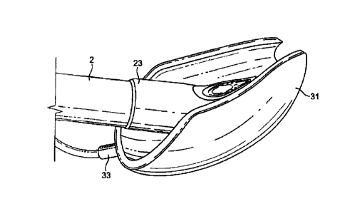
patient, the device 1 comprising an airway tube 2 and a mask 3 provided at
one end of the airway tube 2, the mask comprising a body 4 having a distal
end 5 and a proximal end 6 and a peripheral inflatable cuff 7 which surrounds
a hollow interior space or lumen of the mask, the mask 3 being attached to the
airway tube 2 for gaseous communication between the tube 2 and the outlet 8.
The mask of the present embodiment, and indeed of the further embodiments
described hereinafter, is shaped to conform to and fit readily into the actual
and potential space behind the larynx and to seal around the circumference of
the laryngeal inlet. In the present embodiments, this seal is created without
the
laryngeal mask airway device penetrating into the interior of the larynx. The
reference to actual and potential space will be understood to refer to the
space
normally available and that which can become available on flexure of the
surrounding structures.
As can be seen from Figures 7 to 14, the device 1, in terms of overall
appearance is somewhat similar to prior art devices, in that it consists of
the
basic parts which make up most if not all laryngeal mask airway devices, i.e.
an airway tube 2 and mask 3 which includes a body part 4, and a cuff 7.
For the purposes of description it is appropriate to assign reference names to
areas of the device 1 and accordingly with reference to the Figures, the
device
I has a dorsal side 14, a ventral side 15, a proximal end 16 (in the sense
that
CA 2960267 2017-03-08
WO 2010/100419 PCT/GB2010/000379
14
this is the end nearest the user rather than the patient) a distal end 17 and
right
and left sides 18 and 19.
In the present embodiment, the airway tube 2 is provided as a flexible tube,
although this does not have to be the case. For example, a curved airway tube
could be employed, which may be more rigid and performed with an
anatomically correct curvature.
The mask body 4 comprises two parts, namely an internal web 20 which
defines the interior hollow or lumen of the body of the mask and which is
provided with an aperture 21, and a semi-rigid back-plate 22 which conforms
to the generally oval shape of the web 20 and is adhered to the rear (dorsal
side) thereof. The back-plate 22 extends into a tubing portion 23, one end of
which aligns with the aperture 21, and the other end of which receives the
distal end of the airway tube 2, such that the airway tube 2 is in gaseous
communication with the interior of the body of the mask 3 via the tubing
portion 23 and the aperture 21. The airway tube 2 is connected in a gas-tight
manner into the tubing portion 23 by any suitable means, such as by welding
or adhesive, or by molding in one piece and in the present embodiment the
axis of the airway tube is provided substantially in the same plane as the
major axis of the inflatable cuff 7. As an optional feature the web 20 itself
is provided with two flexible bars 24, 25 formed in the web 20, which bars 24,
25 stretch across the aperture and act to prevent the epiglottis of the
patient
CA 2960267 2017-03-08
WO 2010/100419 PCT/GB2010/000379
from falling into the web 20 and hence interrupting the airway when the
device 1 is in place.
The generally elliptical cuff 7 is formed from a soft, flexible sheet of
silicone
26 and a generally "V"-shaped upper hinged portion 27. The flexible sheet 26
surrounds and is integrally formed with or otherwise hermetically secured to
the periphery of the web 20 on the ventral side 15 of the mask 3 and is bonded
in a gas-tight manner to the outer sides of the "V" of the upper hinged
portion
27, the inner sides of the upper hinged portion being secured to the periphery
of the back-plate 22. The apex of the "V" of the upper i.e. the distal end of
the
upper extends by a small amount, for example 2mm, beyond the distal tip of
the cuff.
In the present embodiment, the hinged nature of the upper portion 27 is
provided by a central hinge 28 (see Fig. 12) provided by a scored line in the
ventral surface of the upper 27 at the central portion of the mask, and two
further lateral hinges 29, 30 in the form of further scored lines, each
extending
down the middle of a respective one of the arms of the "V", on the dorsal side
of the upper 27. As will be appreciated, the central hinge 28 facilitates the
lateral expansion of the mask 3, and the two further lateral hinges 29, 30
facilitate the lateral contraction of the mask 3.
CA 2960267 2017-03-08
WO 2010/100419
PCT/GB2010/000379
16
In the present embodiment, the hinged upper portion 27 is formed from an
extruded hinged tube T, having the cross section shown in Fig 15. According to
this tube (thickness of tube wall and hinge size exaggerated in Fig. 15 for
clarity
of illustration), two hinges H1 are provided on the inside of the tube and two
hinges H2 are provided on the outside of the tube. To form the hinged upper
27,
the tube is cut along its entire axial length along one side thereof, and for
the
majority of its length along the opposite side, to form the "V" shaped hinged
upper. This upper is then welded hermetically around the inner circumference
of
the back plate of the mask and along the edges of the flexible sheet of the
cuff.
Fig. 16 shows a mechanism employed according to the present exemplary
embodiment to achieve a seal at the distal end of the cuff to enable it to be
inflatable. According to this sealing method, the distal walls of the
collapsible
tube are cut so that they are "sharpened" at the leading edges, such that the
inlet to the tube is generally frustoconical. This allows the thin flexible
covering material of the cuff to be glued down to these thin edges, avoiding
the glue getting into the crevices which are otherwise present where the walls
of the collapsible tube are of full thickness.
It will be noted, however, that the present invention is not limited to the
method
of construction outlined above, nor indeed to an arrangement having an
inflatable cuff.
CA 2960267 2017-03-08
WO 2010/100419
PCT/GB2010/000379
17
The entire assembly of the present embodiment collapses when the mask is
deflated, facilitating insertion into the normally closed space behind the
larynx
known as the hypopharynx, but achieves the shape shown in the Figures when
inflated, due to the hinging mechanism of the tube halves which causes the
tube
to open out into its open locked position under the influence of intra-cuff
pressure.
The cuff 7 thus effectively provides two inflatable wings, one on each lateral
side of the mask 3, and which are in fluid communication at the proximal side
of the cuff 7. The edges of each of the wings 31, 32 are chamfered to
minimise the disruption to any tissue caused by the insertion and operation of
the mask 3.
The cuff 7 is provided with a port 33 (see Fig. 11) at its proximal side, in
which one end of a small-diameter inflation tube 34 is fitted in a gas-tight
manner. The other end of the tube is provided with an inflation indicator
bladder 35 and valve 36, for connection to a suitable pump, such as a medical
syringe, for inflating and deflating the cuff 7.
In use, air is extracted from the cuff 7 via the valve 36, which results in
the
wings 31, 32 of the cuff 7 folding inwardly, facilitated by the lateral hinges
29, 30 of the upper hinged portion 27. The mask 3 is then in a condition
which facilitates the insertion of the mask 3 into the pharynx of the patient.
CA 2960267 2017-03-08
WO 2010/100419
PCT/GB2010/000379
18
Preferably, the mask 3 is initially inserted with the interior of the mask 3
(i.e.
ventral side 15) facing towards the rear wall of the pharynx, to facilitate
the
insertion of the mask 3 past the tongue of the patient. Thereafter, the mask 3
is
gently rotated through 180 degrees to face forwards, and the mask 3 is further
inserted until the distal end 5 of the mask 3 comes into contact with the
upper
oesophageal sphincter. This contact indicates to a user that the mask 3 is
correctly positioned.
The cuff 7 is then inflated, thus sealing the artificial airway around the
inlet to
the larynx. Owing to the hinged nature of the upper portion 27, the wings 31,
32 tend to expand laterally of the mask 3, i.e. the wings unfold on inflation
of
the cuff. A space providing a drainage channel is thus created in the pharynx
between the interior walls of the pharynx and the outer (dorsal) side of the
back plate 22 and the hinged upper portion 27. This space on the dorsal aspect
of the mask substantially approximates that which would otherwise be
bounded by the walls of the pharynx without the mask inserted, and hence by
unfolding the wings of the mask, the anatomical space or volume in the
pharynx which would be present without the mask inserted is recreated.
Paradoxically, therefore, when the wings of the mask are unfolded (either by
inflation or otherwise), the mask actually takes up less effective space
within
the pharyngeal passage as compared to prior art masks, as the mask creates a
volume that resembles or approximates to the pharyngeal space which would
otherwise be obstructed by the mask. That is, when in situ within the patient
CA 2960267 2017-03-08
WO 2010/100419
PCT/GB2010/000379
19
and with the wings unfolded, the mask substantially preserves the posterior
anatomical hollow within the pharynx, and thus substantially reduces or
eliminates altogether the resistance to the flow of matter (liquid, gas or
solid)
though the pharynx which would otherwise be created by the presence of a
mask inserted in the pharynx. As such, most of the dorsal aspect of the mask
according to the present embodiment provides a conduit having the approximate
volume and shape of the lower pharynx, such volume being large enough to
effect a significant rise in the pressure of any fluid emerging from the upper
oesophageal sphincter while still providing an inflatable mask shape which
maintains the required seal around the laryngeal orifice to ensure leak-free
delivery of respiratory gases to the lungs using positive pressure mechanical
ventilation.
Advantage is taken of the fact that the space of the lower pharynx is normally
a
closed space but may be expanded when fluids are forced through it or when an
object such as a laryngeal mask is inserted into it. It is possible in
consequence to
provide an adequate volume of space in fluid communication with the
oesophageal sphincter behind the anterior surface of the mask while still
having
sufficient area in the mask perimeter, which may for example be inflatable, to
make the required sealing contact with the perilaryngeal tissues.
Put another way, the present embodiment features an inflatable cuff 7, which
is movable under the action of inflation from a first (un-inflated) condition
CA 2960267 2017-03-08
WO 2010/100419
PCT/GB2010/000379
which facilitates the insertion of the mask 3, to a second (inflated)
condition
which thus re-establishes an approximation of the anatomy of the pharyngeal
space present when the mask 3 is not in place.
The present invention is not however limited to an inflatable arrangement, and
the wings of the mask 3 may be unfolded, and the mask movable from the
first to the second condition, by any other suitable means. For example, a
mask may be used which is inserted into place with an introducer-type spade
device as is known in the art, which mask expands when the spade is removed
to open up the pharyngeal anatomy of the patient to resemble the open
anatomy of the patient when swallowing or retching.
As will be appreciated, unlike prior art devices, the drainage channel of the
present embodiment is not provided by a tube, but rather by the open back
(dorsal side) of the mask itself and the walls of the pharynx. The inlet to
this
channel is defined by the generally "U" shaped guide way or conduit defined
by the tips of the wings at the distal end of the mask when the wings of the
mask are unfolded, as shown in Fig. 14.
The present embodiment thus provides a mask which is effectively "scooped
out" at the back (dorsal) side to define the gastric drainage channel.
CA 2960267 2017-03-08
WO 2010/100419
PCT/G82010/000379
21
In the event of the patient regurgitating or vomiting, the gastric matter
leaving
the oesophageal sphincter is unimpeded by the presence of the mask 3 as
illustrated by Fig. 76, which substantially re-establishes the normal amount
of
space available for drainage through the pharynx when the mask 3 is not
inserted. As a result, the mask 3 does not impede the flow of gastric contents
but on the contrary allows such contents to flow freely behind it, thus
substantially minimising the danger of the mask becoming dislodged from its
position by the pressure of such flow. In addition, the artificial airway
provided by the mask and airway tube remains uninterrupted and the seal
around the inlet to the larynx is not broken, thus making very unlikely the
possibility of gastric matter being aspirated into the lungs of the patient.
Furthermore, and particularly during vomiting conditions, the mask 3 is in
fact drawn more closely into its operating position in the patient's pharynx,
as
a result of a venturi being created by the drainage channel provided by the
mask. Specifically, the gastric matter being vomited by the patient may arrive
at the constriction or sphincter situated at the upper end of the oesophagus
at
pressures approximating 200cm H20 high pressure. The gastric matter passes
through this constricted portion at a higher velocity than it does through the
oesophagus, and in accordance with the Bernoulli Principle, this obligatory
increase in velocity results in a drop in pressure in the gastric matter
locally.
On leaving the upper oesophageal sphincter, the gastric matter enters the flow
channel defined by the mask 3 and the pharynx which is of a larger cross-
CA 2960267 2017-03-08
WO 2010/100419
PCT/GB2010/000379
22
sectional area, in the plane to which the velocity vector of the flow is
normal
or perpendicular, than the upper oesophageal sphincter under vomiting. As a
result, the velocity of the gastric matter passing through this larger flow
channel decreases, and accordingly the pressure of this gastric matter
increases, to greater than that in the oesophageal sphincter. The pressure
differential between the gastric matter in the upper oesophageal sphincter and
in the flow channel defined by the mask 3 and pharynx results in the mask 3
being actively forced further into engagement with the oesophageal sphincter,
thus holding the mask 3 yet more securely in its operational position than
when vomiting is not occurring.
A further feature of the present embodiment is that insertion of the mask 3
into place is facilitated where a gastroscope or similar is already inserted
into
the pharynx of the patient. Unlike prior art devices in which the drainage
channel is provided by a tube, the drainage channel in the present embodiment
is defined by the open back (dorsal side) of the mask. The entrance to this
channel is defined by the notch or trench 37 (see Fig. 10) created between the
tips of the wings 31, 32, which can receive the cable of an inserted
gastroscope or similar, facilitating the guiding of the mask 3 into the
pharynx
of the patient by sliding the mask along the cable as a rail.
Conversely, where the mask 3 is already in place, the later insertion of a
gastroscope or similar is facilitated by being guided into place by the
channel
CA 2960267 2017-03-08
WO 2010/100419
PCT/GB2010/000379
23
defined by the dorsal side of the mask 3 and the pharynx, and in particular by
the trench created between the opposed wings of the mask. Further, as the
present embodiment acts to re-establish the pharyngeal space normally
available for drainage, the insertion of a gastroscope or similar into the
pharynx is not impeded by the presence in the pharynx of the mask 3 itself.
A further advantage provided by the present embodiment is that the hinged
upper permits the cuff to be inverted for cleaning of the cuff, the upper and
the back plate by forcing the wings of the cuff apart. This is particularly
advantageous where the embodiment is intended as a re-useable product.
As will be appreciated by the skilled person, the present invention is not
limited to the types of construction or material identified in connection with
the present or succeeding embodiments. For example, the airway tube 2 and
mask 4 may all be formed from PVC plastics material, especially where a
single-use, disposable device is intended, different parts may be secured by
different means or be integrally formed from a single piece of material, as
appropriate.
A variation of the first embodiment, itself also an embodiment of the present
invention, is shown in Fig. 17, in a partially constructed condition before
the
back plate and airway tube are added to complete the laryngeal mask device.
This variation differs from the first embodiment in that the apex of the "V"
of
..=
CA 2960267 2017-03-08
WO 2010/100419
PCT/GB2010/000379
24
the hinged upper portion is provided as a tubular portion which defines an
inlet 50 of circular cross-section between 10 and 15 mm in diameter at the
distal open end of the mask.
As such, most of the interior volume of the mask according to the present
embodiment provides a conduit having the approximate volume and shape of the
lower pharynx, such volume being large enough to effect a significant rise in
the
pressure of any fluid emerging from the upper oesophageal sphincter while
still
providing an inflatable mask shape which maintains the required seal around
the
laryngeal orifice to ensure leak-free delivery of respiratory gases to the
lungs
using positive pressure mechanical ventilation.
Advantage is taken of the fact that the space of the lower pharynx is normally
a
closed space but may be expanded when fluids are forced through it or when an
object such as a laryngeal mask is inserted into it. It is possible in
consequence to
provide an adequate volume of space in fluid communication with the
oesophageal sphincter behind the anterior surface of the mask while still
having
sufficient area in the mask perimeter, which may for example be inflatable, to
make the required sealing contact with the perilaryngeal tissues.
As in the first embodiment, the hinged upper 27 is formed from a hinged tube
split as shown and welded hermetically around the inner circumference of said
mask, forming a single tube of orifice between 10 and 15 mm II) at the distal
CA 2960267 2017-03-08
WO 2010/100419
PCT/GB2010/000379
open end of said mask. The entire assembly collapses when the mask is
deflated,
facilitating insertion into the normally closed space behind the larynx known
as
the hypopharynx, but achieves the shape shown in Figure 17 when inflated, due
to the hinging mechanism of the tube halves which causes the tube to open out
into its open locked position under the influence of intra-cuff pressure.
The collapse of the mask when the cuff is deflated is in particular
facilitated
by the un-cut length of the hinged tube which defines the inlet to the
drainage
channel, as this tube collapses as shown in Fig. 18 when the cuff is deflated.
This permits a large-diameter gastric tube to be provided once the mask is in
place within the patient and the cuff inflated, as illustrated by Fig. 18, but
at
the same time facilitates insertion of the mask into the patient, as on
insertion
the mask is intended to be in an uninflated state, wherein the inlet of the
gastric drainage tube effectively closes up.
A second embodiment shown in Figures 20 to 21 differs from the first
embodiment only in that a portion 38 of the tubed section of the back-plate 22
is scooped away to facilitate the insertion of the device l into a patient.
According to the third embodiment shown in Figures 22 to 27, the laryngeal
mask airway device I again comprises an airway tube 2 with a mask 39
provided at the distal end thereof
CA 2960267 2017-03-08
WO 2010/100419 PCT/GB2010/000379
26
Similar to the first embodiment, the mask 39 comprises an internal web 40
which defines the interior of the body of the mask 39 and which is provided
with an aperture 41, and a semi-rigid back-plate 42 which conforms to the
generally oval shape of the web 40 and is adhered or otherwise attached to the
rear (dorsal side) thereof. The back-plate 42 extends into a tubing portion
43,
one end of which aligns with the aperture 41, and the other end of which
receives the distal end of the airway tube 2, such that the airway tube 2 is
in
gaseous communication with the interior of the body of the mask 39 via the
tubing portion 43 and the aperture 41. The airway tube 2 is connected in an
gas-tight manner into the tubing portion 43 by any suitable means, such as by
welding or adhesive. The aperture 41 itself is provided with two flexible bars
44, 45 formed in the web 40, which bars 44, 45 stretch across the aperture 41
and act to prevent the epiglottis of the patient from falling into the
aperture 41
and hence interrupting the airway when the mask 39 is in place.
According to the third embodiment, the cuff 46 is again formed from a
flexible sheet 47 of silicone which is integrally formed with or otherwise
hermetically secured to the periphery of the aperture 41, e.g. by adhesive
bonding
or welding, on the ventral side of the mask. Similarly, the inner edges of a
generally "V"-shaped upper 48 surround the back plate 42, but in this
embodiment, as in the first variation of the first embodiment, the apex of the
"V" is provided as a hinged tubular portion which defines an inlet 50 of
circular cross-section at the distal end of the mask 49. As will be
appreciated,
CA 2960267 2017-03-08
WO 2010/100419
PCT/GB2010/000379
27
as in the variation of the first embodiment this facilitates the collapse of
the
inlet and hence the insertion of the device into a patient on deflation of the
cuff, whilst still providing a large diameter drainage channel (e.g. 10 mm
diameter) once the cuff is inflated.
The flexible sheet 47 surrounds this inlet 50 and is joined to the outer sides
of
the "V" of the upper 48. Further, a flexible and somewhat elastic triangular
sheet 51 is attached between the opposed sides 52, 53 of the flexible sheet of
the cuff where they meet the upper 48. In the present embodiment, the
triangular sheet 51 is transparent. Similar to the first embodiment,
inflatable
wings 54, 55 are thus created on respective lateral sides of the device, which
wings are joined along their edges by the triangular elastic sheet.
A generally frustro-conical channel is thus created within the body of the
mask, having a generally circular inlet 50 at the distal end of the mask and
an
outlet region 56 at the posterior end of the mask, which outlet region 56 has
a
greater cross-sectional area than the inlet 50 in the plane perpendicular to
the
longitudinal axis of the mask. As such, most of the interior volume of the
mask
provides a conduit having the approximate volume and shape of the lower
pharynx, such volume being large enough to effect a significant rise in the
pressure of any fluid emerging from the upper oesophageal sphincter while
still
providing an inflatable mask shape which maintains the required seal around
the
CA 2960267 2017-03-08
WO 2010/100419
PCT/GB2010/000379
28
laryngeal orifice to ensure leak-free delivery of respiratory gases to the
lungs
using positive pressure mechanical ventilation.
Advantage is taken of the fact that the space of the lower pharynx is normally
a
closed space but may be expanded when fluids are forced through it or when an
object such as a laryngeal mask is inserted into it. It is possible in
consequence to
provide an adequate volume of space in fluid communication with the
oesophageal sphincter behind the anterior surface of the mask while still
having
sufficient inflatable area in the mask perimeter to make the required sealing
contact with the perilaryngeal tissues.
Similar to the first embodiment, the device of the third embodiment is placed
into an insertion condition by deflating the cuff 46. The reverse hinges 57,
58
provided as scored portions on the dorsal side of the upper portion 48 again
facilitate the inward folding of the wings of the mask so as to adopt as small
a
size as possible.
After insertion, the cuff 46 of the mask 39 is then inflated, which in
particular
causes the wings 54, 55 of the mask to separate. The separation of the wings
is not as great as in the first embodiment, however, as a result of the edges
of
the wings being linked by the triangular sheet 51. This prevents undue
pressure from being placed on the lateral walls of the pharynx by the
expanding wings 54, 55, and in particular seeks to avoid undue stretching of
_
CA 2960267 2017-03-08
WO 2010/100419 PCT/GB2010/000379
29
the hyoid bones and any possible pressure on the hypoglossal nerve. Also, as
the edges of the wings are joined by the triangular sheet to form a generally
flush dorsal surface to the mask, the small potential of trauma to the tissue
of
the pharynx caused by the edges of the wings contacting the walls of the
pharynx on insertion of the mask is averted. The flexibility and
stretchability
of the triangular sheet 51 does permit opening of the wings 54, 55, however.
As will be appreciated, according to this embodiment gastric drainage is not
provided by a tube which extends out of the mouth of the patient, as in prior
art devices, but rather by an orifice at the distal end of the mask which
defines
an inlet to a drainage channel within the body of the mask. Thereafter,
drainage of gastric matter is simply provided by the normal anatomy of the
patient i.e. the pharynx.
Similar effects and advantages as in the first embodiment are achieved, and
any gastric discharge is guided away from the upper oesophageal sphincter by
the internal channel created within the mask body.
A fourth embodiment shown in Figs 28 to 29 differs from the third
embodiment in that a generally triangular region of the back-plate is removed
and replaced with a transparent window 57a, which is bonded into the back-
plate in an air tight manner such that the effectiveness of gas delivery by
the
fourth embodiment is not effected. Further, the internal web is omitted. As a
CA 2960267 2017-03-08
WO 2010/100419 PCT/GB2010/000379
result of these changes, the inlet to the larynx may be viewed through the
mask by use of a suitable viewing device e.g. an endoscope, to confirm the
correct positioning of the mask and adequacy of space created behind the
mask when inflated.
A further difference between the fourth and third embodiments is that the
triangular sheet is omitted, which permits greater separation of the wings of
the mask upon inflation of the cuff
As will be appreciated by the skilled person, similar effects and advantages
as
in the third embodiment are achieved by the present embodiment.
A fifth embodiment is shown in Figs. 30 to 32. The fifth embodiment differs
from the fourth embodiment in .that the cuff 58a is internally bonded to the
back plate in two lateral positions, thus forming dimples 59 in the surface of
the cuff. These dimples 59 seek to prevent the possibility of stretching of
the
hyoid bones and consequential pressure on the hypoglossal nerve on inflation
of the mask, as in the third embodiment. By avoiding the sheet of the third
embodiment, however, the mask of the present embodiment is easier to clean,
facilitating its employment as a re-usable device.
As will be appreciated by the skilled person, similar effects and advantages
as
in the fourth embodiment are achieved by the present embodiment.
CA 2960267 2017-03-08
WO 2010/100419
PCT/GB2010/000379
31
A sixth embodiment is shown in Figs. 33 to 35. The sixth embodiment differs
from the fourth embodiment in that the back-plate is cut away in the regions R
indicated in Fig. 35 to avoid excessive pressure on the posterior surface of
the
cricoid cartilage. The function of the muscles covering this cartilage may
otherwise become compromised and since these muscles (the posterior crico-
arytenoid muscles) are necessary to maintain the lateral pull on the vocal
cords which keeps the larynx open, avoidance of impairing their function is of
great importance in maintaining a clear airway.
As will be appreciated by the skilled person, similar effects and advantages
as
in the fourth embodiment are achieved by the present embodiment.
A seventh embodiment is shown in Figures 36 to 42. According to this
embodiment, the device 60 comprises a pair of airway tubes 61, 62 which
open into the interior of a mask body 63 at the distal end of the device 60
and
which join at a forked portion 64 to provide a single airway tube connection
65 at the proximal part of the device 60. A wedge portion 66 is provided
between the airway tubes in the region of the forked portion.
The airway tubes 61, 62 themselves form the back plate of the mask 67, in
conjunction with a gastric drainage tube 68 located between the airway tubes
in the mask 67. To form the back plate, the top portion (ventral side) of each
CA 2960267 2017-03-08
WO 2010/100419
PCT/GB2010/000379
32
airway tube 61, 62 is cut off where it meets an inflatable cuff 69 which
surrounds the mask body 63, one side of each tube is bonded to the underside
(dorsal side) of the cuff 69, and the other side of each tube is bonded to
respective sides of the central gastro-drainage tube 68, which gastro-drainage
tube 68 is further bonded to the dorsal side of the proximal region of the
cuff
69 to complete the back plate of the mask 67.
Also bonded to the dorsal side of the cuff 69 is a curved flexible sleeve 70,
into which the gastric-drainage tube 68 empties, and which surrounds the
airway tubes 61, 62 in a loose-fitting manner. A cut-out portion 71 (shown in
phantom) is provided in the dorsal side of the drainage tube so as to
facilitate
the entry of gastric matter into the sleeve from the drainage tube, the other
(inlet) end 72 of the gastric-drainage tube 68 protruding through the distal
end
of the cuff 69. The inlet of the gastric-drainage tube is provided by a hinged
tube having an internal diameter of approximately 1 Omm, similar to the
variation of the first embodiment, and which thus facilitates the collapse and
hence insertion of the device upon deflation of the cuff, but at the same time
permits a large-diameter drainage tube to be provided within the patient once
the mask is inflated, as on insertion the mask is intended to be in an
uninflated
state, wherein the inlet of the gastric drainage tube effectively closes up.
The
length of the drainage tube from the inlet 72 to the point at which the cut-
out
portion begins is approximately 24mm, the length of the cut-out portion
extends for approximately 50mm along the axial length of the drainage tube
_
CA 2960267 2017-03-08
WO 2010/100419
PCT/GB2010/000379
33
and the length of the drainage tube as a whole is approximately 93mm,
although the present invention is of course not limited to these specific
dimensions.
Similar to the above embodiments, the cuff 69 is provided with a port 33 into
which one end of a small diameter inflation tube 34 is fitted in a gas-tight
manner. The other end of the inflation tube is provided with an inflation
indication bladder 35 and a valve 36, through which air is fed or extracted to
inflate or deflate the cuff 69.
As in the above embodiments, the cuff 69 is first deflated to facilitate
insertion of the device 60 into a patient, and subsequently inflated once the
mask is in place with the distal end seated in the oesophageal upper sphincter
and the interior of the mask closed over the inlet to the larynx.
In the event of gastric discharge, any gastric material is passed into the
sleeve
70 via the gastric tube 68. In the event of the patient vomiting, the high-
speed,
low pressure gastric matter from the upper oesophageal sphincter is quickly
passed by the relatively short-length of gastric tube into the plastic sleeve
70,
which may readily expand so as to adopt a large diameter. The pressure of the
vomited matter thus increases in the flow channel of the expanded sleeve 70,
thus forcing the mask into further contact with the upper oesophageal
sphincter.
CA 2960267 2017-03-08
WO 2010/100419
PCT/GB2010/000379
34
Also, it will be appreciated that whilst the flexible sleeve 70 may provide a
flow channel of large cross-sectional area when required, the sleeve 70 does
not itself particularly restrict access through the pharynx, as it may simply
fold and distort as required, nor, by virtue of being thin-walled, does it
occupy
a significant obstructing volume within the pharynx.
An eighth embodiment is shown in Figures 43 to 50.
Similar to the seventh embodiment, the eighth embodiment is provided with
two airway tubes 73, 74 which open into the interior of a mask portion, which
mask portion is encircled by an inflatable cuff 76. The airway tubes 73, 74
are
joined at a forked portion 77 at the proximal end of the device 78 to provide
a
single airway connector 79.
The airway tubes 73, 74 themselves form the back plate of the mask 80, in
conjunction with a gastric drainage tube 81 located centrally in the mask
between the airway tubes. To form the back plate, the top (ventral) portion of
each airway tube 73, 74 within the mask 80 is cut off, one side of each cut
off
portion of each tube is bonded to the underside (dorsal side) of the cuff 76,
and the other side of each cut-off portion of each tube is bonded to a
respective side of the drainage tube 81, which drainage tube is further bonded
¨ ¨
CA 2960267 2017-03-08
WO 2010/100419 PCT/GB2010/000379
to the underside of the proximal end of the cuff 76 to complete the back plate
of the mask 80.
The drainage tube 81 protrudes through the distal end of the cuff 76 and
provides a channel through which gastric discharge may exit. The ventral side
of the discharge tube is cut away from where it meets the posterior side of
the
cuff to form a discharge chute 82 extending from the posterior of the cuff for
the continued guidance of gastric matter away from the mask 80.
The inlet 83a of the gastric discharge tube is arranged so as to define a
generally linear aperture or slit having a width of approximately 25mm when
the mask is uninflated. Inflation of the cuff results however in the inlet
opening up, to form a large-diameter opening of generally circular cross-
section with a diameter of approximately 17mm. The objective of achieving
this slit-shape of the distal end of the drain tube for insertion is to reduce
the
front-to-back diameter of the leading (distal) edge of the mask, so permitting
it to pass easily into the normally closed-off space of the lower one-third of
the pharynx. This permits a large-diameter gastric tube to be provided once
the mask 80 is in place within the patient, but at the same time facilitates
insertion of the mask into the patient, as on insertion the mask is intended
to
be in an uninflated state, wherein the inlet of the gastric drainage tube
effectively closes up. The longitudinal length of the drainage tube which
extends through the cuff i.e. up until the point at which the chute 82 begins
is
CA 2960267 2017-03-08
WO 2010/100419
PCT/GB2010/000379
36
approximately 90mm. and the axial length of the chute 82 is approximately
60mm. The present invention is not however limited by the dimensions of the
aperture, drainage tube and chute given above.
A ninth embodiment of the present invention is shown in Figs 51 to 58, and
comprises a relatively rigid, curved airway tube 83 with a mask 84 attached at
the distal end thereof. The body 85 of the mask is formed by an internal web
86 which defines the interior of the body of the mask 84 and which is
provided with an aperture 87, and a semi-rigid back-plate which conforms to
the generally oval shape of the web and which is adhered or otherwise secured
to the rear thereof. The back-plate extends into a tubing portion 89, one end
of
which aligns with the aperture 87, and the other end of which receives the
distal end of the airway tube 83, such that the airway tube 83 is in gaseous
communication with the interior of the body 85 of the mask via the tubing
portion 89 and the aperture 87. The airway tube 82 is provided with a
flattened face on its dorsal side and provides an airway channel or lumen of
substantially pentagonal cross-section, with one face thereof curved, as shown
in Fig. 58. The airway tube is connected in an gas-tight manner into the
tubing
portion 89 by any suitable means, such as by welding or adhesive. The
aperture 87 itself is provided with two flexible bars 90, 91 formed in the
web,
which bars stretch across the aperture and act to prevent the epiglottis of
the
patient from falling into the aperture and hence interrupting the airway when
the mask is in place.
CA 2960267 2017-03-08
WO 2010/100419
PCT/GB2010/000379
37
The periphery of the body of the mask is defined by an inflatable cuff 92,
which is formed from a flexible sheet 93 of silicone which surrounds the
periphery of the web 86 on the ventral side of the mask and which surrounds
the periphery of the back plate 88 on the dorsal side of the mask. As in the
first embodiment, the cuff 92 is provided with a port 33 into which one end of
a small-diameter gas inlet tube 34 is affixed in a gas tight manner, to enable
the supply of air to and from the cuff 92 via an inflation indicator bladder
35
and valve 36 at the other end of the gas inlet tube 34.
The inlet 94 of a short-lengthed gastric drainage tube 95 protrudes through
the
distal end of the cuff 92 and the outlet is aligned with a flexible sleeve 96
provided on the dorsal side of the device. In the present embodiment, the
drainage tube is formed from a short length of hinged, collapsible extruded
hosing as in the variation of the first embodiment. This again allows the
inlet
to adopt a configuration of a generally linear slit when the cuff is deflated
to
facilitate insertion of the device, and a generally circular cross-section of
comparatively large cross-section, relative to prior art devices, when the
cuff
is inflated. When inflated, the internal diameter of the circular cross-
section of
the inlet is approximately lOmm. The tube itself is cut at an angle to the
axis
of the tube at the posterior end of the tube, to match the contour of the
cuff.
The shortest axial length of the tube, at the dorsal side of the tube, is
approximately 16mm, and the longest length of the tube, at the ventral side of
CA 2960267 2017-03-08
WO 2010/100419
PCT/GB2010/000379
38
the tube, is approximately 30mm. The present invention is not however
limited to these dimensions.
The sleeve 96 is adhered to the flat surface of the airway tube and to the
distal
end of the cuff. Its thin wall is made of sufficiently high-durometer material
to
allow it to act as a semi-rigid posterior wall enclosing a space corresponding
to the normal anatomical space available in the pharynx when there is no
device in place. The sleeve is however otherwise free from connection with
the remainder of the cuff 92.
The stiffness of the material of the sleeve is due to its high durometer,
while
it's flexibility is due to its thin wall. The stiffness may be translated as a
relative lack of elasticity compared to lower durometer materials. As such, in
a preferred embodiment, the sleeve is provided as a curved moulded shape to
correspond to the anatomical curve of the upper pharynx and oral cavity.
A high durometer is preferred so that the part of the sleeve which forms a
dome covering the back of the mask will resist collapse when in place within
the patient with the mask inflated, so as to create a space behind the mask
approximating that which would otherwise be present when the mask is not
inserted.
Similar to the eighth embodiment, a large cross-section flow channel is thus
provided as required by the sleeve 96 of the ninth embodiment, and similar
CA 2960267 2017-03-08
WO 2010/100419
PCT/GB2010/000379
39
effects and advantages as in the preceding embodiments may potentially be
achieved.
Thus, it can be seen that the above described embodiments address the
problems of prior art devices in novel and inventive ways.
Features of the above-described embodiments may be re-combined into
further embodiments falling within the scope of the present invention.
Further, the present invention is not limited to the exemplary materials and
methods of construction outlined above in connection with the exemplary
embodiments, and any suitable materials or methods of construction may be
employed.
For example, although the cuff may be formed using a sheet of soft flexible
silicone rubber, other materials such as latex or PVC may be used. PVC as a
material is particularly suited to embodiments intended for single use,
whereas the use of silicone rubber is preferred although not essential for
embodiments intended to be re-used in a number of medical procedures.
Further, for example, the sheet of the cuff may be integrally formed with the
web, or provided as a separate piece from the web which is subsequently
secured thereto e.g. by bonding with adhesive.
CA 2960267 2017-03-08
WO 2010/100419
PCT/GB2010/000379
Further, and as would be appreciated by the skilled person, various features
of
the present invention are applicable to a wide range of different laryngeal
mask airway devices, and the invention is not limited to the exemplary
embodiments of types of mask described above.
For example, aspects of the invention may be applied to laryngeal mask
airway devices featuring epiglotic elevator bars over the mask aperture, which
bars are operable to lift the epiglottis of a patent away from the aperture
upon
insertion of an endotracheal tube or other longitudinally-extended element
inserted through the airway tube so as to emerge into the hollow or lumen of
the mask through the mask aperture. Aspects of the present invention may for
example be applied to single or re-useable devices, devices featuring aperture
bars or not, "intubating" devices which permit an endotracheal tube or similar
to be introduced into the larynx via an airway tube of a mask, devices
incorporating fiberoptic viewing devices and so forth, without restriction or
limitation on the scope of the present invention.