Note: Descriptions are shown in the official language in which they were submitted.
CA 2961078 2017-03-15
PATENT APPLICATION
TITLE OF THE INVENTION
MULTI-LAYER DRESSINGS, SYSTEMS, AND METHODS FOR APPLYING
REDUCED PRESSURE AT A TISSUE SITE
10001)
BACKGROUND
[0002] Clinical studies and practice have shown that providing a reduced
pressure in
proximity to a tissue site augments and accelerates the growth of new tissue
at the tissue site.
The applications of this phenomenon are numerous, but application of reduced
pressure has
been particularly successful in treating wounds. This treatment (frequently
referred to in the
medical community as "negative pressure wound therapy," "reduced pressure
therapy," or
"vacuum therapy") provides a number of benefits, including faster healing, and
increased
formulation of granulation tissue.
[0003] Reduced-pressure treatment systems are often applied to large, highly
exudating
wounds present on patients undergoing acute or chronic care, as well as other
severe wounds
that are not readily susceptible to healing without application of reduced
pressure. Low-severity
wounds that are smaller in volume and produce less exudate have generally been
treated using
advanced dressings instead of reduced-pressure treatment
1
CA 2961078 2017-03-15
BRIEF SUMMARY
[0004] Shortcomings with certain aspects of wound care systems and dressings
are
addressed by the present invention as shown and described in a variety of
illustrative, non-
limiting embodiments herein. According to an illustrative embodiment, a
dressing for applying
reduced pressure at a tissue site includes a dressing material for
transferring the reduced
pressure to the tissue site and for receiving liquid from the tissue site. The
dressing material
includes a tissue-interface layer for contacting the tissue site, the tissue-
interface layer being a
hydrophobic layer; a manifold for distributing reduced pressure, the manifold
being a
hydrophobic layer; and a first absorbent layer for absorbing liquid from the
tissue site via the
tissue-interface layer and the manifold. The manifold may be disposed between
the tissue-
interface layer and the first absorbent layer. The dressing may further
include a drape covering
at least a portion of the dressing material.
[0005] According to another illustrative, non-limiting embodiment, a system
for
applying a reduced pressure at a tissue site includes a reduced-pressure
source for supplying
reduced pressure, a reduced-pressure delivery conduit for transferring reduced
pressure, a
dressing material, and a drape covering at least a portion of the dressing
material. The dressing
material is in fluid communication with the reduced-pressure source via the
reduced-pressure
delivery conduit. The dressing material delivers reduced pressure to the
tissue site and receives
liquid from the tissue site. The dressing material includes a tissue-interface
layer adapted to
contact the tissue site, which is a hydrophobic layer; a manifold, which is a
hydrophobic layer,
for distributing reduced pressure; and a first absorbent layer for absorbing
liquid from the tissue
site via the tissue-interface layer and the manifold. The manifold may be
disposed between the
tissue-interface layer and the first absorbent layer.
[0006] According to another illustrative, non-limiting embodiment, a method
for
applying reduced pressure at a tissue site includes the steps of applying a
dressing material to
the tissue site, covering at least a portion of the dressing material with a
drape, and supplying
reduced pressure to the dressing material. The dressing material transfers
reduced pressure to
the tissue site and receives liquid from the tissue site. The dressing
material includes a tissue-
interface layer, which is a hydrophobic layer, for contacting the tissue site;
a manifold, which is
a hydrophobic layer, for distributing reduced pressure; and a first absorbent
layer for absorbing
liquid from the tissue site via the tissue-interface layer and the manifold.
The manifold is
disposed between the tissue-interface layer and the first absorbent layer.
2
CA 2961078 2017-03-15
[0007] According to another illustrative, non-limiting embodiment, a method of
manufacturing a dressing for applying a reduced pressure at a tissue site
includes the steps of
providing a tissue-interface layer, which is a hydrophobic layer; providing a
manifold having a
tissue-facing side; coupling at least a portion of the tissue-facing side of
the manifold to the
tissue-interface layer; and providing a first absorbent layer having a tissue-
facing side and that
absorbs liquid. The manifold is a hydrophobic layer that distributes reduced
pressure. The
method of manufacturing may further include the steps of coupling at least a
portion of the
tissue-facing side of the first absorbent layer to the manifold. The method
may also include
providing a second absorbent layer having a tissue-facing side. The second
absorbent layer
includes a hydrophilic layer for absorbing liquid from the tissue site via the
tissue-interface
layer, the manifold, and the first absorbent layer. The method may also
include coupling at least
a portion of the tissue-facing side of the second absorbent layer to the first
absorbent layer.
100081 According to still another illustrative, non-limiting embodiment, a
reduced-
pressure wound dressing includes a non-adherent hydrophobic layer having a
first side and a
second, tissue-facing side; a porous, hydrophobic manifold layer, having a
first side and a
second, tissue-facing side; a quick-absorbing hydrophilic layer having a first
side and a second,
tissue-facing side; a fluid-storage layer having a first side and a second,
tissue-facing side; and a
sealing member having a first side and a second, tissue-facing side. The
second, tissue-facing
side of the porous, hydrophobic manifold layer is adjacent to the first side
of the non-adherent
hydrophobic layer. The second, tissue-facing side of the quick-absorbing
hydrophilic layer is
adjacent to the first side of the porous, hydrophobic manifold layer. The
second, tissue-facing
side of the fluid-storage layer is adjacent to the first side of the quick-
absorbing hydrophilic
layer. The second, tissue-facing side of the sealing member is adjacent to the
first side of the
fluid-storage layer.
[0009] Other features and advantages of the illustrative embodiments will
become
apparent with reference to the drawings and detailed description that follow.
3
CA 2961078 2017-03-15
BRIEF DESCRIPTION OF THE DRAWINGS
[0010] FIGURE 1 is a schematic diagram, with a portion in cross section, of an
illustrative, non-limiting system for applying reduced pressure at a tissue
site;
[0011] FIGURE 2 is an exploded, schematic, perspective view of an
illustrative, non-
limiting dressing for applying reduced pressure at a tissue site; and
[0012] FIGURE 3 is a schematic, cross-sectional view of an illustrative, non-
limiting
dressing for applying reduced pressure at a tissue site.
DETAILED DESCRIPTION
[0013] In the following detailed description of the preferred embodiments,
reference is
made to the accompanying drawings that form a part hereof, and in which is
shown by way of
illustration specific preferred embodiments in which the invention may be
practiced. These
embodiments are described in sufficient detail to enable those skilled in the
art to practice the
invention, and it is understood that ether embodiments may be utilized and
that logical
structural, mechanical, electrical, and chemical changes may be made.
To avoid detail not necessary to enable those skilled in the
art to practice the invention, the description may omit certain information
known to those
skilled in the art. The following detailed description is, therefore, not to
be taken in a limiting
sense, and the scope of the present invention is defined only by the appended
claims.
[0014] Referring now primarily to FIGURE 1, an illustrative reduced-pressure
treatment
system 100, which includes a dressing 102 and which applies reduced pressure
to a tissue site
104, is presented. The dressing 102 includes a dressing material 106. The
dressing material
106 may include any number of layers or components and a number of
illustrative, non-limiting
examples will be provided herein. Unless otherwise indicated, as used herein,
"or" does not
require mutual exclusivity. The dressing material 106 may include one or more
laminar layers.
The dressing 102 may further include a sealing member 108 and a reduced-
pressure connector
110, reduced-pressure interface, or connection member.
[0015] The dressing material 106 serves as a manifold for distributing reduced
pressure.
The term "manifold" as used herein generally refers to a substance or
structure that is provided
to assist in applying reduced pressure to, delivering fluids to, or removing
fluids from a tissue
site. The manifold typically includes a plurality of flow channels or pathways
to improve
distribution of fluids provided to and removed from the tissue site. The
dressing material 106
that serves as a manifold may include a number of layers as will be described
further below.
4
CA 2961078 2017-03-15
100161 The tissue site 104 may be the bodily tissue of any human, animal, or
other
organism, including bone tissue, adipose tissue, muscle tissue, dermal tissue,
vascular tissue,
connective tissue, cartilage, tendons, ligaments, or any other tissue. While
the tissue site 104
may include a wound, diseased tissue, or defective tissue, the tissue site 104
may also be healthy
tissue that is not wounded, diseased, or defective.
[0017] The application of reduced pressure to the tissue site 104 may be used
to promote
the drainage of exudate and other liquids from the tissue site 104, as well as
to stimulate the
growth of additional tissue. In the case in which the tissue site 104 is a
wound site, the growth
of granulation tissue and removal of exudates and bacteria promotes healing of
the wound. The
application of reduced pressure to non-wounded or non-defective tissue,
including healthy
tissue, may be used to promote the growth of tissue that may be harvested and
transplanted to
another tissue location.
[0018] As used herein, "reduced pressure" generally refers to a pressure less
than the
ambient pressure at a tissue site 104 that is being subjected to treatment. In
most cases, this
reduced pressure will be less than the atmospheric pressure at which the
patient is located.
Alternatively, the reduced pressure may be less than a hydrostatic pressure at
the tissue site 104.
Unless otherwise indicated, values of pressure stated herein are gauge
pressures. The reduced
pressure delivered may be static or varied (patterned or random) and may be
delivered
continuously or intermittently. Although the terms "vacuum" and "negative
pressure" may be
used to describe the pressure applied to the tissue site, the actual pressure
applied to the tissue
site may be more than the pressure normally associated with a complete vacuum.
Consistent
with the use herein, an increase in reduced pressure or vacuum pressure
typically refers to a
relative reduction in absolute pressure.
[0019] The reduced pressure is provided to the reduced-pressure connector 110
by a
reduced-pressure delivery conduit 112. The reduced-pressure delivery conduit
112 receives
reduced pressure from a reduced-pressure source 114. The reduced-pressure
source 114 may be
any device or subsystem for supplying a reduced pressure, including but not
limited to a
manually operated pump, a powered vacuum pump, a wall vacuum source, etc.
While the
amount and nature of reduced pressure applied to a tissue site will typically
vary according to
the application, the reduced pressure will typically be between -5 mm Hg and -
500 mm Hg and
more typically between -100 mm Hg and -200 mm Hg. In one illustrative
embodiment, the
reduced-pressure source 114 may be a battery-driven vacuum pump. In one
illustrative
5
CA 2961078 2017-03-15
example, the pump uses low amounts of power and is capable of operating for an
extended
period of time on a single charge of the battery.
[0020] One or more devices may be fluidly coupled between the reduced-pressure
connector 110 and the reduced-pressure source 114. For example, representative
device 116 is
.. shown fluidly coupled on a portion of the reduced-pressure delivery conduit
112. The
representative device 116 may be a fluid reservoir, or collection member, to
hold exudates and
other fluids removed. Other illustrative, non-limiting examples of devices 116
that may be
included on the reduced-pressure delivery conduit 112 or otherwise fluidly
coupled to the
reduced-pressure delivery conduit 112 include the following non-limiting
examples: a pressure-
feedback device, a volume detection system, a blood detection system, an
infection detection
system, a flow monitoring system, a temperature monitoring system, etc. Some
of these devices
may be formed integrally to the reduced-pressure source 114 or other aspects
of the system 100.
[0021] The dressing 102 is adapted to contact or cover the tissue site 104
that is to be
treated. As used herein, the term "cover" includes partially or fully
covering. Also, a first
object that covers a second object may directly or indirectly touch the second
object, or may not
touch the second object at all.
[0022] The dressing material 106 is covered fully or partially by the sealing
member
108, or drape. The sealing member 108 may be any material that provides a
fluid seal over the
dressing material 106 and a portion of a patient's epidermis 118. The sealing
member 108 may,
for example, be an impermeable or semi-permeable, elastomeric material.
"Elastomeric" means
having the properties of an elastomer. It generally refers to a polymeric
material that has
rubber-like properties. More specifically, most elastomers have elongation
rates greater than
100% and a significant amount of resilience. The resilience of a material
refers to the material's
ability to recover from an elastic deformation. Examples of elastomers may
include, but are not
limited to, natural rubbers, polyisoprene, styrene butadiene rubber,
chloroprene rubber,
polybutadiene, nitrile rubber, butyl rubber, ethylene propylene rubber,
ethylene propylene diene
monomer, chlorosulfonated polyethylene, polysulfide rubber, polyurethane, EVA
film, co-
polyester, and silicones. Specific examples of sealing member materials
include a silicone
drape, 3M Tegaderm drape, acrylic drape such as one available from Avery
Dennison, or an
incise drape.
[0023] The sealing member 108 may be provided in "sheet" form, or in a
pourable or
sprayable form that is applied over the dressing material 106 after placement
of the dressing
material 106 in contact with the tissue site 104. Ins some embodiments,
sealing member 108
6
CA 2961078 2017-03-15
may include a device that is placed over the dressing material 106 and the
tissue site 104 to
provide sealing functionality, including but not limited to, a suction cup, a
molded cast, and a
bell jar. The sealing member 108 has a first side 120 and a second, tissue-
facing side 122.
[0024] An attachment device 124 may be used to hold the sealing member 108
against
the patient's epidermis 118 or another layer, such as a gasket or additional
sealing member. The
attachment device 124 may take numerous forms. For example, the attachment
device 124 may
be a medically acceptable, pressure-sensitive adhesive 126 that extends about
a periphery, or
perimeter, 128 of the sealing member 108.
[0025] In one embodiment, the sealing member 108 is configured to provide a
sealed
connection with the patient's epidermis 118 (or other tissue surrounding the
dressing material
106) and the tissue site 104. The sealed connection may be provided by the
adhesive 126
positioned along the perimeter 128 of the sealing member 108, or on any
portion of the sealing
member 108, to secure the sealing member 108 to the dressing material 106 or
the tissue
surrounding the tissue site 104. The adhesive 126 may be pre-positioned on the
sealing member
108 or may be sprayed or otherwise applied to the sealing member 108
immediately prior to
installing the sealing member 108. Prior to the application of the sealing
member 108 to the
tissue site 104, the adhesive 126 may also be covered by an adhesive support
layer or removable
backing. The adhesive support layer may provide rigidity to the sealing member
108 prior to
application and may also aid in the actual application of the sealing member
108 onto the tissue
site 104 or tissue near the tissue site 104. The adhesive support layer may be
peeled off or
otherwise removed before applying the sealing member 108.
[0026] The reduced-pressure connector 110 is coupled to the sealing member 108
and
provides reduced pressure to an interior space 130 formed between the second,
tissue-facing
side 122 of the sealing member 108 and the tissue site 104. In another
embodiment, the
reduced-pressure delivery conduit 112 may directly couple the reduced-pressure
source 114 to
the dressing 102.
[0027] The reduced-pressure delivery conduit 112 may be any tube or flow path
through
which a gas, liquid, gel, or other fluid may flow. The possible embodiments of
the reduced-
pressure delivery conduit 112 are numerous, and non-limiting examples follow.
The reduced-
pressure delivery conduit 112 may have any cross-sectional shape, such as a
circle, oval, or
polygon. In addition, the reduced-pressure delivery conduit 112 may be made
from any
material, and may be either flexible or inflexible. In FIGURE 1, the reduced-
pressure delivery
conduit 112 couples the reduced-pressure connector 110 to the representative
device 116, and
7
CA 2961078 2017-03-15
couples the representative device 116 to the reduced-pressure source 114.
However, reduced-
pressure delivery conduit 112 may directly couple reduced-pressure source 114
to the dressing
102. Also, the reduced-pressure delivery conduit 112 may include one or more
paths or lumens
through which fluid may flow. For example, the reduced-pressure delivery
conduit 112 may
include two lumens with one lumen used to administer pressure measurements to
determine the
amount of reduced pressure being applied at the tissue site 104. The other
lumen may be used
to deliver fluids, such as air, antibacterial agents, antiviral agents, cell-
growth promotion agents,
irrigation fluids, or other chemically active agents, to the tissue site 104.
The fluid source from
which these fluids originate is not shown in FIGURE 1.
[0028] The reduced-pressure connector 110 permits the passage of a fluid (such
as
exudates, air, etc.) from the dressing material 106 to reduced-pressure
delivery conduit 112, and
vice versa. In another illustrative embodiment (not shown), the reduced-
pressure treatment
system 100 does not include the reduced-pressure connector 110. In this
illustrative
embodiment, the reduced-pressure delivery conduit 112 may be inserted directly
into the sealing
member 108 or the dressing material 106 such that an end of the reduced-
pressure delivery
conduit 112 is adjacent to or in contact with the sealing member 108 or any of
the dressing
material 106 in a manner that allows for the delivery of reduced pressure. In
the non-limiting
example shown in FIGURE 1, the sealing member 108 includes an aperture 1 1 1
through which
the reduced-pressure connector 110 is disposed.
[0029] The reduced-pressure connector 110 may be located anywhere relative to
the
dressing material 106. For example, although FIGURE 1 shows the reduced-
pressure connector
110 and the opening or aperture 1 1 1 in the sealing member 108 through which
the reduced-
pressure connector 110 extends as centrally located relative to the dressing
material 106, the
reduced-pressure connector 110 and the opening or aperture 111 may be located
adjacent to the
edges of the dressing material 106 or at other locations.
[0030] In operation, the dressing 102 is deployed on the tissue site 104 and
reduced
pressure is delivered to the tissue site 104. More specifically, the dressing
material 106 is
deployed proximate the tissue site 104 where treatment is desired. The sealing
member 108 is
deployed over the dressing material 106 and at least a portion of the
patient's epidermis 118 to
form the interior space 130, or sealed space. If not already accomplished, the
aperture 111 may
be formed in the sealing member 108 and the reduced-pressure connector 110
applied. If not
already accomplished, the reduced-pressure delivery conduit 112 is fluidly
coupled to the
8
CA 2961078 2017-03-15
reduced-pressure connector 110 and to the reduced-pressure source 114. The
reduced-pressure
source 114 is activated and reduced pressure is delivered to the tissue site
104.
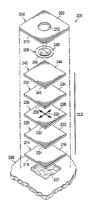
[0031] Referring now primarily to FIGURE 2, an exploded view of an
illustrative
dressing 200, which is suitable for use as dressing 102 in FIGURE 1, is
presented. The dressing
200 includes a dressing material 202, a sealing member 204 covering the
dressing material 202,
and a reduced-pressure connector 206. The reduced-pressure connector 206 may
be disposed in
part between the dressing material 202 and the sealing member 204. The
dressing material 202
may be used to manifold, or distribute, pressure to a tissue site 207, e.g., a
wound. The sealing
member 204 provides a seal over the dressing material 202 and a portion of a
patient's
epidermis 209. The sealing member 204 has a first side 208 and a second,
tissue-facing side
210. The sealing member 204 may also be formed with an aperture 212.
[0032] The dressing material 202 includes a number of components, e.g., layers
or
portions of material. First, a tissue-interface layer 214 has a first side 216
and a second, tissue-
facing side 218. The tissue-interface layer 214 is adapted to contact the
tissue site 207. In an
.. example in which the dressing 200 is used to treat a wound, the tissue-
interface layer 214 may
be partially or fully in contact with the tissue site 207. The tissue site 207
may directly contact
any portion of the second, tissue-facing side 218 of the tissue-interface
layer 214, including the
center or peripheral portions of the tissue-interface layer 214. The second,
tissue-facing side
218 of the tissue-interface layer 214 may also directly contact a periwound
area of the tissue site
207, which may include healthy tissue that surrounds the tissue site 207. In
the illustrative
embodiments, the tissue-interface layer 214, either alone or when used in
conjunction with other
layers, may reduce or eliminate maceration at or near the tissue site 207,
including the
periwound area and healthy epidermis 209 that surrounds the tissue site 207.
[0033] In an illustrative embodiment, the tissue-interface layer 214 is a
hydrophobic
layer. The hydrophobic characteristics of the tissue-interface layer 214
prevent the tissue-
interface layer 214 from directly absorbing liquid, such as exudate, from the
tissue site 207, but
allow the liquid to pass through. Thus, the liquid may be drawn away from the
tissue site 207
via the tissue-interface layer 214 using a reduced-pressure source, such as
the reduced-pressure
source 114 in FIGURE 1, or may be absorbed by one or more other layers in the
dressing
material 202. Thus, the tissue-interface layer 214 permits the passage of
liquid away from the
tissue site 104, while maintaining contact with the tissue site 207.
[0034] Because the tissue-interface layer 214 does not absorb (or hold)
liquid, the tissue
site 207 is not exposed, or otherwise in contact, with any hydrophilic
material that is
9
CA 2961078 2017-03-15
substantially saturated with liquid and that could promote maceration. Also,
no capillary action
takes place in a direction along the surface of the tissue site 207. Thus, the
hydrophobic
characteristics of the tissue-interface layer 214 may also restrain or prevent
the spread of liquid
along an interface between the tissue site 207 and the second, tissue-facing
side 218 of the
tissue-interface layer 214.
[0035] The tissue-interface layer 214 may be composed of any of a variety of
materials,
and have a variety of structures, including materials and structures that
allow fluid, e.g., liquid
or gas, to pass through the tissue-interface layer 214. In one example, the
tissue-interface layer
214 may be composed of or include nylon. In another example, the tissue-
interface layer 214
may be composed of or include a polymer-based mesh fabric. In another example,
the tissue-
interface layer 214 may be composed of or include Teflon -impregnated
polyethylene. The
tissue-interface layer 214 may also be composed of or include spandex or
Elastane material.
The tissue-interface layer 214 may be a thin, non-adherent, hydrophobic, non-
stitch layer. The
tissue-interface layer 214 may function--typically under reduced-pressure--to
quickly transport
moisture from the tissue site 207. The tissue-interface layer 214 may be non-
absorbent in
nature.
[0036] The tissue-interface layer 214 may also exhibit non-adherent properties
such that
the tissue-interface layer 214 does not stick to or adhere to the tissue site
207. The tissue-
interface layer 214 may also be stretchable, or elastic, in nature. The
stretchable properties of
the tissue-interface layer 214 may facilitate placement of the tissue-
interface layer 214 adjacent
to tissue sites and wounds having a variety of shapes, topologies, or
flexibility requirements.
[0037] The tissue-interface layer 214 may be used to promote granulation at
the tissue
site 207 when reduced pressure is applied through the dressing 200. For
example, any or all of
the surfaces of the tissue-interface layer 214 may have an uneven, coarse, or
jagged profile that
causes microstrains and stresses at the tissue site 707 when a pressure is
applied. Thus, the
reduced pressure supplied may cause the tissue-interface layer 214 to create
microstrain and
thereby encourage granulation. The tissue-interface layer 214 may further
serve as a scaffold
for new cell-growth, or a scaffold material may be used in conjunction with
the tissue-interface
layer 214 to promote cell-growth. A scaffold is a substance or structure used
to enhance or
promote the growth of cells or formation of tissue, such as a three-
dimensional porous structure
that provides a template for cell growth.
[0038] The tissue-interface layer 214 may have any size, shape, or thickness
depending
on a variety of factors, such as the type of treatment being implemented or
the nature of the
CA 2961078 2017-03-15
tissue site 207. The size and shape of the tissue-interface layer 214 may be
customized by a
user to cover a particular portion of the tissue site 207 or nearby tissue.
The tissue-interface
layer 214 may also have a laminar size or thickness that is the same or
different from any one of
the other layers in the dressing material 202. In another example, the tissue-
interface layer 214
may be thinner than any of the other layers in the dressing material 202.
[0039] The dressing material 202 also includes a manifold 220, or manifold
layer or
manifold member, for distributing reduced pressure from a reduced-pressure
source, such as
reduced-pressure source 110 in FIGURE 1. The manifold 220 may also distribute
liquid, such
as exudate, from the tissue-interface layer 214 to other layers in the
dressing material 202. The
manifold 220 has a first side 222 and a second, tissue-facing side 224. The
second, tissue-
facing side 224 of the manifold 220 is disposed proximate the first side 216
of the tissue-
interface layer 214.
10040] The manifold 220 may be a hydrophobic, porous material that is capable
of
distributing reduced pressure to the tissue site 207. The manifold 220 may be
made from foam,
gauze, felted mat, or any other material suited to a particular biological
application. The
manifold 220 may include a plurality of flow channels or pathways to
facilitate distribution of
reduced pressure or fluids to or from the tissue site 207. In one embodiment,
the manifold 220
is a porous foam and includes a plurality of interconnected cells or pores
that act as flow
channels. The porous foam may be a polyurethane, open-cell, reticulated foam,
such as the
GranuFoam dressing available from Kinetic Concepts, Inc. of San Antonio,
Texas. If an open-
cell foam is used, the porosity may vary. The flow channels allow fluid
communication
throughout a portion of the manifold 220 having open cells. The cells and flow
channels may
be uniform in shape and size, or may include patterned or random variations in
shape and size.
Variations in the shape and size of the cells of the manifold 220 result in
variations in the flow
channels, and such characteristics may be used to alter the flow
characteristics of fluid through
the manifold 220. In one non-limiting example, the manifold 220 may be made
from the same
material as the tissue-interface layer 214.
[0041] A number of additional layers may be added to absorb fluid from the
manifold
220 or tissue-interface layer 214. The additional layers may be absorbers. The
additional layers
are selected so that the farther the additional layers are located in situ
from the tissue site 207,
the more the layers can absorb. The additional layers thus may be increasingly
hydrophilic. In
the illustrative embodiment of FIGURE 2, a first absorbent layer 226, which
has a first side 228
and a second, tissue-facing side 230, may be included with the dressing
material 202. The
11
CA 2961078 2017-03-15
second, tissue-facing side 230 may be disposed proximate to the first side 222
of the manifold
220. The first absorbent layer 226 is for receiving and absorbing the liquids
distributed by the
manifold 220.
[0042] A second absorbent layer 232, or reservoir layer, which has a first
side 234 and a
second, tissue-facing side 236, may also be included with the dressing
material 202. Additional
absorbent layers analogous to the first absorbent layer 226 or second
absorbent layer 232 may
also be included in other embodiments. The second, tissue-facing side 236 of
the second
absorbent layer 232 may be disposed proximate the first side 228 of the first
absorbent layer
226. As with other layers, the first absorbent layer 226 and second absorbent
layer 232 may be
coextensive or may be different sizes.
[0043] The absorbent layers 226, 232 receive and absorb liquids from the
manifold 220.
The manifold 220 may facilitate the migration of liquid from the tissue site
207 radially outward
toward the edges of the manifold 220 so that the liquid is distributed more
uniformly across
either or both of the absorbent layers 226 and 232 as indicated generally by
the multi-directional
arrows 238 shown for reference on the first absorbent layer 226. The absorbent
layers 226 and
232 will retain more liquid if the liquid is more uniformly distributed across
the surface of the
absorbent layers 226 and 232. Also, such distribution of the liquid from the
tissue site 207 in
the directions indicated by the multi-directional arrows 238 may occur with or
without the
presence of reduced pressure. Thus, a fuller utilization of either or both of
the absorbent layers
226 and 232 may be achieved using the manifold 220 even when reduced pressure
is not being
applied to the dressing 200.
100441 The manifold 220 may also act as a separator between the tissue-
interface layer
214 and either or both of the absorbent layers 226 and 232. In this example,
the manifold 220,
reduces, restrains, or prevents liquid, such as exudate, that has been
absorbed by either or both
of the absorbent layers 226 and 232 from contacting either or both of the
tissue-interface layer
214 or the tissue site 207. Thus, the manifold 220 may further help to prevent
maceration at or
near the tissue site 207.
10045] The manifold 220 may have any size, shape, or thickness depending on a
variety
of factors, such as the type of treatment being implemented or the nature of
the tissue site 207.
For example, the thickness of the manifold 220 may be increased or decreased
to optimize the
effectiveness of the manifold's 220 role as a separator between the tissue-
interface layer 214
and either or both of the absorbent layers 226 and 232. In applications in
which the tissue site
207 releases a large amount of liquid that is absorbed by either or both of
the absorbent layers
12
CA 2961078 2017-03-15
226 and 232, a relatively thicker manifold 220 may be desirable to restrain or
prevent the liquid
that is absorbed by either or both of the absorbent layers 226 and 232 from
contacting either or
both of the tissue-interface layer 214 or the tissue site 207. On the other
hand, a relatively
thinner manifold 220 may be desirable in applications in which a lower amount
of liquid is
present. In illustrative, non-limiting illustrations, the manifold 220 may be
about 4 millimeters,
2 millimeters, or I millimeter thick. The manifold 220 may also have a laminar
size or
thickness that is the same or different from any one of the other layers in
the dressing material
202.
[0046] The first absorbent layer 226 may be disposed adjacent to the manifold
220 and
absorb liquid, such as exudate, from the tissue site 207 via the tissue-
interface layer 214 and the
manifold 220. In one example, the first absorbent layer 226 is disposed
between the manifold
220 and the second absorbent layer 232.
100471 The first absorbent layer 226 may be formed from a hydrophilic material
to
facilitate absorption of the liquid from the tissue site 207. In one
embodiment, the first
absorbent layer 226 is formed of a material that absorbs liquid from the
tissue site 207 at a faster
rate than the second absorbent layer 232. For example, the first absorbent
layer 226 may
include a fast-wicking material, such as cotton, terrycloth, paper towel
material, etc. To quicken
the rate at which the first absorbent layer 226 absorbs liquid from the tissue
site 207, the surface
area of the first absorbent layer 226 may be increased. The first absorbent
layer 226 may also
be made from a woven or mesh material.
100481 In one embodiment, the fast-wicking characteristics of the first
absorbent layer
226, including the first absorbent layer's 226 ability to absorb liquid at a
faster rate than the
second absorbent layer 232, helps to quickly draw liquid away from the tissue
site 207 and
toward the second absorbent layer 232, which may have a higher absorptive
capacity than the
first absorbent layer 226. The first absorbent layer's 226 ability to quickly
draw liquid away
from the tissue site 207 may prevent the accumulation of liquid at or near the
tissue site 207,
and therefore may help to prevent maceration at or near the tissue site 207.
[00491 The first absorbent layer 226 may have any size, shape, or thickness
depending
on a variety of factors, such as the type of treatment being implemented or
the nature of the
tissue site 207. The first absorbent layer 226 may also have a size or
thickness that is the same
or different from any one of the other layers in the dressing material 202.
[00501 In one embodiment, the second absorbent layer 232 absorbs liquid from
the
tissue site 207 via the tissue-interface layer 214, the manifold 220, and the
first absorbent layer
13
CA 2961078 2017-03-15
226. In another illustrative embodiment (not shown), the dressing material 202
does not
include the first absorbent layer 226, in which case the second absorbent
layer 232 is the only
absorbent layer present in the dressing material 202. In this embodiment, the
second absorbent
layer 232 absorbs liquid from the tissue site 207 via the tissue-interface
layer 214 and the
manifold 220. In other embodiments, more than two absorbent layers may be
included.
[0051] The second absorbent layer 232 may be composed of any material capable
of
absorbing liquid, such as exudate, from the tissue site 207. The material from
which the second
absorbent layer 232 is composed is also capable of transferring reduced
pressure. In one
embodiment, the second absorbent layer 232 has a higher fluid storage capacity
than the first
absorbent layer 226. The difference in fluid storage capacity may be due to
the respective
materials from which absorbent layers 226 and 232 are composed. In one
example, the second
absorbent layer 232 may be capable of storing liquid that is 20 or more times
heavier or
voluminous than the dry weight or volume, respectively, of the second
absorbent layer 232.
[0052] In the illustrative example of FIGURE 2, the second absorbent layer 232
wicks
liquid away from the first absorbent layer 226, and stores that liquid. To
facilitate the second
absorbent layer's 232 function of wicking liquid away from the first absorbent
layer 226, the
second absorbent layer 232 may be composed of a material that is more
hydrophilic than the
material from which the first absorbent layer 226 is composed.
[0053] In one embodiment, the second absorbent layer 232 may be composed of a
hydrocolloid or hydrogel, which may be, for example, a First Water Net20
hydrogel from First
Water, Ltd. of Wiltshire, II.K. The hydrogel from which the second absorbent
layer 232 is
composed may also include polyethylene glycol. Further, although hydrogel
infers the
inclusion of water, the hydrogel from which the second absorbent layer 232 may
be composed
may be a dried back hydrogel polymer base, which substantially or completely
lacks water.
[0054] In another illustrative example, the second absorbent layer 232 may be
made
from a super absorbent fiber material. The super absorbent fibers may hold
onto or bond to the
liquid in conjunction with a physical or chemical change to the fibers. In one
non-limiting
example, the super absorbent fiber may include the Super Absorbent Fiber (SAF)
material from
Technical Absorbents, Ltd. of Lincolnshire, UK. The fibers may thus form a
fibrous material in
which the fibers absorb liquid from the tissue site 207. Also, the fibers in
the second absorbent
layer 232 that contact the liquid may gel upon contact with the liquid,
thereby trapping the
liquid. Spaces or voids between the fibers may allow a reduced pressure that
is applied to the
14
CA 2961078 2017-03-15
dressing 200 to be transferred within and through the second absorbent layer
232. The structure
of the second absorbent layer 232 that contains the fibers may be either woven
or non-woven.
[00551 The second absorbent layer 232 may have any size, shape, or thickness
depending on a variety of factors, such as the type of treatment being
implemented or the nature
__ of the tissue site 207. For example, the width or thickness of the second
absorbent layer 232
may be increased to cause a corresponding increase in the fluid storage
capacity of the second
absorbent layer 232. The second absorbent layer 232 may also have a size or
thickness that is
the same or different from any one of the other layers in the dressing
material 202.
[00561 The dressing material 202 also includes a distribution manifold 242
that is
__ adjacent to the second absorbent layer 232. The distribution manifold 242
has a first side 244
and a second, patient-facing side 246. The second, patient-facing side 246 of
the distribution
manifold 242 is disposed adjacent to the first side 234 of the second
absorbent layer 232. The
distribution manifold 242 distributes reduced pressure to one or more layers
in the dressing
material 202 that are nearer the tissue site 207 and may do so more uniformly
across an entire
__ surface of the one or more layers in the dressing material 202. Because the
distribution
manifold 242 is disposed further away from the tissue site 207 than the
absorbent layers 226 and
232, liquid, such as exudate, from the tissue site 207 does not typically
reach the distribution
manifold 242. In one illustrative embodiment, however, liquid may be allowed
to reach the
distribution manifold 242.
[0057] The distribution manifold 242 may be made from any material capable of
distributing gas or liquid. In one example, the distribution manifold 242 is
formed from a
reticulated polyurethane foam layer or other porous manifolding material. In
another example,
the distribution manifold 242 may be formed from the same or similar material
as the manifold
220. The distribution manifold 242 may also distribute liquid, such as
exudate, from the tissue
__ site 207 that is not absorbed by either or both of the absorbent layers 226
and 232. The
distribution manifold 242 may also have any size, shape, or thickness.
[0058] Although not explicitly shown in the embodiment of FIGURE 2, the
dressing
200 may also include a hydrophobic filter that is capable of restraining or
preventing the flow of
liquid, such as exudate from the tissue site 207, from reaching the reduced-
pressure connector
__ 206 or a reduced-pressure conduit that may be connected to the dressing
200. By preventing
liquid from reaching the reduced-pressure conduit, the hydrophobic filter also
prevents the
liquid from reaching a reduced-pressure source, such as reduced-pressure
source 114 in
FIGURE 1, which may be connected to the reduced-pressure delivery conduit.
CA 2961078 2017-03-15
[0059] In one illustrative embodiment, the hydrophobic filter is disposed
adjacent to the
distribution manifold 242. The second, tissue-facing side of the hydrophobic
filter may abut the
first side 244 of the distribution manifold 242 and the first side of the
hydrophobic filter may
abut the second, tissue-facing side 210 of the sealing member 204 or the
reduced-pressure
connector 206. As used herein, the term "abut" includes both fully and
partially abutting.
[0060] The hydrophobic filter may also restrict or prevent the passage of
reduced
pressure to the tissue site 207 when the hydrophobic filter becomes
substantially saturated,
clogged, blocked, or wetted with liquid from the tissue site 207. The
hydrophobic filter may
also prevent the passage of reduced pressure to the tissue site 207 when a
layer that abuts the
hydrophobic filter becomes substantially saturated with liquid. For example,
if the second
absorbent layer 232 abutted the hydrophobic filter in a particular embodiment,
the substantial
saturation of the second absorbent layer 232 with liquid may cause the
hydrophobic filter to
prevent the passage of reduced pressure.
[0061] The hydrophobic filter may have any size, shape, or thickness. In one
example,
the hydrophobic filter may be smaller than other layers in the dressing
material 202 or may be
larger than other layers. The hydrophobic filter may also be wider than the
reduced-pressure
connector 206 and an aperture 212 in the sealing member 204 so that liquid
from the tissue site
207 cannot reach the reduced-pressure connector 206 or the aperture 212.
[0062] The dressing 200 may include the sealing member 204. The sealing member
204
may cover at least a portion of the dressing material 202. In this embodiment,
the sealing
member 204 may fully cover the dressing material 202 and may secure the
dressing material
202 to the tissue site 207. The sealing member 204 may also assist in
maintaining a fluid seal
around a portion of the tissue site 207. The sealing member 204 may also
provide a protective
covering for the dressing 200. As used herein, "fluid seal," or "seal" means a
seal adequate to
maintain reduced pressure at a desired site given the particular reduced-
pressure source
involved.
10063] In one illustrative embodiment, the sealing member 204 may be an
adhesive
drape. In this embodiment, the adhesion of the sealing member 204 may be due
to the nature of
the material with which the sealing member 204 is made, or may be due to an
adhesive layer,
e.g., like adhesive 126 in FIGURE 1, on a surface of the sealing member 204.
Any portion of
the sealing member 204 may be adhesive. For example, the entire second, tissue-
facing side
210 of the sealing member 204 may be adhesive. In this example, the second,
tissue-facing side
210 of the sealing member 204 may adhere to at least a portion of the reduced-
pressure
16
CA 2961078 2017-03-15
connector 206, a portion of the tissue site 207 (or epidermis 209 around and
that may be
regarded as part of the tissue site 207), or any layer or component of the
dressing material 202.
[0064] In another embodiment, only the peripheral portions of the second,
tissue-facing
side 210 of the sealing member 204 may be adhesive. In this embodiment, the
peripheral
portions are adjacent to the edges of the sealing member 204. The adhesive
peripheral portions
on the tissue-facing side of the sealing member 204 may be adapted to adhere
to the tissue site
207 to secure the dressing material 202 to the tissue site 207.
[0065] In another illustrative example, the sealing member 204 may be a drape
and may
be designed such that the drape will not adhere to wet surfaces, but will
adhere to dry surfaces.
Thus, when applying such a drape, the drape will not stick to moistened gloves
or hands,
thereby will permit easier handling of the drape until the drape is placed on
a dry tissue site,
such as a dry periwound region. The sealing member 204 may have any size,
shape, or
thickness. In one example, the sealing member 204 may be wider or larger than
any layer or
components of the dressing material 202.
[0066] Reduced pressure may be applied to the dressing material 202 via the
reduced-
pressure connector 206 extending through the aperture 212 in the sealing
member 204. In the
illustrative example of FIGURE 2, the aperture 212 is shown centrally located
on the sealing
member 204. However, the aperture 212 may be located anywhere on the sealing
member 204,
including a peripheral portion of the sealing member 204 that is adjacent to
an edge of the
sealing member 204. Although the aperture 212 is shown to be circular, it
should be understood
that the aperture 212 may have any shape, e.g., square, elliptical, irregular,
etc. In one example,
the shape of the aperture 212 is adapted to contour, or substantially
coordinate, with one or
more portions of the reduced-pressure connector 206.
[0067] The reduced-pressure connector 206 may provide an interface between a
reduced-pressure conduit and the dressing material 202. In particular, the
reduced-pressure
connector 206 may be adapted to be in fluid communication, or fluidly coupled,
to a reduced-
pressure conduit, such as reduced-pressure delivery conduit 112 in FIGURE 1.
The reduced-
pressure conduit transfers reduced pressure to the dressing 200 or the tissue
site 207 via the
reduced-pressure connector 206.
[00681 The reduced-pressure connector 206 may be a connector pad that is
adapted to
abut the aperture 212. In particular, the reduced-pressure connector 206 may
be adapted to be
partially disposed within the aperture 212. Although the reduced-pressure
connector 206 is
shown to have a low profile dome shape, the reduced-pressure connector 206 may
have any
17
CA 2961078 2017-03-15
shape. The low profile of the reduced-pressure connector 206 may help to keep
the dressing
200 compact and convenient for use by a user. The reduced-pressure connector
206 may
includes a flanging portion 248, which is disposed around the periphery of the
reduced-pressure
connector 206. In the example of FIGURE 2, the tissue-facing side of the edge
defining the
aperture 212 may be adapted to adhere to the flanging portion 248 such that
the reduced-
pressure connector 206 is secured to at least one layer or component of the
dressing material
202.
[0069] Although not shown in FIGURE 2, in one embodiment the dressing material
202
may include an odor filter. The odor filter may retrain or prevent odor from
exiting the dressing
200. The odor filter may be a carbon odor filter, which may include charcoal.
For example, the
odor filter may be a charcoal cloth. The odor filter may be positioned
anywhere in the dressing
material 202. For example, in the embodiment in which the dressing 200
includes a
hydrophobic filter, the odor filter may be disposed adjacent to a first, drape-
facing side of the
hydrophobic filter. When in use, the odor filter may also abut the first,
drape-facing side of the
hydrophobic filter.
[0070] Although the sealing member 204, the distribution manifold 242, the
absorbent
layers 226 and 232, the manifold 220, and the tissue-interface layer 214 are
each shown to have
a square shape, each of these components, as well as other layers disclosed
herein with respect
to other embodiments, may have any shape as desired or required to provide
adequate reduced-
pressure therapy to the tissue site 207. For example, these components and
layers may have any
polygonal shape, a rectangular shape, a circular shape, an oval shape, an
irregular shape, a
customized shape, etc. The shape of these components and layers may also be
customized to
contour the tissue site 207.
[0071] The layers forming the dressing material 202 may be manufactured in the
order
shown in FIGURE 2 or any other order. As previously noted, one or more layers
may be
omitted. The layers forming the dressing material may be bonded to form an
integrated member
or remain as separate stacked members. As used herein, "bonding" may include
coupling items
using any known technique, including without limitation welding (e.g.,
ultrasonic or RF
welding), bonding, adhesives, cements, material attraction, etc. The layers
may be bonded and
then cut.
[0072] Referring now primarily to FIGURE 3, the illustrative dressing 200 of
FIGURE
2 is shown assembled and deployed to treat the tissue site 207. The second,
tissue-facing side
218 of the tissue-interface layer 214 is shown abutting the tissue site 207,
which includes a
18
CA 2961078 2017-03-15
wound and a portion of the epidermis 209 in this illustration. The first side
244 (or at least a
portion) of the distribution manifold 242 may abut the sealing member 204.
[0073] The second, tissue-facing side 250 of the reduced-pressure connector
206 abuts
the distribution manifold 242. Also, a portion of the reduced-pressure
connector 206 is shown
to protrude from the aperture 212 in the sealing member 204. The flanging
portion 248 of the
reduced-pressure connector 206 is sandwiched between the sealing member 204
and the
distribution manifold 242. The sealing member 204 helps secure the reduced-
pressure
connector 206 relative to at least one component or layer in the dressing
material 202, such as
the distribution manifold 242.
[0074] Although empty space is shown between the peripheral portions of the
sealing
member 204 and the tissue site 207, in one example when under reduced
pressure, little or no
space is present between the peripheral portions of the sealing member 204 and
the tissue site
207. Also, although the tissue-interface layer 214, the manifold 220, the
absorbent layers 226
and 232, and the distribution manifold 242 are shown to have a uniform width,
the width of any
.. combination of these layers may vary from one another. Similarly, the
thickness of any
combination of these layers may be uniform or may vary from one another. In
one example, the
second absorbent layer 232 is thicker than the first absorbent layer 226.
[0075] When reduced pressure from a reduced-pressure delivery conduit, such as
reduced-pressure delivery conduit 112 in FIGURE 1, passes to the dressing 200,
the reduced
pressure is applied to the tissue site 207 via the dressing material 202 and
the reduced-pressure
connector 206. The reduced-pressure delivery conduit may be connected to the
reduced-
pressure connector 206 using a recess 252 in the reduced-pressure connector
206, an attachment
base, or other device. Under reduced pressure, the second absorbent layer 232
(or reservoir
layer) may absorb liquid from the tissue site 207 via the tissue-interface
layer 214, the manifold
220, and the first absorbent layer 226.
[0076] In one embodiment, a method of using the dressing 200 includes
deploying the
dressing material 202 adjacent the tissue site 207. The method may also
include covering at
least a portion of the dressing material 202 with the sealing member 204, and
applying reduced
pressure.
[0077] In one illustrative example of the operation of the dressing 200 as
part of a
reduced-pressure system, reduced pressure is delivered to the dressing 200 and
causes liquid,
such as exudate, to be drawn away from the tissue site 207. The liquid passes
through the
tissue-interface layer 214 while the tissue-interface layer 214 maintains
substantial contact with
19
CA 2961078 2017-03-15
the tissue site 207. The hydrophobic nature of the tissue-interface layer 214
prevents liquid
from being directly absorbed (or held) by the tissue-interface layer 214 and
remaining near the
surface of the tissue site 207. In addition, the fast-wicking characteristics
of the first absorbent
layer 226 allows the first absorbent layer 226 to absorb liquid via the
manifold 220 such that the
liquid is drawn quickly away from the tissue site 207. Upon being wicked by
the first absorbent
layer 226, the second absorbent layer 232 may receive and store the liquid.
The manifold 220
provides an intervening layer that prevents liquid that is absorbed by either
or both of the
absorbent layers 226 and 232 from returning to the tissue site 207. In this
example of the
operation of the dressing 200, maceration of the epidermis 209 near the tissue
site 207, is
reduced or prevented due to the liquid being quickly drawn away from the
tissue site 207 and
stored at a location that has little or no effect on the tissue site 207.
[0078] One aspect upon which the operation of the dressing 200 may be
implemented is
that one or more faster-absorbing, lower-storage-capacity absorbing layers,
such as the first
absorbent layer 226, may be positioned closer to the tissue site 207 than
slower-absorbing,
higher-storage-capacity absorbing layers, such as the second absorbent layer
232. Using this
approach, liquid may be drawn away from the tissue site 207 before the liquid
is able to damage
the surface at or near the tissue site 207, while also providing a storage
layer for this liquid that
has a large storage capacity. The tissue-interface layer 214 or hydrophobic
layers may be
disposed between the absorbent layers 226 and 232 and the tissue site 207.
Such a hydrophobic
layer help keep liquids away from the tissue site 207. Due, at least in part,
to the uptake of
liquid by the absorbent layers 226 and 232, these hydrophobic layers also
reduce or prevent the
lateral spread of the liquid along the interface between the tissue-interface
layer 214 and the
surface of the tissue site 207, and thereby further prevents or reduces
maceration of the tissue at
or near the tissue site 207.
[0079] The illustrative dressings and systems herein include a dressing
material adapted
to transfer reduced pressure to a tissue site and that may store liquids and
help avoid maceration.
The illustrative embodiments provide numerous non-limiting examples of
materials and non-
limiting examples of layer configurations that may be included in the dressing
material.
Moreover, each of the layers described herein may be used in any combination
with one
another. For example, in each of the figures and examples showing or
describing a non-limiting
configuration of the dressing material, any one or more of the shown or
described layers or
components may be excluded, any one or more layers or components from the same
or different
example or figures may be added, or any one or more layers or components from
the same or
CA 2961078 2017-03-15
different example or figure may substitute another layer or component shown in
the example or
figure. In addition, the order, size, thickness, position, and other
characteristics of the layers or
components in each of the described layer configurations in the examples and
figures may be
altered.
[0080] According to one illustrative embodiment, a dressing material has a
plurality of
channel walls that form a plurality of channels. The plurality of channels may
be parallel to one
another. In another illustrative embodiment, the channels may be slanted
relative to a skin
surface at the tissue site. The plurality of channels may form an acute angle
with the skin
surface at the tissue site.
100811 According to one illustrative embodiment, a wound dressing for use with
a
reduced-pressure treatment system includes at least one laminar layer having a
first side and a
second, tissue-facing side. The laminar layer includes a plurality of channel
walls forming a
plurality of channels; wherein the channel walls are gas permeable and liquid
impermeable;
wherein the channels are angled with an angle alpha (a) to a surface on the
second, tissue-facing
side of the laminar layer; and wherein the angle alpha (a) is an acute angle.
The walls may be
gas permeable and liquid impermeable.
[0082] According to one illustrative embodiment, a reduced-pressure wound
dressing
includes a non-adherent hydrophobic layer having a first side and a second,
tissue-facing side; a
porous, hydrophobic manifold layer, having a first side and a second, tissue-
facing side; a
quick-absorbing hydrophilic layer having a fist side and a second, tissue-
facing side; a fluid-
storage layer having a first side and a second, tissue-facing side; and a
sealing member having a
first side and a second, tissue-facing side. The second, tissue-facing side of
the porous,
hydrophobic manifold layer is adjacent to the first side of the non-adherent
hydrophobic layer.
The second, tissue-facing side of the quick-absorbing hydrophilic layer is
adjacent to the first
side of the porous, hydrophobic manifold layer. The second, tissue-facing side
of the fluid-
storage layer is adjacent to the first side of the quick-absorbing hydrophilic
layer. The second,
tissue-facing side of the sealing member is adjacent to the first side of the
fluid-storage layer.
[0083] Although the present invention and its advantages have been disclosed
in the
context of certain illustrative, non-limiting embodiments, it should be
understood that various
changes, substitutions, permutations, and alterations can be made without
departing from the
scope of the invention as defined by the appended claims. It will be
appreciated that any feature
that is described in a connection to any one embodiment may also be applicable
to any other
embodiment.
21