Note: Descriptions are shown in the official language in which they were submitted.
NANOPARTICLES FOR MAGNETIC RESONANCE IMAGING APPLICATIONS
FEDERAL SUPPORT
This invention was made with Government support under Grant Nos. RO1 CA126642
and U54 CA151884 awarded by the National Institutes of Health and under Grant
No. CHE-
0714189 awarded by the National Science Foundation and under Contract No.
W911NF-13-
D-0001 awarded by the Army Research Office. The Government has certain rights
in the
invention.
FIELD OF THE INVENTION
The invention relates to nanoparticles for imaging applications.
BACKGROUND
Nanometer sized particles often exhibit interesting electrical, optical,
magnetic, and
chemical properties, which cannot be achieved by their bulk counterparts.
Magnetic
nanoparticles can find applications in magnetic memory devices, ferrofluids,
refrigeration
systems, medical imaging, drug targeting, and catalysis. Magnetic oxide
nanoparticles can be
synthesized by using microemulsion and other methods.
SUMMARY
In one aspect, a method of preparing a coated nanoparticle can include
decomposing a
compound in a solvent including an acid to produce a nanoparticle, oxidizing
the nanoparticle
with a reagent to produce an oxidized nanoparticle, and coating the oxidized
nanoparticle
with a zwitterionic ligand to produce the coated nanoparticle.
In certain embodiments, the coated nanoparticle can be magnetic.
In certain embodiments, the acid can include an oleic acid. The acid can
include a
stearic acid. . The solvent can include a 1-hexadecene, a 1-octadecene, a 1-
eicosene, a 1-
dococene, or a 1-tetracosane, or a mixture thereof.
1
Date Recue/Date Received 2022-01-21
CA 02961358 2017-03-14
WO 2016/044068 PCMJS2015/049524
In certain embodiments, the compound can include an iron oleate. The coated
nanoparticle can include an iron oxide. The reagent can include an alkyl amine
oxide. . A
hydrodynamic diameter of the coated nanoparticle can be between 5 nm and 10
nm. Size
is a diameter of the nanoparticle.
In certain embodiments, the coated nanoparticle can have a size of between 2.5
nm and 3 nm. An inorganic core of the coated nanoparticle can have a size of
between 2.5
nm and 7 nm. The coated nanoparticle can have a hydrodynamic diameter of less
than 5
nm.
In certain embodiments, the zwitterionic ligand can include a zwitterionic
dopamine sulfonate ligand. The zwitterionic ligand can be switched to a
dopamine
sulfonate ligand.
In another aspect, a Ti contrast agent for magnetic resonance imaging or
magnetic
resonance angiography can include a nanoparticle, wherein an inorganic core of
the
nanoparticle can have a size of between 2.5 and 4 nm, wherein the nanoparticle
can have
a hydrodynamic diameter of less than 5 nm, and wherein the nanoparticle can be
magnetic.
In certain embodiments, the inorganic core can have a size of between 2.5 and
3.5
nm. A surface of the nanoparticle can include a zwitterionic dopamine
sulfonate ligand. A
surface of the nanoparticle can include a dopamine sulfonate ("DS") ligand.
The
nanoparticle can include an iron oxide.
In another aspect, a method for magnetic resonance imaging or magnetic
resonance angiography can include introducing a Ti contrast agent comprising a
nanoparticle into a subject, wherein an inorganic core of the nanoparticle has
a size of
between 2.5 and 4 nm, wherein the nanoparticle has a hydrodynamic diameter of
less than
5 nm, and wherein the nanoparticle is magnetic; and creating an imaging signal
of the
subject.In certain embodiments, the inorganic core of the nanoparticle can
have a size of
between 2.5 and 3.5 nm. A surface of the nanoparticle can include a
zwitterionic
dopamine sulfonate ligand. A surface of the nanoparticle can include a DS
ligand. The
nanoparticle can include an iron oxide.
Other aspects, embodiments, and features will be apparent from the following
description, the drawings, and the claims.
2
BRIEF DESCRIPTION OF THE DRAWINGS
FIG. 1(A) shows a synthetic route of a series of sizes of monodisperse iron
oxide
nanoparticles ("NPs"); and FIG. 1(B)-(E) show HR TEM images of iron oxide NPs
with 7.0,
5.5, 3.0, and 2.5 nm inorganic core diameter, respectively.
FIG. 2 shows ri and r2 relaxivity measurements of a series of iron oxide NPs.
FIG. 3 is mice urine showing the renal clearance of iron oxide NPs in vivo in
mice.
FIG. 4 shows Ti-weighted MRI in vivo in mice and rats.
FIG. 5 shows that ES-SPIONs exhibit a significantly longer blood half-life and
MRI
contrast enhancement compared to Magnevist.
FIG. 6 shows ES-SPIONs are a powerful contrast agent for Ti-weighted magnetic
resonance angiography at clinical field strength (1.5T).
FIG.7 shows that ES-SPIONs show leakage into advanced brain tumors (U87 glioma
model in mice) using Ti-weighted MR imaging.
DETAILED DESCRIPTION
Magnetic resonance imaging (MRI) has played an important role in clinical
imaging
and diagnosis since its development in 1980s and it has recently served as an
excellent tool in
the biomedical research domains. See, for example, Gore, J. C. et al.,
Magnetic Resonance
Imaging 2011, 29, 587. After 30 years of rapid and steady progress, Ti and T2
weighted
MRI techniques nowadays possess the advantages of high spatial resolution,
significant tissue
and cellular contrast, in situ visualization of the functions of organs (e.
g., brain) in living
animals, as well as three-dimensional and non-invasive detection ability. See,
for example,
Na, H. B.; Song, I. C.; Hyeon, T. Adv. Mater. 2009, 21, 2133-2148; Zhu, D. R.;
Liu, F. Y.;
Ma, L. N.; Liu, D. J.; Wang, Z. X. International Journal of Molecular Sciences
2013, 14,
10591. More recently, the Ti weighted MRI research and applications has been
very
promising, for the reason that the Ti contrast agents demonstrate bright
signals that can be
exempted from bleeding or metal deposition and Ti weighted MRI generally shows
higher
spatial resolutions by reducing the artifacts that are caused by breathing or
air/tissue
boundary in T2 weighted MRI. See, for example, Kim, B. H.; Lee, N.; Kim, H.;
An, K.; Park,
Y. I.; Choi, Y.; Shin, K.; Lee, Y.; Kwon, S. G.; Na, H. B.; Park, J. G.; Alm,
T. Y.; Kim, Y.
W.; Moon, W. K.; Choi, S. H.; Hyeon, T. Journal of the American Chemical
Society 2011,
133, 12624.
3
Date Recue/Date Received 2022-01-21
The uses of contrast agents, which enhance the contrasts of MRI by changing
the water
proton relaxation time, are essential to acquire high-contrast Ti weighted MR
images. See,
for example, Harisinghani, M. G.; Barentsz, J.; Hahn, P. F.; Deserno, W. M.;
Tabatabaei, S.;
van de Kaa, C. H.; de la Rosette, J.; Weissleder, R. New England Journal of
Medicine 2003,
348, 2491.
The r2/ri ratio is an important value for the evaluation of contrast agents,
i.e.
low(high) r2/ri ratio results in good Ti(T2) weighted MR images. r2 can
escalate with the
increase of saturation magnetization ("Ms")and hydrodynamic diameter ("HD").
Therefore, in
order to achieve a low r2/ri ratio for high-quality Ti weighted MRI, the
magnetic core needs
to be small to ensure a low Ms and the ligand coating shell needs to be thin
for small r2.
Hydrophobic and hydrophilic Gd-based chelates and gadolinium oxide
nanoparticles can be
used as Ti contrast agents in clinics and they can have high Ti contrast
because of their high
ri and low r2 (i.e. low r2/ri ratio). However, Gd-based compounds have
recently shown long-
term and severe toxicity towards senior adults and patients with deficient
kidney functions.
.. See, for example, Bruns, 0. T. et al., Nature Nanotechnology 2009, 4,
193;Penfield, J. G. et
al., Nat. Clin. Pract. Nephrol. 2007, 3, 654. Gadolinium has been related with
nephrogenic
systemic fibrosis in these cases. See, for example, Bennett, Charles L.; al.,
et Clin Kidney J
2012, 5, 82 82. The high toxicity of gadolinium also made it impossible for in
vivo specific
targeting, where the contrast agents can remain in human body for an extended
period of
time. In addition to the r2/ri ratio and non-toxicity, renal clearance is also
an important
property that can benefit contrast agents in clinical uses. Because the renal
clearance of
contrast agents would allow rapid urinary excretions, minimizing the exposure
of human
body to contrast agents and enabling a more efficient in vivo specific
targeting as non-specific
contrast agents are cleared.
NPs can be coated with hydrophobic ligands, which can be exchanged for
appropriate
ones that give high colloidal stability in aqueous biofluids and to avoid
aggregation. The
nanoparticle hydrodynamic diameter can be defined as the apparent size of a
dynamic
hydrated/solvated particle, and can be highly related to their capabilities
for effectively
overcoming the biological defense system and vascular barriers. For example,
NPs with a
large hydrodynamic diameter (e.g. >100 nm) can be taken up by phagocytes.
Smaller NPs
(e.g. 1-30 nm) can escape from phagocytes and travel through
4
Date Recue/Date Received 2022-01-21
blood vessels. Small-sized NPs can have enhanced permeability and retention
effects at the
target tissues because they can easily pass through the larger fenestrations
of the blood
vessels in the vicinity of cancerous tissues.
Superparamagnetic iron oxide nanoparticles (SPIONs) are single-domain magnetic
iron oxide particles with their sizes of a few nanometers to tens nanometers.
See, for
example, Harisinghani, M. G.; Barentsz, J.; Hahn, P. F.; Deserno, W. M.;
Tabatabaei, S.; van
de Kaa, C. H.; de la Rosette, J.; Weissleder, R. New Engl. J. Med. 2003, 348,
2491; Hyeon,
T.; Lee, S. S.; Park, J.; Chung, Y.; Bin Na, H. J. Am. Chem. Soc. 2001, 123,
12798; Jun, Y.
W.; Lee, J. H.; Cheon, J. Angewandte Chemie-International Edition 2008, 47,
5122. The
iron oxide magnetic nanoparticles (e.g., magnetite and maghemite) are known
for their
monodispersity in synthesis, superior stability to organic solvents and
aqueous media, high
saturation magnetic moment, and well-defined nontoxicity towards living
animals. See, for
example, Latham A. H.; Williams, M. E. Accounts of Chemical Research 2008, 41,
411. As
a result, iron oxide nanoparticle-based FeridexTM and ResovistTM are both
clinically approved
commercially available T2 contrast agents and FerahemeTM is clinically
approved
commercially available iron supplements. Consequently, there remains a need
for the
development of iron oxide nanoparticle-based Ti contrast agents. Polyethylene
glycol (PEG)
coated iron oxide nanoparticles with a 3 nm inorganic core diameter and a 15
nm HD and an
r2/ri = 6.1 at 3 T can be prepared. Moreover, citrate-coated superparamagnetic
iron oxide
nanoparticles (VSOP) with a 4 nm inorganic core diameter and a 7 nm HD and an
r2/ri = 2.1
at 1.5 T can be prepared. See, for example, Schnorr, J.; al, et Cardiac
Magnetic Resonance
2012, 184, 105 105. However, these iron oxide nanoparticles have HDs larger
than 5.5 nm,
which is the threshold for nanoparticles to be renal cleared. See, for
example, Choi, H. S.;
Liu, W.; Misra, P.; Tanaka, E.; Zimmer, J. P.; Kandapallil, B.; Bawendi, M.
G.; Frangioni, J.
V. Nature Biotechnology 2007, 25, 1165.
Iron oxide is more biocompatible than gadolinium- or manganese based materials
because the
iron species are rich in human blood. An ideal Ti contrast agents should have
high LI value
and low r2/ri ratio to maximize the Ti contrast effect. Although ferric (Fe')
ions having 5
unpaired electrons increase the LI value, the high r2 of iron oxide
nanoparticles derived from
innate high magnetic moment prevents them from being utilized as Ti contrast
agent. This
problem can be resolved by decreasing size of the
5
Date Recue/Date Received 2022-01-21
CA 02961358 2017-03-14
WO 2016/044068 PCMJS2015/049524
magnetic nanoparticles. The magnetic moment of magnetic nanoparticles rapidly
decreases as their sizes decrease. The small size iron oxide nanoparticles can
be used as
Ti contrast agents. A Ti contrast agent for magnetic resonance imaging can
include a
nanoparticle, wherein the inorganic core has a size of between 2 and 4 nm,
wherein the
nanoparticle has a hydrodynamic diameter of less than 5 nm, and wherein the
nanoparticle is magnetic.
Iron oxide nanoparticles with ultra-small inorganic diameter of 3 nm and HD of
5
nm can be prepared, endowing them with lower r2/r1 value and renal clearance
property as
high T1 contrast agents.
A method of preparing a coated nanoparticle can include decomposing a
compound in a solvent including an acid to produce a nanoparticle, oxidizing
the
nanoparticle with a reagent to produce an oxidized nanoparticle, and coating
the oxidized
nanoparticle with a zwitterionic ligand to produce the coated nanoparticle.
The coated
nanoparticle can include an iron oxide. The reagent can include an alkyl amine
oxide,
such as a trimethylamine N-oxide.
A method of preparing a nanoparticle can include decomposing a compound at a
temperature of 290 C - 390 C in a solvent, adding an acid to the solvent to
form a
reaction mixture, increasing the temperature of the reaction mixture to
boiling point of the
reaction mixture, and heating the reaction mixture at the boiling point for 60
to 120
minutes to produce the nanoparticle.
Small zwitterionic ligands for inorganic nanoparticles can provide bio-
compatible
nanoparticles with small HDs, a low level of non-specific interactions, and
stability with
respect to time, pH and salinity. In general, a ligand for a nanoparticle can
include a
moiety having affinity for a surface of the nanoparticle, one or more linker
moieties; and
two or more charged or ionizable groups that when in aqueous solution, under
at least
some conditions (e.g., at least some pH values), take on opposite charges. In
some
embodiments, the opposite charges are permanent charges. In other words, the
ligand can
bind to the nanoparticle and possess zwitterionic character. Preferably, the
ligand can be
small, such that the HD of the ligand-bound inorganic nanoparticle is not
greatly
increased over the diameter of the inorganic portion of the nanoparticle. In
some cases,
the ligand can have a molecular weight of 1,000 Da or less, 500 Da or less,
400 Da or
less, 300 Da or less, or 200 Da or less.
A zwitterionic ligand can include a first charged or ionizable group. A
zwitterionic ligand can include a second charged or ionizable group. When in
aqueous
6
solution, under at least some conditions (e.g., at least some pH values), the
first and second
charged or ionizable groups can take on opposite charges, thereby imparting
zwitterionic
character. Groups suitable for providing a positive charge for a zwitterionic
ligand can
include an amine, such as a primary amine, a secondary amine, a tertiary or
quaternary
amines. A group suitable for providing a negative charge can include alcohols,
thiols,
carboxylates, phosphates, phosphonates, sulfates, or sulfonates. In some
embodiments, the
group can include -NR-, -NR2R3- (i.e., a quaternary amine), or an ionized form
thereof. In
some embodiments, the group can include -OH, -SH,
-CO2H, -0P03H2, -P03H, -0S03H, -S03H, or an ionized form thereof.
A zwitterionic ligand can include an alkylene group; an alkenylene group; an
alkynylene group; a cycloalkylene group; a cycloalkenylene group; a
heterocycloalkylene
group; an arylene group; or a heteroarylene group. A zwitterionic ligand can
include a halo,
hydroxy, cyano, nitro, amino, carboxy, carboxyalkyl, alkyl, alkoxy,
cycloalkyl,
heterocycloalkyl, aryl, or heteroaryl groups. A zwitterionic ligand can
include one or more of
-C(0)-, -C(0)NRe-,
-0-, -0C(0)-, -0C(0)0-, -0C(0)NRc-, -NRc-, -NR`C(0)-, -NR`C(0)0-, -NR`C(0)NRc-
, or -
S-.
Methods of preparing particles include pyrolysis of reagents, such as iron
oleate, injected into
a hot, coordinating solvent. This permits discrete nucleation and results in
the controlled
growth of macroscopic quantities of nanoparticles. Preparation and
manipulation of
nanoparticles are described, for example, in U.S. Patent 6,322,901 and
6,576,291. The
method of manufacturing a nanoparticle is a colloidal growth process.
Colloidal growth
occurs by rapidly injecting an M donor and an X donor into a hot coordinating
solvent. The
injection produces a nucleus that can be grown in a controlled manner to form
a nanoparticle.
The reaction mixture can be gently heated to grow and anneal the nanoparticle.
Both the
average size and the size distribution of the nanoparticles in a sample are
dependent on the
growth temperature. The growth temperature necessary to maintain steady growth
increases
with increasing average crystal size. The nanoparticle is a member of a
population of
nanoparticles. As a result of the discrete nucleation and controlled growth,
the population of
nanoparticles obtained has a narrow, monodisperse distribution of diameters.
The
monodisperse distribution of diameters can also be referred to as a size. The
process of
controlled growth and annealing of the nanoparticles in the coordinating
solvent that follows
nucleation can also
7
Date Recue/Date Received 2022-01-21
CA 02961358 2017-03-14
WO 2016/044068 PCMJS2015/049524
result in uniform surface derivatization and regular core structures. As the
size
distribution sharpens, the temperature can be raised to maintain steady
growth. By
adding more M donor or X donor, the growth period can be shortened.
The M donor can be an inorganic compound, an organometallic compound, or
.. elemental metal. M is iron, cadmium, zinc, magnesium, mercury, aluminum,
gallium,
indium or thallium. The X donor is a compound capable of reacting with the M
donor to
form a material with the general formula MX. Typically, the X donor can a
chalcogenide
donor or a pnictide donor, such as a phosphine chalcogenide, a bis(sily1)
chalcogenide,
dioxygen, an ammonium salt, or a tris(sily1) pnictide. Suitable X donors
include
.. dioxygen, bis(trimethylsily1) selenide ((TMS)25e), trialkyl phosphine
selenides such as
(tri-n-octylphosphine) selenide (TOPSe) or (tri-n-butylphosphine) selenide
(TBPSe),
trialkyl phosphine tellurides such as (tri-n-octylphosphine) telluride (TOPTe)
or
hexapropylphosphorustriamide telluride (HPPTTe), bis(trimethylsilyl)telluride
((TMS)2Te), bis(trimethylsilyl)sulfidc ((TMS)2S), a trialkyl phosphinc sulfide
such as (tri-
n-octylphosphinc) sulfide (TOPS), an ammonium salt such as an ammonium halide
(e.g.,
NH4C1), tris(trimethylsily1) phosphide ((TMS)3P), tris(trimethylsily1)
arsenide
((TMS)3As), or tris(trimethylsily1) antimonide ((TMS)3Sb). In certain
embodiments, the
M donor and the X donor can be moieties within the same molecule.
A coordinating solvent can help control the growth of the nanoparticle. The
coordinating solvent is a compound having a donor lone pair that, for example,
has a lone
electron pair available to coordinate to a surface of the growing
nanoparticle. Solvent
coordination can stabilize the growing nanoparticle. Typical coordinating
solvents
include alkyl phosphines, alkyl phosphine oxides, alkyl phosphonic acids, or
alkyl
phosphinic acids, however, other coordinating solvents, such as pyridines,
furans, and
.. amines may also be suitable for the nanoparticle production. Examples of
suitable
coordinating solvents include pyridine, tri-n-octyl phosphine (TOP), tri-n-
octyl phosphine
oxide (TOPO) and tris-hydroxylpropylphosphine (tHPP). Technical grade TOPO can
be
used. 1-hexadecene, a 1-octadecene, a 1-eicosene, a 1-dococene, a 1-
tetracosane, an oleic
acid, a stearic acid, or a mixture thereof can be used.
Size distribution during the growth stage of the reaction can be estimated by
monitoring the absorption line widths of the particles. Modification of the
reaction
temperature in response to changes in the absorption spectrum of the particles
allows the
maintenance of a sharp particle size distribution during growth. Reactants can
be added
to the nucleation solution during crystal growth to grow larger crystals. The
nanoparticle
8
CA 02961358 2017-03-14
WO 2016/044068
PCMJS2015/049524
has a diameter of less than 50 nm. A population of nanoparticles can have
average
diameters in the range of 1 nm to 35 nm. The nanoparticles can have average
diameters of
more than 35 nm.
The nanoparticle can be a member of a population of nanoparticles having a
narrow size distribution. The nanoparticle can be a sphere, rod, disk, or
other shape. The
nanoparticle can include a core of a material. The nanoparticle can include a
core having
the formula MX, where M is cadmium, iron, gadolinium, zinc, magnesium,
mercury,
aluminum, gallium, indium, thallium, or mixtures thereof, and X is oxygen,
sulfur,
selenium, tellurium, nitrogen, phosphorus, arsenic, antimony, or mixtures
thereof.
The core can have an overcoating on a surface of the core. The overcoating can
be a material having a composition different from the composition of the core.
The
overcoat of a material on a surface of the nanoparticle can include a Group I
compound, a
Group a-An compound, Group II-VI compound, a Group IT-V compound, a Group III-
VI
compound, a Group III-V compound, a Group IV-VI compound, a Group
compound, a Group II-IV-VI compound, and a Group II-IV-V compound, for
example,
Cu, CoO, MnO, NiO, ZnO, ZnS, ZnSc, ZnTe, CdO, CdS, CdSc, CdTc, MgO, MgS,
MgSe, MgTe, Hg0, HgS, HgSe, HgTe, AIN, AlP, AlAs, AlSb, GaN, GaP, GaAs, GaSb,
InN, InP, InAs, InSb, TIN, T1P, TlAs, T1Sb, T1Sb, PbS, PbSe, PbTe, or mixtures
thereof.
An overcoating process is described, for example, in U.S. Patent 6,322,901. By
adjusting
the temperature of the reaction mixture during overcoating and monitoring the
absorption
spectrum of the core, over coated materials having high emission quantum
efficiencies
and narrow size distributions can be obtained. The overcoating can be between
1 and 10
monolayers thick.
The particle size distribution can be further refined by size selective
precipitation
with a poor solvent for the nanoparticles, such as methanol/butanol as
described in U.S.
Patent 6,322,901. For example, nanoparticles can be dispersed in a solution of
10%
butanol in hexane. Methanol can be added dropwise to this stirring solution
until
opalescence persists. Separation of supernatant and flocculate by
centrifugation produces
a precipitate enriched with the largest crystallites in the sample. This
procedure can be
repeated until no further sharpening of the optical absorption spectrum is
noted. Size-
selective precipitation can be carried out in a variety of solvent/nonsolvent
pairs,
including pyridine/hexane and chloroform/methanol. The size-selected
nanoparticle
population can have no more than a 15% rms deviation from mean diameter,
preferably
10% rms deviation or less, and more preferably 5% rms deviation or less.
9
The outer surface of the nanoparticle can include compounds derived from the
coordinating solvent used during the growth process. The surface can be
modified by
repeated exposure to an excess of a competing coordinating group. For example,
a dispersion
of the capped nanoparticle can be treated with a coordinating organic
compound, such as
.. pyridine, to produce crystallites which disperse readily in pyridine,
methanol, and aromatics
but no longer disperse in aliphatic solvents. Such a surface exchange process
can be carried
out with any compound capable of coordinating to or bonding with the outer
surface of the
nanoparticle, including, for example, phosphines, thiols, amines and
phosphates. The
nanoparticle can be exposed to short chain polymers which exhibit an affinity
for the surface
.. and which terminate in a moiety having an affinity for a suspension or
dispersion medium.
Such affinity improves the stability of the suspension and discourages
flocculation of the
nanoparticle. Nanoparticle coordinating compounds are described, for example,
in U.S.
Patent No. 6,251,303.
More specifically, the coordinating ligand can have the formula:
Y+X¨EL
k-n
wherein k is 2, 3 or 5, and n is 1, 2, 3, 4 or 5 such that k-n is not less
than zero; X is 0, S,
S=0, S02, Se, Se=0, N, N=0, P. P=0, As, or As=0; each of Y and L,
independently, is aryl,
heteroaryl, or a straight or branched C2-12 hydrocarbon chain optionally
containing at least
one double bond, at least one triple bond, or at least one double bond and one
triple bond.
The hydrocarbon chain can be optionally substituted with one or more C1-4
alkyl, C2-4
alkenyl, C2-4 alkynyl, C1-4 alkoxy, hydroxyl, halo, amino, nitro, cyano, C3_5
cycloalkyl, 3-5
membered heterocycloalkyl, aryl, heteroaryl, C1-4 alkylcarbonyloxy, C1-4
alkyloxycarbonyl,
C1-4 alkylcarbonyl, or formyl. The hydrocarbon chain can also be optionally
interrupted
by -0-, -S-, -N(Ra)-, -N(Ra)-C(0)-0-, -0-C(0)-N(Ra)-, -N(Ra)-C(0)-N(Rb)-, -0-
C(0)-0-, -P(
Ra)-, or -P(0)(Ra)-. Each of W and Rb, independently, is hydrogen, alkyl,
alkenyl, alkynyl,
alkoxy, hydroxylalkyl, hydroxyl, or haloalkyl.
An aryl group is a substituted or unsubstituted cyclic aromatic group.
Examples
include phenyl, benzyl, naphthyl, tolyl, anthracyl, nitrophenyl, or
halophenyl. A heteroaryl
group is an aryl group with one or more heteroatoms in the ring, for instance
furyl, pyiridyl,
pyrrolyl, phenanthryl.
Date Recue/Date Received 2022-01-21
CA 02961358 2017-03-14
WO 2016/044068 PCMJS2015/049524
For a zwitterion dopamine sulfonate (ZDS) ligand, the dopamine moiety can
provide strong coordination to the iron oxide surface, the sulfonate group can
convey high
water solubility, and the combination of a quaternary amine group and the
sulfonate
group can provide the ligand with a zwitterionic character, enabling pH
stability and
minimizing non-specific interactions with proteins.
The ZDS ligand can be synthesized from commercially available dopamine via a
two step reaction: first, the sulfonation of dopamine was accomplished by ring
opening of
the 1,3-propane sultone, followed by methylation of the amino group by
addition of
iodomethane (supporting information).
ZDS, dopamine sulfonate (DS), or mixtures of ZDS with thiol-terminated
catechol-derivative (TD) can replace the ligand on a surface of the
nanoparticles, such as
iron oxide nanoparticles.
The resulting water soluble ZDS ligand-exchanged nanoparticles (ZDS-NPs) can
be stable and well dispersible at high NP concentrations in solvent, such as
phosphate
buffered saline (PBS). In addition, the HD of ZDS-NPs can be insensitive to pH
over the
pH range of 6.0 - 8.5, indicating good colloidal stability over physiological
pHs.
The negatively charged DS-NPs can have a high non-specific affinity towards
serum proteins. The negative charge from the sulfonate group on the DS ligands
can
electrostatically interact with some of the proteins in FBS, and electrostatic
interactions
are thought to be important for the binding between iron oxide NPs and bovine
serum
albumin. In comparison with DS-NPs, ZDS-NPs can show a reduced non-specific
affinity
towards serum proteins. ZDS ligands can provide good solubility and a small
size to iron
oxide NPs and can assure their nearly neutral overall charge, which in turn
can decrease
the non-specific interactions between NPs and serum proteins. Zwitterionic ZDS-
NPs can
be more suitable than DS-NPs for in-vivo experiments and that their overall
electrically
neutral (e.g. zwitterionic) nature can be important to their design.
A binary coating can be used, in which ZDS ligands can provide water-
solubility
and short-chain ligands can offer functionality. A short-chain ligand (TD
ligand) can
include a catechol, a polyalkylene glycol, and a thiol. After ligand exchange
with a
mixture of 85% ZDS ligand and 15% TD ligand (mol%), the resulting TD/ZDS-NPs
can
be conjugated by a dye and a streptavidin-maleimide (SA) via a thiol-maleimide
conjugation scheme.
By using a zwitterionic dopamine sulfonate ligand coating on uperparamagnetic
iron oxide nanoparticles, aqueous iron oxide nanoparticles which are water-
soluble,
11
CA 02961358 2017-03-14
WO 2016/044068 PCMJS2015/049524
compact, and easily functionalized can be prepared. Due to their zwitterionic
nature, the
ZDS-NPs can have have reduced nonspecific binding to serum proteins. The
functionalized iron oxide nanoparticles can be suitable for in-vivo and in-
vitro
applications, where antibodies, peptides, or aptamers can be conjugated to
TD/ZDS-NPs
for targeting and imaging, and when combined with metal-binding proteins,
TD/ZDS-NPs
can serve as MRI-based metal ion sensors.
EXAMPLE
.. Small Iron Oxide Nanoparticles
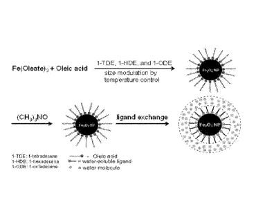
As shown in FIG. la, a size series of monodisperse iron oxide nanoparticles
were
synthesized upon the decomposition of iron precursors (such as iron oleate or
iron
pentacarbonyl) in a solvent mixture of 1-tetradecene, 1-hexadecene, and 1-
octadecene in
the presence of oleic acid followed by oxidation with trimethylamine N-oxide.
By
modulating the boiling point of solvent mixture through the change of its
component
ratios, the reaction mixture was kept at high temperatures between 270 C and
300 C for
a reaction time of 1-2 hours. The resulting hydrophobic nanoparticles were
first ligand
exchanged with 242-(2-methoxyethoxy)ethoxy]acetic acid (MEAA) to ensure their
water
solubility in a mixture of dimethylformamide (DMF) and water, in which they
were
further ligand exchanged with dopamine sulfonate (DS) or zwitterionic dopamine
sulfonate (ZDS). The dopamine sulfonate (DS) ligand also has a high solubility
in water
and a strong binding affinity to iron oxide surface, except that the DS is not
zwitterionic.
Transmission electron microscopy (TEM) images (FIG. 1B-1E) and high-
performance
liquid chromatography (HPLC, FIG. 3) with size-exclusion column revealed that
these
nanoparticles have inorganic cores as 7.0, 5.5, 3.0, 2.5 nm, respectively and
that the
smallest nanoparticles can have a 3.0 nm inorganic core and a 5.0 nm HD. In
FIG. 2, it is
shown that ZDS-coated nanoparticles can have an r2/r1 ratio as low as 11 at 7
Tesla (two
times lower than the r2/r1 ratio of commercially available FerahemeTM) and 1.5
at 0.5
Tesla (T), which can lead to a high-contrast T1 weighted MR imaging. According
to
approved Massachusetts Institute of Technology (MIT) institutional protocols,
the ZDS-
coated nanoparticles were injected into mice and rats, the urine of mice were
collected at
a series of time points (FIG. 3) and Ti weighted MR images of rats were taken
(FIG. 4).
A rapid renal clearance of ZDS-coated nanoparticles was observed and the size
of the
ZDS-coated nanoparticles injected was not affected in vivo (FIG. 3). FIG. 4
also
12
CA 02961358 2017-03-14
WO 2016/044068
PCMJS2015/049524
demonstrated that, ZDS-coated nanoparticles injected into rats showed Ti
contrast and
renal clearance, where the red circle indicates the accumulation of ZDS-coated
nanoparticles in urine in the bladder.
The MRI contrast enhancement of Magnevist (the most commonly used
gadolinium-based contrast agents, GBCAs) and ES-SPIONs in blood were compared
over
time. According to approved animal protocols, mice were scanned in a 1.5T
clinical MRI
machine. After intravenous injection of Magnevist or ES-SPIONs, the Ti-
weighted
images of a single slice in mice brain were taken using clinical MRI sequences
(-3.5 mins
time length), and then the half-life of contrast enhancements of both agents
were
compared side-by-side. FIG. 5 shows that ES-SPIONs exhibit a significantly
longer blood
half-life and MRI contrast enhancement compared to Magnevist. As shown in FIG.
5,
Magnevist has a half-life of ¨2 mins while ES-SPIONs provide constant contrast
enhancement for at least ¨3.5 mins. This result shows that the ES-SPIONs have
a
significantly longer blood half-life than that of Magnevist and that the ES-
SPIONs offer a
more stable MRI contrast enhancement within the length of clinical MRI scan
time.
Moreover, to demonstrate the capability of ES-SPIONs as blood pool contrast
agent for
MR angiography (MRA), mice were scanned in a 1.5T clinical MRI machine, where
a
TI-weighted 3D MRA sequence was used to image their brain. FIG. 6 shows that
ES-
SPIONs are a powerful contrast agent for TI -weighted MRA at the clinical
field strength
(1.5T). Left and right images show corresponding side views of the image in
the middle.
It can be seen in FIG. 6 that a three dimensional profile highlighting the
blood vessels in
brain was generated. The MRA of mice using ES-SPIONs are expected to be
enhanced
relative to MRA of mice using Magnevist , because ES-SPIONs provide a more
durable
contrast enhancement, especially for MRA that lasts longer than 2 mins.
It was also tested whether the ES-SPIONs are able to leak into brain tumors
and
enhance the MR contrast of brain tumors. A U87 glioma mice model was used, in
which
the blood-brain-barrier was compromised by their brain tumor. According to
approved
animal protocols, these mice were scanned in a 9.4T MRI machine for small
animals. A
Tl-weighted MRI sequence was used to image the mouse head before (pre) and
after
.. (post) the intravenous injection of ES-SPIONs and then the pre-images were
subtracted
from the post images to highlight the contrast enhancement. FIG.7 shows that
ES-SPIONs
leak into advanced brain tumors (U87 glioma model in mice) using Tl-weighted
MR
imaging. The images from top left to bottom right show spatially consecutive
transversal
slices of the head of a mouse. As shown in FIG. 7, ES-SPIONs successfully leak
into U87
13
tumor and then they enhance the Ti contrast of U87 tumor area in the mouse
brain. This
result suggests that the ES-SPIONs could serve as a non-toxic MRI agent
highlighting
glioma, which is a major indication where GBCAs are used in the clinic.
The present invention has been described with regard to one or more
embodiments.
However, it will be apparent to persons skilled in the art that a number of
variations and
modifications can be made without departing from the scope of the invention as
defined in
the claims.
14
Date Recue/Date Received 2022-01-21