Note: Descriptions are shown in the official language in which they were submitted.
METHODS FOR TREATING BRAIN METASTATIS USING GAP JUNCTION INHIBITORS
PRIORITY CLAIM
This application claims priority to United States Provisional Application No.
62/052,966 filed September 19, 2014.
GRANT INFORMATION
This invention was made with government support under Grant Nos, P01-
CA129243 and P30-CA008748 awarded by National Institutes of Health and Grant
No. W81XWH-12-0074 awarded by the Department of Defense (DoD). The
government has certain rights in the invention.
1. INTRODUCTION
This present invention relates to gap junction inhibitors for use in treating
brain metastasis. As such, these inhibitors may be used in methods of treating
cancer
patients.
2. BACKGROUND OF THE INVENTION
Brain metastases occur in 20-40% of advanced stage cancers and represent the
most prevalent intracranial malignancy in adults (Gavrilovic and Posner, 2005;
Maher
et al., 2009). Lung and breast cancers arc thc most common sources. Despite
treatment advances at other metastatic sites, current clinical management of
brain
metastases affords limited disease control and most patients succumb to tumor
progression less than twelve months after diagnosis (Gavrilovic and Posner,
2005;
Stelzer, 2013). The mechanisms underlying this disease process must therefore
be
understood so that they may be parlayed into rational therapeutic strategies.
The brain's unique microenvironment poses a formidable barrier to metastatic
cancer cells. Recent progress has begun to unravel the complex cellular and
molecular interactions responsible for the initiation of brain metastases.
Circulating
cancer cells that mechanically lodge in brain capillaries must first traverse
the
reinforced vessel walls that constitute the blood-brain barrier (BBB) (Eichler
et al.,
2011). Genes have been identified that mediate cancer cell cxtravasation
through the
BBB in experimental models and predict brain metastasis in the clinic (Bos et
alõ
1
Date Recue/Date Received 2022-03-30
CA 02961894 2017-03-20
WO 2016/044790
PCT/US2015/051057
2009; Li et al., 2013). Once inside the brain parenchyma, metastatic cells
remain
associated with the microvasculature (Kienast et al., 2010; Lorger and Felding-
Habermann, 2010). Expression of the cell adhesion molecule Ll CAM in the
cancer
cells mediates their tight adhesion to the ablurninal capillary basal lamina
as a
requirement for the initiation of metastatic outgrowth (Valiente et al.,
2014). Wnt is
one of the signaling pathways supporting the outgrowth (Nguyen et al., 2009).
However, the vast majority of cancer cells that infiltrate the brain perish
(Chambers et
al., 2002; Heyn et al., 2006; Kienast et al., 2010), arid they are rejected by
the most
abundant cell type in the brain, the astrocyte (Valiente et al., 2014).
Functionally pleiotropic, astrocytes maintain the BBB, orchestrate
neurovascular coupling, sustain homeostasis of a tissue under stringent
metabolic
demands (Oberheim et al., 2012) and react acutely against disturbances like
injury or
infiltrating cells (Pekny and Nilsson, 2005). Reactive astrocytes generate
plasmin,
which mobilizes the pro-apoptotic cytokine FasL to kill infiltrating cancer
cells
(Valiente et al., 2014). Plasmin additionally cleaves cell surface LiCAM in
the
cancer cells to suppress their ability to coopt the vasculature (Valiente et
al., 2014).
To evade astrocyte attack, brain metastatic cells from breast cancer and lung
cancer
express serpin inhibitors of plasminogen activator (PA) (Valiente et al.,
2014).
Although these observations indicate that astrocytes guard the brain against
metastatic
invasion, there is also evidence that the role of astrocytes in metastasis may
not be
uniformly axitagonistic. In vitro, astrocyte co-culture protects melanoma cell
lines
from chemotherapeutic drugs (Kim et al., 2011), and in vivo astrocytes can
activate
Notch signaling in cancer cells (Xing et al., 2013).
3. SUMMARY OF THE INVENTION
The present invention relates to methods for treating brain metastasis
by inhibiting gap junction functionality. It is based, at least in part, on
the discovery
that cancer cells expressing Protocadherin 7 and Connexin 43 form gap
junctions with
astrocytes that promote the growth of brain metastases, and that inhibition of
Protocadherin 7 and/or Connexin 43 expression in cancer cells reduces
progression of
brain metastases. It is further based on the discovery that treatment with gap
junction
inhibitors tonabersat and meclofenamate inhibited progression of brain
metastatic
lesions and enhanced the anti-cancer activity of the conventional
chemotherapeutic
agent, carboplatin.
2
CA 02961894 2017-03-20
WO 2016/044790 PCT/US2015/051057
Certain non-limiting embodiments provide for a method for treating a subject
having a cancer comprising administering, to the subject, an amount of a gap
junction
inhibitor that inhibits metastatic progression of thc cancer in the brain. In
particular
non-limiting examples, the gap junction inhibitor is a Connexin 43 inhibitor
or a
Protocadherin 7 inhibitor, or a combination thereof. In particular non-
limiting
examples, the inhibitor is tonabersat or meclofenamate or a combination
thereof. In
particular non-limiting examples, the cancer is breast cancer or lung cancer,
and/or the
cancer cells of the subject express Connexin 43 and/or Protocadherin 7. In
particular
non-limiting examples, the method further comprises administering, to the
subject, a
therapeutically effective amount of an anti-cancer agent such as, but not
limited to,
carboplatin. When the method of the invention is applied, the subject may be
known
to have one or more brain metastases, or alternatively, was not known to have
a brain
metastasis prior to treatment.
Certain non-limiting embodiments provide for a method for inhibiting growth
and/or survival of metastatic cancer cells in the brain of a subject,
comprising treating
the subject with a therapeutically effective amount of a gap junction
inhibitor.
In particular non-limiting examples, the gap junction inhibitor is a Connexin
43
inhibitor or a Protocadherin 7 inhibitor, or a combination thereof. In
particular non-
limiting examples, the inhibitor is tonabersat or meclofenamate or a
combination
thereof. In particular non-limiting examples, the cancer is breast cancer or
lung
cancer, and/or the cancer cells of the subject express Connexin 43 and/or
Protocadherin 7. In particular non-limiting examples, the method further
comprises
administering, to the subject, a therapeutically effective amount of an anti-
cancer
agent such as, but not limited to, earboplatin. When the method of the
invention is
applied, the subject may be known to have one or more brain metastases, or
alternatively, was not known to have a brain metastasis prior to treatment.
Certain non-limiting embodiments provide fur a method for treating brain
metastasis in a subject having a cancer, comprising administering, to the
subject, a
therapeutically effective amount of a gap junction inhibitor. In particular
non-limiting
examples, the gap junction inhibitor is a Connexin 43 inhibitor or a
Protocadherin 7
inhibitor, or a combination thereof. In particular non-limiting examples, the
inhibitor
is tonabersat or meclofenamate or a combination thereof. hi particular non-
limiting
examples, the cancer is breast cancer or lung cancer, and/or the cancer cells
of the
subject express Connexin 43 and/or Protocadherin 7. In particular non-limiting
3
CA 02961894 2017-03-20
WO 2016/044790 PCT/US2015/051057
examples, the method further comprises administering, to the subject, a
therapeutically effective amount of an anti-cancer agent such as, but not
limited to,
carboplatin. When the method of the invention is applied, the subject may be
known
to have one or more brain metastases, or alternatively, was not known to have
a brain
metastasis prior to treatment.
Certain non-limiting embodiments provide for, in a subject having a cancer, a
method of preventing metastasis of the cancer to the brain, comprising
administering,
to the subject, a therapeutically effective amount of a gap junction
inhibitor. In
particular non-limiting examples, the gap junction inhibitor is a Connexin 43
inhibitor
or a Protocadherin 7 inhibitor, or a combination thereof. In particular non-
limiting
examples, the inhibitor is tonabersat or meclofenamate or a combination
thereof. In
particular non-limiting examples, the cancer is breast cancer or lung cancer,
and/or the
cancer cells of the subject express Connexin 43 and/or Protocadherin 7. In
particular
non-limiting examples, the method further comprises administering, to the
subject, a
therapeutically effective amount of an anti-cancer agent such as, but not
limited to,
carboplatin. When the method of the invention is applied, the subject may be
known
to have one or more brain metastases, or alternatively, was not known to have
a brain
metastasis prior to treatment.
Certain non-limiting embodiments provide for in a subject having a cancer, a
method of reducing the risk of detectable metastasis of the cancer to the
brain,
comprising administering, to the subject, a therapeutically effective amount
of a gap
junction inhibitor. In particular non-limiting examples, the gap junction
inhibitor is a
Connexin 43 inhibitor or a Protocadherin 7 inhibitor, or a combination
thereof. In
particular non-limiting examples, the inhibitor is tonabersat or meclofenamate
or a
.. combination thereof. In particular non-limiting examples, the cancer is
breast cancer
or lung cancer, and/or the cancer cells of the subject express Connexin 43
and/or
Protocadherin 7. In particular non-limiting examples, the method further
comprises
administering, to the subject, a therapeutically effective amount of an anti-
cancer
agent that can attain therapeutic levels in the brain, such as, but not
limited to,
carboplatin. When the method of the invention is applied, the subject may be
known
to have one or more brain metastases, or alternatively, was not known to have
a brain
metastasis prior to treatment.
Certain non-limiting embodiments provide for, in a subject having a cancer, a
method of reducing the risk of detectable metastasis of the cancer to the
brain,
4
CA 02961894 2017-03-20
WO 2016/044790
PCT/US2015/051057
comprising administering, to the subject, a therapeutically effective amount
of a
Protocadherin 7 inhibitor. In particular non-limiting examples, the
Protocadherin 7
inhibitor is an interfering RNA. In particular non-limiting examples, the
cancer is
breast cancer or lung cancer, and/or the cancer cells of the subject express
Connexin
43 and/or Protocadherin 7. In particular non-limiting examples, the method
further
comprises administering, to the subject, a therapeutically effective amount of
an anti-
cancer agent such as, but not limited to, carboplatin. When the method of the
invention is applied, the subject may be known to have one or more brain
metastases,
or alternatively, was not known to have a brain metastasis prior to treatment.
Certain non-limiting embodiments provide for a method for lengthening the
period of survival of a subject having a cancer, comprising administering to
the
subject an effective amount of a gap junction inhibitor, for example, wherein
administering the gap junction inhibitor inhibits metastatic progression of
the cancer
in the brain. In particular non-limiting examples, the gap junction inhibitor
is a
Connexin 43 inhibitor or a Protocadherin 7 inhibitor, or a combination
thereof. In
particular non-limiting examples, the inhibitor is tonabersat or
meclofenarnate or a
combination thereof In particular non-limiting examples, the cancer is breast
cancer
or lung cancer, and/or the cancer cells of the subject express Connexin 43
and/or
Protocadherin 7. In particular non-limiting examples, the method further
comprises
administering, to the subject, a therapeutically effective amount of an anti-
cancer
agent such as, but not limited to, carboplatin. When the method of the
invention is
applied, the subject may be known to have one or more brain metastases, or
alternatively, was not known to have a brain metastasis prior to treatment.
Certain non-limiting embodiments provide for an assay for evaluating gap
junction activity, for example assessing inhibition, by measuring levels of
cGAMP,
where a decrease in cGAMP correlates with gap junction inhibition. Particular
non-
limiting embodiments provide for a method for inhibiting growth and/or
survival of
metastatic cancer cells in the brain of a subject, comprising treating the
subject with a
therapeutically effective amount of a gap junction inhibitor that produces a
decrease
in cGAMP relative to the level of cGAMP in the absence of that amount of gap
junction inhibitor. Further non-limiting embodiments provide for a method of
determining whether a brain tumor or metastatic brain tumor in a subject will
receive
therapeutic benefit from treatment with a gap junction inhibitor, comprising
determining whether, in a sample from said tumor, exposure to a gap junction
5
CA 02961894 2017-03-20
WO 2016/044790 PCT/US2015/051057
inhibitor leads to a decrease in cGAMP, where a decrease in cGAMP is
indicative of
therapeutic benefit.
4. BRIEF DESCRIPTION OF FIGURES
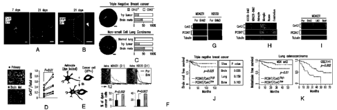
Figure 1A-K. Cx43 and PCDH7 association with brain metastasis. (A)
GFP+ H2030-BrM3 cells (green) are surrounded by GFAP+ activated astrocytes
(red)
in the brain parenchyma at early (day 7) and later (day 21) time points
following
intracardiac inoculation in mice. Blue, collagen IV (ColIV) staining in
vessels. Scale
bar, 10 pm. (B) Cx43 staining (arrowhead) at the interface of GFP+ H2030-BrM3
(green) and GFAP+ astrocytes (blue). Scale bar, 10 p.m. (C) Representative
images of
Cx43 staining in human brain metastasis samples from triple-negative breast
cancer
and non-small cell lung carcinoma. The proportion of CX43-positive samples was
quantified in primary (lry) tumours, brain metastases, and normal lung
tissues. Scale
bar, 100 pm. (D) Representative images and quantification of Cx43
immunostaining
in matched primary and brain metastatic samples from non-small lung carcinoma
patients. Scale bar, 100 pm. (E) Schematic illustration of dye transfer assay.
(F)
Quantification of dye transfer from astrocytes to cancer cells. Histograms
show red
fluorescent signal in parental (Par) and BrM cells. All values are mean
S.E.M. (n=3
biological replicates). n=3 independent experiments. (G-I) Cx43 and PCDH7
western
immunoblotting in the indicated parental and brain metastatic derivatives ((G)
n=3
independent experiments), in brain metastatic cells compared to brain cell
types ((H)
n=-2 independent experiments), and in MDA231 derivatives metastatic to brain,
lung
(LM) or bone (BoM) ((I) n=2 independent experiments). (J-K) Kaplan-Meier plot
of
cumulative brain metastasis-free survival in 189 cases of triple-negative
breast cancer
(3) and 129 cases (MSKCC set2) and 58 cases (GSE3141) of lung adenocarcinoma
(K), based on Cx43/PCDH7 expression in the primary tumour.
Figure 2A-G. Cx43/PCDH7 carcinoma-astrocyte gap junctions mediate
brain metastasis. (A) Histograms (top) and quantification (bottom) of dye
transfer
from astrocytes to control and Cx43-depleted or PCDH7-depleted brain
metastatic
cells. Values are mean S.E.M. (n=3 biological replicates). n=3 independent
experiments. (B) Luciferase complementation assay to detect Cx43-PCDH7
interactions. NLuc and CLuc, N-terminal and C-terminal firefly luciferase
halves. The
table (top) numerically identifies the cell line combinations used in the
assays
(bottom), and bioluminescence imaging (BLI) of a representative plate. BLI (C)
and
6
CA 02961894 2017-03-20
WO 2016/044790 PCT/US2015/051057
quantification (D) of brain metastatic lesions formed by control, Cx43-
depleted, or
PCDH7-depleted brain metastatic cells. n=3 independent experiments. (E,F) Wild
type (WT) or T154A mutant (Mut) Cx43 was re-expressed in Cx43-depleted
MDA231-BrM2 cells (Cx43 sh2). The cells were subjected to astrocyte dye
transfer
analysis ((E) n=3 independent experiments), or to brain metastasis assays and
BLI
quantification ((F) n=2 independent experiments). (G) Schematic summary of
Cx43-
and PCDH7-mediated interactions between cancer cells and astrocytes in brain
metastasis.
Figure 3A-1. Gap junctions activate STATI and NF-KB pathways in
cancer cells. (A) Signaling pathway analysis of TRAP-Seq data from MDA231-
BrM2 cells after co-culture with astrocytes. Control (Ctrl) or Cx43-depleted
MDA231-BrM2 cells expressing an L 1 Oa-GFP ribosomal protein fusion were co-
cultured with astrocytes for 24 h prior to polysome immunoprecipitation and
mRNA
sequencing. Heatmap depicts blue (down-regulated) and red (up-regulated)
pathways.
n=2 biological replicates. (B,C) STAT1 and NF-KB p65 phosphorylation in
M0A231-BrM2 cells after a 2 h incubation with conditioned media (CM) from
astrocyte co-culture. CM were collected after 24 h co-culture of astrocytes
with
control or Cx43-depleted MDA231-BrM2 cells (B), or from Cx43-depleted MDA231-
BrM2 cells that were transduced with wild type Cx43 (WT) or Cx43(T154A) mutant
(Mut) (C). n>3 independent experiments. (D) ELISA of 1FNa and TNFot in CM from
astrocyte co-cultures with the indicated M1JA231-BrM2 cells. All values arc
mean
S.E.M. (n=4 technical replicates). n>2 independent experiments. (E) Relative
mRNA
levels of IFNA and TNFA in astrocytes re-isolated after co-culture with MDA231-
BrM2 cancer cells. All values are mean S.E.M. (n=3 biological replicates).
n=2
independent experiments. (F) Relative levels of cleaved easpase 3 in MDA231-
BrM2
cells treated with various concentrations of carboplatin (Carbo) in the
presence or
absence of 10 units/ml (39 units/ng) IFNaA or 10 pg/ml TNFo. All values are
mean
S.E.M. (n=5 technical replicates), n=3 independent experiments. (G) STAT1
levels in
control and STAT1-knockdown MDA231-BrM2 cells. (H) NF-KB renilla luciferase
reporter assay in MDA231-BrM cells expressing control pBABE or SR-IxBa vector.
All values are mean S.E.M. (n=3 technical replicates). (1) Quantification of
BLI
signal from brain metastases formed by control, STAT1-knockdown, and SR-IKBa
MDA231-BrM2 cells. n=2 independent experiments.
7
CA 02961894 2017-03-20
WO 2016/044790 PCT/US2015/051057
Figure 4A-H. Gap junctions mediate a cytosolie dsDNA response in
astrocytes. (A) MDA231-BrM2 cells expressing control shRNA (Ctrl sh) or shRNA
targeting Cx43, were cultured for 18 h with or without astrocytes, and
subjected to
immunobloting analysis of phosphorylated TBK1 and IRF3 (n=3 independent
experiments). (B) MDA231-BrM2 alone, astrocytes alone, or 18 h co-cultures,
were
harvested for sample preparation and cGAMP analysis by LC-MS/MS. Histogram
(right) corresponds to normalized cGAMP peaks in (left), and is representative
of 5
biological replicates. n-3 independent experiments. See also Figure 16. (C)
Representative images of dual immunofluoreseent staining of IRF3 and GFP. DAN,
nuclear staining. In co-cultures: white arrows, nuclear accumulation of IRF3
in
astrocytes; green arrows, even distribution of IRF3 in GFP+ MDA231-BrM2 cells.
Scale bar, 20 1AM. n=2 independent experiments. (D) Quantification of dsDNA in
the
indicated cellular fractions from 2x10⁷ cells. Values are mean - S.E.M. (n=3
biological replicates). n=2 independent experiments. (E) Representative image
of
immunotluorescence staining of dsDNA, GFP, and Cox IV (mitochondrial marker)
in
MDA231-BrM2 cells. DAPI, nuclear staining. Scale bar, 10 um. n=2 independent
experiments. (F,G) EdU labeled MDA231-BrM2 cells were co-cultured with
astrocytes for 6 h. Transfer of EdU-labeled DNA from cancer cells to
astrocytes was
visualized using confocal microscopy (F), or quantified by flow cytometry (G).
Cancer cells and astrocytes are delineated by green and white dotted lines,
respectively. Scale bar, 10 gm. Values are mean S.E.M. (n=3 biological
replicates,
n=2 independent experiments). (H) Schematic summary of gap junction mediated
anti-dsDNA response, production of IFNa and TNFa in astrocytes, and consequent
activation of STAT1 and NF-KB pathways in cancer cells to support brain
metastasis.
Figure 5A-L Inhibition of gap junction activity controls brain metastatic
outgrowth. (A) Dye transfer from astrocytes to MDA231-BrM2 cells in the
presence of the indicated concentrations of Tonabersat or meclofenamate. a> 3
independent experiments. (B) ELISA of IFNa and TNFa in conditioned media from
co-cultured MDA231-BrM2 cell and astrocytes in the presence of Tonabersat
(Tona)
or meclofenamate (Meclo) with indicated concentrations. All graphs shown are
mean
S.E.M. (n=4 technical replicates). n=2 independent experiments. (C) Tonabersat
or
rneclofenamate was administered daily starting one day after cancer cell
inoculation
in mice. Brain metastatic lesions were quantified based on BLI. n2 independent
experiments. (0) GFP staining of 14-day brain metastatic lesions.
Representative
8
CA 02961894 2017-03-20
WO 2016/044790 PCT/US2015/051057
images show large, progressive lesions. DAPI, nuclear staining. Scale Bar,
40gm.
n=10 experimental mice. (E) 14 days after inoculation with MDA231-BrM2 cells
transduccd with inducible control, CX43 or PCDH7 shRNAs, mice were treated
with
doxycycline and carboplatin, as illustrated in the scheme. Brain metastatic
lesions
.. were quantified based on BLI. (F,G) Representative images of matched ex
vivo brain
BLI and red fluorescence imaging. n=2 independent experiments. (H) 14 days
after
inoculation with MDA231-BrM2 cells, mice were treated with Tonabersat,
meclofenamate, and carboplatin. Following the indicated regimens, brain
metastatic
lesions were quantified based on BLI. n=2 independent experiments (1).
Figure 6A-D. Cancer cell-astrocyte interactions. (A) Cancer cells used in
this study. (B) Astrocyte co-culture protects cancer cells. As illustrated in
schema
(left), cleaved caspase 3+/GFP+ apoptotic BrM cells were quantified after
sFasL- or
chemo-treatments. n=3 independent experiments. (C,D) Gap junction
communications between astrocytes and BrM cells. Time-lapse images of dye
transfer
from MDA231-BrM2 cells to astrocytes (C). Scale bars, 100 JAM. Quantification
of
dye transfer from astrocytes to MDA231-BrM2 cells by flow cytornetry over time
(D). n=3 independent experiments.
Figure 7. Elevated expression of Cx43 and PCDH7 in brain metastatic
cancer cells and astrocytes. (A) 6x43 and PCDH7 mRNA in parental (Par) and BrM
cells. Values are mean S.E.M. (n-3 technical replicates). n=3 independent
experiments, (B) Cx43 and PCDH7 western blotting in ErbB2 parental and brain
cells, as well as Kras/p53 cell lines. n=3 independent experiments. (C) Cx43
and
PCDH7 mRNA in BrM cells compared to brain cells. n=3 independent experiments,
(D) (..1x26 and Cx30 mRNA in MDA231 parental (Par) and the metastatic
derivatives
.. of brain (BrM2), lung (LM) and bone (BoM). (E) Kaplan-Meier plot
illustrates the
probability of cumulative metastasis free survival in 63 cases (GSE8893) of
lung
adenocarcinorna based on Cx43/PCDH7 expression in the primary tumour. (F,G)
Knockdown of Cx43 and PCDH7 with short hairpin RNAs (shRNA) as assessed by
RT-PCR (F) and western blotting (G). Ctrl, control. Values are mean S.E.M.
(n=3
technical replicates). n=3 independent experiments.
Figure 8A-H. PCDH7 facilitates gap junction communication. (A,B)
Histograms and quantification of dye transfer from astrocytes to control and
Cx43-
depleted or PCDH7-depleted Kras/p53-393N1 cells (A), and from astrocytes to
control or Cx43-depleted MDA231-BrM2 cells, in comparison to Carbenoxolone (50
9
CA 02961894 2017-03-20
WO 2016/044790 PCT/US2015/051057
uM) treatment (B). (C,D) PCDH7 in astrocytes facilitate gap junctions. PCDH7
western blotting in control or PCDH7-depleted astrocytes (C). Quantification
of dye
transfer from MDA231-BrM2 cells to PCDH7-depleted astrocytes (D). (E)
Quantification of dye transfer from human brain microvascular endothelial
cells
(HBMEC) to control, Cx43- or PCDH7-depleted MDA231-BrM2 cells. (F) Dye
transfer from MDA231-BrM2 cells to a mixed population of astrocytes and HBMEC.
(G) Quantification of dye transfer from control or Cx43-depleted MDA231-BrM2
cells to human microglia. (H) As illustrated in schema, 'x43 mRNA in MDA231-
BrM2 cells (left) or astrocytes (right) was detected after 24 h co-culture,
separated by
transwell, with microglia, astrocytes or cancer cells. For dye transfer
assays, values
are mean S.E.M. (n---3 biological replicates). n > 2 independent
experiments. In h,
values are mean S.E.M. (n=4 biological replicates).
Figure 9A-D. Cx43 directly interacts with PCDH7, but not with E
cadherin or N cadherin. (A) Cx43 and PCDH7 western immunoblotting in cancer
cells overexpressing fusion proteins. (B) Quantification of BLI after co-
culture of
Cx43-CLue/PCDH7-NLue(+) cancer cells and astrocytes for 15 min. c-e,
Luciferase
split assay to detect Cx43-E cadherin or Cx43-N cadherin interactions. NLuc
and
CLuc: N-terminal and C-terminal firefly luciferase halves. The table (C)
numerically
identified the cell line combinations used in the assays, western
immunoblotting (D)
indicated E or N cadherin expression in cancer cells overexpressing fusion
proteins,
and bioluminescence imaging (BLI) of a representative plate (e). n? 2
independent
experiments.
Figure 10A-E. Inhibition of gap junction activity prevents brain
metastatic outgrowth. (A) Bioluminescent imaging (BLI) quantification of brain
metastatic lesions formed by control (Ctrl), Cx43- or PCDH7-depleted Kras/p53-
393N1 cells. n-2 independent experiments. (B) Representative images of GFP-F
brain
metastatic lesions formed by control, Cx43- or PCDH7-depleted MDA231-BrM2
cells. Brain sections or brain metastatic lesions are delineated by dotted
white line or
dotted red line, respectively. Scale bar, 1000 m. (C) BLI (images) and
quantification
(bar graph) of lung metastatic lesions thrilled by MDA231-BrM2 cells. Values
are
mean S.E.M. (n=5 mice in each group). n=2 independent experiments. (D,E) Gap
junction-mediated brain metastasis requires channel function of Cx43. Wild
type
(WT) or T154A mutant (Mut) Cx43 was re-expressed in Cx43 depleted MDA231-
BrM2 cells (CX43 s1i2). Cx43 expression was detected by western blotting (D)
and
CA 02961894 2017-03-20
WO 2016/044790 PCT/US2015/051057
brain metastatsis formed by these cells was quantified by BLI (E). n=2
independent
experiments.
Figure 11A-D. Role of Cx43 and PCDH7 in brain metastasis. (A) Cx43
and PCDH7 do not mediate trans-BBB Migration. Quantification of control
(Ctrl),
Cx43- or PCDH7-depleted MDA231-BrM2 cells in 7-day brain lesions. Values are
mean S.E.M. (n=5 brains in each group). (B) Cx43 and PCDH7 mediate cancer
cell
colonization in 14-day brain lesions. Representative images are GFP (green)
and Ki67
(red) staining. DAPI, nuclear staining. Scale bar, 20 um. Bar graph is the
proportion
of Ki67+ cancer cells. Values are mean S.E.M. (n=5 brains in each group).
(C)
Cx43 and PCDI17 mediate cancer cell survival. Brain slice assays.
Representative
images are GFP (green) and cleaved caspase 3 (Casp3)(red) staining. Scale bar,
30
um. Histogram is the proportion of caspase 3+ apoptotic cancer cells. Values
are
mean S.E.M. (n=5 brain slices in each group). Scale bars, 30 um. (D) Cx43
and
PCDH7 do not affect vascular cooption of cancer cells in 14-day brain lesions.
Representative images are GFP (green) staining and vascular structure filled
with
TR1TC dextran (red). Scale bar, 20 um. n=2 independent experiments.
Figure 12A-D. Translating ribosome affinity purification (TRAP) and
cytokine array. (A) Schematic illustration of TRAP experimental set up to
isolate
translating niRNA from MDA231-BrM2 cells under 3 conditions (#1, #2, #3). (B)
Principle component (PC) analysis of TRAP mRNA sequencing. (C) Scatter plot of
1og2 fold-changes regulated by astrocytes and gap junction communications
between
BrM cells and astrocytes. (D) STAT1 and NF-x13 p65 phosphorylation in H2030-
BrM3 cells after a 2 h incubation with conditioned media (CM) from astrocyte
co-
cultures. CM were collected after 24 h co-culture of astrocytes with control
or Cx43-
depleted H2030-BrM3 cells. n=3 independent experiments.
Figure 13A-F. Gap junction-generated signaling activates IFN and NF-Kb
pathways in cancer cells. (A) Cytokine array analysis of the conditioned media
collected after 24 h co-culture of astrocytes with control or Cx43-depleted
MDA231-
BrM2 cells. Log2 fold-changes were plotted. (B) ELISA of IFNa and INFa in CM
from astrocyte co-cultures with the indicated H2030-BrM3 cells. All values
shown are
mean S.E.M. (n=4 technical replicates). n=2 independent experiments. (C)
Relative
levels of cleaved caspase 3 in H2030-BrM3 cells treated with various
concentrations
of Taxol in the presence or absence of 10 units/m1 (39 unitsing) recombinant
IFNaA
or 10 pg/m1 recombinant TNFa. All values are mean S.E.M. (n=5 technical
11
CA 02961894 2017-03-20
WO 2016/044790 PCT/US2015/051057
replicates). n=3 independent experiments. (D, E) STAT1 levels in control and
STAT1-knockdown H2030-BrM3 cells. (F) Quantification of BL1 signal from brain
metastases formed by control, STAT1-knockdown cells. n=2 independent
experiments.
Figure 14A-G. Gap junctions initiate cytosolic DNA response in
astrocytes. (A) Control or Cx43-deplated H2030-BrM3 cells were co-cultured for
18
h with or without astrocytes, and subjected to immunobloting analysis of
phosphorylated TBK1 and 1RF3 (n=3 independent experiments). (B) cGAMP
identification. The peak at 4.47 min contains all 3 SRM transitions specific
for
cGAMP. RT: retention time, AA: automatically integrated peak area. (C)
Quantification of dsDNA in the indicated cellular fractions from 2x10⁷ H2030-
BrM3
cells. Values are mean S.E.M. (n=3 biological replicates). n=2 independent
experiments. (D) Ratio of eytosol dsDNA and nuclear dsDNA in indicated cancer
cells and non-neoplastic cells. (E) Representative image of immunofluorescent
staining of dsDNA, GFP, Cox IV (mitochondria marker) in H2030-BrM3 cells.
DAPI,
nuclear staining. Scale bar, 10 lam. (F) Representative image of
immunofluorescent
staining of dsDNA, Cox IV (mitochondria marker) in astrocytes. DAPI, nuclear
staining. Phalloidin, cytoskeletal staining. Scale bar, 10 p.m. (G) EdU
labeled H2030-
BrM3 cells were co-cultured with astrocytes for 6 h. Transfer of EdU-labeled
DNA
from cancer cells to astrocytes was visualized using con-focal microscopes.
Cancer
cells or astrocytes were delineated by green or white dotted lines,
respectively. Scale
bar, 10 um. n-2 independent experiments.
Figure 15. Inhibition of Gap Junction Activity Prevents Brain Metastatic
Outgrowth. (A-D) Following treatment with Tonabersat (Iona) or meclofenamate
(Meclo) (A), brain metastasis (B), primary tumour growth in mammary fat pads
(C),
or lung metastasis (D) were quantified by BLI. Values are mean S.E.M. (n-5
mice
in each group). n=2 independent experiments. (E,F) Knockdown of Cx43 and
PCDH7 in MDA231-BrM2 cells with tet-on inducible short hairpin RNAs (shRNA),
as assessed by RT-PCR (E) and Western immunoblotting (F), after doxycyclinc
treatment in vitro. n=2 independent experiments. (G) Brain ex vivo
Bioluminescent
imaging (BLI) 14 days after inoculation of MDA231-BrM2 cells.
Figure 16. Confirmation of cGAMP identification. A pooled sample from
all experimental conditions shown in Fig. 4b analyzed by LC-MS/MS. Only the
peak
at 4.47 min contains all 3 SRM transitions specific for cGAMP. The peak at
4.47 min
12
CA 02961894 2017-03-20
WO 2016/044790 PCT/US2015/051057
is increased by the addition of 5 lit of 40 nM cyclic [G(2',5')pA(30,5')p]
(cGAMP)
to the pooled sample. As internal and negative control, c-di-GMP contains all
2 SRM
transitions at 4.97 min peak and the peak does not change by adding standard
eGAMP. dRT: retention time, AA: automatically integrated peak area.
5. DETAILED DESCRIPTION
For clarity and not by way of limitation the detailed description of the
invention is divided into the following subsections:
(i) Gap junction inhibitors;
(a) Connexin 43 inhibitors; and
(b) Protocadherin 7 inhibitors;
(c) Assay for gap junction activity/inhibition;
(ii) cancer targets;
(iii) pharmaceutical formulations; and
(iv) methods of treatment.
5.1 GAP JUNCTION INHIBITORS
The present invention provides inhibitors of gap junctions (e.g., gap junction
antagonists) for use in the disclosed methods. In certain embodiments, gap
junction
inhibitors can include compounds, small molecules, chemicals, polypeptides,
nucleic
acids and proteins that inhibit and/or reduce the expression and/or activity
of gap
junction components or inhibit and/or reduce the formation, patency, signaling
and/or
activity of gap junctions.
In certain non-limiting embodiments, gap junction inhibitors that are small
molecules include carbenoxolone, glycyrrhetinic acid, quinine, quinidine,
mefloquine,
heptanol, octanol, anandamide, fenamates, 2-aminoethoxy-diphenyl-borate (2-
APB),
retinoic acid, oleamide, sperinine, aminosulfonates, sodium propionate,
tonabersat
and meclofenamate (meclofenamic acid). Additional non-limiting examples of gap
junction inhibitors arc disclosed in U.S. Patent Nos. 5,843,989; 6,211,211;
7,632,866,
6,251,931; 7,704,946; and PCT Patent Application No. WO 1999/026584.
In certain embodiments, the gap junction inhibitor comprises a compound of
Formula I having the following structure:
13
CA 02961894 2017-03-20
WO 2016/044790
PCT/US2015/051057
0 WI 0
0
In certain embodiments, the gap junction inhibitor comprises a compound of
Formula II having the following structure:
0 OH
CI H
CI
In certain embodiments, the gap junction inhibitor comprises a compound of
Formula III having the following structure:
CI FE coo- Na
CI
In certain non-limiting embodiments, the gap junction inhibitor can be a salt,
a
stereoisomer, an analog or a derivative form of the compounds of Formulas I-
III. For
example, and not by way of limitation, the gap junction inhibitor can include
a sodium
salt form of Formula II.
In certain non-limiting embodiments, the gap junction inhibitor can be an
antibody or antibody fragment that can partially or completely block gap
junction
forination and/or gap junction patency between cells, gap junction signaling
and/or
activity. See,.19r example, Ernesto Oviedo-Orta et al., The FASEB Journal,
Vol. 15:
768-774 (2001). In certain non-limiting embodiments, the gap junction
inhibitor can
be an anti-Connexin compound and/or a Connexin mimetic peptide. See, Pr
example,
Evans and Boitano, Biochem. Soc. Trans., Vol. 29(4):606-612 (2001); Dahl,
Biophys.
J ., Vol. 67(5):1816-1822 (1994); European Patent Application Nos. EP2510939
and
EP2252320; and U.S. Patent Application No. 2009/0142295.
Further non-limiting examples of gap junction inhibitors include ribozymes,
antisense oligonucleotides, short hairpin RNA (shRNA) molecules and siRNA
molecules that specifically inhibit and/or reduce the expression or activity
of gap
junction components. A "ribozyme" refers to a nucleic acid capable of cleaving
a
14
specific nucleic acid sequence. In certain non-limiting embodiments, a
ribozyme
refers to RNA molecules that contain anti-sense sequences for specific
recognition,
and an RNA-cleaving enzymatic activity, see, for example, U.S. Pat. No.
6,770,633.
In contrast, "antiscnsc oligonueleolides" generally are small oligonucleotidcs
complementary to a part of a gene to impact expression of that gene. Gene
expression
can be inhibited through hybridization of an oligonucleotide to a specific
gene or
messenger RNA (mRNA) thereof. Methods for using antisense techniques for
specifically inhibiting gene expression of genes whose sequence is known are
well
known in the art (e.g., see U.S. Patent Nos. 6,566,135; 6,566,131; 6,365,354;
6,410,323; 6,107,091; 6,046,321; and 5,981,732). "Small interfering RNA" or
"short
interfering RNA" or "siRNA" or "short hairpin RNA" or "shRNA" are forms of RNA
interference (RNAi). An interfering RNA can be a double-stranded RNA or
partially
double-siranded RNA molecule that is complementary to a target nucleic acid
sequence. Micro RNAs (miRNA) can also fall in this category. Various
modifications to the oligonucleotides of the present invention, e.g.,
antisense, shRNA
or siRNA molecules, can be introduced as a means of increasing intracellular
stability
and half-life. Non-limiting examples of such modifications include the
addition of
flanking sequences of ribonucleotides or deoxyribonucleotides to the 5' and/or
3'
ends of the molecule, or the use of atypical or non-naturally occurring
residues such
as phosphorothioate or 2'-0-inethyl rather than phosphodiesterase linkages
within the
oligonucleotide backbone.
The RNA molecules of the invention can be expressed from a vector or
produced chemically or synthetically. Methods for selecting an appropriate
dsRNA or
dsRNA-encoding vector are well known in the art for genes whose sequence is
known
(e.g., see Tuschl, T. et al., "Targeted mRNA degradation by double-stranded
RNA in vitro,"
Genes & Development 13:3197 (1999); Elbashir et al., "RNA interference is
mediated by 21-
and 22-nucleotide RNAs," Genes & Development 15:188-200 (2001); Hannon, G.J.,
"RNA
interference, "Nature 418:244-251 (2002); McManus, M.T., et at., "Small
Interfering RNA-
Mediated Gene Silencing in T-Lymphocytes, " J. Immunol 169:5754-5760 (2002);
Brummelkamp, T.R., et al., "Stable suppression of tumorigenicity by virus-
mediated RNA
interference," Cancer Cell 2:243-247 (2002); U.S. Pat. Nos. 6,573,099 and
6,506,559; and
PCT Patent Application Nos. WO 2001/036646, WO 1999/032619 and WO 2001/68836).
Date Recue/Date Received 2022-03-30
5.1.1. CONNEXIN 43 INHIBITORS
In certain non-limiting embodiments, the gap junction can be specific for a
gap junction component. For example, and not by way of limitation, gap
junction
components include the Connexin family of proteins. A non-limiting example of
a
Connexin protein is Connexin 43 (Cx43), which is encoded by the gene
15a
Date Recue/Date Received 2022-03-30
CA 02961894 2017-03-20
WO 2016/044790 PCT/US2015/051057
gap junction protein, al (vat). A Cx43 nucleic acid or protein may be a human
Cx43 nucleic acid having the sequence as set forth in NCBI database accession
no.
NM 000165, N(1_008308 or M65188, or a nucleic acid encoding a human Cx43
protein molecule that has the amino acid set forth in NCBI database accession
no.
NP 000156. According to the present invention, inhibitors of the expression
and/or
function of such Cx43 nucleic acids and/or proteins may be used as gap
junction
inhibitors. For example, and not by way of limitation, a gap junction
inhibitor can
include a Cx43 inhibitor such as, but not limited to, ioxynil or ioxynil
octanoate. In
certain embodiments, a Cx43 inhibitor can include a Cx43 antibody, antibody
fragment or a mimetic peptide (see Danesh-Meyer et al., Brain, 135:506-520
(2012)).
One non-limiting example of a gap junction inhibitor comprises an antisense,
shRNA or siRNA nucleic acid sequence homologous to at least a portion of a
Cx43
nucleic acid sequence, disclosed above, wherein the homology of the portion
relative
to the Cx43 sequence is at least about 75 or at least about 80 or at least
about 85 or at
.. least about 90 or at least about 95 or at least about 98 percent, where
percent
homology can be determined by, for example, BLAST or FASTA software. In
certain
non-limiting embodiments, the complementary portion may constitute at least 10
nucleotides or at least 15 nucleotides or at least 20 nucleotides or at least
25
nucleotides or at least 30 nucleotides and the antisense nucleic acid, shRNA
or siRNA
molecules may be up 1o15 or up to 20 or up to 25 or up to 30 or up to 35 or up
to 40
or up to 45 or up to 50 or up to 75 or up to 100 nucleotides in length. Non-
limiting
examples of a shRNA that inhibit Cx43 are set forth in the Example below. In
non-
limiting embodiments, a Cx43 inhibitor, which is a nucleic acid, may be
provided in a
Cx43-expressing cancer cell via a vector, for example a lentivirus, which may
be
selectively targeted to said cancer cell and/or wherein expression of the Cx43
inhibitor nucleic acid may be directed by a promoter which is selectively
active in
tumor cells.
5.1.2 PROTOCADHERIN 7 INHIBITORS
The present invention provides Protocadherin 7 (PCDH7) inhibitors for use in
the disclosed methods. Non-limiting examples of PCDH7 inhibitors include
compounds, molecules, chemicals, polypeptides, proteins that inhibit and/or
reduce
the expression and/or activity of PCDH7. A PCDH7 nucleic acid or protein may
be a
human PCDH7 nucleic acid having the sequence as set forth in NCBI database
16
CA 02961894 2017-03-20
WO 2016/044790
PCT/US2015/051057
accession no. NM 001173523, NM 032457, NM_032456 or NM_002589, or a
nucleic acid encoding a human PCDH7 protein molecule that has the amino acid
set
forth in NCBI database accession no. NP 001166994, NP 115832, NP 115833 or
NP 002580.
In certain non-limiting embodiments, PCDH7 inhibitors can include
ribozymes, antisense oligonucleotides, shRNA molecules and siRNA molecules
that
specifically inhibit and/or reduce the expression or activity of PCDH7. One
non-
limiting example of a PCDH7 inhibitor comprises an antisense, shRNA or siRNA
nucleic acid sequence homologous to at least a portion of a PCDH7 nucleic
acid sequence, wherein the homology of the portion relative to the PCDH7
sequence
is at least about 75 or at least about 80 or at least about 85 or at least
about 90 or at
least about 95 or at least about 98 percent, where percent homology can be
determined by, for example, BLAST or FASTA software. In certain non-limiting
embodiments, the complementary portion may constitute at least 10 nucleotides
or at
least 15 nucleotides or at least 20 nucleotides or at least 25 nucleotides or
at least 30
nucleotides and the antisense nucleic acid, shRNA or siRNA molecules may be up
to
15 or up to 20 or up to 25 or up to 30 or up to 35 or up to 40 or up to 45 or
up to 50 or
up to 75 or up to 100 nucleotides in length. In certain embodiments,
antisense,
shRNA or siRNA molecules of the present invention may comprise DNA or atypical
or non-naturally occurring residues as disclosed above, for example, but not
limited
to, phosphorothioate residues. Non-limiting examples of a shRNA that inhibits
PCDH7 are set forth in the Example below. In non-limiting embodiments, a PCDH7
inhibitor, which is a nucleic acid, may be provided in a PCDH7-expressing
cancer cell
via a vector, for example a lentivirus, which may be selectively targeted to
said cancer
cell and/or wherein expression of the PCDH7 inhibitor nucleic acid may be
directed
by a promoter which is selectively active in tumor cells.
In non-limiting embodiments, a PCDH7 inhibitor can be an antibody or
antibody fragment or single chain antibody that specifically binds to PCDH7.
Non-
limiting examples of such antibodies include ab55506 (Abeam Inc.) and
HPA011866
(Sigma-Aldrich). In certain non-limiting embodiments, an anti-PCDH7 antibody
or
antibody fragment may be used to prepare a human, humanized or otherwise
chimeric
antibody that is specific for PCDH7 for use according to the invention.
17
CA 02961894 2017-03-20
WO 2016/044790
PCT/US2015/051057
5.1.3 ASSAY FOR GAP JUNCTION ACTIVITY/INHIBITION
Certain non-limiting embodiments of the invention provide for an assay for
evaluating gap junction activity, for example assessing inhibition, by
measuring levels
of cyclic guanosine monophosphate¨adenosine monophosphate, e.g.,
[G(2',5')pA(3',5f)p] ("cGAMP"), where a decrease in cGAMP correlates with gap
junction inhibition. This aspect of the invention is based, at least in part,
on the
discovery that cGAMP increases when gap junctions form between astrocytes and
cancer cells that have metastasized to the brain, and that said elevated cGAMP
decreases with Connexin 43 inhibition (see, for example, Figures 4B and 14B).
Particular non-limiting embodiments provide for a method for inhibiting
growth and/or survival of metastatic cancer cells in the brain of a subject,
comprising
treating the subject with a therapeutically effective amount of a gap junction
inhibitor
that produces a decrease in cGAMP relative to the level of cGAMP in the
absence of
that amount of gap junction inhibitor.
Particular non-limiting embodiments provide for a method of determining
whether a brain tumor or metastatic brain tumor in a subject will receive
therapeutic
benefit from treatment with a gap junction inhibitor, comprising determining
whether,
in a sample from said tumor, exposure to a gap junction inhibitor leads to a
decrease
in eGAMP, where a decrease in cGAMP is indicative of therapeutic benefit.
Further non-limiting embodiments of the invention provide for a method of
inhibiting growth and/or survival of metastatic cancer cells in the brain of a
subject,
comprising (i) determining whether the subject will receive therapeutic
benefit from
treatment with a gap junction inhibitor, comprising determining whether cancer
cells
of the subject (which may be obtained from a brain metastasis, the primary
tumor, or
a metastatic tumor outside the brain), when exposed to a gap junction
inhibitor,
exhibit a decrease in cGAMP relative to the cGAMP level in the absence of the
inhibitor, where a decrease in cGAMP is indicative of therapeutic benefit; and
(ii)
where a decrease in cGAMP is observed, treating the subject with the gap
junction
inhibitor or, where a decrease in cGAMP is not observed, either assaying
another gap
junction inhibitor for its ability to decrease cGAMP in the tumor cells or
treating the
subject with another modality, such as chemotherapy, immunotherapy, radiation
therapy, etc.. Said determination may be performed, for example, using an in
vitro
18
assay as described in the working example below, or a comparable cGAMP
measuring system known in the art.
Further non-limiting embodiments of the invention provide for a method of
inhibiting growth of a brain tumor in a subject, comprising (i) determining
whether
the subject will receive therapeutic benefit from treatment with a gap
junction
inhibitor, comprising determining whether a tumor cell(s) of the subject, when
exposed to a gap junction inhibitor, exhibits a decrease in cGAMP relative to
the
cGAMP level in the absence of the inhibitor, where a decrease in cGAMP is
indicative of therapeutic benefit; and (ii) where a decrease in cGAMP is
observed,
treating the subject with the gap junction inhibitor or, where a decrease in
cGAMP is
not observed, either assaying another gap junction inhibitor for its ability
to decrease
cGAMP in the tumor cell(s) or treating the subject with another modality, such
as
chemotherapy, immunotherapy, radiation therapy, etc.. Said determination may
be
performed, for example, using an in vitro assay as described in the working
example
below, or a comparable cGAMP measuring system known in the art.
eGAMP may be measured by any method known in the art. In certain non-
limiting embodiments of the invention, a cGAMP level is determined by Liquid
Chromatography Mass Spectrometry/Mass Spectrometry ("LC-MS/MS"). the LC-
MSIMS may he normalized to an internal standard (for example, to account for
any
losses in the purification steps). As one specific non-limiting example, an
assay is
described in the working example below, section "cGAMP quantitation by LC-
MS/MS." See also
Figure 16.
In certain non-limiting embodiments, the present invention provides for a kit
to be used in said assay, comprising at least one cGAMP standard, and
information
regarding decrease of eGAMP with gap junction inhibition in brain tumors.
In certain non-limiting embodiments, the present invention provides for a kit
for detecting the amount of cGAMP present within a sample. In certain
embodiments, a kit can comprise isotopically labeled cGAMP. For example, and
not
by way of limitation, the isotopically labeled eGAMP can be used as an
internal
control in analytical chemistry techniques, e.g., mass spectrometry (MS) and
Liquid
chromatography (LC)-MS/MS. In certain embodiments, the isotopically labeled
cGAMP can be enriched with a low abundance stable isotope such as, but not
limited
to, 2H (deuterium), 13C (carbon-13), 15N (nitrogen-15) or 180 (oxygen-18).
19
Date Recue/Date Received 2022-03-30
CA 02961894 2017-03-20
WO 2016/044790
PCT/US2015/051057
In certain non-limiting embodiments, a kit of the present invention can
further
include instructions for using the kit to detect the amount of cGAMP in a
sample. For
example, and not by way of limitation, the instructions can describe the
amount of
isotopically labeled cGAMP to add to a sample prior to analysis. In certain
embodiments, the instructions can further describe how to calculate the amount
of
cGAMP in the sample from the amount of isotopically labeled cGAMP added to the
sample. In certain non-limiting embodiments, the instructions can describe
that
reduction in the amount or level of cGAMP in a sample from a subject in
response to
a gap junction inhibitor, as compared to a reference control level, is
indicative of
.. therpeutic benefit from use of the gap junction inhibitor.
5.2 CANCER TARGETS
In certain embodiments, the present invention provides methods for treating
brain metastasis. "Metastasis," as used herein, refers to the presence of one
or more
cancer cells at a location that is not physically contiguous with the original
location of
the cancer (e.g., primary cancer). For example, and not by way of limitation,
the
cancer can include lung cancer, breast cancer, melanoma, colon cancer, kidney
cancer, renal cell carcinoma, mesotheliorna, ovarian cancer, pancreatic
cancer,
sarcoma, leukemia, lymphoma, urothelial cancer, head and neck cancer,
osteosarcoma
and bladder cancer. In certain embodiments, the cancer can include ghoblastoma
and
astrocytorna.
A "detectable" metastasis is a cluster of cells that may be identifiable by
magnetic resonance imaging, computerized tomography or positron emission
tomography. In certain non-limiting embodiments, a cluster of metastatic cells
may
include at least about 1 x 10⁷ cells. In certain embodiments, a detectable
metastasis
can include a cluster of cells having a size greater than about 5 mm or about
10 mm.
5.3 PHARMACEUTICAL FORMULATIONS
In certain non-limiting embodiments, the present invention provides for
pharmaceutical formulations of the gap junction inhibitors disclosed above in
section
5.1 for therapeutic use. In certain embodiments, the phamiaceutical
formulation
comprises a gap junction inhibitor and a pharmaceutically acceptable carrier.
"Pharmaceutically acceptable," as used herein, includes any carrier which
does not interfere with the effectiveness of the biological activity of the
active
CA 02961894 2017-03-20
WO 2016/044790 PCT/US2015/051057
ingredients, e.g., inhibitors, and that is not toxic to the patient to whom it
is
administered. Non-limiting examples of suitable pharmaceutical carriers
include
phosphate-buffered saline solutions, water, emulsions, such as oil/water
emulsions,
various types of wetting agents and sterile solutions. Additional non-limiting
examples of pharmaceutically acceptable carriers can include gels,
bioadsorbable
matrix materials, implantation elements containing the inhibitor and/or any
other
suitable vehicle, delivery or dispensing means or material. Such carriers can
be
formulated by conventional methods and can be administered to the subject.
In certain non-limiting embodiments, the pharmaceutical formulations of the
present invention can be formulated using pharmaceutically acceptable carriers
well
known in the art that are suitable for oral administration. Such carriers
enable the
pharmaceutical compositions to be folinulated as tablets, pills, capsules,
liquids, gels,
syrups, slurries, suspensions and the like, for oral or nasal ingestion by a
patient to be
treated. In certain embodiments, the pharmaceutical formulation can be a solid
dosage form. In certain embodiments, the tablet can be an immediate release
tablet.
Alternatively or additionally, the tablet can be an extended or controlled
release tablet.
In certain embodiments, the solid dosage can include both an immediate release
portion and an extended or controlled release portion. In certain embodiments,
the
pharmaceutical formulations of the present invention can be formulated using
pharmaceutically acceptable carriers well known in the art that are suitable
for
parenteral administration.
In certain embodiments, the pharmaceutical formulations suitable for use in
the present invention can include formulations where the active ingredients,
e.g., gap
junction inhibitors, are contained in a therapeutically effective amount. A
"therapeutically effective amount" refers to an amount that is able to achieve
one or
more of an anti-cancer effect, prolongation of survival and/or prolongation of
period
until relapse. The therapeutically effective amount of an active ingredient
can vary
depending on the active ingredient, e.g., gap junction inhibitor, formulation
used, the
cancer and its severity, and the age, weight, etc., of the subject to be
treated. In
certain embodiments, a patient can receive a therapeutically effective amount
of a gap
junction inhibitor in single or multiple administrations of one or more
formulations,
which can depend on the dosage and frequency as required and tolerated by the
patient.
21
CA 02961894 2017-03-20
WO 2016/044790
PCT/US2015/051057
An "anti-cancer effect" or "therapeutic benefit" as used herein, refers to one
or
more of a reduction in aggregate cancer cell mass, a reduction in cancer cell
growth
rate, a reduction in cancer cell proliferation, a reduction in tumor mass, a
reduction in
tumor volume, a reduction in tumor cell proliferation, a reduction in tumor
growth
rate and/or a reduction in tumor metastasis. In certain embodiments, an anti-
cancer
effect can refer to a complete response, a partial response, a stable disease
(without
progression or relapse) and/or a response with a later relapse or progression-
free
survival in a patient diagnosed with cancer. In certain embodiments, an anti-
cancer
effect can refer to the prevention and/or reduction of metastasis of a primary
cancer
within a subject, e.g., the prevention and/or reduction of metastasis of a
cancer to the
brain in a subject.
In certain non-limiting embodiments, the gap junction inhibitors described
above can be used alone or in combination with one or more anti-cancer agents.
An
"anti-cancer agent," as used herein, can be any molecule, compound, chemical
or
composition that has an anti-cancer effect. Anti-cancer agents include, but
are not
limited to, chemotherapeutic agents, radiotherapeutic agents, cytokines, anti-
a.ngiogenic agents, apoptosis-inducing agents, anti-cancer antibodies, anti-
cyclin-
dependent kinase agents and/or agents which promote the activity of the immune
system including, but not limited to, cytokines such as but not limited to
interleukin 2,
interferon, anti-CTLA4 antibody and/or anti-PD-I antibody. Non-limiting
examples
of anti-cancer agents include paclitaxel, temozolomide, vinorelbine,
procarbazine,
lomustine, vincristine, sFasL and carboplatin. For example, but not by way of
limitation, a gap junction inhibitor, e.g., meclofenamate and/or tonabersat,
can be
used in combination with carboplatin. "In combination with," as used herein,
means
that the gap junction inhibitor and the one or more anti-cancer agents are
administered
to a subject as part of a treatment regimen or plan. In certain embodiments,
being
used in combination does not require that the inhibitor and one or more anti-
cancer
agents are physically combined prior to administration or that they be
administered
over the same time frame.
In certain embodiments, where an inhibitor is used in combination with an
anti-cancer agent, the amount of each may in some instances be less than a
therapeutically effective amount for that agent taken singly, but when both
are used
therapeutically effectiveness is achieved.
22
CA 02961894 2017-03-20
WO 2016/044790
PCT/US2015/051057
5.4 METHODS OF TREATMENT
The present invention relates to methods for treating brain metastasis by
inhibiting
gap junction functionality. As described in detail in the Example section
below, the
studies presented in the instant application indicate that inhibition of gap
junction
signaling and/or formation between the cancer cell and astrocyte can be used
to treat
brain metastasis. It is based, at least in part, on the discovery that cancer
cells
expressing Protocadherin 7 and Connexin 43 form gap junctions with astrocytes,
which promote the growth of brain metastases, and that inhibition of
Protocadherin 7
and/or Connexin 43 expression in cancer cells reduce progression of brain
metastases.
It is further based on the discovery that treatment with gap junction
inhibitors
tonabersat and meclofenamate inhibited progression of brain metastatic lesions
and
enhanced the anti-cancer activity of the conventional chemotherapeutic agent,
carboplatin.
Accordingly, the present invention provides methods of treating brain
metastasis by inhibiting gap junction signaling and/or formation by the
administration
of a gap junction inhibitor, disclosed above. Non-limiting examples of gap
junction
inhibitors, and pharmaceutical formulations thereof, are disclosed in sections
5.1 and
5.3, above. Cancers that can be treated with the methods of the present
invention are
disclosed above in section 5.2. As such, the present invention relates to
methods for
inhibiting gap junction functionality to produce an anti-cancer effect in a
subject.
A "subject" or "patient," as used interchangeably herein, refers to a human or
a non-human subject. Non-limiting examples of non-human subjects include non-
human primates, dogs, cats, mice, rats, guinea pigs, rabbits, pigs, fowl,
horses, cows,
goats and sheep.
In certain non-limiting embodiments, the present invention provides for a
method of treating a subject having a cancer comprising administering, to the
subject,
an amount of a gap junction inhibitor that inhibits metastatic progression of
the cancer
in the brain. In certain embodiments, the gap junction inhibitor can be
meclofenamate, tonabersat, a Cx43 inhibitor and/or a PCDH7 inhibitor. In
certain
embodiments, the cancer can be breast cancer. In certain embodiments, the
cancer
can be lung cancer. In certain non-limiting embodiments, one or more cells of
the
cancer of the subject express Connexin 43 and/or Protocadherin 7. In certain
embodiments, the subject was known to have one or more brain metastases prior
to
23
CA 02961894 2017-03-20
WO 2016/044790
PCT/US2015/051057
treatment. In certain non-limiting embodiments of the present invention, the
subject
was not known to have a brain metastasis prior to treatment.
In certain embodiments, the method of treating a subject having a cancer
comprises administering, to the subject, an amount of tonabersat to inhibit
metastatic
progression of the cancer in the brain.
In certain embodiments, the method of treating a subject having a cancer
comprises administering, to the subject, an amount of meclofenamate to inhibit
metastatic progression of the cancer in the brain.
In certain embodiments, the method of treating a subject having a cancer
comprises administering, to the subject, an amount of a Cx43 inhibitor to
inhibit
metastatic progression of the cancer in the brain.
In certain embodiments, the method of treating a subject having a cancer
comprises administering, to the subject, an amount of a PCDH7 inhibitor to
inhibit
metastatic progression of the cancer in the brain.
In certain non-limiting embodiments, the present invention further provides
for a method for inhibiting growth and/or survival of metastatic cancer cells
in the
brain of a subject, comprising administering, to the subject, a
therapeutically effective
amount of a gap junction inhibitor, disclosed above. In certain embodiments,
the gap
junction inhibitor can be meclofenamate, tonabersat, a Cx43 inhibitor and/or a
PCDH7 inhibitor. In certain embodiments, the cancer is lung cancer and/or
breast
cancer. In certain non-limiting embodiments, one or more cells of the cancer
of the
subject express Connexin 43 and/or Protocadherin 7. In certain embodiments,
the
subject was known to have one or more brain metastases prior to treatment. In
certain
non-limiting embodiments of the present invention, the subject was not known
to
have a brain metastasis prior to treatment.
In certain embodiments, the method for inhibiting growth and/or survival of
metastatic cancer cells in the brain of a subject comprises administering, to
the
subject, a therapeutically effective amount of tonabersat.
In certain embodiments, the method for inhibiting growth and/or survival of
metastatic cancer cells in the brain of a subject comprises administering, to
the
subject, a therapeutically effective amount of meclofenamate.
In certain embodiments, the method for inhibiting growth and/or survival of
metastatic cancer cells in the brain of a subject comprises administering, to
the
subject, a therapeutically effective amount of a Cx43 inhibitor.
24
CA 02961894 2017-03-20
WO 2016/044790
PCT/US2015/051057
In certain embodiments, the method for inhibiting growth and/or survival of
metastatic cancer cells in the brain of a subject comprises administering, to
the
subject, a therapeutically effective amount of a PCDH7 inhibitor.
In certain non-limiting embodiments, the present invention provides for a
method of treating brain metastasis in a subject comprising administering, to
the
subject, a therapeutically effective amount of a gap junction inhibitor,
disclosed
above. In certain non-limiting embodiments, the gap junction inhibitor can be
meclofenamate, tonabersat, a Cx43 inhibitor and/or a PCDH7 inhibitor. In
certain
embodiments, the cancer is lung cancer and/or breast cancer. In certain non-
limiting
embodiments, one or more cells of the cancer of the subject express Connexin
43
and/or Protocadherin 7. In certain embodiments, the brain metastasis is a
detectable
metastasis.
In certain embodiments, the method of treating brain metastasis in a subject
comprises administering, to the subject, a therapeutically effective amount of
tonabersat.
In certain embodiments, the method of treating brain metastasis in a subject
comprises administering, to the subject, a therapeutically effective amount of
meclofenamate.
In certain embodiments, the method of treating brain metastasis in a subject
comprises administering, to the subject, a therapeutically effective amount of
a Cx43
inhibitor.
In certain embodiments, the method of treating brain metastasis in a subject
comprises administering, to the subject, a therapeutically effective amount of
a
PCDH7 inhibitor.
In certain non-limiting embodiments, the present invention provides for a
method of preventing metastasis of a cancer to the brain in a subject
comprising
administering, to the subject, a therapeutically effective amount of a gap
junction
inhibitor, disclosed above. In certain embodiments, the gap junction inhibitor
can be
naeclofenamate, tonabersat, a Cx43 inhibitor and/or a PCDH7 inhibitor. In
certain
embodiments, the cancer is lung cancer and/or breast cancer. In certain non-
limiting
embodiments, one or more cells of the cancer of the subject express Connexin
43
and/or Protocadherin 7. In certain non-limiting embodiments of the present
invention,
the subject was not known to have a brain metastasis prior to treatment.
CA 02961894 2017-03-20
WO 2016/044790
PCT/US2015/051057
In certain embodiments, the method of preventing metastasis of a cancer to the
brain in a subject comprises administering, to the subject, a therapeutically
effective
amount of tonabersat.
In certain embodiments, the method of preventing metastasis of a cancer to the
brain in a subject comprises administering, to the subject, a therapeutically
effective
amount of meelofenamate.
In certain embodiments, the method of preventing metastasis of a cancer to the
brain in a subject comprises administering, to the subject, a therapeutically
effective
amount of a Cx43 inhibitor.
In certain embodiments, the method of preventing metastasis of a cancer to the
brain in a subject comprises administering, to the subject, a therapeutically
effective
amount of a PCDH7 inhibitor.
In certain non-limiting embodiments, the present invention provides for a
method of reducing the risk of detectable metastasis of a cancer to the brain
in a
subject having cancer comprising administering, to the subject, a
therapeutically
effective amount of a gap junction inhibitor, disclosed above. In certain
embodiments, the gap junction inhibitor can be meclofenamate, tonabersat, a
Cx43
inhibitor and/or a PCDH7 inhibitor. In certain embodiments, the cancer is lung
cancer and/or breast cancer. In certain non-limiting embodiments, one or more
cells
of the cancer of the subject express Connexin 43 and/or Protocadherin 7. In
certain
embodiments, the subject was known to have one or more brain metastases prior
to
treatment. In certain non-limiting embodiments of the present invention, the
subject
was not known to have a brain metastasis prior to treatment.
In certain embodiments, the method of reducing the risk of detectable
metastasis of a cancer to the brain in a subject having cancer comprises
administering,
to the subject, a therapeutically effective amount of tonabersat.
In certain embodiments, the method of reducing the risk of detectable
metastasis of a cancer to the brain in a subject having cancer comprises
administering,
to the subject, a therapeutically effective amount of meclofenamate.
In certain embodiments, the method of reducing the risk of detectable
metastasis of a cancer to the brain in a subject having cancer comprises
administering,
to the subject, a therapeutically effective amount of a Cx43 inhibitor.
26
CA 02961894 2017-03-20
WO 2016/044790
PCT/US2015/051057
In certain embodiments, the method of reducing the risk of detectable
metastasis of a cancer to the brain in a subject having cancer comprises
administering,
to the subject, a therapeutically effective amount of a PCDH7 inhibitor.
In certain embodiments, the present invention provides a method for
lengthening the period of survival of a subject having a cancer comprising
administering, to the subject, a therapeutically effective amount of a gap
junction
inhibitor, disclosed above. In certain embodiments, the gap junction inhibitor
can be
meclofenamate, tonabersat, a Cx43 inhibitor and/or a PCDH7 inhibitor. In
certain
embodiments, the cancer is lung cancer and/or breast cancer. In certain non-
limiting
embodiments, one or more cells of the cancer of the subject express Connexin
43
and/or Protocadherin 7. In certain embodiments, the subject was known to have
one
or more brain metastases prior to treatment. In certain non-limiting
embodiments of
the present invention, the subject was not known to have a brain metastasis
prior to
treatment.
In certain embodiments, the method for lengthening the period of survival of a
subject having a cancer comprises administering, to the subject, a
therapeutically
effective amount of tonabersat.
In certain embodiments, the method for lengthening the period of survival of a
subject having a cancer comprises administering, to the subject, a
therapeutically
effective amount of meclofenamate.
In certain embodiments, the method for lengthening the period of survival of a
subject having a cancer comprises administering, to the subject, a
therapeutically
effective amount of a Cx43 inhibitor.
In certain embodiments, the method for lengthening the period of survival of a
subject having a cancer comprises administering, to the subject, a
therapeutically
effective amount of a PCDH7 inhibitor.
In certain embodiments, the methods of the present invention can lengthen the
survival period of a subject having cancer by about 1 month, about 2 months,
about 3
months, about 4 months, about 6 months, about 8 months, about 10 months, about
12
months, about 14 months, about 18 months, about 20 months, about 2 years,
about 3
years, about 4 years, about 5 years, about 6 years or more.
In certain embodiments, a method for treating cancer cell metastasis in a
subject in need of such treatment comprises administering, to the subject, a
27
CA 02961894 2017-03-20
WO 2016/044790
PCT/US2015/051057
therapeutically effective amount of a gap junction inhibitor, disclosed above,
to
inhibit cancer eell-astrocyte gap junction functionality.
In certain embodiments, the present invention provides a method of producing
an anti-cancer effect in a subject having a cancer comprising administering,
to the
subject, a therapeutically effective amount of a gap junction inhibitor,
disclosed
above.
In certain embodiments, the present invention provides a method of producing
an anti-cancer effect in a subject having a cancer comprising administering,
to the
subject, a therapeutically effective amount of a gap junction inhibitor,
disclosed
above, to inhibit cancer cell-astrocyte gap junction functionality.
In certain embodiments, the present invention provides a method of producing
an anti-cancer effect in a subject having a cancer comprising administering,
to the
subject, a therapeutically effective amount of a gap junction inhibitor to
inhibit gap
junction functionality.
In certain embodiments, the present invention provides methods for treating a
subject that has cancer, for inhibiting the growth and/or survival of cancer
cells, for
preventing and/or delaying the reoccurrence of a cancer, for inhibiting the
infiltration
of cancer cells and for lengthening the period of survival of a subject having
cancer,
comprising, administering, to the subject, a therapeutically effective amount
of a gap
junction inhibitor, disclosed above. In certain embodiments, the cancer is
glioblastoma and/or astrocytorna.
In certain embodiments, the methods of the present invention can further
comprise administering to the subject an anti-cancer agent, as described
above. For
example, and not by way of limitation, a method of the present invention
comprises
administering, to the subject, a therapeutically effective amount of a gap
junction
inhibitor and a therapeutically effective amount of an anti-cancer agent that
can
penetrate the blood brain barrier to achieve therapeutic levels, such as, but
not limited
to ACNU, BCNU, CCNU, hydroxyurea, topotecan, temozolomide, dacarbazine,
methotrexate, Ara-C, capecitabine, cisplatin, vinorelbine, carboplatin, or
combinations thereof
In certain embodiments, a method of the present invention comprises
administering, to the subject, a therapeutically effective amount of
meclofenamate and
a therapeutically effective amount of carboplatin.
28
CA 02961894 2017-03-20
WO 2016/044790
PCT/US2015/051057
In certain embodiments, a method of the present invention comprises
administering, to the subject, a therapeutically effective amount of
tonabersat and a
therapeutically effective amount of carboplatin.
In certain embodiments, a method of the present invention comprises
administering, to the subject, a therapeutically effective amount of a Cx43
inhibitor
and a therapeutically effective amount of carboplatin.
In certain embodiments, a method of the present invention comprises
administering, to the subject, a therapeutically effective amount of a PCDH7
inhibitor
and a therapeutically effective amount of carboplatin.
In a specific non-limiting embodiment, a gap junction inhibitor can be
administered at an amount of about 1 mg/kg to about 30 mg/kg. For example, and
not
by way of limitation, a gap junction inhibitor can be administered at an
amount of
about 1 mg/kg to about 25 mg/kg, about 1 mg/kg to about 20 mg/kg, about 1
mg/kg to
about 15 rag/kg, about 1 mg/kg to about 10 nag/kg, about 1 mg/kg to about 5
nig/kg,
about 5 mg/kg to about 30 mg/kg, about 10 mg/kg to about 30 mg/kg, about 15
mg/kg
to about 30 mg/kg, about 20 mg/kg to about 30 mg/kg or about 25 mg/kg to about
30
mg/kg. In certain non-limiting embodiments, the gap junction inhibitor can be
administered at an amount of about 0.08 mg/kg to about 3.6 mg/kg (see Reagan-
Shaw
et at., The FASEB J., Vol. 22: 659-661 (2008)). In certain non-limiting
embodiments,
the gap junction inhibitor can be administered at an amount of about 0.15
mg/kg to
about 18 mg/kg.
In certain non-limiting embodiments, the gap junction inhibitor can be
administered at an amount of about 1 mg to about 200 ing. For example, and not
by
way of limitation, a gap junction inhibitor can be administered at an amount
of about
1 mg to about 200 mg, about 10 mg to about 200 mg, about 20 mg to about 200
mg,
about 30 mg to about 200 mg, about 40 mg to about 200 mg, about 50 mg to about
200 mg, about 60 mg to about 200 mg, about 70 mg to about 200 mg, about 80 mg
to
about 200 mg, about 90 mg to about 200 mg, about 100 mg to about 200 mg, about
110 mg to about 200 nig, about 120 mg to about 200 mg, about 130 mg to about
200
mg, about 140 mg to about 200 nig, about 150 mg to about 200 mg, about 160 mg
to
about 200 nig, about 170 ing to about 200 mg, about 180 mg to about 200 mg,
about
190 mg to about 200 mg, about 1 mg to about 190 mg, about 1 mg to about 180
mg,
about I mg to about 170 mg, about 1 mg to about 160 mg, about 1 mg to about
150
mg, about 1 mg to about 140 mg, about 1 mg to about 130 mg, about 1 mg to
about
29
CA 02961894 2017-03-20
WO 2016/044790
PCT/US2015/051057
120 mg, about 1 mg to about 110 mg, about I mg to about 100 mg, about I mg to
about 90 mg, about I mg to about 80 mg, about 1 mg to about 70 mg, about 1 tug
to
about 60 mg, about 1 mg to about 50 mg, about 1 mg to about 40 mg, about 1 mg
to
about 30 mg, about 1 mg to about 20 mg, about 1 mg to about 10 mg or about 1
mg to
.. about 5 mg.
In certain embodiments, the gap junction inhibitor tonabersat can be
administered at an amount of about 10 mg/kg. In certain embodiments, the gap
junction inhibitor tonabersat can be administered at an amount of about 0.8
mg/kg to
about 1.2 mg/kg. In certain embodiments, the gap junction inhibitor tonabersat
can be
administered at an amount of about 0.01 mg/kg to about 9 mg/kg. In certain
embodiments, the gap junction inhibitor meclofenamate can be administered at
an
amount of about 20 mg/kg. In certain embodiments, the gap junction inhibitor
meclofenamate can be administered at an amount of about 1.6 mg/kg to about 2.4
mg/kg. In certain embodiments, the gap junction inhibitor meclofenamate can be
administered at an amount of about 0.1 mg/kg to about 19 mg/kg. In certain
embodiments, the gap junction inhibitor meclofenamate can be administered at
an
amount of between about 100 mg to about 400 mg daily. In certain embodiments,
the
gap junction inhibitor meclofenamate can be administered at an amount of about
100
mg twice daily. In certain embodiments, a subject is treated concurrently with
a
proton-pump inhibitor and meclofenamate. In certain embodiments, the gap
junction
inhibitor meclofenamate can be administered at an amount of about 100 mg twice
daily, the subject may be treated concurrently with a proton-pump inhibitor
and
meclofenamate, and the treatment period may be at least about 2 months, at
least
about 4 months, or at least about 6 months.
In a specific non-limiting embodiment, an anti-cancer agent can be
administered at an amount of about I nM to about 1 M and/or about 10 mg/kg to
about 100 mg/kg. In a specific non-limiting embodiment, an anti-cancer agent
can be
administered at an amount of about 0.8 mg/kg to about 8 mg/kg. In a specific
non-
limiting embodiment, an anti-cancer agent can be administered at an amount of
about
.. 1.2 ingikg to about 60 mg/kg. For example, and not by way of limitation,
the anti-
cancer agent carboplatin can he administered at an amount of about 500 nM
and/or
about 50 mg/kg. In certain embodiments, the anti-cancer agent carboplatin can
be
administered at an amount of about 4 to about 6 mg/kg. In certain embodiments,
the
anti-cancer agent Paclitaxel can be administered at an amount of about 25 nM.
CA 02961894 2017-03-20
WO 2016/044790 PCT/US2015/051057
hi certain embodiments, the gap junction inhibitors of the present invention
can be administered once, twice, three, four, five or six times per week, or
daily. In
certain embodiments, the anti-cancer agents of the present invention can be
administered once, twice, three, four, five, or six times per week, or daily.
In certain
embodiments, the inhibitors and/or anti-cancer agents of the presently
disclosed
subject matter can be administered one or more times per day. For example, and
not
by way of limitation, the gap junction inhibitors and/or anti-cancer agents of
the
present invention can be administered once, twice, three, four, five or more
times a
day.
An inhibitor and/or an anti-cancer agent, disclosed herein, can be
administered
to the subject using standard methods of administration. In certain
embodiments, the
inhibitor can be administered to the subject orally or parenterally. For
example, and
not by way of limitation, the route of administration can be intravenous,
intraarterial,
intrathecal, intraperitoneal, intramuscular, subcutaneous, topical,
intradermal, locally
or combinations thereof. In certain embodiments, the inhibitor can be
administered to
the patient from a source implanted in the patient. In certain embodiments,
administration of the inhibitor can occur by continuous infusion over a
selected period
of time.
The following example is merely illustrative of the presently disclosed
invention and should not be considered as a limitation in any way.
6. EXAMPLE 1: PROTOCADHERIN 7 AND CONNEXIN 43
MEDIATE CARCINOMA-ASTROCYTE GAP JUNCTIONS AND BRAIN
METASTASIS
6.1 MATERIALS AND METHODS
Cell culture. Human MDA-MB-231 (MDA231), murine MMTV-neu, their
metastatic derivatives, and murine 373N1, 393N1, 482N1, 2691N1 cell lines were
cultured in DMEM with 10% fetal bovine serum (FBS) and 2 mM L-Glutarnine.
Human H2030 cells and metastatic derivatives were cultured in RPMI 1640 medium
supplemented with 10% FBS and 2 mM L-Glutamine. For lentivirus production,
293T
cells were cultured in DME media supplemented with 10% fetal bovine serum and
2
mM L-glutamine. Human primary astrocytes, brain microvascular endothelial
cells
(HBMEC), adult dermal fibroblasts, and microglia were cultured in media
specified
31
by the supplier (ScienCell), and used between passages 2-6. All cells tested
negative
for micoplasma.
Animal studies. All experiments using animals were done in accordance to
protocols approved by the MSKCC Institutional Animal Care and Use Committee.
Athyrnic NCR nu/nu mice (NCI-Frederick), Cr:NIH bg-nu-xid mice (NCI-Frederick)
and B6129SF1/I mice (Jackson Laboratory) were use at 5-6 weeks of age_ For
long-
term brain metastasis assays we followed previously described procedures(Bos,
Nguyen et al. 2010). In brief, 104 MDA231-BrM2 cells, 5x10⁴ H2030-BrM3 cells,
or
105393N1 cells suspended in 1001.11 of PBS were injected into the left cardiac
ventricle. At the experimental endpoint, anesthetized mice (ketamine 1
00mg/kg,
xylazine 10 mg/kg) were injected retro-orbitally with D-luciferin (150mg/kg),
and
brain colonization was quantified by ex vivo Bio-luminescent imaging (BLI).
For
short-term (7-day and 14-day) brain metastasis experiments, we injected 5x10⁵
cells.
TRITC dextran (70 KD) (Life Technologies) was intravenously injected to stain
vascular structures. For inducible knockdown experiments, mice were given
doxycycline hyclate (Sigma-Aldrich) in the drinking water (2 mg/mL) and the
diet
(Harlan) 14 days after injection of cancer cells. For lung colonization
assays, 2x10⁵
MDA231-BrM2 cells in 100 l.LL PBS were injected into the lateral tail vein.
For
orthotopic tumour implantation, 5x10³ cells in 50 pl of 1:1 mix of PBS/growth
factor
reduced matrigel (BD Biosciences) were injected into the 4th right mammary fat
pad
of female mice. For drug treatment experiments, mice were intraperitoneally
injected
with carboplatin (Hospira)(50 mg/kg/5days), Tonabersat (MedChem Express)(10
mg/kg/day), or meclofenamie acid sodium salt (Sigma-Aldrich) (20 mg/kg/day).
Vehicle (10% DMSO in Polyethylene glycol 400) was used in control mice. BLI
was
performed using an IVISTSpectrum Xenogen instrument (Caliper Life Sciences)
and
TM
analysed using Living Image software, v. 2.50. For brain metastasis assays, 8-
10 mice
were used in each group. For drug treatment experiments, mice were inoculated
with
cancer cells and randomly assigned to treatment groups. Gap junction
modulators and
chemotherapeutic agents were blindly administered in the MSKCC Antitumotir
Assessment Core.
Knockdown and overexpression constructs. For stable knockdown of Cx43
and PCDI-17, we used shRNAs in lentiviral vectors. For inducible knockdown,
shRNAs in TRIPZ lentivial vector were used. 1 pg/triL doxycyeline hyclate
(Sigma-
Aldrich) was added to induce the expression of shRNA. Targeted sequences of
32
Date Recue/Date Received 2022-03-30
shRNAs are listed Table 1, below. pBabe-Puro-IKBalpha-mut (Addgene) was used
for stable expression of SR-Ika For expression of wild type Cx43 (Origene), or
Cx43(T154A) mutant (ACC to GCC), we used pLVX vector.
nilINA and protein detection. Total RNA was extracted using the PrepEase
RNA spin kit (USB). To prepare cDNA, 1 tig of RNA was treated using the
Transcriptor First Strand cDNA synthesis kit (Roche). Cx43, Cx30 and Cx 26
expression was quantified by Taqman gene expression assay primers: (Cx 43:
Hs00748445_sl, Mm00439105_ml; Cx30: Hs00922742_sl, Mm00433661_s1;
Cx26: Hs00269615_s1, Mm00433643_s1; Applied Biosystems). Relative gene
expression was normalized relative to fi2-microglobulin (Hs99999907_ml,
Mm00437762_m1). The PCDH7 primer pair was designed to detect all PCDH7
isoforms: 5'-agttcaacgtggtcatcgtg-3'(sense), 5'-acaateagggagttgttgetc-
3'(antisense).
TM
Reactions were performed using SYBR Green I Master Mix (Applied Biosystems).
TM
Quantitative expression data were analyzed using an ABI Prism 7900H1 Sequence
Detection System (Applied Biosystems). For western immunoblotting, cell
pellets
were lysed with RIPA buffer and protein concentrations determined by BCA
Protein
Assay Kit (Pierce). Protein lysates of primary human astrocytes, neurons,
microglia
and HBMEC were purchased from ScienCell. Proteins were separated by SDS-PAGE
and transferred to nitrocellulose membranes (BioRad). Antibodies used for
western
blotting arc listed in Table 2, below.
Dye transfer and EdU transfer assays. Monolayers of cancer cells or
astrocytes were labeled with 2.5 gg/ml calcein Red-Orange AM dye (Life
Technologies) at 37 C for 30 min. Single cell suspensions were mixed at a
ratio of 2:1
labeled:unlabeled cells for 6 h. Certain experiments used a mix of three cell
populations, MDA231-BrM2 (GFP ), HBMEC (pre-labeled with Cell Proliferating
Dye Fluor@670, eBioscience), and unlabeled astrocytes. Dye transfer was
visualized
by Zeiss LSM 5 Live confocal microscopy (20-min time-lapse) or quantified by
FACSCalibur flow cytometry (BD Biosciences) at different time points. For DNA
transfer assays, cancer cells were labeled overnight with EdU (101.iM,
Molecular
Probes) and maintained in culture for additional 3 days. Single cell
suspensions of
labeled cancer cells and astrocytes were mixed at 2:1 ratio for 6 h. EdU
transfer was
visualized using Zeiss LSM 5 Live confocal microscopy or quantified by
FACSCalibur flow cytometry (BD Biosciences) following the manufacturer's
instructions (Molecular Probes).
33
Date Recue/Date Received 2022-03-30
Cancer cell and astrocyte co-culture experiments. Astrocytes and cancer cells
were mixed at ratio of 1:1. For apoptosis assays, overnight co-cultures were
treated
with 500 ng/ml sFasL (Peprotech) in serum free media, 500 nM carboplatin
(Sigma-
Aldrich) or 25 nM Paclitaxel (Sigma-Aldrich) for 24 h. Single cell suspensions
were
stained with APC-conjugated cleaved caspase 3 antibody (Cell Signaling),
apoptotic
GFP+ cancer cells were detected by flow eytometery. For translating ribosome
affinity purification (TRAP), EGFP-L10a expressing cancer cells were co-
cultured
with astrocytes for 24 h. Following previously published protocols,(Heimart,
Schaefer et al. 2008, Zhang, Jin et al. 2013) mRNA purified from cancer cells
were
TM
was used for library construction with TruSeq RNA Sample Prep Kit v2
(Illumina)
following the manufacturer's instructions. Samples were barcoded and run on a
Hiseq
2000 platform in a 50bp/50bp paired-end run, using the TruSeq SBS Kit v3
(Illumina). An average of 50 million paired reads were generated per sample.
For
conditioned media analysis, media were collected after 24 h, and cytokines in
the
conditioned media were either identified using Human Cytokine Array (R&D
systems) or measured by 1FNa or TNFa ELISA kits (R&D systems). To detect the
activity of IFNa or TNFa in the collected conditioned media, cancer cells were
treated
with the collected conditioned media for 2 h and phosphorylation status of
STAT1 or
NF-KB p65 was determined by western blotting. For cGAMP and TB1-IRF3
activation experiments, cancer cells and astrocytes were co-cultured for 18 h.
The
phosphorylation status of TBK1, IRF3 was determined by western immunoblotting.
Nuclear translocation of IRF3 was determined by immunofluorescence staining
with
Zeiss LSM 5 Live confocal microscopy. cGAMP levels were determined by LC-
MS/MS.
Cytokine treatment and pathway reporter assays. Cancer cells were treated
with 10 units/nil (39 u/ng) recombinant IFNaA (R&D Systems) or 10 pg/ml
TM
recombinant TN Fa (R&D Systems) in combination with carboplatin or Taxol
(Sigma-
TM
Aldrich) for 24 h. Apoptosis was quantified by Caspase-Glo 3/7 assay
(Prornega). For
NFKB reporter assays, the NF-KB responsive sequence from the pHAGE NFKB-TA-
LIJC-UBC-dTornato-W construct (Addgene)(Wilson, Kwok et al. 2013) was cloned
into a pGL4.82 Renilla luciferase reporter (Promega). Cancer cells were co-
transfected with this vector and a LeGO-02 mCherry vector (Addgene). Renilla
TM
luciferase activity was determined using RenillaGlo Luciferase system
(Promega).
Red fluorescence signal was used to normalize transfection efficiency.
34
Date Recue/Date Received 2022-03-30
Immunohistochemical staining. Mouse brains were fixed with 4%
paraformaldehyde, sectioned by vibratome (Leica) or cryostat (Leica) and
stained
following established protocols(Valiente, Obenauf et al. 2014). For brain
slice
assays(Valiente, Obenauf et al. 2014), 250 pm thick slices of adult mouse
brain were
prepared with a vibratome (Leica) and placed on top of 0.8 pm pore membranes
(Millipore) in brain slice culture medium (DMEM, complete HBSS, 5% FBS, imM
L-glutamine, 100 1U/inL penicillin, 100 pg/mL streptomycin). 3 x10⁵ cancer
cells
were placed on the surface of the slice. After 48 h of incubation, brain
slices were
fixed with 4% paraformaldehyde, and stained. For immunostaining in chamber
slide
cultures, cells were fixed with 4% paraformaldehyde and stained. Antibodies
used for
immunochemical staining are listed in Table 2. Images were acquired with Zeiss
Axio
Imager.Z1 microscope or Leica SP5 upright eonfocal microscope, and analyzed
with
IrnageJ, Irnaris and Metamorph softwares. Antibodies used for immunostaining
are
listed in Table 2.
Split luciferase assay. Fusion cDNAs were generated by deleting the stop
codon in human Cx43 (Origene), PCD117 (Origene), E-eadherin (Addgene) or N-
cadherin (Addgene) cDNAs and splicing the N-terminal or C-terminal fragment of
firefly luciferase(Luker, Smith et al, 2004). (Addgene). Constructs were
cloned into
pLVX lentiviral expression vector and transduced into non-GFP-luciferase-
labeled
parental MDA-MB-231 or H2030 cells. To detect luciferase activity, 7.5mg/m1D-
luciferin potassium salt was added in the culture media. BLI was performed by
IVIS
Spectrum Xenogen instrument, using Living Image software v.2.50.
Cytosolic dsDNA detection. For visualization of dsDNA, cells were
immunostained with anti-dsDNA antibody. Anti-GFP staining was used to
delineate
cancer cell bodies, DAPI to distinguish nuclei, and anti-CoxIV antibody (a
mitochondrial marker) to distinguish mitochondria. Phalloidin staining
(Molecular
Probe) was used to delineate astrocyte cell bodies. For quantification of
dsDNA,
nuclear, cytosolie and mitochondrial fractions were prepared using a
mitochondria
isolation kit (Thenno Scientific). DNA from all subcellular fractions was
purified by
TM TM
QIAamp DNA mini kit (Qiagen) and quantified by QuantoFluor dsDNA system
(Promega).
Bioinformatic and statistical analysis. Bioinfonnatic analysis was performed
in R (ver. 3.1.2) unless otherwise noted. The data were analyzed using the
TopHat2-
HTSeq-DESeq2 pipeline(Anders, McCarthy et at. 2013, Kim, Pertea et al. 2013,
Date Recue/Date Received 2022-03-30
CA 02961894 2017-03-20
WO 2016/044790
PCT/US2015/051057
Love, Huber et al. 2014). Differential gene expression was compared with
cooksCutoff and independentFiltering turned off. Scatter plot showing fold
changes
was produced using the ggp1ot2 package. Principal component analysis (PCA) was
performed using prcontp. Pathway gene response signatures were analyzed and
scored
by the sum of z-score method._ (Zhang, Jill et al. 2013), as previously
described (Nguyen, Chiang et al. 2009, Gatza, Lucas et al. 2010). Multiple
hypothesis
testing was adjusted using the Benjanaini & Hochberg false-discovery-rate
method.
Statistical analysis was performed using GraphPad software (Prism) and
Student's t-
test (two-tailed). P values <0.05 were considered statistically significant.
Values are
averages standard en-or of the mean (S.E.M.).
Clinical sample analysis. CX43 and PCDH7 transcript levels were analyzed in
the rnicroarray data of primary breast cancer (EMC-MSK) and adenocarcinoma
datasets (MSKCC set2, GSE3141 and GSE8893). Multiple probes mapping to the
same gene were combined by selecting the probe with maximal variance across
.. samples. Triple-negative breast cancer subtypes were identified either
based on
clinical annotation of the data set or on ESRI and ERBB2 transcript levels.
The hazard
ratio of the CX43 and PCDH7 values was computed based on Cox proportional
hazards model, as implemented by the "coxph" command in R. P values were
calculated from a Cox proportional hazard model, with CX43 and PCDH7
expression
treated as a continuous variable. For Cx43 immunohistochemistry, normal lung
tissue
array (75 cases), primary triple negative breast cancer tissue array (98
cases) and
primary non-small cell lung carcinoma tissue array (138 cases) were purchased
from
US Biomax. Paraffin embedded tissue rnicroarrays from brain metastases (117
case of
triple-negative breast cancer, 91 cases of non-small cell lung carcinoma) were
.. obtained from the MSKCC Department of Pathology in compliance with the
MSKCC
Institutional Review Board. Informed consent was obtained from all subjects.
Immunohistochemical staining for Cx43 was performed by the MSKCC Pathology
Core Facility using standardized, automated protocols. For matched primary-
brain
metastatic lesions, Cx43 staining images was quantified by positive staining
area
(Metamorph software).
cGAMP quantitation by LC-MS/MS. Cells (2.4 million MDA231-BRM2 or
Human Astrocytes alone, 2.4 million Human Astmcytes 4- 2.4 million MDA231-
BRM2 co-culture) were seeded in 10 cm dishes. After 1811 culture media was
aspirated and replaced with 2 IEL 80:20 methanol:water containing 4 nM c-di-
GMP
36
CA 02961894 2017-03-20
WO 2016/044790
PCT/US2015/051057
internal standard. Dishes were incubated at -80 C overnight to promote protein
precipitation, scraped and transferred to 2 riaL centrifuge tubes. Samples
were
subjected to 2 vortex, freeze/thaw cycles in liquid nitrogen, sonicated in an
ice water
bath at full power for 5 nin, and clarified by centrifugation at 21,000 x g
for 20 min at
4 C. Extracts were dried using a bench top evaporator (Genevac) and
reconstituted in
100 gL of 0.1% formic acid in water. Liquid chromatography separation was
performed using a Shirnadzu HPLC, Accela Open autosampler (Thermo) and Cortecs
Cl 8+ column (Waters, 150 mm x 2.1 mm, 2.7 gm). Samples were maintained at 4 C
and injection volume was 15 gL. The aqueous mobile phase (A) was 0.1% formic
acid in water and the organic mobile phase (B) was 0.1% formic acid in
acetonitrile.
Initial conditions were 0% B with gradient program: 1.0 min: 0% B; 7 min: 20%
B;
7.1 min: 90% B; 9.0 min: 90% B and 5 min re-equilibration time. Flow rate was
400
plimin, with a post-column solvent of 90:10 acetone:DMSO added to the LC
stream
using a zero-dead volume tee at 120 pUmin to boost detection sensitivity.
Cyclic
nucleotides were detected using a TSQ Vantage mass spectrometer (Thermo)
operating in SRM and positive ionization modes. Source parameters were: spray
voltage: 4000 V; vaporizer temperature: 200 C, sheath gas pressure: 70 psi;
aux gas
pressure: 20 psi, capillary temperature: 400 C. Compound-specific S-lens
values
were: 164 V (cGAMP) arid 190 V (c-di-GMP). Individual reactions monitored and
collision energies were: cGAMP rn/z 675.1 ¨> m/z 512.1 (CE: 19 V), m/z 312.0
(CE:
40 V), m/z 136.0 (CE: 39 V)* and c-di-GMP m/z 675.1 ¨> in/z 540.1 (CE: 19 V),
m/z
248.0 V (CE: 27 V), m/z 152.0 (CE: 31 V)*, * indicating the primary transition
used
to quantify each cyclic nucleotide. Retention times and transitions were
confirmed
relative to cyclic [G(2',5')pA(3',5')p] and c-di-GMP metabolite standards
(BioLog).
Data analysis was performed using Xcalibur software (Thermo) and Prism
(GraphPad).
6.2 RESULTS
Brain metastasis linked to Cx43 gap junction formation Lung and breast
cancers are the most common sources of brain metastasis(Gavrilovic and Posner
2005). We employed four brain metastatic models derived from mammary (MDA231-
BrM2, ErbB2-BrM) or lung adenocarcinornas (112030-BrM3, Kras/p53-BrM), of
either human or murine origin (Fig. 6a) (Bos, Zhang et al. 2009, Nguyen,
Chiang et
al. 2009, Winslow, Dayton et al. 2011, Valiente, Obenauf et al. 2014). When
37
CA 02961894 2017-03-20
WO 2016/044790
PCT/US2015/051057
implanted as orthotopic tumours or inoculated into the arterial circulation of
mice,
these cells form lesions that replicate key histopathologic features of brain
metastasis,
including marked astrocytosis (Fig. la)(Bos, Zhang et al. 2009, Nguyen, Chiang
et al.
2009, Val iente, Obenauf et al. 2014). hi all these models, brain metastatic
cells
produce anti-PA serpins to prevent generation of lethal plasmin by reactive
astrocytes(Valiente, Obenauf et al. 2014). However, co-culture with astrocytes
protected cancer cells from chemotherapy and the pro-apoptotic cytokine FasL
(Fig.
6b), congruent with previous in vitro findings(Kim, Kim et al. 2011). These
results
suggested a possible dual role of astrocytes in brain metastasis.
Astrocytes interact in a vast gap-junction network(Theis and Giaume 2012,
Haydon and Nedergaard 2015). Connexin 43 (Cx43) is one of the principal gap
junction proteins in astrocytes. In our brain metastatic mouse model, we
observed
Cx43 expression at the interface of cancer cells and surrounding astrocytes
(Fig. lb).
Cx43 can mediate interactions between cancer cells and endothelial cells(Cai,
Jiang et
al. 1998) and astrocytes(Zhang, lwakuma et al. 2009) proposed to be pro-
metastatic(Pollmann, Shao et al. 2005) or anti-metastatic(Sharma, Abraham et
al.
2010). To determine the clinical association of Cx43 with brain metastasis, we
assayed patient tissue samples. In triple-negative breast cancer and non-small
cell
lung cancer (NSCLC), we found a higher level of Cx43 staining in brain
metastases
than in primary tumours or normal tissues (Figure le-d).
Gap junctions arc formed by hexamcric connexin hemi-channcls. Pairwise
interactions between hemi-channels on adjacent cells form pores for the
traffic of
cytosolic molecules(Bennett and Goodenough 1978, Oshima 2014). Not all gap
junctions form functional pores(Stoletov, Strnadel et at. 2013),(Sharma.
Abraham et
al. 2010). However, we observed time-dependent transfer of calcein from brain
metastatic cells to astrocytes, as shown by time-lapse fluorescence microscopy
(Figure le; Fig. 6c), and from astrocytes to metastatic cells, as shown by
flow
cytometry (Fig. 6d).
Brain metastases upregulate protocadherin 7. Astrocyte calcein transfer
occurred more readily with brain metastatic cells than with their parental
counterparts
(Fig. 10. This phenotype was not fully explained by higher Cx43 expression in
the
brain metastatic derivatives (Fig, 1g, Fig. 7a,b). Moreover, Cx43 expression
in the
metastatic cells was lower than, or similar to that in astrocytes, neurons, or
brain
microvascular endothelial cells (Fig. lh, Fig. 7c). The expression level of
other
38
CA 02961894 2017-03-20
WO 2016/044790 PCT/US2015/051057
astrocytic connexins (Cx26, Cx30) in brain metastatic cells was similar to
that of
parental cells (Fig. 7d). These observations raised the question of how
metastatic cells
could compete for gap junction formation with resident astrocytes.
Reasoning that cancer cells must use another component besides Cx43 to
engage astrocytes, we investigated protocadherin 7 (PCDH7), one of a small
group of
genes that are upregulated in brain metastatic cells from both breast and lung
tumours(Bos, Zhang et at. 2009, Nguyen, Chiang et al. 2009, Valiente, Obenauf
et al.
2014). Protocadherins are integral membrane proteins with seven cadherin
repeats
that direct cell-cell contacts by homophilic interaction. PCDH7 (also known as
cadherin-related neuronal receptor) is the sole protocadherin expressed
predominantly
in the brain(Yoshida, Yoshitomo-Nakagawa et al. 1998, Kim, Chung et al. 2007);
its
function is unknown. PCDH7 levels were higher in brain metastatic derivatives
than
in parental cell lines (Fig. 1g. Fig. 7a,b) or in matched derivatives that are
highly
metastatic to bone or lung but not brain (Fig. Ii; refer to Fig. 6a). The
PCDH7 level in
brain metastatic cells was higher than in astrocytes, neurons, microglia or
endothelial
cells (Fig. lh, Fig. 7c).
In clinical cohorts of triple-negative breast cancer with site of relapse
annotation, combined expression of PCDH7 and Cx43 in primary tumours was
associated with brain metastasis, but not bone or lung metastasis (Fig. 1j).
Although
most NSCLC datasets are not annotated with site-specific metastasis
information, a
large proportion (up to 70%) of relapses in these patients include brain
metastases(Gaspar, Chansky et al. 2005). Due to the profound morbidity and
mortality
associated with brain inetastases(Gaspar, Scott et al. 2000), these contribute
disproportionately to metastasis-free survival. Indeed, Cx43 and PCDH7
expression
was associated with decreased metastasis-free survival of NSCLC patients in
three
cohorts (Fig. lk, Fig. 7e). These results all support the hypothesis that
PCDH7 and
Cx43 are relevant in brain metastasis.
PCDH7 directs carcinoma-astrocyte gap junctions. Brain-metastatic cells
depleted of either PCDH7 or Cx43 by means of short hairpin RNAs (shRNA)(Fig.
7f,g) showed reduced capacity for dye transfer to astrocytes compared to
controls (Fig
2a, Fig. 8a). The extent of dye-transfer inhibition after Cx43 depletion was
comparable to that obtained with the pan-connexin inhibitor, carbenoloxone
(Fig 8b).
Given the ability of cadherins to establish homophilic binding between
molecules on
adjacent cells(Yagi and Takeichi 2000), we hypothesized that astrocyte PCDH7
might
39
CA 02961894 2017-03-20
WO 2016/044790
PCT/US2015/051057
participate in the formation of gap junctions with cancer cells. Indeed, PCDH7
depletion in astrocytes (Fig. 8c) also inhibited dye transfer from M0A231-BrM2
cells
(Fig. 8d).
Human brain microvascular endothelilal cells (HBMECs) express much lower
levels of Cx43 than astrocytes, and have no detectable PCDH7 expression (Fig.
lh,
Fig. 7c). A low level of PCDH7-independent gap junction communication occurred
between cancer cells and HBMECs (Fig. Sc). In a competition experiment, dye
transfer between cancer cell and astrocyte was favored over dye transfer
between
cancer cell and endothelial cell (Fig 80. Primary microglia cells expressed
very low
levels of Cx43 and PCDH7 and did not accept calcein from cancer cells (Fig
8g).
Cx43 levels in astrocytes and cancer cells remained constant after co-culture
with
microglia (Fig. 8h). Thus, PCDH7 directs cancer cells to preferentially form
Cx43 gap
junctions with astrocytes.
We employed a split luciferase complementation assay(Luker, Smith et al.
2004) to detect PCDH7 interactions with Cx43 in live cells. Constructs
encoding
PDCH7 and Cx43 fused to the N-terminal (NLuc) and C-terminal (CLue) halves of
firefly luciferase were expressed in relevant combinations in non-GFP-
luciferase
labeled parental cells (Fig. 2b). When NLuc and CLuc come into proximity,
luciferase
activity is reconstituted. Because Cx43 self-assembles into hexameric semi-
channels
in the cell membrane, transduction of cells with Cx43-NLuc and Cx43-CLuc
vectors
served as positive control (Fig. 2b). We detected specific luciferase activity
in cells
expressing both Cx43-CLuc and PCDH7-NLuc (Fig. 2b). The expression level of
PCDH7 and Cx43 was higher than the endogenous levels in the parental cells but
lower than, or comparable to the levels in brain metastatic cells (Fig. 9a).
Moreover,
co-culture with astrocytes increased the luciferase signal in the cancer cells
(Fig. 9b)
suggesting that astrocyte Cx43 and PCDH7 induce further clustering of cancer
cell
Cx43-CLuc and PCDH7-NLuc. No activity was detected when N-cadhetin or E-
cadherin were fused with NLuc and co-expressed with Cx43-CLuc (Fig. 9c-e).
Cx43 and PCDH7 mediate brain metastatic colonization. shRNA-
mediated depletion of either Cx43 or PCDH7 inhibited formation of brain
metastases
by breast cancer and lung cancer cells in xenografl (Fig. 2c-d) and
immunocompetent
models (Fig. 10a). Immunohistologic staining for GFP in brain sections
confirmed
this result and demonstrated a marked reduction in lesion size as a result of
Cx43 or
CA 02961894 2017-03-20
WO 2016/044790
PCT/US2015/051057
PCDH7 depletion (Fig. lob). Depletion of Cx43 or PCDH7 did not affect the
formation of lung lesions by MDA231-BrM2 cells after tail vein injection (Fig.
10c).
Because connexins may mediate cell-cell interactions independently of
channel function, we employed the Cx43(T154A) mutant that lacks channel
function
but still assembles hemichannels (Fig. 2e)(Beahm, Oshima et al. 2006). Cx43,
either
wild type or T1 54A mutant, was re-expressed in Cx43-depleted brain metastatic
cancer cells (Fig. 10d). The mutant Cx43 was unable to mediate caleein
transfer from
astrocyte to MDA231-BrM cells (Fig. 2e). Wild-type Cx43 rescued brain
metastatic
activity in Cx43-depleted MDA231-BrM and H2030-BrM cells, whereas
Cx43(T154A) did not (Fig. 2, Fig, 10e). Together, these observations support
a
model in which PCDH7 directly and specifically interacts with Cx43 to
selectively
promote functional gap junction formation between cancer cells and astrocytes
(Fig.
2g).
To define the stage at which PCDH7 and Cx43 contribute to the formation of
brain metastases, we performed short-term metastasis assays with MDA231-BrM2
cells. In this model, extravasation across the BBB is complete 7 days post-
inoculation,
vascular cooption and overt outgrowth occur by day 14(Valiente, Obenauf et al.
2014). Cx43 or PCDH7 depletion in the cancer cells did not significantly
diminish the
number of GFP+ cancer cells in the brain parenchyma 7 days after inoculation
(Fig.
11a). Fourteen days after inoculation, micrometastases resulting from Cx43 or
PCDH7 depleted cells showed decreased proliferation, as determined by Ki67
staining (Fig. 11b). Apoptosis of brain metastatic cells was determined in the
ex-vivo
brain slice assay(Valiente, Obenauf et al. 2014). With this approach, we found
increased caspase 3 staining in Cx43 or PCDH7-depleted cells, consistent with
increased apoptosis. (Fig. 11e). Of note, the Cx43-depleted or PCDH7-depleted
cells
were still able to closely interact with capillaries (Fig. lid). Thus, cancer
cell-
astrocyte gap junctions support brain metastasis development after initial
extravasation and vascular cooption.
Cancer cells gap junctions trigger astrocyte cytokine release. To
determine the mechanism behind this Cx43-mediated brain metastatic growth, we
employed translating ribosome affinity purification (TRAP)(Heiman, Schaefer et
al.
2008) to assay cancer cell gene expression in mixed co-cultures (Fig 12a). We
expressed the eGFP-tagged 1,10a ribosomal subunit in MDA231-BrM2 cells with
either basal or reduced Cx43 expression. After cancer cell co-culture with
astrocytes
41
CA 02961894 2017-03-20
WO 2016/044790
PCT/US2015/051057
for 24 h, eGFP iminunoprecipitation and polysome-associated inRNA harvest from
cancer cells was followed by global transcriptome sequencing (TRAP-RNAseq)
(Fig
12h.c). Gene signature analysis revealed that the interferon (1FN) and NF-KB
pathways were the most activated pathways in brain metastatic cells after co-
culture
with astrocytes, and these effects required Cx43 (Fig. 3a). Other upregulated
pathways included Her2/AKT and TGFP. Conditioned media from astrocyte--
MDA231-BrM2 co-cultures was sufficient to activate the IFN and NF-KB signaling
in
the cancer cells, as determined by increased phosphorylation of STAT1 and NF-
KB
p65 (Fig. 3b, Fig. 12d). This effect was not observed with conditioned media
from
astrocyte co-cultures with Cx43-depleted or Cx43(T154A) reconstituted cancer
cells
(Fig. 3c).
Analysis of conditioned media generated in MDA231-BrM2-astrocyte co-
cultures (Fig. 3d) demonstrated accumulation of type I interferon, IFNa, and
TNFa in
a gap-junction dependent manner (Fig. 3e, Fig. 13a-b); no type II interferon,
1FNy,
was detected (data not shown). MDA231-BrM2, either alone or co-cultured with
astrocytes, did not express these cytokines as detected by TRAP-RNAseq (data
not
shown). Upregulation of INFa and TNFo mRNA was detected in the astrocytes
reisolated after the co-culture (Fig. 30. These results suggested that the
heterocellular
gap junction communication elicited production of IFNa and TNFa in astrocytes,
triggering STAT1 and NF-KB pathway activation in the cancer cells.
Addition of IFNa and TNFa inhibited the apoptotic response of brain metastatic
cancer cells to cytotoxic chemotherapy in vitro (Fig. 3g, Fig. 13c). To assess
the
functional importance of these pathways in brain metastasis, we knocked down
STAT1 by shRNAs (Fig. 3h, Fig. 13d) or inhibited NF-KB by overexpression
of1KBa
super suppressor (SR-IKBa)(Boehm, Zhao et al. 2007)(Fig. 3i) in brain
metastatic
cells. When inoculated into mice, these cells produced smaller brain
metastases than
control counterparts (Fig. 3j, Fig. 13e), suggesting that STAT1 and NF-KB
activators
provide a survival advantage for metastatic cells in the brain.
Cancer cell gap junctions activate the cytosolic dsDNA response in
astrocytes. Whereas IFNa and TNFa may be individually induced by diverse
inputs,
the joint upregulation of both cytokines was reminiscent of a cellular
response to
cytosolic double stranded DNA (dsDNA)(Cai, Chiu et al. 2014). Cytosolic dsDNA
triggers the cGAS-STING pathway, in which cyclic GMP-AMP synthase (cGAS)
senses cytosolic dsDNA and synthesizes the second messenger 2'3'-cyclic GMP-
AMP
42
CA 02961894 2017-03-20
WO 2016/044790
PCT/US2015/051057
(cGAMP). cGAMP binding to STING triggers phosphorylation and activation of
TBKI and IRF3, nuclear accumulation of IRF3, and transcriptional activation of
IRF3
target genes IFNA and TNFA(Wu, Sun et al. 2013). This pathway represents an
ancient anti-viral innate immune response(Cai, Chiu et al. 2014).
Co-incubation of MDA231-BrM2 cells and astrocytes triggered
phosphorylation of TBK1 and IRF3 in a Cx43-dependent manner (Fig 4a, Fig.
14a).
Nuclear accumulation of IRF3 occurred only in the astrocytes in co-cultures,
and not
in astrocytes or cancer cells cultured alone (Fig. 4b). Using LC-MS/MS, we
detected
cGAMP in MDA231-BrM2 cells but not in astrocytes cultured alone (Fig. 4c-d,
Fig.
14b). Co-culture of a fixed number of MDA231-BrM2 cells with astrocytes led to
a
Cx43-dependent increase in the levels of cGAMP (Fig. 4c-d). Using stress
conditions
that release mitochondrial dsDNA into the cytosol, we confirmed that
astrocytes are
competent to produce cGAMP in response to cytosolic dsDNA(Rongvaux, Jackson et
al. 2014).
Subcellular fractionation demonstrated that these brain metastatic cells and
other human cancer cell lines contain cytosolic dsDNA whereas astrocytes and
other
non-neoplastic human cells do not (Fig. 4c, Fig. 14c,d). By
immunofluorescence, we
detected cytosolic dsDNA in brain metastatic cancer cells (Fig 4f, Fig. 9e),
but not in
astrocytes (Fig. 140. To determine if cancer cell DNA passes to astrocytes
through
Cx43 gap junctions, we labeled cancer cell DNA with 5-ethyny1-2'-deoxyuridine
(Edt1), co-cultured the cells with astrocytes and analyzed the distribution of
labeled
DNA by microscopy (Fig. 4g, Fig. 14g) or flow cytometry (Fig. 4h). Both
methods
demonstrated transfer of DNA from the cancer cell to the astrocyte in a Cx43-
dependent manner.
Taken together, these results support a model in which brain metastatic cancer
cells contain cytosolic dsDNA and cGAMP, and employ PCDH7 to engage astrocytes
in Cx43-based gap junctions. The gap junctions allow passage of cytosolic
dsDNA
(and cGAMP) from cancer cells into astrocytes to trigger the generation of
additional
cGAMP, TBK1 and IRF3 activation, and production of IFNct and TNFa. Acting as
paracrine factors, these cytokines activate STAT I and NF-x13 signaling in the
cancer
cells, which support the growth and survival of the cancer cells in the face
of
microenvironmental and chemotherapeutic stresses (Fig. 4i),
43
CA 02961894 2017-03-20
WO 2016/044790 PCT/US2015/051057
Pharmacologic inhibition of gap junction activity.
The evidence that genetic inhibition of gap junction components decreased
brain
metastatic outgrowth provided a rationale for testing phannacologic
suppressors of
gap junction activity against brain metastasis. To this end, we selected two
orally
bioavailable compounds for pre-clinical trials. In addition to anti-
inflammatory
activity, meclofenamate inhibits Cx43 gap junction gating(Harks, de Roos et
al.
2001), inhibits epileptogenesis in animal models(lin, Dai et al. 2013), passes
the BBB
after systemic administration(Harks, de Roos et al. 2001), is well tolerated
systemically(Holmes 1966) and is currently an FDA-approved NSAID. Tonabersat
is
an benzopyran derivative that binds to a unique stereoselective binding site
in
astrocytes(Herdon, Jerman et al. 1997, Chan, Evans et al. 1999), inhibits gap-
junction-mediated pathophysiologieal processes including cortical spreading
depression(Read, Smith et al. 2000) and trigeminal ganglion neuronal-satellite
cell
signaling in animal models(Damodaram, Thalakoti et al. 2009), and was
systemically
well-tolerated and safe in patients with migraine(Dahlof, Hauge et al. 2009).
Both Tonabersat and meclofenamate inhibited dye transfer from astrocytes to
cancer
cells as measured by flow cytometry (Fig. 5a), and the release of IFNa and
TNFa in
co-cultures of these cells (Fig. 5b), recapitulating the phenotype seen in
knockdown of
Cx43 or PCDH7. Mice were treated with either vehicle or with these compounds
from
day 1 following arterial inoculation of MDA231-BrM2 cells or H2030-BrM3 cells
in
immunodeficient mice, or KRas/p53-393N1 cells in immunoeompetent mice (Fig.
Sc,
Fig. 15a,b). Both drugs prevented the emergence of brain metastases,
consistent with
our evidence that gap junction activity is relevant for metastatic outgrowth.
However,
this treatment did not restrict growth of MDA231-BrM2 cells as lung metastatic
lesions or as orthotopic tumours (Fig. 15c, d).
Gap junction directed therapy. To test the effect of Cx43 or PCDH7
depletion in established metastases, we transduced MDA231-BrM2 cells with let-
inducible shRNA expression vectors (Fig. 5e). A red fluorescence protein (UP)
under the control of the same promoter provided a marker of hairpin expression
in
vivo (Fig. 10e). Cells transduced with inducible Cx43 or PCDH7 shRNA vectors
showed doxycycline-dependent depletion of Cx43 or PCDH7, respectively (Fig.
150.
These cells were injected intracardially and allowed to form brain metastases
for 14
days. At this stage, brain lesions are apparent by BLI in all mice (Fig. 15g);
the
44
CA 02961894 2017-03-20
WO 2016/044790
PCT/US2015/051057
aggressive lesions engulf the microvasculature (Fig. 5d) and will result in
death of the
animals in 2-3 weeks(Bos, Zhang et a), 2009, Vali ente, Obenauf et al, 2014).
Doxycycline administration starting on day 14 resulted in reduced brain
metastatic
burden three weeks later, compared to controls (Fig. 5f,g).
Brain metastases are distinguished by pronounced resistance to
chemotherapy(Zhang, Price et a). 1992, Deeken and Loscher 2007). Carboplatin
crosses the BBB(Pitz, Desai et al. 2011), with modest improvement in overall
survival
in patients with brain metastases from breast(Lim and Lin 2014) or lung
cancer(Taimur and Edelman 2003). Carboplatin alone (50 mg/kg/5 days) starting
on
day 14 inhibited brain metastasis to a similar extent as depletion of Cx43 or
PCDH7
(Fig. 5f,g); combination carboplatin and doxycycline reduced the metastatic
burden
further (Fig. 5f,g). Therefore, we assessed the effectiveness of combination
gap
junction modulatory therapy with chemotherapy (Fig. 5h). Treatment with
carboplatin
alone minimally inhibited brain metastasis growth (Fig. 5i). Either Tonabersat
(10
mg/kg) or rneclofenamate (20 mg/kg) as single agents (Fig. 5i) significantly
inhibited
progression of inetastatic lesions at the 35-day end point. The combination of
carboplatin with either Tonabersat or meclofenamate profoundly inhibited brain
metastasis (Fig. 51).
6.3 DISCUSSION
The brain represents a unique and formidable metastatic target, with
astrocytes
a predominant feature of the rnicroenvironment. We present evidence that
cancer cells
employ PCDH7 to selectively engage astrocytes in vital Cx43 gap junctions.
Cadherin
family members are important mediators of cell-cell communication in
development
.. and tissue homeostasis(Yagi and Takeichi 2000), particularly in the nervous
system(Hirano, Suzuki et al. 2003). It is remarkable that brain metastatic
cells adopt a
particular member of this family whose normal expression is largely restricted
to the
brain(Yoshida, Yoshitomo-Nakagawa et al. 1998). PCDH7 therefore joins
ST6GALNAC5 (Bos, Zhang et al. 2009), and neuroserpin(Valiente, Obenauf et al.
2014) as brain-restricted components that brain metastatic cells from breast
and lung
carcinomas selectively express to colonize the brain.
PCDH7 and Cx43 contribute to brain metastatic colonization and
chemoresistance. Functional Cx43-based gap junctions between cancer cells and
astrocytes allow cancer cells to disseminate cytosolic dsDNA to the astrocyte
CA 02961894 2017-03-20
WO 2016/044790
PCT/US2015/051057
network. This activates the astrocytic cGAS-STING pathway, culminating in
release
of cytokines including 1FNa and TNFo. These cytokines provide a growth
advantage
for brain metastatic cells by protecting against physiologic and
chemotherapeutic
stressors. Other upregulated pathways include lier2/AKT and TGFp. Our results
therefore provide in vivo evidence and mechanistic underpinnings for a
previously
observed chernoprotective effect of astrocytes on cancer cells in vitro(Kin,
Kim et al.
2011). The present evidence together with previous work suggests that cancer
cells
protect themselves from astrocytic attack in two ways, first, through
production of
serpin inhibitors of cytotoxic plasmin generation, and second, by engaging
astrocytes
through gap junctions and appropriating the dsDNA response.
Cytosolic dsDNA was first defined as an activator of innate immunity against
viral infection(Stetson and Medzhitov 2006). In cancer cells, there are a
number of
possible sources of dsDNA including genomic instability, mitochondrial stress,
and
exposure to DNA-damaging agents. DNA-triggered innate immune responses and,
specifically, cGAMP, can pass to other cells through gap junctions(Patel, King
et al.
2009, Ablasser, Schmid-Burgk et al. 2013). Fitting with these observations, we
find
that malignant cells, including brain metastatic derivatives, contain high
levels of
cytosolic dsDNA and cGAMP compared with astrocytcs and other stromal cells.
importantly, in brain metastasis the dsDNA response emerges from intrinsic
cytosolic
dsDNA in the cancer cells, is Cx43-dependent, and involves host tissue
astrocytes,
thus representing an unprecedented pro-metastatic process.
Brain metastases are a major contributor to cancer patient morbidity and
mortality, with few therapeutic options available. Early steps in the brain
metastatic
cascade, including cancer cell dissemination and extravasation through the
BBB, have
not been amenable to therapy(Maher, Mietz et al. 2009, Eichler, Chung et al.
2011).
However, cancer cell dependency on Cx43/PCDH7 gap junctions for survival and
outgrowth of metastatic lesions suggests a therapeutic opportunity. Our pre-
clinical
results using combinations of chemotherapy and gap junction modulators provide
proof-of-principle for the therapeutic potential of these interventions
against brain
metastasis.
46
CA 02961894 2017-03-20
WO 2016/044790
PCT/US2015/051057
TABLE 1. Target Sequences of shRNAs (SEQ ID NOS:1-14, top to
bottom)
PLKO 1 lentvirus vectors ¨ human genes
Name of sh Catalog number Sequence
Cx.4,3 sh I TRCNO000059773 GCCCAAACTGATGGT6TCAA
Cx43 sh2 TRCN0000059775 GCGACAGAAACANITCliTCTT
PCDH7 shl TRCN0000055744 GCAGGAGACAACAT1TCAAT
PCDH7 sh2 1RCN00I0291663 GCTGGCATTATGACGGTGAT
STATI shl 1RCN0000280021 CTGGAAGATTTACAAGATGAA
STATI sh2 TRCN0000004265 CCCTGAAGTATCTGTATCCAA
TR1PZ inducible lenivirus vectors ¨ human genes
Name of sh Catalog number Sequence
Cx43 sh 1 V3THS 411733 TAAGGACPATCCICTGICT
Cx43 sh2 V3THS_411729 TGAGTGGAATCTTGATGCT
PCDH7 shl V31 1-1S_338930 GAATCAACACTOCCATCCG
PCDH7 sh2 V3THS_152694 TIAAGATGATTAGAATCAC
GIPZ lenivirus vectors -- mouse genes
Name of sh Catalog number Sequence
Cx43 shi V31_1.18_411730 TGAGTACCACCTCCACCGG
PCDH7 sh1 V31.1v11\1_510718 TAACTTTAAACTCATACCT
PCOH7sh2 V2LMM_11270 TAAACTTAGGGICGTTGIC
Control sh
Name of sh
Ctrlsh SHC016 CCGGGCGCGATAGCGCTAAT
AAT1TCTC
47
072734.0176PCT
0
,-,
=
TABLE 2. Antibodies
i
.0
Western blotting antibodies Immunochemical staining
antibodies =
Antibody against Company Catalog Antibody against Company
Catalog
number number
_.
Cx43 . ... _,. . . ,L ,.. Cell Signaling 3512 1
rx43 1t*Nt e 11 Signaling 3512
.'..,,A,.1,ri..v;I:1=:--,..k....,,- . - .õ.,,,- -!..µ:'.1-0..R
!' ' ...., ' ' ' .--,,,,....r'y ,
PCDH7 ......-::-..-..:1.s.,...i-....;:.;:-.'::?.....-;-
.. Sigma-Aldrich HPA011866 .:=-=:;'..oFP ...'-' -:!- 'Ayes Labs
:::=-=.::;:::::..pFP---1020
.. .,':'
,"r..
o-tubulin ' = -Sigma-Aldrich T6074 - Ki67
Vector VP-K451 0
Laboratories
0
E-cadherin Cell Signaling
3195 .
,..
GFAP Dako Z0334 N-cadherin '
Sigma-Afdrich C3865 .
GFAP EFL) Millipore
tvlAB360 '. .
,
Phospho-STAT1 Cell Signaling 9167
.
STAT1
. Collagen IV EM Millipore
AB756P .
Cell Signaling 9'172 -
. .
, _ , , .
IRF3 Cell Signaling
9172
Phospho-NF-KB p65 Cell Signaling 3033
_ dsDNA .- : - EMD Millipore
MAB1293 , . ...
NF-KB pri5- . Cell Signaling 6242 ' 3 -
Phospho-TBK1 Cell Signaling 5483
:: Cox IV . Cell Signaling
4850
TBK1 . .. Cell Signaling 30,13 -- :4
"4
Phospho-1RF3 Cell Signaling 4947
, IRF3 Cell Signaling 11001:1:
-,
ti.
-6-
IKBa Cell Signaling 4812
... tA
r
=
-4
48
CA 02961894 2017-03-20
WO 2016/044790 PCT/US2015/051057
7. REFERENCES
Axelsen, L.N., Calloe, K., Holstein-Rathlou, N.H., and Nielsen, M.S. (2013).
Managing the complexity of communication: regulation of gap junctions by post-
translational modification. Front Phannacol 4, 130.
Bartzatt, R. (2012). Anti-inflammatory drugs and prediction of new structures
by comparative analysis. Antiinflamm Antiallergy Agents Med Chem 11, 151-160.
Bennett, M.V., and Goodenough, D.A. (1978). Gap junctions, eleetrotonic
coupling, and intercellular communication. Neurosci Res Program Bull 16, 1-
486.
Bos, P.D., Zhang, X.H., Nadal, C., Shu, W., Gomis, R.R., Nguyen, D.X.,
Minn, A.J., van deVijver, M.J., Gerald, W.L., Foekens, J.A., et al. (2009).
Genes that
mediate breast cancer metastasis to the brain. Nature 459, 1005-1009.
Bradley, D.P., Smith, M.1., Netsiri, C., Smith, J.M., Bockhorst, K.H., Hall,
L.D., Huang, C.L., Leslie, R.A., Parsons, A.A., and James, M.F. (2001).
Diffusion-
weighted MRI used to detect in vivo modulation of cortical spreading
depression:
comparison of sumatriptan and tonabersat. Exp Neurol 172, 342-353.
Chambers, A.F., Groom, A.C., and MacDonald, I.C. (2002). Dissemination
and growth of cancer cells in metastatic sites. Nat Rev Cancer 2, 563-572.
Chan, W.N., Evans, J.M., Hadley, M.S., Herdon, H.J., Jerman, J.C., Parsons,
A.A., Read, S.J., Stean, T.O., Thompson, M., and Upton, N. (1999).
Identification of
(-)-cis-6-acetyl-4S-(3-chloro-4-fluoro-benzoylamino)- 3,4-dihydro-2,2-dimethy)-
2H-
benzo[b]pyran-3S-ol as a potential antimigraine agent. Bioorg Med Chem Lett 9,
285-
290.
Dahlof, C.G., Hauge, A.W., and Olesen, J. (2009). Efficacy and safety of
tonabersat, a gap junction modulator, in the acute treatment of migraine: a
double-
blind, parallel-group, randomized study. Cephalalgia 29 Suppl 2, 7-16.
Damodaram, S., Thalakoti, S., Freeman, S.E., Garrett, E.G., and Durham, P.L.
(2009). Tonabersat inhibits trigeminal ganglion neuronal-satellite glial cell
signaling.
Headache 49, 5-20.
DeAngelis, L.M., and Posner, J.B. (2009). Intraeranial Metastasis. In
Neurologic Complications of Cancer, S. Gilman, ed. (Oxford University Press),
pp.
141-274.
Deeken, J.F., and Loseher, W. (2007). The blood-brain barrier and cancer:
transporters, treatment, and Trojan horses. Clin Cancer Res 13, 1663-1674.
49
CA 02961894 2017-03-20
WO 2016/044790 PCT/US2015/051057
Eichler, A.F., Chung, E., Kodack, D.P., Loeffler, J.S., Fukurnura, D., and
Jain,
R.K. (2011). The biology of brain metastases-translation to new therapies. Nat
Rev
Clin Oncol 8, 344-356.
Eugenin, E.A., Basilio, D., Saez, Orellana,
J.A., Raine, CS., Bukauskas,
F.. Bennett, MN., and Berman, J.W. (2012). The role of gap junction channels
during
physiologic and pathologic conditions of the human central nervous system. J
Neuroimmune Phannacol 7, 499-518.
Gaspar, L.E., Gay, E.G., Crawford, J., Putnam, J.B., Herbst, R.S., and Bonner,
J.A. (2005). Limited-stage small-cell lung cancer (stages I-I11): observations
from the
National Cancer Data Base. Clin Lung Cancer 6, 355-360.
Gaspar, L.E., Scott, C., Murray, K., and Curran, W. (2000). Validation of the
RTOG recursive partitioning analysis (RPA) classification for brain
metastases. Int J
Radiat Oncol Biol Phys 47, 1001-1006.
Gavrilovic, IT., and Posner, J.B. (2005). Brain metastases: epidemiology and
pathophysiology. J Neurooncol 75, 5-14.
Gilula, N.B., Reeves, OR., and Steinbach, A. (1972). Metabolic coupling,
ionic coupling and cell contacts. Nature 235, 262-265.
Goadsby, P.J., Ferrari, M.D., Csanyi, A., Olesen, J., Mills, J.G., and
Tonabersat, T.O.N.S.G. (2009). Randomized, double-blind, placebo-controlled,
proof-
.. of-concept study of the cortical spreading depression inhibiting agent
tonabersat in
migraine prophylaxis. Cephalalgia 29, 742-750.
Goldberg, U.S., Lampe, P.D., and Nicholson, B.J. (1999). Selective transfer of
endogenous metabolites through gap junctions composed of different connexins.
Nat
Cell Biol 1, 457-459.
Gupta, G.P., Nguyen, D.X., Chiang, A.C., Bos, P.D., Kim, J.Y., Nadal, C.,
Gornis, R.R., Manova-Todorova, K., and Massague, J. (2007). Mediators of
vascular
remodelling co-opted for sequential steps in lung metastasis. Nature 446, 765-
770.
Harks, E.G., de Roos, A.D., Peters, P.H., de Haan, L.H., Brouwer, A., Ypey,
D.L., van Zoelen, E.J., and Theuvenet, A.P. (2001). Fenamates: a novel class
of
reversible gap junction blockers. J Phannacol Exp Ther 298, 1033-1041.
Herdon, H.J., Jerrnan, J.C., Stean, T.O., Middlemiss, D.N., Chan, W.N., Vong,
A.K., Evans, J.M., Thompson, M., and Upton, N. (1997). Characterization of the
binding of [3F1]-SB-204269, a radiolabelled form of the new anticonvulsant SB-
204269, to a novel binding site in rat brain membranes. Br J Pharmacol 121,
1687-
CA 02961894 2017-03-20
WO 2016/044790 PCT/US2015/051057
1691.
Heyn, C., Ronald, J.A., Ramadan, S.S., Snir, J.A., Barry, A.M., MacKenzie,
L.T., Mikulis, D.J., Palmieri, D., Bronder, J.L., Steeg, P.S., et al. (2006).
In vivo MRI
of cancer cell fate at the single-cell level in a mouse model of breast cancer
metastasis
to the brain. Magn Reson Med 56, 1001-1010.
Hirano, S., Suzuki, S.T., and Redies, C. (2003). The cadherin superfamily in
neural development: diversity, function and interaction with other molecules.
Front
Biosci 8, d306-355.
Holder, J.W., Elmore, E., and Barrett, J.C. (1993). Gap junction function and
cancer. Cancer Res 53, 3475-3485.
Holmes, E.L. (1966). Experimental observations on flufenannic, mefenamic,
and meclofenamic acids. IV. Toleration by normal human subjects. Ann Phys Med
Suppl, 36-49.
Hsu, M., Andl, T., Li, G., Meinkoth, J.L., and Herlyn, M. (2000). Cadherin
repertoire determines partner-specific gap junctional communication during
melanoma progression. J Cell Sci 113 ( Pt 9), 1535-1542.
Jin, M., Dai, Y., Xu, C., Wang, Y., Wang, S., and Chen, Z. (2013). Effects of
meclofenamic acid on limbic epileptogenesis in mice kindling models. Neurosci
Lett
543, 110-114.
Joyce, J.A., and Pollard, J.W. (2009). Microcnvironmental regulation of
metastasis. Nat Rev Cancer 9, 239-252.
Juszczak, CR., and Swiergid, A.H. (2009). Properties of gap junction
blockers and their behavioural, cognitive and electrophysiological effects:
animal and
human studies. Prog Neuropsychophannacol Biol Psychiatry 33, 181-198.
Juul, M.11., Rivedal, E., Stokke, T., and Satinet-, T. (2000). Quantitative
determination of gap junction intercellular communication using flow
cytornetric
measurement of fluorescent dye transfer. Cell Adhes Commun 7, 501-512.
Kienast, Y., von Baumgarten, L., Fuhrmann, M., Klinkert, W.E., Goldbrunner,
R., Herms, J., and Winkler, F. (2010). Real-time imaging reveals the single
steps of
brain metastasis formation. Nat Med 16, 116-122.
Kim, S.J., Kim, J=S., Park, E.S., Lee, J.S., Lin, Q., Langley, R.R., Maya, M.,
He, J., Kim, S.W., Weihua, Z., et al. (2011). Astrocytes upregulate survival
genes in
tumor cells and induce protection from chemotherapy. Neoplasia 13, 286-298.
Kim, S,Y,, Chung, H.S., Sun, W., and Kim, H. (2007). Spatiotemporal
51
CA 02961894 2017-03-20
WO 2016/044790
PCT/US2015/051057
expression pattern of nonclustered protocadherin family members in the
developing
rat brain. Neuroscience 147, 996-1021.
Li, B., Wang, C., Zhang, Y., Zhao, X.Y., Huang, B., Wu, P.F., Li, Q., Li, H.,
Liu, Y.S., Cao, L.Y., et al. (2013). Elevated PLGF contributes to small-cell
lung
cancer brain metastasis. Oncogene 32, 2952-2962.
Lim, E., and Lin, N.U. (2014). Updates on the Management of Breast Cancer
Brain Metastases. Oncology (Williston Park) 28(7).
Lorger, M., and Felding-Habermann, B. (2010). Capturing changes in the
brain microenvironment during initial steps of breast cancer brain metastasis.
Am J
Pathol 176, 2958- 2971.
Loscher, W., and Potschka, H. (2005). Drug resistance in brain diseases and
the role of drug efflux transporters. Nat Rev Neurosci 6, 591-602.
Luker, K.E., Smith, M.G., Luker, 0.D., Gammon, S.T., Piwnica-Worms, H.,
and Piwnica-Worms, D. (2004). Kinetics of regulated protein-protein
interactions
revealed with firefly luciferase complementation imaging in cells and living
animals.
Proc Natl Acad Sci U S A 101, 12288-12293.
MaassenVanDenBrink, A., van dcn Brock, R.W., de Vries, R., Upton, N.,
Parsons, A.A., and Saxena, P.R. (2000). The potential anti-migraine compound
SB-
220453 does not contract human isolated blood vessels or myocardium; a
comparison
with sumatriptan. Cephalalgia 20, 538-545.
Maher, F.A., Mietz, J., Arteaga, C.L., DePinho, R.A., and Mohla, S. (2009).
Brain metastasis: opportunities in basic and translational research. Cancer
Res 69,
6015-6020.
Miyamoto, S., Yagi, H., Yotsurnoto, F., Kawarabayashi, T., and Mekada, E.
(2006). Heparin binding epidermal growth factor-like growth factor as a novel
targeting molecule for cancer therapy. Cancer Sci 97, 341-347.
Nguyen, D.X., Chiang, A.C., Mang, X.H., Kim, J.Y., Kris, M.G., Ladanyi,
M., Gerald, W.L., and Massague, J. (2009). WNT/TCF signaling through LEF1 and
HOXB9 mediates lung adenocarcinoma metastasis. Cell 138, 51-62.
Nilsen, K.E., Kelso, A.R., and Cock, H.R. (2006). Antiepileptic effect of gap-
junction blockers in a rat model of refractory focal cortical epilepsy.
Epilepsia 47,
1169-1175.
Noy, R., and Pollard, J.W. (2014). Tumor-associated macrophages: from
mechanisms to therapy. Immunity 41, 49-61.
52
CA 02961894 2017-03-20
WO 2016/044790 PCT/US2015/051057
Oberheim, N.A., Goldman, S.A., and Nedergaard, M. (2012). Heterogeneity of
astrocytic form and function. Methods Mol Biol 814, 23-45.
Oshima, A. (2014). Structure and closure of connexin gap junction channels.
FEBS Lett 588, 1230-1237.
Pekny, M., and Nilsson, M. (2005). Astrocyte activation and reactive gliosis.
Glia 50, 427-434.
Pit; M.W., Desai, A., Grossman, S.A., and Blakeley, .1Ø (2011). Tissue
concentration of systemically administered antineoplastic agents in human
brain
tumors. J Neurooncol 104, 629-638.
Read, S.J., Hirst, W.D., Upton, N., and Parsons, A.A. (2001). Cortical
spreading depression produces increased cGMP levels in cortex and brain stem
that is
inhibited by tonabersat (SB-220453) but not sumatriptan. Brain Res 891, 69-77.
Read, S.J., Smith, M.I., Hunter, A.J., Upton, N., and Parsons, A.A. (2000).
SB-220453, a potential novel antimigrainc agent, inhibits nitric oxide release
following induction of cortical spreading depression in the anaesthetized cat.
Cephalalgia 20, 92-99.
Sarrouilhe, D., Dejean, C., and Mesnil, M. (2014). Involvement of gap
junction channels in the pathophysiology of migraine with aura. Front Physiol
5, 78.
Silberstein, S.D., Schoenen, J., Gobel, H., Diener, H.C., Elkind, A.H.,
Klapper, J.A., and Howard, R.A. (2009). Tonabersat, a gap-junction modulator:
efficacy and safety in two randomized, placebo-controlled, dose-ranging
studies of
acute migraine. Cephalalgia 29 Suppl 2, 17-27.
Smith, MI, Read, S.J., Chan, W.N., Thompson, M., Hunter, A.J., Upton, N.,
and Parsons, A.A. (2000). Repetitive cortical spreading depression in a
gyrencephalic
feline brain: inhibition by the novel benzoylamino-benzopyran SB-220453.
Cephalalgia 20, 546-553.
Solan, J.L., and Lampe, P.D. (2009). Connexin43 phosphorylation: structural
changes and biological effects. Biochem J 419, 261-272.
Stelzer, K.J. (2013). Epidemiology and prognosis of brain metastases. Surg
Neurol Int 4, S192-202.
Taimur, S., and Edelman, M.J. (2003). Treatment options for brain metastases
in patients with non-small-cell lung cancer. Curr Oncol Rep 5, 342-346.
Theis, M., and Giaume, C. (2012). Connexin-based intercellular
communication and astrocyte heterogeneity. Brain Res 1487, 88-98.
53
CA 02961894 2017-03-20
WO 2016/044790 PCT/US2015/051057
Upton, N., Blackburn, T.P., Campbell, C.A., Cooper, D., Evans, Mi.,
Herdon, H.J., King, P.D., Ray, A.M., Stean, T.O., Chan, W.N., et al. (1997).
Profile of
SB-204269, a mechanistically novel anti convulsant drug, in rat models of
focal and
generalized epileptic seizures. Br J Phannacol 121, 1679-1686.
Valiente, M., Obenauf, AC., Jin, X., Chen, Q., Zhang, X.1-1., Lee, Di., Chan,
J.E., Kris, M.G., Huse, J.T., Brogi, E., et al. (2014). Serpins promote cancer
cell
survival and vascular co-option in brain metastasis. Cell 156, 1002-1016.
Winslow, M.M., Dayton, T.L., Verhaak, R.G., Kim-Kiselak, C., Snyder, E.L.,
Feldser, D.M., Hubbard, D.D., DuPage, M.J., Whittaker, C.A., Hoersch, S., et
al.
(2011). Suppression of lung adenocarcinoma progression by Nkx2-1. Nature 473,
101-104.
Xing, F., Kobayashi, A., Okuda, H., Watabe, M., Pai, S.K., Pandey, P.R.,
Hirota, S., Wilber, A., Mo, Y.Y., Moore, B.E., et al. (2013). Reactive
astrocytes
promote the metastatic growth of breast cancer stem-like cells by activating
Notch
signalling in brain. EMBO Mol Med 5, 384-396.
Yagi, T., and Takeichi, M. (2000). Cadherin superfamily genes: functions,
genomic organization, and neurologic diversity. Genes Dev 14, 1169-1180.
Yoshida, K., Yoshitomo-Nakagawa, K., Seki, N., Sa,saki, M., and Sugano, S.
(1998). Cloning, expression analysis, and chromosomal localization of BH-
protocadherin (PCDH7), a novel member of the cadherin superfamily. Genomics
49,
458-461.
Zhang, R.D., Price, J.E., Fujimaki, T., Bucana, C.D., and Fidler, U. (1992).
Differential permeability of the blood-brain barrier in experimental brain
metastases
produced by human neoplasms implanted into nude mice. Am J Pathol 141, 1115-
1124.
Ablasser, A., J. L. Schmid-Burgk, I. Hemmerling, G. L. Horvath, T. Schmidt,
E. Latz and V. Hornung (2013). "Cell intrinsic immunity spreads to bystander
cells
via the intercellular transfer of cGAMP." Nature 503(7477): 530-534.
Anders, S., D. J. McCarthy, Y. Chen, M. Okoniewski, G. K. Smyth, W. Huber
and M. D. Robinson (2013). "Count-based differential expression analysis of
RNA
sequencing data using R and Bioconductor." Nat Protoc 8(9): 1765-1786.
Beahrn, D. L., A. Oshima, G. M. Gaietta, G. M. Hand, A. E. Smock, S. N.
Zucker, M. M. Toloue, A. Chandrasekhar, B. J. Nicholson and G. E. Sosinsky
(2006).
"Mutation of a conserved threonine in the third transmembrane helix of alpha-
and
54
CA 02961894 2017-03-20
WO 2016/044790 PCT/US2015/051057
beta-connexins creates a dominant-negative closed gap junction channel." J
Biol
Chem 281(12): 7994-8009.
Boehm, J. S., J. J. Zhao, J. Yao, S. Y. Kim, R. Fircstein, I. F. Dunn, S. K.
Sjostrom, L. A. Garraway, S. Weremowicz, A. L. Richardson, H. Greulich, C. J.
Stewart, L. A. Mulvey, R. R. Shen, L. Ambrogio, T. Hirozane-Kishikawa, D. E.
Hill,
M. Vidal, M. Meyerson, J. K. Grenier, G. Hinkle, D. E. Root, T. M. Roberts, E.
S.
Lander, K. Polyak and W. C. Hahn (2007). "Integrative genornic approaches
identify
1KBKE as a breast cancer oncogene." Cell 129(6): 1065-1079.
Bos, P. D., D. X. Nguyen and J. Massague (2010). "Modeling metastasis in the
.. mouse." Curt Opin Pharmacol 10(5): 571-577.
Cai, J., W. G. Jiang and R. E. Mansell (1998). "Gap junctional communication
and the tyrosine phosphorylation of connexin 43 in interaction between breast
cancer
and endothelial cells." Int J Mol Med 1(1): 273-278.
Cai, X., Y. H. Chiu and Z. J. Chen (2014). "The cGAS-cGAMP-STING
pathway of cytosolie DNA sensing and signaling." Mol Cell 54(2): 289-296.
Gaspar, L. E, K. Charisky, K. S. Albain, E. Vallieres, V. Rusch, J. J.
Crowley,
R. B. Livingston and D. R. Gandara (2005). "Time from treatment to subsequent
diagnosis of brain metastases in stage HI non-small-cell lung cancer: a
retrospective
review by the Southwest Oncology Group." J Clin Oncol 23(13): 2955-2961.
Gatza, M. L., J. E. Lucas, W. T. Barry, J. W. Kim, Q. Wang, M. D. Crawford,
M. B. Datto, M. Kelley, B. Mathey-Prevot, A. Potti and J. R. Nevins (2010). "A
pathway-based classification of human breast cancer." Proc Natl Acad Sci U S A
107(15): 6994-6999.
Haydon, P. G. and M. Nedergaard (2015). "How do astrocytes participate in
neural plasticity'?" Cold Spring Harb Perspect Biol 7(3): a020438.
Heiman, M., A. Schaefer, S. Gong, J. D. Peterson, M. Day, K. E. Ramsey, M.
Suarez-Farinas, C. Schwarz, D. A. Stephan, D. J. Sunneier, P. Greengard and N.
Heintz (2008). "A translational profiling approach for the molecular
characterization
of CNS cell types." Cell 135(4): 738-748.
Kim, D., G. Pertea, C. Trapnell, H. Pimentel, R. Kelley and S. L. Salzberg
(2013). "TopHat2: accurate alignment of transcriptomes in the presence of
insertions,
deletions and gene fusions." Genome Biol 14(4): R36.
Love, M. 1., W. Huber and S. Anders (2014). "Moderated estimation of fold
change and dispersion for RNA-seq data with DESeq2." Genome Biol 15(12): 550.
CA 02961894 2017-03-20
WO 2016/044790
PCT/US2015/051057
Patel, S. J., K.. R. King, M. Casali and M. L. Yannush (2009). "DNA-triggered
innate immune responses are propagated by gap junction communication." Proc
Nati
Acad Sci U SA 106(31): 12867-12872.
Pollmann, M. A., Q. Shao, D. W. Laird and M. Sandig (2005). "Connexin 43
mediated gap junctional communication enhances breast tumor cell diapedesis in
culture.' Breast Cancer Res 7(4): R522-534.
Read, S. J., M. I. Smith, A. J. Hunter, N. Upton and A. A. Parsons (2000),
"SB-220453, a potential novel antimigraine agent, inhibits nitric oxide
release
following induction of cortical spreading depression in the anaesthetized
cat."
Cephalaigia 20(2): 92-99.
Rongvaux, A., R. Jackson, C. C. Harman, T. Li, A. P. West, M. R. de Zoete,
Y. Wu, B. Yordy, S. A. Lakhani, C. Y. Kuan, T. Taniguchi, G. S. Shadel, Z. J.
Chen,
A. Iwasaki and R. A. Flavell (2014). "Apoptotic caspases prevent the induction
of
type I interferons by mitochondria] DNA." Cell 159(7): 1563-1577.
Sharma, V., T. Abraham, A. So, M. F. Allard and J. H. McNeill (2010).
"Functional effects of protein kinases and peroxynitrite on cardiac camitine
palmitoyltransferase-1 in isolated mitochondria." Mol Cell Biochem 337(1-2):
223-
237.
Stetson, D. B. and R. Medzhitov (2006). "Recognition of eytosolic DNA
.. activates an 1RF3-dependent innate immune response." Immunity 24(1): 93-
103.
Stoletov, K., J. Stmadel, E. Zardouzian, M. Momiyama, F. D. Park, J. A.
Kelber, D. P. Pizzo, R. Hoffman, S. R. VandenBerg and R. L. Klemke (2013).
"Role
of c,onnexins in metastatic breast cancer and melanoma brain colonization." J
Cell Sci
126(Pt 4): 904-913.
Wilson, A. A., L. W. Kwok, E. L. Porter, J. G. Payne, G. S. McElroy, S. J.
Ohle, S. R. Greenhill, M. T. Blahria, K. Yamamoto, J. C. Jean, J. P. Mizgerd
and D.
N. Kotton (2013). "Lentiviral delivery of RNAi for in vivo lineage-specific
modulation of gene expression in mouse lung macrophages." Mol Their 21(4): 825-
833.
Wu, J., L. Sun, X. Chen, F. Du, IL Shi, C. Chen and Z. J, Chen (2013).
"Cyclic GMP-AMP is an endogenous second messenger in innate immune signaling
by cytosolic DNA." Science 339(6121): 826-830.
56
Zhang, Q., N. lwakuma, P. Sharma, B. M. Moudgil, C. Wu, J. McNeill, H.
Jiang and S. R. Grobmyer (2009). "Gold nanoparticles as a contrast agent for
in vivo
tumor imaging with photoacoustic tomography." Nanotechnology 20(39): 395102.
Zhang, X. H., X. Jin, S. Malladi, Y. Zou, Y. H. Wen, E Brogi, M. Smid, .1. A.
Foekens and J. Massague (2013). "Selection of bone metastasis seeds by
mesenchymal signals in the primary tumor stroma." Cell 154(5): 1060-1073.
Various nucleic acid and amino acid
sequence accession numbers are cited herein, and the complete sequences
referenced
by those accession numbers are references.
57
Date Recue/Date Received 2022-03-30