Note: Descriptions are shown in the official language in which they were submitted.
CA 2961902 2017-03-22
NEURAL MONITORING METHODS AND SYSTEMS
FOR TREATING PHARYNGEAL DISORDERS
FIELD OF THE INVENTION
[00011 The invention generally relates to neural monitoring methods and
systems for
detecting, identifying and treating upper airway disorders such as sleep
apnea/hypopnea,
dysphagia, reflux, ancUor snoring.
BACKGROUND OF THE INVENTION
[00021 The pharynx serves multiple and diverse roles ¨ mastication,
breathing,
swallowing, speaking, taste and smell, heat, humidify and filter air, protect
airway. This single
structure serves diverse and highly complex functions, many of which may not
be carried out
simultaneously. For example, the pharynx is a structure shared by both the
respiratory and
digestive pathways and acts as a mechanical "switch" to direct incoming air
and solids to the
appropriate anatomical systems during breathing and swallowing.
[0003] During normal respiration, structures of the pharynx assume
positions that
maximize the patency of the airway. As air is inhaled, tonic activation
actively maintains
pharyngeal position and phasic activation via the negative pressure reflex
resists vacuum-
induced changes in pharyngeal position. During normal swallowing, the pharynx
propels food
and fluid caudally while simultaneously positioning the airway to prevent
aspiration of the food
and fluid materials into the lungs. Indeed, swallowing is a coordinated
pattern of activity
involving more than 50 muscles throughout the upper airway and is generally
divided into oral,
pharyngeal, and esophageal phases.
[0004] Because the pharynx is situated at the literal crossroad of the
respiratory and
gastrointestinal intakes, pharyngeal structural andJor postural dysfunction
may result in a variety
of disorders including obstructive sleep apnea, dysphagia, snoring, and acid
reflux/GERD. In
addition to the immediate health concerns introduced by this assemblage of
disorders, many of
these disorders are associated with an increased risk of additional
comorbidities such as heart
attack, stroke, hypertension, diabetes, development of carotid artery
atherosclerosis, pulmonary
aspiration and aspiration pneumonia, among others.
1
CA 2961902 2017-03-22
[0005] Existing treatments for pharyngeal disorders such as apnea include
continuous positive air pressure (CPAP) devices, surgical interventions,
weight loss, medication,
changes in sleeping position and/or dental appliances; many of these
treatments suffer from
limited effectiveness or compliance. Implantable monitor devices are under
development that
monitor thoracic pressure, blood oxygenation, or the bioelectric activity of
the diaphragm,
intercostal muscles, upper airway muscles, or the efferent nerves associated
with these muscles.
Other implantable devices have been described that terminate apnea using drug
delivery, atrial
overdrive pacing or electrical stimulation of the nerves or muscles that
control respiratory
activities. To date, the potential for the development of effective methods of
preventing and/or
treating disorders associated with pharyngeal dysfunction remains unfulfilled.
[0006] A need in the art exists for additional methods of detecting,
preventing,
and/or treating adverse pharyngeal conditions and/or treating pharyngeal
disorders such as sleep
apnea, snoring, dysphagia and/or GERD.
SUMMARY OF THE INVENTION
[0007] According to the invention, a method for monitoring a condition in a
subject is
provided. The method comprises obtaining one or more neural signals from one
or more upper
airway afferents of the subject; processing each of the one or more neural
signals to obtain at
least one neural activity profile; comparing each of the at least one neural
activity profiles to one
or more activity criteria to associate each neural activity profile with an
associated activity type:
and processing each of the at least one neural activity profiles to determine
an activity state
characterizing the associated activity type.
[0008] .. Each neural activity profile may be characterized by at least one
of: a neural
signal timing, a neural signal amplitude, a neural signal phase, a neural
signal position, a neural
signal conduction velocity, and any combination thereof.
[00010] An associated activity type may be chosen from a respiratory
activity type, a
deglutition activity type, a vibration activity type, a reflux activity type,
and any combination
thereof.
[00011] The activity state may comprise: a respiratory state comprising
respiratory timing,
respiratory amplitude, respiratory phase, respiratory location, and any
combination thereof; a
deglutition state comprising solid contact, fluid contact, contact velocity,
contact timing, contact
2
CA 2961902 2017-03-22
amplitude, contact pressure, contact texture, contact temperature, a presence
of a bolus, and any
combination thereof; a vibration state comprising vibration timing, vibration
amplitude,
vibration phase, vibration location, vibration pattern, and any combination
thereof; and a reflux
state comprising reflux timing, reflux pH, reflux location, and any
combination thereof.
[00012] The one or more upper airway afferents may be chosen from
pharyngeal afferents,
laryngeal afferents, oral cavity afferents and nasal cavity afferents.
[00013] The one or more activity criteria may comprise: a respiratory
criterion indicating a
respiratory activity, a deglutition criterion indicating a deglutition
activity, a vibration criterion
indicating a vibration activity, and a reflux criterion. The respiratory
criterion may comprise a
time separation between peak neural signal amplitudes ranging from about 1
seconds to about 5
seconds, a periodically repeating pattern of neural signals with a period
ranging from about 12
patterns per minute to about 60 patterns per minute, and any combination
therof. The deglutition
criterion may comprise an anterior to posterior neural activation pattern, a
sterotyped neural
activation pattern with a duration of less than about 1 second, and any
combination thereof. The
vibration criterion may comprise a neural signal frequency ranging from about
10 Hz to about
400 Hz, a time separation between peak neural signal amplitudes ranging from
about 1 second to
about 5 seconds, and any combination thereof. The reflux criterion may
comprise a signal
conduction velocity of less than about 2 m/s.
[00014] Processing the one or more neural signals may further comprise
analyzing a
timing sequence of two or more activity patterns, wherein each of the two or
more activity
patterns is obtained from different upper airway afferents.
[00015] The method for monitoring a condition in a subject may further
comprise
processing the at least one activity state to obtain at least one condition of
the subject. The at
least one condition of the subject may be chosen from a respiratory condition,
a deglutition
condition, a vibration condition, a reflux condition, and any combination
thereof. The respiratory
condition may comprise apnea, tachypnea, hyperpnea, hypopnea polypnea,
dyspnea, bradypnea,
cough, Cheyne-Stokes respiration, Biot's respiration, ataxic respiration,
Kussmaul respiration,
wheezing, irregular respiration, respiratory arrest, restrictive respiration,
shallow breathing,
hypoventilation and any combination thereof. The deglutition condition may
comprise presence
of bolus, occurrence of swallow, occurrence of dysphagic swallow, presence of
acid reflux. and
any combination thereof. The vibration condition may comprise snoring,
stridor, wheezing
3
CA 2961902 2017-03-22
vocalization, and any combination thereof. The reflux condition may comprise
esophageal
reflux, pharyngeal reflux, laryngeal reflux and any combination thereof.
[00016] Alternatively, or in combination with the above, the method may
further comprise
assessing the at least one condition to predict a disorder. The disorder may
be chosen from
obstructive apnea, central apnea, dysphagia, heart failure, uremia, asthma,
cardiac arrest, organ
failure, metabolic acidosis, COPD, pulmonary embolism, Ondine's curse, obesity
hypoventilation syndrome, laryngeal penetration, aspiration, esophageal
reflux, laryngeal reflux,
presence of bolus in esophagus, acid reflux, GERD, laryngeal penetration,
aspiration, and any
combination thereof.
[00017] Any one Or more
of the at least one states, the at least one conditions, the at least
one disorders, and any combination thereof may be displayed on a patient
monitor device.
[00018] Any one or more
of the at least one states, the at least one conditions, the at least
one disorders, and any combination thereof may be communicated to a treatment
system.
[00019] The invention
also provides a system for monitoring a condition in a subject. The
system may comprise at least one processor; a CRM containing a subject monitor
application
comprising a plurality of modules executable on the at least one processor;
and a GUI module for
generating one or more forms used to receive inputs to the system and to
deliver output from the
system. The plurality of modules may comprise: a neural signal acquisition
module for obtaining
one or more neural signals in one or more upper airway afferents of the
subject; a neural activity
profile module for processing each of the one or more neural signals to obtain
at least one neural
activity profile; an activity type module for comparing each of the at least
one neural activity
profiles to one or more activity criteria to associate each neural activity
profile with an associated
activity type; and an activity state module for processing each of the at
least one neural activity
profiles to determine an activity state characterizing the associated activity
type. Each neural
activity profile, activity type, and activity state may be characterized as
described above. Suitable
activity criteria are also described above.
[00020] The neural
activity profile module may further analyze a timing sequence of two
or more activity patterns, wherein each of the two or more activity patterns
is obtained from
different upper airway afferents.
[00021] The plurality
of modules may further comprise a condition module for processing
the at least one activity state to obtain at least one condition of the
subject. The at least one
4
CA 2961902 2017-03-22
condition of the subject may be chosen from a respiratory condition, a
deglutition condition, a
vibration condition, a reflux condition, and any combination thereof. Suitable
respiratory,
deglutition, vibration, and reflux conditions are described above.
[00022] Alternatively, or in combination with the above, the system may
further comprise
a disorder prediction module for assessing the at least one condition to
predict a disorder. The
disorder may be chosen from obstructive apnea, central apnea, dysphagia, heart
failure, uremia,
asthma, cardiac arrest, organ failure, metabolic acidosis, COPD, pulmonary
embolism, Ondine's
curse, obesity hypoventilation syndrome, laryngeal penetration, aspiration,
esophageal reflux,
laryngeal reflux, presence of bolus in esophagus, acid reflux, GERD, laryngeal
penetration,
aspiration, and any combination thereof.
[00023] The invention also provides a first method for treating and/or
preventing a
disorder in a subject in need thereof. The method comprises delivering at
least one stimulation to
modulate at least one reflex chosen from a swallowing reflex, a negative-
pressure reflex, and any
combination thereof. The disorder comprises at least one of: obstructive
apnea, central apnea,
obesity hypoventilation syndrome, dysphagia, esophageal reflux, presence of
bolus in esophagus,
acid reflux, GERD, and any combination thereof. Each of the at least one
stimulations is
delivered at a subthreshold intensity insufficient to elicit the reflex or at
a suprathreshold
intensity sufficient to elicit the reflex. The at least one stimulation is
delivered according to a
delivery schedule chosen from periodic, random, and continuous.
[00024] Each of the at least one stimulations may comprise an electrical
stimulation or a
mechanical stimulation.
[00025] Each electrical stimulation may be delivered to a reflex-related
nerve, a reflex-
related muscle, a reflex-related sensory receptor, and any combination
thereof. Each mechanical
stimulation may be delivered to a reflex-related sensory receptor.
[00026] The reflex-related nerve may comprise an afferent or an efferent.
An afferent may
be chosen from: iSLN branch of vagus nerve, pharyngeal branch of vagus nerve,
pharyngeal
branch of glossopharanEreal nerve, tonsular branch of glossopharangeal nerve,
lingual branch of
glossopharang.,eal nerve, intermediate nerve, palantine nerve, greater
petrosal nerve, any branch
of facial nerve, and pterygopalatine nerve of trigeminal nerve. An efferent
may be chosen from:
recurrent laryngeal nerve, external branch of superior laryngeal nerve,
brancial motor branch of
glossopharyngeal nerve and proximal fibers, mandibular nerve, medial pterygoid
nerve,
CA 2961902 2017-03-22
pharyngeal branch of vagus nerve and proximal fibers; branch of facial nerve
and proximal
fibers, and branch of hypoglossal nerve and proximal fibers.
[00027] The reflex-related sensory receptor may be situated in skin or
mucosa of the
subject, and may be chosen from: a mechanoreceptor sensitive to negative
airway pressure,
positive airway pressure, stretch, position, shear, slip, vibration, texture,
touch, mechanical
compression, muscle stretch. muscle drive, air flow, blood pressure or blood
osmolarity; a
chemoreceptor sensitive to CO2, 02, or pH; a thermoreceptor sensitive to
temperature or
airflow; and a nociceptor sensitive to polymodal pain.
[00028] Each of the at least one stimulations may be chosen from: a
subthreshold
electrical stimulation delivered to the reflex-related nerve or to the reflex-
related sensory
receptor to reduce the threshold of the reflex, to maintain muscle tone, and
any combination
thereof; a subthreshold electrical stimulation delivered to the reflex-related
muscle to maintain
muscle tone; a subthreshold mechanical stimulation delivered to the reflex-
related sensory
receptor to reduce the threshold of the at least one reflex; a suprathreshold
electrical stimulation
delivered to the reflex-related nerve, the reflex-related sensory receptor,
the reflex-related
muscle, or any combination thereof to maintain muscle tone, position and/or
posture of one or
more respiratory and/or deglutition structures of the subject; a
suprathreshold mechanical
stimulation delivered to the reflex-related sensory receptor to maintain
muscle tone, position
and/or posture of one or more respiratory and/or deglutition structures of the
subject; a
suprathreshold electrical stimulation delivered to the reflex-related nerve,
the reflex-related
sensory receptor, the reflex-related muscle, or any combination thereof to
treat the disorder; and
a suprathreshold mechanical stimulation delivered to the reflex-related
sensory receptor to treat
the disorder.
[00029] Each of the at least one stimulations is delivered either according
to a
predetermined schedule, in response to at least one stimulation signal, and
any combination
thereof.
[00030] The at least one stimulation signal may be received from a patient
monitor device.
[00031] The first method for treating and/or preventing a disorder in a
subject in need
thereof may further comprise assessing at least one condition of the subject
chosen from a
respiratory condition, a deglutition condition, a vibration condition, a
reflux condition, and any
6
CA 2961902 2017-03-22
combination thereof to predict the occurrence of the disorder in the subject.
Suitable respiratory,
deglutition. vibration, and reflux conditions are described above.
[00032] The first method for treating and/or preventing a disorder in a
subject in need
thereof may further comprise obtaining one or more neural signals from one or
more upper
airway afferents of the subject; processing each of the one or more neural
signals to obtain at
least one neural activity profile; comparing each of the at least one neural
activity profiles to one
or more activity criteria to associate each neural activity profile with an
associated activity type;
processing each of the at least one neural activity profiles to determine an
activity state
characterizing the associated activity type; and processing the activity state
of the subject to
obtain the at least one condition of the subject. Each neural activity
profile, activity type, and
activity state may be characterized as described above.
[00033] The first method for treating and/or preventing a disorder in a
subject in need
thereof may further comprise generating the at least one stimulation signal
when: the disorder is
predicted to time the delivery of the at least one stimulation to coincide
with an occurrence of the
disorder; the respiratory phase is an exhalation phase to time the delivery of
the at least one
stimulation to coincide with an exhalation of the subject; and any combination
thereof.
[00034] The invention also provides a first system for treating and/or
preventing a disorder
in a subject. The system may comprise at least one processor and a CRM
containing a disorder
treatment application comprising a plurality of modules executable on the at
least one processor.
The plurality of modules may comprise: a reflex stimulation module for
delivering at least one
stimulation to modulate at least one reflex chosen from a swallowing reflex, a
negative-pressure
reflex, and any combination thereof; and a GUI module for generating one or
more forms used to
receive inputs to the system and to deliver output from the system. The
disorder may be chosen
from obstructive apnea, central apnea, obesity hypoventilation syndrome,
dysphagia, esophageal
reflux, presence of bolus in esophagus, acid reflux, GERD, and any combination
thereof. Each of
the at least one stimulations is delivered at an intensity chosen from a
subthreshold intensity
insufficient to elicit the reflex and a suprathreshold intensity sufficient to
elicit the reflex. The at
least one stimulation is delivered according to a delivery schedule chosen
from periodic, random,
and continuous.
[00035] Each of the one stimulations may comprise an electrical stimulation
Or a
mechanical stimulation, as described above.
7
CA 2961902 2017-03-22
[00036] The plurality of modules may further comprise a stimulation timing
module for
timing the delivery of each of the at least one stimulations according to a
predetermined
schedule, in response to at least one stimulation signal, and any combination
thereof.
[00037] The at least one stimulation signal may be received from a patient
monitor
system.
[00038] The plurality of modules may further comprise a disorder prediction
module for
assessing at least one condition of the subject chosen from a respiratory
condition, a deglutition
condition, a vibration condition, a reflux condition, and any combination
thereof to predict the
occurrence of the disorder in the subject. Suitable respiratory, deglutition,
vibration, and reflux
conditions are described above.
[00039] The plurality of modules may further comprise a neural signal
acquisition module
for obtaining one or more neural signals from one or more upper airway
afferents of the subject;
a neural activity profile module for processing each of the one or more neural
signals to obtain at
least one neural activity profile; and an activity type module for comparing
each of the at least
one neural activity profiles to one or more activity criteria to associate
each neural activity
profile with an associated activity type. Each neural activity profile and
activity type may be
characterized as described above.
[00040] The stimulation timing module may generate the at least one
stimulation signal
when: the disorder prediction module predicts the disorder in order to time
the delivery of the at
least one stimulation to coincide with an occurrence of the disorder; the
activity state module
determines that the respiratory phase is an exhalation phase, to time the
delivery of the at least
one stimulation to coincide with an exhalation of the subject; and any
combination thereof.
[00041] The invention also provides a second method for treating and/or
preventing a
disorder in a subject in need thereof. The method may comprise obtaining one
or more neural
signals from one or more upper airway afferents of the subject; processing
each of the one or
more neural signals to obtain at least one neural activity profile; comparing
each of the at least
one neural activity profiles to one or more activity criteria to associate
each neural activity
profile with an associated activity type; processing each of the at least one
neural activity profiles
to determine an activity state characterizing the associated activity type;
processing the activity
state of the subject to obtain the at least one condition of the subject;
assessing the at least one
condition to predict a disorder chosen from obstructive apnea, central apnea,
obesity
8
CA 2961902 2017-03-22
hypoventilation syndrome, dysphagia, esophageal reflux, presence of bolus in
esophagus, acid
reflux, GERD, and any combination thereof; and delivering at least one
stimulation to modulate
at least one reflex chosen from a swallowing reflex, a negative-pressure
reflex, and any
combination thereof.
[00042] Each of the one stimulations may comprise an electrical stimulation
or a
mechanical stimulation, as described above.
[00043] Each neural activity profile, activity type, and activity state may
be characterized
as described above.
[00044] The at least one condition of the subject may be chosen from a
respiratory
condition, a deglutition condition, a vibration condition, a reflux condition,
and any combination
thereof. Suitable respiratory, deglutition, vibration, and reflux conditions
are described above, as
are suitable activity criteria.
[00045] Processing the one or more neural signals further comprises
analyzing a timing
sequence of two or more activity patterns, wherein each of the two or more
activity patterns is
obtained from different upper airway afferents.
[00046] Each of the at least one stimulations is delivered either according
to a
predetermined schedule, in response to at least one stimulation signal, and
any combination
thereof.
[00047] The second method for treating and/or preventing a disorder in a
subject in need
thereof may further comprise displaying any one or more of the at least one
states, the at least
one conditions, the at least one disorders, and any combination thereof on a
patient monitor
device.
[00048] The second method for treating and/or preventing a disorder in a
subject in need
thereof may further comprise generating the at least one stimulation signal
when: the disorder is
predicted, to time the delivery of the at least one stimulation to coincide
with an occurrence of
the disorder; the respiratory phase is an exhalation phase, to time the
delivery of the at least one
stimulation to coincide with an exhalation of the subject; and any combination
thereof.
[00049] The invention also provides a second system for treating and/or
preventing a
disorder in a subject The system may comprise at least one processor; a CRM
containing a
disorder treatment application comprising a plurality of modules executable on
the at least one
processor; and a GUI module for generating one or more forms used to receive
inputs to the
9
CA 2961902 2017-03-22
system and to deliver output from the system. The plurality of modules may
comprise: (i) a
neural signal acquisition module for obtaining one or more neural signals in
one or more upper
airway afferents of the subject; (ii) a neural activity profile module for
processing each of the one
or more neural signals to obtain at least one neural activity profile; (iii)
an activity type module
for comparing each of the at least one neural activity profiles to one or more
activity criteria to
associate each neural activity profile with an associated activity type; (iv)
an activity state
module for processing each of the at least one neural activity profiles to
determine an activity
state characterizing the associated activity type; (v) a condition module for
processing the at least
one activity states to obtain at least one condition of the subject chosen
from a respiratory
condition, a deglutition condition, a vibration condition, a reflux condition,
and any combination
thereof; (vi) a disorder prediction module for assessing the at least one
condition to predict a
disorder chosen from: from obstructive apnea, central apnea, obesity
hypoventilation syndrome,
dysphag,ia, esophageal reflux, presence of bolus in esophagus, acid reflux,
GERD, and any
combination thereof; (vii) a reflex stimulation module for delivering at least
one stimulation to
modulate at least one reflex chosen from a swallowing reflex, a negative-
pressure reflex, and any
combination thereof, wherein: each of the at least one stimulations is
delivered at an intensity
chosen from a subthreshold intensity insufficient to elicit the reflex and a
suprathreshold
intensity sufficient to elicit the reflex; and the at least one stimulation is
delivered according to a
delivery schedule chosen from periodic, random, and continuous; and (viii) a
stimulation timing
module for timing the delivery of each of the at least one stimulations
according to a
predetermined schedule, in response to at least one stimulation signal, and
any combination
thereof. Each neural activity profile, activity type, and activity state may
be characterized as
described above. Suitable respiratory, deglutition, vibration, and reflux
conditions are also
described above, as are suitable activity criteria.
[00050] The neural activity profile module may further analyze a timing
sequence of two
or more activity patterns, wherein each of the two or more activity patterns
is obtained from
different upper airway afferents.
[00051] Each of the one stimulations may comprise an electrical stimulation
or a
mechanical stimulation, as described above.
[000521 The at least one stimulation signal may be received from a monitor
system.
CA 2961902 2017-03-22
[00053] The stimulation timing module may generate the at least one
stimulation signal
when: the disorder prediction module predicts the disorder in order to time
the delivery of the at
least one stimulation to coincide with an occurrence of the disorder; the
activity state module
determines that the respiratory phase is an exhalation phase, to time the
delivery of the at least
one stimulation to coincide with an exhalation of the subject; and any
combination thereof.
Other aspects and features of the disclosure are described more thoroughly
below.
Other aspects and features of the disclosure are described more thoroughly
below.
BRIEF DESCRIPTION OF THE DRAWINGS
[00054] Exemplary embodiments are illustrated in referenced figures of the
drawings. It is
intended that the embodiments and figures disclosed herein are to be
considered illustrative
rather than limiting.
[00055] FIG. 1 is a schematic representation of the human airway relevant
to upper
airway pressure as measured at the larynx during normal respiration;
[00056] FIG. 2 is a graph of airway pressure measured at the larynx during
the normal
breathing process;
[00057] FIG. 3 is a graph of the activity profile measured during the
normal breathing
process.
[00058] FIG. 4 is a schematic representation of the human airway relevant
to upper
airway pressure as measured at the larynx during an obstructive sleep apnea
(OSA) event;
[00059] FIG. 5 is a graph of airway pressure measured at the larynx at the
outset of an
USA event;
[00060] FIG. 6 is a schematic representation of the human airway relevant
to upper
airway pressure as measured at the larynx during a central sleep apnea (CSA)
event;
[00061] FIG. 7 is a graph of airway pressure measured at the larynx at the
outset of a CSA
event;
[00062] FIG. 8 is a schematic diagram illustrating the cranial-caudal
distribution of
structures relevant to a deglutition activity.
[00063] FIG. 9 is a series of graphs showing the anterior-posterior pattern
of activity
profiles measured during a normal deglutition condition; FIG. 9A is an
activity profile of the soft
palette; FIG. 9B is an activity profile of the pharynx; FIG. 9C is an activity
profile of the
epiglottis; FIG. 9D is an activity profile of the esophagus;
11
CA 2961902 2017-03-22
[00064] FIG. 10 is a series of graphs schematically illustrating the
activity profiles of a
variety of neural signals during a reflux condition; FIG. 10A is an activity
profile characterizing
a tonic neural response; FIG. 10B is an activity profile characterizing a
build-up neural response;
FIG. 10C is an activity profile characterizing a on-sustained neural response;
FIG. 10D is an
activity profile characterizing a pauser neural response; FIG. 10E is an
activity profile
characterizing an onset neural response; FIG. 1OF is an activity profile
characterizing an on-off
neural response; FIG. 10G is an activity profile characterizing a tonically-
inhibited neural
response;
[00065] FIG. 11A is a graph of an activity profile measured during a
vibration condition;
FIG. 11B is the activity profile measured during a vibration condition on a
zoomed-in time
scale;
[00066] FIG. 12 is a schematic diagram of a method for monitoring an upper
airway
condition;
[00067] FIG. 13 is a schematic diagram of a method for preventing and/or
treating an
upper airway condition;
[00068] FIG. 14 is schematic diagram of a combined method for monitoring,
preventing,
and/or treating an upper airway condition;
[00069] FIG. 15 is a schematic diagram of a system for monitoring an upper
airway
condition;
[00070] FIG. 16 is a schematic diagram of a system for preventing and/or
treating an
upper airway condition; and
[00071] FIG. 17 is schematic diagram of a combined system for monitoring,
preventing,
and/or treating an upper airway condition.
[00072] FIG. 18 is a schematic illustration of a method of isolating neural
signals
associated with the activity of "C" type fibers.
[00073] Corresponding reference characters and labels indicate
corresponding elements
among the view of the drawings. The headings used in the figures should not be
interpreted to
limit the scope of the claims.
DETAILED DESCRIPTION OF THE INVENTION
12
CA 2961902 2017-03-22
[00074] A novel method of monitoring an upper airway condition in a patient
including,
but not limited to a respiratory condition such as apnea, a deglutition
(swallowing) condition
such as dysphagia, a vibration condition such as snoring, and/or a reflux
condition such as
GERD is provided that includes processing one or more neural signals obtained
from one or
more upper airway afferents. It has been discovered that neural signals
carried by upper airway
afferent nerves including, but not limited to, the internal branch of the
superior laryngeal nerve
(iSLN) may be processed to extract information that may be used to monitor the
respiratory,
deglutition, vibratory, and/or reflux status of the pharynx and to detect and
characterize adverse
conditions. Upper airway afferent neural signals may be obtained and processed
using aspects of
the method described herein below to detect and characterize such diverse
conditions as sleep
apnea, heart failure, hypoventilation syndrome, dysphagia, acid reflux, and
snoring.
[00075] Embodiments of the method exploit the normal function and
organization of the
peripheral nervous system by monitoring the activity of sensory nerve fibers.
By tapping into
the neural communication between the body's own biological sensors and central
nervous
system, the method can directly monitor the intrinsic sensor set of the
subject that gives rise to
the sensory percepts and the physiological responses to stimulation of the
innervated area.
[00076] In addition to the iSLN, other upper airway afferents including,
but not limited to
glossopharyngeal afferents (pharyngeal, tonsilar and lingual branches of
glossopharyngeal
nerve), and other vagus afferents (pharyngeal branch of vagus nerve) may be
used to monitor
could be used to monitor upper airway conditions. In various other
embodiments, two or more
upper airway afferents may be monitored simultaneously. In these other
methods, the processing
of neural signals from multiple upper airway afferents increases the surface
area of pharyngeal
mucosa monitored, potentially resulting in more sensitive detection and
localization of any
obstructions or other anomalies. In addition, the simultaneous monitoring of
multiple afferents
may allow for spatial and/or temporal patterns of activity associated with
upper airway
conditions such as apnea and/or dysphagia/swallowing to be characterized and
to further allow
for the development of a tailored therapy based on the measured
spatial/temporal pattern.
Further, the expanded selection of upper airway afferents available for use in
various aspects of
the method may result in enhanced surgical access for placement of neural
activity measurement
devices including, but not limited to nerve cuffs.
[00077] Section headings as used herein are not intended to be limiting in
scope.
13
CA 2961902 2017-03-22
I. Definitions
[00078] As used herein, the singular forms "a," "an" and "the" include
plural referents
unless the context clearly dictates otherwise. For the recitation of numeric
ranges herein, each
intervening number there between with the same degree of precision is
explicitly contemplated.
For example, for the range 6-9, the numbers 7 and 8 are contemplated in
addition to 6 and 9, and
for the range 6.0-7.0, the numbers 6.0, 6.1, 6.2, 6.3, 6.4, 6.5, 6.6, 6.7,
6.8, 6.9 and 7.0 are
explicitly contemplated.
[00079] The use of "or" means "and/or" unless stated otherwise.
Furthermore, the use of
the term "including'', as well as other forms, such as "includes" and
"included", is not limiting.
[00080] As used herein, unless specified otherwise. the term "apnea"
encompasses any
form of involuntary apnea, bradypnea or hypopnea of obstructive, central or
mixed origin,
including sleep apnea and sleep hypopnea, and also includes any complex
episode of apnea or
hypopnea occurring during sleep or wakefulness, as in Cheyne-Stokes
respiration.
[00081] As used herein to describe a nerve or muscle, the term "swallow-
related" refers to
the nerve or a muscle as one for which normal function includes activity that
effects, or
contributes to effecting, all or any part of a normal oropharyngeal swallow
sequence, wherein a
swallow sequence refers to that reflexive and centrally programmed series of
muscle movements
beginning with muscle movements in an oral phase under voluntary muscular
control and
proceeding with pharyngeal and esophageal phases under involuntary
neuromuscular control.
[00082] As used herein, the terms "subject" and "patient" are used
interchangeably
irrespective of whether the subject has or is currently undergoing any form of
treatment. As used
herein, the terms "subject" and "subjects" refer to any vertebrate, including,
but not limited to, a
mammal (e.g., cow, pig, camel, llama, horse, goat, rabbit, sheep, hamster,
guinea pig, cat, dog,
rat, mouse, non-human primate (including but not limited to a monkey, such as
a cynomolgous
monkey, rhesus monkey, and chimpanzee), and a human). Preferably, the subject
is a human.
[00083] As used herein, the term "apnea"," is defined to mean either an
obstructive,
central, mixed, or complex episode of apnea or hypopnea, occurring during
sleep or when awake
as in Cheyne-Stokes respiration.
[00084] As used herein, the term "snoring" refers to a pharyngeal vibratory
state.
14
CA 2961902 2017-03-22
[00085] Unless otherwise defined herein, scientific and technical terms
used in connection
with the present disclosure shall have the meanings that are commonly
understood by those of
ordinary skill in the art. For example, any nomenclatures used in connection
with, and techniques
of, neural science, electrophysiology, animal and cellular anatomy, cell and
tissue culture,
molecular biology, immunology, and microbiology described herein are those
that are well
known and commonly used in the art. The meaning and scope of the terms should
be clear; in
the event however of any latent ambiguity, definitions provided herein take
precedent over any
dictionary or extrinsic definition. Further, unless otherwise required by
context, singular terms
shall include pluralities and plural terms shall include the singular.
II. Methods of Monitoring, Preventing, and/or Treating an Upper Airway
Condition/Disorder
1. Overview
[00086] In various aspects, a system and method of monitoring an upper
airway condition
or disorder processes at least one neural signal obtained from an upper airway
afferent of the
subject and extracts information characterizing an upper airway condition. In
other aspects, this
information may be further analyzed to predict an upper airway disorder. The
parameters
resulting from the implementation of this system and method in various aspects
may be
communicated to a display, and/or these parameters may be transferred to a
display device, a
patient monitor, and/or a treatment device. In yet other aspects, the
parameters characterizing
the upper airway condition and/or disorder may be communicated to a system and
method of
preventing and/or treating an upper airway condition and/or disorder for use
in generating a
treatment.
[00087] In various other aspects, the system and method of preventing
and/or treating an
upper airway condition and/or disorder delivers at least one stimulation to
modulate at least one
reflex including, but not limited to, a swallowing reflex, a negative pressure
reflex, or any
combination thereof. The stimulation may be delivered to a reflex-related
nerve, a reflex-related
muscle, a reflex-related sensory receptor, and any combination thereof. The
delivery of the
stimulation may reduce the threshold of the reflex by enhancing the intensity
of the neural signal
delivered by an upper airway afferent in one aspect. In another aspect, the
delivery of the
stimulation may maintain the muscle tone of upper airway muscles involved in a
preselected
CA 2961902 2017-03-22
activity. In yet another aspect, the delivery of the stimulation may trigger
the reflex, which may
include but is not limited to a swallow reflex and a negative pressure reflex.
[00088] In these various other aspects, the stimulation may be delivered
autonomously
according to a predetermined schedule. In other aspect, the stimulation may be
delivered in
response to a stimulation signal generated using parameters characterizing the
upper airway
condition and/or disorder. These parameters may be received from an
independent device
including, but not limited to, a patient monitor device in one aspect. In
another aspect, the
parameters may be generated by the system and method of monitoring an upper
airway condition
described herein in various aspects.
[00089] In various additional aspects, the system and method of monitoring
an upper
airway condition and the system and method of preventing and/or treating an
upper airway
condition and/or disorder may be combined into a system and method for
monitoring,
preventing, and/or treating an upper respiratory condition and/or disorder in
other additional
aspects.
[00090] The systems and methods of monitoring an upper respiratory
condition, systems
and methods of preventing and/or treating an upper respiratory conditions
and/or disorders, and
combined systems and methods of monitoring, preventing and/or treating and
upper respiratory
condition and/or disorder are described in detail herein below.
2. Method of Monitoring an Upper Airway Condition
[00091] The method of monitoring an upper airway condition processes a
neural signal
obtained from an upper airway to generate an activity profile characterizing
an upper airway
condition. FIG. 11 is a flow chart illustrating the method 1100 in an aspect.
In this aspect, at
least one neural signal is obtained from an upper airway afferent such as an
iSLN using a
measurement device such as a nerve electrode at step 1102. The at least one
neural signal may be
amplified and processed using an algorithm such as a rectification and bin-
integration (RBI)
algorithm to obtain one or more neural activity profiles at step 1104. The one
or more neural
activity profiles may include information characterizing aspects of the one or
more neural signals
including, but not limited to, the neural signal's timing, phase. amplitude,
conduction velocity,
and position.
16
CA 2961902 2017-03-22
[00092] In this aspect, the one or more neural activity profiles may be
compared to one or
more activity criteria to associate each neural activity profile with an
associated activity type at
step 1106. Each activity criteria may include one or more predetermined
reference values
uniquely characterizing the associated activity type. For example, a reflux
criterion, which
typically involves a pain signal generated by a "C" type, may be characterized
by a conduction
velocity of less than about 2 m/s. Thus, if a neural activity profile is
determined to include a
conduction velocity of less than about 2 m/s during the comparison of step
1106, this neural
activity profile's associated activity type would be a reflux activity type.
The associated activity
type for a particular neural activity profile may be further used to guide
subsequent analysis of
the profile.
[00093] Based on its associated activity type, each neural activity profile
is processed at
step 1108 to determine one or more activity states characterizing, the
profile. For example, for a
neural activity profile associated with a reflux activity type, one or more
reflux states may be
obtained at step 1108 including, but not limited to: reflux timing, reflux pH,
reflux location, and
any combination thereof. In this aspect, the one or more activity states
represent parameters that
characterize and/or quantify a particular activity prior to a diagnosis of the
subject.
[00094] In one aspect, the one or more activity states determined at step
1108 may be
displayed and/or communicated to another device such as a patient monitor
device or treatment
device. In another aspect, the one or more activity states may be processed to
obtain at least one
condition of the subject at step 1110. In this aspect, the one or more
conditions obtained at step
1110 represent a diagnosis regarding the healthy or appropriate function of
the subject with
respect to one or more activities. For example, if the associated activity
type of a neural activity
was a reflux activity type, the one or more reflux activity states may be
processed to obtain one
or more reflux conditions including, but not limited to, esophageal reflux,
pharyngeal reflux,
laryngeal reflux, and any combination thereof.
[00095] The at least one condition of the subject obtained at step 1110 may
be displayed
and/or communicated to another device such as a patient monitor device or
treatment device in
an aspect. In another aspect, the at least one condition may be assessed at
step 1112 to predict a
disorder. In this other aspect, the disorder may represent a broader
characterization of the
subject's health or physiological status. For example, if one or more reflux
conditions were
obtained at step 1110, a related disorder including, but not limited to GERD
or acid reflex may
17
CA 2961902 2017-03-22
be predicted at step 1112. In one aspect, the one or more disorders determined
at step 1112 may
be displayed and/or communicated to another device such as a patient monitor
device or
treatment device. In another aspect, the one or more disorders may be further
processed to
determine a treatment for the disorder by a combined method of monitoring,
preventing, and/or
treating an upper airway disorder as described herein below.
a. Conditions and Disorders
[00096] In various aspects, the method 1100 obtains conditions at step 1110
and
predicts disorders at step 1112. A described previously, the conditions
represent a diagnosis
regarding the healthy or appropriate function of the subject with respect to
one or more activities.
Disorders, by contrast may represent a more systemic dysfunction of the
subject with respect to
one or more activities including, but not limited to a respiratory activity, a
deglutition activity,
vibration activity, a reflux activity, and any combination thereof. A more
detailed description of
the conditions and disorders in various aspects are provided herein below.
i. Respiratory Conditions and Disorders
[00097] In an aspect, the method 1100 may obtain one or more respiratory
conditions at
step 1110. Non-limiting examples of respiratory conditions include normal
breathing, apnea,
tachypnea, hyperpnea, hypopnea. polypnea, dyspnea, bradypnea, cough, Cheyne-
Stokes
respiration. Biot's respiration, ataxic respiration. Kussmaul respiration,
wheezing, irregular
respiration. respiratory arrest, restrictive respiration, shallow breathing,
and hypoventilation.
[00098] In another aspect, the method 1100 may predict one or more
disorders, including
one or more respiratory disorders and/or respiratory-related disorders, at
step 1112. Non-limiting
examples of respiratory disorders and respiratory-related disorders include
obstructive apnea,
central apnea, heart failure, asthma, cardiac arrest, organ failure, metabolic
acidosis, COPD,
hypoventilation syndrome, laryngeal penetration, and aspiration.
[00099] Detailed descriptions of selected respiratory conditions and
disorders are provided
herein below.
Normal Respiration
[000100] During normal inspiration, the diaphragm and intercostal muscles
contract,
creating a negative pressure in the airway and drawing air into the lungs.
Expiration is typically
18
CA 2961902 2017-03-22
passive, resulting from relaxation of the diaphragm and intercostal muscles
back to their resting
position, and elastic recoil of the lungs. The amount of air flow produced by
a given inspiratory
pressure is influenced by resistance from the structures of the upper airway,
including the soft
palate, tongue, and epiglottis.
[000101] A schematic illustration of the human airway 100, in particular
the upper airway
110. is provided in FIG. 1. During normal inspiration, the diaphragm and
intercostal muscles 120
contract to a flattened position 122, inducing a negative pressure in the
airway 100 and drawing
air into the lungs 104. Expiration is typically passive, resulting from
relaxation of the diaphragm
and intercostal muscles 120 back to a upward domed resting position 124, and
elastic recoil of
the lungs 104. The amount of outward air flow through the larynx 102 produced
by the change
in airway 100 pressure may be influenced by resistance from the structures of
the upper airway
110, including the soft palate 112, tongue 114, pharynx 116, and epiglottis
118.
[000102] FIG. 2 is a graph 200 summarizing the airway pressure 201 measured
at the
larynx 102 (see FIG. 1) during a normal breathing process, comprising regular
inspiration 202
and expiration 204 peaks of comparable amplitude and frequency. Airway
pressure at the larynx
102 is perceived by mucosal mechanoreceptors that are sensitive to pressure;
this airway
pressure is communicated to the central nervous system via the internal branch
of the superior
laryngeal nerve (iSLN).
[000103] The activity of pharyngeal afferent fibers exhibit regular bursts
during normal
respiration that correspond to the time and amplitude profile of negative
pressure during
inspiration. FIG. 3 is a graph 300 summarizing the activity profile 302
measured during the
normal breathing process. The activity profile 302 exhibits a similar
regularly-spaced neural
activity surges with little variation in activity surge width 304, peak surge
amplitude 306, time
between bursts 308, and/or separation of surge peaks 310. The amplitude of
these bursts in the
activity profile during each breath occurs within a normal range of amplitudes
which may be
determined using a calibration process during normal respiration of a given
subject using, for
example, polysomnographic techniques. This range of amplitudes may be used to
determine
upper and lower thresholds for normal breath detection using the method 1100.
Bursts with
peaks outside of this normal range may be detected using simple fixed-level
thresholds and
defined as abnormal respiratory events.
19
CA 2961902 2017-03-22
[000104] Using this technique, the pattern of changing respiratory
pressures, encoded on a
breath-to-breath basis by bursts of activity on pharyngeal afferent nerves,
may be used to identify
respiratory pattern, respiratory timing, respiratory phase, and the amplitude
of airway pressure.
Sleep Apnea
[000105] The principal forms of sleep apnea are: 1) obstructive sleep apnea
(OSA),
characterized by a physical blockage of the upper airway during sleep, 2)
central sleep apnea
(CSA), caused by a decreased central respiratory drive during sleep, and 3)
mixed sleep apnea,
which includes components of both OSA and CSA. OSA is the most common and
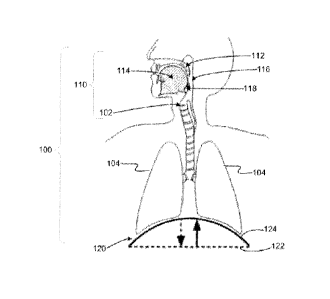
dangerous of
all sleep-related breathing disorders. While CSA is uncommon in its pure form,
it is prevalent in
patients with congestive heart failure, as a component of Cheyne-Stokes
respiration.
[000106] The obstructive component in OSA is related to decreased
pharyngeal tone as the
muscles relax during sleep. During normal respiration, upper airway patency is
maintained by
the negative pressure reflex, which activates pharyngeal dilators in response
to negative
transthoracic pressure during inspiration. In apneic patients, the negative
pressure reflex is
insufficient to maintain patency during sleep. Here, the negative pressure
created during
inspiration, in tandem with gravitational force acting on the surrounding
tissues is sufficient to
constrict or collapse the lumen of the flaccid airway.
[000107] FIG. 4 is a schematic representation of the human airway during an
OSA event. A
lack of muscle tone in the upper airway 110 allows pharyngeal structures 116
to partially or
completely block the lumen 119 of the airway 100, particularly when subjects
sleep on their
back. Respiratory drive continues during the OSA event, the diaphragm and
intercostal muscles
120 contract 122, creating a negative pressure in the airway 100 that draws
flaccid pharyngeal
structures 116 into the airway lumen 119.
[000108] FIG. 5 is a graph 400 of airway pressure 401 measured at the
larynx 102 (see FIG.
3) at the outset of an OSA event, comprising normal breathing process
inspiration 402a and
expiration 404a peaks before the OSA event and then inspiration 402b and
expiration 404b peaks
of a greater amplitude during the OSA event. This increase in the amplitude of
the airway
pressure 401 reflects continuing attempts on the part of the subject to
breathe after airway
obstruction, generating greater than normal airway pressures 401. The outset
of the OSA event
CA 2961902 2017-03-22
403 can then be identified by the sudden increase in amplitude of the
inspiration 402 and
expiration 404 peaks of the airway pressure 401.
[000109] A schematic representation of the human airway 100 during a CSA
event is
illustrated in FIG. 6. The upper airway 110 remains open, but diminished
central respiratory
drive reduces or eliminates diaphragm 120 movement, thereby reducing or
halting air flow
during the CSA event.
[000110] FIG. 7 is a graph 600 of airway pressure 601 measured at the
larynx 102 (see FIG.
5) at the outset of a CSA event, comprising normal breathing process
inspiration 602 and
expiration 604 peaks before the CSA event and then an absence of, or very low
amplitude,
inspiration and expiration peaks 606 during the CSA event. Despite a patent
upper airway 110,
upper airway pressure 601 is not fully modulated after the onset of the CSA
event and
diminution of diaphragm movement. The outset of the CSA event 603 can then be
identified by
the sudden drop 606 in the amplitude of the inspiration 602 and expiration 604
peaks of the
airway pressure 601.
ii. Deglutition Conditions and Disorders
[000111] In an aspect. the method 1100 may obtain one or more deglutition
conditions at
step 1110. Non-limiting examples of deglutition conditions include presence of
bolus,
occurrence of swallow, occurrence of dysphagic swallow, and presence of acid
reflux.
[000112] In another aspect, the method 1100 may predict one or more
disorders, including
one or more deglutition disorders and/or deglutition-related disorders, at
step 1112. Non-limiting
examples of deglutition disorders and deglutition-related disorders include
obstructive apnea,
dysphagia, presence of bolus in esophagus, and aspiration.
[000113] Detailed descriptions of selected deglutition conditions and
disorders are provided
herein below.
Normal Swallowing
[000114] Deglutition or swallowing is a stereotyped reflex that exhibits a
consistent pattern
of activation of 50 muscles throughout the upper airway. This sequence acts to
propel food and
fluid caudally at speeds of about 1M/sec with the pharyngeal stage of the
swallow taking about 1
sec to complete. FIG. 8 is a schematic illustration showing the upper airway
structures relevant
21
CA 2961902 2017-03-22
to deglutition. The swallow sequence is essentially a progressive anterior-to-
posterior wave of
pharyngeal contact that acts to squeeze the bolus from the soft palate 112
posteriorly to the
pharynx 116, further posteriorly to the epiglottis 118 and ultimately toward
the esophagus 902
like a tube of toothpaste while simultaneously protecting the airway from
entry of material.
[000115] The resulting stereotypical pattern of neural activity across the
pharyngeal touch
and pressure sensitive afferents would be apparent both within individual
afferent fibers and
across populations of fibers. A schematic diagram showing the anterior-to-
posterior activation
pattern in the activity profile is provided as FIG. 9. The actual pattern of
the activity profile is
influenced by the pattern of mechanical contact and pressure on a given
mucosal receptor and by
the adaptation properties of the afferent fibers.
[000116] For example, the receptors in pharyngeal suiface of the soft
palate 112 would
experience a stereotyped increasing pressure profile as the palate lifts to
seal the nasal cavity
from the bolus, followed by a stereotyped decreasing pressure profile as the
bolus passes, as
illustrated in FIG. 9A. Depending on the rate of adaptation within an
individual fiber, this may
create a variety of activity profiles. FIG. 10 is a graph summarizing a
variety of activity profiles
associated with different types of individual fibers. In various aspects, the
activity profile may
be a "tonic"profile (FIG. 10A), a -buildup" profile (FIG. 10B) characterizing
relatively slowly
adapting receptors, an "on-sustained" profile (FIG.10C), a "pauser" profile
(FIG.10D), an
"onset" profile (FIG.10E), or an "on-off' profile (FIG.10F) characterizing
progressively more
rapidly adapting receptors. Spontaneously active fibers may exhibit, for
example, a "tonically-
inhibited" activity profile during applied pressure, as illustrated
schematically in FIG. 10G.
[000117] The area of and location of contact between the soft palate and
posterior
pharyngeal wall may also exhibit a stereotyped pattern during swallow,
creating a spatial activity
pattern across a population of fibers, in addition to the temporal activity
pattern within individual
fibers. On a larger spatial scale, the anterior to posterior pharyngeal
contact pattern would act to
create a stereotypical spatial activity pattern, with most anterior fibers
being activated at the
beginning of the swallow sequence and the most posterior fibers being
activated about a second
later, as illustrated schematically in FIG. 9.
[000118] In one aspect, the neural signals recorded from iSLN receptors are
relevant to the
gastrointestinal (GI) condition of a subject. The iSLN mechanoreceptors
normally indicate bolus
contact and trigger a swallow sequence.
CA 2961902 2017-03-22
[000119] Dysphagia, as referred to herein, refers to the medical symptom of
difficulty in
swallowing, and is frequently diagnosed in subjects also presenting with sleep
apnea. Subjects
may have a great deal of difficulty in controlling even saliva in the mouth,
or difficulty in
initiating a swallow, or a cough. Dysphagia thus represents a further example
of a high medical
risk due to impaired pharyngeal motor control.
[000120] The activity profile within and between individual fibers and
fiber populations
may be determined using a calibration process during normal deglutition of a
given subject,
created for example, during volitional "dry" swallowing or in the presence of
an administered
bolus of food or fluid. The activity profile for dysphagic swallow and/or
presence of an
unswallowed bolus may be similarly determined. The range of normal temporal
and/or spatial
activity patterns observed during the calibration process can computed and be
used to set for
example matched-filter templates and upper and lower thresholds for detection
of normal
swallow and dysphagic swallow. Peaks within this range may be detected using
simple fixed-
level thresholds and used to assign a deglutition activity as the associated
activity type of the
neural activity profile.
iii. Vibration Conditions
[000121] In an aspect. the method 1100 may obtain one or more vibration
conditions at step
1110. Non-limiting examples of vibration conditions include snoring, stridor,
wheezing and
vocalization. In another aspect, the method 1100 may predict one or more
disorders, including
one or more vibration disorders and/or vibration -related disorders, at step
1112. Detailed
descriptions of selected deglutition conditions and disorders are provided
herein below.
[000122] Snoring is caused by the vibration of flaccid pharyngeal tissues
during sleep, and
snoring may art early indicator of the development of an obstructive sleep
apnea (OSA). The
walls of the mucosa are known to contain specialized mechanoreceptors that are
sensitive to
vibration. Three different vibration receptor types are known, each responding
best to vibration
over a different range of frequencies. Merkel disks, for example, respond best
to vibrations from
about 5-15 Hz, while Meissner corpuscles have a best frequency of about 50 Hz.
Both of these
receptors types have been histologically identified in the airway mucosa. A
third class of
rnucosal mechanoreceptors is known to respond to vibration up to 300 Hz, with
a best frequency
23
CA 2961902 2017-03-22
of about 150 Hz; these response properties correspond to those known for
Pacinian corpuscle
receptors.
[000123] FIG. 11A is graph 1000 illustrated schematically an activity
profile 1002 of a
vibration-sensitive mechanoreceptor. As illustrated in graph 1000A, the
activity profile 1002
shown at a zoomed in time scale, these vibration-sensitive mechanoreceptors
may exhibit phase-
locked activity at the frequency of the vibration, thereby encoding the
stimulus frequency by a
single action potential 1004 on every cycle, or at higher frequencies, at
integer multiples of the
interval between cycles. This produces a characteristic interspike interval
1006 for these phase
locking fibers that matches or is a multiple of the period of the vibration.
Further, the envelope
of the activity profile during pharyngeal vibration may exhibit amplitude
modulations as a result
of phase locking, as illustrated by the graph The interspike interval 1006,
amplitude modulation
frequency, vector strength and modulation depth occurring at a given vibration
frequency may be
determined using a calibration process during normal pharyngeal vibration of a
given subject,
created for example, by volitional vocalization, snoring, or wheezing.
Artificially induced
vibration using, for example, a piezoelectric vibrator placed on the skin over
the pharynx may
also be used. The range of interspike intervals, amplitude modulation
frequencies, vector
strengths and modulation depths observed during the calibration process can
computed and be
used to set upper and lower thresholds for vibration detection. Peaks within
this range can be
detected using simple fixed-level thresholds and defined as vibration events.
[000124] Using these techniques, the pattern of pharyngeal vibration,
encoded by
characteristic interspike intervals and/or amplitude modulation of the
activity profile of
pharyngeal afferent nerves, can be used to identify vibration pattern,
vibration timing, vibration
phase, and the amplitude of vibration.
[000125] Snoring is an upper airway condition that is characterized by
vibration of the
pharyngeal walls, tongue base, soft palate, and tonsils. It has been
discovered that the principal
frequency range of human snoring occupies a spectrum from 40-300Hz with a peak
spectral
power at about 100 Hz. The frequency spectrum of snoring vibration activity is
well-matched
to the frequency range of pharyngeal vibration sensitive mechanoreceptors and
makes
monitoring of pharyngeal afferents a particularly well-suited method for
snoring detection.
[000126] Specific airway structures are known to vibrate at characteristic
frequencies, for
example, the tonsils and soft palate vibrate at about 170 and 140 Hz,
respectively. This
24
CA 2961902 2017-03-22
characteristic may be used to pinpoint the structural source of snoring by
monitoring pharyngeal
vibration receptors. If multiple upper airway afferents and/or multiple fibers
within a single
nerve are monitored, the location of the source of pharyngeal vibration may
also be pinpointed
based on the receptive fields of active afferent fibers, either by comparing
activity across
multiple nerves, or by comparing activity across fibers within a single nerve.
iv. Reflux Conditions
[000127] In an aspect, the method 1100 may obtain one or more reflux
conditions at step
1110. Non-limiting examples of reflux conditions include esophageal reflux,
pharyngeal reflux,
and laryngeal reflux.
[000128] In another aspect, the method 1100 may predict one or more
disorders, including
one or more reflux disorders and/or reflux-related disorders, at step 1112.
Non-limiting
examples of reflux disorders and reflux-related disorders from esophageal
reflux, laryngeal
reflux, acid reflux, and GERD.
[000129] Detailed descriptions of selected reflux conditions and disorders
are provided
herein below.
[000130] Gastroesophageal reflux disease (GERD), gastric reflux disease, or
acid reflux
disease is a chronic symptom of mucosal damage caused by stomach acid coming
up from the
stomach into the esophagus. GERD may be divided into esophageal and
extraesophageal
syndromes. Acid reflux allowed by a transient relaxation of the lower
esophageal sphincter that
allows acid to pass into esophagus. Once in the esophagus, gastric acid can
travel along the
length of the esophagus, reaching or passing the level of the upper esophageal
sphincter.
Extraesophageal symptoms are caused by the entry of gastric juices in the
larynx and pharynx
through the upper esophageal sphincter. Laryngeal and pharyngeal symptoms are
also known as
laryngopharyngeal reflux (LPR) or extraesophageal reflux disease (EERD).
Extraesophageal
symptoms include dysphagia. voice disorders, asthma, hoarseness, laryngitis,
chronic cough,
pain, vocal fold nodules, unstable voice during speaking or singing.
[000131] The mucosa of the upper airway is known to contain afferent fibers
of different
diameters and conduction velocities. In cutaneous nerves, there exist three
populations of
afferent fibers, each population having a characteristic signal conduction
velocity. "AP" type
fibers have the fastest signal conduction velocity and typically conduct
action potentials along
CA 2961902 2017-03-22
afferent fibers at a rate ranging from about 35 m/s to about 75 m/s. "As" type
fibers have an
intermediate signal conduction velocity and typically conduct action
potentials along afferent
fibers at a rate ranging from about 5 m/s to about 30 m/s. "C" fibers have the
slowest signal
conduction velocity and typically conduct action potentials along afferent
fibers at a rate ranging
from about 0.5 m/s to about 2 m/s.
[000132] It is known that the neural activity of mucosal receptors
sensitive to low pH
(acidic) conditions receptors typically involved in perceiving reflux
conditions- is carried by the
slow-conducting "C" type fibers. It was discovered that a neural activity
profile characterizing
reflux conditions could be isolated from a reading from an upper airway
afferent that included
superimposed activity profiles characterizing other conditions by assessing
the conduction
velocity of the neural signals within the reading.
[000133] To isolate the "C" type fiber activity from superimposed activity
of "AP" type
fibers and "As" type fibers, the activity of these three populations of nerve
fibers can be
differentiated using techniques based on each fiber's known signal conduction
velocity. A
schematic illustration of this isolation technique is shown in FIG. 17. In
this technique, a
peripheral recording device 1702 and a central recording device 1704 are
spaced a known
separation distance along the length of a given set of fibers within an
afferent nerve 1 706 and
used to record two separate neural signals 1708 and 1710, respectively.
[000134] For example, as illustrated in FIG. 17, the recording devices 1702
and 1704 may
be spaced about 1 mm apart along the length of the nerve and thus record the
activity from the
same set of nerve fibers at two different points along their length. In the
signal processing circuit
1712, a 1 ms delay may be introduced in the signal 1708 from the peripheral
recording device
1702 which is then summed with the neural signal 1710 obtained from the
central recording
device 1704. In this example, the "C" fiber activity covers the distance
between the electrodes in
about 1 ms, and thus the signals 1708 and 1710 from the recording devices 1702
and 1704
overlap in time and are added together. as schematically illustrated in graph
1714. However, the
signals associated with activity from Af3 type fibers and M type fibers have
moved well past the
central recording device 1704 during the 1 ms delay as illustrated in graphs
1716 and 1718.
respectively. As a result, the portion of the signals 1708 and 1712 associated
with activity from
AP type fibers and A6 type fibers are not added together. By setting a high
threshold on the
output of the circuit, the activity of only the combined "C" fiber signals may
be isolated. Using a
26
CA 2961902 2017-03-22
similar circuit to that illustrated in FIG. 17, the activity of acid-sensitive
"C" fibers may be
separated from the activity of more rapidly conducting mechanoreceptors in
upper airway
afferents in one aspect.
[000135] The activity profile of acid-sensitive "C" fibers may then be
determined using a
calibration process during acid reflux of a given subject, created for example
during a normally
occurring acid reflux, or created by artificial application of a low pH fluid
to the pharynx. The
range of normal temporal and/or spatial activity patterns observed during the
calibration process
may computed and be used to set, for example, upper and lower thresholds for
detection of reflux
in the airway in an aspect. Peaks within this range may be detected using
simple fixed-level
thresholds and the activity profile may be associated with a reflux activity
type in another aspect.
The temporal and spatial activity profiles during naturally occurring or
artificial reflux conditions
may also be determined in an additional aspect.
b. Neural Signals
[000136] In various aspects, the method obtains neural signals from
upper airway
afferents in order to monitor a condition and/or a disorder including, but not
limited to a
respiratory condition, a deglutition condition, a reflux condition, and a
vibration condition. A
detailed description of upper airway afferents suitable for use in the method
are provided herein
below.
i. Upper Airway Afferents
[000137] In an aspect, the upper airway afferents include nerves associated
with mucosal
sensory receptors situated throughout the upper airway of the subject. The
neural signals
produced by these mucosal sensory receptors provide a rich source of
information to identify and
characterize a variety of upper airway conditions and disorders, as described
herein previously.
Non-limiting examples of upper airway afferents include pharyngeal afferents,
laryngeal
afferents, oral cavity afferents and nasal cavity afferents.
[000138] Pharyngeal afferents are known to transmit information from
sensory receptors in
the mucosa lining the upper airway to the brain. As used herein, pharyngeal
afferents
innervating additional areas of the upper airway, such as the larynx, are
included in the ter-n
"pharyngeal afferents". Non-limiting examples of pharyngeal afferent include
the iSLN branch
27
CA 2961902 2017-03-22
of the vagus nerve, the pharyngeal branch of the vagus nerve, the pharyngeal
branch of the
glossopharangeal nerve, the tonsular branch of the glossopharangeal nerve, the
lingual branch of
the glossopharangeal nerve, the intermediate nerve, the palantine nerve, the
greater petrosal
nerve, any branch of the facial nerve, the pterygopalatine nerve of the
trigeminal nerve, and any
combination thereof. Various aspects of the methods described herein are
intended to include
both the pharyngeal and extrapharyngeal sensory receptor transmission
functions. For example,
the iSLN innervates vocal folds in the larynx as well as mucosal sensory
endings in the pharynx.
[000139] These mucosal receptors may be sensitive to stimuli including, but
not limited to
airway pressure characterizing respiratory conditions such as apnea, contact
with food or fluid
characterizing deglutitation conditions such as dysphagia, vibrations
characterizing vibration
conditions such as snoring, and pH characterizing reflux conditions. Non-
limiting examples of
additional neural signals carried by upper airway afferents include
chemoreceptors modulated by
pH, taste receptors modulated by various chemical compounds, thermoreceptors
modulated by
temperature or airflow, or noiceptors modulated by polymodal pain,
[000140] It has been discovered that the measurements of neural activity,
including but not
limited to the electroneurogram (ENG) of pharyngeal afferent nerves is
modulated by changes in
these variables. This relationship can be demonstrated by obtaining one or
more neural activity
profiles of the pharyngeal afferent nerve that is indicative of the amplitude
and timing of the
ENG signal in one aspect. The neural activity profile may be calculated for
example by applying
a rectification and bin-integration (RBI) algorithm to an amplified pharyngeal
afferent signal and
used to detect and/or monitor an upper airway condition and/or disorder.
Neural response
characteristics such as the number of elicited spikes, the interval between
spikes, and the
temporal spike pattern in response to continued stimulation may vary with
stimulus variables
such as intensity and duration.
[000141] Each sensory nerve fiber responds to adequate stimuli applied
within their
-receptive field", defined herein as the specific area of the sensory
epithelium innervated by a
sensory nerve fiber. In an aspect, the spatial extent of sensory stimuli may
be determined by
comparing neural response between fibers with different receptive fields,
which are often
organized in a topographic map. For example, spinal nerves are organized
segmentally and
typically innervate specific regions of skin (dermatomes) in an organized
fashion, with cervical
nerves innervating the upper body and sacral nerves innervating the lower
body. On a finer
CA 2961902 2017-03-22
scale, individual spinal nerves can also be somatotopically organized, with
fibers from receptors
close to one another within a dermatome grouped together into bundles called
fascicles within
the nerve. In an aspect, the receptive field characteristics associated with
one or more sensory
fibers within an upper airway afferent may be used to determine the spatial
extent, distribution,
patterning, or any other spatially-related characteristic of a stimulus
associated with an upper
airway condition and/or disorder monitored using neural signals from one or
more upper airway
afferents.
[000142] Various aspects of the method are designed to monitor and
interpret respiratory,
reflux, vibration, and/or de!lutitation conditions based on the neural signal
characteristics
including, but not limited to a firing pattern, an active fiber population, a
signal conduction
speed, and any combination thereof in pharyngeal afferent nerves.
Specifically, these the neural
signal may be compared to previously analyzed responses to calibration stimuli
in order to
detect pharyngeal disorders such as apnea, snoring, dysphagia or GERD, as
described previously
herein.
[000143] In one aspect, the method may obtain one or more neural signals
from the iSLN.
In other aspects, the method may obtain one or more neural signals from other
pharyngeal
afferents including, but not limited to trigeminal nerve afferents, facial
nerve afferents associated
with the from the oropharynx or nasopharynx cavity to monitor respiratory
conditions and/or
disorders, and glossopharyngeal afferents to monitor deglutitation conditions
including, but not
limited to the introduction of a bolus into the oropharynx and/or hypopharynx.
i. Upper Airway Efferents and Muscles
[0001441 In other aspects, upper airway conditions and/or disorders may
also be monitored
by obtaining neural signals from nerves carrying efferent signals to muscles
of the upper airway,
diaphragm, or intercostal muscles, or by monitoring the activity of these
respiratory muscles
themselves, alone, or in some combination with other nerves or muscles
modulated by
respiratory activity.
ii. Neural Signal Measurement Devices
[000145] In various aspects, any known device capable of detecting and
recording neural
signals from nerves including, but not limited to, upper airway afferents may
be used to obtain
29
=
CA 2961902 2017-03-22
the neural signals used to obtain the at least one neural activity profile in
the method. Non-
limiting examples of suitable neural signal measurement devices include
electrical sensors such
as nerve cuffs; optical sensors such as optic fibers used in combination with
voltage or current
sensitive dyes injected into the nerve to be monitored; biological sensors;
and mechanical
sensors. In various aspects, any known method of recording peripheral nerve
activity, such as
percutaneous microneurography or optical recording of nerve activity based on
voltage sensitive
dyes may be used without limitation. In a different aspect, the size of the
neural signal
measurement devices may have a form factor ranging from about the size of a
pacemaker to
about the size of a cell phone application.
[000146] By way of non-limiting example. an electroneurogram (ENG) of
pharyngeal
afferents may be monitored recording electrodes placed in, around, or near the
nerve. In one
aspect, a "cuff' electrode may be situated around the nerve to record the
aggregate activity of all
nerve fibers in the vicinity of a single recording site. In another aspect,
electrode designs and
signal processing techniques may facilitate the selective recording of neural
signals from smaller
groups of nerve fibers ("multi units"), recording from individual nerve fibers
("single units"),
discriminating the direction of an action potential propagation to
differentiate between motor
nerve fiber activity and sensory nerve fiber activity, and/or isolating a
subset of neural signals
based on the signal conduction velocity (isolating nerve fibers with a
specific diameter).
[000147] It will be appreciated that multiple recording electrodes may be
used, depending
in the application and anatomical location to be monitored, in order to
simultaneously or
sequentially monitor multiple signal sources. The recording electrode may also
target other
nerves carrying afferent signals from peripheral receptors that exhibit
modulations of bioelectric
potentials correlated with upper airway conditions. Non-limiting examples of
receptors that may
be monitored to characterize an upper airway condition respiratory condition
include:
mechanoreceptors sensitive to negative airway pressure, positive airway
pressure, stretch,
position. shear or slip, vibration, texture, touch, touch and pressure, muscle
stretch, muscle
"drive", air flow, blood pressure or osmolarity; chemoreceptors sensitive to
CO2, 02, or pH;
thermoreceptors sensitive to temperature or airflow; nociceptors sensitive to
polymodal pain, or
any combination thereof. In one aspect, the method may include at least one
electrode sensitive
to at least one upper airway characteristic including, but not limited to
upper airway pressure,
CA 2961902 2017-03-22
upper airway stretch, and upper airway airway temperature, or alternatively
may include multiple
electrodes sensitive to a combination of the upper airway characteristics.
[000148] In another aspect, the monitoring of an upper airway condition may
include
monitoring the upper airway-related activity of other nerves, or monitoring
other physical
indicators of upper airway state, including, but not limited to airway
pressure, muscle activity or
airway flow as described in further detail herein below. The monitoring in
this aspect may be
achieved using any means capable of detecting a physical signal and
transducing the signal to an
electrical signal suitable for analysis. For example, vaiious physical
indicators of respiration and
respiratory state are amenable to detection and monitoring, including but not
limited to airway
pressure, air flow, muscle stretch, muscle position, muscle "drive", blood
pressure, blood
osmolarity, blood gas (CO2 and 02), heart rate, and blood pH. Techniques and
apparatus for
detecting and monitoring such physical indicators are well known and widely
available and may
he used alone or in combination, and are generally coupled to leads that
transmit data to analytic
components. For example, multiple electrodes may be placed in or on the body
to measure, for
example, breathing rate and heart rate. Changes in abdominal or thoracic
circumference related
to respiration can be measured using belt-based systems with sensors based on
piezoelectric or
impedance sensors. An oximeter can be used to detect and monitor blood oxygen
levels in the
blood. A blood pressure cuff or arterial catheter may also be used, to detect
and monitor blood
pressure. EMG leads can be used to detect breathing muscle activity. A
manometer can be
placed in the nasal cavity to detect airway pressure.
[000149] In other additional aspects, upper airway conditions may be
monitored using any
of a number of anatomical elements involved in respiration and control of
respiration. For
example, respiratory activity may also be monitored from nerves carrying
efferent signals to
muscles of the upper airway, diaphragm, or intercostal muscles, or by
monitoring the activity of
these respiratory muscles themselves, alone, or in some combination with other
nerves or
muscles modulated by respiratory activity.
c. Neural Activity Profiles
[000150] In various aspect of the method, one or more neural activity
profiles
characterizing aspects of the neural signals including, but not limited to,
neural signal timing,
neural signal amplitude, neural signal phase, neural signal position, neural
signal conduction
31
CA 2961902 2017-03-22
velocity, and any combination thereof, may be obtained using one or more
signal processing
techniques described herein above in connection with the upper airway
conditions. Additional
signal processing techniques are described in detail herein below.
i. Signal Processing
[000151] In various aspects, the neural signals obtained from the one or
more upper airway
afferents are processed to obtain at least one neural activity profile. In one
aspect, the neural
signals obtained from the one or more upper airway afferents may be
conditioned by any known
method including but not limited to amplification prior to performing any
additional signal
processing. In another aspect, the neural activity profile may be obtained by
applying a
rectification and bin-integration (RBI) algorithm to the conditioned (i.e.
amplified) neural
signals.
[000152] By way of non-limiting example, a pressure in the upper airway may
be
monitored by obtaining an electroneurogram (ENG) of the iSLN, which is
correlated with
pressure in the upper airway. An index of respiratory activity (IRA) may be
calculated by
applying a rectification and bin-integration (RBI) algorithm to the amplified
iSLN neural signal.
The amplitude of peaks in the IRA during each breath that occur within a
normal range of
amplitudes may be determined using a calibration process during normal
respiration of a given
subject using, for example, polysomnographic techniques. This range of
"normal" amplitudes
can be used to define upper and lower thresholds for the detection of one or
more respiratory
conditions. Peak amplitudes falling outside of this normal range may be
detected using simple
fixed-level thresholds and defined as a respiratory conditions. The defined
upper and lower
thresholds may further be used to classify, in real-time, a detected apneic
event as being either an
OSA event or a GSA event, as described herein previously.
[000153] The signal conditioning may be implemented by any known signal
processing
circuitry including, but not limited to, a signal amplifier and a rectifier
circuit. Non-limiting
examples of suitable amplifiers and rectifier circuits are disclosed in U.S.
Patent Application
Publication No. 2006/0189881 entitled "IMPLANTABLE SIGNAL AMPLIFYING CIRCUIT
FOR ELECTRONEUROGRAPHIC RECORDING", published Aug. 24, 2006, by Ban] Fassio
and U.S. Pat. No. 7,282,980 entitled `PRECISION RECTIFIER CIRCUIT FOR HIGH-
32
CA 2961902 2017-03-22
DENSITY, LOW-POWER IMPLANTABLE MEDICAL DEVICE", issued Oct. 16,2007, to
Baru Fassio.
[000154] It is to be understood that the activity profile may be calculated
by applying a
rectification and bin-integration (RBI) algorithm to the amplified pharyngeal
afferent signal in
one aspect, other signal processing algorithms may also be applied to
calculate the activity
profile including, but not limited to: high pass filter, low pass filter,
bandpass filter, notch filter.
FIR filter, IIR filter, smoothing, moving average, Wiener (optimal) filter,
matched filter,
rectification, bin-integration, multichannel noise reduction, principal
components analysis,
independent components analysis, wavelet analysis, Fourier transformation,
matched filtering,
variance/variance ratio calculations, signal-to-noise ratios, cross-
correlation, auto-correlation,
Rayleigh statistic, and any combination thereof. In another aspect, the raw
pharyngeal afferent
ENG waveform may also be used directly. Activity profiles based on neural
network analyses,
cluster analysis in multidimensional feature space, cluster cutting using k-
means, Bayesian
expectation-maximization, likelihood ratios, closest centers, or manual
cluster cutting methods
may also be used in various aspects.
[000155] It is to be also understood that an activity profile could be
computed from any
number of other pharyngeal afferent ENG signal features that vary with
pharyngeal state such as
event or waveform timing, interval, amplitude, duration, rise time, fall time,
slope, presence,
absence, pattern. 1st derivative, 2nd derivative, 3rd derivative, root mean
square amplitude,
peak-to-peak amplitude, variance, statistical probability or probability
relative to baseline or
running average.
[000156] It is also to be understood that detection of pharyngeal events in
the activity
profile using methods other than fixed-level thresholding may be used, for
example noise-
tracking or other adaptive thresholds, energy or non-linear energy thresholds,
or any variety of
other detection operations on the raw or processed data.
[01)01571 In other additional aspects, the signal processing of the one or
more neural signals
may further include analyzing a timing sequence of two or more activity
patterns, wherein each
of the two or more activity patterns is obtained from different upper airway
afferents. In yet
other additional aspects. the signal processing of the one or more neural
signals may further
include assessing the spatial extent or spatial location of the one or more
detected neural signals
33
CA 2961902 2017-03-22
using information characterizing the receptive fields associated with the one
or more detected
neural signals.
d. Activity Types
[000158] In various aspects, the at least one neural activity profiles may
be compared to
one or more activity criteria to associate each neural activity profile with
an associated activity
type chosen from a respiratory activity type, a deglutition activity type, a
vibration activity type,
a reflux activity type, and any combination thereof. A detailed description of
activity criteria in
various aspects are described in details herein below.
i. Activity Criteria
[000159] The activity criteria describe various characteristics of a neural
activity profile
that may uniquely associate the neural activity profile with a particular
upper airway activity
including, but not limited to a respiratory activity, a deglutition activity,
a vibration activity, and
a reflux activity. The characteristics may include ranges, and/or threshold
values of neural
activity profile characteristics including, but not limited to a time
separation of a neural signal
feature such as a peak amplitude, a pattern of neural signals, and a signal
conduction speed.
Respiratory Activity Criteria
[000160] In an aspect, the respiratory criterion indicating a respiratory
activity may include:
a time separation between peak neural signal amplitudes ranging from about 1
seconds to about 5
seconds; a periodically repeating pattern of neural signals with a period
ranging from about 12
patterns per minute to about 60 patterns per minute; and any combination
thereof.
Deglutition Activity Criteria
[000161] In an aspect, a deglutition criterion indicating a deglutition
activity may include:
an anterior to posterior neural activation pattern; a stereotyped neural
activation pattern with a
duration of less than about 1 second; and any combination thereof
Vibration Activity Criteria
[000162] In an aspect. a vibration criterion indicating a vibration
activity may include
a neural signal frequency ranging from about 10 Hz to about 400 Hz; a time
separation between
peak neural signal amplitudes ranging from about 1 second to about 5 seconds,
and any
combination thereof.
34
CA 2961902 2017-03-22
Reflux Activity Criteria
[000163] In an aspect, a reflux criterion indicating a reflux activity may
include a signal
conduction velocity of less than about 2 m/s.
e. Activity States
[000164] In various aspects of the method, the at least one neural activity
profiles may be
processed to determine an activity state characterizing the associated
activity type. The activity
state may include a respiratory state, a deglutition state, a vibration state,
and a reflux state.
i. Respiratory State
[000165] The respiratory state may include respiratory timing, respiratory
amplitude,
respiratory phase, respiratory location, and any combination thereof. The
respiratory phase may
include either an inspiratory phase, expiratory phase, or zero flow phase
between inspiratory
phase, and expiratory phases. The respiratory phase may be defined for example
with reference
to peak amplitude in the IRA during each breath, as determined based on a
calibration of normal
respiration of a given subject using, for example, polysomnographic
techniques.
ii. Deglutition State
[000166] The deglutition state may include solid contact, fluid contact,
contact velocity,
contact timing, contact amplitude, contact pressure, contact texture, contact
temperature, a
presence of a bolus, and any combination thereof;
iii. Vibration State
[000167] The vibration state may include vibration timing, vibration
amplitude, vibration
phase, vibration location, vibration pattern, and any combination thereof.
iv. Reflux State
[000168] The reflux state may include reflux timing, reflux pH, reflux
location, and any
combination thereof.
f. Condition of Subject
[000169] In an aspect, the at least one activity states may be processed to
obtain at least one
condition of the subject chosen from a respiratory condition, a deglutition
condition, a vibration
condition, a reflux condition, and any combination thereof.
CA 2961902 2017-03-22
i. Respiratory Condition
[000170] In an aspect,the respiratory condition may include apnea,
tachypnea, hyperpnea,
hypopnea, polypnea, dyspnea, bradypnea. cough, Cheyne-Stokes respiration,
Biot's respiration,
ataxic respiration, Kussmaul respiration, wheezing, irregular respiration,
respiratory arrest,
restrictive respiration, shallow breathing, hypoventilation and any
combination thereof.
ii. Deglutition Condition
[000171] In an aspect, the deglutition condition may include presence of
bolus, occurrence
of swallow, occun-ence of dysphagic swallow, presence of acid reflux, and any
combination
thereof.
iii. Vibration Condition
[000172] In an aspect, the vibration condition may include snoring,
stridor, wheezing
vocalization, and any combination thereof.
iv. Reflux Condition
[000173] In an aspect, the reflux condition may include esophageal reflux,
pharyngeal
reflux, laryngeal reflux and any combination thereof.
[000174] In various aspects of the method, any one or more of the at least
one states, the at
least one conditions, the at least one disorders, and any combination thereof
may be displayed on
a patient monitor device, and/or communicated to a treatment system.
g. Disorder Prediction
[000175] In various aspect, the at least one condition may be assessed to
predict a disorder
chosen from obstructive apnea, central apnea, dysphagia, heart failure,
uremia, asthma, cardiac
arrest, organ failure, metabolic acidosis, COPD, pulmonary embolism. Ondine's
curse, obesity
hypoventilation syndrome, laryngeal penetration, aspiration, esophageal
reflux, laryngeal reflux,
presence of bolus in esophagus, acid reflux, GERD, laryngeal penetration,
aspiration, aspiration
pneumonia, SIDS, Charcot-Marie-Tooth disease and any combination thereof.
[000176] In other aspects, the detection and classification of apnea events
as described
herein is consistent with the detection and classification of apnea events as
described in U.S.
Patent Application Publication No. 2010/0125310, i.e. involves calculating an
index of
36
CA 2961902 2017-03-22
respiratory activity (IRA) that is indicative of the amplitude and timing of
respiratory activity
based on the amplitude and timing of a respiratory signal, such as an
electroneurogram (ENG)
signal from a nerve such as the internal branch of the superior laryngeal
nerve (iSLN), or another
sensor of respiratory activity as described elsewhere herein. Details for
calculating an IRA that is
indicative of the amplitude and timing of a respiratory signal are described
in U.S. Patent
Application Publication No. 2010/0125310.
[000177] The algorithm executed by the apnea monitoring and detection
module
implements steps in the processes as discussed in further detail herein below.
Upon the detection
of an apnea event, the apnea monitoring and detection module sends a trigger
to the therapy
control module along with an identification of the type of apnea event, i.e.
obstructive, central, or
mixed; and apnea or hypopnea, depending on the implemented algorithm, which
generates a
stimulus appropriate for the type of apnea event. Optionally, the apnea
monitoring and detection
module may also send an indication of the severity level of the apnea event,
as well as timing
information of previous or continuing respiration patterns, to the therapy
control module 1106.
[000178] As described in detail in U.S. Patent Application Publication No.
2010/0125310,
the outset of an OSA event or a CSA event may be identified by features of the
IRA, for example
with reference to an upper and a lower threshold as described above. For
example, the fu-st
instance of a crossing of the upper threshold by inspiration related peaks of
the IRA can be used
as a criterion for identifying the outset of an OSA event. Alternatively, the
peak durations of the
RBI ENG may be used to identify the outset of an OSA event by setting an
appropriate
threshold. For a CSA event, the outset of the CSA event can be identified, for
example, by
noting the first absence of crossing of the lower threshold by inspiration
related peaks, in a set
time period. This period of time may be set, for example, to represent the
average time between
one or more respiration cycles. It should be understood that for both OSA and
CSA events, other
IRAs may be calculated in order to identify the outset of such an event. For
example, peak
durations and interpeak intervals of the RBI ENG can be used, by setting
appropriate levels and
thresholds. It is to be understood that the absence of measurements at a
specified level may
indicate a CSA event.
[000179] Additionally, apnea event severity can be determined from the IRA.
For example,
severity of the apnea event may be determined by comparing the amplitude of
the apneic IRA to
37
CA 2961902 2017-03-22
that observed during normal breathing. More severe apnea is characterized by
IRA peaks having
amplitudes far from the upper and lower thresholds, while less severe apnea or
hypopnea is
characterized by IRA peaks having amplitudes just above Or below the upper and
lower
thresholds. The level of apnea thus determined can be used to adjust the
parameters and
characteristics of the applied neurostimulation treatment. This may include
changing the
stimulation waveform, increasing or decreasing the stimulus amplitude,
increasing or decreasing
the number of stimuli delivered, selecting electrodes in specific locations or
changing the number
of stimulation electrodes used. Severity levels may be assigned predetermined
thresholds. It is
to be understood that the number of OSA and CSA severity levels may vary
depending on the
precision of the circuitry and/or algorithm used.
[000180] Apneic events may be further identified as hypopnea events. i.e.
OSA events can
be distinguished from obstructive sleep hypopnea (OSH) events, and CSA events
can be
distinguished from central sleep hypopnea (CSH) events with reference to the
IRA. For example,
an IRA value between a first upper threshold and a second upper threshold,
wherein the second
upper threshold is higher than the first upper threshold, may be associated
with OSH, while an
IRA value greater than the second upper threshold, may be associated with OSA.
Accordingly,
IRA peaks between the two upper thresholds can be identified as OSH while IRA
peaks above
the second, higher upper threshold can be identified as OSA. Conversely, an
IRA value between
a first lower threshold and a second lower threshold, wherein the second lower
threshold is lower
than the first lower threshold, may be associated with CSH, while an IRA value
lower than the
second lower threshold may be associated with OSA. The range of values for
which IRA peaks
are defined as OSH as opposed to OSA, as well as CSH as opposed to CSA, may be
determined
using a calibration process during abnormal respiration of a given subject
using, for example,
polysomnographic techniques.
[000181] It is to be understood that OSH, OSA, CSH and CSA may be
subdivided into
multiple severity levels depending on the precision of the circuitry and/or
algorithm used.
[000182] As described above for the OSA and CSA event detection, the
variation in IRAs
calculated using algorithms other than RBI ENG may also be used to determine
the severity of
the apneic or hypopneic event.
[000183] Apneic events may be further identified by the location(s) of the
airway
obstruction using, for example, the temporal profile of the IRA activity
pattern acquired from a
38
CA 2961902 2017-03-22
single electrode or sensor. Alternatively, or in addition, an apneic event may
be further
identified by the location(s) of the airway obstruction using, for example,
the temporal pattern of
IRA activity acquired across multiple electrodes or sensors, indicating, for
example, the
instantaneous pressure at multiple locations in the upper airway.
g. Other Uses of Monitoring Methods
[000184] In various aspects, the upper airway states, conditions, and/or
disorders obtained
using the method may be displayed or used to drive an alarm or alert. In
various other aspects,
the upper airway states, conditions, and/or disorders obtained using the
method may be used in
the implementation of condition prevention/treatment methods as described
herein below, or may
be transmitted to other devices including but not limited to monitor devices
and/or treatment
devices. For example, the method in various aspects may detect respiration
rate, phase, and
timing. This capability provides for general monitoring of vital signs, aside
from apnea detection.
and may be used to provide respiration-related or other upper airway-related
parameters to other
devices such as external monitoring equipment, or implanted devices such as
pacemakers or
implantable defibrillators. The upper airway states, conditions. and/or
disorders obtained using
the method may be transferred for use to any known implantable apnea treatment
devices that
terminate apnea using drug delivery, atrial overdrive pacing or electrical
stimulation of the nerves or muscles that control respiratory activities
2. Method of Treating and/or Preventing an Upper Airway Disorder
[000185] FIG. 13 is a flow chart illustrating a method 1200 of preventing
and/or treating an
upper airway disorder. The method 1200 includes delivering at least one
stimulation to a
modulate a reflex including, but not limited to a swallowing reflex and/or a
negative-pressure
reflex.
a. Disorders
[000186] In an aspect, the method 1200 prevents and/or treats an upper
airway disorder
including, but not limited to: obstructive apnea, central apnea, obesity
hypoventilation syndrome,
dysphagia, esophageal reflux, presence of bolus in esophagus, acid reflux,
GERD, and any
combination thereof;
39
CA 2961902 2017-03-22
b. Stimulation of Reflex
i. Reflexes Overview
[000187] In an aspect, the method 1200 delivers at least one stimulation to
a modulate a
reflex including, but not limited to a swallowing reflex and/or a negative-
pressure reflex.
ii. Swallowing Reflex
[000188] As used herein, the term "swallow" refers to all or part of
swallow sequence.
Swallow stimuli are at least one of: electrical stimulation to at least one
swallow-related nerve,
electrical stimulation to at least one swallow related muscle, and mechanical
stimulation to at
least one swallow-related sensory receptor.
[000189] Without being limited to any particular theory, the act of
swallowing activates and
repositions airway structures that are commonly involved in obstructive sleep
apnea. In various
aspects, stimulation of the swallow reflex may be used effectively to
reposition airway
structures, between breaths, to reestablish airway patency. Specifically
pharyngeal reflexes are
exploited to treat upper airway disorders. A swallow activates all of the
structures that are
commonly involved in OSA including the tongue, soft palate, epiglottis, and
pharyngeal walls.
In addition, the swallow includes active components at the end of the sequence
that return
pharyngeal structures to their "normal" positions. Triggering the swallow
reflex in USA subjects
may activate and reposition any dysfunctioning pharyngeal structures and
return the airway to a
patent state.
[000190] In various aspects, a swallow stimulus may include an electrical
or mechanical
stimulus to a reflex-related nerve, muscle, or sensory receptor in the subject
that is sufficient to
elicit all or part of the reflexive and pre-programmed coordinated activity of
a swallow. For
example, the swallow stimulus may include electrical stimulation to at least
one swallow-related
nerve, electrical stimulation to at least one swallow-related muscle,
mechanical stimulation to at
least one swallow-related sensory receptor in the skin or mucosa of the
subject, or any
combination thereof provided that the swallow stimulus is sufficient to elicit
all or part of a
swallow sequence in the subject. Stimulation of multiple targets may be
delivered
CA 2961902 2017-03-22
simultaneously, or in a sequence designed to elicit natural activation
patterns in all or part of the
50 muscles normally involved in the swallow sequence. For electrical stimuli,
the stimulus
target may be an afferent nerve or an efferent nerve, and may include at least
two swallow-
related nerves wherein each swallow-related nerve is independently an afferent
nerve or an
efferent nerve. An afferent target is selected based on the ability of the
afferent nerve, when
stimulated, to elicit all or part of reflexive swallow pattern activity from
the central nervous
system of the subject. The target nerve may be, for example, the internal
branch of the superior
laryngeal nerve (iSLN), or the pharyngeal branch of the glossopharyngeal
nerve. Alternatively
or in addition, the swallow-related nerve may be an efferent nerve. An
efferent target is selected
based on the ability of the efferent nerve, when stimulated, to elicit motor
activity in at least one
effector in a swallow sequence, the motor activity comprising all or part of a
swallow sequence
in the subject. Mechanical stimulation may comprise stimulation to at least
one swallow-related
sensory receptor in the skin or mucosa of the subject, such as for example
delivery of a liquid to
at least one of the oral, nasal, or pharyngeal cavity of the subject that is
sufficient to elicit all or
part of a swallow sequence in a subject.
Treatment of Apnea Using Swallowing Reflex Stimulation
[000191] In an aspect, the method 1200 may be used to reposition and hold
the pharynx in a
patent state by triggering a swallowing reflex in order to treat apnea. Any
system and method
described herein which involves detection of a swallow may be used to deliver
a swallow
stimulus during or concurrent with detection of a swallow, for example to
augment a
spontaneously occurring swallow. Any system and method described herein which
involves
detection of a dysphagic swallow, may be used to deliver a swallow stimulus
during or
concurrent with detection of the dysphagic swallow, for example to assist with
initiation of a
non-dysphagic swallow.
Treatment of Dysphagia Using Swallowing Reflex Stimulation
[000192] In an aspect, dysphagia may be treated by delivering a swallow
stimulus. For
example, the methods and systems may be suitably configured to deliver
preventative
mechanical or electrical stimulation of swallow in order to prevent dysphagia
before dysphagia
occurs using an open loop system that is not configured to rely on a dysphagia
or swallow
41
CA 2961902 2017-03-22
detection event. The methods and systems may also be suitably configured to
deliver
preventative mechanical or electrical stimulation of swallow in order to
prevent dysphagia using
a closed loop system that is configured to detect a bolus or attempted swallow
in a dysphagic
patient. In another aspect, the method may also used to deliver therapeutic
mechanical or
electrical stimulation of a swallow in order to treat dysphagia during a
dysphagic swallow using
a closed loop system that is configured to detect a dysphagic swallow in a
dysphagic patient.
[000193] The stimulated swallow may propel the bolus, clear the airway and
prevent
penetration or aspiration of saliva, mucus, and/or a bolus of fluid. In an
additional aspect, the
method may also be used to train, strengthen and coordinate spontaneous (i.e.
unstimulated)
patient-initiated swallows. In another additional aspect, the swallow stimulus
may be triggered
by means of a manual input by the subject; for example, the subject may
trigger a swallow
stimulus to enhance a dysphagic swallow.
Treatment of GERD Using Swallowing Reflex Stimulation
[000194] In an aspect, the method may be used to treat GERD using
stimulation of the
swallow reflex. Esophageal exposure to gastric juice is normally minimized
chemically by saliva
and mechanically by esophageal peristalsis. In GERD patients, esophageal
motility, particularly
secondary peristalsis, is impaired and results in increased duration of
exposure to acid. "Also,
sleeping people tend to swallow less frequently. This slows the regular
esophageal contractions
that normally keep food moving down the esophagus and prevent acid from moving
back up.
Sleepers also produce less saliva, which plays a role in returning esophageal
pH levels to normal
after an incident of acid reflux." The present invention describes
neurostimulation of pharyngeal
afferents to decrease both esophageal and extraesophageal symptoms of acid
reflux, by inducing
swallow and thus induce primary esophageal peristalsis, clear the upper
digestive tract, and
return acidic gastric juices to the stomach.
iii. Negative-Pressure Reflex
[000195] A negative pressure stimulus is at least one of: electrical
stimulation to at least
one negative pressure reflex-related nerve, electrical stimulation to at least
one negative pressure
reflex related muscle, and mechanical stimulation to at least one negative
pressure reflex -related
sensory receptor. Any of these stimuli may be delivered as one or more (as
series of) discrete
42
CA 2961902 2017-03-22
stimulus bursts designed to elicit at least one negative pressure reflex per
each burst. Mechanical
stimulation for eliciting the negative pressure reflex includes, in non
limiting example, a pulling
vacuum. Electrical nerve stimulation for eliciting the negative pressure
reflex may include, in
non limiting example, stimulating the iSLN. Electrical muscle stimulation for
eliciting the
negative pressure reflex includes but is not limited to electrical stimulation
of the tensor palatini,
hypoglossal, and/or superior pharyngeal constrictor. Efferent nerve
stimulation for stimulating
the negative pressure reflex include the nerves innervating the tensor
palatini, genioglossus,
and/or pharyngeal constrictor muscles.
[000196] In any method or system using stimulation of the negative pressure
reflex, signals
from the iSLN may be monitored and the monitored signals may be used to
trigger negative
pressure reflex stimulation and thereby trigger the reflex, and/or to
synchronize delivery of the
stimulation to occur during a certain phase of the respiratory cycle.
[000197] In any of the above systems including fully open, partially open,
partially closed
and fully closed, monitoring of iSLN signals can be used for synchronization
and/or triggering of
the negative pressure reflex stimulus. Further, signals from sensors, or from
nerves other than
iSLN may be monitored for synchronization and/or triggering of the stimulus,
as described
herein above with respect to delivery of a negative pressure reflex stimulus.
Treatment of Apnea Using Negative Pressure Reflex Stimulation
[000198] In another aspect, negative pressure reflex stimulation may be
used for as a
therapy for apnea, wherein negative pressure reflex stimulation is any
stimulation sufficient to
elicit all or part of the negative pressure reflex, which stiffens,
repositions and/or holds the
pharynx in a patent state. Thus, negative pressure reflex stimulation serves
to stiffen, reposition
and/or hold the pharynx in a patent state.
ii. Stimulation of Reflexes
[000199] In other aspects, the swallow reflex and/or negative pressure
reflex may be
stimulated by delivering an electrical stimulation and/or a mechanical
stimulation, defined herein
below.
iii. Type of Stimulation
43
CA 2961902 2017-03-22
[000200] A burst of electrical or mechanical stimulation is defined here as
a temporally
discrete occurrence of one (a single), or more (a series) of stimulus
pulse(s), defined by a total
duration from burst start to burst end of about 200 sec to about 3 seconds.
[000201] Nerve stimulation means may be accomplished by any means
including, but not
limited to direct, transcutaneous, magnetic, electrical, optical, mechanical
or any other means.
Receptor stimulation may be accomplished by any means including, but not
limited to direct,
transcutaneous, magnetic, electrical, optical, mechanical or any other means.
Access to the nerve
or receptor by any means, electrode and lead, injectable bion-like platform,
light through skin,
magnetic stimulation through skin, electrical stimulation through skin,
communication
transcutaneously, percutaneously, a fully implanted, partially implanted, or
fully external system.
Biological, mechanical or electrical sensors may be used to provide
information for the
configuration of the stimulation.
Electrical Stimulation
[900202] In various aspects, each electrical stimulation may be delivered
to a reflex-related
nerve, a reflex-related muscle, a reflex-related sensory receptor, and any
combination thereof.
The reflex-related nerve comprises:
[000203] an afferent chosen from: iSLN branch of vagus nerve, pharyngeal
branch of va.qus
nerve, pharyngeal branch of glossopharangeal nerve, tonsular branch of
glossopharangeal nerve,
lingual branch of glossopharangeal nerve, intermediate nerve, palantine nerve,
greater petrosal
nerve, any branch of facial nerve, and pteryaopalatine nerve of trigeminal
nerve; or an efferent
chosen from: recurrent laryngeal nerve, external branch of superior laryngeal
nerve, brancial
motor branch of zjossopharyngeal nerve and proximal fibers, mandibular nerve,
medial
pteryaoid nerve, pharyngeal branch of vaErus nerve and proximal fibers; branch
of facial nerve
and proximal fibers, and branch of hypoglossal nerve and proximal fibers
[000204] The stimulus target may be an afferent nerve or an efferent nerve,
and may
include at least two swallow-related nerves wherein each swallow-related nerve
is independently
an afferent nerve or an efferent nerve. An afferent target is selected based
on the ability of the
afferent nerve, when stimulated, to elicit all or part of reflexive swallow
pattern activity from the
central nervous system of the subject. The target nerve can be, for example,
the internal branch
of the superior laryngeal nerve (iSLN), or the pharyngeal branch of the
glossopharyngeal nerve.
44
CA 2961902 2017-03-22
Alternatively or in addition, the swallow-related nerve can be an efferent
nerve. An efferent
target is selected based on the ability of the efferent nerve, when
stimulated, to elicit motor
activity in at least one effector in a swallow sequence, the motor activity
comprising all or part of
a swallow sequence in the subject. The target nerve can be, for example, the
recurrent laryngeal
nerve, the external branch of the superior laryngeal nerve, the brancial motor
branch of the
glossopharyngeal nerve, the mandibular nerve, the medial pterygoid nerve, or
pharyngeal branch
of the vagus nerve.
[000205] An electrical swallow stimulus may comprise electrical stimulation
to at least one
swallow-related nerve or at least one swallow-related muscle, provided that
the stimulation is
sufficient to elicit all or part of a swallow sequence in the subject.
[000206] A swallow stimulus may therefore, alternatively or in addition to,
comprise
mechanical stimulation to at least one swallow-related sensory receptor, such
as a
mechanoreceptor, in the skin or mucosa of the subject.
Mechanical Stimulation
[0002071 In an aspect. each mechanical stimulation may be delivered to a
reflex-related
sensory receptor. The reflex-related sensory receptor may be situated in the
skin or mucosa of the
subject. Non-limiting examples of reflex-related sensory receptors include: a
mechanoreceptor
sensitive to negative airway pressure, positive airway pressure, stretch,
position, shear, slip,
vibration, texture, touch, mechanical compression, muscle stretch, muscle
drive, air flow, blood
pressure or blood osmolarity; a chemoreceptor sensitive to CO,, 02, or pH; a
thermoreceptor
sensitive to temperature or airflow; and a nociceptor sensitive to polymodal
pain. Swallow
stimuli may include temporally discrete stimulus bursts configured to elicit
at least one swallow
per each burst, but also a continuous stimulus delivery, such as but not
limited to delivery to the
oral cavity of a continuous "dribble" of fluid such as water or juice. A
"dribble" is a continuous
but slow rate of fluid flow, as determined by one of average skill, but in non-
limiting example is
about 1 ml/minute or within the range of 0.5 ml/minute to about 5 ml/minute.
iv. Stimulation Devices
[000208] In any of the systems described herein, the stimulation output
device is configured
to generate one or more stimuli that target at least one swallow-related nerve
or muscle, or
CA 2961902 2017-03-22
swallow-related receptor, to elicit all or part of the reflexive and pre-
programmed coordinated
activity of a swallow.
Electrical Stimulation Devices
[000209] A stimulation electrode may be placed in, around or near a
peripheral nerve that
carries afferent and/or efferent neural activity. Depending on the choice of
stimulation output
device, an alternative system may include a therapy output device including an
electrical
stimulation output device.
[000210] Any system may be further configured to control, or to control and
deliver a
swallow stimulus to multiple targets. Selection of targets for stimulation may
vary depending on
the identified apneic event and the type (mechanical, electrical or
combination thereof) of
stimulation used. The system may be configured for example with a single
electrode that is used
as both a recording and stimulation electrode, for example when the iSLN is
used for both
recording and stimulation. Furthermore, multiple electrodes may be used, some
or all of them
being used both as recording and stimulation electrodes while others are used
only as recording
or stimulation electrodes.
[0002111 The electrodes may be, for example, cuff electrodes such as, but
limited to, that
described in U.S. Pat. No. 5,824,027. Other types of electrodes, leads,
probes, cuff-electrodes,
etc., may be used as well. Other examples of cuff electrodes that may be used
are disclosed in
U.S. Patent Application Publication No. 2008/0065184 entitled "NERVE CUFF,
METHOD
AND APPARATUS FOR MANUFACTURING SAME", published Mar. 13, 2008, by Hoffer et
al. and PCT Patent Application Publication No. WO 2008/025155 entitled "NERVE
CUFF
INJECTION MOLD AND METHOD OF MAKING A NERVE CUFF', filed Aug. 29, 2007, by
Imbeau et al.
Mechanical Stimulation Devices
[000212] A mechanical stimulation device may be used for oral, nasal or
pharyngeal
delivery of a mechanical stimulus to the subject. The mechanical stimulation
entails delivery of
an amount of a liquid of relatively low viscosity such as water or saline, to
the oral, nasal, or
pharyngeal cavity of the subject. The amount of liquid may be delivered as a
continuous flow, or
46
CA 2961902 2017-03-22
may delivered as a small discrete bolus, for example about 0.1 ml up to about
10 ml, preferably
about 0.5 ml to about 2 ml, delivered as short a burst with an overall
duration between about 200
sec to about 3 seconds. For example, the mechanical stimulus may comprise a
continuous
delivery of a liquid at a flow rate of about 1 ml/minute over the course of
the entire night.
Alternatively, the liquid may be delivered as discrete bursts of liquid, as
described further below.
[000213] In an aspect, a liquid delivery device may be operatively coupled
to the
stimulation module via a wire lead or wireless communication (not shown), and
the stimulation
module may be configured to generate the mechanical swallow stimulus through
the liquid
delivery device. In various aspects, the liquid delivery device may include a
gravity-fed spout or
a tube coupled to a liquid reservoir via a solenoid valve configured to open
and close in response
to electrical signals from the stimulation module. It should be understood
that any device or
apparatus can be used for liquid delivery device, provided that it is capable
of containing or
providing a volume of liquid of at least about 0.5 ml, and includes an element
such as the
solenoid valve that can control the timing and volume of liquid delivery.
iv. Subthreshold versus Suprathreshold Reflex Stimulation
[000214] In various aspects, each of the at least one stimulations may be
delivered at a
subthreshold intensity insufficient to elicit the reflex or at a
suprathreshold intensity sufficient to
elicit the reflex. The method in an aspect may be used to provide sensory
enhancement to
augment the detection of weak sensory signals by adding noise to the signal
that is configured to
improve the ability of spontaneously occurring sensory signals to trigger
neural responses. Such
systems and methods use stimulation configured using the principles of
stochastic resonance
phenomena, wherein the stimulation is can provide afferent facilitative
stimulation to the iSLN to
improve negative pressure reflex for apnea patients.
[000215] Each of the at least one stimulations may include: a subthreshold
electrical
stimulation delivered to the reflex-related nerve or to the reflex-related
sensory receptor to
reduce the threshold of the reflex, to maintain muscle tone, and any
combination thereof; a
subthreshold electrical stimulation delivered to the reflex-related muscle to
maintain muscle
tone; a subthreshold mechanical stimulation delivered to the reflex-related
sensory receptor to
reduce the threshold of the at least one reflex; a suprathreshold electrical
stimulation delivered
to the reflex-related nerve, the reflex-related sensory receptor, the reflex-
related muscle, or any
47
CA 2961902 2017-03-22
combination thereof to maintain muscle tone, position and/or posture of one or
more respiratory
and/or deglutition structures of the subject; a suprathreshold mechanical
stimulation delivered to
the reflex-related sensory receptor to maintain muscle tone, position and/or
posture of one or
more respiratory and/or deglutition structures of the subject; a
suprathreshold electrical
stimulation delivered to the reflex-related nerve, the reflex-related sensory
receptor, the reflex-
related muscle, or any combination thereof to treat the disorder; and a
suprathreshold mechanical
stimulation delivered to the reflex-related sensory receptor to treat the
disorder.
Subthreshold Electrical Stimulation Characteristics
[000216] In an aspect, the method may augment neural signal initiating,
and/or control
negative pressure reflex in apnea patients using a "background pulse train"
delivered to iSLN.
The firing rate of the background pulse train is intentionally subthreshold
and so as not to trigger
the reflex independently. Rather, the background pulse train merely augments
the weak firing
rate resulting from naturally occurring sensory stimuli such as the negative
pressure signal on
iSLN during sleep in apneic patients. The additive effect of the weak firing
rate due to the
naturally occurring stimuli, with the added background pulse train, results in
a suprathreshold
firing rate which elicits the desired response, for example the negative
pressure reflex. The
background pulse train may be individual pulses or bursts of pulses. An
interpulse interval,
which refers to the time between the end of one delivered pulse and the
beginning of the next
pulse, may be approximated by white noise or noise filtered using one or more
of a band pass,
high pass or low pass filter, Such stimuli may be delivered with or without
regard to respiratory
phase or apnea event, or may be adjusted to occur during desirable phases of
respiratory cycle or
during period when apnea is likely to occur.
[000217] The timing and frequency of a background pulse train may be
varied. A
background pulse train can be composed for example of single pulses delivered
with an
interpulse interval approximated by a band pass noise centered around, e.g.
about 1 second. A
background pulse train could be composed for example of bursts of pulses, each
burst lasting
tens of seconds (e.g. 10 sec, 20, 30 sec or more), which are delivered at an
interburst interval
approximated by a band pass noise centered around a predetermined time period
of one or more
minutes (e.g. 1-10 minutes, for example 5 minutes), or of single pulses
delivered at intervals
centered around 1 minute, or single pulses delivered at intervals centered
around ¨30 seconds, or
48
CA 2961902 2017-03-22
single pulses delivered at intervals centered around ¨30 seconds stimuli
adjusted to occur during
desirable phase of respiratory cycle.
[0002181 In an aspect, the method described herein may provide a background
pulse train
as described herein above to be delivered to the iSLN, to augment neural
signal initiating or to
control swallow in dysphagia patients using "background pulse train" delivered
to iSLN. As
described herein above, the firing rate of the background pulse train is
purposefully subthreshold
and not designed to trigger the reflex independently. The background pulse
train may be
delivered with or without regard to respiratory phase or presence of a bolus
event, or may be
adjusted to occur during desirable phases of respiratory cycle and/or during
meals. Such stimuli
may be delivered with or without regard to respiratory phase or apnea event.
Or may be adjusted
to occur during desirable phases of respiratory cycle or during period when
apnea is likely to
occur.
[000219] In an aspect, the method described herein can be configured to
provide
stimulation that augments neural signal initiating or controlling muscle tone
(e.g. stretch reflex)
in the UAW of apnea or dysphagia patients using "background pulse train"
delivered to UAW
afferents. As described herein above, the firing rate of the background pulse
train is purposefully
subthreshold and not configured to trigger movement of the UAW muscles
independently.
Rather, the background pulse train is configured to augment the weak firing
rate of naturally
occurring sensory stimuli (e.g. the stretch of UAW muscle spindle afferents
via La afferent
fibers), wherein the additive effects of the weak firing rate due to naturally
occurring sensory
stimuli together with the background pulse train result in a suprathreshold
firing rate which
elicits the desired response (e.g. stretch reflex and restoration of UAW
muscle tone). The
background pulse train may be provided in individual pulses or bursts of
pulses. The interpulse
interval of the background pulse train may be approximated by white noise or
noise filtered
using one or more of a band pass, high pass or low pass filter. In such
systems and methods, the
background pulse train may be delivered with or without regard to respiratory
phase or apnea or
dysphagia event. Stimulation may comprise selective stimulation of Ia
afferents or may include
other afferent or efferent fibers. It will be appreciated that increased UAW
muscle tone acts to
prevent collapse of the UAW. Increased muscle tone also acts to enhance the
physiological
response to naturally occurring muscle control signals from efferent fibers.
Nerves that innervate
the UAW muscles include branches of the Facial, Hypoglossal, Vagus and
Glossopharyngeal
49
CA 2961902 2017-03-22
nerves and their proximal fibers. The stimulation may be delivered with or
without regard to
respiratory phase or apnea or presence of bolus or dysphagia event, or may be
adjusted to occur
during desirable phases of respiratory cycle and/or during meals for dysphagia
or during sleep
for apnea or snoring.
[000220] In an aspect, the method described herein can be further
configured to provide
stimulation of UAW muscles by continuous direct stimulation of UAW efferents,
thereby
improving muscle tone for treating apnea or dysphagia. In such systems and
methods, stimuli
are configured to remain subthreshold and are not capable of eliciting
discrete movements but
rather only a continuous change in muscle tone. The resulting increase in
muscle tone also
enhances the physiological response to any naturally occurring efferent
control signal(s), and
also prevents UAW collapse.
[000221] Systems and methods described herein can be further configured to
stimulate co-
contraction for UAW muscle tone by continuous direct stimulation of UAW
efferents to
opposing muscle groups, to elicit co-contraction of muscles. Such stimulation
may be used to
treat apnea or dysphagia. Such stimuli remain subthreshold and are not capable
of eliciting
discrete movements but rather only a continuous change in muscle tone.
Suprathreshold Electrical Stimulation Characteristics
[000222] In various aspects, the method may deliver a burst of stimulation
to a reflex-
related nerve or muscle, wherein the stimulation may be electrical or
mechanical. A burst of
stimulation is understood to be one (a single) pulse, or multiple stimulus
pulses, wherein the
single or multiple pulses together have a minimum duration of about 100-200
sec, and a
maximum duration of about 3 seconds, or about the maximum duration of an inter-
breath
interval. Amplitude of any stimulus pulse may vary depending on the type of
stimulus being
used and sensitivity of the individual subject as previously determined. For
example, a burst
comprising a single pulse of electrical stimulation may have a total duration
of about 100-200
sec. A burst comprising multiple electrical pulses may have a total duration
of about 500 sec
to about 3 seconds. A burst comprising multiple electrical pulses may include
3, 5, 10, 20, 30,
40, 50, 60, 70, 80, 90, 100 or more individual electrical pulses.
[000223] For electrical stimulation, individual stimulus pulses can have
for example an
amplitude of at least about 0.1 mA, and a duration of about 100 sec to about
50011 sec,
CA 2961902 2017-03-22
preferably about 200 p sec, presented as a single pulse, or multiple pulses.
Two or more
individual pulses can be presented, for example, at a frequency of at least
about 20 Hz to about
40 Hz, preferably at about 30Hz.
[000224] The produced stimulation signals may be square pulses or arbitrary
waveforms,
constant voltage, constant current, single stimuli or bursts of signal pulses.
Stimulation location,
amplitude, and/or waveform may be adjusted in a closed-loop based on current
respiratory
conditions such as respiratory phase. or based on conditions relayed by the
apnea monitoring and
detection module 1104 in response to previous stimulation. Stimulation
waveforms may also
contain features allowing for selective stimulation using current steering,
directionally selective
stimulation of efferent or afferent fibers, selectivity for stimulating axons
of a particular
diameter, or features designed to block transmission of undesired bioeleetric
activity.
[000225] The therapy control module can be configured to generate a signal
to the
stimulation module to deliver a burst of electrical stimulation to a swallow-
related nerve or
muscle, wherein a burst is understood to be any series of stimulus pulses
delivered at a frequency
of between about 20Hz to about 40Hz, with a pulse amplitude of greater than
about 0.1 mA, a
pulse duration of about 200 p sec, and a total burst duration of between about
200 sec to about 3
seconds; or to deliver a burst of mechanical or electrical stimulation to a
swallow-related
mechanoreceptor in the skin or mucosa of the subject, wherein a burst is
understood to be any
series of one or more mechanical stimuli with a total burst duration of
between about 200 p sec to
about 3 seconds.
Suprathreshold Mechanical Stimulation Characteristics
[000226] In an aspect, the method mat deliver a burst of mechanical
stimulation to a
mechanoreceptor in the skin or mucosa of the subject, and may include a single
pulse, or
multiple stimulus pulses. It will be understood that the minimum achievable
duration of each
single mechanical pulse will be longer than the minimum achievable duration of
each single
electrical pulse due to physical limitations inherent in actuating mechanical
stimulus delivery.
An exemplary burst of mechanical stimulation is one comprised of a single
stimulus pulse lasting
about 0.5 seconds, the burst having a total duration of about 0.5 seconds. A
burst comprised of
51
CA 2961902 2017-03-22
multiple mechanical stimuli may have a total duration of between about 0.5 and
3 seconds, or up
to about the maximum the duration of an inter-breath interval in the subject.
[000227] For mechanical stimulation, a burst can comprise a series of one
(a single) or
more (a series) of mechanical stimulus pulses with a total duration from burst
start to burst end
of about 200 p sec to about 3 seconds. For mechanical stimulation, two or more
individual
stimulus pulses may be presented at a frequency of at least about 0.1 Hz to
about 10 Hz,
preferably about 0.33 Hz. It should be understood however that mechanical
stimulation at a
frequency approaching the physical limits of the physical apparatus may be
faster than 10 Hz and
can be used. particularly when pulses of small amplitude are being used. For
mechanical
stimulation, the characteristics of an individual stimulus pulse are
determined by the nature of
the mechanical stimulus being used. For example, a fluid mechanical stimulus
pulse delivered to
a mechanoreceptor in the skin or mucosa of the subject, would have a total
volume determined
by the flow rate multiplied by the duration of the stimulus pulse. In the case
of fluid delivery, a
fluid pulse may have a volume of about 0.5 ml to about 5 ml.
[000228] The mechanical stimulation may include delivery of an amount of a
liquid of
relatively low viscosity such as water or saline, to the oral, nasal, or
pharyngeal cavity of the
subject. The amount of liquid may be delivered as a continuous flow, or may
delivered as a
small discrete bolus, for example about 0.1 ml up to about 10 ml, preferably
about 0.5 ml to
about 2 ml, delivered as short a burst with an overall duration between about
200 sec to about 3
seconds. For example, the mechanical stimulus may comprise a continuous
delivery of a liquid
at a flow rate of about 1 ml/minute over the course of the entire night.
Alternatively, the liquid
may be delivered as discrete bursts of liquid, as described further below.
[000229] The liquid delivery device may be operatively coupled to a
controller via a lead
wire or wireless communication link (not shown. The liquid delivery device
1340 may include a
gravity-fed spout or tube coupled to a liquid reservoir via a solenoid valve
configured to open
and close in response to electrical signals from the stimulation module. It
should be understood
that any device or apparatus can be used for liquid delivery device, provided
that it is capable of
containing or providing a volume of liquid of at least about 0.5 ml, and
includes an element such
as the solenoid valve that can control the timing and volume of liquid
delivery to the subject.
v. Timing of Stimulation
52
[000230] A swallow initiated during or prior to expiration is
considered the safest
respiratory phase for swallowing in adult humans and to minimize the potential
for food or fluid
entering the airway. Each of the at least one stimulations may be delivered
either according to a
predetermined schedule (open loop) such as random or periodic, or in response
to at least one
stimulation signal (Partially open loop and closed loop), and any combination
thereof. The at
least one stimulation signal may be received from a patient monitor device or
using methods
described herein above to identify event and generate stimulation signal.
[000231] The at least one stimulation signal may be generated when: the
disorder is
predicted to time the delivery of the at least one stimulation to coincide
with an occurrence of the
disorder, the respiratory phase is an exhalation phase to time the delivery of
the at least one
stimulation to coincide with an exhalation of the subject; and any combination
thereof.
[000232] In various aspects, the methods described herein above may
determine an
expiration phase and generate a signal to deliver the stimulus burst such that
delivery of the
stimulus is timed to coincide with the occurrence of expiration or zero flow
phase, i.e. between
and not during inspiratory phases. A pulse generator may providing current
and/or voltage
stimulation signals to muscles, nerves or tissue. Examples of pulse generators
that may be used
but are not limited to those described in U.S. Patent No. 8,588,927 entitled
"IMPLANTABLE PULSE GENERATOR", filed on Oct. 9, 2007, by Roy et al.
[000233] Another exemplary process involves application of bursts of
electrical or
mechanical stimulation as the swallow stimulus, and further involves a timing
requirement such
that the delivery of the burst stimulation is timed to coincide with the
expiratory phase or zero
flow phase of respiration in the subject. The result is that the stimulus
burst is delivered between
inspiratory phases of the subject. This method is advantageous in constraining
elicited swallow
to respiratory phases considered safest for swallow in adult human subjects,
and also to avoid
undesirable side effects of iSLN stimulation, including central apnea.
Open Loop Stimulation
[000234] In an aspect, the reflex stimulation may be delivered
according to a
delivery schedule chosen from periodic, random, and continuous to implement an
open loop
stimulation. In an aspect, the method may deliver preventative mechanical or
electrical
53
CA 2961902 2018-07-11
CA 2961902 2017-03-22
stimulation of swallow for prevention of apnea before apnea occurs, or in an
open loop system
that is not configured to rely on an apnea detection event. The system may be
configured, for
example, to deliver or mechanical or electrical stimulation capable of
eliciting swallow,
continuously or at regular or random intervals. The methods and system may
thus be configured
to reposition and hold the pharynx in a patent (open) state. Such a
configuration is thus
preventative and delivered without regard to presence or absence of apnea and
without regard to
respiratory phase. The system may thus be deemed fully open loop.
[000235] The methods and systems also encompass those in which detection of
apnea is not
performed prior to delivery of a therapy such as swallow therapy. lin other
words, delivery of
swallow therapy may be decoupled from apnea detection, such that the swallow
therapy is
simply delivered continuously, e.g. as a swallow stimulus consisting of
continuous fluid flow to
the oral cavity, or in periodic bursts of fluid or as periodic burst of
electrical stimuli to a nerve or
effector muscle. Additionally, swallow therapy that is decoupled as described
from apnea
detection may be used to treat other indications, such as but not limited to
snoring and dysphagia.
Partially Open Loop Stimulation
[000236] The methods and system may also use mechanical or electrical
stimulation of
swallow for apnea, which is capable of eliciting swallow delivered at semi-
regular or semi-
random intervals. Such a configuration also repositions and holds the pharynx
in a patent (open)
state. In such a system, stimulation is still preventative and delivered
without regard to presence
or absence of apnea. The system may be deemed partially open loop, in that
stimuli are
synchronized to occur during desirable phase of respiratory cycle.
[000237] The methods and system may use triggered mechanical or electrical
stimulation of
swallow for apnea, wherein stimuli that is capable of eliciting swallow is
delivered following
apnea detection. In such a system, stimulation repositions and hold pharynx in
patent state,
stimulation may be considered therapeutic because it follows apnea detection,
but once
stimulation is triggered it is delivered without regard to respiratory phase.
The system may be
deemed partially closed loop, in that stimuli are triggered by apnea detection
but then delivered
without regard to respiratory phase.
[000238] In normal breathing, expiration commonly occurs at a fixed
interval after the
offset of iSLN stimulus burst, for example at a threshold of about 2 seconds
in an unanesthetized
54
CA 2961902 2017-03-22
canine, which in part determines breathing rate. Systems and methods as
described herein can be
configured to promote faster breathing rate by reducing the interval between
the offset of the
iSLN stimulus burst and the beginning of expiration, thereby increasing blood
oxygen level more
quickly than otherwise following occurrence of an apneic event. Such an
approach may also be
used to induce breathing during a central apnea.
Closed Loop Stimulation
[000239] Additionally, other inputs obtained by neural monitoring may be
used to trigger or
synchronize delivery of the swallow therapy.
[000240] A fully closed loop system as described in detail herein above,
includes triggered
and synchronized mechanical or electrical stimulation of the swallow, i.e.,
stimuli capable of
eliciting swallow delivered during identified apnea. This system is also
configured to reposition
and hold the pharynx in a patent state, however the stimulation is therapeutic
and also
synchronized to occur during a certain phase of the respiratory cycle. The
system is thus deemed
fully closed loop.
[000241] In any of the above systems including fully open, partially open,
partially closed
and fully closed, monitoring of iSLN signals can be used for synchronization
and/or triggering of
the stimulus. Further, signals from sensors, or from nerves other than iSLN
may be monitored
for synchronization and/or triggering of the stimulus. For example, various
implantable devices
have been described which detect apnea by monitoring the bioelectric activity
of the diaphragm,
intercostal muscles, or their efferent nerves. Other devices monitor the
bioelectric activity of
upper airway muscles or their efferent nerves. Still others monitor implanted
sensors for
indications of, for example, thoracic pressure or blood oxygenation. Any of
these and
comparable devices can be used for monitoring of signals which are then used
to trigger and/or
synchronize the stimulus.
2. Combined Method of Monitoring, Treating, and/or Preventing a Disorder
[000242] In another aspect, the method may combine monitoring methods and
treatment/prevention methods described herein previously. FIG. 14 is a flow
chart illustrating
the method in an aspect. In this aspect, at least one neural signal is
obtained from an upper
CA 2961902 2017-03-22
airway afferent such as an iSLN using a measurement device such as a nerve
electrode at step
1102. The at least one neural signal may be amplified and processed using an
algorithm such as a
rectification and bin-integration (RBI) algorithm to obtain one or more neural
activity profiles at
step 1104. In this aspect, the one or more neural activity profiles may be
compared to one or
more activity criteria to associate each neural activity profile with an
associated activity type at
step 1106. Based on its associated activity type, each neural activity profile
is processed at step
1108 to determine one or more activity states characterizing the profile. For
example, for a
neural activity profile associated with a reflux activity type, one or more
reflux states may be
obtained at step 1108 including, but not limited to: reflux timing, reflux pH,
reflux location, and
any combination thereof. In another aspect, the one or more activity states
may be processed to
obtain at least one condition of the subject at step 1110. In another aspect,
the at least one
condition may be assessed at step 1112 to predict a disorder. In this other
aspect, the disorder
may represent a broader characterization of the subject's health or
physiological status. For
example, if one or more reflux conditions were obtained at step 1110, a
related disorder
including, but not limited to GERD or acid reflex may be predicted at step
1112. In another
aspect, the one or more disorders may be further processed to trigger the
delivery of at least one
stimulation at step 1202.
HI. Systems for Monitoring, Preventing, and/or Treating an Upper Airway
Condition/Disorder
1. Overview
[000243] A system 1400 for monitoring a upper airway condition is
illustrated
schematically in FIG. 15. The system 1400 includes one or more processors 1402
and a CRM
1404 containing a condition monitor application including a plurality of
modules.
a. Neural Signal Acquisition Module
In an aspect, a neural signal acquisition module 1406 obtains one or more
neural signals
in one or more upper airway afferents of the subject.
56
CA 2961902 2017-03-22
b. Neural Activity Profile Module
In an aspect, a neural activity profile module 1408 processes each of the one
or more
neural signals to obtain at least one neural activity profile, each neural
activity profile
characterized by at least one of: a neural signal timing, a neural signal
amplitude, a neural signal
phase, a neural signal position, a neural signal conduction velocity, and any
combination thereof;
i. Activity Type Module
In an aspect, an activity type module 1410 compares each of the at least one
neural activity profiles to one or more activity criteria to associate each
neural activity
profile with an associated activity type chosen from a respiratory activity
type, a
deglutition activity type, a vibration activity type, a reflux activity type,
and any
combination thereof;
ii. Activity State Module
In an aspect, an activity state module 1412 processes each of the at least one
neural activity profiles to determine an activity state characterizing the
associated activity
type. The activity state may include, but is not limited to: a respiratory
state comprising
respiratory timing, respiratory amplitude, respiratory phase, respiratory
location, and any
combination thereof: a deglutition state comprising solid contact, fluid
contact, contact
velocity, contact timing, contact amplitude, contact pressure, contact
texture, contact
temperature, a presence of a bolus, and any combination thereof; a vibration
state
comprising vibration timing. vibration amplitude, vibration phase, vibration
location,
vibration pattern, and any combination thereof; and a reflux state comprising
reflux
timing, reflux pH, reflux location, and any combination thereof; and
iii. Condition Module
In an aspect, condition module 1408 processes the at least one activity states
to
obtain at least one condition of the subject chosen from a respiratory
condition, a
57
CA 2961902 2017-03-22
deglutition condition, a vibration condition, a reflux condition, and any
combination
thereof.
iv. Disorder Prediction Module
In an aspect, a disorder prediction 1416 assesses the at least one condition
to
predict a disorder chosen from: obstructive apnea, central apnea, dysphagia,
heart failure,
uremia, asthma, cardiac arrest, organ failure, metabolic acidosis, COPD,
pulmonary
embolism, Ondine's curse, obesity hypoventilation syndrome, laryngeal
penetration,
aspiration, esophageal reflux, laryngeal reflux, presence of bolus in
esophagus, acid
reflux, GERD, laryngeal penetration, aspiration, and any combination thereof.
v. GUI Module
In an aspect, a GIU module 1418 generatea one or more forms used to receive
inputs to the system and to deliver output from the system.
3. Disorder Treatment Application
[000244] A system 1500 for preventing and/or treating a upper airway
condition or disorder
condition is illustrated schematically in HG. 16. The system 1500 includes one
or more
processors 1402 and a CRM 1404 containing a disorder treatment application
including a
plurality of modules.
a. Reflex Stimulation Module
[000245] In an aspect, a reflex stimulation module 1506 delivers at least
one stimulation to
modulate at least one reflex chosen from a swallowing reflex, a negative-
pressure reflex, and any
combination thereof.
b. Stimulation Timing Module
[000246] In an aspect, a stimulation timing module 1508 times the delivery
of each of the at
least one stimulations according to a predetermined schedule or in response to
at least one
stimulation signal, and any combination thereof. The stimulation timing module
1508 may
58
CA 2961902 2017-03-22
receive a signal from patient monitor system or from an integrated condition
monitor system as
described herein previously
4. Combined Monitor/Treatment Application
[000247] FIG. 17 is a block diagram of a combined monitoring and
prevention/treatment
device 1600. The device 1600 combines the modules of the systems 1400 and 150o
illustrated in
FIGS. 15 and 16 and described herein above.
[000248] Having now described the present disclosure in detail, examples
will be more
clearly understood by reference to the following examples of laboratory test
procedures and
methods which are included for purposes of illustration only and not intended
to limit the scope
of the present disclosure.
EXAMPLES
Example 1: Testing of Fluid Stimuli
[000249] Subjects are fitted with a nasal catheter and fully instrumented
for
polysomnography. The nasal catheter is a commercially available, Luer-lock,
one-eyed,
pediatric feeding tube with an outer 4 French diameter. The catheter is
lubricated with a non-
analgesic lubricant and advanced transnasally into the pharynx. The fluid
delivery port of the
catheter is positioned ¨2 cm rostra! to the upper esophageal sphincter (Dua et
al., 2007) and
oriented toward the posterior pharyngeal wall. Catheter position is verified
laryngoscopically
before being fixed in place using, tape at the nostrils. A small diameter
catheter is chosen to
minimize possible increases in airway resistance which may influence
swallowing patterns
relative to respiration. A small catheter may also eliminate the need for
analgesic lubricants,
which have been shown to influence swallow function.
[000250] The optimal parameters for pharyngeal swallow stimulation in any
given subject
using fluid delivery are determined. Stimulus flow, volume, and timing are
controlled in using a
high accuracy peristaltic pump (Harvard Instruments, model 77). The pump is
capable of flow
rates from 0.01 ¨750 ml/minute and can be controlled remotely using TTL logic.
The pump is
controlled using control logic from a digital signal processing workstation
(Tucker-Davis
59
CA 2961902 2017-03-22
Technologies RX5). To reduce acoustic and electrical noise, the pump and
digital control unit
are isolated in an adjacent room and connected to the nasal catheter by a
length of tubing.
[000251] Inspiration is detected using an abdominal piezoelectric belt and
used to control
stimulation in real-time. Stimuli can be appropriately timed for delivery
between breaths to elicit
swallow during the between breath interval while maintaining normal
respiratory drive.
Stimulation begins shortly after the end of inspiration and is timed (based on
respiration rate) to
end before the onset of the subsequent inspiration.
Example 2: Determination of Swallow Stimulus Thresholds
[000252] Swallow threshold measurements are carried out in awake subjects
in the upright
position. Subjects are fully instrumented for stimulation and recording, and
stimulation is timed
to occur in bursts between successive inspirations. All fluid stimuli consist
of room-temperature,
bottled "Sterile Water for Irrigation, USP" obtained from a medical supplier.
[000253] Thresholds are determined at a number of preselected flow rates.
For each
measurement, a flow rate is fixed and stimulus duration changed between
successive stimuli until
threshold is determined. The resulting stimulus volumes are calculated as flow
X duration.
Stimuli are delivered in discrete bursts between successive inspirations.
Threshold events are
defined as swallow, laryngeal reflex, or subject indication of discomfort. One
goal of threshold
measurements is to define the shortest duration/smallest volume that will
reliably elicit swallow
to single stimulus bursts. Another goal is to define stimuli that minimize the
potential for
discomfort, expulsive reflexes, or sensory arousal during sleep.
[000254] A minimum flow rate of 1 ml/minute is used. Additional flow rates
are selected
at increasing 2x intervals up to the limits of the equipment or subject
acceptance. To obtain an
upper estimate of acceptable flow rates, informal testing in adult humans has
been performed,
using water delivered orally through an 8 French catheter. Stimulation at a
flow rate of ¨5
ml/sec (-300 ml/minute) did not produce discomfort. Subject feedback is
collected during the
threshold measurement process and stimuli eliciting discomfort (e.g. at high
flow rates or
volumes) are eliminated from further testing.
[000255] Stimulus durations stall at a minimum of 0.5 seconds and selected
at increasing
0.5 second intervals to a maximum of 3.0 seconds. The maximum 3.0 second
duration is
estimated from normal waking respiration of 10-12 breaths/minute (5-6 second
interval) (Dozier
et al, 2006) and assuming a 50% duty cycle for inspiration. Preselected flow
rates and durations
CA 2961902 2017-03-22
result in the stimulus volumes shown in the table below. These volumes include
the full range of
threshold volumes reported for single swallows in previous studies (0.1 ml -
2.0 ml) (Teramato
et al., 1999; Jobin et al., 2007).
FLOW RATE VOLUME (ML) AT SELECTED DURATIONS (SEC)
ML
ML PER PER
MIN SEC 0.5 SEC 1 SEC 1.5 SEC 2.0 SEC 2.5 SEC 3.0 SEC
1.00 0.02 0.01 0.02 0.03 0.04 0.05 0.06
2.00 0.03 0.02 0.03 0.05 0.07 0.08 0.10
4.00 0.07 0.03 0.07 0.10 0.13 0.17 0.20
8.00 0.13 0.07 0.13 0.20 0.27 0.33 0.40
16.00 0.27 0.13 0.27 0.40 0.53 0.67 0.80
32.00 0.53 0.27 0.53 0.80 1.07 1.33 1.60
64.00 1.07 0.53 1.07 1.60 2.13 2.67 3.20
128.00 2.13 1.07 2.13 3.20 4.27 5.33 6.40
256.00 4.27 2.13 4.27 6.40 8.53 10.67 12.80
512.00 8.53 4.27 8.53 12.80 17.07 21.33 25.60
[000256] Stimuli at low flow rates or volumes are not always sufficient to
elicit a swallow
to a single stimulus burst. Nonetheless, these sub-threshold stimuli deliver a
bolus that remains
in the pharynx until swallowed. To avoid any additive influence of preceding
stimuli, the
pharynx should be cleared by voluntary swallow or suction after each sub-
threshold stimulus
before a new stimulus can be delivered. This process is cumbersome and time
consuming. As
an alternative, an adaptive Bekesy-type threshold determination method is
used, using a 1 up-1
down staircase to determine swallow threshold at each flow rate. The stimulus
sequence begins
at 0.5 sec. and stepped up between successive stimuli at 0.5 sec increments
until a swallow or
other threshold event occurred. At this "reversal point", stimulus duration is
stepped down by
0.5 sec until no response is observed. This "staircase" process is repeated
with the reversal
points progressively bracketing the actual threshold. It is estimated that
thresholds for 10 flow
61
CA 2961902 2017-03-22
rates can be obtained using this method in less than 1 hour, resulting in a
range of acceptable
flow rates, stimulus durations, and volume thresholds.
[000257] After thresholds have been determined in the upright position,
subjects assume a
supine position and threshold stimuli are redelivered to the awake subject.
Additional subject
feedback is collected to deten-nine which flow rates and volumes are most
comfortable while
supine and considered by the subject to be least likely arouse them during
sleep.
Example 3: Evaluation by Polysomnography
[000258] Polysomnographic recording methods, terminology, and scoring rules
for sleep-
related events are based on AASM guidelines (lber et al., 2007). These are
used to evaluate the
effectiveness of a swallow stimulus for sleep apnea. All procedures are
carried out by
experienced sleep lab personnel. Acquired data includes EEG, EOG, submental
EMG. ECG,
thermistor-based nasal and oral airflow, nasal air pressure, pulse oximetry,
respiratory inductance
plethysmography at ribcage and abdomen, and body position.
[000259] The sleep EEG is derived by default from positions C3 and C4,
using the
contralateral mastoid (MI) as reference. Additional electrodes at F4 and 02,
also relative to MI,
are recommended by AA SM guidelines. The electrooculogram (EOG) are derived
from
electrodes at El (lower left canthus) and E2 (upper fight canthus) relative to
M2. Submental
EMG is recorded using one electrode placed at midline above the chin and 2
lateral electrodes
placed below the chin. The subject is monitored at all times by experienced
sleep laboratory
personnel.
[000260] Sleep, respiratory, and swallow related variables are acquired
across all subjects
and treatments.
Sleep Architecture per Session:
1. Recording time
2. Total sleep time (TST)
3. Sleep efficiency
4. Sleep threshold
5. REM threshold
6. Wake threshold
7. Number of arousals
8. Number of stage 0 (wake) periods
62
CA 2961902 2017-03-22
9. % Stage 1 sleep
10. % Stage 2 sleep
11. % Stage 3 and/or 4 sleep (SWS)
12. % REM sleep
13. Number of REM periods
Cardiorespirato7 Variables per Session:
I. A1-11
2. Apnea Index
3. Hypopnea Index
4. Duration of apnea/hypopnea
5. Mean, minimum, and maximum oxygen saturation
6. Mean, minimum, and maximum respiration rate
7. Mean, minimum, and maximum heart rate
8. Mean saturation change in apnea/hypopnea
9. Number of desaturations > 4%
10. Number of desaturations > 10%
11. Length of desaturations > 4%
12. Length of desaturations > 10%
13. % apnea/hypopnea (duration/TST)
14. Arousal index (n per hour TST)
15. Swallow index (n per hour TST)
16. Expiratory reflex index (n per hour TST)
Event-by-event (stimulus-related) variables:
1. Swallow reflex, as indicated by submental EMG, airflow, video, and
respiratory
inductance plethysmography.
2. Expiratory reflex (e.g. expiration, cough, sneeze) as indicated by
submental EMG,
airflow, video, and respiratory inductance plethysmography.
3. Apnea, including respiratory effort, Spa), and airflow.
4. Arousals, as indicated by increased respiratory rate, increased heart
rate, or lighter
sleep stage as measured by polysomnography.
63
CA 2961902 2017-03-22
10002611 Event-by-event analysis is comparable to that used by Page and
Jeffrey (1998).
Each stimulus is classified according to the sleep stage in the I minute epoch
immediately prior
to delivery. The epoch immediately before the stimulus serves as a control and
the epoch
immediately after as a treatment period for each stimulus. The effect of
stimulation is made by
comparing events in these epochs. For example, respiratory rate, heart rate.
Sp02 are averaged
for the control period and treatment periods and quantified as % change.
[000262] Categorical events observed in the treatment period, such as
swallow, arousal, or
expiratory reflex are expressed as go of total number of stimuli. The effect
of treatment group
and sleep state on occurrence of categorical events in the treatment period is
determined using x2
test. The effect of treatment group and sleep state on categorical events in
the control period is
determined in the same manner.
[000263] It will be readily apparent to those skilled in the art that other
suitable
modifications and adaptations of the methods of the present disclosure
described herein are
obvious and may be made using suitable equivalents without departing from the
scope of the
present disclosure or the embodiments disclosed herein.
[000264] While a number of exemplary aspects and embodiments have been
discussed
above, those of skill in the art will recognize certain modifications,
permutations, additions and
sub combinations thereof. It is therefore intended that the claims hereafter
be giVell the
broadest interpretation consistent with the description as a whole.
64