Note: Descriptions are shown in the official language in which they were submitted.
POROUS FOAMS DERIVED FROM EXTRACELLULAR MATRIX, POROUS
FOAM ECM MEDICAL DEVICES, AND METHODS OF USE AND MAKING
THEREOF
FIELD OF THE INVENTION
The invention described herein is directed to tissue restoration porous foams
derived from extracellular matrix material of mammalian tissues, medical
devices
made therefrom, methods of use, and methods of making thereof.
BACKGROUND
Materials useful for restoring wounds derived from the extracellular matrix
(ECM) of mammalian tissues have been described in numerous publications
including but not limited to ECMs described in U.S. Patent No, 6,576,265,
4,902,508, 4,956,178, 5,554,389, and, 6,379,710. ECMs include but are not
limited to
small intestine submucosa (SIS), urinary bladder submucosa (UBS), urinary
bladder
matrix (UBM; includes epithelial basement membrane), dermis (PD), and
liver basement membrane (LBW). ECMs usetiil for restoring wounds as wound
healing materials are typically applied as a sheet, a gel, a powder or a
particulate of
various sizes, a liquid, or as a three dimensional non-sheet like shape.
A disadvantage of forms of ECM derived wound healing materials in the
prior art is their relative two-dimensional (planar) nature. Other prior art
wound
healing materials are sheets, powders, or gels that are challenging to use in
void-
filling applications, for example, voids in trauma-induced wounds, and are
challenging to use as hemostats. Furthermore, flowable ECM scaffolds useful
for
direct-to-wound delivery or coating of other synthetic polymer scaffolds often
require
enzymatic degradation of the ECM scaffold for their production of the
flowable ECM scaffold. Enzymatic degradation is undesirable because it is
CA 2962203 2019-06-19
CA 02962203 2017-03-21
WO 2016/048946
PCT/US2015/051328
desirable to remove the enzyme from medical materials that are introduced into
a
human. Removal of the enzyme in medical materials is technically challenging.
Accordingly, new ECM compositions with improved flowability, improved
coating properties, improved formability to three-dimensional constructs for
applications such as void filling, improved ease of use from multiple
applications to
a wound to less frequent applications, e.g., a single application to a wound,
and
accelerated healing are needed in the field of regenerative medicine.
Additionally,
new ECM compositions with improved flexibility and coating properties can be
addressed from preparation techniques that would provide additional advantages
from a rnanutacturability perspective, e.g., elimination of enzymatic
degradation.
Additionally, new ECM compositions should be more resistant to separation from
its
carrier, e.g., saline, than compositions described in prior art, Known ECM
compositions tend to settle out from the carrier within hours, whereas the
current
ECM composition remains in suspension fix extended periods of time, es.,
greater
than 1 week. The invention described below is advantageous over known ECM
wound compositions because it solves the problems described above of known
F,CMs.
SUMMARY OF THE INVENTION
The foregoing and other objects, features and advantages of the invention
will become apparent from the following more particular description of the
preferred
embodiments of the invention.
in one aspect, the invention is directed to a method for making a medical
foam device. In one embodiment, the method begins with an extraceilular matrix
material such as HEM, LEM, UBS, SIS or others, dehydrating the ECM, followed
by solubilizing the dehydrated ECM in a solution comprising, for example, a pH
less
than 4.0 or a pH greater than 9Ø The solubilized ECM is blended, for
example, in
an industrial blender at speeds of 500-2500 RPM to form a foamy extracellular
matrix slurry. The foamy ECM slurry is next neutralized in solution by the
addition
of acid or base as required to about 7 and mixed,
In one embodiment, the foamy
ECM slurry may be used as the "ink" in a three-dimensional printer for making
a
three-dimensional medical device, coated on a three dimensional object such as
a
surgical implant and dehydrated, Alternatively, the ECM slurry may be added to
a mold followed
by dehydrating the molded slurry to make a medical device.
The dehydrated slurry may also be particularized and used for medical
applications. A
medical gel may be made from the dehydrated particularized slurry by mixing
the dehydrated
particularized slurry in a solution.
In another aspect, the invention is directed to a medical device manufactured
from ECMs
as described above. The device may be a mineralized device including one or
more of the
following materials: calcium, phosphate, calcium and phosphate salts, calcium
nitrate, calcium
hydroxide, calcium carbonate, calcium oxide, sodium phosphate, sodium
dihydrogen phosphate,
phosphoric acid, demineralized or decellularized bone matrix, powdered
allogenic bone,
hydroxyapatite and tricalcium phosphates.
The medical device of the invention may be a gel, a sheet-form or a three-
dimensional
form shaped to mimic an anatomical structure, or shaped to fill a void, as non-
limiting examples,
or otherwise configured for implantation at a site of injury. The medical
device comprises at least
the dehydrated foamy extracellular matrix material having pore sizes in the
range of 1 micron to
500 microns, 100 micron to 250 microns, or 100-150 microns, for example.
In one embodiment of the invention, the medical device is molded, in an
alternative
embodiment, the medical device is 3-D printed (printed from a three-
dimensional printing
printer),
In yet another embodiment, the medical device of the invention is a
conventional medical
device that is coated with the foamy extracellular matrix slurry material. The
medical device may
take on a variety of shapes depending on the tissue void to be filled, the
tissue needing
augmentation, the size and shape of the injured tissue, and porosity, such as
sponge-like, needed
for the particular application, to name but a few applications and shapes.
Additionally, the foamy
extracellular matrix material has anti-inflammatory, analgesic, and anti-
microbial properties.
In another embodiment of the invention, the dehydrated extracellular matrix
slurry
material may be particularized and added to a solution such as water, saline,
or
3
CA 2962203 2019-06-19
other physiological buffers to form a gel, a tissue glue, or other solubilized
forms
of the ECM made from an ECM slurry according to the invention described
herein.
In accordance with an aspect of the present invention there is provided a
method for making an extracellular matrix material for making a medical foam
device, comprising:
(a) solubilizing a dehydrated extracellular matrix material
(ECM)
from a mammalian tissue in a non-enzymatic solution comprising a pH of less
than
4.0 or a pH greater than 9.0;
(b) blending said non-enzymatically solubilized extracellular matrix
material in a blender at speeds in the range of about 500 RPM to about 2500
RPM
to form a foamy extracellular matrix material slurry; and
(c) mixing said foamy extracellular matrix material slurry in
a
buffering solution to neutralize said foamy extracellular matrix material
slurry.
BRIEF DESCRIPTION OF THE FIGURES
The invention is described with particularity in the appended claims, The
further advantages of the invention described herein may be better understood
by
referring to the following description taken in conjunction with the
accompanying
drawings.
Figure 1 illustrates a porous cuboidal ECM foam according to one
embodiment of the invention;
Figures 2A-2C illustrate various embodiments of a mesh according to the
invention;
Figure 3 illustrates a porous ECM foam in the shape of a body part according
to one
embodiment of the invention.
Figure 4 is a column graph comparing wound healing of the porous ECM
foam compared to MicroMatrix I X (applied once), MicroMatrix 4X (repeated
applications of intact UBM particulate on Day 0, Day 4, Day 7, and Day 14),
and a
moist bandage at various time points after a wound is introduced into the
dorsal skin of
a pig.
4
CA 2962203 2018-10-18
DESCRIPTION OF THE INVENTION
Compared to prior art wound healing ECM derived materials, the porous ECM-
derived
foams according to the invention described below have at least the following
advantages:
(I) the use of a sheet-like architecture as a starting material
to generate a three
dimensional porous architecture for a medical device;
(2) controlled pore size of foams derived from ECM;
(3) mineralized ECM foams with selectable porosity;
(4) enhanced ability to stay in a suspension sufficiently long to generate
flowable matrices;
4a
CA 2962203 2018-10-18
CA 02962203 2017-03-21
WO 2016/048946
PCT/US2015/051328
(5) enhanced coatability of medical devices with a flowahle slurry of
ECM foam;
(6) preparation of three-dimensional shapes, for example, shapes of a
medical device, non-limiting examples such as pins, wraps, tubes or other
hollow
structures, splints, valves, staples, sponges, bone implants, or meshes;
(7) enhanced capacity to promote rapid endogenous wound healing;
(8) anti-inflammatory, analgesic, and anti-microbial properties;
(9) non-enzymatic degradation;
(10) single application of the disclosed porous ECM foam promotes faster
healing than single application of prior art ECM wound healing products;
(11) enhanced flowability;
(12) printable (3-D printing);
(13) moldable.
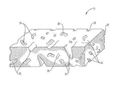
In one aspect, referring to Figure 1, the invention relates to a wound healing
material 10 comprising an extracellular matrix (ECM) but not limited to SIS,
!IBS,
UBM and LEIM, for example, that is processed to form a porous ECM foam. A
tham is defined as porous, if the majority of the volume in the three
dimensional
foam comprises cavities (pores) 20 that are empty or capable of being tilled
with a
gas such as air or a fluid. These cavities may be filled with, for example,
body
fluids, such as blood, or other solutions such as a growth factor cocktail,
vascular
endothelial growth factor, nerve growth factor, fibroblast growth factor,
epidermal
growth factor, or saline. The porous ECM foam may be partially or entirely
solidified by lyophilization or air-drying to form a sheet, i.e., a planar
shape, or other
three dimensional (i.e., non-planar) medical device. The porous ECM foam may
be
shaped to form any shape including but not limited to euboidal ECM foams for
surgical staple thickness compensation and reinforcement, porous bone implants
and
sponges, porous tube shapes for applications as a nerve graft or arterial
prosthesis, or
porous sheets of various thickness as porous wound healing matrices and porous
dermal repair scaffolds, and specifically shaped materials for filling tissue
defects
and/or for tissue augmentation after tissue resection or plastic surgery.
CA 02962203 2017-03-21
WO 2016/048946
PCT/US2015/051328
Referring to Figure 1, the size of the pores 20 in the interior of the porous
ECM foam 10, according to the invention, may differ from the size of the pores
20
that appear on the exterior of the porous ECM foam, The internal diameter of
the
pores range from about 1 micron to 500 microns, about 100 microns to 250
microns,
and more particularly, about 100-150 microns, for example. Pore sizes in this
range
are ideal for cell infiltration and exchange of body fluids or cell culture
media and
the nutrients associated with those fluids,
In a particular embodiment of the invention, porous foams are mineralized by
the addition of, for example, calcium salts or phosphate salts, calcium
nitrate,
calcium hydroxide, calcium carbonate, calcium oxide and other calcium salts or
phosphate salts from sodium phosphate, sodium dihydrogeri phosphate and
phosphoric acid, and combinations thereof. Mineralized foams are applicable to
repair of honey orthopedic injuries, such as filling gaps in a patient's bone
fracture
which otherwise would require harvesting bone from another site in the patient
to fill
the gap, spinal injuries, or head (skull) injuries.
In another embodiment of the porous ECM foam according to the invention,
the solidified porous ECM foam is milled to produce a fine porous ECM
particulate
or powder. Such powdered porous ECM foams, for example, may be aspirated into
a syringe for injection as a wound healing composition at the site of tissue
injury in a
patient. The size of the particulate in particulate porous ECM foams varies,
for
example, from about I urn to 1 millimeter, more particularly from I micron to
1
millimeter. Specifically, particulate size in the range of 100 microns to 500
microns
is preferred for flowable mixtures of the particulates. Particulates, upon
mixing with
appropriate amounts of liquid for infusion, for example, water, saline, or
phosphate
buffered saline, can produce a flowable mixture such as a gel, for example. As
used
herein, the term "flowable" means capable of being poured or extruded at room
temperature. Typical applications of the porous ECM foam particulate-
containing
flowable mixtures include but are not limited to wound healing, dermal
fillers, bone
and spinal applications (especially the mineralized foams), and intra-
articular
applications including applications for the treatment of arthritis including
but not
limited to osteoarthritis, rheumatoid arthritis, other inflammatory arthritis
types,
6
CA 02962203 2017-03-21
WO 2016/048946
PCT/US2015/051328
degenerative arthritis, septic arthritis including but not limited to Lyme
disease, gout,
and traumatic arthritis.
in another embodiment of the porous ECM foams according to the invention,
the porous foams may he applied to a medical device by coating, for example,
including but not limited to coating a surgical mesh, suture material, and
other planar
and substantially three-dimensional medical device structures. As used
throughout,
the term "medical" means related to the practice of medicine or surgery.
Coating
may be accomplished by, for example, spraying, dipping, application with a
brush or
rolling,
In another embodiment of the invention, the porous ECM foam slurry
according to the invention may be applied to the lumen of a tube or to
otherwise
form an ECM rod. The porous ECM foam slurry-filled rod may be used for
applications such as regeneration of nerve fibers or fistula closure. In a non-
limiting
embodiment, the rod may be fbrtned, for example, from a sheet or a multi-layer
sheet
of an ECM, IIBM, for example, or a sheet made from the ECM slurry. The rod is
formed by rolling the sheet(s) into a cylindrical shape and filling the tube
with the
porous ECM thain slurry. Alternatively the porous ECM foam slurry may be
spread
on the ECM sheet before it is rolled up, and then rolled up into a cylindrical
shape
enclosing the porous ECM slurry.
For neuro-regeneration applications, for example, one end of a severed nerve
may be joined to one end of a ECM rod and the other end of the severed nerve
may
be joined to the other end of the ECM rod, therefore acting as a guidance
channel to
promote neurogenesis.
For fistula repair applications, for example, the rod formed by rolling a ECM
sheet and foam into a cylinder can be inserted into the fistula. In one
embodiment of
the invention, the ECM composition could be modified such that after insertion
through the fistula tract the rod would swell to fill the irregular geometry
upon
hydration, for example, with saline.
In another embodiment of the invention with respect to a mesh 30, referring
to Figure 2A, the porous ECM foam according to the invention may be embedded
within spaces 12 of the mesh 30 as shown in Figure 2A, applied on the warp
and/or
7
weft 14, 16 of the mesh as a coating as shown in Figure 2B, or both embedded
within
spaces 12 and coated on the warp 14 and/or weft 16 of the mesh 30 as shown in
Figure 2C. The embedded porous ECM foam mesh may be made, for example, by
sandwiching a layer of mesh between two layers of foam slurry, as described
above, and lyophilized. The lyophilized foams that are obtained can be vacuum
pressed either after hydration in water or saline or without hydration, in a
vacuum
press to obtain one continuous laminated mesh like construct. By this
approach, the
ECM on one side of the foam integrates with the ECM on the other side by
becoming
embedded through the pores of the mesh.
Typical applications for such porous ECM foam enhanced medical devices
include but are not limited to hernia repair, application to infected fields,
minimization of tissue adhesions to synthetic mesh, breast reconstruction,
tissue
expanders and/or tissue augmentation, anti-inflammatory or anti-microbial
applications, and analgesia.
In yet another embodiment, the porous ECM foam according to the
invention, operates as a carrier for bioactive molecules, drugs, and other
pharmaceutical agents. For example, porous ECM foams, according to the
invention, are applied to tissue voids as defect fillers following tumor
resection.
Chemotherapeutic drugs may he added to these foams. For example, the porous
ECM foams are carriers for growth factors, small molecules and other molecules
targeted to the treatment of diseases such as cancer and diabetes, anti-
inflammatory
drugs such as steroids and non-steroidal anti-inflammatory agents (NSA1DS),
anti-
microbial agents, and analgesics for pain relief.
In another aspect, the invention is directed to a method for making porous
foams derived from ECMs, Sources of ECMs include but are not limited to UBS,
UBM, SIS and 1,BM described above.
In one embodiment of the manufacturing method of the invention, UBM, is
prepared as
described in U.S. Patent No, 6,576,265. Briefly, the urinary bladder is
removed from a mammal,
e.g., pig, sheep, or cow, and the bladder wall is delaminated from the luminal
epithelial cells by,
for example, but not limited to, soaking the urinary bladder in a hypertonic
saline
8
CA 2962203 2018-10-18
CA 02962203 2017-03-21
WO 2016/048946
PCT/US2015/051328
solution for 10 minutes to 120 minutes. Soaking removes the epithelial cells
from
the underlying epithelial basement membrane. The layers of the epithelial
tissue that
remain after this initial step are the epithelial basement membrane and all of
the
layers abluminal to the epithelial basement membrane, i.e., at least the
tunica
propria, tunica submucosa, tunica muscularis and tunica serosa. One or more
tissue
layers, for example, tunica propria, tunica muscularis mucosa, tunica
submucosa,
Mica muscularis and -mica serosa are selectively removed by mechanical
abrasion
or other mild chemical treatment to form the LIBM matrix.
After the one or more abluminal layers are selectively removed from the
urinary bladder or other epithelial tissue, the resulting matrix includes the
epithelial
basement membrane lining the laminal surface of the matrix and from which
epithelial cells and substantially all cellular elements are removed, and one
or more
tissue layers, for example, tunics muscularis mucosa, tunica propria, tunica
submucosa, tunica muscularis and tunica serosa abluminal to the epithelial
basement
membrane.
The ECM, such as LIBM described above, is rebydrated in hydrochloric acid
or sodium hydroxide at a concentration range from about 1 N to 0,001 N,
preferably
at 0.1-0.01 N I-ICI more preferably at 0.01 N HC1, for approximately 5 minutes
and,
in a blending step, blended in an industrial blender at speeds of about 500
RPM to
2500 RPM, preferably at 2000 RPM, to produce a foamy flowable ECM slurry, The
foam ECM slurry may then be neutralized, at room temperature, in, for example,
a
base such as NaOH, ranging in concentration from about 00 IN to IN, preferably
between 0.1N and IN, more preferably at IN. Other bases such as KCI, or
NaHCO3, are also useful for neutralizing the acid used to make the foamy ECM
slurry. Once the base, such as NaOH, is added to the acidic foamy ECM slurry,
the
slurry is again briefly blended to uniformly neutralize the slurry prior to
scaffold
fabrication. Typically, the than-1y ECM slurry is then poured into a mold to
form a
planar or anatomically shaped three-dimensional porous ECM foam, based on the
anticipated wound healing application. For example the three dimensional
porous
rigid or semi-rigid ECM foam may be shaped as a cylindrical rod, a tube, a
cube, or a
body part 10, e.g., a nose as shown in Figure 3, or ear, breast, cardiac
valve, dental
alveolus, and other complex three-dimensional tissues. As described above,
molds
9
CA 02962203 2017-03-21
WO 2016/048946
PCT/US2015/051328
for specific device applications include, for example, but are not limited to,
cuboida.1
molds for cuboidal porous ECM thams for stapled surgical staple thickness
compensation and reinforcement, for bone implants and sponges, for tube shaped
molds to form porous ECM tubes for applications as a nerve graft, venous or
arterial
prosthesis, tracheal prosthesis, esophageal or intestinal replacement or
anastomosis,
or sheet molds for forming porous ECM sheets as porous ECM topical wound
healing or hernia repair matrices, and/or tbr porous ECM dermal repair
scaffolds.
After the foam ECM slurry is introduced into the mold, the slurry is then
lyophilized or air dried under specific conditions, as described below, to
produce a.
molded solid or semi-solid, Le., not flowa.ble, porous ECM tbam device. The
pore
sizes on the interior of the porous ECM foam device as well as on the exterior
of the
porous ECM foam device can be controlled by lyophilization temperatures as
well as
by the materials used as the mold. Using the process described below, pore
sizes on
the exterior to the interior of the foam device ranging from, for example, 1
micron
(11M) to 500 microns, 100 microns to 250 microns, or 100-150 microns may be
obtained. Pore size may vary based on the concentration of ECM in solution and
based on the rate for freezing.
The general lyophilization step of the ECM slurry includes pre-cooling of the
lyophilizer shelves to a temperature ranging from 25 C to -40 C, from 4 C to -
20 C,
or specifically from -10 C to -20 C. Pre-cooling is followed by stabilizing
the
molded slurry at a temperature ranging from 0 C to -40 C, from 0 C to -20 C,
specifically from -10 C to -20')C, for periods of time ranging, for example,
from
between 0 minutes to 240 minutes, 0 minutes to 120 minutes, or 60 minutes to
120
minutes, to allow for ice crystal formation. During this step, the
lyophilize'. shelves
are cooled at rates of 0,01C/ min to 1C/min, for example or at 0.1C/min to
IC/min, The ice formed from the ECM slurry during this step is then sublimated
by
vacuum at the temperatures for stabilizing the molded porous ECM foam slurry
described above. The vacuum pressure used is typically in the range of 100-120
mm
Hg.
In a particular embodiment of the method of making porous ECM foams,
calcium and phosphate salts, ranging from calcium nitrate, calcium hydroxide,
calcium carbonate, calcium oxide and other calcium salts and phosphate salts
from sodium
phosphate, sodium dihydrogen phosphate and phosphoric acid are added to the
blending step above
to make mineralized porous ECM foams. The ratios of the above salts can be
varied by altering
the molar ratios, specifically to yield calcium phosphate concentrations known
in the art to mimic
native bone in vivo. These salts react in-situ to form mineralized three
dimensional foams, as
described above. The choices of acid (e.g., phosphoric vs hydrochloric), molar
ratios of the calcium
and the phosphate salts, and pH can lead to changes in the microstructure of
the mineral component
of the foam, such as brushite, apatite, monetite. Alternatively, the foams can
also be fabricated in
sodium hydroxide, with the addition of one of the components o f the mineral,
for example calcium
ions or phosphate ions, and lyophilized first and then the alternate salt
(phosphate, if calcium is
used in the first step) can be included in solution, immediately following
Syophilization, to allow
for mineralization in the foams. This is then followed by re-jyophilization to
obtain mineralized
foams.
Furthermore, other sources of mineral, specifically titanium or magnesium
derived, or
"bone-like" resorbable mineral silicate derived mineral can also be used as
alternatives to simple
calcium phosphate salts. Also, demineralized or decellularized bone matrix,
powdered allogenic
bone, hydroxyapatite and tricalcium phosphates can be used as calcium
phosphate sources during
blending for scaffold preparation.
For example, in order to manufacture one of the above foams with tricalcium
phosphate,
0.3M calcium nitrate is added along with 0.2 molar ammonium sodium phosphate,
during the
blending process described above. Alternatively other combinations can be
achieved by reacting a
range of phosphoric acid solutions from 0.01M to 1M, with several sources of
calcium, for
example, calcium nitrate, calcium acetate, calcium hydroxide, or combinations
thereof, with
concentration in the range of 0.1M to 1M, for example, 0.1M to 0.5M. The
ratios of calcium nitrate
to calcium hydroxide can be tailored from 1:1 to 10:1, depending on the type
of mineral to be
obtained as mentioned earlier.
11
CA 2962203 2019-06-19
CA 02962203 2017-03-21
WO 2016/048946
PCT/US2015/051328
In another embodiment, the porous ECM fbam devices are milled to produce
a fine powder (e.g, less than 250 microns) and loaded into a syringe. The
milling
process can vary from mortar and pestle, cryo-milling, blade milling to wire
milling.
The size and the volume of the porous ECM foam particles required will dictate
the
milling process that is used. The size of the milled particles ranges from
about I am
to 1 millimeter, particularly from about 1 micron to I millimeter. For
flowable
mixtures of porous ECM particulates, particulate sizes in the range of 10
microns to
500 microns are preferred. To form a solution of particulate porous ECM foams,
an
appropriate amount of water, saline, or phosphate buffered saline, for
example, is
used to produce a flowabie mixture that does not separate into different
phases over
a significant period of time (e.g., more than 7 days, 1-7 days, 2-5 days, or 3-
4 days,).
Typically for 10-1000 mg of porous ECM foam powder, a range of 100 microliters
to 1 milliliter saline or other fluid would yield flowable mixtures with
varying
properties for different applications, Typical applications include wound
healing,
I 5 dermal fillers, fillers for hair transplantation, hone and spinal
applications (for
mineralized foams), including applications for treatment of ostecarthritis.
In another embodiment of the method, the porous ECM foams described
above can be applied to medical devices such as a mesh, sutures, and other
planar or
three dimensional structures. Such applications on medical devices lead to
reduced
scarring and enhanced healing. In one embodiment, mesh and similar
architectures
are embedded within or laid on either side of the ECM foamy slurry prior to
lyophilization of the porous ECM foamy slurry. Alternatively, the medical
device
structure may be dipped into, painted, or sprayed with the ECM foam slurry
prior to
its lyophilization. In yet another embodiment, the ECM. foamy slurry is the
"ink" in
a 3-13 printing process used to manufacture a planar or three-dimensional
medical
device. Similarly, porous 'ECM foams may be made into three dimensional
meshes.
Typical applications for such porous ECM foam meshes include but are not
limited
to hernia repair, fistula repair, contaminated site application, anti¨adhesive
applications, breast reconstruction and application of tissue expanders and
tissue
augmentation.
12
CA 02962203 2017-03-21
WO 2016/048946
PCT/US2015/051328
Exemplification of the invention
Porous foams were venerated as UBM from urinary bladder as described
above. Sheets of UBM were soaked in 0.01 M hydrochloric acid for 30 minutes to
obtain a uniform ECM slurry. The slurry was poured in a mold and was
lyophilized
to obtain a uniform porous foam. The lyophilized porous foam was made with a
thickness of about 6 mm, and was cut to a diameter of 20 mm using a biopsy
punch
to form a foam disc. The foam discs were packaged in double-peel Tyvek pouches
and terminally-sterilized with electron beam irradiation for evaluation in
management of full-thickness wounds in the dorsal skin of three adult pigs.
At least 10 lyophilized foam discs per pig, one foam disc per defect site,
were
implanted on the skin defect on the dorsal side. The foam discs were implanted
dry
and then covered with saline moistened gauze to hydrate the foam disc. The
saline
soak gauze was covered with a non-adherent dressing. Wound dimensions were
measured at multiple time points including immediately post-implantation, Day
1,
Day 2, Day 12, Day 14, Day 18 and Day 21. Control wounds on the same animal
were treated with either saline-moistened gauze, a single application of
intact UBM
particulate (Microrviatrixe, ACell, Inc.) (IX), or repeated applications of
intact
Italy! particulate on Day 0, Day 4, Day 7, and Day 14 (4X). The wound size was
measured using calipers and quantified using images taken with a Aranz
Silouette
.. camera. For example, healing was designated as 100% (100% of the wound
diameter
remained) and 0% (the wound was completely healed leaving no defect).
Histology
was also performed to evaluate the healing potential and the quality of
healing post-
foam application.
Figure 4 is a column graph comparing the percentage of the defect diameter
that remained at time zero, 1 day, 12 days, 14 days, 18 days, and 21 days for
wounds
treated with the lyophilized tham discs, 1X (UBM particulate applied one day),
4X
(UBM particulate applied on .four days), and control.
The full thickness wounds treated with a lyophilized TIBM foam disc showed
complete healing of the wound defect at 18 days and 21 days grossly and
histologically. Histology of the foam disc treated wounds showed that healing
was
complete by 21 days. Wounds treated with a single application of foam disc
showed
13
CA 02962203 2017-03-21
WO 2016/048946
PCT/US2015/051328
healing that was marginally faster than multiple applications of the UBM
particulate,
and significantly faster than a single application of UBM particulate, The
treated
wound site showed no signs of infection. Vascularization, fat granules and
epithelialization at the defect site were observed in the samples implanted
with
foams.
14