Note: Descriptions are shown in the official language in which they were submitted.
- 1 -
METHODS AND COMPOSITIONS FOR MODULATING TH-GM CELL
FUNCTION
RELATED APPLICATION
[0001] This application claims the benefit of Singapore Patent Application
No.
10201406130P, filed September 26, 2014.
BACKGROUND OF THE INVENTION
[0002] A significant body of research has led to the current model of
immunity and
inflammation, as well as the dysregulation in immune and inflammatory
disorders. It is
currently understood that CD4+ helper T (TH) cells play a crucial role in host
defense against
various pathogens by orchestrating adaptive and innate immune responses. Upon
T-cell
receptor (TCR) activation by cognate antigen, naïve CD4 T cells are committed
to
differentiate into at least five major subsets: TH1, TH2, TH17, iTr,g and TFH,
which are
modulated by cytokine milieus. TH1 and TH17 cells are known to be the primary
effectors of
inflammation. However, the pathogenic roles of either TH1 or TH17 in various
inflammatory
disorders remain unclear. For example, recent studies conflict with previously
understood
paradigm of TH17 in multiple sclerosis (MS) pathogenicity (Haak et al., 2009),
making it
more challenging to identify potential drug targets for MS therapy. Similarly,
while
rheumatoid arthritis (RA) is traditionally understood to be a disorder
mediated by tumor
necrosis factor a (TNF-a), up to 40% of RA patients fail to respond to anti-
TNF-a treatment.
[0003] Accordingly, there remains a significant unmet need for effective
treatment
methods for autoimmune and inflammatory disorders such as, e.g., MS and RA.
SUMMARY OF THE INVENTION
[0004] The present disclosure relates, in part, to the identification of an
interleukin-7 (IL-
7) /signal transducer and activator of transcription 5 (STAT5)-regulated
granulocyte
macrophage colony-stimulating factor (GM-CSF)/IL-3-producing TH cells, termed
TH-GM,
which represent a distinct helper T cell subset with unique developmental and
functional
characteristics. Identified herein is an inflammatory pathway mediated by TH-
GM cells (TH-
GM-mediated inflammatory pathway) , which represents an independent
inflammatory
Date Recue/Date Received 2020-09-24
CA 02962757 2017-03-27
WO 2016/048247 PCT/SG2015/050344
- 2 -
pathway apart from known non-TH-GM-mediated inflammatory pathways (e.g., TNF-
a, IL-6,
and IL-lb pathways of inflammation). The present disclosure provides methods -
and
compositions for diagnosing inflammatory disorders that are Ti-GM-mediated,
and
modulating Ti-GM cell function for the treatment of inflammatory disorders
mediated by the
T1-1-GM pathway,
[0005] Accordingly, in one aspect, the present disclosure provides a method
of
diagnosing a TH-GM-mediated inflammatory disorder in a patient suffering from
an
inflammatory disorder, comprising: a) contacting a sample collected from a
patient suffering
from an inflammatory disorder with a detecting agent that detects a
polypeptide or nucleic
acid level of STAT5 (e.g., phospho-STAT5 (Tyr694)), IL-7, GM-CSF or IL-3, or a
combination thereof; and b) quantifying the polypeptide or nucleic acid level
of STAT5 (e.g.,
phospho-STAT5 (Tyr694)). IL-7. GM-CSF or IL-3, or a combination thereof,
wherein an
increased level of STAT5 (e.g., phospho-STAT5 (Tyr694)). interleukin-7 (IL-7),
GM-CSF or
interleukin-3 (IL-3), or a combination thereof relative to a reference level
indicates that the
patient suffers from a TH-GM-medialed inflammatory disorder.
[0006] In another aspect, the present disclosure provides an isolated
population of GM-
CSF-secreting T-helper cells (TH-GM), wherein the Ti-GM cells are
differentiated from
cluster of differentiation 4 (CD4+) precursor cells in the presence of IL-7
and activated
STAT5, and wherein the T11-GM cells express GM-CSF and 1L-3. =
100071 In another aspect, the present disclosure provides a method of
modulating T11-GM
function, comprising contacting the Tt,rGM, or CD4+ precursor cells, or both,
with a
modulating agent that modulates TH-GM function.
[0008] in some aspects, the present disclosure provides a method of
treating a T0-GM-
mediated inflammatory disorder in a patient in need thereof, comprising
administering to said
patient an effective amount of a modulating agent that modulates T11-GM cell
function.
[0009] In other aspects, the present disclosure provides a method of
treating rheumatoid
arthritis in a patient who exhibits limited response to anti-tumor necrosis
factor alpha (TNF-a)
therapy, comprising administering to said patient an effective amount of a
modulating agent
that modulates T1:1-GM function.
[0010] In another aspect, the present disclosure provides a method of
treating a STAT5-
mediated inflammatory disorder in a patient in need thereof, comprising
administering to the
patient an effective amount of an agent that modulates STAT5 function.
CA 02962757 2017-03-27
WO 2016/048247 PCT/SG2015/050344
- 3 -
[WM In further aspects, the present disclosure provides a method of
screening to
identify a modulator of TH-GM cell function, comprising contacting an isolated
population of
T1-GM cells, or an isolated population of CD4+ precursor cells, with a
candidate agent, and
determining a readout of TH-GM function in the presence or absence of the
candidate agent,
wherein a change in the readout of T11-GM function indicates that the
candidate agent is a
modulator of T11-GM function.
[0012] The present disclosure enables the identification or classification
between
inflammatory disorders that are either primarily T11-GM-mediated, or primarily
non-T11-GM-
mediated (e.g., mediated by TNE-a. IL-6, and/or IL-113), or both. Thus, using
the methods
described herein, it is possible to determine whether a patient suffering
from, e.g., RA, suffers
from an RA that is primarily TH-GM-mediated, or primarily non-T0-GM-mediated,
or both.
This differentiation allows for a more targeted and tailored method of
treating inflammatory
disorders such as RA, for which current treatments are only 40% effective.
Further, the
present disclosure provides methods and compositions for prognosing the
progression of an
inflammatory disorder so as to tailor the treatment according to the stage of
the disease.
Also provided herein are compositions and methods for and the treatment of
inflammatory
disorders, particularly those that are T11-GM-mediated.
BRIEF DESCRIPTION OF THE DRAWINGS
[0013] The foregoing will be apparent from the following more particular
description of
example embodiments of the invention.
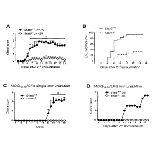
[0014] FIGS. 1A-3 D depict Sia/5-conditional mutant mice are resistant to
EAE. Clinical
EAE scores (FIG. 1A) and incidence (FIG. 1B) of Stat5+/+ and Stat5-/- mice
immunized twice
with MOG55/CFA. Data are representative of three independent experiments (FIG.
1A) or
pooled from three experiments (FIG. 111, n=18 per group). Clinical scores of
EAE mice
immunized once with MOG35_55/CFA (FIG. IC, n=5 per group) or immunized twice
with
MOG35_55/LPS (FIG. ID). Data are representative of Iwo independent
experiments.
[0015] FIGS. 2A-2D depict reduced ncuroinflammation in Siat5 conditional
mutant mice.
Histology of spinal cord sections obtained from EAE mice at day 9 after 2n11
immunization
(FIG. 1A). Images shown are representative of two independent experiments with
three mice
per group. Scale bars, 200 1,tm (top), 50 luri (bottom). CD4+ and CD11.b+
cells in spinal cord
sections were stained by immunofluorescence (FIG. 1B), Images shown are
representative of
CA 02962757 2017-03-27
WO 2016/048247 PCT/SG 2015/050344
- 4 -
t wo independent experiments with three mice per group. Scale bars, 200 um.
CNS
mononuclear cells were analyzed by flow cytometry at peak of disease (FIGS. 2C
and 2D).
Right panels are cell proportions (FIG. 2C, right) or cell numbers (FIG. 2D,
right) pooled
from two experiments (n=9).
[0016] FIGS. 3A and 3B
depict the resistance to EAE in 5m/5-deficient mice is
independent of Tul. T1117 or Tre, cells. Flow cytometric
analysis of IL-17 and IFN-y
expression by CNS-infiltrating CD4+ T cells at peak of disease (FIG. 3A). Data
arc
representative of three independent experiments. Percentage of CD25+ among
CD4* T cells
in the CNS of Sta/5*/* and Slati-/- -FINE mice at peak of disease were
analyzed by flow
cytometry (FIG. 3B).
[0017] FIGS. 4A-4C
depict conditional 81at5 mutant mice have no defect in CD4* T cell
generation in periphery. Spleens were obtained from MOGA5_55/CFA-immunized
Slat5+/+ and
Stat51- mice at day 7 (FIG. 4A) and clay 21 (FIG. 4B). The proportions of CD4+
and CD84- T
cells were analyzed by flow cytometry. The absolute number of CD4* T cells was
calculated
(right panels). Data are representative of two independent experiments (FIG.
4A) or pooled
from two independent experiments (FIG. 4B). 1L-17 and IFN-y expression by
splenic CD4+ T
cells of Stat.5*/* and Stol5"/- EAE mice was determined by intracellular
cytokine staining
(FIG. 4C). Data are representative of three independent experiments. *p<0.5,
**p<0.005,
***p<0.0005.
1100181 FIGS. 5A-5D
depict Sta15-deficient CD4* T cells can infiltrate CNS but fail to
induce effective neuroinflammation. CCR6. CXCR3 and CD69 expression by splenic
CD4+
T cells of 5tat5*/* and Stat51- EAE mice was measured. Data are representative
of two
independent experiments with three to five mice per group (FIG. 5A). CNS-
infiltrating CD4*
T cells were analyzed at day 7, 9 and 2.1 after first M0G3.5_55/CFA
immunization (FIGS. 5B-
5D). Cell numbers were calculated (FIG. 5D). Data are representative of two
independent
experiments with three mice per group. *p<0.5.
[0019] FIGS. 6A-6C show
resistance to EAE in Stat5-1- mice is not caused by any defect
in the survival of CD4* T cells in the absence of STAT5. CD4+ T cell
infiltration (FIG. 6A)
and clinical scores (FIG. 6B) of Rag2-/. recipient mice transferred with
different numbers of
Stai5+(+ and Siat51- CD4* T cells. Clinical scores and frequencies of CDT' T
cells in the CNS
at day 21 (disease peak) of EAE induction (FIG. 6C). *p<0.05, ***p<0.0005.
CA 02962757 2017-03-27
WO 2016/048247 PCT/SG2015/050344
- 5 -
[0020] FIGS. 7A-7C depict the intrinsic defect of Stat5-deficient CD4+ T
cells in
encephalitogenicity. Clinical EAE scores (FIG. 7A) and incidence (FIG. 7B) of
Rag21" mice
(n=5 per group) after adoptive transfer of 2 million MOG35_55-specific Stal5+/
or Slat51-
CD4+ T cells respectively. IL-17 and IFN-y expression by CNS-infiltrating CD4+
T cells was
measured at peak of disease (FIG. 7C). Data represent two independent
experiments.
*p<0.05.
[0021] FIGS. 8A-8D depict the diminished induction of GM-CSF in splenic
Siat.5-f-CD4+
T cells. In FIGS. 8A-8D, splenocytes were obtained from MOG35_55/CFA-immunized
Stas5+/+ and Sia/5"/" mice (n..3 per group) before disease onset and
challenged with M0G35_55
at various concentrations for 24 h. GM-CSF secretion was measured by ELISA
(FIG. 8A).
Golgiplug was added in the last 4 h of M0G35..55 (20 pg/m1) challenge and the
frequencies of
IL-17+ and GM-CSF+ cells among CD4+CD44hi T cells were measured (FIG. 8B). In
FIGS.
8C and 8C, splenocytes were obtained from MOG35_55/CFA-immunized Siat51.,
Stat3-l" or
wild-type control mice and stimulated with PMA/Ionomycin in the presence of
Golgiplug for
4h. The frequencies of IL-17+ and GM-CSF+ cells among splenic CD4+CD441' T
cells were
measured by intracellular cytokine staining. *p<0.05, ***p<0.001.
[0022] FIGS. 9A-9C depict the diminished induction of GM-CSF in CNS-
infiltrating
Stat5-/- CD4+ T cells. In FIG. 9A, IL-17, IFN-y and GM-CSF expression by CNS-
infiltrating
CD4+ T cells of Stat5+R- and Stat5I- mice was measured at peak of disease. The
percentage of
GM-CSF+ cells amongIL-17+ or IFN-y+ cells was calculated (bottom right. FIG.
9A). IL-17,
IFN-y and GM-CSF expression by CNS-infiltrating CD4+ T cells of Ragli-
recipient mice at
peak of disease in adoptive transfer EAE (FIG. 9B). Time-course analysis of
cytokine mRNA
expression in the CNS of naïve and -M0G35_53/CFA-immunized S'tat.54/+ and
Stat5'1. mice
(n=3 per group at each time point). The RT-PCR data were normalized to Rn18S,
and
expression in naïve mice was set to 1 (FIG. 9C). Data represent two
independent
experiments. *p<0.05.
[0023] FIGS. 10A-1.0C show STAT5-mediated GM-CSF induction is independent
of IL-
23 or 1L-l3 signaling. In FIG. 10A, purified CD4+ T cells were cultured with
TGF-(3 and EL-
6 for 3 days, followed by starvation for 6 h. Then cells were treated with
various cytokines
for 30 min, and ,pSTAT3 and pSTAT5 was determined by immunoblotting. STAT3 and
STAT5 were further detected after stripping. FIG. 1.0B shows the mRNA
expression of IL-
23R and IL-IRI in splenic CD4+ T cells of Stal5+4 and Stat5I- EAE mice (n=3
per group).
CA 02962757 2017-03-27
WO 2016/048247 = PCT/SG2015/050344
- 6 -
The RT-PCR data were normalized to 13-Actin. In FIG. 10C, splenoeytes were
obtained from
MOG35_55/CFA-immunized WT mice before disease onset and challenged with
MOG35_55 (20
1.1.2/m1) in the absence or presence of IL-2 for 48 h. The frequencies of GM-
CSF+ and IL-17+
cells among CD4+CD4411IT cells were measured by flow cytometry. *p<0.05.
[0024] FIGS. 11A-11C depict 11,7-induced STAT5 activation promotes GM-CSF
expression in autoreactivc CD4+ T cells. Splenocytes were obtained from
MOG35_55/CFA-
immunized Siat-51-/+ and Stat5"1" mice before disease onset and challenged
with MOG35..55 (2(
Ki/m1) in the absence or presence of 1L-7 for 48 h. Frequencies of GM-CSF+ and
IL-17+ cells
among CD4+CD4411 T cells were measured by flow cytometry (FIG. 11A). GM-CSF
secretion was measured by ELISA (FIG. 11B). Data represent two independent
experiments
with two to three mice per group. Splenic CD62LhiCD441' and CD62LI0CD44h1 T
cells from
MOG35_55/CFA-immunized mice were sorted out. Cells were stimulated with anti-
CD3 and
anti-CD28 in the absence or presence of .IL-7 for 4 h and then harvested for
the analysis of
GM-CS F expression by RT-PCR (FIG. 11C). *p<0.05
[0025] FIGS. 12A-1.2F depict EL-7Ra neutralization attenuates GM-CSF
expression and
ameliorates EAE. Clinical scores of EAE mice (n=5) treated with anti-IL-7Ra or
normal IgG
given every other day from day 5 after 2"a immunization, as indicated by
arrows. Data
represent two independent experiments (FIG. 12A). Spinal cord sections were
obtained from
EAE mice at day 11 after 2'd immunization. Immune cell infiltration was
assessed
histologically. Images shown are representative of three individuals per
group. Scale bars,
200 am (top), 50 am (FIG. 12B, bottom). The percentages of CD4+ and CD8+ T
cells in
spleens of EAE mice. Data represent two independent experiments (FIG. 12C).
FIGS. 12D
and 12E illustrate the frequencies of GM-CSF+, IL-17+ and IFN-y+ cells among
CD4+ T cells
in the CNS of EAE mice receiving different treatment. The mRNA expression of
IFN-y,, IL-
17 and GM-CSF in the CNS of EAE mice (FIG. 12F). *p<0.05
[0026] FIGS. 13A and 13B depict the differentiation of GM-CSF-expressing
Tit cells is
distinct from T1117 and THL Naive CD4f T cells were primed with plate-bound
anti-CD3 and
soluble anti-CD28 in the presence of a combination of various cytokines and
neutralizing
antibodies as indicated. GM-CSF, 1L-17 and IFN-y expression was analyzed by
intracellular
staining (FIG. 13A) or RT-PCR (FIG. 13B)
[0027] FIGS. 14A-14D show the effect of IL-2 and IL-6 on TH-GM
differentiation from
naïve T cells. GM-CSF and IFN-y expression in naive CD4+ T cells activated for
72 h with
CA 02962757 2017-03-27
WO 2016/048247 PCT/SG2015/050344
- 7 -
anti-CD3 alone or plus anti-CD28 (FIG. 14A). In FIG. 14B, sorted naive CD4+ T
cells were
stimulated with anti-CD3 and anti-CD28 in the presence of neutralizing
antibodies against
IL-12 and IFN-y without or with the addition of IL-6. The frequencies of GM-
CSF+ and IL-
17+ cells were measured by intracellular staining (FIG. 14B). In FIG. 14C,
naive CD4+ T
cells from Stat3+4 and Stat.34- mice were polarized under conditions as
indicated for 72 h.
The frequencies of GM-CSF+ and IL-17+ cells were analyzed. In FIG. 14D, naive
CD4+ T
cells were activated with anti-CD3 and anti-0O28 in the presence of IL-2 or
anti-IL-2. The
frequencies of GM-CSF+, IL-17+ and 1FN-y4 cells were analyzed.
100281 FIGS. I5A-15F depict IL-7-STAT5 signaling programs TH-GM
differentiation
from naive precursor cells. Naïve CD4+ T cells were primed with plate-bound
anti-CD3 and
soluble anti-CD28 in the presence of various concentration of IL-7 as
indicated. GM-CSF and
IFN-y expression was analyzed by intracellular staining (FIG. 15A) or EL1SA
(FIG. 15B). In
FIGS. I.5C and 1.5D, Stat5+4 and Star5./' naïve CD4+ T cells were activated
with anti-CD3
and anti-CD28 in the presence IL-7 for 3 days. GM-CSF, 1L-17 and IFIN-y
expression was
analyzed by intracellular cytokine staining (FIG. 1.5C). GM-CSF secretion was
measured by
ELISA (FIG. 15D). Immunoblotting of pSTAT5 and STAT5 in IL-7-stimulated CD4+ T
cells
isolated from Slat5-/- or control mice (FIG. 15E). The ChIP assay was
performed with
Stat5+1+ and Stal5-/- CD4+ T cells using normal IgG or STAT5-specific
antibody. The binding
of antibodies to pi2 promoter region was detected by RT-PCR (FIG. 15F).
100291 FIGS. 16A and 16B depict the differentiation conditions for T11-GM
subset.
Naive CD4+ T cells were activated with anti-CD3 and anti-CD28 in the presence
of 1L-7
or/and anti-IFN-y as indicated. GM-CSF. IL-17 and IFN-y expression was
analyzed (FIG.
16A). The mRNA expression of T-bet and RORyt in naive, THI (1L-12 + anti-IL-
4), T1117
(TGF-13 + 1.1,6 + anti-IFN-y + anti-IL-4) and TH-GM cells (IL-7 + anti-IFN-y)
(FIG. 16B).
The RT-PCR data were normalized to Gapdh, and expression in naive T cells was
set to I.
100301 FIGS. 17A-17E illustrate that IL-7 but not 1L-2 induces STAT5
activation and
GM-CSF expression in naive CD4+ T cells. FIGS. 17A-17C show flow cytometry of
CD25
and CD127 on the surface of naïve CD4+ T cells or cells activated with anti-
CD3 and anti-
CD28 at various time points as indicated. Activation of STAT5 in naive CD4+ T
cells
stimulated with 1L-2 or IL-7 for 30 min (FIG. 17D). FIG. 17E shows the mRNA
expression
of GM-CSF in naive CD4+ T cells stimulated with anti-CD3 and anti-CD28 in the
presence
CA 02962757 2017-03-27
WO 2016/048247 = PCT/S62015/050344
- 8 -
of 1L-2 or IL-7. The RT-PCR data were normalized to 13-Actin, and expression
in naive T
cells activated for 2 h without cylokine was set to 1.
[0031] FIGS. 18A-18C
show that both IL-2 and 1L-7 can induce STAT5 activation and
GM-CSF expression in activated CD4+ T cells. As shown in FIGS. 18A and 18B,
CD4+ T
cells were activated with anti-CD3 and anti-CD28 for 3 days. After resting in
fresh medium,
cells were stimulated with 1L-2 or IL-7 at various time points. The pTyr-STAT5
and 13-Actin
were detected by immunoblotting (FIG. 18A). GM-CSF tnRNA expression was
measured by
RT-PCR (FIG. 18B). The RT-PCR data were normalized to 13-Actin, and expression
in cells
without cytokine stimulation was set to I . The ChIP assay shown in FIG. 18C
was performed
with normal IgG or STAT5-specific antibody. The binding of antibodies to C's/2
promoter
region was detected by RT-PCR.
[0032] FIG. 19 depicts
surface molecules selectively expressed at high level or low level
in T11-GM subset as characterized by rnicrommy analysis. These surface
molecules specific
for each lineage serves as markers, signatures and potential targets for novel
diagnosis,
treatment and prevention of autoimmune inflammation including. but not limited
to multiple
sclerosis and rheumatoid arthritis. These cell surface molecules are listed in
detail in Table 1.
The order of naive, Th I. Th17, and Th-GM as indicated in the figure insert is
the same as that
appears for the bars in each graph.
[0033] FIGS. 20A-20D
show that IL-3 is preferentially expressed in T11-GM cells. In
FIGS. 20A and 20B, naive CD4+ T cells were activated with anti-CD3 and anti-
CD28 under
THI- (IL-12 + anti-IL-4), T1117- (TGF-13 + TL-6 + anti-IFN-7 + anti-IL-4) and
TH-GM- (GM-
CSF+ TH, IL-7 + anti-TN-7 + anti-IL-4) polarizing conditions. GM-CSF and IL-3
expression
was analyzed by intracellular staining (FIG. 20A). The mRNA expression of IL-
3, E131-3,
.PENK or RANKL cytokines was measured by RT-PCR 20B). Frequency
of IL-3+ cells
differentiated without or with 1L-7 (FIG. 20C). GM-CSF and IL-3 expression by
WT or
STAT5-deficient GM-CSF-producing TH cells (FIG. 20D).
[0034] FIG. 21 depicts
clinical EAE scores of Rag.2-7" mice (n=3-6 mice per group) after
adoptive transfer of 6X10⁵ various M0G35_55-specifie TH subsets.
[0035] FIGS. 22A-27E
depict inhibition of STAT5 activation suppresses T11-GM cell
differentiation in vitro. CD4+ T cells were pre-incubated with STAT5 inhibitor
(Calbiochem)
(FIG. 22A) or JAK3 inhibitor (FIG. 22B) at indicated concentrations or vehicle
(-) for 1 11
before stimulation with 1L-7 for 30 min. Activation (Tyr694 phosphorylation)
of STAT5 was
CA 02962757 2017-03-27
WO 2016/048247 PCT/SC20151050344
- 9 -
determined by immunoblotting. CD4+ T cells were pre-incubated with STAT5
inhibitor at
indicated concentrations or vehicle (-) for 1 h before stimulation with IL-6
for 30 min.
Activation (Tyr705 phosphorylation) of STAT3 was determined by immunoblotting
(FIG.
22C). In FIG. 22D, CD4+ T cells were pre-incubated with STAT5 inhibitor at
indicated
concentrations or vehicle (-) for 1 h before stimulation with 1FN-y for 30
min. Activation
(Tyr701 phosphorylation) of STATI was determined by immunoblotting. In FIG.
22E, naïve
CD4+ T cells were isolated and activated under neutral condition or T11-GM
cell-favoring
condition with the addition of different concentrations of STAT5 inhibitor for
3 days. GM-
CSF and IFN-y expression was analyzed by intracellular cytokine staining and
flow
eytotnetry.
100361 FIGS. 23A-23D depict targeting STAT5 activation by chemical
inhibitor
ameliorates EAE. (FIG. 23A) Clinical EAE scores of wild-type control mice
(n=5) or
administrated with STAT5 inhibitor (Calbiochem). Arrow indicates the treatment
points.
(FIG. 23B) Histology of spinal cords at day 18 of EAE mice receiving different
treatments.
(FIG. 23C) Intracellular staining and flow cytometry of CNS-infiltrating CD4+
T cells at peak.
of disease. (FIG. 23D) Whole CNS was harvest for RNA extraction. GM-CSF mRNA
expression was analyzed by RT-PCR. Data represent two independent experiments.
*p<0.05.
[0037] FIGS. 24A-24E depict GM-CSF-producing TH cells are in association
with human
RA. Plasma concentrations of GM-CSF and TNF-o. in healthy control HC (n=32)
and RA
(n=47) were quantified by ELISA (FIG. 24A). In FIGS. 24B and 24C. peripheral
blood
mononuclear cells (PBMCs) were collected from healthy control (I-IC) and
rheumatoid
arthritis (RA) patients, and were stimulated for 4 h with PMA/Ionomycin in the
presence of
Golgiplug, followed by intracellular eytokine staining. Representative flow
cytometry of
GM-CSF. IFN-y and IL-17 in CD4+ T cells (FIG. 24B) and statistics of n>=9 per
group (FIG.
24C) are shown. FIG. 24D shows the correlation between the frequency of GM-
CSF1FN-1-
T11 cells and the level of plasma GM-CSF in peripheral blood of RA patients
(n,--.18).
Cytokine expression by CD4+ T cells derived from synovial fluid of RA patients
was
analyzed by intracellular cytokine staining and flow cytometry (FIG. 24E). A
representative
image of three cases was shown. *p<0.05, **p<0.01, ***p<0.001; ns, not
significant.
[0038] FIGS. 25A-25E depict distinguishable effects of GM-CSF and TNF-ct in
mouse
A.R. FIG. 25A shows knee joint swelling of wild-type mice over 7 days after
intraarticular
injection of 100 lig mBSA in AIA model, receiving treatment with control IgG,
GM-CSF-
- 10 -
specific and TNF-a-specific neutralizing antibodies separately or in
combination (n=5 per
group) at indicated times (arrows). FIG. 25B shows knee joint swelling of
Stat.5+I+ and Stat5-1-
mice (n=6 per group) over 7 days after arthritis induction. Data are
representative of more
than three independent experiments. Representative images of joint sections
stained with
H&E (FIG. 25C) or Safranin-OTm/Fast Green (FIG. 25D) at day 7 after arthritis
induction as
in FIG. 25C. Bars, 500 pm (FIG. 25C upper panels and FIG. 25D) or 100 pm (FIG.
25C
lower panels). Arrow in upper panels (FIG. 25C) indicated bone destruction. In
FIG. 25E,
serum concentrations of GM-CSF, IFN-y and TNF-a in Stat5+I+ and Stat5-1- AIA
mice were
quantified by ELISA. Statistics of n>=8 per group were shown. *p<0.05,
**p<0.01,
***p<0.001.
100391 FIGS. 26A-26D depicts mice with Stat5 deletion in T cells are
resistant to CIA.
(FIG. 26A) Representative images of paw swelling of Stat5+1+ and Stat.54- mice
at day 40 after
collagen II/CFA immunization in CIA model. (FIG. 26B) Clinical score of
Stat5+I+ and Stat5-
I- mice (n=5 per group) over 40 days after collagen II/CFA immunization. Data
are
representative of two independent experiments. (FIG. 26C) Representative
images of paw
sections stained with H&E at day 40. (FIG. 26D) Serum concentrations of TNF-a
(n=8 per
group) were quantified by ELISA. *p<0.05, **p<0.01, ***p<0.001.
100401 FIGS. 27A-27E depicts STAT5-deficient CD44 T cells are defective in
arthritogenic potential. (FIGS. 27A and 27B) Representative flow cytometry of
CD4+ T cells
in spleens (FIG. 27A) and inguinal lymph nodes (FIG. 27B) of Stat5+/ and
Stat5-/- mice at
day 7 after AIA induction. (FIGS. 27C and 27D) Synovial tissues were harvested
from
Stat5 / and Stat5-/- mice at day 7 after AIA induction, and dissociated into
single cells. Cell
numbers of CD45+ leukocytes were calculated (FIG. 27C). The percentages of
CD4+ T cells
among CD45+ fraction were analyzed by flow cytometry, and cell numbers were
calculated
(FIG. 27D). (FIG. 27E) Histological analysis of joint sections from wild-type
naïve mice at
day 7 after being transferred with in vitro-expanded antigen-reactive CD4+ T
cells and
followed with intraarticular injection of mBSA. Bars, 100 pm. Data represent
two
independent experiments. *p<0.05; ns, not significant.
100411 FIGS. 28A-28G depicts STAT5-regulated GM-CSF-producing TH cells are
crucial
for AIA. Spleens and synovial tissues were collected from Stat5 / and Stat5-/-
mice at day 7
after arthritis induction. (FIG. 28A) Splenic fractions of wild-type AIA mice
(n=3) were
stimulated under indicated conditions for 18 h. GM-CSF levels in supernatant
were
Date Recue/Date Received 2021-02-25
CA 02962757 2017-03-27
WO 2016/048247 PCT/SC2015/050344
- I I -
quantified by ELISA. (FIGS. 28B-28D) Intracellular staining and flow cytometry
of GM-
CSF. IL-17 and IFN-7 in splenie CD4+CD44hi effector T cells (FIG. 28B) or in
synovial
infiltrating CD4+ T cells (FIGS. 28C and 28D) after restimulation for 4 h with
PMA/Ionomycin in the presence of Golgiplug. Representative images and
statistics of n=5
(FIG. 2813, right panels) or n=3 (FIG. 28D. right panels) per group were
shown. Data
represent two independent experiments. (FIG. 28E) Protein expression of
several
proinflammatory cytokines in synovial tissues was measured by ELISA. (FIGS.
2SF and 28G)
Representative images of joint sections stained with H&E (FIG. 28F) or
Safranin-O/Fast
Green (FIG. 28G) at day 7 after intraarticular injection of mBSA alone to the
right knee joints
and InBSA supplemented with GM-CSF to the left knee joints. Bars, 500, 50 or
200 pm as
indicated. Data represent two independent experiments.*p<0.05, "p<0.01,
***p<0.001 us,
not significant. "Splenoeytes" represent the left-most bars in each group,
"splenoeytes
depleted of CD4+ T cells" represent the middle bars in each group, and "CD4+ T
cells"
represent the right-most bars in each group.
[0042] FIGS. 29A-29C depicts loss of STAT5 results in impaired GM-CSF
production by
antigen-specific CD44" T cells. Spleens and inguinal LNs were collected from
Stat544 and
Stat5-/- mice at day 7 after arthritis induction, and dissociated into single
cell suspensions.
Then, cells were stimulated with mBSA (20 pg/ml) for 24 h. (FIG. 29A)
Gol.giplug was
added in the last 4 Ii of culture. followed by intracellular staining and flow
cytometry.
Representative plots of GM-CSF, 1L-17 and IFN-7 expression in CD4+CD44hi
effector T cells
was shown, representing two independent experiments. (FIGS. 29B and 29C)
Secreted
cytokines in the supernatant (n=3 per group) were quantified by ELISA. Data
represent two
independent experiments. *p<0.05; ns, not significant.
100431 FIGS. 30A-30C depicts loss of STAT5 impairs IL-6 and IL-113
expression in
synovial tissues of arthritic mice. (FIGS. 30A-30C) The mRN.A (FIGS. 30A and
30 C) and
protein (FIG. 30B) expression of several proinflammatory cytokines in synovial
tissues of
Stat5+4 and Stat5-/- mice (n>=3 per group) at day 5 or 7 after arthritis
induction was
measured by qPCR and ELISA. The qPCR data were normalized to Rn18S.
[0044] FIGS. 31A-31D depicts SAT5-induced GM-CSF expression mediates CD11b+
cell accumulation in inflamed synovial tissues. (FIG. 31A) The frequencies of
CD] lb+ cells
in spleens of Stat5+/+ and Stat.51- AIA mice were analyzed by flow eytometry.
Statistics of
n=3 per group (right panel) were shown. (FIG. 31B) Synovial tissues were
harvested from
CA 02962757 2017-03-27
WO 2016/048247 PCT/S62015/050344
- 12 -
Stat5+7+ and Stat51" mice at day 7 after arthritis induction, and dissociated
into single cell
suspensions. The percentage of CD1 lb + myeloid cells among CD45+ fraction was
analyzed
by Bow cytometry. Statistics of n=5 per group were shown in right panel. (FIG.
3.1C)
Representative flow cytometry of CDI lb+ and CD4+ cells gated on synovial
CD45+ fraction
over 7 days after arthritis induction. (FIG. 31D) Flow cytometric analysis of
CD4+, CD11b+
and 16220+ cell infiltration in synovial tissues of Stat5+/+ and Stat.5-1-
mice at day 7 after
intraarticular injection of mBSA alone to the right knee joints and mBSA
supplemented with
GM-CSF to the left knee joints. Representative images were shown. All data
shown are
representative of two independent experiments. "p<0.01; ns, not significant.
[00451 FIGS. 32A-32D depicts GM-CSF mediates neutrophil accumulation in
arthritic
mice. (FIG. 32A) Flow cytometric analysis of Ly6C and Ly6G expression gated on
synovial
CD45+CD11b+ fraction over 7 days after arthritis induction. (FIG. 32B) Giemsa
stain of
sorted Ly6Ch1Ly6G- and Ly6CLy6Gh1 cells from synovial tissues of AlA mice.
Scale bar,
100 pm (left) or 20 pm (right). (FIG. 32C) Flow cytometric analysis of
Ly6ChiLy6G- and
Ly6CI'Ly6Gill populations in synovial tissues of Stat5+/+ and Stat.5-1- mice
at day 7 after
intraarticular injection of mBSA alone to the right knee joints and mBSA
supplemented with
GM-CSF to the left knee joints. (FIG. 32D) Knee joint swelling of wild-type
mice treated
with Ly6G-specific neutralizing antibody (1A8) or lgG control (n=5 per group)
over 3 days
after intraarticular injection of mBSA in AIA model. Arrows indicate time
points of antibody
administration. *p<0.05.
[0046] FIGS. 33A-33C depicts (IM-CSF enhances neutrophil transmigration and
delay
apoptosis in vitro. (FIG. 33A) Percentages of migrated neutrophils with or
without GM-CSF
as chemoattractant in transmigration assay at 3 h post start. (FIG. 33B)
Microscopic images
of CFSE-labeled neutrophils in the bottom of the lower chamber in
transmigration assay.
(FIG. 33C) Sorted neutrophils were cultured in vitro with or without GM-CSF
(20 ng/ml) for
24 h. Neutrophils undergoing apoptosis were examined by Annexin V and
propidium iodide
(PI) co-staining. A representative flow cytometry of three repeats was shown.
*p<0.05.
[0047] FIGS. 34A-341 depicts GM-CSF mediates proinflatnmatory cytokine
expression
by myeloid cells and synovial fibroblasts in arthritic mice. Synovial tissues
were dissected
from wild-type AIA mice and dissociated into single cell suspensions. (FIG.
34A) Flow
cytometry plots depicting the fractionation into different populations based
on differential
expression of surface markers. (FIG. 34B) The inRNA expression of several
proinflamtnatory
CA 02962757 2017-03-27
WO 2016/048247 PCT/SC 2015/050344
- 13 -
eytokines in sorted CD45+TCR[3+ (TCRI3+ in short), CD45+TCR.f3-CD11c-CD11h+
(CD1 lb)
and CD454TCRp-CD11e+ (CD11c+) populations was measured by qPCR. The qPCR data
were normalized to GAPDH. (FIG. 34C) The mRNA expression of 1L-6, IL-113 and
TNF-a in
sorted Ly6Ch1Ly6G- and Ly6Ckty6Ghi populations (gated on CD1 lb + cells). The
qPCR data
were normalized to GAPDH. (FIGS. 341) and 34E) The mRNA expression of IL-6 and
IL-1.13
by BMDMs (FIG. 34D) and BMDCs (FIG. 34E) upon stimulation with 20 ng/ml GM-CSF
for 1 h. The qPCR data were normalized to GAPDH. (FIGS. 34F and 34G) BMDMs
(FIG.
34F) and BMDCs (FIG. 34G) were stimulated with GM-CSF at indicated
concentrations
(n=3 per group) for 18 h. The secretion of IL-6 in the culture supernatant was
quantified by
ELISA. (FIG. 34H) BMDMs were primed with LPS (100 lig/m1) in the presence of
GM-CSF
at indicated concentrations (n=3 per group) for 6 h., followed by stimulation
with ATP (5 mM)
for 30 min. The secretion of IL-10 in the culture supernatant was quantified
by ELIS A. (FIG.
341) Cells were cultured in DMEM medium supplemented with 10% FBS for over 20
days
with more than 5 passages to obtain synovial fibroblasts. Synovial fibroblasts
were stimulated
with GM-CSF (20 ng/m1) for 1 h and harvested for RNA extraction. The mRNA
expression
of IL-113 was measured by qPCR. The qPCR data were normalized to GAPDH. All
data
shown represent two independent experiment s.1:p<0.05,
DETAILED DESCRIPTION OF THE INVENTION
[0048] A description of example embodiments of the invention follows.
[0049] The present disclosure relates, in part, to the identification of a
granulocyte
macrophage colony stimulating factor (GM-CSF)-secreting T helper cell. termed
"TH-GM".
As detailed herein, 1L-7/STAT5 signaling programs the differentiation of
precursor CD4+
cells to Ti-GM, a process which is further modulated by 1L-2 and IL-23
signaling. 111-GM
cells are characterized by, e.g., GM-CSF and 1L-3 production. T11-GM cells are
distinct from
the known helper T cells T111 and T1117, with respect to, e.g..
differentiation conditions,
transcriptional regulation and effector cytokine expression. For example, IL-
12/IFN-7 and
TGF-13/IL-6. which mediate (e.g., promote the development of) TH1 and T1I7,
respectively,
potently suppress the development of T0-GM from naive CD4+ precursor cells,
establishing
that TH-GM cel.l.s develop via a lineage distinct from T111 and TH17. Thus,
the present
disclosure provides a distinct network of factors, unique from factors known
to mediate 'fill
or TH17, that mediate TH-GM function (e.g., its differentiation and
pathogenicity).
CA 02962757 2017-03-27
WO 2016/048247 PCT/SG201M150344
- 14 -I-00501 As shown herein,
T11-GM cells preferentially induce EAE as compared with T111
and Tli 1 7 cells, indicating that Ti-GM cells represent the primary effectors
in the
pathogenesis of autoimmune neuroinflammation in humans. Moreover, blockade of
IL-7
signaling and/or inhibition of STAT5 function (e.g., abrogation of expression
or inhibition of
STAT5 activity) attenuates autoimmune neuroinflammation associated with
diminished GM-
CSF production by TH-GM cells. Further, blockade of T11-GM cell-secreted GM-
CSF
ameliorates experimental arthritis in a TNF-a-independent manner, indicating
an approach
for the treatment of, e.g.. rheumatoid arthritis patients who are unresponsive
to TNF-ct
antagonistic drugs. Thus, the present disclosure enables one to distinguish
between an
inflammatory disorder (e.g., RA) that is mediated by the TH-GM pathway (e.g.,
a disorder
that results from T11-GM pathogenicity through the action of, e.g., GM-CSF
and/or IL-3, or
any factor associated with the Ti-GM pathway), or an inflammatory disorder
that is mediated
by,e.g., TNF-a, IL-6, and/or IL-1 13 pathways non-T11-GM-
mediated pathway). For
example, a patient who has, e.g., RA inay be afflicted with a type of RA that
is primarily T11-
GM-mediated, or primarily non-T11-GM-mediated (e.g.,TNE-a-mediated or 1L-6
mediated).
The present disclosure enables the classification between TH-GM-mediated and
non-TH-GM-
mediated inflammation, allowing for a more precise diagnosis, prognosis, and
treatment in an
individual who is afflicted with an inflammatory disorder such as RA or MS.
[0051] As demonstrated
herein, the present disclosure identifies a helper T cell subset (T-
11-GM). provides the molecular basis for the commitment and development of
this subset
from naïve precursor cells in vitro and in vivo, and demonstrates T11-GM cells
as the primary
pathogenic cells in autoimmune diseases and inflammatory disorders, for
example, MS and
RA. Thus, provided herein are compositions and methods for diagnosing
inflammatory
conditions primarily mediated by T3-GM cells, thereby enabling the
identification of, e.g.,
RA patients who are non-responsive to TNF-a therapy (e.g.. TNF-a inhibitor
based therapy),
as well as compositions and methods for modulating TH-GM function to treat
autoimmune
and inflammatory disorders. The methods of modulating T11-GM function include,
e.g.,
administering agents to modulate the function (e.g., signaling, expression or
activity) of the
network of factors (e.g., IL-2/IL-7/STAT5/GM-CSF/IL-3) that mediate T11-GM
function in an
effective amount to modulate the function (e.g., development and
pathogenicity) of T11-GM
cells. In particular, the disclosure provides methods and composition for
differentiating and
diagnosing an inflammatory disorder, e.g., multiple sclerosis (MS), rheumatoid
arthritis (RA)
CA 02962757 2017-03-27
WO 2016/048247 PCT/S6201.5/1)5(1344
- 15 -
as primarily mediated by either T1-GM cells (i.e.õ To-GM pathway mediated) or
by non -TB-
GM mechanism (e.g., TNF-a, IL-6, and/or IL-113 pathways), or both. Also
provided herein
are compositions and methods for and the treatment of inflammatory disorders,
particularly
those that are TB-GM-mediated.
[0052] Accordingly, in one aspect, the present disclosure provides a method
of
diagnosing a To-GM-mediated inflammatory disorder in a patient suffering from
an
inflammatory disorder. In some embodiments, the method comprises contacting a
sample
collected from a patient suffering from an inflammatory disorder with a
detecting agent that
detects a polypeptide or nucleic acid level of a To-GM-mediating factor, such
as, e.g.,
STAT5, IL-7, GM-CSF or 1L-3, or a combination thereof; and quantifying the
polypeptide or
nucleic acid level of the To-GM-mediating factor (e.g., STAT5, IL-7, GM-CSF or
IL-3, or a
combination thereof), wherein an increased level of a TH-GM-mediating factor
(e.g., STAT5,
IL-7, GM-CSF or 1L-3, or a combination thereof) relative to a reference level
indicates that
the patient suffers from a To-GM-mediated inflammatory disorder, thereby
diagnosing a T11-
GM-mediated inflammatory disorder in a patient suffering from an inflammatory
disorder.
[00531 As used herein, a "TR-GM-mediated" inflammatory disorder refers to a
subtype of
an inflammatory disorder (e.g., a subtype of RA or MS) that results from the
physiological
action of any one or more of the network of factors in the pathway that
modulate To-GM
function (a "TB-GM-mediating factor"). as described herein. Such factors
include, e.g., GM-
CSF, activated STAT5, IL-7, IL-2, and IL-3. In a particular embodiment, STAT5
is activated
STAT5, wherein tyrosine at position 694 is phosphorylated.
[0054] In some embodiments, the level of a To-GM-mediating factor (e.g..
STAT5, 1L-7,
GM-CSF or 1L-3, or a combination thereof) that is not increased relative to a
reference level
indicates that the patient suffers from a non-TB-GM-mediated inflammatory
disorder.
[0055] In certain embodiments, the method further comprises administering
to the patient
a TNF-a therapy. as described herein, if the level of a TB-GM-mcdiating factor
(e.g., STAT5,
GM-CSF or IL-3, or a combination thereof) is not increased relative to a
reference
level.
[0056] As used herein, a "non-T1-GM-mediated" inflammatory disorder refers
to an
inflammatory disorder (e.g., RA or MS) that is primarily caused by, e.g., TNF-
a, IL-6, or IL-
113 (and/or factors in the TNF-a, IL-6, or IL-113 pathway). As such, a "To-GM-
mediated"
inflammatory disorder results primarily (or exclusively) from a pathway that
is distinct from
CA 02962757 2017-03-27
WO 2016/048247 PCT/SC2015/050344
- 16 -
one or more of the pathways that leads to a "non-TH-GM-mediated" inflammatory
disorder
(e.g., the pathways associated with TNF-a. IL-6, or IL-113).
[0057] However, as those of skill in the art would appreciate. a TH-GM-
mediated
inflammatory disorder does not necessarily exclude the possibility that the
inflammatory
disorder could also he partially non-TH-GM-mediated (e.g., mediated by TNF-a,
11...-6, or IL-
I f3, and/or factors in the TNF-a. IL-6, or IL-10 pathway). Thus, a
classification or diagnosis
as "TH-GM-mediated" is synonymous with "primarily / predominantly TH-GM-
mediated",
and a classification as "non-TH-GM-mediated" is synonymous with "primarily /
predominantly non-TH-GM-mediated." For example, without wishing to be bound by
Any
particular theory, an inflammatory disorder in its early stage may be T11-GM-
mediated. As
the inflammatory condition advances to a late stage characterized by, e.g..
tissue damage, the
inflammatory disorder becomes progressively non-T11-GM-mediated. In some
embodiments,
a TH-GM-mediated inflammatory disorder is a condition that is responsive to
modulation of
In-GM function, as determined by clinical standards; a non-TH-GM-mediated
inflammatory
disorder is a condition that is responsive to, e.g., TNF-a, IL-6, or IL-1(3
therapy. as
determined by clinical standards. In certain embodiments, an inflammatory
disorder can be
responsive to modulation of 'F11-GM function as well as TNF-a, IL-6, and/or IL-
1(3 therapy.
[0058] In some embodiments, the sample can he e.g., peripheral blood,
cerebrospinal
fluid, synovial fluid, or synovial membrane, or a combination thereof.
[0059] In some embodiments, the inflammatory disorder is an autoimmune
disorder. In
certain embodiments, the inflammatory disorder can be any inflammatory
disorder mediated
by T11-GM cells, and includes, hut is not limited to rheumatoid arthritis,
multiple sclerosis.
ankylosing spondylitis, Crohn's disease, diabetes. Hashimoto's thyroiditis,
hyperthyroidism,
hypothyroidism. Irritable Bowel Syndrome (IBS), lupus erythematosus,
polymyalgia
rheumatic, psoriasis, psoriatic arthritis. Raynaudls syndrome/phenomenon,
reactive arthritis
(Reiter syndrome), sarcoidosis, scleroderma, Sjogren's syndrome, ulcerative
colitis, uveitis,
or vasculitis.
[0060] As used herein, a "detecting agent" refers to, e.g., an antibody, a
peptide, a small
molecule, or a nucleic acid that binds to a polypeplide or nucleic acid to he
detected (e.g.,
STAT5 (e.g.,- phospho-STAT5 (Tyr694)), IL-7. GM-CSE or IL-3), and enables the
quantification of the polypeptide or nucleic acid to be detected. The
detecting agent can be
detcctably labeled, or quantifiable by other means known in the art.
CA 02962757 2017-03-27
WO 2016/048247 KT/S(32015/05(1344
- 17-
[00611 In some embodiments, the detecting agent is an antibody that binds
to the
polypeptide of STAT5, 1L-7, GM-CSF or IL-3. In one embodiment, the antibody is
one that
binds to an activated STAT5 (e.g., phosphorylated STAT5), as described herein.
Antibodies
to STAT5 (e.g., phospho-STAT5 (Tyr694)), 1L-7, GM-CSF or IL-3 suitable for use
in the
present method are known and commercially available in the art (e.g., STAT5
Ab: C-17 from
Santa Cruz Biotech; Phospho-STAT5 (Tyr694) Ab: #9351 or #9359 from Cell
Signaling; IL-
7 Ab: clone - BVD10-40F6 from BD Phanmingen; IL-7R Ab: clone SB/14 from BD
Phartningen; GM-CSF Ab: clone MP1-22E9 from BD Pharmingen; IL-3 Ab: clone MP2-
8F8
from BL) Ph a rmi ng,en.
100621 In other embodiments, the detecting agent is a nucleic acid that
binds to the
nucleic acid of STAT5, IL-7, GM-CSF and/or IL-3. Nucleic acid molecules
encoding a, e.g.,
STAT5, IL-7, GM-CSF and/or IL-3 sequence, or fragments or oligonucleotides
thereof, that
hybridize to a nucleic acid molecule encoding a e.g., STAT5, IL-7, GM-CSF
and/or IL-3
polypeptide sequence at high stringency may be used as a probe to monitor
expression of
nucleic acid levels of STAT5, 1L-7, GM-CSF and/or 111,3 in a sample for use in
the
diagnostic methods of the disclosure. Methods of quantifying nucleic acid
levels are routine
and available in the art.
[0063] In some embodiments, the method further comprises contacting the
sample with a
detecting agent that detects a polypeptide or nucleic acid level of one or
more genes (as well
as the gene product) listed in Table 1. As described herein. Table 1 lists
genes that. are
differentially expressed in T1-GM cells as well as genes that are
differentially expressed on
the surface of TH-GM cells, as compared to TH1 or TH17 cells.
[0064] in a particular embodiment, the method further comprises contacting
the sample
with a detecting agent that detects the polypeptide or nucleic acid level of
basic helix-loop-
helix family member e40 (BHLI-le40), chemokine (C-C Motif) Receptor 4 (CCR4).
and/or
CCR6.
[00651 Standard methods may be used to quantify polypeptide levels in any
sample. Such
methods include, e.g., ELISA. Western blotting, immunohistochemistry,
fluorescence
activated cells sorting (FACS) using antibodies directed to a polypeptide, and
quantitative
enzyme immunoassay techniques known in the art. Such methods are routine and
available
in the art. Similarly, methods for quantifying nucleic acid levels (e.g.,
mRNA) arc known in
the art.
CA 02962757 2017-03-27
WO 2016/048247 .PCT/S62015/050344
- 18 -
[00661 In the diagnostic method of the present disclosure, an increased
level of STAT5
(e.g., activated phospho-STAT5 (Tyr694)), IL-7, GM-CSF and/or IL-3 relative to
a reference
level indicates that the patient suffers from a TH-GM-mediated inflammatory
disorder.
I00671 In some embodiments, a STAT5 (e.g., activated phospho-STAT5
(Tyr694)). IL-7,
GM-CSF and/or IL-3 level that is increased by at least 10%, at least 20%, at
least 30%, at
least 40%, at least 50%, at least 60%. at least 70%. at least 80%, at least
90%, at least 100%,
at least 110%, at least 120%, at least 130%. at least 140%, at least 150%, at
least 160%, at
least 170%, at least 180%, at least 190%, at least 200%, at least 220%, at
least 240%, at least
260%, at least 280%, at least 300%, at least 350%, at least 400%, at least
450%, at least
500%, at least 550%, or at least 600% relative to a reference level indicates
that the patient
suffers from a Tit-GM-mediated inflammatory disorder. In a particular
embodiment, an
increase of at least 40%, at least 50%, at least 60%, at least 70%, at least
80%, at least 90%,
at least 100%, or at least 150% relative to a reference level indicates that
the patient suffers
from a T11-GM-mediated inflammatory disorder.
[00681 In some embodiments, a STAT5 (e.g., activated phospho-STAT5
(Tyr694)), IL-7.
GM-CSF and/or IL-3 level that is not increased by at least 40%, at least 50%,
at least 60%, at
least 70%, at least 80%, at least 90%, at least 100%, or at least 150%
relative to a reference
level indicates that the patient suffers from a non-T11-GM-mediated
inflammatory disorder.
[0069] In certain embodiments, a STAT5 (e.g., activated phospho-STAT5
(Tyr694)),
IL-
7, GM-CSI- and/or IL-3 level that is comparable (or unchanged) relative to a
reference level
indicates that the patient suffers from a non-TH-GM-mediated disorder. As used
herein, a
level that is "comparable" to that of a reference level refers to a level that
is unchanged, or a
change relative to the reference level that is statistically insignificant
according to clinical
standards. In certain embodiments, a comparable level (or unchanged level) can
include a
level that is not increased by at least 40%, at least 50%, at least 60%, or at
least 70% relative
to a reference level as, for example, it may not indicate a clinically
significant change. In
some embodiments, a level of a TH-GM-mediating factor (e.g., STAT5 (e.g.,
activated
phospho-STAT5 (Tyr694)). IL-7, GM-CSF. and/or .IL-3) that is decreased
relative to a
reference level can also indicate that the patient suffers from a non-I'll-GM-
mediated
disorder.
[0070] hi some embodiments, the reference level is a level that is used for
comparison
purposes, and may be obtained from, for example, a prior sample taken from the
same
CA 02962757 2017-03-27
WO 2016/048247 .PCT/S62015/1)50344
- 19 -
patient: a normal healthy subject; a sample from a subject not having an
autoimmune disease
or an inflammatory disorder; a subject that is diagnosed with a propensity to
develop an
autoimmune disease but does not yet show symptoms of the disease; a patient
that has been
treated for an autoimmune disease; or a sample of a purified reference
polypeptide or nucleic
acid molecule of the disclosure (e.g.. STAT5) at a known normal concentration.
= By
"reference standard or level" is meant a value or number derived from a
reference sample, or
a value or range accepted in the art as indicative of being healthy (e.g., an
individual that does
not have an inflammatory disorder). A normal reference standard or level can
also be a value
or number derived from a normal subject who does not have an autoimmune
disease. In one
embodiment, the reference sample, standard, or level is matched to the sample
subject by at
least one of the following criteria: age. weight, body mass index (BMI),
disease stage, and
overall health. A standard curve of levels of purified DNA. RNA or InRNA
within the normal
reference range can also be used as a reference. A standard curve of levels of
purified protein
within the normal reference range can also be used as a reference.
100711 In some embodiments, the patient afflicted with an inflammatory
disorder who has
been diagnosed or classified as having a 'fir-GM-mediated inflammatory
disorder does not
have a non-T11-GM-mediated inflammatory disorder (Le., does not have a TNF-ot,
IL-6, or
IL-1(3 -mediated inflammatory disorder). That is, the patient diagnosed as
suffering from a
T11-GM-mediated inflammatory disorder responds to modulation of TH-GM function
(e.g.,
inhibition of STAT5, IL-7. (iM-CS F and/or 1L-3), but does not respond (or
exhibits a limited
response) to TNF-ct, therapy, as determined by clinical standards. However, as
described
herein, a Tr-GM-mediated inflammatory disorder does not exclude the
possibility that the
inflammatory disorder is also partially (though not primarily) contributed by
a non-TH-GM-
mediated pathway (e.g., TIµIF-a,, IL-6. IL-I13).
[0072] In some embodiments, the methods of the present disclosure further
comprise
administering an effective amount of a modulating agent that modulates T11-GM
cell function
to the patient diagnosed or classified as having a T1-GM-mediated inflammatory
disorder.
As described herein, in some embodiments, the modulating agent inhibits T0-GM
function.
[0073] In some embodiments, the methods of the present disclosure further
comprise
administering an effective amount of. e.g., a TNF-ct therapy, an IL-6 therapy,
or an IL-1
therapy to a patient diagnosed or classified as having a non-TH-GM-mediated
inflammatory
disorder, as described herein.
CA 02962757 2017-03-27
WO 2016/048247 .PCT/SG2015/050344
- 20 -
100741 In some aspects,
the present disclosure also provides a method of classifying a
patient suffering from an inflammatory disorder as having a T0-GM-mediated
inflammatory
disorder or a non-TH-GM-mediated inflammatory disorder. In some embodiments,
the
method comprises contacting a sample collected from a patient suffering from
an
inflammatory disorder with a detecting agent that detects a polypeptide or
nucleic acid level
of a TH-GM-mediating factor, such as, e.g., STAT5 (e.g., phosphorylated STAT5,
Tyr694),
IL-7, GM-CSI-7 or IL-3, or a combination thereof. In certain aspects, the
method further
comprises quantifying the polypeptide or nucleic acid level of the Ti-GM.-
mediating factor,
such as, e.g., STAT5, IL-7, GM-CSF or 1L-3, or a combination thereof, wherein
an increased
level of the TH-GM-mediating factor, such as, e.g., STAT5, IL-7, GM-CSF or 1L-
3, or a
combination thereof relative to a reference level indicates that the patient
suffers from a TN-
GM-mediated inflammatory disorder; or a comparable level of the T11-GM-
mediating factor,
such as, e.g., STAT5. GM-CSF or IL-3,
or a combination thereof relative to a reference
level indicates that the patient suffers from a non-TR-GM-mediated
inflammatory disorder,
thereby classifying the patient suffering from an inflammatory disorder as a
TH-GM-mediated
inflammatory disorder or a non-T11-GM-mediated inflammatory disorder.
[00751 In other aspects
of the present disclosure, the methods disclosed herein can further
comprise measuring the polypeptide or nucleic acid level of a factor that
mediates a non-T11-
GM-mediated inflammatory disorder. Such factors include, e.g., TNF-a, IL-6,
and IL-113.
[00761 For example, in
some aspects, the present disclosure provides a method of
determining a treatment regimen in a patient suffering from an inflammatory
disorder.. To
illustrate, the method comprises quantifying a polypeptide or nucleic acid
level of, e.g.,
activated STAT5 or GM-CSF in a sample collected from a patient suffering from
an
inflamm.atory disorder, and quantifying the polypeptide or nucleic acid level
of, e.g., TNF-a,
in a sample collected from the patient. At least four scenarios can be
considered.
[00771 In the first
scenario, if the activated STAT5 or GM-CSF level is increased (e.g.,
by at least 40%, at least 50%. at least 60%, at least 70%, at least 80%. at
least 90%, at least
100%, or at least 150% ) relative to a first reference level and the TNF-a
level is comparable
to a second reference level, then the patient is classified as having a TH-GM-
mediated
inflammatory disorder and the patient can be treated with an agent that
modulates TH-GM
function, as described herein.
CA 02962757 2017-03-27
WO 2016/048247 PCT/SG21115/050344
-21-
100781 In a second scenario, if the activated STAT5 or GM-CSF level is
comparable to
the first. reference level and the TNF-a. level is increased (e.g., by at
least 40%. at least 50%,
at least 60%, at least 70%. at least 80%, at least 90%, at least 100%, or at
least 150%) relative
to the second reference level, then the patient is classified as having a non-
T,-GM-mediated
inflammatory disorder and the patient can be treated with, e.g., a TNF-a
therapy.
100791 In a third scenario, if the activated STAT5 or GM-CSF level is
increased relative
to the first reference level and the TNF-a level is also increased relative to
the second
reference level, and the increase is equivalent within clinical and/or
statistical standards (e.g.,
both GM-CSF and TNF-a are at least 50% increased relative to the respective
reference
levels), then the patient is classified as having an inflammatory disorder
that is equally T11-
GM-mediated and non-T11-GM mediated (e.g., TNF-a-mediated). In such a case,
the patient
can be treated with an effective amount of an agent that modulates T11-GM
function and an
effective amount of, e.g., a TINF-a therapy. As demonstrated herein, the
combination of both
agents can have a synergistic effect.
[00801 In a fourth scenario, if the activated STAT5 or GM-CSF level is
increased relative
to the first reference level and the TNF-a level is also increased relative to
the second
reference level, but one is increased more than the other, then the
inflammatory disorder is
primarily mediated by the pathway that shows a greater increase. For example.
if GM-CSF is
increased by 40% relative to a reference level, and TNF-a is increased by 90%
relative to a
reference level, then the inflammatory disorder is primarily non-TH-GM-
mediated. However,
in this scenario, the patient may receive a combined treatment with an agent
that modulates
TH-GM function as well a TNF-a therapy (e.g., anti-TNF-a therapy), since GM-
CST is
increased by, e.g., at least 40% relative to a reference level.
[00811 In some embodiments, the first and second reference levels arc
obtained from the
same reference sample.
[0082] In a related aspect, the disclosure also provides a method of
tailoring the treatment
of a patient suffering from an inflammatory disorder according to the
progression of a
patient's inflammatory disorder. In the above illustrative example. the first
scenario
(increased TH-GM-mediating factor, e.g. STAT5 or GM-CSF hut TNF-ct level is
comparable
to a reference level) may indicate that the patient is in an early stage of an
inflammatory
disorder. Without wishing to be bound by any particular theory. during, for
example., the
early stages of an inflammatory disorder, naïve T cells are stimulated by
antigen and
CA 02962757 2017-03-27
WO 2016/048247 PCT/SG2015/950344
- 22 -
programmed by IL-7/STAT5 to differentiate into GM-CSF/IL-3 producing T11-GM
cells.
During, for example, the late stages of an inflammatory disorder. Ti-GM
cytokincs (eõqõ IL-
3 and GM-CSF) progressively stimulate more inflammatory cells such as
macrophages and
neutrophils resulting in the production of, e.g., TNF-a, IL-6, IL- 113,
resulting in full-scale
inflammation. Thus, in thc above illustrative example, the second scenario
(activated STAT5
or GM-CSF level is comparable to the first reference level and the TNF-a level
is increased)
may indicate that the patient is in a late stage of an inflammatory disorder
characterized by,
e.g., tissue damage. Accordingly, the present disclosure enables the prognosis
of a patient
depending on the quantifiable level of one or more To-GM-mediating factor
(e.g., STAT5
. (e.g., activated phospho-STAT5 (Tyr694)). GM-CSR and/or
IL-3) and one or more
non-TH-GM-mediating factor (e.g., TNF-a, IL-6. IL-1(3), thereby tailoring the
treatment
according to the progression of the disease. Accordingly. as would be
appreciated by those
of skill in the art, a patient suffering from an inflammatory disorder can he
monitored for
disease progression to ensure effective and tailored treatment according to
the level of one or
more T11-GM - medi at n g factor, as described herein, and one or more non-TH-
GM-mediating
factor (e.g., TNF-a, IL-6, IL-113).
100831 In related
aspects, the present disclosure also provides a method of prognosing
progression of an inflammatory disorder in a patient in need thereof. In some
embodiments.
the method comprises a) quantifying a polypeptide or nucleic acid level- of a
T11-GM-
mediating factor, such as, e.g., STAT5, IL-7, GM-CSF or FL-3, or a combination
thereof, in a
first sample collected from a patient suffering from an inflammatory disorder,
and b)
quantifying a polypepti.de or nucleic acid level of, e.g., TNF-a, IL-6. or IL-
10, or a
combination thereof, in a second sample collected from the patient, wherein an
increased
level of the TH-GM-mediating factor, such as, e.g., STAT5, 1L-7, GM-CSF or IL-
3, or a
combination thereof relative to a first reference level and an unchanged level
of TNF-a, IL-6,
or IL-113, or a combination thereof relative to a second reference level
indicates that the
patient is in an early stage of the inflammatory disorder, as described
herein; or ii) an
unchanged level of the TH-GM-mediating factor, such as, e.g., STAT5. GM-CSF
or IL-
3. or a combination thereof relative to the first reference level and an
increased level of TNE-
1L-6, or IL-113, or a combination thereof relative to the second reference
level indicates
that the patient is in a late stage of the inflammatory disorder, as described
herein. In some
CA 02962757 2017-03-27
WO 2016/048247 PCT/SG2015/050344
- 23 -
embodiments, the method further comprises administering an effective amount of
an agent
that modulates T11-GM function and/or, e.g., a TNF-a therapy, as described
herein.
[0084] In some embodiments, the first sample and the second sample are the
same.
[00851 In various aspects, the present disclosure also provides an isolated
population of
GM-CSF-secreting T-helper cells (T11-GM). In one embodiment, the TH-GM cells
are
differentiated from a precursor cell (e.g., CD4+ cells) in the presence of
signal transducer and
activator of transcription 5 (STAT5) and/or IL-7, and wherein the TH-GM cells
express GM-
CSF and 1L-3.
[0086] In some embodiments, the Ti-GM cells are differentiated from a
precursor cell
(e.g.. CD4+ cells) in the presence. of an agent that inhibits IL-12, 1FN-y,
TGF-B, and/or IL-6.
Similarly, the differentiation of a precursor cell (e.g., CD4+ precursor cell)
into a T11-GM cell
is inhibited by IL-I2, IFN-y, TFG-13, and/or IL-6.
[0087] In some embodiments, the TH-GM cells are differentiated from a
precursor cell in
vitro, under artificial conditions, but wherein the TH-GM cells retain
physiological properties
as described herein.
[0088] In some embodiments, the T11-GM cells are further characterized by
an
overexpression of one or more genes listed in Table 1. For example, the T11-GM
cells are
further characterized by an overexpression of, for example, basic helix-loop-
helix family,
member e40 (BHLHe40), preproenkephalin (PENK), IL-2, serine (or cysteine)
peptidase
inhibitor, clade B member 6b (Serpinb6b), neuritin 1 (Nrnl), stearoyl-Coenzyme
A
desaturase 1 (Sed1), or phosphotriesterase related Clq-like 3 (Pter), or a
combination thereof.
[00891 In some embodiments, the T0-GM cells are further characterized by an
underexpression of one or more genes listed in Table 1. For example, the T11-
GM cells are
further characterized an underexpression of lymphocyte antigen 6 complex,
locus A (Ly6a);
CD27; or selectin, lymphocyte (Sell).
[0090] As described herein, the identification of a distinct network of
factors (unique
from factors known to mediate TH I or TH17) that mediate TH-GM function (e.g.,
its
differentiation and pathogenicity) enables targeted modulation of TH-GM
function to treat
T11-GM-mediated disorders, e.g., disorders that result from aberrant T11-GM
function. Thus,
in some aspects, the present disclosure provides a method of modulating T11-GM
function,
comprising contacting the T11-GM, or cluster of differentiation 4 (CD4+)
precursor cells, or
both, with a modulating agent that modulates TH-GM function. In one
embodiment, the
CA 02962757 2017-03-27
WO 2016/048247 PCT/S G 20150150344
- 24 -
modulating agent is contacted with the TH-GM cells or CD4+ precursor cells in
vitro or in
vivo.
[0091] As used herein, "TH-GM function" refers to the commitment,
developnient,
maintenance, survival, proliferation, or activity, or a combination thereof,
of T11-GM cells.
Thus, an agent that modulates (e.g., enhances or inhibits) 'firciro function
is one that
modulates TH-GM commitment, development, survival, proliferation, or activity,
or
combination thereof, of Ti-GM cells. For example, T11-GM function can be
modulated by
modulating its: commitment from a CD44. precursor T cell; development of a
CD4+ precursor
cell that has been committed to the T11-GM developmental pathway; maintenance
of a T11-
GM phenotype; survival or proliferation under development or effector TH-GM
cells; and/or
activity of effector T1-GM cells (e.g., modulating function of a secreted
factor such as GM-
.
CSF or IL-3)7 For example, a modulation in T1-GM function includes, but is not
limited to, a
modulation in: the number of T11-GM cells; the survival of TH-GM cells; the
proliferation of
TH-GM cells; and/or the activity of T11-GM cells. The activity of TH-GM cells
herein includes
the activity induced by the cytokines, chemokines, growth factors, enzymes and
other factors
secreted by T11-GM cells, as described herein, and the activity induced by
direct contact with
T11-GM cells.
[0092] As used herein, a T helper subset cell "Tti-GM" refers to a
cell that, similar to T111
and T1117 cells, differentiates from precursor CD4+ precursor cells, but which
commits and
develops through a pathway that is mediated by a subset of factors (the TH-GM-
mediating
factors) that is distinct and unique from the known subset of factors that
commit and develop
T11 or TH17 cell subtypes, as described herein. In some embodiments, a TH-GM
cell
produces a distinct and unique set of genes (see, e.g., Table 1) and effects
pathogenicity
through a different mechanism and pathway than the known factors that mediate
pathogenicity of THI or THI7 cell subtypes. For example, a T11-GM cell commits
and
develops by 1L-7/STAT5 function (its regulators), and effects pathogenicity by
0M-CSI/IL-
3 (its effectors).
[0093] In some aspects, the present disclosure provides a method of
treating a T1-GM-
mediated inflammatory disorder in a patient in need thereof, comprising
administering to said
patient an effective amount of a modulating agent that modulates T11-GM cell
function. In
certain embodiments, the patient is previously diagnosed as having a TH-GM-
mediated
inflammatory disorder, as described herein.
CA 02962757 2017-03-27
WO 2016/048247 PCPSG2015/050344
- 25 -
[0094] In sonic aspects, the present disclosure also provides a method of
treating
rheumatoid arthritis in a patient who exhibits limited response to TNF-a
therapy, comprising
administering to said patient an effective amount of a modulating agent that
modulates
Tu-
GM function.
[00951 As used herein, "limited response" refers to no response or
insignificant response
such that a patient is not treated by the therapy, as determined by clinical
standards.
[0096] "Treatment" or "treating" refers to therapy, prevention and
prophylaxis and
particularly refers to the administration of medicine or the performance of
medical.
procedures with respect to a patient, for either prophylaxis (prevention) or
to reduce the
extent of or likelihood of occurrence of the condition or event in the
instance where the
patient is afflicted. It also refers to reduction in the severity of one or
more symptoms
associated with the disease or condition. In the present application, it may
refer to
amelioration of one or more of the following: pain, swelling, redness or
inflammation
associated with an inflammatory condition or an autoimmune disease. As used
herein, and as
well-understood in the art. "treatment" is an approach for obtaining
beneficial or desired
results, including clinical results. For purposes of this disclosure,
beneficial or desired clinical
results include, but are not limited to, alleviation or amelioration of one or
more symptoms,
diminishment of extent of disease, stabilized (e.g., not worsening) state of
disease, delay or
slowing of disease progression, and/or amelioration or palliation of the
disease state.
"Treatment" can also mean prolonging survival as compared to expected survival
if not
receiving treatment.
[0097] An "effective amount" of an agent is that amount sufficient to
effect beneficial or
desired results, including clinical results. An "effective amount" depends
upon the context in
which it is being applied. In the context of administering a composition that
modulates an
a.utoimmune response, an effective amount of an agent which is a modulator of
111-GM
function is an amount sufficient to achieve such a modulation as compared to
the response
obtained when there is no agent administered. An effective amount can confer
immediate,
short term or long term benefits of disease modification, such as suppression
and/or inhibition
of T11-GM function, as defined herein. An effective amount can he administered
in one or
more administrations. An "effective amount" as used herein, is intended to
mean an amount.
sufficient to reduce by at least 10%, at least 25%, at least 50%, at least
75%, or an amount
CA 02962757 2017-03-27
WO 21)16/048247 PCT/SG2015/050344
- 26 -
that is sufficient to cause an improvement in one or more clinically
significant symptoms in
the patient:.
[0098] In some embodiments, the modulating agent inhibits TH-GM
function to, e.g.,
reduce inflammation. The inhibition conferred by the modulating agent (the
inhibitor) does
not imply a specific mechanism of biological action. Indeed, the term
"antagonist' or
"inhibitor" as used herein includes. all possible pharmacological,
physiological, and
biochemical interactions with factors that mediate T11-GM function (e.g., 1L-
7, IL-7 receptor,
STAT5, GM-CSF, IL-3, 1L-2, IL-2 receptor, PENK, RANKL. JAK1/3, or any of the
genes
that are differentially expressed in T1-GM cells, e.g., genes in Tables 1 and
2), whether direct
or indirect, and includes interaction with a factor (or its active fragment)
that mediates T11-
GM function at the protein and/or nucleic acid level, or through another
mechanism.
[0099] In certain embodiments, a modulating agent that inhibits
T11-GM function includes
an antibody, a polypeptide (e.g., a soluble receptor that binds and inhibits,
for example, IL-7),
a small molecule, a nucleic acid (e.g., antisense, small interfering RNA
molecule, short
hairpin RNA, microRNA), or a protein (e.g., cytokine), or a combination
thereof that
prevents the function (e.g., expression and/or activity) of a factor that
mediates T11-GM
function. Methods of designing, producing, and using such inhibitors are known
and
available in the aft.
[NM] As used herein, "binds" is used interchangeably with "specifically
binds," which
means a polypeptidc (e.g., a soluble receptor) or antibody which recognizes
and binds a
pOlypeptide of the present disclosure, but that does not substantially
recognize and bind other
molecules in a sample. for example, a biological sample, which naturally
includes a
= polypeptide of the present disclosure. In one example, an antibody
specifically binds an
activated STAT5 polypeptide does not bind a non-STAT5 polypeptide.
[00101] As used herein, "antibody" refers to an intact antibody or
antigen-binding
fragment or an antibody, including an intact antibody or antigen-binding
fragment that has
been modified or engineered, or that is a human antibody.
[00102] In a particular embodiment, the antibody binds to and
inhibits the function of any
one or more of the factors that mediate T11-GM function. For example, the
antibody binds to
and inhibits the function of 1L-7, IL-7 receptor (IL-7R), IL-2, IL-2 receptor
(IL-2R). STAT5
or janus kinasc 1/3 (JAK1 /3), or a combination thereof. In other examples,
the antibody
binds to and inhibits the function of GM-CSF (or its receptor), IL-3, PENK, or
RANKL, or a
CA 02962757 2017-03-27
WO 2016/048247 PCUSG2015/050344
- 27 -
combination thereof. In some embodiments, the antibody hinds to and inhibits
the function
of a gene listed in Table 1. In some embodiments, the antibody binds to and
inhibits the
protein or any functional fragment thereof. Methods of designing, producing
and using
suitable antibodies are known and available to those of skill in the art.
Examples of
antibodies suitable for use in the present disclosure include, e.g.,
daelizumab, basiliximab,
mayritimumab. MOR103, KB003. namilumab, and M.OR Ab-022.
[00103] The terms "protein" and "polypeptide" are used interchangeably, and
can include
full-length polypeptide or functional fragments thereof (e.g., degradation
products,
alternatively spliced isoforms of the polypeptide, enzymatic cleavage products
of the
polypeptide), the polypeptide bound to a substrate or ligand, or free
(unbound) forms of the
polypeptide. The term "functional fragment", refers to a portion of a full-
length protein that
retains some or all of the activity (e.g., biological activity, such as the
ability to bind a
cognate ligand or receptor) of the full-length polypeptide.
(001041 In some embodiments, the modulating agent that inhibits To-GM function
can he
a particular biological protein (e.g., cytokines) that inhibits, directly or
indirectly, one or more
of the factors that mediate T11-GM function. Such cytokines include, e.g., IL-
12, IFIN-y,
TGF-13, and IL-6.
[001051 In some embodiments, ,the modulating agent that inhibits TH-GM
function can be
a small molecule that inhibits, directly or indirectly, one or more of the
factors that mediate
T11-GM function. As used herein a "small molecule" is an organic compound or
such a
compound complexed with an inorganic compound (e.g., metal) that has
biological activity
and is not a polymer. A small molecule generally has a molecular weight of
less than about 3
kilodaltons. Examples of known small molecules include CAS 285986-31-4
(Calhiochem),
pimozide, and tofacitinib.
[001061 In other embodiments, the modulating agent enhances T0-GM function in
disorders such as, e.g.. viral, fungal and bacterial infections, cancers
and/or conditions
associated with therewith. In one embodiment, modulating agents that enhance
T0-GM
function include, e.g., CD28 activator; IL-7 and/or IL-2 on naïve (precursor)
CD4+ T cells;
activator of STAT5; or effectors of T0-GM cells (e.g., GM-CSF, IL-3).
[00107] In another aspect, the present disclosure provides a method of
treating a STAT5-
mediated inflammatory disorder in a patient in need thereof, comprising
administering to the
patient an effective amount of an agent that modulates STAT5 function.
CA 02962757 2017-03-27
WO 21)16/(148247. .PCT/S62015/050344
- 28 -
[00108] As used herein, "STAT5-mediated" inflammatory disorder refers to an
inflammatory disorder that is caused by aberrant STAT5 function (aberrantly
enhanced or
inhibited), and which is responsive to modulation of STAT5 function, as
determined by
clinical standards. In some embodiments, the STAT5 is activated STAT5 (e.g.
phospho-
STAT5, Tyr694).
[00109] In some embodiments, the inflammatory disorder is an autoimmune
disorder. In .
certain embodiments, the inflammatory disorder can be any inflammatory
disorder mediated
by STAT5 (e.g., activated STAT5), and includes, but is not limited to
rheumatoid arthritis,
multiple sclerosis, ankylosing spondylitis, Crohn's disease, diabetes.
Hashimoto's thyroiditis,
hyperthyroidism, hypothyroidism, Irritable Bowel Syndrome (IBS). lupus
erythematosus,
polymyalgia rheumatic, psoriasis, psoriatic arthritis, Raynaud's
syndrome/phenomenon,
reactive arthritis (Reiter syndrome). sarcoidosis, scleroderma, Sjogren's
syndrome. ulcerative
uveitis, or vasculitis.
[00110] In some embodiments, the term "patient" refers to a.mammal, preferably
human,
hut can also include an animal in need of veterinary treatment, e.g.,
companion animals (e.g.,
dogs, cats, and the like), farm animals (e.g., cows, sheep, pigs, horses, and
the like), and
laboratory animals (e.g.. rats, mice, guinea pigs, and the like).
[00111] In some embodiments, the agent inhibits STAT5 function (e.g.,
expression and/or
activity). Examples of agents that inhibit STAT5 (e.g., activated STAT5.
Tyr694) are
described herein.
[00112] In certain embodiments, the methods of the present disclosure
further comprise
administering to the patient a TNF-ot therapy. In certain embodiments. TNF-a
therapy is
administered in a patient determined to have an inflammatory condition that is
non-T11-GM-
mediated. As described herein, in certain embodiments, a TNF-a therapy is
administered if a
quantified TNF-a level is increased by. e.g., at least 40% relative to a
reference level.
[00113] Examples of TNF-a therapy include those that are TNF-a-inhibitor
based, and
those that are non-TNF-a-inhibitor based. In particular, TNF-a-inhibitor based
therapy
includes etanercept, adalimumab, infliximab, golimumab, and certolizumab
pcgol. Examples
of non-TNF-a-inhibitor based therapy includes corticosteroid medications
(e.g., prednisone),
nonsteroidal anti-inflammatory drugs (e.g.. methotrexatc), and JAK inhibitors
(e.g.,
tofacitinib). Other examples of non-TNF-a-inhibitor based therapy include
anakinra,
abatacept, rituximab and tocilizumab.
CA 02962757 2017-03-27
WO 2016/048247 PCT/SG2015/050344
- 29 -
[00114] The TNF-a therapy can be administered before, simultaneously with, or
after the
administration of an effective amount of an agent that modulates TH-GM
function.
Accordingly, an agent that modulates TH-GM function and the TNF-a therapy can
be
administered together in a single dose, or can be administered in separate
doses. e.g., either
simultaneously or sequentially, or both. The duration of time between the
administration of
an agent that modulates T11-GM function and a TNF-a therapy will depend on the
nature of
the therapeutic agent(s). In addition, an agent that modulates TH-GM function
and a TNF-a
therapy may or may not he administered on similar dosing schedules. For
example, the agent.
that modulates TH-GM function and the TNF-a, therapy may have different half-
lives and/or
act on different time-scales such that the agent that modulates T0-GM function
is
administered with greater frequency than the TNF-a therapy, or vice-versa. The
number of
days in between administration of therapeutic agents can be appropriately
determined by
persons of ordinary skill in the art according to the safety and
pharmacodynamics of each
drug.
[00115] The identification of the TH-GM cells as well as the identification of
genes
differentially produced by T11-GM cells relative to T111 or TH1 7 enables the
use of T11-GM
cells to identify novel therapeutics for modulating T11-GM function, thereby
enabling new
therapeutics for treating TH-GM-mediated disorders (e.g., inflammatory
disorders). Thus, in
Further aspects, the present disclosure provides a method of screening to
identify a modulator
of T11-GM cell function, comprising contacting an isolated population of T11-
GM cells, or an
isolated population of CD4+ precursor cells, with a candidate agent, and
measuring a readout
of T1-GM function in the presence or absence of the candidate agent, wherein a
change in the
readout of T11-GM function indicates that the candidate agent is a modulator
of TH-GM
function.
[00116] As used herein, a candidate agent refers to an agent that may
modulate TH-GM
function by modulating the function (e.g., expression and/or activity) of a
factor that mediates
T11-GM function. Such candidate agents include, e.g., an antibody, a peptide,
a small
molecule, a nucleic acid (e.g., antisense, small interfering RNA molecule), or
a protein (e.g.,
cytokine), or a combination thereof. A candidate agent can be designed to
target any of the
Factors (at the protein and/or nucleic acid level) that mediate TH-GM
function, as described
herein. including the genes listed in Table 1 (e.g., genes preferentially
upregulated in TH-GM
cells, genes preferentially overexpressed/underexpressed on the surface of TH-
GM cells).
CA 02962757 2017-03-27
WO 2016/048247 PCT/SG2015/050344
- 30 -
[00117] As used herein, "readout" refers to any change (or lack of change) in
TH-GM
function that can be measured or quantified. For example, a candidate agent
can be assessed
for its effect on, e.g.. GM-CSF secretion by T0-GM cells, or its effect on the
abundance of
T11-GM cells (through an effect on the commitment/development/proliferation of
TH-GM
cells), as described herein. Assays for determining such readouts are known
and available in
the art, and are exemplified herein.
[00118] In some embodiments, the change in the presence of the candidate
agent is a
reduction in the measurement of the readout, indicating an inhibition of 'l0-
GM function
(e.g.. decrease in GM-CSF or IL-3 production, or decrease in the abundance of
T11-GM cells),
thereby identifying the candidate agent as an inhibitor of Ti-GM function.
[001119] In certain embodiments, the change in the presence of the candidate
agent is an
increase in the measurement of the readout, indicating an enhancement of TH-GM
function
(e.g., increase in GM-CSF or IL-3 production, or increase in the abundance of
TH-GM cells),
thereby identifying the candidate agent as an enhancer of TH-GM function.
[00120] In some embodiments, the readout can be any one or more of the genes
listed in
Tables I and 2 which are preferentially upregulated or downregulated in Tii-
G.M cells. Thus,
a candidate agent that downregulates a gene that is preferentially upregulaled
in a TH-GM,cell
is a inhibitor of T11-GM function. Similarly, a candidate agent that
upregulates a gene that is
preferentially downregulated in a T11-GM cell is an enhancer or TH-GM
function.
[00121] In certain aspects, the method of screening, if performed with
precursor CD4+
cells, is performed under Ti-GM polarizing conditions, as described herein.
For example, the
method can be performed in the presence of IL-7/STAT5. TCR activation, CD28 co-
stimulation, in combination with the blockade of IFN-gamma and 1L-4.
[00122] Unless indicated otherwise, the definitions of terms described
herein apply to all
aspects and embodiments of the present disclosure
[00123] The practice of the present disclosure includes use of conventional
techniques of
molecular biology such as recombinant DNA, microbiology, cell biology,
biochemistry,
nucleic acid chemistry, and immunology as described for example in: Molecular
Cloning: A
Laboratory Manual, second edition (Sambrook el al., 1989) and Molecular
Cloning: A
Laboratory Manual, third edition (Sambrook and Russel. 2001), jointly and
individually
referred to herein as "Sambrook"); Oligonucleotide Synthesis (M. J. Gait, ed.,
1984); Animal
Cell Culture (R. 1. Freshney, ed., 1987); Handbook of Experimental Immunology
(D. M.
CA 02962757 2017-03-27
WO 2016/048247 PCT/SG2015/050344
-31 -
Weir & C. C. Blackwell, eds.); Gene Transfer Vectors for Mammalian Cells (J.
M. Miller &
M. P. Cabs, eds., 1987); Current Protocols in Molecular Biology (F. M. Ausubel
et al., eds.,
1987, including supplements through 2001); PCR: The Polymerase Chain Reaction.
(Mullis
et al., eds., 1994); Current Protocols in Immunology (J. E. Coligan t a/.,
eds., 1991); The
immunoassay Handbook (D. Wild. ed., Stockton Press NY, 1994); Bioconjugate
Techniques
(Greg T. Hermanson, ed.. Academic Press, 1996); Methods of Immunological
Analysis (R.
Masseyeff. W. H. Albert, and N. A. Staines, eds., Weinheim: VCH Verlags
gesellschalt
mbH., 1993), Harlow and Lane (1988) Antibodies, A Laboratory Manual, Cold
Spring Harbor
Publications, New York, and Harlow and Lane (1999) Using Antibodies: A
Laboratory
Manual Cold Spring Harbor Laboratory Press. Cold Spring Harbor, .N.Y. jointly
and
individually referred to herein as "Harlow and Lane). Be,aucage et al. eds.,
Current Protocols
in Nucleic Acid Chemistry (John Wiley & Sons, Inc., New York, 2000); and
Agrawal, ed.,
Protocols for Oligonucleotides and Analogs, Synthesis and Properties (Humana
Press Inc.,
New Jersey, 1993).
[00124] EXEMPLIFICATION
[00125] Methods
[00126] Mice
[00127] Swig/J. mice were provided by L. Hennighausen (National Institute of
Diabetes
and Digestive and Kidney Diseases). StatO mice were generated as described2.
Cd4-Cre
transgenie mice were purchased from Taconic Farms. Rag2"/- mice were obtained
from Jean-
Pierre Abastado (Singapore Immunology Network). All mice are on a C57BL/6
genetic
background and housed under specific-pathogen-free conditions at National
University of
Singapore. All experiments were performed with mice 6-8 weeks old and approved
by the
Institutional Animal Care and Use Committee of NUS.
[00128] Patients and controls
[00129] Blood samples (n=47) and synovial fluid samples (n=3) were collected
from RA
patients admitted to the Department of Rheumatology and Immunology, the
Affiliated Drum
Tower Hospital of Nanjing University Medical School. All patients fulfilled
the American
College of Rheurnatolog,y criteria for the classification of RA. Age and
gender matched
healthy controls (n=32) mere obtained from Medical Examination Center of the
Affiliated
CA 02962757 2017-03-27
WO 2016/048247 PCT/SG2015/050344
- 32 -
Drum Tower Hospital. The study protocol was approved by the Ethics Committee
of the
Affiliate Drum Tower Hospital of Nanjing University Medical School.
[00130] In vitro T cell differentiation
[00131] CD44" T cells Were obtained from spleens and lymph nodes by positive
selection
and magnetic separation (Miltenyi Biotech), followed by purification of naive
CD4* T cell
population (CD44CD25-CD62LhiCD4e) soiled with PACS Aria, Naive CD4+ T cells
were
stimulated with plate-bound anti-CD3 (3 pg/m1; BD Pharmingen) and anti-CD28 (1
pg/tn1;
BD Pharmingen) in presence of different combinations of neutralizing
antibodies and
cytokines for 3-4 days: for neutral conditions, no addition of any cytokine or
neutralizing
antibody; for TH 1 conditions, 1L-12 (10 ng/ml), and anti-IL-4 (10 pg/ml, BD
Pharmingen);
for TN 17 conditions, hTGF-p (3 ng/ml), IL-6 (20 ngJm1), anti-IFN-7 (10 pg/ml,
eBioscienee),
and anti-IL-4 (10 pg/m1); for an alternative T11 17 conditions, IL-6 (20
ng/ml), IL-23.(10
ng/m1), IL-1p (10 ng/m1), anti-MN-1 (10 pgimp, and anti-IL-4 (10 pg/m1). For
GM-CSF-
expressing cell differentiation, naïve CD4+ T cells were stimulated with plate-
bound anti-
CD3 (2 jig/nil) and soluble anti-D28 (1 pg/m1) with the addition of 1L-7
and/or anti-IEN-7 (10
pg/m1) as indicated. All cytokines were obtained from R&D Systems. All cells
were cultured
in RPMI 1640 supplemented with 10% FI3S, 100 units/ml penicillin. 0.1 ing/m1
streptomycin,
1 inN4 sodium pyruvate, 0.1 inkl nonessential amino acid and 5 jikl beta-
mercaptocthanol.
After 3-4 days polarization, cells were washed and restimulated. with phorhol
12-myristate
13-acetate (PMA) and ionomycin in presence of Golgiplug for 4-5 h, followed by
fixation
and intracellular staining with a Cytofix/Cytoperm kit from BD Pharmingen.
Foxp3 staining
was done with a kit from eBioscience. Cells were acquired on the LSR II (BD
Biosciences)
and analyzed with FlowJo software (Tree Star).
1001321 EAE induction
[00133] EAE induction procedures were modified from previous report3. For
active EAE
induction, mice were immunized in two sites on the hind flanks with 300 jig
MOG35_55 in 100
pl CFA containing 5 mg/ml heat-killed M. tuberculosis strain 1137Ra (Difco) at
clay 0 and
day 7. Pcrtussis toxin (List Bio Lab) was administrated intraperitoneally at
the dosage of 500
ng per mouse at day 1 and day 8. For single mOG,,KTA immunization, the similar
procedure
was performed at day 0 and day 1 only. In an alternative active EAE induction.
LPS (600
pg/ml in IFA, 01.11:B4 from Sigma) was used as adjuvant. For active EAE
induction in
Rag2I- mice, CD4+ T cells derived from Stat5f4. or (d4-Cre: Sot511 mice were
transferred,
- 33 -
followed by m0G35_55/cFA immunization as described above. Clinical symptoms
were scored
as follows: 0, no clinical sign; 1, loss of tail tone; 2, wobbly gait; 3, hind
limb paralysis; 4,
hind and fore limb paralysis; 5, death. IL-7Ra neutralizing antibody (SB/14,
BD Pharmingen)
and isotype control was administrated intraperitoneally at 200 ug per mouse
every other day.
For analysis of CNS-infiltrating cells, both spinal cord and brain were
collected and minced
from perfused mice, and mononuclear cells were isolated by gradient centrifuge
with
PercollTM (GE Healthcare).
[00134] For passive EAE induction with Stat5+1+ or Stat5-1- CD4+ T cells,
splenocytes and
LNs were harvested 10-14 days post-immunization and passed through a 70 gm
cell strainer
(BD Falcon). Cells were cultured in vitro for 3 days with M0G35_55 (20 jig/ml)
in the presence
of IL-23 (5 ng/ml) and IL-113 (2 ng/ml). After harvesting, CD4+ T cells were
purified by
positive selection to a purity >90%. CD4+ T cells (2 million in sterile PBS)
were injected
intraperitoneally into Rag2I- mice, followed by Pertussis toxin administration
on the
following day. Mice were observed daily for the signs of EAE as described
above. For EAE
induction by transferring various TH subsets, similar procedures was performed
as described
above. Different subsets skewing conditions were as follows: Non-skewed,
M0G35_55 only;
TH1: M0G35_55 plus IL-12 (long/ml) and anti-IL-4 (5 gimp; TH17: M0G35_55 plus
TGF-f3
(3ng/m1), IL-6 (10 ng/ml), anti-IFN-y (5 gimp and anti-IL-4 (5 jig/m1); GM-
CSF-
expressing TH: M0G35_55 plus IL-7 (2 ng/ml) and anti-IFN-y (5 gimp. 6X10⁵
CD4+ T cells
were transferred per recipient mouse.
[00135] Antigen-induced arthritis (AIA)
[00136] Briefly, mice were immunized subcutaneously in two sites on the hind
flanks with
100 jig methylated bovine serum albumin (mBSA, Sigma) in 100 jil complete
Freund's
adjuvant (CFA) containing 5 mg/m1 heat-killed M. tuberculosis strain H37Ra
(Difco) at day 0.
Perhissis toxin (List Bio Lab) was administrated intraperitoneally at the
dosage of 250 ng per
mouse at day 1. Arthritis was induced by intraarticular injection of 100 iirg
mBSA (in 10 1.11
saline) into the hind right knee joint at day 7 after immunization. The hind
left knee joint was
injected with same volume of saline as control. Joint swelling was recorded by
measuring the
difference between right and left knee joint diameters with a caliper over 7
days after arthritis
induction. To assess the effect of GM-CSF administration, AIA was induced by
intraarticular
injection of mBSA alone to the right knee joint or mBSA supplemented with 100
ng GM-
CSF (ImmunoTools) to the left knee joint. To assess the effect of GM-CSF
and/or TNF-ci
Date Recue/Date Received 2021-02-25
CA 02962757 2017-03-27
WO 2016/048247 PCT/SG2015/050344
- 34 -
blockade, mice were administrated intraperitoneally with neutralizing
antibodies (100 pg for
each antibody per mouse) specific for GM-CSF (MP1-22E9, BD Pharmingen) and/or
TNE-ct
(M136-XT3, BD Pharmingen) at indicated times.
[00137] For AlA induction by adoptive transfer, splenocytes and inguinal LN
cells were
isolated from mBSA/CFA-immunized mice at day 7, and cultured in vitro with
mBSA (10
gimp in the presence of IL-7 (2 rig/ml) for 3 days. After harvesting, CD4t T
cells were
purified by positive selection (Miltenyi Biotec) to a purity >90%. Then CD4+ T
cells (1
million in sterile PBS) were transferred into WT naive mice, followed by
intraarticular
injection of mBS A on the next day.
[00138] Collagen-induced arthritis (CIA)
[00139] CIA was induced in a similar procedure as AIA as described above, by
immunizing mice with chicken collagen II/CFA emulsion (purchased from
Chondrex. Inc),
followed with pertussi.s toxin injection. Mice were monitored and scored for
arthritis.: 0,
normal; 1, mild swelling of ankle or wrist, or apparent swelling limited to
individual digits; 2,
moderate swelling of entire paw; 3, severe swelling of entire paw with
ankyl.osis. Scores for
four limbs were summed for each mouse.
[00140] Histological analysis
[00141] For paraffin-embedded tissues, spinal cords were fixed in 4% PEA. Knee
joints or
paws were removed, fixed in 10% formalin and decalcified in 5% formic acid
before
dehydration and embedding. Sections (5 pm) were stained with hematoxvlin and
eosin
(H&E) to assess immune cell infiltration and inflammation, or with Safranin-
O/Fast Green to
assess cartilage depletion. For frozen tissues, spinal cords were embedded in
OCT (Tissue-
Tek) and snap frozen on dry ice. Sections (10 pm) were fixed in ice-cold
acetone and stained
with primary anti-CD4 (BMlegend) and anti-CD] lb (eBioscience), followed by
incubation
with fluorescence-conjugated secondary antibodies (Invitrogen). For AIA
experiments, knee
joint were fixed in 10% .formalin for 5 days, followed by decalcification in
5% formic acid
for 5 days. Sections (10 pm) were stained with hematoxylin and eosin (H&E) to
assess
immune cell infiltration and inflammation, or stained with Safranin-O/fast
green to access
cartilage destruction.
[00142] Cell sorting and May GrUnwald-Giemsa staining
[00143] Monocytcs/macrophages (Ly6ChiLy6G-) and neutrophi.ls (Ly6C.10Ly6Gh1)
gated on
CD4.54-CD11W were sorted with FACS Aria from spleens or synovial single cell
suspensions.
- 35 -
Sorted cells were cytospun onto glass slides and subsequently stained with May
Grunwald
and Giemsa dye following a standard procedure.
[00144] Real-Time PCR
[00145] Total RNA was extracted from cells with RNeasy kit (Qiagen) according
to the
manufacturer's instruction. Complementary DNA (cDNA) was synthesized with
SuperscriptTM reverse transcriptase (Invitrogen). Gene expressions were
measured by 7500
real-time PCR system (Applied Biosystems) with SYBRTM qPCR kit (KAPA). Actinb
, Gapdh
or Rn18S was used as internal control. The primer sequences are available upon
request.
[00146] ELISA
[00147] TNF-a, IL-6, IL-1f3, IFN-y, GM-CSF and IL-2 levels were assayed by
Ready-
SET-Go ELISA kit (eBioscience), and IL-17 level was measured by DuoSet ELISA
kit
(R&D Systems) according to the manufactures' instructions.
[00148] Chromatin Immunoprecipitation assays
[00149] CD4+ T cells isolated from Stat5fif or Cd4-Cre; Stat5mice were
activated with
plate-bound anti-CD3 and anti-CD28 for 3 days. Cells were stimulated with IL-7
(20 ng/ml)
or IL-2 (25 ng/ml) for 45 min. Crosslink was performed by addition of
formaldehyde at final
concentration of 1% for 10 min followed by quenching with Glycine. Cell
lysates were
fragmented by sonication and precleared with protein G Dynabeads, and
subsequently
precipitated with anti-STAT5 antibody (Santa Cruz) or normal rabbit IgG (Santa
Cruz)
overnight at 4 C. After washing and elution, crosslink reversal was done by
incubating at
65 C for 8 hr. The eluted DNA was purified and analyzed by RT-PCR with primers
specific
to Csf2 promoter as described previously5
[00150] Statistics
[00151] Statistical significance was determined by Student's t test using
GraphPad Prism
6.01. The p value<0.05 was considered significant. The p values of clinical
scores were
determined by two-way multiple-range analysis of variance (ANOVA) for multiple
comparisons. Unless otherwise specified, data were presented as mean and the
standard error
of the mean (mean SEM).
[00152] EXAMPLE 1. Stat5 conditional knockout mice are resistant to EAE
[00153] STAT5 negatively regulates T1117 differentiation by restraining IL-17
production
(Laurence et al., 2007; Yang et al., 2011). However, the function of STAT5 in
TH17-
mediated pathogenesis is not well understood. To explore this question, EAE
was induced in
Date Recue/Date Received 2021-02-25
CA 02962757 2017-03-27
WO 21)16/048247 PCT/SG201 5/1)5(1344
- 36 -
Cd4-Cre; Sta1.5111 (Stat.5-i) mice, where Stat5 was specifically deleted in T
cell compartment,
and in littermate controls by immunizing the mice with moG35_55/CFA at day 0
and day 7.
Development of paralysis was assessed by daily assignment of clinical scores.
Surprisingly,
diminished occurrence and severity of clinical disease in Stat5-1" mice was
observed (FIGS.
IA and 1B), a result that was opposite to expectations based on an
antagonistic role for
STAT5 in T1117 generation. Similar results were observed when a single
m0G3/cFA
immunization was performed or replaced CFA with LPS as the adjuvant to induce
EAE
(FIGS. IC and ID). Consistent with reduced EAE disease in 5'ta15-/- mice. a
remarkable
reduction of immune cell infiltration in the spinal cord of Siat54- mice was
observed (FIG.
2A). Furthermore, the infiltration of various immune cell populations,
including CD4 , CDS+,
B220+ and CD1 lb+ cells was reduced in StaL5-1" mice (FIGS. 2B-D and data not
shown).
However, the frequencies of IL-17+ and IFN-y+ cells among CD4+ T cells in the
CNS were
comparable between Stot5+4 and Stat.5-1" mice (FIG. 3A), suggesting the
resistance to EAE in
Stat54- mice is independent of T111 and T11I7 cell development. Nevertheless,
decreased
CD4+CD25+ Treg population in the CNS of Stai54" mice was detected (FIG. 3B),
indicating
the resistance to EAE in Stat51- mice was unlikely due to altered Treg cells.
[001541 EXAMPLE 2. Resistance to EAE in ,SVat5-mutant mice is due to an
intrinsic
defect of antigen specific CD4+ T cells independent of T111 and T1117
generation
[00155] Stat5 deletion (Cd4-cre; Slat5f/-) mice was reported to develop
peripheral
lymphopenia, with a reduction of both CD4+ and CD8+ T cells (Yao cud.. 2006).
However,
another study showed that Stat5 deletion (Cd4-ere; Stat5t/f) did not affect
the proportion of
peripheral CD4+ T cells (Burchill et al., 2007). In the experimental setting,
a change in the
absolute number of peripheral CD4+ T cells was not detected by Stat5 deletion
during EAE
development (FIGS. 4A and 4B), suggesting the resistance to EAE in Stat5-1-
mice was not
caused by peripheral lymphopenia. Furthermore, increased frequencies of IL-17+
and IFN-7
cells were detected among CD4+ T cells in spleens of Siat5-/- mice (FIG. 4(1),
which -further
support the idea that the resistance to EAE in Stat5-/- mice is likely
independent of T111 and
TH17 generation. To validate the function of STAT5 in T111 and T1117
generation, the in vitro
differentiation was performed by activating naïve CD4+ T cells under T111- and
T1117-
polarizing conditions. In agreement with previous reports, that STAT5 mediated
the
suppressive effect of IL-2 on TH 1 7 differentiation (data not shown).
Interestingly. IL-7, which
also signals through STAT5, was not observed to have a demonstrable effect on
T1117
CA 02962757 2017-03-27
WO 2016/048247 PCT/S6 2015/050344
- 37 -
differentiation (data not shown). Nevertheless, a slight decrease of IFN-y
cells under T11-
polarizing condition was observed when STAT5 was deleted (data not shown).
[00156] To confirm if the resistance of EAE in Stai.5-/- mice is mediated by
CD4+ T cells,
Rag21- mice were reconstituted with Staff" or StaL5-1- CD4+ T cells followed
by EAE
induction. We found that Rag.2-/- mice that received Stat5-7- CD4+ T cells
were resistant to the
disease compared with mice receiving wild-type cells (data not shown),
demonstrating that
Sta1.5-/- CD4+ T cells were impaired in their ability to promote EAE
development.
[00157] Next, whether the lack of encephalitogenicity was caused by defects in
migration
of Sta/5-1- CD4* T cells to the CNS was examined. It has been shown that the
chemokine
receptor CCR6 is essential for THI7 cell entry into the CNS through the
choroid plexus
(ReboIdi et al., 2009).Thus, CCR6 expression in both Stai5"/- and Stal5+/+CD4+
T cells was
examined. Increased CD4+CCR6+ cells in spleens of Sic t5.1- mice compared with
Stat5+i+
controls (FIG. 5A) was observed. CXCR3 and CD69 expression was also examined,
which
showedincreased expression of both molecules in CD4+ T cells in the absence of
STAT5
(FIG. 5A), These results indicate that Stat5./- CD4+ T cells can infiltrate
CNS. Furthermore.
comparable number of CD4+ T cells present in the CNS of Stat5 1 and Ski/5"i-
mice during
EAE induction was observed (at day 7 and day 9) (FIG. 513). However, CD4+ T
cells in CNS
of Stat.5"/- mice dropped dramatically during disease onset (Day 21) (FIGS. 5C
and 5D).
Together, these results demonstrate that Ska.5"/- CD44. T cells can infiltrate
CNS, but fail to
induce effective inflammation in the CNS in EAE.
[00158] To further exclude the possibility that the resistance of Stat5-
deficient mice to
EAE was caused by any potential defect in the survival of autoreactive CD4+ T
in the CNS,
increased numbers of Stat5-1- CD4+ I cells than wild-type cells were
transferred into Rag24-
mice respectively to make sure comparable numbers of autoreactive CD4+ T cells
were
present in the CNS during EAE development. As shown in FIGS. 6A and 613,
despite similar
numbers of CD4+ T cells in the CNS between two groups of mice, reduced disease
severity
was nevertheless observed in mice receiving Stat5-deficient CD4+ T cells.
Additionally,
certain numbers of Sta15-deficient mice containing similar numbers of CDT T
cells in the
CNS as wild-type mice at peak or EAE disease were observed, yet, they were
relatively
resistant to EAE compared with those wild-type mice (FIG. 6C), further
suggesting that the
resistance to ENE disease in Stat5-deficient mice was unlikely due to impaired
CD4+ T cell
survival in the CNS.
- 38 -
[00159] To further develop a causal link between these observations and the
intrinsic
impairment of Stat5-/- CD4 T cells, M0G35_55-specific Stat5 / and Stat5-/-
CD4 T cells were
transferred into Rag2-/- mice separately without further immunization to test
if these cells
were able to mediate EAE development. As shown in FIGS. 7A and 7B, mice
receiving
Stat5 / CD4 + T cells spontaneously developed EAE disease 1 week after
transfer. In
contrast, mice receiving Stat5-/- CD4+ T cells had significantly reduced
disease severity and
incidence. The frequencies of IL-17 and IFN-y cells among CD4 T cells in the
CNS of
Rag2-/- mice were comparable between two groups (FIG. 7C), further suggesting
that the
intrinsic defect in encephalitogenicity of Stat5-/- CD4 T cells is independent
of TH1 and
TH17. To exclude the possible role of CD8+ T cells in the resistance to EAE
observed in
Stat5-/- mice, Rag2-/- mice were reconstituted with M0G35_55-specific Stat5 /
or Stat5-/- CD4'
T cells together with equal numbers of Stat5 / CD8 T cells. The transfer of
Stat5-/- CD4'
together with Stat5 / CD8+ T cells still failed to induce EAE (data not
shown). Together,
these data demonstrate that Stat5-/- CD4-' T cells have intrinsic defect in
encephalitogenicity.
[00160] EXAMPLE 3. Diminished expression of GM-CSF in Stat5-1- CD4' T cells
[00161] To test whether GM-CSF production was impaired by Stat5 deletion, its
expression was examined in M0G35_55-specific Stat5'/' and Stat5-4 CD4' T
cells.
Splenocytes derived from M0G35_55/CFA-immunized Stat5 / and Stat5-/- mice
were
challenged with various concentrations of M0G35_55 for 24 h, to examine the
secretion of
GM-CSF. GM-CSF production was observed to increase in a M0G35_55 dose-
dependent
manner in Stat5 / cells (FIG. 8A). In contrast, GM-CSF production was
severely diminished
in Stat5-/- cells in all conditions. To further validate this, splenocytes
derived from mice were
stimulated during the development of EAE with PMA/Ionomycin in the presence of
GolgiPlug for GM-CSF and IL-17 intracellular staining. Although IL-17
expression was
enhanced in Stat5-/- cells, a significantly reduced proportion of GM-CSFIL-17-
and GM-
CSFIL-17+ cells was observed among CD4+CD44h1 cells in the absence of STAT5
(FIG.
8B). Moreover, the frequency of M0G35_55-specific GM-CSF+ T cells was also
significantly
reduced in spleens of Stat5-/- mice (FIG. 8C). Together, these results
indicate that STAT5 is
required for GM-CSF expression in autoreactive CD4' T cells. However, STAT3,
an
Date Re9ue/Date Received 2020-09-24
CA 02962757 2017-03-27
WO 2016/048247 PCT/SG2015/050344
- 39 -
important transcription factor in T1117 differentiation, was required for GM-
CSF expression
(FIG. 8D).
[00162] Next, GM-CSF induction in the CNS during EAE development was examined.
Although 1L-17 and IFN-y production by CNS-infiltrating CD4+ T cells was not
impaired by
5tat5 deficiency, a diminished frequency of CDeGM-CSF+ cells in the CNS of
Stat51" mice
was detected compared with control mice (FIG. 9A). Further analysis showed a
reduced GM-
CSF+ percentage among CD4+IL-17+ cells and among CD4+IFN-y+ cells (FIG. 9A).
Similarly, Rag.2-1- mice transferred with MOG35_55-specific Stal5-/- CD4+ T
cells also showed a
reduced frequency of CD4+CiM-CSF T cells in the CNS compared with control
mice (FIG.
9B). GM-CSF niRNA expression in the CNS of Stat5"/" mice was markedly lower
than that of
Stal541+ mice at day 8 after EAE induction (FIG. 9C). when comparable CD4+ T
cell
infiltration was detected in Stat5"/" and Siat5+1+ mice (FIGS. 5C and 5D).
Meanwhile, no
significant difference of 1L-17 and IFN-y expression was detected between
Stat54- and
Stat5+7+ mice (FIG. 9C). The impaired cytokine induction (IL-17 and IFN-y) in
the CNS of
Stat5-/- mice at later stage (day 14, FIG. 9C) could be explained by the
inability of Stat5"/-
CD4+ T to sustain neuroinflammation (FIGS. 5C and 5D). Interestingly, GM-CSF
induction
in the CNS preceded IL-23 induction (FIG. 9C), suggesting IL-23 might not be
required for
GM-CSF expression in the induction phase of EAE. In summary. the results
suggest that
GM-CSF expression in autoreactive CD4+ T cells is regulated by STAT5 and the
impaired
GM-CSF production in the absence of STAT5 caused the resistance of the mice to
EAE.
[00163] EXAMPLE 4. IL-7-STAT5 signaling induces GM-CSF expression in
autoreactive CD44 T cells and contributes to neuroinflammation
[00164] Next, the mechanism by which STAT5 regulates GM-CSF expression was
investigated. As the present disclosure indicates, neither IL-23 nor IL-113
seemed to be potent
STAT5 stimulators (FIG. 1.0A). Furthermore, 11,- 1 RI expression was not
changed. whereas
IL-23Ra expression was increased in Siat51- CD4+ T cells (FIG. 10B). These
data suggest
that the ability of STAT5 to induce GM-CSF expression is likely independent of
1L-23 and
IL-Ill signaling. In contrast, both IL-2 and 1L-7 potently activated STAT5 by
inducing
tyrosine phosphorylation (FIG. 10A). Therefore, the effect of these two
cytokines on GM-
CSF induction in autoreactive T cells was further examined. Splenocytes
derived from
MOG35_55-immunized wild-type mice were challenged with MOG35_55 alone or plus
IL-2.
GM-CSF and EL-17 production by CD4 T cells were analyzed by intracellular
cytokine
CA 02962757 2017-03-27
WO 2016/048247 PCT/SG2015/050344
- 40 -
staining. As shown in FIG. 10C, IL-2 showed modest effects on the frequency of
GM-CSr T
cells. In contrast, 1L-7 significantly promoted GM-CSF expression in both IL-
17- and IL-17+
CD4+CD44b1 T cells (FIG. 11A). Furthermore, IL-7 carried out this function in
a STAT5-
dependent manner, as Staff deletion abrogated its effect on GM-CSF expression
as assessed
by intracellular cytokine staining and EL.ISA (FIG. 11A, lower panels, and
FIG. II B).
[00165] 1L-7Ra is expressed in both CD62LhiCD441eT cells and CD62L`CD44 T
cells,
suggesting IL-7 may directly act on CD4+ T cells to regulate GM-CSF
expression. Thus.
CD62LhICD441' and CD62LI0CD44h1 T cells were sorted from Stal5"i" mice and
littermate
controls during EAE development, and then activated cells in the presence or
absence of
JL-
7. As shown in FIG. I1C, CD62LITD44hi T cells potently expressed GM-CSF, while
CD62LitD441' T cells expressed 30-fold lower GM-CSF amounts. STAT5 deletion
resulted
in reduced basal GM-CSF production in CD62LI0CD44h1 T cells. As expected, IL-7
promoted
GM-CSF expression in both subsets of CD41- T cells in a STAT5-dependent manner
(FIG.
11C).
[00166] To examine the contribution of IL-7-induced GM-CSF expression in
autoreactive
CD4+ T cells to EAE development, mice were treated with IL-7Ru-specific
antibody (clone
SB/14) during RAE development. The treatment resulted in a significant
reduction of disease
severity, which was accompanied with reduced CNS inflammation (FIGS. 12A and
12B). In
agreement with previous report (Lee at all., 2012). this neutralizing antibody
did not have T
cell depleting activity (FIG. 12C). Notably. the blocking of IL-7 signaling
resulted in
decreased GM-CSF expression in CNS-infiltrating CD4+ T cells (FIGS. 12D-12F).
In
summary, the present findings indicate that IL-7 induces STAT5-dependent GM-
CSF
expression in autoreactive CD4+ T cells, which contributes to the development
of
neuroinflammation.
[00167] EXAMPLE 5. GM-CSF-expressing TH cells are distinct from T1117 and THI
[001681 Since both TH17 and T111. can produce GM-CSF, it was determined if the
IL-7-
stimulated phenotype was related to either of these subsets. To further
understand the
characteristics of GM-CSF-expressing CD4+ cells, naïve CD4+ T cells were
stimulated with
plate-bound anti-CD3 and soluble anti-CD28 under THI - or TH17-polarizing
conditions. It
was observed that anti-CD3 together with anti-CD28 induced the expression of
GM-CSF
(FIG. 14A). However, while TH1 differentiation conditions promoted IFN-y
expression and
T1117 conditions promoted .IL-17 expression as expected, both T111 and T1117
differentiation
CA 02962757 2017-03-27
WO 2016/048247 PCT/SG2015/050344
-41 -
conditions greatly suppressed the production of GM-CSF (FIGS. 13A and 1313).
Conversely,
IL-12 and IFN-y neutralization promoted GM-CSF-expressing cell generation
(FIG. 13A),
consistent with a previous report (Codarri el al., 2011). IL-23 and IL-1p did
not increase GM-
CSF-expressing cell differentiation from naïve CD4+ 'IT cells (FIG. 13A),
which was
consistent with the finding that naïve CD4+ T cells did not express their
receptors. TGF-p
inhibits GM:CSF expression (El-Behi et al., 2011). IL-6, an essential cytokine
for T1117
differentiation, had a profound inhibitory effect on GM-CSF expression (FIG.
1413),
indicating STAT3 could be a negative regulator. To address this, naïve CD4+ T
cells were
purified from Stat31" mice for cell differentiation. Strikingly, in the
absence of STAT3, cells
polarized under T1117 condition expressed GM-CSF (FIG. I4C). Interestingly,
even without
exogenous IL-6, STAT3 still had a moderate suppressive effect on GM-CSF-
expressing cell
differentiation (FIG. 14C). In addition, RORyt and 1-bet have been reported
unnecessary for
GM-CSF expression in CD4+ T cells (El-Behi et al., 2011). Thus, the present
datasupport a
model wherein GM-CSF-expressing CD4+ T cells develop via a lineage distinct
from T1117
and TH1.
[00169] EXAMPLE 6. IL-7-STAT5 programs GM-CSF-expressing Til cell
differentiation
[00170] The present findings disclosed herein (including.g., diminished GM-CSF
expression in Siat5-/- CD4+ T cells in vivo, IL-7/STAT5-mediated induction of
(IM-CSF
expression in naive CD4+ T cells, and the distinct features of GM-CSF-
expressing TH cells
versus T111 and T1117 cells) indicates a distinct To cell subset that is
regulated by IL-7-STAT5
signaling. This finding was further explored by examining GM-CSF-expressing TH
cell
differentiation in vitro by activating naïve CD4+ T cells with anti-CD3 and
anti-CD28 in the
presence of different concentrations of IL-7. As shown in FIGS. 15A and 1513.
1L-7 strongly
promoted the generation of GM-CSF-expressing cells and GM-CSF secretion.
Moreover, the
generation of GM-CSF-expressing T11 by IL-7 was mediated by STAT5. Without
STAT5, IL-
7 was unable to promote the generation of GM-CSF-expressing cells (FIGS. 15C
and 15D).
Further investigation showed that IL-7-induced STAT5 activation directly bound
promoter
regions of the Cs/2 gene (FIGS. 15E and 15F).
[001711 Small proportions of IFN-y-expressing cells were generated during GM-
CSF-
expressing TH differentiation (FIG. 15A). Thus, the effect of blocking IFN-y
on GM-CSF-
expressing cell generation was tested, which showed that the combination of IL-
7 and IFN-y
neutralization induced the highest frequency of GM-CSF+ cells, where few IL-
17+ or 1FN-y+
CA 02962757 2017-03-27
WO 2016/048247 .PCT/SG2015/050344
- 42 -
cells were detected (FIG. 16A). Moreover, the expression of subset defining
transcriptional
factors in GM-CSF-expressing TH was examined and observed that the expression
of RORyt
or T-bet in GM-CSF-expressing TH was significantly lower than those in T1117
or T111 cells,
respectively (FIG. 16B), confirming that the GM-CSF-expressing T0 cells are
distinct from
THI and THI7 cells. Together, these data suggest that IL-7-STAT5 signaling
direct the
differentiation of a novel GM-CSF-expressing helper T cell subset, termed T11-
GM.
1001721 Next, it was determined whether IL-2 signaling could influence T1-GM
differentiation from naïve CD4+ T cells. The addition of IL-2 or antibody
against 1L-2 only
had modest effect on the frequency of GM-CSr cells (FIG. 141')), indicating a
minimal
effect of IL-2 on T1.1-GM differentiation. Unlike IL-7Ra, IL-2 high-affinity
receptor IL-2Ra
was not expressed in naïve CD4+ T cells, but its expression was gradually
induced by TCR
activation (FIGS. 17A-17C). Thus, the minimal effect of IL-2 at least in part
is due to the
unresponsiveness of naïve CD4+ T cells to IL-2 stimulation. In support of this
view. IL-7, but
not IL-2, induced STAT5 activation and upregulated GM-CSF mRNA expression in
naïve
CD4+ T cells (FIGS. 171) and 17E). To further confirm this idea, activated
CD4+ T cells
were stimulated with IL-2 or IL-7, which showed that both cytokines induced
STAT5
activation, Csf2 promoter binding and GM-CSF inRNA upregulation (FIGS. 18A-
18C).
Notably, IL-2 induced a prolonged STAT5 activation compared with IL-7 (FIG.
18A).
[00173] EXAMPLE 7. Distinct gene expression profile o1 T11-GM
[00:1741 To demonstrate T0-GM as distinct from known T cell subsets (e.g., TH1
and
T017), a whole transcriptome analysis was performed by microarray to validate
its specificity
compared with known T cell subsets, in particular T1417 cells. Naïve CD4+ T
cells were
differentiated into THI, T0 l7 and T}1-GM. Microarray analysis was performed
to examine
their gene expression profiles. Whole transcriptome clustering indicates T1-GM
cells as
representing a novel subset distinct from TH1 or T11 i7 cells. T cell lineage-
specific gene
expression is shown in Table I. A list of 202 genes preferentially expressed
in TH1. cells were
identified, compared with naive, T1117 or T0-GM cells (fold change >1.7),
among which IFN-
y and T-bet are on the top of the list (Table 1). Similarly, TH17-feature
genes, such as IL-17,
1L-17F, RORyt and RORa, were identified in the list including 411 genes
specific to T1117
cells (Table 1). The T1-GM cell-specific gene list ("Genes preferentially
upregulated in T11-
GM" ¨ the TH-GM signature genes) contains 210 genes including the gene
encoding GM-
CSF as the top gene in the list (Table 1). A set of surface molecules which
were selectively
CA 02962757 2017-03-27
WO 2016/048247 PCT/S62015/050344
- 43 -
expressed at high level in T11-GM subset, and another set of surface molecules
which were
selectively expressed at low level in T11-GM subset compared with other
subsets were
identified (FIG. 19 and Table 1). These molecules (also TH-GM signature genes)
can be used
for further characterization by surface markers to identify the 'cu-GM subset
of I cells.
Several other genes of interest were also identified, including genes encoding
cytokines and
transcriptional factors, in particular 1L-3. Various helper T cells were
differentiated in vitro
and confirmed that 111-GM cells arc potent IL-3 producers as compared with
T111 and T1117
cells (FIGS. 20A, 20C and 201)). In addition, several other cytokines,
including EBI-3, PENL
and RANKL were found preferentially expressed in T11-GM cells (FIG. 20B),
indicating
diverse biological functions of T11-GM cells.
[00175] EXAMPLE 8. Ti-GM cells are the primary pathogenic population
[00176] To test the hypothesis that GM-CSF-expressing TH subset (TH-GM) was
the
primary encephalitogenic effector cells, adoptive transfer of different
subsets of MOG35_55-
specific CD4+ T cells was performed into Rag2-1" mice for EAE induction. As
shown in FIG.
21, GM-CSF-expressing TH cells were preferentially able to induce EAE compared
with
T1117 and T111 subsets.
[00177] EXAMPLE 9. The suppression of .STAT5 activity by chemical inhibitor
attenuates GM-CSF expression by T11-GM and ameliorates EAE
[00178] The effect of disrupting STAT5 activation by chemical inhibitor was
examined to
explore possible methods of treating autoimmune neuroinflammation. The
phosphorylation
on the key tyrosine residue in SH2 domain is crucial for STAT5 activation and
function. A
commercial STAT5 inhibitor (CAS 285986-31-4. Calbiochem) has been reported to
selectively disrupt tyrosine phosphorylation and DNA binding of STAT5 (Muller
et (il.,
2008). First, the inhibitory effect of this inhibitor on STAT5 activation upon
IL-7 stimulation
in CD4+ T cells was tested. At a concentration or 50 uM. the inhibitor had
about 50%
inhibitory effect, which was further enhanced with the increase of
concentration (FIG. 22A).
STAT5 inhibitor had low affinity and thus required a high concentration to
fully block
STAT5 activation, whereas JAK3 inhibitor showed potent inhibitory effect even
at low
concentration (FIG. 22B). The specificity of STAT5 inhibitor was next tested
by examining
its effect on the activation of STAT3 and STAT1. As shown in FIGS. 22C and
22D, this
STAT5 inhibitor at relatively lower concentration (50 or 100 uM) showed
minimal inhibitory
effect on both STAT3 and STAT I activation.
CA 02962757 2017-03-27
WO 2016/048247 PCT/SG2015/050344
- 44 -
[001791 The effect of STAT5 inhibition on T11-GM differentiation was examined.
As
shown, STAT5 inhibitor suppressed T1-GM differentiation in a dosage-dependent
manner
(FIG. 22E). Reduced T111 differentiation upon STAT5 inhibitor treatment was
observed (data
not shown). but T1117 differentiation was not suppressed by STAT5 inhibitor
(data not
shown).
[001801 To explore the therapeutic effect of targeting STAT5 activation in EAE
disease,
the commercial STAT5 inhibitor was administered to wild-type mice
intraperitoneally every
other day after disease onset. Development of paralysis was assessed by daily
assignment of
, clinical scores. STAT5 inhibition ameliorated EAE severity, associated
with reduced immune
cell infiltration in the CNS (FIGS. 23A and 23B). In contrast, although JAK3
inhibitor can
potently block STAT5 activation (FIG. 228), it showed detrimental effect on
EAE (FIG.
23B). Of note, STAT5 inhibitor resulted in reduced GM-CSF production in CNS-
infihrating
CD4+ T cells (FIGS. 23C and 23D). This study indicates that targeting STAT5 by
chemical
inhibitor is useful in therapeutic intervention in MS.
[001811 EXAMPLE 10. GM-CSF-producing TH cells are associated with human RA
[00182] Plasma concentrations of GM-CSF and TNF-a in peripheral blood of RA
patients
were examined in comparison with gendertage-matched healthy control (HC), and
found that
both cytokines were elevated in RA (FIG. 24A). Ex vivo frequencies of IFN-y-.
IL-17- or
GM-CSF-producing TH cells were quantified in RA and HC. High frequencies of
IFN-y-
and/or GM-CSF-producing TH cells were detected in all samples, but observed
low frequency
(<1%) of IL-17-producing TT! cells (FIG. 24B). GM-CSF-single-producing (GM-
CSFIFN-y-)
TH cells represented a substantial population in both RA and HC (FIG. 24B).
More
importantly, the frequency of this population in peripheral blood of RA was
significantly
higher than that of HC (FIG. 24C). In contrast, neither GM-CSFUN-y-double-
producing nor
EFN-y-single-producing TH cells showed any significant difference in their
frequencies
between RA and HC (FIG. 24C). Therefore, the frequency of GM-CST-single-
producing TH
cells in peripheral blood is selectively elevated in RA, suggesting a
functional association of
71'11-cell-secreted GM-CSF with RA. Moreover, a significant correlation
between plasma GM-
CSF concentration and GM-CSF-single-producing TH cell frequency was observed
in RA
(FIG. 24D).
[001831 To further evaluate the association of GM-CSF-producing TH cells with
RA,
mononuclear cells were isolated from synovial fluid of RA patients and
analyzed the
CA 02962757 2017-03-27
WO 2016/048247 PCT/S6201.5/1)5(1344
- 45 - =
abundance of these cells. A marked elevation of GM-CSF-producing TH cell
frequency was
observed in synovial fluid compared with peripheral blood, but most of these
cells co-
expressed IFN-y (FIG. 24E). Similarly, both In 1 and T111 7 frequencies were
also increased in
synovial fluid, with T1117 remaining to be a minor population compared with in
I (FIG. 24E).
[001841 EXAMPLE 11. GM-CSF mediates experimental arthritis in a INF-a-
independent
manner
[001851 The elevation of GM-CSF and INF-a level in plasma of RA in comparison
to BC
may suggest a therapeutic approach by targeting these two cytokines. The
efficacy of
blocking both GM-CSF and INF-a was tested in treating arthritic mice in
antigen-induced
arthritis (AIA) model, which is a I-cell driven RA model and is easily
inducible in C57.BL/6
strain with a rapid and synchronized disease onset, facilitating the
exploration of RA
pathogenesis. Either GM-CSF or INF-a individual blockade attenuated AIA
development
(FIG. 25A). Interestingly, the combination of GM-CSF- and INF-a-specific
neutralizing
antibodies showed better efficacy in controlling arthritis development than
individual
treatments (FIG. 25A). That is, targeting GM-CSF may have beneficial efficacy
in treating
arthritis in a way independent of INF-a activity. To further study the
distinguishable effects
of GM-CSF and INF-a in mediating arthritis development, a mouse strain (014-
Cre; Sta15f/f,
or Stat5'/- in short) with conditional Ska5 deletion was used in T cells for
AIA induction.
These conditional Stat5-knockout mice resisted arthritis development, as
exemplified by
milder joint selling, fewer immune cell infiltration in synovia, and reduced
joint destruction
(FIGS. 25B-25D), even though they had markedly increased level of serum INF-a
as well as
IFN-y (FIG. 25E). In contrast, scrum level of GM-CSF was significantly reduced
in knockout
mice (FIG. 25E). which was likely the causal factor of the resistance to
arthritis development
as further supported by results described below. Consistent results were also
observed in
collagen-induced arthritis (CIA) model (FIGS. 26A-26D). Together, these
findings suggest
that GM-CSF is an important pathogenic mediator in RA and also indicate the
promise of
developing anti-GM-CSF drugs to treat RA patients who are anti-TN-Fa drugs
unresponsive,
marking GM-CSF-producing III cells as a new biomarker for RA diagnosis.
[00186] EXAMPLE 12. STAT5-regulated GM-CSF secretion by autoreactive TH cells
mediates synovial inflammation
[001871 On the basis of association of GM-CSF with RA. the cellular producers
of GM -
CSF and the regulatory mechanism underlying GM-CSF expression in arthritic
mice were
CA 02962757 2017-03-27
WO 2016/048247 MT/SW(115/050344
- 46 -
examined. Splenocytes were collected from wild-type AIA mice and separated
cells into
three fractions: splenocytes, splenocytes depleted of CD4+ T cells and CD4+ T
cells; and
stimulated each fraction at same cell numbers under various conditions.
Splenocytes
produced low but detectable level of GM-CSF without stimulation, which was
markedly
increased by PMA/Ionomycin or mBSA antigen stimulation (FIG. 28A). Under all
conditions.
splenocytes depleted of CD4+ T cells almost completely abrogated GM-CSF
production (FIG.
28A). In contrast, CD4+ T cells produced dramatically elevated GM-CSF in
comparison to
splenocytes under all conditions (FIG. 28A). These results strongly support
that CD4+ 1' cells
are predominant producers of GM-CSF at least in spleens of arthritic mice,
which is
somehow consistent the observed correlation of plasma GM-CSF concentration
with GM-
CSF-single-producing TH cell frequency in RA (FIG. 24D).. Thus, the functional
significance
of T11-cell-secreted GM-CSF was examined in arthritis development. Given T-
cell-specific
C./2-knockout mice is not available and STAT5 is a key regulator of GM-CSF
expression in
Tit cells, conditional Stat5-knockout mice was used, which showed decreased GM-
CSF level
and resistance to arthritis development as described above.
[00188] Consistent with a previous study (Burchill et al., 2007), similar
frequencies of
T cells were observed in peripheral lymphoid tissues as well as in inflamed
synovial
tissues of STAT5-deficient mice compared with wild-type mice at day 7 after
AIA induction
(FIGS. 27A-27D), suggesting loss of STAT5 seems to not impair CD4 T-cell
generation in
periphery and infiltration in synovial tissues. To determine the requirement
of STAT5 for
arthritogenic potential of CD4+ T cells, ex vivo-expanded antigen-reactive
CD44- T cells,
derived from Stat.5+I+ and Stat5-1- AlA mice, were transferred into wild-type
naïve mice
separately, followed by intra-articular injection of mBSA. Mice receiving
Stat5+14. CD4+ T
cells displayed an abundant immune cell infiltration in synovial tissues at
day 7 after AIA
induction (FIG. 27E). In contrast, mice receiving Stat5-1- CD4+ T cells had
marked reduction
of synovial infiltrates (FIG. 27E). Therefore, STAT5-deficient CDT' T cells
are defective in
arthritogenic potential.
[001891 Multiple lines of evidence support a central role of T cells in RA.
However, the
pathogenic mechanism of T cells remains insufficiently understood. Although-
Tul is a
predominant population among synovial infiltrating CD4+ T cells in human RA
(Berner et al.,
2000; Yamada et al., 2008), defective IFN-y signaling results in increased
disease
susceptibility in animal models of arthritis (Guedez et al., 2001; lrmler et
al., 2007: Manoury-
CA 02962757 2017-03-27
WO 2016/048247 PCT/SC2015/050344
- 47 -
Schwartz et al., 1997; Vermeire et al., 1997). In contrast, T1117 cells are
proven crucial in
animal models of arthritis (Pcrnis, 2009). hut predominance of T11I7 cells is
limited in both
peripheral blood and synovial compartment of human RA (Yamada el oh, 2008) and
(FIGS.
1B and 1E). As demonstrated herein, STAT5-regulated GM-CSF-producing TH cells
potentiate arthritis pathogenesis.
[00190] To validate the regulatory role of STAT5 in GM-CSF production,
splenocytcs
derived from AIA mice were stimulated with PMA/Ionomyein plus Golgiplug ex
vivo,
followed by intracellular cytokine staining and flow cytometry. As expected,
the frequency of
GM-CSF-single-producing cells among CD4+CD4411i population was significantly
decreased
in Stot5-1- mice (FIG.28 34B). Notably, no significant differences were
observed in
frequencies of IL-17-single-producing (T1117) or IFN-y-single-producing (T111)
cells between
two groups (FIG. 28B). Further study by combining naBSA rcstimulation and
intracellular
cytokine staining showed that the frequency of mBSA-specific GM-CSF-producing
effector
T cells was much lower in spleens of Stat.54- mice than those in controls
(FIG. 29A). In
addition, ATA mice-derived splenocytes and inguinal lymph nodes (LNs) were
restimulated
with mBSA ex viva to measure cytokine concentrations in culture supernatants
and found a
significant reduction of GM-CSF with deletion of STAT5, but comparable levels
of both IL-
17 and .1.FN-y between two groups (FIGS. 29B and 29C). Together, the results
indicate that
loss of STAT5 may specifically suppress GM-CSF-producing effector Th cells but
not T1117
or T111 cells in experimental arthritis.
[00191] To investigate the involvement of GM-CSF-producing TH cells and their
regulation by STAT5 in synovial inflammation, synovial tissues were dissected
from ALA
mice and examined cytokine production by TH cells. In spite of multiple
cellular sources of
GM-CSF (Cornish et al., 2009), CD4+ TH cells were prominent producers of GM-
CSF in
synovial tissues of AIA mice (FIG. 28C), consistent with the observation in
spleens (FIG.
28A). Moreover, a significantly lower percentage of synovial GM-CSF-producing
cells
was detected in Stat.5-1- mice than Slat5" mice (FIG. 28D). On the other hand,
both T11 and
T1117 cells exhibited similar percentages between two groups (FIG. 28C). A
decrease in GM-
CSF level in synovial compartments of Slat.5-1-mice in comparison to controls
was expected.
To address this, inflamed synovial tissues were harvested from AIA mice for
RNA and
protein extraction to examine cytokine level by qPCR and ELISA. Indeed, lower
synovial
GM-CSF but not IFN-y or IL-17 was detected in Stat.5-1"-tnice than Stat5+4
mice at day 5 or 7
CA 02962757 2017-03-27
WO 2016/048247 = PCT/SG2015/050344
- 48 -
after arthritis induction (FIGS. 28E and 30A-30C). In addition, two important
proinflammatory cytokines IL-6 and IL-1P were also found persistently and
significantly
reduced in STAT5-deficient mice (FIGS. 28E and 30A-30C), indicating the
attenuated
synovial inflammation. Notably. TNF-a production was reduced at day 7 but not
at day 5 in
STAT5-deficient mice (FIGS. 28E and 30A-C). Together, these results indicate
that STAT5-
regulated GM-CSF expression by arthritogenic T11 cells is crucial for evoking
synovial
in
[00192] To determine the critical role of STAT5-regulated GM-CSF production
by T11
cells in mediating synovitis and arthritis development, GM-CSF was
administered via intra-
articular injection in mixture with mBSA to the left knee joints of mBSA/CFA-
immunized
mice, whereas mBSA was injected alone to the right knee joints. Injection with
mBSA alone
was sufficient to induce abundant immune cell infiltration in the synovial
compartments of
Stat5+I+ mice but failed to do so in Siat.54" mice (FIG. 28F). Administration
of GM-CSF
together with mBSA efficiently restored synovial inflammation in Stat.5-1-
mice (Figure 34F).
Consistently. the Safranin-O/Fast Green staining revealed severe cartilage
depletion upon
GM-CSF/m.BSA injection, but not mBSA alone, in Stat5-/- mice (FIG. 28G). These
results
therefore provide support for the notion that STAT5-regulated GM-CSF
production by
arthritogenic TH cells is essential for mediating arthritis pathogenesis.
[00193] EXAMPLE 13. Th-cell-derived GM-CSF mediates neutrophil accumulation in
synovial tissues
[00194] The mechanism by which GM-CSF-producing Th cells evoke synovial
inflammation and drive arthritis development was examined. Myeloid lineage-
derived cells,
including neutrophils. DCs and macrophages, express GM-CSF receptor and are
common
targets of GM-CSF (Hamilton, 2008). Importantly, those cells invade synovial
compartments
in RA patients and mouse arthritis models, and contribute to synovitis
(McInnes and Schett,
2011). The infiltration of myeloid lineage-derived cells in synovial
compartments of AIA
mice was examined. CD11.b+ myeloid cells represented a predominant population
(-70%)
among synovial infiltrating leukocytes (FIG. 31B). Although CD4+ Tn cell
infiltration was
not altered by STAT5 deletion, synovial CD1 1 b+ cell infiltration was
significantly reduced in
Stai5-1- mice compared with Stal5+1+ mice when examined at day 7 after
arthritis induction
(FIG. 31B). This reduction is unlikely due to defective hematopoiesis, as
similar frequencies -
of CD11b+ cells were detected in spleens of two group (FIG. 31A). Further,
CD11b+ cells
CA 02962757 2017-03-27
WO 2016/048247 .PCT/SG2015/050344
- 49 -
continuously increased in synovial tissues of wild-type mice, but not STAT5-
deficient mice,
over a 7-day time course (FIG. 31C). Notably, the selective ablation of
synovial CD11W- cell
accumulation in STAT5-deficient mice can be partially restored by local
administration of
GM-CSF during arthritis induction (FIG. 31D). Together, these results indicate
that myeloid
cell accumulation in synovial compartments may be specifically dampened by T-
cell-speCifie
STAT5 deletion and resultant GM-CSF insufficiency.
[00195] Next, different
populations of CD1 1 b+ cells. including DCs, macrophages and
.neutrophils were analyzed. .Monocyte-derived dendritic cells (MoDCs),
characterized as
CD1lcht`CD1lbiliLy6C+ThiMHCIO, were recently reported to be involved in the
mBSA/IL-113
arthritis model (Campbell el al., 2011). In the AIA model of the present
study. MoDCs were
identified at low abundance in spleens and synovial tissues (data not shown).
Furthermore,
comparable frequencies of MoDCs were detected in both peripheral lymphoid
tissues and
synovial tissues between Stat5+I+ and Stat.54- mice (data not shown). These
results are in
agreement with a previous study showing a dispensable role of GM-CSF in MoDC
differentiation (Greter et al., 2012).
[00196] Neutrophils have
great cytotoxic potential and contribute to the RA initiation and
progression in multiple ways (Wright et al., 2014). It has been suggested that
RA disease
activity and joint destruction directly correlates with neutrophil influx to
joints (Wright et al.,
2014). Based on the differential expression of Ly6C and Ly6G. CD1 1 b+ myeloid
cells can he
.
classified into Ly6C10 Ly6G population
(neutrophils) and Ly6ChiLy6G- population
(monocytes/macrophages). The present study shows that Ly6Ch0Ly6¨kiit1
population continued
to accumulate in synovial tissues over a 7-day time course, and represented a
predominant
population among synovial CD1 1 b+ cells in wild-type mice at day 7 after AIA
induction,
whereas this population was persistently and dramatically diminished in STAT5-
delicient
mice (FIG. 32A). Using Giemsa stain, it was validated that synovial-
infiltraling
Ly6Chly6Ghi- population were neutrophils, which displayed typical
polymorphonuclear
characteristics with ring-shaped nuclei (FIG. 32B). In contrast, synovial-in
filtrating
Ly6ChiLy6G- population had mononuclear morphology and were likely
monocytes/macrophages (FIG. 32B). Importantly, intraarticular administration
of GM-CSF
during arthritis induction efficiently restored neutrophil accumulation in
synovial
compartments of STAT5-deficient mice (FIG. 32C), suggesting a critical role of
TH-cell-
derived GM-CSF in mediating neutrophil accumulation to inflamed joints.
CA 02962757 2017-03-27
WO 2016/048247 PCT/SG20151050344
- 50 -
[001971 Neutrophils are
recruited during inflammation, in which complex interactions
between neutrophils and vascular endothelial cells direct neutrophil adhesion
and
transmigration from circulation to inflamed tissues (Kolaczkowska and Kubes.
2013). In an
in vitro transmigration assay, neutrophil adhesion and migration across
monolayers of
endothelial cells was significantly enhanced by GM-CSF as chem.oattractant
(FIGS. 33A and
33B), suggesting GM-CSF may mediate neutrophil recruitment to inflamed joints
in M.A.
Effective neutrophil apoptosis is crucial for the resolution of inflammation.
However, in
synovitis, neutrophil apoptosis is delayed with a result of extended survival
and persistent
inflammation (Wright el al., 2014). Thus, the effect of GM-CSF on neutrophil
survival was
tested and found that GM-CSF had profound efficacy in delaying neutrophil
apoptosis (FIG.
33C). Together, these results indicate that GM-CSF may mediate neutrophil
recruitment and
sustain neutrophil survival in synovial compartments and contribute to
persistent synovitis.
To determine the critical role of neutrophils in AIA, \ a neutralizing
antibody (1A8) specific
for Ly6G was used to deplete neutrophils in vivo. The administration of
neutralizing antibody
resulted in significant improvement of joint swelling in ALA (FIG. 32D). Thus,
neutrophils
accumulation mediated by 170-cell-derived GM-CSF arc important for AIA
development.
[001981 EXAMPLE 14. GM-CSF enhances proin.flammatory cytokine production by
myeloid cells and synovial fibroblasts
[001991 Cytokines are important mediators in the cross-talk between innate and
adaptive
immunity. As shown herein, several proinflammatory cytokines IL-113 and TNF-
a),
which are in association with RA pathogenesis (Choy and Panayi, 2001), were
significantly
reduced in synovial tissues of STAT5-deficeint AIA mice (FIGS. 28E and 30A-
30C). To gain
insights into the mechanism underlying the observed cytokine reduction, the
cellular sources
of these proinflammatory cytokines were examined by fractionating synovial
cells into
different populations based the differential expression of surface markers
(FIG. 34A).
Cytokine mRNA expression level in CD45 TCRIr population (T cells), CD45+TCR[3-
CD11c-
CD11b+ population (mostly monocytesimacrophages and neutrophils) and
CD45+TCRf3-
CD11c+ population (dendritic cells) was assessed by RT-PCR. GM-CSF, as similar
to IL-17
(as a control), was predominantly produced by synovial T cells (FIG. 34B),
further
reinforcing the importance of GM-CSF-producing T11 cells. In contrast. IL-6
and IL-113 were
mainly produced by myeloid cells, e.g. CD11b+ population and CD11c+ population
(FIG.
34B). TNF-a was expressed by all three populations, with relatively lower
abundance in T
CA 02962757 2017-03-27
WO 2016/048247 PCT/SG2015/050344
-51 -
cells (FIG. 34B). Based on the differential expression of Ly6C and Ly6G in CD1
1b
population as discussed above. Ly6CI0Ly6Gh1 population (neutrophils) and
Ly6ChILy6G-
population (monocytes/macrophages) were further analyzed, which showed that
monocytes/macrophages were likely the major 1L-6 producers whereas neutrophils
seemed to
be better producers of IL-113 and TNIF-o, (FIG. 34C). These results, together
with the Findings
above (FIGS. 28E and 30A-30C), indicate a link that T11eell-secreted GM-CSF
elicits
proinflammatory cytokine expression from myeloid cells in synovitis.
1002001 To test the
regulatory role of GM-CSF in the expression of IL-6 and IL-1[3, bone
marrow-derived macrophages (BMDMS) and bone marrow-derived dendritic cells
(BMDCs)
were cultured, and stimulated with GM-CSF. Indeed, GM-CSF stimulation quickly
upregulated mRNA expression of both 1L-6 and IL-113 within I hour (FIGS. 34D
and 34E). In
addition, GM-CSF markedly increased the secretion of 1L-6 from BMDMs in a
dosage-
dependent manner (FIG. 34F), and from BMDCs even at low dosage (FIG. 34G). To
induce
mature IL-1.13 secretion, BMDMs were primed with LPS for 6 h during which
different
concentrations of GM-CSF was added, followed by ATP stimulation. The addition
of GM-
CSF significantly enhanced the secretion of IL-113 into culture supernatant as
measured by
ELISA (FIG. 34H). Synovial fibroblasts, the active players in synovial
inflammation (Muller-
Ladner et L. 2007), also showed increased IL-1 f3 mRNA expression upon GM-CSF
stimulation (FIG. 341). An inducible effect of GM-CSF on TNF-a expression was
not
observed in BMDMs. BMDCs or synovial fibroblasts (data not shown). Given the
functional .
importance of IL-6 and 13 in arthritis
development (Choy and Panayi, 2001), TH-cell-
secreted GM-CSF may mediate synovial inflammation also via eliciting the
expression of IL-
6 and IL-113 from myeloid cells and synovial
fibroblasts.
=
Table 1. Summary of genes differentially expressed in T111, T1117, and TH-GM
cells
....
..
C
k..)
Genes differentially expressed Genes differentially Genes
differentially Genes unregulated on TH-GM Genes
downregulated on ¨
in THI expressed in THI7 unregulated in TH-GM cells surface
TH-GM cells surface
--
.1:
Gene Gene Title Gene Gene Title Gene Gene Title Gene
Gene Title Gene Gene Title oe
t4
4.
ID ID ID ID
Symbol --.1
10366586 interferon gamma 10353415 interleukin 17F
10385918 interleukin 3 Ly6a lymphocyte
antigen 6 complex,
10435704 ,CD80 antigen
locus A
10598013 chemokine (C-C 10511779 ATPase, H+
10511363 ' killer cell lectin- Cd27 CD27 antigen
motif) receptor 5 //I transporting, preproenkephalin like
receptor
cheinokine (C-C lysosornal VO
subfamily C,
motif) receptor 2 subunit D2 10548409 member I
g
10523717 secreted 10345762 intcrlcukin 1 10497878
interleukin 2 1 tumor necrosis Sell selectin, 0
VI .
phosphoprotein 1 receptor, type I factor
(ligancl) lymphocyte
,s
...,
sum-family,
0,
-.,
10421737 member 11
0
1-,
10420308 granzyme B 10359697 chemoki ne
(C 10385912 colony Ctsw cathepsin W ...,
,
0
motif) ligand 1 stimulating factor
,..
N,
2 (granulocyte- chemokine
(C-C ==.]
macrophage) 10597420 motif)
receptor 4
10545135 interleukin 12 10587639 5'
nucleotidase, 10404422 serine (or Ltb lymphotnxin B
receptor, beta 2 ecto cysteine)
peptidase
inhibitor, clade chemokine
(C-C
!
B, member 6b 10441633 motif)
receptor 6 -0 n
10531724 placenta-specific 8 10501860 formin
binding 10408689 neuritin 1 Gngt2 guanine nucleotide t.
protein I -like
binding protein (G
n
protein), gamma r.A
....
transducing activity
polypeptide 2 ///
Col
early endosome
ABI gene family, CZ
4...
,
10365933 antigenf
member 3 4.
10363070 glyeoprotein 49 A 10345032 interleukin 17A
10467979 stearoyl- Gpr18 G protein-coupled
/// leukocyte Coenzyme A
receptor 18 <0,
<
immunoglobulin- desaturase 1
C
like receptor,
r.)
t---
subfamilyB,
member 4 10404840 CD83
antigen Z:
oe
i4
10363082 leukocyte 10446965 RAS, gunny' ( 0469312
Igtbp4 insulin-like growth
--.1
immunoglobulin- releasing protein 3 phosphotriesteras
factor binding
like receptor, e related fll Fas li
i.incl (TNF protein 4
subfamily B, Clq-like 3
superfamily,
member 4 10359434 member 6)
:10424683 lymphocyte antigen 10565990 ADP-
10435704 CD80 antigen Ill7ra interleukin 17
6 complex, locus G ribosyltransferase
lymphocyte receptor A
',a 10344966 antigen
96
10552406 natural killer cell 10465059 cathepsin W
10502655 cysteine rich interleukin 1 1.118r1 interleukin 18
g
group 7 sequence protein 61 10345752
receptor, type 11 receptor 1
61 0
10603151 glycoprotein mob 10358476 proteoalycan
4 10350159 ladinin Klrdl Rifler cell lectin-
,s
,
(megakaryocyte
like receptor, 0,
,
stimulating factor, T
cell subfamily D,
0
articular
immunoreceptor member 1 ...,
,
0
superficial zone with 1g
and ITIM L.
protein) 10439527 domains
.-.1
10360173 SLAM
family 10471953 activin receptor 10548409 killer cell lectin- Mctp2
multiple C2
member 7 HA like receptor Notch
acne domains,
subfamily C, homolog
2 transmembrane 2
member 1 10494595
(Drosophila)
10455961 interferon inducible 10400006 aryl-
hydrocarbon I 0571399 zinc finger, Ms4a6b membrane-
GTPase 1 receptor DFIFIC domain
chernokine (C-C spanning 4- t,
n
containing 2 motif)
receptor- domains, subfamily -i
10597279 like 2
A, member 6B F1:
C!
i,..)
10400304 EGL nine homolog 10409876 eytotoxic
T 10538791 TNFAIP3 Pld3 phospholipase D
71
3 (C. eleaans) lymphocyte- interacting
family, member 3
associated protein protein 3
tli¨
. 2 alpha . 10485405 CD44
antigen i...i
.
.1.4.
1..
10574023 metallothionein 2 10388591
carboxypeptidase 10407126 polo-like kinase Pyhinl pyrin and HIN
D 2 (Drosophila)
AXL receptor domain family, .. .e.
..e.
10561104 tyrosine ki.nasc
member 1 0
10493108 cellular retinoic 10390640
IKAROS family 10355984 serine (or Slprl sphingosine-l-
cs
acid binding protein zinc finger 3 cysteine)
phosphate receptor
2
11 peptidase
1 oe
r.4
inhibitor, clade
cell adhesion 4..
--a
E, member 2 10585048
molecule 1
10375436 family with
10590623 chemokine (C-X- 10421737 tumor necrosis Slc44a2 solute carrier
sequence similarity C motif) receptor factor (ligand)
family 44, member
71, member B 6 superfamily,
2
member 11
10398039 serine (or cysteine) 10367734 urony1-2- ' 10503023
cystathionase
peptidase inhibitor, sulfotransferase (cystathionine
clade A. member 3F gamma-Iyase)
g
HI serine (or
6,
0
,s,
cysteine) peptidase
,s
.,
inhibitor, clade A,
0,
...3
member 3G
0
10349108 scrim (or cysteine) 10500656 CD101 antigen 10389207
chemokine (C-C .,
1
0
peptidase inhibitor, motif) ligand 5
,..
el.ade B. member 5
.-.1
10607738 carbonic anhydrase 10347895 WD repeat 10361887
PERP, TP53
5b, mitochondrial domain 69 apoptosis
effector
10496539 guanylate binding 10495854 protease, serine, 10530841 insulin-like
protein 5 12 neurotrypsin growth
factor
(motopsin) binding protein 7
v
.
n
10373918 leukemia inhibitory 10425049 apolipoprotein L 10504838
nuclear receptor
cr
factor 9b /// subfamily 4,
CI
t.a
apolipoprotein L group A, member
=
õ.
9a 3
a
t:74
. . . .
. . 4.. .
4.
10455954 predicted gene 4951 10378286 integrin alpha E, 10482762 isopentenyl-
1
epithelial diphosphate delta
associated isomerase
C
i=-)
10598976 tissue inhibitor of 10362896 CD24a antigen 10597420 chemokine
(C-C ..
0=N
metalloproteinase 1 motif) receptor 4
2
x ,
10492136 doublecorti n-1 ike 10409866 cytotoxic
T 10441633 chemokine (C-C IJ
4..
--.1
kinase 1 lymphocyte- motif) receptor 6
associated protein
2 beta
10405211 growth arrest and 10400989 potassium 10595402 family with
,
DNA-damage- voltage-gated sequence
inducible 45 gamma channel, similarity 46,
subfamily H (eag- member A
related), member
g
_______________________________________________________________________________
__________________ 2
,
_______________________________________________________________________________
_________________
10503202 chromodomain 10590242 chemokine
(C-C 10480139 Clq-like 3 /// VI
,.
helicase DNA motif) receptor 8 phosphotriesteras
,
0,
...3
binding protein 7 e related
0
1-µ
10542275 ets variant gene 6
10407435 aldo-keto 10540472 basic helix-1(iop- .,
,
0
- (TEL oncogene) reductase family helix family,
µ..
1, member Cl 8 member e40
...]
1 0 5 56 8 2 0 trans membrane 10592023 amyloid beta
10404429 seri ne (or
protein 159 (A4) precursor- cysteine)
like protein 2 peptidase
inhibitor, clade
B. member 9
10444291 histocornpatibility 10359480 dynamin 3
10595404 family with 6o
n
2, class II antigen A, sequence
-I
beta 1 similarity 46,
v:
CI
member A
t=.)
. .
_____________________________________
10439299 stefin A3 10475544 sema domain, 10365933
early endosome ;
transmembrane antigen 1
domain (TM), and
.6.
cytoplasmic
domain,
(sernaphorin) 6D
C
10547641 solute carrier family 10409767
golgi membrane 10384373 fidgetin-like 1 k=-)
2 (facilitated glucose protein 1
transporter),
2
member 3
*0
[4
a-
10503200 chromodomain 10392464 family
with 10400072 scinderin --.1
helicase DNA sequence
binding protein 7 similarity 20,
member A
10544320 R1KEN cDNA 10504891 transmembrane 10377938 enolase 3, beta
1810009J06 gene /// protein with EGF- muscle
predicted gene 2663 like and two
follistatin-like
domains 1
g
0
10503218 chromodomain 10504817 transforming 10589994 eomesodermin
L.
helicase DNA growth factor, homolog
..
...3
0,
binding protein 7 beta receptor I (Xenopus laevis)
..,
0
10503198 chromociorn ain 10393559 tissue inhibitor of 10404840 CD83 antigen
._3
helicase DNA metalloproteinase
0
L.
binding protein 7 2
...]
10507594 solute carrier family 10474419 leueine-rich
10485624 proline rich Cita
2 (facilitated glucose repeat-containing (.C1-
transporter), CT protein-coupled carboxyglutamic
member 1 receptor 4 acid) 4
(transmembrane)
10438626 ets variant gene 5 10456492 DNA
segment, 10369102 predicted gene -a
Chr 18, ERATO 9766
n
-i
Doi 653,
GI
expressed
c.)
10390328 T-box 21 10345241 dystonin
10505030 fibronectin type ,.
vk
III and SPRY
51¨
domain
. . .
4.-
containing 1- like
10574027 metallothionein 1 10471555 angiopoietin-like
10606868 brain expressed
2 gene .1.
,--
<
C
10493820 S100 calcium 10494821 tetraspanin 2
10501832 ATP-binding -I.)
binding protein protein A6 cassette, sub-
(calcyc,lin) family D (ALL)),
-..
z...,
.I
member 3
OC
h./
4.,
10376324 predicted gene 10542355 epithelial
10457225 mitogen- --,
12250 membrane protein activated protein
1 kinase kinase
kinase 8
10406852 calponin 3, acidic 10500295 pleck.strin 10554521
homology domain phosphodiesteras
containing, family e SA
0 member 1
10412076 gem
(nuclear 10375402 a disintegrin and 10446229
tumor necrosis g
0
organelle) metallopeptidase factor (ligand)
associated protein 8 domain 19 superfamily,
--,i
,s
,
(meltrin beta) member 9
o,
,
10496555 guanylate binding 10484227 SEC 14
and 10593842 tetraspanin 3 0
protein 1 /// spectrin domains
,
1
0
guanylate binding 1
,..
protein 5
==.]
10345074 centrin 4 10472097 forrnin-like 2 10407211 phosphatidic
acid phosphatase
type 2A
10503194 chromoclomain 10587829 procollagen 10488655 BCL2-
like 1
belicase DNA lysine, 2-
binding protein 7 oxoglutarate 5-
n
dioxygenase 2
-i
cr
10537561 RIKEN cDNA 10530536 tee protein 10470182
brain expressed C
c..)
1810009706 gene /// tyrosine kinase myelocymmatosi
:31
predicted gene 2663 s oncogene
--:
:-C
'a
4.=
10439895 activated leukocyte 10586700 RAR-related 10445977 Epstein-B arr
cell adhesion orphan receptor virus induced
molecule alpha gene 3
0
.
t..)
10459772 lipase, endothelial 10354191
ring finger 10587495 interleukin-1 :.---
C-,
protein 149 receptor-
associated kinase
oe
t4
1 binding protein
-.1
1
10439762 S- 10438738 B-cell 10419082 R1KEN cDNA
adenosylhomocystei leukemia/lymph 5730469M10
ne hydrolase ma 6 gene
10482030 stoma tin 10347888 chemokine (C-C 10472212 plakophilin 4
motit) ligand 20
10459905 SET binding 10440131 C protein- 10487588
i nterleu k in 1
protein 1 coupled receptor alpha
g
oo m
10357833 ATPase, Ca++ 10453057
cytochrome P450, 10359434 Fat ligand (TNE .. ,s
,
transporting, plasma family I, superfamily,
...1
membrane 4 subfamily b, member 6)
.
polypeptide 1 II/
.,
1
0
R1KEN cDNA
. L.
1700038P13 gene
10475517 expressed sequence 10542140
killer cell lectin- 10351015 serine (or
AA467197 /II like receptor cysteine)
microRNA 147 subfamily B peptidase
member IF inhibitor, clade C
(antithrombin),
member 1
-o
.
n
10585778 sema domain, 10471880 microRNA 181b- 10344966 lymphocyte
cr
inimunoglobulin 2 antigen 96
co:
,..,
domain (la), and
=
GPI membrane
anchor,
5;
ez
(semaphorin) 7A
4.. ,
4.,
10354529 R1KEN cDNA 10542791 PTPRF 10488415 cystatin C
17000191)03 gene interacting
S
protein, binding
0
r-3
protein I (liprin
beta 1)
10582275 solute carrier family 10583242
sestrin 3 10598771 monoamine 00
hi
7 (cationic amino oxidase A
4..
--.1
acid transporter, y+
system), member 5
10576034 interferon 10489569 phospbolipid 10345752 interleukin 1
regulatory factor 8 transfer protein /// . receptor, type II
cathepsin A
10503222 chromodomain 10523297 cyclin G2 10588577 cytokine
helicase DNA inducible SH2-
binding protein 7 containing
g
protein
c!.t. 0
,s,
.
.
10503220 chromodomain 10381187 ATPase, H+ 10439527 T
cell m
,s
..J
helicase DNA transporting, immunoreceptor
o,
-.3
binding protein 7 lysosomal VO with 1g and IT1M
0
subunit Al domains
.,
1
0
10503210 chromodomain 10346651 hone 10511258 family with
,..
helicase DNA morphogenic sequence
...]
binding protein 7 protein receptor, similarity 132,
type 11 member A
(serine/threonine
kinase)
10476945 cystatin F 10490159 prostate 10403584 nidogen 1
(leukocystatin) transmembrane
n
protein, androgen
-i
induced 1
cr
,
C
10503216 chromodomain 10389581 yippee-like 2 10399973 histone =
c.e
¨
¨
helicase DNA (Drosophila) deacetylase 9
VI
binding protein 7
Zo.")
F.
'
10366983 transmembrane 10581992 avian
10494595 Notch gene
protein 194 musculoaponeurot homolog 2
µ....
...
i.c fibrosarcoma (Drosophila)
C
r.)
(v-maf) AS42
oncogene
37
homolog
2
QC
na
10495675 coagulation factor 10413250
cytoplasmic 10346168 signal transducer 4.
-.1
111 polyadenylated and activator of
homeobox transcription 4
10421697 RIKEN cDNA 10555063 integrator 10350630 family
with '
9030625A04 gene complex subunit 4 sequence
similarity 129,
member A
10445112 ubiquiti n D 10406982 a d is i a tegri n-1
ike 10564667 neurottophic
and tyrosine ki.nase,
g
0
metallopeptidase receptor, type 3
0
(reprolysin type)
0
3..
...3
with
0
-.3
thrombospondin
0
type 1 motif, 6
3
,
0
10530627 leuci ne rich repeat 10596303 acid phosphatase, 10419288 GTP
L.
containing 66 prostate cyclohydrolase I
...]
10440019 transmembrane 10357472 chernokine (C-X- 10407535 ribosomal
protein 45a C motif) receptor protein L1OA ///
4 ribosomal protein
LIOA,
pseudogene 2
10378783 ribosomal protein 10545130 growth arrest and 10468945 acyl-Coenzyme
-0
n
L36 DNA-damage- A binding
*-3
cx:
inducible 45 alpha domain
n
k,.
containing 7
..-=
7ik
10447341 ras hotnolog gene 10436402
claudin domain 10435271 HEG homolog 1
family, member Q containing 1 (zebrafish)
=-?..!.
LI
/// .
4-
4.4
phosphatidylinositol
glycan anchor
.r.
....
biosynthesis, class F
C
r.)
10373452 predicted gene 129 10539135 capping protein
10576639 neuropilin 1 ---;.,
(actin filament),
Z-
a elsolin-like
oc
..,
I-4
10454286 microtuhule- 10428534 ¨ 10505059 T-cell acute
--,
associated protein, trichorhinophalan lymphocytic
RP/EB family, tzeal syndrome I leukemia 2
member 2 (human)
10572497 interleukin 12 10368675 myristoylated 10457091 neuropilin
receptor, beta 1 alanine rich (NRP) and
protein kinase C tolloid (TEL)-
substrate like 1
10368060 epithelial cell 10531910 hydroxysteroid
10428081 heat-responsive g
0
transforming (17-beta) protein 12
c.,
.
sequence 2 dehydrogenase 13
,s
...3
oncogene-like
0,
-.3
10471457 ST6 (alpha-N- 10371)31)3 adenosine
10435712 CD80 antigen 0
acetyl-neuratninyl- deaminase, RNA-
0
2,3-heta-galactosyl- specific, B1
L.
1,3)-N-
.-.1
11C etylualactosamini
de alpha-2,6-
sialyltransferase 4
10374366 epidermal growth 10592888 chemochine (C- 10597279 chernokine (C-C
factor receptor X-C motif) motif) receptor-
receptor 5 like 2
m
.
n
10450501 tumor necrosis 10503259 transformation
10485405 CD44 antigen ¨3
ce
factor related protein 53
CI
"
inducible nuclear
protein 1
;1
10347291 chemokine (C-X-C 10446771 lysocardiolipin - 10436662
microRNA 155
t7..
, motif) receptor 2 acyltransferase 1
. . .i..
4.
10553501 solute carrier
family 10428579 exostoses 10562044 zinc finger and
17 (sodium- (multiple) 1 .BTB domain
dependent inorganic containing 32
0
phosphate
cotransporter).
II,"
member 6
2
00
ne
10345824 interieukin 18 10476314 prion protein
10463599 nuclear factor of 4-
.-.1
receptor accessory kappa fiat
protein polypeptide gene
enhancer in B-
eals 2, p49/p100
,
10458314 transmernbrane 10406598 serine
10456005 CD74 antigen
protein 173 incorporator 5 (invariant
polypeptide of
major
g
histocompatibilit
2
y complex, class
I m
,s
II antigen-
...3
0,
associated)
-3
0
10388430 serine (or cysteine) 10461765 leupaxin 10490903 carbonic
-3
3
peptidase inhibitor, anhydrase 13
0
,..
chicle F. member 1
...]
10496015 phospholipase A2. 10428536 10468762 RI KEN
cDNA
group XI1A trichorhinophalan 4930506M07
geal syndrome I gene
(human)
_
_______________________________________________________________________________
_________________
10510391 spermidine synthase 10362245
erythrocyte 10470316 na
protein band 4.1-
-0
like 2
n
-i
,
_______________________________________________________________________________
_________________
10486396 EH-domain 10604008 predicted gene 10363195 heat
shock factor cs:
- n
containing 4 10058 /// 2
IN,
predicted gene
7;
10230 ///
predicted gene
r.Z
. . .
.
10486 ///
' .4,
predicted gene
14632 111
predicted gene
0
14819 ///
predicted gene
--
4836 ///
2
ce
t..)
predicted eene
4.
-.I
2012 ///
predicted gene
5169 ///
predicted gene
6121 /// Sycp3
like X-linked ///
predicted gene
5168 ///
predicted gene
g
10488 ///
0
cr, .
predicted gene
,.
..J
14525 ///
0,
.,
predicted gene
0
5935
1-.µ
.,
1
1
0
10368054 epithelial
cell 10409857 RIKEN cDNA 10596652 HemK L.
1.,
transforming 4930436L24 gene methyltransferase
.-.1
sequence 2 family member 1
oncogene-like
10608637 na 10522368 NIPA-like 10435693 crocbrome c
domain containing oxidase, subunit
1 XVII assembly
protein homolog
1:1
n
(yeast)
¨3
10595718 carbohydrate 10368720 solute carrier 10544660 oxysterol
cr
C')
sulfotransferase 2 family 16 binding protein-
"
(monocarboxylic like 3
7.71
acid transporters),
a;
member 10
r..4
4.
, ____________________________
10496580 tmanylate binding 10438639 diacylglycerol 10384725
protein 3 kinase, gamma reticuloendotheli 1
40
1 ..r.
osis oncogene CI
r4
10594053 promyelocytic 10499431 synaptotazmin X1 10408600 serine
(or :---
c..
leukemia cysteine)
---
T. .
peptidase
oo
1,4
inhibitor, clade
4.
-4
B, member ba
10544829 JAZF zinc finger 1 10565840 neuraminidase 3 10391444
RUN domain
containing 1 ///
R1KEN cDNA
1700113122 gene
10601778 armadillo repeat 10494023 RAR-related 10561516 nuclear
factor of
containing, X-linked orphan receptor kappa light
3 gamma polypeptide gene
g
enhancer in B-
c'ris 0
,s,
cells inhibitor,
.t.. m
,s
.,
beta
o,
...3
,
10355967 adaptor-related 10391103 junction 10566846
DENN/IVIADD
0
protein complex AP- plakotzlobin domain
.4
1
0
1, sigma 3 containing 5A
,..
10592503 cytotoxic and 10417053 muscleblind-like
10435048 Tete x 1 domain .-.1
regulatory T cell 2 , containing 2
molecule
10496023 caspase 6 10350341 microRNA 18 1 b- 10470175 lipocalin 13
1
10599192 LON peptidase N- 10459071 R1KEN cDNA 10586250 DENN/MADD
terminal domain and 2010002N04 gene domain
en
ring finger 3 containing 4A
¨3
.
_______________________________________________________________________________
______________________ v.,
10467578 phosphoinositide-3- 10463476 Kazal-type serine 10512774 coronin,
actin n
kinase adaptor peptidase inhibitor binding protein
,--
protein 1. domain 1 2A
f.41
cr.--
.4
10585703 ribonuclease P 25 10348537 receptor
10366546 CZ
.
4.. .
' subunit (human) ' (calcitonin) ' carboxypeptidase
4,..
,
activity modifying M
protein 1
_
,
C
10365482 tissue inhibitor of 10348432 ArfGAP with 10354286 KDEL
(Lys- i=J
¨
metalloproteina. se 3 - GTPase domain,
Asp-Glu-Leu) ¨
e,
ankyrin repeat and CM:lining I
PH domain domain 1
=
t4
.4
1046915 I inter-alpha 10576332
tubulin, beta 3 /II 10547621 apolipoprotein B -
..,
(globulin) inhibitor melanocortin 1 mRNA editing
H5 receptor enzyme, catalytic
polypeptide 1
10503192 chromodomain 10554094 insulin-like 10440419 B-cell
helicase DNA growth factor I translocation
=
binding protein 7 receptor gene 3 II/ B-cell
translocation
gene 3
g
pseudogene
C.'0
,s,
10593050 interleukin 10 10495794
10407467 aldo-keto `=31' m
,s
...3
receptor, alpha phosphodiesterase reductase family
0,
-.3
5A, cGM.P- I, member El
0
specific
3
0
10597648 myeloid 10569504 tumor necrosis 10558580 undifferentiated
L.
o
differentiation factor receptor embryonic cell
.-.1
primary response superfamily, transcription
gene 88 member 23 factor I
_
10538290 sorting nexin 10 10452516 ankyrin repeat 10544644 na
domain 12
10503204 chromodomain 10534596 cut-like 10424543 WNTI inducible
helicase DNA homeobox 1 signaling
-o
r -1
binding protein 7 pathway protein
-i
Fl:
1
G''
c,4
10353707 protein tyrosine 10362073 10507137 PDZK1
phosphatase 4a1 /// serunVglucocortic interacting
a
protein tyrosine old regulated protein I
. phospliatase 4a1- . kinase 1 .
. , 4-
4-
like
<-
-ti
0
10377010 SCO cytoch rome 10408331 acyl-CoA 10384691 RIKEN cDNA
oxidase deficient thioesterase 13 0610010E05
'E's
hornolog 1 (yeast) gene
2
QC
10440903 R I K.EN cDNA 10415413 NYN domain and 1056531.5
4-
4932438H23 gene retroviral fumarylacetoacet
--.1
integrase ate hydrolase
containing
10521205 SH3-domain 10598359 synaptophysin 10586248 DENN/MADD
binding protein 2 domain
containing 4A
10604587 microRNA 363 10544114 homeodomain 10561104 AXL receptor
interacting protein tyrosine kinase
g
kinase 2
2
10571958 SH3 domain 10436128 myosin,
heavy 10385837 interleukin 13 8-, .
F 0,
,s
containing ring chain 15
,
o,
,
finger I
0
10357553 i merle ukin 24 10408450 SRY-box
10440393 SAM domain, 1-µ
,
1
containing, gene 4 SH3 domain and
0
,..
nuclear
.-.1
localization
signals, 1
10606730 armadillo repeat 10487011 glycine 10401987 potassium
containing, X-linked arnidi notran sferas channel,
6 e (11,- subfamily K.
arginine:glycine member 10
=-o
amidinotransferas
el
-i
0
, 10564960 ' furin (paired basic 10378833 - slingshot
10453715 R.AB18, member CI
t4
amino acid cleaving hornolog 2 RAS oncogene
enzyme) (Drosophila) family
=,.)
,
4...
,
=
' 44.
10402585 tryptophanyl-tRNA 10521498 collapsin 10496466 alcohol
synthetase response mediator dehydrogenase 4
.e.,
<
protein 1 (class II),
pi 0
r.1
polypepti de
,------, _
10417095 FERM, R hoCiEF I 0538939 enkaryotic 10396712
2.¨
t Arhgef) and translation fucosyltransferas
cc
Ne
pleckstrin domain initiation factor 2 e 8
.4.-
-.1
protein 1 alpha kinase 3
(chondrocyte-
derived)
10442435 ribonucleic acid 10585276 POU domain, 10603708
binding- protein Si class - 2, calcium/cal modu
associating factor 1n-dependent
1 serine protein
kinase (MAGUK
g
family)
0
10394990 membrane bound 10512156 aquaporin 3 10352178 saccharopine
s,
.,
0-acyltran sferase dehydrogenase
0,
...3
domain containing 2 (putative) /II
s,
0
similar to
1-.µ
.,
1
Saccharopine
0
L.
dehydrogenase
s,
.-.1
(putative)
10538753 atonal homolog 1 ' 10469110 USP6 N-terminal 10349081 PH domain and
(Drosophila) like leucine rich
repeat protein
phosphatase I
10351667 signaling 10568392 regulator of G- 10364950 growth
arrest =-si
lymphocytic protein signalling and DNA-
rl
,--3
activation molecule 10 damage-
family member 1 inducible 45 beta
C
c..)
10461844 guanine nucleotide 10603346 proteolipid 10566877 SET binding
¨.
binding protein, protein 2 factor 2
alpha q polypeptide
1"...1
, .
. 4.. .
10422057 ribosomal protein 10353947 transmembrane 10575160 nuclear factor
of
L7A. protein 131 activated T. cells
C
10572897 heme oxyeenase 10452633 TG113--induced 10458090 receptor
F:
(decyc ling) 1 factor homeobox accessory protein
1 5 -
x
1-4
.
4+
10507784 palmitoyl-protein 10380289 monocyte to 10439845
predicted gene
thioesterase 1 macrophage 5486
differentiation-
associated
10445702 ubiquitin specific 10521969 IMP1. inner 10461558
solute carrier
peptidase 49 mitochondria] family 15,
membrane - member 3
peptidase-like (S.
cerevisiae)
g
10569057 10521678 CD38 antigen 10586254 DENN/MADD
ribonucleasetangiog domain
9 m
,s
...3
0,
_ enin inhibitor 1 containing 4A
10370471 1-acylglycerol-3- 10592515
ubiquitin 10574166 copine 11 .
,3
1
phosphate 0- associated and
0
acyltransferase 3 SH3 domain
,..
.-.1
containing, B
10586591 carbonic 10512470 CD72 antigen 10598467
proviral
anyhydrase 12 integration site 2
10512701 translocase of outer 10587085 cDNA sequence 10447084 galactose
rai tochondrial BC031353 mutarotase
membrane 5
..t
homoloe (yeast)
n
-i
10462702 HECT domain 10492689 platelet-
derived 10366346 pleckstrin Fr:
,-,
containing 2 growth factor, C homology-like
polypeptide domain, family
;
A, member 1
:.--
,..1.!
F..1
....
.
.6.
10552740 nucleoporin 62 II/ 10514221 perilipin 2 10355567
transmembrane
Nup62-1.14i1. protein B A.X inhibitor
motif containing
0
1
_______________________________________________________________________________
________________________ ¨
e.,
10581996 chromodomain 10458247 leucine
rich 10407420 neuroepithelial 1...-
protein, Y repeat cell transforming
4-
Oe
1,4
chromosome-like 2 transmemhrane gene 1
4...
--.1
neuronal 2
.
.
10363901 ets variant gene 5 10468898 lymphocyte 10411882
neurolysin
transtnembrane (metallopeptidase
adaptor 1 M3 family)
¨ _
10520862 fos-like antigen 2 10555059 i potassium 10585048
cell adhesion
channel molecule I
tetramerisation
g
domain containing
0
14
"
10526520 procollagen-lysine, 10408629 R I KEN
c DN A 10538890 hypothetical ...i
0,
2-oxoglutarate 5- 1300014106 gene protein
...i
dioxygenase 3 LOC641050
IS
...i
1
10571274 dutathione 10546510 leucine-rich
10406681 adaptor-related 0
L.
reductase repeats and protein complex
...]
immunoglobulin- 3, beta 1 subunit
. like domains 1 1
10351206 selectin, platelet 10544596 transmembrane
10455647 tumor necrosis i
1
.
protein 176B factor, alpha-
induced protein 8
10493474 mucin 1, 10361748 F-box protein 30 10447521
transcription m
n
transmembrane factor B I ,
¨3
rx
mitochondrial III
CI
T-cell lymphoma
=
invasion and
7.71
metastasis 2
. , ____________________________________
. 4-
4.=
10370000 glutathione S- 10356291 RIKEN cDNA 10523772 leucine rich
transferase, theta 1 A530040E14 gene repeat containing
<
<
8D
C
i=.)
10500272 predicted gene 129 10581450 . DEAD (Asp-Glu- 10417759 ubiquitin-
..-:-.
¨
z,
Ala-Asp) box conjugating
---
-1:
polypeptide 28 enzyme E2E 2
oe
1,4
(UBC4/5
4.
-.1
homolog, yeast)
10452815 xanthine 10414417 pellino 2 10586244 DENN/IVIADD
dehydrogenase- . domain
containing 4A
10393823 prolyl 4- 10372528 potassium large 10436500 glucan (1
,4-
hyd roxylase, beta conductance alpha--),
polypeptide calcluin-activated branching
channel, enzyme 1
g
subfamily M, beta
=1.3 0
,s,
member 4 ///
m
,s
RIKEN cDNA
01
-,
1700058G18 gene
0
1-µ
10408280 leucine rich repeat 10408613 tubulin, beta 2B
10556297 adrenomedullin .,
1
containing 1.6A
0
.
,..
10575685 nudix (nucleoside 10411274 synaptic vesicle 10593492 zinc finger
..]
diphosphate linked glycoprotein 2c CCCH type
moiety X)-type containing 12C
motif 7
10599174 interleukin 13 10456357 phorbol-12- 10373358
interleukin 23,
receptor, alpha 1 myristate--13- alpha subunit p19
acetate-induced
.1:
n
protein 1
¨3
.
rx
10458940 zinc finger protein I 0511498 pleckstrin 10358583
hernicentin 1 n
608 homology domain
l.)
containing, family
:A
F (with FYVE
a;
. . domain) member
4.
2
44
=
10476197 inosine 10402136 0 protein- 10567995 nuclear protein 1
triphosphatase coupled receptor
(nucleoside 68
0
t..)
triphosphate
pyrophosphatase)
-...
1:
10419790 ajuba 10549990 vomeronasal 1 10512030 R1KEN cDNA
ao
IJ
receptor, 010 /// 3110043021
4-
--,
yomernasal 1 gene .
receptor V mnlr--
ps4 /II
vomeronasal 1
receptor 3 ///
vomeronasal 1
receptor
.
Vnuil r238 ///
vomeronasal 1
g
0
receptor 2
-1-1
..,
10364909 ornithine 10554789 cathepsin C 10594652 lactamase, beta
,s
,
0,
-,
decarboxylase
antizyme 1 ///
1-µ
.,
ornithine
'
0
L.
decarboxylase
antizyme I
...]
pseudogene
10503190 chromodomain 10427928 triple functional 10344960 transmembrane
hel lease DNA domain (PTPRF protein 70
binding protein 7 interacting)
10516932 sestrin 2 10549162 ST8 alpha-N- 10399908 protein
k.inase,
=-e
acetyl- c AMP dependent
n
'-i
neuraminide regulatory, type
rir:
alpha-2,8- 11 beta .
CI
k..)
sialyltransferase 1 .
=
10585338 KDEL (Lys-Asp- 10482109 mitochondrial 10605766 melanoma
Glu-Lett) containing ribosome antigen, family
c7.74
= 2 recycling factor D, 1
4-
.
.
=
/// RNA binding
motif protein 18
10464425 G protein-coupled 10425092 cytohesin 4 10474141 solute
carrier
receptor kinase 5 family 1 (ghat
17µ
high affinity
glutamate
0c
transporter),
member member 2
10441601 T-ce1.1
activation 10356866 programmed cell 10461909 cDNA sequence
Rho GTPase- death 1 B CO16495
activating protein
10482059 glycoprotein 10554204 ATP/GTP 10548030 CD9 antigen
galactosyltransferase binding protein-
alpha 1,3 like 1
10522411 cell wall biogenesis 10403229
integrin beta 8 10525473 transmembrane 0
43 C-terminal protein 120B
.homolog (S.
cerevisiae)
10369276 coiled-coil domain 10374529 expressed 10435266 HEG homolog
containing 109A sequence (zebrafish)
0
AV249152
10368970 PR domain 10565434 ribosomal protein 10593483 ferredoxin 1
containing 1, with S13
ZNF domain
10369541 hexokinase 1 10431266 ceramide kinase 10476569 RIKEN cDNA
2310003L22
gene
10374236 uridine 10410124 cathepsin L 10526718
sperm motility
phosphoryl ase 1 kinase 3A ///
sperm motility
kinase 3B ///
sperm motility
kinase 3C
Ce-
10489660 engulfment and cell 10441003 runt related 10547613
ribosotnal
motility 2, ced-12 transcription modification
.
homolog (C. factor I protein Milk-like
0
i..t
elegans) family member B
10488797 peroxi soma] 10555303
10511446 aspartate-beta- T.
ac
i.)
membrane protein 4 phosphoalucomut hydroxylase
4-
-.4
ase 2-like 1
10558090 transforming, acidic 10530215 R1KEN cDNA 10375137
potassium large
coiled-coil 1110003E0 I gene conductance
containing protein 2 calcium-activated
channel,
subfamily M,
beta member 1
10409265 AU RNA binding. 10480275 nebulette 10528154 predicted
gene
9
protein/enoyl- 6455 /// RIKEN
0
coenzyme A cDNA
w m
hydratase 4933402N22
,s
..J
0,
...3
gene
,
_______________________________________________________________________________
__________________________ 0
10374364 ' thymoma viral 10434302 kelch-
like 24 10514173 ribosomal
..3
1
proto-oncogene 2 (Drosophila) protein L34 ///
0
L.
predicted gene
.-.1
10154 ///
predicted
pseudogene
10086 ///
predicted gene
6404
-o
10598575 LanC land b ioti c 10565002 CREB regulated 10586227 DENN/MADD
n
.-i
synthetase transcription domain
Fil
component C-like 3 coactivator 3 containing 4A
i.4
(bacterial)
õ.
t11
10439514 growth associated 10413338 na 10402648 brain
protein 44-
protein 43 like
4..
10497842 Bardet-Biedl 10523670 AF4/FMR2
10575745 ATM interactor 44.
syndrome 7 (human) family, member 1
.
.,...
10462091 Kruppel-like factor
10478594 cathepsin A 10346255 ORMI -like 1 (S. .-
C
9 //I predicted gene cerevisiae)
9971
=
10498024 solute carrier family 10514128
tetratricopeptide 10400405 nuclear factor of
,,---
4.-
7 (cationic amino repeat domain kappa light
oe
4.===
acid transporter, y+ 39B polypeptide gene
-4
system), member 11 enhancer in B-
cells inhibitor,
alpha
10483719 chimed') 10535956 StAR-related 10528527 family
with
(chimaerin) 1 lipid transfer sequence
(START) domain similarity 126,
containing 13 member A
10606694 Bruton 10503695 BIB and CNC 10472738 DDB I and
g
0
aga mmaglobu 1 inenn homology 2 CUL4 associated
a tyrosine kinase factor 17
.; m
,s
...3
10443110 synaptic Ras 10584334 si al ic acid 10368534
nuclear receptor
GTPase activating acetylesterase coactivator 7
0
14
._3
protein 1 homolog
3
0
(rat)
,..
.
.-.1
10368062 epithelial cell 10502890 ST6
(alpha-N- 10407543 GTP binding
transforming acetyl- protein 4
sequence 2 neuraminy1-2,3-
oncogene-like beta-galactosyl-
1,3)-N-
acetylgalactosami
nide alpha-2,6-
-e
n
sialyltransferase 3
-i
10575693 vesicle amine 10564467 le uci ne
rich , 10376555 COP9 CI
c..)
transport protein 1 repeat containing (constitutive
¨
homolog-like (T. 28 photomorphogeni .
vi
. californica) c) hornt-Ami,
LI
. . subunit 3
4..
,
+.
(Arabidopsis
=
thaliana)
1,4
10562897 zinc finger protein 10315715 mitegen-activated
10567297 inositot 1,4,5-
473 /// vacc ini a protein kinase triphosphate
related kinase 3 kinase kinase receptor
kinase 4 interacting
protein-like 2
10373709 eukaryotie 10568668 a
disintegrin and 10589886 RIKEN cDNA
translation initiation metallopeptidase 4930520004
factor 4E nuclear domain 12 gene
import factor 1 (mei tri n alpha)
10487238 histidine 10462406 RIKEN cDNA 10423593 lysosomal-
0
decarboxylase C030046E11 gene associated
protein
transmembrane
4B
10594988 mitogen-activated 10472649 myosin
1113 10577954 RA 311 family 0
L.
protein kinase 6 interacting
protein 1 (class 1)
10422436 dedicator of 10363894 inositol 10604528
muscleblind-like
cytokinesis 9 polyphosphate 3 (Drosoph i
multikinase
10459084 synaptopodin 106136058 chemokine
(C -X-- 10432675 RIKEN cDNA
-u
C motif) receptor 1730030.121 gene
3
¨3
10567450 dynein, axonemal, 10439955 family with 10385747 PH D
finger
heavy chain 3 sequence protein 15
similarity 55,
member C
10604751 fibroblast growth 10530615 OCIA domain 10398240 echinoderm
factor 13 containing 2 microtubule
Ø0
...
associated
0
i..I
protein like 1
10584827 myelin protein 10528183
spermatogenesis 10511803 R1K.EN cDNA
e.._-=
zero-like 2 associated 2610029101 gene
4-
=
t..)
glutamate (E)-rich
4-
-,4
protein 4d ///
spermatogenesis
associated
glutamate (E)-rich
protein 4c ///
spermatogenesis
associated
glutamate (E)-rich
protein 4e ///
g
predicted gene
9758 II/ R1KEN
F ..,
,s
,
cDNA
0,
-,
4930572003 gene '
0
///
1-.µ
.,
1
spermatogenesis
0
,..
associated
==.]
glutamate (E)-rich
protein 7,
pseudogene 1 ///
predicted gene
7361
10473356 ubiquitin- 10488507 abhydrolase
10466606 annexin Al -0
conjugating enzyme domain containing
n
¨3
E2L 6 12
c4
CI
10498350 purinergic receptor 10420668 in icroRNA 15a 10520304 ARP3
actin- t...)
P2Y, G-protein related protein 3
--
.--..
coupled, 14 homolog B
(yeast)
r.a
4-
, 10497 862 transient receptor 10469951
ring finger 10425903 na
I
4.
,
potential cation protein 208
channel, subfamily
C, member 3
C
10368056 epithelial cell 10501629 CDC 14 cell 10488709
RIKEN cDNA
c,
transforming division .cycle 14 8430427H17
¨
sequence 2 hornolog. A (S. gene
47-
00
t.s
oncogenelike cerevisiae)
4..
-1
10425357 Smith-Magenis 10386789 - [inc-51 like 10376096 acyl-
CoA
syndrome kinase 2 (C. - synthetase long
-
chromosome region, elegans) chain family
candidate 7-like member 6
(human) .
10498952 guanylate cyclase 1, 10401138 ATPase,
II+ 10429491 activity
soluble, alpha 3 transporting, regulated
lysosomal VI cytoskeletal-
g
subunit D associated
protein
-1.1 2
-"1 m
,s
10548905 epidermal growth 10554118 family
with 10439710 pleckstrin ..J
0,
factor receptor sequence homology-like
...3
pathway substrate 8 similarity 169.
domain, family IS
.,
member B B, member 2
'
0
,..
10579703 calcium 10603843 synapsin 1 10467110 expressed
.-.1
homeostasis sequence
endoplasmic A1747699
reticulum protein ///
R1KEN cDNA
17000301(09 gene .
10404630 RIO kinase I 10575184 WW domain 10536898
interferon
(yeast) containing E3
regulatory factor iz
n
ubiquitin protein 5
-i
ligase 2
n
10518069 EF hand domain 10537712 glutathione S- 10505044 fuk-ntin
"
=
¨
containing 2 transferase kappa
=,/,
.......
1
;II
Z.1
. . .
. 4..
4.
10469672 glutamic acid 10511541 dpy-19-like 4 (C. 10605370 membrane
decarboxylase 2 elegans) protein,
palmitoylated
10526941 R1KEN cDNA 10394816
predicted gene 10363669 Dna.l. (Hsp40)
D830046C22 gene 9282 homolog,
subfamily C,
member 12
10567448 clynein, axonemal, 10587503 S H3 domain 10496727
dimethylarginine
heavy chain 3 binding glutamic dimethylaminohy
acid-rich protein drolase
like 2
10437885 myosin. heavy 10411359 proteoli pid
10587683 B-cell
polypeptide 11, protein 2 leukemianympho
smooth muscle ma 2 related
protein Ala ///
B-cell
0
leukemia/lympho
oo
ma 2 related
protein Aid ///
B-cell
leukemia/lympho
0
ma 2 related
protein A 1 b ///
B-cell
leukemianympho
ma 2 related
protein Ale
10600122 X-linked 10579939 ubiqui.tin
specific 10458816 toll-like receptor
lymphocyte- peptidase 38 /// adaptor molecule
regulated 38 /// X- predicted gene 2
linked lymphocyte- 9725
regulated 3C /// X-
linked lymphocyte-
regulated 3A
Co4
10587665 R1KEN cDNA 10370242 poly(rC) binding 10513008 Kruppellike
4930579C12 gene protein 3 factor 4 (gut)
.e.
<
-.
0
¨
10350753 glutamate- 10550906 plasminogen
i.
=
ammonia 1 igase activator,
-..
(glutamine urokinase
C4e
synthetase) receptor
t.)
4.
10456296 mucosa 10362674 . WA small
associated nuclear RNA
lymphoid tissue .
lymphoma
translocation gene
1
10380571 guanine 10473190 Dn al (Hsp40)
nucleotide binding homolog,
protein (G subfamily C,
g
0
protein), gamma member 10
transducing
,s
,
0,
activity
,
polypeptide 2 ///
ABl. gene family,
,
1
0
member 3
L.
10369413 sphingosine 10477581 ribosomal
,]
phosphate lyase 1 protein L5
10552276 ubiquitin- 10571774
conjugating aspartylglucosam
enzyme E2H /// inidase
predicted gene
2058
n
10394532 ubiquitin- 10395356 anterior gradient
-3
-o:
conjugating homolog 1
n
t..)
enzyme E2F (Xenopus laevis)
_
(putative) 111
VI
-,_
ubiquitin-
g"µ
conjugating
4-
, , 4.
enzyme PF
(putative)
pseudogene
10556463 aryl hydrocarbon 10392440 solute carrier
receptor nuclear family 16
trauslocator-like (monocarboxylic
t`.4
acid
transporters),
member 6
10471994 kinesin family 103528 t 5 interferon
member 5C regulatory factor
6
10395328 sorting nexin 13
10599348 glutamate00
0
receptor,
ionotropic,
AMPA3 (alpha 3)
0
10601595 RIKEN c DNA
3110007F17 gene
0
/// predicted gene
6604 ///
predicted gene
5167 ///
predicted gene
2411 ///
predicted gene
14957
10372891 SLIT-ROBO Rho
GTPase activating
G-1
t,0
protein 1
10355024 islet cell
autoantigen 1-like
44
10518147 podoplanin
10473537
olfactory receptor t=-)
1123
10424411 tumor
oe
susceptibility gene
4-
1 01
10439960 centrosomal
protein 97
10551852 CAP-GLY
domain containing
linker protein 3
10599291 reproductive
homeobox 4E
reproductive
0
8,c
homeobox 40
reproductive
homeobox 4F ///
reproductive
homeobox 4A ///
0
reproductive
homeobox 4C ///
reproductive
horneobox 4B ///
reproductive
homeobox 4D
10587315 glutathione S-
transferase, alpha
4
10447167 metastasis
associated 3
10480288 nebulette
4-
4-
10491300 SKI-like =
10596637 mitogen-activated
protein kinase-
activated protein
kinase 3
1.0
10518019 DNA-damage
inducible protein
2 /// regulatory
solute carrier
protein, family 1,
member 1
10384685 RIKEN cDNA
1700093K21 gene
10439483 Rho GTPase
activating protein
31
10353844 neuralized
homolog 3
homoloa
0
(Drosophila)
10459604 R1KEN cDNA
4933403E05 gene
10488892 transient receptor
potential cation
channel,
subfamily C.
member 4
associated protein
10542822 RABI5 effector
protein
10553354 neuron navigator f74
4-
' 10425966 ataxin 10
C
10360506 thymoma viral
proto-oncogene 3
1053 1610 RasG EF domain
family, member
I B
10417787 guanine
nucleotide binding
protein
protein), gamma 2
10381588 granulin
10437080 tetratrieopeptide
repeat domain 3
Co 0
10509560 ribosomal protein
L38
0
10466886 na
0
10580457 NEDD4 binding
protein 1
10451061 runt related
transcription
factor 2
10433953 yippee-like
(Drosophila)
¨3
10447461 stonin 1
t-;
11)51.11909 m ethyl tran sferase
like 14 /// Sec24
related gene
family. member D
(S. cerevisiae)
10519693 sema domain,
immunoglobulin
domain (10, short
basic domain,
1,4
secreted,
(semaphorin) 3D
10385557 CCR4-NOT
transcription
complex, subunit
=
6
10413047 plasminogen
activator,
urokinase
0
10406663 arylsulfatase B
10430113 ¨Rho GTPase
activating protein
39
0
10475830 mitochondria]
ribosomal protein
S5
1041.0892 RAS p21 protein
activator 1
10515994 stromal
membrane
-
associated
GTPase-activating
1,4
protein 2
10410099 CDC14 cell
division cycle 14
= homolog B (S.
4-
cerevisiae)
10428157 ring finger
protein 19A
10563643 tumor
susceptibility gene
4-
1 0 1
10412260 follistatin ///
thyroid hormone
receptor
associated protein
3
10386539 similar to
ubiquitin A-52
residue ribosomal
0
protein fusion
product 1
10415574 cyclin I
0
=
0
10494978 protein tyrosine
phosphatase, non-
receptor type 22
(lymphoid)
10511416 thymocyte
selection-
associated high
mobility group
box
10562500 dpy-19- I ike 3 (C.
elegans)
10568135 prol the-rich
transmeinhrane
protein 2 ///
R1KEN cDNA
2900092E17 gene
10514466 Jun oncogene
10500847 membrane
associated
4.=
guanylate kinase,
WW and PDZ
domain containing
3
10549760 zinc finger
protein 580
10549377 RIKEN cDNA
1700034:105 gene
0
10430174 apolipoprotein L Co
9a ///
apotipoprotein L
9b
0
10474333 elongation
0
L.
protein 4 homolog
(S. cerevisiae)
10560791 predicted gene,
EG381936 ///
predicted gene
6176
10407159 ankyrin repeat
domain 55
10603659 mediator complex
subunit 14
10576854 cortexin 1
Y.!
ere
10353775 BEN domain
containing 6
10573865 predicted gene
3579
e,
10356886 solute carrier 4-
organ lc anion transporter family,
family,
member 4C1.
10507273
phosphatidylinosit
ol 3 kinase,
regulatory
subunit,
polypeptide 3 =
(p55)
0
10424252 WDYFIV motif
containing 1
10518735 spl Akyanodi ne
1-µ
receptor domain
0
and SOCS box L.
containing 1
10362576 pleck.strin
homology domain
containing, family
F (with FY V E
domain) member
1
10375667 ring finger
protein 130
CI
10528268 protein tyrosine
!7)i
phosphatase, non-
receptor type 1.2
10593205 REX2, RNA
exonuclease =.?
horno log (S.
I
.
. C
t..)
1
t
cerevisiae)
_ __________________
10576056 rn icrot ubule-.
associated protein
1 light chain 3
beta
10547916 parathyrnosin
10377689 gamma-
aninobutyric acid
receptor
associated protein
g
10602307 ovary test is
0
transcribed ///
predicted gene
,s
,
15085 ///
o,
-,
predicted gene
0
1-µ
15127 ///
,
1
0
predicted gene,
,..
OTTMUSG00000
...]
019001 ///
leucine zipper
protein 4 ///
predicted gene
15097 ///
predicted gene
m
15091 ///
n
,-3
predicted gene
-11.
10439 ///
n
,4
_
predicted gene
*
VI
15128
..,..
10426835 DIP2 disco-
.
interacting protein
4.=
2 homolo2 B
(Drosophila)
10439798 DAZ interacting '
protein 3, zinc
finger .
.
.
C
I.)
=
zµ
,-----
4-
10375614 gl via mine
Ne
4.
fructose-6-
--4
phosphate
transaminase 2
10361882 NHS-like I
_ .
10419274 glia maturation
factor, beta
,
10424781 glutamate
g
0
receptor,
ionotropic, N-
\P m
s,
,
methyl D-
0,
,
aspartate-
s,
associated protein
,
,
1 (glutamate
0
L.
binding)
s,
,]
. ' 10546960 na
10514713 WD re peat
domain 78
,
10394954 grainybead-like 1 PI:
(Drosophila)
n
-i
ce 10437205 Pit rk in j e
cell n
hi
protein 4
=
;
10464251 attracti n like 1
a
. . . .
. 4.-
4-
=
10496251 3-
hydrox ybutyrate
dehydrogenase,
C
type 2
10396383 solute carrier
family 38,
oe
member 6
10585794 cytoctirome P450,
family 11,
subfamily
polypeptide 1
10385719 S ec24 related
gene family,
member A (S.
cerevisiae)
0
10407358 pol yaden yl ate
binding protein-
interacting protein
10498775 golgi integral 0
membrane protein
4
10584435 von Willebrand
factor A domain
containint! 5A
10466304 deltex 4 bomolog
(Drosophila)
10598292 forkhead box P3
/// R1KEN eDNA
Ei)11
4930524L23 gene
/// coiled-coil
domain containing
22
CZ
10472440 Taxl (human T-
cell . leukemia
virus type 1.)
binding protein 3
10398455 protein
4-
phosphatase 2,
oc
regulatory subunit
B (B56), gamma
isoform
10493076 SF12 domain
protein 2A
10409152 RIKEN cDNA
1110007C09 gene
10342880 RI-KEN cDNA
=
4833442119 gene 0
10378523 Smg-6 homolog,
nonsense
mediated tuRNA
0
decay factor (C.
0
elegans)
10531560 anthrax toxin
receptor 2
10467319 retinol binding
protein 4, plasma
10395978 predicted gene
527
10471715 mitochondrial
¨3
ribosome
t=.)
recycling factor
.10511.755 WW domain
Y.!
containing E3
17:4
ub iqui tin protein
=
1igase 1
10353754 zinc finger
protein 451
10477572 chromatin
t4
modifying protein
4B
10359161 sterol 0-
acyltransferase 1
10462035 lactate
debydrogenase B
10543319 family with
sequence
similarity 3,
0
member C
N.)
10579052 predicted gene
10033
1-µ
10475532 sulfide q u i none
0
reductase-like
L.
(yeast)
10428857 metastasis
suppressor 1
10475144 calpain 3 ///
glucosidase,
alpha; neutral C
-a
10396645 zinc finger and
BTB domain
containing 1
t-4
10428302 Kruppel -like
factor 10
10577882 heparan-alplia-
.
ghicosaminide N-
acetyltransferase
Isa
10548069 dual-specificity
tyrosine-(Y)-
phosphorylation
regulated kinase 4
4-
-.3
10436053 developmental
pluripotency
associated 2
10401564 RIKEN cDNA
1110018(107 gene
10471535 family with
sequence
similarity 129,
mernber B
0
10349404 mannoside
'
acetylglucosaminy
ltransferase 5
0
1-µ
10520173 amiloride-
.
sensitive cation
channel 3
10508860 solute carrier
family 9
(sodium/hydrogen
exchanger),
member I
10374500 vacuolar protein
sortina 54 (yeast)
10387723 RIKEN cDNA
2810408A1l gene
72;
10488020 thioredoxin-
related
transmembrane
protein 4
10411126 junction-
mediating and
ty.
regulatory protein
OC
10345706 DNA segment,
Chr 1, Brigham & =
Women's Genetics
0212 expressed
10364375 cystatin
B =
10480379 mitochondrial
ribosomal protein
S5
10521243 G protein-
coupled receptor
kinase 4
10497920 ankyrin repeat
1-µ
domain 50
0
L.
10593723 acyl-CoA =
synthetase
bubblegum family
member 1
10375634 mitotzen-activated
protein kinase 9
10384555 aftiphil in
10468113 Kv channel-
interacting protein
10423363 progressive
ankylosis
4-
10538150 transmembrane
protein 176A
10396485 synaptic nuclear
envelope 2
1040 I 007 protein
ac
phosphatase 2,
regulatory subunit
B (B56), epsilon
isofonn
10419151 eosinophil-
associated,
ribonuclease A
family, member 1
10390768 SWI/SNI7 related,
matrix associated,
0
actin dependent
regulator of
chromatin,
subfamily e,
member
0
10478145 protein
phosphatase
regulatory
(inhibitor) subunit
168
10433057 calcium binding
and coiled coil
domain 1
10545921 MAX
cra
di merization
protein 1
10392449 WD repeat
domain,
phosphoinositide
interacting 1
10545608 sema domain,
immunoglobulin
domain (4), TM
domain, and short
t=4
cytoplasmic
domain
10567219 ADP-ribosylation
factor-like 6
interacting protein
1
10471201 c-abl onco2ene 1,
receptor tyrosine
kinase
,h 0
10505841 predicted gene
13271 /II
predicted gene
13290 ///
0
predicted gene
0
13277 ///
L.
predicted gene
13276
10414360 lectin, galactose
binding, soluble 3
10403258 guanosine
diphosphate
(GDP)
dissociation
inhibitor 2
10476759 Ras and Rab
interactor 2
5;
4-
10430866 cytochrome P450,
family
subfamily d,
t=J
polypeptide 10
c.µ
10432619 POU domain,
= = class 6,
1,4
transcription
factor factor 1
10521972 protocadherin 7
10350646 ER degradation
enhancer.
mannosidase
alpha-like 3
10440993 regulator of
0
calcineurin 1
10505008 solute carrier
family 44,
0
member 1
1-µ
10566670 olfactory receptor 0
L.
478
10356172 phosphotyrosine
interaction domain
containing 1
10418506 stabilin I
10419429 olfactory receptor
723 /1/ olfactory
receptor 724
10581434 dipeptidase 2
10401365 zinc finger,
FY V E domain
containing 1
10591188 olfactory receptor
843
71.."
1065846 signal peptidase
complex subunit 2
homolog (S.
cerevisiae)
10467258 myoferl in
10548547 predicted gene
6600
10523012 deoxycytidine
ki na se
0
10348547 ubiquitin-
conjugating
enzyme E2F
0
(putative)
0
10483667 corepressor
interacting with
RBPJ, 1
10584071 PR domain
containing 10
10585249 protein
phosphatase 2
(formerly 2A),
-q
regulatory subunit
A (PR 65), beta
isoform
7/1
10546137 ankyrin repeat
and BTB (POZ)
r:4
-
domain containing
1
10484720 olfactory receptor
1166
10571415 Vacuolar protein 0e
ts.)
sorting 37A
(yeast)
10595189 solute carrier
family 17
(anion/sugar
transporter),
member 5
10584426 olfactory receptor
910
10585986 myosin IX.a
0
10401753 VPS33B
0
interacting
protein, apical-
0
basolateral
polarity regulator
10349793 dual
serine/threonine
and tyrosine
protein kinase
10527528 SWI/SNF related,
matrix associated,
¨3
actin dependent
regulator of
chromatin,
subfamily e,
member I
=
10485767 olfactory receptor
1277
10557459 rnitogen-activated
protein kinase 3
10471486 e ndogl in
10420846 frizzled homolog
3 (Drosophila)
10405849 olfactory receptor
466
10568691 RIKEN cDNA
A I 30023124 gene
10351111 dynamin 3,
0
opposite strand HI
microRNA 214 /8
microRNA 199a-2
10540785 RIKEN cDNA
1-µ
6720456B07 gene
0
10540923 makorin, ring
finger protein, 2
10413416 interleukin 17
receptor D
10386636 abiquitin specific
peptidase 22
1 03 83799 transcobal amin 2
¨3
r.r
k.4
tit
C.4
4.=
4.=
CA 02962757 2017-03-27
WO 2016/048247 -101- .PCT/SG2015/050344
1002011 REFERENCES
[00202] Afkarian, M., Sed.y, J.R., Yang, J., Jacobson, N.G., Cereb, N.,
Yang, S.Y.,
Murphy, T.L., and Murphy, K.M. (2002). T-bet is a STATi-induced regulator of
IL-12R
expression in naive CD4+ T cells. Nature immunology 3, 549-557.
[00203] Ansel, K.M., Djuretic, 1.. Tanasa. B.. and Rao, A. (2006). Regulation
of Th2
differentiation and 114 locus accessibility. Annual review of immunology 24.
607-656.
[00204] Baron, J.L. Madri, J.A., Ruddle, N.H., Hashitn, G.. and Janeway,
C.A., Jr. (1993).
Surface expression of alpha 4 integrin by CD4 T cells is required for their
entry into brain
parenchyma. j Exp Med /77. 57-68.
[00205] Bell, A.L., Magill, M.K., McKane. W.R., Kirk, E., and Irvine, A.E.
(1995).
Measurement of colony-stimulating factors in synovial fluid: potential
clinical value.
Rheumatol Int 14, 177-182.
[00206] Berner, B., Akca, D., Jung, T., Muller, G.A., and Reuss-Borst, M.A.
(2000).
Analysis of Th 1 and Th2 cytokines expressing CD4+ and CD8+ T cells in
rheumatoid
arthritis by flow cytometry. J Rheumatol 27, 1128-1135.
[00207] Ben-ell', E., Carrier, Y., Gao, W., Korn, T., Strom, T.B., Ouk.ka,
M., Weiner, H.L.,
and Kuchroo, V.K. (2006). Reciprocal developmental pathways for the generation
of
pathogenic effector TH17 and regulatory T cells. Nature 441, 235-238.
[00208] Brustle, A.. Heink, S., Huber, M., Rosenplanter, C., Stadelmann.
C., Yu, P.,
Arpaia. E.. Mak, T.W., Kamradt, T., and Lohoff, M. (2007). The development of
inflammatory T(H)-17 cells requires interferon-regulatory factor 4. Nature
immunology 8,
958-966.
[00209] Burchill, M.A., Yang, J., Vogtenhuber. C., Blazar,13.R., and
Farrar. M.A. (2007).
IL-2 receptor beta-dependent STAT5 activation is required for the development
of Foxp3+
regulatory T cells. J Immunol 178, 280-290.
[00210] Campbell, I.K., Rich, M.J., Bischof, R.J., Dunn, A.R., Grail, D.,
and Hamilton,
J.A. (1998). Protection from collagen-induced arthritis in granulocyte-
macrophage colony-
stimulating factor-deficient mice..1Imtnunol /6/, 3639-3644.
[00211] Campbell, I.K., van Nieuwenhuijze, A., Segura, E.. O'Donnell, K.,
Coghill, E.,
Hommel, M., Gerondakis, S., Villadangos, JA., arid Wicks, I.P. (2011).
Differentiation of
inflammatory dendritic cells is mediated by NF-kappaBl-dependent GM-CSF
production in
CD4 T cells. J Immunol 186, 5468-5477.
CA 02962757 2017-03-27
WO 2016/048247 -102- PCT/SG2015/050344
[00212] Choy, E.H., and Panayi, G.S. (2001). Cytokine pathways and joint
inflammation
in rheumatoid arthritis. N Engl J Med 344, 907-916.
[00213] Codarri, L.,
Gyulveszi, G.. Tosevski, V., Hesske, L., Fontana, A., Magnenat, L.,
Suter. T., and Becher, B. (2011). R.ORgammat drives production of the cytokine
GM-CSF in
helper T cells, which is essential for the effector phase of autoimmune
neuroinflamination.
Nat -Immunol 12, 560-567.
[00214] Cook, A.D.,
Braille, E.L., Campbell, 1.K., Rich, M.J., and Hamilton, J.A. (2001).
Blockade of collagen-induced arthritis post-onset by antibody to granulocyte-
macrophage
colony-stimulating factor (GM-CSF): requirement for GM-CSF in the effector
phase of
disease. Arthritis Res 3, 293-298.
[00215] Cope, A.P.,
Schulze-Koops, H., and Aringcr, M. (2007). The central role or T
cells in rheumatoid arthritis. Clin Exp Rheumatol 2.5. S4-11.
[00216] Cornish, A.L.,
Campbell, LK., McKenzie, B.S., Chatfield, S., and Wicks, I.P.
(2009). G-CSF and GM-CSF as therapeutic targets in rheumatoid arthritis. Nat
Rev
Rheumatol 5, 554-559.
Croxford, A.L., .Mair, F., and Becher, B. (2012). IL-23: one cytokine in
control of
autoimmunity. European journal of immunology 42, 2263-2273.
[00217] Cua, DJ., Sherlock, J., Chen, Y., Murphy, C.A.õloyee, B.. Seymour, B.,
Lucian,
L., To, W., Kwan, S., Churakova, T., et al. (2003). Interleukin-23 rather than
interleukin-12
is the critical cytokine for autoimmune inflammation of the brain. Nature 42/,
744-748.
[00218] El-Behi, M.,
Ciric, B., Dai., H., Yan, Y., Callimore, M., Safavi, F.. Zhang, G.X.,
Dittel, B.N.. and Rostami, A. (2011). The encephalitogenicity of T(H)17 cells
is dependent
on IL-1- and IL-23-induced production of the cytokine GM-CSF. Nat Immunol 12,
568-575.
[00219] Gran, B., Zhang,
G.X., Yu, S., Li, J., Chen. X.H., Ventura, E.S., Kamoun, M., and
Rostami, A. (2002). IL-12p35-deficient mice are susceptible to experimental
autoimmune
encephalomyelitis: evidence for redundancy in the IL-12 system in the
induction of central
nervous system autoimmunc clem.yelination. Immunol /69, 7104-7110.
[00220] Gregory, S.G.,
Schmidt, S., Seth, P., Oksenbcrg. IR., Hart, J., Prokop, A.,
Caillier, S.J., Ban, M., Goris, A., Barcellos, L.F., et al. (2007).
Interleukin 7 receptor alpha
chain (IL7R) shows allelic and functional association with multiple sclerosis.
Nature genetics
39, 1083-1091.
[00221] Grcter, M.,
Helft, J., Chow, A., Hashimoto, D., Mortha, A., Agudo-Cantero, J.,
Bogunovic. M.. Gautier, E.L., Miller, J., LeboeuL M., et a/. (2012). GM-CSF
controls
CA 02962757 2017-03-27
WO 2()16/048247 -103- .PCT/SG2015/050344
nonlymphoid tissue dendritic cell homeostasis but is dispensable for the
differentiation of
inflammatory dendriti.c cells. immunity 36, 1031-1046.
[00222] Gucdcz, Y.B., Whittington, K.B.> Clayton. U., Joosten. L.A., van de
Loo, F.A.,
van den Berg, W.B., and Rosloniec, E.F. (2001). Genetic ablation of interferon-
gamma up-
regulates interleukin-lbeta expression and enables the elicitation of collagen-
induced arthritis
in a nonsuseeptible mouse strain. Arthritis Rheum 44, 2413-2424.
[002231 Gutcher. I., and Becher, B. (2007). APC-derived cytokines and T cell
polarization
in autoimmune inflammation. J Clin invest 117, 1119-1127.
[00224] Haak. S., Croxford, A.L., Kreymborg, K., Heppner, F.L., Pouly, S..
Becher, B.,
and Waisman. A. (2009). IL-17A and IL-17F do not contribute vitally to
autoitnmune neuro-
inflammation in mice. J Clin Invest 119, 61-69.
[00225] Hamilton, J.A. (2008). Colony-stimulating factors in inflammation and
autoimmunity. Nat Rev Immunol 8, 533-544.
[00226] Harrington, L.E., Hatton, R.D., Mangan, P.R., Turner, H., Murphy,
T.L., Murphy.
K.M., and Weaver, C.T. (2005). Interleuki.n 17-producing CD4+ effector T cells
develop via
a lineage distinct from the T helper type l and 2 lineages. Nat Immunol 6,
1123-1132.
1002271 Hofstetter, H.H., Ibrahim. S.M., Koczan, D., Kruse, N., Weishaupt, A.,
Toyka,
K.V., and Gold, R. (2005). Therapeutic efficacy of IL-17 neutralization in
murinc
experimental autoimmune encephalomyelitis. Cell Immunol 237, 123-130.
[002281 IrmIer, I.M., Gajda., M., and Brauer, R. (2007). Exacerbation of
antigen-induced
arthritis in IFN-gamma-deficient mice as a result of unrestricted IL-17
response. J Immunol
179, 6228-6236.
[002291 Ivanov, IL McKenzie, B.S.. Thou. L., Tadokoro. C.E., Lepelley, A..
Lafaille, J.J..
Cua, Di.. and Littman, D.R. (2006). The orphan nuclear receptor RORgammat
directs the
differentiation program of proinflammatory IL-17+T helper cells. Cell /26,
1121-1133.
[00230] Kaplan, M.H., Schindler, U., Smiley, S.T., and Grusby, MI (1996a).
Stat6 is
required for mediating responses to IL-4 and for development of Th2 cells.
Immunity 4, 313-
319.
[00231] Kaplan, M.H., Sun, Y.L., Hoey, T., and Grusby, M.J. (1996b).
Impaired IL-12
responses and enhanced development of Th2 cells in Stat4-deficient mice.
Nature 382, 174-
177.
[00232] Kolaczkowska, E., and Kubes, P. (2013). Neutrophil recruitment and
function in
health and inflammation. Nat Rev Immunol 13, 159-175.
CA 02962757 2017-03-27
WO 2016/048247 -104- PCT/SG2015/050344
[00233] Komatsu, N.. and Tak.ayanagi, H. (2012), Autoimmune arthritis: the -
interface
between the immune system and joints. Adv Immunol 115, 45-71.
1002341 Korn, T., Bettelli, E., Gil , W.õAwasthi, A.. Jager, A., Strom,
T.B., Oukka,.M.,
and Kuchroo, V.K. (2007). IL-21 initiates an alternative pathway to induce
proinflamtnatory '
T(H)17 cells. Nature 448, 484-487.
[00235] Langrish, C.L., Chen, Y., Blumenschein, W.M., Mattson, J., Basham, B..
Scdgwick, J.D., McClanahan, T., Kastelein, R.A., and Cua, D.J. (2005). IL-23
drives a
pathogenic T cell population that induces autoimmune inflammation. J Exp Med
201, 233-
240.
[00236] Laurence. A., Tato, C.M., Davidson, T.S., Kanno, Y., Chen, Z., Yao,
Z., Blank,
R.B., Meylan, F., Siegel, R.. Hennighausen, L., et al. (2007). Interleukin-2
signaling via
STAT5 constrains T helper 17 cell generation. Immunity 26, 371-381.
[002371 Lawlor, K.E., Wong, P.K., Campbell, 1.K.. van Rooijen, N., and Wicks,
I.P.
(2005). Acute CD4+ T lymphocyte-dependent intcrleukin- 1-driven arthritis
selectively
requires interieukin-2 and interleukin-4, joint macrophages, granulocyte-
macrophage colony-
stimulating factor. interleukin-6, and leukemia inhibitory factor. Arthritis
Rheum 52, 3749-
3754.
[00238] Lee, L.F., Logronio, K., Tu, G.H., Thai, W., Ni, I., Mei, L.,
Dilley, J., Yu, J.,
Rajpal, A., Brown, C., et al. (2012). Anti-IL-7 receptor-alpha reverses
established type I
diabetes in nonobese diabetic mice by modulating effector T-cell function.
Proc Natl Acad
Sci U S A 109, 12674-12679.
[00239] Leonard, J.P,õ Waldburger, K.E., and Goldman. S.J. (1995). Prevention
of
experimental autoinunune encephalomyelitis by antibodies against interleukin
12. J Exp Med
181. 381-386.
[00240] Lighvani, A.A., Frucht. D.M., lankovic, D., Yamane, H.. Aliberti.
J., Hissong.
B.D., Nguyen. B.V., Gadina, M., Sher, A., Paul, W.E.. and O'Shea, J.J. (2001).
T-bet is
rapidly induced by interferon-gamma in lymphoid and myeloid cells. Proc Nat!
Acad Sci U S
A 98, 15137-15142.
[00241] Liu, X., Lee, Y.S., Yu, CR., and Egwuagu, C.E. (2008). Loss of STAT3
in CD4+
T cells prevents development of experimental autohninune diseases. J Itninunol
180, 6070-
6076.
[002421 Lock, C., Hermans, G., Pedotti, R., Brendolan, A., Schadt, E.,
Garren, H., Langer-
Gould, A., Strobel., S.. Cannella, B., Allard, J., et al. (2002). Gene-
microarray analysis of
CA 02962757 2017-03-27
WO 2016/048247 -105- PCI7SG2015/050344
multiple sclerosis lesions yields new targets validated in autoimmune
encephalomyelitis. Nat
Med 8, 500-508.
[00243] Lundmark, F., Duvcfelt, K., Iacobaeus, E., Kockum, I., Wallstrom, E..
Khademi,
M., Oturai, A., Ryder, L.P., Saarela, J., Harbo, H.F., et al. (2007).
Variation in interleukin 7
receptor alpha chain (IL7R) influences risk of multiple sclerosis. Nature
genetics 39, 1108-
1113.
[00244] .Manoury-Schwartz, B., Chiocchia, G., Bcssis, N., Abchsira-Amar, O.,
Batteux-, E.,
Muller, S., Huang, S.. Boissier, M.C., and Fournier, C. (1997). High
susceptibility to
collagen-induced arthritis in.mi.ce lacking 1FN-gamma receptors. J Immunol
158, 5501-5506.
[00245] McGeachy, Mi., Chen, Y., Tab, C.M., Laurence, A., Joyce-Shaikh, B..
Blumenschcin, W.M., McClanahan, T.K., O'Shea, J.J., and Cua, D.J. (2009). The
interleukin
23 receptor is essential for the terminal differentiation of interleukin 17-
producing effector T
helper cells in vivo. Nat Immunol /0, 314-324.
[00246] McInnes, LB., and Schett, G. (2007). Cytokines in the pathogenesis of
rheumatoid
arthritis. Nat Rev Immunol 7, 429-442.
[00247] McInnes, LB., and Schett, G. (2011). The pathogenesis of rheumatoid
arthritis. N
Engl J Med 365, 2205-2219.
[00248] Muller-Ladner, U., spelt, C., Gay, S., Distler, 0,, and Pap, T.
(2007). Cells of
the synovium in rheumatoid arthritis. Synovial fibroblasts. Arthritis Res Ther
9, 223.
[00249] Muller, J., Sperl, B., Reindl, W., Kiess-ling, A., and Berg, T.
(2008). Discovery of
chromone-based inhibitors of the transcription factor STAT5. Chembiochem : a
European
journal of chemical biology 9, 723-727.
[00250] Nuricva, R., Yang, XØ, Martinez, G.. 'Lilting, Y., Panopoulos.
A.D., Ma, L.,
Schluns, K., Tian, Q., Watowich, S.S., Jetten, A.M., and Dong, C. (2007).
Essential autocrinc
regulation by IL-21 in the generation of inflammatory T cells. Nature 448. 480-
483.
[00251] Okada, Y., Wu, D., Trynka, G., Raj, T., Tem , C., Ikari, K., Kochi,
Y., Ohmura,
K., Suzuki, A.. Yoshida, S., et at. (2014). Genetics of rheumatoid arthritis
contributes to
biology and drug discovery. Nature 506,376-381.
1002521 Pernis, A.B. (2009). Till 7 cells in rheumatoid arthritis and
systemic lupus
erythematosus. J Intern Med 265, 644-652.
[00253] Plater-Zyberk, C., Joosten, L.A., Helscn. M.M., .H.cpp, J..
Bacuerle, P.A.. and van
den Berg, W.B. (2007). GM-CSF neutralisation suppresses inflammation and
protects
cartilage in acute streptococcal cell wall arthritis of mice. Ann Rheum Dis
66, 452-457.
CA 02962757 2017-03-27
WO 2016/048247 -106- PCT/S62015/050344
[00254] Ponornarev, ED., Shriver, LP., Maresz, K., Pedras-Vasconcelos, J.,
Verthelyi, D.,
and Dittel., B.N. (2007). GM-CSF production by autoreactive T cells is
required for the
activation of microglial cells and the onset of experimental autoimmune
encephalomyelitis. J
Immunol 178, 39-48.
[00255] Reboldi, A., Coisne, C.. Bautnjohann, D., Benvenuto, F..
Bottinelli, D., Lira, S.,
Uccelli, A.. Lanzavecchia, A., Engelhardt, B., and Sallusto, F. (2009). C-C
chemokine
receptor 6-regulated entry of TH-17 cells into the CNS through the choroid
plexus is required
for the initiation of EA.E. Nat Immunol /0, 514-523.
[00256] Segal, B.M., and Shevach, E.M. (1996). IL-12 unmasks latent autoimmune
disease in resistant mice. J Exp .Med 184, 771-775.
[00257] Shimoda, K., van Dcursen, J., Sangster, M.Y., Sarawar, SR., Carson,
R.T., Tripp,
R.A., Chu. C., Queue. F.W., Nosaka, T., Vignali, D.A., et a/. (1996). Lack of
IL-4-induced
Th2 response and IgE class switching in mice with disrupted Stat6 gene. Nature
380, 630-
633.
[00258] Sonderegger, I., lezzi, G., Maier, R., Schmitz, N., Kurrcr, M., and
Kopf, M.
(2008). GM-CSF mediates autoimmunity by enhancing IL-6-dcpendent Th17 cell
development and survival. J Exp .M.ed 205, 2281-2294.
[00259] Steinman, L. (2007). A brief history of T(H)17, the first major
revision in the
T(H)1/T(H)2 hypothesis of T cell-mediated tissue damage. Nat Med /3, 139-145.
[00260] Stritesky, Ci.L., Yeh, N., and Kaplan, M.H. (2008). IL-23 promotes
maintenance
but not commitment to the Th17 lineage. J Immunol 18], 5948-5955.
[00261] Szabo, S.J., Sullivan, B.M., Steinmann, C., Satoskar, A.R..
Sleckman, IF., and
Glimcher, L.H. (2002). Distinct effects of T-bet in TH1 lineage commitment and
IFN-gamma
production in CD4 and CD8 T cells. Science 295, 338-342.
[00262] Takcda, K., Tanaka. T., Shi. W., Matsumoto, M.. Miami, M..
Kashiwamura, S..
Nakanishi, K., Yoshida, N., Kishitnoto, T., and Akira. S. (1996). Essential
role of Stat6 in IL-
4 signalling. Nature 380, 627-630.
[00263] Thierfelder, W.E., van Deursen, J.M., Yamamoto, K., Tiipp, R.A.,
Sarawar, SR.,
Carson. R.T., Sangster, M.Y., Vignali, D.A., Doherty, P.C., Grosveld, G.C.,
and Bile, J.N.
(1996). Requirement for Stat4 in interleukin-12-mediated responses of natural
killer and T
cells. Nature 382, 171-174.
[00264] Veldhoen, M.. Hirai, K.. Westendorf. A.M., Bucr, J.. Dumoutier, L..
Rcnauld,
J.C., and Stockinger, B. (2008). The aryl hydrocarbon receptor links TII7-cell-
mediated
autoimmunity to environmental toxins. Nature 453, 106-109.
CA 02962757 2017-03-27
WO 2016/048247 -107- PCT/SG2015/050344
[00265] Veldhoen, M., Hocking, R.J., Atkins, C.j., Locksley, R.M., and
Stockingcr, B.
(2006). TGFbela in the context of an inflammatory cytokine milieu supports de
novo
differentiation of IL-17-producing T cells. Immunity 24, 179-189.
[00266] Venneire, K., Heremans, H., Vandeputte, M., Huang, S., Billiau, A.,
and Matthys,
P. (1997). Accelerated collagen-induced arthritis in IFN-gamma receptor-
deficient mice. J
Immunol 158, 5507-5513.
[00267] Willenborg, D.O., Fordham, S.. Bernard. C.C., Cowden, W.B., and
.Rams.haw. I.A.
(1996). IFN-gamma plays a critical down-regulatory role in the induction and
effector phase
of myelin oligode.ndrocyte glycoprotein-induced autoimmune encephalomyelitis.
J Immunol
157, 3223-3227.
[00268] Wright, H.L., Moots, R.J., and Edwards, S.W. (2014). The
multifactorial role of
neutmphils in rheumatoid arthritis. Nat Rev Rheumatol 10. 593-601.
[00269] Yamada, H., Nakashima, Y.. Okazaki, K.. Mawatari, T.. Fukushi.
1.1.. Kaibara, N.,
Ron, A.. Iwamoto, Y., and Yoshikai, Y. (2008). Thl but not Th17 cells
predominate in the
joints of patients with rheumatoid arthritis. Ann Rheum Dis 67, 1299-1304.
[00270] Yang, XØ. Panopoulos, A.D., -Nurieva. R.. Chang, S.H., Wang, D.,
Watowich,
S.S., and Dong, C. (2007a). STAT3 regulates cytokine-mediated generation of
inflammatory
helper T cells. The Journal of biological chemistry 282, 9358-9363.
[00271] Yang, X.O., Pappu, B.P., Nurieva, R., Akimzhanov, A., Kang, H.S.,
Chung. Y.,
Ma, L., Shah, B., Panopoulos, A.D., Schluns, K.S., el al. (2008). T helper 17
lineage
differentiation is programmed by orphan nuclear receptors ROR alpha and ROR
gamma.
Immunity 28, 29-39.
[00272] Yang, X.P., Ghoreschi, K., Steward-Tharp. S.M., Rodriguez-Canales,
J., Zhu, J.,
Grainger, J.R., Hirahara, K., Sun, H.W., Wei, L., V ahedi, C., et al. (2011).
Opposing
regulation of the locus encoding IL-17 through direct, reciprocal actions of
STAT3 and
STAT5. Nat Immunol 72, 247-254.
[00273] Yang, Y., Ochando, J.C., Bromberg, J.S., and Ding. Y. (2007b).
Identification of a
distant T-het enhancer responsive to IL-12/Stat4 and IFNgamma/Statl signals.
Blood 110,
2494-2500.
[00274] Yang, Y.H., and Hamilton, J.A. (2001). Dependence of interleukin- I -
induced
arthritis on granulocyte-macrophage colony-stimulating factor. Arthritis Rheum
44,111-119.
= [002751 Yao, Z., Cui, Y., Watford, W.T., Bream, J.H., Yamaoka, K.,
Hissong, B.D.. Li,
D., Durum, S.K., Jiang, Q., Bhandoola. A., et al. (2006). Stat5a/b are
essential for normal
lymphoid development and differentiation. Proc Nad Acad Sci U S A 103, 1000-
1005.
CA 02962757 2017-03-27
WO 2016/048247 -108- .PCT/S6201.5/050344
[00276] Zhang, G.X., Gran. B., Yu, S., Li, j., Siglienti, I., Chen, X.,
Kamoun, M., and
Rostami, A. (2003). Induction of experimental autoimmune encephalomyelitis in
IL-12
receptor-beta 2-deficient mice: IL-12 responsiveness is not required in the
pathogenesis of
inflammatory demyclinat.ion in the central nervous system. J Immunol 170, 2153-
2160.
[00277] Zhou, L., Ivanov, IL Spolski, R., Min, R., Shenderov, K.. Egawa. T.,
Levy. D.E.,
Leonard, W.I., and Littman. D.R. (2007). 1L-6 programs T(H)-17 cell
differentiation hy
promoting sequential engagement of the IL-21 and IL-23 pathways. Nature
immunology 8,
967-974.
[00278] Zhu, J., and Paul, W.E. (2010). Peripheral CD4+ 'I-cell
differentiation regulated
by networks of cytokines and transcription factors. Immunological reviews 238.
247-262.
[00279] While this invention has been particularly shown and described with
references to
example embodiments thereof, it will be understood by those skilled in the art
that various
changes in form and details may be made therein without departing from the
scope of the
invention encompassed by the appended claims.