Note: Descriptions are shown in the official language in which they were submitted.
CA 02963274 2017-03-30
WO 2016/090278
PCT/US2015/064051
INSERTABLE VARIABLE FRAGMENTS OF ANTIBODIES AND MODIFIED Al-
A2 DOMAINS OF NKG2D LIGANDS
BACKGROUND OF THE INVENTION
FIELD OF THE INVENTION
[0001] This application relates generally to the production of polypeptides
having
specific antigen-binding properties of Fv domains, for example, insertable
variable fragments
of antibodies, and modified al -a2 domains of NKG2D ligands.
BACKGROUND INFORMATION
[0002] An antibody (Ab), Figure 1, also known as an itnmunoglobulin (Ig),
in many
mammals including humans is a large, Y-shape protein used by the immune system
to
identify and neutralize foreign objects such as bacteria and viruses (Charles
Janeway (2001).
Immunobiology. (5th ed.), Chapter 3. Garland Publishing. ISBN 0-8153-3642-X.
(electronic
full text via NCBI Bookshelf). The antibody recognizes a unique part of the
foreign target,
called an antigen. Each tip of the two arms of the "Y" of an antibody contains
an antigen
binding site, or a paratope, (a structure analogous to a lock) that is
specific for one particular
epitope (similarly analogous to a key) of an antigen, allowing these two
structures to bind
together with precision. Using this binding mechanism, an antibody can tag a
microbe or an
infected cell for attack by other parts of the immune system or can neutralize
its target
directly, for example, by blocking a part of a microbe that is essential for
its invasion and
survival. The production of antibodies is the main function of the hurnoral,
or "adsptive",
immune system. Antibodies are secreted by plasma cells. Antibodies in nature
can occur in
two physical forms, a soluble form that is secreted from the cell, and a
membrane-bound
form that is attached to the surface of a B cell via the "stem" of the Y.
[0003] Antibodies are glycoproteins belonging to the immunoglobulin
superfamily
and are typically made of basic structural units¨each with two large heavy
chains and two
1
CA 02963274 2017-03-30
WO 2016/090278
PCT/US2015/064051
small light chains. There are several different types of antibody heavy
chains, and several
different kinds of antibodies, which are grouped into different isotypes based
on which heavy
chain they possess. Five different antibody isotypes are known in mammals
(Market E,
Papavasiliou FN (October 2003). "V(D)J recombination and the evolution of the
adaptive
immune system". PLoS Biol. 1(1): E16. doi:10.1371/journal.pbio.0000016. PMC
212695.
PMID 14551913). Although the general structure of all antibodies is very
similar, a small
region at the tip of each arm of the Y-shaped protein is extremely variable,
allowing millions
of antibodies with slightly different tip structures, or antigen-binding
sites, to exist. This
region is known as the hypervariable or variable region. Each of these natural
variants can
bind to a different antigen. This enormous diversity of antibodies allows the
immune system
to adapt and recognize an equally wide variety of antigens (Hozumi N, Tonegawa
S (1976).
"Evidence for somatic rearrangement of immunoglobulin genes coding for
variable and
constant regions". Proc. Natl. Acad. Sci. USA. 73 (10): 3628-3632.
doi:10.1073/pnas.73.10.3628. PMC 431171. PMID 824647.)
100041 The natural
"Y"-shaped Ig molecule consists of four polypeptide chains; two
identical heavy chains and two identical light chains connected by disulfide
bonds, Figure 1.
Each heavy chain has two major regions, the constant region (CH) and the
variable region
(VH). The constant region is essentially identical in all antibodies of the
same isotype, but
differs in antibodies of different isotypes. A light chain also has two
successive domains: a
smaller constant region (CL) and the variable region (VL) (Woof 3, Burton D
(2004).
"Human antibody-Fe receptor interactions illuminated by crystal structures."
Nat Rev
Immunol 4 (2): 89-99. doi:10.1038/nri1266. PMID 15040582).
100051 Some parts
of an antibody have the same functions. Each of the two arms of
the Y, for example, contains the sites that can bind to antigens and,
therefore, recognize
specific foreign objects. This region of the antibody is called the Fv
(fragment, variable)
2
CA 02963274 2017-03-30
WO 2016/090278 PCT/US2015/064051
region. It is composed of one variable domain from the heavy chain (VH) and
one variable
region from the light chain (VL) of the antibody(Hochman J, Inbar D, Givol D
(1973). An
active antibody fragment (Fv) composed of the variable portions of heavy and
light chains.
=
Biochemistry 12(6): 1130-1135. doi:10.1021/bi00730a018. PMID 4569769). The
paratope
is shaped at one end of the Fv and is the region for binding to antigens. It
is comprised of
variable loops of 0-strands, three each on the VL and on the VH and is
responsible for binding
to the antigen, Figure 2. These 6 loops are referred to as the complementarity
determining
regions (CDRs) (North B, Lehmann A, Dunbrack RL (2010). "A new clustering of
antibody
CDR loop conformations". J Mol Biol 406 (2): 228-256.
doi:10.1016/j.jmb.2010.10.030.
PMC 3065967. PMID 21035459).
[0006] Useful polypeptides that possess specific antigen binding function
can be
derived from the CDRs of the variable regions of antibodies. These two
antibody variable
domains, one of the light chain(VL) and one from the heavy chain (VH), each
with 3 CDRs
can be fused in tandem, in either order, using a single, short linker peptide
of 10 to about 25
amino acids to create a linear single-chain variable fragment (scFv)
polypeptide comprising
one each of heavy and light chain variable domains, Figure 3 (Bird, R. E.,
Hardman, K. D.,
Jacobson, J. W., Johnson, S., Kaufman, B. M., Lee, S. M., Lee, T., Pope, S.
H., Riordan, G.
S., and Whitlow, M. (1988) Single-chain antigen-binding proteins, Science 242,
423-426;
Huston, J. S., Levinson, D, Mudgett-Hunter, M, Tai, M-S, Novotny, J,
Margolies, M.N.,
Ridge, R., Bruccoleri, RE., Haber, E., Crea, R., and Opperman, H. (1988).
Protein
engineering of antibody binding sites: Recovery of specific activity in an
anti-digoxin single-
chain Fv analogue produced in Escherichia coli. PNAS 85: 5879-5883).
[0007] The linker is usually rich in glycine for flexibility, as well as
serine, threonine,
or charged amino acids for solubility, and can either connect the N-terminus
of the VH with
the C-terminus of the VL, or vice versa. This protein retains the specificity
of the original
3
CA 2963274
immunoglobulin, despite removal of the constant regions and the introduction
of the single
linker. This format enables one ordinarily skilled in the art of recombinant
DNA technology to
genetically fuse the linear scFv to the N- or C-terminus of a parent protein
in order to impart to
the parent protein the antigen binding properties of the scFv. There are
numerous other
proposed or created arrangements of polyvalent and tandem scFv regions, but
importantly as
described below, all have at least two spatially distant termini, Figure 4 (Le
Gall, F.;
Kipriyanov, SM; Moldenhauer, G; Little, M (1999). "Di-, tri- and tetrameric
single chain Fv
antibody fragments against human CD 19: effect of valency on cell binding".
FEBS Letters 453
(1): 164-168. doi:10.1016/50014-5793(99)00713-9. PMID 10403395).
SUMMARY OF THE INVENTION
[0008] The present disclosure relates to modified al -a2 domains of NKG2D
ligands
attached to polypeptides, in some embodiments antibodies or fragments of
antibodies. In some
aspects, the present disclosure relates to antigen-binding peptides derived
from light and heavy
chain antibody variable domains, which contain two linker regions and a split
variable domain.
[0008A] Various embodiments of the claimed invention relate to an al-a2
domain
molecule comprising an amino acid sequence having at least 90% identity to the
amino acid
sequence of SEQ ID NO: 17, wherein said domain molecule has a glycine residue
at the
position corresponding to position 162 of SEQ ID NO: 17, and wherein said
domain molecule
exhibits a greater binding affinity to a human NKG2D as compared to SEQ ID NO:
17.
4
Date Recue/Date Received 2023-02-03
CA 2963274
BRIEF DESCRIPTION OF THE DRAWINGS
[0009] Figure 1. A cartoon of a typical mammalian antibody showing its Y-
shaped
structure and structural components.
[0010] Figure 2. A cartoon of the structure of an Fy region of a natural
mammalian
antibody showing the 3 labeled (Complementarity Determining Regions) CDRs of
the VH and
the 3 unlabeled loops of the VL CDRS, which form the paratope or antigen
binding site.
4a
Date Recue/Date Received 2022-01-24
CA 02963274 2017-03-30
WO 2016/090278 PCT/US2015/064051
[0011] Figure 3. A cartoon of the two possible structures of a single-chain
variable
fragment (scFv), with the antigen binding sites including the N-termini on the
left and the C-
termini on the right. The single linker region, or linker peptide, in each
scFv is shown as an
arrow.
[0012] Figure 4. Polyvalent single-chain variable fragments (scFv's).
Structure of
divalent (top) and trivalent (bottom) scFvs, tandem (left) and di-
/trimerization format (right).
Note that each has 2 or more spatially distant free termini.
[0013] Figures 5A and 5B. Diagram of an insertable variable fragment, iFv.
Diagram of an insertable variable fragment, iFv. (A) Structure of variable
light (VL) and
variable heavy (VH) domains from FGFR3-binding antibody showing the domain
topology
of the iFv format. Grey arrows represent the 2 linker regions (LR), one and
only one of which
is used traditionally to connect the termini of VL and VH to create an scFv.
The LR with a
dotted border connected the C-terminus of VL to the N-terminus of VH (visible
behind the
molecule). The LR with a solid border connected the C-terminus of VH to the N-
terminus of
VL. Segments of the split VL domain are labeled Nt and Ct as described in
text. As a result
of the creation of non-natural pair of N- and C-termini between strand 1 (Si)
and strand 2
(S2) the VL has been divided into an N-terminal segment (VLN) and a C-terminal
segment
(VLC). The 6 CDRs of VL and VII are represented as the loops at the top of the
figure. (B)
Scheme of the domain layout for inserting an iFv into loop 1 (L1) of MICA-a3
with or
without a spacer region (SR). An iFv could also be similarly inserted into
loop 2 (L2) and/or
loop 3 (L3).
[0014] Figure 6. Titration curves for the modified sMICA molecules binding
to
FGFR3 coated wells. Bound sMICA was detected by ELISA using NKG2D-Fc to
confirm
the bispecific activity. Both versions of the inserted variable fragments
(MICA-a3-iFv.1 and
CA 02963274 2017-03-30
WO 2016/090278 PCT/US2015/064051
MICA-a3-iFv.2) bound FGFR3 comparably to the C-terminal fusion of an scFv
(MICA-
scFv).
100151 Figures 7A and 7B. Thermal stability of MICA-a3-iFv.2. ELISA
titration
curves of MICA-scFv (A) or MICA-a3-iFv.2 (B) binding to FGFR3-coated wells
after
exposure to the indicated temperatures (degrees Celsius) for 1 hour. The MICA-
a3-iFy
exhibited strong binding to FGFR3 after exposure to 80 C, whereas MICA-scFv
lost
significant activity after exposure to 70 C.
[0016] Figure 8. NK-mediated target cell lysis assays. NKL effector cells
were co-
incubated with caleein-loaded, FGFR3-expressing P815 target cells at a
effector:target ratio
of 15:1. Increasing concentrations of a negative control MICA (sMICA) had no
effect on
target cell lysis, whereas the indicated FGFR3-binding MICA-a3-iFy variants
stimulated
target cell lysis. Compared to MICA-scFv, both MICA-a3-iFy variants directed
greater target =
cell lysis.
[0017] Figures 9A and 9B. Target binding and cell lysis activity of a CD20-
specific
sMICA variant. MICA-a3-iFv.3 exhibited titratable binding to CD20-coated wells
in an
ELISA (A), and also enhanced NK-mediated cell lysis of CD20-expressing Ramos
cells (B).
In (B), NKL effector cells were co-incubated with calcein-loaded CD20-
expressing Ramos
cells at a effector:target ratio of 15:1, and increasing concentrations of
either the negative
control (sMICA) or MICA-a3-iFv.3.
[0018] Figure 10. Titration curves for the NKG2DL-a3-iFv.2 proteins binding
to
FGFR3-coated wells. Bound protein was detected by ELISA using NKG2D-Fc to
confirm the
bispecific activity. All versions of the NKG2DL-a3-iFv.2 proteins tested
(OMCP, ULBP1, 2,
3, 4, 6) bound FGFR3 similarly.
[0019] Figure 11. NK-mediated target cell lysis assays. NKL effector cells
were co-
incubated with calcein-loaded, FGFR3-expressing P815 target cells at a
effector:target ratio
6
CA 02963274 2017-03-30
WO 2016/090278
PCT/US2015/064051
of 15:1. Increasing concentrations of a negative control MICA (sMICA) had no
effect on
target cell lysis, whereas each indicated NKG2DL-a3-iFv.2 protein stimulated
target cell
lysis.
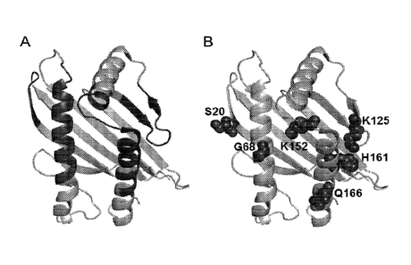
[00201 Figure 12. Structure-directed mutagenesis of the al -a2
domain of MICA for
enhanced NKG2D affinity. (A) Structure of the al -a2 domain of MICA (PDB 1HYR)
with
the NKG2D-binding surface mapped to 57 residues colored dark grey. (B) Six
positions were
identified as key sites for NKG2D affinity mutations. The wild-type amino acid
residues are
labeled and their side chains shown in dark grey spheres.
100211 Figures 13A and 13B. NKG2D-Fc competition ELISAs to
affinity rank al-
a2 variants. (A) Titration data for a panel of al -a2 affinity variants (15-
18), wild-type (WT),
or WED soluble MICA proteins inhibiting human NKG2D-Fc binding to plate-coated
MICA.
(B) The same set of proteins in (A) titrated against mouse NKG2D-Fc. In both
assays
variants 15, 16, 17, and 18 display IC50 values significantly less than both
WT and WED
proteins. The equilibrium IC50 values are shown in Table 3.
100221 Figure 14. Analysis of the association and dissociation
kinetics for al-a2
variants binding to NKG2D, as measured by biolayer interferometry on an Octet
instrument.
Kinetic traces for a panel of al-a2 variants. The association and dissociation
phases were fit
using a single exponential 1:1 binding equation and on- and off-rate constants
derived from
the fits are shown in Table 3.
100231 Figure 15. NK-mediated target cell killing assay for the
al -a2 variants
targeting FGFR3-expressing target cells. NKL effector cells were co-incubated
with calcein-
,
loaded, FGFR3-expressing P815 target cells at a effector:target ratio of 15:1.
Increasing
concentrations of a negative control MICA (sMICA) had no effect on target cell
lysis,
whereas the indicated c&1-a2 variants stimulated target cell lysis. Relative
to WT and WED-
MICA, variants 16, 17, and 18 exhibited significantly increased killing at low
concentrations.
7
CA 02963274 2017-03-30
WO 2016/090278 PCT/US2015/064051
[0024] Figure 16. Analysis of the association and dissociation kinetics for
al -a2
variants 20, 25, and 48 binding to NKG2D, as measured by biolayer
interferometry on an
Octet instrument. The association and dissociation phases were fit using a
single exponential
1:1 binding equation, and on- and off-rate constants derived from the fits are
shown in Table
5,
[0025] Figure 17. NK-mediated target cell killing, calcein-based assay for
al -a2
variants 16, 25 and WED targeting FGFR3-expressing P815 target cells.
[0026] Figure 18. Protein sequence alignment of al-a2 domains from MICA and
ULBPs (SEQ ID NOs.: 77-83). Amino acids highlighted in grey were selected for
NNK
mutagenesis in ULBP2 (60 amino acids) and ULBP3 (36 amino acids). Residues
highlighted
in black were identified as key positions for selected and identified as
mutations that
modulate binding affinity to NKG2D (Tables 6 and 7).
[0027] Figures 19A and 19B. Phage ELISA titrations of ULBP variants binding
to
NKG2D. (A) ULBP2 variants displayed on phage were titrated against NKG2D and
relative
binding affinities were measured relative to native ULBP2 (WT, black circles).
(B) ULBP3
variants displayed on phage were titrated against NKG2D and relative binding
affinities were
measured relative to native ULBP3 (WT, black circles).
[0028] Figures 20A-D. Fusions of native (WT), modified variant WED, 25 or
48 a 1 -
a2 domains to heavy chain (A) or light chain (B) of an FGFR3-specific antibody
affected
NK-dependent target cell killing. Fusions of variants 25 and 48 to either
heavy chain (C) or
light chain (D) significantly enhanced the extent of killing and the potency
of killing
compared to the WED variant and to the native (WT) fusions.
[0029] Figures 21A-C. Fusions of variant 25 al -a2 domain to the heavy
chains or
light chains of antibodies targeting human EGFR (A), HER2 (B), or PDL1 (C)
each enhanced
8
CA 02963274 2017-03-30
WO 2016/090278 PCT/US2015/064051
NKL cell-mediated target cell killing The poor or absent killing by the
respective parent
antibodies, cetuximab (A), trastuzumab (B), and anti-PDL1 (C) are shown.
100301 Figures 22A and 22B. Trastuzumab-based fusions of variant 25 a 1 -a2
domain arm NK cells in vivo. Parent trastuzumab, trastuzumab HC_25 fusion, and
trastuzumab LC_25 fusion were conjugated with Alexa Flour. Groups of three
C57BL/6
mice were injected with a single dose of 100 jig of parent, HC fusion or LC
fusion; and blood
was drawn from each animal at indicated times for plasma PK ELISAs (A) and
flow
cytometric analyses of the fluorescently labeled molecules bound to peripheral
NK cells (B).
100311 Figures 23A-C. Anti-drug antibodies raised in the same animals
described in
Example 7 and Figure 21 administered Trastuzumab parent (A),Trastuzumab-based
HC (B)
and Trastuzumab-LC (C) fusions to variant 25. The control (Ctrl) plasma was
from a mouse
not administered any antibody-containing agent.
10032] Figures 24A and 24B. Antibodies generated in animals administered
variant
25 a 1 -a2 domain fusions to trastuzumab-HC and ¨LC, as described in Example 7
and Figures
21-22, bound to both the parent antibody (A) and to the al-a2 domain (B).
100331 Figure 25. Anti-tumor activity of an anti-PDL,1 fusion to variant
25.
Syngeneic MC38 tumors were implanted subcutaneously in C57BL/6 mice, and
tumors grew
to an average of 100 mm3 before the initiation of treatment. Upon initiation
of treatment four
cohorts of 10 mice per group were treated parenterally with vehicle, anti-
CTLA4 (100 ug
i.p.), parent anti-PDL1 (300 ug i.v.), or anti-PDL1 HC_25 fusion (300 ug iv.)
on days 1, 4,
and 7. Tumor volumes (cubic mm) were measured in each animal at the indicated
times.
9
CA 02963274 2017-03-30
WO 2016/090278
PCT/US2015/064051
DETAILED DESCRIPTION OF THE INVENTION
[0034] In some
aspects, the present invention relates to insertable variable fragment
(iFv) peptides. Because the C-terminus and N-terminus of scFv molecules
including
polyvalent scFv structures are far apart spatially, scFv structures cannot be
inserted into a
loop region embedded within a protein fold of a parent or recipient protein
without disrupting
or destabilizing its fold(s) and/or without disrupting the Fv framework
required to properly
position the CDRs or hypervariable regions to retain their antigen-binding
properties.
[0035] To insert
the variable fragment of an antibody containing up to 6 CDRs into
one or more loop regions of a nascent parent protein molecule without
disrupting structural
folds of the variable fragment or of the parent protein, we invented a new
class of antigen-
binding peptides derived from the light and heavy chain antibody variable
domains. The new
structures contained two linker regions, rather than the traditional single
linker of scFv
structures, plus a split variable domain. Conceptually the canonical termini
of the variable
light (VL) and heavy (VH) domains were fused into a continuous or "circular"
peptide. That
circular peptide structure containing all 6 CDRs of the Fv can then
conceptually be split at
one of several possible novel sites to create an insertable Fv (iFv). The non-
natural split site
can be created within either the light or the heavy chain variable domain at
or near the apex
or turn of a loop to create new, unique N- and C-termini spatially positioned
proximal to each
other, preferably within 0.5 to 1.5 nm, so as to be insertable into loops of
other (parent or
recipient) proteins or polypeptides without disrupting the structure,
stability, or desirable
function. This new class of peptides is called an insertable variable fragment
(iFv). The
binding or targeting specificity conveyed by an iFv to a recipient molecule
can be changed by
inserting into the recipient another or different iFV based on a different
antibody or scFv or
by replacing 1 or more of the CDRs of an existing insertable iFv.
CA 02963274 2017-03-30
WO 2016/090278
PCT/US2015/064051
[0036] The insertion of one or more iFy polypeptides exhibiting specific
antigen-
binding properties of Fv domains into other proteins and thereby imparting
novel binding
properties will have multiple utilities. Such uses include but are not limited
to enabling the
parent protein to bind the specific antigen, target the antigen, detect the
presence of antigen,
remove the antigen, contact or draw near the antigen, to deliver a payload to
the antigen or
antigen-expressing cell, recruit the antigen, and image the presence of the
antigen. A payload
could be conjugated directly to one or both the amino-terminus and carboxy-
terminus of an
iFy or indirectly to an iFy via a parent protein or peptide. Examples of
payloads include but
are not limited to a chromophore, a fluorophore, a pharmacophore, an atom, a
heavy or
radioactive isotope, an imaging agent, a chemotherapeutic agent, or a toxin. A
payloaded iFy
can be used to locate or identify the presence of a target molecule to which
the iFy
specifically binds and as such can serve as in vitro or in vivo imaging agents
or diagnostic
agents that are small and stable. In addition, to one or both the amino-
terminus and carboxy-
terminus of an iFy peptide a chemotherapeutic agent or toxic molecule can be
conjugated in
order to create an iFv-drug conjugate, for example, as treatment for a
malignancy or
infection. A single payload may be conjugated to both the amino-terminus and
the carboxy-
terminus of an iFy peptide so as to span or connect the two termini; such
spanning may
further stabilize the iFy by blocking the termini from exopeptidase
degradation or protecting
the iFy from denaturation or unfolding.
[0037] Examples of parent or recipient proteins or polypeptides that are
candidates
for insertions of iFy peptides include but are not limited to antibodies,
proteins comprised of
Ig folds or Ig domains, globulins, albumens, fibronectins and fibronectin
domains, integrins,
fluorescent proteins, enzymes, outer membrane proteins, receptor proteins, T-
cell receptors,
chimeric antigen receptors, viral antigens, virus capsids, viral ligands for
cell receptors, high
molecular weight bacteriocins, histones, hormones, Icnottins, cyclic peptides
or polypeptides,
11
CA 02963274 2017-03-30
WO 2016/090278 PCT/US2015/064051
major histocompatibility (MHC) family proteins, MIC proteins, lectins, and
ligands for
lectins. It is also possible to insert iFy structures into non-protein
recipient molecules such a
polysaccharides, dendrimers, polyglycols, peptidoglycans, antibiotics, and
polyketides.
10038] Natural killer (NK) cells and certain (CD8+ ari and yo) T-cells of
the
immunity system have important roles in humans and other mammals as first-
line, innate
defense against neoplastic and virus-infected cells (Cerwenka, A., and L.L.
Lanier. 2001. NK
cells, viruses and cancer. Nat. Rev. Immunol. 1:41-49). NK cells and certain T-
cells exhibit
on their surfaces NKG2D, a prominent, homodimeric, surface immunoreceptor
responsible
for recognizing a target cell and activating the innate defense against the
pathologic cell
(Lanier, LL, 1998. NK cell receptors. Ann. Rev. Immunol. 16: 359-393; Houchins
JP et al.
1991. DNA sequence analysis of NKG2, a family of related cDNA clones encoding
type II
integral membrane proteins on human NK cells. J. Exp. Med. 173: 1017-1020;
Bauer, S et at.,
1999. Activation of NK cells and T cells by NKG2D, a receptor for stress-
inducible MICA.
Science 285: 727-730). The human NKG2D molecule possesses a C-type lectin-like
extracellular domain that binds to its cognate ligands, the 84% sequence
identical or
homologous, monomeric MICA and MICB, polymorphic analogs of the Major
Histocompatibility Complex (MHC) Class I chain-related glycoproteins (MIC)
(Weis et al.
1998. The C-type lectin superfamily of the immune system. Immunol. Rev. 163:
19-34;
Bahrain et al. 1994. A second lineage of mammalian MHC class I genes. PNAS
91:6259-
6263; Bahram et al. 1996a. Nucleotide sequence of the human MHC class I MICA
gene.
Immunogenetics 44: 80-81; Bahram and Spies TA. 1996. Nucleotide sequence of
human
MHC class I MICB cDNA. Immunogenetics 43: 230-233). Non-pathologic expression
of
MICA and MICB is restricted to intestinal epithelium, keratinocytes,
endothelial cells and
monocytes, but aberrant surface expression of these MIC proteins occurs in
response to many
types of cellular stress such as proliferation, oxidation and heat shock and
marks the cell as
12
CA 02963274 2017-03-30
WO 2016/090278
PCT/US2015/064051
pathologic (Groh et al. 1996. Cell stress-regulated human MHC class I gene
expressed in GI
epithelium. PNAS 93: 12445-12450; Groh et al. 1998. Recognition of stress-
induced MHC
molecules by intestinal yoT cells. Science 279: 1737-1740; Zwimer et al. 1999.
Differential
expression of MICA by endothelial cells, fibroblasts, keratinocytes and
monocytes. Human
Immunol. 60: 323-330). Pathologic expression of MIC proteins also seems
involved in some
autoimmune diseases (Ravetch, JV and Lanier LL. 2000. Immune Inhibitory
Receptors.
Science 290: 84-89; Burgess, SJ. 2008. Immunol. Res. 40: 18-34). The
differential regulation
of NKG2D ligands, such as the polymorphic MICA and MICB, is important to
provide the
immunity system with a means to identify and respond to a broad range of
emergency cues
while still protecting healthy cells from unwanted attack (Stephens HA, (2001)
MICA and
MICB genes: can the enigma of their polymorphism be resolved? Trends Immunol.
22: 378-
85; Spies, T. 2008. Regulation of NKG2D ligands: a purposeful but delicate
affair. Nature
Immunol. 9: 1013-1015).
[0039] Viral infection is a common inducer of MIC protein expression and
identifies
the viral-infected cell for NK or T-cell attack (Groh et al. 1998; Groh et al.
2001. Co-
stimulation of CD8+ c43T-cells by NKG2D via engagement by MIC induced on virus-
infected cells. Nat. Immunol. 2: 255-260; Cerwenka, A., and L.L. Lanier.
2001). In fact, to
avoid such an attack on its host cell, cytomegalovirus and other viruses have
evolved
mechanisms that prevent the expression of MIC proteins on the surface of the
cell they infect
in order to escape the wrath of the innate immunity system (Lodoen, M., K.
Ogasawara, J.A.
Hamerman, H. Arase, J.P. Houchins, E.S. Mocarski, and L.L. Lanier. 2003. NKG2D-
mediated NK cell protection against cytomegalovirus is impaired by gp40
modulation of
RAE-1 molecules. J. Exp. Med. 197:1245-1253; Stern-Ginossar etal., (2007) Host
immune
system gene targeting by viral miRNA. Science 317: 376-381; Stern-Ginossar et
al., (2008)
Human microRNAs regulate stress-induced immune responses mediated by the
receptor
13
CA 02963274 2017-03-30
WO 2016/090278 PCT/US2015/064051
NKG2D. Nature Immunology 9: 1065-73; Slavuljica, I A Busche, M Babic , M
Mitrovic, I
Gagparovic, D Celcinovic, E Markova ear, EP Pugel, A Cikovic, VJ Lisnic, WJ
Britt, U
Koszinowslci, M Messerle, A Krmpotic and S Jonjic. 2010. Recombinant mouse
cytomegalovirus expressing a ligand for the NKG2D receptor is attenuated and
has improved
vaccine properties. J. Clin. Invest. 120: 4532-4545).
[0040] In spite of their stress, many malignant cells, such as those of
lung cancer and
glioblastoma brain cancer, also avoid the expression of MIC proteins and as a
result may be
particularly aggressive as they too escape the innate immunity system (Busche,
A et at. 2006,
NK cell mediated rejection of experimental human lung cancer by genetic over
expression of
MHC class I chain-related gene A. Human Gene Therapy 17: 135-146; Doubrovina,
ES, MM
Doubrovin, E Vider, RB Sisson, RJ O'Reilly, B Dupont, and YM Vyas, 2003.
Evasion from
NK Cell Immunity by MHC Class I Chain-Related Molecules Expressing Colon
Adenocarcinoma (2003) J. Immunology 6891-99; Friese, M. et al. 2003.
MICA/NKG2D-
mediated immunogene therapy of experimental gliomas. Cancer Research 63: 8996-
9006;
Fuertes, MB, MV Girart, LL Molinero, CI Domaica, LE Rossi, MM Barrio, J
Mordoh, GA
=
Rabinovich and NW Zwimer. (2008) Intracellular Retention of the NKG2D Ligand
MHC
Class I Chain-Related Gene A in Human Melanomas Confers Immune Privilege and
Prevents
NK Cell-Mediated Cytotoxicity. J. Immunology, 180: 4606 -4614).
[0041] The high resolution structure of human MICA bound to NKG2D has been
solved and demonstrates that the a3 domain of MICA has no direct interaction
with the
NKG2D (Li et al. 2001. Complex structure of the activating immunoreceptor
NKG2D and its
MHC class I-like ligand MICA. Nature Immunol. 2; 443-451; Protein Data Bank
accession
code 1HYR). The a3 domain of MICA, like that of MICB, is connected to the al-
a2 platform
domain by a short, flexible linker peptide, and itself is positioned naturally
as "spacer"
between the platform and the surface of the MIC expressing cell. The 3-
dimensional
14
CA 02963274 2017-03-30
WO 2016/090278 PCT/US2015/064051
structures of the human MICA and MICB a3 domains are nearly identical (root-
mean square
distance <1 A on 94 C-aa's) and functionally interchangeable (Holmes et al.
2001. Structural
Studies of Allelic Diversity of the MHC Class I Homolog MICB, a Stress-
Inducible Ligand
for the Activating Immunoreceptor NKG2D. J Immunol. 169: 1395-1400).
[0042] As used herein, a "soluble MIC protein", "soluble MICA" and "soluble
MICB" refer to a MIC protein containing the al, a2, and a3 domains of the MIC
protein but
without the transmembrane or intracellular domains.
[0043] The al-a2 platform domain of a soluble MIC protein is tethered to
the a3
domain and is diffusible in the intercellular or intravascular space of the
mammal. Preferably
the al-a2 platform domains of the non-natural MIC proteins of the invention
are at least 80%
identical or homologous to a native or natural al -a2 domain of a human MICA
or MICB
protein and bind NKG2D. In some embodiments, the al-a2 platform domain is 85%
identical to a native or natural al -a2 platform domain of a human MICA or
MICB protein
and binds NKG2D. In other embodiments, the al-a2 platform domain is 90%, 95%,
96%,
97%, 98%, or 99% identical to a native or natural al -a2 platform domain of a
human MICA
or MICB protein and binds NKG2D.
[0044] In some embodiments, a heterologous peptide tag may be fused to the
N-
terminus or C-terminus of an al -a2 domain or a soluble MIC protein to aid in
the purification
of the soluble MIC protein. Tag sequences include peptides such as a poly-
histidine, myc-
peptide or a FLAG tag. Such tags may be removed after isolation of the MIC
molecule by
methods known to one skilled in the art.
[0045] As used herein "peptide", "polypeptide", and "protein" are used
interchangeably; and a "heterologous molecule", "heterologous peptide",
"heterologous
sequence" or "heterologous atom" is a molecule, peptide, nucleic acid or amino
acid
CA 2963274
sequence, or atom, respectively, that is not naturally or normally found in
physical conjunction
with the subject molecule.
[0046] The term "comprising," which is used interchangeably with
"including,"
"containing," or "characterized by," is inclusive or open-ended language and
does not exclude
additional, unrecited elements or method steps. The phrase "consisting of
excludes any
element, step, or ingredient not specified in the claim. The phrase
"consisting essentially of
limits the scope of a claim to the specified materials or steps and those that
do not materially
affect the basic and novel characteristics of the claimed invention. The
present disclosure
contemplates embodiments of the invention compositions and methods
corresponding to the
scope of each of these phrases. Thus, a composition or method comprising
recited elements or
steps contemplates particular embodiments in which the composition or method
consists
essentially of or consists of those elements or steps.
[0047] As used herein, the terms "a", "an", and "any" are each intended
to include both
the singular and plural forms.
[0048] Having now fully described the invention, it will be appreciated
by those skilled
in the art that the same can be performed within a wide range of equivalent
parameters,
concentrations, and conditions without departing from the spirit and scope of
the invention and
without undue experimentation. While this invention has been described in
connection with
specific embodiments thereof, it will be understood that it is capable of
further modifications.
This application is intended to cover any variations, uses, or adaptations of
the invention
following, in general, the principles of the invention and including such
departures from the
present disclosure as come within known or customary practice within the art
to which the
invention pertains and may be applied to the essential features hereinbefore
set forth.
16
Date Recue/Date Received 2022-01-24
CA 02963274 2017-03-30
WO 2016/090278
PCT/US2015/064051
EXAMPLES of ilµv and of Modified al- a2 Domains of NKG2D Ligands
100491 Example 1 (iFv). As specific examples, we synthesized a 1126 bp and
a 1144
bp DNA fragment (SEQ ID NO:! and 2, respectively) encoding in the following
order: the a3
domain of human MICA (as a parent peptide) amino acid 182 to amino acid 194
(the
beginning of loop 1 of the a3 domain), no spacer or a GGS amino acid spacer
region (SR), an
ihr peptide based on the structure of a Fibroblast Growth Factor Receptor 3
(FGFR3)-
binding antibody (MAbR3;Qing, J., Du, X., Chen, Y., Chan, P., Li, H., Wu, P.,
Marsters, S.,
Stawicki, S., Tien, J., Totpal, K., Ross, S., Stinson, S., Doman, D., French,
D., Wang, Q. R.,
Stephan, J. P., Wu, Y., Wiesmann, C., and Ashkenazi, A. (2009) Antibody-based
targeting of
FGFR3 in bladder carcinoma and t(4;14)-positive multiple myeloma in mice, The
Journal of
clinical investigation 119, 1216-1229.), no spacer or another GGS spacer
region, the distal
portion of loop 1 of the a3 domain starting at amino acid 196 and including
the remaining
carboxy-terminal portion of the a3 domain to amino acid 276 of a soluble MICA
molecule.
Each synthetic, double stranded DNA polynucleotide then encoded a polypeptide
that
contained 6 CDRs in the form of an iFy inserted into loop 1 of the a3 domain
of MICA.
100501 This inf peptide itself (SEQ ID NO. :3), encoded by SEQ ID NO. :4,
contained
two identical, typical linker regions (LR) corresponding to residues
GGSSRSSSSGGGGSGGGG (SEQ ID NO. :5) (Andris-Widhopf, J., Steinberger, P.,
Fuller,
R., Rader, C., and Barbas, C. F., 3rd. (2011) Generation of human Fab antibody
libraries:
PCR amplification and assembly of light- and heavy-chain coding sequences,
Cold Spring
Harbor protocols 2011). One LR joined the C-terminus of VL to the N-terminus
of the VH
domain, and the second LR joined the C-terminus of the VH domain to the N-
terminus of
VL. Conceptually this new structure is the continuous or "circular" peptide
referred to above
and contained 6 CDRs of the starting Fv. The variable VL chain of the antibody
was
17
CA 02963274 2017-03-30
WO 2016/090278
PCT/US2015/064051
effectively split within the loop region between beta-strands 1 and 2 (S1 and
S2) and thereby
created a new N-terminal segment (VLN) and a new C-terminal segment (VLC) with
an
accompanying pair of new, non-natural C- and N-termini, respectively, Figure
5A. This pair
of termini created a sole site for attachment or conjugation of the iFy to the
recipient
molecule such as a protein. The schematic of the inserted iFy in the parent a3
domain is
shown in Figure 5B.
100511 To produce the soluble MICA proteins with a heterologous iFy peptide
inserted into the a3 domain we generated a baculoviral expression vector to
accommodate the
DNA sequences (SEQ ID NO.s:1 and 2) encoding the a3-iFv.1 (SEQ ID NO.:6) and
a3-iFv.2
(SEQ ID NO.:7), respectively. The DNA fragments were amplified by PCR,
digested using
NcoI and EcoRI restriction enzymes, and subcloned into the baculoviral
expression vector,
SW403, replacing the wild-type a3 domain. SW403 is a baculoviral expression
vector
derived from pVL1393 (Invitrogen, Inc.) into which wild-type sMICA (residues 1-
276) had
previously been cloned using 5' BamHI and 3' EcoRI sites. The new expression
vector was
co-transfected with baculoviral DNA into SF9 insect cells, and baculovirus was
grown for
two amplification cycles and used to express the His-tagged MICA-a3-iFy
proteins in T.ni
insect cells according to manufacturer's protocol (Invitrogen). The expression
was carried out
in a 100 mL volume for three days and the growth medium was harvested for
purification of
the secreted soluble protein using Ni-affmity chromatography. Monomeric MICA-
a3-iFy was
purified to >90% purity with the expected molecular weight of 60.9 kDa as
determined by
SDS-PAGE. Functional characterization was carried out using binding ELISAs and
in vitro
target cell killing assays.
100521 The purified MICA-a3-iFy proteins were tested in a FGFR3-binding
ELISA to
confirm simultaneous binding to the FGFR3 target and the NKG2D receptor. FGFR3
in
phosphate buffered saline (PBS) was coated onto Maxisorp plates at 2 ug/ml
concentration.
18
CA 02963274 2017-03-30
WO 2016/090278
PCT/US2015/064051
Each MICA protein was titrated, allowed to bind FGFR3 for 1 hour, and washed
to remove
unbound sMICA protein. Bound MICA-a3-iFy protein was detected using NKG2D-Fc
and
anti-Fc-HRP conjugate. Figure 6 shows that the binding of both MICA-a3-iFv.1
and MICA-
a3-iFv.2 to FGFR3 was comparable to that of a MICA-scFv, made by fusing to the
C-
terminus of soluble MICA a traditional scFv constructed from MAbR3. These
ELISA results
also indicated that both the FGFR3 and NKG2D binding specificities of the scFv
and the al-
a2 domain, respectively, were retained by the modified MICA and demonstrated
that the iFy
peptide inserted using different spacer formats was functional.
[0053] We tested and compared the thermal stability of sMICA-a3-iFv.2 to
that of
sMICA-scFv. Both proteins were subjected for 1 hr to increasing temperatures
from 60-90
C and then allowed to equilibrate to room temperature for 1 hour before being
assayed for
binding properties by ELISA. The results in Figure 7 showed that MICA-a3-iFv.2
can be
subjected to temperatures as high as 80 C with no loss in specific binding to
FGFR3. The
traditional MICA-scFv lost binding activity at 70 C. This result indicated
that soluble MICA
containing the invented iFy format is significantly more stable than terminal
fusions of a
traditional scFv (Miller, B. R., Demarest, S. J., Lugovskoy, A., Huang, F.,
Wu, X., Snyder,
W. B., Croner, L. J., Wang, N., Amatucci, A., Michaelson, J. S., and Glaser,
S. M. (2010)
Stability engineering of scFvs for the development of bispecific and
multivalent antibodies,
Protein engineering, design & selection : PEDS 23, 549-557; Weatherill, E. E.,
Cain, K. L.,
Heywood, S. P., Compson, J. E., Heads, J. T., Adams, R., and Humphreys, D. P.
(2012)
Towards a universal disulphide stabilised single chain Fv format: importance
of interchain
disulphide bond location and vL-vH orientation, Protein engineering, design &
selection.'
PEDS 25, 321-329).
100541 The ability of MICA-a3-iFy to redirect NK cell-mediated lysis of
FGFR3-
expressing target cells was demonstrated in vitro in a calcein-release assay.
The Natural
19
CA 02963274 2017-03-30
WO 2016/090278
PCT/US2015/064051
Killer (NK) cell line, NKL, was co-cultured with calcein-loaded P815 target
cells ectopically
expressing FGFR3. The results in Figure 8 showed that the two MICA-a3-iFy
molecules
induced significantly greater NK-mediated lysis compared to the traditional
MICA-scFv
fusion, while the non-targeted soluble MICA control had no killing activity.
These results
confirmed that the invented iFy bound FGFR3 on target cells and in the context
of the
complete parent protein molecule, soluble MICA, induced potent NK cell-
mediated lysis.
[0055] The applicability of the iFIT format to other antibody variable
domains was
demonstrated by similarly constructing an a3-iFv.3 (SEQ ID NO. :8), which
contained an iFIT
derived from a CD20-specific antibody (Du, J., Wang, H., Zhong, C., Peng, B.,
Zhang, M.,
Li, B., Huo, S., Guo, Y., and Ding, J. (2007) Structural basis for recognition
of CD20 by
therapeutic antibody Rituximab, The Journal of biological chemistry 282, 15073-
15080).
Figure 9 shows that MICA-a3-iFv.3 was able to specifically bind wells coated
with CD20 in
a plate-based ELISA as described above and also induced NK-mediated lysis of
Ramos cells
expressing CD20 in a calcein-release assay.
[0056] Example 2 (Modified al- a2 Domains of NKG2D Ligands). Human
proteins designated ULBP-1 through ULBP-6 are, like MICA and MICB, naturally
occurring,
stress-induced, cell surface ligands that bind NKG2D receptors on and activate
human NK
cells and certain T-cells (15; Cerwenka A, Lanier LL (2004). NKG2D ligands:
unconventional MHC class 1-like molecules exploited by viruses and cancer.
Tissue Antigens
61(5): 335-43. doi:10.1034/j.1399-0039.2003.00070.x. PMID 12753652). In
addition, the
cowpox virus protein OMCP is a secreted domain that like the al-a2 domain of
MIC proteins
binds NKG2D. OMCP exhibits a very high affinity for NKG2D, apparently in order
to block
NKG2D's recognition of the natural stress ligands induced by the virus on its
infected host
cell (Eric Lazear, Lance W. Peterson, Chris A. Nelson, David H. Fremont. J
Virol. 2013
January; 87(2): 840-850. doi: 10.1128/JVI.01948-12). While the ULBPs and OMCP
are
CA 02963274 2017-03-30
WO 2016/090278 PCT/US2015/064051
considered NKG2D ligands (NKG2DLs) that share the canonical al-a2 domain
structure, the
sequence homology with MICA al -a2 is less than 27%, and they all naturally
lack an a3
domain for tethering targeting domains. We constructed a series of non-natural
ULB and
OMCP proteins by attaching the heterologous polypeptides that specifically
targeted and
killed FGFR3-expressing cells as the result of fusing to each of ULBP-1, ULBP-
2, ULBP-3,
ULBP-4, ULBP-6 and OMCP, a modified a3 domain of MICA into which a targeting
iFy had
been inserted. In addition, we modified the al-a2 domain of MICA to enhance
the affinity of
al-a2 domain for NKG2D and then attached to the modified al-a2 domains
heterologous
molecules such as polypeptides. To produce the proteins consisting of ULBP and
OMCP al-
a2 domains attached to modified a3-iFy domains we generated a baculoviral
expression
vector to accommodate the DNA fragments (SEQ ID NOs:9-14) that encoded the
different
al-a2 domains of ULBP-1, ULBP-2, ULBP-3, ULBP-4, ULBP-6, and OMCP (SEQ ID
NOs:15-20, respectively). The DNA fragments were amplified by PCR, digested
using BlpI
and NcoI restriction enzymes, and individually subcloned into the baculoviral
expression
vector, KLM44, replacing the MICA al-a2 domain. KLM44 was a baculoviral
expression
vector derived from SW403 into which MICA-a3-iFv.2 had previously been cloned
(example
1). The new NKG2DL-a3-iFv.2 constructs, containing the ULBPs and OMCP al-a2
domain
fusions to a3-iFv.2 (ULBP1-a3-iFv.2, ULBP2-a3-iFv.2, ULBP3-a3-iFv.2, ULBP4-a3-
iFv.2,
ULBP6-a3-iFv.2, and OMCP-a3-iFv.2; SEQ ID NO. :21-26, respectively), were co-
transfected with baculoviral DNA into SF9 insect cells. Baculovirus was grown
for two
amplification cycles and used to express these His-tagged NKG2DL-a3-iFv.2
proteins in T.ni
insect cells according to manufacturer's protocol (Invitrogen). The expression
was carried out
in a 100 mL volume for three days and the growth medium was harvested for
purification of
the secreted soluble protein using Ni-affinity chromatography. Monomeric
proteins of correct
21
CA 02963274 2017-03-30
WO 2016/090278
PCT/US2015/064051
molecular weight were purified to >90% purity as determined by SDS-PAGE.
Functional
characterization was carried out using binding ELISAs and in vitro target cell
killing assays.
100571 The 6 purified NKG2DL-a3-iFv.2 proteins were tested in a FGFR3-
binding
ELISA to confirm simultaneous binding to the FGFR3 target and the NKG2D
receptor.
FGFR3 in phosphate buffered saline (PBS) was coated onto Maxisorp plates at 2
ug/ml
concentration. Each NKG2DL-a3-iFv.2 protein was titrated, allowed to bind
FGFR3 for 1
hour, and washed to remove unbound protein. The bound NKG2DL-a3-iFv.2 protein
was
detected using NKG2D-Fc and anti-Fc-HRP conjugate. Figure 10 shows that all 6
NKG2DL-
a3-iFv.2 proteins bound potently to FGFR3, as expected, through interaction
with the iFv.2
domain, and the NKG2D binding activity was retained by the attached NKG2DL al-
a2
domains, which demonstrated that the attached a3-iFy domain imparted
functional FGFR3
binding activity to the ULBP and OMPC proteins that, like MIC proteins, bind
NKG2D.
100581 The ability of the NKG2DL-a3-iFv.2 proteins to redirect NK cell-
mediated
lysis of FGFR3-expressing target cells was demonstrated in vitro in a calcein-
release assay.
The Natural Killer (NK) cell line, NKL, was co-cultured with calcein-loaded
P815 target
cells ectopically expressing FGFR3. The results in Figure 11 showed that OMCP-
a3-iFv.2
induced the greatest NK-mediated lysis, while the other NKG2DL-a3-iFv.2
proteins all
displayed specific killing activity with varying degrees of potency and amount
of lysis. These
results confirmed that the invented in/ imparts specific binding activity to
other proteins that
retained their own functional properties and induced different levels of cell-
mediated lysis of
iFv-targeted cells.
100591 Example 3 (Modified al-o2 Domains of NKG2D Ligands). These are
examples of attaching polypeptides to NKG2DLs which were modified to
significantly
enhance their binding affinity to the human NKG2D receptor. The al-z2 domain
of MIC
proteins is an NKG2DL for the NKG2D receptor. This affinity is sufficient for
physiologic
22
CA 02963274 2017-03-30
WO 2016/090278 PCT/US2015/064051
activation of NK cells and stimulating lysis of cells expressing native full-
length MIC
proteins irreversibly tethered to the two-dimensional plasma membrane surface
of a "target
cell" (Bauer S, Groh V, Wu J, Steinle A, Phillips JH, Lather LL, Spies T.,
Science. 1999 Jul
30;285(5428):727-9.). However, because engineered soluble MIC proteins of the
instant
invention reversibly bind specific target antigens on the surface of a target
cell, the binding
affinity of the engineered soluble MIC protein to NKG2D will directly affect
the stability of
the soluble MIC-dependent complex formed between NK cells and cells expressing
target
antigens. Especially if the affinity between sMICA and NKG2D is increased by a
substantially slower dissociation rate or off-rate of the modified sMICA from
NKG2D, the
NK cell-based killing would be expected to be greater at lower densities of
soluble MIC
molecules bound to a target cell. Prior to the instant invention there had not
been identified
any al-a2 mutations that alter the killing activity of soluble MIC proteins or
significantly
reduce the binding off-rate to enhance affinity of MIC proteins to NKG2D. A
computational
design effort showed that three mutations in the al -a2 domain of wild-type
MICA: N69W,
K152E, and K154D (WED-MICA) in combination can moderately affect NKG2D binding
affinity by affecting the stability of unbound MICA and thereby its
association rate or on-rate
of binding to NKG2D (Lengyel CS, Willis LJ, Mann P, Baker D, Kortemme T,
Strong RK,
McFarland BJ.J Biol Chem. 2007 Oct 19;282(42):30658-66. Epub 2007 Aug 8);
Subsequent
extensive computational design work by the same group scanning by iterative
calculations 22
amino acid positions of MICA theoretically in contact with NKG2D, according to
the
published structural descriptions (Li P, Morris DL, Willcox BE, Steinle A,
Spies T, Strong
RK., Nat Immunol. 2001 May;2(5):443-451), showed experimentally that when
combined
with the earlier designed 3 changes, further rational, iterative computational
design of MICA
qualitatively changed its affinity for NKG2D from weak (Kd ¨2.5 [i.M) to
moderately tight
(Kd = 51 nM) with a total of seven combined mutations (Henager, Samuel H.,
Melissa A.
23
CA 02963274 2017-03-30
WO 2016/090278 PCT/US2015/064051
Hale, Nicholas J. Maurice, Erin C. Dunnington, Carter J. Swanson, Megan J.
Peterson,
Joseph J. Ban, David J. Culpepper, Luke D. Davies, Lisa K. Sanders, and
Benjamin J.
McFarland, 2102, Combining different design strategies for rational affinity
maturation of the
MICA-NKG2D interface. Protein Science 21:1396-1402). In contrast, the
experimental
approach described in the instant invention experimentally selected amino acid
modifications
of MICA that slowed the off-rate between the al-a2 domain of MICA and NKG2D,
commencing with a MICA stabilized by the 3 WED changes of Lengyel et al
(Lengyel CS,
Willis LJ, Mann P, Baker D, Kortemme T, Strong RK, McFarland BJ., J Biol Chem.
2007
Oct 19;282(42):30658-66. Epub 2007 Aug 8).
[0060] This example of the instant invention relates to modifying the NKG2D
binding affinity of soluble MIC proteins through engineering specific
mutations at selected
amino acid positions within the al-a2 domain that influence the off-rate
binding kinetics and
thereby alter the NK cell-mediated killing activity of the invented non-
natural, targeted MIC
molecules.
[0061] To engineer soluble non-natural al-a2 domains with altered affinity
to
NKG2D 57 residues in the al -a2 domain were chosen for extensive mutagenesis
(Figure 12).
Synthetic DNA libraries coding for the al-a2 domain and containing NNK
mutagenic codons
at each of the 57 amino acid positions were synthesized, individually cloned
as fusions to the
pIII minor coat protein of M13 phage, and phage particles displaying the
mutagenized al-a2
variants were produced in SS320 E. coil cells according to standard
methodologies (Andris-
Widhopf, J., Steinberger, P., Fuller, R., Rader, C., and Barbas, C. F., 3rd.
(2011) Generation
of human Fab antibody libraries: PCR amplification and assembly of light- and
heavy-chain
coding sequences, Cold Spring Harbor protocols 2011). The al -a2 phage
libraries were
sorted for increased binding affinity using recombinant biotinylated NKG2D as
the target
antigen and cycled through iterative rounds of intentionally prolonged
binding, prolonged
24
CA 02963274 2017-03-30
WO 2016/090278 PCT/US2015/064051
washing, and eluting of the phage clones in order to select high affinity
variants enriched for
slow dissociation- or off-rates. A set of specific amino acid mutations
occurred at high
frequencies at 6 positions in al -a2 and were selected as preferred amino acid
substitutions
with enhanced NKG2D binding affinity (Figure 12, Table 1).
[0062] Table 1. Selected affinity mutations at the indicated 6 amino acid
positions of
the al-a2 domain of MIC. The amino acids of SEQ ID NOs.: 35 at each of the 6
positions are
shown in bold in the first row of the table. The identified affinity mutations
are listed in
decreasing frequency from top to bottom. All amino acids are represented by
the single letter
IUPAC abbreviations.
S20 G68 K125 E152 H161 Q166,
L T R
V $
A
A A
Y A Y G W
I. N A L. V
V Q
T
- -
[0063] We synthesized DNA polynucleotides (SEQ ID NOs. 27-30) encoding the
a 1-
a2 domains of 4 representative variants 15, 16, 17, 18 that contained
different combinations
of specific discovered mutations (Table 2).
[0064] Table 2. Sequences of specific al-a2 domain variants. The specific
amino
acid substitutions for variants 15, 16, 17, and 18 (SEQ ID NOS.: 31-34,
respectively) are
CA 02963274 2017-03-30
WO 2016/090278 PCT/US2015/064051
listed relative to the amino acids of SEQ ID NO. :35 in bold. All amino acids
are represented
by the single letter IUPAC abbreviations.
Variant .SEQ ID NO.: S20 068 K125 H161
15 31 G N ft
16 32
17 33
18 34
10065] To the NKG2DLs in the above example, we directly attached
heterologous
molecules such as a polypeptide to each of these 4 modified al-a2 NKG2DLs
using a linker
peptide. Four His-tagged proteins (SEQ ID NOs.: 31-34) consisting of modified
NKG2DLs
with attached heterologous molecules were expressed in insect cells and
purified to
characterize their NKG2D binding affinities and kinetic binding parameters.
Using a
competitive binding ELISA, we determined the relative NKG2D binding affinities
of the 4
modified al-a2 variants. A soluble wild type (WT) NKG2DL, sMICA protein, was
coated in
all wells of a maxisorp ELISA plate to provide a binding partner for the human
NKG2D-Fc
reagent. Solutions of the four al -a2 variants as well as WT and WED- a 1 -a2
domains (SEQ
ID NO.: 35) were titrated in the ELISA wells and allowed to competitively
inhibit 2nM
human NKG2D-Fc binding to the WT sMICA coated on the plate. The level of human
NKG2D-Fc that bound to the WT NKG2DL on the plate was detected using an anti-
Fc-HRP
antibody. Figure 13A shows variants 16, 17, and 18 exhibited IC50 values of
0.7, 0.6, 0.5 riM
while variant 15 exhibited an IC50 value of 1.7 nM, all possessing
significantly better binding
to NKG2D, 27, 32-, 38- and 11-fold better, than WT NKG2DL, respectively, as
well as
substantially better than WED-MICA (Table 3).
1006611 Table 3. Equilibrium and kinetic binding parameters for al -a2
variants. IC50
values were derived from 4-parameter fits to the competition binding
titrations (Figure 12)
26
CA 02963274 2017-03-30
WO 2016/090278
PCT/US2015/064051
and the kinetic binding parameters were derived from single exponential fits
to the binding
kinetics (Figure 13). Equilibrium binding constants (Kd) were derived from the
kinetic
binding parameters using the equation Kd = koFF "oN=
Kinetic Binding Parameters
al-a2 Variant IC50 (nM) IcoN (M4s4) icoFF (s.i) K (nM)
WT 19.4 1.3x 10⁵ 1.8 x 10⁻³ 13.8
WED 4.4 2.9 x 10⁵ 1.7 x 10⁴ 5.9
15 1.7 0.7 x 10⁵ 1.1 x 10⁻⁴ 1.5
16 0.7 2.0 x 10⁵ 0.9x 10⁴ 0.5
17 0.6 2.0 x 10⁵ 0.7 x 10⁴ 0.4
18 0.5 2.3x 10⁵ 0.9x 10⁴ 0.4
[0067]
Importantly, the relative IC50 differences also translated to better binding
to
murine NKG2D-Fc (Figure 13B), and demonstrated the ability to improve binding
of soluble,
modified al-a2 domains across human and non-human NKG2D receptors, an
important
property for preclinical drug development.
[0068] In order to understand
the kinetic basis for the altered affinities, both the on-
rates and off-rates for the al-a2 variant NKG2DLs binding to surface coated
biotinylated
human NKG2D were measured using biolayer interferometry (Octet) at 100 nM of
each of
the modified al-a2 proteins. Consistent with results from the IC50 ELISAs,
variants 16, 17
and 18 each displayed significant reductions in the off-rate (18-fold relative
to WT), which is
largely responsible for the affinity increase (-30-fold relative to WT al-
a2)(Figure 14; Table
3). Although variant 15 displayed a similar slow off-rate as did 16, 17, and
18, its on-rate was
decreased, resulting in an affinity stronger than WT but weaker variants 16,
17 and 18.
Because the only difference between variant 15 (SEQ ID NO.:31 ) and 16 (SEQ ID
NO.:32)
was K1 25N versus K125L, the mutation at position 125 clearly altered the on-
rate while the
decreased off-rate was attributed to the H161R mutation. Therefore, while the
selected set of
27
CA 02963274 2017-03-30
WO 2016/090278 PCT/US2015/064051
NKG2DL mutations (Table 1) was used to increase the al -a2 affinity for NKG2D
through
significant off-rate reduction, certain substitutions also altered the on-rate
resulting in a range
of incremental affinity increases that we showed in this invention to have
differential activity
in the NK cell-mediated killing assays as described below.
[0069] The ability of the al-a2 affinity variants to redirect NK cell-
mediated lysis of
FGFR3-expressing target cells was demonstrated in vitro in a calcein-release
assay. The
human Natural Killer (NK) cell line, NKL, was co-cultured with calcein-loaded
P815 target
cells ectopically expressing FGFR3 and titrated with soluble modified MIC
proteins. The
results in Figure 15 showed that the killing activities of the FGFR3-specific
soluble MIC
variants correlated with their engineered al -a2 affinities. Specifically,
variants 16, 17, and 18
exhibited ¨15-fold more killing than WT at 0.78 nM. The WED-MICA (SEQ ID
NO.:35)
was only slightly better than WT. Therefore, the invention describes amino
acid substitutions
within the al-a2 domain that increased the NKG2D binding affinity by reducing
the off-rate
of soluble MIC protein binding to human NKG2D and consequentially led to the
predictably
increased killing potency. WED-MICA, which exhibited somewhat greater affinity
than WT
MICA to NKG2D (Figure 13A) by increasing on-rate rather than reducing off-rate
(Figure
14), did not exhibit substantial improvement of target cell killing (Figure
15). Furthermore,
as shown in Figure 13B, WED-MICA exhibited substantially poorer binding to
murine
NKG2D than even WT MICA, while variants 15, 16, 17, and 18 each exhibited
greater
affinity for both human and murine NKG2D, Figure 13A-B.
[0070] These al-a2 NKG2DL affinity variants 15, 16, 17, and 18 enhanced the
binding affinity of the attached polypeptide to the NKG2D receptor and thereby
enhanced
NK cell-mediated lysis of targeted cells, Figure 15.
[0071] Example 4 (Modified al-u2 Domains of NKG2D Ligands). This
embodiment of the instant invention relates to additional al-a2 NKG2DL
affinity variants
28
CA 02963274 2017-03-30
WO 2016/090278
PCT/US2015/064051
derived through engineering specific mutations at selected amino acid
positions within the
al -a2 domain of soluble MIC molecules, as described in Example 3 (Table 1),
that also
influence the off-rate binding kinetics and thereby alter the NK cell-mediated
killing activity
of the non-natural al -a2 domains. While variants 15-18 focused on specific
mutations found
at positions S20, G68, K125, and H161, another set of variants were isolated
with additional
mutations at E152, H158, and Q166 (Table 4).
100721 Table 4. Sequences of specific al-a2 domain variants. The specific
amino
acid substitutions for variants 20, 25, and 48 are listed relative to the
amino acids of SEQ ID
NO.:35, shown in bold in the first row of the table. All amino acids are
represented by the
single letter IUPAC abbreviations.
Variant SEQ ID NO.: S20 G68 K125 E152 H158 H161 Q166
20 39 A LOR HF
25 40 SGL E H R S
48 41 SGL A IR A
[0073] DNA polynucleotides (SEQ ID NOs. 36-38) encoding the al -a2 domains
of 3
representative variants 20,25, 48 (SEQ ID NOs. 39-41, respectively) that
contained different
combinations of specific discovered mutations (Table 4), were synthesized. To
the
NKG2DLs in the above example, heterologous molecules, such as an FGFR3-binding
polypeptide, were directly attached to each of these 3 modified al -a2 NKG2DLs
using a
linker peptide. The constructs were cloned into the Xbal and BamHI sites of
pD2509, a
CMV-based mammalian cell expression vector. Three His-tagged proteins (SEQ ID
NOs.:
39-41), consisting of modified NKG2DLs with attached heterologous molecules
that bind to
FGFR3, were transiently expressed in HEK293 cells using the Expi293 expression
system
according to the manufacturer's protocol (Life Technologies), and purified
using Ni-affinity
chromatography to obtain the isolated proteins for biochemical and activity-
based analysis.
29
CA 02963274 2017-03-30
WO 2016/090278
PCT/US2015/064051
[0074] In order to characterize the NKG2D binding affinities, both the on-
rates and
off-rates for the three al-a2 variant NKG2DLs binding to surface-coated
biotinylated human
NKG2D were measured using biolayer interferometry (Octet). Binding titrations
were
performed for each protein using a titration range of 1-100 nM, and the
kinetic data were
fitted to obtain on-rates, off-rates, and equilibrium binding constants.
[0075] Variant 25 (SEQ ID NO.: 40) contains only the addition of the Q166S
mutation relative to variant 16 (SEQ ID NO.: 32) (Table 2), and exhibited a
NKG2D binding
affinity of 62 pM largely due to decreased off-rate (Figure 16 and Table 5).
This represented
an 8-fold enhancement in equilibrium binding affinity due to the Q166S
mutation (compare
Table 3 and Table 5), and demonstrated that specific mutations at Q166
influenced binding
affinity through decreased off-rate.
[0076] Table 5. Kinetic binding parameters for al-a2 variants. Kinetic
binding
parameters were derived from single exponential fits to the binding kinetics
(Figure 16).
Equilibrium binding constants (IQ) were derived from the kinetic binding
parameters using
the equation Kd = koFF Ikw
Kinetic Bind in Parameters
al-a2 Variant 'koN (M-1s-1) koFF (S-i) Kd (nM),
20 3.6 x 10⁵ 3.0 x 1 0-5 0.083
25 =4.7 x 10⁵ 2.9,x11:15 0.062
48 2.0x 10⁵ 3.0 x 10⁻³ 15
[0077] Variant 20 (SEQ ID NO.: 39) contained the specific mutations G68A,
E152Q,
H158R and Q166F, and maintained binding parameters similar to variant 25
(Table 5),
CA 02963274 2017-03-30
WO 2016/090278 PCT/US2015/064051
suggesting that this unique combination of specific mutations also has
improved NKG2D
binding affinity due to a decreased off-rate.
10078] Variant 48 (SEQ ID NO.: 41) contained the K125L and H161R mutations
found in variant 16 (Table 2); however the addition of mutations E 152A,
H1581, and Q166A
(Table 4) significantly increased the off-rate, resulting in a 250-fold
reduction in NKG2D
binding affinity (Figure 16 and Table 5). The Q166A mutation is not one of the
favored
affinity enhancement mutations selected for position Q166 (Table 1) and may
have
contributed to the reduction in off-rate observed. These data clearly
demonstrated that unique
combinations of engineered, mutations selected and identified at defined
positions within al -
a2 domains tuned the NKG2D binding affinity through off-rate modulation.
100791 The non-natural al-a2 affinity variants with attached polypeptides
redirected
NK cell-mediated lysis of FGFR3-expressing target cells, as demonstrated in
vitro in a
calcein-release assay. The human Natural Killer (NK) cell line, NKL, was co-
cultured with
calcein-loaded P815 target cells ectopically expressing FGFR3, and titrated
with soluble
modified NKG2D ligand al -a2 proteins. The results in Figure 17 showed that
the killing
potencies of the FGFR3-targeted soluble MIC variants correlated with their
engineered al -a2
affinities. Specifically, variant 25 exhibited ¨3-fold greater killing than
variant 16 at 0.2 nM,
representing an ¨5-fold improvement in the EC50 for cell killing. In addition,
the data clearly
showed preferred killing activity across representative soluble MIC variants
in the order of
variant 25>16>WED (Figure 17).
[0080] Example 5 (Modified al-a2 Domains of NKG2D Ligands). This
embodiment relates to additional al -a2 NKG2DL affinity variants derived
through
engineering the al-a2 domains of ULBP proteins. ULBP proteins contain al -a2
domains,
which are NKG2D ligands capable of binding to the NKG2D receptor (Cerwenka A,
Lanier
LL (2004). NKG2D ligands: unconventional MHC class I-like molecules exploited
by viruses
31
CA 02963274 2017-03-30
WO 2016/090278 PCT/US2015/064051
and cancer. Tissue Antigens 61 (5): 335-43. doi:10.1034/j.1399-
0039.2003.00070.x.
PMID 12753652). This affinity of NKG2D binding is sufficient for physiologic
activation of
NK cells and stimulating lysis of cells expressing native full-length ULBP
proteins naturally
and irreversibly tethered to the two-dimensional plasma membrane surface of a
"target cell"
(Cerwenka A, Lanier LL (2004). NKG2D ligands: unconventional MHC class I-like
molecules exploited by viruses and cancer. Tissue Antigens 61(5): 335-43.
doi:10.1034/j.1399-0039.2003.00070.x. PMID 12753652). However, because
engineered
soluble al-a2 domains fused to heterologous polypeptides in certain
embodiments of the
instant invention reversibly bind specific target antigens on the surface of a
target cell, the
binding affinity of the engineered ULBP al-a2 domains to NKG2D will directly
affect the
stability of the artificial synapse formed between NK cells and cells
expressing target
antigens, as already shown by engineered soluble MIC proteins (Examples 2-4).
In order to
diversify the repertoire of engineered non-natural al-a2 domains as NKG2D
ligands, ULBP
proteins were used as a substrate or starting point for phage display-based
engineering of
their NKG2D binding affinity. Despite the structural homology observed between
ULBPs
and MICA (Radaev, S., Rostro, B., Brooks, AG., Colonna, M., Sun, PD. (2001)
Conformational plasticity revealed by the cocrystal structure of NKG2D and its
class I MHC-
like Ligand ULBP3. Immunity 15, 1039-49.), the sequence homology is <50% for
the ULBP
al-a2 domains relative to MICA (Figure 18). Thus, we sought the identities of
codon
positions in ULBP a1-a2 domains that improve NKG2D binding affinity.
[0081] To engineer soluble, non-natural al-a2 domains from ULBP proteins,
ULBP2
and ULBP3 were chosen for phage display and selection of mutants with high
affinity
NKG2D binding. Sixty amino acid positions in the al-a2 domain of ULBP2 (SEQ ID
NO.:16), and thirty-six amino acid positions in the al-a2 domain of ULBP3 (SEQ
ID NO.:
17), were chosen for extensive mutagenesis (Figure 18). In addition,
conservative cysteine-
32
CA 02963274 2017-03-30
WO 2016/090278 PCT/US2015/064051
to-serine mutations were made at C103S in ULBP2 (SEQ ID NO.:16) and C8S in
ULBP3
(SEQ ID NO.: 17) in order to remove unpaired free cysteines that could
interfere with phage
panning. Synthetic DNA libraries coding for these al-o2 domains, and
containing NNK
mutagenic codons at each of the selected amino acid positions, were
synthesized,
individually; cloned as fusions to the pIII minor coat protein of Ml3 phage;
and phage
particles displaying the mutagenized -a2 ULBP2 or ULBP3 variants were produced
in
SS320 E.coli cells according to standard methodologies (Andiis-Widhopf, J.,
Steinberger, P.,
Fuller, R., Rader, C., and Barbas, C. F., 3rd. (2011). Generation of human Fab
antibody
libraries: PCR amplification and assembly of light- and heavy-chain coding
sequences, Cold
Spring Harbor protocols 2011). The al -a2 phage display libraries were sorted
for increased
binding affinity to NKG2D using human NKG2D-Fc as the target protein, and
cycled through
iterative rounds of intentionally prolonged binding, prolonged washing, and
eluting of the
phage clones in order to select high affinity variants enriched for slow
dissociation- or off-
rates. For ULBP2, specific amino acid mutations were found at high frequencies
at positions
R80, V151, V152, and A153 in al-a2, and were identified as preferred amino
acid
substitutions with enhanced NKG2D-binding affinity (Figure 19 A and Table 6).
[0082] Table 6. Selected affinity mutations at the indicated 4 amino acid
positions of
the al-a2 domain of ULBP2. The amino acids of SEQ ID NOs.:16 at each of the 4
positions
are shown in bold in the first row of the table. The identified affinity
mutations are listed in
decreasing frequency from top to bottom. All amino acids are represented by
the single letter
IUPAC abbreviations.
33
CA 02963274 2017-03-30
WO 2016/090278 PCT/US2015/064051
R80 V151 V152 A153
D L E
W E
V a
1
A T
100831 For ULBP3, specific amino acid mutations were found at high
frequencies in
different locations relative to ULBP2 (Figure 18). Positions R162 and K165 in
the al-a2
domain of ULBP3 contained specific mutations that were identified as preferred
amino acid
substitutions with enhanced NKG2D-binding affinity (Figure 19 B and Table 7).
These
modified non-natural al-a2 domains derived from ULBP2 and ULBP3 can be used
for
enhanced NKG2D binding in multiple therapeutic formats as single proteins or
fusions to
heterologous peptides or polypeptides.
100841 Table 7. Selected affinity mutations at the indicated 2 amino acid
positions of
the a1-a2 domain of ULBP3. The amino acids of SEQ ID NOs.:17 at each of the 2
positions
are shown in bold in the first row of the table. The identified affinity
mutations are listed in
decreasing frequency from top to bottom. All amino acids are represented by
the single letter
IUPAC abbreviations.
34
CA 02963274 2017-03-30
WO 2016/090278 PCT/US2015/064051
R162 K165
G S
A P
A
[0085] Example 6 (Modified al-a2 Domains fused to antibody peptides).
These
are examples of attaching antibody polypeptides to NKG2DLs which were modified
to
significantly enhance their binding affinity to the human NKG2D receptor. The
al-a2
domain of MIC proteins is an NKG2DL for the NKG2D receptor. Antibodies are
highly
stable glycoproteins made up of two large heavy chains and two small light
chains (Figure 1).
The large amount of diversity that can be generated within the CDR regions of
the variable
domains allows for specific antibodies to be generated to specific antigen
targets (Hozumi N,
Tonegawa S (1976). "Evidence for somatic rearrangement of immunoglobulin genes
coding
for variable and constant regions". Proc. Natl. Acad. Sci. U.S.A. 73 (10):
3628-3632.
doi:10.1073/pnas.73.10.3628. PMC 431171. PMID 824647.) Antibodies have become
a
significant therapeutic platform for drug development and can mediate both
target binding
and neutralization, as well as modulate the immune system through complement
and Fc
receptor binding (Vidarsson, G., Dekkers, G., Rispens, T. (2014) IgG
subclasses and
allotypes: from structure to effector functions. Frontiers in Immunology 5,
520.). Prior to the
present invention, there did not exist an IgG antibody format that can
directly activate
immune cells using non-natural al-a2 domains that bind more tightly than
native NKG2DLs
to the NKG2D receptor. Previous work has demonstrated that the mouse NKG2D
ligand,
CA 02963274 2017-03-30
WO 2016/090278
PCT/US2015/064051
Rae 1 beta, can be fused to an anti-Her2 antibody for use as an anti-tumor
agent in mice (Cho,
RM., Rosenblatt, JD., Tolba, K., Shin, SJ., Shin, D., Calfa, C., Zhang, Y.,
Shin, SU. (2010)
=
Delivery of NKG2D ligand using and anti-Her2 antibody-NKG2D ligand fusion
protein
results in an enhanced innate and adaptive antitumor response. Cancer Research
70, 10121-
30.). However, mouse NKG2D ligands do not bind human NKG2D, and there are no
natural
human NKG2D ligands with high affinity to human and mouse NKG2D. Fusions
between
the engineered a1-a2 NKG2D ligands of the instant invention and the heavy
chain or light
chain of IgG antibodies (Figure 20 A and B) overcame these limitations and
highlighted the
versatility of fusions of modified al-a2 domains to heterologous proteins or
peptides.
[0086] To
generate variant al-a2 domain fusions to antibodies, the DNA sequences
encoding al-a2 domain for MIC WT, variants WED, 25, and 48, were synthesized
and
cloned as C-terminal fusions to either the heavy chain (HC_WT, HC_WED, HC_25,
HC_48)
or light chain (LC_WT, LC_WED, LC_25, LC_48) sequence from the FGFR3-specific
antibody (Qing, J., Du, X., Chen, Y., Chan, P., Li, H., Wu, P., Marsters, S.,
Stawicki, S.,
Tien, J., Totpal, K., Ross, S., Stinson, S., Doman, D., French, D., Wang, Q.
R., Stephan, J. P.,
Wu, Y., Wiesmaim, C., and Ashkenazi, A. (2009) Antibody-based targeting of
FGFR3 in
bladder carcinoma and t(4;14)-positive multiple myeloma in mice, The Journal
of clinical
investigation 119, 1216-1229.) (SEQ ID NOs.: 42-49, respectively). The
resulting fusions
were cloned into the mammalian expression vector pD2509 and expressed as
paired full IgG
antibodies with either heavy or light chain fusions of the modified al-a2
domains (SEQ ID
NOs.: 50-57, respectively). Transient expressions were carried out in HEK293
cells using the
Expi293 expression system according to the manufacturer's protocol (Life
Technologies),
and purified using standard protein A affinity chromatography. The ability of
the non-natural
al-a2-antibody fusions to redirect NK cell-mediated lysis of FGFR3-expressing
target cells
was demonstrated in vitro in a calcein-release assay. The human Natural Killer
(NK) cell
36
CA 02963274 2017-03-30
WO 2016/090278
PCT/US2015/064051
line, NKL, was co-cultured with calcein-loaded P815 target cells ectopically
expressing
FGFR3 and titrated with the engineered antibody fusion proteins. The results
in Figures 20C
and 20D showed that the killing activities of the FGFR3-specific non-natural
u1-a2-antibody
fusions correlated with their engineered NKG2D affinities. Specifically,
antibodies that
contained either heavy chain or light chain fusions of non-natural variants 25
and 48 (HC_25,
HC_48 and LC_25, LC_48) killed FGFR3-expressing cells more effectively than
antibody
fusions containing either WT or WED al-a2 domains.
100871 This was further demonstrated to be a general and useful approach to
fusing
modified al-a2 domains to antibodies, by fusing the variant 25 al -a2 domain
to the C-
terminal of either the heavy chain or light chain of EGFR-specific antibody
cetuximab (US
patent 6217866), Her2-specific antibody trastuzumab (Carter, P., Presta, L.,
Gorman, CM.,
Ridgway, JB., Henner, D., Wong, WL., Rowland, AM., Kotts, C., Carver, ME.,
Shepard,
HM. (1992) Proc Natl Acad Sci 15,4285-9), or an anti-PDL1 antibody (US Patent
20140341917) (SEQ ID NOs.:58-63, respectively). The resulting fusions were
expressed as
paired light and heavy chain full IgG antibodies with either heavy or light
chain fusions of the
variant 25 al-a2 domain. Transient expressions were carried out in HEIC293
cells using the
Expi293 expression system according to the manufacturer's protocol (Life
Technologies),
and purified using standard protein A affinity chromatography. The ability of
the variant 25
antibody fusions to redirect NK cell-mediated lysis of target-expressing cells
was
demonstrated in vitro in a calcein-release assay. The human Natural Killer
(NK) cell line,
NKL, was co-cultured with calcein-loaded A431 EGFR-expressing target cells,
SKBR3
Her2-expressing target cells, or PDL1-expressing B16 cells and titrated with
the respective
target-specific engineered antibody fusion proteins. The results in Figures
21A, 21B, and
21C showed that the killing activities of the target-specific variant 25-
antibody fusions were
in all cases drastically improved over the non-fused parent antibody and very
potent with sub-
37
CA 02963274 2017-03-30
WO 2016/090278 PCT/US2015/064051
nanomolar EC50 values. These data show that modified al-a2 variant-antibody
fusions are a
universal platform for allowing IgG antibodies to bind tightly to NKG2D and to
direct
antigen-specific cell lysis.
[0088] Example 7 (irastuzumab fusions to al-a2 variant 25 bind NK cells in
vivo
and elicit potent antigen presentation). Fusion proteins containing al -a2
domain variants
that bind NKG2D with high affinity bound NK cells in vivo. Thus, antigen-
specific
antibodies containing modified al-a2 fusions bind NKG2D tightly and thereby
effectively
armed the surface of NK cells in vivo with antibodies to seek out target cells
expressing a
particular antigen. This activity was similar to engineered CAR cells (Gill,
S., and June, CH.
(2015) Going viral: chimeric antigen receptor T-cell therapy for hematologicaH
malignancies.
Immunol Rev 263, 68-89.), but did not require genetic modification of the
NKG2D-
expressing cell type.
[0089] To demonstrate that antibodies containing modified al -a2 fusions
bind NK
cells in vivo, trastuzumab and the corresponding heavy and light chain fusions
of variant 25
were analyzed in vivo for serum pharmacokinetic (PK) profiles and the
pharmacodynamics
(PD) of NK cell labeling. All three antibodies: parent trastuzumab;
trastuzumab HC_25
fusion; and trastuzumab LC 25 fusion, were conjugated with Alexa Flour 647
according to
the manufacturer's protocol (Life Technologies). Groups of three C57BL/6 mice
were
injected with a single dose of 10014 of each antibody, and blood was drawn at
indicated time
points for plasma PK ELISAs and flow cytometry of peripheral NK cells. The PK
profile of
the parent trastuzumab antibody displayed typical alpha-phase distribution
within 24-hrs and
beta-phase elimination consistent with greater than a 1 week half-life of
antibodies in mice
(Figure 22A). For both the heavy chain and light chain fusions with variant
25, the initial
alpha-phase showed a much greater volume of distribution relative to the
parent antibody,
consistent with an NKG2D-sink, while the beta-phase elimination was also
consistent with
38
CA 02963274 2017-03-30
WO 2016/090278
PCT/US2015/064051
typical antibody clearance in mice (Figure 22 A). Using flow cytometry of
peripheral NK
cells from the mouse blood, the level of NK cell staining with Alexa Fluor 647
showed a
clear time-dependent increase in the percent of NK cells labeled with the
antibody fusion, but
not the parent antibody (Figure 22 B). The increase in labeling by the fusions
peaked within
24 hrs, consistent with the sink observed in the PK profiles for the fusions,
and was stable at
least for three days post injection. The combined PK and PD data demonstrate
that the
trastuzumab antibodies containing variant 25 al -a2 fusions formed stable
complexes with
NKG2D on NK cells in vivo.
[0090] To assess the appearance of anti-drug antibodies (ADAs) to the human
IgG
trastuzumab antibody, the plasma samples from the PK/PD study were assessed
for ADAs
using an ELISA. In Figures 23A-C, ELISAs for mouse IgG binding to wells coated
with the
3 respective dosed antibodies revealed that only the antibodies fused with
variant 25 elicited
ADAs within seven days after a single dose of antibody. The parent trastuzumab
antibody
gave no ADA signal. In order to determine whether the antibody fusions
elicited an immune
(ADA) response to both the al -a2 domain and the antibody (trastuzumab )
component when
the trastuzumab antibody itself did not elicit an immune response, the ADA-
positive plasma
from the antibody fusions were tested against the parent antibody and the
variant 25 al -a2
domain individually; both moieties reacted with ADAs from plasma (Figures 24A
and 24B).
These data demonstrate that the fusion of high affinity variant 25 to the
parent antibody
mediated NKG2D-dependent uptake and antigen presentation to elicit potent and
rapid
immune responses to the parent antibody, which alone was not so immunogenic in
mice.
Thus, a high affinity variant al-a2 domain attached to an antigen or immunogen
provided
potent presentation of the antigen and epitope spreading, effectively serving
as a potent
adjuvant for immunization.
39
CA 02963274 2017-03-30
WO 2016/090278
PCT/US2015/064051
[0091] The demonstrated combined effects of arming circulating NK cells for
directed target cell lysis and enhancing antigen presentation are important
activities for
antibody fusions to modified al-a2 domains that can provide therapeutic
benefit.
[0092] Example 8 (Antibody heavy chain fusion to al-a2 variant 25 exhibited
anti-tumor activity in vivo). To examine the potential for antigen-specific
antibodies fused
to modified a1-a2 to have anti-target cell activity, an anti-PDL1 antibody
heavy chain fusion
to variant 25 al -a2 was evaluated in a syngeneic MC38 tumor model. MC38
tumors were
implanted sub-cutaneously in C57BL/6 mice and tumors grew to an average of 100
mm3
before the initiation of treatment. Upon initiation of treatment, four cohorts
of 10 mice per
group were treated with vehicle, anti-CTLA4 (100 ug i.p.), parent anti-PDL1
(300 ug iv), or
anti-PDL1 HC_25 fusion (300 ug i.v.) on days 1, 4, and 7. In Figure 25, the
tumor growth
curves showed that anti-PDL1 HC_25 mediated the most effective anti-tumor
activity within
the first two weeks of treatment. Tumor growth inhibition was significantly
better than the
established anti-CTLA4 treatment and the parent anti-PDL1 antibody over the
first 12 days
after initiation of treatment. By day 16, the anti-PDL1 HC_25 treatment began
to lose
efficacy consistent with the occurrence of an ADA response as observed for
trastuzumab
fusions (Example 7). The significant anti-tumor activity observed for the
antibody heavy
chain fusion to variant 25 relative to both the parent antibody and standard
anti-CTLA4
treatments demonstrated the impressive therapeutic effect of antibody fusions
to modified al-
a2 domains that served as high affinity NKG2D ligands.