Note: Descriptions are shown in the official language in which they were submitted.
CA 02963508 2017-04-03
WO 2016/060714 PCT/US2015/035936
1
Title: BIOLOGICAL INDICATOR
Technical Field
This invention relates to biological indicators. These biological indicators
may be used for determining the effectiveness of sterilization processes.
Background
In the health care industry as well as in many other commercial and
industrial applications, it is often necessary to monitor the effectiveness of
processes used to sterilize equipment such as medical and non-medical devices,
instruments and other articles and materials. Sterilization monitors can be
included in the batch of articles to be sterilized to assay the lethality of
the
sterilization process. They can also be used to validate the effectiveness of
sterilization equipment and sterilization cycles used in such equipment.
Summary
Classical methods of sterility assurance typically involve exposing a
sterilization indicator containing one or more test organisms to the
sterilization
process and then measuring the outgrowth of any surviving test organisms.
Sterility may be assured if there is no outgrowth of the test organisms
following
exposure to the sterilization process. Bacterial spores are typically used as
the
test organisms. Upon completion of the sterilization process, the
sterilization
indicator is exposed to a liquid growth support medium under conditions that
would promote the growth of any surviving test organism cells. The growth
support medium often contains a chemical dye which changes color in response
to actively growing (metabolizing) cells. Because of the requirement for
growth
and metabolism, the processes employing these test organisms typically require
about 24 to 72 hours of incubation before the effectiveness of the
sterilization
process can be determined.
A problem with this process relates to the fact that many users of sterilized
articles, such as health care facilities and the like, have limited resources
and
may reuse the "sterilized" articles within 24 to 72 hours and sometimes
immediately. In such settings, the 24 to 72 hour holding period for sterility
verification may be impractical, costly and inefficient.
CA 02963508 2017-04-03
WO 2016/060714 PCT/1JS2015/035936
2
A detection process for reading out test results more rapidly for certain
121 C and 132 C gravity and prevacuum steam sterilization cycles and ethylene
oxide sterilization cycles has been proposed. The time necessary to observe
evidence of surviving indicator cells is reported to be as little as one hour.
This
process involves detecting the catalytic activity of the enzyme alpha
glucosidase.
This enzyme is produced by a microorganism as a normal component of its
metabolism and may be present in the spore coat of the microorganism both
before and during sterilization. The presence of this enzyme can be detected
by
reading fluorescence produced by the breakdown of a non-fluoresent enzyme
substrate. Breakdown of the enzyme substrate can be an early detection
alternative to waiting for a visual pH color change to indicate a failed
sterilization
process. Neither growth nor metabolism is required for the fluorometric
signal.
This results in a reduction in the time required to observe a failure in the
sterilization process. However, the enzyme alpha glucosidase is thermophilic
in
origin, and may be more resistant to heat than the microorganism from which it
is
derived. This can lead to nuisance failures, a circumstance in which the test
microorganism has been, in fact, killed but the indicator enzyme indicates
that
the test microorganism remains viable. In addition, since the enzyme alpha
glucosidase may be present in the spore coat of the test microorganism and its
presence does not necessitate metabolism, the detection of this enzyme may not
be a direct indication of life.
There are situations where the use of enzyme alpha glucosidase may fail
to discriminate an unsuccessfully sterilized load. Successful steam
sterilization is
dependent upon achieving an effective temperature and pressure for a minimum
length of time. Bacterial spores are typically selected as the test organism
for
this process because they are highly resistant to this combination of
parameters.
It takes a particularly lethal combination of temperature, pressure and time
to kill
bacterial spores. Although the target/reporter molecule (alpha glucosidase) is
a
catalytic enzyme associated with a thermophilic organism, and thus somewhat
resistant to heat, it is the heat of the process which ultimately destroys the
function of the enzyme. That is, pressure and time play a reduced role in the
denaturation of alpha glucosidase. Therefore, under sub-lethal pressure or
time
CA 02963508 2017-04-03
WO 2016/060714 PCMJS2015/035936
3
conditions the indicator enzyme may be destroyed even though the bacterial
spores may not be destroyed. This can result in a failure to detect a non-
sterilized load.
The inability of existing technology to account for all the parameters
relating to cell death means that "grow out" may be required to provide the
final
confirmatory result. However, a major drawback with processes requiring what
is
traditionally known as grow out relates to the time delay in obtaining results
for
the sterilization test. Sterilization indicators requiring grow out normally
employ
the use of bacterial spores which must be cultured for at least about 24 to 72
hours to assure adequate detection of any surviving spores. During this time,
the
articles that went through the sterilization process and are under evaluation
should not be used until the results of the spore viability test have been
determined. However, as indicated above, this may be impractical for many
users of articles requiring sterilization.
U.S. Patent No. 8,372,624 discloses a process for detecting the
effectiveness of a sterilization process wherein a genetically engineered
biological indicator is exposed to the sterilization process. The biological
indicator comprises a test organism, a reporter gene for producing an
indicator
enzyme, and a repressor gene that inhibits expression of the reporter gene
until
the reporter gene is exposed to an inducer (e.g., xylose). The biological
indicator
may be used in a device that includes two compartments, one compartment for
containing the biological indicator, and the other compartment for containing
a
growth medium that includes the inducer and an enzyme substrate. Once the
sterilization is complete, the biological indicator is combined with the
growth
medium, and any cells from the biological indicator that have survived the
sterilization process are incubated. The living cells from the biological
indicator
are detected when the indicator enzyme acts upon the enzyme substrate to form
a product that can be detected. A problem that often occurs with this process
involves discoloration due to degradation of the inducer. The inducer is
degraded by heating or by exposure to various sterilization mediums (e.g.,
vaporous hydrogen peroxide, ethylene oxide, etc.), and as a result turns brown
or
is otherwise discolored. This browning or discoloration can interfer with
detecting
CA 02963508 2017-04-03
WO 2016/060714 PCMJS2015/035936
4
changes associated with the success or failure of the sterilization, and
thereby
reduces the sensitivity of the test.
Thus, a problem that has been presented by the art is to provide a
biological indicator that accurately detects the effectiveness of a
sterilization
process within a relatively short period of time, and in doing so, does not
rely on
the use of an inducer. This invention provides a solution to this problem.
This invention relates to a composition that may be used to form a
biological indicator. The composition comprises: a host organism comprising a
spore forming bacteria; a reporter gene for producing an indicator enzyme; a
regulatory gene; and a vehicle for inserting the reporter gene and the
regulatory
gene in the host organism; the host organism bearing a transposable genetic
element in its genome for inserting an insertion sequence in the regulatory
gene;
the insertion sequence comprising a transposase, a pair of terminal inverted
repeat sequences, and at least one open reading frame for expressing the
transposase. The vehicle may comprise a plasmid or a viral vector. The vehicle
may be taken up by the host organism. The insertion sequence may be inserted
in the regulatory gene. The host organism may then undergo sporulation to form
the biological indicator. The biological indicator may comprise spores derived
from the foregoing composition. While not wishing to be bound by theory, it is
believed that the insertion sequence modifies the regulatory gene to allow
expression of the reporter gene upon being hydrated without the necessity of
employing an inducer. This allows for use of the biological indicator for
monitoring the effectiveness of a sterilization without the problems
associated
with using an inducer.
In an embodiment, the reporter gene comprises bgaB, the regulatory gene
comprises xyIR, the insertion sequene comprises IS5376, the vehicle is a
plasmid, and the host organism comprises Geobacillus stearothermophilus.
In an embodiment, the host organism comprises Geobacillus
stearothermophilus, the host organism containing a plasmid construct, the
plasmid constructing comprising the sequence set out in SEQ ID No. 1.
This invention relates to a biological indicator, comprising: a host organism
comprising Geobacillus stearothermophilus, the host organism containing a
CA 02963508 2017-04-03
WO 2016/060714 PCMJS2015/035936
plasmid construct, the plasmid construct having the sequence set out in SEQ ID
No. 1.
This invention relates to a process, comprising: exposing an article to be
sterilized and the above-indicated biological indicator to a sterilization
medium.
5 This
invention relates to a process, comprising: exposing an article to be
sterilized and the above-indicated biological indicator to a sterilization
medium;
and detecting the presence of the indicator enzyme to determine the
effectiveness of the sterilization. In an embodiment, the indicator enzyme
acts
upon an enzyme substrate to form an enzyme-modified product. The enzyme-
modified product may comprise a luminescent, fluorescent or colored material
that can be detected.
This invention relates to a sterilization monitor, comprising: a first
compartment containing the above-indicated biological indicator, the first
compartment being adapted to permit the biological indicator to be brought
into
contact with a sterilization medium during a sterilization process; and a
second
compartment containing a recovery medium, the second compartment being
adapted to maintain the recovery medium separate from the biological indicator
during the sterilization process, and to permit the recovery medium to contact
the
biological indicator after the biological indicator has been exposed to the
sterilization medium.
Brief Description of the Drawings
In the annexed drawings, like parts and features have like references.
Figs. 1A-1B disclose a series of sequence listings for the IS4 family of
insertion sequences.
Figs. 2A-2C disclose a schematic illustration and a series of sequence
listings for the IS21 family of insertion sequences.
Fig. 3 is a schematic illustration of a sterilization monitor suitable for use
with the present invention, the sterilization monitor being shown in a pre-
activated
configuration.
Fig. 4 is a schematic illustration of the sterilization monitor of Fig. 3 in
an
activated configuration.
Fig. 5 is a schematic illustration of another embodiment of a sterilization
CA 02963508 2017-04-03
WO 2016/060714 PCMJS2015/035936
6
monitor suitable for use with the present invention, the sterilization monitor
being
shown in pre-activated configuration.
Fig. 6 is a graph showing the results of sterilization tests employing the
inventive biological indicator.
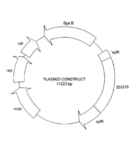
Fig. 7 is a schematic illustration of a plasmid construct containing 11223
base pairs (bp).
Detailed Description
All ranges and ratio limits disclosed in the specification and claims may be
combined in any manner. It is to be understood that unless specifically stated
otherwise, references to "a," "an," and/or "the" may include one or more than
one,
and that reference to an item in the singular may also include the item in the
plural.
The phrase "and/or" should be understood to mean "either or both" of the
elements so conjoined, i.e., elements that are conjunctively present in some
cases and disjunctively present in other cases. Other elements may optionally
be present other than the elements specifically identified by the "and/or"
clause,
whether related or unrelated to those elements specifically identified unless
clearly indicated to the contrary. Thus, as a non-limiting example, a
reference to
"A and/or B," when used in conjunction with open-ended language such as
"comprising" can refer, in one embodiment, to A without B (optionally
including
elements other than B); in another embodiment, to B without A (optionally
including elements other than A); in yet another embodiment, to both A and B
(optionally including other elements); etc.
The word "or" should be understood to have the same meaning as
"and/or" as defined above. For example, when separating items in a list, "or"
or
"and/or" shall be interpreted as being inclusive, i.e., the inclusion of at
least one,
but also including more than one, of a number or list of elements, and,
optionally,
additional unlisted items. Only terms clearly indicated to the contrary, such
as
"only one of" or "exactly one of," may refer to the inclusion of exactly one
element
of a number or list of elements. In general, the term "or" as used herein
shall
only be interpreted as indicating exclusive alternatives (i.e. "one or the
other but
CA 02963508 2017-04-03
WO 2016/060714 PCMJS2015/035936
7
not both") when preceded by terms of exclusivity, such as "either," "one of,"
"only
one of," or "exactly one of."
The phrase "at least one," in reference to a list of one or more elements,
should be understood to mean at least one element selected from any one or
more of the elements in the list of elements, but not necessarily including at
least
one of each and every element specifically listed within the list of elements
and
not excluding any combinations of elements in the list of elements. This
definition also allows that elements may optionally be present other than the
elements specifically identified within the list of elements to which the
phrase "at
least one" refers, whether related or unrelated to those elements specifically
identified. Thus, as a non-limiting example, "at least one of A and B" (or,
equivalently, "at least one of A or B," or, equivalently "at least one of A
and/or B")
can refer, in one embodiment, to at least one, optionally including more than
one,
A, with no B present (and optionally including elements other than B); in
another
embodiment, to at least one, optionally including more than one, B, with no A
present (and optionally including elements other than A); in yet another
embodiment, to at least one, optionally including more than one, A, and at
least
one, optionally including more than one, B (and optionally including other
elements); etc.
The transitional words or phrases, such as "comprising," "including,"
"carrying," "having," "containing," "involving," "holding," and the like, are
to be
understood to be open-ended, i.e., to mean including but not limited to.
The term "sterilization" refers to rendering a substance incapable of
reproduction, metabolism and/or growth. While this is often taken to mean
total
absence of living organisms, the term may be used herein to refer to a
substance
free from living organisms to a degree previously agreed to be acceptable.
Unless otherwise indicated, the term sterilization is used herein to also
refer to
methods and procedures less rigorous than sterilization, for example,
disinfection, sanitization, and the like. The biological indicator and the
processes
and apparatus described herein may be used in health care fields, scientific
fields, and the like. These may be used in commercial and industrial
applications
where sterilization, disinfection, sanitization, decontamination, cleaning,
and the
CA 02963508 2017-04-03
WO 2016/060714 PCMJS2015/035936
8
like, may be desired. The commercial and industrial applications may include
processes such as food processing, pasteurization, soil remediation, water
remediation, and the like.
The term "insertion sequence" (also known as an IS, an insertion
sequence element, an IS element, a transposable genetic element, transposon,
or jumping gene) refers to a short DNA sequence that acts as a simple
transposable element. Insertion sequences typically have two major
characteristics: they are small relative to other transposable elements
(generally
around 700 to 2500 bp in length) and only code for proteins implicated in the
transposition activity. They are different from other transposons, which also
carry
accessory genes such as antibiotic resistance genes.
The term "base pair" or "bp" refers to building blocks of the DNA double
helix which contribute to the helical and folded structures of both DNA and
RNA.
The term "kilobase" or "kb" refers to a unit of measurement equal to 1000 base
pairs.
The sterilization process for which the inventive biological indicator may be
used may comprise any sterilization process. The sterilization process may
include sterilization processes wherein the sterilization medium or sterilant
may
comprise steam, dry heat, radiation, plasma, as well as one or more gaseous
sterilants, one or more liquid sterilants, and the like. The radiation may
comprise
electron beam or any electromagnetic spectra including ionizing radiation,
pulsed
white or ultraviolet light, microwave, and the like. The radiation may
comprise
gamma or beta radiation. The gaseous sterilants may comprise ethylene oxide,
gaseous hydrogen peroxide, and the like. The liquid sterilants may comprise
formalin (formaldehyde gas dissolved in water and optionally containing
methanol to inhibit the formation of toxic substances), glutaraldehyde,
peracetic
acid, liquid hydrogen peroxide, and the like.
The biological indicator of the present invention may be used to examine
the lethality of sterilants against any microorganism with less resistance to
the
sterilization process than the host organism provided with the inventive
biological
indicator. These microorganisms may include bacteria such as Escherichia coil,
Legionella sp., Campylobacter sp., and other enteric bacteria, as well as
CA 02963508 2017-04-03
WO 2016/060714 PCT/1JS2015/035936
9
Staphylococcus and Streptococcus species and other human pathogenic
microorganisms such as Cryptosporidium.
The growth of an organism may comprise the combined result of a
multitude of cellular processes. In typical biological indicator applications
this
may be observed in several ways. As cells grow and divide their individual
numbers increase to a point at which the support medium of the cells may
change from clear to opaque (turbid). To facilitate this observation of
growth, a
pH indicator dye may be used. Growth requires energy. This energy may be
provided by the ability of the cell to metabolize nutrients contained in the
support
medium. The breakdown products of this process may cause the support
medium to become acidic. This acidity may induce a pH indicator dye (e.g.,
phenol red) to change color. As a result, growth may be observed as the
conversion of the support medium from a clear red to yellow color, for
example,
to a turbid yellow condition. Although these processes are slow, they
represent
compelling evidence of life and are generally accepted as the benchmark by the
various sterility assurance regulatory bodies.
With the present invention, a biological indicator is provided which is
derived from a composition comprising: a host organism comprising a spore
forming bacteria; a reporter gene capable of producing an indicator enzyme; a
regulatory gene; and a vehicle for inserting the reporter gene and the
regulatory
gene in the host organism; the host organism bearing a transposable genetic
element in its genome for inserting an insertion sequence in the regulatory
gene;
the insertion sequence comprising a transposase, a pair of terminal inverted
repeat sequences, and at least one open reading frame for expressing the
transposase. The vehicle, which may comprise a plasmid or a viral vector, is
taken up by the host organism. The insertion sequence is inserted in the
regulatory gene. The host organism is sporulated to form the biological
indicator.
Expression of the reporter gene occurs when the reporter gene is hydrated,
which can occur when the reporter gene is exposed to a recovery medium.
Advantageously, the recovery medium is characterized by the absence of an
inducer (e.g., xylose). What may be exposed to the sterilization process are
the
various and vital mechanisms the host organism uses to survive and grow and
CA 02963508 2017-04-03
WO 2016/060714 PCMJS2015/035936
which are also used for the production of the indicator enzyme. These may
include the DNA polymerases used for cellular growth (and replication of the
plasmid or viral vector), RNA polymerases for transcription of the metabolic
requirements of the host organism (and the plasmid or viral vector borne
reporter
5 gene, e.g., bgaB) and the ribosomal polysomes required for the
translation of
cellular proteins and expression of the reporter gene. In order to be
effective for
determining the effectiveness of a sterilization process, the biological
indicator
should be more resistant to the sterilization than the organisms that are to
be
destroyed by the sterilization.
10 The host organism may comprise any spore forming bacteria that bears a
transposable genetic element in its genome that is capable of modifying the
regulatory gene by inserting an insertion sequence in the regulatory gene. The
type of host organism used may be dependent upon a variety of factors
exemplified by the type of sterilization process being used. The host organism
may comprise bacteria of the Bacillus or Clostridia genera. These may include
Geobacillus stearothermophilus, Bacillus atrophaeus, Bacillus sphaericus,
Bacillus anthracis, Bacillus pumilus, Bacillus coagulans, Clostridium sporo
genes,
Clostridium difficile, Clostridium botulinum, Bacillus subtilis globigii,
Bacillus
cereus, Bacillus circulans, or a mixture of two or more thereof, and the like.
Geobacillus stearothermophilus is particularly useful.
Geobacillus stearothermophilus is widely distributed in nature. Many
species can be isolated from soils and muds. They are also associated with
heated materials, such as formation waters of oil fields in Russia,
Kazakhstan,
and China, and hot springs in Yellowstone National Park. Both G.
stearothermophilus and G. kaustophilus strains have also been isolated from
mud samples taken from the Mariana Trench. One Bacillus Genetic Stock Center
(BGSC) strain was also isolated from rotting wood in Florida, USA.
Geobacillus stearothermophilus (NRRL B-1172) may be equivalent to the
American Type Culture Collection (ATCC) strain 12980 and strain 26 from the
collection of the National Canning Association. This is a thermophilic spore-
forming organism with optimal growth conditions between 55-65 C. This
particular strain of Geobacillus stearothermophilus is a source of the
restriction
CA 02963508 2017-04-03
WO 2016/060714 PCMJS2015/035936
11
endonuclease BstPl. Geobacillus stearothermophilus is classified by ATCC as a
Biosafety Level 1 organism, according to U.S. Public Health Service
guidelines.
It is not known to cause disease in healthy adult humans, animals or plants
and
is not harmful to the environment.
The vegetative form of Geobacillus stearothermophilus is rod-shaped
cells that produce one endospore per cell. The cell length ranges from 2.0-3.5
micrometers with a cell width ranging from 0.6-1.0 micrometers. Cells occur
either singly or in short chains and are motile by means of peritrichous
flagella.
The cell wall structure is gram-positive, but the gram stain reaction may vary
between positive and negative depending on the age of the culture.
Geobacillus stearothermophilus can utilize hydrocarbons (C10, C11). It will
produce acid but no gas upon utilization of glucose, fructose, maltose,
mannose
and sucrose. Phenylalanine is not deaminated, tyrosine is not degraded, indole
is
not produced, and the Voges-Proskauer reaction is negative.
Geobacillus stearothermophilus is a thermophilic organism whose
distinctive diagnostic characteristics include its capacity to grow at 65 C
and a
limited tolerance to acid.
The reporter gene may comprise lacZ, bgaB, xylE, cat, gfp, or a mixture of
two or more thereof. The term "lacZ" refers to a gene coding for p-
galactosidase.
The term "bgaB" refers to the gene coding for thermostable R-galactosidase
from
G. stearothermophilus. The term "xylE" refers to gene coding for catechol-2,3-
dioxygenase from Pseudomouas putida. The term "cat" refers to the gene
coding for chloramphenicol acetyltransferase. The term "gfp" refers to the
gene
for coding thermostable green fluorescent protein variants.
The regulatory gene may comprise xyIR, lad, tetR, or a mixture of two or
more thereof. The term "xylR" refers to a regulator of the xylose operon. The
term "lad" refers to a regulator of the lac operon. The term "tetR" refers to
a
regulator of the tet operon. The thermostable counterparts of these may be
used. The regulatory gene may be taken up by the test organism with the same
vehicle used to insert the reporter gene in the test organism.
The insertion sequence may be derived from the host organism and
added to the regulatory gene. The insertion sequence may comprise an IS4 or
12
an 1521 family insertion sequence. The nomenclature used in Mahillon et al.,
"Insertion Sequences," Microbiology and Molecular Biology Reviews, 1998,
62(3): 725, is used in this disclosure.
The IS4 family is depicted in Figs. 1A-B. The insertion sequences of this
family comprise a transposase, a pair of terminal inverted repeat sequences
(IRs), and a single open reading frame for expressing the transposase. The
open reading frame extends along the length of the insertion sequence between
the terminal inverted repeat sequences. Fig. 1A is a dendrogram based on
alignments of the putative Tpases. The term "Tpase" is an abbreviation for
transposase. Fig. 1B discloses terminal IRs of selected members.
The IS4 family of insertion sequences contains 41 members, including 13
isoforms. Many members, such as IS10 and IS50, are involved in compound
transposons. Several members carry GATC methylation sites, which, for both
IS10 and IS50, may play a modulating role in transposition activity.
IS10 and IS50 may be the best-characterized members of the IS4 family.
Both transpose by a "cut-and-paste" mechanism, as does IS231A. IS10 forms
part of the composite tetracycline resistance transposon Tn10.
The IS4 family of insertion sequence may comprise IS4, IS10, IS50,
IS186, IS231, IS701, IS942, IS1151, IS1170, IS1452, IS5377, IS8402, ISH27-1,
ISH27-2 or ISH51-3. IS5377 may be particularly useful.
The IS21 family is depicted in Figs. 2A-2C. The insertion sequences of
this family comprise a transposase, a pair of terminal inverted repeat
sequences,
and two consecutive open reading frames for expressing the transposase.
Referring to Figs. 2A-20, the IS21 family members have terminal inverted
repeat
sequences (IRL and IRR) with two consecutive reading frames (istA and istB)
positioned between the terminal inverted repeat sequences. The terminal
inverted repeat sequences IRL and IRR are shown as solid boxes in Fig. 2A. The
position of the istA and istB reading frames is also shown in Fig. 2A. The
horizontal lines below show the relative positions of the multiple repeat
elements
whose sequences are presented in Fig. 2C. IstA (hatched box) together with the
potential DDE motif (stippled box) and IstB (open box) are indicated in Fig.
2A.
Date Recue/Date Received 2020-11-04
CA 02963508 2017-04-03
WO 2016/060714 PCMJS2015/035936
13
The possibility of translational coupling between the two reading frames is
indicated in Fig. 2A. The dendrogram shown in Fig. 2B is derived from the
alignment of the istA and istB gene products. Nucleotide sequences of the
multiple terminal repeats, together with their coordinates are shown in Fig.
2C.
CS (complementary strand) L1, L2, and L3, and R1 and R2, indicate internal
repeated sequences at the left and right ends, respectively.
There are 15 distinct members of the IS21 family together with 6 iso-ISs.
They carry related terminal IRs whose lengths may vary between 11 bp (IS21)
and 50 bp (IS5376) and generally terminate in the dinucleotide 5'-CA-3'.
Several
members, but not IS21 itself, carry multiple repeated sequences at their ends
which include part of the terminal IRs and which may represent Tpase binding
sites. Insertion of these elements results in a direct target repeat of 4 bp
or 5 bp,
while two members (IS53 and IS408) may generate 8 bp. They exhibit two
consecutive open reading frames: a long upstream frame designated istA and a
shorter downstream frame designated istB (Fig. 2A). The putative IstA and IstB
proteins carry several blocks of highly conserved residues. Overall identities
range from 10 to 59% for IstA and from 25 to 67% for IstB. The istB frame may
be located in a relative reading phase of -1 (e.g., IS21 and IS5376) or +1
(e.g.,
IS232 and IS1326) compared to istA. It can be slightly separated from istA (17
bp
for IS408) or can overlap for 1 bp (IS21) or for several base pairs (IS232,
IS5376,
and IS1326); it is generally preceded by a potential ribosome binding site.
The
arrangement of the two reading frames suggests that translational coupling may
occur (Fig. 2A).
The IS21 family of insertion sequence may comprise IS21, IS53, IS232A,
IS408, IS1162, IS1326, IS1415 or IS5376. IS5376 is a
particularly useful
insertion sequence.
IS5376 is depicted in SEQ ID No. 1 at coordinates (2060). .(4166).
IS5376 may be described, using slightly different terminology, as having the
following segments:
Coordinates in SEQ ID No. 1
1) First inverted repeat sequence (2060). . (2109)
2) tnpA gene (ATP binding protein) (2120) . . (2875)
3) tnpB gene (transposase) (2872) . . (4074)
4) RBS (ribosome binding site) (4082) . . (4088)
5) Second inverted repeat sequence (4177) . . (4166)
CA 02963508 2017-04-03
WO 2016/060714 PCMJS2015/035936
14
The "tnpA" and "tnpB" genes code for transposases needed by the IS5376 to
insert into a new site. The term "ATP-binding protein" refers to a sequence of
protein subunits (i.e., genomic DNA base pairs) that promote the attachment of
ATP (adenosine-5'-triphosphate) to a target protein.
The vehicle for inserting the reporter gene and the regulatory gene in the
host organism may comprise one or more plasmids or one or more viruses (or
viral vectors). When added to the host organism, the insertion sequence may be
transferred from the host organism to the vehicle. In an embodiment, the
insertion sequence is inserted in the regulatory gene. In an embodiment, the
regulatory gene is xylR and the insertion sequence is IS5376 which is inserted
in
the xylR regulatory gene. The vehicle may be referred to as a vector. The
plasmids may comprise circular double-stranded DNA that are separate from
chromosomal DNA. The plasmids may be linear. The size of the plasmids may
be in the range from about 2000 to about 20000 base pairs (bp), or in the
range
from about 5000 to about 10000 bp. One or more copies (for example, from 1 to
about 3000 copies, or from 1 to about 60 copies, or from about 20 to about
3000
copies) of the same plasnnid may be taken up by a cell of the test organism.
The
plasmids may contain one or more DNA sequences that serve as an origin of
replication (on). The plasmids may contain one or more genetic markers. The
plasmids may contain a polylinker or multiple cloning site (MCS) which may be
a
relatively short region containing one or more restriction sites allowing the
insertion of DNA fragments. The plasmids may contain one or more genes that
provide a selective marker to induce the test organism to retain the plasmid.
The
selective marker may comprise an antibiotic resistance gene and/or or a gene
with nutritional capability. The plasmids may comprise conjugative plasmids
which contain tra-genes which perform the process of conjugation, the sexual
transfer of plasmids to another bacterium.
Naturally occurring plasmids exist over a broad range of host organisms in
nature. They may comprise genes, regulatory elements and/or structural pieces
of DNA. Plasmids usually provide some advantage to their host organism (e.g.
antibiotic resistance or the ability to use certain nutritional sources of
energy) and
CA 02963508 2017-04-03
WO 2016/060714 PCMJS2015/035936
may be tolerated by their host organisms for as long as this advantageous
relationship may exist. Genetically engineered plasmids may comprise a
patchwork of genes, regulatory elements and/or structural pieces of interest.
Since there are so many naturally occurring (and previously engineered)
5 plasmids available, there is a wide choice of genes to choose from. The
genes
employed may be selected based on the desired properties of the finished
plasmid construct. These properties may include the ability to transform the
full
range of useful host organisms, provide some selective advantage to the host
organism (e.g., antibiotic resistance), produce a thermostable and rapidly
10 detectable signal on demand. This may be accomplished by piecing
together
(ligation) the required attributes in the form of DNA segments from a variety
of
source plasmids. For example, the fragments may comprise origins of
replication
for both gram positive and gram negative organisms, a cat gene for
chloramphenicol resistance, a bgaB gene for thermostable 13-galactosidase, and
15 an xylR regulator to regulate the bgaB gene product until needed.
A plasmid of specific design may be constructed by assembling the
desired genetic elements. The genetic elements may be assembled by
restriction digest of the desired genetic sequence from a donor plasmid or
organism to produce ends of the DNA which may then be readily ligated to
another genetic sequence. Typically, a 5' or 3' overhang may be produced via
restriction digest on both sequences targeted for ligation. Following
digestion,
the target sequences may be purified and then ligated together with an enzyme
(ligase). The plasmid may be constructed by assembling a base plasmid
containing origins of replication for both gram positive and gram negative
organisms as well as a cat gene for chlorannphenicol resistance. The regulator
gene (e.g., xylR) may be attached to the base plasmid by restriction digest of
the
base plasmid and ligation of the regulator gene segment. Following
confirmation
of the proper attachment of the regulator segment to the base segment, the
process may be repeated for the reporter gene segment (e.g., bgaB). Upon
complete assembly of the genetic elements and confirmation of proper assembly
and orientation, the plasmid may be inserted into a host organism. The
insertion
sequence may be transferred from the host organism and inserted in the plasmid
CA 02963508 2017-04-03
WO 2016/060714 PCMJS2015/035936
16
construct at any desired location for which there is a complementary insertion
site (e.g., the xylR).
The resulting plasmid may comprise a plasmid construct comprising a
reporter gene, regulatory gene and insertion sequence. The plasmid construct
may further comprise at least one origin of replication, at least one
selectable
marker, at least one inducible promoter. The selectable marker may comprise
an antibiotic resistance gene and/or a gene with exogenous nutritional
capability.
These may include chloramphenicol, ampicillin or spectinomycin antibiotic
genes,
and/or xylose or lactose nutritional genes. The inducible promoter may
comprise
PxylA. The term PxylA refers to a transcription promoter that requires xylose
to
remain active. The reporter gene may comprise lacZ, bgaB, xylE, cat, gfp, and
the like. The plasmid may comprise two origins of replication. One of the
origins
of replication may comprise a gram negative origin of replication and the
other
origin of replication may comprise a gram positive origin of replication. The
gram
negative origin of replication may comprise Escherichia coll. The gram
positive
origin of replication may comprise Geobacillus stearothermophilis or Bacillus
atrophaeus. The plasmid constructs that may be useful may contain from about
2000 bp to about 20000 bp, or from about 5000 bp to about 15000 bp, or from
about 10,000 bp to about 12,000 bp.
The plasmid construct that may be used is illustrated in Fig. 7 and set out
in SEQ ID No. 1. This plasmid construct contains 11223 pb. This plasmid
construct contains the following segments, which are set out in SEQ ID No. 1
at
the indicated coordinates:
Coordinates
xylR' ¨ regulatory gene (1746. . (2059)
IS5376 ¨ insertaiton sequence (2060) . . (4166)
'xylR¨ regulatory gene (4167) . . (5023)
mob¨ mobility factor gene (6769) . . (8016)
rep ¨ gene for replication (8245) . . (9249)
cat ¨ chloroamphenicol acetyl transferase (9356).. (10006)
bgaB ¨ reporter gene for producing beta- (1..1196, 10401..11223)
galactosidase
CA 02963508 2017-04-03
WO 2016/060714 PCT/1JS2015/035936
17
A complete virus particle, which may be referred to as a virion or a viral
vector, may be a gene transporter that comprises nucleic acid surrounded by a
protective coat of protein, which may be referred to as a capsid. A capsid may
comprise proteins encoded by the viral genome and its shape may serve as a
basis for morphological distinction. Virally coded protein units, which may be
referred to as promoters, may self-assemble to form the capsid, requiring no
input from the virus genome; however, a few viruses may code for proteins
which
can assist the construction of their capsid. Proteins associated with nucleic
acid
may be more technically known as nucleoproteins, and the association of viral
capsid proteins with viral nucleic acid may be referred to as a nucleocapsid.
The
viruses may not be considered to be living organisms and may lack the means
for self-reproduction outside a host cell. The viruses used herein with
bacteria
may be referred to as bacteriophages or phages. Examples of the viruses that
may be used may include lambda and M13 bacteriophages. The reporter gene,
regulatory gene and insertion sequence may be inserted in the virus by first
cleaving the non-recombinant phage DNA with an endonuclease and then
ligating a piece of DNA to the two newly formed ends.
The vehicle (e.g., plasmid or viral vector) is taken up by the host organism
by transformation or conjugation, for example, with plasmids, or transduction
or
transfection, for example, with viral vectors. Whether using a plasmid or a
viral
vector as the vehicle for the transformation of the host, the resulting
transforming
DNA and the genes it contains may remain separate from the host organisms'
DNA or may become integrated into the genome of the host organism. The
insertion sequence may be inserted in the regulatory gene.
The host organism containing the vehicle may be sporulated to form the
biological indicator. Spores are a highly resistant, dormant cell type formed
from
the spore forming of bacteria. Endospores (or simply spores) form within the
vegetative mother cell in response to adverse changes in the environment, most
commonly nutrient depletion. The mother cell undergoes an asymmetrical cell
division, where it replicates its genetic material, which is then surrounded
by
multiple concentric and spore specific layers. The mother cell then
disintegrates,
CA 02963508 2017-04-03
WO 2016/060714 PCMJS2015/035936
18
releasing the mature dormant spore which requires neither nutrients, water nor
air for survival and is protected against a variety of trauma, including
extremes of
temperature, radiation, and chemical assault. These spores are useful as
biological indicators for monitoring the effectiveness of sterilization
processes.
The indicator enzymes, which may be produced by the reporter gene, may
comprise beta-D-galactosidase, beta-D-glucosidase, alpha-D-glucosidase,
alkaline phosphatase, acid phosphatase, butyrate esterase, caprylate esterase
lipase, myristate lipase, leucine aminopeptidase, valine aminopeptidase,
chymotrypsin, phosphohydrolase, alpha-D-galactosidase,
alpha-L-
arabinofuranosidase, N-acetyl-beta-glucosaminidase, beta-D-cellobiosidase,
alanine aminopeptidase, proline aminopeptidase, tyrosine aminopeptidase,
phenylalanine aminopeptidase, beta-D-glucuronidase, fatty acid esterase, or a
mixture of two or more thereof. Thermostable counterparts of these may be
used.
The enzyme substrate may comprise a substance or mixture of
substances which when acted upon by the indicator enzyme is converted into an
enzyme-modified product. In
general, the enzyme-modified product may
comprise a luminescent, fluorescent, or colored material. Alternatively, the
enzyme substrate may comprise one or more compounds which when acted
upon by the enzyme, may yield a product which reacts with an additional
compound or composition to yield a luminescent, fluorescent, or colored
material.
There are two basic types of enzyme substrates that may be used for the
detection of specific indicator enzymes. The first type of enzyme substrate
may
be either fluorogenic or chromogenic, and may be given a chemical formula such
as, AB. When acted upon by the indicator enzyme, AB, may break down to A+B.
B, for example, may be either fluorescent or colored. In one embodiment, two B
compounds may react together to produce the fluorescent or colored signal. A
specific example of a fluorogenic substrate of this type may be 4-
methylumbelliferyl phosphate. In
the presence of the indicator enzyme
phosphatase, the substrate may be broken down into 4-nnethylumbelliferone and
phosphate. Other fluorogenic substrates of this type may include the
derivatives
of 4-methylumbelliferyl, 7-amido-4-methylcoumarin, indoxyl and fluorescein. An
CA 02963508 2017-04-03
WO 2016/060714 PCMJS2015/035936
19
example of a chromogenic substrate of this type may be 5-bromo-4-chloro-3-
indoly1 phosphate. In the presence of phosphatase, the substrate may be broken
down into indigo blue and phosphate. Other chromogenic substrates of this type
may include derivatives of 5-bromo-4-chloro-3-indolyl, nitrophenol and
phenolphthalein.
The second type of enzyme substrate may be given by the chemical
formula CD, for example, which may be converted by a specific enzyme to C+D.
However, neither C nor D may be fluorescent or colored, but D may be capable
of being further reacted with compound Z to give a fluorescent or colored
lo
compound, thus indicating enzyme activity. A specific fluorogenic example of
this type may be the amino acid lysine. In the presence of the enzyme lysine
decarboxylase, lysine may lose a molecule of CO2. The remaining part of the
lysine may then be called cadaverine, which is strongly basic. A basic
indicator
such as 4-methylumbelliferone may be incorporated and may be fluoresce in the
presence of a strong base. A chromogenic substrate of this type may be 2-
naphthyl phosphate. The indicator enzyme phosphatase, may react with the
enzyme substrate to yield beta-naphthol. The liberated beta-naphthol may react
with a chromogenic reagent containing 1-diazo-4-benzoylamino-2, 5-
diethoxybenzene to produce a violet color.
The enzyme substrate may comprise a fluorogenic compound, defined
herein as a compound capable of being enzymatically modified, e.g., by
hydrolysis, to provide a derivative fluorophore which has an appreciably
modified
or increased fluorescence.
The fluorogenic compounds may in themselves be either non-fluorescent
or meta-fluorescent (i.e., fluorescent in a distinctly different way, e.g.,
either by
color or intensity, than the corresponding enzyme-modified products) and
appropriate wavelengths of excitation and detection, may be used to separate
the fluorescence signal developed by the enzyme modification from any other
fluorescence that may be present.
A number of enzyme substrates for indicator enzymes of diverse origins
may be used. These may include fluorogenic 4-methylumbelliferyl derivatives
CA 02963508 2017-04-03
WO 2016/060714 PCMJS2015/035936
(hydrolyzable to 4-methylumbelliferone); derivatives of 7-amido-4-methyl-
coumarin; diacetylfluorescein derivatives; and fluorescamine.
Derivatives of 4-methylumbelliferyl that may be used as the enzyme
substrate may include: 4-methylumbellifery1-2-acetamido-4,6-0-benzylidene-2-
5 deoxy-beta-D-lucopyranoside; 4-methylumbelliferyl acetate; 4-
methylumbelliferyl-
N-acetyl-beta-D-galactosam in ide; 4-
methylumbelliferyl-N-acetyl-alpha-D-
glucosaminide; 4-methylumbelliferyl-N-acetyl-beta-D-glucosam in ide;
2'-(4-
methylumbelliferyl)-alpha-D-N-acetyl neuraminic acid; 4-methylumbelliferyl-
alpha-
L-arabinofuranoside; 4-methylumbelliferyl alpha-L-arabinoside; 4-
10 methylumbelliferyl butyrate; 4-
methylumbelliferyl-beta-D-cellobioside;
methyl umbel I iferyl-beta-D-N, N'-d iacetyl chitobioside; 4-
methylumbelliferyl
elaidate; 4-methyl um bel liferyl-beta-D-fucoside; 4-
methylum bell iferyl-al pha-L-
fucoside; 4-methylumbelliferyl-beta-L-fucoside; 4-methylumbelliferyl-alpha-D-
galactoside; 4-methylumbelliferyl-beta-D-galactoside; 4-
trifluoromethylumbelliferyl
15 beta-D-galactoside; 6,8-difluoro-4-methylumbelliferyl-beta-D-galactoside; 4-
methylumbelliferyl-alpha-D-glucoside; 4-methylumbelliferyl-beta-D-glucoside; 4-
methyl umbel I ifery1-7,6-su Ifo-2-acetam ido-2-deoxy-beta-D-g I ucoside; 4-
methyl umbel I iferyl-beta-D-gl ucu ron ide; 6,8-
d ifl uor-4-methylum bell iferyl-beta-D-
glucuronide; 4-methylumbelliferyl p-guanidinobenzoate; 4-methylumbelliferyl
20 heptanoate; 4-methylumbelliferyl-alpha-D-mannopyranoside; 4-
methylumbelliferyl-beta-D-mannopyranoside; 4-methylumbelliferyl oleate; 4-
trifluoromethylumbelliferyl oleate; 4-methylumbelliferyl
palmitate; 4-
methylumbelliferyl phosphate; 4-methylumbelliferyl
propionate; 4-
methylumbelliferyl stearate; 4-methylumbelliferyl sulfate; 4-
methylumbelliferyl-
beta-D-N, N', N"-triacetylchitotriose; 4'-methylumbelliferyl 2,3,5-tri-beta-
benzoyl-
alpha-L-arabinofuranoside; 4-
methyl u m bel I iferyl-beta-tri methylam mon ium
cinnamate chloride; 4-methylumbelliferyl 4-guanidinobenzoate; and 4-
methyl umbel I iferyl-beta-D-xyloside.
Derivatives of 7-amido-4-methylcoumarin that may be used as the enzyme
substrate may include: L-alanine-7-amido-4-methylcoumarin; L-proline-7-amido-
4-methylcoumarin; L-tyrosine-7-amido-4-methylcoumarin; L-arginine-7-amido-4-
methylcoumarin; L-citrulline-7-amido-4-methylcoumarin; L-leucine-7-amido-4-
CA 02963508 2017-04-03
WO 2016/060714 PCMJS2015/035936
21
methylcoumarin; L-methionine-7-amido-4methylcoumarin; L-pyroglutamic acid 7-
amido-4-methylcoumarin; L-aspartic acid beta-(7-amido-4-methylcoumarin); L-
glutamic acid 1-(7-amido-4-methylcoumarin); L-phenylalanine-7-amido-4-
methylcoumarin; and 7-glutaryl-phenylalanine-7-amido-4-methylcoumarin.
Peptide derivatives of 7-amido-4-methyl coumarin that may be used as the
enzyme substrate may include: N-t-B0C-11e-Glu-Gly-Arg 7-amido-4-
methylcoumarin; N-t-BOC-Leu-Ser-Thr-Arg 7-amido-4-methylcoumarin; N-CBZ-
Phe-Arg 7-am ido-4-methylcoumarin; N-
succinyl-Leu-Tyr-7-am ido-4-
methylcoumarin; Gly-Pro 7-amido-4-methylcoumarin; Pro-Phe-Arg 7-amido-4-
methylcoumarin; N-t-BOC-Val-Pro-Arg 7-amido-4-methylcoumarin; and N-
glutaryl-Gly-Arg 7-amido-4-methylcoumarin.
Derivatives of diacetylfluorescein that may be used as the enzyme
substrate may include fluorescein diacetate, fluorescein dibutyrate, 2',7'-
dichlorofluorescein diacetate, fluorescein di-(beta-D-N-acetygalactosamine),
fluorescein di-(beta-D-galactoside), fluorescein mono-(beta-D-galactoside),
and
fluorescein dilaurate.
Where the indicator enzyme whose activity is to be detected is alpha-D-
glucosidase, chymotrypsin or fatty acid esterase, a fluorogenic enzyme
substrate
that may be used may be 4-methylumbelliferyl-alpha-D-glucoside, 7-
glutarylphenylalanine-7-amido-4-methyl coumarin, or 4-methylumbelliferyl
heptanoate, respectively. Where the indicator enzyme whose activity is to be
detected is alpha-L-arabinofuranosidase, a fluorogenic enzyme substrate that
may be used may be 4-methylumbelliferyl-alpha-L-arabinofuranoside. Where the
indicator enzyme whose activity is to be detected is beta-D-glucosidase, a
fluorogenic enzyme substrate that may be used may be 4-methylumbelliferyl-
beta-D-glucoside.
An enzyme substrate that may be used may be a chromogenic compound
capable of being enzymatically modified to give a derivative chromophore, or a
product which reacts with another compound to give a derivative chromophore,
which chromophore has a different or more intense color. The chronnogenic
compounds may be non-colored or colored in a distinctly different way, e.g.,
either by color or intensity, than the corresponding enzyme-modified products.
CA 02963508 2017-04-03
WO 2016/060714 PCMJS2015/035936
22
Appropriate wavelengths of excitation and detection, in manners well known to
users of colorometric instrumentation, may be used to separate the colored
signal developed by the enzyme modification from any other color that may be
present.
Chromogenic compounds that may be used as enzyme substrates may
include 5-bromo-4-chloro-3-indoly1 derivatives; nitrophenyl derivatives;
indoxyl
derivatives; and phenolphthalein derivatives.
Derivatives of 5-bromo-4-chloro-3-indoly1 that may be used may include 5-
bromo-6-chloro-3-indoly1 acetate, 5-bromo-4-chloro-3-indoly1 acetate, 5-bromo-
4-
chloro-3-indoxyl-beta-D-galactopyranoside, 5-bromo-4-
chloro-3-indolyI-1,3-
diacetate, 5-bronno-4-chloro-3-indolyl-beta-D-fucopyranoside, 5-bromo-4-chloro-
3-indolyl-beta-D-glucopyranoside, 5-bromo-4-chloro-3-indolyl-beta-D-gl ucuron
ic
acid, 5-bromo-4-chloro-3-indoly1 phosphate, and 5-bromo-4-chloro-3-indoly1
sulfate.
Derivatives of nitrophenyl that may be used may include p-nitrophenol and
o-nitrophenol derivatives. These include diethyl-p-nitrophenyl phosphate; di-p-
n itrophenyl phosphate; p-
nitropheny1-2-acetamido-2-deoxy-3-0-beta-
galactopyranosyl-beta-glucopyranoside; p-n
itrophenyl-2-acetam ido-2-deoxy-
beta-glucopyranoside; p-n itrophenyl acetate; p-n itrophenyl-N-acetyl-beta-D-
glucosaminide; p-nitrophenyl-beta-D-N, N'-diacetylchitobioside; p-nitrophenyl-
al pha-glucopyranoside; p-n itrophenyl-alpha-maltoside; p-n
itrophenyl-beta-
maltoside; p-n itrophenyl-alpha-mannopyranoside; p-n
itrophenyl-beta-
mannopyranoside; p-nitrophenyl myristate; p-nitrophenyl palmitate; p-
nitrophenyl
phosphate; bis(p-n itrophenyl)phosphate;
tris(p-n itrophenyl)phosphate; p-
nitrophenyl-beta-glucopyranoside; p-nitrophenyl-
beta-glucuronide; alpha-p-
nitrophenylglycerine; p-nitrophenyl-alpha-rhamnopyranoside; p-
nitrophenyl
stearate; p-n itrophenyl sulfate; p-
nitropheny1-2,3,4,6-tetra-0-acetyl-beta-
glucosaminide; p-nitrophenyl thymidine mono-phosphate; p-nitropheny1-2,3,4-tri-
0-acetyl-beta-glucuronic acid methyl ester; and p-nitrophenyl valerate.
Useful o-nitrophenols may include o-nitrophenyl acetate, o-nitrophenyl-
beta-glucoside and o-nitrophenyl-beta-D-glucopyranoside.
Other useful
nitrophenyl derivatives may include nitrophenyl-beta-fucopyranoside;
nitrophenyl-
CA 02963508 2017-04-03
WO 2016/060714 PCMJS2015/035936
23
al pha-galactopyranoside; nitrophenyl-beta-galactopyranoside;
nitrophenyl
butyrate; nitrophenyl caprate; nitrophenyl caproate; nitrophenyl caprylate;
nitrophenyl laurate; and nitrophenyl propionate.
Indoxyl derivatives that may be used may include indoxyl-acetate; indoxyl
beta-D-glucoside; 3-indoxyl sulfate; and 3-indoxyl phosphate.
Phenolphthalein derivatives that may be used may include:
phenolphthalein dibutyrate; phenolphthalein diphosphate; phenolphthalein
disulfate; phenolphthalein glucuronic acid; phenolphthalein mono-beta-
glucosiduronic acid; phenolphthalein mono-beta-glucuronic acid; and
phenolphthalein mono-phosphate.
The above-described chromogenic enzyme substrates may react directly
with an appropriate indicator enzyme to produce a chromophore.
Additional enzyme substrates containing 1-naphthyl, 2-naphthyl and
Napthyl-AS-BI derivatives may be employed if the derivative enzyme modified
product is further reacted with a chromogenic reagent, such as diazotized
dyes,
e.g., 1-diazo-4-benzoylamino-2, 5-diethoxybenzene, 1-diazo-4-benzoylamino-2,
5-d iethoxybenzene, p-diazo-2,5-diethoxy-N-benzoyalanine, 4-
chloro-2-
methylbenzene diazonium chloride, and o-aminoazotoluene diazonium salt, to
produce a chromophore.
Derivatives of 1-napthyl that may be used may include 1-naphthyl-N-
acetyl-beta-D-glucosam in ide.
Derivatives of 2-naphthyl that may be used may include 2-naphthyl-
phosphate; 2-naphthyl-butyrate; 2-naphthyl-caprylate; 2-naphthyl-myristate; L-
leucy1-2-naphthylamide; L-valyI-2-naphthylamide; L-cysty1-2-naphthylamide; N-
benzoyl-DL-arginine-2-naphthylamide; N-glutaryl-phenylalanine 2-naphthyl-
amine; 2-naphthyl-phosphate; 6-Br-2-naphthyl-alpha-D-galacto-pyranoside; 2-
naphthyl-beta-D-galacto-pyranoside; 2-naphthy1-2-D-glucopyranoside; 6-bromo-
2-naphthol-beta-D-glucopyranoside; 6-bromo-2-naphthy1-2-D-mannopyranoside;
and 2-naphthyl-alpha-L-fucopyranoside.
Derivatives of naphthyl-AS-BI that may be used may include naphthyl-AS-
BI-phosphate; and naphthyl-AS-BI-beta-D-glucuronide.
CA 02963508 2017-04-03
WO 2016/060714 PCMJS2015/035936
24
Where the indicator enzyme whose activity is to be detected is alpha-D-
glucosidase, the enzyme substrate may be p-nitrophenyl-alpha-glucopyranoside.
Where the indicator enzyme whose activity is to be detected is alpha-L-
arabinofuranosidase, the enzyme substrate that may be used may be p-
nitrophenyl-alpha-L-arabinofuranoside. Where the indicator enzyme whose
activity is to be detected is R-galactosidase, the enzyme substrate may be 5-
bromo-4-chloro-3-indolyl-R-D-galactopyranoside or 4-methylumbelliferone-R-D-
galactopyranoside.
The enzyme substrate that may be used may depend upon the identity of
the indicator enzyme whose activity is under study. Below is a list of a
number of
enzyme substrates, and corresponding indicator enzymes which may react with
the enzyme substrate to produce a product having appreciably modified or
increased fluorescence or color.
Enzyme Substrate Indicator Enzyme
4-Methylumbelliferyl acetate Esterase
4-Methylumbelliferyl butyrate Esterase
4-Methylumbelliferyl elaidate Lipase
4-Methylumbellifery1-13-D- galactopyranoside 13-D-Galactosidase
4-Methylumbelliferyl-a-D- galactopyranoside a-D-Galactosidase
4-Methylumbelliferyl-a-D- glucopyranoside a-D-Glucosidase
4-Methylumbellifery143-D- glucopyranoside (3-D-Glucosidase
4-Methylumbelliferyl heptanoate Esterase
4-Methylumbelliferyl oleate Lipase
4-Methylumbelliferyl phosphate Acid or Alkaline
Phosphatase
4-Methylumbelliferyl propionate Esterase
4-Methylumbellifery113-D-galactoside 13-D-Galactosidase
4-Methylumbellifery113-D-glucoside [3-D-Glucosidase
4-Methylumbelliferyl-a-D-glucoside a-D-Glucosidase
4-Methylumbelliferyl-a-L- arabinofuranoside a-L- Arabinofuranosidase
L-Leucine-7-amido-4-methylcoumarin Leucine aminopeptidase
7-glutaryl-phenylalanine-7- amido-4-methylcoumarin Chymotrypsin
D-Melibiose a-D-Galactosidase
p-Nitrophenyl phosphate Alkaline or Acid
phosphatase
p-Nitrophenyl acetate Lipase
CA 02963508 2017-04-03
WO 2016/060714 PCMJS2015/035936
o-Nitrophenyl-p-D-galactopyranoside p-D-Galactosidase
p-Nitrophenyl-a-D-galactopyranoside a-D-Galactosidase
o-Nitrophenyl-p-D-glucopyranoside 13-D-Glucosidase
p-Nitrophenyl-a-D-glucopyranoside a-D-Glucosidase
5 p-Nitrophenyl-P-D-glucuronide P-D-Glucuronidase
p-Nitrophenyl-a-L-arabinofuranoside a-L-Arabinofuranosidase
p-Nitrophenyl laurate Esterase
p-Nitrophenyl myristate Esterase
p-Nitrophenyl palmitate Esterase
10 p-Nitrophenyl phosphate diNa salt Alkaline Phosphatase
Phenolphthalein dibutyrate Esterase
Phenolphthalein diphosphate Acid or Alkaline
phosphatase
Phenolphthalein diphosphate pentaNa salt Acid or Alkaline
phosphatase
Phenolphthalein-3-D- glucuronide Na salt p-D-Glucuronidase
15 Phenolphthalein-p-D-glucuronide p-D-Glucuronidase
L-Phenylalanine ethylester HCI Chymotrypsin
Phenyl-p-D-galactopyranoside p-D-Galactosidase
Phenyl-p-D-glucuronide p-D-Glucuronidase
Phenyl-p-D-glucopyranoside p-D-Glucosidase
20 Phenyl-p-D-glucuronide p-D-Glucuronidase
Phenyl-a-D-glucoside a-D-Glucosidase
Sodium P-glycerophosphate Acid or Alkaline
phosphatase
Sodium 1-naphthyl phosphate Acid or Alkaline
phosphatase
Sodium 2-naphthyl phosphate Acid or Alkaline
phosphatase
25 2-Naphthyl-butyrate Esterase
p-Naphthyl acetate Lipase
6-Br-2-naphthyl-p-D-glucoside p-D-Glucosidase
L-Leucy1-2-naphthylamide aminopeptidase Leucine
L-ValyI-2-naphthylamide aminopeptidase Valine
N-glutaryl-phenylalanine-2- naphthylamine Chymotrypsin
Naphthyl-AS-Bl-phosphate Phosphohydralase
Indoxyl acetate Lipase
N-Methylindoxyl acetate Lipase
N-Methylindoxyl myristate Lipase
5-Bromoindoxyl acetate Lipase
CA 02963508 2017-04-03
WO 2016/060714 PCMJS2015/035936
26
3-Indoxyl phosphate Acid
or Alkaline phosphatase
Indoxyl-P-D-glucoside 13-D-Glucosidase
5-Br-4-C1-3-lndoly1 acetate Lipase
5-Br-4-C1-3-Indoly1 phosphate
Alkaline or Acid phosphatase
5-Br-4-C1-3-Indoly1-(3-D- glucuronic acid (3-D-Glucuronidase
Diacetylfluorescein Lipase/esterase
Where the indicator enzyme is P-galactosidase, the enzyme substrate
may comprise 5-Bromo-4-chloro-3-indolyl-p-D-galactopyranoside (X-gal), 5-
Bromo-6-chloro-3-indolyl-3-galactopyranoside (Mag-gal), 5-Bromo-3-indolyl- p-D-
galactopyranoside (Bluo-gal), 6-Bromo-2-naphthyl-p-D-galactopyranoside, 6-
chloro-3-indolyl-3-D-galacotpyranoside (Rose-gal), 3-
1ndoxyl-3-D-
galactopyranoside (Y-gal), 5-lodo-3-indoxyl-p-D-galactopyranoside,
N-
methyl indoxyl-p-D-galactopyranoside, 2-N
itrophenyl-p-D-galactopyranoside
(ONPG), 4-Nitrophenyl-p-D-galactopyranoside (PNPG),
Phenyl-p-D-
galactopranoside (P-gal), 2-Ch loro-4-n itrophenyl-p-D-lactoside,
4-
methyl umbel I iferyl-p-D-galactopyranoside, 4-
trifluoromethylumbelliferyl-3-D-
galactopyranoside, Fluorescein di(P-D-galactopyranoside) (FDG), Fluorescein
mono-p-D-galactopyranoside, Fluorescein di-(-D-acetyl galactosamine), 4-
Methylumbelliferyl-p-D-lactopyranoside, 2-Napthyl-3-D-galactopyranoside, 8-
Hydroxyquinoline-p-D-galactopyranoside, Resorufin p-D-galactopyranoside, 3-
Carboxyurnbell iferyl-P-D-galactopyranoside, 4-
Chloromethy1-6,8-
difluoroumbelliferyl-p-D-galactopyranoside, 6,8-Difluor-4-methylum bell iferyl-
p-D-
galactopyranoside, 6,8-Difluoro-4-heptadecylumbelliferyl-P-D-
galactopyranoside,
5-(Pentafluorobenzoylamino)-fluorescein-3-D-galactopyranoside, C2-fluorescein-
3-D-galactopyranoside, C8-fluorescein-3-D-galactopyranoside, C12-fluorescein-3-
D-galactopyranoside, 5-Chloromethylfluorescein-p-D-galactopyranoside, C12-
resorufin-13-D-galactopyranoside, 7-Hydroxy1-9H-(1,3-dichlor-9,9-
dimethylacridin-
2-one) (DDAO), or a mixture of two or more thereof.
After the sterilization process has been completed, the biological indicator
may be contacted with or placed in a recovery medium containing a nutrient
growth media and an enzyme substrate. The recovery medium may comprise an
aqueous medium or aqueous solution that provides for germination, metabolism
CA 02963508 2017-04-03
WO 2016/060714 PCT/1JS2015/035936
27
and subsequent grow out of organisms as required. The aqueous medium or
aqueous solution may be buffered. If the biological indicator survives the
sterilization, the indicator enzyme acts upon the enzyme substrate resulting
in
the formation of the enzyme-modified product having a detectable color or
fluorescence.
The inventive biological indicator may be exposed to a sterilization
medium during a sterilization process using any suitable procedure. This may
be
effected using a sterilization monitor containing the biological indicator.
The
sterilization process may comprise any sterilization process. The biological
indicator is exposed to a sterilization medium during the sterilization
process, and
then to the recovery medium to determine whether the sterilization process was
effective. The sterilization medium may comprise a gaseous or liquid
sterilant,
dry heat, radiation, and the like. The biological indicator along with the
articles to
be sterilized are exposed to the sterilization medium during the sterilization
process. Upon completion of the sterilization process, the biological
indicator is
combined with the recovery medium. The biological indicator is then incubated
in
the presence of the recovery medium for a desired period of time and examined
to determine whether the sterilization process was effective. The inventive
biological indicator may be used in a sterilizer to test and validate the
performance of the sterilizer and/or sterilization cycle to determine whether
the
sterilizer or sterilization cycle is effective.
The sterilization monitor may be a self-contained sterilization monitor
comprising a container with two separate compartments. One
of the
compartments may contain the biological indicator. The other compartment may
contain the recovery medium. In use, the sterilization monitor and the
articles to
be sterilized are exposed to the sterilization medium. Following
sterilization, the
sterilization monitor is activated so that the biological indicator comes into
contact
with the recovery medium sufficiently to determine whether the sterilization
process is effective. The sterilization monitor may be used with any
sterilization
process wherein the biological indicator is exposed to the sterilization
medium,
for example, sterilization processes employing gaseous sterilants.
CA 02963508 2017-04-03
WO 2016/060714 PCT/1JS2015/035936
28
Referring to Figs. 3 and 4, a sterilization monitor 10 is disclosed. The
sterilization monitor 10 includes cap 20 that is mounted on container 30.
Container 30 includes a closed bottom end 31, an open upper end, and interior
space 34. The cap 20 has an outer wall 22, an open lower end, and a closed
upper end 23. The cap 20 also includes an inner wall 24, and an inner chamber
26. The inner chamber 26 includes an opening 25 adjacent to the bottom end of
the wall 24. The inner chamber 26 contains a growth medium 50. The cap 20
includes a breakable barrier 40 covering the opening 25 and encapsulating the
growth medium 50 within the chamber 26. The sterilization monitor 10 is
configured for the cap 20 to be mounted to the container 30 in a snap-fit
relationship. In other embodiments, not shown, the sterilization monitor 10
may
be configured for the cap 20 to be mounted to the container 30 in a threaded
relationship in which the cap 20 is engaged with the container 30 by threads
and
the system is activated by rotating the cap 20 with respect to the container
30,
i.e., screwing the cap 20 further onto the container 30.
The container 30 includes an annular projection 32 forming a ridge or lip
adjacent or near the upper end of the container 30. The cap 20 includes an
annular projection 29 forming a ridge or lip adjacent the bottom of the cap
20.
The cap 20 may be mounted onto the container 30 by sliding the ridge 29 of the
cap over the ridge 32 of the container. The ridge 32 of the container 30
engages
the ridge 29 on the cap 20 to prevent the cap 20 and container 30 from
decoupling. The cap 20 and container 30 may be sized such that the ridge 32
exerts a sufficient amount of pressure against the cap 20 to prevent the cap
20
from sliding downward without applying an external downward force to the cap
20.
The container 30 includes one or more puncture members 36 which is
adapted to break or puncture breakable barrier 40 when the cap 20 is moved
downward, and the barrier 40 contacts the point 38 of puncture member 36. The
puncture member 36 is shown as extending upwardly from bottom wall 37 of
container 30. In another embodiment, not shown, puncture member 36 may
extend upwardly from side wall 35, or from both the side wall 35 and bottom
wall
37.
CA 02963508 2017-04-03
WO 2016/060714 PCT/1JS2015/035936
29
To evaluate a sterilization process, a calibrated concentration of the
inventive biological indicator is positioned within the interior 34 of the
container
30. The biological indicator may be positioned directly on the walls 35 of the
container or provided on a support member (e.g., support member 70) that is
positioned within the container 30. The inner chamber 26 is filled with
recovery
medium 50. The sterilization monitor 10 is then assembled by mounting the cap
20 on the container 30. The cap 20 may be mounted by snap-fitting the cap 20
onto the container 30 as described above, or, for example, by a threaded
mounting. With reference to Fig. 3, the cap 20 is mounted on the container 30
in
a first, non-activated (or open) position such that the breakable barrier 40
remains intact and is not punctured by the puncture member 36.
With the sterilization monitor 10 assembled such as shown in Fig. 3, the
sterilization monitor 10 then can be subjected to a sterilization process. The
cap
has apertures 28 through which a sterilant vapor enters the sterilization
15 monitor
10. The sterilant enters the cap through the apertures 28 (into the space
between the wall 22 and the wall 24) and flows into the container 30 through a
space 60 defined between the exterior surface of the inner wall 24 of the cap
20
and the inner surface of the wall 35. The sterilant vapor flows into the
container
and contacts the biological indicator.
20 After
the sterilization process is completed, the sterilization monitor 10
may be activated by moving the cap 20 downward toward the container 30 to a
second (or closed or activated) position, which is illustrated in Fig. 4. The
cap 20
is moved downward by applying a sufficient downward force or pressure on the
cap 20. As the cap 20 is moved downward, the breakable barrier 40 is brought
25 into
contact with the point 38 of puncture member 36, and eventually moved into
a position such that the point 38 punctures the breakable barrier 40. When the
breakable barrier 40 is punctured, the growth medium 50 drains into the
interior
space 34 of the container 30 and contacts the biological indicator. It may be
desirable to move the cap 20 downward with a twisting motion to effect a
greater
30 or
maximum opening of the breakable barrier 40 to ensure complete drainage of
the inducer fluid into the container.
30
The inner surface of the cap 20 includes a second annular projection 27.
The cap 20 may be moved downward to a position such that the upper portion of
the projection 27 engages the bottom of ridge 32 of the container 30 to hold
the
cap 20 in a second, closed/activated position. The closed/activated position
holds the cap 20 in a sealed relationship with the container 30. The
sterilization
monitor 10 is then incubated for a sufficient period of time to allow
microorganism
viability to be determined. During incubation, any viable microorganisms from
the biological indicator will metabolize and grow. This metabolism and growth
releases byproducts into the recovery medium 50. The byproducts may be
detected by any selected property including, for example, pH change, color
change, opacity, fluorescence, and the like.
In another embodiment, the cap 20 does not include the second projection
27 to maintain the container in the closed position. The container 30 may
include
another annular projection or a set of detents (not shown) on the outside of
the
container 30 and located below the ridge 32, which projection or detents may
be
adapted to engage the ridge 29 on the cap to maintain the container 30 in a
closed position. U.S. Patent No. 5,770,393 illustrates such a configuration.
In another embodiment, the inner surface of the cap 20 and the outer
surface of the container 30 may be threaded, and the cap 20 may be moved into
and maintained in a closed position by screwing the cap 20 onto the container
30, in which the cap 20 may be threaded as shown, e.g., in U.S. Patent No.
8,173,388 B2.
The cap 20, in the embodiment illustrated in Figs. 3 and 4 is shown as
having the aperture 28 to allow for the ingress of sterilant into the
sterilization
monitor 10. It will be appreciated, however, that the cap 20 need not be
provided
with such a feature. The number, size, shape, and/or location of the
aperture(s)
may be selected as desired, with consideration of the particular sterilant
with
which the sterilization indicator is to be used. For example, the location,
shape,
and size of the apertures in the cap 20 and/or the container 30 may be
selected
to provide a tortuous path for the entrance and exit of the sterilization
medium
Date Recue/Date Received 2020-11-04
CA 02963508 2017-04-03
WO 2016/060714 PCT/1JS2015/035936
31
between the biological indicator and the surrounding environments. In another
embodiment, the space between the side wall of the cap 22 and the outer wall
of
the vial 30 may be sufficient to provide a torturous path without having to
provide
an aperture in either the cap or the vial. The tortuous path may also serve to
inhibit or prevent contamination from external agents, and to make certain
that
an adequate amount of sterilant is available. By including the tortuous path,
it is
more likely that the entire load will be exposed to the sterilant thereby
killing any
extant microorganisms before the test organism in the sterilization monitor 10
is
killed.
Apertures may be provided in the container 30 in addition to or as an
alternative to providing apertures in the cap 20. Additionally, if apertures
are
provided in the container 30, they may be located such that the growth medium
50 does not leak or spill out through such apertures when the sterilization
minitor
10 is activated and the barrier 40 is broken.
Fig. 5 depicts sterilization monitor 10 in which an aperture 80 is formed in
the sidewall 35 of the container 30 at an appropriate position, in addition to
the
apertures 28 in the cap 20. The aperture 80 shown in Fig. 5 is in the sidewall
35
of the container 30 near the top of the container 30, in the vicinity of the
point 38
of the puncture member 36, to avoid leakage or spilling after activation. As
can
be seen from Fig. 5, after activation, the aperture 80 at this location will
be
covered by the cap 20 in the activated position. It is noted that the
sterilization
monitor 10 shown in Fig. 5 includes the aperture 28 in the cap 20, but this
may
not be necessary. In one embodiment (not shown), the container 30 includes the
aperture 80 and is used with a cap similar to the cap 20, but which does not
include the aperture 28. Thus, an aperture can be provided either in the cap
20
or in the container 30, or in both the cap 20 and the container 30.
Alternatively,
no aperture may be required so long as a pathway is provided between the cap
20 and container 30 while in the unactivated state.
After the sterilization process has been completed, the cap 20 is pressed
or twisted downward such that the point 38 of the puncture member 36
penetrates and breaks the breakable barrier 40 releasing the recovery medium
CA 02963508 2017-04-03
WO 2016/060714 PCMJS2015/035936
32
50 in the space 26 to mix with and incubate any of the microorganisms of the
biological indicator that may have survived the sterilization process.
The recovery medium may comprise an aqueous medium or aqueous
solution that includes an enzyme substrate and provides for germination,
metabolism and subsequent grow out of organisms as required. The recovery
medium may be buffered. The indicator enzyme (e.g., beta-galactosidase), if
present as a result of having been produced by the reporter gene in any
surviving
biological indicator microorganisms, may act upon the enzyme substrate to form
an enzyme-modified product which can be detected.
The recovery medium may comprise a lysogeny broth (LB). LB broth is a
nutritionally rich medium used for the growth of bacteria. An example of an LB
broth that may be used is as follows:
Deionized water 800 mL
NaCI 10 g/I
Tryptone 10 g/I
Yeast extract 5 g/I
Deionized water to provide final volume of 1 liter
5N NaOH to adjust pH to 7.0
The pH of the recovery medium may be in the range from about 5 to about 9.5,
or about 6.5 to about 7.5, or about 7Ø
The concentration of enzyme substrate in the recovery medium may be
dependent upon the identity of the enzyme substrate and the indicator enzyme,
the amount of enzyme-modified product that must be generated to be detectable,
either visually or by instrument, and the amount of time required to determine
whether indictor enzyme is present. The amount of enzyme substrate that may
be sufficient may be the amount needed to react with any indicator enzyme that
may be present after the sterilization has been completed such that an enzyme-
modified product at a molar concentration of at least about 10-15 molar may be
produced within a period of up to about 4 hours, or a molar concentration of
at
least about 10-8 molar within a period up to about 2 hours.
The recovery medium may be combined with the biological indicator after
the biological indicator has been subjected to the sterilization cycle. The
CA 02963508 2017-04-03
WO 2016/060714 PCMJS2015/035936
33
recovery medium containing the biological indicator can then be incubated.
Incubation may be continued for a period of time and under conditions
sufficient
to liberate a detectable amount of the indicator enzyme, assuming any of the
biological indicator remains functional. In
general, the amount of indicator
enzyme which may be detectable may be as low as about 1 x 10-15 molar. The
incubation conditions may be sufficient to generate at least about 1 x 10⁻⁸
molar
of indicator enzyme, or from about 1 x 10⁻⁶ to about 1 x 10⁻⁵ molar of
indicator
enzyme. The incubation time and temperature needed to produce a detectable
amount of indicator enzyme may depend upon the identity of the indicator
enzyme, and the concentration of the indicator enzyme in the growth medium.
The incubation temperature may be in the range from about 20 C to about 70 C.
The incubation time may be in the range up to about 4 hours, or in the range
from about 0.01 to about 4 hours, or in the range from about 0.01 to about 3
hours, or in the range from about 0.01 to about 2 hours, or in the range from
about 0.01 to about 1 hour. The indicator enzyme acts upon the enzyme
substrate to form an enzyme-modified product which can be detected. Detection
can be achieved within a period of time of up to about 4 hours, or about 0.01
to
about 4 hours, or about 0.01 to about 3 hours, or about 0.01 to about 2 hours,
or
about 0.01 to about 1 hour, or about 0.01 to about 0.7 hour, or about 0.01 to
about 0.5 hour.
Generally applicable methods for detecting the enzyme-modified product
may include photometric, potentiometric, gravimetric, calorimetric,
conductometric, or amperometric techniques.
Fluorometric or
spectrophotometric methods may be used.
The biological indicator, although herein described primarily in terms of a
single indicator enzyme, may provide a plurality of indicator enzymes. For
example, the biological indicator may provide three types of indicator
enzymes,
one enzyme being resistant to heat, a second being resistant to gaseous
sterilizing media, and a third being resistant to radiation, e.g., gamma or
beta
irradiation.
This invention provides a number of advantages over the prior art. These
may include sourcing an enzyme (e.g., beta galactosidase) solely on the basis
of
CA 02963508 2017-04-03
WO 2016/060714 PCMJS2015/035936
34
signal generation strategy. By limiting the role of the indicator enzyme to
signal
generation, the need to match or correlate the susceptibility of the indicator
enzyme to that of the host organism may be eliminated.
Advantages of using the inventive biological indicator include providing
results of whether the sterilization is effective within a relatively short
period of
time in the range up to about 4 hours, or in the range from about 0.01 to
about 4
hours, or in the range from about 0.1 to about 3 hours, or in the range from
about
0.1 to about 2 hours, or in the range from about 0.2 to about 1 hour. By
virtue of
the use of the inventive biological indicator, it may be possible to measure
the
viability of a host organism directly, rather than by indirect measurement of
a
surrogate molecule. The use of the biological indicator may not be limited to
any
particular method of sterilization. That is, the biological indicator may be
used for
any sterilization process. The effectiveness of a sterilization process may be
determined using the inventive biological indicator without requiring grow out
to
provide final confirmation of the effectiveness of the sterilization. By using
the
disclosed biological indicator, it may not be necessary to employ an
electrochemical sensor to determine whether the sterilization is effective,
although more rapid results with a sensor may be possible. The biological
indicator may be amendable to use with instant read applications such as chip
or
sensor applications. The biological indicator may be used with any
sterilization
process employing a most resistant organism, clinically significant organism
or
bio-warfare organism.
The use of the inventive biological indicator for detecting the effectiveness
of a sterilization process may involve the use of measurement based on a
genetic theory model (only a living cell can express a gene). The biological
indicator may respond to any lethal event or combination of lethal events
(transcription, translation, etc.). The biological indicator may provide a
fast acting
response to any biocidal mode of action (steam, peracetic acid, ethylene
oxide,
liquid formaldehyde, gaseous formaldehyde, stabilized liquid hydrogen
peroxide,
vaporous hydrogen peroxide, dry heat, ozone, ortho-phthalaldehyde,
gluteraldehyde, chloramines, quaternary amines, phenolics, iodophores,
ionizing
CA 02963508 2017-04-03
WO 2016/060714 PCMJS2015/035936
radiation, ultraviolet radiation, pulsed white light, plasma, microwave
radiation,
etc.).
The plasmid construct illustrated in Fig. 7 and set out in SEQ ID No. 1
comprises a base plasmid with a disrupted xylR regulatory gene segment, a
5 bgaB reporter gene segment, and an insertion sequence. The insertion
sequence is IS5376, which is inserted in the xylR regulatory gene segment.
Referring to Fig. 7 and SEQ ID No. 1, mob, cat and rep are part of the
original
base plasmid. Mob (mobility factor gene) may enhance mobility of the plasmid
between hosts via conjugation. Cat (chloroamphenicol acetyl transferase) may
10 provide selective pressure to ensure that the host cells that grow
include the
plasmid. Rep is a gene for replication. The plasmid construct may be
constructed by ligation of an intact xylR regulatory gene segment to the base
plasmid. Following successful attachment of the xylR regulatory gene segment
to the base plasmid, the process may be repeated for the bgaB reporter gene
15 segment. The plasmid construct may be taken up by Geobacillus
stearothermophilus, which functions as a host organism. The host organism may
modify the xylR regulatory gene segment by inserting the insertion sequence
IS5376 in the xylR regulatory gene segment. The plasmid construct may contain
11223 base pairs. The host organism may then be sporulated to form a
20 biological indicator.
Example 1
Geobacillus stearothermophilus (NRRL B-1172, also known as ATCC
12980) is the recipient organism and acts as a host for insertion of the
plasmid
construct illustrated in Fig. 7 and set out SEQ ID No. 1 (hereinafter
sometimes
25 referred to as the SEQ ID No. 1 plasmid construct). The Geobacillus
stearothermophilus is chemically transformed by a modification of the common
processes described in Sambrook et al. (Molecular Cloning a laboratory
manual-3rd edition). The resulting transformant bears from 5 to 50 copies of
the
SEQ ID No. 1 plasmid construct, depending on culture conditions in the
30 laboratory, and is stable indefinitely in the presence of selective
pressure. The
transformed cells are propagated in the presence of the antibiotic
chloramphenicol to ensure the selection of the transformed state and then
CA 02963508 2017-04-03
WO 2016/060714 PCMJS2015/035936
36
sporulated, cleaned and dispensed into a Self Contained Biological Indicator
(SCBI). Spores prepared in this manner are stable in the absence of a
selective
antibiotic during storage and through to their use in the SCBI. Once removed
from selective pressure and allowed to germinate, the vegetative form of these
cells reverts to their original phenotypic sensitivity to chloramphenicol
within four
passages on plates not containing the antibiotic. With the exception of the
plasmid, the spores remain equivalent to their wild-type counterparts in all
other
ways including resistance to steam sterilization.
The SEQ ID No. 1 plasmid construct is made up of fragments from a pre-
existing plasmid and DNA from three donor cells. The plasmid vector backbone
is originally obtained from a donor plasmid pNW63. The donor plasmid pNW63
is derived in whole from its precursor pNW33N after a duplicated fragment has
been removed. The donor plasmid pNW63 is the source for the necessary
mobility (mob), replication (rep) and chloramphenicol resistance (cat) genes
used
in the construction of the SEQ ID No. 1 construct. The original plasmid
pNW33N, is a fifth generation vector that stably replicates in Geobacillus
stearothermophilus. The donor plasmid pNW63 is obtained from the Bacillus
Genetics Stock Center (BGSC) in an E. coil host strain JM109. It features a
large
multiple cloning site and encodes a thermostable chloramphenicol
acetyltransferase variant that is expressed in both gram-positive and gram-
negative lab strains.
In addition to the plasmid DNA described above, three additional DNA
elements are donated from other organisms. Terminators (T1T2) are derived
from the rmB gene of Escherichia coli K12. The reporter element is derived
from
the bgaB gene and is donated by Bacillus stearothermophilus (subsequently
reclassified as Geobacillus kaustophilus). The expression module xylR comes
from the xylose-utilization operon of the non-pathogenic Bacillus megaterium
strain DSM319.
The remaining DNA segments making up the SEQ ID No. 1 plasmid
construct are non-coding, miscellaneous structural features carried over from
previous hosts and constructs. They are desirable in that they provide
structural
CA 02963508 2017-04-03
WO 2016/060714 PCMJS2015/035936
37
integrity, engineered restriction sites and help maintain appropriate open
reading
frames.
Once the various sources for the desired genes are located and obtained,
they are studied relative to the location of useful restriction sites. These
are short
DNA sequences that interact with specific restriction endonucleases that are
readily available from multiple commercial sources. The selection of which
restriction endonucleases to use is based on the presence of the corresponding
restriction sites flanking the coding regions for each gene desired. Once the
restriction endonucleases are selected, they are combined with the source DNA
and incubated under conditions known for each enzyme (e.g. 37 C for 30
minutes). This results in DNA fragments which can be identified by their
relative
lengths when analyzed by gel electrophoresis (all methods are contained in
Sambrook and many other commonly available lab manuals and also in technical
information from the enzyme vendors). The desired fragments are isolated and
purified by known methods.
The desired fragments are then combined in order and exposed to a DNA
ligase enzyme under conditions also detailed in Sambrook et al. This rejoins
the
formerly separate fragments into the sequence (e.g. incubation at 37 C for 30
minutes or at 4 C for an hour). Each restriction product has two ends on a
linear
fragment. It should also be noted that in the ligation step like ends bind to
like
ends only in this process. In other words, digestion of pNW63 results in a
linear
fragment with a 'green' end and a 'black' end which when combined with the
PxylA + xylR fragments from B. megaterium (itself having a 'brown' end and a
'green' end) the two fragments are joined through their respective 'green'
ends.
Thus, after combining all of the various restriction fragments through their
common ends that are fragments flanked by two 'black' ends. The order may be
green/green, brown/brown, orange/orange leading to a long fragment with two
black ends which can then be joined by established means to join black/black
and thus forming a circular construct. Any sequence of joining steps involving
like
to like may result in the same final product.
Example 2
A series of tests using a biological indicator (BI) in the form of Geobacillus
CA 02963508 2017-04-03
WO 2016/060714 PCT/1JS2015/035936
38
stearothermophilus spores that contain the plasmid construct set out in SEQ ID
No. 1 is conducted. Four unsterilized Bls (positive control) are run side-by-
side
with four Bls that are sterilized. The sterilized Bls are processed in a AMSCO
SV120 steam autoclave using a standard gravity 121 C, 30 minute cycle. The
Bls are placed in a recovery medium that contains an enzyme substrate, the
enzyme substrate being 4-methylumbelliferyl-beta-D-galactopyranoside (MUG).
The Bls are analyzed in a fluorescent incubator reader with the results being
shown in Fig. 6. The curve labeled A is for the Bls that are sterilized, and
the
curve labeled B is for the Bls that are not sterilized. As indicated by the
positive
slope in curve B, active beta-galactosidase is constitutively and continuously
produced which in the presence of the MUG produces a fluorescence. Only
living spores can do this (proof of life). The curve labeled A is from
identical BI
comprising the same materials but after having been exposed to the steam
sterilization cycle. The flat line demonstrates that when the spores are
killed, no
beta-galactosidase is produced, and an increasing fluorescence is not observed
over the same time interval (proof of death). The flat-line fluorescence which
is
seen is "native or back-ground" fluorescence seen in most materials but which
does not increase over time.
While the disclosed invention has been explained in relation to specific
embodiments, it is to be understood that various modifications thereof will
become apparent to those skilled in the art upon reading the specification.
Therefore, it is to be understood that the invention disclosed herein is
intended to
cover such modifications as may fall within the scope of the appended claims.