Note: Descriptions are shown in the official language in which they were submitted.
CA 02963604 2017-04-03
WO 2016/057829
PCT/US2015/054756
NANOPORE-BASED POLYMER ANALYSIS WITH
MUTUALLY-QUENCHING FLUORESCENT LABELS
BACKGROUND
[0001] DNA sequencing technologies developed over the last decade have
revolutionized the biological sciences, e.g. Lerner et al, The Auk, 127: 4-15
(2010);
Metzker, Nature Review Genetics, 11: 31-46 (2010); Holt et al, Genome
Research, 18:
839-846 (2008); and have the potential to revolutionize many aspects of
medical practice,
e.g. Voelkerding et al, Clinical Chemistry, 55: 641-658 (2009); Anderson et
al, Genes, 1:
38-69 (2010); Freeman et al, Genome Research, 19: 1817-1824 (2009); Tucker et
al, Am.
J. Human Genet., 85: 142-154 (2009). However, to realize such potential there
arc still a
host of challenges that must be addressed, including reduction of per-run
sequencing cost,
simplification of sample preparation, reduction of run time, increasing read
lengths,
improving data analysis, and the like, e.g, Baker, Nature Methods, 7: 495-498
(2010);
Kircher et al, Bioessays, 32: 524-536 (2010); Turner et al, Annual Review of
Genomics
and Human Genetics, 10: 263-284 (2009). Single molecule sequencing using nano-
pores
may address some of these challenges, e.g.. Maitra et al. Electrophoresis, 33:
3418-3428
(2012); Venkatesan et al, Nature Nanotechnology, 6: 615-624 (2011); however,
this
approach has its own set of technical difficulties, such as, reliable nanopore
fabrication,
control of DNA translocation rates, nucleotide discrimination, detection of
electrical
signals from large arrays of nanopore sensors, and the like, e.g. Branton et
al, Nature
Biotechnology, 26(10): 1146-1153 (2008); Venkatesan et al (cited above).
[0902] Optical detection of nucleotides has been proposed as a potential
solution to
some of the technical difficulties in the field of nanopore sequencing, e.g.
Huber, U.S.
patent 8,771,491; Russell, US. patent 6,528,258; Pittaro, US. patent
publication
2005/0095599; Joyce, U.S. patent publication 2006/0019259; Chan, US. patent
6,355,420;
McNally et al, Nano Lett., 10(6): 2237-2244 (2010); and the like. However,
optically-
based nanopore sequencing has not been realized for a variety of reasons,
including the
lack of suitable fabrication techniques and understanding of how elements of
such systems
interact.
[0003] In view of the above, it would be advantageous to nanopore sensor
technology in
general and its particular applications, such as optically based nanopore
sequencing, if
1
SUBSTITUTE SHEET (RULE 26)
CA 02963604 2017-04-03
WO 2016/057829
PCT/US2015/054756
there were available materials and configurations of optical elements that
permitted
successful optical sensing and analysis of analytes, such as sequences of
nucleic acids.
SUMMARY OF THE INVENTION
[0004] The present invention is directed to methods, kits and systems for
optical
detection and analysis of polymers, such as polynucleotides, in microfluidic
and/or
nanofluidic devices; in particular, the invention includes methods and systems
using
nanopores for determining nucleotide sequences of nucleic acids,
[0005] In one aspect, the invention include a method for determining a
monomer
sequence of a polymer comprising the following steps: (a) translocating a
polymer through
a nanopore, wherein monomers of the polymer are labeled with fluorescent
labels such that
in free solution fluorescent labels of adjacent monomers substantially quench
each other's
-fluorescence emissions (that is, such labels are in a "quenched state" or
"quenched
configuration") and wherein the nanopore constrains fluorescent labels within
its bore into
a constrained state such that no detectable fluorescent signal, or
substantially no detectable
fluorescent signal, is generated; (b) exciting the fluorescent label of each
monomer upon
exiting the nanopore and prior to formation of a quenched configuration with
an adjacent
fluorescent label; (c) measuring a fluorescent signal generated by the exiting
fluorescent
label to identify the monomer to which the fluorescent label is attached; and
(d)
determining a monomer sequence of the polymer from a sequence of fluorescent
signals.
[0006] In another aspect, the invention includes a method of determining a
nucleotide
sequence of a at least one poly-nucleotide comprising the steps of: (a)
translocating a at
least one single stranded polynucleotide through a nanopore, wherein
nucleotides of the
single stranded polynucleotide are labeled with fluorescent labels such that
in free solution
fluorescent labels of adjacent nucleotides are in a quenched state quenching
fluorescence
emissions of the parts of the polynucleotide outside the nanopore (that is,
parts of the
polynucleotide that have not yet entered Or that have already exited the
nanopore), and
wherein the nanopore forces the fluorescent labels within the nanopore into a
constrained
state wherein substantally no detectable signal is generated; (b) exciting the
fluorescent
label of each nucleotide upon exiting the nanopore and prior to forming a
quenched state
with an fluorescent label of an adjacent nucleotide; (c) measuring a
fluorescent signal
generated by the exiting fluorescent label to identify the nucleotide to which
the fluorescent
label is attached; and (d) determining a nucleotide sequence of the
polynucleotide from a
2
SUBSTITUTE SHEET (RULE 26)
83992829
sequence of fluorescent signals. In some embodiments of this aspect,
nucleotides of the
polynucleotides are labeled with second members of a FRET pair, each second
member
producing a FRET signal indicative of the nucleotide to which it is attached,
so that
nucleotides of the polynucleotide pass in sequence by a first member of the
FRET pair
positioned adjacent to the exit of the nanopore so that each second member
upon exiting
the nanopore passes within a FRET distance of the first member of the FRET
pair.
[0006A] The present invention as claimed relates to a method of determining a
nucleotide sequence of at least one polynucleotide, the method comprising the
steps of:
translocating at least one single stranded polynucleotide through a nanopore,
wherein
nucleotides of the single stranded polynucleotide are labeled with fluorescent
labels from a
mutually quenching set, wherein the nanopore forces the fluorescent labels
within the
nanopore into a constrained state wherein substantially no detectable signal
is generated,
and wherein each fluorescent label of the mutually quenching set (i) quenches
fluorescence of every fluorescent label of the set such that fluorescent
labels of adjacent
nucleotides are in a quenched state in free solution and (ii) generates a
distinct fluorescent
signal when excited and when in a non-quenched state; exciting the fluorescent
label of
each nucleotide upon exiting the nanopore and prior to formation of a quenched
state with
an adjacent nucleotide; measuring a fluorescent signal generated by the
exiting fluorescent
label to identify the nucleotide to which the fluorescent label is attached;
and determining
a nucleotide sequence of the polynucleotide from a sequence of fluorescent
signals.
3
Date Recue/Date Received 2022-01-05
83992829
100071 In some embodiments, nanopores are fabricated in a solid phase
membrane such
that first members of a FRET pair are attached to the solid phase membrane
adjacent to
substantially each nanopore. In other embodiments, nanopores comprise protein
nanopores
disposed in apertures fabricated in a solid phase membrane wherein first
members of a
FRET pair are attached to the protein nanopore.
[00081 The present invention is exemplified in a number of implementations
and
applications, some of which are summarized below and throughout the
specification.
BRIEF DESCRIPTION- OF THE DRAWINGS
[0009] Fig. IA illustrates schematically an exemplary embodiment of the
invention.
100101 Fig. I B illustrates expected signals for different times to self-
quenching after a
fluorescent label or acceptor exits a nanopore.
[00111 Fig. IC illustrates expected signals for specified time to self-
quenching and
compares to recorded signal from a labeled target polynucleotide translocating
a nanopore.
[0012] Figs, 2A-2C illustrate one embodiment of a hybrid biosensor.
[0013] Fig. 2D illustrate an embodiment of the device of the invention
with positioning
of a member of a FRET pair using oligonuelcotide hybridization.
[0014] Fig. 2.F. illustrates one embodiment of a hybrid nanopore where the
surface of the
solid state membrane (201) coated with a hydrophobic layer (202) to which a
lipid layer is
adhered (203). The lipids form a gigaohm seal with the inserted pore protein.
[0015] Figs. 3A-3E1 show reaction diagrams of various orthogonal linking
chemistries
for attaching fluorescent labels to bases of polymicleotides.
3a
Date Recue/Date Received 2022-01-05
83992829
DETAILED DESCRIPTION OF THE INVENTION
[00161 While the invention is amenable to various modifications and
alternative forms,
specifics thereof have been shown by way of example in the drawings and will
be
described in detail. It should be understood, however, that the intention is
not to limit the
invention to the particular embodiments described. On the contrary, the
intention is to
cover all modifications, equivalents, and alternatives falling within the
spirit and scope of
the invention. For example, particular nanopore types and numbers, particular
labels. FRET
pairs, detection schemes, fabrication approaches of the invention are shown
for purposes of
illustration. It should he appreciated, however, that the disclosure is not
intended to be
limiting in this respect, as other types of nanopores, arrays of nanopores,
and other
fabrication technologies may be utilized to implement various aspects of the
systems
discussed herein. Guidance for aspects of the invention is found in many
available
references and treatises well known to those with ordinary skill in the art,
including, for
example, Cao, Nanostmetures & Nanomaterials (Imperial College Press, 2004);
Levinson,
Principles of Lithography, Second Edition (SPIE Press, 2005); Doering and
Nishi, Editors,
Handbook of Semiconductor Manufacturing Technology, Second Edition (CRC Press,
2007); Sawyer et al, Electrochemistry for Chemists, 2nd edition (Wiley
Interscience, 1995);
Bard and Faulkner, Electrochemical Methods: Fundamentals and Applications, 2nd
edition
(Wiley, 2000); Lakowicz, Principles of Fluorescence Spectroscopy, 3rd edition
(Springer,
2006); Hermanson, Bioconjugate Techniques, Second Edition (Academic Press,
2008); and
the like.
[0017] In one aspect, the invention relates to the use of nanopore,s,
fluorescent
quenching, and fluorescent signaling to sequentially identify monomers of
polymer
analytes. Such analysis of polymer analytes may be carried out on single
polymer analytes
or on pluralities of polymer analytes in parallel at the same time, for
example, by using an
array of nanopores. In some embodiments, monomers are labeled with fluorescent
labels
that are capable of at least three states while attached to a target polymer:
(i) A
substantially quenched state wherein fluorescence of an attached fluorescent
label is
quenched by a fluorescent label on an immediately adjacent monomer; for
example, a
fluorescent label attached to a polymer in accordance with the invention is
substantially
quenched when the labeled polymer is free in conventional aqueous solution for
studying
and manipulating the polymer. (ii) A sterically constrained state wherein a
labeled
polymer is transiocating through a nanopore such that the free-solution
movements or
4
Date Recue/Date Received 2022-01-05
CA 02963604 2017-04-03
WO 2016/057829
PCT/US2015/054756
alignments of an attached fluorescent label is disrupted or limited so that
there is little or no
detectable fluorescent signal generated from the fluorescent label. (iii) A
transition state
wherein a fluorescent label attached to a polymer transitions from the
sterically constrained
state to the quenched state as the fluorescent label exits the nanopore
(during a "transition
interval") while the polymer translocates through the nanopore.
100181 In part, the invention is an application of the discovery that
during the transition
interval a fluorescent label (on an otherwise substantially fully labeled and
self-quenched
polymer) is capable of generating a detectable fluorescent signal. Without the
intention of
being limited by any theory underlying this discovery, it is believed that the
fluorescent
signal generated during the transition interval is due to the presence of a
freely rotatable
dipole in the fluorescent label emerging from the nanopore, which renders the
fluorescent
label temporarily capable of generating a fluorescent signal, for example,
after direct
excitation or via FRET. In both the sterically constrained state as well as
the quenched
state, the dipoles are limited in their rotational freedom thereby reducing or
limiting the
number of emitted photons. In some embodiments, the polymer is a
polynucleotide,
usually a single stranded polynueleotide, such as. DNA or RNA, but especially
single
stranded DNA. In some embodiments, the invention includes a method for
determining a
nucleotide sequence of a polynticleotide by recording signals generated by
attached
fluorescent labels as they exit a nanopore one at a time as a polyrincleotide
translocates
through the nanopore. Upon exit, each attached fluorescent label transitions
during a
transition interval from a constrained state in the nanopore to a quenched
state on the
poly-nucleotide in free solution. In other words, in some embodiments, a step
of the
method of the invention comprises exciting each fluorescent label as it is
transitioning from
a constrained stale in the nanopore to a quenched state on the polymer in free
solution. As
mentioned above, during this transition interval or period the fluorescent
label is capable of
emitting a detectable fluorescent signal indicative of the nucleotide it is
attached to,
[0019] In some embodiments, the invention is an application of the
discovery that
fluorescent labels and nanopores may be selected so that during translocation
of a polymer
through a nanopore fluorescent labels attached to monomers are forced into a
constrained
state in which they are incapable (or substantially incapable) of producing a
detectable
fluorescent signal. In some embodiments, nanopores are selected that have a
bore, or
lumen, with a diameter in the range of from I to 4 nm; in other embodiments,
nanopores
are selected that have a bore or lumen with a diameter in the range of from 2
to 3 nm. In
SUBSTITUTE SHEET (RULE 26)
CA 02963604 2017-04-03
WO 2016/057829
PCT/US2015/054756
some embodiments, such bore diameters are provided by a protein nanopore. In
some
embodiments, such nanopores are used to force fluorescent labels into a
constrained state
in accordance with the invention, so that whenever a fluorescent label exits a
nanopore, it
transitions from being substantially incapable of generating a fluorescent
signal to being
detectable and identifiable by a fluorescent signal it can be induced to emit.
Thus,
fluorescent labels attached to each of a sequence of monomers of a polymer may
be
detected in sequence as they suddenly generate a fluorescent signal in a
region immediately
adjacent to a nanopore exit (a "transition zone" or "transition volume"). In
some
embodiments, organic fluorescent dyes are used as fluorescent labels with
nanopores of the
above diameters. In some embodiments, at least one such organic fluorescent
dye is
selected from the set consisting of xanthene dyes, rhodamine dyes and cyanine
dyes. Some
embodiments for determining a monomer sequence of a polymer may be carried out
with
the following steps: (a) translocating a polymer through a nanopore, wherein
monomers of
the polymer are labeled with fluorescent labels wherein the nanopore
constrains fluorescent
labels within its bore into a constrained state such that substantially no
detectable
fluorescent signal is generated therein; (b) exciting the fluorescent label of
each monomer
upon exiting the nanopore; (e) measuring a fluorescent signal in a transition
zone generated
by the exiting fluorescent label to identify the monomer to which the
fluorescent label is
attached; and (d) determining a monomer sequence of the polymer from a
sequence of
fluorescent signals. In further embodiments, fluorescent labels are acceptors
of a FRET
pair and one or more donors of the FRET pair are attached to the nanopore
within a FRET
distance of the exit.
100201 in some embodiments, "substantially quenched" as used above means a
fluorescent label generates a fluorescent signal at least thirty percent
reduced from a signal
generated under the same conditions, but without adjacent mutually quenching
labels. In
some embodiments, "substantially quenched" as used above means a fluorescent
label
generates a fluorescent signal at least fifty percent reduced from a signal
generated under
the same conditions, but without adjacent mutually quenching labels.
100211 In some embodiments, a nucleotide sequence of a target
polynucleotide is
determined by carrying out four separate reactions in which copies of the
target
polynucleotide have each of its four different kinds of nucleotide (A, C, G
and T) labeled
with a single fluorescent label. In a variant of such embodiments, a
nucleotide sequence of
a target polynucleotide is determined by carrying out four separate reactions
in which
6
SUBSTITUTE SHEET (RULE 26)
CA 02963604 2017-04-03
WO 2016/057829
PCT/US2015/054756
copies of the target polynucleotide have each of its four different kinds of
nucleotide (A, C,
G and T) labeled with one fluorescent label while at the same time the other
nucleotides on
the same target polynucleotide are labeled with a second fluorescent label.
For example, if
a first fluorescent label is attached to A's of the target polynucleotide in a
first reaction,
then a second fluorescent label is attached to C's, G's and T's (i.e. to the
"not-A"
nucleotides) of the target polynucleotides in the first reaction. Likewise, in
continuance of
the example, in a second reaction, the first label is attached to C's of the
target
polynucleotide and the second fluorescent label is attached to A's, G's and
T's (i.e. to the
"not-C" nucleotides) of the target polynucleotide. And so on, for nucleotides
G and T.
[0022] The same labeling scheme may be expressed in terms of conventional
terminology for subsets of nucleotide types; thus, in the above example, in a
first reaction,
a first fluorescent label is attached to A's and a second fluorescent label is
attached to B's;
in a second reaction, a first fluorescent label is attached to C's and a
second fluorescent
label is attached to D's; in a third reaction, a first fluorescent label is
attached to G's and a
second fluorescent label is attached to II's; and in a fourth reaction, a
first fluorescent label
is attached to T's and a second fluorescent label is attached to V's.
[0023] In some embodiments, a polymer, such as a poly-nucleotide or peptide,
may be
labeled with a single fluorescent label attached to a single kind of monomer,
for example,
every T (or substantially every T) of a polynucleotide is labeled with a
fluorescent label,
e.g. a cyanine dye. In such embodiments, a collection, or sequence, of
fluorescent signals
from the polymer may form a signature or fingerprint for the particular
polymer. In some
such embodiments, such fingerprints may or may not provide enough information
for a
sequence of monomers to be determined.
[0024] In some embodiments, a feature of the invention is the labeling of
substantially
all monomers of a polymer analyte with fluorescent dyes or labels that are
members of a
mutually quenching set. The use of the term "substantially all" in reference
to labeling
polymer analytes is to acknowledge that chemical and enzymatic labeling
techniques are
typically less than 100 percent efficient. In some embodiments, "substantially
all" means
at least SO percent of all monomer have fluorescent labels attached. In other
embodiments,
"substantially all" means at least 90 percent of all monomer have fluorescent
labels
attached. In other embodiments, "substantially all" means at least 95 percent
of all
monomer have fluorescent labels attached. Mutually quenching sets of
fluorescent dyes
7
SUBSTITUTE SHEET (RULE 26)
83992829
have the following properties: (i) each member quenches fluorescence of every
member
(for example, by FRET or by static or contact mechanisms), and (ii) each
member
generates a distinct fluorescent signal when excited and when in a non-
quenched state.
That is, if a mutually quenching set consists of two dyes, D1 and D2, then (i)
DI is self-
quenched (e.g. by contact quenching with another D1 molecule) and it is
quenched by D2
(e.g. by contact quenching) and (ii) D2 is self-quenched (e.g. by contact
quenching with
another D2 molecule) and it is quenched by DI (e.g. by contact quenching),
Guidance for
selecting fluorescent dyes or labels for mutually quenching sets may be found
in the
following references: Johansson, Methods in Molecular Biology,
335: 17-29 (2006); Marras eta!, Nucleic Acids Research, 30: e122 (2002);
and the like. In some embodiments, members of a mutually quenching set
comprise organic fluorescent dyes that components or moieties capable of
stacking
interactions, such as aromatic ring structures. Exemplary mutually quenching
sets of
fluorescent dyes, or labels, may be selected from rhodamine dyes, fluorescein
dyes and
cyanine dyes. In one embodiment, a mutually quenching set may comprise the
rhodamine
TM TM
dye, TAMRA, and the fluorescein dye, FAM, In another embodiment, mutually
quenching
sets of fluorescent dyes may be formed by selecting two or more dyes from the
group
TM TM
consisting of Oregon Green 488, Fluorescein-EX, fluorescein isothiocyanate,
Rhodamme
TM TM TM
Red-X, Lissamine rhodamine B. Calcein, Fluorescein, Rhodamme, one or more
BODIPY
TM TM TM
dyes, Texas Red, Oregon Green 514, and one or more Alexa Fluors. Res-
presentative
TM TM TM TM TM
BODIPY dyes include BODIPY FL, BODIPY R6G, BODIPY TMR, BODIPY 581/591,
TM TM TM TM
BODIPY TR, BODIPY 630/650 and BODIPY 650/665. Representative Alexa Fluors
TM
include Alexa Fluor 350, 405, 430, 488, 500, 514, 532, 546, 555, 568, 594,
610, 633, 635,
647, 660, 680, 700, 750 and 790.
[09251 As above, in some embodiments, a monomer sequence of a target polymer
is
determined by carrying out separate reactions (one for each kind of monomer)
in which
copies of the target polymer have each different kind of monomer labeled with
a mutually-
or self-quenching fluorescent label. In other embodiments, a monomer sequence
of a target
polymer is determined by carrying out separate reactions (one for each kind of
monomer)
in which copies of the target polymer have each different kind of monomer
labeled with a
different mutually quenching fluorescent label selected from the same mutually
quenching
set. In embodiments in which a mutually quenching set contains only two dyes,
then a
selected monomer (say, monomer X) is labeled with a first mutually quenching
dye and
8
Date Recue/Date Received 2022-01-05
CA 02963604 2017-04-03
WO 2016/057829
PCT/US2015/054756
every other kind of monomer (i.e., not-monomer X) is labeled with a second
mutually
quenching dye from the same set. Thus, steps of the embodiment generate a
sequence of
two different fluorescent signals, one indicating monomer X and another
indicating not-
monomer X.
100261 In some embodiments, a single fluorescent label (for example,
attached to a
single kind of monomer in a polymer comprising multiple kinds of monomers) may
be
used that is self-quenching when attached to adjacent monomers (of the same
kind) on a
polymer, such as adjacent nucleotides of a poly-nucleotide. Exemplary self-
quenching
fluorescent labels include, but are not limited to, Oregon Green 488,
fluorescein-EX, F1TC,
Rhodamine Red-X, Lissamine rhodamine B. calcein, fluorescein, rhodamine,
BODIPYS,
and Texas Red, e.g. which are disclosed in Molecular Probes Handbook, llth
Edition
(2010).
100271 In some embodiments, fluorescent labels are members of a FRET pair. A
FRET
pair generally is one or more FRET donors and one or more FRET acceptors where
each
donor is capable of a FRET reaction with each acceptor. In one aspect, this
means that the
donors of the FRET pair have an emission spectrum that substantially overlaps
the
absolution spectrum of the acceptors. In another aspect, the transition dipole
of the donor
and the acceptor have to be aligned in a way that allows efficient energy
transfer. In some
aspects, the invention in part is based on the discovery and appreciation of a
fluorescence,
particularly, FRET suppressing property of nanopores and the application of
this property
to enable detection of labeled polymers translocating through a nanopore. It
is believed,
although the invention is not intended to be limited thereby, that a nanopore
may be
selected with a bore dimensioned so that a FRET pair label cannot orient to
engage in a
FRET interaction while translocating through the nanopore. The dipoles of the
labels of the
polynucleoide in the bore of the nanopore are constrained in their rotational
freedom based
on the limited diameter of the nanopore. This reduction in dipole alignment
with the
alignment of the corresponding FRET pair attached to the nanopore limits the
FRET
efficiency dramatically. Labeled polynucleotides can engage in a FRET
interaction after
exiting the nanopore at which point the FRET acceptor or donor on the polymer
(e.g.
polynucleoti de) regains rotational freedom which allows for a FRET event.
[0028] The invention may have a wide range of embodiments depending on the
type of
analytes being detected, the types of donors and acceptors employed, the
physical
9
SUBSTITUTE SHEET (RULE 26)
CA 02963604 2017-04-03
WO 2016/057829
PCT/US2015/054756
arrangement of the nanopore, donors and acceptors, whether analytes are
labeled with
donors or with acceptors, and the like. In some embodiments, analytes measured
by the
invention are acceptor-labeled polymers, especially acceptor-labeled
polynucleotides. In
one species of the latter embodiment, different nucleotides of a
polynucleotide analyte are
labeled with one or more different kinds of acceptors, so that a nucleotide
sequence of the
polynucleotide may be determined from measuring FRET signals generated as it
translocates through a nanopore, In another embodiment, analytes measured by
the
invention are donor-labeled polymers, especially donor-labeled
polynucleotides. The
sequence of the polynucleotide may be determined from measuring FRET signals
as it
translocates through a nanopore. In yet another embodiment of the present
invention, at
least one of the four nucleotides of a polynucleotide analyte is labeled with
a member of a
FRET pair. The positions of the labeled nucleotides in the polynucleotide are
determined
by translocating the labeled polynucleotide through a labeled nanopore and
measuring
FRET events. By labeling the remaining nucleotides of the same polynucleotide
sample
and subsequently translocating said samples through a labeled nanopore, sub-
sequences of
the polynucleotide are generated. Such sub-sequences can be aligned resulting
in a full
sequence of the polynucleotide,
[0029] Some of the above aspects and embodiments of the invention are
illustrated
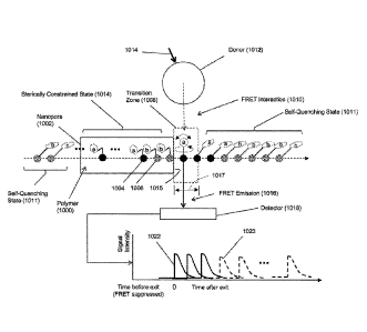
diagrammatically in Fig. 1A. Polymer analyte (1000), such as a polynucleotide,
is driven,
e.g. electrophoretically, through nanopore (1002), which constrains the
conformation of
polymer (1000) so that its monomeric units translocatc through the nanopore in
the same
order as their primary sequence in the polymer. In the embodiment shown in
Fig. 1A,
fluorescent labels are assumed to be members of FRET pairs, but this is not
intended to
limit the present invention; fluorescent labels may also include fluorescent
labels that are
directly excited, for example with a laser emitting at an appropriate
wavelength, to generate
a fluorescent signal,
1011301 As mentioned above, whenever an acceptor-labeled monomeric unit is
within the
bore of nanopore (1002), FRET interactions between such acceptors and the
donors of its
FRET pair are suppressed because acceptors are in a constrained state (1014).
Such
suppression typically means that no detectable FRET signal is produced even if
such
acceptors are within a FRET distance of a donor, for example, due to
unfavorable
orientation of the acceptor and donor dipoles, or due to contact quenching, or
like
mechanism. On the other hand, when an acceptor-labeled monomeric unit emerges
from
tn)
SUBSTITUTE SHEET (RULE 26)
CA 02963604 2017-04-03
WO 2016/057829
PCT/US2015/054756
the bore of, or exits, the nanopore into transition zone (1008), FRET
interaction (1010)
occurs and FRET emission (1016) is produced and detected by detector (1018)
until the
acceptor enters a self-quenching state (1011) with an adjacent acceptor and as
the distance
between the acceptor and donor increases with the movement of polymer (1000)
Out of
FRET interaction distance. Signal (1022) is produced by a single acceptor as
it moves
through transition zone (1008). Transition zone (1008), which is a spatial
region
immediately adjacent to exit (1015) of nanopore (1002), is defined by several
factors,
including the speed of the translocation of polymer (1000) through nanopore
(1002), the
vibrational and rotational mobility of the fluorescent labels, the
physiochemical nature of
the fluorescent labels, and the like, In some embodiments, transition zone
(1008) may be
defined by a perpendicular distance (1017) between the exit (1015) of nanopore
(1002) and
the point at which an exiting fluorescent label takes on a quenched
configuration with an
adjacent fluorescent label. In some embodiments, transition zone (1008) may be
defined
by its corresponding transition interval, or the time it takes a fluorescent
label to travel
distance (1017). In some embodiments, transition distance (1017) is in the
range of from
20 to 50 angstroms; in other embodiments, transition distance is in the range
of from 20 to
40 angstroms. In some embodiments, corresponding transition intervals are in
the range of
from 0.2 to 2.0 msee; in still other embodiments, transition intervals are in
the range of
from 0.2 to 1.0 msee. In Fig. 1A, only one type of monomeric unit, illustrated
as solid
circles (1004) carries a first fluorescent label (designated as "a"); the rest
of the monomeric
units, illustrated as speckled circles (1006), carry a second fluorescent
label (designated as
"b"). In this embodiment, first fluorescent labels quench adjacent first
fluorescent labels
and adjacent second fluorescent labels; likewise, second fluorescent labels
quench adjacent
first fluorescent labels and adjacent second fluorescent labels; moreover, the
first and
second fluorescent labels generate FRET signals that are distinguishable from
one another,
for example, recorded signal (1022) for label "a" and recorded signal (1023)
for label "b"
in Fig. IA, so that each fluorescent label (and hence, monomer) may be
identified by a
signal detected by detector (1018).
10031] As illustrated in Fig. 1B, the degree to which successive signals
(1022) or (1023)
are resolved by detector (1018) depend at least in part on the translocation
speed of
polymer (1000). Curve A and curve B of Fig. 1B illustrate results from
simulations of
fluorescent signal generation based on the Forrester equation under different
auto-
quenching conditions. As illustrated, under both conditions readily
discernable signals are
11
SUBSTITUTE SHEET (RULE 26)
CA 02963604 2017-04-03
WO 2016/057829
PCT/US2015/054756
generated. In Fig. 1C, a further simulation showing signal peaks (1033) is
compared to
actual data (1035) generated as a fluorescently labeled single stranded DNA
analyte
translocated through a nanopore. The single stranded DNA used to generate the
data (1035)
was 200nt long and each cytosine was exchanged with a fluorescently labeled
counterpart.
The labeled DNA was translocated through a continuously excited hybrid
nanopore at an
applied potential of 300mV and FRET events were captured using a mos camera
operated
at 2kilz acquisition rate. At the 3'end of the labeled DNA a short homopolymer
stretch of 3
consecutive cytosines shows an elevated baseline fluorescent with clearly
distinguishable
peaks for each of the three cytosines. Similar to the modeled data the
fluorescent trace in
the inset of Fig. 1C shows an elevated baseline fluorescence and individual
peaks for each
member of the homopolymer stretch. The sequence of labeled DNA is as follows
(SEQ ID
NO: 1):
5'- GCT ATG TGG CGC GGT ATT ATC AAG AAG GAG ACT GAG AGG AGA
GTA GGA GCG AGA AGG AAA CGA GAG TGA GAG GAG AGT AGG AGC
AAG AAG GAA ACG AGA GTG AGA GGA GAG TAG GAG CAA GAA GGA
AAC GAG AGT GAG AGG AGA GTA GGA GCA AGA AGG AAA CTG AGA
GGA GAG TAG GAG TTA CTC TAG CTT CCC GGC AA -3'
[00321 In some embodiments, a nanopore is hybrid nanopore comprising a
protein
nanopore inserted into a pore of a solid phase membrane, as described more
fully below.
In hybrid nanopores, a first member of a FRET pair may be attached directly to
the protein
nanopore, or alternatively, directly to the solid phase membrane using
conventional linking
chemistries, such as "click" chemistries, e.g. Kolb et at. Angew. Chem. Int.
Ed., 4): 2004-
2021 (2001), or the like. In one embodiment, a first member of a FRET pair is
attached
directly or indirectly to the protein nanopore, for example, as discussed in
reference to Fig.
2D. In another embodiment, the first member of the FRET pair is a donor and a
quantum
dot. Quantum dots are typically much larger than acceptors, especially
acceptors that are
organic dyes, which typically have molecular weights in the range of from 200
to 2000
daltons.
Nanopores and Nanopore Sequencing
12
SUBSTITUTE SHEET (RULE 26)
83992829
10033-I Nanopores used with the invention may be solid-state nanopores,
protein
nanopores, or hybrid nanopores comprising protein nanopores or organic
nanotubes such as
carbon nanotubes, configured in a solid-state membrane, or like framework.
Important
features of nanopores include (i) constraining analytes, particularly polymer
analytes, to
pass through a detection zone in sequence, or in other words, so that monomers
pass the
detection zone one at a time, or in single file, (ii) compatibility with a
translocating means,
that is, whatever method is used to drive an analyte through a nanopore, such
as an electric
field, and (iii) suppression of fluorescent signals within the lumen, or bore,
of the
nanopore, for example, by contact quenching, or the like. Nanopores used in
connection
with the methods and devices of the invention may be used singly or in the
form of arrays,
either a regular array, such as a rectilinear array of a plurality nanopores
in a planar support
or membrane, or a random array, for example, where a plurality of nanopores
are spaced in
accordance with a Poisson distribution in a planar support or membrane.
[0034] Nanopores may be fabricated in a variety of materials including but not
limited to,
silicon nitride (Si3N4), silicon dioxide (SiO2), and the like. The fabrication
and operation
of nanopores for analytical applications, such as DNA sequencing, are
disclosed in the
following exemplary references Russell, U.S. patent 6,528,258;
Feier, U.S. patent 4,161,690; Ling, U.S. patent 7,678,562; Flu et al, U.S.
patent
7,397,232; Golovehenko et al, U.S. patent 6,464,842; Chu et al, U.S. patent
5,798,042;
Sauer et al, U.S. patent 7,001,792; Su et al, U.S. patent 7,744,816; Church et
al, U.S, patent
5,795,782; Bayley et al, U.S. patent 6,426,231; Akeson et al, U.S. patent
7,189,503; Bayley
et al, U.S. patent 6,916,665; Akeson et al, U.S. patent 6,267,872; Meller et
al, U.S. patent
publication 2009/0029477; H.oworka et al, International patent publication
W02009/007743; Brown et al, International patent publication W02011/067559;
Keller et
al, International patent publication W02009/020682; Polonsky et al,
International patent
publication W02008/092760; Van der Zaag et al, International patent
publication
W02010/007537; Yan et al, Nano Letters, 5(6): 1129-1134 (2005); 1qbal et al,
Nature
Nanotechnology, 2: 243-248 (2007); Wanunu et al, Nano Letters, 7(6): 1580-1585
(2007);
Dekker, Nature Nanotechnology, 2: 209-215 (2007); Storm et at, Nature
Materials, 2: 537-
540 (2003); Wu et al, Electrophoresis, 29(13): 2754-2759 (2008); Nakane et al.
Electrophoresis, 23: 2592-2601 (2002); The et al, J. Mieromech, Mieroeng., 17:
304-313
(2007); Henriquez et al, The Analyst, 129: 478-482 (2004); Jagtiani et al, J.
Micromech.
Microeng., 16: 1530-1539 (2006); Nakane et al, J. Phys. Condens. Matter, 15
R1365-
13
Date Recue/Date Received 2022-01-05
CA 02963604 2017-04-03
WO 2016/057829
PCT/US2015/054756
R1393 (2003); DeBlois et al, Rev. Sci. Instruments, 41(7): 909-916 (1970);
Clarke et al,
Nature Nanotechnology, 4(4): 265-270 (2009); Bayley et al. U.S. patent
publication
2003/0215881; and the like.
[00351 Briefly, in one aspect, a 1-50 urn channel is formed through a
substrate, usually a
membrane, through which an analyte, such as single stranded DNA, is induced to
translocate. The solid-state approach of generating nanopores offers
robustness and
durability as well as the ability to tune the size and shape of the nanopore,
the ability to
fabricate high-density arrays of nanopores on a wafer scale, superior
mechanical, chemical
and thermal characteristics compared with lipid-based systems, and the
possibility of
integrating with electronic or optical readout techniques. Biological
nanopores on the other
hand provide reproducible narrow bores, or lumens, especially in the 1-10
nanometer
range, as well as techniques for tailoring the physical and/or chemical
properties of the
nanopore and for directly or indirectly attaching groups or elements, such as
fluorescent
labels, which may be FRET donors or acceptors, by conventional protein
engineering
methods. Protein nanopores typically rely on delicate lipid bilayers for
mechanical
support, and the fabrication of solid-state nanopores with precise dimensions
remains
challenging. Combining solid-state nanopores with a biological nanopore
overcomes some
of these shortcomings, especially the precision of a biological pore protein
with the
stability of a solid state nanopore. For optical read out techniques a hybrid
nanopore
provides a precise location of the nanopore which simplifies the data
acquisition greatly.
The lateral diffusion of nanopore proteins inserted in a lipid bilayer makes
an optical
detection challenging. Since the biological part (i.e. protein nanopore part)
of a hybrid
nanopore does not rely on the insertion in a lipid bilayer, the degrees of
freedom for
modifications made to such a protein are greatly increased e.g. a genetically
modified
nanopore protein that does not spontaneously insert in a lipid bilayer may
still be used as a
protein component of a hybrid nanopore. Also, bilayer-destabilizing agents
such as
quantum dots may be used to label a protein component of a hybrid nanopore.
100361 In one embodiment, the invention is directed to a method for
analyzing one or
more polymer analytes, such as determining a nucleotide sequence of a
polynucleotide,
which comprises the following steps: (a) translocating a polymer analyte
through a
nanopore having a bore and an exit, the polymer analyte comprising a. sequence
of
monomers, wherein substantially each monomer is labeled with a fluorescent
label such
that fluorescent labels of adjacent monomers are in a quenched state by self-
quenching one
14
SUBSTITUTE SHEET (RULE 26)
CA 02963604 2017-04-03
WO 2016/057829
PCT/US2015/054756
another outside of the nanopore and fluorescent labels are in a sterically
constrained state
and incapable of generating a detectable fluorescent signal inside of the
nanopore; (b)
exciting each fluorescent label at the exit of the nanopore as it transitions
from a sterically
constrained state to a quenched state so that a fluorescent signal is
generated which is
indicative of the monomer to which it is attached; (e) detecting the
fluorescent signal to
identify the monomer. As used herein, "substantially every", "substantially
all", or like
terms, in reference to labeling monomers, particularly nucleotides,
acknowledges that
chemical labeling procedures may not result in complete labeling of every
monomer; to the
extent practicable, the terms comprehend that labeling reactions in connection
with the
invention are continued to completion; in some embodiments, such completed
labeling
reactions include labeling at least fifty percent of the monomers; in other
embodiments,
such labeling reactions include labeling at least eighty percent of the
monomers; in other
embodiments, such labeling reactions include labeling at least ninety-five
percent of the
monomers; in other embodiments, such labeling reactions include labeling at
least ninety-
nine percent of the monomers.
[0037] In another embodiment, the invention is directed to a method for
analyzing one
or more polymer analytes comprising the following steps: (a) attaching a
fluorescent label
substantially every monomer of one or more polymer analytes such that
fluorescent labels
of adjacent monomers are in a quenched state, (b) translocating the polymer
analytes
through nanopores so that monomers of each polymer analyte traverses the
nanopore in
single file and wherein each nanopore has a bore and an exit, the bore
sterically
constraining the fluorescent labels in a constrained state so that no
fluorescent signal is
generated therefrom inside the bore; (c) exciting during a transition interval
each
fluorescent label at the exit of the nanopore as each fluorescent label
transitions from a
sterically constrained state to a quenched state, thereby generating a
fluorescent signal that
is indicative of the monomer to which it is attached; (c) detecting the
fluorescent signal to
identify the monomer.
[0038] In another embodiment the invention is directed to a device for
analyzing one or
more labeled polymer analytes, such as a device for determining a nucleotide
sequence of
one or more labeled polynucleotide analytes, such device comprising the
following
elements: (a) a solid phase membrane separating a first chamber and a second.
chamber,
the solid phase membrane having at least one nanopore fluidly connecting the
first chamber
and the second chamber through a bore or lumen, the bore or lumen having a
cross-
SUBSTITUTE SHEET (RULE 26)
CA 02963604 2017-04-03
WO 2016/057829
PCT/US2015/054756
sectional dimension such that labels of a labeled polymer translocating
therethrougli are
sterically constrained so that detectable signals are not generated, and so
that the labels of
adjacent monomers of the labeled polymer are self-quenching; (b) an excitation
source for
exciting each label when it exits the nanopore and enters the second chamber
so that a
signal is generated indicative of a monomer to which the label is attached;
and (c) a
detector for collecting at least a portion of the signal generated by each
excited label; and
(d) identifying the monomer to which the excited label is attached by the
collected signal.
[00391 In another embodiment, the invention is directed to a system for
analyzing
polymers comprising a polymer comprising monomers that are substantially all
labeled
with a mutually quenching dye set and a nanopore device for sequentially
detecting optical
signals from the dyes of the mutually quenching dye set which are attached to
the polymer.
Such an embodiment for determining a sequence of a polynucleotide may comprise
the
following elements; (a) a solid phase membrane separating a first chamber and
a second
chamber, the solid phase membrane having at least one aperture connecting the
first
chamber and the second chamber, and having a hydrophobic coating on at least
one
surface; (b) a lipid layer disposed on the hydrophobic coating; (c) a protein
nanopore
immobilized in the aperture, the protein nanopore having a bore with an exit,
and the
protein nanopore interacting with the lipid layer to form a seal with the
solid phase
membrane in the aperture so that fluid communication between the first chamber
and the
second chamber occurs solely through the bore of the protein nanopore, and the
protein
nanopore being cross-sectionally dimensioned so that nucleotides of the
polynucleotide
pass through the exit of the bore in sequence and so that fluorescent labels
attached to the
poly-nucleotide are sterically constrained so that generation of fluorescent
signal therein is
inhibited or prevented; and (d) a first member of the FRET pair attached to
the solid phase
membrane or the protein nanopore, so that whenever nucleotides of the
polynucleotide
emerge from the bore, a plurality of the nucleotides are within a FRET
distance of the first
member of the FRET pair. In some embodiments, the first member of the FRET
pair is a
quantum dot that functions as a FRET donor.
100401 In some embodiments, the hydrophobic coating is optional in that the
surface of the
solid phase membrane is sufficiently hydrophobic itself so that a lipid layer
adheres to it
stably. The at least one aperture will have an inner surface, or wall,
connected to, or
contiguous with the surfaces of the solid phase membrane. In some embodiments,
the at
least one aperture will be a plurality of apertures, and the plurality of
apertures may be
16
SUBSTITUTE SHEET (RULE 26)
CA 02963604 2017-04-03
WO 2016/057829
PCT/US2015/054756
arranged as a regular array, such as a rectilinear array of apertures, the
spacing of which
depending in part on the number and kind of FRET pairs employed and the
optical
detection system used. Each of the apertures has a diameter, which in some
embodiments
is such that a protein nanopore is substantially immobilized therein. In some
embodiments,
substantially immobilized means that a protein nanopore may move no more than
5 nm in
the plane of the solid phase membrane relative to the wall of the aperture. In
another
embodiment, substantially immobilized means that a protein nanopore may move
no more
than 5 nm in the plane of the solid phase membrane relative to the wall of the
aperture.
The protein nanopores each have a bore, or passage, or lumen. which permits
fluid
communication between the first and second chambers when the protein nanopore
is
immobilized in an aperture. Generally, the bore is coaxially aligned with the
aperture.
One function of the hydrophobic layer is to provide a surface to retain lipids
in and/or
immediately adjacent to the at least one aperture. Such lipids, in turn,
permit disposition
and immobilization of a protein nanopore within an aperture in a functional
conformation
and in a manner that forms a fluid seal with the wall of the aperture. In some
embodiments, such seal also prevents electrical current passing between the
first and
second chambers around the protein nanopore. In some embodiments, charged
analytes are
disposed in an electrolyte solution in the first chamber and are translocated
through the
bore(s) of the protein nanopore(s) into an electrolytic solution in the second
chamber by
establishing an electrical field across the solid phase membrane. For
convenience of
manufacture, in some embodiments the hydrophobic coating will be on one
surface of the
solid phase membrane and the wall(s) of the aperture(s).
100411 In some embodiments of the devices of the invention, the at least
one nanopore
in a solid phase membrane is a plurality of nanopores, or a nanopore array; in
some
embodiments such nanopores are spaced regularly in the solid phase membrane
with their
bores oriented perpendicularly to the plane of the solid phase membrane. In
sonic
embodiments, nanopores are spaced in a rectilinear pattern in the solid phase
membrane; in
other embodiments, nanopores are spaced in a random pattern in the solid phase
membrane; in some embodiments, such random pattern is Poisson distributed. In
some
embodiments, nanopores are regularly spaced in a solid phase membrane with a
minimal
inter-nanopore distance of at least 10 nm; in other embodiments, such minimal
inter-
nanopore distance is 50 nm; in other embodiments, such minimal inter-nanopore
distance
17
SUBSTITUTE SHEET (RULE 26)
83992829
is 100 run, in other embodiments, such minimal inter-nanopore distance is 200
nm: in
other embodiments, such minimal inter-nanopore distance is 500 nm.
[00421 In some embodiments, methods and devices of the invention comprise
a solid
phase membrane, such as a SiN membrane, having an array of apertures
therethrough
providing communication between a first chamber and a second chamber (also
sometimes
referred to as a "cis chamber" and a "trans chamber") and supporting a lipid
bilayer on a
surface facing the second, or trans, chamber. In some embodiments, diameters
of the
aperture in such a solid phase membrane may be in the range of 10 to 200 nm,
or in the
range of 20 to 100 urn. In some embodiments, such solid phase membranes
further include
protein nanopores inserted into the lipid bilayer in regions where such
bilayer spans the
apertures on the surface facing the trans chamber. In some embodiments, such
protein
nanopores are inserted from the cis side of the solid phase membrane using
techniques
described herein. In some embodiments, such protein nanopores have a structure
identical
to, or similar to, a-hemolysin in that it comprises a barrel, or bore, along
an axis and at one
end has a "cap" structure and at the other end has a "stern" structure (using
the terminology
from Song et al. Science, 274: 1859-1866 (1996)). In some embodiments using
such
protein nanopores, insertion into the lipid bilayer results in the protein
nanopore being
oriented so that its cap structure is exposed to the cis chamber and its stem
structure is
exposed to the trans chamber.
100431 In some embodiments, methods and devices of the invention comprise
droplet
interface bilayers, either as single droplets or as arrays droplets, for
example, as disclosed
in Bayley et at, U.S. patent publication 2014/0356289; Huang et al, Nature
Nanotechnology, 10.1038/nnano,2015.189. [Epub ahead of print]; or like
reference.
Briefly, protein nanopores (1.2 nM) are placed in a 200-350 n1 droplet
(for example, 1.32 M KC1, 8.8 mM HEPES, 0.4 mM EDTA, pH 7.0 (aHL)
or 8.0 (MspA), and incubated in, for example, 3 m114 1,2-diphytanoyl-sn-
glycero-3-
phosphocholine (DPhPC) in hexadeeane to form a lipid monolayer coating. A
droplet may
then be transferred by pipetting onto a coverslip in a measurement chamber,
for example,
that permits application of voltages to move analytes and optical detection,
for example, by
TIRE The coverslip may be spin coated (3,000 r.p.m., 30 s) with a thin layer (-
200 inn) of
agarose (0.66 M CaCl2, 8.8 mM HEPES, pH 7.0 (aHL)/8.0 (MspA)) and subsequently
incubated with 3 mM DPhPC in hexadecane. On contact with the monolayer on the
a.garose, a lipid coated droplet spontaneously forms a droplet interface
bilayer. A ground
18
Date Recue/Date Received 2022-01-05
83992829
electrode (Ag/AgCI) may be inserted into the droplet, with a corresponding
active electrode
(Ag/AgCI) in the substrate agarose. Voltage protocols may be applied with a
patch clamp
amplifier (for example, Axopatch 200B, Molecular Devices). Nanopores present
in the
droplet spontaneously insert into the droplet interface bilayer, and the ion
flux may be
detected both electrically and/or optically (for example, by way of an ion-
sensitive dye,
such as Fluo-8, or the like).
[0044] In some embodiments, the solid phase membrane may be treated with a
low
energy ion beam to bleach its a.utofluorescence, e.g. as described in Huber et
al, U.S. patent
publication 2013/0203050.
[0045] Figs. 2A-2C are diagrams of hybrid biosensors. A nanometer sized hole
(102) is
drilled into a solid-state substrate, or solid phase membrane, (103) which
separates two
chambers, or compartments cis (101) and trans (107). A protein biosensor (e.g
a protein
nanopore) (104) attached to a charged polymer (105), such as a single stranded
DNA, is
embedded into the solid-state nanohole by electrophoretic transport. In Fig.
IC the protein
biosensor is inserted. In a nanometer sized hole which surface has a
hydrophobic coating
(106) and a lipid layer (109) attached thereto. A nanopore may have two sides,
or orifices.
One side is referred to as the "cis" side and faces the (-) negative electrode
or a negatively
charged buffer/ion compartment or solution. The other side is referred to as
the "trans"
side and faces the (4-) electrode or a positively charged buffer/ion
compartment or solution.
A biological polymer, such as a labeled nucleic acid molecule or polymer can
be pulled or
driven through the pore by an electric field applied through the nanopore,
e.g., entering on
the cis side of the nanopore and exiting on the trans side of the nanopore.
[0046] Fig. 2D shows protein nanopore (104) inserted into an aperture drilled
in a solid
state membrane (103). Attached to the protein nanopore (104) is an
oligonucleotide (108)
to which a complementary secondary oligonucleotide (111) is hybridized. Said
secondary
oligonucleotide (111) has one or more second members of a FRET pair (110)
attached to it.
Alternatively, a member of a FRET pair may be directly attached to an amino
acid of a
protein nanopore. For example, a hemolysin subunit may be modified by
conventional
genetic engineering techniques to substitute a cysteine for a suitably located
amino acid
adjacent to the exit of the nanopore, e.g. the threonine 129. An
oligonucleotide or members
of a FRET pair may be attached via the thio group of the cysteine using
conventional linker
chemistries, e.g. Hennanson (cited above).
19
Date Recue/Date Received 2022-01-05
CA 02963604 2017-04-03
WO 2016/057829
PCT/US2015/054756
[09471 In some embodiments, the present invention employs a hybrid nanopore,
particularly for optical-based nanopore sequencing of polynueleotides. Such
embodiments
comprise a solid-state orifice, or aperture, into which a protein biosensor,
such as a protein
nanopore, is stably inserted. A protein nanopore (e.g. alpha hemolysin) may be
attached to
a charged polymer (e.g. double stranded DNA) which serves as a drag force in
an applied
electric field, and which may be used to guide a protein nanopore into an
aperture in a
solid-state membrane. In some embodiments, the aperture in the solid-state
substrate is
selected to be slightly smaller than the protein, thereby preventing it from
translocating
through the aperture. Instead, the protein will be embedded into the solid-
state orifice. The
solid-state substrate can be modified to generate active sites on the surface
that allow the
covalent attachment of the plugged-in protein biosensor resulting in a stable
hybrid
biosensor,
100481 The polymer attachment site in the biosensor can be generated by
protein
engineering e.g, a mutant protein can be constructed that will allow the
specific binding of
the polymer. As an example, a cysteine residue may be inserted at the desired
position of
the protein. The cysteine can either replace a natural occurring amino acid or
can be
incorporated as an addition amino acid. Care must be taken not to disrupt the
biological
function of the protein. The terminal primary amine group of a polymer (i.e.
DNA) is then
activated using a hetero-bifunctional crosslinker (e.g. SMCC). Subsequently,
the activated
polymer is covalently attached to the cysteine residue of the protein
biosensor. In some
embodiments, the attachment of the polymer to the biosensor is reversible. By
implementing a cleavable crosslinker, an easily breakable chemical bond (es.
an S-S bond)
is introduced and the charged polymer may be removed after insertion of the
biosensor into
the solid-state aperture.
[00491 For someone skilled in the art it is obvious that a wide variety of
different
approaches for covalent or non-covalent attachment methods of a charged
polymer to the
protein biosensor are possible and the above described approach merely serves
as an
example. The skilled artisan will also realize that a variety of different
polymers may be
used as a drag force, including, but not limited to, single or double stranded
DNA,
polyethyleneglycol (PEG), polyvinylpyrrolidone (PVP), poly-L-lysine, linear
polysaccharides etc. It is also obvious that these polymers may exhibit either
a. negative (-)
or positive (+) charge at a given pH and that the polarity of the electric
field may be
adjusted accordingly to pull the polymer-biosensor complex into a solid-state
aperture.
SUBSTITUTE SHEET (RULE 26)
83992829
[0050-1 In sonic embodiments, a donor fluorophore is attached to the protein
nanopore. This
complex is then inserted into a solid-state aperture or nanohole (for example,
3-10 nin in
diameter) by applying an electric field across the solid state nanohole until
the protein
nanopore is transported into the solid-state nanohole to form a hybrid
nanopore. The
formation of the hybrid nanopore can be verified by (a) the inserting protein
nanopore
causing a drop in current based on a partial blockage of the solid-state
nanohole and by (b)
the optical detection of the donor fluorophore.
[00511 Once stable hybrid nanopores have formed single stranded, fluorescently
labeled
(or acceptor labeled) DNA is added to the cis chamber (the chamber with the
(+)
electrode). The applied electric field forces the negatively charged ssDNA to
translocate
through the hybrid nanopore during which the labeled nucleotides get in close
vicinity of
the donor fluorophore.
100521 Solid state, or synthetic, nanopores may be preprared in a variety of
ways, as
exemplified in the references cited above. In some embodiments a helium ion
microscope
may be used to drill the synthetic nanopores in a variety of materials, e.g.
as disclosed by
Yang et al, Nanotechnolgy, 22: 285310 (2011).
A chip that supports one or more regions of a thin-film material, e.g. silicon
nitride, that
has been processed to be a free-standing membrane is introduced to the helium
ion
microscope (HIM) chamber, HIM motor controls are used to bring a free-standing
membrane into the path of the ion beam while the microscope is set for low
magnification.
Beam parameters including focus and stigmation are adjusted at a region
adjacent to the
free-standing membrane, but on the solid substrate. Once the parameters have
been
properly fixed, the chip position is moved such that the free-standing
membrane region is
centered on the ion beam scan region and the beam is blanked. The HIM field of
view is set
to a dimension (in gm) that is sufficient to contain the entire anticipated
nanopore pattern
and sufficient to be useful in future optical readout (i.e. dependent on
optical
magnification, camera resolution, etc.). The ion beam is then rastered once
through the
entire field of view at a pixel dwell time that results in a total ion dose
sufficient to remove
all or most of the membrane autofluoreseence, The field of view is then set to
the proper
value (smaller than that used above) to perform lithographically-defined
milling of either a
single nanopore or an array of nanopores. The pixel dwell time of the pattern
is set to result
in nanopores of one or more predetermined diameters, determined through the
use of a
21
Date Recue/Date Received 2022-01-05
CA 02963604 2017-04-03
WO 2016/057829
PCT/US2015/054756
calibration sample prior to sample processing. This entire process is repeated
for each
desired region on a single chip and/or for each chip introduced into the HIM
chamber.
[90531 In some embodiments, the solid-state substrate may be modified to
generate active
sites on the surface that allow the covalent attachment of the plugged in
protein biosensor
or to modify the surface properties in a way to make it more suitable for a
given
application. Such modifications may be of covalent or non-covalent nature. A
covalent
surface modification includes a silanization step where an organosilane
compound binds to
silanol groups on the solid surface. For instance, the alkoxy groups of an
alkoxysilane are
hydrolyzed to form silanol-containing species. Reaction of these silanes
involves four
steps. Initially, hydrolysis of the labile groups occurs. Condensation to
oligomers follows.
The oligomers then hydrogen bond with hydroxyl groups of the substrate.
Finally, during
drying or curing, a covalent linkage is formed with the substrate with
concomitant loss of
water. For covalent attachment organosilanes with active side groups may be
employed.
Such side groups consist of, but are not limited to epoxy side chain,
aldehydes, isocyanates,
isothiocyanates, azides or alkynes (click chemistry) to name a few. For
someone skilled M
the art it is obvious that multiple ways of covalently attaching a protein to
a surface are
possible. For instance, certain side groups on an organosilane may need to be
activated
before being capable of binding a protein (e.g. primary amines or carboxyl
side groups
activated with an N-hydroxysuccinimidester). Another way of attaching a
protein to the
solid surface may be achieved through affinity binding by having one affinity
partner
attached to the protein and the second affinity partner being located on the
solid surface.
Such affinity pairs consist of the group of, but are not limited to biotin-
strepavidin, antigen-
antibody and aptamers and the corresponding target molecules. In a preferred
embodiment
the surface modification of the solid state nanopore includes treatment with
an
organosilane that renders the surface hydrophobic. Such organosilanes include
but are not
limited to, alkanesilanes (e.g. octaciecyldimethylchlorosilane) or modified
alkanesilanes
such as fluorinated alkanesilanes with an alkane chain length of 5 to 30
carbons. The
hydrophobic surface is then treated with a dilute solution of a lipid in
pentane. After drying
of the solvent and immersing the surface in an aqueous solution the lipid will
spontaneously form a layer on the surface. A layer of lipid on the solid
surface might proof
beneficial for the formation of a hybrid nanopore. The lipid layer on the
solid phase might
reduce the leak current between protein and solid state nanopore and it might
increase the
stability of the inserted protein pore. Combining a low capacitance solid
substrate as well
22
SUBSTITUTE SHEET (RULE 26)
CA 02963604 2017-04-03
WO 2016/057829
PCT/US2015/054756
as a lipid coating of said substrate may render the hybrid nanopore system
amenable to an
electrical readout based on current fluctuations generated by translocation of
DNA through
the hybrid nanopore. To achieve electrical read out with such a system a means
of
decreasing the translocation speed of unmodified DNA must be combined with a
lipid
coated hybrid nanopore. Molecular motors such as polymerases or helicases may
be
combined with a hybrid nanopore and effectively reduce the translocation speed
of DNA
through the hybrid nanopore. The lipids used for coating the surface are from
the group of
sphingolipids, phospholipids or sterols. A method and/or system for sequencing
a
biological polymer or molecule (e.g., a nucleic acid) may include exciting one
or more
donor labels attached to a pore or nanopore. A biological polymer may be
translocated
through the pore or nanopore, where a monomer of the biological polymer is
labeled with
one or more acceptor labels. Energy may be transferred from the excited donor
label to the
acceptor label of the monomer as, after the labeled monomer passes through,
exits or enters
the pore or nanopore. Energy emitted by the acceptor label as a result of the
energy
transfer may be detected, where the energy emitted by the acceptor label may
correspond to
or be associated with a single or particular monomer (e.g., a nucleotide) of a
biological
polymer. The sequence of the biological polymer may then be deduced or
sequenced based
on the detection of the emitted energy from the monomer acceptor label which
allows for
the identification of the labeled monomer. A pore, nanopore, channel or
passage, e.g., an
ion permeable pore, nanopore, channel or passage may be utilized in the
systems and
methods described herein.
[00541
[0055j A nanopore, or pore, may be labeled with one or more donor labels. For
example,
the cis side or surface and/or trans side or surface of the nanopore may be
labeled with one
or more donor labels. The label may be attached to the base of a pore or
nanopore or to
another portion or monomer making up the nanopore or pore A label may be
attached to a
portion of the membrane or substrate through which a nanopore spans or to a
linker or
other molecule attached to the membrane, substrate or nanopore. The nanopore
or pore
label may be positioned or attached on the nanopore, substrate or membrane
such that the
pore label can come into proximity with an acceptor label of a biological
polymer, e.g., a
nucleic acid, which is transiocated through the pore. The donor labels may
have the same
or different emission or absorption spectra. The labeling of a pore structure
may be
achieved via covalent or non-covalent interactions.
23
SUBSTITUTE SHEET (RULE 26)
83992829
[09561 A donor label (also sometimes referred to as a "pore label") may be
placed as close
as possible to the aperture, for example, the exit, of a nanopore without
causing an
occlusion that impairs translocation of a nucleic acid through the nanopore. A
pore label
may have a variety of suitable properties and/or characteristics. For example,
a pore label
may have energy absorption properties meeting particular requirements. A pore
label may
have a large radiation energy absorption cross-section, ranging, for example,
from about 0
to 1000 rim or from about 200 to 500 mn. A pore label may absorb radiation
within a
specific energy range that is higher than the energy absorption of the nucleic
acid label,
such as an acceptor label. The absorption energy of the pore label may be
tuned with
respect to the absorption energy of a nucleic acid label in order to control
the distance at
which energy transfer may occur between the two labels. A pore label may be
stable and.
functional for at least 106 to 109 excitation and energy transfer cycles.
Labels for Nanopores and Analytes
100571 In some embodiments, a nanopore may be labeled with one or more quantum
dots.
In particular, in some embodiments, one or more quantum dots may be attached
to a
nanopore, or attached to a solid phase support adjacent to (and within a FRET
distance of
an entrance or exit of a nanopore), and employed as donors in FRET reactions
with
acceptors on analytes. Such uses of quantum dots are well known and are
described
widely in the scientific and patent literature, such as, in U.S. patents
6,252,303; 6,855,551;
7,235,361; and the like.
100581 One example of a Quantum dot which may be utilized as a pore label is a
CdTe
quantum dot which can be synthesized in an aqueous solution. A CdTe quantum
dot may
be functionalized with a nucleophilic group such as primary amines, thiols or
functional
groups such as carboxylic acids. A CdTe quantum dot may include a
mercaptopropionic
acid capping ligand, which has a carboxylic acid functional group that may be
utilized to
covalently link a quantum dot to a primary amine on the exterior of a protein
pore. The
cross-linking reaction may be accomplished using standard cross-linking
reagents (homo-
bifunctional as well as hetero-bifunctional) which are known to those having
ordinary skill
in the art of bioconjugation. Care may he taken to ensure that the
modifications do not
impair or substantially impair the translocation of a nucleic acid through the
nanopore. This
may be achieved by varying the length of the employed crosslinker molecule
used to attach
the donor label to the nanopore.
24
Date Recue/Date Received 2022-01-05
CA 02963604 2017-04-03
WO 2016/057829
PCT/US2015/054756
[09591 For example, the primary amine of the lysine residue 131 of the natural
alpha
hemolysin protein (Song, L. et al.. Science 274, (1996): 1859-1866) may be
used to
covalently bind carboxy modified CdTe Quantum dots via I -Ethyl-3[3-
dimetwaminopropyl]carbodiimide hydrochloride/ N-
hydroxysulfosuccinimide (EDC/NHS) coupling chemistry. Alternatively, amino
acid 129
(threonine) may be exchanged into cysteine. Since there is no other cysteine
residue in the
natural alpha hemolysin protein the thiol side group of the newly inserted
cysteine may be
used to c.,=ovalently attach other chemical moieties.
[09601 A variety of methods, mechanisms and/or routes for attaching one or
more pore
labels to a pore protein may be utilized. A pore protein may be genetically
engineered in a
manner that introduces amino acids with known properties or various functional
groups to
the natural protein sequence. Such a modification of a naturally occurring
protein sequence
may be advantageous for the bioconjugation of Quantum dots to the pore
protein. For
example, the introduction of a cysteine residue would introduce a thiol group
that would
allow for the direct binding of a Quantum dot, such as a CdTe quantum dot, to
a pore
protein. Also, the introduction of a Lysin residue would introduce a primary
amine for
binding a Quantum dot. The introduction of glutamic acid or aspartic acid
would
introduce a carboxylic acid moiety for binding a Quantum dot. These groups are
amenable
for bioconjugation with a Quantum dot using either homo- or hetero-
bifunctional
crosslinker molecules. Such modifications to pore proteins aimed at the
introduction of
functional groups for bioconjugation are known to those having ordinary skill
in the art.
Care should be taken to ensure that the modifications do not impair or
substantially impair
the translocation of a nucleic acid through the nanopore.
[0061] The nanopore label can be attached to a protein nanopore before or
after insertion of
said nanopore into a lipid bilayer. Where a label is attached before insertion
into a lipid
bilayer, care may be taken to label the base of the nanopore and avoid random
labeling of
the pore protein. This can be achieved by genetic engineering of the pore
protein to allow
site specific attachment of the pore label, as discussed below. An advantage
of this
approach is the bulk production of labeled nanopores. Alternatively, a
labeling reaction of a
pre-inserted nanopore may ensure site-specific attachment of the label to the
base (trans-
side) of the nanopore without genetically engineering the pore protein.
SUBSTITUTE SHEET (RULE 26)
CA 02963604 2017-04-03
WO 2016/057829
PCT/US2015/054756
[0062] A biological polymer, e.g., a nucleic acid molecule or polymer, may be
labeled with
one or more acceptor labels. For a nucleic acid molecule, each of the four
nucleotides or
building blocks of a nucleic acid molecule may be labeled with an acceptor
label thereby
creating a labeled (e.g., fluorescent) counterpart to each naturally occurring
nucleotide.
The acceptor label may be in the form of an energy accepting molecule which
can be
attached to one or more nucleotides on a portion or on the entire strand of a
converted
nucleic acid.
[0063] A variety of methods may be utilized to label the monomers or
nucleotides of a
nucleic acid molecule or polymer. A labeled nucleotide may be incorporated
into a nucleic
acid during synthesis of a new nucleic acid using the original sample as a
template
("labeling by synthesis"). For example, the labeling of nucleic acid may be
achieved via
PCR, whole gnome amplification, rolling circle amplification, primer extension
or the like
or via various combinations and extensions of the above methods known to
persons having
ordinary skill in the art.
[0064] Labeling of a nucleic acid may be achieved by replicating the nucleic
acid in the
presence of a modified nucleotide analog having a label, which leads to the
incorporation
of that label into the newly generated nucleic acid. The labeling process can
also be
achieved by incorporating a nucleotide analog with a functional group that can
be used to
covalently attach an energy accepting moiety in a secondary labeling step.
Such
replication can be accomplished by whole genome amplification (Zhang, L, et
al,, Proc.
Natl. Acad. Sci. USA 89 (1992): 5847) or strand displacement amplification
such as rolling
circle amplification, nick translation, transcription, reverse transcription,
primer extension
and polymerase chain reaction (PCR), degenerate oligonucleotide primer PCR
(DOP-PCR)
(Telenius, H. et al., Genomics 13 (1992): 718-725) or combinations of the
above methods.
[0065] A label may comprise a reactive group such as a nucleophile (amines,
thiols etc.).
Such nucleophiles, which are not present in natural nucleic acids, can then be
used to attach
fluorescent labels via amine or thiol reactive chemistry such as NHS esters,
maleimides,
epoxy rings, isocyanates etc. Such nucleophile reactive fluorescent dyes (i.e.
NHS-
dyes) are readily commercially available from different sources. An advantage
of labeling
a nucleic acid with small nucleophiles lies in the high efficiency of
incorporation of such
labeled nucleotides when a "labeling by synthesis" approach is used. Bulky
fluorescently
labeled nucleic acid building blocks may be poorly incorporated by polymerases
due to
26
SUBSTITUTE SHEET (RULE 26)
CA 02963604 2017-04-03
WO 2016/057829
PCT/US2015/054756
steric hindrance of the labels during the polymerization process into newly
synthesized
DNA.
[90661 In some embodiments, DNA can be directly chemically modified without
polymerase mediated incorporation of labeled nucleotides. One example of a
modification
includes cis-platinum containing dyes that modify Guanine bases at their N7
position
(Hoevel, T. et al., Bio Techniques 27 (1999): 1064-1067). Another example
includes the
modifying of pyrimidines with hydroxylamine at the C6 position which leads to
6-
hydroxylamino derivatives. The resulting amine groups can be further modified
with amine
reactive dyes (e.g. NHS-Cy5). Yet another example are azide or alkyne modified
nucleotides which are readily incorporated by polymerases (Gierlich et al.,
Chem. Bur. J.,
2007, 13, 9486-0404), The alkyne or azide modified polynueleotide is
subsequently labeled
with an azide or alkyne modified fluorophore following well established click
chemistry
protocols.
100671 As mentioned above, in some embodiments, DNA may be labeled using
"click
chemistry," e.g. using commercially available kits (such as "Click-It" from
Life
Technologies, Carlsbad, CA). Click chemistry in general refers to a synthetic
process in
which two molecules are linked together by a highly efficient chemical
reaction, one which
is essentially irreversible, in which the yield is nearly 100%, and which
produces few or no
reaction byproducts.. More recently, the meaning has come to refer to the
cyclization
reaction of a substituted alkyne with a substituted azide to form a 1,2,3-
triazole bearing the
two substituents. When catalyzed by copper at room temperature the reaction is
known as
the Raisgen cycloaddition, and it fully satisfies the requirements for click
chemistry in that
no other chemical functionality on the two molecules is affected during the
reaction. Thus
the coupling reaction has found broad application in bioconjugate chemistry,
for example,
in dye labeling of DNA or proteins, where many amino, hydroxy, or thiol groups
may be
found. The key requirement is that an alkyne group and an azide can easily be
introduced
into the molecules to be coupled. For example, in the coupling of a
fluorescent dye to a
DNA oligonueleotide, the azide group is typically introduced synthetically
into the dye,
while the alkyne group is incorporated into the DNA during oligonucleotide
synthesis.
Upon mixing in the presence of Cu-i- the two components are quickly coupled to
form the
itiazole, in this ease bearing the oligonucleotide as one .substituent and the
dye as the other.
Another more recent advance provides the alkyne component within a strained
ring
structure. in this case the reaction with an azide does net require the copper
catalyst, being'
27
SUBSTITUTE SHEET (RULE 26)
83992829
driven by release of the ring strain energy as the triazole is formed. This is
better known as
the copper-free click reaction. Guidance for applying click chemistry to
methods of the
invention may be found in the following references :
R.ostovtsev VV, Green TeCl; Fokin, Valery V. Sharpless KB (2002). "A Stepwise
Huisgen
Cycloaddition Process: Copper(1)-Catalyzed R egi oselecti ve "Ligation" of
Azides and
Terminal Alkynes", Angewandte Chernie International Edition 41 (14): 2596-
2599, Moses
JE and Moorhouse Al) (2007.) "The growing applications of click chemistry",
Chem. Soc.
Rev. 36(8): 1249-1262.
[00681 Whenever two or more mutually quenching dyes are used, such dyes may be
attached to DNA using orthogonal attachment chemistries. For instance NHS
esters can be
used to react very specifically with primary amines or maleimides will react
with thiol
groups. Either primary amines (N112) or thiol (SH) modified nucleotides are
commercially
available. These relatively small modifications are readily incorporated in a
polymerase
mediated DNA synthesis and can be used for subsequent labeling reactions using
either
NHS or maleimide modified dyes. Guidance for selecting and using such
orthogonal linker
chemistries may be found in Hermanson (cited above).
[00691 Additional orthogonal attachment chemistries are shown in Figs. 3A-
3H. Fig.
3A shows typical attachment positions of linking moieties on nucleoside bases.
Fig. 3B
shows a reaction diagram for Huisgen-type cycloaddition for a copper-catalyzed
reaction
and an uncatalyzed reaction, e.g. disclosed in the references cited above.
Fig. 3C shows a
reaction diagram for Acne plus nitrile oxide cycloaddition, e.g. as disclosed
in Gutsmiedl
et al, Org. Lett., 11: 2405-2408 (2009). Fig. 3D shows a reaction diagram for
Diels-Alder
cycloaddition, e.g. disclosed in Seelig et al, Tetrahedron Lett., 38: 7729-
7732 (1997). Fig.
3E shows a reaction diagram for carbonyl ligation, e.g. as disclosed in Casi
et al, J. Am.
Chem. Soc., 134: 5887-5892 (2012); Shao et al J. Am. Chem. Soc., 117: 3893-
3899
(1995); Rideout, Science, 233: 561-563 (1986); or the like. Fig. 3F shows a
reaction
diagram for Michael addition, e.g. disclosed in Brinkley, Bioconjugate
Chemistry, 3: 2-13
(1992). Fig. 3G shows a reaction diagram for native chemical ligation, e.g.
disclosed in
Schuler et al, Bioconjugate Chemistry, 13: 1039-1043 (2002); Dawson et al,
Science, 266:
776-779 (1994); or the like. Fig. 311 shows a reaction diagram for amide
formation via an
active ester, e.g. disclosed in Hennanson (cited above).
28
Date Recue/Date Received 2022-01-05
CA 02963604 2017-04-03
WO 2016/057829
PCT/US2015/054756
[09701 A nucleic acid molecule may be directly modified with N-
Bromosuccinimide which
upon reacting with the nucleic acid will result in 5-Bromocystein, 8-
Bromoadenine and 8-
Bromoguanine. The modified nucleotides can be further reacted with di-amine
nucleophiles. The remaining nucleophile can then be reacted with an amine
reactive dye
(e.g. NHS-dye) (Hermanson G. in Bioconjugate Techniques, cited above).
100711 A combination of 1, 2, 3 or 4 nucleotides in a nucleic acid strand may
be exchanged
with their labeled counterpart. The various combinations of labeled
nucleotides can be
sequenced in parallel, e.g., labeling a source nucleic acid or DNA with
combinations of 2
labeled nucleotides in addition to the four single labeled samples, which will
result in a
total of 10 differently labeled sample nucleic acid molecules or Dl\TAs (G, A,
T, C, GA,
GT, GC, AT, AC, TC). The resulting sequence pattern may allow for a more
accurate
sequence alignment due to overlapping nucleotide positions in the redundant
sequence
read- out.
100721 A method for sequencing a polymer, such as a nucleic acid molecule
includes
providing a nanopore or pore protein (or a synthetic pore) inserted in a
membrane or
membrane like structure or other substrate. The base or other portion of the
pore may be
modified with one or more pore labels. The base may refer to the Trans side of
the pore.
Optionally, the Cis and/or Trans side of the pore may be modified with one or
more pore
labels. Nucleic acid polymers to be analyzed or sequenced may be used as a
template for
producing a labeled version of the nucleic acid polymer, in which one of the
four
nucleotides or up to all four nucleotides in the resulting polymer is/are
replaced with the
nucleotide's labeled analogue(s). An electric field is applied to the nanopore
which forces
the labeled nucleic acid polymer through the nanopore, while an external
monochromatic
or other light source may be used to illuminate the nanopore, thereby exciting
the pore
label. As, after or before labeled nucleotides of the nucleic acid pass
through, exit or enter
the nanopore, energy is transferred from the pore label to a nucleotide label,
which results
in emission of lower energy radiation. The nucleotide label radiation is then
detected by a
confocal microscope setup or other optical detection system or light
microscopy system
capable of single molecule detection known to people having ordinary skill in
the art.
Examples of such detection systems include but are not limited to eonfocal
microscopy,
epifluorescent microscopy and total internal reflection fluorescent (TIRF)
microscopy.
Other polymers (e.g., proteins and polymers other than nucleic acids) having
labeled
monomers may also be sequenced according to the methods described herein. In
some
29
SUBSTITUTE SHEET (RULE 26)
CA 02963604 2017-04-03
WO 2016/057829
PCT/US2015/054756
embodiments, fluorescent labels or donor molecules are excited in a THU system
with an
evanescent wave, sometimes referred to herein as "evanescent wave excitation."
[0073] Energy may be transferred from a pore Of nanopore donor label (e.g., a
Quantum
Dot) to an acceptor label on a polymer (e.g., a nucleic acid) when an acceptor
label of an
acceptor labeled monomer (e.g,, nucleotide) of the polymer interacts with the
donor label
as, after or before the labeled monomer exits, enters or passes through a
nanopore. For
example, the donor label may be positioned on or attached to the nanopore on
the cis or
trans side or surface of the nanopore such that the interaction or energy
transfer between
the donor label and acceptor label does not take place until the labeled
monomer exits the
nanopore and comes into the vicinity or proximity of the donor label outside
of the
nanopore channel or opening, As a result, interaction between the labels,
energy transfer
from the donor label to the acceptor label, emission of energy from the
acceptor label
and/or measurement Of detection of an emission of energy from the acceptor
label may take
place outside of the passage, channel or opening running through the nanopore,
e.g., within
a cis or trans chamber on the cis or trans sides of a nanopore. The
measurement or
detection of the energy emitted from the acceptor label of a monomer may be
utilized to
identify the monomer.
[0074] The nanopore label may be positioned outside of the passage, channel or
opening of
the nanopore such that the label may be visible or exposed to facilitate
excitation or
illumination of the label. The interaction and energy transfer between a donor
label and
accepter label and the emission of energy from the acceptor label as a result
of the energy
transfer may take place outside of the passage, channel or opening of the
nanopore. This
may facilitate ease and accuracy of the detection or measurement of energy or
light
emission from the acceptor label, e.g., via an optical detection or
measurement device.
[0075] A donor label may be attached in various manners and/or at various
sites on a
nanopore. For example, a donor label may be directly or indirectly attached or
connected
to a portion or unit of the nanopore. Alternatively, a donor label may be
positioned
adjacent to a nanopore.
[0076] Each acceptor labeled monomer (e.g., nucleotide) of a polymer (e.g.,
nucleic acid)
can interact sequentially with a donor label positioned on or next to or
attached directly or
indirectly to the exit of a nanopore or channel through which the polymer is
translocated.
The interaction between the donor and acceptor labels may take place outside
of the
SUBSTITUTE SHEET (RULE 26)
83992829
nanopore channel or opening, e.g., after the acceptor labeled monomer exits
the nanopore
or before the monomer enters the nanopore. The interaction may take place
within or
partially within the nanopore channel or opening, e.g., while the acceptor
labeled monomer
passes through, enters or exits the nanopore.
100771 When one of the four nucleotides of a nucleic acid is labeled, the time
dependent
signal arising from the single nucleotide label emission is converted into a
sequence
corresponding to the positions of the labeled nucleotide in the nucleic acid
sequence. The
process is then repeated for each of the four nucleotides in separate samples
and the four
partial sequences are then aligned to assemble an entire nucleic acid
sequence.
[00781 When multi-color labeled nucleic acid (DNA) sequences are analyzed, the
energy
transfer from one or more donor labels to each of the four distinct acceptor
labels that may
exist on a nucleic acid molecule may result in light emission at four distinct
wavelengths or
colors (each associated with one of the four nucleotides) which allows for a
direct sequence
read-out.
Translocation Speed
100791 A major obstacle associated with nanopore based sequencing approaches
is the high
translocation velocity of nucleic acid through a nanopore (-500.000 ¨
1.000.000
nucleotides/sec) which doesn't allow for direct sequence readout due to the
limited
bandwidth of the recording equipment. A way of slowing down the nucleic acid
translocation with two different nanopore proteins was recently shown by Cherf
et al, (Nat
Biotechnol. 2012 Feb 14; 30(4):344-8) and Manrao et al. (Nat Biotechnol, 2012
Mar 25;
30(4):349-53). Both groups used a DNA polymerase to synthesize a
complementary strand from a target template which resulted in
the step-wise translocation of the template DNA through the nanopore. Hence,
the
synthesis speed of the nucleic acid polymerase (10-500nuc1e0ticle5isec)
determined the
translocation speed of the DNA and since it's roughly 3-4 orders of magnitude
slower than
direct nucleic acid translocation the analysis of single nucleotides became
feasible.
However, the polymerase-aided translocation requires significant sample
preparation to
generate a binding site for the polymerase and the nucleic acid synthesis has
to be blocked
in bulk and can only start once the nucleic acid-polymerase complex is
captured by the
nanopore protein. This results in a rather complex set-up which might prevent
the
implementation in a commercial setting. Furthennore, fluctuation in polymerase
synthesis
31
Date Recue/Date Received 2022-01-05
CA 02963604 2017-04-03
WO 2016/057829
PCT/US2015/054756
reactions such as a stalled polymerization as well as the dissociation of the
polymerase
from the nucleic acid may hamper the sequence read-out resulting in a high
error rate and
reduced read-length, respectively. In some embodiments, a target nucleic acid
is
enzymatically copied by incorporating fluorescent modified nucleotides. In
other
embodiments, modified nucleotides with reactive groups are incorporated which
can be
labeled in a post-extension reaction. The resulting labeled nucleic acid has
an increased
nominal diameter which results in a decreased translocation velocity when
pulled through a
nanopore. The preferred translocation rate for optical sequencing lies in the
range of 1-
1000 nucleotides per second with a more preferred range of 200-800 nucleotides
per
second and a most preferred translocation rate of 200-600 nucleotides per
second.
100801 Alternatively, translocation speed of a polynucleotide, especially a
single stranded
polynucleotide, may be controlled by employing a nanopore dimensioned so that
adducts
and/or labels, e.g. organic dyes attached to bases, inhibit but do not prevent
polynucleotide
translocation. A translocation speed may be selected by attaching labels
and/or adducts at
a predetermined density. Such labels and/or adducts may have regular spaced
attachments,
e.g. every third nucleotide or the like, or they may have random, or
pseudorandom
attachments, e.g. every C may be labeled. hi some embodiments, a selected
number of
different nucleotides may be labeled, e.g. every A and C, or every A and 0, or
every A and
T., or every C, or the like, that results in an average translocation speed.
Such average
speed may he decreased by attaching adducts to unlabeled nucleotides. Adducts
include
any molecule, usually and organic molecule, that may be attached to a
nucleotide using
conventional chemistries. Typically adducts have a molecular weight in the
same range as
common organic dyes, e.g. fluorescein, Cy3, or the like. Adducts may or may
not be
capable of generating signals, that is, serving as a label. In some
embodiments, adducts
and/or labels are attached to bases of nucleotides, In other embodiments,
labels and/or
adducts may be attached to linkages between nucleosides in a polynucleotide.
In one
aspect, a method of controlling translocation velocity of a single stranded
polynucleotide
through a nanopore comprises the step of attaching adducts to the
polynucleotide at a
density, wherein translocation velocity of the single stranded polynucleotide
monotonically
decreases with a larger number of adducts attached, or with the density of
adducts attached.
In some embodiments, not every kind of nucleotide of a polynucleotide is
labeled. For
example, four different sets of a polynucleotide may be produced where
nucleotides of
each set are labeled with the same molecule, e.g. a fluorescent organic dye
acceptor, but in
32
SUBSTITUTE SHEET (RULE 26)
CA 02963604 2017-04-03
WO 2016/057829
PCT/US2015/054756
each set a different kind of nucleotide will be labeled. Thus, in set I only
A's may be
labeled; in set 2 only C's may be labeled; in set 3 only G's may be labeled;
and so on.
After such labeling, the four sets of polynucleotides may then be analyzed
separately in
accordance with the invention and a nucleotide sequence of the polynucleotide
determined
from the data generated in the four analysis. In such embodiments, and similar
embodiments, e.g. two labels are used, where some of the nucleotides of a
polynucleotide
are not labeled, translocation speed through a nanopore will be affected by
the distribution
of label along the polynucleotide. To prevent such variability in
translocation speed, in
some embodiments, nucleotides that are not labeled with an acceptor or donor
for
generating signals to determine nucleotide sequence, may be modified by
attaching a non-
signal-producing adduct that has substantially the same effect on
translocation speed as the
signal-producing labels.
100811 This disclosure is not intended to be limited to the scope of the
particular forms
set forth, but is intended to cover alternatives, modifications, and
equivalents of the
variations described herein. Further, the scope of the disclosure fully
encompasses other
variations that may become obvious to those skilled in the art in view of this
disclosure.
The scope of the present invention is limited only by the appended claims.
Kits
[0082] The invention may include kits for carrying out the methods of the
invention. In
some embodiments, kits include reagents for adding reactive groups to target
polynucleotides. For example, a target polynucleotide for analysis in
accordance with the
invention may be obtained by transcribing its complement from a sample using a
nucleic
acid polymerase in the presence of nucleoside triphosphate analogs that
include reactive
groups, such as amines or thiols. Thus, in some embodiments kits comprise one
or more
nucleoside triphosphate analogs with reactive groups. Kits may further
comprise one or
more mutually quenching fluorescent labels with complementary functionalities
to the
reactive groups. Kits may further comprise a nucleic acid polymerase for
incorporating
nucleoside triphosphates into a target polynucleotide. Nucleic acid
polymerases may
include a reverse transcriptase when mRNA is used to produce target
polynucleotides, or
nucleic acid polymerases may include a DNA polymerase when genomic DNA is used
to
produce target polynucleotides. Kits may further comprise buffers, co-factors
and like
reagents for carrying out polymerase reactions. Likewise, kits may further
include buffers
33
SUBSTITUTE SHEET (RULE 26)
CA 02963604 2017-04-03
WO 2016/057829
PCT/US2015/054756
and other reaction components for carrying out reactions between reactive
groups and
complementary functionalities on mutually quenching fluorescent labels in
order to
produce a labeled target polynucleotide. Kits may further include solid phase
membranes
and protein nanopores for assembly into an operable nanopore array. Such
latter kits may
further include donor members of a FRET pair for attachment to protein
nanopores or to a
solid phase membrane. Kits may include assembled nanopore arrays comprising a
solid
phase membrane including incorporated protein nanopores and donor members of a
FRET
pair.
Definitions
[0083] "FRET" or "Forrester, or fluorescence, resonant energy transfer"
means a non-
radiative dipole-dipole energy transfer mechanism from a donor to acceptor
fluorophore. The efficiency of FRET may be dependent upon the distance between
donor
and acceptor as well as the properties of the fluorophores (Stryer, L., Annu
Rev Biochem.
47 (1978): 819-846). "FRET distance" means a distance between a FRET donor and
a
FRET acceptor over which a FRET interaction can take place and a detectable
FRET signal
produced by the FRET acceptor.
[0084] "Kit" refers to any delivery system for delivering materials or
reagents for
carrying out a method of the invention. In the context of reaction assays,
such delivery
systems include systems that allow for the storage, transport, or delivery of
reaction
reagents (e.g., fluorescent labels, such as mutually quenching fluorescent
labels,
fluorescent label linking agents, enzymes, etc. in the appropriate containers)
and/or
supporting materials (e.g., buffers, written instructions for performing the
assay etc.) from
one location to another. For example, kits include one Or more enclosures
(e.g., boxes)
containing the relevant reaction reagents and/or supporting materials. Such
contents may
be delivered to the intended recipient together or separately. For example, a
first container
may contain an enzyme for use in an assay, while a second or more containers
contain
mutually quenching fluorescent labels.
[0085] "Microfluidics" or "nanofluidics" device means an integrated system
of one or
more chambers, ports, and channels that are interconnected and in fluid
communication
and designed for carrying out an analytical reaction or process, either alone
or in
cooperation with an appliance or instrument that provides support functions,
such as
sample introduction, fluid and/or reagent driving means, temperature control,
detection
34
SUBSTITUTE SHEET (RULE 26)
83992829
systems, data collection and/or integration systems, and the like.
Microfluidics and
nanofluidics devices may further include valves, pumps, filters and
specialized functional
coatings on interior walls, e.g. to prevent adsorption of sample components or
reactants,
facilitate reagent movement by eleetroosmosis, or the like. Such devices are
usually
fabricated in or as a solid substrate, which may be glass, plastic, or other
solid polymeric
materials, and may have a planar format for ease of detecting and monitoring
sample and
reagent movement, especially via optical or electrochemical methods. In some
embodiments, such devices are disposable after a single use. Features of a
microfluidic
device usually have cross-sectional dimensions of less than a few hundred
square
micrometers and passages typically have capillary dimensions, e.g. having
maximal cross-
sectional dimensions of from about 500 jun to about 0.1 pm. Microfluidics
devices
typically have volume capacities in the range of from 1 }II to a few fiL, e.g.
10-100 nL.
Dimensions of corresponding structures in nanofluidics devices are typically
from I to 3
orders of magnitude less than those for microfluidics devices. The fabrication
and
operation of microfluidies and nanofluidics devices are well-known in the art
as
exemplified by the following references: Ramsey, U.S. patents
6,001,229; 5,858,195; 6,010,607; and 6,033,546; Soane et al, U.S. patents
5,126,022 and 6,054,034; Nelson et al, U.S. patent 6,613,525; Maher et al,
U.S. patent
6,399,952; Rice et al, International patent publication WO 02/24322; Bjornson
et al,
International patent publication WO 99/19717; Wilding et al, U.S. patents
5,587,128;
5,498,392; Sia et al, Electrophoresis, 24: 3563-3576 (2003); Unger et al,
Science, 288: 113-
116 (2000); Enzelberger et al, U.S. patent 6,960,437; Cao, "Nanostmetures &
Nanomaterials: Synthesis, Properties & Applications," (Imperial College Press,
London,
2004).
[09861 "Nanopore"
means any opening positioned in a substrate that allows the passage
of analytes through the substrate in a predetermined or discernable order, or
in the case of
polymer analy-tes, passage of their monomeric units through the substrate in a
predetermined or discernible order. In the latter case, a predetermined or
discernible order
may be the primary sequence of monomeric units in the polymer. Examples of
nanopores
include proteinaceous or protein based nanopores, synthetic or solid state
nanopores, and
hybrid nanopores comprising a solid state nanopore having a protein nanopore
embedded
therein. A nanopore may have an inner diameter of 1-10 mu or 1-5 nm Or 1-3
rim.
Examples of protein nanopores include but are not limited to, alpha-
hemolysiri, voltage-
Date Recue/Date Received 2022-01-05
83992829
dependent mitochondrial porin (A7DAC), OmpF, OrnpC, MspA and LamB
(maltoporin),
e.g. disclosed in Rhee, M. etal., Trends in Biotechnology, 25(4) (2007): 174-
181; Bayley
et al (cited above); Gundlach et al, U.S. patent publication 2012/0055792; and
the like.
Any protein pore that allows the transiocation of single nucleic
acid molecules may be employed. A nanopore protein may be labeled at a
specific site on the exterior of the pore, or at a specific site on the
exterior of one or more
monomer units making up the pore forming protein. Pore proteins are chosen
from a
group of proteins such as, but not limited to, alpha-hemolysin, MspA, voltage-
dependent
mitochondria] porin (VDAC), Anthrax porin, OrnpF, Ornpfl and 1.arnB
(maltoporin).
Integration of the pore protein into the solid state hole is accomplished by
attaching a
charged polymer to the pore protein. After applying an electric field the
charged complex is
electrophoretically pulled into the solid state hole. A synthetic nanopore, or
solid-state
nanopore, may be created in various forms of solid substrates, examples of
Which include
but are not limited to silicones (e.g. Si3N4, SiO2), metals, metal oxides
(e.g. A1203)
plastics, glass, semiconductor material, and combinations thereof. A synthetic
nanopore
may be more stable than a biological protein pore positioned in a lipid
bilayer membrane.
A synthetic nanopore may also be created by using a carbon nanotuhe embedded
in a
suitable substrate such as but not limited to polymerized epoxy. Carbon
nanotubes can
have uniform and well-defined chemical and structural properties. Various
sized carbon
nanotubes can be obtained, ranging from one to hundreds of nanometers. The
surface
charge of a carbon nanotube is known to be about zero, and as a result,
electrophoretic
transport of a nucleic acid through the nanopore becomes simple and
predictable (Ito, T. et
al., Chem. COMITILI11. 12 (2003): 1482-83). The substrate surface of a
synthetic nanopore
may be chemically modified to allow for covalent attachment of the protein
pore or to
render the surface properties suitable for optical nanopore sequencing. Such
surface
modifications can be covalent or non-covalent. Most covalent modification
include an
organosilane deposition for which the most common protocols are described:1)
Deposition
from aqueous alcohol. This is the most facile method for preparing silylated
surfaces, A
95% ethanol-5% water solution is adjusted to pH 4.5-5.5 with acetic acid.
Silane is added
with stirring to yield a 2% final concentration. After hydrolysis and silanol
group formation
the substrate is added for 2-5min. After rinsed free of excess materials by
dipping briefly in
ethanol. Cure of the silane layer is for 5-10 mm at 110 degrees Celsius, 2)
Vapor Phase
Deposition. Silanes can be applied to substrates under dry aprotic conditions
by chemical
36
Date Recue/Date Received 2022-01-05
CA 02963604 2017-04-03
WO 2016/057829
PCT/US2015/054756
vapor deposition methods. These methods favor monolayer deposition. In closed
chamber
designs, substrates are heated to sufficient temperature to achieve 5mm vapor
pressure.
Alternatively, vacuum can he applied until silane evaporation is observed. 3)
Spin-on
deposition. Spin-on applications can be made under hydrolytic conditions which
favor
maximum funetionalization and polylayer deposition or dry conditions which
favor
monoiayer deposition. In some embodiments, single nanopores are employed with
methods of the invention. In other embodiments, a plurality of nanopores are
employed.
In some of the latter embodiments, a plurality of nanopores is employed as an
array of
nanopores, usually disposed in a planar substrate, such as a solid phase
membrane.
Nanopores of a nanopore array may be spaced regularly, for example, in a
rectilinear
pattern, or may be spaced randomly. In a preferred embodiment, nanopores are
spaced.
regularly in a rectilinear pattern in a planar solid phase substrate.
100871 "Peptide," "peptide fragment," "polypeptide," "oligopeptide," or
"fragment" in
reference to a peptide are used synonymously herein and refer to a compound
made up of a
single tmbranched chain of amino acid residues linked by peptide bondsõAmino
acids in a
peptide or polypeptide may be derivatized with various moieties, including but
not limited
to, polyethylene glycol, dyes, biotin, haptens, or like moieties. The number
of amino acid
residues in a peptide varies widely; however, preferably, peptides or
oligopeptides referred
to herein usually have from 2 to 70 amino acid residues; and more preferably,
they have for
2 to 50 amino acid residues. Polypeptides and peptide fragments referred to
herein usually
have from a few tens of amino acid residues, e.g. 20, to up to a few hundred
amino acid
residues, e.g. 200, or more.
[00881 "Polymer" means a plurality of monomers connected into a linear
chain.
Usually, polymers comprise more than one type of monomer, for example, as a
polynucleotide comprising A's, C's, G's and T's, or a polypeptide comprising
more than
one kind of amino acid. Monomers may include without limitation nucleosides
and
derivatives or analogs thereof and amino acids and derivatives and analogs
thereof. In
some embodiments, polymers are polynucleotides, whereby nucleoside monomers
are
connected by phosphodiester linkages, or analogs thereof
[00891 "Polynucleotide" or "oligonucleotide" are used interchangeably and
each mean a.
linear polymer of nucleotide monomers. Whenever the use of an oligonucleotide
or
polynucleotide requires enzymatic processing, such as extension by a
polymerase, ligation
by a ligase, or the like, one of ordinaty skill would understand that
oligonucleotides or
37
SUBSTITUTE SHEET (RULE 26)
CA 02963604 2017-04-03
WO 2016/057829
PCT/US2015/054756
polynucleotides in those instances would not contain certain analogs of
intemucleosidic
linkages, sugar moieties, or bases at any or some positions. Polynucleotides
typically
range in size from a few monomeric units, e.g. 540, when they are usually
referred to as
"oligormcleotides," to several thousand monomeric units. Whenever a
polynucleotide or
oligonucleotide is represented by a sequence of letters (upper or lower case),
such as
"ATGCCTG," it will be understood that the nucleotides are in 5l--->3' order
from left to
right and that "A" denotes deoxyadenosine, "C" denotes deoxycytidine, "G"
denotes
deoxyguanosine, and "T" denotes thymidine, "I" denotes deoxyinosine, "U"
denotes
uridine, unless otherwise indicated or obvious from context. Unless otherwise
noted the
terminology and atom numbering conventions will follow those disclosed in
Strachan and
Read, Human Molecular Genetics 2 (Wiley-Liss, New York, 1999). Usually
polynucleotides comprise the four natural nucleosides (e.g. deoxyadenosine,
deoxycytidine, deoxyguanosine, deoxythymidine for DNA or their ribose
counterparts for
RNA) linked by phosphodiester linkages; however, they may also comprise non-
natural
nucleotide analogs, e.g. including modified bases, sums, or intemucleosidic
linkages. It is
clear to those skilled in the art that where an enzyme has specific
oligonucleotide or
polynucleotide substrate requirements for activity, e.g. single stranded DNA.
RNA/DNA
duplex, or the like, then selection of appropriate composition for the
oligonucleotide or
polynucleotide substrates is well within the knowledge of one of ordinary
skill, especially
with guidance from treatises, such as Sambrook et al, Molecular Cloning,
Second Edition
(Cold Spring Harbor Laboratory, New York, 1989), and like references.
Likewise, the
oligonucleotide and polynucleotide may refer to either a single stranded form
or a double
stranded form (i.e. duplexes of an oligonucleotide or polynucleotide and its
respective
complement). It will be clear to one of ordinary skill which form or whether
both forms
are intended from the context of the terms usage.
[00901 "Sequence determination", "sequencing" or "determining a nucleotide
sequence"
or like terms in reference to polymers, such as polynucleotides, includes
determination of
partial as well as full sequence information of the polymer. In some
embodiments,
sequence determination may include detection or measurement of an identifying
characteristic, or fingerprint, of a polymer, such as a unique, or
substantially unique,
sequence of signals that is correlated to a particular polymer sequence. In
some
embodiments, such correlation is a one-to-one correspondence. In other
embodiments,
such correlation may not be unique. In other embodiments, such correlation
permits
38
SUBSTITUTE SHEET (RULE 26)
83992829
identification of a polymer with a particular sequence with a probability of
greater than
ninety percent; in other embodiments, such identification can be made with a
probability of
greater than ninety-nine percent. In the case of polynucleotides, the above
terms include
identifying sequences of subsets of the full set of four natural nucleotides,
A, C, G and T,
such as, for example, a sequence of just A's and C's of a target
polynucleotide. That is, the
terms include the determination of the identities, ordering, and locations of
one, two, three
or all of the four types of nucleotides within a target polynucleotide. In
some
embodiments, the terms include the determination of the identities, ordering,
and locations
of two, three or all of the four types of nucleotides within a target
polynucleotide. In some
embodiments sequence determination may be accomplished by identifying the
ordering
and locations of a single type of nucleotide, e.g. cytosines, within the
target polynucleotide
"ca.tcgc õ " so that its sequence is represented as a binary code, e.g.
"100101 . . . "
representing "c-(not c)(not c)c-(not "and the
like. In some embodiments, the terms
may also include subsequences of a target polynucleotide that serve as a
fingerprint for the
target polynucleotide; that is, subsequences that uniquely identify a target
polynucleotide
within a set of .polynucleotides, e.g. all different RNA sequences expressed
by a cell,
SEQUENCE LISTING IN ELECTRONIC FORM
In accordance with Section 111(1) of the Patent Rules, this description
contains
a sequence listing in electronic form in ASCII text format (file: 83992829 -
Seq 21-JUN-17 vl.txt).
A copy of the sequence listing in electronic form is available from the
Canadian
Intellectual Property Office.
39
Date Recue/Date Received 2022-01-05