Note: Descriptions are shown in the official language in which they were submitted.
IMPLANT FOR STABILIZING FRACTURED OR NON-FRACTURED BONES
The invention relates to a bone implant, the use of an implant
and a method of stabilizing a fractured or non-fractured bone.
Bone implants are widely used for stabilizing fractured bones.
US 2009/0157078 for example discloses a cannulated screw for re-
pairing defects in the humerus or the femur. Part of the screw
does not contain a thread but holes through which cement may be
introduced into a void into the bone. This device comprises a
screw head on one side of the implant which requires the screw
to partly extend outside the bone. The other end of the implant
is threaded so that the screw is only fixed in case the thread
can be fixed inside the bone on the side opposite of the screw
head. Such an implant is limited in its applications.
WO 2012/142032 discloses a method and a device for bone prepara-
tion. The device comprises an insertion structure with perfora-
tions through which a fluid may be introduced into the bone. The
device may be used for internal fixation of fractions or can be
implemented into possibly weak and/or cancerous bone. The fluid
may be bone cement. The implant part of the device is fixed on
the outside of the bone and is therefore very complicated. Fur-
thermore, several implants are used in one bone, which weakens
the bone tissue due to the number of introduction channels.
WO 2012/066236 is directed to a device combining two intersect-
ing implants for preventive or curative treatment of fractures
of the femur. The intersecting implants are fixed relative to
each other and thereby prevent any movement of the implants.
Date Recue/Date Received 2022-01-14
063703 2017-005
WO 2016/071089 PCT/EP2015/073845
2
Such a device can only be applied in bones that allow for inter-
secting implants.
It is therefore an object of the present invention to avoid the
drawbacks of the prior art and to create a bone implant, the use
of a bone implant and a method for stabilizing a fractured or
non-fractured bone which can be versatilely used in different
bones of the human body and allows for a stable positioning in-
side the bone.
The object is accomplished by a bone implant for stabilizing
fractured or non-fractured bones comprising an implant body
which preferably is a cylindrical body. The implant body extends
along a longitudinal axis from a front side to an end side. The
implant body has an implant width expending perpendicularly to
the longitudinal axis, wherein a length of the implant body
along the longitudinal axis is at least five times the implant
width. The implant body has an outer surface which is at least
divided into a first surface and a second surface wherein the
first surface consists of an anchorage area which extends at
least partially over the outer surface, preferably maximum over
half of the outer surface.
The anchorage area according to the invention comprises a sur-
face that improves the fixation of the implant when implanted in
a bone. The bone tissue can more easily grow into the implant
and the anchorage of the implant in the bone is improved.
Such an implant is easily introduced and fixed inside a bone,
especially a vertebra. The anchorage area preferably is located
at the proximal end of the implant when implanted into a bone.
The implant body preferably has a constant implant width at
least across the first surface and the second surface. Further-
more the implant does not comprise a widening on the proximal
end like for example a screw head. Hence, preferably the implant
CA 02 963703 2017-04-05
WO 2016/071089 PCT/EP2015/073845
3
width is constant over at least the first and the second sur-
face, while the implant width in other areas only may be small-
er.
The first surface and the second surface respectively preferably
extend along the longitudinal axis and 3600 around the longitu-
dinal axis.
The implant can comprise a bore extending along the longitudinal
axis or parallel to the longitudinal axis having at least one,
preferably two openings at the front side and/or at the end
side.
A bore inside the bone implant leads to a lighter implant and in
case of two openings the possibility of introduction of fluids
into the bone.
The length of the bone implant can be in a range from 10 mm to
250 mm.
The specific bone implant can be used for different bones and
still has a length needed for stabilizing the respective bone.
The width of the bone can be in a range from 5 mm (for ribs)to
50 mm (for femoral diaphysis) or 80 mm (for humeral head). The
width of the bone implant can be in a range from 2mm to lOmm.
Such a bone implant can easily be introduced into a bone without
destroying further bone tissue and nevertheless delivers enough
stability to serve its purpose of stabilizing the bone.
The outer surface, preferably the second surface, can comprise
holes having a wall around a hole axis.
The bone implant having holes on the outer surface or second
surface respectively on the one hand leads to the possibility of
bone tissue growing into the holes and thereby improving posi-
CA 02963703 2()17-04
WO 2016/071089 PCT/EP2015/073845
4
tioning of the implant and in case of bone implant having a bore
leading to the possibility of directing fluid into the bone tis-
sue around the implant, where holes are located.
The fluid preferably is bone cement, such as PMMA bone cement,
bio-resorbable bone cement or any other product allowing implant
fixation in a bone.
The holes can have a diameter of 0.2 mm to 5 mm on the outer
surface or the second surface respectively.
Through such a hole bone tissue can quickly grow in and fluid,
such as bone cement, can easily be introduced into the bone.
The walls of the holes can have a cylindrical, preferably a con-
ical shape.
Cylindrical holes are easy to manufacture and thereby lower man-
ufacturing costs and the conical shape optimizes the distribu-
tion of the fluid that is introduced into the bone through the
bone implant.
The implant can comprise a first set of holes, wherein the hole
axis of the first set of holes is arranged substantially perpen-
dicular to the longitudinal axis.
A set of holes can comprise one or more holes.
A hole with a hole axis arranged perpendicular to the longitudi-
nal axis leads to the distribution of fluid through the hole ra-
dially away from the implant and thus the bone cement reaches as
far as possible.
The implant can comprise a second set of holes, wherein the hole
axis of the second set of holes is inclined relative to the lon-
gitudinal axis, preferably inclined as an angle greater than 90
while smaller than 150 relative to the longitudinal axis.
CA 02963703 2()17-04
WO 2016/071089 PCT/EP2015/073845
An inclined hole axis enables the distribution of a fluid to a
specific point inside the bone when the implant is placed inside
the bone.
The implant can comprise a third set of holes, wherein the hole
5 axis of the third set of holes is inclined relative to the lon-
gitudinal axis at an angle different from the second set of
holes, preferably inclined at an angle smaller than 90 while
larger than 30 relative to the longitudinal axis.
An inclined hole axis enables the distribution of a fluid inside
the bone to a specific area when the implant is already inside
the bone. Furthermore, inclined holes enable the introduction of
bone cement even in sensitive areas of nerves, since it is pos-
sible to direct the flow of the cement into specific areas.
Especially a combination of the first, second and third set of
holes enables the direction of fluid from inside the implant to
specific areas of the bone relative to the implant.
The holes can be distributed substantially equal in an area of
360 around the longitudinal axis of the outer surface or the
second surface and preferably distributed in rows along the ion-
gitudinal axis such that a distance between neighbouring holes
in a row is substantially the same.
Such an arrangement allows for an optimised distribution of flu-
id and a uniform distribution of tissue growing into the im-
plant.
The holes can be located in an area from 180' to 270' around a
longitudinal axis on the outer surface or the second surface and
preferably distributed in rows along the longitudinal axis such
that the distance between neighbouring holes and a row is sub-
stantially the same.
CA 02 963703 2017-04-05
WO 2016/071089 PCT/EP2015/073845
6
The arrangement of the holes in an area from 180' to 270' around
the longitudinal axis leads to the possibility of directing flu-
id through the implant only to a part of the bone, where the
fluid is needed.
A first hole of a first row and a first hole of a second neigh-
bouring row can have a different distance to the front side,
preferably the difference in the distance is equal to half the
distance of two neighbouring holes in a row.
Such a distribution of neighbouring holes of neighbouring rows
leads to an optimised distribution of fluid being introduced
through the implant into the bone.
The distribution of holes is preferably chosen such that the
stability of the implant is not, or not significantly compro-
mised and nevertheless the distribution of fluid is optimal for
the specific situation. In spine, the holes will be placed in
the distal part of the implant which is implanted in the verte-
bral body, and the holes will be arranged such that cement is
injectable at 270 to 300 around the longitudinal axis, not on
the side of an upper endplate to avoid leakage in case of end-
plate fracture.
In the humerus application, the holes are placed all along the
implant, at 360 around the longitudinal axis, to allow a 360
cement flow. This allows a good implant fixation, a bone rein-
forcement and to full fill tumour if applicable.
The anchorage area can comprise means for improving the fixation
of the implant within a bone, preferably a surface structure
and/or a roughness and/or recesses.
The surface structure can be grooves, a thread or a ring shaped
structure, while the thread or groove pitches or ring structures
can be square, symmetrically triangular or asymmetrically trian-
CA 02 963703 2017-04-05
WO 2016/071089 PCT/EP2015/073845
7
gular. Alternatively or additionally, the surface structure can
comprise recesses that are straight, helicoidal or comprise
crossed or diamond shaped pitches. A recess in the anchorage ar-
ea can be from 0.5mm to 3 mm deep.
All depth values according to this invention are measured from
top to bottom. Of course statistical variations can occur.
A surface structure improves the anchorage of the implant.
The roughness can vary from 1 micrometer to 0,5mm.
The anchorage area can comprise a surface structure in form of
grooves, preferably 6 or 8 grooves, distributed substantially
equally around the longitudinal axis, extending coaxially along
the longitudinal axis.
The grooves can have the same shape and/or height as the recess-
es and surface structures being a thread or ring shaped.
The use of surface structures in form of grooves improves the
anchorage of the implant and therefore leads to a more durable
and safer implant.
The anchorage area can comprise a surface structure in form of a
thread or ring shaped grooves. Such an anchorage area improves
the fixation of the implant inside the bone.
A cross section of the grooves can be U-shaped, V-shaped or
square. By means of the grooves, the surface contact between the
bone and the implant is increased and stress concentration on
the bone is limited. It also allows implant stabilisation in ro-
tation before the cement injection, which is for example im-
portant for spinal implants where the injection holes have to be
oriented. After cement injection the implant stabilisation is
CA 02963703 2()17-04
WO 2016/071089 PCT/EP2015/073845
8
done by the cement e.g. stabilisation of the implant in rotation
and translation.
The implant can comprise a fixation connector allowing holding
of the implant during introduction into a bone.
The fixation connector can for example be a thread by means of
which the bone implant is connected to a tool. Preferably the
fixation connector is a thread on the inside of the bore of the
bone implant, while the inner thread preferably has a wider di-
ameter than the rest of the bore to improve an easy introduction
of a tool. By means of such an inner thread, the outer surface
can be optimized for fixation of the implant inside the bone.
Furthermore, the fluid can be introducible through the tool and
the implant when connected by the fixation connector. This leads
to any easy handling of the implant when introducing and fixing
the implant.
The outer surface can comprise a third surface having an at
least partially conical shape. Such a third surface is arranged
opposite of the anchorage surface and leads to the possibility
of easier introducing the bone implant into the bone.
The implant can be made from any implantable material, such as
PEEK, titanium, stainless steel or Nitinol or a combination
thereof.
The object is further accomplished by the use of an implant as
previously described for restoring a fractured bone.
The object is further accomplished by the use of an implant as
previously described for preventing a fracture in a bone.
Fractured bones can for example be the humeral head or diaphy-
sis, the calcaneus, the wrist radius, the tibia, the pelvis or
CA 02963703 2017-04-05
WO 2016/071089 PCT/EP2015/073845
9
the ribs. Examples for an application for preventing the frac-
ture of the bone is e. g. the humerus or the ribs or spinal ver-
tebral body for example in case of severe osteoporosis or lytic
lesion tumour induced.
The object is further accomplished by a method of stabilizing a
fractured or non-fractured bone by inserting a bone implant as
previously described into a bone and preferably introducing bone
cement into the bone through the bone implant.
Such a method leads to an easy introduction and fixation of the
bone implant inside the bone without the need of any plates or
additional fixation means.
In the following, the invention is described in embodiments by
means of figures. It shows:
Figure 1: a bone implant in a first embodiment,
Figure 2: a first section of a bone implant in a second em-
bodiment
Figure 3: a cross-section through a bone implant in a third
embodiment,
Figure 4: a bone implant in a fourth embodiment,
Figure 5: a cross-section through a bone implant according
to figure 4,
Figure 6: a bone implant in a fifth embodiment,
Figures 6a
to 6c: a detailed view of figure 6,
Figure 7: .. a cross-section through a bone implant in a sixth
embodiment
CA 02963703 2()17-04
WO 2016/071089 PCT/EP2015/073845
Figure 8: two bone implants stabilizing a fracture in a ver-
tebra,
Figure 9: two implants according to the third embodiment for
stabilizing a fracture in a vertebra,
5 Figure 10: a bone implant according to the first embodiment
for stabilizing the humerus.
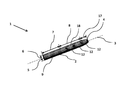
Figure 1 shows a bone implant 1 according to a first embodiment.
The bone implant 1 comprises an implant body 2 having a longitu-
10 dinal axis 3. The implant body 2 comprises a front side 4 and an
end side 5. Additionally, the implant body 2 is separated into a
first surface 7 and a second surface 8. The first surface 7 com-
prises an anchorage area 9 for improving the anchorage of the
Implant in a bone. Perpendicular to the longitudinal axis, the
implant body 2 comprises an implant width 6. The implant width 6
is constant along the first surface 7 and the second surface 8
and not exceeded at any other point of the implant 1. The second
surface 8 comprises holes 12 and a bore 10 inside the Implant
body 2. The holes 12 enable the introduction of a fluid such as
bone cement through the implant 1 into the bone and optimize the
fixation of the implant 1 inside the bone due to growing bone
tissue into the holes 12. The implant body 2 further comprises a
third surface 17 which has a conical shape. The width of the im-
plant is reduced in the third surface such that the introduction
of the implant 1 into the bone is easier. The front side 4 of
the implant body 2 is rounded such that it forms a semi-sphere
to facilitate introduction of the implant 1 into the bone. The
anchorage area 9 of the first surface 7 comprises a surface
structure for improving the anchorage of the implant 1 inside
the bone. The holes are distributed 360 around the circumfer-
ence of the implant body 2, while the holes 12 are arranged in
CA 02 963703 2017-04-05
WO 2016/071089 PCT/EP2015/073845
11
rows. The rows are offset relative to each other such that a
first hole 12 of a first row 18 has a different distance from
the front side 4 than a first hole 12 of a second neighbouring
row (not shown). The length of an implant body is 100 mm while
the implant width is 5 mm. The diameter of the holes 12 is 2.5
mm. The wall of the holes 12 has a cylindrical shape.
For example values for standard spinal implant will be: length
from 50 to 85mm, preferably mean 70mm, diameter from 4 to 7mm,
preferably 5mm, holes from lmm to 3mm. preferably 2-2,5mm.
Figure 2 shows a cross-section through a second embodiment of
the invention. In this embodiment, the implant body 2 comprises
a bore 10 along the longitudinal axis. On the end side 5 a fixa-
tion connector is arranged to enable a connection of the implant
body 2 with an Insertion tool (not shown). Contrary to the first
embodiment in figure 1, the holes 12 in the second embodiment
are arranged over a larger second surface 8 relative to a small-
er first surface 7. A first set of holes 12 comprises a hole ax-
is ha which is arranged perpendicular to the longitudinal axis
3. A second set of holes comprises an inclined axis 11b, while
the inclination of the hole axis llb is 120 relative to the
longitudinal axis 3. The front side 4 further comprises a third
surface 17 for facilitating introduction of the implant into a
bone.
Figure 3 shows a cross section of a third embodiment of the in-
vention. In this embodiment the bore 10 comprises a fixation
connector 16 on the end side 5 of the implant body 2 which is
threaded. By means of this thread a tool can be fixed in the im-
plant. The holes 12 are arranged 270 around the longitudinal
axis 13 and hence a fluid such as bone cement is only directed
260 from the implant. This way, sensitive areas will not be
filled with fluid or specific bone cement.
CA 02963703 2()17-04
WO 2016/071089 PCT/EP2015/073845
12
Figure 4 shows a fourth embodiment of the invention. This embod-
iment corresponds to the first embodiment in figure 1 apart from
the first surface 7 comprising the anchorage area 9. The anchor-
age area 9 comprises a thread in which the thread pitches extend
from the implant width 6. Such an anchorage area 9 improves the
fixation of the implant inside the bone. Furthermore, the third
surface 17 in this embodiment is shorter relative to the embodi-
ment in figure 1 and thereby a conical shape of the third sur-
face 17 comprises a steeper inclination relative to the embodi-
ment in figure 1. Additionally, the front side 4 is more peaked
relative to the embodiment in figure 1.
Figure 2 shows the embodiment as disclosed in figure 3 while the
first surface 7 comprises an anchorage area 9 having a thread.
The anchorage area 9 in this embodiment corresponds to the an-
chorage area 9 shown in figure 4.
Figure 6 shows an embodiment of the implant 1 which comprises a
first surface 7 having longitudinal grooves 13. The longitudinal
13 grooves improve the anchorage of the bone implant inside the
bone. The longitudinal grooves can comprise a cross-sectional
shape that is square, such that it avoids rotation (figure 6a),
semi-spherical such that insertion is easier and the bone con-
tact is better compared to square shapes or angles (figure 6b) or
triangular such that the surface contact is maximised(figure
6c). The holes 12 in the embodiment according to figure 6 are
only distributed from 270 up to 300 around the circumference
of the implant 1.
Figure 7 shows a cross-section through the embodiment according
to figure 6. The bore 10 along the longitudinal axis 3 comprises
a fixation connector 16 which enables a threaded connection to a
tool (not shown). The holes 12 are arranged along three differ-
ent hole axis 11a to 11c. The first hole axis 11a is arranged
perpendicular to the longitudinal axis. The hole axis 11b for
CA 02963703 2017-04-05
WO 2016/071089 PCT/EP2015/073845
13
the second set of holes is arranged at 1300 relative to the lon-
gitudinal axis 3. The hole axis 11c for the third set of holes
is arranged at 60' relative to the longitudinal axis. Such a
hole arrangement is especially suitable for an implant in a ver-
tebra since bone cement introduced through the bore 10 and holes
12 is optimally distributed in the vertebra. The purpose is to
avoid the risk of leakage through the vertebral body walls, an-
terior or posterior, that could have been damaged by a fracture.
Figures 8a to 8c show an exemplary embodiment of the use of the
implant in a vertebra. Figure 8a shows the top view, figure 8b
shows a side view and figure 8c shows a rear view from a verte-
bra in which two implants are introduced for stabilizing the
vertebra. The implants introduced in this embodiment are im-
plants according to figure 1.
Figures 9a to 9c show the same views as figure 8 applying an em-
bodiment of the implant according to figure 4.
Figure 10 shows an implant 1 used as a stabilizing implant in a
humerus. The humerus is not fractured. The implant 1 is never-
theless introduced into the bone for stabilizing it. The arrow
shows the way of introducing implant 1 into the humerus.