Note: Descriptions are shown in the official language in which they were submitted.
CA 2963720
DOSAGE AND ADMINISTRATION OF NON-FUCOSYLATED ANTI-CD40
ANTIBODIES
CROSS-REFERENCES TO RELATED APPLICATIONS
[0001] This application claims the benefit of U.S. Provisional Application No.
62/072,031,
filed on October 29, 2014 and U.S. Provisional Application No. 62/134,955,
filed on March 18,
2015.
FIELD OF THE INVENTION
[0002] This disclosure relates methods of using a non-fucosylated anti-CD40
antibody for
treatment of cancer and chronic infectious diseases.
BACKGROUND OF THE INVENTION
[0003] CD40 is a member of the tumor necrosis factor (TNF) receptor
superfamily. It is a
single chain type I transmembrane protein with an apparent MW of 50 kDa. CD40
is expressed
by some cancer cells, e.g., lymphoma cells and several types of solid tumor
cells. CD40 also
functions to activate the immune system by facilitating contact-dependent
reciprocal interaction
between antigen-presenting cells and T cells. Although a number of anti-CD40
antibodies have
been tested in clinical trials, to date none have exhibited sufficient
activity. The present
disclosure solves this and other problems.
BRIEF SUMMARY OF THE INVENTION
[0004] This disclosure provides a method of treating cancer, by administering
an anti-CD40
antibody to a patient in need of such treatment. The anti-CD40 antibody
comprises the heavy
chain variable region of SEQ ID NO:1 and the light chain variable region of
SEQ ID NO:2, and a
human constant region. The constant region has an N-glycoside-linked sugar
chain at residue
N297 according to the EU index as set forth in Kabat and less than 5% of the N-
glycoside-linked
sugar chains include a fucose residue, i.e., a fucose bound to the reducing
terminal of the sugar
chain via an a1,6 bond to N-acetylglucosamine ("GlcNAc"). Administration of
the anti-CD40
1
Date Recue/Date Received 2020-10-27
CA 02963720 2017-03-31
WO 2016/069919 PCT/US2015/058108
antibody is at a dose level between 0.1-300 ug/kg (14 antibody per kilogram
patient body
weight). In one embodiment, the anti-CD40 antibody dose level is between 0.6-
150 ug/kg. In
another embodiment, the anti-CD40 antibody dose level is between 1.0-100
us/kg. In another
embodiment, the anti-CD40 antibody dose level is between 5-25 1g/kg. In
another embodiment,
the anti-CD40 antibody dose level is between 8-12 g/kg. In another
embodiment, the anti-
CD40 antibody dose level is about 10 ug/kg. In another embodiment, the anti-
CD40 antibody
the dose level is 10 jig/kg.
[00051 In another aspect, this disclosure provides a method of treating
cancer, by administering
an anti-CD40 antibody to a patient in need of such treatment. The anti-CD40
antibody comprises
the heavy chain variable region of SEQ ID NO:1 and the light chain variable
region of SEQ ID
NO:2, and a human constant region. The constant region has an N-glycoside-
linked sugar chain
at residue N297 according to the EU index as set forth in Kabat and less than
5% of the N-
glycoside-linked sugar chains include a fucose residue, i.e., a fucose bound
to the reducing
terminal of the sugar chain via an a1,6 bond to N-acetylglucosamine
("GlcNAc").
Administration of the anti-CD40 antibody is at a dose level between 0.1-2000
jig/kg (us
antibody per kilogram patient body weight). In one embodiment, the dose level
is between 10-
1000 us/kg. In another embodiment, the dose level is between 50-800 g/kg. In
a further
embodiment, the dose level is between 75-600 us/kg. In another embodiment, the
dose level is
between 100-500 jig/kg. in further embodiments, the dose level is a range
selected from the
following: 100-300 jig/kg, 300-500 jig/kg, 500-700 g/kg, 700-900 jig/kg, and
900-1100 ps/kg.
In other embodiments, the dose level is a range selected from the following:
100-150 jig/kg, 150-
200 jig/kg, 200-250 jig/kg, 250-300 us/kg, 300-350 Kg/kg, 350-400 jig/kg, 400-
450 ug/kg, 450-
500 jig/kg, 500-550 jig/kg, 550-600 jig/kg, 600-650 jig/kg, 650-700 jig/kg,
700-750 jig/kg, 750-
800 jig/kg, 800-850 jig/kg, 850-900 us/kg, 900-950 jig/kg, 950-1000 jig/kg,
1000-1050 jig/kg,
and 1050-1100 jig/kg. In further embodiments, the dose level is selected from
the following:
about 60 us/kg, about 100 ug/kg, about 150 jig/kg, about 200 jig/kg, aabout
250 jig/kg, about
300 jig/kg, about 350 jig/kg, about 400 jig/kg, about 450 jig/kg, about 500
jig/kg, about 550
jig/kg, about 600 jig/kg, about 650 jig/kg, about 700 jig/kg, about 750 Kg/kg,
about 80014/kg,
about 850 us/kg, about 900 jig/kg, about 950 g/kg, about 1000-1050 jig/kg,
about 1050 jig/kg,
and 1110 jig/kg.
2
CA 02963720 2017-03-31
WO 2016/069919 PCT/US2015/058108
[0006] In one embodiment, the anti-CD40 antibody is administered every three
weeks. In
another embodiment the anti-CD40 antibody is administered every six weeks. In
another
embodiment the anti-CD40 antibody is administered every ten weeks. In another
embodiment
the anti-CD40 antibody is administered every twelve weeks. In another
embodiment the anti-
CD40 antibody is administered every fifteen weeks. In another embodiment the
anti-CD40
antibody is administered every eighteen weeks.
[0007] In another embodiment, the patient has a CD40 positive cancer. In
another
embodiment, the patient has a CD40 negative cancer. In a further embodiment,
the patient has a
cancer that is a solid tumor. In yet another embodiment, the patient has a
cancer that is a blood
cancer. In another embodiment, the cancer is a melanoma, a breast cancer,
including metastatic
breast cancer, a lung cancer, including a non-small cell lung cancer, or
pancreatic cancer.
[0008] In a further aspect, this disclosure provides methods of treating
cancer by administering
to the patient a combination of the anti-CD40 antibody and an antibody that
blocks an immune
checkpoint. One example of an antibody that blocks an immune checkpoint is an
anti-cytotoxic
T-lymphocyte-associated protein 4 (CTLA4) antibody. Examples of anti-CTLA4
antibodies
include, e.g,, ipilimumab or tremelimumab. Another example of an antibody that
blocks an
immune checkpoint is an anti-programmed cell death protein 1 (PD1) antibody.
Examples of
anti-PDl antibodies include, e.g., nivolumab, pidilizumab, or pembrolizumab. A
further example
of an antibody that blocks an immune checkpoint is an anti-programmed death-
ligand (PD-L1)
antibody. Examples of anti-PD-Li antibodies include, e.g., MEDI4736 and
MPDL3280A.
[0009] In another embodiment, the patient has a CD40 positive cancer and is
treated with a
combination of the anti-CD40 antibody and an antibody that blocks an immune
checkpoint, e.g.,
an anti-CTLA4 antbody, an anti-PD1 antibody, or an anti-PD-L1 antibody. In
another
embodiment, the patient has a CD40 negative cancer and is treated with a
combination of the
anti-CD40 antibody and an antibody that blocks an immune checkpoint, e.g., an
anti-CTLA4
antbody, an anti-PD1 antibody, or an anti-PD-Li antibody. In a further
embodiment, the patient
has a cancer that is a solid tumor and is treated with a combination of the
anti-CD40 antibody
and an antibody that blocks an immune checkpoint, e.g., an anti-CTLA4 antbody,
an anti-PD1
antibody, or an anti-PD-Ll antibody. In yet another embodiment, the patient
has a cancer that is
a blood cancer and is treated with a combination of the anti-CD40 antibody and
an antibody that
3
CA 2963720
blocks an immune checkpoint, e.g., an anti-CTLA4 antbody, an anti-PD1
antibody, or an anti-
PD-Li antibody. In another embodiment, the cancer is a melanoma, a breast
cancer, including
metastatic breast cancer, a lung cancer, including a non-small cell lung
cancer, or pancreatic
cancer, and is treated with a combination of the anti-CD40 antibody and an
antibody that
blocks an immune checkpoint, e.g., an anti-CTLA4 antbody, an anti-PD1
antibody, or an anti-
PD-Li antibody.
[0009A] In a further aspect, this disclosure provides a use of a composition
comprising an anti-
CD40 antibody and a physiologically acceptable carrier for treatment of cancer
in a patient in
need of such treatment, wherein the anti-CD40 antibody comprises the heavy
chain variable
region of SEQ ID NO:1 and the light chain variable region of SEQ ID NO:2, and
a human
constant region; wherein the constant region has an N-glycoside-linked sugar
chain at residue
N297 according to the EU index; and wherein less than 10% of N-glycoside-
linked sugar
chains in the composition comprise a fucose residue. In a further aspect, this
disclosure
provides a use of a composition comprising an anti-CD40 antibody for
preparation of a
medicament for treatment of cancer in a patient in need of such treatment,
wherein the anti-
CD40 antibody comprises the heavy chain variable region of SEQ ID NO:1 and the
light chain
variable region of SEQ ID NO:2, and a human constant region; wherein the
constant region has
an N-glycoside-linked sugar chain at residue N297 according to the EU index;
and wherein less
than 10% of N-glycoside-linked sugar chains in the composition comprise a
fucose residue.
[0009B] In a further aspect, this disclosure provides a use of a composition
comprising an
anti-CD40 antibody and a physiologically acceptable carrier for preparation of
a medicament
for treatment of cancer in a patient in need of such treatment, wherein the
anti-CD40 antibody
comprises the heavy chain variable region of SEQ ID NO:1 and the light chain
variable region
of SEQ 1D NO:2, and a human constant region; wherein the constant region has
an N-
glycoside-linked sugar chain at residue N297 according to the EU index; and
wherein less than
10% of N-glycoside-linked sugar chains in the composition comprise a fucose
residue.
[0009C] In a further aspect, this disclosure provides a use of an anti-PD1
antibody and a
composition comprising an anti-CD40 antibody and a physiologically acceptable
carrier for
treatment of cancer in a patient in need of such treatment, wherein the anti-
CD40 antibody
comprises the heavy chain variable region of SEQ ID NO:1 and the light chain
variable region
4
Date Recue/Date Received 2023-02-14
CA 2963720
of SEQ ID NO:2, and a human constant region; wherein the constant region has
an N-
glycoside-linked sugar chain at residue N297 according to the EU index; and
less than 10% of
N-glycoside-linked sugar chains in the composition comprise a fucose residue.
In a further
aspect, this disclosure provides a use of an anti-PD1 antibody and a
composition comprising an
anti-CD40 antibody and a physiologically acceptable carrier for preparation of
a medicament
for treatment of cancer in a patient in need of such treatment, wherein the
anti-CD40 antibody
comprises the heavy chain variable region of SEQ ID NO:1 and the light chain
variable region
of SEQ ID NO:2, and a human constant region; wherein the constant region has
an N-
glycoside-linked sugar chain at residue N297 according to the EU index; and
less than 10% of
N-glycoside-linked sugar chains in the composition comprise a fucose residue.
[0009D] In a further aspect, this disclosure provides a use of an anti-PD-Li
antibody and a
composition comprising an anti-CD40 antibody and a physiologically acceptable
carrier for
treatment of cancer in a patient in need of such treatment, wherein the anti-
CD40 antibody
comprises the heavy chain variable region of SEQ ID NO:1 and the light chain
variable region
of SEQ ID NO:2, and a human constant region; wherein the constant region has
an N-
glycoside-linked sugar chain at residue N297 according to the EU index; and
wherein less than
10% of N-glycoside-linked sugar chains in the composition comprise a fucose
residue. In a
further aspect, this disclosure provides a use of an anti-PD-Li antibody and a
composition
comprising an anti-CD40 antibody and a physiologically acceptable carrier for
preparation of a
medicament for treatment of cancer in a patient in need of such treatment,
wherein the anti-
CD40 antibody comprises the heavy chain variable region of SEQ ID NO:1 and the
light chain
variable region of SEQ ID NO:2, and a human constant region; wherein the
constant region has
an N-glycoside-linked sugar chain at residue N297 according to the EU index;
and wherein less
than 10% of N-glycoside-linked sugar chains in the composition comprise a
fucose residue.
[0009E] In a further aspect, this disclosure provides a composition comprising
an anti-CD40
antibody and a physiologically acceptable carrier for use to treat cancer in a
patient, wherein
the anti-CD40 antibody comprises the heavy chain variable region of SEQ ID
NO:1 and the
light chain variable region of SEQ ID NO:2, and a human constant region;
wherein the constant
region has an N-glycoside-linked sugar chain at residue N297 according to the
EU index; and
4a
Date Recue/Date Received 2023-02-14
CA 2963720
wherein less than 10% of N-glycoside-linked sugar chains in the composition
comprise a
fucose residue.
10009F1 In a further aspect, this disclosure provides a method of making a
composition
comprising an anti-CD40 antibody and a physiologically acceptable carrier
comprising:
culturing a host cell expressing an anti-CD40 antibody in the presence of a
fucosylation
inhibitor, wherein the anti-CD40 antibody comprises the heavy chain variable
region of SEQ
ID NO:1 and the light chain variable region of SEQ ID NO:2, and a human
constant region;
wherein the constant region has an N-glycoside-linked sugar chain at residue
N297 according
to EU index; and wherein less than 10% of N-glycoside-linked sugar chains in
the composition
comprise a fucose residue.
[0009G] In a further aspect, this disclosure provides a method of making a
composition
comprising an anti-CD40 antibody and a physiologically acceptable carrier
comprising:
culturing a host cell expressing an anti-CD40 antibody, wherein the activity
of FUT8 (alpha 1,6-
fucosyltransferase enzyme) is inhibited, wherein the anti-CD40 antibody
comprises the heavy
chain variable region of SEQ ID NO:1 and the light chain variable region of
SEQ ID NO:2, and
a human constant region; wherein the constant region has an N-glycoside-linked
sugar chain at
residue N297 according to EU index; and wherein less than 10% of N-glycoside-
linked sugar
chains in the composition comprise a fucose residue.
DEFINITIONS
[0010] A "polypeptide" or "polypeptide chain" is a polymer of amino acid
residues joined by
peptide bonds, whether produced naturally or synthetically. Polypeptides of
less than about 10
amino acid residues are commonly referred to as "peptides."
[0011] A "protein" is a macromolecule comprising one or more polypeptide
chains. A
protein may also comprise non-peptidic components, such as carbohydrate
groups.
Carbohydrates and other non-peptidic substituents may be added to a protein by
the cell in
which the protein is produced, and will vary with the type of cell. Proteins
are defined herein
in terms of their amino acid backbone structures; substituents such as
carbohydrate groups are
generally not specified, but may be present nonetheless.
4b
Date Recue/Date Received 2023-02-14
CA2963720
[0012] The terms "amino-terminal" and "carboxyl-terminal" are used herein to
denote
positions within polypeptides. Where the context allows, these terms are used
with reference to
a particular sequence or portion of a polypeptide to denote proximity or
relative position. For
example, a certain sequence positioned carboxyl-terminal to a reference
sequence within a
polypeptide is located proximal to the carboxyl terminus of the reference
sequence, but is not
necessarily at the carboxyl terminus of the complete polypeptide.
[0013] The term "antibody" is used herein to denote immunoglobulin proteins
produced by the
body in response to the presence of an antigen and that bind to the antigen,
as well as antigen-
binding fragments and engineered variants thereof. Hence, the term "antibody"
includes, for
example, intact monoclonal antibodies comprising full-length immunoglobulin
heavy and light
chains (e.g., antibodies produced using hybridoma technology) and antigen-
binding antibody
fragments, such as F(ab')2 and Fab fragments. Genetically engineered intact
antibodies and
fragments, such as chimeric antibodies, humanized antibodies, single-chain Fv
fragments, single-
chain antibodies, diabodies, minibodies, linear antibodies, multivalent or
multispecific (e.g.,
4c
Date Recue/Date Received 2022-03-04
CA 02963720 2017-03-31
WO 2016/069919 PCT/US2015/058108
bispecific) hybrid antibodies, and the like are also included. Thus, the term
"antibody" is used
expansively to include any protein that comprises an antigen-binding site of
an antibody and is
capable of specifically binding to its antigen.
[0014] An "antigen-binding site of an antibody" is that portion of an antibody
that is sufficient
to bind to its antigen. The minimum such region is typically a variable domain
or a genetically
engineered variant thereof. Single-domain binding sites can be generated from
camelid
antibodies (see Muyldermans and Lauwereys, J. Mol. Recog. 12:131-140, 1999;
Nguyen et al.,
EMBO J. 19:921-930, 2000) or from VH domains of other species to produce
single-domain
antibodies ("dAbs"; see Ward et al., Nature 341:5V-546, 1989; US Patent No.
6,248,516 to
Winter et al.). In certain variations, an antigen-binding site is a
polypeptide region having only 2
conwlementarity determining regions (CDRs) of a naturally or non-naturally
(e.g., mutagenized)
occurring heavy chain variable domain or light chain variable domain, or
combination thereof
(see, e.g., Pessi et al., Nature 362:367-369, 1993; Qiu et al., Nature
Biotechnol. 25:921-929,
2007). More commonly, an antigen-binding site of an antibody comprises both a
heavy chain
variable (VH) domain and a light chain variable (VL) domain that bind to a
common epitope.
Within the context of the present invention, an antibody may include one or
more components in
addition to an antigen-binding site, such as, for example, a second antigen-
binding site of an
antibody (which may bind to the same or a different epitope or to the same or
a different
antigen), a peptide linker, an immunoglobulin constant region, an
immunoglobulin hinge, an
amphipathic helix (see Pack and Pluckthun, Biochem. 31:1579-1584, 1992), a non-
peptide linker,
an oligonucleotide (see Chaudri et al., FEBS Letters 450:23-26, 1999), a
cytostatic or cytotoxic
drug, and the like, and may be a monomeric or multimeric protein. Examples of
molecules
comprising an antigen-binding site of an antibody are known in the art and
include, for example,
Fv, single-chain Fv (scFv), Fab, Fab', F(ab')2, F(ab),, diabodies, dAbs,
minibodies, nanobodies,
Fab-scFv fusions, bispecific (scFv)4-IgG, and bispecific (scFv)2-Fab. (See,
e.g., Hu et al.,
Cancer Res. 56:3055-3061, 1996; Atwell et al., Molecular Immunology 33:1301-
1312, 1996;
Carter and Merchant, Curt-. Opin. Biotechnol. 8:449-454, 1997; Zuo et al.,
Protein Engineering
13:361-367, 2000; and Lu et al., J. Immunol. Methods 267:213-226, 2002.)
[0015] As used herein, the term "immunoglobulin" refers to a protein
consisting of one or
more polypeptides substantially encoded by immunoglobulin gene(s). One form of
CA 02963720 2017-03-31
WO 2016/069919 PCT/US2015/058108
immunoglobulin constitutes the basic structural unit of native (i.e., natural)
antibodies in
vertebrates. This form is a tetramer and consists of two identical pairs of
immunoglobulin chains,
each pair having one light chain and one heavy chain. In each pair, the light
and heavy chain
variable regions (VL and VH) are together primarily responsible for binding to
an antigen, and
the constant regions are primarily responsible for the antibody effector
functions. Five classes of
immunoglobulin protein (IgG, IgA, IgM, IgD, and IgE) have been identified in
higher
vertebrates. IgG comprises the major class; it normally exists as the second
most abundant
protein found in plasma. In humans, IgG consists of four subclasses,
designated IgGl, IgG2,
IgG3, and IgG4. The heavy chain constant regions of the IgG class are
identified with the Greek
symbol y. For example, immunoglobulins of the IgG1 subclass contain a yl heavy
chain
constant region. Each immunoglobulin heavy chain possesses a constant region
that consists of
constant region protein domains (CHI, hinge, CH2, and CH3; IgG3 also contains
a CH4 domain)
that are essentially invariant for a given subclass in a species. DNA
sequences encoding human
and non-human immunoglobulin chains are known in the art. (See, e.g., Ellison
et al., DNA
1:11-18, 1981; Ellison et al., Nucleic Acids Res. 10:4071-4079, 1982; Kenten
et al., Proc. Natl.
Acad. Sci. USA 79:6661-6665, 1982; Seno et al., Nuc. Acids Res. 11:719-726,
1983; Riechmann
et al., Nature 332:323-327, 1988: Amster et al., Nuc. Acids Res. 8:2055-2065,
1980; Rusconi and
Kohler, Nature 314:330-334, 1985; Boss et al., Nuc. Acids Res. 12:3791-3806,
1984; Bothwell et
al., Nature 298:380-382, 1982; van der Loo et al., Immunogenetics 42:333-341,
1995; Karlin et
al., J. Mol. Evol. 22:195-208, 1985; Kindsvogel et al., DNA 1:335-343, 1982;
Breiner et al.. Gene
18:165-174, 1982; Kondo et al., Eur. J. lmmunol. 23:245-249, 1993; and GenBank
Accession
No. J00228.) For a review of immunoglobulin structure and function, see
Putnam, The Plasma
Proteins, Vol V, Academic Press, Inc., 49-140, 1987; and Padlan, Mol. Immunol.
31:169-217,
1994. The term "irnmunoglobulin" is used herein for its common meaning,
denoting an intact
antibody, its component chains, or fragments of chains, depending on the
context.
[0016] Full-length immunoglobulin "light chains" (about 25 Kd or 214 amino
acids) are
encoded by a variable region gene at the amino-terminus (encoding about 110
amino acids) and a
by a kappa or lambda constant region gene at the carboxyl-terminus. Full-
length
immunoglobulin "heavy chains" (about 50 Kd or 446 amino acids) are encoded by
a variable
region gene (encoding about 116 amino acids) and a gamma, mu, alpha, delta, or
epsilon
constant region gene (encoding about 330 amino acids), the latter defining the
antibody's isotype
6
CA 02963720 2017-03-31
WO 2016/069919 PCT/US2015/058108
as IgG, IgM, IgA, IgD, or IgE, respectively. Within light and heavy chains,
the variable and
constant regions are joined by a "J" region of about 12 or more amino acids,
with the heavy
chain also including a "D" region of about 10 more amino acids. (See generally
Fundamental
Immunology (Paul, ed., Raven Press, N.Y., 2nd ed. 1989), Ch. 7).
[0017] An immunoglobulin light or heavy chain variable region (also referred
to herein as a
"light chain variable domain" ("VL domain") or "heavy chain variable domain"
("VH domain"),
respectively) consists of a "framework" region interrupted by three
hypervariable regions, also
called "complementarity determining regions" or "CDRs." The framework regions
serve to
align the CDRs for specific binding to an epitope of an antigen. Thus, the
term "hypervariable
region" or "CDR" refers to the amino acid residues of an antibody that are
primarily responsible
for antigen binding. From amino-terminus to carboxyl-terminus, both VL and VH
domains
comprise the following framework (FR) and CDR regions: 1-R1, CDR1, FR2, CDR2,
FR3,
CDR3, 11(4. The assignment of amino acids to each domain is in accordance with
the
definitions of Kabat. Sequences of Proteins of Immunological Interest
(National Institutes of
Health, Bethesda, MD, 1987 and 1991), or Chothia & Lesk, J. Mol. Biol. 196:901-
917, 1987;
Chothia et al., Nature 342:878-883, 1989. Kabat also provides a widely used
numbering
convention (Kabat numbering) in which corresponding residues between different
heavy chains
or between different light chains are assigned the same number. CDRs 1, 2, and
3 of a VL
domain are also referred to herein, respectively, as CDR-L1, CDR-L2, and CDR-
L3; CDRs 1, 2,
and 3 of a VH domain are also referred to herein, respectively, as CDR-H1, CDR-
H2, and CDR-
H3.
[0018] Unless the context dictates otherwise, the term "monoclonal antibody"
as used herein is
not limited to antibodies produced through hybridoma technology. The term
"monoclonal
antibody" refers to an antibody that is derived from a single clone, including
any eukaryotic,
prokaryotic, or phage clone, and not the method by which it is produced.
[0019] The term "chimeric antibody" refers to an antibody having variable
domains derived
from a first species and constant regions derived from a second species.
Chimeric
immunoglobulins or antibodies can be constructed, for example by genetic
engineering, from
immunoglobulin gene segments belonging to different species. The term
"humanized antibody,"
as defined infra, is not intended to encompass chimeric antibodies. Although
humanized
7
CA 02963720 2017-03-31
WO 2016/069919 PCT/US2015/058108
antibodies are chimeric in their construction (i.e., comprise regions from
more than one species
of protein), they include additional features (i.e., variable regions
comprising donor CDR
residues and acceptor framework residues) not found in chimeric
immunoglobulins or antibodies,
as defined herein.
[0020] The term "humanized VH domain" or "humanized VL domain" refers to an
immunoglobulin VH or VL domain comprising some or all CDRs entirely or
substantially from a
non-human donor immunoglobulin (e.g., a mouse or rat) and variable region
framework
sequences entirely or substantially from human immunoglobulin sequences. The
non-human
immunoglobulin providing the CDRs is called the "donor" and the human
immunoglobulin
providing the framework is called the "acceptor." In some instances, humanized
antibodies may
retain non-human residues within the human variable domain framework regions
to enhance
proper binding characteristics (e.g., mutations in the frameworks may be
required to preserve
binding affinity when an antibody is humanized).
[0021] A "humanized antibody" is an antibody comprising one or both of a
humanized VH
domain and a humanized VL domain. Immunoglobulin constant region(s) need not
be present,
but if they are, they are entirely or substantially from human immunoglobulin
constant regions.
[0022] Specific binding of an antibody to its target antigen means an affinity
of at least 106,
107, 108, 109, or 1010 WI. Specific binding is dete,ctably higher in magnitude
and distinguishable
from non-specific binding occurring to at least one unrelated target. Specific
binding can be the
result of formation of bonds between particular functional groups or
particular spatial fit (e.g.,
lock and key type) whereas nonspecific binding is usually the result of van
der Waal s forces.
Specific binding does not, however, necessarily imply that a monoclonal
antibody binds one and
only one target.
[0023] With regard to proteins as described herein, reference to amino acid
residues
corresponding to those specified by SEQ ID NO includes post-translational
modifications of
such residues.
[0024] The term "diluent" as used herein refers to a solution suitable for
altering or achieving
an exemplary or appropriate concentration or concentrations as described
herein.
8
CA 02963720 2017-03-31
WO 2016/069919 PCT/US2015/058108
[0025] The term "container" refers to something into which an object or liquid
can be placed
or contained, e.g., for storage (for example, a holder, receptacle, vessel, or
the like).
[0026] The term "administration route" includes art-recognized administration
routes for
delivering a therapeutic protein such as, for example, parenterally,
intravenously,
intramuscularly, or subcutaneously. For administration of an antibody for the
treatment of
cancer, administration into the systemic circulation by intravenous or
subcutaneous
administration may be desired. For treatment of a cancer characterized by a
solid tumor,
administration can also be localized directly into the tumor, if so desired.
[0027] The term "treatment" refers to the administration of a therapeutic
agent to a patient,
who has a disease with the purpose to cure, heal, alleviate, delay, relieve,
alter, remedy,
ameliorate, improve or affect the disease.
[0028] The term "patient" includes human and other mammalian subjects that
receive either
prophylactic or therapeutic treatment.
[0029] The term "effective amount," "effective dose," or "effective dosage"
refers to an
amount that is sufficient to achieve or at least partially achieve the desired
effect, e.g., sufficient
to inhibit the occurrence or ameliorate one or more symptoms of a disease or
disorder. An
effective amount of a pharmaceutical composition is administered in an -
effective regime." The
term "effective regime" refers to a combination of amount of the composition
being administered
and dosage frequency adequate to accomplish prophylactic or therapeutic
treatment of the
disease or disorder.
[0030] As used herein, the term "about" denotes an approximate range of plus
or minus 10%
from a specified value. For instance, the language "about 20 jig/Kg"
encompasses a range of 18-
22 pg/Kg. As used herein, about also includes the exact amount. Hence "about
20 jig/Kg" means
"about 20 ps/Kg" and also "20 jig/Kg."
BRIEF DESCRIPTION OF THE DRAWINGS
[0031] Figure 1 provides the binding of SEA-CD40 (solid line) and dacetuzumab
(dashed line)
for the human CD40 protein present on the surface of PBMCs.
9
CA 02963720 2017-03-31
WO 2016/069919 PCT/US2015/058108
[0032] Figures 2A and 2B provides the binding affinities of SEA-CD40 (open and
closed
squares) and dacetuzumab (open and closed circles) for the human FcyIIIa
receptor variants.
Figure 2A provides a graphical representation and Figure 2B provides KD
values. SEA-CD40
values are shown in the left column; decetuzumab values are shown in the right
column.
[0033] Figure 3 provides a dose relationship and time course of B-cell
depletion from human
peripheral blood mononuclear cells (PBMCs) as a result of treament with SEA-
CD40.
[0034] Figures 4A and 4B demonstrate representative cytokine production by
human whole
blood after twenty-four hours of treatment with SEA-CD40 or an isotype control
(SEA-h00).
Antibodies were adminstered in units of [1g/m1. Figure 4A shows production of
tumor necrosis
factor-a and Figure 4B shows production of MIP-113.
[0035] Figures SA and 5B demonstrate representative cytokine production by
human PBMCs
after twenty-four hours of treatment with SEA-CD40 or an isotype control (SEA-
h00).
Antibodies were adminstered in units of pstml. Figure 5A shows production of
tumor necrosis
factor-a (TNF-a) and Figure 5B shows production of MIP-10.
[0036] Figure 6 provides a time course of B-cell depletion from human PBMCs as
a result of
treament with SEA-CD40 (closed squares); dacetuzumab (grey circles); or SEA-
CD40 F(ab')2
(grey squares).
[0037] Figure 7 provides interferon-y (IFNy) production by PBMCs as a result
of treament
with SEA-CD40 (closed squares); dacetuzumab (grey circles); or SEA-CD40
F(ab')2 (grey
squares).
[0038] Figure 8 demonstrates induction of 1-ILA-DR/DQ/DP as a marker for
antigen presenting
cell maturation by PBMCs as a result of treament with SEA-CD40 (closed
squares);
dacetuzumab (grey circles), or SEA-CD40 F(ab')2 (grey squares).
[0039] Figure 9 provides concentration vs. normalized response curves for
immune activation
markers in PBMCs treated with varying concentration of SEA-CD40.
[0040] Figures 10A and 10B compare the immune response to the MI flu peptide
by PBMCs
incubated with SEA-CD40 or dacetuzumab. Figure 10A shows levels percentages of
antigen
specific T-cells; Figure 10B shows levels of IFN-y production.
CA 02963720 2017-03-31
WO 2016/069919 PCT/US2015/058108
[0041] Figure 11 demonstrates enhancement of the immune response to the M1 flu
peptide by
PBMCs incubated with a combination of SEA-CD40 and either an anti-CTLA-4
antibody or an
anti-PD-1 antibody. IFNy levels are shown in Figure 11.
[0042] Figure 12 demonstrates enhancement of the immune response to the M1 flu
peptide by
PBMCs incubated with a combination of SEA-CD40 and either an anti-CTLA-4
antibody or an
anti-PD-1 antibody. Levels of antigen specific T cells are shown in Figure 12.
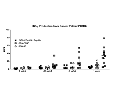
[0043] Figure 13 provides the immune response (IFN7 production) of PBMCs from
donors
with cancer to common tumor antigen peptides (MAGEA1/MAGE3/NY-ES0), PBMC's
were
incubated in the presence or absence of increasing concentrations of SEA-CD40
or SGN-40 for 5
days.
[0044] Figure 14 provides the immune response (IFNy production) of PBMCs from
donors
with cancer to common tumor antigen peptides (MAGEA1/MAGE3/NY-ES0). PBMC's
were
incubated in the presence or absence of increasing concentrations of SEA-CD40
and/or a
constant concentration of an anti-CTLA4 or anti-PD1 blocking antibody.
[0045] Figures 15A and 15B demonstrate the binding of fucosylated and non-
fucosylated anti-
mouse CD40 antibodies to murine Fey receptors. Fcy receptor were either FcyRI
(Figure 15A)
or FcyRIV (Figure 15B).
[0046] Figure 16 demonstrates in vivo activity of fucosylated and non-
fucosylated anti-CD40
antibody surrogates in the mouse B16 melanoma model.
[0047] Figure 17 demonstrates B-cell activation activity of SEA-CD40, antibody
21.4.1, and
CD40 hexameric ligand. Experiments were performed using purified B-cell
cultures.
[0048] Figure 18 demonstrates B-cell activation activity of SEA-CD40, antibody
21.4.1, and
CD40 hexameric ligand. Experiments were performed using PBMC cultures.
[0049] Figure 19 demonstrates monocyte/macrophage activation activity of SEA-
CD40,
antibody 21.4.1, dacetuzumab and an SEA-isotype control.
[0050] Figure 20 demonstrates induction of interferon-y (IFN-y) levels by SEA-
CD40,
antibody 21.4.1, dacetuzumab or an SEA-isotype control.
11
CA 02963720 2017-03-31
WO 2016/069919 PCT/US2015/058108
[0051] Figure 21 demonstrates induction of of interleulcin 10 (IL10) levels by
SEA-CD40,
antibody 21.4.1, dacetuzumab or an SEA-isotype control.
[0052] Figure 22 demonstrates induction of interferon-y (IFN-y) levels by SEA-
CD40,
antibody 21.4.1, or dacetuzumab. Incubation was done in the presence of flu
peptide.
[0053] Figure 23 demonstrates induction a flu-antigen specific T-cell response
by SEA-CD40,
antibody 21.4.1, or dacetuzumab.
[0054] Figure 24 demonstrates changes in IL10 levels following incubation of
PBMCs with flu
peptide and SEA-CD40, antibody 21.4.1, or dacetuzumab.
DETAILED DESCRIPTION
[0055] This disclosure provides description of the activity of a non-
fucosylated anti-CD40
antibody, SEA-CD40. SEA-CD40 is an agonistic antibody and has enhanced binding
to Fey
receptors III and. surprisingly exhibits enhanced activation of the CD40
signaling pathway.
Because of its enhanced activation of the CD40 pathway SEA-CD40 is a potent
activator of the
immune system and can be used to treat cancer or to treat infectious diseases,
particularly
chronic viral diseases, such as hepatitis C, human immunodeficiency virus,
Epstein-Barr virus,
cytomegalovirus, John Cunningham virus, and human papilloma virus. Other
infectious diseases,
include, e.g., tuberculosis. The enhanced activation of the immune system
allows SEA-CD40 to
be dosed at low levels, as compared to a fucosylated parent antibody.
CD40 description and function.
[0056] CD40 is a member of the tumor necrosis factor (TNF) receptor
superfamily. It is a
single chain type I transmembrane protein with an apparent MW of 50 kDa. Its
mature
polypeptide core consists of 237 amino acids, of which 173 amino acids
comprise an
extracellular domain (ECD) organized into 4 cysteine-rich repeats that are
characteristic of TNF
receptor family members. Two potential N-linked glycosylation sites are
present in the
membrane proximal region of the ECD, while potential 0-linked glycosylation
sites are absent.
A 22 amino acid transmembrane domain connects the ECD with the 42 amino acid
cytoplasmic
tail of CD40. Sequence motifs involved in CD40-mediated signal transduction
have been
identified in the CD40 cytoplasmic tail. These motifs interact with
cytoplasmic factors called
12
CA 02963720 2017-03-31
WO 2016/069919 PCT/US2015/058108
TNF-R-associated factors (TRAFs) to trigger multiple downstream events
including activation of
MAP kinases and NEKB, which in turn modulate the transcriptional activities of
a variety of
inflammation-, survival-, and growth-related genes. See, e.g., van Kooten and
Banchereau,
Leukoc. Biol. 67:2-17 (2000); Elgueta et al., Immunol. Rev. 229:152-172
(2009).
[0057] Within the hematopoietic system, CD40 can be found on B cells at
multiple stages of
differentiation, monocytes, macrophages, platelets, follicular dendritic
cells, dendritic cells (DC),
eosinophils, and activated T cells. In normal non-hematopoietic tissues, CD40
has been detected
on renal epithelial cells, keratinocytes, fibroblasts of synovial membrane and
dermal origins, and
activated endothelium. A soluble version of CD40 is released from CD40-
expressing cells,
possibly through differential splicing of the primary transcript or limited
proteolysis by the
metalloproteinase TNFa converting enzyme. Shed CD40 can potentially modify
immune
responses by interfering with the CD40/CD4OL interaction. See, e.g., van
Kooten and
Banchereau, J. Leukoc. Biol. 67:2-17 (2000); Elgueta et al., Immunol. Rev.
229:152-172 (2009).
[0058] The endogenous ligand for CD40 (CD4OL) is a type 11 membrane
glycoprotein of 39
kDa also known as CD154. CD4OL is a member of the TNF superfamily and is
expressed as a
trimer on the cell surface. CD4OL is transiently expressed on activated CD4+,
CD8+, and ye) T
cells. CD4OL is also detected at variable levels on purified monocytes,
activated B cells,
epithelial and vascular endothelial cells, smooth muscle cells, and DCs, but
the functional
relevance of CD4OL expression on these cell types has not been clearly defined
(van Kooten
2000; Elgueta 2009). However, expression of CD4OL on activated platelets has
been implicated
in the pathogenesis of thrombotic diseases. See, e.g., Ferroni et al., Curr.
Med. Chem. 14:2170-
2180 (2007).
[0059] The best-characterized function of the CD40/CD4OL interaction is its
role in contact-
dependent reciprocal interaction between antigen-presenting cells and T cells.
See, e.g., van
Kooten and Banchereau, J. Leukoc. Biol. 67:2-17 (2000); Elgueta et al.,
Immunol. Rev. 229:152-
172 (2009). Binding of CD4OL on activated T cells to CD40 on antigen-activated
B cells not
only drives rapid B cell expansion, but also provides an essential signal for
B cells to
differentiate into either memory B cells or plasma cells. CD40 signaling is
responsible for the
formation of germinal centers in which B cells undergo affinity maturation and
isotype switching
to acquire the ability to produce high affinity antibodies of the Ig.G, IgA,
and IgE isotypes. See,
13
CA 02963720 2017-03-31
WO 2016/069919 PCT/US2015/058108
e.g.,Kehry, .1. Immunol. 156:2345-2348 (1996). Thus, individuals with
mutations in the CD4OL
locus that prevent functional CD40/CD4OL interaction suffer from the primary
immunodeficiency X-linked hyper-IgM syndrome that is characterized by over-
representation of
circulating IgM and the inability to produce IgG, IgA, and IgE. These patients
demonstrate
suppressed secondary humoral immune responses, increased susceptibility to
recurrent pyrogenic
infections, and a higher frequency of carcinomas and lymphomas. Gene knockout
experiments in
mice to inactivate either CD40 or CD4OL locus reproduce the major defects seen
in X-linked
hyper-IgM patients. These KO mice also show impaired antigen-specific T cell
priming,
suggesting that the CD4OL/CD40 interaction is also a critical factor for
mounting cell-mediated
immune responses. See, e.g., Elgueta et al., Immunol. Rev. 229:152-172 (2009).
[0060] The immune-stimulatory effects of CD40 ligation by CD4OL or anti-CD40
in vivo have
correlated with immune responses against syngeneic tumors. See, e.g., French
et al., Nat. Med.
5:548-553 (1999). A deficient immune response against tumor cells may result
from a
combination of factors such as expression of immune checkpoint molecules, such
as PD-1 or
CTLA-4, decreased expression of MHC antigens, poor expression of tumor-
associated antigens,
appropriate adhesion, or co-stimulatory molecules, and the production of
immunosuppressive
proteins like TGFf3 by the tumor cells. CD40 ligation on antigen presenting
and transformed cells
results in up-regulation of adhesion proteins (e.g., CD54), co-stimulatory
molecules (e.g., CD86)
and MHC antigens, as well as inflammatory cytokine secretion, thereby
potentially inducing
and/or enhancing the antitumor immune response, as well as the irnmunogenicity
of the tumor
cells. See, e.g., Gajewski et al., Nat. Immunol. 14:1014-1022 (2013).
[0061] A primary consequence of CD40 cross-linking is DC activation (often
termed
licensing) and potentiation of myeloid and B cells ability to process and
present tumor-associated
antigens to T cells. Besides having a direct ability to activate the innate
immune response, a
unique consequence of CD40 signaling is APC presentation of tumor-derived
antigens to CD8+
cytotoxic T cell (CTL) precursors in a process known as 'cross-priming'. This
CD40-dependent
activation and differentiation of CTL precursors by mature DCs into tumor-
specific effectors
CTLs may enhance cell-mediated immune responses against tumor cells. See,
e.g., Kurts et al.,
Nat. Rev. Immunol. 10:403-414 (2010).
14
CA 02963720 2017-03-31
WO 2016/069919 PCT/US2015/058108
[0062] Agonistic CD40 mAbs including dacetuzumab, the SEA-CD40 parent
molecule, have
shown encouraging clinical activity in single-agent and combination
chemotherapy settings.
Dacetuzumab demonstrated some clinical activity in a phase 1 study in NHL and
a phase 2 study
in diffuse large B-cell lymphoma (DLBCL). See, e.g., Advani et al., J. Clin.
Oncol. 27:4371-
4377 (2009) and De Vos et al., J. Hematol. Oncol. 7:1-9 (2014). Additionally
CP-870,893, a
humanized IgG2 agonist antibody to CD40, showed encouraging activity in solid
tumor
indications when combined with paclitaxel or carboplatin or gemcitabine. In
these studies,
activation of antigen presenting cells, cytokine production, and generation of
antigen- specific T
cells were seen. See, e.g., Beatty et al., Clin. Cancer Res. 19:6286-6295
(2013) and
Vonderheide et al., Oncoirnmunology 2:e23033 (2013).
Anti- CD40 antibodies
[0063] Because of its role in immune function, antibodies have been raised
against the CD40
antigen. Such antibodies can be classified into three groups, antagonistic
antibodies, which
inhibit CD40 activity; partially agonistic antibodies, which partially induce
CD40 activity; and
fully agonistic antibodies, which fully stimulate CD40 activity. Members of
each of the groups
have been tested in clinical trials; none have been approved to date.
SEA-CD40
[0064] This disclosure provides a non-fucosylated hS2C6 antibody, SEA-CD40.
S2C6 was
originally isolated as a rnurine monoclonal antibody raised against a human
bladder carcinoma
referred to herein as mS2C6. See, e.g., Paulie et al., Cancer Immunol.
Immunother. 17:165-179
(1984). The S2C6 antibody is a partial agonist of the CD40 signaling pathway
and thus has the
following activities: binding to human CD40 protein, binding to cynomolgus
CD40 protein,
activation of the CD40 signaling pathway, potentiation of the interaction of
CD40 with its ligand,
CD4OL. See, e.g., US Patent No. 6,946,129.
[0065] As a next step in development, S2C6 was humanized and this humanized
antibody is
referred to as humanized S2C6, herein, and alternatively as dacetuzumab, or
fucosylated,
humanized S2C6 (fhS2C6), or SGN-40. See, e.g., WO 2006/128103. SGN-40 was
tested in
human clinical trials and was found not to be sufficiently active to warrant
further development.
[0066] SEA-CD40 is a non-fucosylated humanized S2C6 antibody. The amino acid
sequences
of the heavy and light chain for SEA-CD40 are disclosed as SEQ ID NO:1 and 2,
respectively.
CA 2963720
The variable region of the heavy chain is from amino acids 1-113 of SEQ ID
NO:1; the
variable region of the light chain is from amino acids 1-113 of SEQ ID NO:2.
The generation
of the antibody backbone of SEA-CD40 is disclosed at WO 2006/128103.
[0067] This disclosure provides a non-fucosylated, humanized S2C6 antibody,
referred to
herein as nf hS2C6 or SEA-CD40. In addition to enhanced binding to Fc
receptors, SEA-CD40
also enhances activity of the CD40 pathway, as compared to the parent
antibody, dacetuzumab.
The SEA-CD40 antibody thus, is administered to patients at at lower doses and
using different
schedules of administration.
Non-fucosylated antibodies
[0068] SEA-CD40 is a non-fucosylated antibody and exhibits enhanced binding to
FcyIII
receptors, and surprsingly enhanced ability to activate the CD40 signaling
pathway in immune
cells.
Methods of making non-fucosylated antibodies
[0069] This disclosure provides compositions and methods for preparing
humanized S2C6
antibodies with reduced core fucosylation. As used herein, "core fucosylation"
refers to
addition of fucose ("fucosylation") to N-acetylglucosamine ("GlcNAc") at the
reducing
terminal of an N-linked glycan.
[0070] Fucosylation of complex N-glycoside-linked sugar chains bound to the Fc
region (or
domain) of the SEA-CD40 antibody backbone is reduced. As used herein, a
"complex N-
glycoside-linked sugar chain" is typically bound to asparagine 297 (according
to the EU index
as set forth in Kabat, "Sequences of Immunological Interest, 5th Ed., Pub. No.
91-3242, U.S.
Dept. Healtth & Human Services, NIH, Bethesda, MD, 1991). As used herein, the
complex N-
glycoside-linked sugar chain has a biantennary composite sugar chain, mainly
having the
following structure:
16
Date Recue/Date Received 2020-10-27
CA 02963720 2017-03-31
WO 2016/069919 PCT/US2015/058108
+/-Fucal
+/-Ga1131 4GIcNAc01 2 Man al N.
vir
6 6
+1- GIcNA031 4Man131¨¶G10NAc131 LIGIcNAc
3
+/-Galr31¨ 4GIcNAc131¨ 2Mana1
where + indicates the sugar molecule can be present or absent, and the numbers
indicate the
position of linkages between the sugar molecules. In the above structure, the
sugar chain
terminal which binds to asparagine is called a reducing terminal (at right),
and the opposite side
is called a non-reducing terminal. Fucose is usually bound to N-
acetylglucosamine ("GlcNAc")
of the reducing terminal, typically by an a1,6 bond (the 6-position of GlcNAc
is linked to the 1-
position of fucose). "Gal" refers to galactose, and "Man" refers to mannose.
[0071] A "complex N-glycoside-linked sugar chain" includes 1) a complex type,
in which the
non-reducing terminal side of the core structure has one or more branches of
galactose-N-
acetylglucosamine (also referred to as "gal-G1cNAc") and the non-reducing
terminal side of Gal-
GlcNAc optionally has a sialic acid, bisecting N-acetylglucosamine or the
like; or 2) a hybrid
type, in which the non-reducing terminal side of the core structure has both
branches of a high
mannose N-glycoside-linked sugar chain and complex N-glycoside-linked sugar
chain.
[0072] In some embodiments, the "complex N-glycoside-linked sugar chain"
includes a
complex type in which the non-reducing terminal side of the core structure has
zero, one or more
branches of galactose-N-acetylglucosamine (also referred to as "gal-G1cNAc")
and the non-
reducing terminal side of Ca1-G1cNAc optionally further has a structure such
as a sialic acid,
bisecting N-acetylglucosamine or the like.
[0073] According to the present methods, typically only a minor amount of
fucose is
incorporated into the complex N-glycoside-linked sugar chain(s) of the SEA-
CD40 molecule.
For example, in various embodiments, less than about 60%, less than about 50%,
less than about
40%, less than about 30%, less than about 20%, less than about 15%, less than
about 10%, less
than about 5%, or less than about 3% of the antibody has core fucosylation by
fucose. In some
embodiments, about 2% of the antibody has core fucosylation by fucose.
17
CA 2963720
[0074] In certain embodiments, only a minor amount of a fucose analog (or a
metabolite or
product of the fucose analog) is incorporated into the complex N-glycoside-
linked sugar
chain(s). For example, in various embodiments, less than about 40%, less than
about 30%, less
than about 20%, less than about 15%, less than about 10%, less than about 5%,
or less than
about 3% of the SEA-CD40 antibody has core fucosylation by a fucose analog or
a metabolite
or product of the fucose analog. In some embodiments, about 2% of the SEA-CD40
antibody
has core fucosylation by a fucose analog or a metabolite or product of the
fucose analog.
[0075] Methods of making non-fucosylated antibodies by incubating antibody-
producing cells
with a fucose analogue are described, e.g., in WO/2009/135181. Briefly, cells
that have been
engineered to express the humanized S2C6 antibody are incubated in the
presence of a fucose
analogue or an intracellular metabolite or product of the fucose analog. As
used herein, an
intracellular metabolite can be, for example, a GDP-modified analog or a fully
or partially de-
esterified analog. A product can be, for example, a fully or partially de-
esterified analog. In some
embodiments, a fucose analogue can inhibit an enzyme(s) in the fucose salvage
pathway. For
example, a fucose analog (or an intracellular metabolite or product of the
fucose analog) can inhibit
the activity of fucokinase, or GDP-fucose-pyrophosphotylase. In some
embodiments, a fucose
analog (or an intracellular metabolite or product of the fucose analog)
inhibits fucosyltransferase
(preferably a 1,6-fucosyltransferase, e.g., the FUT8 protein). In some
embodiments, a fucose analog
(or an intracellular metabolite or product of the fucose analog) can inhibit
the activity of an enzyme in
the de novo synthetic pathway for fucose. For example, a fucose analog (or an
intracellular
metabolite or product of the fucose analog) can inhibit the activity of GDP-
mannose 4,6-dehydratase
or/or GDP-fucose synthetase. In some embodiments, the fucose analog (or an
intracellular metabolite
or product of the fucose analog) can inhibit a fucose transporter (e.g., GDP-
fucose transporter).
[0076] In one embodiment, the fucose analogue is 2-flurofucose. Methods of
using fucose
analogues in growth medium and other fucose analogues are disclosed, e.g., in
WO/2009/135181.
[0078] Other methods for engineering cell lines to reduce core fucosylation
included gene
knock-outs, gene knock-ins and RNA interference (RNAi). In gene knock-outs,
the gene
encoding FUT8 (alpha 1,6- fucosyltransferase enzyme) is inactivated. FUT8
catalyzes the
18
Date Recue/Date Received 2020-10-27
CA 02963720 2017-03-31
WO 2016/069919 PCT/US2015/058108
transfer of a fucosyl residue from GDP-fucose to position 6 of Asn-linked (N-
linked) GlcNac of
an N-glycan. FUT8 is reported to be the only enzyme responsible for adding
fucose to the N-
linked biantennary carbohydrate at Asn297. Gene knock-ins add genes encoding
enzymes such
as GNTIII or a golgi alpha mannosidase II. An increase in the levels of such
enzymes in cells
diverts monoclonal antibodies from the fucosylation pathway (leading to
decreased core
fucosylation), and having increased amount of bisecting N-acetylglucosamines.
RNAi typically
also targets FUT8 gene expression, leading to decreased mRNA transcript levels
or knocking out
gene expression entirely. Any of these methods can be used to generate a cell
line that would be
able to produce a non-fucosylated antibody, e.g.. an SEA-CD40 antibody.
[0078] Those of skill will recognize that many methods are available to
determine the amount
of fucosylation on an antibody. Methods include, e.g., LC-MS via PLRP-S
chromatography and
electrospray ionization quadrupole TOE MS.
[0079] The non-fucosylated antibody, SEA-CD40, when adminstered to a patient
induces
activation of monocyte maturation into macrophages and induce production of
cytokines,
including, e.g., interferon-'y (IFN- y) and chemokine that elicit robust T-
cell response to immune
system challenges. Unlike fully agoninstic antibodies, such as antibody
24.4.1., SEA-CD40 does
not induce production of immune-dampening cytokines, such as interleukin-10
(IL-10). IL-10,
in turn, induces activity of T-regulatory cells, wwhich dampen the immune
resopnse. Thus, SEA-
CD40 is useful for induction of a robust T-cell mediated immune response
without promoting
activity of T-regulatory cells.
Dosage and administration of SEA-CD40
[0080] Pharmaceutical compositions for parenteral administration are
preferably sterile and
substantially isotonic and manufactured under GMP conditions. Pharmaceutical
compositions
can be provided in unit dosage form (i.e., the dosage for a single
administration).
Pharmaceutical compositions can be formulated using one Or more
physiologically acceptable
carriers, diluents, excipients or auxiliaries. The formulation depends on the
route of
administration chosen. For injection, antibodies can be formulated in aqueous
solutions,
preferably in physiologically-compatible buffers to reduce discomfort at the
site of injection.
The solution can contain formulatory agents such as suspending, stabilizing
and/or dispersing
19
CA 02963720 2017-03-31
WO 2016/069919 PCT/US2015/058108
agents. Alternatively antibodies can be in lyophilized form for constitution
with a suitable
vehicle, e.g., sterile pyrogen-free water, before use.
[0081] SEA-CD40 is administered intravenously. In other embodiments, SEA-CD40
is
administered subcutaneously. In a further embodiment, SEA-CD40 is administered
subcutaneously at the site of a tumor.
[0082] The non-fucosylated SEA-CD40 antibody has surprisingly enhanced immune
activation
activity as compared to its parent antibody, dacetuzumab. Thus, SEA-CD40 can
be administered
to patients at lower doses and on different schedules as compared to
dacetuzumab.
[0083] As an example, SEA-CD40 can be adminstered to patients at levels
between between
0.1-2000 jig/kg (jig antibody per kilogram patient body weight). Other
possible dosage ranges
are 10-1000 jig/kg, 50-800 g/kg, 75-600 jig/kg, 100-500 g/kg. Other possible
dosage ranges
are the following: 100-300 jig/kg, 300-500 g/kg, 500-700 jig/kg, 700-900
Kg/kg, and 900-1100
g/kg. Still more dose ranges are the following: 100-150 g/kg, 150-200 g/kg,
200-250 jig/kg,
250-300 g/kg, 300-350 Rs/kg, 350-400 g/kg, 400-450 g/kg. 450-500 jig/kg,
500-550 g/kg,
550-600 jig/kg, 600-650 jig/kg, 650-700 jig/kg, 700-750 jig/kg, 750-800
jig/kg, 800-850 g/kg,
850-900 jig/kg, 900-950 jig/kg, 950-1000 jig/kg, 1000-1050 jig/kg, and 1050-
1100 jig/kg. Other
possible dosage ranges are 0.3-200 g/kg, 0.6-150 g/kg, 1.0-100 jig/kg, 2-50
jig/kg, 5-25 g/kg,
7.5-15 g/kg, and 8-12 Wks.
[0084] In other embodiments, SEA-CD40 is administered to patients at 0.6
g/kg, 1.0 g/kg,
2.5 g/kg, 5.0 jig/kg, 7.5 g/kg, 10 g/kg, 30 g/kg, 50 jig/kg, 75 g/kg, 100
jig/kg, or 200
jig/kg. In a preferred embodiment, SEA-CD40 is administered to patients at 10
jig/kg.
[0085] In further embodiments, SEA-CD40 is administered to patients at about
60 jig/kg,
about 100 g/kg, about 150 jig/kg, about 200 jig/kg, aabout 250 jig/kg, about
300 jig/kg, about
350 jig/kg, about 400 jig/kg, about 450 jig/kg, about 500 jig/kg, about 550
g/kg, about 600
jig/kg, about 650 jig/kg, about 700 jig/kg, about 750 g/kg, about 800 g/kg,
about 850 jig/kg,
about 900 jig/kg, about 950 jig/kg, about 1000-1050 jig/kg, about 1050 jig/kg,
and 1110 jig/kg.
[0086] In some embodiments, SEA-CD40 is administered in a manner to reduce the
likelihood
of immune exhaustion. For example, SEA-CD40 can be administered at three week
intervals,
six week intervals, eight week intervals, ten week intervals, twelve week
intervals, or 14 wek
CA 02963720 2017-03-31
WO 2016/069919 PCT/US2015/058108
intervals. Intervals can also be on a monthly schedule, e.g., one month
intervals, two month
intervals, or three month intervals.
[0087] Because SEA-CD40 activates the immune system to respond against tumor-
related
antigens, its use is not limited to cancers that express CD40. Thus SEA-CD40
can be used to
treat both CD40 positive and CD40 negative cancers.
[0088] SEA-CD40 is preferably used to treat tumors that are known to be immune
responsive,
particularly if the cancer expresses low levels of CD40 or does not detectably
express CD40.
Immune responsive cancers include, e.g., melanoma; bladder cancer; lung
cancer, e.g., small cell
lung cancer and non-small cell lung cancer; ovarian cancer; kidney cancer;
pancreatic cancer;
breast cancer; cervical cancer; head and neck cancer, prostate cancer;
glioblastoma; non-hodgkin
lymphoma; chronic lymphocytic leukemia; hepatocellular carcinoma; or multiple
myeloma.
[0089] In another embodiment, SEA-CD40 is used to treat solid tumors. In a
further
embodiment, SEA-CD40 is used to treat blood cancers, e.g., lymphoma, including
non-Hodgkin
lymphoma and Hodgkin lymphoma; chronic lymphocytic leukemia; or multiple
myeloma.
SEA-CD40 combination therapy
[0090] Because of its immune stimulatory function, SEA-CD40 can be used in
combination
with other therapeutic agents that activate the immune system. Drugs with
immune stimulatory
function include, e.g., T-cell modulators, including immune checkpoint
inhibitors; immune
activators; and chemotherapeutic agents that induce immunogenic cell death. As
an example,
certain antibodies function by blocking activity of molecules that serve as
immune checkpoints
on T cells. SEA-CD40 can, therefore be used in combination with antibodies
that target immune
checkpoint proteins.
T-cell modulators
[0091] T-cells play a role in the ability of the immune system to recognize
and eliminate
cancers from the body. T-cell modulators include antibodies that block the
function of immune
checkpoints. See, e.g., Pardoll, Nature Rev. Cancer, 12:252-264 (2012).
Antibodies that block
immune checkpoints include, e.g., anti-PD-1 antibodies, anti-PD-Ll antibodies,
and anti-CTLA4
anibodies. Other checkpoint inhibitors/activators include LAG3 and TIM3.
Antibodies against
some proteins can be used to modulate T-cell activity or preferably activate T-
cell activity, e.g.,
21
CA 02963720 2017-03-31
WO 2016/069919 PCT/US2015/058108
antibodies against 41BB, CD27, ICOS, and 0X40. Other T-cell modulators include
inhibitors of
the enzyme indolamine 2,3-dioxygenase (IDO).
[0092] Anti-CTLA4 antibodies recognize the protein cytotoxic lymphocyte 4
(CTLA-4), also
known as cluster of differentiation 152 or CD152. The CTLA-4 protein is
expressed on T cells,
which recognize antigens that are suitable for attack by the immune system.
Activation of
CTLA-4 dampens the immune response. See e.g., Nirschi and Drake, Clin. Cancer
Res.,
19:4917-4924 (2013). Antibodies specific for CTLA-4 and that block its
activity have been used
to treat cancer by upregulating the immune response to cancers. Examples of
CTLA-4
antibodies include ipilimumab or tremelimumab. SEA-CD40 can be administered in
combination with ipilimumab or tremelimumab to treat cancer.
[0093] Anti-PD1 antibodies recognize the protein programmed death-1 (PD-1).
Like CTLA-
4, PD-1 is expressed on T cells, and dampens the immune response. See e.g.,
Nirschi and Drake,
Clin. Cancer Res., 19:4917-4924 (2013). Antibodies specific for PD-1 and that
block its
activity have been used to treat cancer by upregulating the immune response to
cancers.
Examples of PD-1 antibodies include MEDI0680, AMP-224, nivolumab,
pembrolizumab, and
pidilizumab. Other PD-1 binding proteins that act as checkpoint inhibitors and
can be used in
combination with SEA-CD40 include, e.g., B7-DC-Fc. SEA-CD40 can be
administered in
combination with MEDI0680, AMP-224, nivolumab, pembrolizumab, or pidilizumab
to treat
cancer.
[0094] PD-Ll is a ligand of the PD-1 protein. PD-Ll is expressed on cancer
cells and its
interaction with PD-1 allows PD-Ll -expressing cancer cells to evade the
immune system. Anti-
PD-Li antibodies have been generated and used to treat cancer. Examples of PD-
L1 antibodies
include, e.g., MEDI4736, BMS-936559/MDX-1105, MSB0010718C and MPDL3280A. SEA-
CD40 can be administered in combination with MEDI4736, BMS-936559/MDX-1105,
MSB0010718C or MPDL3280A to treat cancer.
[0095] Other antibodies that block the function of immune checkpoint proteins
include
antibodies directed against e.g., LAG3 and THVI3, and can be used in
combination with SEA-
CD40.
22
CA 02963720 2017-03-31
WO 2016/069919 PCT/US2015/058108
[0096] Antibodies against 41BB, CD27, ICOS, and 0X40 are used to activate T-
cell activity
and can be used in combination with SEA-CD40. 0X40 antibodies include, e.g.,
MEDI6469 and
MEDI6383. An example of an agonistic anti-CD27 antibody is CDX-1127, which can
be used
in combination with SEA-CD40.
[0097] The enzyme indolamine 2,3-dioxygenase (IDO) catalyzes the degradation
of the amino
acid tryptophan. Inhibitors of IDO can be small molecules, such as rosmarinic
acid, COX-2
inhibitors, and alpha-methyl-tryptophan.
Chemotherapeutic agents that induce immunogenic cell death
[0098] In most humans, millions of cells die via apoptosis and are removed
without generating
an immune response. However, after treatment with some chemotherapeutic
agents, immune
cells have been observed to infiltrate tumors. Thus, some tumor cells killed
by chemotherapeutic
agents act as vaccines and raise a tumor-specific immune response. This
phenomenon is referred
to as immunogenic cell death (ICD). See, e.g., Kroemer et al., Annu. Rev.
Immunol., 31:51-72
(2013). The ability of a chemotherapeutic agent to induce 1CD can be
determined
experimentally. Two criteria must be met. First, injection of an
immunocompetent mouse with
cancer cells that have been treated in vitro with a chemotherapeutic agent
must elicit a protective
immune response that is specific for tumor antigens, in the absence of
adjuvant. Second, 1CD
occurring in vivo, e.g., a mouse syngeneic model with treatment using a
potential ICD-inducing
chemotherapeutic agent, must drive an immune response in the tumor that is
dependent on the
immune system.
[0099] Chemotherapeutic agents that induce ICD include, e.g., anthracyclines,
anti-EGFR
antibodies, bortezomib, cyclophosphamide, gemcitabine, irradiation of the
tumor, and
oxaliplatin. SEA-CD40 can be used in combination with any of these agents to
generate an
enhanced immune response and treat cancer in a patient.
Immune activation
[0100] Cancer can is also treated by administering agents that directly
stimulate the immune
system. Such agents include, e.g., GM-CSF, IFN-gamma, interleukin-2, GVAX, and
TLR9
agonists. Other immune activators include, e.g., cancer vaccines, Bacillus
Calmette-Guerin
(BCG), nonspecific immunostimulants (e.g. imiquimod) and cellular therapies
like CAR-T cells.
23
CA 02963720 2017-03-31
WO 2016/069919 PCT/US2015/058108
SEA-CD40 can be used in combination with any of these agents to generate an
enhanced
immune response and treat cancer in a patient.
Other combinations
[0101] Other combinations with SEA-CD40 can be used to treat cancer. Examples
include,
e.g., SEA-CD 40 in combination with an anti-PD I antibody, e.g., nivolumab,
pembrolizumab,
and pidilizumab, MEDI0680, or AMP-224; SEA-CD40 in combination with
Gemcitabine, with
or without paclitaxel or cisplatin or oxaliplatin; SEA-CD-40 in combination
with a BRAF
inhibitor, e.g., vemurafenib or dabrafenib; or SEA-CD40 in combination with
cyclophosphamide, Adriamycin. vincristine, and prednisone (CHOP) or rituximab,
ifosfamide,
carboplatin, and etopiside (RICE) or rituximab, gemcitabine, dexamethasone and
cisplatin
(RGDP).
EXAMPLES
The following examples are offered to illustrate, but not to limit the claimed
invention.
Example 1: Synthesis of non-fucosylated hS2C6 antibody
[0102] The humanized anti-CD40 antibody, S2C6 with heavy and light light
chains of SEQ ID
NOs: 1 and 2 was expressed in CHO cells. A fucosylation inhibitor, 2-
fluorofucose, was
included in the cell culture media during the production of antibodies
resulted in non-
fucosylated antibody, SEA-CD40. See, e.g., Okeley et al., Proc. Nat'l Acad.
Sci. 110:5404-
55409 (2013). The base media for cell growth was fucose free and 2-flurofucose
was added to
the media to inhibit protein fucosylation. Ibid. Incorporation of fucose into
antibodies was
measured by LC-MS via PLRP-S chromatography and electrospray ionization
quadrople TOF
MS. Ibid. Data not shown.
Example 2: Characterization of non-fucosylated hS2C6 antibody
[0103] CD40 Binding affinity determination of SEA-CD40: For isolation of
peripheral
blood mononuclear cells (PBMCs), human whole blood was supplied by Astarte
Biologics.
Briefly, blood was collected into heparin tubes and delivered to Seattle
Genetics within four
24
CA 02963720 2017-03-31
WO 2016/069919 PCT/US2015/058108
hours of being drawn. Upon arrival blood was aliquoted into 50 ml conical
tubes (falcon) and
spun at 200g in an Eppendorf 5810R (A-4-62 rotor) for 20 minutes at 25 C,
without break to
separate the platelet rich fraction. Following centrifugation, three distinct
layers were formed:
bottom layer, red blood cells (accounting for 50-80% of the total volume);
middle layer, very
thin band of white blood cells; top layer, straw-colored platelet rich plasma
(PRP).
[0104] The upper straw colored layer with which is enriched in platelets was
removed with a
one ml pipette. Once the platelet rich plasma was removed blood was diluted
with equal
volumes of sterile PBS (Gibco, lot 1618435, ept 2016-07). 15 rnls of
Histopaque-1077 (Sigma,
lot number RNBD2965, Expt. 5/2017) warmed to room temperature was underlayered
below the
blood. Histopaque samples were spun at 1500 rpm for 25 minutes at 25 C with
outbreak.
Following centrifugation three layers are formed again: bottom layer, red
blood cells (accounting
for 50-80% of the total volume); middle layer, thick band of white blood cells
(also called "buffy
coat"); top layer, PBS and remaining platelets.
[0105] The upper PBS/Platelet layer was removed with a 1 ml pipet and
discarded. The thick
band of white blood cells was gently removed and placed into a clean 50 ml
sterile conical tube.
Tubes were filled to 50 mls and cells are spun at 800g for 10 minutes. Wash
solution was
removed and pellets were resuspended in 10 mls of ACK red blood lysis buffer
(Gibco, lot
1618488) for ten minutes. Fifty milliliter conical tubes were then topped off
with 35 ml sterile
PBS and cells were spun at 800g for ten minutes. The wash solution was removed
and pellet was
resuspended in 50 mls of PBS. Five hundred pi of sample was removed and PBMC
were counted
with a Vi-cell-XR (Beckman Coulter). Cells were spun again at 800g for ten
minutes. The wash
solution was removed and pellet re-suspended at 1x10⁶/m1 in FACs staining
solution (BD). One
hundred 0 of resuspended PBMC's were plated into a 96 well U-bottom plate
(Corning) and
placed on ice. To block non-specific FcyRIIIa binding, PBMC's were pre-treated
100 [ig/m1 of
human Fc-fragments (Calbiochem,) for thirty minutes. Ten-fold serial dilutions
of biotinylated
SEA-h00 (non-fucosylated control antibody), SEA-CD40, or SGN-40 were prepared
to create a
dilution series of (100, 10,1, ,l, .01, .001, .0001 Rg/m1).
[0106] Samples were washed twice in ice cold FACs buffer and incubated with
saturating
concentrations of PE-Streptavidin (BD) on ice for thirty minutes. Samples were
washed twice in
ice cold FACs buffer and re-suspended in 200 0 of FAC's buffer. Binding was
assessed using a
CA 02963720 2017-03-31
WO 2016/069919 PCT/US2015/058108
BD LSRII and DIVA software. FCS were analyzed in Flowio and GeoMean
fluorescence of
positively stained cells was determined and plotted in Prism Graph Pad. Data
was fit to non-
linear regression assuming one binding site in Prism and and binding KD values
calculated by
dividing lAg/m1 calculation by molecular weight of SEA-CD40.
[0107] Results: The binding affinity of SEA-CD40, and the parental antibody
dacetuzumab,
to CD40 on human peripheral blood mononuclear cells (PBMC) was determined by
flow
cytometry. Background binding of an appropriate isotype control was subtracted
and mean
fluorescence intensity (MEI) was plotted against antibody concentration.
Results are shown in
Figure 1. SEA-CD40 and the parental antibody dacetuzumab gave virtually
overlapping binding
curves and both saturated PBMC's at concentrations of approximately 1.17 nM.
These data
suggest that changes in fucosylation do not affect SEA CD40 affinity for CD40.
[0108] Fc7RIIIa Binding affinity determination of SEA-CD40: CHO cells that
express the
high (158V) or low (158F) version of human FcyRIIIa were generated. 20x106
cells were
centrifuged, washed once in 20m1 lx PBS, and resuspended in 8m1 BD stain
buffer. Cells were
aliquoted in the following density: 2.0x10⁶ cells/ml in 100u1 volume. 0.20x10⁶
cells were
aliquoted to each well. Cells were centrifuged at 1250rpm, for five minutes at
room temperature.
Antibodies were diluted to either 0.14ug/m1 (SGN) or 0.04ug/m1 (SEA).
Dilutions are provided
in Table 1.
Table 1
Biotinylated abs Vol (ul) Vol. stain Highest stain
dilutions Conc. Mg/ml antibody buffer conc ug/ml
SGN-40-Biotin 3.29 18.23 581.7 100
SEA40-Biotin 3.27 15.11 584.7 100
h00-SGN-Biotin 1.55 38.7 561.0 100
h00-SEA biotin 3.61 16.6 583 100
Supernatants were aspirated from the spun cells and 60u1 of corresponding
antibody dilutions
were added with a multichannel pipet. Corresponding concentrations were 100,
33.3, 11.1, 3.7,
1.23, 0.41, 0.14 mcg/ml. Samples were incubated at 4 C for 1 hour. Samples
were centrifuged,
and washed twice with 200 tl BD stain buffer per well. One milliliter of
Streptavidin-PE was
added to 20m1 BD stain buffer (excess 2 ) to make streptavidin buffer. 100 ill
of streptavidin
buffer was added to each sample and they were incubated for 30 min in the dark
at 4 C. Samples
26
CA 02963720 2017-03-31
WO 2016/069919 PCT/US2015/058108
were then centrifuged and washed twice with 200u1 BD Stain buffer per well.
Samples were
analyzed by Flow cytometry in HTS mode on the LSRII and graph MFI to calculate
Kd's in
PRIZM.
[0109] Results: Binding of SEA-CD40 and the parent antibody dacetuzumab to
Chinese
hamster ovary (CHO) cells expressing the low (158F) or high (158V) affinity
form of FcyRIIIa
was assessed. Results are shown in figures 2A and 2B. SEA-CD40 bound to both
the low
(158F) and high (158V) form of FcyRIlla with similar affinity (KD 27.5 nM and
5.2 nM,
respectively). SEA-CD40 binding to the low affinity (158F) form was
significantly better than
the fucosylated parental antibody dacetuzumab (KD 302.7 nM), and SEA-CD40 even
bound the
high affinity 158V form better than the fucosylated dacetuzumab parent (5.2 nM
vs. 37.9 nM
respective] y).
[0110] SEA-CD40 mediated ADCC activity: Human PBMC's were isolated as above
and
were treated with various concentrations of SEA-CD40 or an SEA-isotype control
(SEA-h00) for
6, 24, or 48 hours. Cultures were stained for CD19+ B cells and cell numbers
were quantified by
flow cytometry.
[0111] Results: Human PBMC cultures, were treated with 100, 10, 1, 0.1. 0.01,
0.001, or
0.0001 lig /mL of SEA CD40 or a non-binding SEA-isotype control (SEA-h00) for
6, 24, and 48
hours and the number of CD40 positive cells were then assessed. Results are
shown in Figure 4.
SEA-CD40 treatment resulted in a significant decrease in CD40+ CD19+ B cells
in a dose- and
time-dependent manner, even down to low sub [ig/mL concentrations. There was
no significant
effect of SEA-CD40 on monocyte/DC numbers (monocyte/DC data not shown).
[0112] Assessment of cytokine production in human whole blood or PBMCs: Human
whole blood was supplied by Astarte Biologics. Briefly, 100 mls of blood was
collected into
heparin tubes and delivered to Seattle Genetics within 4 hours of the draw.
Half of the blood was
set aside for whole blood cultures while the other half was used to isolate
PBMCs as described
above. One hundred p1 of whole blood was aliquoted into 3-96 flat bottom
tissue culture plates
(Costar). Isolated PBMCs were counted in a Viacell and resuspended at lx106
cells/ml in
DMEM containing10% FBS (Atlanta Biologics), lx penicillin/strepA, and X
glutamine (PBMC
media). For PBMCs, one hundred IA of resuspened, purified PBMCs were aliquoted
into 3-96
flat bottom tissue culture plates. 10X serial dilutions of SEA-h00 and SEA-
CD40 were made in
27
CA 02963720 2017-03-31
WO 2016/069919
PCT/US2015/058108
PBMC media and whole blood and isolated PBMC cultures were treated with
descending
concentrations of either SEA-h00 or SEA-CD40 (100, 10, 1.0, 0.1, 0.01, 0.001,
0.0001 or 0
Rgim1). SEA-CD40 treatment was performed in duplicate for both whole blood and
PBMC
cultures at each time point. At each of the pre-determined times points (6,
24, and 48 hours) a 96
well plate containing whole blood or purified PBMCs was spun with a plate
adapter in an
Eppendorf 5810R at 800 rpm for 5 minutes. Serum or tissue culture supernatants
were removed
and transferred to a 96 strip tube rack and samples were frozen at -80 C until
processing.
[01131 Frozen
tissue culture supernatants and serum were thawed overnight at 4 C and
processed for cytokine production using a Luminex multiplex Kit from
Millipore. Custom kits
were designed to analyze IFNy, IL-12p40, IL-6, IL-8, MCP-1, MIP-la, IL-10, MIP-
113, TNF-a,
sCD4OL. Analytes were picked based on cytokines observed with dacetuzemab in
previous
studies. Tissue culture supernatants and serum samples were processed as per
the manufactures
instructions. Briefly, assay plates were washed with 200 1_, of wash buffer
per well, followed by
addition of 25 ti.L standard or buffer. 25 iL matrix or sample, and 25 1_, of
multiplexed analyte
beads to each well. Samples were incubated overnight with vigorous shaking at
4 C. Plates are
washed the assay plates twice with wash buffer. Twenty-five 1.11, of detection
antibodies were
added to each well and incubated at room temperature for one hour. Twenty-five
nt of
streptavidin-phycoerythrin (SA-PE) were added and samples incubated at room
temperature for
thirty minutes. The plate was washed twice with wash buffer and beads were
resuspended with
150 juL of sheath fluid. The samples were analyzed using Luminex MagPix
systems in
combination with the Xponent software system. Cytokine levels were calculated
from the
standard curve.
[0114] Results: Human whole blood cultures were treated with a SEA-isotype
control Or
SEA-CD40 (100, 10, 1, 0.1, 0.01, 0.001, or 0.0001 [tg/mL) for 6, 24, or 48
hours. Serum or
tissue culture supernatants were collected and inflammatory cytokines assessed
by multiplexed
Luminex analysis. The data are plotted as a fold increase in cytokine
production compared to a
SEA-isotype control. SEA-CD40 stimulated robust production of IFNy, MIP113,
and TNFa at 6,
24, and 48 hours in whole blood, as shown in Table 2, below. SEA-CD40 levels
are provided in
the leftmost columns. Activity was observed at levels as low as 0.010 lig/mL
SEA-CD40.
Stimulation of MIP113, and TNFa at twenty-four hours are shown in Figures 4A
and 4B.
28
CA 02963720 2017-03-31
WO 2016/069919 PCT/US2015/058108
Table 2
Whole Blood
6hrs IFNy IL-8 MCP-1 MIPla MIP1f3 TNFa
100.00 4.52 2.01 2.75 1.30 30.86 3.22
10.00 10.81 1.05 2.33 1.04 26.69 µ1.90
1.00 4.99 1.13 1.62 0.93 4.59 2.20
0.10 3.42 0.84 0.96 1.02 0.88 1.65
0.01 1.83 1.00 ,1.29 1.26 0.96 9.98
0.00 1.20 0.94 _1.25 1.24 0.93 _1.04
0.00 1.16 1.05 1.29 1.15 0.98 1.02
24hr IFNy 1L-8 MCP-1 M1P1a M1P113 TNFa
100.00 3.01 1.95 2.19 2.28 6.77 3.51
10.00 3.23 1.52 2.84 2.34 8.42 3.26
1.00 2.31 1.70 2.36 1.75 6.77 3.62
0.10 1.32 1.36 µ1.19 0.89 3.95 2.12
0.01 0.95 1.10 1.01 0.74 0.96 2.11
0.00 0.55 0.92 1.04 0.95 1.15 1.03
0.00 0.40 0.82 0.79 1.01 1.66 1.44
48 hrs IFNy IL-8 MCP-1 MIP 1 a MIP113 TNFa
100.00 3.59 1.11 1.19 1.26 2.03 3.47
10.00 2.37 1.21 1.22 1.24 2.27 2.71
1.00 2.15 1.08 1.07 1.07 1.76 2.63
0.10 1.01 0.76 1.05 1.09 1.43 2.53
0.01 0.86 0.81 1.16 1.17 1.27 1.93
0.00 0.87 0.97 1.18 1.18 0.97 1.18
0.00 0.96 0.93 0.87 1.05 0.68 1.17
[0115] Human PBMC were treated with a SEA-isotype control or SEA-CD40 (100,
10, 1, 0.1,
0.01, 0.001, or 0.0001 kg/mL) for 6, 24, or 48 hours. Serum or tissue culture
supernatants were
collected and inflammatory cytokines assessed by multiplexed Luminex analysis.
The data are
plotted as a fold increase in cytokine production compared to a SEA-isotype
control. SEA-CD40
stimulated robust production of IFNy, MW 1I3, and TNFa at 6, 24, and 48 hours
PBMCs, as
shown in Table 3, below. SEA-CD40 levels, in lag/mL, are provided in the
leftmost columns.
Activity was observed at levels as low as 0.010 [tg/mL SEA-CD40. Stimulation
of MIP1I3, and
TNFa at twenty-four hours are shown in Figures 5A and 5B.
29
CA 02963720 2017-03-31
WO 2016/069919 PCT/US2015/058108
Table 3
PBMC
6hr 1FNy 1L-8 MCP-1 M1P1a MIP1I3 TNFa
100 8.75 1.18 3.00 2.51 6.91 11.66
13.68 1.16 7.70 3.11 11.59 17.72
'
1 6.21 0.89 2.71 1.48 4.55 5.58
0.1 3.89 0.89 1.61 1.26 3.40 3.04
0.01 1.49 0.75 1.07 1.26 2.11 2.26
0.001 1.60 ,0.71 0.89 1.31 1.30 1.28
0.0001 1.58 0.71 0.77 0.83 1.33 1.10
24hr IFNy IL-8 MCP-1 MIPla MIPlp TNFa
100 8.51 4.79 5.69 µ2.84 13.43 19.91
10 8.98 3.96 7.87 1.91 7.58 14.97
1 3.32 1.35 3.10 1.28 8.73 3.79
0.1 1.80 1.04 1.38 1.03 5.85 5.58
, 0.01 1.66 0.85 1.28 1.08 2.87 1.22
0.001 1.12 0.71 0.90 0.96 1.18 2.75
0.0001 0.40 0.71 0.80 0.74 1.11 1.02
48 hrs IFNy IL-8 MCP-1 MIPla MIP113 TNFa
100 17.92 2.58 11.47 1.51 2.81 14.02
10 8.81 ,3.61 3.39 1.46 2.33 5.58
'
1 4.09 2.07 2.36 1.32 1.91 6.47
0.1 1.82 1.19 0.84 0.96 1.00 2.30
0.01 1.03 1.02 1.41 ,0.95 1.13 2.09
0.001 0.83 0.86 1.13 0.93 0.96 1.93
-
0.0001 0.82 0.97 0.97 0.91 1.05 1.73
[0116] Assessment of Activation markers on PBMCs: Co-stimulatory molecule
surface
expression was assessed on the cell pellets remaining from the cytokine
analysis described
above. Cell pellets were resuspended in 50 ml of BD FACs buffer and
transferred to and 96 well
round bottomed microtiter plates Fc receptors were blocked with human
10Oug/m1Fc-fragments
(Millipore) for 30 minutes on ice. A master mix composed of PE-CD86 (BD) and
MHCII (Pan
anti-DR,DP,DQ antibody BD) diluted at 1:100 was prepared in BD FACs buffer
containing 100
mg/ml human Fc fragments, Five I..(1 of the master mix was added to each well
containing ninety
CA 02963720 2017-03-31
WO 2016/069919 PCT/US2015/058108
[L1 and samples were incubated for one hour on ice. Cells were then spun at
400g in a pre-coolled
Eppendorf 5810R centrifuge for five minutes. Supernatants were removed and
cells washed with
200 ml of BD Facs buffer. Cells were washed twice and then resuspended in 200
ml of FACs
buffer. Samples were then analyzed on an LSRII (BD biosciences) with DIVA
software (BD
biosciences). CD86 and MHCII geo mean fluorescence was assessed using FlowJo
analysis
software. A ratio between SEA-h00 and SEA-CD40 was calculated and the fold
change used to
calculate a range of SEA-CD40 potency.
[0117] Results: Activation of CD40, in addition to eliciting cytokine
production, promotes the
maturation of antigen presenting cells. DC maturation can be followed by
upregulation of
activation markers including CD86 and MHCII. Human PBMC cultures were
stimulated with
SEA CD40 and an SEA-isotype control for 6, 24, or 48 hours and surface
expression of MHC
Class II antigens (HLA DR, DP, DQ) and CD86 was assessed. SEA-CD40
stimulation, but not
the isotype control resulted in a significant increase in both MHCII (Table 4)
and CD86 (data not
shown) at concentrations as low as 0.01 [tg/mL.
Table 4: MHCII
SEA-
CD40 6hrs 24hrs 48hrs
100 1.13 1.64 2.10
1.10 1.62 2.10
1 1.14 1.76 1.57
0.1 0.90 1.19 1.50
0.01 0.90 1.21 1.38
0.001 1.00 1.07 1.10
0.0001 0.85 1.05 0.99
[0118] Role of fucose in immune activation by SEA-CD40: Human PBMCs were
isolated
as described above. PBMCs were treated with various concentrations of SEA-
CD40, the
parental antibody SGN40, or a F(ab)'2 version of SEA-CD40 and incubated for 24
hrs. For
assessment of B cell depletion, BPMC cultures were stained with a PE-CD19 and
B-cell
31
CA 02963720 2017-03-31
WO 2016/069919 PCT/US2015/058108
numbers were quantified by flow cytometry. For assessment of cytokine
production, PBMC
tissue culture supernatants were collected and cytokine production assessed by
multiplex
analysis on the Luminex platform. IFI\Iy production is shown is shown in
Figure 7, (ng/mL) and
similar trends were seen for the other cytokines. For assessment of antigen
presenting cell
maturation, PBMC's were collected after the twenty-four hour incubation and
stained with aPE-
anti-CD86 or APC-pan MHC Class II antigen (DR, DQ, DP) antibody and the
percent positive
cells were assessed by flow cytometry. The data are shown as mean fluorescent
intensity for pan
MHC markers.
[0119] Results: To assess B cell depletion, human PBMC cultures were treated
with multiple
concentrations of SEA-CD40, dacetuzumab, or SEA-CD40 F(ab')2 for twenty-four
hours.
Results are shown in Figure 6. The ADCC-mediated depletion of B cells was
significantly higher
in SEA-CD40-treated cultures compared with dacetuzumab-treated cultures and
this activity was
lost with the SEA-CD40 F(ab)'2.
[0120] Additionally immune activation endpoints (cytokines and APC
activation/maturation
markers) were assessed in SEA-CD40, dacetuzumab, and SEA-CD40 F(ab')2 PBMC
cultures
stimulated for twenty-four hours. SEA-CD40 stimulation of both cytokine
production (Figure 7)
and APC maturation (Figure 8) was significantly higher than that of
dacetuzumab or SEA-CD40
F(ab)'2. These data demonstrate that the lack of fucose on the IgG domain does
not alter CD40
binding, but does increase FcyRIIIa binding resulting in increased CD40
activity and ultimately
increased CD40 immune modulatory activity.
Example 3: Immune modulatory activity of non-fucosylated hS2C6 antibody
[0121] Identification of active doses of SEA-CD40: SEA-CD40 is proposed to be
active at
dose levels that activate antigen-presenting cells, which can be characterized
by upregulation of
activation markers, such as MHC class I or II, or CD86. Activation markers on
PBMCs
following treatment with various concentrations of SEA-CD40 from 6 to 48 hours
were assessed
as described above (Assessment of Activation markers on PBMCs). The difference
between
treatments with isotype control SEA-h00 and SEA-CD40 for each activation
marker were
calculated and plotted versus SEA-CD40 concentrations and treatment time. The
steepest
response-concentration curves were observed at 24 hours for CD86 and MHCII and
48 hours for
32
CA 02963720 2017-03-31
WO 2016/069919 PCT/US2015/058108
MHCI. Response-concentration data (24 hr CD86, 24 hr MHCII and 48 hr MHCI)
were fitted by
nonlinear regression using the following equation, where 0% and 100% responses
are defined as
the smallest and largest values in the data set for each activation marker.
EC50 is the
concentration of SEA-CD40 that gives a 50% response.
100
Response ¨
1 + 10(1 gio EC50-log10 Concentration)
[0122] Results: Normalized response versus¨ log (concentration) and nonlinear
fitted
regression lines for activation markers are illustrated in Figure 9. Estimated
EC50 values for
MHCI, CD86 and MHCII were 0.011, 0.14 and 0.41 pg/mL, respectively. SEA-CD40
is
estimated to induce approximately 90%, 60%, and 30% maximal upregulation of
MHCI, CD86
and MHCII, respectively, at 0.21u g/mL, corresponding to a theoretical plasma
Cmax achievable
by an IV dose of 10 g/kg in humans. This dose is proposed as the theoretical
first anticipated
active dose.
Example 4: Immune modulatory activity of non-fucosylated hS2C6 antibody
[0123] T-cell response generated by SEA-CD40: An anti-MI T cell line was
generated at
Astarte Biologics from a HLA-A2 donor that was shown to be highly reactive to
the M1 flu
peptide. These cells were labeled with carboxyfluorescein succinirnidyl ester
(CFSE) and
combined with autologous PBMC's. Cultures were stimulated with 10 tg/m1 M1 flu
peptide in
the presence or absence of decreasing concentrations (1, 0.1, 0.01 jig /ml) of
SEA-CD40 or
dacetuzumab for 5 days. Cultures supernatants were collected and analyzed for
cytokines by
multiplex analysis on the Luminex platform and antigen specific T-cells were
identified by MI
specific Tetramer binding.
[0124] Results: PBMC's from an HLA-A2 donor shown to be highly reactive to the
M1 flu
peptide were stimulated with M1 flu peptide in the presence or absence of
decreasing
concentrations of SEA-CD40 or dacetuzumab for five days. Results are shown in
Figures 10A
and 10B. SEA-CD40 stimulated cultures showed increased response to M1 flu
antigen as seen
by an increase in IFNy production and an increase in antigen specific T-cells
as determined by
increased Tetramer binding. SEA-CD40 stimulated an antigen specific T-cell
response down to
0.1ug/m1 and this activity was more robust than the response associated with
dacetuzumab.
33
CA 02963720 2017-03-31
WO 2016/069919 PCT/US2015/058108
[0125] T-cell response generated by SEA-CD40 in combination with anti-immune
checkpoint inhibitor antibodies: An anti-M1 T cell line was generated at
Astarte Biologics
from a HLA-A2 donor that was shown to be highly reactive to the M1 flu
peptide. These cells
were labeled with CFSE and combined with autologous PBMC's. Cultures were
stimulated with
ug/ml M1 flu peptide in the presence or absence of decreasing concentrations
(1, 0.1, 0.01
ug/ml) of SEA-CD40 and/or lug/m1 of an anti-CTLA4 or an anti-PD-1 antibody for
five days.
Culture supernatants were collected and analyzed for cytokines by multiplex
analysis on the
Luminex platform and antigen specific T-cells were identified by M1 specific
Tetramer binding.
[0126] Results: PBMC's from an HLA-A2 donor shown to be highly reactive to the
M1 flu
peptide were stimulated with M1 flu peptide in the presence or absence of
decreasing
concentrations of SEA-CD40 and/or a constant concentration of an anti-CTLA4
blocking
antibody or an anti-PD-1 antibody. Results are shown in Figures 11 and 12.
While SEA-CD40,
anti-CTLA4 antibodies and anti-PD-1 antibodies alone stimulated an antigen
specific T-cell
response, increased response to MI flu antigen as seen by an increase in IFN-y
production
(Figure 11) and an increase in antigen specific T-cells as determined by
increased Tetramer
binding (Figure 12, using the Tetramer/APC - HLA-A*02:01 Influenza-M1
(GILGFVFIL)
tetramer from MBL) was observed when SEA-CD40 and anti-CTLA4 antibodies or
anti-PD-1
antibodies were combined. SEA-CD40 stimulated an antigen specific T-cell
response down to
0.1ug/m1 and this activity was enhanced by combination with an immune
checkpoint antibody.
[0127] T-cell response generated by SEA-CD40 in PCMCs from cancer patients:
For
assessment of anti-CD40 antibodies alone, PBMCs were isolated from 10 mls of
tumor patient
blood and 0.25 million cells were plated in a 24 well plate. Samples were
treated with increasing
concentrations of SEA-CD40 only, or lug/ml of a combined peptide pool
containing
MageAl/MageA3/NY-ESO and increasing concentration of either SEA-CD40 or SGN-
40.
Samples were cultured in 10% CO, at 37 C for five days, tissue culture
supernatants were
collected and INF-y levels were assessed.
[0128] For assessment of SEA-CD40 in combination with immune checkpoint
blocking
antibodies, PBMCs were isolated from 10 mls of blood from patients diagnosed
with either
breast, pancreatic, of melanoma cancer, and 0.25 million cells were plated in
a 24 well plate.
Samples were treated with increasing concentrations of SEA-CD40, 1 pg/m1 of a
combined
34
CA 02963720 2017-03-31
WO 2016/069919 PCT/US2015/058108
peptide pool containing MageAl/MageA3/NY-ESO, and either 11.1g/m1 of anti-PD1
or anti-
CTLA4. Samples were cultured in 10% CO-, at 37 C for 5 days, tissue culture
supernatants were
collected and INF-T levels were assessed.
[0129] Results: PBMC's from donors were isolated from whole blood as described
above.
The donors were three patients diagnosed with melanoma, three patients
diagnosed with breast
cancer, and three patients diagnosed with pancreatic cancer. Donor PBMC's were
stimulated
with a pool of peptides of the common tumor antigen proteins (MAGEA1/MAGE3/NY-
ESO) in
the presence or absence of increasing concentrations of SEA-CD40 or SGN-40 for
5 days. Tissue
culture supernatants were collected and INF-7 production was assessed. Results
are shown in
Figure 13. Six out of the nine patients exhibited an antigen dependent INF-y
response that was
significantly enhanced by SEA-CD40 treatment as compared to treatment with SGN-
40. In the
SEA-CD40 treated PBMC's, stimulation was observed at concentrations as low as
10 vg/ml.
[0130] PBMC's from donors were isolated from whole blood as described above.
As above,
the donors were three patients diagnosed with melanoma, three patients
diagnosed with breast
cancer, and three patients diagnosed with pancreatic cancer. Donor PBMC's were
stimulated
with a pool of peptides of the common tumor antigen proteins (MAGEA1/MAGE3/NY-
ESO) in
the presence or absence of increasing concentrations of SEA-CD40 and/or a
constant
concentration of an anti-CTLA4 or anti-PD1 blocking antibody. Results are
shown in Figure 14.
While SEA-CD40 and antibodies against the checkpoint blockade targets PD1 and
CTLA4
stimulated an antigen specific T-cell response alone, a robust signal to the
tumor antigen as
measured by INF-y production was observed when SEA-CD40 was combined with
either anti-
CTLA4 antibodies or anti-PD1 antibodies.
Example 5: Mouse models for activity of non-fucosylated anti-CD40 antibodies
[0131] Mouse models have been proven to be very useful in assessing efficacy
and
mechanisms of new cancer therapeutics. Study of SEA-CD40 in mouse models of
cancer has
been difficult because SEA-CD40 does not recognize murine CD40. Therefore, to
assess the
activity of the non-fucosylated anti-CD40 antibodies a syngeneic murine tumor
model was
developed. The murine functional equivalents of human IgG1 and human
Fc7RIII/CD16 are
murine IgG2a and EcyRIV, respectively, and binding of murine IgG2a to murine
EcyRIV
mediates ADCC. See, e.g., Bruhns, Blood 119:5640-5649 (2012) and Nimmeriahn et
al.,
CA 02963720 2017-03-31
WO 2016/069919 PCT/US2015/058108
Immunity 23:41-51 (2005). The rat antibody 1C 10 was used to generate a
surrogate of SEA-
CD40. See, e.g., Heath etal., Fur. J. Immunol. 24:1828-1834 (1994). Briefly,
the VL and VH
gene fragments of a rat monoclonal antibody that recognizes murine CD40, the
1C10 antibody
were cloned in-frame 5' to murine Ckappa and murine IgG2a CH1-CH2-CH3
fragments,
respectively. Expression of the resulting genes in CHO cells generated a
chimeric 1C10
antibody with rat VL and VH domains and murine light and heavy chain domains
of the IgG2a
isotype (mIgG2a 1C10). mIgG2a ICIO was expressed in the presence of 2-
fluorofucose in the
CHO cell growth medium using the methods described in Example 1, to generate a
non-
fucosylated form of mIgG2a 1C10 (mIgG2a SEA 1C10). Fucosylated mIgG2a 1C10 and
mIgG2a SEA 1C10 were tested for anti-tumor activity using a mouse B16 melanoma
model.
[0132] Assessment of non-fucosylated murine antibody binding to murine Fey
receptors:
CHO cells stably expressing murine FcyRI or FcyRIV were incubated with
increasing
concentrations of fucosylated mIgG2a ICIO or non-fucosylated mIgG2a 1C10
(mIgG2a SEA-
1C10). Samples were washed and a saturating amount of PE-anti-mouse IgG was
added and
incubated with the samples on ice for thirty minutes. Samples were washed
again and labeled
cells were analyzed by flow cytometry.
[0133] Results: Binding of the surrogate anti-CD40 antibodies to Chinese
hamster ovary
(CHO) cells expressing the murine FcyR1 or FcyRIV (the murine equivalent to
human
FcyRIII/CD16) was assessed. Results are show in Figures 15A and 15B. As
expected mIgG2a
1C10 bound with similar affinity as mIgG2a SEA 1C10 to FcyR1. See, e.g.,
Figure 15A.
However, non-fucosylated mIgG2a SEA ICI 0 bound to FcyRIV at significantly
higher affinity
than the fucosylated parental antibody mIgG2a 1C10. See, e.g., Figure 15B.
[0134] Assessment of non-fucosylated anti-CD40 antibodies in a murine tumor
model:
250,0E+3 B16F10 melanoma cells were given subcutaneously to C57BL/6 mice. Mice
were
randomized into cohorts each with tumor size of approximately 50 mm3 on
average. Mice were
then given inteiperitoneal injections of either an isotype control (mIgG2a),
fucosylated mIgG2a
1C10, or non-fucosylated mIgG2a 1C10 (mIgG2a SEA 1C10), every other day for a
total of
three doses. Mice were monitored until the tumor size reached 1000 mm3, at
which point the
mice were sacrificed.
36
CA 2963720
101351 Results: The B16F10 sygeneic melanoma model was used to assess the in
vivo
efficacy of our non-fucosylated anti-CD40 antibody surrogates. C57BL/6 mice
were implanted
with B16F10 melanoma cells, and then treated with either mIgG2a isotype
control, fucosylated
mIgG2a 1C10, or non-fucosylated mIgG2a 1C10 (mIgG2a SEA 1C10). Tumor burden
was
monitored and mice were sacrificed when the tumor size reached 1000mm3.
Results are shown
in Figure 16. Mice treated with non-fucosylated SEA-1C10 mIgG2a showed a
significant
survival benefit and tumor delay compared to the fucosylated parent 1C10 IgG2a
antibody.
Example 6: SEA-CD40 depletes B-cells and promotes T-cell activation
101361 SEA-CD40 activity was compared to a related fucosylated antibody and to
a fully
agonistic anti-CD40 antibody, clone 21.4.1. Antibody 21.4.1 is a human anti-
CD40 IgG2k
agonistic antibody that is the parent clone of CP-870,893, an antibody that is
currently being tested
in a clinical trial of solid tumors in combination with PDLl. For amino acid
sequence information
for antibody 21.4.1, see, e.g., U.S. Patent No. 7,338,660. Three functional
areas were tested for the
antibodies: ability to drive human B-cells differentiation, activation, and
depletion, ability to
activate primary human PBMC cultures, and ability to drive an antigen specific
response.
101371 Assessment of B-cell activation by anti-CD-40 antibodies: Experiments
were
performed using purified B-cells from fresh human whole blood or human
peripheral blood
mononuclear cells (PBMCs). B-cells were isolated from fresh human whole blood
using
RosetteSep isolation kit. The isolated, purified B-cells were cultured with
increasing
concentrations of SEA-CD40, antibody 21.4.1, or hexameric CD40 ligand, Enzo
Life Sciences
(10, 1, 0.1, 0.01, or 0.001 fig/mL) for 24 hours. B-cell activation was
assessed as upregulation
of CD80 by Flow Cytometry.
101381 PBMCs were isolated as described above and were then cultured with
increasing
concentrations of SEA-CD40, antibody 21.4.1, or hexameric CD4OL (10, 1, 0.1,
0.01, or 0.001
ug/mL) for 24 hours. The total number of B-cells assessed with CD1 9 staining
assessed by
flow cytometty.
[0139] Results: SEA-CD40 immune modulatory activity is dependent on the Fe
portion of the
antibody and its interaction with the CD16, the FcyRIII receptor. Results are
shown in Figure
37
Date Recue/Date Received 2020-10-27
CA 02963720 2017-03-31
WO 2016/069919 PCT/US2015/058108
17. SEA-CD40 does not induce B-cell activation in purified B-cells cultures
which lack cells
that express the Fcy receptors needed for crosslinking of SEA-CD40. This
differs from the CD40
activating antibody 21.4,1 which is able to drive B-cell activation in pure B-
cell cultures similar
to CD40 ligand.
[0140] PBMCs include cells that express the Fcy receptors. Results for that
cell population are
shown in Figure 18. For PBMC cultures, SEA-CD40 was able to promote ADCC
depletion of
B-cells, while antibody 21.4.1 treatment did not deplete B-cells.
[0141] Assessment of monocyte/macrophage activation by anti-CD-40 antibodies:
Human
PBMC cultures were isolated as described above. PBMC cultures were stimulated
with
increasing concentrations (0.0, 0.001, 0.01, 0.1, 1.0, or 101.ig/mL) of SEA -
CD40, dacetuzumab,
antibody 21.4.1, or an SEA-isotype control for twenty-four hours. Upregulation
of CD80 is a
marker of monocyte maturation. Surface expression of CD80 was assessed by flow
cytometry.
[0142] Results: Results are shown in Figure 19. SEA-CD40 treatment of PBMCs
induces
robust activation of monocyte/ macrophages as measured by CD80 up-regulation
and this
activity is on par with the activation seen with the CD40 activating antibody
21.4.1.
[0143] Assessment of cytokine induction by anti-CD40 antibodies: Human PBMC
cultures
were isolated as described above. PBMC cultures were stimulated with
increasing
concentrations (0.0, 0,001, 0,01, 0.1, 1Ø or 10 pg/mL) of SEA -CD40,
dacetuzumab, antibody
21.4.1, or an SEA-isotype control for twenty-four hours. Following
stimulation, tissue culture
supernatants were collected and inflammatory cytolcines assessed by
multiplexed Luminex
analysis.
[0144] Results: Results are shown in Figure 20 and Figure 21. Figure 20 shows
that SEA-
CD40 and the CD40 activating antibody 21.4.1 induce cytokines IFN-y and
chemokines
important for eliciting robust T-cell responses. Figure 21 shows the induction
of interleukin 10
(IL10) by the antibodies. In contrast to antibody 21.4.1, which promotes ILIO
production, SEA-
CD40 reduces the levels of the immune dampening cytokine IL-10.
[0145] Assessment of T-cell induction by anti-CD40 antibodies: Human PBMC
cultures
were isolated as described above, 1X10⁶ PBMCs were cultured in DMEM +10% FBS
and
incubated with 5ug of M1 flu peptide, and with increasing concentrations (0.0,
0.001, 0.01, 0.1,
38
1.0, or 10 tig/mL) of SEA-CD40, dacetuzumab, or antibody 21.4.1 for five days.
Cells and
cell culture supernatants were then collected. IFN-y levels were assessed in
supernatants. Flu
antigen-specific T-cells assessed by tetramer staining using by flow
cytometry. Percent T-
regulatory cells, a CD4+, CD25+, CD127 low population of cells was determined
using flow
cytometry.
[0146] Results: Results are shown in Figures 22-24. After the five day
incubation, SEA-
CD40 induced higher levels of IFN-y, as compared to dacetuzumab or antibody
21.4.1, see,
e.g., Figure 22. Figure 23 shows that SEA-CD40 induces a robust flu antigen
specific T-cell
response, similar to that seen with antibody 21.4.1. However, Figure 24 shows
that SEA-CD40
also reduces the number of immune inhibitory T-regulatory cells present after
flu peptide
stimulation. This activity is likely related to the decreased IL10 production
seen after treatment
of PBMCs with SEA-CD40. In contrast, after incubation with antibody 21.4.1,
PBMCs
showed increased numbers of T-regulatory cells, as demonstrated in Figure 24.
It is understood that the examples and embodiments described herein are for
illustrative purposes only and that various modifications or changes in light
thereof will be
suggested to persons skilled in the art and are to be included within the
spirit and purview of
this application and scope of the appended claims.
39
Date Recue/Date Received 2020-10-27
CA 02963720 2017-03-31
WO 2016/069919
PCMJS2015/058108
INFORMAL SEQUENCE LISTING
SEQ ID NO:1; hS2C6 heavy chain
EVQLVE SGGGLVQPGGS LRLS CAASGYSFTGYYT HWVRQAPGKGLEWVARV I PNAGGT SY
70 80 90 100 110 120
I I I I I I I I I I I I
NQKFKGRFTLSVDNSKNTAYLQMNSLRAEDTAVYYCAREG I YWWGQGTLVTVSSAS TKGP
130 140 150 160 170 180
I I I I I I I I I 1 I I
SVFPLAPS SESTSGGTAALGCLVKDYFPEPVTVSWNSGALT SGVHTFPAVL QS SGL YS L S
190 200 210 220 230 240
I I I I I I I I I I I I
SVVTVP SS SLGTQTY I CNVNHKP SNTKVDKKVEPKS CDKTHTCPPCPAPEL LGGPSVFLF
250 260 270 280 290 300
I I I I I t I I I I I *1
PPKPKDTLMI S RTPEVTCVVVDVSHE DPEVKFNWYVDGVEVHNAKTKPREEQYNST YRVV
310 320 330 340 350 360
I I I I I I I I I I I I
SVLTVLHQDWLNGKEYKCKVSNKALPAP TEKT I SKAKGQPREPQVYTLPPS REEMTKNQV
370 380 390 400 410 420
I I I I I I I I I I I I
S L TCLVKGFYP S DIAVEWE SNGQPENNYKT TPPVLD SDGSF FLYSKL TVDK SRWQQGNVF
430 440
I I I I
SCSVMHEALHNHYTQKSLSLSPGK
SEQ ID NO:2, hS2C6 light chain
D I QMTQ SP SSL SASVGDRVT I TCRS S QS LVHSNGNTFLHWYQQKPGKAPKL L I YTVSNRF
70 80 90 100 110 120
I I I I I I I I I I I I
SGVP SRFSGSGSGTDF TLT IS SLQPEDFATYFCSQT THVPWTFGQGTKVEIKRTVAAPSV
CA 02963720 2017-03-31
WO 2016/069919 PCT/US2015/058108
13C 140 150 160 170 180
I I I I I I I I I I I I
F I FPP S DEQLKSGIASVVCLLNNFYPREAKVQWKVDNALQ SGN SQE SVTEQDSKDS TYSL
190 200 210
I I I I I I I
S STL IL SKADYEKHKVYACEVTHQGLSSPVTKSFNRGEC
41