Note: Descriptions are shown in the official language in which they were submitted.
CA 02964183 2017-04-10
WO 2016/119002 PCT/AU2015/000619
1
AN INTRA VAGINAL DEVICE TO AID IN TRAINING AND
DETERMINING MUSCLE STRENGTH
FIELD
[0001] The present invention relates to intra vaginal devices to aid in
determining muscle
strength and more particularly but not exclusively to perineometers.
BACKGROUND
[0002] A perineometer is a medical instrument which measures the strength of
voluntary
contractions of pelvic floor muscles. It widely believed that pelvic floor
muscle strengthening
leads to a lower likelihood of suffering from urinary incontinence. Typically
a kegel exercise is
used to improve strength. Several types of perineometers exist, most devices
use vaginal
pressure in order to provide a correlation to pelvic floor muscle strength.
[0003] The group of muscles involved in performing a kegel exercise (and hence
responsible
for continence) is the levator ani. Making up part of the levator ani is the
pubococcygeus and
the puborectalis. The pubococcygeus arises from pubis (pubic bone) and inserts
into the lateral
part of the coccyx (sides of coccyx) and so when contracted, presses
bilaterally against the walls
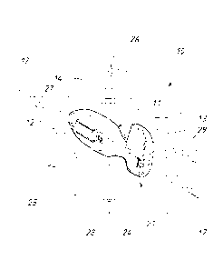
of the vagina. The puborectalis arises from the superior and inferior pubic
rami (front part of
pelvis, either side of pubis) and forms a sling around the rectum. Hence when
contracted, it
"pulls forward" to aid in closing off the canals. The strength of both is
essential in maintaining
continence.
[0004] Many perineometers currently available measure the pressure change
inside the vaginal
canal upon muscle contract. These devices have the disadvantage that they do
not give any
indication of muscle movement or actual contraction force. This may lead to
deterioration of a
patient's condition of the patient is performing the contraction incorrectly ¨
the problem being
that "bearing down" using the stomach muscles can also increase the pressure
inside the vaginal
canal, thus giving an incorrect indication of muscle contraction.
2
[0005]
Known perineometers are described in Australian Patent 739990, Australian
Patent
780359, International Patent Publication WO 92/20283, International Patent
Publication
WO 2012/142646 and USA Patent Application 2010/174218.
[0006] The device of WO 2012/142646 differs from other types of devices
through the use of a
superior direct muscle force measurement and positioning of the device in the
vagina.
[0007] The above discussed devices have the disadvantage that they still fail
to provide
sufficiently accurate information in respect of contraction of the pelvic
floor muscle group.
OBJECT OF THE INVENTION
[0008] It is the object of the present invention to overcome or substantially
ameliorate the
above disadvantage.
SUMMARY OF THE INVENTION
[0009] There is disclosed herein an intra vaginal device to aid in determining
muscle
contraction. The device includes a body comprising an end portion, a base
spaced from the end
portion in a direction along a longitudinal axis of the body. The base is
elongated in a direction
transverse of the longitudinal axis. A longitudinally extending side wall
extends between the end
portion and the base. The side wall comprises a first side wall portion and a
second side wall
portion. The body is configured to be inserted into a vagina of a patient with
the end portion and
side wall positioned within the vagina and the base outside of the vagina
engaging a vaginal
entrance of the vagina. A motion detector is positioned within the base of the
body and is
configured to detect acceleration and angular movement of the body resulting
from muscular
contraction adjacent the vagina and to generate a signal indicative of the
acceleration and
angular movement. A first sensor is mounted on the side wall portion and
configured to
provide a first signal indicative of pressure applied to the first sensor by
puborectalis
contractions of the patient, wherein the first sensor is elongated relative to
the longitudinal
axis of the body. A second sensor is mounted on the side wall portion, wherein
the second
sensor is angularly positioned about the longitudinal axis from the first
sensor by
approximately 80 to 90 . The second sensor is configured to provide a second
signal
indicative of pressure applied to the second sensor by pubococcygeus
contractions of the
Date Recue/Date Received 2021-06-03
2a
patient, and wherein the second sensor is elongated relative to the
longitudinal axis of the
body. A circuit is connected to the motion detector, the first sensor and the
second sensor so
as to receive the signals therefrom. The motion detector comprises an
accelerometer
configured to detect and provide a third signal to the circuit indicative of
acceleration in three
mutually perpendicular directions; and a gyroscope configured to detect and
provide a fourth
signal to the circuit indicative of angular movement about three mutually
perpendicular axes.
The first signal is indicative of pressure applied to the first sensor, the
second signal is
indicative of pressure applied to the second sensor, the third signal is
indicative of the
acceleration detected by the motion detector, and the fourth signal is
indicative of the angular
movement detected by the motion detector. The signals together provide an
indication that
the pressure applied to the first sensor and the pressure applied to the
second sensor do not
correlate to correct exercises associated with puborectalis contractions and
pubococcygeus
contractions.
[0010] Preferably, the motion detector detects acceleration in at least one
linear direction.
[0011] Preferably, the motion detector detects acceleration in three mutually
perpendicular
directions.
[0012] Preferably, the motion detector detects angular movement about at least
one axis.
[0013] Preferably, the motion detector detects angular movement about three
mutually
perpendicular axes. _____________________________________________________
Date Recue/Date Received 2021-06-03
CA 02964183 2017-04-10
WO 2016/119002 PCT/AU2015/000619
3
[0014] Preferably, the motion detector includes a gyroscope
[0015] Preferably, the motion detector includes an accelerometer.
[0016] Preferably, the body is elongated so as to have an end portion, a base
spaced from the
end portion, and a longitudinally extending side wall extending between the
end portion and the
base;
and wherein the device further includes:
a first sensor, the sensor being mounted on the side wall and to provide an
indication of
pressure applied thereto; and
a second sensor, the second sensor being mounted on the side wall so as to be
spaced
angularly about said axis from the second sensor, and to provide an indication
of the pressure
applied to the second sensor.
[0017] Preferably, said end portion is convex.
[0018] Preferably, said side wall includes a first side wall portion to which
the first sensor is
attached, and a second side wall portion to which the second sensor is
attached, with the second
sensor being angularly displaced about said axis from the first sensor by
approximately 80' to
90 .
[0019] Preferably, said side wall includes a third side wall portion, and the
device further
includes a third sensor attached to the third side wall portion, with the
third sensor being spaced
angularly about said axis from the first and second sensors.
[0020] Preferably, the third sensor is spaced approximately 80 to 90 from
the first sensor.
[0021] Preferably, the wall portions are generally planar.
[0022] In an alternative preferred form, the wall portions are convex.
[0023] Preferably, at least one of the sensors provides an electrical
resistance that diminishes
with an increase of pressure applied thereto.
CA 02964183 2017-04-10
WO 2016/119002 PCT/AU2015/000619
4
[0024] Preferably, the sensors are elongated longitudinally of said body.
[0025] Preferably, said base is elongated in a direction transverse of said
direction.
[0026] Preferably, said base is adapted to engage the vaginal entrance to aid
in correctly
locating the sensors.
BRIEF DESCRIPTION OF THE DRAWINGS
[0027] A preferred form of the present invention will now be described by way
of example
with reference to the accompanying drawings wherein:
[0028] Figure 1 is a schematic isometric view of a intra vaginal device to aid
in measuring
muscle strength;
[0029] Figure 2 is a schematic top plan view of the device of Figure 1;
[0030] Figure 3 is a schematic side elevation of the device of Figure 1;
[0031] Figure 4 is a schematic diagram of an electronic circuit employed in
the device of
Figure 1;
[0032] Figure 5 is a schematic end elevation of the device of Figure 1.; and
[0033] Figure 6 is a schematic sectioned isometric view of the device of
Figure 1.
DETAILED DESCRIPTION OF THE PREFERRED EMBODIMENT
[0034] In the accompanying drawings there is schematically depicted a device
10 to be
inserted in a woman's vagina to aid in measuring muscles operatively
associated with the
women's vagina.
[0035] The device 10 includes an elongated hollow body 11 having an end
portion 12, a base
13 and a longitudinally extending side wall 14. The side wall 14 includes side
wall portions 15,
CA 02964183 2017-04-10
WO 2016/119002 PCT/AU2015/000619
16 and 24 Preferably, the side wall portions 15, 16 and 24 are generally
planar (or convex) and
the portion 12 generally convex.
[0036] The device 10 has a longitudinal axis 17.
[0037] Secured to each wall portion 15 and 24 is a sensor 19, while secured to
the wall portion
16 is a sensor 20. Each of the sensors 19 and 20 is adapted to provide an
indication of the
pressure applied thereto. As a particular example, the sensors 19 and 20 could
provide an
electrical resistance that increases or decreases with pressure applied
thereto, preferably
decreases.
[0038] Preferably, the sensor 20 is spaced angularly about the axis 17 by an
angle of
approximately 80 to 90 from each of the sensors 19.
[0039] Preferably, the sensors 19 are spaced from the base 13 by the said
distance. The sensor
20 is placed at a desired distance from the base 13, that may be the same or
smaller distance
from the base 13 than the sensors 19. Preferably, the sensors 19 and 20 are
elongated
longitudinally relative to the body 11.
[0040] Preferably, the base 13 is transversely elongated, relative to the axis
17, to aid a user to
manipulate the device 10 and to aid in correctly positioning the device 10 by
having the base 13
engage the vaginal entrance.
[0041] The sensor 20 provides an indication of the puborectalis contraction
forces, the sensors
19 provide an indication of the pressure applied by the pubococcygeus.
[0042] The device 10 is shaped in such a way that once inserted into the
vagina, it is able to
measure both modes of contraction. The device 10 is inserted in the direction
18. The sensor 20
is preferably on top of the device 10 and measures the force applied to the
device 10 by the
urethral wall ¨ thus capturing the contraction strength contributed by the
puborectalis.
[0043] The sensors 19 are on the longitudinal sides of the device 10.
CA 02964183 2017-04-10
WO 2016/119002 PCT/AU2015/000619
6
[0044] The base 13 is spaced from the sensors 19 and 20 so that the base 13
upon engaging the
entrance of the vagina, correctly locates the sensors 19 and 20.
[0045] The sensors 19 are able to separately measure the force directly
applied by the bilateral
contraction of the pubococcygeus.
[0046] Preferably, the device 10 includes an electronic circuit 21 (printed
circuit board)
incorporating the sensors 19 and 20. The circuit 21 includes a processor 22
that interrogates the
sensors 19 and 20 to determine their resistance, and then to provide a signal
for a read out 23
that provides information in respect of the muscles associated with the user's
vagina. The read
out 23 may be remote from body 11 and communicates via wireless (Bluetooth)
with the
processor 22.
[0047] The circuit 21 also includes a motion detector 24. The motion detector
24 in one
preferred form detects acceleration in at least one direction, and preferably
detects acceleration
in three mutually perpendicular linear directions. Again the detector is
interrogated by the
processor 22 that provides a signal to the read out 23.
[0048] In another preferred form, the motion detector 24 is a gyroscope that
detects angular
movement about at least one axis, and preferably detects angular movement
about three
mutually perpendicular axes. Most preferably the motion detector 24 is a
combination of a
gyroscope and accelerometer. One example of such a device is an Invesense MPU-
6050 device
that also has a standard 12C communications interface.
[0049] In one embodiment the motion detector 24 is a gyroscope that provides a
signal
indicative of angular movement about the mutually perpendicular axes 17, 25
and 26.
[0050] In an alternative embodiment, the motion detector 24 provides a signal
indicative of
acceleration in the directions of the three axes 17, 25 and 26.
[0051] In a further embodiment, the motion detector 24 provides signals
indicative of angular
movement about the axes 17, 25 and 26, as well as acceleration in linear
directions along the
axes 17, 25 and 26.
CA 02964183 2017-04-10
WO 2016/119002 PCT/AU2015/000619
7
[0052] In respect of the above it should be appreciated that where the motion
detector 24
provides a signal indicative of angular movement and acceleration, the axes
about which the
angular movement is measured, and the axes along which the acceleration takes
place, need not
be coincident. That is the axes about which the angular movement is measured
may be
displaced from the axes along which the acceleration is measured.
[0053] However, most preferably the axes are coincident.
[0054] Connected to the circuit 21 is a rechargeable battery 27, with the
circuit 21 provided
with a coupling 28 that may include a voltage regulator/charge controller. The
coupling 28
provides for releasably attaching the battery 27 to the circuit 21. The
circuit 21 is also provided
with contacts 29 that provide for connection of the circuit 21 to a power
supply for the purposes
of recharging the battery 27.
[0055] Preferably the circuit 21 is constructed so that the motion detector 24
is located in or
adjacent the base 13.
[0056] In operation of the above device 10, the force measurements provided by
the sensors 19
and 20 can also be combined to give an average contraction strength output.
[0057] Separation of the measurements enables a more thorough understanding of
the overall
contraction and may lead to easier diagnosis of incontinence problems, as well
as an invaluable
teaching aide. There are many factors involved in incontinence, and this may
enable clinicians
to identify the muscle group that is contributing to incontinence in different
case studies.
[0058] This specific feedback is also essential in encouraging and maintaining
consistency
with patients using the device.
[0059] The ability to distinguish between the specific muscles and modes of
contraction may
also be helpful in addressing a common issue of over-clenching of the pelvic
floor. Many
women suffer from this condition and need to be taught how to relax these
muscles. The device
would be able to offer a more accurate picture of the clenching problem by
measuring the full
input of each muscle, and possibly pinpointing which area to focus on.
CA 02964183 2017-04-10
WO 2016/119002 PCT/AU2015/000619
8
[0060] The motion detector 24 enables the device 10 to provide a more accurate
determination
in respect of whether the muscle movement correlates to correct exercise, as
well as being able
to detect a larger range of incorrect movement, that is movement in respect of
lift and bearing
down. The motion detector 24 also provides information in respect of offset
angular movement.
[0061] Further to the above, the motion detector 24 also provides a better
indication of whether
contractions are correct.
[0062] The motion detector 24 also enables the device 10 to provide
orientation detection such
that the user's position during exercise can be recorded (standing or lying
down, etc) in order to
segregate the results.
[0063] Preferably in use of the device 10, the device 10 is covered by a
sheath. As a particular
example, the sheath may be of a synthetic rubber.
