Note: Descriptions are shown in the official language in which they were submitted.
CA 02964459 2017-04-12
WO 2016/061418
PCT/US2015/055835
MAGNETIC FIELD STRUCTURES, FIELD GENERATORS,
NAVIGATION AND IMAGING FOR UNTETHERED ROBOTIC
DEVICE ENABLED MEDICAL PROCEDURE
BACKGROUND
1. Technical Field
[0001] The embodiments described herein are related to the use of
magnetic fields and imaging with respect to medical procedures, and more
particularly to robotic magnetic medicine.
2. Related Art
[0002] A wide variety of medical procedures are currently performed
with undesirable and unavoidable effects on the patient that include damage to
healthy tissue during surgery and distribution of therapeutic substances
(drugs,
antibodies, vaccines and regenerative cells) to sites other than the intended
target. Non-disease related surgery increases the risk of sepsis, scarring,
blood
loss and decreased motor function. Non-specific therapeutic side effects
include impacts on metabolic organs and nervous tissue, undesired
accumulation in the liver, fatty tissue and digestive tract, and widespread
dilution in the circulatory system.
[0003] Many of these effects are unavoidable. In most surgical
procedures, a cavity must be created through the skin and sub-derma much
larger than the actual lesion. In addition, tools, implants and related
devices
commonly require large tethers, such as the surgeon's hands, catheters,
clamps, etc., for manipulation. For most bio-therapeutics, encapsulation,
localization and site-specific delivery are limited because related technology
is
in its infancy. The vast majority of drugs, antibodies and vaccines depend on
molecular specificity to accomplish intended functions and minimize side
effects. The latter remains non-optimal due to non-specific substance
distribution.
[0004] Desired effector functions, e.g., removal of a tumor,
clearance
of a blocked artery, activation of B or T immune cells, antibody tagging of a
specific cell type, etc., are relatively well defined. Unfortunately,
procedures
that accomplish those effector functions also negative impact healthy tissue.
In
1
CA 02964459 2017-04-12
WO 2016/061418
PCT/US2015/055835
addition, some avoidable or ameliorable diseases remain because procedures
to address them result in collateral damage disproportionate to the amount of
benefit. The shared causative factor is that medical technology is currently
disadvantaged by an inability to limit operator, electro-mechanical, and
biochemical procedures to necessary effector functions.
[0005] Current options for therapeutics delivery that attempt to
maximize targeting and avoid widespread pharmaco-distribution (PD) include
magnetic particles, ligand-coated liposomes and antibody coated micro- or
nanoscale capsuled drugs. Work on the latter two have been on-going for
decades and focus on two main areas: (1) Encapsulation, including
containment of payload during transport to target, assurable release of
payload
to target, reproducible manufacturing and storage life for regulatory
purposes,
and (2) Surface functionalization, including engineering of antibodies and
ligands for maximal specificity, affinity and avidity to targets, maximal
shelf
life, pH stability and minimal immunogenicity (immune stealth).
[0006] Efforts to incorporate magnetic fields with magnetically
susceptible bio-therapeutic laden spheres and colloids have focused on
accumulation at the site using permanent magnets or electromagnets
positioned at the skin proximal to the target site. Interestingly, magnetic
particle thermal effects have been researched, including efforts to elicit
tissue
damage via antibody or ligand coated particles moving rapidly in pulsating
magnetic fields.
[0007] The majority of these efforts depend on molecular specificity
of
effector molecules for target proteins. In rare cases, cancer or viral DNA is
targeted but these are early stage efforts. In most cases, critical parameters
for
determining the efficacy of therapeutics are completely out of operator
control
after application of the therapeutic, including when, where and how much
payload was delivered. The pharmaco-kinetic (PK) question of why an effect
or lack thereto occurred often depends on radioactive and other complex and
expensive tracing to determine PK/PD.
[0008] Even in magnetic, ultrasonic and radio-frequency controlled
capsules, conclusions regarding target specification depend on limited
biochemical data and broad physical effects, not on the real-time ability to
2
CA 02964459 2017-04-12
WO 2016/061418
PCT/US2015/055835
control targeting, application and dosing. In all cases, monitoring of
encapsulated payload is not possible except when using magnetic resonance or
ultrasonic imaging (MRI, USI) of capsules modified for compatibility with
such systems, modifications thereto potentially detrimental to the
biotherapeutic payload. Protocols do not yet exist to combine tMRI and USI
with both real-time control and accurate targeting of capsules or robotic
devices.
[0009] More elegant efforts to combine MRI and USI with robotics for
drug delivery and surgery include the diverse options of: (1) completely
passive or magnetic field-slaved robots having screw or star geometries, and
(2) completely autonomous endoscopic devices with on-board computers,
propellers, navigation fins, optical cameras and radio-frequency (RF)
transmitters. While the latter depend on batteries or, as being researched, RF-
based remote energization of on-board power supplies, the former are entirely
dependent on external magnetic fields for propulsion. Propulsion-related
fields
include pulsed attractive or repulsive linear fields, alternating attraction
and
repulsion gradients produced by orthogonally aligned electromagnetic coils,
and rotating fields that impart flagella-like movement. Current endoscopic
robots are relatively large and not applicable to vessels and vascular tissue
smaller than about 1 cm in diameter. Thus, protocols for cardiovascular,
lymphatic and metabolic organs with more narrow vascularization are not
possible with current endoscopic robot technology.
[0010] In contrast to many medical procedures, dependent technology
for medical robots is relatively advanced. Motors, RF transmitters, antennae,
microprocessors and even optical detectors can be made on the millimeter
[mm] and even micrometer [um] scale. Significant electro-mechanical
parameters scale with great linearity from the centimeter [cm] scale, where
ubiquitous end products that include servomotors, fans, cameras and mobile
phones depend on [mm - um] scale electro-mechanical components. Interest in
[mm] scale drone aircraft and gyroscopes, systems also sharing many qualities
with ideal medical robots, is high; however, rapid translation of these
technologies is hampered by their incompatibility with current MRI and USI
systems that only perform diagnosis. Moreover, current MRI technology is
3
CA 02964459 2017-04-12
WO 2016/061418
PCT/US2015/055835
incompatible with most robots as well as many implants because of their
electrical sensitivity and magnetic susceptibility. Thus, in most cases,
diagnosis is maintained separately from therapy.
SUMMARY
[0011] Systems and methods for generate magnetic fields (fields) for
the positioning and energization of medical devices are described herein.
According to one aspect, a magnetic field generating apparatus
comprises two or more co-facing, coaxial magnetic field generators configured
to generate equivalent magnetic fields directed toward a symmetrically central
convergence plane; a magnetically shielding encasement configured to contain
all of the associated magnetic fields generated by the coaxial magnetic field
generators; and articulation frames and supports for positioning of the
apparatus about a fixed point, wherein the generated magnetic fields are
counter-rotated relative to one another.
[0012] These and other features, aspects, and embodiments are
described below in the section entitled "Detailed Description."
BRIEF DESCRIPTION OF THE DRAWINGS
[0013] Features, aspects, and embodiments are described in
conjunction with the attached drawings, in which:
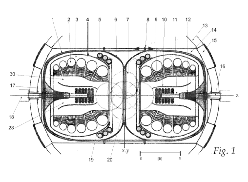
[0014] Figure 1 is a diagram of a diagnostic-therapeutic apparatus
comprising two coaxial, co-facing equivalent magnetic field generators
disposed about a central therapeutic space large enough to accommodate a
person in accordance with one embodiment.
[0015] Figure 2 is a diagram illustrating a neurological diagnostic-
therapeutic apparatus comprising two coaxial, co-facing field generators
disposed about a central therapeutic space in accordance with one
embodiment.
[0016] Figure 3 is a diagram illustrating main coil electromagnetic
components of the apparatus of figures 1 and 2 in greater detail.
4
CA 02964459 2017-04-12
WO 2016/061418
PCT/US2015/055835
[0017] Figure 4 is a diagram illustrating a cross section of two coil
segments separated by another coil segment (not shown; implied by the
intervening space) that can be used in the apparatus of figures 1 and 2.
[0018] Figure 5 is a diagram illustrating the fitting between the two
adjacent coil segments of figure 4.
[0019] Figure 6 is a diagram illustrating discreet winding pattern of
wires in the curvilinear (Right) versus standard winding (Left), base layers,
generated magnetic fields in each outermost wire and the ensemble field
produced as a result of each winding pattern of the coils of figure 4.
[0020] Figure 7 is a diagram illustrating the radial signal
acquisition
of, and triangulation-based antenna component assignment by, a neurological
RF source (MIddle) by the hemispherical antenna array (Right) in accordance
with one embodiment.
[0021] Figure 8 describes devices within and component potions of an
antenna array ring segment. In increasing magnification (Top to Bottom) are
described a portion of the ring segment, an assembly of antenna cells, and a
single antenna cell composed of a fractal antenna, base mount, and current
leads.
[0022] Figure 9 describes the random attachment of antenna assembly
leads on the back of the hemispherical array onto plug mates on the face of a
helically wound hemispherical take-up coil base.
[0023] Figure 10 describes correspondence of antenna ring frequency
preference with cell assembly lead-wiring onto helical leads on the take-up
coil base.
[0024] Figure 11 is more detail diagram of magnetic fields produced
by each field generator in figure 2.
[0025] Figure 12 describes the volume about which fields diverge
from the convergence plane and back into the direction of generator bores.
[0026] Figure 13 generally describes magnetic field intensities on a
scale illustrating the central shielding of two equivalent field generators as
in
figure 1.
[0027] Figure 14 describes the direction of propagation of static
magnetic fields in a simplified version of an apparatus such as in figure 1.
CA 02964459 2017-04-12
WO 2016/061418
PCT/US2015/055835
[0028] Figure 15 describes the direction of propagation of more
highly
energized static fields combined with rotating magnetic fields in a simplified
apparatus.
[0029] Figure 16 describes the apparatus and fields in figure 15
combined with a boundary field from energized tertiary electromagnetic coils.
[0030] Figure 17 describes qualities of MINRB structures (A) and
MICRB structures in the presence of non-rotating (B) and rotating (C)
boundary fields as well as MICRB structures (D) and DGP induced hybrid
structures (E) resembling geometric passerelles.
[0031] Figure 18 describes in more detail the static and rotating
fields,
and field gradients in the proximity of a null space (A), and the effect of
compressing the field structure with greater main coil energies onto a robotic
device (B).
[0032] Figure 19 describes converging static, counter-rotating and a
boundary field on both sides of a convergence plane emphasizing rotational
vector magnitudes and directions from a planar representation of a 3D radial
effect.
[0033] Figure 20 describes the translocation of a FFZ along a bore
axis
(+/-z) with resulting asymmetry (*) as compared to the starting point (0,0,0).
[0034] Figure 21 describes the translocation and changes in drive
coil
energy states of a simplified robotic device as in figure 20 when it is
subjected
to an imbalance in relative field generator gradients.
[0035] Figure 22 describes a robotic device and simplified field
illustrations as in figure 21, where a free field zone similar to that in
figure 18
is generated off-center from the robot, resulting in activation of one drive
coil
and translocation of the robot in the direction of greater axial field
gradients.
[0036] Figure 23 describes the change in geometry and drive coil
energy states of a twin hull robotic device when overall field intensities are
intensified. Co-facing fields are equal.
[0037] Figure 24 describes a homopolar motor drive coil for robotic
devices viewed cutaway from the side. Integral thereto are gyroscopic masses
and a rechargeable battery.
6
CA 02964459 2017-04-12
WO 2016/061418
PCT/US2015/055835
[0038] Figure 25 describes the homopolar drive coil viewed cutaway
from the top along two planes. Rotating gyro-mass and rotor bars are
illustrated on the top half
[0039] Figure 26 describes the direction of magnetic fields generated
by homopolar motor rotor bars within the stack of inductive plates (Top), and
electric currents along conductive pathways in one direction (Bottom).
[0040] Figure 27 describes a rotor stator drive coil for robotic
devices
viewed cutaway from the side. Integral thereto are gyroscopic masses and a
rechargeable battery.
[0041] Figure 28 describes the direction of magnetic fields generated
by rotor-stator motor rotor bars within the inner mantle (Top), and electric
currents along conductive pathways in one direction (Bottom).
[0042] Figure 29 describes a generalized robotic device having a
homopolar drive coil (left side) and rotor stator drive coil (right side).
Exposed
rotor stator motor rotor bar sets integrate hydrodynamic fins.
[0043] Figure 30 describes the generalized robot from FIG 31 encased
within a cranial implant dock with its bow end to the brain (Left), and the
robot in more detail including on-board devices and scale (Right).
[0044] Figure 31 describes an exemplary neurological and other
electro-active tissue robot implant having bow and stern enclosed homopolar
drive coils and exposed electrodes.
[0045] Figure 32 describes an exemplary surgical robot with two rotor
stator drives coils.
[0046] Figure 33 describes an exemplary therapeutics delivery robot
with a capsule payload shell disposed centrally about an inflexible axis
tethering the drive coils. Robot lacks autonomous capability.
[0047] Figure 34 describes an exemplary non-autonomous therapeutics
delivery robot with a capsule payload shell disposed centrally about a
flexible
axis. Drive coils are encased within mating hull sections.
[0048] Figure 35 describes a dynamic geometry robot for adaptive
susceptibility to magnetic fields, in both "stealth"/MRI compatible mode (A)
and "activated" mode (B).
7
CA 02964459 2017-04-12
WO 2016/061418
PCT/US2015/055835
[0049] Figure 36 describes an flexible geometry robot for biopsy
collection or vascular clearance.
[0050] Figure 37 describes a method for performing vascular clearance
by a flexible geometry robot with the bow drive coil (left side) dis-
integrating
a tissue sample and transferring the cellular matter through the stern drive
coil
(middle) with material collection in a towed bag (right side), and a method of
peristaltic transfer of material within a robot, including flexible tubing
tethering drive coils.
[0051] Figure 38 describes a dynamic geometry robot for vascular
clearance using retractable ablation tools, disposing an alternative on-board
power supply regenerative capacity.
[0052] Figure 39 describes a method for performing vascular clearance
of blocked vasculature using the dynamic geometry robot with retractable
ablation tools.
[0053] Figure 40 describes a dynamic geometry robot for placement of
conductive wire that is tether for robot hulls and is temporarily contained
within a device shell (A).
[0054] Figure 41 describes a method of delivering a robot into a
patient via injection using a standard clinical needle, the robot being one of
a
number of types including, as illustrated, the type described in Figure 40
(within the needle), one of the types described for microsurgery as described
in Figure 32 (top right) and the adaptive geometry type as illustrated in
Figure
35.
[0055] Figure 42 describes a method for translocating a surgical
robot
through a therapeutic volume that utilizes 180 degree turns.
[0056] Figure 43 describes a method for translocating a surgical
robot
through a therapeutic volume that does not utilize successive 180 degree
turns.
Illustrated surgery performed is in a back-and-forth manner from one end of
the therapeutic volume.
[0057] Figure 44 describes a method for performing surgery using an
adaptive geometry robot of the type described in Figure 40.
[0058] Figure 45 describes a method of bio-therapeutics delivery to a
target site (curved parallelogram).
8
CA 02964459 2017-04-12
WO 2016/061418
PCT/US2015/055835
[0059] Figure 46 describes an electro-active tissue application robot
(Left), and its payload (Right) being a spherical synaptic monitoring device,
conductive wire and bio-adhesive capsule.
[0060] Figure 47 describes the exemplary synaptic monitoring device
components in more detail including bio adhesive containing compartments,
sensor probe and equatorial RF antenna.
[0061] Figure 48 describes a method for attachment of the synaptic
monitoring device to ganglionic tissue using bio-adhesive to secure placement.
The sensor probe has been extended into the bundle of electrically active
cells.
[0062] Figure 49 describes a method of creating a conductive pathway.
Briefly, the robot is translocated to the ganglionic target, the synaptic
monitoring device is secured and the connecting wire unraveled as the robot
pulls away (Top).
[0063] Figure 50 describes magnetic field gradients and potentials
produced close to a robot in the case of static fields (upper quadrant) and
when
a strong pulse field is generated (lower quadrant).
[0064] Figure 51 describes generally magnetic intensities and net
magnetization vectors of resonant targets along a radial plane at two orders
of
magnitude of net magnetic strength relative to a robot.
[0065] Figure 52 describes a toroidal coordinate system for a point
(x,y,z) on the toroidal surface, including static (BTOR) and rotating (BROT)
gradients, with respective magnitudes and directions of net magnetization
(Mx,y,z and MROT, respectively).
[0066] Figure 53 describes net magnetization vector magnitudes and
directions along four cardinal points at a given field strength and axial
distance
(z) from a robot, when static (wide cones) and static plus rotating pulse
fields
(narrow cones) are applied, as viewed from along the x-axis (Left) and z-axis
(Right).
[0067] Figure 54 describes a RF array disposed around the therapeutic
space where the PSLP transmitter is contained and can be rotated 360 degrees
and articulated along several axes.
[0068] Figure 55 describes the PSLP transmitter unit main parts (A)
and disposition within the ring shaped RF transmitter array (B).
9
CA 02964459 2017-04-12
WO 2016/061418
PCT/US2015/055835
[0069] Figure 56 describes a modified Bloch Sphere with tri-bit
(three
quantum state) of magnitudes, directions and precessions of net magnetization
in both the low energy toroidal (small wide cone) and high energy rotated
(large narrow cone) states, with disposition of the PSLP transmitter.
[0070] Figure 57 describes the sequence of energization, rotation,
transverse alignment, spin locking and relaxation for both perfectly spin-
locked (A-E) and partially spin-locked (A-C, F-G) locations.
[0071] Figure 58 describes net magnetizations of both static,
baseline
fields and rotating, angularly rotating fields during the 250 ms rotating
pulse
sequence (A), and the sequence in RF inputs from both the transverse
orthogonal (B1) and PSLP pulses, the latter rotating after application (B).
[0072] Figure 59 describes output RF sequences of pseudoT1T2 (Ti,
reverse Ti, rotationally angled T2 and toroidal T2) in voxels optimally spin
lock pulse locked (A) and sub-optimally locked (B).
[0073] Figure 60 describes a method of MAS/MAT imaging in the
presence of a linear magnetic field with axially (z) rotating device (Left),
and
axially rotating toroidal magnetic field with device (Right).
[0074] Figure 61 describes a LOG device, at middle magnification
illustrating the levitation/rotation coils with levitated detector sphere in
cross-
section.
[0075] Figure 62 describes at highest magnification the LOG
equatorial dipole moment leads and proximal levitation/rotation coil
components at one equatorial location.
[0076] Figure 63 describes the detector and control gyroscope units
with laser source, photodetector and calibration electrodes..
DETAILED DESCRIPTION
[0077] In the embodiments described herein, millimeter-scale multi-
functional medical robots can be configured to carry out specific effector
functions while at the same time avoiding collateral damage to healthy cells
tissue. Untethered, magnetically-levitated devices incorporate surgical tools,
payload spaces and real-time functional control and navigation for enhanced
medical protocol efficacy with minimal necessary size and ideal robotic
geometries. With optimal development, such robots can be navigated to
CA 02964459 2017-04-12
WO 2016/061418
PCT/US2015/055835
specific tissues and disintegrate tumors by kinetic effect, cavitation or
thermal
cauterization. Robots can, for example, deliver pharmaco-active drugs,
antibodies, vaccines, stem cells, tissue scaffolds and other bio-therapeutics.
Further, such robots can clear passages, collect tissue, perform biopsies and
deliver the samples to an insertion site for analysis. Such robots can also
perform diagnostic, pallative and modulatory functions on electro-active
tissue, advancing pain management, neurological analysis and cognitive
studies.
[0078] MRI, USI, computer aided x-ray tomography (CAT) and other
non-invasive techniques are seen as ideal platforms to support more
efficacious, effector-focused medical protocols. All of these function by
energy input to the body, potentially enabling robot functions. MRI, in
particular, is seen as the most promising option with current technology
advanced in many ways to accurately diagnose a wide range of diseases with
minimal collateral damage. The vast majority of current MRI systems use
linear fields generated either (1) in the bores of scanners composed of arrays
of cylindrical electromagnetic coils, or (2) between North and South poles of
powerful permanent magnets. These systems are well-developed, reliable,
accurate, relatively safe and provide benefit to manufacturers, investors,
care
providers and patients.
[0079] Thus, as described herein, MRI technology, or other
technologies noted above can be used in conjunction with such robots to
perform open-bore imaging and to provide curved and rotating magnetic fields
to navigate and energize robots. As explained in detail below, modified
electromagnetic coils, magnetic shielding and field gradients can be used to
produce magnetic field structures for optimal robot stability, localization,
navigation, energization and detection. The use of such MRI technology and
robots can enable real-time diagnosis and therapy, providing a truly
"theranostic" platform.
[0080] It is desirable to: (1) perform surgery in a manner that
maximizes destruction of target tissue while minimizing collateral damage to
healthy tissue, (2) contain and site specifically release the minimal required
amount of pharmacologic drug or antibody to target cells and organs, (3)
11
CA 02964459 2017-04-12
WO 2016/061418
PCT/US2015/055835
contain, transport and subsequently release vaccines to germinal centers and
other immune tissue to optimize immune system re-programming, and (4) use
smaller devices that accomplish these and other effector functions. Ideally,
these procedures dispense with a tether, include remote control, remote
energization, accurate navigation and real-time imaging. The embodiments
described herein can provide such benefits.
[0081] Because of the shortcomings of conventional slaved devices,
much smaller and also autonomous robots, such as those described herein
would be much more desirable. As described below, in various embodiments
these, e.g., millimeter size robots also (1) carry on-board analytical,
optical
and RF devices, (2) be able to navigate complex paths (further provided
because of their small size), (3) move through tissue with minimal collateral
damage if necessary without need for external, potentially damaging
propellers and fins, and (4) be able to carry out surgery and therapeutics
delivery. More ideally, the robot can be monitored and navigated in real-time
using MRI or USI. Further, the persistence time and spectrum of effector
protocols of the robot can be expanded through remote energization.
[0082] It is further desirable to perform procedures where medical
devices modulate physiological functions in disordered and diseased states.
Metabolic disorders that result in ulcers, kidney stones and coronary artery
blockages are commonly addressed pharmacologically and surgically.
Gastrointestinal tracts are now being mapped by untethered endoscopic robots
with cameras, however, kidney stones are still being shattered with ultrasonic
transmitters and blocked coronary arteries are still being enlarged, but not
cleared, with catheter delivered balloons or rotating blades. The latter
procedures require pushing a long tether through urinary or venous tracts to
deliver a comparatively large effector tool to the therapeutic site. But with
the
systems and methods described herein, a small-as-possible tool can be
delivered untethered and monitored in real-time, to carry out the necessary
procedure. Relatedly, it is possible to biopsy a potential cancerous tissue in
a
similarly non-invasive manner.
[0083] The MRI-compatible robotics described herein provide levels
of robustness, reproducibility and versatility that pass regulatory
qualification
12
CA 02964459 2017-04-12
WO 2016/061418
PCT/US2015/055835
as medical devices and implants, with impact to the patient that is
proportionally limited to effector functions.
[0084] In certain embodiments, diagnosis and therapy are combined
into a unitary procedure, with the robot contributing to both. Thankfully, a
wide variety of magnetic field structures can be generated, including those
more suitable to robotic device power systems. Ideally, these magnetic field
structures also enable MRI imaging. In this vein, it may not be necessary to
rely on linear fields. Clinically approved MRI of any kind depends on (1)
target proton, nucleus or magnetic resonance imaging contrast agent (MRICA)
resonance in a strong linear magnetic field (BO), (2) energization of targets
with a secondary electromagnetic field (B1), and (3) measurement of RF
energy output during relaxation of targets back to the resonant equilibrium
state.
[0085] In certain embodiments, these operations, and subsequent
spatial encoding and image reconstruction, are carried out using other types
of
MRI scanners that produce magnetic field structures that are compatible with
robotic devices.
[0086] It is desired that MRI-compatible robotics be developed, at
levels of robustness, reproducibility and versatility that pass regulatory
qualification as medical devices and implants, ideally with impact to the
patient that is proportionally limited to effector functions.
[0087] A wide range of neurological disorders are attributable to
either
insufficient or excessive electrical activity, including Parkinson's,
Dementia,
Epilepsy, Chronic Pain and the disease spectra of Post Traumatic Stress
Disorder (PTSD). The efficacy of procedures that address these disorders,
including deep brain stimulation (DBS), trans-cranial magnetic stimulation
(TMS), surgery, pharmacologics and regenerative cells is being determined. In
certain embodiments, discreet portions of the central and peripheral nervous
systems can be analyzed to identify problematic ganglia, which are then
electrically modulated to improve cognitive and motor functions. If necessary,
delicate surgery can be performed to remove and replace sub-optimal target
tissue, again using untethered and real-time controlled devices as described
herein.
13
CA 02964459 2017-04-12
WO 2016/061418
PCT/US2015/055835
[0088] Certain embodiments described herein comprise apparatuses
that generate magnetic fields (fields) for the positioning and energization of
medical devices. Such embodiments can further comprise or make use of
magnetic field structures, heretofore defined as one or more magnetic field
geometries, gradients, potentials, and elements or layers commonly illustrated
by magnetic field lines. Such field structures can include rotating,
compressive, constrictive and torsion fields compatible with the robotic
devices described herein. Radio-frequency (RF) transmission and reception
devices compatible with field generators and field structures are provided.
Methods and apparatuses are provided for field structure modulation using
focusing shields, coil geometries, generator articulation and other types of
modulations. Methods are also provided for robotic device-based diagnostic,
therapeutic, prophylactic and cybernetic function. Additionally provided are
novel magnetic resonance imaging (MRI) methods in reference frames and
analysis volumes (voxels) compatible with components, robots and field
structures produced in the invention.
[0089] Certain embodiments include an apparatus having components
comprised of electromagnetic coils having uniform or varying radius. These
main coils generate strong (-1-9 Tesla [T]) and generally invariant field
gradients in apparatus bores herein referred to as static or main fields.
Static
fields can be applied to facilitate robotic control, spatial encoding and
signal
acquisition in voxels outside bores. A plurality of main coils generate
toroidal
field structures characteristic of Helmholtz, Maxwell, Tesla, Rodin, Solenoid
and other electromagnetic coil types.
[0090] Certain embodiments can also comprise a second set of
physically revolving electromagnetic components that generate and focus
rotating field elements peripheral to and concentric with static fields.
Revolving permanent magnets, conductive components and electromagnetic
coils add general rotational quality and distinct rotating field elements to
ensemble magnetic fields, facilitate larger gradients in main coil bores and
assist in synonymous motile field propagation within shielded enclosures.
Revolving field structures enable robotic device navigation, translocation,
and
imaging strategies.
14
CA 02964459 2017-04-12
WO 2016/061418
PCT/US2015/055835
[0091] In certain embodiments, a tertiary set of non-rotating coils
are
provided to generate boundary fields for partitioning of rotating field
elements
produced by secondary coil sets. Tertiary coils are disposed peripheral to all
other field coils, partitioned by shielding, and produce the outermost field
elements within an apparatus. Boundary fields can vary through energization
and coil positioning, either dynamically to enable pulsed MRI imaging of
therapeutic space or discreetly to enable stable positioning and energization
of
robotic devices. Tertiary coils also generate rotating magnetic fields through
application of alternating current (AC) through helically-wound conduction
pathways.
[0092] Also disclosed are magnetic shielding encasements and
partitions. Shields of low magnetic field susceptibility and transparency of
significant mechanical integrity confine fields within the apparatus, present
geometries that focus field energies onto desired targets, and disposed to
partition autonomous field elements and physically support field coils. Also
provided is fine [mm scale] field focusing in the patient through alignment
and
articulation of field generators, and selective energization of secondary,
tertiary and peripheral field coils.
[0093] When integrated into encasements, a coaxial assembly of all
components associated with main coils, revolving secondary coils, fixed
tertiary coils and field focusing encasements define a field generator
(generator). Support equipment including cryogenics, RF transmitters,
antennae, and other components and devices are understood to be integral in
field generators.
[0094] A plurality of field generators can be disposed and energized
to
generate converging (co-facing) toroidal magnetic fields. Disposition is
either
coaxial along a common bore axis [z, per convention] in the case of two field
generators, or at equivalent angles to shared axes (orthogonal disposition)
when three or more field generators are applied. Magnetic fields can be
generated and focused into higher flux densities toward a convergence plane
disposed midway between field generators.
[0095] Patient tables are also disclosed that can be used with the
systems and methods described herein to provide a diagnostic/therapeutic
CA 02964459 2017-04-12
WO 2016/061418
PCT/US2015/055835
platform. Size, geometry and disposition of main coils create an outside bore
therapeutic space where the patient can stand, lay down or be seated. Bed and
chair components should be MRI compatible, i.e., passively or actively
magnetically transparent. Patient contacting magnetic field focusing helmets
or body units can be integrated.
[0096] Because of their magnetic qualities, robotic devices and
certain
payloads can provide tangental function as magnetic resonance imaging
contrast agents (MRICA). In some embodiments the devices alter either the
main magnetic (BO) or transverse RF (B1) field to improve signal acquisition
in the vicinity of the device, particularly in aspects where field intensities
below current clinical MRI art and proximal to the robot exist. In related
embodiments, devices generate magnetic fields to achieve similar goals. In
additional embodiments, devices generate RF signals, for example matching
the Larmor frequency of nearby resonant targets or soluble MRICA.
[0097] In certain embodiments, signal acquisition and spatial
encoding
for real-time analysis of the device-proximal therapeutic space is provided.
Briefly, in contrast to conventional MRI applications, which useeither (1)
generally linear intra-bore fields in in apparatuses using cryogenic
electromagnets, or (2) generally linear fields between North and South poles
in
apparatuses using strong permanent magnets, the systems and methods
described herein provide rotating, radial, curvilinear and null field
geometries,
often of dynamic quality, and disposed outside main coil bores. Signals
acquisition of resonant target relaxation after B1 stimulation can be provided
in non-linear and transient geometries.
[0098] A coaxial disposition of main, revolving and non-revolving
electromagnetic coils all energized in the same direction produce an ensemble
toroidal magnetic field. For description, terminology of elements and layers
is
used herein to describe geographically distinct field structures propagating
within a generator, and in a manner preserving element autonomy as
illustrated by closed field lines. Terminology and illustrations are not meant
to
contradict convention, which establishes that physical separation of field
elements by shields and other components creates geographically distinct
16
CA 02964459 2017-04-12
WO 2016/061418
PCT/US2015/055835
structures originating proportionally, but not distinctly, from field elements
generated in those partitioned volumes.
[0099] Secondary and tertiary electromagnetic coils as briefly
described above can present varied field strengths, rotational speeds,
electrical
modulation (amplitudes, frequencies and wave patterns), spatial position and
other variable parameters. Secondary and tertiary coils can also be moved
along a common axis (z) or at an azimuth (A) to main coils. Integrated can be
revolving or immobile magnetic shields and lenses providing both bulk
focusing of field energies within enclosures and subtle focusing of fields on
an
outside bore target. In all, non-cryogenic electromagnetic components provide
for and modulate (1) overall ensemble field strength, (2) the magnitude and
dynamic qualities of rotating magnetic field elements, specifically their
geometries, relative potentials and rotational rates, (3) the stability of
structures generated by converging rotating fields, and the (4) positions,
structures and persistence of torsional fields which are used to position and
energize robotic devices.
[00100] Field elements generated by main coils are understood to
remain confined within each field generator and not interact with static
fields
produced by other field generators as such interaction may interfere with
imaging capabilities. Also, static fields do not productively interact with
robotic devices. Strong static gradients are provided to compress and focus
rotating and boundary fields around a small (-1 cm3) toroidal pocket. In most
cases, two mirror image counter rotating fields and boundary envelope field(s)
converge to produce closely disposed counter-rotating elements. Due to main
coil compression, rotating fields are focused close (+1- 5 mm) to the magnetic
pocket. Because the toroidal pocket is in most cases the location of a robotic
device, terminology of magnetic pocket, magnetic trap and null volume are
used interchangeably. Null terminology does not imply any absence of fluxes
or potentials except at the central point (0,0,0), by convention.
[00101] An array of RF transmitters generating a wide spectrum of
frequencies can image a relatively large volume around a magnetic pocket.
Such RF transmitters contribute transverse fields (B1). RF signals generated
by relaxation of energized protons or nuclei can be acquired through radial
17
CA 02964459 2017-04-12
WO 2016/061418
PCT/US2015/055835
antenna arrays, and mounted on the main coils similar to conventional MRI
practice. The antenna array can be wide spectrum sensitive, composed of a
large plurality of concentric rings with identical micron scale fractal
antenna
units (cells). The array can be programmed by applying different voltages to
each array ring resulting in cells with distinctive frequency and modulation
s ens itives .
[00102] Determination of an RF source geographical position can be
performed through acquisition by all similarly energized cells, however at
different times (except with axially disposed sources), enabling conical
geometry triangulation. Acquisition by cells in adjacent rings is less
efficient
and output signals are distinct from cells in the perfectly modulated ring.
The
antenna array can be mounted on a take-up coil base that harvests magnetic
energy from main coils to avoid requirement of an external power source.
Base leads from each cell can be wound in a toroidal manner to increase
current travel distance for greater signal discrimination.
[00103] Complexity in attaching a large number of cell leads (-
1,000,000) to their correct plugs on the base is avoided by random attachment.
An assembled antenna array is then programmed for spatial discrimination by
moving a pinging multi-frequency RF source while different voltages are
applied to each ring and signals are processed to correlate RF source location
and frequency with spatial processing. This method allows each antenna cell
to be assigned a unique frequency, modulation and spatial coordinate set.
When used for imaging, k-space data sets can match each cell signal and its
optimal modulation with input data acquired in radial coordinates to determine
the frequency and location of signal source. As practiced in the art, final
signal
processing can be carried out by Fourier Transform to reconstruct the image.
[00104] In certain embodiments, two exactly similar field generators
disposed coaxially with co-facing positive bores are used, where all coils
immobilized and energized to generate equally balanced fields, mirror image
non-rotating blended (MINRB) fields are produced. The outermost field
elements from each generator combine into a unitary structure at a central
plane at circular coordinates defining a convergence ring, producing a two
dimensional (2D) field of radially symmetric potential and geometry directed
18
CA 02964459 2017-04-12
WO 2016/061418
PCT/US2015/055835
inward towards the central point with flux density the sum of outermost field
elements. Un-blending back into distinct fields occurs around the central
point
in a toroidal manner with divergence location at circular coordinates defining
a divergence ring. Field elements then propagate back into generators and into
either main, secondary or tertiary coil sets depending on shielding and
encasement geometries. In this comparative aspect, field potentials are
constant at all locations in the apparatus and have no angularity when viewed
from an axial reference.
[00105] Similarly, in certain embodiments two exactly similar field
generators disposed coaxially with co-facing positive bores are used, where
all
coils equally energized and motile coils revolving at the same rate to
generate
equally balanced fields, mirror image counter rotating blending (MICRB)
fields are produced. As in MINRB fields, outermost elements blend into a 2D
field at convergence, diverge back into distinct elements, and the convergence
plane does not rotate. Importantly, the non-rotating blended field cannot be
maintained indefinitely because motile electromagnetic components
generating those elements continue to revolve. Rotation-induced gradient
between rotating and non-rotating field elements and magnetic torque induced
on field coils increase with increasing angular displacement. Produced in the
vicinity of the convergence plane are (1) 3D radial structures that transition
between completely blended and non-blended qualities, and (2) torque
imposed on both field generators and proximal magnetically susceptible
compounds or devices, including robots.
[00106] Highly structured and regulated magnetic torque can be used
proximal to the null space to position and energize robotic devices.
Terminology of torque is herein also used to describe rotating field
potentials
that induced rotational force on revolving magnetically susceptible
assemblies.
Energization and motility of field generator components is performed in a
cooperative manner that maintains the synonymous quality of the ensemble
field, i.e., by creation of magnetic field structures that minimize acute
angularity in field vectors and avoid rapid changes in localized field
potentials
- in particular, the cutting of magnetic field lines that can generate RF
signals
19
CA 02964459 2017-04-12
WO 2016/061418
PCT/US2015/055835
that interfere with MRI and robotic functions. Robotic effector protocols are
also understood to be performed in a synonymous manner.
[00107] Alternatively, revolving coils can provide more dynamic
MICRB structures characterized by rapid transitions between a non-rotating
2D blended disk and rotating 3D structures along and close to the convergence
plane. Produced are radial magnetic pockets composed of formerly blended
elements that rapidly un-blend to recover synonymy with revolving
components and resume low field densities before extinguishing back into
blended field structures. Structures are torqued in the direction of coil
revolution, mirror image counter rotate about the central plane and can be
described as two sets of diametrically opposed passerelles. The various field
structures described herein are collectively referred to as a dynamic gradient
pocket (DGP).
[00108] At low gradients, blending/un-blending events can occur
outside the divergence ring and preserve the toroidal pocket; however,
transitions between a 2D converged plane and 3D radially directed and
rotationally torqued null spaces can result in structural fluctuations that
occur
at the rate of blending/un-blending transitions, heretofore referred to as the
DGP pulse frequency. Critical factors determining the DGP pulse frequency
include overall ensemble field strength and motile coils revolution rate.
Generation of DGP structures with high DGP pulse frequencies are provided
by rapid coil revolution rates, large current loads and rapidly oscillating
high
voltage currents.
[00109] Related but tangental to this aspect, counter rotating equally
energized coils that revolve at different speeds are expected to produce at
the
central plane sharply angled field vectors and rapidly changing potentials.
Such asymmetric MICRB fields will blend and un-blend in an asynchronous
manner resulting in non-synchronic DGP pulses, and non-uniformly
distributed torqued radial null zones producing non-flat convergence planes.
[00110] In the absence of or when main coil energies are minimal,
diverged formerly blended MICRB fields will form a large null space of
similar scale as the bore radius. Compressive magnetic energy may be absent,
and constrictive magnetic energy and the DGP pulse frequency may be low.
CA 02964459 2017-04-12
WO 2016/061418
PCT/US2015/055835
Thus, little usable magnetic torque or diametrically opposed force is provided
for robotic devices. To overcome this deficiency, high DGP pulse frequencies
can be provided by, for example, rapid coil revolution rates and high
frequency currents in motile coils; however, ensemble gradients will remain
low unless revolving components generate field densities approaching those of
cryogenic coils. This will compress the null space but is unsustainable.
[00111] An additional benefit of performing simultaneous MRI-based
diagnosis and control of robotic devices provided by strong main coils is
lost.
Preferably, compression by static fields tightly focuses the divergence ring
into a ¨ 8-10 mm diameter circle and compresses a ¨ 64-125 mm3 toroidal
pocket. Axial locations of torsion fields, where counter rotating elements can
provide rotational magnetic torque, are thus only ¨ 10-16 mm apart along the
z-axis or roughly the same scale as robots.
[00112] In the continuing aspect of MICRB fields, robotic device or
assembly of magnetically-susceptible objects, as described herein, placed
centrally in the null zone will experience diametrically opposed expansive and
contractive forces at the DGP pulse frequency in addition to dynamic counter-
rotating constrictive forces. A plurality of non-diamagnetic, magnetically-
susceptible particles (1) substantially smaller than the 64-125 mm3 toroidal
pocket, (2) loosely contained in an enclosing matrix or other field
transparent
container, and (3) disposed centrally will be moved in two equal populations
in a linear manner along the z-axis away from and then back towards the
pocket at the DGP pulse frequency, in addition to being moved in counter
rotating directions.
[00113] A portion of the magnetic particles will remain relatively
immobilized in the low flux zone during each DGP pulse event, exchanging
locations with the larger population. If using a homogenous population of
spherical super-paramagnetic particles of aforementioned scale and density
whereby particle mean free path provides 1-on-1 interaction at a given AC
frequency in secondary coils, the invention also provides dipole-dipole
coupling of particles facilitated by field-induced transient magnetic moments
in particles, resulting in generally uniform intra-particle spacing.
21
CA 02964459 2017-04-12
WO 2016/061418
PCT/US2015/055835
[00114] In certain other embodiemtns, two exactly similar field
generators disposed coaxially with co-facing positive bores and all coils,
including boundary coils, energized and equally rotated to produce equal
fields are user, where mirror image counter rotating separated (MICRS) fields
are generated. This can be an ideal field structure for robotic device
management. Again, rotating elements from each ensemble field propagate
synonymously with revolving electromagnetic components, herein in a
manner that re-synchronizes field vectors with motile components to
maximize induced magnetic torque on robots while minimizing torque on field
generator coils. Synonymy also compensates for (1) potential losses and field
harvest by robotic devices, (2) gradients relative to less than transparent
surfaces, (3) interaction with non-parallel or unequal fields, and (4) other
phenomena that negatively impact the linearity of field vectors and torsional
geometry.
[00115] In contrast to MICRB, MICRS fields provide several
operational advantages. First, because rotating field elements are maintained
as distinct structures, convergence of counter rotating elements does not
occur.
Induced torque on revolving coils is reduced by approximately 50%
preserving ensemble field synonymy and reducing field vector angularity,
field line cutting and RF noise. If the boundary field was also rotated
synchronously with the secondary field, the secondary coils would experience
no induced torque during boundary field rotation. Secondly, because the null
space becomes compressed with each boundary coil pulse contributing flux,
magnetic torque on robots increases. Of note, as revolving secondary coils can
also act as boundary coils and generate pulsed fields, distinction between non-
cryogenic coils generating pulsed rotating outermost field elements in a field
generator is de-emphasized. Thirdly, the probability of contaminating main
coil field elements by their counterpart(s) approaches zero as
main/compressive elements must overcome two sets of secondary/rotating and
tertiary/boundary elements. Additionally, DGP structures are reduced in both
gradient and pulse frequency as counter-rotating fields (1) no longer
interact,
if a constant boundary field, or two counter-rotating boundary fields
22
CA 02964459 2017-04-12
WO 2016/061418
PCT/US2015/055835
undulating in phase, is used as the partition or (2) interact less often, if a
pulsed boundary field is used.
[00116] In certain embodiments, rotational magnetic torque for the
navigation and energization of robotic devices can be provided. Rotating field
gradients exist in field generators between (1) rotating and non-rotating
fields,
(2) rotating fields and low magnetic susceptibility surfaces, (3) field layers
rotating at different speeds, and (4) rotating fields and magnetically-
susceptible particles, assemblies and revolving components on robotic devices.
Outside the null space from the divergence ring to the points of conical
peaks,
field intensity and rotational speed increase with increasing distance (n)
from
the central point (0,0,0). Conical radii decrease and magnetic fields beyond
these points assume greater linearity and rotational speed matching that of
revolving components. The difference in field intensities and rotational
speeds
between the central target (zero flux and no provided torque) and the two
coaxial con-facing conical termini (maximal flux and maximal torque) result
in two magnetic torsion fields. Terminology of torsion is used herein to
emphasize a combination of (1) diametrically opposed attractive (pulling)
forces, (2) diametrically opposed compressive forces, mainly due to main coil
gradients, (3) diametrically opposed constrictive (twisting) forces produced
by
revolving electromagnetic coils and/or boundary coils powered by AC current
that provide (4) rotational torque for magnetically susceptible objects.
[00117] It is understood that no magnetic flux exists at the central
target
per convention but that weak, non-zero potentials exist at every point (x,y,z
or
z, 0, r > 0), even inside the null space; however these can be neglected.
Priority can be given to the geometrical and functional relationship between
the null zone and an assembly of particles or a mechanical robot having
magnetic susceptibility specifically optimized for function in torsion fields
as
previously defined. Therefore, free field zone (FFZ) is heretofore used to
describe the volume (1) encompassing the central target point, (2) bound by
the divergence ring (x,y plane), and (3) two coordinates along the common
axis where field intensities and rotational torque are sufficient to overcome
the
activation threshold of magnetic drive coils on a robotic device (z +/- AT).
These two points are generally, but not exclusively, locations of torsion
fields.
23
CA 02964459 2017-04-12
WO 2016/061418
PCT/US2015/055835
Terminology is dependent only on the target point, which in many aspects
describes the preferred coordinates of a robotic device, and the geometry and
activation thresholds of robot drive coils, which varies for different devices
and applications.
[00118] In certain embodiments a relatively large FFZ is provided that
can immobilize a smaller robot or a robot having a higher activation threshold
within a space that provides insufficient navigational and functional energy.
The same FFZ will activate a larger robot having drive coils that extend
further outward into higher magnetic flux space, or a smaller robot having a
lower activation threshold. FFZ per se is dependent mainly on device qualities
and not limited to field characteristics. It does not depend on the flux
density,
if any, in a null or other space which the FFZ encompasses or the actual
location of torsion fields.
[00119] Because a null volume is disposed between two con-facing,
coaxial counter rotating torsion fields in the continuing aspect, a symmetric
and diametrically opposed magnetic potential is produced in the FFZ along the
common axis from non-zero field intensity (z = -n), to no field at the target
point (x,y,z = 0), to an equivalent non-zero potential (z = +n). Also evident
is
that a non-zero magnetic potential exists from the target point (0,0,0)
outward
along the convergence plane (x,y) to radial points of divergence (y, r = D).
These potential gradients and field structures provide a novel experimental
condition having useful qualities. Briefly, it is understood in the art that
regions of very high magnetic potential are applicable to electronics, the
physical and materials sciences particularly micro and nano-electromechanical
systems (MEMS/NEMS).
[00120] Regions of very high magnetic potential, in some applications
counter rotating fields, in certain applications diametrically opposed
constricting fields, in specific applications symmetrical torsion fields in
the
millimeter scale or below can be used in the life sciences to influence
susceptible metabolic, biochemical or electro-active processes. In many of
these applications, biomolecule dipole moments (native, induced, and
generally ensemble in large molecules), biopolymer and charged membrane
24
CA 02964459 2017-04-12
WO 2016/061418
PCT/US2015/055835
magnetic susceptibilities, electric potentials and electron flow determine
viable function.
[00121] Further, high potentials and geometries provided in the
embodiments described herein influence chemical processes, particularly those
at interfaces of magnetic particles, polymers and solvents where molecules,
substances and surfaces possess charge, conductance or magnetic
susceptibility. Overall, FFZ structures provided in the invention can be used
to
manipulate biological, chemical and physical processes. In short, focused high
intensity and dynamic counter rotating torsion fields provide both a novel
environment and useful analytical tool for a wide range of investigations.
[00122] It is understood that magnetic pockets or null spaces can
localize diamagnetic materials, minimizing their energy states. Similarly
understood in the art, paramagnetic, ferromagnetic and ferrimagnetic materials
will tend to accumulate near torsion fields and other constricting magnetic
field zones and driven beyond into higher flux zones, in the continuing aspect
along the common axis (z > In). Materials having magnetic dipole moments
will tend to align their vectors with proximal field lines. Because torsion
fields
herein rotate, materials will also tend to revolve or otherwise change
position
with the fields. Further, materials with gyroscopic qualities will tend to
rotate
at the same speed as the fields to achieve equilibrium, and with dipole
moments aligned to minimize their potential energy. Further still in the
continuing aspect, two equivalent magnetic gyroscopic assemblies of the same
scale as torsion fields will tend to remain stably positioned thereto if they
are
rigidly or flexibly tethered to each other along a common axis.
[00123] In certain embodiments, stable positioning and energization of
a robotic device having coaxial counter-rotating magnetic drive coils with
revolving gyroscopic components is provided. Magnetically susceptible
components will be attracted in diametrically opposite directions along the
common axis (z) and revolve synonymously with rotating field gradients if
allowed to interact with potentials significantly above activation thresholds.
If
geometrically symmetric and constrictive, field gradients produce stable
torsion fields that provide rotational torque to device drive coils. It can be
preferred that torsion fields sufficiently activate but not overwhelm drive
coils,
CA 02964459 2017-04-12
WO 2016/061418
PCT/US2015/055835
specifically neither too strong or rapid that electro-mechanical magnetic
components on robots are challenged beyond their operational limits.
[00124] In the continuing aspect of embodiments that use MICRS
fields, an axially and radially symmetric FFZ is created and sized such that
rotating field elements at each constriction zone are disposed closely to
revolving magnetically-susceptible components on device drive coils.
Activation thresholds, load limits, power efficiencies and other terminology
understood in the art apply to all motors with revolving components. In
aspects where robots are kept inactive, FFZ geometry and characteristics can
be such that torsion fields are maintained outside device drive coils and/or
field rotation rates are kept low. In aspects where robots are kept hyper-
active
or when on-board batteries require regeneration, FFZ can be structured such
that torsion fields are within drive coil spaces and also rotate rapidly.
[00125] In embodiments where robots must be navigated with precision
and simultaneously carry out an effector function requiring high energies such
as in ablative surgery, the sizing and energization of FFZ for maximal
constrictive potential on drive coils, while also moving the FFZ to provide
device navigation, is provided.
[00126] Absence of field rotation, rotation in only one direction
and/or
insufficient rotational rate, even in the presence of equal and diverging
fields
as in the aspect of MINRB fields, are understood to be inadequate for stable
device positioning due to insufficient induction of gyroscopic effect. Such
meta-stable conditions create the tendency for a device to escape from a FFZ
and, in the MINRB aspect, be propelled in either axial direction. This
instability is significant in conventional applications that use linear or non-
torsional rotating magnetic fields to position and propel magnetically
susceptible objects. In some conventional solutions, position escape is
avoided
by rapid pulsing of generally linear but non-rotating or inadequately rotating
fields. Alternatively, a larger plurality (> 2) of field generators or field
coils
can be symmetrically disposed in relative orthogonality to a target and
produce less intense fields.
[00127] Thus, the embodiments described here that use two co-facing
field generators and disclosed method of producing balanced counter rotating
26
CA 02964459 2017-04-12
WO 2016/061418
PCT/US2015/055835
fields that radially converge, orthogonally diverge and create symmetrical,
con-facing torsion fields in an axially balanced robotic device that is
disposed
in a magnetic null zone can present a significant improvement over
conventional solutions. AS do the disclosed magnetic assemblies having two
balanced counter-rotating magnetic inertial gyroscopes which also function are
robotic positioning and energization coils.
[00128] Navigation of robotic devices is provided herein by selective
energization of field coils and electromagnetic components thereto. In some
cases, unequal energization and rotational rate of coaxial coil sets in
different
field generators enable dominance of one coil set in positioning of a robotic
device along a common axis resulting in device translocation along the axis
towards the dominant coil set. In the same or other cases, it is understood
that
cryogenic main coils may be de-energized to enable fields produced by
secondary and peripheral field coils to overcome those produced in the other
field generator to achieve the aforementioned asymmetry. Thereto, when real-
time high resolution imaging of the proximal space around the robot is less
important than device navigation, the invention provides positioning and
translocation of a robotic device at orthogonal axes using only weak, i.e.,
generally non-compressive, constrictive and torsional fields.
[00129] It is understood that the patient will have undergone a
standard,
high resolution MRI prior to implant of robotic device and that significant un-
changing geographic details of the therapeutic space will have been
determined. Tissues thereto provide non real-time, however useful 3D
landmarks for robot navigation which, in the continuing cases, can include
bone, other high density tissue or implanted MRICA pellets location-secured
with bio-adhesive.
[00130] Robot navigation can also be provided by repositioning of one
or more field generators, for example along the z-axis to maintain preferred
co-axial disposition. Briefly, one field generator can be kept immobile and
the
other moved closer or further from the other field generator resulting in
translocation of the FFZ. Repositioning of generators at an azimuth to a
shared
axis can also be performed. In combination with selective energization of main
coils other coils, FFZ structure can be maintained while also being moved
27
CA 02964459 2017-04-12
WO 2016/061418
PCT/US2015/055835
along any axis depending on apparatus structural and functional limitations
and the presence of patient bed or chair mounts that can limit apparatus
articulation.
[00131] In general, robot navigation is provided by keeping the
patient
immobile and repositioning field generators that maintain relative position,
moving the FFZ along desired paths through the use of concentric articulation
frames. Disposition of field generators in this preferred aspect allows the
patient to remain comfortable in the therapeutic space while the field
generators are articulated along yaw, pitch and roll axes. Similarly, the
patient
bed can be moved along the z-axis, elevated (y) and shifted (x) to accomplish
similar functions.
[00132] Real-time imaging of the robotic device space can be
performed by transient energization of non-cryogenic coils to field densities
in
the FFZ vicinity that approach that of commonly performed imaging. For
example, 1.0 T fields generated by combined energies from revolving and
boundary coils along all axes enables acquisition of 43,5 MHz signal from
water protons at conical coordinates about the null space. Robotic devices can
be navigated and effector functions performed herein; however the FFZ
torsion fields must be sufficiently strong to overcome device activation
thresholds implying large field gradients to achieve both robot function and
target imaging at 1.0 T close to the device. Brief resonant bursts as in
pulsed
MRI, ultra-short time echo imaging (USTEI) and other technology can be
used. For example, high resolution imaging in a pulsed or strobe-like manner
of the immediate robotic space can be carried out secondary and tertiary coil
sets to intensities approaching 1 T.
[00133] Robotic drive coil magnetically susceptible components have
geometries that provide electromagnetic motor function. Returning to the
aspect of a balanced and symmetrical FFZ as in MICRS fields, coaxial and
counter-rotating fields enable remote energization of on-board batteries
contributing to robot autonomy. Drive coil AT can be diverse as is well
understood. Therefore, FFZ and torsion fields can be provided as greatly
variable and dynamic in size, magnetic potential, geometry symmetry and
rotational speed. Provided in devices are a spectra of drive coil activation
28
CA 02964459 2017-04-12
WO 2016/061418
PCT/US2015/055835
thresholds that vary with robot status, effector function and surrounding
media. It is understood that activation thresholds will be higher in media
such
as bone versus soft tissue due to greater resistance to drilling, coring and
other
functions requiring greater induced torque from torsion fields.
[00134] For aspects such as neurological or neonatal monitoring, it is
understood that robots will have contained, unexposed drive coils minimizing
collateral tissue damage. For the purposes of stable positioning, trapping and
robot recharging, the invention provides methods to tune and modulate FFZ so
that torsion fields produce magnetic torque that either (1) remains below, (2)
achieves or (3) surpasses robot drive coil activation thresholds.
[00135] Robotic drive coils can be simple and robust assemblies of
homopolar motors and single or dual phase rotor-stator motors that are
straight-forward to fabricate at device scales (¨ 1 x 3 mm), mass produce,
create industrial standards and validate as integral components of medical
devices, probes and implants for regulatory and quality control purposes.
Homopolar motors can provide efficient generation of current for charging on-
board batteries or capacitors in aspects where FFZ are modulated to provide
excess energies for applications requiring rapid current release such as pain
modulation and tumor cavitation. Low phase motors can provide optimal
efficiency of converting battery voltage to rotation of exposed components
that contribute to device propulsion, such as fins for applications such as
endoscopic diagnosis and screws for applications such as calcified tissue
ablation.
[00136] Drive coils include a plurality of magnetically susceptible
rotor
components having radially balanced dipole moments directed away from
device center. The latter is generally both the robot's central axis and the
field
generator bore axis (z). These magnetic rotor bars are assembled such that
dipole moments are in dis-equilibrium to, and a group magnetic moment
persists even in the absence of, an external magnetic field. The group dipole
moment of each drive coil is generally curvilinear and directed in axial
directions away from the FFZ. In most cases, devices have bow and stern
drive coils with the same activation threshold to maximize stable positioning
in an axially symmetric FFZ where the robot center is disposed at the central
29
CA 02964459 2017-04-12
WO 2016/061418
PCT/US2015/055835
point and the drive coils at torsion fields. In the aspect of rotor bars as
permanent dipole magnetic wires or rods, positive poles are directed to the
bow and stern of each robot and angled at an azimuth to the negative poles. In
the aspect of super-paramagnetic wire or particles encased in a bent tubular
shell, a similar geometry can be utilized. In both aspects, when a torsional
or
rotating linear magnetic field is applied, device positioning along a common
axis is provided by the tendency - never achieved - of each rotor bar or rotor
tube to align its dipole moment vector in parallel with the applied field.
Minimal group dipole moment in each drive coil is only achieved when (1) the
entire drive coil aligns in parallel to the applied field, rotating in the
case of
torsion fields, and (2) bow and stern drive coils are disposed in opposite
directions, counter rotating in the case of FFZ torsion fields.
[00137] In summary, because drive coils have (i) dipole moments
minimized only when aligned in group parallel to a magnetic field, (ii)
gyroscopic inertial masses which revolve with rotating fields, and (iii)
function as both electromagnetic generators and tools or analytical devices,
(1)
robot position stability, (2) navigation and (3) remote energization are
provided.
[00138] In embodiments where homopolar motors are utilized, acutely
angled (< 90 deg) magnetic rotor bars form a nearly triangular geometry that
produces an axially directed, generally linear internal magnetic field upon
rotation of bars. Linear fields are ideal for generation of an electric field
in the
homopolar motor's inductive metal disc pile for current production in
applications such as capacitor charging. If desired, rapid rotation of rotor
bar
set is provided by current discharge from a capacitor or current release from
a
battery to the inductor pile, resulting in generation of a rotating magnetic
field.
Homopolar motors operating in this reverse mode are acknowledged to
generate gyroscopic effect less efficiently than rotor-stator motors.
[00139] In embodiments where rotor-stator motors are used, obtusely
angled (> 90 deg) magnetic rotor bars form a cylindrical-conical geometry
that, upon rotation, produces a radial internal magnetic field directed to (1)
a
inner core magnet, hollow and centrally insulated to house electrical leads,
and
(2) a high ferrite content mantle, facilitating radial orthogonality of the
internal
CA 02964459 2017-04-12
WO 2016/061418
PCT/US2015/055835
field. Longitudinally wound conducting wire provides generation of an electric
field to recharge on-board batteries in one current direction. Rotation of
rotor
bars is provided in the other current direction with a charged battery, even
in
the absence of a rotating external field, facilitating device propulsion in
autonomous mode. Autonomous navigation is provided when robot
components include biochemical or electro-optical sensors. Screws, propellers,
flagellum, adaptive geometries such as tails and other propulsive components
integral to exposed rotors contribute to navigation.
[00140] Torsion fields provide on board battery recharging through
induced rotation of rotor bars. Inactive devices will tend to remain
immobilized during such recharging sessions so long as the FFZ is
symmetrical, immobile, stable, persistent and torsion fields provide magnetic
torques beyond that of drive coil activation thresholds; however, recharging
can occur during device translocation and therapeutic protocols in a motile
FFZ if induced torque and subsequent rotor bar set rotational speed overcomes
battery drainage as the robot performs its functions. In cases such as
ablative
surgery, tissue evisceration and other highly kinetic effector procedures, it
is
understood that torsion fields will have to be applied regularly and
intensively
to the robot to insure both accurate device navigation and maintain energy
levels of on-board components.
[00141] Both homopolar and rotor-stator motors can be attached to
batteries, capacitors, computer control, RF, optical and other components to
accomplish a wide variety of functions. Homopolar motors are sealed inside
device shells or capsules due to exposed conductive elements and electrically
conducting and lubricating fluid. Stators are similarly contained, however
rotors can be exposed without sacrificing recharge or navigation function at
adequate activation thresholds and exposed rotors further provide navigation
and effector tool functions.
[00142] Within a given robot, both activation threshold and gyroscopic
effect can be balanced for drive coils at the bow and stern. Alternative
thereto
being a bias in an asymmetric drive coil robot for navigation towards one
axial
direction of a symmetric FFZ. Similarly, the same robot can maintain position
31
CA 02964459 2017-04-12
WO 2016/061418
PCT/US2015/055835
in an asymmetric FFZ, as is produced when one field generator is energized
and/or rotationally distinct from the other.
[00143] Homopolar and rotor-stator motors can be attached to
integrally
rotating inertial masses providing additional gyroscopic benefit. In many
embodiments, rotating components free float relative to immobile components
but are contained within shells or conical geometries and positive poles of
rotor bars are disposed at greater distances from attachment points near
rotational axes; however, because of the small overall length (-12 mm) and
radius (-3 mm) of each robot, rotating coil bars are relatively light in mass
(-1
mg each), thus inadequate to provide stabilizing gyroscopic effect unless
rotated at very high speeds (- 3000 rpm).
[00144] Inertial gyro-mass free operation requires very high field
intensities and revolving coil rotational speeds, shortening operational life
of
both field generators and robot drive coils. Optimal gyroscopic effect and
stable robotic positioning are provided by rotating at low-to-medium speeds (-
60-240 rpm) a relatively heavy inertial mass (-50-100 mg) in each drive coil.
Gyro-masses can be non-functional rings, disks, hemispheres or function as
batteries, sensors and conductive elements. In embodiments where rotating
gyro-masses are exposed, they can be surgical, tissue sampling or ablative
tools. In embodiments where a gyro-mass is a delivered or acquired payload,
drive coils may adapt to altered activation thresholds and devices may adapt
to
changing center of mass or center of magnetic balance.
[00145] In embodiments where autonomous robot function is undesired
or in applications where only slaved devices have clinical approval, device
drive coils can dispense with energy generation or charge carrying capacity
and be simple assemblies of magnetizable wire or bars plus rotating gyro-
masses, linearly or azimuthally angled with the torsion field. This passive
configuration retains the benefit of positioning stability and navigation
through the torsion fields of a FFZ.
[00146] A variety of robotic device configurations can be provided. A
cylindrical hull (for optimal passage through tissue with minimal drag
coefficient, CD) with two AT-balanced gyro-magnetic drive coils disposed
close to device termini and with payload volume in the middle form the basic
32
CA 02964459 2017-04-12
WO 2016/061418
PCT/US2015/055835
template. Diverse operational parameters include robot size, mass, center of
balance, drive coil AT, energy load and the number, thickness, angle and
length of rotor bars. Devices can carry one or two homopolar, rotor-stator or
passive motors, or any combination thereto and dispose a spectrum of
components, devices, payloads, payload volumes, effector and navigation
tools.
[00147] In some embodiments, devices carry charge measurement and
RF devices to serve as implants for monitoring and reporting neurological
activity. Sensors, stents, electrodes and other current in art devices which
accomplish these functions will require modification for compatibility with
drive coil power systems and to fit into robots. In other aspects, larger
robots
may carry optical devices to analyze the therapeutic space and supplement
MRI-based positioning of the robot, as applicable in robot semi-autonomous
mode where some operator control exists and when signal acquisition in or
proximal to the FFZ is either infeasible or undesired.
[00148] In other embodiments, robots fitted with two rotor-stator
drive
coils have a drill, auger or boring tool attached at one end for biopsy or
evisceration of tumors, fibrous and infected tissue. Disposed on the rotor at
the other end could be fins, propellers or screws for navigation.
[00149] In still other embodiments, passive coils carry a central
payload
of chemotherapeutic drug, antibody, vaccine or regenerative cells directly to
a
tumor site, germinal center or lesion. The payload is released when the drive
coils compress or pull apart, shattering the payload capsule. Also provided is
a
device geometry comprising a two-part shelled capsule and elastic wires that
contain the robot termini after payload release and facilitate re-assembly
back
into a closed unit.
[00150] In yet other embodiments, a robotic device mounts passive
hollow-core drive coils with substantially straight rotor bars. Alignment
tendency with an applied field is preserved however longer rotor bars with
greater individual dipole moments are required to achieve positioning
stability
approaching angled bars. Benefit provided is that these drive coils permit
passage of biological substances through drive coil centers and robotic space.
In one specific embodiments, the bow drive coil mounts a drill and chipping
33
CA 02964459 2017-04-12
WO 2016/061418
PCT/US2015/055835
blades, and is connected by an elastic tube to a hollow stern drive coil.
Attached to the latter is a flexible tube which terminates in a sieve for
collection of dis-aggregated tissue while allowing passage of fluid and very
small particles. This aspect and device configuration provides ability to
perform clearance of vasculature as in coronary artery disease.
[00151] Further provided for the vascular clearance aspects are
dynamic
FFZ that enable robot translocation through non-linear and constrictive paths
within veins and arteries. Benefit is provided when the mean linear path of a
vascular bottleneck is shorter than the overall length of the robot (including
tubular tether and collection bag), locale where arterial blockages and
calcified
tissue frequently occur. The asymmetric expansion/compression quality of the
tether provides a peristaltic action which hydraulically pumps dis-aggregated
tissue towards the stern. Force is provided when FFZ torsion fields are
expanded and contracted as when boundary fields are pulsed to generate
inflation and compression waves which move (+/- z) the location foci of FFZ
torsion fields. Robot length thus changes depending on path limits while
navigation is provided as before by movement and articulation of field
generators. Collection bag is kept slack to maneuver around obstacles. When
full, the robot is navigated back to the insertion or another point.
[00152] An abbreviated version of this embodiment provides capability
to perform biopsies. If tissue dis-aggregation is undesired, bow drive coils
can
mount a hollow boring tool to enable collection of a cylindrical plug of
tissue
in the robot's center volume. The rear drive coil may still need to mount a
sieve or filter to equalize hydraulic pressure. Fluid and small particles may
be
eliminated thusly or through more centrally disposed pores. Related to this
aspect, viable tissue may be chemically preserved in the robot immediately
after harvest with fixative released into the payload space after elimination
of
excess fluid and closure of ejection ports.
[00153] In a related embodiment, retractable abrasion or chipping
blades can be used to clear vascular blockages from robots having adaptive
geometry. Robots can navigate to the pre-thrombotic site in coronary arteries
then, preferably upon reception of an operator RF signal, increased FFZ
energies can pull bow and stern hull sections apart, extending tools from
34
CA 02964459 2017-04-12
WO 2016/061418
PCT/US2015/055835
storage volumes in hull midsections. Coring or chipping in counter-rotating
directions maintains robot positioning stability, particularly in how
volumetric
flow rate conditions, and helps accumulate dis-engaged plaque deposits into a
centrally disposed waste bag if necessary.
[00154] To minimize collateral tissue damage, robot navigation to
target location from insertion site avoids nervous and highly vascularized
tissue. Briefly, FFZ are generated as small as possible and the trapped robot
translocated with precision in paths previously determined in a high
resolution
MRI scan from the location of insertion to the therapeutic target. During
translocation, pulsed MRI data acquired in torsional reference frames provide
real-time navigation. Kinetic tools are preferably mounted on the robot stern
with the bow being a low CD hydrophobic and oleo-phobic surface. In a case
of ablative surgery with potential thermal and cavitation effects, upon
arrival
at the tumor, necrotic or other target tissue, the robot can execute a 180
degree
turn to direct the stern end surgical tool towards the target.
[00155] A therapeutic protocol can be carried out by movement of the
FFZ into and out of the target with the robot drilling-out tissue in its path,
entry/exit locations changed until all of the target tissue is disaggregated.
The
robot can be retrieved by following the entry path bow forward without the
need for another 180 degree maneuver.
[00156] Specifically provided are adaptive geometry devices where
portions of robots alter their magnetic susceptibility. In one embodiment, a
solenoid pump, electromagnetic piston or shape memory polymer is activated
by RF signal from an operator or on-board algorithm to extend device drive
coils out from a magnetically non-susceptible housing. This is provided by (1)
a bow or stern extension of a previously hidden drive coil out into the
therapeutic space, and (2) movement of a drive coil from a magnetically
shielded to a transparent section of the hull. In this aspect, a device can
assume
magnetic stealth mode and remain immobile even in the presence of a high
external field such as in 1.0-3.0 T brain scans with linear BO fields.
[00157] Also specifically provided are adaptive geometry robotic
devices that release or unravel non-insulated wires that are attached to high
voltage capacitors for use as surgical tools in applications such as thermo-
CA 02964459 2017-04-12
WO 2016/061418
PCT/US2015/055835
ablation of tumors or micro-surgery cauterization. After translocation of the
implanted robot to a lesion site, operator RF signal, on-board algorithm or
expanded torsion fields separate bow and stern hull sections, unraveling an
conductive tether wire. Through FFZ movement and device translocation, the
extended robot can move the stiffened wire in a carving or slicing manner
through the entire tumor. Energy for thermal effect can be provided by on-
board batteries and capacitors until exhaustion, or replenished for the life
of
the coils through torsion fields. After completion of effector function, the
wire
can be reeled back into the robot.
[00158] Additionally provided are coordinated function of sequential
robots. In one embodiment, an adaptive geometry robot can navigate to a
healthy nerve bundle or to damaged nerves and mount a signal-modulating
sensor on a ganglion via bio-adhesive. Attached to the sensor is an insulated
wire having on the other end a conductive terminus and small bio-adhesive
capsule. The robot can attach the cybernetic device and unravel the wire along
a predetermined path. Upon completion of this segment, an RF signal or
current can shatter the capsule releasing the bio-adhesive at the distal end
forming a semi-permanent mount. Follow-up robots can attach successive
series of connectors, adhesive mounted plugs and entire devices to create a
cybernetic network. The network can terminate in either a skin port for direct
connection to ex vivo equipment or an implanted device having RF functions.
Robotic implants positioned at intersections provide ability to report on
ganglionic functions at different locations within the network. Provision is
made for stimulating specific network locations through
activation/deactivation of devices.
[00159] The cybernetic skin port can also serve as a robot dock for
implantation, retrieval and maintenance of robotic devices. The dock
comprises housing for the robot, electrodes for re-generation of device
batteries, provision for retrieval of tissue samples, and application of
successive robots to the patient with reduced puncturing or injections. The
dock can be semi-permanent as in the case of brain tumors and traumatic
stress injury requiring long term care, a plurality of different procedures,
cognitive monitoring and brain reconstruction. Components include a blunted
36
CA 02964459 2017-04-12
WO 2016/061418
PCT/US2015/055835
large gauge MRI-compatible needle partially filled with flexible polymer to
help seal the robot in its dock. The inner needle end partially encloses the
robot bow. The outer end is sealed to the patient skull with strong bio-
adhesive
and capped with a screw-top for device insertion and retrieval. An aseptic
port
facilitates device retrieval and replacement. The port can be inserted in
other
body locations, single use or semi-permanent. The robot sheath can also be
field transparent if FFZ-based re-energization of the robot while in its dock
is
desired.
[00160] MRI-based imaging at a distance from field generator bores can
also be provided. Certain embodiments provide distal magnetic resonance
through apparatus components that include variable radii main coils wound in
a non-linear manner concentrically about the (z-axis). Specifically,
conductive
wire, bars or other current carrying paths for cryogenically cooled
electromagnetic coils are disposed with greater density and decreasing inner
coil radii in the positive direction facing towards other generators similarly
disposed in an apparatus. This variable radii constrictive geometry coil sets
and shielding encasements focus and compress generated fields. Coil segment
wire can be wound on frames and cores as common in the art but with
segments fitting together to produce the overall geometry. Energization of all
coil segments to direct a field in the target direction results in large field
intensities at a distance, specifically outside the bore, when combined with
shielding encasements.
[00161] A standard in art MRI scanner can modified with revolving
coils, other electromechanical and algorithmic components, and shielding to
serve as a field generator in an un-preferred aspect. The scanner can be
coaxially mated with an variable coil radii field generator to create a
combination diagnostic/therapeutic apparatus that utilizes very similar in the
art MRI-based software and imaging technology. In short, the patient could
undergo a standard MRI scan, then be moved out of that bore and into the
plane of convergence with an invention field generator. Injection of a robotic
device, creation of a FFZ around the device and invention imaging and robot
effector function could then commence. Conceptually, two or more standard
MRI scanners can be used to generate a distal resonant field; however,
37
CA 02964459 2017-04-12
WO 2016/061418
PCT/US2015/055835
because standard scanners are optimized to produce maximal field intensities
in their bores, significant re-engineering would be necessary to produce both
imaging and robot control out of bore. Intra-bore device trapping, navigation
and energization is not possible due to the linear, unidirectional nature of
the
static field (BO) and the absence of counter-rotating torsion fields. Counter
rotating secondary fields (B1) cannot adequately provide power for robot
navigation and energization. The latter would also be extremely limited even
if
a FFZ and torsion fields could be created - as would be the case with two MRI
scanners disposed co-facing in close proximity - due to limited ability to
reposition and articulate the scanners, and vary and pulse their generated
fields.
[00162] A variety of methods are provided to spatially encode water
protons, resonant nuclei or MRICA in the immediate robotic space (within 1
cm) and acquire relaxation RF signal for image reconstruction. These
methods can be optimized for the apparatus components, functional
capabilities and limits described herein as well as the described
diagnostic/therapeutic methods carried and magnetic field structures. These
methods can also be compatible with other MRI-based technologies.
[00163] In certain embodiment, rapidly rotating and intense field
pulses
are generated in co-facing in MICRB and MICRS fields. In an exemplary 100
ms secondary coil pulse, elements peripheral to main fields increase by 1000-
fold producing a transient 1 T layer within 1 cm of a robot. Rapid rotation of
motile coils and/or sudden large AC amplitudes axially rotates the field pulse
and shifts the net magnetic potential of susceptible nuclei from the toroidal
direction (ZTOR) to the new direction of net magnetization (ZROT). Fields
intersecting robot drive coils increase 1000-fold to their device activation
limit, specifically, 1 cm from the robot/target center, the former 1 Gauss [G]
field line generated by main coils increases to 1000G / 0.1 T. This intensity
of
field enables transient energization, resonance, spatial encoding, relaxation
and image reconstruction using novel MRI-based resonance and signaling
technology.
[00164] In certain embodiments, to analyze points having similar net
magnetization azimuths to the laboratory plane (z) and disposed on the
38
CA 02964459 2017-04-12
WO 2016/061418
PCT/US2015/055835
toroidal surface of the transient 1 T field line (one on each side of the
pulse-
energized FFZ), MRI is carried out distinctly from standard art MRI. Briefly,
a
transverse B1 pulse (+31/2) oscillating at 43.5 MHz is directed to the FFZ as
common in the art; however, a transverse spin lock pulse (SLP) is then applied
to lock the net magnetization of spatially distinct protons into the
transverse
plane (MTV). The SLP is applied at the Larmor frequency (co1000G),
oscillated in phase, on resonance and is linearly polarized.
[00165] Because net magnetizations are directed toroidally and not
linearly, only protons having transverse vectors parallel to the applied SLP
become spin locked. This is unlike standard MRI where all protons similarly
resonated by a uniformly linear BO and cohered by an orthogonal B1 are
simultaneously spin locked because their transverse magnetizations are all
parallel to one another.
[00166] After application, the SLP is then rotated to bring the
transverse
magnetization of rotated and spin locked protons (MROT) parallel with the
original net magnetization vector of weak protons (MTOR). Both the pulse
gradient and polarized spin lock pulse (PSLP) are then terminated
simultaneously. Magnetization vectors are allowed to collapse from a strong
(1000 G) magnetization in the direction of rotation (ZROT) with comitant
strong PSLP-normalized transverse signal, to a weak magnetization in the
equilibrium toroidal direction at 1G (ZTOR). The relaxation of (1) formerly
spin locked planar 1000 G magnetizations and (2) formerly angled
longitudinal 1000 G magnetizations, into a 1 G longitudinal vector has aspects
of both spin-lattice (Ti) and spin-spin (T2) relaxation. Herein named pseudo
T1T2 provides imaging of weak fields characteristic of the FFZ around a
robotic device, compatible with rotating pulsed fields.
[00167] In the transverse plane, the abrupt termination of a strong
rotating field results in the net magnetization quickly losing transverse
quality
and a rapid T2 signal is produced as net magnetization directions equilibrate
into toroidal alignment. Also, in the brief interim between termination of B1
and maximal SLP amplitude, some loss of magnetization transverse to ZROT
occurs and a brief pre-T2 relaxation signal can be acquired. Alternatively,
the
transverse RF signal can be modulated with the SLP to blend both signals
39
CA 02964459 2017-04-12
WO 2016/061418
PCT/US2015/055835
(RFIN) as the oscillation changes from transverse (coTV) to spin-lock (coSL).
In pseudoT1T2, longitudinal effects also contribute to transverse relaxation
phenomena and resultant signals (RFOUT).
[00168] In the longitudinal plane, the abrupt termination of the SLP
that
is plane-locked to the low energy magnetization vector produces unique
relaxation profiles distinct from classic Ti. Spin unlocking allows generally
planar vectors to re-align longitudinally back to ZTOR. However, pulse
termination also results in tilted vectors withdrawing back into 1 G net
magnetization. This reverse Ti (rT1) thus occurs both longitudinally and
transversely with the latter contributing to the T2 component of the overall
pseudo T1T2 spectrum. Lastly, a relatively minor contribution from high
energy spins previously pointing in the (-ZROT) direction are expected to
generate a classic Ti signal, i.e., relaxation back into the positive
direction of
the longitudinal axis (+ZTOR).
[00169] This return to equilibrium is not as quickly reflected in
secondary and boundary coils due to remanence. However, to ensure that
gradients return to nearly their equilibrium levels within 0.1 sec, selected
pulse
coil materials and engineering, cryogenic cooling (if necessary), control
algorithms, coil localization and non-transparent magnetic shutters can be
employed to maximize field intensities, abruptness of both field pulses, RF
signal initiation and termination.
[00170] Of note, adiabatic conditions are maintained since the Larmor
frequencies are much higher than the anticipated maximal induced rotational
rate of the transient pulse (3000 rpm). Also, because the PSLP brings
transverse magnetizations parallel to adiabatic equilibrium, a second RF pulse
(-7c/2) to restore locked magnetizations (MSL) back into the longitudinal
direction (ZTOR) may not be necessary. In the absence of stimulatory RF
signal, adiabatic conditions direct re-equilibrium of proton magnetizations
back to levels determined only by the static field (co 1 G). The resulting
slow
but high gradient relaxation is anticipated to improve signaling contrast at
low
field levels (---, 1-100 G) as a plurality of target protons relax in diverse
ways in
the pseudo T1T2 profile.
CA 02964459 2017-04-12
WO 2016/061418
PCT/US2015/055835
[00171] Spatial encoding is provided by the specificity of the SLP
transmitter's frequency, oscillation, modulation and position for transverse
magnetization spins that are resonating at a specific frequency and aligned
parallel to the applied signal. In an exemplary of the continuing aspect, a
polarized SLP locks onto parallel MTV at points on the 1 T toroidal surfaces
directly above the bow ventral (+V, 1T) and stern dorsal (-D, 1T) sides of a
robot for subsequent rotation and pseudo T1T2 signal collection. In contrast,
SLP pulse initiation will have a detrimental effect on 1 T points directly to
the
left and right of the robot, i.e., the bow starboard (+S, 1T) and stern port (-
P,
1T), because their magnetizations are 90 degrees off-set. The initial SLP
directed to V and D are parallel to the ZROT of points P and S and, because of
SLP frequency and modulation, will briefly de-resonate the protons therein
until the SLP is rotated away from ZROT(-P, 1T) and ZROT(+S, 1T).
However, the RSLP will eventually align parallel to the now recovered
protons at points P and S and pseudo T1T2 therein can be carried out.
Similarly, for all other points pulse resonated at 1 T, the RSLP will enable
pseudo T1T2 imaging as the SLP transmitter matches polarization with
transverse magnetizations at those points.
[00172] A similar strategy can be carried out for all other points
along
the 1T toroidal surface and for all other points of higher and lower field
intensity by variation of RSLP frequency, polarization, positioning (if
modulation is mechanical versus electronic) and other parameters.
[00173] It is understood that magnetically susceptible components of
robots, particularly rotor bars in drive coils, will distort the toroidal
magnetic
geometry in and around the FFZ. Distortion can be both positive, in aspects
where drive coils generate their own magnetic fields as with permanent dipole
magnetic bars, or negative, in aspects where net negative fields are produced
when super paramagnetic components attract field elements. Drive coil
options can also include current carrying wires that generate fields at
selective
times, geometries and intensities. In one aspect, AC propagates towards both
device termini to generate counter rotating magnetic fields describing bulbous
or knob geometries. Whichever drive coil option is used, the resulting field
41
CA 02964459 2017-04-12
WO 2016/061418
PCT/US2015/055835
distortion and change in net magnetization angle can be taken into account
during imaging.
[00174] In an embodiment, the robot can carry RF transmitters to
accomplish a wide variety of functions that can include (1) direct location
indicator, or pinging, by the device, (2) RF transmission at Larmor
frequencies
of resonant targets in the therapeutic space, (3) proximal RF signal
modulation
to enhance apparatus RF imaging of targets, by constructive or destructive
interference, (4) direct RF imaging of targets resonated by magnetic field
generating components of robots.
[00175] In another embodiment, a form of magic angle spinning (MAS)
can be applied to image spatial locations where rotating field elements
intersect the robot at the MA of 54.7 degrees. Because of toroidal geometry,
few elements interact with the robot at or near this angle. In addition, both
rotating fields and device drive coils are anticipated to revolve at rates (-
60-
300 rpm) much lower than ideal MAS speeds. Thus, application of magic
angle turning (MAT) pulse sequences modulated for resonant frequencies
intersecting the robot about 54.7 degrees (MA +/- 5 degrees) can more
comprehensively apply MAS to the invention. To supplement this effort and
contribute to other device functions, a micro-gyroscope can be carried as
payload, placed in the device at the intended location of field elements
intersecting at ¨ 54.7 degrees, and spun more rapidly than either motile
portions of drive coils or rotating apparatus magnetic fields. An even number
of counter rotating micro-gyroscopes will provide device stabilization even in
the absence of a FFZ.
[00176] In another embodiment, the robotic payload can include a laser
optical gyroscope (LOG) to measure discreet (uT) brain magnetic fields. The
unit is completely insulated in axial directions, i.e., from the magnetic
field of
device drive coils and on-board devices. In one embodiment, the LOG is a
rotating quartz sphere with an equatorial disk composed of (1) polarized
crystal or bonded micron scale crystal sheets, (2) coaxial disks with polar-to-
equatorial directed magnetic dipole moments in the uT range, and (3)
circumpolar disks that that resist an induced electric current. The described
detection sphere acts as a levitated gyroscope and is disposed between two
42
CA 02964459 2017-04-12
WO 2016/061418
PCT/US2015/055835
enclosing drive coils which free-float the sphere and induce rotation as well
understood in the art. Disposed at 180 opposed equatorial locations,
respectively, are a polarized laser and photomultiplier chip. The rotating,
levitated sphere and laser components function as a LOG as understood in the
art.
[00177] The internal dipole elements can extend equatorially outward
to
create two latitudinal disks of opposite polarity. Upon rotation, the
polarized
equatorial extensions generate a small, rotating magnetic field (BD). Any uT
range potential (BO) impacting orthogonally (x,y) to the extensions will cause
the sphere to tilt its rotational axis and mis-align the polarized equatorial
crystal from perfect alignment with the polarized laser beam, creating a
detectable precession. The detector sphere is mated to a control unit
functioning simultaneously and identical in all aspects except that it lacks
an
internal dipole moment and corrects for purely mechanical fluctuations.
Symmetric Field Generator
[00178] Referring now to FIG 1, in a preferred embodiment, an
apparatus comprises two exactly similar field generators, coaxial, co-facing
and all coils energized to generate equivalent magnetic fields directed toward
a symmetrically central convergence plane heretofore defined as the x,y-axis
that evenly divides the therapeutic space (7) provided for a patient. Within
each field generator is set of main coils (4) being cryogenically-cooled
superconducting electromagnetic field-generating apparatuses that produce up
to 9.0 T of field intensity in the therapeutic space. Main coils include all
coolant (3), electrical, insulating and structural systems clearly understood
and
commonly applied in the art of cryogenic/superconducting-type MRI scanners
and are included herein in their entirety by reference. Cryogenic coils (2)
have
varying radii, reducing in the central direction to focus field energies
toward
fields produced by the other generator.
[00179] Also provided for convergent focusing of fields is a central
curvilinear surface of magnetic field non-transparent and deflective shielding
(6) having geometry resembling the null space between two toroids with
circular apex being the convergence plane. An RF transmitter array (20) for
43
CA 02964459 2017-04-12
WO 2016/061418
PCT/US2015/055835
MRI-related scanning is preferably disposed around the convergence plane.
Disposed at extremities of the field generators are concentric magnetic
shields
(17) resembling the axial null space of a toroid. Heretofore, terminology of
toroid and toroidal are used generously to describe both apparatuses and
magnetic fields sharing this geometric template. A magnetic shield (1)
continuous with both the central (6) and terminal (12) shields encloses both
field generators and contains all generated fields within the apparatus.
[00180] Disposed intra-bore of each main coil are a second set of
revolving electromagnetic coils (9) which produce the rotating elements of an
ensemble magnetic field within a generator. Concentric with secondary coils
are magnetic shielding (10) and revolving magnetized rods (11) that further
focus rotating field elements within intra-bore spaces (28) toroidally
peripheral to the spaces (30) occupied by non-rotating static fields. Focusing
rods are attached to axial drive shafts (16) through rotating non-transparent
toroidal and conical fittings (12). An additional set of non-revolving
concentric tertiary coils (5) are disposed peripheral to all other coils and
partitioned from the main coils by a layer of magnetic shielding (19). The
central convergent shielding (6) can be moved axially (+/- z) to distort or
translocate the magnetic pocket produced in the center of the convergence
plane.
[00181] Between the sliding surfaces thereto (8) is a groove opening
to
allow a bed or chair post for the patient. The entire apparatus can axially
articulate approximately four feet and approximately two feet up, down, left
and right (+/- x,y), enabling the patient to remain immobile while the
magnetic
pocket is translocated. The entire apparatus is mounted through rollers (15)
and a frame (14) to a circular articulation rail (13) providing 360 degrees of
roll (z-axis) and yaw (y-axis) articulation of the apparatus about the
geographical center of the therapeutic space (7). Coordinate convention used
herein are the z-axis being common bore axis, up/down is the y-axis, and
left/right for the patient and in/out of the illustration plane the x-axis.
Approximately 30 degrees of pitch (+/- 15 degrees, x-axis) articulation is
provided, i.e., the limit of articulation defined by the grooved opening or
the
bed/chair post relative to the main or tertiary coil surfaces closest to the
44
CA 02964459 2017-04-12
WO 2016/061418
PCT/US2015/055835
therapeutic space. The apparatus articulation frame mounts drive shafts (16)
extending out from encasement (1), and drive motors (28) for revolution of
secondary electromagnetic components, enabling generation of rotating
magnetic fields in any apparatus orientation.
[00182] Generous apparatus articulation and freedom of movement
provides the ability to move robotic devices in the generated magnetic pocket
at the center of the produced convergence plane with linear distance (2-4
feet)
and azimuth (30 degrees). Greatly stable generation of static fields (BO) by
main coils, and greatly synonymous generation of rotating (BROT) and
boundary fields by secondary and tertiary coils provides for a highly stable
magnetic pocket with proviso for energizing robotic devices. Therefore,
preferred effector methods carried out in the Symmetric Field Generator
include magnetic tumor and tissue ablation and robotic delivery of
therapeutics including pharmaceuticals, antibodies and vaccines.
Asymmetric Field Generator
[00183] Referring now to FIG 2, in another preferred embodiment, an
apparatus comprises two dis-similar field generators, however similar to the
apparatus in FIG 1 in that Asymmetric Field Generators are coaxial, co-facing
and energized with equivalent magnetic fields directed toward a central
convergence plane (x,y-axis) dividing the therapeutic space (7). One field
generator can be a more powerful version of a generators from FIG 1,
including larger main (4), additional revolving secondary (9) and additional
immobile tertiary (5) coils. An RF antenna array (32) is provided for wide-
band acquisition of MRI-based relaxation signals. It is understood but not
illustrated that a similar RF antenna array exists on one or both field
generators in FIG 1.
[00184] The other field generator in the Asymmetric aspect can be a
common in the art MRI scanner, modified for inclusion in the invention in a
novel fashion. Referring now to the Right field generator in FIG 2, is an MRI
scanner (25) with coil sub-set on the therapeutic end having enhanced field
generating capacity (22), standard in practice field coils (24), and gradient
coils (23) modified and disposed for both intra-bore and extra-bore field
CA 02964459 2017-04-12
WO 2016/061418
PCT/US2015/055835
modulation. Disposed intra-bore and concentric with all coils in the Left
generator are a revolving secondary field generating and field focusing coil
set
composed of a coil (29), concentric magnetized rod (11) and toroidal geometry
shielding (10) collectively mounted to an axial drive shaft (16). This
secondary coil set revolves synonymously with a tertiary set of rotating coils
of greater radii (27) disposed substantially to the rear/terminus of the
modified
MRI scanner field generator. Larger tertiary field coils can physically
revolve
to produce rotating elements of the ensemble field within the Right generator,
or be immobile and generate rotating field elements via conduction of AC
current through pathways directed a helical manner as understood in the
magnetics art. In a preferred enablement, the tertiary coils are affixed to,
and
revolve concurrently with, the secondary coil set.
[00185] Magnetic field elements produced by secondary and tertiary
coils (BROT) pass through the volume inside the secondary shielding (28) and
process through the wider rear opening peripheral to elements generated by
main coils (BO) which process away from the central plane in the space
between the secondary shielding and the inner bore (30). In a preferred
enablement, net field intensities and rotating field rotational rates equal
those
produced in the Left generator.
[00186] Disposed substantially within the bore of the Right field
generator is a patient bed supported by a post (21) positioned between the
left
(32) and right (33) RF antenna arrays. The bed can slide a distance limited by
the spacing of the RF arrays, approximately three feet, or along internal bed
rails on a fixed post. Approximately 10 degrees of pitch (+/- 5 degrees) and
somewhat less yaw is provided by freedom of relative movement of the bed
within the Right field generator's main coil bore. 360 degrees of roll are
provided, however, in a preferred enablement, the Asymmetric Field
Generator apparatus lacks an articulation frame and corresponding rails.
Translocation of a robotic device within a magnetic pocket is provided by
relative energization of secondary and tertiary coils in each field generator,
and discreet (< 1 foot) x,y,z-axial and azimuthal movement of the patient
through the bed, particularly in neurological applications.
46
CA 02964459 2017-04-12
WO 2016/061418
PCT/US2015/055835
[00187] A standard in the art MRI scan can be performed with the
Asymmetric Field Generator and is a preferred method in preparation for
theranostic procedures utilizing this aspect. The apparatus magnetic shielding
encasement can open a the separation (26) substantially at the rear of the
Right
generator, and also at the convergence intersection between field generators
shown in FIG 2 as the location of the RF transmitter array (20). After the
secondary and tertiary field coils are removed, the modified MRI scanner (25)
remains. Re-assembly of the Asymmetric Field Generator can be carried out
by reversing the process.
[00188] In comparison to the Symmetric Field Generator, provided in
this aspect are (1) increased field intensities, (2) enhanced RF signal
acquisition and (3) greater rotating field strength in both generators
(substantially much greater in the Right generator). Thus, in a preferred
enablement, the Asymmetric Field Generator is used to carry out methods
requiring smaller robots that translocate limited distances (< 1 foot), with
delicate navigation (azimuths of 0.1 mm) and prioritize regeneration of
smaller on-board power systems over mechanical effect of exposed gyroscopic
tools. Protocols thereto include robotic clearance of vascular blockages and
the placement and energization of cybernetic and neuromuscular pathways
components in electro-active tissues, including the use of robotic devices as
electric and magnetic field modulating implants.
Field Coil Geometry and RF Antenna Arrays
[00189] Referring now to FIGS 3-6, conductive wire, cable, disk or
other current pathway component (37) is preferably wound in the manner
illustrated wherein pathways are linear when viewed from the orthogonal
reference (36) but are wound curvilinearly producing a decreasing coil radii
in
direction of the therapeutic space (to the Right). Individual pathway
components can have variable radii, e.g., wire of differing thicknesses, to
produce curvilinear geometries. Wire bases (38) and frames (39) that,
respectively, support distinctive layers and sections of wound wire can
likewise describe curvilinear geometries for ease of construction, and to
create
47
CA 02964459 2017-04-12
WO 2016/061418
PCT/US2015/055835
fin-shaped (40) coil units (FIG 4) that inter-connect to form a continuous or
gap-free (FIG 5) curvilinear main coil set.
[00190] Electromagnetic coils of the most commonly used type having
winding patterns, base and frame geometries generally illustrated on the left
half of FIG 3. These common in the art coils have conductive pathways (35)
energized (41) to generate fields both peripheral and intra-bore that process
in
a generally linear manner substantially close to the coil (43), producing the
overall toroidal field geometry common to these coils. A plurality of common
geometry field coils with varying radii can be assembled in a curvilinear
manner as illustrated on the left half of FIG 3. However, this is not a
preferred
enablement because of the linear nature of the generated field in each coil
unit.
Fin-shaped coils are a preferred enablement with conductive pathways (37)
wound in a curvilinear geometry (38). Although individual conductive path
field directions (42) are orthogonal per convention, ensemble fields both
peripheral and intra-bore process in a generally curvilinear manner
substantially close to the coil (44), producing the overall focused and
constricted toroidal field geometry when combined with magnetically
shielding encasements (1).
[00191] Integrated on the therapeutic terminus of at least one main
coil
in an apparatus is a RF antenna array. Antenna array geometry can be (1)
generally concave and hemispherical (32) if disposed in a flush-fit manner on,
and sharing geometry with, an indented main coil end and very closely
peripheral to a spherical therapeutic space, or (2) generally convex and
toroidal (33) if disposed in a flush-fit manner on, and sharing geometry with,
an toroidal main coil end and more distant therapeutic space. Referring now to
FIGS 7-9, The antenna array (45) disposes a plurality of single frequency
biased ring-shaped antenna frames comprising one ring (47) having a voltage-
induced bias to the desired frequency of RF signal specific to 500 KHz (herein
designated 43.5 MHz, the Larmor frequency of water protons at 1 T), plus
many other rings disposing greater and less voltage bias (48). Upon relaxation
of a resonant target in a FFZ (50), RF signal (49) is acquired by all antenna
rings to varying degrees of efficiency as understood in RF receiver art.
Spatial
discrimination in RF source is provided by the antenna ring most efficiently
48
CA 02964459 2017-04-12
WO 2016/061418
PCT/US2015/055835
excited, and the time when singular antenna cells and arrays within the ring
are excited which depends on their distance from the RF source (as illustrated
by the distance and azimuth of the RF source in the patient's head in FIG 7).
[00192] Referring now to FIG 8, each perfectly (47) or imperfectly
(48)
biased antenna ring comprises a plurality of antenna sets (51) lead-soldered
to
a 3-way junction gap (54). Upon excitation of any antenna cell, thereto being
a
fractal antenna as understood in the art (52), within a plurality of cells in
the
antenna set, a circuit is closed in the gap resulting in current being sent to
the
antenna base. The antenna set signal contributes to spatial encoding of RF
source by its distinct frequency bias corresponding to a specific voltage
(e.g., a
antenna set in the 43.5 MHz RF signal biased ring expressing a 9 V current).
[00193] The antenna array preferably comprises 100 uniquely voltage
biased ring frames, each ring comprising 100 antenna sets, and each set
comprising 100 distinct fractal antenna cells. A unique fractal cell is thus
provided for 1,000,000 voxels or approximately 1000 cm3 of analysis volume
at 1 mm3/voxel. This is sufficient to monitor the immediate proximity
(approximately 10 cm in any direction) around a 1 cm robot. Referring now to
FIG 9, the daunting engineering requirement of correctly corresponding
1,000,000 antenna cells (63) to distinct leads for subsequent Fourier analysis
in a k-space data storage unit (not shown) is avoided by random attachment of
leads (62) from each cell in the back of the antenna array to mounts (64) on
helically wound wire (60) on the antenna base (55). The winding pattern
geographically separates signals from antenna rings disposed in a concentric
manner (63) into corresponding leads on the antenna base disposed in a helical
manner (60). The probability of closely-spaced antenna sets exciting the same
base wire are thus avoided. 1,000,000 leads and base mounts can be fabricated
on 1 m3 of both antenna array and antenna base, with leads and mounts,
respectively on each device, being 1 mm apart.
[00194] Voxel assignment of each antenna cell is provided after random
attachment and testing of the unit at multiple voltages with an axially
disposed
RF pinging source as commonly practiced in the art. Referring back to FIG 7,
a multi-frequency RF source (50, without the patient) is allowed to transmit
the frequency range of each antenna array. Anticipated ideal frequency, e.g.,
49
CA 02964459 2017-04-12
WO 2016/061418
PCT/US2015/055835
again 43.5 MHz for water proton MRI, is biased-assigned to the innermost
ring (47), higher and lower frequencies preferentially bias-assigned to
smaller
and larger (48) diameter rings, respectively. Bias and spatial correspondence
of each antenna cell is provided by (1) modulating the RF output, 100 KHz at
a time, until the entire RF spectrum of the antenna is electronically covered,
and (2) articulating the RF source slowly about a 1000 cm3 volume
corresponding to a human brain, 1 mm3 at a time, until the entire analysis
volume is physically covered. Each voxel is therein assigned an antenna cell
via computer correspondence of each cell to a unique set of distance and
frequency settings. RF signal acquisition is then carried out as commonly
practiced in the art.
[00195] Referring now to FIG 10, the distinction between antenna array
(45) latitudinal and antenna base (55) helical patterns is illustrated.
Energized
by a current regulated (58) power source, each antenna ring is energized to a
different voltage (57) depending on the aforementioned bias. Excitement of
closely biased rings (43.3 - 43.8 MHz) from an RF source at an azimuth (r, see
FIG 7) generating a water proton relaxation output at the 1T Larmor
Frequency results in all said rings being excited, the 43.5 MHz ring most
efficiently, and at cell set locations also at an azimuth firstly before all
other
cells and sets due to closest proximity of the RF source to those sets. The
helical pattern of reception leads is coordinated in an 16:00 clock pattern,
heretofore the six immediately excited antenna sets causing current to process
into helical wire at the 15:00, 16:00, 1:00, 2:00, 3:00 and 4:00 positions on
the
base (1:00 corresponding to 43.5 MHz on the antenna). The helical leads are
translated at the base core into a linear pattern (60) that preserves the
clock
pattern, with each unique lead (56) from the antenna eventually attaching to a
smaller set of lines to the k-space unit. The process is repeated upon
reception
of voltage from subsequent cell sets, at or near the correct frequency, events
contributing in order of distance from the RF source. Spatial distinction in
all
acquired voxels is thus provided by timing of cell excitation, current
modulation and geographical localization processed in the terminal core base.
[00196] The antenna base can be composed of low ferrite metal, high
[Cu] content alloy, ferrite impregnated polymer or other field inductive
CA 02964459 2017-04-12
WO 2016/061418
PCT/US2015/055835
material and can function as a take-up coil to harvest excess magnetic energy
from cryogenic coils. The overall unit (antenna rings, leads and base) can
dispense with traditional high voltage lines to and from a power source,
avoiding potentially deleterious RF noise. In a preferred enablement, the
winding density of the base coil is many times greater than the aforementioned
18 conduits per revolution, providing many more and more densely packed
(preferably 1 mount/mm) mounting points. A hemispherical geometry for both
the antenna array and the base (FIG 2 - 32) is preferred for optimal function
as
an take-up coil powered autonomous RF receiver unit.
Compression, Boundary and Counter-Rotating Torsion Fields
[00197] Referring now to FIG 11, elements of the ensemble magnetic
field produced within an Asymmetric Field Generator type apparatus are
illustrated. As described in FIG 2, all components in both co-facing field
generators are coaxial and concentric to a common bore axis (z). Field
elements of the ensemble produced in each field generator are herein
summarized and simplified according to electromagnetic component origin,
heretofore referred-to as the non-rotating static field (68) produced by
cryogenic main coils, the rotating field (66) produced by secondary and
tertiary coils, and the flux and rotational gradient between static and
rotating
fields (67). Being the core or innermost elements of the ensemble, static
fields
process in volumes (30) inside rotating fields more closely to main coil
elements (2). Rotating fields being the outermost elements of the ensemble
process peripherally to static fields. In the bore space, rotating fields
process
specifically in spaces (28) closely disposed to the bore axis. Implied in the
Left generator and described in the Right generator are revolving
electromagnetic components which facilitate focusing of both static and
rotating field elements. In the Right generator, secondary coils (9) disposed
within intra-bore shielding (10) are energized with current direction to
produce
fields (70) which focus rotating field elements (66) very closely to the bore
axis. Static field elements (68) and gradients thereto (67) are focused in
spaces
(30) around the intra-bore shielding.
51
CA 02964459 2017-04-12
WO 2016/061418
PCT/US2015/055835
[00198] Also described are toroidal (33) and hemispherical (32)
geometry RF antenna arrays.
[00199] Fields from each generator converge on a central plane (69),
in
this aspect being also the transverse plane (x,y), the geographical middle of
which along the z-axis is disposed a toroidal geometry magnetic pocket (50).
In the illustrated aspect, net ensemble field strength produced in the Left
generator is less than that produced in the Right generator, resulting in an
axial
shift leftward of the magnetic pocket from the presupposed midpoint (20)
which is also the equatorial edge of the RF transmitter array when it is in
its
equilibrium position (z=0) due to asymmetry of converging fields. The
magnetic pocket is also distorted to a geometry with unequal toroidal cones.
Also described is the ability of the Left generator to articulate axially
(65),
enabling either recovery of magnetic pocket symmetry or or exaggeration of
asymmetry.
[00200] The aforementioned magnetic pocket is produced upon
convergence of mirror-image equal, coaxial toroidal geometry fields and
defined by a dual toroidal null space geometry surface of non-specific field
intensity below which flux densities, although non-zero, are insufficient for
the operation and imaging of a robotic device. Referring now to FIG 12, in a
preferred enablement, the magnetic pocket (50) is defined by the 3D surface
described by divergence of 1 G/0.0001 T field lines from the convergence
plane (69) until resumption of nearly linear character at increasing distances
from the central point (0,0,0). In a preferred enablement with high apparatus
field energies, a 1 G pocket is compressed to present an equatorial divergence
ring (73) 5 mm in radius, with each magnetic pocket constriction zone (72L,
72R) being 6 mm distant from the central point. In a preferred and enabled
practice with counter rotating field elements on both sides of the convergence
plane (66L, 66R), constriction points are also torsion fields which provide
rotational torque to magnetically susceptible gyroscopic components disposed
at those locations. Lower torques are provided by weaker field elements (74)
peripheral to the rotating elements, more closely disposed to both the
convergence plane and bore axis.
52
CA 02964459 2017-04-12
WO 2016/061418
PCT/US2015/055835
[00201] Referring now to FIG 13, described are summarized and
simplified magnetic field elements of an ensemble field produced by a
Symmetric Field Generator-type apparatus disposed closely to the
convergence plane, at both apparatus (A) and robot (B) scales. Of note, the
boundary field is included herein and described at a device scale
illustration.
[00202] At the apparatus scale with illustration diameter (FIG 13, A)
approximately 150 cm, a centrally disposed curvilinear focusing shield (6)
compresses static (68) and rotating (66) elements from both field generators
toward the x,y-axis. Outermost rotating elements combine into a non-rotating
unitary field at the convergence ring (71), process with radial linearity
inward
along the central plane (69), and dis-integrate back into distinct field
elements
at the divergence ring (73) around and outside the magnetic pocket (50), then
process back into main coil bores with resumed linear geometry. The magnetic
pocket is enlarged for description and not illustrated to scale.
[00203] At the robotic device / magnetic pocket scale with
illustration
diameter (FIG 13, B) approximately 15 mm, static (68) and rotating (66)
elements from both field generators diverge away from the convergence plane
(69). Distinctly, outermost elements of rotating fields from each generator
are
not allowed to interact directly. Instead, tertiary electromagnetic coils
produce
a boundary field (76), peripheral to all other fields and either non-rotating
or
rotating synonymously with fields produced by secondary coils. Outermost
elements of the boundary field similarly combine into a non-rotating, unitary
field at convergence (71) and diverge (73) outside the magnetic pocket (50),
herein a toroidally symmetric structure with base point disposed at the
central
target (75). More details of static (68), static-to-rotational gradient (67),
and
rotating fields either at (66) or below (74) a preferred field strength of 1 G
are
illustrated. Con-facing magnetic pocket constriction zones (72L, 72R) are
still
provided, however the addition of a boundary layer contributes axial thickness
to the generally linear fields processing back into apparatus bores.
[00204] Invention apparatuses can be operated with only main coils
energized, main and secondary coils energized, or main, secondary and
tertiary coils energized. Methods can be carried out in all operational modes,
including (i) classic MRI-based diagnostic scanning if only cryogenic coils
are
53
CA 02964459 2017-04-12
WO 2016/061418
PCT/US2015/055835
energized, (ii) DGP with high energy pulses producing highly torqued, non-
rotating convergence fields which rapidly collapse back into rotating fields
in
each generator, and (iii) robotic device translocation, medical effector
protocols and novel imaging when rotating boundary fields are included with
secondary and static fields.
[00205] In FIGS 14-16, a generic Symmetric Field Generator type
apparatus is described equivalently in the aforementioned operational modes.
Summarized fields are illustrated at both the apparatus (Left) and robotic
device (Right) scales. All apparatus and field components are replicated
exactly similar on both sides of the central plane (x,y) describing mirror-
image
systems.
[00206] Referring now to the FIG 14 Left illustration, static fields
(68)
produced by co-facing, equivalent, coaxial and concentric main coils (4) in
each generator process within the apparatus (30) confined by the encasement
(1) toward the central plane (x,y), and diverge back with wide azimuth (77)
into the bore axis (14 > 0) avoiding intra-bore secondary coil components.
Convergence at the central plane produces a relatively large magnetic pocket
(50) defined by a large volume of low flux density disposed about the central
point produced by divergence of outermost elements of two static fields
(transverse arrows) processing back intra-bore (axial arrows). As more clearly
illustrated and again as understood in the scientific convention, weak but non-
zero fluxes exist al all points lx,y,z1> 0. The magnetic pocket described
herein
(50) in the FIG 14 Right illustration is disposed centrally in an
approximately
50 mm sphere and is understood to have field densities much lower than the
preferred enablement of 1 G. Field lines corresponding to 1 G are herein
illustrated as the thinnest continuous lines (68). Of note, the wide
divergence
of bulk static fields in apparatuses operating in static field-only mode
produces
a partially compressed magnetic pocket, however field elements diverging
from the central point immediately diverge at a wide azimuth (77). This
generally describes the MINRB aspect.
[00207] Referring now to the FIG 15 Left illustration, both static
(68)
and rotating (66) fields are produced in each generator. Distinctly due to
production by these components, rotating fields process peripherally to static
54
CA 02964459 2017-04-12
WO 2016/061418
PCT/US2015/055835
fields confined by the encasement (1), specifically within intra-bore spaces
(28) wherein secondary coil components are disposed. Directions of secondary
components revolution, and thus rotating field direction, are indicated by
arrowed ellipses on axial locations in the left (81L) and right (81R) field
generators, describing counter-rotating systems. Also distinctly, after
divergence from the central plane, rotating field elements diverge back with
narrow azimuth (78) into the bore axis (14 > 0) and directly into intra-bore
secondary coil components. Referring now to the FIG 15 Right illustration,
field elements of the overall ensemble from both generators converge at the
central plane to produce a relatively small magnetic pocket (50) defined by a
small volume of low flux density disposed about the central point produced by
divergence of outermost elements of two static and two rotating fields
(transverse arrows) processing back intra-bore with increasing distance 1z1 >
0
(axial arrows). The geographical limits of weak though non-zero magnetic
fluxes, again arbitrarily designated as < 1 G, about the central point are
decreased due to the addition of a powerful rotating envelope (66), herein
designated the new 1 T field line. A non-rotating static 1 G field line (68)
persists, as well as gradients from the latter to the 1 T rotating line (67),
and
from each 1 T rotating field line to its mirror-image counterpart (74). In a
preferred enablement, static fields are energized sufficiently to compress
both
the rotating fields with toroidal gradients (83) at 45 degree azimuths to both
axes to dispose 1 T field lines within 1 cm of the central point, i.e.,
sufficiently
small and strong enough to energize and control a robotic device. This
generally describes the MICRB aspect.
[00208] Gradients between counter-rotating fields (74) are expected to
have decreased rotational quality, approaching zero at the central plane by
convention. Rotational magnetic energy that could potentially be applied to
robotic devices is thus decreased due to rotational neutralization of
outermost
elements, resulting in increased distance between torsion fields. In a
preferred
enablement, tertiary coils produce rotating boundary fields which rotate
synonymously with fields from secondary coils. Field intensity of boundary
elements need not be great, i.e., approximately 100 G is preferred. Benefit
provided is that rotational neutralization of secondary fields is avoided.
CA 02964459 2017-04-12
WO 2016/061418
PCT/US2015/055835
[00209] Referring now to the FIG 16 Left illustration, static (68),
rotating (66) and boundary (76) fields are produced in each generator. All
generated fields are again confined by the outer encasement (1), and both non-
rotating and rotating fields process within designated intra-bore spaces (30,
28
respectively). Rotating field direction are indicated by arrowed ellipses on
axial locations in the left and right (81R) field generators, describing
counter-
rotating systems. Because of compression by main coils, secondary and
boundary fields diverge back with narrow azimuth (78) into the bore axis (z>
0) and directly into intra-bore secondary coil components - rotating fields
proportionally into secondary electromagnetic coils (FIG 1, 9) and boundary
fields proportionally into rotating, permanent dipole axial rods (FIG 1, 11).
Referring now to the FIG 16 Right illustration, ensemble field elements from
both generators converge at the central plane to produce a relatively small
magnetic pocket (50) produced by divergence of outermost elements of two
static, two rotating and two boundary fields (transverse arrows) processing
back intra-bore (axial arrows). Described are the 1 T rotating envelope (66),
non-rotating static 1 G field line (68) gradients between the latter (67), and
gradients from each 1 T rotating field line to the boundary field (74L, 74R).
This generally describes the MICRS aspect.
[00210] The addition of a 1 T rotating field eliminates the need for
static fields to generate sufficient magnetic flux at the therapeutic space to
produce a magnetic pocket 1 cm in size, and eliminates the need for the static
fields to themselves rotate, which would add complexity to apparatus design
and operation. The addition of a peripheral rotating field additionally
reserves
contribution of static fields to a proportionally compressive role.
[00211] Continuous and autonomous generation of rotating magnetic
fields at a 1 T intensity is acknowledged to be impractical for non-cryogenic
and highly motile electromagnetic components. It is also not necessary for the
trapping, energization and translocation of a robotic device at magnetic
pocket
scale. Therefore, secondary coils are preferably energized with rapid (-250
ms) high voltage DC current pulses whilst the components revolve.
Alternatively, secondary coils can be wound in a helical manner and energized
with rapid (240 Hz) high voltage AC currents to generate similar rotating
56
CA 02964459 2017-04-12
WO 2016/061418
PCT/US2015/055835
fields. Coils in the latter configuration can revolve or remain fixed. These
methods and apparatuses for generating rotating magnetic fields are well
understood in the art.
[00212] Non-rotation at the central plane between two mirror-image
counter rotating, coaxial equivalent fields as in the MICRB aspect produces
increasing torque imposed on secondary coils (mechanical, if secondary coils
revolve; electromagnetic, if secondary coils are AC powered and stationary) as
coil and/or field rotation proceeds. The condition wherein a central blended
field remains non-rotating while rotating fields and components continue to
mechanically or electromagnetically revolve is untenable. As understood by
convention, the blended field will collapse with dynamic quality that
increases
with intensity and rotation rate of fields. The invention minimizes this
phenomena with the use of pulsed, rotating boundary fields.
[00213] In FIG 17, a preferred temporal sequence of energization
pulses
and rotational quality is described enabled by tertiary coil-generated
boundary
fields. In all aspects illustrated, it is understood that an invariant,
compressive
static field is disposed within each aspect, with increased compressive
intensity in the preferred aspects illustrated in FIG 17, D. Further
understood
per convention that distinct fields in an ensemble is conceptual however
distinction is made to illustrate field elements, portions or geometries
resulting
from differential activity of separate electromagnetic components, in
particular
regarding rotating vs. non-rotating and fields. Also, regardless of labeling
or
description, fields on both sides of the central planes in each illustration
portion are identical, i.e., mirror image, differing only in the relative
counter-
rotational directions of motile field elements.
[00214] Referring now to FIG 17, A, described is the non-preferred
comparative MINRB aspect, describing significant magnetic field structures
that include non-rotating secondary (66) and tertiary (76) fields, and their
respective rotational vectors (81S, 81T) - herein net = 0. In the aspect of
low
static field intensities, as previously described, diverging fields process
back
into apparatus bores with relatively wide azimuth (77) to bore axis.
[00215] Referring now to FIG 17, B, described is the MICRS aspect
where the boundary field (76) separating counter-rotating (81S) fields (66) in
57
CA 02964459 2017-04-12
WO 2016/061418
PCT/US2015/055835
each generator does not rotate (81T). Gradient between non-rotation close to
the central plane and rotation (227) exists on both sides of the central plane
between each rotating field and both sides of the boundary field. In a further
enablement, the boundary field is as thin as possible (-1 mm = +/- 0.5 mm
about the x,y-axis) and in the order of 100 G (as described in FIG 16).
Energizing the boundary field to intensity within 1% of the maximal rotational
field intensity of 1 T, combined with adequate compression from static fields
(78), enables a thin gradient (0.01 - 1 T) from the central plane being also
the
geographical center of a robot trapped in the magnetic pocket, to the robot
drive coils disposed ¨ 5 mm axially distant from the central point. To prevent
excessive build-up of torque (227), the boundary field (76) is preferably
terminated and resumed within 50 ms every 250 ms with secondary coils
revolving, or fixed and producing, a rotating magnetic field at 60 Hz
[revolutions/min]. Undesired blending of outermost elements of the rotating
field (66) are thus limited to brief 50 ms transients occurring four times per
rotational cycle of 1 sec, limiting maximal axial distance of torque in each
event to R/12 or 15 degrees. Very transient, although extant and unavoidable,
blending of rotating fields produces counter-rotating neutralization-induced
torque as described in FIG 17, D.
[00216] Referring now to FIG 17, C, described is the preferred
enablement of the MICRS aspect where the boundary field (76) from each
generator rotates (81T), preferably synonymously, most preferably at the same
rotational rate (81S) as fields produced in secondary coils (66). Thereto, no
rotational gradient exists between secondary and tertiary fields. However,
outermost elements of counter-rotating boundary fields blend with no rotation
at the convergence plane resulting in torque between these fields (228). To
prevent excessive build-up of this torque, similar to the aspect described in
FIG 17, B, the rotating boundary field herein (76) is preferably terminated
and
resumed within 50 ms every 250 ms with secondary coils revolving /
producing a rotating magnetic field of 60 Hz. Undesired torquing of outermost
elements of the rotating boundary field are thus limited to 250 ms transients
occurring four times per rotational cycle of 1 sec, limiting maximum axial
torquing to (R/4 - R/12) or 75 degrees. Also similar to the aspect described
in
58
CA 02964459 2017-04-12
WO 2016/061418
PCT/US2015/055835
FIG 17 B, transient, blending of rotating boundary fields produces counter-
rotating neutralization-induced torque as also described in FIG 17, D.
[00217] Buildup of rotational torque due to convergence-induced
neutralization of motility in blended fields is undesired. Torque of the kind
provided to robotic device drive coils requires only compressed and
constrictive rotating fields disposed about drive coils. Divergence of counter-
rotating fields provides diametrically directed torsion fields but also
convergence-induced torquing whether the boundary field rotates (as in FIG
17, C) or not (as in FIG 17, B). Both aspects have advantages and drawbacks.
In MICRS fields with non-rotating tertiary fields pulsing four times every 1
second rotation as described in FIG 17, B, axial torquing of 75 degrees every
250 ms is generated between the secondary and tertiary fields. Mechanical and
electromagnetic stresses on both coil sets is produced, however, there is no
torque stress between tertiary coils of each field generator because the
boundary fields are axially linear. In contrast, in MICRS fields having
boundary fields that rotate synonymously with secondary fields and tertiary
coils pulsing at 240 Hz as described in FIG 17, C axial torquing of 75 degrees
every 250 ms is also generated between the two counter-rotating tertiary
fields. Mechanical and electromagnetic stresses on the secondary coils are
avoided except for the brief 50 ms tertiary transients and stresses on
tertiary
coils are more pronounced. Because tertiary coils are fixed and not motile,
however, they can be mounted more securely and withstand stresses of
rotational torque more easily than high voltage, heavy and rapidly rotating
secondary coils, magnetized bars, drive shafts and other components which
depend on absolute concentricity and lack of azimuthal inclination for optimal
operation.
[00218] In addition, pulsing of current is more readily performed in
fixed versus motile coils due to conduction pathway requirements. Likely high
voltage power lines to tertiary coils will extend from main coils or apparatus
encasement (not shown but understood by persons skilled in the art). High
voltage lines to secondary coils will likely be wired through the drive
shafts,
requiring current transfer through a physical gap between motile and fixed
components. This can be a junction, brushing or induction type gap as
59
CA 02964459 2017-04-12
WO 2016/061418
PCT/US2015/055835
commonly practiced and well understood in the art. In any enablement thereto,
conduction of a pulsed current is non-ideal and risks sparking, RF noise and
undesired short circuiting. As a pulsed field is necessary to avoid buildup of
rotational torque in magnetic fields, pulsed currents are more preferably
carried through fixed conduction pathways. Therefore, the aspect described in
FIG 17, C is the most preferred enablement.
[00219] Referring now to FIG 17, D, described is the MICRB aspect
which occurs during brief 50 ms transients when the rotating boundary field is
terminated to prevent excessive buildup of rotational torque on tertiary coil
components and large rotational gradients between field elements. Counter-
rotating (81) secondary fields (66) from each generator are allowed to briefly
blend, producing a non-rotating radial field at the convergence plane (227).
As
described above, angular torque imposed on secondary coils is 15 degrees
every 250 ms. Compression (83) by static fields (68), again not illustrated
but
implied in all other aspects, including the upper portion of FIG 17, D,
results
in focusing of divergent fields in a more axial direction (78). The 50 ms of
R/12 torque imposed on secondary coils every revolution is considered to be
manageable.
[00220] Mechanical and electromagnetic torque-imposed drawbacks
aside, if MICR fields were allowed to blend continuously, field lines would
not "twist" and build an infinite amount of torque at the convergence plane.
Un-blending would occur in a highly dynamic manner producing magnetic
field structures with high radial angularity and also un-blended MICR fields
that "detached" from the convergence plane. The structure would not be static
and immediately resume the characteristics of a semi-stable MICRB field until
field coil revolutions/field rotations again imposed torque beyond the ability
of
converged fields to remain blended. Most likely, the transient structure would
have properties of both MICRB and MICR fields wherein a radial
arrangement of blended field elements alternates with un-blended elements.
Referring now to FIG 17, E, illustrated is the dynamic structure having
counter-rotational quality (81) on both sides of the central plane, axially
distal
disposed rotating fields (66), and both non-rotating blended (227) and blended
rotating (228) components. The latter may not be flat (as illustrated by the
CA 02964459 2017-04-12
WO 2016/061418
PCT/US2015/055835
alternating dashed lines) and instead be disposed in an radial undulating or
passerelle structure (229).
[00221] The lifetime of this structure is expected to be equal to the
time
between complete un-blending and resumption of semi-stable blending which
is the DGP frequency, estimated to be 5 ms. In a preferred enablement for
robotic device management, the DGP pulse frequency is zero from avoidance
of these dynamic structures, brief tertiary coil down times (50 ms),
synonymous rotation of secondary and tertiary coils, and minimal mechanical
revolution/field rotation rates. However, if more intense field energies are
required, the probability of DGP events will increase. In such aspect, at the
preferred revolution/rotation rate of 60 Hz, 5 ms events would populate 2% of
the ensemble convergence structures, increasing to 12% at 360 Hz of coil
revolution/field rotation. Lastly, if field lines were cut and excess field
angularity were resolved with rapid coil de-energizations, generation of wide-
band RF noise may occur. Though not a focus of the invention, dynamic
structures hereto produced and disposed closely to the convergence plane may
provide benefit for modulation of electro-active tissues.
[00222] Referring now to FIG 18, A, in an aspect of the most preferred
enablement, a magnetic pocket (50) is created at the exact center (75) of the
convergence plane wherein axially counter-rotating (81L, 81R) 1 G field
intensities are generated in diverging toroidal structures (66) with
progressively decreasing field intensities and rotational quality (74) until
no
appreciable flux or torque exists in the actual pocket (50). Outside the
magnetic pocket at increasing distance would be the gradient (67) between the
rotating and non-rotating static field, and the static field (68). For
purposes of
illustration, the tertiary fields are heretofore neglected. At the
illustration
scale, the magnetic pocket is not expected to generate sufficient rotational
torque to a robotic device disposed centrally within due to excessive size and
inadequate rotational rates of the 1G surface.
[00223] Referring now to FIG 18, B, in the continuing aspect of the
most preferred enablement, the magnetic pocket is compressed in toroidal
directions (83) by more energized static fields (68), also resulting in
compression of static-to-rotating gradient field elements (67), the 1 G
rotating
61
CA 02964459 2017-04-12
WO 2016/061418
PCT/US2015/055835
magnetic pocket surface (66), and gradients less than 1 G until negation (75)
by definition inside the pocket. Additionally, secondary field rotational
rates in
both counter directions are increased (86L, 86R). Resultant thereto are
axially
disposed, con-facing constriction field structures that provide both enough
field strength (>= 1 G) and field rotational rates (86) to the axially
disposed
drive coils (89) of a centrally disposed robotic device (85). Constriction
fields
are heretofore referred-to as torsion fields because of the rotational torque
they
provide to rotating components of robot drive coils disposed concentrically
about said torsion fields. Also, the entire magnetic structure comprising the
radial volume disposed inside the divergence ring of the 1 G field line (or AT
of drive coils of a robot disposed within) and extending axially 1z1 > 0 until
torsion fields resume general axial linearity / longitudinal geometry is
heretofore referred-to as the Free Field Zone or FFZ.
[00224] Referring now to FIG 19, rotational gradients and directions
of
convergent and separated counter-rotating fields are described by a 2D
vectorial approximation of 3D rotation along a line (z) normal to the
convergence plane (x) at the scale indicated. The line can intersect the
convergence plane at any point between the convergence and divergence
rings. Described on both side of the central plane are the static field lines,
decreasing in intensity in the direction of convergence (68), the 1 G rotating
field line (66), the gradients between the static and rotating fields which
decrease in field intensity and increase in rotation (67), the gradients
between
the rotating and boundary fields which decrease in field intensity and
decrease
in rotation (74), and a single boundary field (76) disposed 1 mm towards the
counter-rotating direction (87CR > 87R). Directions and magnitudes of field
rotation (81) are indicated, specifically describing the counter-rotational
quality of fields. A 10 cm long robotic device disposed along the normal line
(z) would experience maximal rotational torque at distances +/- 5 mm from the
central point. The FFZ for this trans-central point would approximately extend
to the upper and lower limits of the illustration or about +/- 15 mm [z].
[00225] Referring now to FIG 20, described is a FFZ having
diametrically opposed torsion fields disposed at coordinates (0,0,0) with a
robotic device not shown but disposed within having its bow (B) and stern (S)
62
CA 02964459 2017-04-12
WO 2016/061418
PCT/US2015/055835
ends at the indicated axial locations. Described is a method for translocating
the robot by translocation of the FFZ at distances from the origin (0,0,0) and
re-orientation at azimuths to any coordinate axis. Coordinates thereto are as
illustrated with the x-axis being into and out of the page. Inclusive are
translocation of the robot to the left (-z direction, 0,0,-)* and right (+z
direction, 0,0,+)* along the apparatus bore axis. Asterisks indicate that the
bilateral symmetry of the original magnetic pocket has been distorted as the
result of selective field generator energization, as previously described in
FIG
11. Continuing exemplary descriptions include translocation and re-orientation
of the FFZ in a backward, upward and rightward direction (-,+,-),
translocation
and re-orientation in a forward, downward and leftward direction (+,-,-), and
translocation and re-orientation in a forward, downward and rightward
direction (+,-,+) which results in the robot bow being directed normal to the
reader. The method is a preferred enablement for translocation and re-
orientation of a robotic device through movement and articulation of field
generators.
[00226] In another preferred enablement of robotic device
translocation
and other management, continuing from descriptions given in FIGS 11, 18
and 20, a robotic device can be translocated by an asymmetric FFZ wherein
unequal torsion fields and gradients move a device. Referring now to FIG 21,
illustrated is a bilaterally asymmetric magnetic pocket, proximal field
structures and their effect on a robotic device disposed therein. The magnetic
pocket (50) has been distorted rightward by net energization of the rightward
field generator over that of the left generator. Also distorted are rotating
field
gradients at (66) and beyond the robotic device's drive coil activation
thresholds, and lesser rotating and field strength gradients, including
diverged
elements (74). Resultant is that the original disposition of a robotic device
(850) has been translocated in an axial direction (90) for a distance (91) to
its
new destination (85D). In the field structure described herein, field
strengths
and torsion field rotational rates and insufficient to activate device drive
coils
(88) in a robot significantly unmoved from its original position (850).
However, torsion field properties achieve (89) and surpass (89*) drive coils
activation thresholds at the new robot position (85D).
63
CA 02964459 2017-04-12
WO 2016/061418
PCT/US2015/055835
[00227] In another preferred enablement of robotic device
translocation
and other management, a FFZ can be created on one side of a robot, forcing
the robot to translocate in an axial direction away from the pocket in the
direction of bias. Referring now to FIG 22, described is a bilaterally
symmetric magnetic pocket (50), proximal gradients (74) and rotating fields at
(66) and beyond device AT, and their effect on a robotic device disposed
axially but significantly off-center. The robot (85) is pulled axially in the
field
bias direction (90) by magnetic forces greater on the left side of the robot
than
the right. Also resultant are torsion field energization beyond AT of the
leftward drive coil (89) but not of the right drive coil (88) due to field
structure. The robot will continue to process leftward in a homogenous
permissive medium so long as the field structure is maintained.
[00228] In another preferred enablement of robotic device management,
a FFZ can be created centrally to an adaptive geometry robot, forcing the
robot
to separate into equally balanced hull sections in diametrically opposed axial
directions away from the pocket. Referring now to FIG 23, described is a
bilaterally symmetric magnetic pocket (50), proximal gradients (74) and
rotating fields at (66) and beyond device AT, and their effect on a robotic
device formerly disposed centrally to the central point (75). The robot hull
sections (85L, 85R) have been pulled apart in axially opposed directions (90L,
90R) due to field bias overcoming hull sections connective integrity and by
allowance of on-board algorithm or operator RF signal. After unreeling of the
entire connective wire (112), the robot will remain immobilized and axially
oriented, so long as the described field structure is maintained and
mechanical
connections are not overcome by magnetic forces. Of note, torsion field
energizations are beyond AT of both drive coils (89) at the new locations of
hull sections and will continue to re-energize on-board power supplies as
desired.
Regenerative, Gyro-Stabilized Drive Coils
[00229] In addition to robotic device navigation via disposition
within a
motile magnetic pocket, other magnetic pocket-related navigation methods,
and energization through torsion fields, robot positional stabilization is
64
CA 02964459 2017-04-12
WO 2016/061418
PCT/US2015/055835
provided by magnetic and gyroscopic characteristics of drive coils. Integral
to
drive coils, concentric, radially balanced, rotating inertial masses are
disposed
substantially to the terminus (bow or stern) of a robot for positioning
optimal
stabilization. Revolving magnetically-susceptible rotor bars, to which
aforementioned gyro-masses are attached, are disposed substantially to radial
extremities for optimal induction of rotational magnetic torque from torsion
fields and for additional gyroscopic effect
[00230] Referring now to FIG 24, described is a cross-sectional view
of
a homopolar motor type magnetic gyroscopic drive coil enclosed within a
magnetic field transparent casing (97) composed of aluminum, hard polymer,
titanium or [Ti] alloy in a preferred enablement. A set (preferably between
four and thirty six) of rotor bars (92) is disposed directly inside the field
transparent casing. Rotor bars are magnetically susceptible, approximately
0.15 mm diameter x 4 mm length and can be composed of magnetized metal
wire or permanent dipole magnetic bars with positive poles disposed towards
termini (+z). Also disposed with rotor bars is a conductive bar (95) of
copper,
silver or gold alloy that serves as the revolving homopolar contact-less
bushing. An inertial balance is disposed 180 degrees opposite thereto.
Integral
to extremities of rotor bars is a wheel or disc-shaped gyro-mass (104) which
can be a non-functional inertial weight or have function such as a battery,
electric or optical sensor. Rotor bars are aligned from NEG poles (disposed
proximal to device axis) approximately 45 degrees at an outward azimuth then
bent inward toward the device axis wherein the POS pole is disposed close to
the drive coil core. Disposed in the mantle volume between rotor bars &
bushing and core are a stack of weakly conductive, magnetically permissive
discs (93) of varying diameter, composed of soft iron, Fe203-impregnated
hard polymer or silver particle-impregnated ceramic, that serves as the
homopolar inductive pile. Rotor bars and gyro-mass free-float revolve
between the outer casing and inductive stack suspended in a thin (-0.2 mm)
layer of low melting temperature, high conductivity fluid (101) composed of
liquid mercury, a ¨50:50 molar ratio mix of [K] and [Na] ions in low density
and low viscosity ionic liquid, or a eutectic mixture of ¨75:25 [K:Na].
CA 02964459 2017-04-12
WO 2016/061418
PCT/US2015/055835
[00231] Upon exposure to a terminally (+z) directed magnetic field,
the
drive coil tends to align parallel to the field due to individual dipole
moments
of bent rotor bars and group dipole moment of rotor bar set. Minimized
collective dipole moment is only achieved with co-linear alignment of
magnetically-susceptible concentric and co-axial drive coil components with
the external field. Upon exposure to a terminally (+z) directed rotating
magnetic field sufficient to overcome drive coil inertia, i.e., drive coil
activation threshold, rotor bars, gyro-mass and bushing bar revolve with
increasing revolution rate until synonymy with the external field is achieved.
Gyroscopic stability is provided with increasing benefit with external field
rotational rate.
[00232] Referring now to FIGS 24, 25 and 26, rotor bar set (92) dipole
moment and bent geometry creates an approximately linear internal magnetic
field (100) in the inductive pile, processing from transversely bent North
poles
disposed at drive coil termini to azimuthally angled South poles disposed
further inward to the drive coil. Consistent with homopolar physics
convention, the longitudinally linear, axially rotating internal magnetic
field
(100) induces an orthogonal electric current (99) in the inductive stack (93)
that, in one direction as illustrated, processes radially outward from the
inductive stack through the conductive liquid layer to the rotating bushing
that
is attached to a rotating, lubricated [Cu], [Au] or silvered disc (96) that is
in
electrical contact with the radial NEG terminal (102) of a hollow core battery
(94). Current then flows into the battery, recharging it as understood in the
art,
and outward through the radial POS terminal at the other end. Herein, current
is conducted through [Cu], [Au] or conductive polymer wire, sealed within an
electrical insulator cap (103), that leads from radial POS electrodes back
into
the battery core that is itself electrically insulated (98). Current processes
in a
terminal (+z) direction closing the circuit in the non-electrically insulated
core
section of the inductive stack.
[00233] This is a preferred enablement of battery recharging using a
homopolar motor type drive coil, torsion fields and current direction. In the
absence or lack of sufficient torsion field gradient or rotational rate,
current in
the reverse direction can induce rotor bars revolution for gyroscopic effect.
Per
66
CA 02964459 2017-04-12
WO 2016/061418
PCT/US2015/055835
convention, this current induced rotation less efficient in generating
mechanical rotation than in rotor-stator type drive coils (see: below). In a
preferred enablement not illustrated herein but obvious to persons skilled in
the art is that the circuit can extend further terminally to electrodes,
junction
gaps or circuit components disposed at, or terminal to, gyro-masses before
extending back to bushing terminals, providing short high voltage transients
potentially useful for effector methods such as tissue ablation, cauterization
and device energization.
[00234] Referring now to FIG 27, described is a cross-sectional view
of
a rotor-stator motor type magnetic gyroscopic drive coil wherein a set of
rotor
bars (92) free-float in the therapeutic milieu outside a magnetic field
transparent casing (97) and secured to the apparatus via concentric rings or
shell. Integral to extremities of rotor bars is a gyro-mass (104) which, being
also exposed to the milieu and disposed to drive coil termini, is preferably a
surgical tool or propulsion aid such as a propeller or screw. Disposed on
outer
edges of rotor bars can be propulsive fins (110). Rotor bars are aligned from
South poles (disposed proximal to device axis) approximately 30 degrees at an
outward azimuth then bent inward toward the device terminus (+z) and
parallel to the device casing wherein the North pole is integrated with the
gyro-mass. Disposed within the magnetically transparent portion of the drive
coil casing (97) on the North terminus is a weakly conductive, preferably soft
iron mantle (108) that focuses internal magnetic fields. All components inside
the magnetically transparent and non-transparent (105) sections of casing are
fixed, eliminating the need for conductive liquid or lubricant inside. The non-
transparent casing sections can be ceramic-coated pure iron, [Fe]-impregnated
polymer or Mu metal. The rotor bars (92) can be coated with PTFE, Nylon or,
in a preferred enablement, super hydrophobic/oleophobic micron scale coating
to enhance passage through tissue by reducing drag coefficient (CD).
[00235] Similar to homopolar motor type drive coils, rotor-stator
drive
coils upon exposure to a terminally (+z) directed rotating magnetic field, the
drive coil tends to align parallel to the net field direction. Rotor bars plus
gyro-
mass revolve with increasing rate until synonymy with the external field,
67
CA 02964459 2017-04-12
WO 2016/061418
PCT/US2015/055835
providing both gyroscopic stability and positioning within the magnetic
pocket.
[00236] Referring now to FIGS 27 and 28, rotor bar set (92) dipole
moment and bent geometry creates a transverse internal magnetic field (100)
in the focusing mantle (108), processing from longitudinally aligned rotor bar
North poles disposed circumferentially about drive coil termini to the South
portion of a tubular magnet (107-S) disposed in the core.
[00237] Per convention, the radially directed inward, axially rotating
internal magnetic field (100) induces an orthogonal electric current (99) in
conductive wire dispersed longitudinally outside the mantle (108) and non-
insulated South pole (107-S) of the hollow magnet that, in one direction as
illustrated, processes longitudinally rearward (-z) into the insulated (FIG 27
-
98) North pole (107-N) of the core magnet, then through the insulated core of
a hollow battery (94) to the other terminal. Herein, current is conducted
through wire, capped with electrical insulator (103), that leads to radial
electrodes in the POS terminal and back into the battery, recharging it as
understood in the art, and processing in a terminal (+z) direction through to
the
NEG terminal (102). The NEG terminal leads (96) to an wire trunk that fills
most of the conical volume between the battery and the mantle. Trunking
insulation (109) terminates at the section of drive coil where rotor bar North
Poles, i.e., the forward half of rotor bars, begin and also where the casing
is
transparent. Exposed electrically conductive wire wound around the mantle
herein carries current in a terminal direction (+z) then back radially inward
until the conductive pathway (99) returns to the non-insulated South pole of
the hollow magnet, closing the circuit.
[00238] This is a preferred enablement of battery recharging using a
rotor-stator drive coil, torsion fields and current direction. In the absence
or
lack of sufficient torsion field gradient or rotational rate, current in the
reverse
direction can induce rotor bars revolution for gyroscopic effect. In a
preferred
enablement not illustrated specifically herein but obvious to persons skilled
in
the art is that the circuit can extend further terminally to circuit
components
disposed at, or terminal to, gyro-masses before processing back to the South
68
CA 02964459 2017-04-12
WO 2016/061418
PCT/US2015/055835
pole of the core magnet, providing efficient voltage potentially useful for
charge carrying devices having therapeutic or analytical function.
[00239] Rotor-stator drive coil function is dependent on efficient (i)
focusing of internal magnetic fields, (ii) exposure of conductive pathways to
magnetic fields, (iii) insulation of internal electrical current, and (iv)
shielding
from external magnetic fields. These are provided by, respectively, (i) field
transparent hull sections (97) that facilitate rotating internal fields (100),
(ii)
lack of magnetic or electrical insulation that facilitates generation of
electric
current in this volume (FIG 28, B - 108), (iii) magnetic (105) and electrical
(FIG 27 - 98) shielding that prevents field generation between South poles of
rotor bars (92, 110) and the North pole of the core magnet (107-N) - which
would disrupt current generation by creation of longitudinal rather than
radial
internal magnetic fields, and (iv) terminal shielding (FIG 27 - 105, Top) that
prevents torsion fields from processing through the drive coil and focuses
external constrictive and rotational gradients into the rotor bars.
[00240] Referring now to FIG 29, a generic robotic device is
described.
Encased in a magnetic field transparent bow end hull section (97), the robot
disposes a homopolar motor type drive coil (89B) attached on an on-board
battery at that end (94B). Both the bow and stern of the main hull section
(111B, 111S) are understood to be composed of non-transparent/shielding
material (FIG 27 - 105) and enclose an electrical or electromechanical
connection (112) to an on-board battery at the stern end (94S). Connected
thereto is a rotor-stator type drive coil (89S) disposing propeller-type
extensions (110) on the rotor bars concentric to the field transparent hull
section (97).
[00241] The device described in FIG 29 is a functional medical drone,
capable of translocation through a permissive medium within a motile free
field zone that is generated in a patient and which disposes con-facing,
coaxial, counter-rotating torsion fields providing stable robot positioning
through actuation of gyro-magnetic drive coils and their disposition within
torsion fields. Algorithmic and analytic functions are provided by enabling
micron-scale devices, including microprocessor, RF transmission and
reception, optical and electrical - well understood and ubiquitous in the art.
If
69
CA 02964459 2017-04-12
WO 2016/061418
PCT/US2015/055835
disposing batteries and other charge-carrying devices, the robot can function
indefinitely for the life of drive coil and other components, recharged via
counter-rotating torsion fields. The device described can serve as the basic
template for a wide variety of robots.
Medical Robots, Untethered Tools and Implants
[00242] In a preferred enablement, the robot is placed in a patient
through a multi-purpose probe. Referring now to FIG 30, a neurological
robotic device (85) is illustrated encased in a trans-cranial port disposed
through a patient's skull and pericranium (116) about an inch into the brain
(118). The port is secured to the shaved and partially un-dermalized perimeter
via bio-adhesive (114). The robot is disposed in the dock portion (113) of the
port, which can be a blunted 7 Gauge hypodermic needle, with the terminus of
the Bow end exposed in the brain. On the access side is an aseptic screw cap
(115) and conduction rod (119) enabling intra-port recharging of the robot
battery (94) through electrodes (117) leading to robot socket (125). The robot
additionally disposes a Bow end rotor-stator drive coil encapsulated with
hydrophobic/oleophobic-coated soft (25-50 Shore) medical device grade
silicone (110), an optical camera (124) for real-time navigation reporting, RF
antenna (121), RF signal processor (122) and microprocessor computer
controls (120). The Stern end of the robot disposes an unexposed homopolar
type drive coil (89S). The entire robot is non-stick coated similarly to the
Bow
drive coil. If intra-dock robot recharging through torsion fields is desired,
the
dock, skull gasket and access cap can be composed of MRI transparent
materials such as [Al], [Ti] or ceramic coated hard polymer.
[00243] The robot is translocated from the port to the therapeutic
site by
creation of a FFZ in the dock. The robot is then pulled-out as the Bow drive
coil rotates clockwise and the Stern drive coil counter-clockwise (or vis
versa).
The Bow presents a slippery, pliable and low friction implant insult for
minimal damage to tissue as it navigates, monitored in real-time by both
invention-provided pulsed-field MRI in toroidal geometries (see: below) and
optical images. The Stern provides diametrically-opposed magnetic attraction,
CA 02964459 2017-04-12
WO 2016/061418
PCT/US2015/055835
gyro-stabilization and potential therapeutic via charge delivery to electro-
active tissue.
[00244] The illustrated scale can be used for all robot configurations
described in FIGS 30-40. Obvious to persons skilled in the art practicing the
invention, robots can be developed and manufactured that are an order of
magnitude smaller than those described in the invention. All components,
devices, tools and medical effector functions described can be carried out
with
robots approximately 2 mm long and 0.5 mm wide if FFZ and torsion fields
are created at those scales. It is acknowledged that field generator apparatus
energies are likely to be much greater than those described to enable this
scale
of theranostics. Though fabrication of robot drive coils is likely not a
limitation through the use of MEMS gyroscope technology, inertial gyro-
stabilization will be greatly diminished from lack of angular momentum even
with drive coils rotating at speeds exceeding those in preferred enablements
(60-360 rpm). In such case, robot positioning can be provided more
proportionally by diametrically-opposed magnetic fields, and which actuate
transiently to avoid robots being uncontrollably propelled toward or away
from field generators.
[00245] Referring now to FIG 31, described is a neurological robotic
device equipped with many similar charge carrying and RF devices (120, 121,
122) as the robot described in FIG 30, however disposing two homopolar
motor type drive coils (89) and a much larger battery (94). Specifically
designed for effector function in electro-active tissues, this robot
configuration
disposes anodic (126-A) and cathodic (126-C) electrodes at the terminal
extremes of the hull. The robot exhibits low CD and surface tension,
translocating through brain tissue via motile FFZ without exposed propulsive
components and is significantly shorter [z] and narrower [x,y] than other
configuration. In a preferred enablement, the robot functions as a
neurological
implant for selective energization and de-energization of discreet brain
tissue.
[00246] Referring now to FIG 32, described is a surgical robotic
device
equipped with many similar charge carrying and RF devices (120, 121, 122) as
the aforementioned robots, however disposing two rotor-stator motor type
drive coils (89) with exposed rotating components. On one drive coil are
71
CA 02964459 2017-04-12
WO 2016/061418
PCT/US2015/055835
disposed propulsive fins (110) and a recharging socket (125) which also
functions as part of a gyroscopic inertial mass (104). Specifically designed
for
effector function in tumors, diseased or necrotic tissue, the robot disposes
on
the other drive coil (89), a screw-type tool (127) for surgical applications.
The
inertial mass on that drive coil includes a receptacle through which fittings
(139) can attach other surgical tools, including those for boring (128) and
abrasion (129). All surgical tools can also function as gyroscopic inertial
masses. In a preferred enablement, the robot carries out surgical effector
functions through real-time operator guidance.
[00247] Referring now to FIG 33, described is an adaptive geometry
bio-therapeutics delivery robotic device having no charge carrying or RF
devices. This configuration functions by mechanical actuation to convert FFZ
and torsion field magnetic energies into site-specific delivery. The robot
disposes two equivalent passive drive coils (89) composed of counter-rotating
inertial masses having rotor bars (92) dipole moment-angled as in homopolar
motor type drive coils for optimal coaxial alignment with torsion fields.
Rotor
bars are secured to the rest of the device via an axial knob (131) and
contained
within a non-rotating hull. Disposed centrally and symmetrically is a cargo
volume (130) with a central, hollow axial core through which a inter-hull
partially elastic connection (112) is disposed, retaining the two drive coils
(132). In a preferred enablement, the robot is translocated to its target
wherein
more energized torsion fields compel the robot to partially expand, shattering
the cargo capsule and releasing its contents.
[00248] Referring now to FIG 34, described is another mechanically
actuating, adaptive geometry bio-therapeutics delivery robotic device that is
free of charge-carrying devices. This robot disposes two similar equivalent
drive coils composed of counter-rotating inertial masses having rotor bars
(92)
dipole moment-angled as in rotor-stator motor type drive coils. Rotor bars are
secured to the rest of the device with via an axial knob (131), however are
integral to mated, magnetic field transparent hull sections one of which (97M)
fits into the other (97F) to minimize volume during translocation to target.
Similar to the configuration described in FIG 33, the robot disposes a central
cargo volume (130) through which is threaded an inelastic elastic connection
72
CA 02964459 2017-04-12
WO 2016/061418
PCT/US2015/055835
(112) retaining the two drive coils. In a preferred enablement, the robot is
translocated to its target wherein more energized torsion fields compel the
robot hull sections to move diametrically apart along the [z] axis, shattering
the cargo capsule and releasing its contents.
[00249] In both bio-therapeutics delivery robot configurations, the
lack
of charge-production components in drive coil mantles and cores provides
additional cargo volume for delivery of payload, acquisition of sample, or
disposition of charge-carrying devices.
[00250] Referring now to FIG 35, described is an adaptive geometry
metabolic, endocrine and other viable function monitoring robotic device
having the ability to become compatible to standard in the art MRI scans. A
portion of the robot comprises an open cylindrical hull disposing a recharging
socket (125) on one sealed terminus (closed end), and both electromagnetic
field transparent (97) and magnetic field deflecting (105) sections, the field
transparent section being more centrally located than shielded sections at
termini. Fixed within the hull section is a central axis (133) about which the
rest of the robot portion articulates in a piston-like fashion.
[00251] Referring now to FIG 35, A, in the MRI-compatible or
"stealth" mode, most robot portions are entirely enclosed within the hull
section. Describing now devices in the closed section, a battery that also
serves as an inertial gyro-mass (94, 104) is in electrical contact with the
recharging socket (125) via pole electrodes (FIG 35, B - 126). Proceeding now
towards the other terminus, the closed end homopolar motor type drive coil
(89C) is enclosed in a magnetically shielding portion of the hull (105).
Attached to the drive coil are a disc geometry RF antenna (121) and a
cylindrical geometry RF signal processor (122). The mantle volume disposed
inside the antenna components is empty except for the piston rod (133). The
antenna and signal processor devices are disposed in an unshielded portion of
the hull (97). Proceeding further is axially disposed a shielded solenoid pump
(134) slightly smaller in diameter than the RF devices' void volume. In the
core of the solenoid pump is disposed the connecting rod (112) to the open end
rotor-stator motor type drive coil (FIG 35, B - 890). Between the solenoid
pump and rotor-stator motor is a water-tight wall (135) and gasket for the
73
CA 02964459 2017-04-12
WO 2016/061418
PCT/US2015/055835
connecting rod. The rotor stator motor disposes propulsion fins (110) which
are partially compressed within a shielded portion of the hull (105). Lastly,
the
open end drive coil disposes another battery/gyro-mass (94, 104) and larger,
more complex microprocessor controls with sensing device (FIG 35, B - 120).
The connecting rod and central axis provide electrical connection throughout
the device. All robot components, with the exception of the closed end socket
(125) and open end drive coil (890), are concentric with the central axis.
[00252] In the "active" mode, upon receipt of a RF command, on-board
algorithm or analytical determination by sensor, the inner portion of the
robot
slides outward from the hull toward the open terminus in a piston-like
fashion,
enabled by the magnetic actuation of the solenoid pump. Referring now to FIG
35, B, the closed end homopolar motor (89C) processes rightward
approximately 4 mm into the unshielded portion of the hull (97), exposing
drive coil rotor bars to exterior rotating magnetic fields. Concurrently, the
RF
components (121, 122) encompass the solenoid pump (134) by populating the
pump's concentric void space. Also concurrently, the solenoid pump drives
the rotor-stator motor (890) rightward and out into the milieu, exposing it to
exterior rotating magnetic fields and allowing the propulsion fins (110) to
extend. Of note, the cylindrical RF signal processor (122) is now contained in
a shielded portion of the robot hull (105), while the RF disc antenna (121)
can
receive signals through the unshielded portion (97).
[00253] The aforementioned process is reversed in returning to
'stealth"
mode. In unison, the homopolar motor slides back into the closed end shielded
portion of the hull, the RF components all slide into an unshielded portion,
and
the rotor-stator motor retracts back into the closed end shielded portion, all
actuated by the solenoid pump. Of note, a hole through the open end shielded
hull section (136) provides hydraulic equilibrium when the rotor stator motor
retracts back into that volume. It is understood that all RF components are
MRI-compatible as understood in the art.
[00254] In a preferred enablement, the robot is translocated to its
target
in "active" mode via a FFZ and torsion fields as previously described, or be
carried as payload by another robot, and placed in a therapeutic space. The
robot can sense and report on biochemical processes through on-board
74
CA 02964459 2017-04-12
WO 2016/061418
PCT/US2015/055835
sensors, advanced microprocessors and RF devices, and go into "stealth"
mode if the patient needs to undergo a standard MRI scan. If required, the
robot can translocate autonomously to another destination using its Bow
propulsion fins and biochemical sensor to locate an ideal target, reporting
its
location in real-time via RF.
[00255] Referring now to FIG 36, described is a flexible geometry
robotic device having no charge carrying or RF devices. This configuration
functions by mechanical actuation to process through liquid, solid and semi-
solid tissues to process biological matter for collection or evisceration. The
robot disposes a Bow end drive coil (89B) comprising a cylindrical geometry
set of magnetized rotor bars (92) secured by front (138) and rear (139) disc-
shaped retains which also serve as rotating gyro-masses. Integral to the
latter
is a Bow gyro-mass (104) with cutting blades in its wide (-2 mm) open bore
(137). The rotor bars, retains and gyro-mass chipper freely rotate within a
magnetically transparent shell (97) that is secured to a rear-ward fitting
(140)
about which the rotor bar retain (139) freely revolves. All components herein
are preferentially PTFE coated. The gyroscopic retain (139) can attach to the
fitting (140) in a female-to-male manner. Proceeding now Stern-wise, a
longitudinally ribbed tubular tether of medical grade silicone (141T) attaches
to a Stern drive coil (89S) through another mating of tether fitting (140) to
rotor bars retain (139). The Stern drive coil disposes a set of rotor bars
(92) in
a homopolar motor type drive coil geometry, however dispensing with charge-
generation components and rotor bars secured with an axial knob (131). The
Stern drive coil is secured within a transparent hull (97) with which it
rotates
in unison. In another enablement, the Stern drive coil can be composed of
longitudinal rotor bars (92) secured by front and rear rotating gyro-masses
(104A, 104B), all disposed circumferentially around a cylinder (137)
connecting the tether tube (141T) and a Stern collection bag (141S).
[00256] Referring now to FIGS 36 and 37, in a preferred enablement,
after translocation to a therapeutic target and upon exposure to a FFZ and
torsion fields, the Bow drive coil (89) will rotate in one direction, driving
fluids and solutes (147) through the robot in the Stern-wise direction (150B).
The Bow end bore will capture and accumulate particles significantly smaller
CA 02964459 2017-04-12
WO 2016/061418
PCT/US2015/055835
than 2 mm (145) in the central tubular section (141T) which has sufficient
flexibility to provide both navigation through non-linear and constricted
passages, and sufficient rigidity to maintain both drive coils in a generally
coaxial arrangement. Particles will continue to be accumulated until a Stern-
wise filter (143), preferentially composed of medical grade polycarbonate,
polystyrene or ceramic, is clogged. In the interim thereto, particles
substantially smaller than filter pores will pass and process through the
Stern
drive coil, around the axial knob (131), and into the rear collection bag (FIG
36 - 141S, FIG 37 - 141) which itself disposes a Stern terminal filter (136).
[00257] In a preferred enablement, the Stern drive coil can dispose
perimeter propeller-type fins (not shown) to promote pumping action
rearward. In a preferred enablement, the central tether tubing (FIG 36 - 141T,
FIG 37 - 141) can be a medical grade silicone peristaltic tube well understood
in the art and providing dynamic geometry capability to the robot. The
equilibrium length of the tube can be short (¨ 5 mm) and cylindrical (FIG 37 -
B, Left). Upon increased torsion field energization, the tube can stretch in a
peristaltic manner preferentially initiating from the Bow end (148) resulting
in
hydrodynamic transfer of collected biological matter (145) in both the Bow-
wise (150F) and, more proportionally, the Stern-wise (150P) direction (FIG 37
-B, Middle). Further increasing diametrically-opposed magnetic forces on
drive coils results in stretching of the peristaltic tether (149) to its
maximal
length with further net Stern-wise (150P) transfer of particles (145) and
fluid
(FIG 37 -B, Right). In a preferred enablement, the Bow drive coil disposes a
cylindrical blade which can be an un-beveled, edge-sharpened 12 Gauge
needle for collection of "plugs" of biopsy tissue. The needle is preferably
coated with a micron-scale super hydrophobic/oleophobic coating to promote
efficient mechanical boring. In such aspect, a stern collection bag (FIG 37 -
141) may not be required as the sample may be retained in the central tether
(141T).
[00258] In another preferred enablement, the counter-rotational
character of the drive coils may be used to elicit tissue transverse
evisceration.
In contrast to the longitudinal mechanism of tissue evisceration and
collection
for biopsy as described immediately prior, transverse tissue processing is
76
CA 02964459 2017-04-12
WO 2016/061418
PCT/US2015/055835
provided by tools that can extend for effector function and retract for safe
robot translocation. In a preferred enablement, the robot would carry out
therapy of pre-thrombotic plaques characteristic of coronary artery disease
[00259] Referring now to FIG 38, described is an adaptive geometry
robot for transverse tissue processing. The robot follows the standard
template
described in multiple preferred enablements above, herein with two rotor-
stator motor geometry drive coils (89) however with current generation
components and other devices in a novel configuration more suited to the
application. Proceeding from a pivotable optical sensor at the Bow end (124)
supported by RF reception (121), modulation (122) and microprocessor
components (120), angled rotor bars (92) using the aforementioned devices as
gyro-masses are connected through South poles to an axial hub (132) which is
retained by an axial knob (131) at the termini of the robot central axis
(112).
Within a magnetic field transparent, axially articulating hull section (97),
is
disposed a retracted set of rotating chipper blades (227R), preferably
composed of PTFE-coated hard (¨ 75 Shore) silicone or nylon, that are axially
connected to pivoting hinges on a hollow axial tube through which the central
axial robot connection is disposed.
[00260] Within the rotor bars is a mantle-covered solenoid pump (134)
powered by current from the robot central battery (94) to which it is hard-
wired, and regulated by either central (122) or terminal microprocessors.
Counter-rotation of drive coil (89) rotor bars in the retracted configuration
results in rotation of POS (FIG 38, Top - 228) and NEG (FIG 38, Top - 229)
poles, which are in electrical contact with mantles of drive coils, through,
respectively, the NEG (FIG 38, Bottom Right - 229) and POS (FIG 38,
Bottom Right -228) axial poles of the central magnet. Disposed peripheral to
the ring geometry central axis NEG pole is a shielded plate (105) protecting
central axis RF (120) and computer (122) components. Current is generated in
the central battery (FIG 38, Bottom Right - 94) through rotation of an axial
electric field from the drive coils through a micro-induction generator
composed of alternating poles (228) commonly used in the MEMS art. This
arrangement is preferably replicated on both mirror-image symmetric sides of
the shielded (105) central components compartment. The battery can be
77
CA 02964459 2017-04-12
WO 2016/061418
PCT/US2015/055835
continuously recharged via counter-rotating torsion fields or contribute to
semi-autonomous device navigation by powering drive coils, however the
latter is not a preferred enablement for long term coronary artery effector
protocols.
[00261] Referring now to FIGS 38 and 39, upon arrival at a therapeutic
target (159) being a blood vessel (155) with flow in one direction (150), and
receipt of operator RF signal or internal algorithm, the electrical
connections
between the central battery (FIG 38, Bottom Right - 94) and two rotating axial
cylinders (FIG 38, Top - 228 and 229) terminate, opening an electromagnetic
safety lock. A current is then processed through the solenoid (99E) in a
direction that generates a net South magnetic field in the solenoid coil,
repelling the focused South ends of the rotor bars, focused axially on their
set
hub (132), outward. No longer retained by the non-rotating central hull
section
(105), the chipper blades extend to their equilibrium positions (227E) wherein
ends are slightly (1-2 mm) peripheral to robot circumferential hull limits.
The
robot (85) can initiate the effector function of dis-integrating the coronary
plaque into smaller pieces, preferably of size (145) that can safety traverse
the
circulatory system for disposal, through a counter-rotating chipping action.
Not shown is optional disposition of a collection bag for waste biological
matter as described previously (FIG 37 - 141).
[00262] During the effector protocol, one or both optical cameras can
be pivoted at an azimuth to the central axis (FIG 38, Top Left - 124) for real-
time monitoring. This is predicted to cause significant counter-rotational
precession, alternating between constructive and destructive phase rotational
torquing. Although this phenomenon may contribute mechanical benefit to the
effector protocol, it is preferred that optical monitoring of both bow (124B)
and stern (124S) vessel conditions are carried out with all robot components
either disposed concentrically or inertial mass-balanced to avoid any
precession-type movement. Readily understandable by those skilled in the art,
robot geometries can be varied to dispose longer chipper blades or the torsion
fields can be rotated more quickly to facilitate more efficient clearance of
blocked arteries.
78
CA 02964459 2017-04-12
WO 2016/061418
PCT/US2015/055835
[00263] After completion of the protocol, while held in place in the
cleared therapeutic space (159), the robot can retract chipper blades by
actuation of both drive coil solenoids (134) to process a current in a
direction
(99R) that generates a net North magnetic field in the solenoid coil,
attracting
the focused South ends of the rotor bars inward. Upon contact with the central
hull (105), the chipper blades pivot back to their retracted positions (227R)
within the unshielded hull sections (97) of both drive coils. The drive coils
are
pulled inward until rotating axial electrical connections contact those of the
central battery, and the magnetic locks are re-engaged, securing the robot in
the "safe" geometry (FIG 38 - Bottom Left) for translocation away from the
therapeutic space.
[00264] FIG 40 describes a multi-hull, adaptive geometry robot
disposing a flexible tether substantially longer than the length of the
assembled device, and a discardable hull in which all of the tether is
initially
contained and additionally substantial portions of both Bow and Stern hulls in
a dock-like manner. The robot disposes substantial charge storage and current
discharge capability for applications that include vascular cauterization and
thermal ablation of tumor, infected or necrotic tissue. Referring now to FIG
40, A, the Bow (85B), tether (112) and Stern (85S) robot sections are enclosed
within the temporary magnetic field transparent hull. A longitudinal groove
(not shown) can extend along the length of the temporary hull to facilitate
passage of an extended tether. The Bow hull section disposes a recharging
socket (125) at the outer terminus, a homopolar motor type drive coil (89B),
substantial battery (94B) and current discharge capacitor (146B) which can be
a regulated semiconductor, non-linear resistor or switch all known in the art.
The tether - initially insulated then bare wire - extends from the capacitor
at
the Bow section inner terminus to an insulated segment at the inner terminus
of the Stern hull section. The Stern hull section in this description
replicates
the Bow hull section in every way.
[00265] Upon formation of a FFZ and torsion fields, receipt of RF
command, on-board algorithm or analytical determination by sensor - the
latter with the robot acting in semi-autonomous mode - the hull sections can
translocate out the hull dock in opposite directions unraveling and
79
CA 02964459 2017-04-12
WO 2016/061418
PCT/US2015/055835
straightening the tether along the [z] axis. Referring now to FIG 40, B, in an
alternative device configuration, the Bow hull section (85B) disposes a
smaller
battery and additional RF and microprocessor components for autonomous
function, as well as a discharge capacitor (146). The extended tether wire
includes capsules disposed at intervals, which can contain bio-adhesive (114),
bio-therapeutic, MRICA or bio-luminescent, and which can be shattered to
release contents through application of current at specific modulation.
Referring now to FIG 40, C, disposed at the other extended extreme the Stern
hull section (85S) carries its own capacitor pole (146), additional RF
components (121, 122), substantial battery (94S), substantial microprocessor
capacity (120) and optical sensor (124). The battery can also function as a
rotating gyro-mass.
[00266] During drive coil actuation in torsion fields, all components
except for rotor bars (and other smaller components in homopolar motor type
coils as previously described) and gyro-masses remain immobile and are non-
rotating. Most components dispose a small axial hole for intra-robot
electrical
and analytical connection, the majority of the connection being the tether.
The
tether is preferably composed of strong, flexible and conductive wire having
appropriate electrical resistance such as low [C] steel, conductive nylon
variant or [Cu-Al] co-wound thread. In a preferred enablement understood by
persons skills in the art, current is discharged through the tether from one
hull
section to the other, and then in the reverse direction. Electrosurgery is
then
performed in a bipolar/bi-directional manner where current from one hull
section is converted to (i) component conversion losses, (ii) Joule heating of
the wire, (iii) diathermy of the exposed tissue, and (iv) collection in the
other
hull section. The pulses are increased in frequency, current and persistence
until on-board sensors or real-time imaging determine that the therapeutic
target has been neutralized. The extended robot may also be navigated across
the target to facilitate neutralization as explained below, or be fixed using
bio-
adhesive for long term therapy.
Effector Protocols
CA 02964459 2017-04-12
WO 2016/061418
PCT/US2015/055835
[00267] Following are described preferred enabling methods for
delivery, articulation and functional application of robotic devices. Robot
navigation is understood to be carried out as previously described utilizing
compression, boundary and counter-rotating torsion fields to construct a FFZ
about robots, translocate robots from delivery site through healthy and
unrelated tissue to their therapeutic targets, in particular utilizing motile
FFZ
which articulate along all coordinates as described in FIG 20, symmetric and
asymmetric FFZ as described in FIGS 18, 19, 21 and 22, and field structure-
enabled adaptive robot geometry as described in FIGS 23, 35, 38, 39 and 40.
[00268] In a preferred enablement, robotic devices are inserted trans-
dermal or trans-cranial as described in FIG 30. Referring now to FIG 41,
described is a simpler method for robot (85) delivery similar to that
described
in FIG 40. The robot in FIG 41, A disposes a tether (112), within a disposable
or integral hull, that flexibly connects two hull sections. The robot is
disposed
in a dock (113) of the port, which can be a beveled 7 Gauge hypodermic
needle,. Also described are surgical (FIG 41, B) and viable function
monitoring (FIG 41, C) robots, similar to those described in FIGS 32 and 35,
respectively. The surgical robot is preferably delivered to the patient with,
for
example, a low CD optical device (124) disposed on the Bow drive coil (89B)
and the surgical tool (128) disposed on the Stern drive coil (89S). The
surgical
robot can use RF telemetry to report its position in greater detail using on-
board devices (121). The functional monitoring robot can be of adaptive
geometry design disposing both exposed, external (89 ext) and hidden, internal
and axially articulating (89 int) drive coils. Partial stealth mode can be
provided by both magnetic field transparent (97) and non-transparent (105)
hull sections. Robot removal may require a trans-dermal/cranial dock as in
FIG 30 or minor surgery at another location to create an egress.
[00269] In an enablement for carrying-out evisceration-type surgical
procedures such as those for neutralization of cancerous, necrotic or infected
tissue, the surgical robot is translocated with the surgical tool initially
disposed
Stern-wise to reduce damage to, and promote greater slippage through, healthy
tissue. Referring now to FIG 42, described is a method for carrying out
evisceration surgery on a therapeutic target. The robot (85), having reached a
81
CA 02964459 2017-04-12
WO 2016/061418
PCT/US2015/055835
destination just outside the therapeutic target (155), execute a 180 degree
turn
(180) facilitated by a 180 degree articulation of field generators, similar to
those described in FIG 1, about a therapeutic space (FIG 1- 7), resulting in
rotational articulation of the FFZ within which the robot is contained. Herein
now disposing the Bow device (124) away from, and its surgical tool (128)
towards, the target, the robot proceeds to carry out an effector function by
processing a pre-determined path (91), eviscerating a cylindrical volume as it
processes inside (152 in). After departing the therapeutic target (155) on the
Right side, the robot (not shown) can execute another 180 degree turn and
repeat the evisceration protocol through another cylindrical volume (152 out)
as it processes surgical tool end-first through the target in the other
direction.
After multiple 180 degree turns on both sides of the therapeutic target, with
navigation in 3D to carve-out a designated volume, the robot can make its
final evisceration pass and leave the therapeutic space in a path (151) that
takes it to its final 180 degree turn before departure to the site of removal.
[00270] The aforementioned method reduces collateral damage to tissue
outside the therapeutic target via disposition of smooth terminus as the Bow
during translocation and via untethered delivery of the surgical device.
However, the multiple 180 degree turns peripheral to the target may result in
unacceptable levels of collateral damage. In a more preferred enablement for
for carrying-out evisceration-type surgical procedures, the robot executes a
series of eviscerations with only one 180 degree turn. Referring now to FIG
43, in a preferred enablement the robot translocates to, and executes a 180
degree turn just outside, its therapeutic target, then performs one
cylindrical
effector step (152) along a determined path (91 in), tool-end (128) Bow-wise,
and sensor end (124) Stern-wise as described in FIG 42. However, upon
reaching the other end of the therapeutic target (155), the robot retraces its
path along the now completely effected volume (152) in the reverse direction
(91 out). The robot, with its surgical tool still directed towards the target
volume (155), then executes a transverse maneuver and performs another
evisceration step in a different entry location. Repeating the process into
and
out of multiple entry locations (153), with robot maneuvers limited to
longitudinal effectors and transverse adjustments, significantly reduces
82
CA 02964459 2017-04-12
WO 2016/061418
PCT/US2015/055835
collateral damage and can limit effect generally to the desired portions (147)
of the therapeutic target which are desired for evisceration.
[00271] Mechanical evisceration may result in tissue hemmorage,
spread of metastatic cancer cells and other undesired effects. Thereto, a
preferred enablement for electrosurgery is provided based on the preferred
enablements described above in the description for and after FIG 40. Referring
now to FIG 43, in a preferred enablement of an electrosurgery method, an
adaptive geometry robot such as that described in FIG 41, A is translocated to
a therapeutic target by the aforementioned methods. Upon reaching its
destination just outside the target volume, the robot separates into male
(85M)
and female (85F) hull sections, the latter disposing a non-discarded, field
transparent hull sheath (97). A length of tether (112) is released which is
just
sufficient to transect a vector across a specific portion (147) of the target
volume (155), the aforementioned tether being the un-insulated portion of the
central wire between insulated electrical bridges on the inner termini of the
male (105M) and female (105F) hull sections. Current is then released through
hull sections capacitors (146M, 146F) heating and destroying tissue in a 3D
volume about the vector transected by the un-insulated portion of the wire. To
electro-cauterize other, heterogeneously sized portions of the desired tissue
(147), un-insulated tether length can be varied and the robot can maneuver in
longitudinal directions as well as left-to-right (91 1-to-r) and up-and-down
(91
u-&-d). After completion of the effector protocol, cauterizing the desired
tissue, the robot can reassemble and return to its entry point or another
dermal
location for removal.
[00272] Site-specific delivery of bio-therapeutics is provided by, for
example, adaptive geometry robots disposing surgical tools. In a preferred
enablement, payload such as chemotherapeutic compound, antibody, vaccine,
regenerative cells, magneto-opaque tracking substance, or any combination
thereto and others commonly applied in the art is delivered to the center of
defined therapeutic volumes. Referring now to FIG 45, an adaptive geometry
robot with characteristics similar to those described in FIGS 32, 34 and 35
disposes, during translocation to its destination (91, open arrow), a drilling
tool (127) and propulsion fins (110) on the Stern drive coil. Disposed within
a
83
CA 02964459 2017-04-12
WO 2016/061418
PCT/US2015/055835
magnetic field transparent female-type hull section (97F) are disposed an
elastic tether (112), inelastic material lined payload space (130) and optical
sensor (124) terminal to a Bow drive coil. Similar to as described in FIG 42,
the robot executes a 180 degree maneuver (180) directly outside the
therapeutic volume, creating a defined amount of collateral damage (154)
mainly due to exposed drive coil and surgical tool rotation. The robot then
processes through the therapeutic volume (155) along a predetermined
cylindrical path (152) to a desired location. Torsion fields, possibly with
participation of on-board algorithms and analytical devices, force the hull
sections to separate axially, an action which stretches the tether (112) past
its
equilibrium length, shattering the payload lining (130) and releasing bio-
therapeutic (156) in the proximal milieu.
[00273] After on-board analytical determination of payload release and
disposition (157), the robot can re-assemble facilitated largely by the tether
returning to its equilibrium length that re-mates the two hull sections. The
assembled robot is now in the correct geometry for reverse course (91, closed
arrow) translocation out of the therapeutic space (151), with its smooth
terminus (124) Bow-wise, payload and tether secured in the central hull (97F)
and surgical tool participating as a Stern-wise gyro-mass, having successfully
delivered bio-therapeutic in a highly site-specific manner, and limiting
collateral damage (excepting during translocation from the entry site) in the
180 degree turn (154) and just prior to entry into the target (155). In a
preferred enablement, after payload delivery, the robot can take a tissue
sample for biopsy by, for example, collection of a tissue plug by a boring
action of the female section of the robot and sealing by the male section. The
tether may also function mechanically to slice tissue sections if its
properties
are so applicable.
[00274] Electro-mechanical properties of tethers can be utilized as
components of semi-permanent implants for the modulation of electro-active
tissue. In concert with robot devices contributing modulation of electrical,
optical or magnetic energies, networks of robots, tethers and other devices
can
improve neurological function, create new motor networks, ameliorate pain
and modulate autonomic functions amongst other applications by serving as
84
CA 02964459 2017-04-12
WO 2016/061418
PCT/US2015/055835
cybernetic pathways to enhance function of those electro-active tissue
systems. Referring now to FIG 46, a carrier robot (85) with characteristics
similar to those described in FIGS 31 and 35 has translocated to a target
site.
The robot disposes a retracted tether (112) and electro-active sensor device
which attaches to, for example, a ganglionic bundle. The robot's payload
serves as the first segment of a cybernetic network comprising the ganglionic
sensor (160), insulated tether (112), terminal bio-adhesive capsule (114) and
terminal electrode (126). Of note, the payload of tether and sensor are
carried
loosely and, in a preferred enablement, do not affect drive coil functions.
[00275] Referring now to FIG 47, in a preferred enablement the
ganglionic sensor has an approximate spherical shape of dimension with the
robotic delivery device (FIG 46 - 85). Disposing familiar components,
including the insulated tether (112) on one pole, equatorial RF antenna (121),
RF modulation pack (122) and battery (94), the sensor also disposes, on the
opposite pole, spaces for release of electrically-conductive bio-adhesive
polymer (114), magnetic particles (160) to focus discreet ganglionic magnetic
fields, and a terminal electrode sensor (126). All components described herein
are considered to be MRI-compatible to standard in the art scans.
[00276] Referring now to FIG 48, upon placement at a specific location
(158) on the target tissue, the carrier robot sends an electrical signal
through
the tether to release bio-adhesive (114) into the intervening space. The
terminal electrode is disposed forward to close an electrical pathway (99)
between the nerve bundle and the sensor unit (160). Obvious to persons skilled
in the art, the battery can sensitize the terminal electrode to detect
discreet
electrical currents and magnetic fields, data thereto processed and reported
through the RF components (121) or tether (112).
[00277] Referring now to FIG 49, the carrier robot has placed the
sensor
unit (160) on the ganglion, released the bio-adhesive (114) and retracted the
tether (112) along a predetermined path describing the first segment of a
cybernetic network. In the Top illustration, the robot is about to release the
stretched tether by detaching the terminal electrode (126) which it has held
using on-board electromagnets (not shown but in a preferred enablement are
similar to the battery core magnetic locks described in FIG 38). Of note, the
CA 02964459 2017-04-12
WO 2016/061418
PCT/US2015/055835
carrier robot has placed the ganglionic sensor and bio-adhesive mount
avoiding other nervous (118) and vascular (159) tissue. Upon release of the
terminal electrode, the terminal bio-adhesive capsule shatters (114T),
securely
mounting that end of the cybernetic network, as was previously secured the
ganglionic end (114G).
[00278] Not illustrated but obvious to persons skilled in the art
practicing the invention is that additional segments can be fabricated using
this
method. Carrier robots can place additional intelligent or RF component and
microprocessor-free intersection spheres at the terminal electrode (126),
repeating the process as many times as needed to complete an entire network.
Also obvious, robotic devices, preferably with adaptive geometry such as
those described in FIG 35, can serve as functioning sensor implants at
specific
locations. Benefit provided therein is the ability to recharge the network
remotely using torsion fields and the ability of the patient to undergo
standard
MRI scans as the robotic implants go into "stealth" mode hiding their drive
coils.
Spatial Encoding and Acquisition in FFZ
[00279] Provided in the invention are methods and apparatuses for
carrying out spatial encoding of, and acquisition of relaxation RF signal
from,
magnetic field-resonated nuclei or substances in the encoding-free
homogeneous toroidal geometry magnetic structures created proximal to the
central magnetic pocket that is created for robotic device management.
[00280] Referring now to FIG 50, described are toroidal geometry field
structures proximal to a centrally disposed (75) robotic device when static
(BO,
upper quadrant) or static plus rotating pulse (BO + BROT, lower quadrant)
gradients are applied in the invention. Utilizing log10 coordinates, static
fields
are described with decreasing field intensity from 1 T to 1 G, as labeled.
Upon
application of a transient magnetic pulse, field intensities at points on 2D
circular coordinates on the longitudinal (162) and transverse (163) axes
increase from 0.001 T (161, 1X) to 1 T (161, 1000X). The magnetic pulse is
rotating (66) and peripheral to the static field (68), as previously
described.
86
CA 02964459 2017-04-12
WO 2016/061418
PCT/US2015/055835
[00281] Referring now to FIG 51, described are magnetic field
intensities and net magnetization vectors during the transient rotating pulse
from an axial perspective and on a plane at a given longitudinal coordinate
(FIG 50 - 162) on the toroidal structure looking toward the robotic device
(85).
If field blending at the convergence plane has occurred, field intensities are
greater (T 1T) where rotational rates are lower, and field intensities are
lower
(T0.01T) where rotational rates are greater. This further describes the field
structures in FIG 17, C, wherein constricted rotating, or "twisting,"
gradients
are produced proximal to robot drive coils.
[00282] Referring now to FIG 52, described is a toroidal coordinate
system and MRI-related magnetic components for a single point (161) on the
coordinate circle described in FIG 50. Illustrated are direction for a
diverging
static field (BTOR) at intensity sufficient to weakly resonate water protons,
resultant net magnetization (MXYZ) at the point, and conical precession
(aPREC) directed in the positive direction of the toroidal longitudinal axis
at
that point (165, z'TOR), all per convention. Application of a rotating field
(BROT), significantly stronger than the toroidal field, results in both an
increase in magnitude and angular shift of the net magnetization (165, z'ROT)
in the direction of field rotation (open arrow).
[00283] Referring now to FIG 53, A, described are toroidal field lines
at
one intensity (68, BTOR), field rotational direction (166), net magnetizations
(z'), precessional cones and transverse vectors normal to toroidal axes for
points on the aforementioned 2D circular coordinates that are directly above
(D, dorsal), below (V, ventral), to the left (S, starboard) and to the right
(P,
port) of the robotic device (85) disposed centrally on the reference
coordinate
system (FIG 53, B). When viewed from the right three of the four points in
consideration are visible, point S being covered by point P. All indicated
points have unique longitudinal toroidal axes (z'D, z'P, z'V) collectively
describing a 45 degree cone with base at the circular coordinates and point
along the Z-axis in the intra-bore direction (+z). The net magnetization of
point P (z'P) directs into the plane as indicated. Transverse planes
collectively
describe a truncated cone, or "pie pan," (167) with bases at the extremities
of
net transverse magnetization vectors if all water protons along the circular
87
CA 02964459 2017-04-12
WO 2016/061418
PCT/US2015/055835
coordinates were aligned by a (R/2) RF pulse at the Larmor frequency, again
per convention.
[00284] Referring now to FIG 53, B, described are precessional cones
at
two field intensities, i.e., the weakly static (170) and strongly rotating
(171)
gradients at the four cardinal points (D,V,S,P) about the robot (85).
Application of a rotating gradient (166) increases net magnetization
magnitudes and decreases precessional angles, as illustrated by the relative
directions and geometries of the precessional cones. If a homogeneous or
otherwise non-spatially encoded volume was interrogated, no distinction could
be made between relaxation signals originating from any of the cardinal (or
any other) points at a given (or other) field intensity once the transient
pulse
was terminated. Therefore, the invention provides application of a polarized
spin lock pulse (PSLP) which creates spatial bias at points 180 degrees apart
along a given circular coordinate. The PSLP is RF modulated and directed, as
illustrated, to the dorsal and ventral cardinal points which, upon application
of
a transverse RF field (168) have their transverse magnetization planes
parallel
to the polarity of the PLSP RF signal. All other resonant targets at points
with
transverse magnetization planes not aligned to the initial PSLP signal will
not
be biased as such. Thus, only protons or other resonant targets at or close to
those 180 separated points (D and V) will be perfectly biased. As the PSLP
rotates (open arrow), all points along the circular coordinate can be biased.
As
the PSLP signal modulation (specific for each Larmor frequency) and
direction (169) are known, and the patient is maintained on the coordinate
system, spatial encoding is provided to relaxation signals.
[00285] In a preferred enablement, the PSLP RF transmitter is either
physically or electronically rotated along a circular RF transmitter array
such
as that surrounding an apparatus therapeutic space (7). Referring now to FIG
54, described is the RF array (20) having a semi-toroidal volume (191) in
which is disposed a PSLP transmitter (190) which can be physically or
electronically revolved (192) along the RF array. To provide bias for a range
of field intensities and geometries, the transmitter array is, in addition to
being
electronically modulated as well understood in the art. physically articulated
in
a pitch-wise (193) and pitch plus yaw-wise (194) manner. The array may also
88
CA 02964459 2017-04-12
WO 2016/061418
PCT/US2015/055835
be rotated at an axis (195) to the limits of the therapeutic space. The
transmitter for the transverse RF signal is not shown and may be integrated
into the array or be disposed in another location. If electronic rotation of
the
PSLP transmitter is enabled, there would be no moving parts. If physical
revolution is enabled, a revolving ring such as that used for computer aided
tomography (CAT), commonly practiced in the radiology art, could be
integrated with the RF array.
[00286] Referring now to FIG 55, A, in one of many enablements
applicable by persons skilled in the art practicing the invention, the
polarized
spin lock pulse transmitter comprises a spatial location system (196) and RF
shielding encasement (190) enclosing a frequency-regulated transmitter (197)
which is raised above a parabolic reflector (199). RF signals directed toward
the reflector (199) are reflected back aligned in the same direction. Upon
encounter with a polarizing filter (200), which can have both physical and
electronic qualities as commonly practiced, transmitted RF signals (201) are
polarized. Referring now to FIG 55, B, the PSLP transmitter housing (190)
with enclosed RF transmitter (197), reflector (198) and polarizing filter
(200)
are disposed within the semi-toroidal volume (191) of the RF array (20).
[00287] Referring now to FIG 56, a Bloch Sphere is revised to describe
three quantum states of net magnetization magnitudes, directions and
precessional angles of the ventral point from, and utilizing the coordinate
system described in, FIG 53. The ventral point is used to center the revised
Bloch Sphere with its transverse plane (172) bisecting it. As previously
described, at a weak field intensities, the net magnetization vector at the
point
is directed in the longitudinal toroidal direction with a relatively low net
magnetization (165) and large precession (170). Upon application of a strong
rotating pulse (166), the net magnetization shifts and increases (164) with
decreased precession (171). If a standard MRI transverse pulse were applied at
the Larmor frequency of the rotating field, spin vectors would rotate about
the
new transverse plane (173). Invention-provided spatial encoding and novel
signal acquisition of relaxing protons is provided by application of a PLSP
with modulation, location (169) and polarity (174) matching the new
transverse plane.
89
CA 02964459 2017-04-12
WO 2016/061418
PCT/US2015/055835
[00288] Referring now to FIG 57, coordinated functions of static and
rotating fields (BROT), generally directed transverse (RFTV), and PSLP RF
signals directed at specific angles (SLP(0)) provide magnetic resonance,
energization, spatial encoding, relaxation and RF signal acquisition.
[00289] Statuses of rotating, standard transverse and PSLP fields are
indicated by triangles (shaded being on). Illustrations are in 2D coordinates
in
the laboratory frame along an axis within the transverse planes of both the
static, weak gradient and weak plus strong rotating gradient conditions. The
coordinate abscissa is time and the ordinate is magnitude of net magnetization
with generalized magnetization directions indicated.
[00290] Described in FIG 57, A is the initial state previously
described
wherein weak ¨10 G, static fields at a point (161), produce a widely
precessing (170) toroidal magnetization vector (165). Referring now to FIG
57, B, upon energization by a strong -4 T, rotating magnetic gradient (166),
the static magnetization remains (175), however is overwhelmed and net
magnetization at the point can be accurately described by the rapidly
precessing (171), angled and increased in magnitude vector (176). Referring
now to FIG 57, C, upon application of a transverse RF field and, in a
preferred
enablement, immediately thereafter application of a PSLP, the former net
magnetization angled in the direction of rotation (164) is directed in the
transverse direction (177). Referring now to FIG 57, D, if the transverse
plane
of the point is aligned with PSLP signal polarity, the high energy
magnetization spins (179) will be stabilized (178) in the transverse plane,
even
after termination of the transverse RF signal, as well understood in the art.
A
small net magnetization may be generated in the direction of rotation (171) in
the brief interval between full termination of the transverse RF signal and
full
application of the PSLP.
[00291] Of note, afterward, both the strong rotating field gradient
and
the PSLP signal are preferably simultaneously terminated. Field gradients at
the point will decline from 1 T directed in the rotational direction to a 10 G
intensity directed normal to the azimuth. Return to initial state of field
gradient
and direction is dictated by electro-mechanical properties of field coils with
expected remanence contributing to noticeable lag time. In contrast, cessation
CA 02964459 2017-04-12
WO 2016/061418
PCT/US2015/055835
of PSLP RF signal is expected to cause magnetization spins to immediately
return to longitudinal directions as understood in the art, particularly in
the
phenomenon of rapid T2 relaxation, specifically if a strong longitudinal
(rotated or toroidal) magnetic field exists to drive relaxation in that
direction.
[00292] Referring now to FIG 57, E, upon cessation of BROT and
SLP(0), transversely magnetized spins (179) immediately return to the
longitudinal direction. During the aforementioned lag time, magnetization in
the rotational direction (176) partially resumes (dashed line) however
immediately collapses (182) back into the toroidal direction, generating
longitudinal relaxation in the reverse direction. Additionally, some
transverse
relaxation is contributed by longitudinal spins (181) re-aligning back to the
toroidal direction.
[00293] Referring now to FIG 57, F, upon cessation of the transverse
RF signal, if the transverse plane at the point is not correctly aligned with
PSLP signal polarity, high energy transverse magnetization spins (179) will be
either not stabilized or only partially stabilized (184) in the transverse
plane by
the PSLP signal. Thus, more spins relax in a T2 manner back into the
longitudinal direction of the rotated pulse resulting in greater net
magnetization in the direction of rotation (171). Referring now to FIG 57, G,
upon cessation of BROT (with SLP(0) being either absent or irrelevant)
transversely magnetized spins will have already returned to the longitudinal
direction. During the aforementioned lag time, magnetization in the rotational
direction (176) is more pronounced (solid line) however also immediately
collapses, herein with greater magnitude (185) back into the toroidal
direction.
A large longitudinal relaxation in the reverse direction is greater than if
the
PSLP was aligned correctly. Similarly, greater transverse relaxation is
contributed by previously angled longitudinal spins (181) re-aligning back to
the toroidal direction during the lag time.
[00294] In a preferred enablement, four 250 ms rotational pulses
provided by secondary coils are applied per revolution of the field rotating
at
60 Hz as described in FIG 16. The static field provided by main coils is held
invariant. Within a 250 ms window are provided the approximately 1000-fold
intensity pulse, its spatial rotation, the transverse RF signal, PSLP at given
180
91
CA 02964459 2017-04-12
WO 2016/061418
PCT/US2015/055835
degree disposed points and acquisition of complex relaxation-related RF
signals. All descriptions in FIGS 58 and 59 are preferred enablements
optimized for creation of torsion fields for robotic device navigation and
energization. However, readily applicable by persons skilled in the art
practicing the invention, a wide range of rotational frequencies, pulse
frequencies and field gradient levels can be applied while maintaining the
spirt
and novelty of the invention.
[00295] Referring now to FIGS 58 and 59, time (abscissa) plots
describe relative magnitudes of magnetic fields, and applied angles if
applicable (FIG 58, A), input RF signals, and relative angles if applicable
(FIG
58, B), longitudinal and transverse relaxation profiles in cases of optimal
(FIG
59, A) or sub-optimal (FIG 59, B) PSLP locking of high energy transverse
spin states. As mentioned, baseline magnetic field strength is arbitrarily set
at
1 G (dashed lines) for a given set of circular coordinate points on the
toroidal
surface. Field strengths both net (B[G]) and longitudinal (MZ) are relative to
the static baseline; 1000-fold being 1 T. Strengths for transverse (MXY)
fields
are relative to their maximal values (Max) in transversely-aligned, perfectly
spin-locked, high energy states, to minimal values (Min) in longitudinal,
toroidal low energy states. The ordinate for RF strength is dimensionless
(RFIN [-]). Signal acquisition from longitudinal and transverse relaxation
profiles is preferably performed in two heterogeneous time blocks, one
immediately after termination of the transverse RF signal at 85 ms until
termination of the PSLP at 185 ms, and the other thereafter. As in FIG 57, all
signal acquisition curves are taken in the laboratory frame.
[00296] Referring now to FIG 58, A, described are relative magnitudes
(B[G]) of applied static and rotating magnetic fields, and the angular
distance
traversed by the rotating field. Within a 250 ms, quarter-turn secondary coil
rotational cycle, the static field (68) is held constant while the rotating
field
(66) is increased slowly from nearly zero to 1000-times the static field
strength
in approximately 85 ms (202). The rotating field is then maintained for 100 ms
(203), then de-energized and allowed to dissipate until extinguishing
completely after approximately another 65 ms. During energization, the
secondary field is rotated (204) at a nearly constant rate from its initial
92
CA 02964459 2017-04-12
WO 2016/061418
PCT/US2015/055835
alignment (0, right ordinate) to one quarter turn (R/2) in approximately 185
seconds, at which point it is terminated.
[00297] Referring now to FIG 58, B, described is the very brief
transverse RF pulse (168), optimally applied at the point of maximal rotating
field strength, i.e., 85 +/- 25 ms. No directional preference is specified,
however the RFTV is applied orthogonally as commonly practiced in the art.
Also described is the PSLP (169) - aligned correctly (RSLP(0) = 0) with
desired 180 degree separated toroidal surface targets - which is energized
immediately upon achievement of maximal rotational gradient at 85 ms. The
PSLP oscillates in phase and on resonance with target points. After a 100 ms
SLP, the signal is terminated concurrently with termination of the rotational
gradient at 185 ms, and the PSLP transmitter is electronically or physically
rotated (204) 1/8 of a turn (R/4) to spatially encode another two 180 degree
disparate target points. All 1 mm3 voxels along a 10 cm diameter toroidal
analysis circle (including the D, V, S, P and all intervening points) can be
interrogated with thirty two PSLP cycles rotating at (R/16) every 250 ms
secondary field pulse, or with discreet angular shifts to cover the entire
analysis zone, in 8 seconds at 60 Hz of secondary field rotation.
[00298] Referring now to FIG 59, A, described are longitudinal and
transverse relaxation profiles in the case of optimal spin locking of high
energy transverse spin states. In the first time block, the Ti curve describes
the
small, partial Ti relaxation (171) that occurs immediately after RFTV
intensity apex but limited by the correctly modulated PSLP. In the second time
block, the longitudinal relaxation is described by a bi-phasic curve that
includes (i) rapid Ti relaxation driven by the declining but still substantial
rotating field pulse, resulting in longitudinal energization increasing from <
1
G to approximately 100 G in ¨ 15 ms (176), and (ii) classic Ti relaxation
(182) as the rotating field continues to decline and net longitudinal
magnetization degrades further until baseline. In the first time block, the T2
curve describes the partial T2 relaxation (179) of transverse spins after
cessation of the transverse signal, but maintenance of the SLP. In the second
time block, the T2 curve describes the rapid collapse of all remaining
transverse magnetizations into longitudinal ones as the correctly modulated
93
CA 02964459 2017-04-12
WO 2016/061418
PCT/US2015/055835
PSLP is terminated and transversely oriented spin states relax (179), and as
high energy, angled longitudinal magnetization vectors tilt back to the
toroidal
normal (181), losing any remaining transverse quality excepting precession.
[00299] Referring now to FIG 59, B, described are longitudinal and
transverse relaxation profiles in the case of sub-optimal spin locking of high
energy transverse spin states. In the first time block, the Ti curve describes
(i)
the substantial Ti energization (179) that occurs immediately after RFTV
intensity apex, only partially limited by the incorrectly modulated PSLP, and
driven by the strong rotating gradient until plateau (176), and (ii) classic
Ti
relaxation (185) as the rotating field continues to decline and net
longitudinal
magnetization degrades further until baseline. In the first time block, the T2
curve describes the rapid T2 relaxation (179) of transverse spins after
cessation of the transverse signal, due to inadequate SLP. In the second time
block, the T2 curve describes the very rapid collapse of all remaining
transverse magnetizations into longitudinal ones as angled longitudinal
magnetization vectors tilt back to the toroidal normal (181). The latter is
estimated to extinguish before the end of the 250 ms cycle.
Magic Angles and Laser Optical Gyroscopes
[00300] Referring now to FIG 60, described are standard magic angle
spinning (FIG 60, A), and invention provided magic angle turning (FIG 60,
B). In the MAS illustration of FIG 60, A, a cylindrical sample (202) rotates
(204) along an axis exposed to linear magnetic fields directed at the magic
angle. Fields intersect with the robot at the MA along a 2D curved rectangular
surface (205). In a preferred enablement described in FIG 60, B, the toroidal
magnetic field (66) rotates (166) allowing field lines at a given intensity to
intersect with the robot (85) at the MA along a 2D narrow cylindrical surface
(205). Robot drive coils disposed at the MA location can likewise rotate or an
on-board, rapidly rotating gyroscope can be carried and exposed to MA
rotating fields for enhanced MAT type image resolution, particularly at very
low field intensities with improved resolution due to increased relative
rotation
of fields vs. rotating sample. The gyroscope can carry out other functions
specific to analysis of very low magnetic field intensities, such as those
94
CA 02964459 2017-04-12
WO 2016/061418
PCT/US2015/055835
produced in neural synapses, through analytical performance approaching
superconducting quantum interference detectors (SQUID), herein not
obligated to cryogenic cooling of detector or signal processor components.
[00301] Referring now to FIG 61, a spherical assembly (208) is stably
disposed, untethered between two concentric inductor ring sections utilizing
magnetic levitation technology well understood in the art. The sphere disposes
two permanent magnetic dipole sub-polar (+/- z) to trans-equatorial
components, one disposing a sub-polar North magnetic pole (213) leading to a
trans-equatorial South disc (214), the other disposing a sub-polar South
magnetic pole (215) leading to a trans-equatorial North disc (216). Between
the two equatorial discs, which are separated a distance, is disposed a stack
(206) of approximately twenty 10 um thick optical glass, quartz or 5i02 discs
bonded together, or a disc shaped block of optical crystal polarized (207) in
the equatorial direction. The optical sphere disposes charge-carrying inductor
plates (212) from which micro-fabricated field coils (211) in the suspension
rings (209) both levitate and rotate the sphere along the z-axis and
magnetized
poles. Magnetized counter-poles on the ring can provide additional levitation
stability by North-to-North (186) and South-to-South (187) repulsion. Laser
energy that is polarized parallel to the equatorial polarization processes
into
(201) and out of (210) the optical section of the sphere at an efficiency
determined by the sphere orientation.
[00302] Referring now to FIG 62, the magnetic field generated between
the equatorial disc poles is described. Gradients (215) process from North to
South discs beyond a field non-transparent, insulated coating (214). The
equatorial discs extend beyond the polarized equatorial disc (207).
Extremities
of the levitation ring dispose electro-magnetically insulating sections (218)
to
block interference from field coils, and conductive termini (217) to promote
encounters with exterior magnetic fields. Upon exposure to an exterior
magnetic gradient directed an an azimuth (219), the sphere is tilted at an
angle
(220). The sensitivity of the LOG to exterior magnetic fields is decreased by
more rapid sphere rotation resulting in greater gyroscopic stability.
Sensitivity
is increased by slower rotation and/or decreased energization of field coils,
wherein the optical sphere is levitated largely by counter poles and induced
CA 02964459 2017-04-12
WO 2016/061418
PCT/US2015/055835
current to sphere inductor plates of a non-rotational quality, i.e.,
unidirectional
and not revolving. In a preferred enablement, the dipole moment of the
equatorial disc-and-submerged pole components can be electronically
modulated to contribute additional sensitivity to exterior magnetic fields.
[00303] Referring now to FIG 63, the support components are
described. A laser source (221) and polarizing filter (200) are disposed
opposite a photomultiplier optical chip (223) with the equatorial optical disc
stacks of two LOG units in the beam path (222). Disposed adjacent to the
detector LOG unit sphere (208) and suspension ring (209) is a control LOG
sphere (224) that replicates the detector sphere in every way except that the
equatorial disc-and-submerged pole components of the control sphere (225)
lack a magnetic moment. The entire assembly can be fabricated on a ¨4 mm
wide (x,y-axis, from laser to photomultiplier chip) base board. Test
electrodes
(226) are energized to generate different magnitudes and directions of
magnetic fields to calibrate the LOG.
[00304] While certain embodiments have been described above, it will
be understood that the embodiments described are by way of example only.
Accordingly, the systems and methods described herein should not be limited
based on the described embodiments. Rather, the systems and methods
described herein should only be limited in light of the claims that follow
when
taken in conjunction with the above description and accompanying drawings.
96