Note: Descriptions are shown in the official language in which they were submitted.
CA 2964564 2017-04-13
DEVICE FOR DRUG EVALUATION AND LOCAL TREATMENT
FIELD
[0002] Described here are devices, systems, and kits for the early evaluation
of
substances in humans. Specifically, devices that locally deliver microdose
amounts of the
substances are described. Methods for assessing the effect of the substances
on a target tissue, as
well as delivery and retrieval of the devices from the target tissue are also
described. Devices
that locally release substances to aid diagnosis of various medical conditions
are further
described.
BACKGROUND
[0003] Understanding the metabolism or efficacy of a candidate drug is crucial
in
determining whether the drug can be commercialized. Current methods of
investigating such
drug aspects before entering human studies rely heavily on animal and in vitro
models. Thus,
when taking drugs into humans for the first time, there is a concern that drug
metabolism
pathways, effect on target tissues and organs, pharmacokinetics, etc., might
differ substantially
from those predicted from the model studies. Some of these differences are of
no practical
consequence, while others are so serious that the development program must be
abandoned.
Information about efficacy and metabolism is also useful in determining the
optimal drug or drug
combination to use in treating a given patient.
[0004] Given that the results of currently available methods for screening
candidate
compounds are unpredictable, drug development is a long, complex, and
expensive endeavor.
Typical development times may be between 10 and 15 years. Furthermore, the
cost of
developing a newly marketed drug may reach between about one to two billion
dollars (Di Masi,
J. A. et al. The Price of Innovation: New Estimates of Drug Development Costs.
J Hlth. Econ.,
Vol. 22: 151-185 (2003)).
1
CA 2964564 2017-04-13
[00051 In view of the importance of drug development in treating medical
conditions,
devices having improved predictability would be useful. Devices for obtaining
profiles of
candidate drug data in humans would be desirable. In particular, devices
capable of
providing human data on the local effect of a drug candidate on the target
tissue or organ
would be desirable.
SUMMARY
[00061 Described here are devices, systems, methods and kits for delivering
substances to tissues. The devices generally include one or more chambers and
a reservoir
within each chamber. The reservoir may locally deliver a microdose amount of a
substance
to a target tissue. The term "tissue" as used herein generally refers to
groups of cells that
perform a particular function, including bodily fluids such as blood, lymph,
and saliva, as
well as organs, which are aggregates of tissues. By "locally" it is meant
administration or
delivery to a target tissue location from a source that is at the target
tissue location, or
adjacent to or in close proximity to the target tissue location. As used
herein, "microdose"
refers to an amount of a substance that is locally delivered to a tissue to
determine one or
more parameters, such as efficacy or metabolism, of the substance. In some
variations, a
microdose amount is used in early human studies, e.g., before a phase I
clinical trial, to
evaluate the effect of the substance on a target tissue, or to obtain
pharmacokinetic or
metabolic data. In other variations, a microdose amount is used to locally
treat a medical
condition, e.g., a cancer or tumor. In yet other variations, a microdose
amount is used to
locally deliver a contrast agent for a structural or functional imaging
procedure. In view of
this, a microdose amount can be tailored to the specific indication of the
substance delivery.
When the device includes a plurality of chambers, each reservoir may deliver
the same
substance or different substances. A single reservoir having a combination of
substances is
also contemplated.
[00071 The devices may be configured for any route of delivery to the target
tissue or
retrieval from the target tissue. For example, they may be implanted via
percutaneous,
minimally invasive or open procedures into tissue, ingested, or topically
applied. The devices
may also be made to be flexible, bendable, expandable, or collapsible. In some
variations,
the devices are biodegradable or include one or more biodegradable portions.
In other
variations, the devices are nonbiodegradable.
2
CA 2964564 2017-04-13
[0008] The devices described here may be configured as a microchip. In some
variations, the devices include a biopsy mechanism for retaining tissue upon
retrieval. In
other variations, the devices include an assay component capable of evaluating
samples or the
general behavior of a substance in vivo, and preferably in real time. In vitro
methods may
also be used to evaluate samples that are obtained, including samples
withdrawn into a
reservoir of a device, or a cell that has migrated into a vessel of the
device. In further
variations, the devices include one or more sensors capable of sensing one or
more
parameters of the target tissue. The devices may include a memory component to
store
parameter or assay data, which can be downloaded after retrieval, or be
configured to
communicate the data outside the body. In another variation, the devices may
be configured
to deliver an active agent in response to the data or sensed parameter. If
desired, a control
mechanism may be used to time delivery of an active agent(s) to the target
tissue. The
control mechanism may also be used to time delivery of substances from
reservoirs of
devices having a plurality of chambers.
[0009] The systems for delivering the devices may include one or more of the
devices
described above, or a combination of those devices. A deployment tool for
delivering the
device and/or a retrieval tool for removing the device may also be employed.
In one
variation, the system comprises an imaging component for visualizing the
device within the
target tissue. In another variation, the system includes an energy source for
activating release
of a substance from the device. The systems and devices of the present
invention may be
used to perform a diagnostic procedure, a therapeutic procedure, or both.
[0010] The kits described here may also have one or more devices, or a
combination
of devices. When multiple devices are employed, they may be configured to
communicate
with each other, such as via a base station or a hand held wireless
communication device.
Any number of deployment or retrieval tools may also be included. The kit
devices may
deliver the same substance or active agent or different substances or active
agents. Likewise,
they may be designed so that the devices delivery the same microdose or
different microdoses
of a substance. In some variations, the kits include one or more ports or
other assemblies that
may be removably secured to the devices for imaging tissue, delivering
substances or active
agents, or sampling tissue. For example, the port may be a catheter that is
removably secured
at one end of the device within the body, and the other end located outside
the body.
3
CA 2964564 2017-04-13
BRIEF DESCRIPTION OF THE DRAWINGS
[0011] FIG. 1 depicts a side, cross-sectional view of an exemplary substance
delivery
device.
[0012] FIG. 2 shows a side, cross-sectional view of an exemplary substance
delivery
device having a reservoir configured for implantation into tissue.
[0013] FIG. 3 shows a side, cross-sectional view of an exemplary substance
delivery
device having an in vivo assay component.
[0014] FIGS. 4A-4C illustrates an exemplary method of obtaining a tissue
sample
using an exemplary biopsy device.
[0015] FIG. 5 illustrates a schematic view of an exemplary substance delivery
device
having advanceable tissue penetrating reservoirs.
[0016] FIG. 6a illustrates a cross-sectional view of an exemplary reservoir
including
lead screw and plunger substance delivery and multiple outlet ports.
[0017] FIG. 6b illustrates a cross-sectional view of the reservoir of FIG. 6a
with the
plunger having advanced to delivery the substance.
[0018] FIG. 7 illustrates an exemplary substance delivery system including two
devices implanted in a single tumor and a percutaneous tool accessing one of
the devices.
DETAILED DESCRIPTION
[0019] Described here are devices, systems, and kits for delivering substances
to
tissues. The devices may include one or more chambers and at least one
reservoir within
each chamber. The reservoir may locally deliver a microdose amount of a
substance to a
target tissue. The target tissue may be located anywhere in the patient's body
such as
locations including: liver, lung, kidney, prostate, ovary, spleen, lymph node,
thyroid,
pancreas, heart, skeletal muscle, intestine, larynx, esophagus and stomach. In
a preferred
embodiment, the target tissue is tumor tissue including but not limited to:
adenoma,
adenocarcinoma, squamous cell carcinoma, basal cell carcinoma, small cell
carcinoma, large
cell undifferentiated carcinoma, chondrosarcoma, fibrosarcoma. and
combinations of these.
4
CA 2964564 2017-04-13
[0020] In some variations, the devices obtain or "biopsy" a sample of the
target tissue
at the time of implantation, upon removal or some time therebetween. By
"sample" it is
meant a tissue specimen obtained from the human body. In other variations, the
devices are
capable of obtaining in vivo data using an assay component coupled to the
devices, and
preferably configured to gather information in real time. The gathered assay
information may
be used to modify substance delivery and/or initiate or modify another medical
event. In a
preferred embodiment, the sample obtained is tumor cells and the assay
provides information
on tumor response, such as to manually, semi-automatically or automatically
(i.e. closed
loop) modify the delivery of one or more agents. The assay may be used to
detect one or
more of: a degree of agent permeation through the target tissue; detect a
physiochemical
effect of the agent on the target tissue; and detect a pharmacological effect
of the agent on the
tissue. In further variations, the devices may include a sensor for sensing
one or more
parameters of the target tissue after delivery of the substance. An agent may
be delivered as a
result of the response parameter or in response to the data obtained by the
assay and/or
sensor. The assay may be configured to provide various data such as data
related to efficacy
such as chemotherapeutic efficacy; activity such as tumor cell invasiveness;
toxicity such as
toxicity due to one or more agents being delivered or toxicity due to cell
death; and
combinations of these.
[0021] In yet further variations, the substance delivered by the devices is a
position
marker, such as a contrast agent. Image markers such as a radiolabel, a radio-
opaque label, a
fluorescent label, a colorimetric label, a dye, an enzymatic label, a GCMS
tag, avidin, and/or
biotinmay be used. Here the devices are generally employed in conjunction with
an imaging
modality, such as x-ray, ultrasound, computed tomography (CT), magnetic
resonance
imaging (MRI), or nuclear imaging. The contrast agent may be locally delivered
to obtain
structural or functional information from the target tissue. The devices may
also deliver a
sensitizing agent, alone or in combination with a contrast agent. The
sensitizing agent may
increase the sensitivity of the tissue to radiation (either for imaging or
treatment), or increase
the contrastivity or resolution of the contrast agent within a tissue. The
substance delivered
may include controls, such as a negative control and a positive control often
used in the
diagnosis of a cancer. The substance delivered may include an efficacy
indicator, such as an
indicator of the efficacy of a cancer treatment such as chemotherapy. Efficacy
indicators
include but are not limited to: indicator dyes, indicators comprising
nanoparticles or a
nanostructure; and combinations of these.
CA 2964564 2017-04-13
[0022] The devices may be configured for any route of delivery. For example,
the
device may be implanted, ingested, or topically applied. Depending on the form
taken, the
devices will include suitable anchoring, fixation, or adhesive features, or
coatings to aid
delivery or prevent degradation or contamination. The substance delivered may
include
multiple agents, such as multiple agents contained in a single chamber or
reservoir, or the
agents may be stored and delivered singly, without any mixing prior to
delivery.
I. DEVICES
[0023] The devices described here generally include one or more chambers. The
chambers usually have a proximal end and a distal end. A reservoir may be
included within
each chamber. A support structure may also be coupled to the proximal end of
the chambers.
The chambers may be arranged in numerous geometries such as with the axes of
the
chambers relatively parallel, the distal ends of the chambers in a relatively
single plane. In
this configuration the chambers can be arranged in rectangular or circular
arrays. The
chambers may be equally spaced from one another or irregularly spaced.
Alternatively, the
chambers may be arranged in a three-dimensional pattern where the distal ends
of the
chambers lie in multiple planes. In this three-dimensional pattern the axes of
the chambers
may be relatively parallel or be skewed relative to one another The devices
may be made
from any material that does not interfere with delivery of the substance,
assays performed, or
data collection, if employed. The material may be a biodegradable or
nonbiodegradable
material, e.g., a polymer, a metal, etc., or combinations thereof. In some
variations, the
devices include an agent that prevents or reduces biofilm formation or
inflammation or other
foreign body reaction to the device once implanted. Such an agent may be
incorporated
within the material of the device itself, or coated on the device, or portions
thereof. Other
device modifications, including polymer treatments, may also be used to
prevent such
reactions.
[0024] The chamber may be of varying design, so long as its dimensions are
suitable
for the target tissue and allow delivery of the appropriate microdose of a
substance. For
example, the chamber may be formed to have a tubular, rectangular, square,
etc., shape.
When configured to have a length and a width, the chamber may be between about
1.0 mm to
about 10 mm, between about 1.0 mm to about 5.0 mm, or between about 1.0 mm to
about 3.0
mm in length. With respect to width, the chamber may be between about 0.1 mm
to about
5.0 mm, between about 0.1 mm to about 3.0 mm, between about 0.1 mm to about
1.0 mm, or
6
CA 2964564 2017-04-13
between about 0.1 mm to about 0.5 mm in width. Alternatively, the chamber may
have a
volume of between about 0.1 mm3 to about 1.0 mm3, between about 0.1 mm3 to
about 0.5
mm3, or between about 0.1 mm3 to about 0.3 mm3. Other chamber dimensions may
be used,
e.g., to optimize device placement or to tailor the device for specific
applications. For
example, the width, length, and diameter of the chambers may be as small as
0.01 mm or
larger than 10 mm. The chambers may also be configured to hold another
component or
device that would be capable of releasing a substance or active agent.
[0025] As mentioned above, the devices may include one or more chambers. Any
number of chambers may be used. For example, from one to five, from one to 10,
from one
to 15, or from one to 20 or more chambers may be used. When a plurality of
chambers are
employed, the chambers may be directly adjacent to one another or have a space
between
them. In some variations, the chambers are configured to communicate with one
another,
e.g., so that contents of the chambers may be mixed.
[0026] The chambers may be removably attached to one another using an
adhesive, or
coupled to one another via a support structure at their proximal ends. For
example, the
support structure may include wells, depressions, or other connective elements
to which the
chambers may be friction fit, snap fit, or otherwise fixed to the support
structure. The
chambers may also be formed by molding, e.g., injection molding. If desired,
the support
structure may also include microfluidic channels.
[0027] The support structure may also be configured to have one or more areas
of
separation. For example, depending on such factors as the material used and
number of
chambers, the areas of separation may include perforations, a material of
enhanced flexibility
or lower durometer, hinges, joints, etc., which allow portions of the support
structure to be
separated. The chambers may be separated, e.g., when samples are to be run
using different
in vitro assays, or to group chambers by the substance delivered, particular
response
parameter sensed, or particular assay run in vivo. In some instances, the
chambers may be
formed in the support structure, by process such as etching, molding, or other
machining, to
form, e.g., a microchip.
[0028] The chambers and support structures may be made from any material or
combination of materials. The material is generally biocompatible and provides
the device
with the desired residence time within the target tissue. In some instances a
non-
7
CA 2964564 2017-04-13
biocompatible material may be employed that is coated with another material to
render the
chambers and support structures biocompatible.
[0029] Any biodegradable polymer may be employed. For example, biodegradable
polymers such as a poly(lactide); a poly(glycolide); a poly(lactide-co-
glycolide); a poly(lactic
acid); a poly(glycolic acid); a poly(lactic acid-co-glycolic acid);
poly(lactide)/poly(ethylene
glycol) copolymers; a poly(glycolide) /poly(ethylene glycol) copolymers; a
poly(lactide-co-
glycolide) /poly(ethylene glycol) copolymers; a poly(lactic acid)
/poly(ethylene glycol)
copolymers; a poly(glycolic acid) /poly(ethylene glycol) copolymers; a
poly(lactic acid-co-
glycolic acid) /poly(ethylene glycol) copolymers; a poly(caprolactone);
poly(caprolactone)
/poly(ethylene glycol) copolymers a poly(orthoester); a poly(phosphazene); a
poly(hydroxybutyrate) or a copolymer including a poly(hydroxybutyrate); a
poly(lactide-co-
caprolactone); a polycarbonate; a polyesteramide; a polyanhidride; a
poly(dioxanone); a
poly(alkylene alkylate); a copolymer of polyethylene glycol and a
polyorthoester; a
biodegradable polyurethane; a poly(amino acid); a polyetherester; a
polyacetal; a
polycyanoacrylate; a poly(oxyethylene)/poly(oxypropylene) copolymer, or a
blend or
copolymer thereof, may be used. Biodegradable shape memory polymers, such as
those
commercialized by nmemoScience in Aachen, Germany, or those described in U.S.
5,189,110 or U.S. 5,139,832, may also be employed.
[0030] If a nonbiodegradable polymer is used in forming the chamber or support
structure, suitable nonbiodegradable polymers include, but are not limited to,
poly(ethylene
vinyl acetate), poly(vinyl acetate), silicone polymers, polyurethanes,
polysaccharides such as
a cellulosic polymers and cellulose derivatives, acyl substituted cellulose
acetates and
derivatives thereof, copolymers of poly(ethylene glycol) and poly(butylene
terephthalate),
polystyrenes, polyvinyl chloride, polyvinyl fluoride, poly(vinyl imidazole),
chorosulphonated
polyolefins, polyethylene oxide, and copolymers and blends thereof.
[0031] In some variations, the chambers or support structure may be made from
a
metal. Examples of suitable metals include, but are not limited to, cobalt,
chromium, nickel,
platinum, gold, silver, silicon metal, stainless steel, titanium, tantalum,
and any of their
alloys, e.g., nickel-titanium alloys, and combinations thereof. Biodegradable
metals such as
magnesium-based metals may also be used.
8
CA 2964564 2017-04-13
[0032] Each chamber will usually include a reservoir, but not necessarily. For
example, some of the chambers may serve as a control, from which no substance
is delivered.
The reservoir may be of any geometry and of any type so long as it delivers
the substance to
the target tissue in the desired manner and at the desired microdose. For
example, the
reservoir may include the substance in a liquid, solution, gel, film, layer,
or particulate form.
In some variations, the reservoir comprises a polymer matrix that encapsulates
the substance.
In other variations, the reservoir includes a pump such as an osmotic pump, a
microfluidic
pump, or a microelectronic pump, or delivers the substance using such pumps
operably
coupled to the device. The reservoir may comprise a compressible bladder
configured to
deliver a substance while being compressed, such as a continually compressed
bladder in
fluid connection with a controllable valve. The reservoir may be pressurized,
such as a gas
pressurized reservoir, and the timing of the opening and closing of one or
more valves causes
the desired amount of substance to be delivered at the desired rate. The
reservoir may
include a cylinder and piston construction, such as a lead screw and plunger
or a hydraulic or
pneumatically driven piston.
[0033] Release of the substance from the reservoir may also be variously
controlled.
Control may be achieved through microcontroller or other form (e.g.
mechanical) control of
the various fluid driving mechanisms described above. Rates may be programmed
into the
pump prior to use, such as prior to implantation, or may be varied during use,
such as an
implanted delivery device that is in communication with an external
controller. For devices
wherein the substance includes multiple agents delivered independently,
variable control is
provided for each agent. Alternatively or additionally, the substance may be
held within a
matrix formed of a biodegradable material or a material which releases the
incorporated
substance by diffusion out of or degradation of the matrix, or by dissolution
of the substance
into surrounding interstitial fluid. When provided in a matrix, the substance
may be
homogeneously or heterogeneously distributed within the matrix.
[0034j Selection of the matrix may be dependent on the desired rate of release
of the
substance. Both biodegradable and nonbiodegradable matrices (release systems)
can be used
for delivery of the substances. Suitable release systems include, without
limitation, polymers
and polymeric matrices, non-polymeric matrices, or inorganic and organic
excipients and
diluents such as, but not limited to, calcium carbonate and sugar. The release
systems may be
natural or synthetic. In some variations, the release system may be selected
based on the
9
CA 2964564 2017-04-13
period over which release is desired, e.g., from about one day to about one
week, from about
one week to about one month, from about one month to about three months, or
more. In
other variations, the release duration may be as short as a few minutes to a
few hours.
[0035] The reservoir may also be configured to release the substance
continuously or
non-continuously. In one variation, when non-continuous release is desired,
the reservoir
may be formed to provide one or more pulses of the substance to the target
tissue. The
pulsed substance may be delivered from one reservoir or multiple reservoirs.
Incorporation
of several layers of a release system and/or other materials into a single
reservoir to achieve
pulsatile delivery from a single reservoir is also contemplated. When
continuous release is
desired, the reservoir may include a release system that degrades, dissolves,
or allows
diffusion of the substance from it over a period of time. In some variations,
a pump may be
employed to achieve continuous or non-continuous delivery. Delivery may also
be controlled
by a remote signal.
[0036] The reservoir may be made from any material so long as it provides the
reservoir with the desired release kinetics of the substance. In one
variation, the reservoir
may be formed from a biodegradable material such as a biodegradable polymer.
Biodegradable polymers suitable for use with the reservoirs described here
include, but are
not limited to, polymers such as a poly(lactide); a poly(glycolide); a
poly(lactide-co-
glycolide); a poly(lactic acid); a poly(glycolic acid); a poly(lactic acid-co-
glycolic acid);
poly(lactide)/poly(ethylene glycol) copolymers; a poly(glycolide)
/poly(ethylene glycol)
copolymers; a poly(lactide-co-glycolide) /poly(ethylene glycol) copolymers; a
poly(lactic
acid) /poly(ethylene glycol) copolymers; a poly(glycolic acid) /poly(ethylene
glycol)
copolymers; a poly(lactic acid-co-glycolic acid) /poly(ethylene glycol)
copolymers; a
poly(caprolactone); poly(caprolactone) /poly(ethylene glycol) copolymers a
poly(orthoester);
a poly(phosphazene); a poly(hydroxybutyrate) or a copolymer including a
poly(hydroxybutyrate); a poly(lactide-co-caprolactone); a polycarbonate; a
polyesteramide; a
polyanhidride; a poly(dioxanone); a poly(alkylene alkylate); a copolymer of
polyethylene
glycol and a polyorthoester; a biodegradable polyurethane; a poly(amino acid);
a
polyetherester; a polyacetal; a polycyanoacrylate; a
poly(oxyethylene)/poly(oxypropylene)
copolymer, or a blend or copolymer thereof.
[0037] If a nonbiodegradable polymer is used in forming the reservoir,
suitable
nonbiodegradable polymers include, but are not limited to, poly(ethylene vinyl
acetate),
CA 2964564 2017-04-13
poly(vinyl acetate), silicone polymers, polyurethanes, polysaccharides such as
a cellulosic
polymers and cellulose derivatives, acyl substituted cellulose acetates and
derivatives thereof,
copolymers of poly(ethylene glycol) and poly(butylene terephthalate),
polystyrenes,
polyvinyl chloride, polyvinyl fluoride, poly(vinyl imidazole),
chorosulphonated polyolefins,
polyethylene oxide, and copolymers and blends thereof. In some instances the
reservoir is
made from a nonbiodegradable polymer that is porous to allow absorption and/or
diffusion of
the substance.
100381 In other variations, the reservoir includes natural polymers.
Representative
natural polymers that may be employed include, but are not limited to,
proteins, such as zein,
modified zein, casein, chitin, gelatin, gluten, serum albumin, or collagen;
and
polysaccharides, such as cellulose, dextrans, and polyhyaluronic acid.
Hydrogel or sol-gel
mixtures of polysaccharides are may also be employed. The reservoir may also
be filled with
a porous polymer that provides controlled diffusion of the substance.
[0039] The reservoir may locally deliver any substance to the target tissue.
The
substance may be any compound, molecule, drug, prodnig, protein, peptide, gene
therapy
preparation, cell, diagnostic agent, contrast or imaging agent, etc., or
combinations thereof.
Such substances may be in bound or free form, liquid or solid, colloid or
other suspension,
solution, particles, including nanoparticles, or may be in the form of a gas
or other fluid. For
example, the substance may be a small molecule, DNA, RNA, polysaccharide,
enzyme, or
radioactive compound. The substance may be a candidate compound being
evaluated for
treatment of a medical condition or a substance for use in locally treating a
medical condition
(e.g., a commercially available drug). As previously mentioned, the substance
may also be a
contrast or imaging agent for use during a structural or functional imaging
procedure.
[0040j When the substance is being evaluated as a candidate compound, it may
be
evaluated for the local treatment of various medical conditions (including the
local cure of
various medical conditions). For example, it may be evaluated to treat
autoimmune
conditions, cancer, cardiac conditions, endocrine conditions, dermatologic
conditions,
gastrointestinal conditions, genitourinary conditions, gynecologic,
hematologic conditions,
infectious conditions, inflammatory conditions, ischemic conditions,
neurologic conditions,
obstetric conditions, orthopedic conditions, proliferative conditions,
pulmonary conditions,
renal conditions, and vascular conditions, including cerebrovascular and
peripheral vascular
conditions.
11
CA 2964564 2017-04-13
[0041] In view of the above, exemplary categories of substances/candidate
compounds that may be locally delivered to target tissues and evaluated,
include without
limitation, anti-inflammatory substances, antiproliferative substances, and
chemotherapeutic/antineoplastic substances. Examples of anti-infective
substances include,
but are not limited to, antibacterial agents, antifungal agents, antiparasitic
agents, antiviral
agents, and antiseptics. Examples of anti-inflammatory substances include
without
limitation, steroidal and nonsteroidal anti-inflammatory agents. In addition
to that listed
above, these substances/candidate compounds may be polypeptides,
polynucleotides,
including antisense oligonucleotides, and naturally occurring or synthetic
small molecule
compounds.
[0042] In one variation, the substances delivered to the target tissue are
naturally
occurring or synthetic small molecule compounds having a molecular weight of
more than
about 50 and less than about 2,500 daltons. The substances may include
functional groups
necessary for structural interaction with proteins, particularly hydrogen
bonding, and may
include at least an amine, carbonyl, hydroxyl or carboxyl group. The
substances may also
comprise cyclical carbon or heterocyclic structures and/or aromatic or
polyaromatic
structures substituted with one or more of the above functional groups. In
some variations,
the substances may be saccharides, fatty acids, steroids, purines,
pyrimidines, derivatives,
structural analogs, or combinations thereof
[0043] When the substance is a protein, it may be a human protein or a homolog
or a
protein (or fragment thereof) from another species, i.e., another animal
species, e.g., rodents,
such as mice and rats; domestic animals such as horses, cows, dogs, or cats;
and primates,
e.g., monkeys, or baboons. By "homolog" it is meant a protein having at least
about 35%,
usually at least about 40% and more usually at least about 60% amino acid
sequence identity
to the corresponding human protein (sequence identity may be measured by the
BLAST
Compare Two Sequences program available on the NCBI website using default
settings).
[0044] In another variation, the substance delivered to the target tissue site
is a
polynucleotide or nucleic acid. The nucleic acid may be coding sequences,
e.g., genes, gene
fragments etc., which may be present in expression vectors, where such vectors
generally
have convenient restriction sites located near the promoter sequence to
provide for the
insertion of nucleic acid sequences. A transcription cassette may be prepared
that includes a
transcription initiation region, the target gene or fragment thereof, and a
transcriptional
12
CA 2964564 2017-04-13
termination region. The transcription cassette may be introduced into a
variety of vectors,
e.g., plasmid; retrovirus, e.g., lentivirus; adenovirus; and the like, where
the vectors are able
to transiently or stably be maintained in the cells for the desired time
period.
[0045] In other variations, the substance is an antisense oligonucleotide,
particularly a
synthetic antisense oligonucleotide having chemical modifications from native
nucleic acids,
or nucleic acid constructs that express such anti-sense molecules as RNA. The
antisense
sequence may be complementary to the mRNA of a targeted gene, and may inhibit
expression of the targeted gene products. Antisense molecules inhibit gene
expression
through various mechanisms, e.g., by reducing the amount of mRNA available for
translation, through activation of RNAse H, or steric hindrance. One or a
combination of
antisense molecules may be used as a substance. When a combination is used,
the substance
may comprise multiple different sequences.
[0046] Alternatively, the substance may be double-stranded RNA molecules.
RNAi,
otherwise known as double-stranded RNA interference (dsRNAi) or small
interfering RNA
(siRNA), has been extensively documented in the nematode C. elegans (Fire, A.,
et al,
Nature, 391, 806-811,1998). The RNAi molecules may be small ribonucleic acid
molecules
(also referred to herein as interfering ribonucleic acids), i.e.,
oligoribonucleotides, that are
present in duplex structures, e.g., two distinct oligoribonucleotides
hybridized to each other
or a single ribooligonucleotide that assumes a small hairpin formation to
produce a duplex
structure. By "oligoribonucleotide" it is generally meant a ribonucleic acid
that does not
exceed about 100 nt in length, and usually does not exceed about 75 nt length.
However, in
some instances, the length may be less than about 70 nt. Where the RNA agent
is a duplex
structure of two distinct ribonucleic acids hybridized to each other, e.g., an
siRNA, the length
of the duplex structure may range from about 15 to about 30 bp or from about
15 to about 29
bp. Where the RNA agent is a duplex structure of a single ribonucleic acid
that is present in a
hairpin formation, i.e., a shRNA, the length of the hybridized portion of the
hairpin may be
the same as that provided above for the siRNA type of agent or longer by 4-8
nucleotides. In
this instance, the weight of the RNAi agents may range from about 5,000
daltons to about
35,000 daltons.
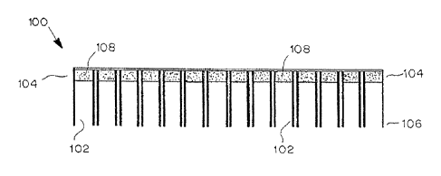
[0047] Referring to the figures, an exemplary device is shown in FIG. 1. In
this
variation, substance delivery device (100) includes a plurality of chambers
(102) having a
proximal end (104) and a distal end (106). A reservoir (108) lies within each
chamber (102).
13
CA 2964564 2017-04-13
Although the reservoir (108) is located at the proximal end (104) of the
chamber (102), other
configurations are contemplated. For example, as illustrated in FIG. 2, the
chambers (202) of
device (200) include elongate reservoirs (204) that extend from the proximal
end (206) of the
chambers (202). The chambers may be configured to penetrate tissue, e.g., by
employing a
sharp or needle-like distal end. The reservoirs or portions thereof, may also
be similarly
configured as a penetrating member, with a sharpened distal end, in a near-
linear and/or
curved geometry. These penetrating members may be configured to be deployed
during use,
such as being advanced after the substance delivery device has been attached
to the patient's
skin or after it has been implanted within the patient. Alternatively or
additionally, the
penetrating members may be configured to be advanced or retracted at any time,
such as prior
to use, just prior to explantation, or after explantation. The advancement or
retraction of the
penetrating members may be image guided, such as real-time guidance or
guidance based on
an image taken previously. The penetrating members may be hollow or include
one or more
lumens, and include stiffening means to aid in advancement or retraction, such
as to prevent
buckling during advancement. In a preferred embodiment, the stiffening means
comprises a
dissolvable biocompatible substance, not shown but preferably an inert
compound such as
salt which is dissolved shortly after the device is implanted. Alternatively,
a removable
mandrel may be included within the penetrating member to provide stiffness.
[0048] The penetrating members may be driven by one or more linear actuators,
such
as hydraulic or pneumatic pistons, magnetic drives, lead screw drives, thermal
expansion or
contraction assemblies, and other linear actuating assemblies configured to
advance or retract
the penetrating members in a continuous movement and/or in discrete steps.
These
penetrating members may be advanced or retracted on demand by a user such as a
clinician or
the patient, or may automatically advance or retract. The substance delivery
device may
include a sensor, such as a sensor on or near the penetrating member, to
detect and/or
measure the movement of the penetrating member. The distal ends of the three
or more
penetrating members may lie in a single plane or multiple planes. In a
preferred method, the
distal ends of the penetrating members reside, with or without deployment, in
an area or
volume with a substantially constant width, thickness or diameter, such as in
an area defined
by the long axis of a tumor. After delivery of one or more agents, this
defined area or volume
is excised and analyzed. A comparison of efficacy or other tissue response is
performed
based on the independent delivery of two or more agents to the defined area.
Tissue excision
is performed at a time related to efficacy or other agent-related time
parameter. In a preferred
14
CA 2964564 2017-04-13
embodiment, excision is performed two to seven days after initiation of agent
delivery. In
another preferred embodiment, excision is performed one week to one year after
initiation of
agent delivery.
[0049] The devices described here may also include one or more sensors for
sensing a
response parameter in vivo. Any type of sensor may be employed. For example,
chemical
sensors, mechanical sensors, optical sensors, radiation sensors, temperature
sensors, or a
combination of these sensors may be used. Nanosensors may be employed. The
response
parameter may be any parameter capable of being sensed or measured by the
sensor in the
target tissue, and which relates to an effect or response of the target tissue
to the substance.
The response parameters may include without limitation, levels of metabolites
or precursors;
levels of glucose, oxygen, or other nutrients; cytokine levels; pH; or
osmolality. In some
variations, the response parameters are structural in nature, and are obtained
through
visualization, e.g., via an optical sensor. For example, visualization of
cellular or
hiqtological/histopathological changes may be obtained. When devices that are
removed take
samples from the target tissue, further in vitro characterization of the
samples may occur. A
docking station or other device may be used to collect the tissue samples or
perform various
assays in further characterizing the samples.
[0050] The devices may also be configured to locally deliver an active agent
in
response to the response parameter. Exemplary active agents that may be
locally delivered
include, but are not limited to, anti-infective agents, anti-inflammatory
agents, anti-
proliferative agents, and chemotherapeutic/antineoplastic agents. Examples of
anti-infective
agents include, but are not limited to, antibacterial agents, antifungal
agents, antiparasitic
agents, antiviral agents, and antiseptics. Examples of anti-inflammatory
agents include
without limitation, steroidal and nonsteroidal anti-inflammatory agents.
[0051] Examples of antibacterial agents that may be locally delivered include,
but are
not limited to, aminoglycosides, amphenicols, ansamycins, 0-lactams,
lincosamides,
macrolides, nitroftwans, quinolones, sulfonamides, sulfones, tetracyclines,
vancomycin, and
any of their derivatives, or combinations thereof. In one variation, P-lactams
are the active
agents.
[0052] The 13-lactams that may be used include, but are not limited to,
carbacephems,
carbapenems, cephalosporins, cephamycins, monobactams, oxacephems,
penicillins, and any
CA 2964564 2017-04-13
of their derivatives. In one variation, penicillins (and their corresponding
salts) are the active
agents.
[0053] The penicillins that may be locally delivered by the devices described
here
include, but are not limited to, amdinocillin, amdinocillin pivoxil,
amoxicillin, ampicillin,
apalcillin, aspoxicillin, azidocillin, azlocillin, bacampicillin,
benzylpenicillinic acid,
benzylpenicillin sodium, carbenicillin, carindacillin, clometocillin,
cloxacillin, cyclacillin,
dicloxacillin, epicillin, fenbenicillin, floxacillin, hetacillin,
lenampicillin, metampicillin,
methicillin sodium, mezlocillin, nafcillin sodium, oxacillin, penamecillin,
penethamate
hydriodide, penicillin G benethamine, penicillin G benzathine, penicillin G
benzhydrylamine,
penicillin G calcium, penicillin G hydrabamine, penicillin G potassium,
penicillin G
procaine, penicillin N, penicillin 0, penicillin V, penicillin V benzathine,
penicillin V
hydrabamine, penimepicycline, phenethicillin potassium, piperacillin,
pivampicillin,
propicillin, quinacillin, sulbenicillin, sultamicillin, talampicillin,
temocillin, and ticarcillin.
Penicillins combined with clavulanic acid such as Augmentin (amoxicillin and
clavulanic
acid) may also be used.
[0054] Examples of antifungal agents suitable for local delivery include, but
are not
limited to, allylamines, imidazoles, polyenes, thiocarbamates, triazoles, and
any of their
derivatives. In one variation, imidazoles are the preferred antifungal agents.
Antiparasitic
agents that may be employed include such agents as atovaquone, clindamycin,
dapsone,
iodoquinol, metronidazole, pentamidine, primaquine, pyrimethamine,
sulfadiazine,
trimethoprim/sulfamethoxazole, trimetrexate, and combinations thereof.
[0055] Examples of antiviral agents suitable for local delivery include, but
are not
limited to, acyclovir, famciclovir, valacyclovir, edoxudine, ganciclovir,
foscamet, cidovir
(vistide), vitrasert, formivirsen, HPMPA (9-(3-hydroxy-2-
phosphonomethoxypropyl)adenine), PMEA (9-(2-phosphonomethoxyethyl)adenine),
HPMPG (9-(3-Hydroxy-2-(Phosphonomet- -hoxy)propyl)guanine), PMEG (9-[2-
(phosphonomethoxy)ethyl]guanine), HPMPC (1-(2-phosphonomethoxy-3-
hydroxypropy1)-
cytosine), ribavirin, EICAR (5-ethyny1-1-beta-D-ribofuranosylimidazole-4-
carboxamine),
pyrazofurin (3-[beta-D-ribofuranosy1]-4-hydroxypyrazole-5-carboxamine), 3-
Deazaguanine,
GR-92938X (1-beta-D-ribofuranosylpyrazole-3,4-dicarboxami- -de), LY253963
(1,3,4-
thiadiazol-2-yl-cyanamide), RD3-0028 (1,4-dihydro-2,3-Benzodithiin), CL387626
(4,4'-
bis[4,6-d][3-aminophenyl-N- -,N-bis(2-carbamoylethyl)-sulfonilimino]-1,3,5-
triazin-2-
16
CA 2964564 2017-04-13
ylamino-biphenyl-- 2-,2'-disulfonic acid disodium salt), BABIM (Bis[5-Amidino-
2-
benzimidazoly- 11-methane), NIH351, and combinations thereof.
[0056] Antiseptic agents that may be locally delivered include, but are not
limited to,
alcohol, chlorhexidrine, iodine, triclosan, hexachlorophene, and silver-based
agents( e.g.,
silver chloride, silver oxide, silver nanoparticles).
[0057] The devices may also locally deliver an anti-inflammatory agent such as
a
steroidal anti-inflammatory agent (corticosteroid). Exemplary steroidal anti-
inflammatory
agents include, but are not limited to, 21-acetoxypregnenolone, alclometasone,
algestone,
amcinonide, beclomethasone, betamethasone, budesonide, chloroprednisone,
clobetasol,
clobetasone, clocortolone, cloprednol, corticosterone, cortisone, cortivazol,
deflazacort,
desonide, desoximetasone, dexamethasone, diflorasone, diflucortolone,
difluprednate,
enoxolone, fluazacort, flucloronide, flumethasone, flunisolide, fluocinolone
acetonide,
fluocinonide, fluocortin butyl, fluocortolone, fluorometholone, fluperolone
acetate,
fluprednidene acetate, fluprednisolone, flurandrenolide, fluticasone
propionate, formocortal,
halcinonide, halobetasol propionate, halometasone, halopredone acetate,
hydrocortamate,
hydrocortisone, loteprednol etabonate, mazipredone, medrysone, meprednisone,
methylprednisolone, mometasone furoate, paramethasone, prednicarbate,
prednisolone,
prednisolone 25-diethylamino-acetate, prednisolone sodium phosphate,
prednisone,
prednival, prednylidene, rimexolone, tixocortol, triamcinolone, triamcinolone
acetonide,
triamcinolone benetonide, triamcinolone hexacetonide, any of their
derivatives, and
combinations thereof.
[0058] In some variations, a nonsteroidal anti-inflammatory agent is locally
delivered.
For example, nonsteroidal anti-inflammatory agents that may be used include,
but are not
limited to, COX inhibitors (COX-1 or COX nonspecific inhibitors) (e.g.,
salicylic acid
derivatives, aspirin, sodium salicylate, choline magnesium trisalicylate,
salsalate, diflunisal,
sulfasalazine and olsalazine; para-aminophenol derivatives such as
acetaminophen; indole
and indene acetic acids such as indomethacin and sulindac; heteroaryl acetic
acids such as
tolmetin, dicofenac and ketorolac; arylpropionic acids such as ibuprofen,
naproxen,
flurbiprofen, ketoprofen, fenoprofen and oxaprozin; anthranilic acids
(fenamates) such as
mefenamic acid and meloxicam; enolic acids such as the oxicams (piroxicam,
meloxicam)
and alkanones such as nabumetone) and selective COX-2 inhibitors (e.g., diaryl-
substituted
17
CA 2964564 2017-04-13
furanones such as rofecoxib; diaryl-substituted pyrazoles such as celecoxib;
indole acetic
acids such as etodolac and sulfonanilides such as nimesulide).
[0059] In other variations, chemotherapeutic/antineoplastic agents are locally
delivered. For example, chemotherapeutic/antineoplastic agents that may be
delivered by the
devices described here include, but are not limited to, antitumor agents
(e.g., cancer
chemotherapeutic agents, biological response modifiers, vascularization
inhibitors, hormone
receptor blockers, cryotherapeutic agents or other agents that destroy or
inhibit neoplasia or
tumorigenesis) such as alkylating agents or other agents which directly kill
cancer cells by
attacking their DNA (e.g., cyclophosphamide, isophosphamide), nitrosoureas or
other agents
which kill cancer cells by inhibiting changes necessary for cellular DNA
repair (e.g.,
carmustine (BCNU) and lomustine (CCNU)), antimetabolites and other agents that
block
cancer cell growth by interfering with certain cell functions, usually DNA
synthesis (e.g., 6-
mercaptopurine and 5-fluorouracil (5FU), antitumor antibiotics and other
compounds that act
by binding or intercalating DNA and preventing RNA synthesis (e.g.,
doxorubicin,
daunorubicin, epirubicin, idarubicin, mitomycin-C and bleomycin) plant (vinca)
alkaloids and
other anti-tumor agents derived from plants (e.g., vincristine and
vinblastine), steroid
hormones, hormone inhibitors, hormone receptor antagonists and other agents
which affect
the growth of hormone-responsive cancers (e.g., tamoxifen, herceptin,
aromatase inhibitors
such as aminoglutethamide and formestane, triazole inhibitors such as
letrozole and
anastrazole, steroidal inhibitors such as exemestane), antiangiogenic
proteins, small
molecules, gene therapies and/or other agents that inhibit angiogenesis or
vascularization of
tumors (e.g., meth-1, meth-2, thalidomide), bevacizumab (Avastin), squalamine,
endostatin,
angiostatin, Angiozyme, AE-941 (Neovastat), CC-5013 (Revimid), medi-522
(Vitaxin), 2-
methoxyestradiol (2ME2, Panzem), carboxyamidotriazole (CAI), combretastatin A4
prodrug
(CA4P), SU6668, SU11248, BMS-275291, COL-3, EMD 121974, IMC-1C11, IM862, TNP-
470, celecoxib (Celebrex), rofecoxib (Vioxx), interferon alpha, interleukin-12
(IL-12),
biological response modifiers (e.g., interferon, bacillus calmette-guerin
(BCG), monoclonal
antibodies, interleukin 2, granulocyte colony stimulating factor (GCSF),
etc.), PGDF receptor
antagonists, herceptin, asparaginase, busulphan, carboplatin, cisplatin,
carmustine,
cchlorambucil, cytarabine, dacarbazine, etoposide, flucarbazine, flurouracil,
gemcitabine,
hydroxyurea, ifosphamide, irinotecan, lomustine, melphalan, mercaptopurine,
methotrexate,
thioguanine, thiotepa, tomudex, topotecan, treosulfan, vinblastine,
vincristine, mitoazitrone,
18
CA 2964564 2017-04-13
oxaliplatin, procarbazine, streptocin, taxol or paclitaxel, taxotere,
analogs/congeners,
derivatives of such compounds, and combinations thereof.
[0060] In some variations, a closed feedback loop is generated that is
dependent on
the sensed response parameter. For example, when a device is used to locally
deliver a
chemotherapeutic agent to treat a malignant tumor, or to determine the optimal
agent or agent
combination to use for chemotherapy, decreased or increased levels of the
chemotherapeutic
agent may be released from the device based on the amount of tumor cell
apoptosis that is
sensed. Similarly, when a device is used to locally deliver an anti-
inflammatory agent to treat
inflammation, decreased or increased levels of the anti-inflammatory agent may
be released
from the device based on the level of cytokines sensed in the target tissue.
In other
variations, the sensed response parameter is linked to systemic
administration, e.g.,
intravenous administration, of an active agent.
[0061] The devices described here may also include elements that aid its
identification or detection by imaging modalities. With respect to detection,
the devices may
have a radiopaque or fluorescent marker. A radiofrequency tag may be used for
identification purposes. In some variations, the devices include a visual
indicator for
indicating upon explants whether a particular effect or response has occurred
in the target
tissue. The visual indicator may be a color change of all or a portion of the
device.
[0062] The substance delivery devices so far described generally include one
or more
chambers and a reservoir within each chamber. The reservoirs may contain a
substance for
delivery to a target tissue. Taking this general structure, devices having a
particular
functionality or application, as further elaborated below, may be designed.
For example, the
substance delivery devices may be constructed to include microchips, biopsy
mechanisms, or
various assay components. Devices with biopsy mechanisms may be suitable for
implantation, while devices including microchips or assay components may be
suitable for
either implantation, ingestion, or topical application. As shown in FIG. 3,
the substance
delivery device (300) includes a microchip (302) coupled to the proximal end
(304) of
chambers (306). When an assay component is employed, it may also be coupled to
the
proximal end of the chambers.
19
CA 2964564 2017-04-13
Microchip Devices
100631 In one variation, the substance delivery device includes a microchip.
The
microchip may be used to locally deliver the substance to a target tissue,
control substance
delivery, etc. For example, each of the reservoirs of a microchip may be
loaded with
different substances and/or different microdose amounts of the substances,
which can be
released independently. Release from a microchip device may be controlled by a
preprogrammed microprocessor, remote control, or by sensors.
[0064] Instead of chambers, the microchip device may include a plurality of
reservoirs that are etched into or otherwise formed in a biocompatible
substrate, which are
filled with a substance(s). Release of the substance from each reservoir may
be separately
controlled, for example, by a barrier membrane or other controllable member
that
controllably effects release of the substance from the reservoir. Reservoirs
may be filled with
different drugs, and the reservoirs can be capped with materials that either
degrade or allow
the drugs to diffuse passively out of the reservoir over time. The capping
material may be
structured such that upon exposure to an energy source, it erodes quickly,
changes
permeability or otherwise responds to a signal to release the substance. The
sites and times
of this substance release may then be controlled by a remote controller, by an
integrally
implanted programmed microprocessor, by an implanted but externally
programmable unit,
or other effective arrangement.
[0065] The microchip devices and other devices of the present invention may
also
include a pump such as a microfluidic pump, an osmotic pump, or a
microelectronic pump.
In general, the pump assembly, or multiple pump assemblies, will deliver a
carrier fluid to a
fluid outlet, and a fluid delivery pathway will extend from the outlet past a
reservoir to a
distal ported outlet, which is implanted at a target tissue site. In this
configuration, the
reservoir, positioned in or in communication with the fluid delivery pathway,
releases a
substance into the carrier fluid, which is delivered by the pump assembly at a
rate effective to
establish a local pressure gradient in the region of the ported outlet at the
target tissue site, so
that the substance is delivered into the tissue at the target tissue site. The
carrier may be, e.g.,
a biologically inert or inactive fluid such as physiologic saline, or it may
be an endogenous
body fluid. In an alternative embodiment, multiple pump assemblies deliver
carrier fluid to
one or more fluid conduits and fluid outlets, such as to deliver different
types of carrier fluids
for combination with different agents released by different reservoirs into
the fluid conduits
CA 2964564 2017-04-13
or outlets. Each pump assembly is preferably independently controllable, to
allow
independent control of each agent's delivery rates, time of infusion and
amount of infusion.
[00661 When the reservoir is a pressurized assembly, such as a pressure-driven
bellows, the pump assembly may work by simply providing one or more valves,
restrictors or
other elements that regulate the time and/or the rate at which the substance
is allowed to pass
from the reservoir. Alternatively, the pump may be an electrically powered
assembly, having
a power source and a controller.
[00671 The precise pump structure may include any suitable structure as known
in the
art, either with an electromechanically-actuated peristaltic or displacement
pumping
mechanism, or with a pressurized reservoir or osmotically-driven source
connected to a
control valve or restrictor assembly to regulate the provision of fluid into
the fluid delivery
path. In either case, whether powered by pressure or electromechanically, the
pump
assembly will be configured to produce an accurate and sustainable flow of a
total volume of
fluid at a suitable flow rate.
[0068] The control circuitry may consist of a timer, a demultiplexer, a
microprocessor, and an input source, for example, a memory source, a signal
receiver, or a
biosensor. The timer and demultiplexer circuitry may be designed and
incorporated directly
onto the surface of the microchip during electrode fabrication. The
microprocessor will
generally be of small size, have a low power requirement, and have the ability
to translate the
output from memory sources, signal receivers, or biosensors into an address
for the direction
of power through the demultiplexer to a specific reservoir on the substance
delivery device.
Selection of a source of input to the microprocessor such as memory sources,
signal
receivers, or biosensors depends on the particular application of the delivery
device and
whether the device operation is preprogrammed, controlled by remote means, or
controlled
by feedback from its environment.
[00691 The criteria for selection of a power source for a microchip may be
small size,
sufficient power capacity, ability to be integrated into the control
circuitry, the ability to be
recharged, and the length of time before recharging is necessary. Several
lithium-based,
rechargeable microbatteries have been described by S. D. Jones and J. R.
Akridge,
"Development and performance of a rechargeable thin-film solid-state
microbattery", Journal
of Power Sources, 54:63 67 (1995); and J. B. Bates et al., "New amorphous thin-
film lithium
21
CA 2964564 2017-04-13
electrolyte and rechargeable microbattery", IEEE 35th International Power
Sources Symposium,
337 39 (1992). These batteries are typically only about 10 [im thick and
occupy about 1 em2 of
area. One or more of these batteries may be incorporated directly onto the
microchip device.
[0070] Referring now to FIG. 5, a substance delivery device of the present
invention is
illustrated. Device 500 includes housing 505, which surrounds an electronic
controller,
microcontroller 501 which is electrically attached to wireless transceiver 503
and a power
supply, battery 502. Device 500 is configured for placement near target
tissue, such as via
adhesive attachment of a portion of housing 505 to the patient's skin, or via
implantation within
the patient such as by using suture and suture loops 504 to fixate housing 505
to tissue proximate
the target tissue. Device 500 includes a series of chambers, including chamber
520a. Each
chamber includes and slidingly receives a movable reservoir, such as
reservoirs 550a, 550b, 550c
and 550d. Each reservoir is configured to contain one or more agents, as are
described and listed
in detail throughout the application, and deliver these one or more agents to
the target tissue. The
target tissue can be any location in the patient's body, such as organ tissue
and tumor tissue. In a
preferred method, the target tissue includes both tumor and healthy tissue,
such as when
substance is delivered into, and/or tests are performed on, both tumor and
healthy tissue, such as
to include a control. A typical embodiment of a reservoir is described in
reference to FIGs. 6a
and 6b below. Chamber 520a and reservoir 550a are sized and constructed such
as to form a seal
such that gas pressure created within chamber 520a causes reservoir 550a to
advance. A sealing
component, not shown but preferably an 0-ring, can be included between
reservoir 550a and
chamber 520a to form the seal. Reservoir 550a is attached to microcontroller
501 via a spiral
wire 551a. Spiral wire 551a is configured to accommodate the advancement of
reservoir 550a,
such as the advancement of reservoir 550b and 550c shown in FIG. 5. Reservoir
550a includes at
its distal end tip 555a, preferably of an anti-coring needle configuration
with a lumen configured
to deliver the one or more agents contained in reservoir 550a, such as through
a hole in the distal
end of the tip or from side holes located along the side of the tip, not shown
but described in
detail in reference to FIGS. 6a and 6b below. Cylinder 522a, contained within
chamber 520a, is
preferably a gas delivering element such as a gas generator assembly, or a
compressed gas vessel
controlled by one or more valves.
22
CA 2964564 2017-04-13
[0071] Cylinder 522a is electrically connected to microcontroller 501 such
that a precise
amount of gas can be released by cylinder 521 to specifically advance
reservoir 550a and tip
555a into the target tissue. One or more sensors, not shown but preferably an
optical or magnetic
sensor, can be placed to detect and/or quantify the motion of cylinder 550a
and provide closed
loop motion information to microcontroller 501. While the chambers and
reservoirs are shown in
a linear configuration, numerous two and three dimensional arrangements of
chambers can be
employed, such as a ten by ten square array of chambers and reservoirs. While
each reservoir of
FIG. 5 is advanced by increasing the pressure in the associated chamber, other
linear actuators
can be employed. In a preferred embodiment, a lead screw is driven by a
rotational motor, both
not shown, wherein the reservoir is rotationally attached to the lead screw
and the reservoir can
be advanced or retracted by associated forward and reverse rotations of the
motor. Advancement
and retraction of the reservoir can be performed prior to, during, and/or
after skin attachment or
implantation of the substance delivery device of the present invention. The
fluid delivery and
other moving components of the substance delivery devices of the present
invention may be
constructed using semiconductor-like machinery, such as with
microelectromechanical system
(MEMS) construction. MEMS assemblies and components include motors, valves,
actuators and
other electromechanical components that can be produced at extremely small
dimensions, such
as the dimensions that are preferred for the components and devices of the
present invention.
[0072] Referring now to FIGS. 6a and 6b, a preferred embodiment of a reservoir
of the
present invention is illustrated. Reservoir 550 includes one or more attached
wires, not shown
but preferably for connection to one or more electronic circuits, such as
microcontroller 501 of
FIG. 5. Motor 552, a rotational motor such as a stepper motor including one or
more gear
reducing assemblies, is connected to lead screw 554 such that rotation of
motor 552 causes lead
screw 554 to rotate. Motor 552 may include one or more rotational sensors such
as optical
encoders or Hall effect motion sensors. Plunger 553 is rotatingly attached to
threads of lead
screw 554, not shown but of a constant or otherwise thread pitch such that the
linear
advancement of plunger 553 can be calculated based on known angle of rotation
of motor 552.
Plunger 553 forms a seal against the walls 558 of reservoir 550 such that
linear advancement of
plunger 553 causes a specific amount of agent 10 to be delivered to target
tissue. Plunger 553
may have an eccentric cross-section, or include one or more notches that mate
with walls 558
such as to prevent rotation of plunger 553. The distal end of reservoir 550
includes a sharpened
distal tip configured to penetrate
23
CA 2964564 2017-04-13
tissue, and outlet ports 557a, 557b and 557c, all fluidly connected to the
agent 10 contained
within the walls 558 of reservoir 550. Reservoir 550 may include varied
placement of one or
more fluid delivery outlet ports, and the outlet ports may have different
geometries and/or
cross-sectional areas. For example, a first outlet port distal to a second
outlet port may have a
bigger cross-sectional area such as to cause the same amount of agent to be
delivered through
each outlet port. The outlet ports may be equally spaced and/or the ports may
be oriented in a
spiral pattern.
[0073] Retraction of plunger 553 is caused by rotation of the motor in the
opposite
direction to that causing advancement. Retraction of plunger 553 can be used
to extract fluid
from the patient into reservoir 550. Such extraction may occur when reservoir
550 is void of
agent 10, such as after plunger 553 has been fully advanced or when plunger
550 is provided
in the fully advanced position (i.e. no agent included). The extraction can be
used to
withdraw one or more body fluids including but not limited to: blood;
lymphatic fluid; urine;
semen; cerebral spinal fluid; interstitial fluid; and combinations of these.
[0074] Reservoir 550 includes a first sensor, volume detector 523 which is
integrated
into reservoir 550 behind plunger 553, and attaches to electronic circuitry,
not shown but
preferably similar to microcontroller 501 of FIG. 5. Advancement or retraction
of plunger
553 can be confirmed and/or quantified by the change in volume behind plunger
553. In a
preferred embodiment, volume detector 523 includes circuitry to produce, or
otherwise is
provided (e.g. from the microcontroller) a range of frequencies which are
converted to sound
by a speaker of volume detector 523. A microphone of volume detector 523
records the
sounds created within the confined space or cavity around volume detector 523
such that a
resonant frequency can be detected. This resonant frequency, the Helmholtz
resonance, is
proportional to the volume of the confined space.
[0075] Reservoir 550 further includes, at or near its distal tip, sensor 556
configured
to be advanced into the target tissue as reservoir 550 penetrates the target
tissue, such as been
described above in reference to FIG. 5. Sensor 556 may be configured to detect
motion, such
as an optical detector configured to detect and/or quantify the motion of
reservoir 550.
Alternatively or additionally, sensor 556 may be a sensor such as a strain
gauge, an
accelerometer, a temperature sensor, a pH sensor, a chemical sensor, a
mechanical sensor, a
radiation sensor and/or a physiologic sensor. Numerous physiologic sensors can
be
employed such as those configured to assess one or more cell activities.
24
CA 2964564 2017-04-13
Biopsy devices
[0076] In another variation, the substance delivery device may be configured
to
obtain a sample upon its removal from the target tissue. The sample may be
cells or portions
of tissue from any organ (e.g., liver) or target tissue (e.g., a cancer or
tumor) at the target site.
The sample may also be a body fluid sample such as serum, blood, blood cells
(e.g., white
cells), plasma, sputum, urine, peritoneal fluid, pleural fluid, cerebrospinal
fluid, or lymphatic
fluid. As mentioned above, the samples may be obtained using a sampling port.
[00771 The devices may obtain samples using any type of biopsy mechanism. In
one
variation, as shown in FIG. 4B, device (400) has a biopsy mechanism that
causes the distal
end (402) of the chambers (404) to move from an open position (FIG. 4A) to a
closed
position (FIG. 4B). Such a biopsy mechanism may include hinges, springs, shape
memory
elements, wires, or combinations thereof. The biopsy mechanism may be
activated by an
external controller, pressure changes, temperature changes, etc., or be
automatically activated
after a predetermined period of time. Furthermore, the biopsy mechanism may
include a
chamber having a cutting edge at its distal end.
[0078] Upon closure of the distal ends (402) of the chambers (404), a sample,
e.g.,
tissue (406) may be retained within the chambers (404). Tissue (406) may then
be subjected
to various in vitro assays for evaluation of response parameters. Suitable in
vitro assays are
well known in the art. For example, binding assays, spectrophotometry, gel
electrophoresis,
chromatography, etc., may be performed using the samples.
Assay Devices
[0079] In other variations, the devices include an assay component (see, e.g.,
FIG. 3,
element 302) for evaluating a sample obtained from the target tissue in vivo.
The assay may
incorporate one or more sensors, as previously described, or other elements
capable of
analyzing the sample. For example, the assay may include elements capable of
detecting
levels of antibodies, serum proteins, enzymes, viral or bacterial proteins,
cholesterol, glucose,
polysaccharides, nucleic acids, metabolites, cytokines, tumor antigens or
cancer markers, and
apoptosis. In some variations, the assay devices are configured to deliver an
active agent in
response to the data obtained from the assay. The active agents that may be
delivered are the
same as those previously described above. However, specific examples of what
the assay
CA 2964564 2017-04-13
component may detect and analyze, and the corresponding active agents that may
delivered in
response to the assay data is further provided below.
[0080] In some variations, the assay devices are used to monitor or locally
treat
various types of tumors and cancers. Here the assay component may be
configured to detect
and analyze genes or their products which are over-expressed or over-active in
cells
undergoing unwanted proliferation. For example, the assay device may be
implanted into a
tumor or a tissue suspected of containing a tumor such as a cavity or space
left behind
following a biopsy procedure. If the assay component detects increased
concentrations of
such biological analytes or mutated over-active forms of such analytes (both
disease
markers), then the assay device may be configured to release an active agent
such as a
cytotoxic agent. Similarly, a cytotoxic agent may be released in response to
analytes
corresponding to neointimal proliferation, among other pathologic conditions.
[0081] In other variations, the biological analytes are tumor specific
antigens, which
may be expressed on the surface of or released from cancer cells, for example
the tumor
specific antigen MUC-1. Here the assay device may be configured to release a
cytotoxic
agent in response to the detection of MUC-1.
[0082] In yet other variations, the assay component detects the presence of
receptor
tyrosine kinases (RTKs) in the sample. These receptors are frequently present
in common
human cancers such as breast cancer; squamous cell cancer of the lung; bladder
cancer;
esophageal cancer; gastrointestinal cancer such as colon, rectal or stomach
cancer; leukemia;
ovarian cancer; bronchial cancer; and pancreatic cancer. Accordingly,
detection of
abnormally high levels of RTK expression or signaling activity through nucleic
acid detection
or by protein activity can constitute a disease marker and can warrant the
release of RTK
inhibitors or cytotoxic agents as active agents.
[0083] In further variations, the assay component is configured to detect
analytes that
may be indicative of inflammation, such as TNF-alpha, IL-1, 1L-8, IL-2, IL-3,
IL-4, GM-
CSF, INF-gamma, MIF, and TNF-beta. The detection of abnormally high
concentrations of
such analytes may trigger the localized release of anti-inflammatory drugs or
antibodies as
active agents.
[0084] In yet further variations, the assay component is configured to detect
analytes
that may be indicative of infection by a microorganism. Here the analytes may
include viral
26
CA 2964564 2017-04-13
=
or bacterial proteins or nucleic acids or fragments thereof. For example,
detection of analytes
such as bacterial toxins including exotoxins and enterotoxins as well as TSST-
1, or other
bacterial superantigen, or botulinurn toxin, diphtheria toxin, anthrax
protective antigen,
anthrax edema factor, and anthrax lethal factor, etc., as well as viral
proteins such as
influenza hemagglutinin or neuramimidase, may indicate an infection and might
trigger
localized release of an anti-infective agent or a toxin-specific antibody as
active agents.
[0085] In another variation, the assay component is configured to detect
abnormal
cellular proliferation, and coordinate localized release of an active agent
that has an anti-
proliferative effect. For example, sirolimus (rapamycin) or paclitaxel, which
are effective in
inhibiting smooth muscle cell proliferation during neointimal hyperplasia, may
be released.
In yet another variation, 5-FU chemotherapy is locally released from the assay
device in
response to detected analytes associated with abnormal cellular proliferation.
5-FU-based
chemotherapy may comprise administration of 5-FU, its derivatives, alone or
with other
chemotherapeutics, such as leucovorin or with a DPD inhibitor such as uracil,
ethynyluracil, bromovinyluracil, thymine, benzyloxybenzyluracil (BBU) or 5-
chloro-2,4-
dihydroxypyridine.
[0086] Alternatively, genotoxic agents such as DNA alkylating agents and DNA
intercalating agents may be delivered. For example, psoralens, antineoplastic
antibiotics,
which include, but are not limited to, amsacrine; actinomycin A, C, D
(alternatively known as
dactinomycin) or F (alternatively KS4); azaserine; bleomycin; carminomycin
(carubicin);
daunomycin (daunorubicin), or 14-hydroxydaunomycin (adriamycin or
doxorubicin);
mitomycin A, B or C; mitoxantrone; plicamycin (mithramycin); and the like, may
be
delivered. Another general class of genotoxic agents that may be locally
delivered, and
which alkylate DNA, are those that include the haloethylnitrosoureas or
chloroethylnitrosoureas. Representative members of this class include, but are
not limited to,
carmustine, chlorozotocin, fotemustine, lomustine, nimustine, ranimustine and
streptozotocin.
[0087] In some instances, platinum coordination compounds such as cisplatin or
oxaliplatin may be delivered alone or in combination in response to analytes
associated with
testicular, endometrial, cervical, gastric, squamous cell, adrenocortical, and
small cell lung
carcinomas, as well as medulloblastomas and neuroblastomas.
27
CA 2964564 2017-04-13
[00881 Yet another general class of genotoxic agents that may be locally
delivered in
response to a detected tumor analyte includes the sulfur and nitrogen
mustards. These
compounds damage DNA primarily by forming covalent adducts at the N7 atom of
guanine.
Representative members of this broad class include chlorambucil,
cyclophosphamide,
ifosfamide, melphalan, mechloroethamine, novembicin, and trofosfamide.
Oligonucleotides
or analogs thereof that interact covalently or noncovalently with specific
sequences in the
genome of selected cells may also be used as genotoxic agents, if it is
desired to select one or
more predefined genomic targets as the locus of a genomic lesion.
[0089] In some variations, the assay component is configured to detect an
analyte
indicative of a microbial pathogen. In response, the device may be configured
to locally
release an active agent that has an antimicrobial effect. For example, an
antibiotic or antiviral
agent may be released.
[00901 In other variations, the assay component is configured to detect an
analyte
indicative of hyperglycemia and the assay device, in response, designed to
locally release an
active agent suitable to reduce serum glucose levels. For example, when
excessively high
levels of glucose are detected by the assay component, the assay device may
respond by
releasing a sufficient amount of insulin to noinialize the blood glucose
level.
Patch devices
The substance delivery devices may also be formed as a patch. The patch may be
useful
when topical application of the device to the skin is desired. In this
variation, the reservoir
containing the substance may be a layer underlying an upper backing layer. The
patch may
contain a single reservoir, or it may contain multiple reservoirs. When
multiple reservoirs are
employed, they may include the same substance or different substances, or each
reservoir
may include a combination of substances. The patches may also be configured to
include a
component that modifies delivery of a substance therefrom. For example, a rate-
limiting
membrane may be placed between the reservoirs to modify release of the
substance.
Representative materials useful for forming rate-controlling membranes include
polyolefins
such as polyethylene and polypropylene, polyamides, polyesters, ethylene-
ethacrylate
copolymer, ethylene-vinyl acetate copolymer, ethylene-vinyl methylacetate
copolymer,
ethylene-vinyl ethylacetate copolymer, ethylene-vinyl propylacetate copolymer,
polyisoprene, polyacrylonitrile, ethylene-propylene copolymer, and the like.
28
CA 2964564 2017-04-13
[0091] In some variations, the reservoirs may comprise a polymeric matrix of a
pharmaceutically acceptable adhesive material that serves to affix the patch
to the skin. For
example, the adhesive material may be a pressure-sensitive adhesive (PSA)
including, but not
limited to, polyethylenes; polysiloxanes; polyisobutylenes; polyacrylates;
polyacrylamides;
polyurethanes; plasticized ethylene-vinyl acetate copolymers; and tacky
rubbers such as
polyisobutene, polybutadiene, polystyrene-isoprene copolymers, polystyrene-
butadiene
copolymers, and neoprene (polychloroprene).
[0092] The backing layer may function as the primary structural element of the
patch
and may provide the device with flexibility and in certain variations,
occlusivity. The
backing may be comprised of a flexible elastomeric material that serves as a
protective
covering to prevent loss of the substance via transmission through the upper
surface of the
patch, and may impart a degree of occlusivity to the patch, such that the area
of the body
surface covered by the patch becomes hydrated during use. The material used
for the backing
layer may permit the patch to follow the contours of the skin and be worn
comfortably on
areas of skin such as at joints or other points of flexure that are normally
subjected to
mechanical strain, with little or no likelihood of the patch disengaging from
the skin due to
differences in the flexibility or resiliency of the skin and the patch. The
materials used as the
backing layer may be either occlusive or permeable, as noted above, and may be
made from
synthetic polymers (e.g., polyester, polyethylene, polypropylene,
polyurethane, polyvinyl
chloride, and polyether amide), natural polymers (e.g., cellulosic materials),
or macroporous
woven and nonwoven materials. In other variations, a microchip and/or assay
component
may be provided in lieu of a backing layer. Alternatively or additionally, the
adhesive may
be applied to the skin-contacting surface of the patch in discrete areas, such
as to allow
certain portion of the patient's skin under the patch to remain unattached
(i.e. the areas void
of adhesive). This discontinuous configuration may avoid undesired cosmesis
and other skin
effects, as well as accommodate skin motion thus avoiding undesired detachment
of the
patch. In an alternative embodiment, the patch is flexible, or includes one or
more hinge
portions configured to permit motion without detachment of the patch.
[0093] When a layered patch device is used, samples may be obtained via
microneedles that are fixed or removably secured to the skin-contacting layer
of the patch.
The microneedles may be about the size of a human hair and have an integrated
microreservoir. The microneedle will be generally configured to painlessly
penetrate the
29
CA 2964564 2017-04-13
skin. The microneedles may be constructed out of silicon or other suitable
metals and
polymers, and may be about 10 pm to about 200 t.un, about 5011M to about 150
pm, or about
100 p.m in diameter. In some instances, the microneedle will be designed to
obtain blood
samples. Here the microneedles may collect about 0.01 to about 1.0 microliter,
about 0.05 to
about 0.5 microliters, or about 0.1 to about 0.3 microliters of capillary
blood. The
microneedles may be deployable, injectable through the skin after placement of
the patch on
the patient's skin. The microneedles may be deployable simultaneously, or in
subsets of the
entire group, and may be deployed on demand, at a particular time event,
and/or when an
analysis, such as an assay analysis, produces specific results. The tips of
the microneedles
may be arranged in a single plane, or may lie in multiple planes. One or more
of the
microneedles may be straight or curved. The diameter of the microneedles may
be consistent
or may vary. The microneedles may be deployed into tumor tissue, such as skin
cancer or
other cancer close to the epidermal layer of the patient's skin.
[0094] In general, microfabrication processes that may be used in making the
microneedles disclosed herein include lithography; etching techniques, such as
wet chemical,
dry, and photoresist removal; thermal oxidation of silicon; electroplating and
electroless
plating; diffusion processes, such as boron, phosphorus, arsenic, and antimony
diffusion; ion
implantation; film deposition, such as evaporation (filament, electron beam,
flash, and
shadowing and step coverage), sputtering, chemical vapor deposition (CVD),
epitaxy (vapor
phase, liquid phase, and molecular beam), electroplating, screen printing, and
lamination.
Alternatively, the needles may be molded in silicon wafers and then plated
using
conventional wire cutting techniques with nickel, gold, titanium or various
other
biocompatible metals. In another variation, the needles may be fashioned from
biopolymers.
Ingestible Devices
[0095] The ingestible device may be any one of the devices described above
that has
been modified for ingestion. Thus, the ingestible device may be a microchip or
assay device
having a coating or other component that protects it from premature
degradation and/or
contamination. The device or portions thereof may be made from ingredients
included in
conventional oral dosage forms. Such ingredients are known, or will be
apparent, to those
skilled in the art (see, e.g., Remington's Pharmaceutical Sciences, Mack
Publishing
Company, Easton, Pa., 17th edition, 1985; Remington: The Science and Practice
of
Pharmacy, A. R. Gen_naro, (2000) Lippincott, Williams & Wilkins).
CA 2964564 2017-04-13
Imaging Devices
[0096] The substance delivering device may also be used as an imaging device.
The
imaging device may be of any configuration described herein, so long as it
delivers a contrast
agent detectable by an imaging modality. Examples of imaging modalities
include x-ray, CT,
MRI, ultrasound, PET scan, fluoroscopy, and the like. Here the contrast agent
is used to
obtain structural or functional information about the target tissue. Contrast
agents that may
be employed include without limitation, barium sulfate, iodinated contrast
agents, water,
gadolinium, iron oxide, and fluorescent imaging agents. Exemplary iodinated
contrast agents
that may be delivered include, but are not limited to, diatrizoate,
metrizoate, ioxaglate,
iopamidol, iohexal, ioxilan, iopromide, and iodixanol. Contrast agents that
may be activated
to indicate the presence of particular analytes may also be employed. As
mentioned above, a
port may also be included or removably secured to the device for imaging
purposes.
II. SYSTEMS
[0097] In general, the systems will include a substance delivery device as
herein
described, e.g., a biopsy device, an assay device, a microchip device, an
ingestible device, a
patch device, an imaging device, or a combination thereof, and a deployment
tool for delivery
of the device to a target tissue. Any deployment tool may be used to deliver
the device. The
configuration of the deployment tool may depend on such factors as the type of
device used,
the route of delivery employed, and the particular target tissue. The
deployment tool may be
designed for delivering the device via any route. For example, the deployment
tool may
include features useful for delivery during open, laparoscopic, endoscopic,
arthroscopie,
percutaneous, and robotic procedures. In some variations, the deployment tool
comprises a
catheter/pusher assembly. In other variations, the deployment tool includes
jaws.
[0098] The systems may also include a retrieval tool for removing the device
from the
target tissue. These retrieval tools may be formed to include suction, jaws,
hooks, magnets,
etc. to aid removal. In some variations, an energy source is provided in the
systems to
activate release of the substance contained within the reservoirs. In other
variations, e.g.,
when an imaging device is part of the system, an imaging modality as
previously described
may be included in the system. Additional components, e.g., in vitro assays,
may also be
included to tailor the system.
31
CA 2964564 2017-04-13
[0099] Referring to FIG. 7, a substance delivery system of the present
invention is
illustrated. System 600 includes substance delivery devices 500a and 500b, of
similar
construction to delivery device 500 of FIG. 5, with the same reference numbers
used to refer to
the same or like parts. Devices 500a and 500b each include a microcontroller
which transmit
and receive data from wireless transceiver 503a and 503b respectively.
Wireless transceiver
503a and 503b are further configured to communicate with each other such that
information
obtained or produced from device 500a or 500b can be transmitted to the other.
In the
configuration of FIG. 7, devices 500a and 500b have been implanted below the
skin of the
patient at the site of a single tumor. In an alternative method, device 500a
may be implanted in a
first tumor, and device 500b may be implanted in a different tumor of the same
patient. In
another alternative method, device 500a may be implanted in a tumor and device
500b implanted
in healthy tissue, such as to act as a control. Device 500a and 500b further
include ingrowth
assembly 560a. Ingrowth assembly 560a comprises a vessel configured to allow
cells, such as
cancer cells, to migrate into the vessel. In a preferred embodiment, the
vessel is coated or
otherwise includes a biologic growth factor such as epidermal growth factor
(EGF). Numerous
growth factors can be included in or on ingrowth assembly 560a including but
not limited to
colony stimulating factor (CSF) and one or more cytokines such as SDF1-alpha.
Each ingrowth
assembly may receive the same or different growth factor than a different
ingrowth assembly.
Ingrowth assembly 560a may be removably attached to device 500a or may be a
separate
component, such as a separate implant assembly that include communication
means, such as
wired or wireless communication means to communicate information to one or
more separate
devices such as device 500a. Device 500a and/or device 500b (hereinafter
device 500a only for
simplicity) may include a second ingrowth assembly, such as to attract a
different type of cells.
Alternatively ingrowth assembly 560a may be configured to attract different
cells, such as where
one cell type is a housekeeping or control cell, used to confirm results or
compare to results
obtained from ingrowth assembly 560a, such as when ingrowth assembly 560a is
configured to
attract cancer cells.
[00100] The ingrowth assemblies may include an assay function or component, as
has
been described hereabove. Ingrowth assembly 560a of device 500a includes assay
component
561 configured to produce data based on the cells that migrate into assembly
560a. In a
preferred embodiment, assay component 561a performs an analysis of cancer
cells such as to
assess the invasiveness of a cancer. The data produced by assay component 561a
32
CA 2964564 2017-04-13
may be used by the microcontroller to start, stop or modify the delivery of
one or more agents
by device 500a, such as to maintain or increase delivery of a therapeutic
agent (e.g.
chemotherapeutic) with confirmed benefit and/or decrease or stop delivery of a
therapeutic
agent with no confirmed benefit.
[00101] Device 500a further includes access port 570, containing a
resealable
membrane, septum 571 configured to be repeatedly accessed with a sharp tool
such as a
needle while maintaining a seal. System 600 further includes a percutaneous
access tool,
syringe 610 shown accessing port 570 through septum 571. Access port 570 can
be used to
deliver one or more agents into ingrowth assembly 560a, and/or to remove
material, such as
the migrated cells, from ingrowth assembly 560a. Alternatively or
additionally, a similar
access port can be integrated into or attached to another part of device 500a
such as to add an
agent to a reservoir of device 500a, or otherwise add or remove a material
from device 500a.
[001021 Device 500a of system 600 further includes an electrode on
the distal
end of each reservoir. Reservoir 550a' includes electrode 580a', reservoir
550a" includes
electrode 580a", reservoir 550a" includes electrode 580a", and reservoir 550a"
" includes
electrode 580a". Each of the electrode 580a are configured to produce an
electrical field in
or about device 500a and/or the target tissue, a tumor as shown in FIG. 7,
such as by delivery
of current between one or more electrodes 580. Alternatively or additionally,
device 550b
may include one or more electrodes and current may be delivered between an
electrode of
device 500a and an electrode of device 500b. Alternatively or additionally,
one or more
electrodes may be included at a different location of reservoir 550a or device
500a, such as
on or in chamber 520a, or on housing 505a. The energy delivered by one or more
electrodes
580 may be based on the results of an analysis, such as an analysis performed
by an assay
component of device 500a. Electrodes 580 may be placed into the target tissue,
such as in the
tumor of FIG.7, due to reservoir 580" and 580" ' having been previously
advanced into the
tumor as has been described in reference to FIG. 5. The electric fields
generated by
electrodes 580 may be used to enhance delivery of drug through iontophoresis
and/or
electroporation, drug delivery enhancement means well known to those of skill
in the art.
The electric fields generated may be an additional or alternative way to
modify the actual
amount of agent delivered, such as when device 500a delivers fluid at a
continuous rate, but
modifies the electric field produced by electrodes 580 to enhance or diminish
fluid transfer
through iontophoresis, or cellular uptake with electroporation.
33
CA 2964564 2017-04-13
III. METHODS
1) Delivery and Retrieval
[00103] The devices described here may be delivered in any manner.
For
example, they may be delivered via an open surgical procedure, or by a
minimally invasive
procedure such as laparoscopy, endoscopy, arthroscopy, and catheter-based
procedures. The
devices may also be delivered percutaneously or topically. Delivery using a
robotic device is
also contemplated. Retrieval of the devices may occur via the same processes.
An image of
the target tissue, such as a tumor, may be performed prior to, during, or
after use of the
device. In a preferred embodiment, the device is applied to the patient's skin
or implanted in
the patient with image guidance. The devices of the present invention include
one or more
penetrating or advanceable members such as advanceable reservoirs. In a
preferred
embodiment, the penetrating or advanceable members penetrate or advance into
tissue using
image guidance, such as images created before or during penetration and/or
advancement.
[00104] In general, the device including a microdose of a substance
is delivered
to a target tissue, e.g., by implantation into the target tissue, such as to
perform a diagnostic
and/or therapeutic procedure. The substance is locally delivered in a
concentration adequate
to result in a pharmacological effect. A sample from the target tissue such as
a biopsy of the
tissue, is then obtained. In one variation, the sample is obtained by the
device that delivers
the substance. In another variation, the sample is obtained via procedures
such as
percutaneous biopsy, open biopsy, needle aspiration, and the like. The sample
tissue can then
be subjected to various assays for evaluating the substance effect on the
target tissue. For
example, the sample may be subjected to genomic, proteomic, biochemical,
and/or
histopathological characterization.
2) Dosing
[00105] The devices described here deliver a microdose amount of a
substance
to a target tissue. A microdose amount may be from about 0.001 1.ig (or less)
to about 1,000
pg , or about 10,000 ug (or more) of the substance. Those of skill will
readily appreciate that
microdose levels may vary as a function of the specific substance employed,
the target tissue,
and/or the medical condition being treated.
34
CA 2964564 2017-04-13
[00106] The substance may be delivered in a controlled release,
sustained
release, delayed release, or pulsatile fashion. Delivery may also occur over
any time period.
For example, it may occur over a period of minutes to hours, days to weeks,
weeks to
months, or even one year or more.
3) Medical Conditions and Target Tissues
[00107] In some instances, the devices described here may be used to
locally
deliver a substance that is a candidate compound being evaluated for the
treatment of a
medical condition. In other instances, the substance itself is locally
delivered to treat a
medical condition. Medical conditions that are contemplated, include, but are
not limited to,
autoimmune conditions, cancer, cardiac conditions, gastrointestinal
conditions, genitourinary
conditions, hematologic conditions, infectious conditions, inflammatory
conditions, ischemic
conditions, neurologic conditions, obstetric conditions, orthopedic
conditions, proliferative
conditions, pulmonary conditions, and vascular conditions.
[00108] Furthermore, the devices may be delivered to any target
tissue within
the human body. Target tissues may include the neurologic tissues, pulmonary
tissues,
gastrointestinal tissues, genitourinary tissues, cardiac tissues, vascular
tissues, muscle, bone,
skin, and any fluids such as blood or lymph.
IV. KITS
[00109] The kits may provide one or more devices described here,
e.g., one or
more biopsy devices, microchip devices, assay devices, ingestible devices,
patch devices, or
imaging devices. The devices may include reservoirs that deliver the same
substance or
different substances. Likewise, the devices may include reservoirs that
deliver the substances
in the same or different microdoses. Any number and type of deployment tools,
retrieval
tools, energy components, and imaging devices may also be included. The kits
may also
contain additional in vitro assays for evaluating samples.
[00110] The kits may further include instructions for using the
devices, tools,
and/or assays contained therein. These instructions may be present in the kits
in a variety of
forms, one or more of which may be present in the kit. One form in which these
instructions
may be present is as printed information on a suitable medium or substrate,
e.g., a piece or
pieces of paper on which the information is printed, in the packaging of the
kit, in a package
CA 2964564 2017-04-13
insert, etc. Yet another form would be a computer readable medium, e.g.,
diskette, CD, etc.,
on which the infonuation has been recorded. In some variations, a website
address may be
provided that can be accessed via the intemet to obtain the instructions.
36