Note: Descriptions are shown in the official language in which they were submitted.
SYSTEM AND METHOD FOR NON-INVASIVELY CONTROLLING AUTONOMIC
NERVE ACTIVITY
CORSS-REFERENCE TO RELATED APPLICATIONS
[0001] This application is based on, and claims priority to
US Provisional Application Serial No. 62/065,854 filed on October 20,
2014 and entitled "SYSTEM AND METHOD FOR NON-INVASIVELY MONITORING
AUTONOMIC NERVE ACTIVITY USING SKIN."
GOVERNMENT RIGHTS
[00921 This invention was made with government support under HL071140
awarded by
National Institutes of Health. The government has certain rights in the
invention.
BACKGROUND
[00031 The present disclosure relates generally to systems and methods
for monitoring
nerve activity and, in particular, to systems and methods for non-invasive
monitoring and/or
controlling nerve activity using cutaneous and/or subcutaneous electrodes.
[00041 Many diagnostic and treatment methods in the fields of medicine
and biology rely
on measurements of nerve activity in patients and test subjects. Nerve
activity in humans and
other animals generates electrical signals that are detectable by electronic
equipment such as
oscilloscopes and other electrical signal processing devices. In order to
detect the nerve activity,
one or more electrical conductors, or electrodes, are placed in proximity to
the nerves being
measured. The electrodes may receive the electrical signals for further
medical analysis. In
addition, various medical treatment methods also use electrodes to deliver
electrical signals to
the nerves in order to induce a response in the patient.
100051 Cardiac care is one particular area of medical treatment that
heavily utilizes
measurement of nerve activity. Activity in the autonomic nervous system
controls the variability
of heart rate and blood pressure. The sympathetic and parasympathetic branches
of the
autonomic nervous system modulate cardiac activity. Elevated levels of
sympathetic nerve
activity ("SNA") are known to be correlated with heart failure, coronary
artery disease, and may
- 1 -
Date Regue/Date Received 2022-02-15
CA 02965149 2017-04-19
WO 2016/064843 PCT/US2015/056419
be associated with the initiation of hypertension. SNA is also thought to be
important as a
predictor of heart rhythm disorders, including sudden cardiac death.
100061 Sympathetic nerve activity measurements have many medical uses
including
identification of specific conditions or determination of a treatment course.
For example,
previous studies have shown that directly recorded stellate ganglion nerve
activity ("SGNA")
immediately precedes heart rate acceleration and spontaneous cardiac
arrhythmias. However,
one challenge to measuring nerve activity is that the magnitude of electrical
signals in the
sympathetic nerves is relatively low, while various other electrical signals
present in a patient
provide noise that may interfere with isolation and detection of the
sympathetic nerve activity.
For example, in the human body and the bodies of many animals the electrical
activity in the
cardiac muscle generates electrical signals with much greater amplitudes than
the amplitudes of
electrical signals in the nerves. Other muscles in the body can also generate
large electrical
signals, but the cardiac muscle contractions in a heartbeat occur continuously
during any nerve
monitoring procedure, and the electrical signals from the cardiac muscle
contractions present
difficulties in monitoring the lower amplitude signals in the nerve fibers.
[0007] In general, sympathetic nerve activity is measured by bringing one
or more
electrodes into contact with a target nerve that is insulated from the
surrounding tissue, and then
the grouped action potentials are measured. However, in addition to the fact
that measured
signals are in microvolts, a number of factors, including differences in
contact between the nerve
and the electrodes, could lead to differences in the amplitude of the recorded
signal. In addition,
such procedures are generally invasive in order to gain access to the target
nerves. For example,
direct recording from the stellate ganglion would necessitate an incision into
the pleural space of
the chest.
100081 Cardiac sympathetic innervation derives from the paravertebral
cervical and
thoracic ganglia. In particular, the stellate (cmicothoracic) ganglion is a
major source of cardiac
sympathetic innervation, formed by the fusion of the inferior cervical
ganglion and the first
thoracic ganglion. Clinical studies have shown that the left stellate ganglion
is an important
component in cardiac arrhythmogenesis. Specifically excessive sympathetic
outflow from the
stellate ganglion is a major cause of heart rhythm problems, and may, in part,
account for the
pathophysiology of heart failure.
- 2 -
CA 02965149 2017-04-19
WO 2016/064843 PCT/US2015/056419
[0009] Reducing the sympathetic outflow by stellate ganglion resection has
been known
to be anti-arrhythmic. In addition, stellate ganglion ablation has also been
used as a method for
preventing sudden death in patients with life threatening ventricular
arrhythmias. However,
these approaches generally require surgeons to enter the thoracic cavity of a
subject in order to
find and destroy the stellate ganglion. As such, need for an invasive
procedures has prevented
widespread use, and particularly with respect to patients with less than
lethal cardiac arrhythmia.
[0010] In a previous study, it was found that vagal nerve stimulation can
reduce SGNIA
and control atrial fibrillation. However, the vagal nerve is a vital structure
responsible for a
variety of functions including heart rate, gastrointestinal peristalsis,
sweating, muscle
movements, and so on. Gaining access to the vagal nerve requires an expert
neurosurgeon or
vascular surgeon, and the procedure is considered to be very delicate
involving high risk. If the
vagal nerve is accidentally damaged, the consequences to the subject body
would be severe. As
such, several clinical studies involving vagal nerve stimulation have reported
a number of serious
adverse effects and even death.
[0011] Given the above, there is a continuing need for systems and methods
capable of
monitoring and/or controlling various cardiac and other conditions using
limited or non-invasive
procedures that minimize risk and complications.
SUMMARY
[0012] The present disclosure overcomes the drawbacks of previous
technologies by
providing a system and methods for monitoring and/or controlling nerve
activity in a subject. In
particular, a novel non-invasive, or minimally invasive approach is introduced
that may be used
in the diagnosis and treatment of various cardiac and other medical
conditions. As will become
apparent from the following description, such approach can significantly
reduce potential risk
and complications associated with previous invasive procedures, thus improving
the possibility
of clinical translation.
[0013] In one aspect of the present disclosure, a system monitoring nerve
activity in a
subject is provided. The system includes a plurality of electrodes configured
to be placed in
locations proximate to a subject's skin, and a signal detector configured to
detect electrical
signals from the subject using the plurality of electrodes. The system also
includes a signal
processor configured to receive the electrical signals from the signal
detector, and apply a filter
- 3 -
CA 02965149 2017-04-19
WO 2016/064843 PCT/US2015/056419
to the received electrical signals to generate filtered signals, the filter
configured to attenuate at
least signals having frequencies that correspond to heart muscle activity
during a heartbeat. The
signal processor is also configured to identify a skin nerve activity using
the filtered signals, and
estimate a sympathetic nerve activity using the identified skin nerve
activity. The signal
processor is further configured to generate a report indicative of the
estimated sympathetic nerve
activity.
[0014] In another aspect of the present disclosure, a method for
monitoring nerve activity
in a subject is provided. The method includes amplifying electrical signals
received from a
plurality of electrodes placed in locations proximate to a subject's skin to
generate a plurality of
amplified signals, and applying a filter to the plurality of electrical
signals to generate a plurality
of filtered signals, the filter configured to attenuate at least signals
having frequencies that
correspond to heart muscle activity during a heartbeat. The method also
includes identifying a
skin nerve activity using the plurality of filtered signals, and estimating a
sympathetic nerve
activity using the identified skin nerve activity. The method further includes
generating a report
indicative of the estimated sympathetic nerve activity.
[0015] In yet another aspect of the present disclosure, a method for
controlling nerve
activity in a subject is provided. The method includes placing a plurality of
electrodes at
locations proximate to nerves innervating a subject's skin, and generating an
electrical
stimulation configured to remodel at least one neural structure. The method
also includes
delivering the electrical stimulation to the subject's skin using the
plurality of electrodes to
control a sympathetic nerve activity.
[0016] In yet another aspect of the present disclosure, a method for
controlling nerve
activity in a subject is provided. The method includes acquiring electrical
signals from locations
proximate to a subject's skin using a plurality of electrodes placed
thereabout, amplifying the
electrical signals to generate a plurality of amplified signals, and applying
a filter to the
amplified signals to generate a plurality of filtered signals, the filter
configured to attenuate at
least signals having frequencies that correspond to heart muscle activity
during a heartbeat. The
method also includes identifying a skin nerve activity using the plurality of
filtered signals, and
estimating a sympathetic nerve activity using the identified skin nerve
activity. The method
further includes generating, based upon the estimated sympathetic nerve
activity, an electrical
stimulation configured to remodel at least one neural structure, and
delivering the electrical
- 4 -
CA 02965149 2017-04-19
WO 2016/064843 PCT/US2015/056419
stimulation to the subject's skin using the plurality of electrodes to control
the estimated
sympathetic nerve activity.
[0017] The foregoing and other advantages of the invention will appear
from the
following description.
BRIEF DESCRIPTION OF THE DRAWINGS
[0018] FIG. 1 a schematic diagram of an example system for monitoring and/or
controlling
nerve activity of a subject, in accordance with aspects of the present
disclosure.
[0019] FIG. 2 shows steps of an example process for monitoring nerve activity
in a subject, in
accordance with aspects of the present disclosure.
[0020] FIG. 3 shows steps of an example process for controlling nerve activity
in a subject, in
accordance with aspects of the present disclosure.
[0021] FIG. 4 shows steps of another example process for controlling nerve
activity in a
subject, in accordance with aspects of the present disclosure.
[0022] FIG. 5 is a schematic showing an example electrode lead configurations
on the surface
of an animal subject's skin.
[0023] FIG. 6A shows example time traces of stellate ganglion activity, skin
nerve activity,
cardiac activity and heart rate before and after administration of apamin.
[0024] FIG. 6B shows graphs indicating correlations between stellate ganglion
activity, cardiac
activity and skin nerve activity for an animal subject
[0025] FIG. 7A shows example time traces illustrating spontaneous correlated
events associated
with stellate ganglion activity, skin nerve activity, cardiac activity and
heart rate in an animal
subject.
[0026] FIG. 7B shows graphs indicating correlations between stellate ganglion
activity, skin
nerve activity and heart rate in an ambulatory animal subject.
[0027] FIG. 8 is a graphical illustration showing increased subcutaneous nerve
activity followed
by increased stellate ganglion activity and heart rate following
administration of apamin.
[0928] FIG. 9 is an image showing placement of subcutaneous stimulation wires
in the Xinshu
acupoint of an animal subject.
[0029] FIG. 10 shows example time traces of different nerve activities before
and after
subcutaneous nerve stimulation, in accordance with aspects of the present
disclosure.
- 5 -
CA 02965149 2017-04-19
WO 2016/064843 PCT/US2015/056419
[0030] FIG. 11 are graphs showing effects on subcutaneous nerve activity,
stellate ganglion
nerve activity, heart rate, and vagal nerve activity following subcutaneous
nerve stimulation.
[0031] FIG. 12A is an image of a tyrosine hydroxylase ("TM") stained tissue
sample from the
left stellate ganglion of an animal subject, showing a region of reduced
staining following
subcutaneous nerve stimulation.
[0032] FIG. 12B is an enhanced image of the region shown in FIG. 12A.
DETAILED DESCRIPTION
[0033] Excessive sympathetic outflow from the stellate ganglion is
believed to be a major
cause of heart rhythm problems, and may in part account for the
pathophysiology of heart
failure. Some treatments for managing heart rhythm have included medications
as well as
surgical removal or ablation of the stellate ganglion. Alternatively, it was
recently discovered by
the inventors that stimulating the vagal nerve can induce stellate ganglion
remodeling, thus
decreasing sympathetic nerve activity and providing therapeutic effects, such
as controlling
ventricular rate during atrial fibrillation. However, the vagal nerve is an
anatomical structure
that is critical to many bodily functions. As such, vagal nerve stimulation
procedures carry a
significant risk and require a high degree of technical expertise. In
addition, the need for
accessing the vagal nerve often limits practical clinical usage. As such,
safer techniques directed
to less critical structures that can achieve comparable therapeutic effects
are desirable.
Therefore, the present disclosure introduces a novel approach for monitoring
and/or controlling
sympathetic nerve activity of a subject, that is achievable in a non-invasive
or minimally
invasive manner.
[0034] In particular, in some aspects of the disclosure, sympathetic nerve
activity can be
obtained by measuring skin nerve activity ("SKNA"). That is, electrical
signals acquired using
cutaneous and/or subcutaneous electrodes, placed at various locations about a
subject's skin, may
be used to estimate sympathetic nerve activity, such as stellate nerve
activity ("SGNA"). In this
manner, information useful in the diagnosis and treatment of various medical
conditions, such as
heart rhythm problems, may be generated without need for invasive and more
risky procedures.
For instance, information associated with SGNA, and other nerve activities of
a subject, may be
used to predict cardiac arrhythmia, as well as provide a risk stratification.
- 6 -
CA 02965149 2017-04-19
WO 2016/064843 PCT/US2015/056419
[0035] In addition, in contrast to previous vagal nerve stimulation
techniques, disclosed
herein are a system and methods for controlling sympathetic nerve activity
using electrical
simulations delivered via cutaneous and/or subcutaneous electrodes. In this
manner, specific
neural structures, such as the stellate ganglion, may be stimulated or
remodeled to achieve
therapeutic effects without the risks involved in invasive procedures, such as
vagal nerve
stimulation or surgical resection of the stellate ganglion.
[0036] The description below and the accompanying figures provide a
general
understanding of the environment for system and methods disclosed herein as
well as the details
for the system and methods. In the drawings, like reference numerals are used
throughout to
designate like elements. As used herein, the term "electrode" refers to an
electrical conductor
that is configured to establish an electrical contact with biological tissue
such as tissue in a
patient or test subject. As used herein, the term "arrhythmia" refers to any
abnormal activity in
the heart of a subject. Examples of arrhythmia include, but are not limited
to, tachycardia,
bradycardia, atrial flutter, atrial fibrillation, premature contractions,
ventricular fibrillation, heart
palpitations, and cardiac arrest.
100371 As used herein, the teuns "proximity" and "proximate" when used to
describe the
location of an electrode with respect to the skin of a test subject mean that
the electrode is placed
in a location on the surface (epidermis) of the skin or under the skin near
the hypodermis to
enable the electrode to receive electrical signals corresponding to nerves
that innervate the skin.
For example, in a cutaneous configuration, the electrode is placed in contact
with a surface of the
skin of the test subject, with some embodiments using an electrical conductor
such as a
conductive gel to promote electrical contact between the electrode and the
skin. In a
subcutaneous configuration, the electrode is implanted under the skin of the
test subject to enable
the electrodes to receive electrical signals in nerves that innervate the
hypodcrmis. In a
subcutaneous configuration, the electrode is either in contact with the
hypodermis or located
within a short distance from the hypodermis, such as under a layer of adipose
tissue that is under
the skin.
[0038] As used herein, the term "cutaneous" as applied to use of
electrodes refers to
placing electrodes on the surface of the skin of a subject without puncturing
the skin of the
subject. As described below, the cutaneous electrodes detect electrical
activity associated with
- 7 -
CA 02965149 2017-04-19
WO 2016/064843 PCT/US2015/056419
nerves that are proximate to the skin of the subject, including sympathetic
nerves in the
autonomic nervous system that innervate the skin.
100391 As used herein, the term "subcutaneous" as applied to use of
electrodes refers to
placing electrodes entirely underneath the skin with leads from the electrodes
being electrically
connected to a device that is placed in the body of the test subject, such as
an internal pacemaker,
defibrillator, or cardiac resynchronization device. The subcutaneous
electrodes described herein
are different than electrodes that are used in prior art microneurography
procedures. First, the
subcutaneous electrodes are completely under the skin, with no portion of the
electrode or lead
extending through the skin. Second, the subcutaneous electrodes do not have to
be placed in
close proximity to a particular nerve fiber to be used in detection of
electrical signals from nerve
activity. Third, the subcutaneous electrodes are shaped with a blunt contact
surface without the
sharp needle tips of microneurographic electrodes, which enables the
subcutaneous electrodes to
remain under the skin of an ambulatory subject for long term monitoring of
nerve activity
without injuring the subject. Fourth, the metal housing of an implanted device
can be used to
house subcutaneous electrodes in some embodiments. In the latter situation, no
additional
electrodes are needed.
[0040] In both the cutaneous and subcutaneous configurations described
above, the
electrodes are located proximate to nerves that innervate the skin. As is
known in the medical
art, many nerves that innervate the skin are part of the sympathetic nervous
system, which is in
turn part of the autonomic nervous system in humans and many animals.
Different nerve fibers
in the sympathetic nervous system also innervate cardiac tissue as well as
other muscles and
organs in the body. For example, the sympathetic nervous system is associated
with the "fight or
flight" response where the sympathetic nervous system activity increases and
the pupils dilate,
the heart rate increases, bronchioles in the lungs dilate, blood vessels near
the surface of the skin
constrict, and the sweat glands secrete sweat at a higher rate. The
sympathetic nervous system is
also associated with the "sympathetic outflow" process that occurs when a
subject awakens from
sleep. While the sympathetic nervous system includes a large number of nerve
bundles that
innervate different parts of the body in a subject, the nerves in the
sympathetic nervous system
are associated with each other and the level of activity in one nerve fiber
often corresponds to the
level of activity in other nerve fibers in the sympathetic nervous system.
- 8 -
CA 02965149 2017-04-19
WO 2016/064843 PCT/US2015/056419
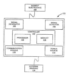
[0041] Turning to FIG. 1 a non-limiting example of a system 100, for use
in accordance
with aspects of the present invention, is shown. In general, the system 100
may include a
controller 102, a signal detector 104, a signal generator 106, and a plurality
of electrodes 108. In
some implementations, the system 100 may also include a communication module
110 and a
power source 112. The system 100 may be an external system, a portable device,
a wearable
system, an implantable device, a partially implantable device, a pacemaker,
and so forth.
[0042] In some aspects, the system 100 may operate autonomously or semi-
autonomously, or may read executable software instructions from a computer-
readable medium
(such as a hard drive, a CD-ROM, flash memory and the like). The system 100
may also receive
data or instructions from a user or clinician, via an input configured on the
system 100, or any
another source logically connected to the system 100. For instance, system 100
may receive
input, data, or instructions from external device(s) 114, as shown in FIG 1,
as well as from a
database, a storage server, a cloud, the internet, and other locations, using
a wired or wireless
communication. Examples external devices 114 may include personal computers,
laptops,
tablets, smartphones, personal digital assistant ("PDA") or other devices or
systems.
[0043] In addition carrying out steps for operating system 100, the
controller 102 may be
configured to monitor and/or control sympathetic nerve activities for
diagnosing and treating a
medical condition of a subject. For example, the controller 102 may be
configured to monitor
and/control stellate ganglion activity. In some aspects, the controller 102
may be configured to
direct the signal detector 104 to acquire electrical signals from electrodes
108 placed about a
subject, for example, cutaneously or subcutaneously, or both. The controller
102 may also be
configured to direct the signal generator 106 to generate and deliver
electrical stimulations to
target tissues, nerves, plexi, and other locations or regions of the patient's
body, using the
electrodes 108. In some aspects, the controller 102 may receive manual
instructions from an
operator externally, or may cause electrical stimulations to be generated and
delivered based on
internal calculations and programming, or based on measurements or estimations
of various
nerve activities.
[0044] In general, the controller 102 shown in FIG. 1 may include a
processor 116, a
memory 118, as well as other hardware components. In particular, the processor
116 can
include one or more microcontrollers, microprocessors, and the like, and be
capable of
performing a number of processing steps, in accordance with aspects of the
present disclosure,
- 9 -
CA 02965149 2017-04-19
WO 2016/064843 PCT/US2015/056419
as described in detail below. The memory 118 may include various memory
portions where a
number of types of data (e.g., internal data, external data instructions,
software codes, status data,
diagnostic data, etc.) may be stored. The memory 118 may include one or more
of random
access memory ("RAM"), dynamic random access memory ("DRAM''), electrically
erasable
programmable read-only memory ("EEPROM"), flash memory, and the like. In some
implementations, the controller 102 may be included in the same housing as the
signal detector
104, signal generator 106, communication module 110 and power source 112.
Alternatively, the
controller 102, along with other components of the system 100 may be housed
separately, as
separate or stand-alone components, devices or systems.
[0045] For example, in one embodiment, the controller 102 may be a mobile
electronic
device, such as a smartphone or tablet, a personal computer ("PC"), or any
suitable computing
device that includes a central processing unit ("CPU") with one or more cores
and a graphical
processing unit ("GPU''). The CPU and optionally the GPU execute stored
software instructions
stored in memory 118 to apply filters to acquired data samples and to perform
other signal
processing functions on the data samples. For example, software configured for
signal
processing tasks in processor 116 may include the PowerLab data acquisition
software
commercially available from ADInstruments of Sydney, Australia. In some
aspects, the
controller 102 may include one or more digital logic devices, including
application specific
integrated circuits ("ASICs"), field programmable gate arrays ("FPGAs"), and
digital signal
processor ("DSP") devices. In addition, in some portable or implantable device
embodiments of
the system 100, the controller 102 may include low-power digital logic devices
that enable long-
term operation between battery recharge or replacement.
[0046] As described, the signal detector 104 is configured to acquire
various electrical
signals from the subject, while the signal generator 106 is configured to
deliver the electrical
stimulations to the subject using various combinations of electrodes 108. In
some
implementations, the signal detector 104 may include one or more amplifier
capable of
amplifying voltage signals, or differential voltage signals, received from the
electrodes 108. The
signal detector 104 may also include a sampler that generates digitized
samples of amplified
signals via an analog to digital converter ("ADC") for further processing by
the processor 116.
By way of example, the signal amplifier and sampler may be configured to
amplify signals in a
frequency range of 1 Hz to 5,000 Hz and to generate digital samples of the
amplified signals at a
- 10 -
CA 02965149 2017-04-19
WO 2016/064843 PCT/US2015/056419
rate of 10,000 samples per second. In one example embodiment, the signal
amplifier and
sampler can be an ML 135 dual-bio amplifier that is manufactured by the
ADInstruments of
Sydney, Australia. In some aspects, the signal amplifier and sampler may be
electrically
connected to the electrodes 108 in a configuration that includes at least one
reference electrode
and two input signal electrodes. On the other hand, the signal generator 106
may include a
variety of hardware, circuitry and components for generating continuous or
intermittent electrical
stimulations, in accordance with aspects of the present disclosure, including
any number of
voltage and current sources.
100471 In accordance with aspects of the disclosure, the electrodes 108 may
be
configured to engage a subject cutaneously and/or subcutaneously, and may be
arranged in any
number of lead configurations. For instance, the electrodes 108 may be
electrically connected,
or proximate to various locations about a subject's body to enable effective
detection of electrical
signals, such as electric signals from nerves that innervate the skin
locations. In some
configurations, the electrodes 108 may be arranged to facilitate monitoring of
both nerve activity
and cardiac activity. In addition, electrodes 108 may be configured to deliver
continuous or
intermittent electrical stimulations generated by signal generator 106. The
electrodes 108 may
also be configured to measure other signals besides nerve activity, including
heart rate,
respiration, and so forth.
100481 In some aspects, the communication module 110 may be configured to
facilitate
communications between the system 100 and various devices. In particular, the
communication
module 110 may be capable of providing transmission and reception of
electronic signals to and
from the external device(s) 114 and other locations using a wired or wireless
connection. The
communication module 110 may include any hardware, software, firmware, and hi
some aspects
be capable of telemetry, Bluetooth or other wireless communication protocol.
In some
implementations, the communication module 110 may also be configured to
receive user input
directly, such as operational instructions, as well as provide various
information, in any form,
related to operational parameters, signals detected and/or processed, such as
cardiac activity,
nerve activity, and the like. The communication module 110 may also be
configured to provide
information regarding provided electrical stimulations. In some aspects, the
communication
module 110 may include capabilities for delivering audio signals or queues, as
well as visual
-11-
CA 02965149 2017-04-19
WO 2016/064843 PCT/US2015/056419
outputs, for example, using a monitor, LCD display, and other output component
configured
therein.
[0049] Referring again to FIG. 1, in some aspects, the processor 116 of
system 100 may
include digital logic device that can perform a number of signal processing
steps to identify
nerve activity in data samples received from the signal detector 104.
Specifically, the processor
116 may be configured to estimate a sympathetic nerve activity, such as a
stellate ganglion
activity, based on identified skin nerve activity, for example, using
determined signal
correlations stored in memory 118.
[0050] As described in more detail below, the electrical activity in the
nerves that
innervate the skin occurs at higher frequencies and lower amplitudes compared
to the electrical
signals generated in the cardiac muscle during a heartbeat. As such, processor
116 may be
configured to identify and monitor the electrical signals corresponding to
specific signals in the
subject, such as nerve or cardiac activity, by processing data samples
received from the signal
detector 104. That is, the processor 116 may apply appropriate filters, such
as low-pass filters,
high-pass filters, or band-pass filters, to the data to obtain signals of
interest. The processor 116
may also scale, multiply or integrate various measured signals.
[0051] For example, a 3 dB high-pass filter lower with a cutoff frequency
adjustable in a
range of approximately 100 ¨ 1 kHz may be utilized. Selection of the proper
high-pass setting
might require consideration of signal specificity and acceptable sensitivity.
For instance, a high-
pass cutoff frequency of 150 Hz would be sufficient to attenuate most the
lower frequency
signals from cardiac muscle activity and electrical signals from other muscles
in the subject
typically observed, but not all muscle noise. On the other hand, a cutoff at
700 Hz would be
more specific to nerve activity, as the muscle noise does not generate signals
with frequencies
above 500 Hz, but such filter setting would result in a reduced measurement
sensitivity. In
some preferred embodiments, the high-pass filter cutoff frequency may be
between 150 Hz and
700 Hz, although other values may be possible.
10052] In some aspects, data samples may also be processed using a low-
pass filter, for
example, with a cutoff frequency approximately in a range between 10 Hz and
150 Hz in order to
detect cardiac activity. Alternatively, a band-pass filter may be applied to
monitor the ECG of
the subject using the amplified signal samples from the signal detector 104.
For example, the
band-pass filter may have a lower cutoff frequency of approximately 0.5 Hz and
an upper cutoff
- 12 -
CA 02965149 2017-04-19
WO 2016/064843 PCT/US2015/056419
frequency of approximately 100 Hz. In some aspects, the same pair of
electrodes 108, such ECG
patch electrodes, may be used to simultaneously record the ECG and skin nerve
activity from the
surface of thoracic skin. In such case, the same signals may be low-pass
filtered for selective
ECG signals and high-pass filtered for SKNA signals. Additionally, where an
alternating current
("AC") electrical signal is used to supply power to one or more components in
system 100, a
band-pass filter also includes a notch-filter that attenuates frequencies near
the primary
frequency of the AC signal, such as 50 Hz or 60 Hz.
[00531 In addition to monitoring the electrical signals that correspond to
the nerve
activity and optionally the ECG, the processor 116 may be configured to
analyze the signals to
identify changes in the level of nerve activity, such as a skin or sympathetic
nerve activity, and
take an appropriate action in response to changes in the nerve activity. For
example, in one
configuration the processor 116 may identify a baseline of a nerve activity
over time including
an average amplitude and variation of the electrical signals that correspond
to a nerve activity.
100541 In some aspects, the processor 116 may be further configured to
determine or
identify a subject condition, for example, using identified nerve activity or
changes thereof
Based on the subject condition, processor 116 may then identify an appropriate
treatment
protocol, either autonomously or by way of user input, to include intermittent
periods of
electrical stimulation, or "ON" periods, as well as time intervals of non-
stimulation, or "OFF"
periods, arranged in any timing pattern. In some aspects, a treatment protocol
may include
intermittent periods of electrical stimulation separated by periods of non-
stimulation, where the
intermittent periods include electrical stimulation described by parameters
including one or more
duration, intensity, frequency, pulse width or waveform, other any combination
thereof. The
intermittent "ON" and "OFF" periods may be unequal in duration and, in this
regard, the process
may be referred to as asynchronous. The processor 116 may then direct the
signal generator 106
to deliver the treatment protocol via electrodes 108.
[0055] In one non-limiting example, intermittent periods of electrical
stimulation may be
delivered using electric pulses with a frequency between 0.1 Hz and 20 Hz,
pulse widths
between 0.1 milliseconds and 5 milliseconds, and stimulation intensities in a
range between 0.1
milliAmperes to 5 milliAmperes, although other values are possible. In some
applications, a
treatment protocol may include brief ON periods, for example, of 1 to 20
seconds in duration,
and long OFF periods, for example, lasting 60 seconds to 15 minutes in
duration, although other
- 13 -
CA 02965149 2017-04-19
WO 2016/064843 PCT/US2015/056419
values may be possible. Advantageously, such treatment protocol would reduce a
stellate
ganglion activity by inducing stellate ganglion remodeling or causing stellate
ganglion tissue
damage. Specifically, short and intermittent pulses would cause sufficient
stellate ganglion
damage during the ON-time and result in reduced nerve firing during the OFF-
time.
[0056] In some aspects, a treatment may be configured such that a reduced
activity of
neural structures, including sympathetic structures, can be achieved. In other
aspects, the
treatment protocol may be customized by taking into consideration a determined
baseline neural
activity, such as a sympathetic nerve activity, or a parasympathetic nerve
activity, and a target
neural activity or target ventricular rate.
[0057] The cardiac activity of the subject is not the only type of medical
event that
corresponds to changes in the nerve activity in the sympathetic nervous
system. Other changes in
the level of nerve activity in the subject can correspond to the onset of
symptoms related to
various other medical conditions including, but not limited to, hyperhidrosis
(sweaty palms),
paralysis, stroke, diabetes, seizure disorder, syncope, disturbance of
consciousness,
hyperthyroidism, hypertension and neuromuscular diseases. Other areas of
treatment include
biofeedback monitoring performed by neurologists to control neuropsychiatric
disorders. In
such approaches, system 100 may be used to identify a suitability of a patient
to receive a
therapy aimed at modifying an identified nerve activity for treatment of
certain medical
conditions or diseases, such as hypertension and cardiac arrhythmia. For
example, a
neuromodulation therapy, such as renal sympathetic denervation, may be
performed to reduce or
modify sympathetic nerve activity. Monitored nerve activity may also be
desirable for providing
guidance while performing a procedure, and also for determining an
effectiveness of a treatment
after delivery with reference to a difference in the identified nerve
activity. Additionally, another
area includes lie-detection tests, because the sympathetic nerve activation is
the mechanism that
regulates sweating, pupil contraction, and other physiological responses that
are measured during
lie detector tests. Thus, the system 100 identifies changes in the nerve
activity of the subject that
correspond to changes in cardiac activity and the onset of symptoms in
different diseases and
conditions that affect the subject.
100581 Turning now to FIG. 2, the steps of a process 200 for monitoring
nerve activity in
a subject using cutaneous or subcutaneous electrodes recording electrical
activity in nerves that
innervate the skin, are shown. In some aspects, the process 200 may be carried
out using a
- 14 -
CA 02965149 2017-04-19
WO 2016/064843 PCT/US2015/056419
system 100, as described with reference to FIG. 1. The process 200 may begin
at process block
202 with receiving electrical signals sampled using cutaneous and/or
subcutaneous electrodes,
for example using system 100, as described above. In some configurations,
three or more
electrodes, may be placed on the skin of the subject in a cutaneous
configuration. Electrodes
may be additionally, or alternatively implanted under the skin of the subject
in a subcutaneous
configuration, although other arrangements are possible. Referring
specifically to the system
100 of FIG. 1, in some aspects, the signal detector 104 may amplify
differential voltage signals
that are received from the electrodes and generate digitized samples of the
signals.
[0059] Process 200 continues with application of a filter to the sampled
electrical signals
to generate filtered signals, as indicated by process block 204. In some
aspects, the filter may be
configured to attenuate at least signals having frequencies that correspond to
heart muscle
activity during a heartbeat. Other signal filtering, as well as processing
steps may also be
possible at process block 204, including scaling, multiplying, or integrating
the signals sampled
at process block 202. In some aspects, a high-pass filter may be applied to
the processed signal
samples. Specifically, the high-pass filter may have a lower cutoff frequency
in a range of 100
Hz to 1 kHz in order to attenuate lower-frequency electrical signals that
correspond to cardiac
activity in the subject instead of the nerve activity. The lower-frequency
cutoff of the high-pass
filter can be adjusted based on the characteristics of different subjects to
enable identification of
the electrical signals in the nerves that innervate the skin while attenuating
the electrical signals
from muscles and other sources of electrical noise in the subject. For
example, the high-pass
filter may have a cutoff frequency of approximately 700 Hz. Thus, at process
block 206, a skin
activity may be identified using high-frequency signals that pass through the
high-pass filter.
[0060] At process block 208, a sympathetic nerve activity may then be
estimated using
the identified skin nerve activity. For instance, predetermined correlations
or relationships
between skin nerve activity and a stellate ganglion nerve activity may be
utilized to determine
the estimates. Such correlations may be stored in a memory, for example. In
this manner, an
estimated sympathetic nerve activity may be provided in the form of a report
at process block
210, enabling a clinician or other healthcare professional to monitor or
assess nerve activity in
the subject. The report may be provided in substantially real time, for
example, using a display,
or stored in a memory to be retrieved at a later time. In some aspects, the
report may be in the
form of graphs or time traces of measured or estimated nerve activity.
Displayed or retrieved
- 15 -
CA 02965149 2017-04-19
WO 2016/064843 PCT/US2015/056419
activities corresponding to estimated nerve activity may then utilized by a
doctor or other
healthcare professional during or following the course of medical treatment
for a subject. The
report may also include information derived from measurements or estimations
of nerve activity,
including average signals, signal variations, signal frequencies, frequency
variations, identified
events, event timings, deviations from a baseline, and so forth.
[0061] In one embodiment, the process 200 may be implemented in a passive
operating
mode, displaying the nerve activity and recording nerve activity in the memory
for subsequent
retrieval and analysis by medical professionals. In such passive operating
mode, therapeutic
devices need not be activated automatically. That is, a doctor or other
healthcare provider would
retrieve and review information or data associated with acquired or estimated
nerve activity as
part of diagnosis and treatment in a patient. The passive operating mode can
be used, for
example, during diagnosis of a medical condition, during long-term monitoring
of a patient to
assess progress in a course of medical treatment, and for studies of subjects
during clinical trials
or other scientific research.
[0062] In another embodiment, the process 200 may be carried out to
generate a baseline
measurement of nerve activity in a subject, such as stellate ganglion nerve
activity baseline. For
example, the baseline nerve activity can include an average signal amplitude,
or signal variation.
The baseline activity could then be used to determine a change in the level of
nerve activity over
time, for example, as a result of a change in medical condition, or as a
result of treatment. A
determined rapid change in the electrical signals corresponding to the
sympathetic nerve activity
that deviates from the baseline by more than a predetermined threshold, could
then initiate an
audio or visual alarm to a clinician in response to the identified change in
nerve activity. In
some aspects message, such as a page, email, or text message, through a data
network may be
sent to alert a remote healthcare professional of the identified event.
[0063] In accordance with another aspect of the present disclosure, FIG. 3
depicts steps
of a process 300 for controlling nerve activity in a subject in accordance
with aspects of the
present disclosure. The process 300 may be carried out using a system 100 as
described with
reference to FIG. 1 or any other suitable system. In some aspects, the process
300 may be
carried out as a result of a determined medical condition, or a deviation of
nerve activity from a
baseline.
- 16 -
CA 02965149 2017-04-19
WO 2016/064843 PCT/US2015/056419
[0064] Specifically, the process 300 may begin at process block 302 where
subcutaneous
and/or cutaneous electrodes may be placed at various locations proximate to
nerves innervating a
subject's skin. In some aspects, selection of electrode locations might take
consideration of
enervations proximate to the skin for the neural structure(s) targeted for
control, in order to
effectively deliver therapeutic effects. For example, electrodes may be placed
at skin locations
about the thorax of a subject, specifically at or above the 5th thoracic
space. In particular, this
location is associated with connections between skin sympathetic nerves and
the stellate
ganglion. Other locations, depending upon the targeted neural structure, or
tissue, may also be
possible.
[0065] At process block 304, an electrical stimulation is then generated,
for example,
using system 100 as described. In accordance with aspects of the present
disclosure, electrical
stimulation parameters may configured to control a sympathetic nerve activity,
such as a stellate
ganglion nerve activity. In particular, the electrical stimulation may be
configured to remodel
one or more neural structures, such as the stellate ganglion. By way of
example, an electrical
stimulation treatment protocol may include an intermittent stimulation that
includes short ON
and long OFF periods. For instance, an ON time may be approximately 14 seconds
in duration,
while the OFF time may be approximately 1 minute to 3 minutes duration. The
stimulation
frequency may be approximately 10 Hz, with a pulse width of 0.5 milliseconds
and an intensity
amplitude in the range of 1.0 milliAmperes to 3.5 milliAmperes. In accordance
with findings of
the present disclosure, such mode of stimulation may be sufficient to control
stellate ganglion
nerve activity and maintain therapeutic effects. It may be appreciated that
other electrical
stimulations protocols may also be possible, depending upon targeted
structures or tissues.
[0066] As indicated by process block 306, in order to control sympathetic
nerve activity
the electrical stimulation may be delivered via cutaneous and/or subcutaneous
electrodes. In
some applications, stimulation output may be adjusted gradually over a period
of time. For
example, the stimulation may be adjusted over 3 weeks, from 0.5 milliAmperes
to 3.5
milliAmperes, and maintained in accordance with target remodeling or nerve
activities.
[0067] In some aspects, changes to a sympathetic nerve activity, such as
the stellate
ganglion nerve activity may also be monitored at process block 308. For
instance, a sympathetic
nerve activity may be estimated using measures of skin nerve activity, as
described, via electrical
signals acquired from locations proximate to a subject's skin. A report may
also be generated at
- 17 -
CA 02965149 2017-04-19
WO 2016/064843 PCT/US2015/056419
process block 308, including information associated with the administered
electrical
stimulations, as well as any changes to sympathetic nerved activities
detected.
[0068] In accordance with yet another aspect of the present disclosure,
FIG. 4 depicts
steps of a process 400 for controlling nerve activity in a subject using
cutaneous and/or
subcutaneous electrodes that deliver electrical stimulations via nerves that
innervate the skin.
The process 400 may be carried out using a system 100 as described with
reference to FIG. 1 or
any other suitable system.
[0069] Similar to process 200 depicted in FIG. 2, a sympathetic nerve
activity may be
estimated using skin measures. Specifically, in some aspects, electrical
signals originating from
cutaneous and/or subcutaneous electrodes (block 402) may be amplified, sampled
and filtered
(block 404). In some aspects, the applied filter may be configured to
attenuate at least signals
having frequencies that correspond to heart muscle activity during a
heartbeat. Using identified
skin nerve activity (block 406) generated using filtered electrical signals,
an estimate of
sympathetic nerve activity may then be obtained using correlations stored in a
memory, for
example, as indicated by process block 408. In some aspects, various
computations may be also
be carried out at process block 408 using the estimated sympathetic nerve
activity, including
computing average signals, signal variations, signal frequencies, frequency
variations. In some
aspects, certain events, event timings, deviations from a baseline, and other
information may also
be obtained at process block 408.
[0070] Then, at process block 410, and electrical stimulation based on the
estimated
sympathetic nerve activity, and or information obtained therefrom, may be
generated, and
subsequently delivered at process block 412. In some aspects, delivered
electrical stimulations
may be used control the sympathetic nerve activity, for example, by remodeling
one or more
neural structures. In addition, nerve activity following delivery of the
electrical stimulation may
also be monitored at process block 412, as described.
[0071] The above-described system and methods may be further understood by
way of
examples. These examples are offered for illustrative purposes only, and are
not intended to
limit the scope of the present invention in any way. Indeed, various
modifications of the
invention in addition to those shown and described herein will become apparent
to those skilled
in the art from the foregoing description and the following examples and fall
within the scope of
the appended claims. For example, certain electrode arrangements and
configurations are
- 18-
CA 02965149 2017-04-19
WO 2016/064843 PCT/US2015/056419
presented, although it may be understood that other configurations may be
possible, and still
considered to be well within the scope of the present invention. Likewise,
specific process
parameters and methods are recited that may be altered or varied based on
variables such as
signal amplitude, phase, frequency, duration, and so forth.
EXAMPLE I
100721 Previous studies have documented a direct relationship between
stellate ganglion
nerve activity ("SGNA") and cardiac arrhytlunias in ambulatory dogs. In
addition to serving as
a source of cardiac sympathetic innervation, the stellate ganglia also gives
rise to sympathetic
nerves that innervate blood vessels and sweat glands in skin. It was shown
recently that it is
feasible to record sub-cutaneous nerve activity ("SCNA") from ambulatory dogs
continuously
over long periods of time, and that the SCNA can be used to estimate the
cardiac sympathetic
tone. The latter observations are extended in the present study by documenting
the feasibility of
directly recording sympathetic nerve activities from the skin of the chest.
Specifically, the
present study was aimed at testing the hypothesis that thoracic skin nerve
activity ("SKNA") can
be used to estimate SGNA in both anesthetized and ambulatory dogs. A method
was developed
for recording skin nerve activity, and a comparison was made between SKNA,
SGNA and heart
rate.
[0073] In a first protocol, Protocol 1, five anesthetized dogs (A, B, C,
D, E) were used to
assess Right SGNA and SKNA. The first two dogs were also used for subcutaneous
nerve
activity ("SCNA") recording. The dogs were intubated and underwent isoflurane
general
anesthesia. Thoracotomy was performed through the right 3rd intercostal space
and the hair on
the thoracic skin was removed. A pair of bipolar electrodes was inserted under
the fascia of the
right stellate ganglion. Electrocardiogram ("ECG") patches (Tyco/Healthcare
Kendall, Medi-
Trace 100, Hampshire, U.K.) were secured on the skin using adhesive tapes for
surface ECG and
SKNA recording. Two pairs of those ECG patch electrodes were taped on the skin
to record
ECG Leads I and II, along with SKNA. As shown in FIG. 5, Lead I was recorded
between
electrodes at the level of the 2"d rib with an inter-electrode distance of 22
cm. Lead II was
recorded between electrodes on right second rib and the left lower abdomen,
with an inter-
electrode distance of 48 cm. An additional patch was secured to the right
lower abdomen to serve
as ground.
- 19 -
CA 02965149 2017-04-19
WO 2016/064843 PCT/US2015/056419
[00741 To explore whether or not other locations on the chest wall can
also be used for
SKNA recording, a pair of bipolar electrodes was placed each at the level of
the right and left 3"1
rib in dogs B and C, respectively, to form bipolar electrodes with 12 cm inter-
electrode distance
(FIG. 5). In dogs D and E, these bipolar electrodes were moved downwards to
the lower 1/3rd of
the chest to determine if SKNA from the lower chest can also be used for
recording cardiac
sympathetic tone.
[00751 These electrodes were connected to a World Precision Instrument Iso-
Damm-8
amplifier (Sarasota, Florida), with a noise level of < 2.5 IV and a recording
bandwidth set at 10
Hz-3 KHz. The signals were digitized by Digidata 1400a using AxoScope software
(Sunnyvale,
Calif) at 10,000 times per second per channel. After all surgical procedures
were performed, the
anesthetic agents were switched from isoflurane to alpha-chloralose (up to 100
mg/kg) and
morphine. One ml apamin (concentration 0.2 ng/ttL) was injected directly into
the right stellate
ganglion, which is a neurotoxin that is a specific blocker of the small
conductance calcium
activated K (SK) channel. Inhibition of the SK channel is known to facilitate
neuronal
discharges. Data was acquired for 10 min after apamin injection.
[0076] In a second protocol, Protocol 2, four ambulatory dogs (F,G, H, 1)
were used to
assess Left SGNA and SKNA. All four dogs were chronically instrumented for
different
research protocols. However, the non-invasive recordings made in this study
did not affect the
results of those research protocols, and only helped reduce the use of
animals.
[00771 The dogs underwent left thoracotomy through the third intercostal
space. A DSI
(Data Sciences International, St. Paul, MN) D7OEEE radiotransmitter was
implanted to record
the left SGNA and the subcutaneous ECG according to methods reported
elsewhere. Dogs F and
G were normal dogs and did not undergo other procedures. Dog H had undergone
left circumflex
coronary artery ligation to create myocardial infarction. Dog I had undergone
a modified Secura
implantable cardioverter-defibrillator (Medtronic, Minneapolis, MN)
implantation for
intermittent rapid left atrial pacing in an attempt to induce paroxysmal AF.
That dog was in
sinus rhythm when used in this study. All dogs were allowed to recover for 2
weeks after the
initial surgery.
[0078] At the time of the study, all wounds have healed and the dogs were
ambulatory.
After clipping the hair on the chest, four ECG patches were placed on the skin
to record surface
ECG Leads I and II according to the methods described in Protocol 1. An
additional two pairs of
- 20 -
CA 02965149 2017-04-19
WO 2016/064843 PCT/US2015/056419
bipolar electrodes were placed on the upper 1/3 rd of the skin of the left and
right thorax for
bipolar ECG recordings. Soft, non-adhesive elastic bands were used to wrap
around the chest to
help secure the ECG patches in place. The locations of surface ECG patch
electrodes were the
same as shown in FIG. 5. These skin electrodes were connected to the same
equipment as
described in Protocol 1. Continuous recordings were made for 30 min while the
dog was awake
and lying or standing in the dog run.
[0079] Recordings were analyzed using custom written DSIView software. The
same
ECG signals were used for both SKNA and ECG analyses. The signals were high-
pass filtered at
700 Hz to display SKNA and low pass filtered at 30 Hz to display the surface
ECG. The latter
was used for heart rate analyses. A quantitative analyses was performed by
integrating SGNA
(iSGNA), SKNA (iSKNA) minute by minute. The data was reported in the form of a
mean and
95% confidence interval (CI). Pearson correlation coefficients were calculated
between heart
rate, iSGNA and iSKNA. A p value of <0.05 was considered statistically
significant
Results
Protocol 1: Correlation between iSGNA and iSKNA after apamin injection
100801 Apamin injection induced robust activity of SKNA and SGNA in all
dogs studied.
FIG. 6A shows a typical recording from Dog D. Apamin-induced SGNA, SKNA and
heart rate
acceleration. FIG. 6B shows the relationship among integrated right SGNA
("iRSGNA"),
integrated SKNA recorded by ECG Lead I ("iSKNA-1"), ECG Lead II ("iSKNA-II"),
right chest
("iSKNA-R"), left chest ("iSKNA-L") and heart rate ("HR") of the same dog. All
of them
strongly correlated with each other.
Protocol 2: Monitoring of Spontaneous SKNAs and SGNA, heart rate in Ambulatory
Dogs
[0081] Simultaneous recording of SGNA and SKNAs was successful in all dogs
studied.
There were electrical signals resembling nerve activities on the surface of
skin. During the
recording period, the sound created by the investigators (speaking, clapping
of the hands, moving
instruments around) or barking of other dogs in the same room often caused
abrupt activation of
the SGNA. FIG. 7 shows correlations between left stellate ganglion activity
("LSGNA"), cardiac
activity and skin nerve activity ("SKNA") in an ambulatory dog. Specifically,
FIG. 7A shows
nerve activities (high pass filtered at 700 Hz) recorded both from LSG and
from all skin
-21-
CA 02965149 2017-04-19
WO 2016/064843 PCT/US2015/056419
electrodes from dog I. The SGNA was associated with elevated heart rate and
the SKNA at all
locations. The third arrow shows that largest heart rate acceleration
associated with largest
LSGNA and SKNA recordings. A large nerve activity ((downward arrows) is
associated with
large increase in heart rate ("HR') (upward arrow). FIG. 7B shows the
correlation between
integrated nerve activities and average heart rates, with each dot
representing data collected in a
one-min window. There are significant correlations for all comparisons in this
dog. Specifically,
significantly positive correlations are shown among integrated left SGNA
(iLSGNA) and
iSKNA-I, iSKNA-II, iSKNA-R, iSKNA-L and heart rate.
Table 1. Correlation Coefficients Between Nerve Activities and Heart Rate for
Each Dog.
,
: Protocol 1. :
: iRSGNA iRSGNA iRSõGNA ; iRSGta i HR vs. ! HR vs. ! HR vs. !!µ 11R vs. ! HR
vs.
vs. VS. VS. : VS. : IR WM i WR4- MO(4- i INS,NA-R :
LaStiA,.-1.
Doglit . ISKNA- . LSICNA- igsm-R 1 ISKNA-L ! I TI i
. !
! I : II
__________________________________________________________________ i
A r = 0_948 1 0.933 / ! / 1 0.834 ! 0.886 !
0.869 ! / ! /
p<0.001 1 <0.001 , ! 1005 ! 0.001 ! 0.003
:
!
r=0.749 1 0.729 1 0.864 ! 0.715 ! 0.875 ! 0.823 ! 0.907
0.725 ! 0.686 ,
: B
; p=0.020 0_026 ! 0.003 : 0.031 I 0.002 0.006 : 0.001
0.027 i 0.097
!
! r = 0.985 ! 0.881 : 0.951 ; 0.885 ! 0.867 ; 0.783 . 0.933
0.600 i 0.683 1
C ,
. p<0.001 1002 <0.001 ! 0.002 : 0.002 0.013 <0.001
0.088 i 0.099 ;
r=0.950 ' 0.773 0.748 : 0.564 : 0.926 ! 0.929 ; 0.715 0.551
! 0.539 1
D !; p<0.001 0.014 ' 0.020 0.114 : <0.001 : <0.001
! 0.030 0.125 ! 0.135 1
: r=0.751 ! 1603 ! 0.802 1772 ! 0.881 ! 0.766 1 0
i-
.648 1453 : 0.507
,
, E p=0.020 , 0,086 1 0.009 : 1015 ! 0.002 ! 0.016
! 0.126 0.221 , 0.163
,
i !
i Mean i 0.877 i 1784 1 0.841 : 1734 i 0.877 1 0.837
! 0.792 ! 0.582 ; 0.554 :
I
__________________________________________________________________ 1
i Protocol 2
ILSGNA 1 iLSGNA ILSGNA f ILSGNA HR vs. HR vs. HR vs.
HR vs. ' HR vs. 1
: vs. : vs. ! vs. ! vs. il-SGNA ,
LSKNA- , ISKNA- LS_KNA-R ! ISKNA-L 1
: Dog# ! ; 1
1 twm- : Lsism- : mst,A-R ! Lgstm-L. : 1 1 I !
i u I , . I :
. : , 1
,
:
! r=0.745 i 0.601 1 0.575 : 0.678 1 0.620 1 0.824 1 1335
0.319 1 0.811 1
: F i
1 p<0.001 i 0.005 ! 0001 : <0.001 ; 0.004 ! <0.001 I 0.149
0.098 ! <0.001 j
I ! r=0.624 0464 1 0444 ; 0.629 i 0.649 0.626 ! 0,377
0.170 ! 0.591
G 1
i ! p=0.001 ! 1026 ! 0.038 ! 0.011 ! 0.001 ; 0.010 0.076
0449 9.004
i :r_9539 i 1. ' 0_547 : 0.350 I 0.719 ; 0.790
:0.722 0.582 0.631 1 0.499 ;
1 H :
! ; p=0.025 ! 0.023 . 0.142 I 0.001 ! <0.001 0.001 1014
0.004 ! 0.030 :
r= 0.660 0.527 ! 0.445 ! 0.743 0.516 0.882 : 0.795 0.847
0.865
, p <0_001 : 0.004 ; 0.018 ; <0.001 0.006 : <0.001 ; <0.001
<0.001 <0.001
! Mean 0.642 ; 0.535 ! 0.454 ; 0.667 ; 1644 : 0.738 !
0.622 0492 0.692 .
[0082] Table 1 shows the correlation coefficient and the p values of all
dogs studied. As
shown in this table, there is consistently a strong positive correlation (mean
0.877) found
- 22 -
CA 02965149 2017-04-19
WO 2016/064843 PCT/US2015/056419
between iSGNA and iSKNA-I in all dogs of Protocol 1. A good correlation (mean
0.642) was
found between iSGNA and iSKNA-I in Protocol 2. Both right and left integrated
SGNA
(iRSGNA and iLSGNA) correlated well with the ipsilateral integrated SKNA
(iRSKNA and
iLSKNA) respectively. All other correlations are positive and mostly
statistically significant.
[0083] Simultaneous recording of SGNA and SKNAs was successful in all dogs
studied.
There were electrical signals resembling nerve activities on the surface of
skin. During the
recording period, the sound created by the investigators (speaking, clapping
of the hands, moving
instruments around) or barking of other dogs in the same room often caused
abrupt activation of
the SGNA. FIG. 7A shows a typical recording from dog I. The SGNA was
associated with
elevated heart rate and the SKNA at all locations. The third arrow shows that
largest heart rate
acceleration associated with largest LSGNA and SKNA recordings. FIG. 7B shows
significantly
positive correlations among integrated left SGNA ("iLSGNA") and iSKNA-I, iSKNA-
II,
iSKNA-R, iSKNA-L and heart rate.
[0084] Results from this study demonstrated that (1) it is feasible to
record sympathetic
nerve activities from the surface of skin and (2) there was a positive and
statistically significant
positive correlation between iSGNA with iSKNA-I, and between iSGNA and the
ipsilateral
iSKNA. These findings indicate that SKNA recorded from the Lead I ECG and from
ipsilateral
bipolar surface ECG leads can be used to estimate the SGNA and cardiac
sympathetic tone.
100851 In the present study, four pairs of bipolar electrodes (leads I,
II, right and left)
were implanted on the surface of the skin to record SKNAs. All dogs (9/9)
showed a significant
correlation between iSKNA-I and iRSGNA or iLSGNA. Eight (8/9) dogs showed a
strong
correlation between iSKNA-11 and iRSGNA or iLSGNA. Seven (7/8) dogs showed a
good
correlation between iSKNA-R or iSKNA-L and iRSGNA or iLSGNA. In addition, all
dogs (9/9)
showed a significant correlation between iSKNA-I and heart rate. Six dogs
(6/9) showed a
strong correlation between iSKNA-II and heart rate. Three (3/8) dogs showed a
good correlation
between iSKNA-R and heart rate and four (4/8) dogs showed a good correlation
between
iSKNA-L and heart rate. Therefore, SKNA-I may be the best recording lead to
estimate the
SGNA and the cardiac sympathetic tone. In addition, a good correlation between
iSGNA and
ipsilateral iSKNA is compatible with the finding that the skin sympathetic
innervation came
from ipsilateral stellate ganglion. However, because the left and right
stellate ganglion usually
- 23 -
CA 02965149 2017-04-19
WO 2016/064843 PCT/US2015/056419
fire simultaneously, the integrated SKNA recorded from any location on the
chest correlated
positively with both right and left SGNA.
[0086] The correlations between SGNA and SKNA for all combinations appear
to be
stronger in Protocol I than Protocol 2. A possible explanation may be that the
equipment used to
record SGNA in Protocol I had much higher frequency bandwidth than that used
in Protocol 2.
The latter study used implanted DSI radiotransmitter which are adequate in
recording the large
nerve discharges associated with the abrupt onset of sinus tachycardia, but
often misses the
smaller changes of nerve discharges associated with transient shortening of RR
interval. The
weaker correlation is likely the result of insufficient frequency content of
the SGNA recording.
[0087] SKNA may be useful clinically for cardiac arrhythmia risk
stratification. It may
also be helpful in determining the relationship between sympathetic tone and
cardiac arrhythmia
in animal models without a need for thoracotomy. Results here also show that
it is possible to
simultaneously record the ECG and SKNA from the surface of thoracic skin using
the same pair
of ECG patch electrodes. The same signals are low-pass filtered for selective
ECG signals and
high-pass filtered for SKNA signals. The latter techniques may be useful in
clinical
investigations for a better understanding of the relationship between
sympathetic nerve activities
and cardiac arrhythmogenesis.
EXAMPLE IT
[0088] Histological studies of human skin biopsy have confirmed the
presence of
abundant sympathetic nerves in arteriovenous anastomoses, arrector pilorum
muscles, and
arterioles. Using horseradish peroxidase as tracer, one group found that all
skin sensory and
sympathetic neurons are located ipsilaterally. Nearly all sympathetic somata
are located in the
middle cervical and stellate ganglia. Because of the direct and extensive
connections among
various nerve structures, it is possible for the sympathetic nerves in the
various structures to
activate simultaneously. Using bipolar electrodes located in the chest wall,
the present study
aimed to obtain good ECG signals for heart rate analyses as well as record
nerve signals over a
wide area in the left lateral thorax. The onset and offset of nerve activities
in the thoracic
subcutaneous space and the stellate ganglion were documented simultaneously or
nearly
simultaneously, showing that nerve activities correlate with the heart rate.
These observations
made it possible to directly assess cardiac sympathetic tone by electrodes
embedded in the
thoracic subcutaneous space of ambulatory dogs.
- 24 -
CA 02965149 2017-04-19
WO 2016/064843 PCT/US2015/056419
[0089] Two acute and three chronic canine experiments were performed to
test the
hypothesis that cutaneous or subcutaneous stimulation can remodel the stellate
ganglion, and
modify SGNA. Previous studies showed that acupoint is richly innervated by
autonomic nerve
fibers. Therefore, subcutaneous tissues near the Xinshu acupoint of the dogs
were explored for
sympathetic nerves. The Xinshu acute point is located on the back, below the
spinous process of
the 5th thoracic vertebra, 1.5 cun (roughly 5.5 cm) lateral to the posterior
midline. (one cun, or
Chinese inch, is roughly 3.715 cm in length.) That area was explored in dogs
and small strands
of subcutaneous nerves running through that region were identified.
[0090] In the acute study, apamin (a neural toxin that increases nerve
discharges) was
injected into that subcutaneous nerves identified at the left Xinshu acupoint.
It was found that
the injection induced increased subcutaneous nerve activity ("SCNA") followed
by increased left
SGNA (FIG. 8). These experiments indicate that activation of the subcutaneous
nerves can lead
to subsequent activation of the left stellate ganglion in approximately 2-3
minutes. There was
heart rate elevation associated with the increased nerve activity, confirming
the physiological
connection. The heart rate correlated well with the integrated SGNA (1-0.86)
and with the
integrated subcutaneous nerve activity (SCNA, r=0.77) after apamin injection.
[0091] If the stellate ganglion can be acutely activated, then chronic
activation of the
stellate ganglion may cause remodeling changes of the left stellate ganglion.
Therefore, instead
of using apamin, electrical stimulations were used to activate the electrodes
and achieve
therapeutic effects. Specifically, stimulating electrodes were placed at
locations associated with
subcutaneous nerves of the animal subject, and the animal was allowed to
recover from surgery.
FIG. 9 shows a picture of the implanted electrodes in the present canine
study. The nerve
stimulation was given by a Cyberonics Demipulse vagal nerve stimulator.
Instead of having
electrodes wrapping around the vagal nerve, electrodes shown in FIG 9 are
wrapped around
small subcutaneous nerves. The results of the acute (non-survival) study
showed similar effects
as the apamin injection, i.e., stimulating the subcutaneous nerve at Xinshu
acupoint can activate
the left stellate ganglion.
[0092] Recordings of subcutaneous electrodes were used to observe the
effects of
stimulation in chronically instrumented ambulatory dogs. For chronic studies
of nerve
recordings, an implanted Data Sciences International ("DSI'') D7OFEE
radiotransmitter was
utilized along with a Cyberonics Demipulse generator to deliver sub-cutaneous
nerve stimulation
- 25 -
CA 02965149 2017-04-19
WO 2016/064843 PCT/US2015/056419
and obtain the data. The stimulation protocol utilized was as follows: 0.5 mA
for 2 days, 1.0 mA
for 2 days, 1.5 mA for 2 days, 2.5 mA for 3 days and 3.5 mA for 2 weeks.
[0093] FIG. 10 shows the simultaneous recording of SGNA, left thoracic
vagal nerve
activity ("VNA") and the subcutaneous nerve activity (SCNA) in an ambulatory
dog. The
stimulation was given at 10 Hz, 14-s duration and 3.5 mA amplitude. The total
duration of
stimulation exceeded two weeks. The SCNA appears saturated with the stimulus
artifact during
the time of stimulation. As shown, there was activation of VNA and reduction
of SGNA during
the stimulation, indicating direct effects of the subcutaneous stimulation on
the SGNA and VNA.
Contrary to what was observed during acute study, the dogs with prolonged
periods (weeks) of
chronic subcutaneous nerve stimulation were associated with reduced overall
SGNA.
[0094] A quantitative analyses was performed on the nerve activities and
heart rate in
dogs with chronic subcutaneous nerve stimulation, with results shown in FIG.
11. Specifically
the effects of subcutaneous stimulation during the OFF time are compared to
baseline values for
subcutaneous nerve activity, stellate ganglion nerve activity, heart rate and
vagal nerve activity.
Of note is that there is significant reduction of stellate ganglion nerve
activity with 3.5 mA of
intermittent stimulation, as compared to baseline. These findings are similar
to observations in
dogs following vagal nerve stimulation, highlighting the feasibility of
subcutaneous stimulation.
[0095] In addition, subjects were then sacrificed and the left stellate
ganglion was fixed
with 4% of formalin for 45 mm, followed by storage in 75% alcohol, The tissue
samples of the
left stellate ganglion were then paraffin embedded and cut into 5 pm thick
sections. An
immunohistochemical staining of tyrosine hydroxylase ("TH") using monoclonal
anti-TH
antibody (Accurate Chemical, Westbury, NY) was then performed.
[0096] As shown in the image of FIG. 12A, there is a region with reduced TH
staining. A
higher magnification view (FIG. 12 B) shows numerous TH-negative ganglion
cells. Some of
them have pyknotie nucleus. These findings suggest significant remodeling of
the stellate
ganglion. The reduced expression of TH and the pyknotic cells indicate that
portion of the
stellate ganglion was damaged by rapid pacing of the subcutaneous nerves. It
was previously
reported that vagal nerve stimulation can also significantly increase the TH-
negative ganglion
cells in the stellate ganglion, along with reduction of the SGNA. Therefore,
it appears that the
subcutaneous nerve stimulation can lead to the same stellate ganglion
remodeling, which would
result in reduced SGNA when the stimulation is turned off.
-26-
CA 02965149 2017-04-19
WO 2016/064843 PCT/US2015/056419
[0097] In summary, disclosed herein is a novel approach that utilizes
measures and
stimuli of the skin to monitor and/or control nerve activity of a subject.
Specifically, a system
and methods are provided for non-invasive or minimally invasive monitoring
and/or controlling
nerve activity using cutaneous and/or subcutaneous electrode configurations.
[0098] In some aspects, provided system and methods utilize measurements
of electrical
signals obtained via electrodes placed at specific locations on a subject's
skin, in order to
estimate a sympathetic nerve activity. Specifically, herein it is shown that
nerve discharges
recorded from the cutaneous and subcutaneous electrodes are correlated to
stellate ganglion
activity, and hence the present disclosure recognizes that such correlation
may be utilized to
estimate stellate ganglion activity from measured skin nerve activity.
[0099] The latter findings may also indicate that cutaneous and
subcutaneous
sympathetic nerves are directly connected to the stellate ganglion. Hence, the
present disclosure
also recognizes that such connection may be utilized to control stellate
ganglion nerve activity.
Hence, system and methods are provided which implement electrical stimulations
to
subcutaneous or cutaneous nerves in order to remodel specific neural
structures, such as the
stellate ganglion. As described, provided system and methods may be utilized
to obtain similar
therapeutic effects as the vagal nerve stimulation, by way of controlling
stellate ganglion
activity, but with much reduced risk to a subject. In some applications,
reduced sympathetic
outflow from the stellate ganglion may be used, for example, in treating
various heart diseases,
such as reducing the ventricular rate during atrial fibrillation or to reduce
the incidence of cardiac
arrhythmia.
[00100] The various configurations presented above are merely examples and
are in no
way meant to limit the scope of this disclosure. Variations of the
configurations described herein
will be apparent to persons of ordinary skill in the art, such variations
being within the intended
scope of the present application. In particular, features from one or more of
the above-described
configurations may be selected to create alternative configurations comprised
of a sub-
combination of features that may not be explicitly described above. In
addition, features from
one or more of the above-described configurations may be selected and combined
to create
alternative configurations comprised of a combination of features which may
not be explicitly
described above. Features suitable for such combinations and sub-combinations
would be
-27 -
CA 02965149 2017-04-19
WO 2016/064843 PCT/US2015/056419
readily apparent to persons skilled in the art upon review of the present
application as a whole.
The subject matter described herein and in the recited claims intends to cover
and embrace all
suitable changes in technology.
- 28 -