Note: Descriptions are shown in the official language in which they were submitted.
CA 2965384 2017-04-28
TITLE: BI-LAYERED BONE-LIKE SCAFFOLDS
BACKGROUND OF THE INVENTION
1. - Field of the Invention
The present invention generally relates to the fields of biomedical scaffolds,
and
methods of treating disease or disorders in a subject that involve
implantation of the scaffolds
set forth herein.
2. Description of Related Art
Considerable research has been reported over the last decade in the use of
polymeric
and ceramic biomaterials for producing scaffolds. However, the ideal material
and fabrication
technique for optimal bone tissue regeneration has yet to be identified. While
current
materials and techniques have met with varying successes, each material and/or
technique
exhibits limitations that must be addressed. In addition, there is an overall
lack of success in
bringing these technologies to the clinic, especially for the reconstruction
and restoration of
.. large bone defects.
Ideally, scaffolds for bone tissue regeneration should 1) exhibit
biocompatibility
without causing an inflammatory response or foreign body/toxic reaction, 2)
have closely
matched mechanical properties when compared to native bone, and 3) possess a
mechanism to
allow diffusion and/or transport of ions, nutrients, and wastes. Strong
bonding with the host
bone, active bone and vascular in-growth, and biodegradation of the scaffolds
(depending on
the applications) are equally desirable. Although the use of biodegradable
polymer scaffolds
has shown some success in terms of beneficial tissue in-growth, there are
controversies over
their use for bone regenerations. Limitations on the use of polymeric
scaffolds have included
the presence of hydrophobic surfaces which are not conducive for bone tissue
regeneration
and the lowering of localized pH during polymeric degradation. Restoration of
bone function
is also dependent on the closely-matched mechanical properties of the scaffold
to the native
bone. This mechanical similarity is important as bone is primarily load
bearing in function
with suitable load transfer necessary to regulate, adapt, and remodel bone
during the normal
healing process. Additionally, the architecture of the scaffolds (pore
size, porosity,
.. interconnectivity and permeability) needed for favorable ion and
transport/diffusion of
nutrients and wastes is generally perceived as critical for achieving
sustained cell proliferation
CAL LAW\ 27186250
=
CA 2965384 2017-04-28
and differentiation within the scaffolds, thereby affecting function and
restoration of the
regenerated tissue. Although calcium phosphates have been used in the past for
scaffold
fabrication, different processes or procedures used have often resulted in
calcium phosphate
scaffolds with different architectures. As such, selection of a manufacturing
process becomes
important in dictating the scaffold architecture needed for successful bone
tissue regeneration.
One example of scaffold architecture and its manufacture is set forth in
Kawamura et
al., U.S. Publ. Appl. No. 2006/0292350. One limitation of this invention is
that it contains no
functional interconnecting pore channels for cell migration, ion transport, or
waste exchange.
This is a limitation of a scaffold discussed by Takata et al., U.S. Pat. No.
4,629,464. Another
example of scaffold architecture and its manufacture is set forth by Li et
al., U.S. Publ. No.
2002/0037799. This invention is limited at least in part by the provision
of only
interconnecting pores for cell migration: no other migration means are
provided. The
scaffolds described by these references and others are limited in the degree
of nutrient and ion
transport to surrounding tissues. A need exists for the manufacture of
scaffolds that better
facilitate such transport to improve hone tissue regeneration.
SUMMARY OF THE INVENTION
The present invention is generally directed to new systems and strategies for
bone and
tissue repair. In particular, the invention generally concerns porous
scaffolds which can be
applied in the treatment of diseases and prevention of infection in a subject,
and methods of
making and using these scaffolds. The term "scaffold" is used herein in its
broadest sense and
is not intended to be limited to any particular shapes, sizes, configurations,
or applications.
The scaffold can be of any size. For example, it may be at least one cm in
length. It refers to
any device or material for implantation that aids or augments tissue formation
or healing. For
example, scaffolds may be applied at a bone defect site, e.g., one resulting
from injury, a
defect brought about during the course of surgery, infection, malignancy or
developmental
malformation. Scaffolds of the present invention can be used in a variety of
surgical
procedures such as the repair of simple fractures, compound fractures,
comminuted fractures,
and bone non-unions. They may also be used to attach non-bony tissues to bone,
such as
tendon, cartilage, and synovium. Additional detail regarding therapeutic
applications is
addressed in the specification below.
2
CAL LAW\ 2718625\1
CA 2965384 2017-04-28
In particular embodiments the scaffold of the present invention is a single-
density or
multi-density porous structure which, upon implantation into a subject,
promotes cellular
and/or nutrient infiltration from adjacent tissues. The micropores and
microchannels can
support the in-growth of cell and/or the formation or remodeling of bone.
Particular embodiments of the present invention concern scaffolds having a
outer
cortical shell and an inner trabecular core. The structure of such scaffolds
resembles the
structure of a long bone. Such a structure allows the outer cortical shell to
be load bearing, as
in native bone.
Other embodiments of the present invention concern biomedical scaffolds that
include
a body having a long axis, wherein the scaffold has an open pore structure of
micropores that
are interconnected and secondary microchannels which are generally
perpendicular to the
long axis of the body.
A "micropore" as used herein refers to a small opening or passageway, having
an
average diameter of about 1 p.m to about 3 mm. For example, the micropore may
have an
average diameter of about 1, 5, 10, 15, 20, 25, 30, 35, 40, 45, 50, 55, 60,
65, 70, 75, 80, 85,
90, 95, 100, 110, 120, 130, 140, 150, 160, 170, 180, 190, 200, 220, 230, 240,
250, 260, 270,
280, 290, 300, 325, 350, 375, 400, 425, 450, 475, 500, 525, 550, 575, 600,
625, 650, 675, 700,
725, 750, 775, 800, 825, 850, 875, 900, 925, 950, 975, 1000, 1100, 1200, 1300,
1400, 1500,
1600, 1700, 1800, 1900, 2000, 2100, 2200, 2300, 2400, 2500, 2600, 2700, 2900,
or 3000 nm
or more, or any range derivable therein. Micropores may or may not be
connected to other
micropores.
In those embodiments of the present scaffolds that possess interconnected
microchannels and/or microporcs, all or only a portion of the present
scaffolds may possess
the microchannels and/or micropores. Microchannels may or may not connect to
micropores.
The micropores may be of uniform shape, or may be distinctly shaped. The
micropores may be of uniform size, or may be of a variety of sizes. They may
be generally
round, oval, cyclindrical, or irregularly shaped. A micropore may he
interconnected with one
or more other micropores or one or more microchannels. In some embodiments the
scaffold
includes latent pores that become actual pores after the scaffold is implanted
in a subject.
3
CALLAN/12718625\1
CA 2965384 2017-04-28
A "microchannel" as used herein refers to a passageway that has an average
diameter
of about 1 um to about 3 mm, wherein the length of the passageway is at least
twice as long as
the average diameter of the passageway. For example, the microchannel may have
an average
diameter of about 1, 5, 10, 15, 20, 25, 30, 35, 40, 45, 50, 55, 60, 65, 70,
75, 80, 85, 90, 95,
100, 110, 120, 130, 140, 150, 160, 170, 180, 190, 200, 220, 230, 240, 250,
260, 270, 280, 290,
300, 325, 350, 375, 400, 425, 450, 475, 500, 525, 550, 575, 600, 625, 650,
675, 700, 725, 750,
775, 800, 825, 850, 875, 900, 925, 950, 975, 1000, 1100, 1200, 1300, 1400,
1500, 1600, 1700,
1800, 1900, 2000, 2100, 2200, 2300, 2400, 2500, 2600, 2700, 2900, or 3000 um
or more, or
any range derivable therein. The microchannel may have any average length.
Length of
microchannels may be dependent on size and shape of the scaffold.
A microchannel may be interconnected with one or more other microchannels or
with
one or more micropores. . In embodiments of the present invention that possess
an outer
cortical shell and an inner trabecular core, the outer cortical shell and/or
inner trabecular core
may possess one or more microchannels or micropores. Microchannels and/or
micropores of
the outer cortical layer may he connected to microchannels and/or micropores
of the inner
travecular core. An interconnected structure of micropores and/or
microchannels allows for
the transport of nutrients, ions, and/or cells from adjacent tissue following
implantation into a
subject or on a surface of a subject. In some embodiments, only the outer
cortical shell
possesses micropores and/or microchannels. In other embodiments, only the
inner trabecular
core possesses micropores and/or microchannels. In particular embodiments,
both the
trabecular core and the outer cortical shell possess micropores and/or
microchannels.
In certain embodiments, the scaffold is cylindrical in shape and includes an
outer
cortical shell and inner trabecular layer to resemble the native structure of
a portion of a long
bone. Some embodiments of such scaffolds possess interconnected secondary
microchannels
in a radial orientation within struts of the scaffolds in order to provide
nutrients and ions to
the interior of the structure to facilitate development of weight-hearing
support. The "strut" is
the main frame of the scaffold structure. The strut may comprise
microchannels.
Particular embodiments of the present invention pertain to biomedical
scaffolds that
include (a) a core component having interconnected micropores; and (h) a
cortical layer in
contact with at least a portion of a surface of the core component, wherein
the cortical layer
comprises micropores and/or microchannels. In embodiments of the present
invention, the
micropores of the core component are interconnected, which allows for the
transport of
4
CALLAW \ 2718625 \ 1
CA 2965384 2017-04-28
nutrients and ions when implanted in a subject. In further embodiments, the
micropores of
the cortical layer arc interconnected. In still further embodiments, the
micropores of the core
component are interconnected with the micropores of the cortical layer.
In particular embodiments of the present invention, the micropores of the
cortical layer
have an average diameter that is less than the average diameter of the
micropores of the core
component. For example, in some embodiments, the core component is comprised
of two
populations of micropores, the first population of micropores having an
average diameter of
about 50 gm to about 1000 gm, and the second population of micropore having an
average
diameter of about 10 gm to about 300 gm. In more particular embodiments, the
first type of
micropore has an average diameter of about 150 gm to about 750 gm, and the
second type of
micropore has an average diameter of about 50 gm to about 120 gm. In
particular
embodiments, the average diameter of the micropores of the cortical layer is
about 1 ptm to
about 300 rim. In more particular embodiments, the average diameter of the
micropores of
the cortical layer is about 10 gm to about 150 gm.
The scaffold composite may be of any density. For example, the density may be
about
5, 4.5, 4.0, 3.5, 3.0, 2.5, 2.0, 1.9, 1.8, 1.7, 1.6, 1.5, 1.4, 1.3, 1.2, 1.1,
1.0, 0.9, 0.8, 0.7, 0.6, 0.5,
0.4, 0.3, 0.2, or 0.1 g/cm3, or any range of densities derivable therein. In
particular
embodiments, the density is between about 0.05 g/cm3 and about 1.60 g/ g/cm3.
In more
particular embodiments, the porous composite has a density of between about
0.07 g/cm3 and
1.1 g/cm3. The density may be less than about 1 g/cm3, less than about g/cm3,
less than about
0.8 g/cm3, less than about 0.7 g/cm3, less than about 0.6 g/cm3, less than
about 0.50 g/cm3,
less than about 0.4 g/cm3, less than about 0.3 g/cm3, less than about 0.2
g/cm3, or less than
= about 0.1 g/cm3.
In embodiments of the present scaffolds that include a porous component, the
porous
component is of any porosity. For example, the porosity may be at least about
30%, at least
about 35%, at least about 40%, at least about 45%, at least about 50%, at
least about 55%, at
least about 60%, at least about 65%, at least about 70%, at least about 75%,
at least about
80%, at least about 85%, at least about 90%, at least about 95%, or more, or
any range of
porosities derivable herein. The core component and cortical layer of the
scaffold can be of
any porosity, including any of the porosities set forth above. In particular
embodiments, the
core component average porosity is 65% to 90% and cortical layer of the
scaffold average
porosity is 30% to 60%.
5
CAL LAW\ 2718625\1
CA 2965384 2017-04-28
The scaffold can be of any shape and configuration. For example, in particular
embodiments, the scaffold is cylindrical, thus resembling a long bone. In
other embodiments,
the scaffold is round, square, or of an irregular shape or comprised of
granules of a size
smaller than the bony defect they will be used to treat. The granules are
generally defined as
having an average diameter of less than 1 cm. The scaffold can be fabricated
into any shape
that is suitable for implantation into a subject. Methods of fabrication of
scaffolds are
discussed in greater detail below.
In some embodiments, the cortical layer is further defined as comprising
microchannels. For example, in scaffolds with a cylindrical shape with a long
axis, the
secondary microchannels have an axis that is generally perpendicular to the
long axis of the
scaffold. There can be any number of microchannels in the cortical structure.
In some
embodiments, the secondary microchannels have an average diameter that is
greater than the
average diameter of the micropores in the cortical layer. In particular
embodiments, the
secondary microchannels have an average diameter of about 10 gm to about 500
pm. In more
particular embodiments, the secondary microchannels have an average diameter
of about 50
gm to about 120 gm.
The core component may include a single population of micropores of uniform
size
and shape, or may include more than one population of micropores. In some
embodiments,
the first population of micropores has an average diameter of about 150 gm to
about 750 gm,
and the second population of micropore having an average diameter of about 50
pm to about
120 gm, wherein the average diameter of the micropores of the cortical layer
is about 10 gm
to about 150 gm.
The scaffold may be composed of any material, so long as the material, when
formed
into a scaffold as set forth herein, does not induce any significant toxicity
or adverse reaction
in the subject. The scaffold may be composed on a single type of material, or
more than one
material. In scaffolds that include more than one component, such as a
scaffold that includes
an inner trabecular core alid outer cortical layer, the components of the
scaffold may be
composed on similar materials or different materials. The scaffold may be
composed of more
than one material, or a composite of materials.
In particular embodiments, the scaffold includes calcium and phosphorus. For
example, the calcium phosphate may be tricalcium phosphate, hydroxyapatite,
amorphous
6
CALLAW\ 271 8625 \ 1
CA 2965384 2017-04-28
calcium phosphate, monocalcium phosphate, dicalcium phosphate, octacalcium
phosphate,
tetracalcium phosphate, fluorapatite, carbonated apatite, an analog thereof,
or a mixture
thereof.
The scaffold may be composed of a composition that includes calcium and
phosphate (a calcium phosphate). A "calcium phosphate" as used herein is
generally defined
as any molecule that includes one or more calcium atoms, one or more
phosphorus atoms, and
one or more oxygen atoms.
The scaffold may include one or more additional components. Examples include
therapeutic agents, such as small molecules, polypeptides, proteins, DNA, RNA,
antibodies,
antibody fragments, metal ions (such as zinc or silver), and so forth.
In particular
embodiments the therapeutic agent is an angiogenic factor or an osteogenic
growth factor.
In some embodiments, the scaffold may further include particles. The particles
in the
composite may have a variety of shapes including spheroidal, plate, fiber,
cuboidal, sheet, rod,
ellipsoidal, string, elongated, polyhedral, and mixtures thereof. The
particles in the composite
=
may be of any size. For example, they may have an average size of about 10,
20, 30, 40, 50,
60, 70, 80, 90, 100, 150, 200, 250, 300, 350, 400, 450, 500, 550, 600, 650,
700, 750, 800, 850,
900, or 1000 microns in diameter, or any range of diameter derivable therein.
In particular
embodiments, the average particle size is about 20 to about 800 microns in
diameter. Particles
of varying sizes may be present within the same scaffold.
The present invention also generally pertains to methods of treating a bone
disease or
bone injury in a subject, comprising implanting into the bone of a subject a
scaffold as
described herein, wherein the bone disease or bone injury is treated. The
subject can be any
subject, but in particular embodiments is a mammal. For example, the mammal
may be a
human, a primate, a dog, a sheep, a horse, a goat, a cat, a horse, a cow, a
rat, or a mouse. In
particular embodiments, the mammal is a human.
In particular embodiments, the subject has a bone fracture or bony defect. The
bone
fracture or bony defect may be of any cause. For example, the bone fracture or
bone defect
may be a fracture or defect of a long bone, a weight-bearing bone or a non-
weight-bearing
bone, such as the tibia, the femur, the radius, the ulna, a vertebrae, the
hip, the maxilla, the
mandible, the zygomatic bone; or craniofacial bones. The bone disease may be
any hone
disease, such as fibrous dysplasia, osteoporosis, osteomalacia, arthritis,
osteomyelitis,
avascular necrosis, Paget's disease, bone cancer, or a traumatic injury. The
bone defect may
7
CA L._ LAW \ 271 8625 \ 1
CA 2965384 2017-04-28
be a defect related to a disease process, to trauma, or to surgical excision
of a bone lesion. In
some embodiments, the method further includes treating the subject with one or
more
secondary forms of therapy for treatment of a bone disease or bone fracture.
The scaffold may be fabricated to conform to a shape of a bone or tissue
defect, or
may be modified by the surgeon at the time of implantation to be of a
particular shape or size.
In some embodiments, the scaffold is in the form of granules which can be
packed into a bony
defect by the surgeon.
In some embodiments of the present scaffolds, channels are created in the
sides of the
scaffolds to create opening into which beads that include one or more
therapeutic agents can
be placed. The beads can be coated with one or more therapeutic agents, or the
therapeutic
agents can be incorporated into the structure of the bead. The bead may or may
not be
resorbable. In particular embodiments, the beads are composed of a polymer,
such as any of
those polymers set forth herein, or are ceramic. The channels, which may be
larger than
microchannels as described herein, can be created using any method known to
those of
ordinary skill in the art. In particular embodiments, the channels are created
by drilling into
the side of the scaffold.
The present invention also generally pertains to kits that include one or more
scaffolds
as set forth above in a sealed container. In some embodiments, the kit
includes literature in
hard copy or electronic format providing information regarding therapeutic
application and
placement of the scaffold in a subject.
The present invention also generally pertains to methods of making a bone
scaffold.
For example, in some embodiments the method includes (a) contacting a porous
polymer
sponge with a composition that includes a suitable material for scaffold
formation such as any
of those examples set forth above and elsewhere in this specification, wherein
at least a
portion of the sponge becomes coated with the composition; and (b) drying the
composition-
coated sponge, wherein a bone scaffold is formed. Examples of materials
contemplated
include those previously set forth.
As used herein, a "sponge" refers to a porous structure. The sponge may be
comprised
of any polymer. Examples include polyurethane, polypropylene, polystyrene, an
acrylic
polymer, a polycarbonate, a polyester, acrylics, polyacrylates,
polymethacrylates,
fluorocarbons, hydrogels, polyacetals, polyamides, poly(ether, ketones) (PEK),
polyim ides
8
CAL LAW\ 2718625\1
CA 2965384 2017-04-28
(nylons), polyolefins, polystyrene, polysulfones, latex, silicone, or a
mixture thereof. In
particular embodiments, the sponge is comprised of polyurethane.
In particular embodiments, the method further comprises contacting the sponge
with
about 5% to about 20% sodium hydroxide prior to contacting the sponge with the
composition. In more particular embodiments, the method comprises contacting
the sponge
with about 10% to about 15% sodium hydroxide prior to contacting the sponge
with the
composition.
The sponge can be of any porosity. In particular embodiments, the sponge has
about
10, 15, 20, 25, 30, 35, 40, 45, 50, 55, 60, 65, 70, 75, 80, 85, 90, 95 or 100
pores per inch (ppi),
or any range of ppi derivable therein. The pores can be of any size or
configuration, including
those sizes and configurations set forth above for micropores. In particular
embodiments, the
sponge has an average porosity of about 40 ppi to about 100 ppi, or about a 40
ppi to about 80
ppi. In some embodiments, the sponge is further defined as having secondary
microchannels.
The secondary microchannels can be of any size or configuration, examples of
which are set
forth above.
In some embodiments, the scaffold that is formed includes an inner core and an
outer
cortical layer. In some embodiments, the core component has an open pore
structure of
micropores that are interconnected. The cortical layer is in contact with at
least a portion of
the core component. In some embodiments, the cortical layer includes
micropores. In some
embodiments, a first sponge is used to fabricate the core component and a
second sponge is
used to fabricate the cortical layer, and wherein the first sponge and second
sponge are each
coated with the composition. The first and second sponges may be composed of
similar
materials or distinct materials. Examples of such materials have been
previously set forth.
In some embodiments, the method involves: (a) contacting a core polymer sponge
with a composition that includes a composition that includes any of the
materials suitable for
scaffold material as set forth above, wherein at least a portion of the sponge
becomes coated
with the composition; and (b) drying the composition-coated sponge, wherein a
bone scaffold
is formed, may he repeated once, or more than once. In particular embodiments,
the
composition includes a calcium phosphate.
In particular embodiments, the method further involves sintering the first
sponge and
second sponge after drying. In some embodiments, the first sponge has an
average pore
9
cALLAw\ 271 8625 \ 1
CA 2965384 2017-04-28
diameter of about 150 p.m to about 800 nrn after scintering, and the second
sponge has an
average micropore diameter of about 50 nm to about 250 nm after scintering.
The sponge may be placed in a mold following contacting of the sponge with the
composition. In further embodiments, the sponge is placed into a mold prior to
contacting the
-- sponge with the composition. The mold allows for shaping of the sponge into
a particular
desired configuration for therapeutic application. For example, the sponge may
be configured
to resemble a portion of a bone, such as a long bone. Examples of additional
configurations
are set forth in the following sections of the specification.
The composition that is contacted with the sponge may include any number of
-- additional components. For example, in some embodiments, the composition
includes zinc or
silver. The composition may further include a binder. Examples of binders are
set forth
elsewhere in the specification.
Some or all of the surface of the sponge may be coated with the composition.
In
particular embodiments, the entire surface of the sponge is coated with the
composition. The
.. sponge may be coated or immersed in the composition. Excess composition may
be drained
or removed from the sponge by other means.
In some embodiments, a first sponge is used to form an inner core of the
scaffold, and
a second sponge is used to form a cortical layer of the scaffold. The sponge
can be of any
size, shape, or configuration, as discussed above in the section of the
summary pertaining to
scaffolds of the present invention. Additionally, channels can be created,
such as by drilling or
piercing the cortical layer or the trabecular inner core of the scaffold with
a piercing element,
or incorporating material into the scaffold which provides for pore formation.
In a further embodiment of the invention a biologically active substance is
integrated
into the scaffold and/or into a coating applied to the scaffold, or coating
the inner aspect of the
-- micropores of the scaffold. Thus, a controlled delivery of the biologically
active substance is
enabled. The amount of the biologically active substance may easily be defined
by controlling
the coating process, for example. By integrating biologically active substance
into a
submerged coating layer or region, or into the composition, a controlled
retarded release of
the biologically active substance may be accomplished. The biologically active
substance can
.. also be encapsulated in biodegradable microspheres or polymeric scaffolds
and incorporated
CAL LAW\ 2718625\1
CA 2965384 2017-04-28
into channels of the scaffold using any method known to those of ordinary
skill in the art, or
incorporated into a particle.
It is specifically contemplated that any limitation discussed with respect to
one
embodiment of the invention may apply to any other embodiment of the
invention.
Furthermore, any composition of the invention may be used in any method of the
invention,
and any method of the invention may be used to produce or to utilize any
composition of the
invention.
The use of the term "of. in the claims is used to mean "and/or" unless
explicitly
indicated to refer to alternatives only or the alternative are mutually
exclusive, although the
disclosure supports a definition that refers to only alternatives and
"and/or."
Throughout this application, the term "about" is used to indicate that a value
includes
the standard deviation of error for the device and/or method being employed to
determine the
value.
As used herein the specification, "a" or "an" may mean one or more, unless
clearly
indicated otherwise. As used herein in the claim(s), when used in conjunction
with the word
"comprising," the words "a" or "an" may mean one or more than one. As used
herein
"another" may mean at leas't a second or more.
Other objects, features and advantages of the present invention will become
apparent
from the following detailed description. It should be understood, however,
that the detailed
description and the specific examples, while indicating preferred embodiments
of the
invention, are given by way of illustration only, since various changes and
modifications
within the spirit and scope of the invention will become apparent to those
skilled in the art
from this detailed description.
BRIEF DESCRIPTION OF THE FIGURES
= 25 The following figures form part of the present specification
and are included to further
demonstrate certain aspects of the present invention. The invention may be
better understood
by reference to one or more of these drawings in combination with the detailed
description of
specific embodiments presented herein.
11
CALLAW \ 2718625 \ 1
CA 2965384 2017-04-28
FIGS. 1A-C. Schematic illustrating an example of a hi-layered and multi
structural
bone-like three-dimensional calcium phosphate scaffold for bone augmentation.
FIG. IA
shows the longitudinal cross section of a hi-layered scaffold with the porous
outer cortical
shell (1) and porous inner trabecular core structure (2). FIG. 1B shows cross-
section of a hi-
layered scaffold with the porous outer cortical shell (1) and porous inner
trabccular core
structure (2). FIG. IC shows the cross-section of a dense calcium phosphate-
coated strut (3)
with the presence of triangular secondary microchannel (4) within the strut.
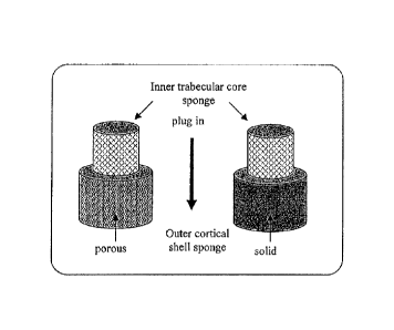
FIG. 2. Schematic showing one example of the making of a bi-layered templates,
with the inner trabecular core sponge snuggly fitted into the outer cortical
shell sponge.
FIG. 3. An exemplary 8-step sintering profile of a calcium phosphate-coated
polyurethane sponge after the first calcium phosphate coating procedure.
FIG. 4. An exemplary 5-step sintering profile for second coated calcium
phosphate
scaffold sintering procedure.
FIG. 5. Flow chart showing a process for producing silver-doped hydroxyapatite
sol.
FIG. 6. An exemplary 3-step sintering profile of a scaffold after coating the
scaffold
with or without silver- or zinc-doped calcium phosphate sol.
FIG. 7. Representative scanning electron micrographs showing a) the untreated
surface of the sponge template, and b) the polyurethane sponge surface after a
20 minute
treatment in 10% NaOH. Microcracks on the treated sponge surface are observed
and these
cracks allow the nucleation of calcium phosphate coatings and ensure the
uniformity of the
coating on the sponge surface.
FIG. 8. TG/DTA curve of a polyurethane sponge template.
FIG. 9. Representative scanning electron micrograph showing a dense and smooth
scaffold surface after sintering (magnification x 5,000). Grain boundaries of
calcium
phosphate on the scaffold surface are also observed.
FIGS. 10A-C. Representative scanning electron micrographs (SEM) of a scaffold
of
the present invention after 2nd sintering showing a) cross-section of the
interconnecting
secondary microchannels within the strut, b) high magnification (magnification
x 1,500) of
12
CAL LAW\ 2718625\1
CA 2965384 2017-04-28
the strut showing the triangular secondary microchannel, with length of each
side ranging
from 30 pini to 120 pm, and c) a primary pore having diameter ranging from 150
lam to 750
pm (magnification x 150).
FIGS. 11A-B. Representative scanning electron micrographs (SEM) showing the
surface and thickness of a strut after a) 16t time coating and sintering, and
b) 2nd time coating
and sintering.
=
FIG. 12. Representative cross section of a calcium phosphate scaffold
infiltrated with
bone tissue and vascular in-growth after 12 weeks post surgery, 200X (S =
scaffold, V =
vessel). This histological section is viewed under a phase contrast
microscope.
FIG. 13. A non-limiting method of the present invention.
FIGS. 14A-D. Scanning electron microscopy of different cross sections of one
scaffold of the present invention showing (FIG. 14A) - outer cortical shell
with microchannel;
(FIG. 14B) - inner layer of strut; (FIG. 14C) ¨ roughed surface of strut; and
(FIG. 14D) ¨
cross section of the hollow strut.
FIGS. 15A-XX. A pictorial description of a method of preparing a scaffold of
the
present invention. Comments regarding certain figures are as follows:
FIG. 15A.
Polyurethane (PU) sponges may be used to produce interconnected porous CaP
scaffolds.
FIG. 15C-G. To change PkI sponge surface characteristics from hydrophobic to
hydrophilic
and increase wetability, a prepared PU sponge may be ultrasonically treated in
10% Na0II
solution for 20-30 minutes prior to use. Cleaning with flowing water for 15-20
minutes
followed. During cleaning, the sponge may be squeezed and expanded 3-4 times
to rinse
residual NaOH inside the PU sponge. Ultrasonically cleaning with distilled
water for 15-20
Minutes may follow. After removing water with, e.g., a paper towel, the sponge
may be
placed in a 60-80 C oven until completely dry (e.g., 80 C for 5 hours). FIG.
15H. After
completely drying the core sponge (cancellous bone part), it may he plugged
into an outside
shell (cortical bone part) porous sponge or solid shell depending on what is
desired in the final
structure and application. FIG. 15J-K. To make a slip casting slurry, a binder
is preferably
added to the dispersion. The binders may be carboxymethylcellulose (CMC),
polyvinyl
alcohol, starch, sodium silicate, polyvinyl butyral, methacrylate emulsion,
water soluble
polyacrylate, polyacrylic acid, polyethylene glygol, etc. A particularly
preferable binder, in
certain embodiments, is carboxymethylcellulose and sodium silicate. The amount
of
13
CAL_LAW \ 2718625 \ 1
CA 2965384 2017-04-28
carboxymethylcellulose added is preferably 5-10% by mass and sodium silicate
solution
added is preferably 2-5% by mass based on 100% by mass of calcium phosphate
powder.
After adding carboxymethylcellulose into distilled water, further stirring is
conducted until
completely dissolved then add sodium silicate solution and stirring. FIG. 15L.
To keep
homogeneity and prevent rapid sedimentation of calcium phosphate powder,
ammonium
polyacrylate may be added (e.g., 5-10% by mass based on 100% by mass of powder
for
dispersant). FIG. 15M. To prevent cracks due to rapid drying during the drying
process,
N,N-dimethylformamide may be added (e.g., 10-15% by mass based on 100% by mass
of
powder for drying agent). FIG. 15N. To make the calcium phosphate slurry,
calcium
phosphate powder is slowly spread into the solution. FIG. 150. After adding
the calcium
phosphate powder, further stirring is conducted and also the slurry is heated
at 40-50 C for
water evaporation during stirring until the powder/liquid ratio is 0.3-0.4.
FIG. 15P. Calcium
phosphate slurry may be poured plaster cast mold for casting solid outside
shell. After the
slurry is poured, the plaster cast mold is rotated to obtain a homogeneously
thick solid outside
shell. This may be repeated several times until the desired outside shell
thickness is achieved.
FIG. 150. After the solid outside green body shell is completely castcd, it
may be dried at
30 C and above 80% humidity in a chamber. The green body may then be separated
from the
plaster cast mold and dried at 25 C, under 30% humidity air conditions, for 6-
24 hours
depending on green body size. It is then placed into a furnace for sintering.
FIG. 15R. First
step: heat until 600 C. FIG. 15T-U. To make the 1st coating calcium phosphate
paste, a
binder is preferably added to the dispersion. Such binders are described
herein. After adding
polyvinyl alcohol into distilled water, further stirring is conducted until
all is completely
dissolved; then sodium silicate solution is added with continued stirring.
FIG. 15V-W. The
amount of carboxymethylcellulose as shown in this figure added is preferably 3-
5% by mass.
-- After adding carboxymethylcellulose into solution, further stirring is
conducted until all is
completely dissolved, then ammonium polyacrylate is added (3-5% by mass based
on 100%
by mass of calcium phosphate powder) with stirring. FIG. 15X. To prevent
cracks due to
rapid drying during the drying process, N,N-dimethylformamide may be added
(e.g., 5-10%
by mass amount based on 100% by mass of powder for drying agent). FIG. 15Y.
The
calcium phosphate slurry may be made by slowly spreading calcium phosphate
powder into
the solution. FIG. 15AA. After adding the calcium phosphate, powder further
stirring is
conducted and the slurry is heated at 40-50 C for water evaporation during
stirring until the
powder/liquid ratio is 1.0-1.25. If stirrer bar is stopped during stirring,
stir with a Teflon bar
until to get the desired powder/liquid ratio. FIG. 15BB. The pre-treated hi-
layered PU
14
CAL LAW \ 2718625 \ 1
CA 2965384 2017-04-28
sponge is immersed in the calcium phosphate paste then squeezed and expanded 5-
7 times
using a Teflon bar. Excess paste is removed with air to avoid the primary
pores being filled
with paste. The homogeneous coating may be examined using a stereo microscope.
FIG.
15CC-DD. After examining the homogeneous coating, the pre-formed scaffold is
dried at
30 C, 50-70% humidity. Then the pre-dried calcium phosphate coated mono or hi-
layered
pre-formed scaffold is dried at 25 C, under 30% humidity air conditions, for 6-
24 hours
depending on the pre-formed size. After completely driying, the pre-formed
scaffold is put
into a furnace for 1st sintering. FIG. 15FF-KK. See comments of FIG. 15T-Y.
FIG. 15MM.
After adding the calcium phosphate powder, further stirring is conducted and
the slurry is
heated at 40-50 C for water evaporation during stirring until powder/liquid
ratio is 0.3-0.4.
FIG. 15NN. The 1st sintered mono or hi-layered scaffold is immersed in the
calcium
phosphate slurry and taken out after 5 seconds. Excess slurry is removed using
air to avoid
filling the primary pores with slurry. FIG. 1500. The 2nd time coated mono or
hi-layered
scaffold is centrifuged to remove the 2nd excess slurry and to obtain a
homogeneous coating
for 10-20 seconds at 1000-2000 rpm, depending on scaffold size and slurry
viscosity. FIG.
15PP-QQ. After centrifuging, the scaffold is dried at 25 C, under 30% humidity
air
conditions, for 6-24 hours depending on the pre-formed size. After completely
drying the 2nd
time coated scaffold, it is placed into a furnace for 2nd sintering. FIG.
15RR. Antibacterial
calcium phosphate doped with silver or zinc can be synthesized using the Sol-
Gel method.
FIG. 15SS. The silver- or zinc- doped calcium phosphate sol is prepared by
synthesizing the
calcium (Ca), silver (Ag) precursor and the phosphorus (P) precursor. FIG.
15TT. The silver-
or zinc-doped calcium phosphate sol is then synthesized by reacting calcium
and phosphorus
precursors for a period of I to 2 hours and with vigorous stirring. The
reaction is performed
under an argon atmosphere. FIG. 15UU. The synthesized silver- or zinc-doped
calcium
phosphate sol is then filtrated through a 0.20 Jim to 0.45 p.m syringe filter,
followed by aging
at temperatures ranging from 40 C to 80 C and for a period ranging from 12 to
204 hours.
FIG. 15VV. The fabricated porous calcium phosphate scaffolds are then immersed
in the
aged calcium phosphate sol doped with or without silver or zinc. After
immersing for 5 to 10
seconds, the scaffold is then removed from the sol and air blown to unclog the
pores. FIG.
15WW. The scaffolds are centrifuged to remove excess sol. FIG. 15XX. The
calcium
phosphate sol coated scaffold is then baked and dried in an oven at
temperatures ranging from
50 C to 100 C and for a period ranging from 3 to 8 hours. After they are
completely dried,
the calcium phosphate sol-coated scaffolds are then heat-treated at
temperatures ranging from
600 C to 700 C using a muffle furnace in air for a period ranging from 1 hour
to 5 hours.
CALLAW\ 2718625\1
CA 2965384 2017-04-28
FIG. 16. Curved custom made scaffold to fit the shape, anatomical structure
and size
of rabbit tibia.
FIGS. 17A-B. Granules of the present invention. FIG. 17B ¨ imaging of granules
following placement in bony defect.
DESCRIPTION OF ILLUSTRATIVE EMBODIMENTS
The present invention is based on the development of scaffolds for bone and
tissue
repair that permit facile transport of nutrients and ions from the surrounding
environment into
the scaffold, thereby promoting restoration of tissue structure and function.
A. Scaffold Components
The scaffolds of the present invention may be composed of a variety of
components.
The components can be obtained from natural sources, commercial sources, or
can be
chemically synthesized. In
particular embodiments, the scaffold includes a calcium
phosphate. Regarding natural sources, calcium phosphates are found in bone,
teeth and shells
of a large variety of animals. It exists in a variety of forms known in the
art, and non-limiting
examples include hydroxyapatite (Hydroxyapatite,
Ca10(PO4)6(0II)2,
Ca/P=1.67), tricalcium phosphate (TCP, Ca3(PO4)2, Ca/P=1.5) and
brushite
(CaHPO4.2H20, Ca/P=1. Hydroxyapatitc has characteristics similar to
mineralized
matrix of natural bone, and is biocompatible. Non-limiting examples of calcium
compounds
include calcium nitrate tetrahydrate, calcium nitrate, and calcium chloride.
Non-limiting
examples of phosphorus .compounds include triethylphosphate, sodium phosphate,
and
ammonium phosphate dibasic. One of ordinary skill in the art would be familiar
with the
wide variety of calcium phosphates known in the art, and sources of such
compounds.
There are several reported methods for the synthesis of hydroxyapatite.
Processes
include aqueous colloidal precipitation, sol-gel, solid-state and mechano-
chemical methods.
Information regarding stabilized calcium phosphate complexes can be found in
U.S. Patent
App. Pub. No. 20080075675. Additional information regarding synthesis of
hydroxyapatite
can be found in U.S. Patent App. Pub. No. 20080095820 and U.S. Pat. 6,171,610.
This method includes reacting calcium and a non-acidic ionic phosphate, such
as
trisodium phosphate, in the presence of hydroxyl ions. U.S. Pat. Nos.
5,258,044, 5,306,305,
16
CA L_LAW \ 2718625 \ 1
CA 2965384 2017-04-28
5,543,019, 5,650,176, 5,676,976, 5,683,461, 5,783,217, 5,843,289, 6,027,742,
6,033,582,
6,117,456, 6,132,463 and 6,214,368 disclose methods of synthesizing calcium
phosphate
particles and a variety of biomedical uses.
The scaffolds of the present invention may include any component known to
those of
ordinary skill in the art to be suitable for inclusion in a biomedical
scaffold. Other non-
limiting examples of such components include polymethylmethacrylate (PMMA),
calcium
sulfate compounds, calcium aluminate compounds, aluminum silicate compounds,
bioceramic
materials, or polymers. Examples of the bioceramic material include calcium
phosphate-
based oxide, such as apatite, BIOGLASSTM, glass oxide, titania, zirconia, and
alumina. Other
suitable materials include alginate, chitosan, coral, agarose, fibrin,
collagen, bone, silicone,
cartilage, aragonite, dahlite, calcite, amorphous calcium carbonate, vaterite,
weddellite,
whewellite, struvite, urate, ferrihydrite, francolite, monohydrocalcite,
magnetite, goethite,
dentin, calcium carbonate, calcium sulfate, calcium phosphosilicate, sodium
phosphate,
calcium aluminate, a-tricalcium phosphate, a dicalcium phosphate, P-tricalcium
phosphate,
tctracalcium phosphate, octacalcium phosphate (OCP), fluoroapatite,
chloroapatite,
magnesium-substituted tricalcium phosphate, carbonate hydroxyapatite, and
combinations and
derivative thereof. Examples of silicon compounds include
tetraethylorthosilicate, 3-
mercaptopropyltrimethoxysilane, and 5,6-cpoxyhexyltriethoxysilane.
The scaffolds of the present invention may optionally include any number of
additional additives. In some embodiments, additives are added to a portion of
the scaffold.
For example, a scaffold may include additives in the cortical shell but not in
the inner
trabecular core, or vice versa. In some embodiments, there are additives in
both the cortical
shell and trahecular core. Non-limiting examples of additives include
radiocontrast media to
aid in visualizing the scaffold with imaging equipment. Examples of
radiocontrast materials
include barium sulfate, tungsten, tantalum, or titanium. Additives that
include osteoinductive
materials may be added to promote bone growth into the hardened bone
augmentation
material. Suitable osteoinductive materials may include proteins from
transforming growth
factor (TGF) beta superfamily, or bone-morphogenic proteins, such as BMP2 or
BMP7.
In preferred embodiments of the present invention the scaffolds set forth
herein are
biocompatible. The term "biocompatible" is intended to describe any material
which upon
implantation does not elicit a substantial detrimental response in vivo.
17
CALLAW1 2718625\1
CA 2965384 2017-04-28
In particular embodiments of the present invention, the scaffold is
biodegradable,
bioerodible, or resorbable, unless a permanent matrix is desired. The terms
"biodegradable",
"bioerodable" and "resorbable" are used herein interchangeably. When used to
characterize
materials, they refer to materials that degrade under physiological conditions
to form a
product that can be metabolized or excreted without damage to the subject. In
certain
embodiments, the product is metabolized or excreted without permanent damage
to the
subject. Biodegradable materials may be hydrolytically degradable, may require
cellular
and/or enzymatic action to fully degrade, or both. Other degradation
mechanisms, e.g.,
thermal degradation due to body heat, are also envisioned. Biodegradable
materials also
include materials that are broken down within cells. Degradation may occur by
hydrolysis,
enzymatic processes, phagocytosis, or other processes.
Either natural or synthetic polymers can be used to form the scaffold matrix.
U.S. Pat.
Nos. 6,171,610, 6,309,635 and 6,348,069, disclose a variety of matrices for
use in tissue
engineering.
In some embodiments which include an outer cortex and an inner core, only the
outer
cortex is biodegradable. In further embodiments, only the inner core is
biodegradable. Non-
limiting examples of synthetic polymers suitable for inclusion in the
scaffolds of the present
invention include fibrin, collagen, glycosaminoglycans (GAGs), such as chitin,
chitosan and
hyaluronic acid, polysaccharides, such as starch, carrageenan, alginate,
heparin, glycogen and
cellulose, polylactidc (PLA), polylactide-co-glycolide (PLGA), polyglycolic
acid (PGA),
polyurethanes, polycaprolactone, polymethyl methacrylate (PMMA), polyamino
acids, such
as poly-L-lysine, polyethyleneimine, poly-
anhydrides, polypropylene-fumarate,
polyearbonates, polyamides, polyanhydricles, polyortho esters,
polyacetals,
polycyanoacrylates and degradable polyurethanes.
Useful non-erodible polymers include without limitation, polyacrylates,
ethylene-vinyl
acetate polymers and other acyl substituted cellulose acetates and derivatives
thereof, non-
erodible polyurethanes, polystyrenes, polyvinyl chloride, polyvinyl fluoride,
poly(vinyl
imidazole), chlorosulphonated polyolifins, polyethylene oxide, polyvinyl
alcohol,
TEFLON.TM., nylon, stainless steel, cobalt chrome, titanium and titanium
alloys, and
bioinert ceramic particles (e.g., alumina and zirconia particles),
polyethylene,
polyvinylacetate, polymethylmethacrylate, silicone, polyethylene oxide,
polyethylene glycol,
18
CAL_LAW \ 2718625 \ 1
CA 2965384 2017-04-28
polyurethanes, and natural biopolymers (e.g., cellulose particles, chitin,
keratin, silk, and
collagen particles), and fluorinated polymers and copolymers (e.g.,
polyvinylidene fluoride).
In some embodiments, the scaffold is coated with compounds to facilitate
attachment
of cells to the scaffold. Examples of such compounds include basement membrane
components, agar, agarose, gelatin, gum arabic, collagens types I, II, III,
IV, and V,
fibronectin, laminin, glycosaminoglycans, polyvinyl alcohol, and mixtures
thereof.
In some embodiments, mammalian cells are incorporated into the scaffolds.
Information regarding incorporation of mammalian cells can be found in U.S.
Pat. App.
20080085292. For example, mammalian cells may he seeded or cultured with the
scaffolds
of the present invention prior to implantation in a subject. Examples of such
cells include, but
are not limited to, bone .marrow cells, smooth muscle cells, stromal cells,
stem cells,
mesenchymal stem cells, synovial derived stem cells, embryonic stem cells,
umbilical cord
blood cells, umbilical Wharton's jelly cells, blood vessel cells,
chondrocytes, osteoblasts,
osteoclasts, precursor cells derived from adipose tissue, bone marrow derived
progenitor cells,
kidney cells, intestinal cells, islets, beta cells, pancreatic ductal
progenitor cells, Sertoli cells,
peripheral blood progenitor cells, fibroblasts, glomus cells, kcratinocytes,
nucleus pulposus
cells, annulus fibrosus cells, fibrochondrocytcs, stem cells isolated from
adult tissue, oval
cells, neuronal stem cells, glial cells, macrophages and genetically
transformed cells or
combination of the above cells. The cells can be seeded on the scaffolds for a
short period of
time just prior to implantation (such as one hour, six hours, 24 hours), or
cultured for longer
periods of time (such as 2 days, 3 days, 5 days, 1 week, 2 weeks) to promote
cell
proliferation and attachment within the scaffold prior to implantation.
B. Fabrication of Scaffolds
= 1. Formation of Pores and Microchannels
Formation of pores and microchannels in the scaffolds set forth herein may be
accomplished using any method known to those of ordinary skill in the art. In
some
embodiments, as discussed in the Example section below, micropores and
microchannels are
created in a scaffold using a template, such as a sponge. A composition, such
as a calcium
phosphate, is then applied to the template. For example, in some embodiments
the method
= 30 includes (a) contacting a porous polymer sponge with a
composition that includes a suitable
19
CALLAW \ 27186250
material for scaffold formation, wherein at least a portion of the sponge
becomes coated with
the composition; and (b) drying the composition-coated sponge, wherein a bone
scaffold is
formed. In some embodiments, the sponge is burned out of the scaffold.
Other methods of creating micropores or microchanncls that may be applied in
the
context of the present invention include, but are not limited to, leaching
processes, gas
foaming processing, supercritical carbon dioxide processing, sintering, phase
transformation,
freeze-drying, cross-linking, molding, porogen melting, polymerization, melt-
blowing, and
salt fusion (reviewed in Murphy et al., 2002, Tiss. Eng. 8(1):43-52;
Karageorgiou et al., 2005,
Biomaterials 26(27):5474-5491). Porosity may be a feature of the composition
during
manufacture or before implantation, or the porosity may only be available
after implantation.
Additional information regarding formation of pores in a scaffold can be found
in U.S. Patent
App. Pub. No. 20080069852. In some embodiments, microchanncls and/or larger
channels are
drilled into the scaffold following molding.
The present invention also contemplates applications using porogens to create
latent
pores in a composite. These latent pores may arise from including porogens in
the composite.
The porogen may be any chemical compound that will reserve a space within the
composite
while the composite is being molded and will diffuse, dissolve, and/or degrade
prior to or after
implantation leaving a pore in the composite. Porogens may be of any shape or
size, such as
spheroidal, cuboidal, rectangular, clongantcd, tubular, fibrous, disc-shaped,
platelet- shaped,
or polygonal. In certain embodiments, the porogcn is granular. The porogen may
be a gas,
liquid, or solid. Exemplary gases that may act as porogcns include carbon
dioxide, nitrogen,
argon, or air. Exemplary liquids include water, organic solvents, or
biological fluids (such as
blood, lymph, plasma). Examples of possible solid porogens include water
soluble compounds
such as carbohydrates (e.g., sorbitol, dextran poly(dextrose), starch), salts,
sugar alcohols,
natural polymers, synthetic polymers, and small molecules.
Additional information regarding incorporation of pores into a material can be
found
in U.S. Patent App. Pub. NII. 20080103227.
2. Shaping
The scaffolds set forth herein can be formed into a desired shape using any
method
known to those of ordinary skill in the art. For example, the scaffold may be
molded into a
desired shape or fractured into granules. The granules retain the essential
micropores and/or
CA 2965384 2019-08-01
CA 2965384 2017-04-28
microchannels. The scaffold may be configured by the surgeon prior to
implantation or at the
time of implantation into a desired shape, such as a curved custom made
scaffold to fit the
shape, anatomical structure and size of tibia shown as FIG. 16. In some
embodiments, a
scaffold of the present invention is fractured into granules which in turn can
he packed into a
bony defect by the surgeon. The granules may be of a uniform size, or of
varying sizes.
3. Formation of Cortex and Coatings
Certain embodiments of the present scaffolds include an outer cortex or
coating.
Formation of an outer cortex or coating on a core component can be performed
using any
method known to those of ordinary skill in the art. As discussed in the
Examples below, a
template (such as a sponge) may be applied in forming an outer cortex on a
scaffold. U.S.
Patent App. Pub. No. 20080097618 provides information regarding deposition of
calcium
phosphate coatings on surfaces. In some embodiments, forming a coating
involves dipping or
immersing a scaffold in a *composition or a plasma spray deposition process.
Information
concerning immersion techniques can be found in U.S. Pat. Nos. 6,143,948,
6,136,369 and
6,344,061.
C. Therapeutic Applications
Accordingly, methods and scaffolds of the present invention may also be used
to treat,
or prevent, a hone disease, bone disorder, or bone injury (e.g., a bone
fracture). "Treatment"
and "treating" as used herein refer to administration or application of a
therapeutic agent to a
subject or performance of a procedure or modality on a subject for the purpose
of obtaining a
therapeutic benefit of a disease or health-related condition.
The term "therapeutic benefit" or "therapeutically effective" as used
throughout this
application refers to anything that promotes or enhances the well-being of the
subject with
respect to the medical treatment of a condition. This includes, hut is not
limited to, a
.. reduction in the frequency or severity of the signs or symptoms of a
disease.
"Prevention" and "preventing" are used according to their ordinary and plain
meaning
to mean "acting before" or such an act. In the context of a particular disease
or health-related
condition, those terms refer to administration or application of an agent,
drug, or remedy to a
subject or performance of a procedure or modality on a subject for the purpose
of blocking the
onset of a disease or health-related condition. For example, a scaffold of the
present invention
21
CAL LAW\ 2718625\1
CA 2965384 2017-04-28
may be used to prevent bone disease in a subject. The scaffolds of the present
invention may,
in certain embodiments, be utilized as an implant for a therapeutic benefit.
In particular
embodiments, the implants are used for bone augmentation, such as in large
defects. In
certain embodiments, the scaffolds of the present invention are shaped to
duplicate bone lost
by a subject, such as a subject who has lost bone matter due to, e.g., an
accident, war,
gunshot, or surgery. Scaffolds shaped in this matter, may, for example, be
implanted in the
subject such that the body may regenerate bone tissues to replace the lost
matter.
Therapeutic agents may be added to the scaffolds or incorporated into the
scaffolds of
the present invention using any method known to those of ordinary skill in the
art. A
"therapeutic agent" as used herein refers to any agent that can be applied in
the diagnosis,
treatment, or prevention of a disease or health-related condition in a
subject. Therapeutic
agents include biomolecules. The term "biomolecules", as used herein, refers
to the class of
molecules (e.g., proteins, amino acids, peptides, polynucleotides,
nucleotides, carbohydrates,
sugars, lipids, glycoproteins, nucleoproteins, lipoproteins, steroids, etc.)
that are commonly
found in cells or tissues, whether the molecules themselves are naturally-
occurring or
artificially created (e.g., by synthetic or recombinant methods). For example,
biomolecules
include, but are not limited to, enzymes, receptors, neurotransmitters,
hormones, cytokines,
cell response modifiers such as growth factors and chemotactic factors,
antibodies, vaccines,
=
haptens, toxins, interferons, ribozymes, anti-sense agents, plasmids, DNA, and
RNA.
7() Thus, the
therapeutic agent may be any agent known to those of ordinary skill in the
art. One or more therapeutic agents may be coated on the surface of the
scaffold,
incorporated into the matrix, incorporated into microspheres which are
suspended and
distributed in the matrix, or the scaffold can he immersed in a composition
comprising one or
more agents prior to implantation in a subject.
Examples of classes of therapeutic agents include osteogenic, osteoinductive,
and
osteoconductive agents, anti-cancer substances, antibiotics, anti-inflammatory
agents,
immunosuppressants, anti-viral agents (including anti-HIV agents), enzyme
inhibitors,
neurotoxins, opioids, hypnotics, antihistamines, lubricants, tranquilizers,
anti-convulsants,
muscle relaxants, anti-Parkinson agents, antispasmodics, antibiotics,
antiviral agents,
antifungal agents, modulators of cell-extracellular matrix interactions
including cell growth
inhibitors and anti-adhesion molecules, vasodilating agents, inhibitors of
DNA, RNA, or
protein synthesis, antiypertensives, analgesics, anti-pyretics, steroidal and
non-steroidal anti-
22
CAL LAW\ 2718625\1
CA 2965384 2017-04-28
inflammatory agents, anti-angiogenic factors, angiogenic factors, anti-
secretory factors,
anticoagulants and/or antithrombotic agents, local anesthetics,
prostaglandins, targeting
agents, chemotactic factors, receptors, neurotransmitters, proteins, cell
response modifiers,
cells, peptides, polynucleotides, viruses, vaccines, amino acid, peptide,
protein, glycoprotein,
lipoprotein, antibody, steroidal compound, antibiotic, antimycotic, cytokine,
vitamin,
carbohydrate, lipid, extracellular matrix, extracellular matrix component,
chemotherapeutic
agent, cytotoxic agent, growth factor, anti-rejection agent, analgesic, anti-
inflammatory agent,
viral vector, protein synthesis co-factor, hormone, endocrine tissue,
synthesizer, enzyme,
polymer-cell scaffolding agent with parenchymal cells, angiogenic drug,
collagen lattice,
antigenic agent, cytoskeletal agent, mesenchymal stem cells, bone digester,
antitumor agent,
cellular attractant, fibronectin, growth hormone cellular attachment agent,
immunosuppressant, nucleic acid, surface active agent, hydroxyapatite, and
penetration
enhancer, anti-inflammatory agents, growth factors, angiogenic factors,
antibiotics,
analgesics, chemotactic factors, bone morphogenic protein, and cytokines.
In particular embodiments the therapeutic agent is an agent that promotes
wound
healing or prevent infection.. Non-limiting examples of such agents include
antibiotics, anti-
inflammatory drugs, or analgesics.
Non-limiting examples of therapeutic agents include non-collagenous proteins
such as
osteopontin, osteonectin, bone sialo proteins, fibronectin, laminin,
fibrinogen, vitronectin,
trombospondin, proteoglycans, decorin, proteoglycans, beta-glycan, biglycan,
aggrecan,
veriscan, tanascin, matrix gla protein hyaluran, cells; amino acids; peptides;
inorganic
elements; inorganic compounds; organometallic compounds; cofactors for protein
synthesis;
cofactors for enzymes; vitamins; hormones; soluble and insoluble components of
the immune
system; soluble and insoluble receptors including truncated forms; soluble,
insoluble, and cell
surface bound ligands including truncated forms; chemokines, interleukines;
antigens;
bioactive compounds that are endocytozed; tissue or tissue fragments;
endocrine tissue;
enzymes such as collagenase, peptidases, oxidases, etc; polymeric cell
scaffolds with
parenchymal cells; angiogenic drugs, polymeric carriers containing bioactive
agents;
encapsulated bioactive agents; bioactive agents in time-release form; collagen
lattices,
antigenic agents; cytoskeletal agents; cartilage fragments; living cells such
as chondrocytes,
osteoblasts, osteoclasts, fibroclasts, bone marrow cells, mesenchymal stem
cells, etc; tissue
transplants; bioadhesives; bone morphogenic proteins (BMPs), transforming
growth factors
(TGF-.beta.), insulin-like growth factor, platelet derived growth factor
(PDGF); fibroblast
23
CAL LAW1 2718625\1
CA 2965384 2017-04-28
growth factors (FGE), vascular endothelial growth factors (VEGF), epidermal
growth factor
(EGF), growth factor binding proteins, e.g., insulin-like growth factors;
angiogenic agents;
bone promoters; cytokines; interleukins; genetic material; genes encoding bone
promoting
action; cells containing genes encoding bone promoting action; cells
genetically altered by the
hand of man; externally expanded autograft or xenograft cells; growth hormones
such as
somatotropin; bone digesters; anti-tumor agents; fibronectin; cellular
attractants and
attachment agents; immunosuppressants; bone resorption inhibitors and
stimulators;
mitogenic factors; bioactive factors that inhibit and stimulate second
messenger molecules;
cell adhesion molecules, e.g., cell-matrix and cell-cell adhesion molecules;
secondary
messengers; monoclonal antibodies specific to cell surface determinants on
mesenchymal
stem cells; portions of monoclonal antibodies specific to cell surface
determinants on
mesenchymal stem cells; portions of monoclonal antibodies specific to cell
surface
determinants on mesenchymal stem cells; clotting factors; polynucleotides; and
combinations
thereof.
The amount of therapeutic agent included in scaffold can vary widely and will
depend
on such factors as the agent being delivered, the site of administration, the
patient's
physiological condition, etc. The optimum levels will be determined in a
specific case based
upon the intended use of the implant.
In some embodiments, a therapeutic nucleic acid is incorporated into the
scaffold.
Information regarding incorporation of a therapeutic nucleic acid into
scaffolds can be found
in U.S. Patent App. Pub. No. 20080095820. Thus, the scaffolds set forth herein
can be
applied as gene delivery vehicles.
The scaffolds of the present invention may be used in many applications. Non-
limiting examples of such applications include the repair of defects or
degeneration of bone,
cartilage, tendons, and ligaments. The scaffolds set forth herein can have
therapeutic
application in other organs of the body as well.
The scaffolds of the present invention can have any desired shape, and the
selection of
such shape will depend largely on the application of the scaffold. Non-
limiting examples of
such shapes include cylinder, block, morsel, wedge, and sheet.
In particular embodiments the scaffold will be cylinder shaped for application
in the
repair of bony defects of long bones. In some embodiments, the scaffold is
configured for the
24
CAL LAW\ 271862511
CA 2965384 2017-04-28
repair of a simple fracture, compound fracture or non-union; as an external
fixation device or
internal fixation device; for joint reconstruction, arthrodesis, arthroplasty
or cup arthroplasty
of the hip; for femoral or humeral head replacement; for femoral head surface
replacement or
total joint replacement; for repair of the vertebral column, spinal fusion or
internal vertebral
fixation; for tumor surgery; for deficit filling; for discectomy; for
laminectomy; for excision
of spinal tumors; for an anterior cervical or thoracic operation; for the
repairs of a spinal
injury; for scoliosis, for lordosis or kyphosis treatment; for intermaxillary
fixation of a
fracture; for mentoplasty; fir temporomandibular joint replacement; for
alveolar ridge
augmentation and reconstruction; as an inlay osteoimplant; for implant
placement and
revision; for sinus lift; for a cosmetic procedure; and, for the repair or
replacement of the
ethmoid, frontal, nasal, occipital, parietal, temporal, mandible, maxilla,
zygomatic, cervical
vertebra, thoracic vertebra, lumbar vertebra, sacrum, rib, sternum, clavicle,
scapula, humerus,
radius, ulna, carpal bones, metacarpal bones, phalanges, ilium, ischium,
pubis, femur, tibia,
fibula, patella, calcaneus, tarsal bones or metatarsal bones.
Some aspects of the present invention concern methods of treating a subject
that
=
involve implanting a scaffold of the present invention into the subject. In
particular
embodiments the subject is a vertebrate, such as a mammal, reptile, fish,
bird, etc. In
particular embodiments the mammal is a human. The subject may be suffering
from a bone
fracture or a bone defect. The subject may have a bone defect due to trauma, a
congenital
abnormality, a genetic abnormality a fracture, an iatrogenic defect, a bone
cancer, a bone
metastasis, an inflammatory disease, an autoimmune disease, a metabolic
disease, or a
degenerative bone disease.
Other examples of bone diseases or disorders include iatrogenic defects, bone
cancer,
bone metastases, inflammatory diseases (such as rheumatoid arthritis),
autoimmune diseases,
metabolic diseases, and degenerative bone disease such as osteoarthritis and
osteoporosis. The
scaffold may be fabricated for the repair of a simple fracture, compound
fracture, or non-
union; as an external fixation device or internal fixation device; for joint
reconstruction,
arthrodesis, arthroplasty, or cup arthroplasty of the hip; for femoral or
humeral head or shaft
repair or replacement; for femoral head surface replacement or total joint
replacement; for
repair of the vertebral column, spinal fusion or internal vertebral fixation;
for discectomy; for
lam inectomy; for excision of spinal tumors; for the repairs of a spinal
injury; for scoliosis, for
lordosis or kyphosis treatment; for for the repair or replacement of the
ethmoid, frontal, nasal,
occipital, parietal, temporal, mandible, maxilla, zygomatic, cervical
vertebra, thoracic
= 25
CAL LAW\ 2718625\1
CA 2965384 2017-04-28
vertebra, lumbar vertebra, sacrum, rib, sternum, clavicle, scapula, humerus,
radius, ulna,
' carpal bones, metacarpal bones, phalanges, ilium, ischium, pubis, femur,
tibia, fibula, patella,
calcaneus, tarsal bones, or metatarsal bones.
D. Kits
In yet another aspect, the present invention provides kits that include a
scaffold of the
present invention. The scaffold may be sterilely packaged. In some embodiments
the kit
includes more than one scalTold of the present invention. The kit may include
instructions for
implanting the scaffold included in the kit. It may further include one or
more therapeutic
agents that can be administered concurrently or consecutively with
implantation of the
scaffold. The therapeutic agents include any such agent known to those of
ordinary skill in
the art, such as any of those agents discussed previously. In some
embodiments, the kit
includes hardware for placement of the scaffold in the subject, or a device
for further shaping
the scaffold into a desired configuration. In some embodiments, the kit
includes a device for
packing in granules into a bony defect.
E. Combination Therapy
= Some embodiments of the methods of treatment of the present invention
contemplate
administering one or more secondary forms of therapy to the subject. For
example, a method
of treating a bone fracture that involves implantation of one of the scaffolds
of the present
invention as set forth herein may involve the administration of one or more
secondary forms
of therapy (e.g., administration of an antimicrobial agent or administration
of an anti-
inflammatory agent).
= The secondary form of therapy can be any type of secondary therapy for
the treatment
or prevention of a disease or disorder. In particular embodiments, the
secondary form of
therapy involves administration of one or more additional pharmacologic
therapies using
conventional methods of administration. Therapy can involve administration of
any
pharmacological agent, examples of which have been set forth elsewhere in this
specification.
For example, administration may be oral administration or intravenous
administration.
Another example of secondary is surgical therapy.
26
CAL LAO/1 2718625\1
CA 2965384 2017-04-28
Administration of the compositions of the present invention to a patient will
follow
general protocols for the administration of therapeutic agent therapy, taking
into account the
toxicity, if any, of these agents. It is expected that treatment may be
repeated as necessary.
F. Examples
The following examples are included to demonstrate preferred embodiments of
the
invention. It should be appreciated by those of skill in the art that the
techniques disclosed in
the examples which follow represent techniques discovered by the inventor to
function well in
the practice of the invention, and thus can be considered to constitute
preferred modes for its
practice. However, those of skill in the art should, in light of the present
disclosure,
appreciate that many changes can be made in the specific embodiments which are
disclosed
and still obtain a like or similar result without departing from the spirit
and scope of the
invention.
EXAMPLE 1
Procedure for Porous Calcium Phosphate Scaffold Fabrication
1.1 Polymeric sponge template preparation
Selection of the template material: A polyurethane (PU) sponge template is
used to produce
uniform interconnected porous calcium phosphate scaffolds. This sponge is used
to provide
the primary structure for the formation of the scaffold struts as well as the
formation of
secondary microchannels within the scaffold struts. The polyurethane sponge
template
chosen may range from 45 pores per inch (ppi) to 80 ppi for the inner
trabecular core,
depending on the final desired pore size. The pore sizes in the inner
trabecular core may range
from 150 gm to 800 gm after sintering to allow bone cell migration, blood
vessel
vascularization, and nutrient supply. Additionally, the 80 ppi to 100 ppi
polyurethane sponge
template or solid calcium phosphate ceramics (with channels and/or pores
produced by slip
casting) may be chosen for the outer cortical shell, depending on the final
desired pore size.
The pores and/or channel or holes for the outer cortical shell may be in the
range of about 50
gm to about 250 pm after sintering, depending on the desired application
place.
Template sponge preparation: The polyurethane sponge is used as a template
and is
first cut to the desired shape and dimension. The cut polyurethane is then
ultrasonically
27
CAL_LAW\ 2718625\1
CA 2965384 2017-04-28
treated in 10% to 15% sodium hydroxide (NaOH) solution for 20 to 30 minutes,
followed by
cleaning in flowing water for 30 to 60 minutes. The treated polyurethane is
then rinsed with
distilled water. During cleaning with water and rinsing with distilled water,
the polyurethane
is squeezed and then allowed to expand for 5 to 10 times in order to remove
the residual
NaOH inside polyurethane sponge template. The polyurethane sponge template is
then
ultrasonically cleaned again in distilled water for 20 to 30 minutes. This is
followed by
squeezing the template sponge with paper towel in order to remove excess
water. The
=
template sponge is then placed in an oven at 60 C to 80 C until completely
drying. The
completely dried sponge template for the inner trabccular core is then snuggly
fitted into the
outer .sponge template for the cortical shell or solid outer shell (with
channels and/or holes
depends on final desired structure and application). At this point, as shown
in FIG. 2, the
sponge template is now one piece (outer cortical shell and inner trabecular
core) and is ready
for calcium phosphate coating.
1.2 1st time coating calcium phosphate slurry preparation
In order to produce a stable and well-shaped three-dimensional interconnective
porous
scaffold, a preferred binder is added to the dispersion. The binders used may
be
carboxymethylcellulose, polyvinyl alcohol, starch, sodium silicate, polyvinyl
butyral,
methacrylate emulsion, water soluble polyacrylate, polyacrylic acid,
polyethylene glygol, etc.
In order to avoid slurry agglomeration and cracking of the scaffold during
drying, a dispersant
and drying agent is added to the dispersion. The preferred binders are
polyvinyl alcohol,
carboxymethylcellulose and sodium silicate. In this invention, ammonium
polyacrylate and
N, W-dimethylformamide will be use as a dispersant and a drying agent,
respectively. The
preferred amount of polyvinyl alcohol, carboxymethylcellulose, sodium
silicate, and
methacrylate emulsion added are 2% to 4% by mass, 2% to 4% by mass, 1% to 2%
by mass
and 1% ¨ 2% by mass, respectively (based on 100% by mass of calcium phosphate
powder).
The polyvinyl alcohol is added to distilled water, heated and stirred until
the polyvinyl
alcohol is completely dissolved. The solution should be clear after complete
dissolution of
the polyvinyl alcohol. As the solution is cooled down to room temperature,
carboxymethylcellulose is added. After complete dissolution of the
carboxymethylcellulose,
sodium silicate solution and methacrylate emulsion are added to the mixture
and stirred.
Additionally 5% to 7% by mass of ammonium polyacrylate dispersant and 3% to 5%
by mass
of N, N-dimethylformamide drying agent are added to the mixture and stirred
continuously.
28
CAL LAW\ 2718625\1
CA 2965384 2017-04-28
The calcium phosphate powders are then slowly dispersed into the solution,
followed by
stirring. In this invention, calcium phosphate powder is generic and refers to
all the different
phases of the calcium phosphate group, including hydroxyapatite, tricalcium
phosphate,
amorphous calcium phosphate, monocalcium phosphate, dicalcium phosphate,
octacalcium
phosphate, tetracalcium phosphate, fluorapatite, carbonated apatite and the
different mixtures
of the different phases. Using continuously slow heating, the solution is
slowly stirred in
order to evaporate the water content and until a powder/liquid ratio of 1.20
to 1.50 is obtained.
The slurry is then allowed to cool down to the room temperature before being
used for
coating.
1.3 1" time calcium phosphate coating, drying and sintering
The treated one piece sponge template containing the outer cortical shell and
inner
trabecular core (from section 1.1) is then immersed into the calcium phosphate
slurry until the
calcium phosphate slurry is fully absorbed into the sponge template scaffold.
The
polyurethane is then rolled on a glass plate with rod bar then allowed to
expand for 5 to 10
times in order to remove excess slurry. After removing the excess slurry, some
of pores may
be clogged up with slurry because of high slurry viscosity. In order
to ensure
interconnectivity, uniformity, and open pores, the scaffolds are slightly
blown with air. In this
process, it is preferred that=thc template is homogeneously coated on the
inside and outer the
sponge template. If this homogeneous coating is not achieved, the calcium
phosphate-coated
sponge template scaffold will collapse after sintering or fracture during
handling.
Additionally, the homogeneous coating is preferred for the successful
production of the
secondary microchannels within the main scaffold struts.
Based on the thermoanalysis of polyurethane sponge template and nano-size
powders,
the calcium phosphate-coated sponge template scaffolds dry at 25 C to 35 C and
at 60% ¨
80% humid environment. Drying time will range from 12 to 72 hours, depending
on the size
of the sponge template scaffolds. After drying, the calcium phosphate-coated
sponge
template scaffold typically shrinks about 8% to 10%. After the sponges are
completely dried,
the coated sponges are then placed on an alumina plate, placed in a high
temperature furnace,
and sintered for 2 to 5 hours at 1200 C to 1250 C using an 8-step sintering
profile shown in
FIG. 3. Sintering will further shrink the calcium phosphate-coated sponge
template scaffolds
by 22% ¨ 25%.
29
CAL_LAW\ 271E3625 \1
CA 2965384 2017-04-28
1.4 2"d time coating calcium phosphate suspension preparation
The second time coating is performed to fill up coating defects from first
time coating
performed in section 1.3. This second time coating will improve the
compressive strength of
the scaffold and to ensure a more rounded strut to enhance cell attachment. In
order to make
the second time coating calcium phosphate suspension, different amounts of the
same binders
and chemical agents used in the first time coating calcium phosphate slurry
preparation
(section 1.2) are utilized. However, the concentrations of the binders and
chemical agents
used are different from the 1st time coating calcium phosphate slurry
preparation (section 1.2).
The preferred amount of polyvinyl alcohol, carboxymethylcellulose, sodium
silicate, and
methacrylate emulsion used in the second time coating calcium phosphate
suspension
preparation are about 3% to about 7% by mass, about 3% to about 7% by mass,
about 1% to
about 2% by mass and about 1% to about 2% by mass, respectively (based on 100%
by mass
of calcium phosphate powder).
The polyvinyl alcohol is added to distilled water, heated and stirred until
the polyvinyl
alcohol is completely dissolved. The solution should be clear after complete
dissolution of
the polyvinyl alcohol. As the solution is cooled down to room temperature,
carboxymethylcellulose is added. After complete dissolution of the
carboxymethylcellulose,
sodium silicate solution and methacrylate emulsion are added to the mixture
and stirred.
Additionally 7% to 10% by mass of ammonium polyacrylate dispersant and 5% to
7% by
mass of N, N-dimethylformamide drying agent are added to the mixture and
stirred
continuously. The calcium phosphate powders are then slowly dispersed into the
solution,
followed by stirring. Using continuously slow heating, the solution is slowly
stirred in order
to evaporate the water content and until a powder/liquid ratio of 0.4 to 0.5
is obtained. The
slurry is then allowed to cool down to the room temperature before being used
for coating.
1.5 2nd coating, drying and sintering
Scaffolds after the first coating and sintering (section 1.3) is immersed into
second
time coating calcium phosphate suspension (section 1.4) for 10 to 20 seconds.
After
immersion, the scaffolds are removed from the suspension. Most of the scaffold
pores will be
clogged up by the calcium phosphate suspension. In order to ensure
interconnectivity,
uniformity, and open pores, the scaffolds are slightly blown with air. The
calcium
phosphate-coated sponge template scaffolds are then dried at 25 C to 35 C and
at 60% to
CALLAW \ 271 8625 1
CA 2965384 2017-04-28
70% humid environment. Drying timc will range from 12 to 48 hours, depending
on the size
of the sponge template scaffolds. After the sponges are completely dried, the
coated sponges
are then placed on an alumina plate, placed in a high temperature furnace, and
sintered for 2
to 3 hours at 1200 C to 1250 C using a 5-step sintering profile shown in FIG.
4.
= 5 1.6 Solid outer shell with channels and/or holes calcium
phosphate suspension
preparation
For scaffolds to be used in loading hearing applications, solid outer shells
with
channels having diameters ranging from about 100 ttm to about 200 tim and/or
holes having
diameter ranging from about 200 )im to about 500 tim can fabricated by slip
casting and
freeze drying method. In order to produce a slip casting suspension, the same
binders and
same chemical agents in section 1.2 as well as section 1.4 are used. The
preferred amount of
polyvinyl alcohol, carboxymethylcellulose, sodium silicate, and methacrylate
emulsion used
in the second time coating calcium phosphate suspension preparation are 3% to
7% by mass,
3% to 7% by mass, 0.5% to 1% by mass and 0.5% to 1% by mass, respectively
(based on
100% by mass of calcium phosphate powder).
The polyvinyl alcohol is added to distilled water, heated and stirred until
the polyvinyl
alcohol is completely dissolved. The solution should be clear after complete
dissolution of
the polyvinyl alcohol. As the solution is cooled down to room
temperature,
carboxymethylcellulose is added. After complete dissolution of the
carboxymethylcellulose,
sodium silicate solution and methacrylate emulsion is added to the mixture and
stirred.
Additionally about 7% to about 10% by mass of ammonium polyacrylate dispersant
and about
5% to about 7% by mass of N, N-dimethylformamide drying agent are added to the
mixture
and stirred continuously. The calcium phosphate powders are then slowly
dispersed into the
solution, followed by stirring. Continuously slow heat and stir the solution
to evaporate the
water content until a powder/liquid ratio of about 0.4 to about 0.5 is
obtained. The slurry is
then allowed to cool down to the room temperature before being used for slip
casting.
1.7 Slip casting, freeze dry and sintering for solid outer shell
The prepared slip casting calcium phosphate suspension is poured into a
designed
gypsum mold with a polyurethane sponge mesh having about 10 ppi to about 20
ppi. The
gypsum mold containing polyurethane sponge is then rolled at 10 to 20 rpm
until all water is
completely absorbed by the gypsum mold and a desired thickness is achieved.
The gypsum
31
CAL LAW1 271862511
CA 2965384 2017-04-28
mold is then dried using a freeze dryer for a period ranging from 24 to 72
hours. After drying,
the solid outer shell is separated from gypsum mold and holes with diameter
ranging from
200 um to 500 RM are drilled through the outer shell. The well-prepared outer
shell is then
placed in the high temperature furnace and sintered using a 8-step sintering
profile shown in
FIG. 3. After sintering, the ahout 10 ppi to about 20 ppi polyurethane sponge
mesh is burnt
out, resulting in the formation of channels within the shell.
1.8 Antibacterial calcium phosphate sol preparation
Variable calcium phosphates ceramics such as hydroxyapatite (Ca/P=1.67), tri-
calcium phosphate (Ca/P=1.50), meta-calcium phosphate (Ca/P=0.50), calcium
polyphosphate (Ca/P=0.50), dicalcium phosphate dehydrate (Ca/P=1.00),
monocalcium
phosphate anhydrous (Ca/P=0.50) sol can be synthesized using the correct
amount of calcium
and phosphorous precursors and with controlled aging conditions. Additionally,
antibacterial
calcium phosphate sol can be synthesized using silver- or zinc-doped in the
phosphorous
precursor. To make the calcium precursor, a correct amount of calcium nitrate
tetrahydrate
[Ca(NO3)2.4H20 (Aldrich 99%, USA)] is fluxed in sufficient amount of methyl
alcohol and is
dehydrated at temperatures ranging from 150 C to 200 C. After solvent
evaporation, the
calcium precursor is refluxed in sufficient amount of methyl alcohol. In order
to make the
phosphorous precursor, a correct amount of triethyl phosphite [(C21150)3P
(Fluka 97%,
Japan)] is fluxed in sufficient amount of methyl alcohol. This fluxed
phosphorous precursor
is also pre-hydrolyzed for 5 hours in the presence of a catalyst (acetic acid
ICH3COOH1
containing 0.5 mol% to 1.5'mol% of silver nitrate [Ag(NO3)] or 0.5 mol% to
1.5mo1% of zinc
nitrate hydrate [Zn(NO3)2. xH20] and distilled water [H20]). The silver- or
zinc-doped
calcium phosphate sol is then synthesized by reacting calcium and phosphorus
precursors for
a period of 1 to 2 hours and with vigorous stirring. The reaction is performed
under an argon
atmosphere. The synthesized silver- or zinc-doped calcium phosphate sol is
then filtrated
through a 0.20 um to 0.45 um syringe filter, followed by aging at temperatures
ranging from
40 C to 80 C and for a period ranging from 12 to 204 hours. After aging, the
calcium
phosphate sol viscosity will be between about 8.0 cps to about 160 cps,
depending on the
aging temperature, aging time, methods of sealing the beakers/vials containing
the precursor
during aging, and whether aging is performed in air circulation or without
circulation
condition. This means the calcium phosphate sol viscosity will govern the
thickness,
porosity, and density of the coating layer.
32
CAL LAW\ 2718625\1
CA 2965384 2017-04-28
1.9 Calcium phosphate sol coating, drying and sintering
The fabricated porous calcium phosphate scaffolds from section 1.5 and 1.7 are
immersed in the aged calcium phosphate sol doped with or without silver or
zinc. After
immersing for 5 to 10 seconds, the scaffolds are removed from the sol and
centrifuged to
remove excess sol. The calcium phosphate sol coated scaffold is then baked and
dried in an
oven at temperatures ranging from 50 C to 100 C and for a period ranging from
3 to 8 hours.
After completely dried, the calcium phosphate sol-coated scaffolds are then
heat-treated at
temperatures ranging from ,600 C to 700 C using a muffle furnace in air for a
period ranging
from 1 hour to 5 hours shown in FIG. 6.
EXAMPLE 2
Examples of Fabricated Hydroxyapatite Scaffolds
Using the process described in Example 1, an example on how this technology is
used
to fabricate hydroxyapatite scaffolds coated with silver-doped hydroxyapatite
is as follows:
2.1 Polymetric sponge template preparation for bi-layered porous scaffold
fabrication:
A 60 ppi (pore per inch) polyurethane sponge template is chosen for trabecular
core
fabrication and the 100 ppi polyurethane sponge template is chosen for outer
cortical shell
fabrication. The polyurethane sponge template for the trabecular core is cut
to a shape of a
solid cylinder with a length of 36 mm and a diameter of 28 mm. The
polyurethane sponge
template for the outer cortical shell is cut to resemble a cylindrical pipe
and is hollow core in
the middle. Dimension for the polyurethane sponge template for the outer
cortical shell is 36
mm in length, with an outer diameter of 30 mm and an inner diameter of 28 mm,
thereby
having a 2 mm wall thickness. These polyurethane sponges are ultrasonically
treated in 10%
sodium hydroxide (NaOH) solution for 20 minutes, followed by cleaning in
flowing water for
40 minutes and then rinsed with distilled water. During cleaning, the sponges
arc squeezed
and then allowed to expand for 10 times to remove the residual NaOH inside
polyurethane
sponge templates. These sponges are then ultrasonically cleaned again in
distilled water for
minutes. The sponges are then squeezed with paper towels to remove excess
water. This
is followed by drying at 80 C in an oven for 5 hours until completely dry is
achieved. After
30 completely drying, the polyurethane sponge template for the trabecular
core is then snuggly
33
CAL LAW\ 2718625\1
CA 2965384 2017-04-28
fitted into the pipe-like polyurethane sponge template for the outer cortical
shell or pipe-like
solid outer shell (with channels and/or holes depends on final desired
structure and
application). At this point, as shown in FIG. 2, the sponge template is now
one piece (outer
cortical shell and inner trabecular core).
2.2 First time
coating hydroxyapatite powder and il-tricalcium phosphate mixed
slurry preparation
As an example of this invention, nano-sized hydroxyapatite powder and nano-
sized 0-
tricalcium phosphate powder are used for the fabrication of scaffolds because
of their ability
to sinter. A 3% (by mass) polyvinyl alcohol (molecular weight of 89,000 to
98,000) is added
to 20 ml of distilled water, heated on the hot plate to 60 C and stirred until
the polyvinyl
alcohol is completely dissolved. The solution should be clear after complete
dissolution of
the polyvinyl alcohol. As the solution is cooled down to room temperature, a
3% (by mass)
carboxymethylcellulose (molecular weight of 10,000; viscosity of 53,000 cps at
25 C) is
added. After complete dissolution of the carboxymethylcellulose, a 1% (by
mass) of sodium
silicate solution and a 1% (by mass) of methacrylate emulsion are added to the
mixture and
stirred. Additionally, a 7% (by mass) of ammonium polyacrylate dispersant and
a 5% (by
mass) of N, N-dimethylformamide drying agent are added to the mixture and
stirred
continuously. The percentage of these binders and drying agents are based on
100% by mass
of calcium phosphate powder. Three grams of hydroxyapatite powder and 3 grams
of 3-
tricalcium phosphate powders are then slowly dispersed into the solution,
followed by
stirring. Using continuously slow heat, the solution is slowly stirred in
order to evaporate the
water content and until a powder/liquid ratio of 1.50 is obtained. The slurry
is then cool down
to the room temperature before being used for coating the polyurethane
sponges.
2.3 1st time calcium phosphate coating, drying and sintering
The treated one piece sponge template containing the (outer cortical shell and
inner
trabecular core (from section 2.1) is immersed in the first time coating
slurry (from section
2.2) until the calcium phosphate slurry is fully absorbed in the sponge
template scaffold.
While in the slurry, the immersed polyurethane sponge template is manually
compressed with
the aid of a stirrer and allowed to expand for 8 times. The sponge template is
then removed
from the slurry and excess slurry is removed by the sponge on glass plate with
a rod bar.
After removal of the excess slurry, some of the pores in the sponge template
may be clogged
with slurry because of the high slurry viscosity. In order to ensure
interconnectivity,
34
CAL LAW\ 2718625\1
CA 2965384 2017-04-28
uniformity, and open pores, the scaffolds are slightly blown with air.
Based on the
thermoanalysis of polyurethane sponge templates and nano-size powders, the
calcium
phosphate slurry-coated sponge template scaffolds are then dried at 27 C (in
80% humidity in
still air environment) for 60 hours. After drying, the calcium phosphate-
coated sponge
template scaffolds shrink by 8%. After completely drying, the sponge template
scaffolds are
placed on an alumina plate and sintered in a furnace. The dried calcium
phosphate coated
sponge template scaffolds are sintered by using the 8-step sintering profile
shown in FIG. 3.
After sintering, the calcium phosphate coated sponge template scaffolds shrink
by 22%. In
addition to FIG. 3, details of the 8-step sintering profile, with heating rate
and final
temperature is as follows:
Step 1: heat 2 C/minute until 230 C.
Step 2: heat 1 C/minute until 280 C.
Step 3: heat 0.5 C/minute until 400 C.
Step 4: heat 3 C/minute until 600 C.
Step 5: keep 600 C for 1 hour.
Step 6: heat 5 C/minute until 1230 C.
Step 7: keep 1230 C for 3 hours.
Step 8: cool 5 C/minute to room temperature.
2.4 2nd time coating calcium phosphate suspension preparation
For the second time coating calcium phosphate suspension, different amounts
but the
same binders and chemical agents (in section 2.2) are used. A 3% (by mass)
polyvinyl
alcohol (molecular weight of 89,000 to 98,000) is added to 20 ml of distilled
water, heated on
a hot plate to 60 C and stirred until the polyvinyl alcohol is completely
dissolved. The
solution should be clear after complete dissolution of the polyvinyl alcohol.
As the solution is
cooled down to room temperature, a 5% (by mass) carboxymethylcellulose
(molecular weight
of 10,000; viscosity of 53,000 cps at 25 C) is added. After complete
dissolution of the
carboxymethylcellulose, a 1% (by mass) of sodium silicate solution and a 1%
(by mass) of
methacrylate emulsion are added to the mixture and stirred. Additionally, a
10% (by mass) of
ammonium polyacrylate dispersant and a 7% (by mass) of N, N-dimethylformamide
drying
agent are added to the mixture and stirred continuously. The percentage of
these binders and
drying agents are based on 100% by mass of calcium phosphate powder. 1.5 grams
of
hydroxyapatite powder and 1.5 grams of f3-1ricalcium phosphate powders are
then slowly
dispersed into the solution, followed by stirring. Using continuously slow
heat, the solution is
slowly stirred in order to evaporate the water content and until a
powder/liquid ratio of 0.50 is
obtained. The slurry is then cool down to the room temperature before being
used for coating.
CA L_LAW \ 2718625 \ 1
CA 2965384 2017-04-28
2.5 2nd coating, drying and sintering
The scaffolds from the first time coating and sintering (from section 2.3) are
immersed
in the second time coating slurry (from section 2.4) for 20 seconds. The
scaffold is then
removed from the slurry. Most of the scaffold pores may be clogged with slurry
because of
the high slurry viscosity. In order to ensure interconnectivity, uniformity,
and open pores, the
scaffolds are slightly blown with air. The calcium phosphate-coated scaffolds
are then dried
at 30 C (in 70% humidity in still air environment) for 24 hours. After
complete drying, the
calcium phosphate-coated are placed on an alumina plate and sintered in a
furnace. The dried
calcium phosphate-coated scaffolds are sintered by using the 5-step sintering
profile shown in
FIG. 4. In addition to FIG. 4, details of the 5-step sintering profile, with
heating rate and final
temperature is as follows: .
Step 1: heat 3 C/minute until 600 C.
Step 2: keep 600 C for 1 hour.
Step 3: heat 5 C/minute until 1230 C.
Step 4: keep 1230 C for 3 hours.
Step 5: cool 5 C/minute to room temperature.
2.6 Solid outer shell with channels and/or holes calcium phosphate
suspension
preparation
In order to make a slip casting suspension, the same binders and same chemical
agents
(in sections 2.2 and 2.4) are used. A 3% (by mass) polyvinyl alcohol
(molecular weight of
89,000 to 98,000) is added to 20 ml of distilled water, heated on the hot
plate to 60 C and
stirred until the polyvinyl alcohol is completely dissolved. The solution
should be clear after
complete dissolution of the polyvinyl alcohol. As the solution is cooled down
to room
temperature, a 5% (by mass) earboxymethylcellulose (molecular weight of
10,000; viscosity
of 53,000 cps at 25 C) is added. After complete dissolution of the
carboxymethylcellulose, a
0.5% (by mass) of sodium silicate solution and a 0.5% (by mass) of
methacrylate emulsion
are added to the mixture and stirred. Additionally, a 10% (by mass) of
ammonium
polyacrylate dispersant and a 7% (by mass) of A T, N-dimethylformamide drying
agent are
added to the mixture and stirred continuously. The percentage of these binders
and drying
agents are based on 100% by mass of calcium phosphate powder. 1.5
grams of
hydroxyapatite powder and 1.5 grams of p-tricalcium phosphate powders are then
slowly
dispersed into the solution, followed by stirring. Using continuously slow
heat, the solution is
36
CALLAVV\ 2718625\1
CA 2965384 2017-04-28
slowly stirred in order to evaporate the water content and until a
powder/liquid ratio of 0.50 is
obtained. The slurry is then cool down to the room temperature before being
used for slip
casting.
2.7 Slip casting, freeze dry and sintering for solid outer shell
The prepared slip casting calcium phosphate suspension is poured into a 33 mm
length
by 27 mm diameter gypsum mold containing a 10 ppi polyurethane sponge mesh.
The mold
is rolled at 12 rpm until all water is completely absorbed by the gypsum mold
and a desired 2
mm thickness is achieved. The gypsum mold is then dried using a freeze dryer
for 48 hours.
After drying, the solid outer shell is separated from gypsum mold and drilled
holes having
diameter of 300 pm. In order to fabricate a hi-layered scaffold, the 1st
calcium phosphate-
coated trabecular core sponge template (section 2.3) is then snuggly fitted
into the dried solid
outer shell and dried at 27 C (in 80% humidity and still air environment) for
60 hours. After
complete drying, the hi-layered scaffolds are placed on an alumina plate and
sintered in a
= furnace. The dried calcium phosphate-coated scaffolds are sintered by
using the 8-step
sintering profile shown in FIG. 3 (section 2.3). Following sintering, the hi-
layered coatings
arc then coated using the 25(1 time coating calcium phosphate suspension
preparation (section
2.4) and sintered using the 2nd coating, drying and sintering procedure
(section 2.5).
2.8 Antibacterial calcium phosphate sol preparation
In this invention, silver-doped hydroxyapatite solution is prepared by fluxing
0.03 mol
calcium nitrate tetrahydrate [Ca(NO3)2.4H20 (Aldrich 99%, USA)] in 0.3 mol
methyl alcohol
and is dehydrated at 150 C. After solvent evaporation, the calcium precursor
is refluxed in
0.3 mol methyl alcohol for 1 hour. The 0.018 mol triethyl phosphite [(C21-
150)3P (Fluka 97%,
Japan)] is fluxed in 0.15 mol methyl alcohol and pre-hydrolyzed for 5 hours in
the presence of
a catalyst (0.045 mol acetic acid [CH3COOH] with 0.0003 mol silver nitrate
[Ag(NO3)] and
0.09 mol distilled water [H201). The silver-doped hydroxyapatite sol is then
synthesized by
reacting calcium and phosphorus precursors for 1 hour with vigorous stirring.
All work is
performed under an argon atmosphere. The synthesized silver-doped
hydroxyapatite sol is
then filtrated through a 0.45 p.m syringe filter and aged at 40 C for 120
hours. After aging,
the viscosity of the silver-doped hydroxyapatite sol viscosity is 36 cps. The
flow chart of the
silver-doped hydroxyapatite sol preparation is shown in FIG. 5.
37
CAL LAW\ 2718625\1
CA 2965384 2017-04-28
=
2.9 Calcium phosphate sol coating, drying and sintering
The fabricated porous calcium phosphate scaffold is immersed in the aged
silver-
doped hydroxyapatite sol for 5 seconds. The scaffold is then removed from the
sol and
centrifuged for 10 seconds at 1000 rpm to remove excess sol. The silver-doped
hydroxyapatite sol-coated scaffold is immediately baked and dried for 5 hours
at 70 C,
followed by a heat treatment at 650 C for 3 hours using the following 3-step
heating profile
(FIG. 6):
Step 1: heat 3 C/minute until 650 C.
Step 2: keep 650 C for 3 hours.
Step 3: cool 3 C/minute to room temperature.
EXAMPLE 3
Examples of Properties of Hydroxyapatite Scaffolds
The materials discussed below were prepared by the methods of Example 1 and
Example 2.
3.1 Polymeric sponge template
After 20 minutes of treating the polymeric sponge template with 10% sodium
hydroxide, the surface of the sponge is changed from smooth to rough and is
more
hydrophilic. As shown in FIG. 7, the shape of the originally cut sponge
remains intact,
together with its elastic property.
3.2 First time calcium phosphate coated, dried and sintered scaffold
After immersing the treated polyurethane sponge template in a first time
coating slurry
followed by drying at 27 C in a 80% humidity environment for 60 hours, the hi-
layered
calcium phosphate coated sponge template appears hardened. The coated surface
also appears
dense and smooth, with only a few cracks observed. Additionally, the scaffold
shrinks by
8%.
After sintering the coated sponge using a 8-step sintering profile shown in
FIG. 3, the
resulting scaffolds appear strong, with uniform coating and well
interconnected. The sintered
scaffold shows 87% porosity as measured using a gas chromatography method. The
38
CAL LAW\ 2718625\1
CA 2965384 2017-04-28
compressive strength of sintered scaffold is in the range of the compressive
strength of the
human cancellous bone (2 ¨ 180 MPa). As shown in FIG. 8, the TG/DTA curve of
polyurethane sponge indicates that the polyurethane sponge burn out occurs
from 230 C.
Violent burn out of the sponge occurs at temperature from 280 C to 400 C, with
the
triangular scaffold struts remaining interconnected after the sponge burn out.
Length of the
triangular secondary microchannels inside the strut is 40 p.m on each side.
During this
temperature range, the powders in the slurry become semi-molten, thereby
allowing viscous
flow of the powders and resulting in neck formation between powders.
At temperatures between 400 C to 600 C and between 600 C to 1230 C, the
calcium
phosphate-coated layer interconnects the pores and coarsened the coated
surfaces,
respectively. Densification of the coated layer occurs when scaffold is
sintered at 1230 C for
3 hours. After sintering, the calcium phosphate scaffolds appears to shrink by
22%. The
sintered scaffold surface is dense and smooth, with shown clear grain
boundaries as observed
using a scanning electron tnicroscopy (FIG. 9). Cross-sectioning of the
scaffold shows the
presence of triangle shape secondary microchannels within the triangular
struts (FIG. 10).
The function of these secondary microchannels is to allow the transport and
diffusion of
nutrients and waster when the scaffold is implanted in the human bone. These
features allow
the regenerated bone tissues to be kept alive and functional over time.
3.3 Second time calcium phosphate coated, dried and sintered
scaffold
After immersing the first time coating and sintering in the second time
coating slurry,
= followed by drying at 30 C in a 70% humidity environment for 24 hours,
the coated surface
appears dense and smooth, but is slightly thicker than first time coated
surface (FIG. 11).
Sintering of the scaffold using a 5-step sintering profile (as shown in FIG.
4) occurs after
drying, with the scaffold shape and size remaining intact after shrinking. No
additional
shrinkage occurs during the second time coating process. Additionally, there
is no change in
the size of the secondary microchannels. The final grain size of sintered
scaffold surface
remains the same as the observations made on the first time coating and
sintered scaffold.
The triangular-shaped strut observed during the first time coating and
sintering
process becomes rounded after the second time coating and sintering process
(FIG. 11). This
rounded strut shape is friendlier for encouraging bone or osteoblast cells to
attach on the
scaffold surface when compared to the triangular-shaped strut.
Additionally, in this
39
CAL_LAW\ 2718625\1
CA 2965384 2017-04-28
invention, the complete interconnectivity and uniformity in the pores allow
bone/osteoblast
cell migration to the center of scaffold. The ability to allow cells to
migrate throughout the
entire scaffold also means that communications between bone/osteohlast cells
in the scaffolds
are not hindered. In addition to the complete interconnectivity and uniformity
in the pores,
.. continuous secondary microchannels within the struts also allow the
transport of blood,
nutrients, and wastes between the implanted scaffold and natural hone as well
as within the
scaffolds. These functional structures (interconnectivity and uniform pores
as well as
secondary microchannels) also allow the bridging of the scaffolds to the
natural bone by the
bone/osteoblast cells and vascular ingrowth (FIG. 12).
3.4 Antibacterial silver-doped hydroxyapatite sol coating, drying and
sintering
Antibacterial silver-doped hydroxyapatite sol coating is performed after the
second
time coating and sintering process. No change in shape, structure, and
mechanical strength
occurs after the coating process, drying at 70 C for 5 hours in still air
environment, and heat-
treated at 650 C for 3 hours using a 3-step heat treatment profile (as shown
in FIG. 6). When
the silver-doped hydroxyapatite sol is coated on a 2 dimensional metallic
implant surface, low
or minimal bacteria adhesion is observed when compared to the non-coated or
non-silver-
doped hydroxyapatite coatings: thus, the silver-doped hydroxyapatite sol
coating on the 3
dimensional scaffolds of the present invention will similarly provide a strong
antibacterial
property. Zinc-doped hydroxyapatite sol coating on scaffolds will have the
same antibacterial
property.
CALLAW\ 2718625\1